Soft tissue
Fibroblastic / myofibroblastic
Fibroma of tendon sheath
Editorial Board Member: Jose G. Mantilla, M.D.
Deputy Editor-in-Chief: Borislav A. Alexiev, M.D.


Last author update: 14 December 2021
Last staff update: 12 March 2024
Copyright: 2002-2025, PathologyOutlines.com, Inc.
PubMed Search: Fibroma of tendon sheath
Table of Contents
Definition / general | Essential features | ICD coding | Epidemiology | Sites | Etiology | Clinical features | Diagnosis | Radiology description | Radiology images | Prognostic factors | Case reports | Treatment | Clinical images | Gross description | Gross images | Microscopic (histologic) description | Microscopic (histologic) images | Cytology description | Cytology images | Positive stains | Negative stains | Electron microscopy images | Molecular / cytogenetics description | Sample pathology report | Differential diagnosis | Additional references | Practice question #1 | Practice answer #1 | Practice question #2 | Practice answer #2Cite this page: Ashfaq Z, Anjum S, Ud Din N. Fibroma of tendon sheath. PathologyOutlines.com website. https://www.pathologyoutlines.com/topic/softtissuefibromatendon.html. Accessed September 16th, 2025.
Definition / general
- Benign fibroblastic / myofibroblastic nodular proliferation usually attached to a tendon / tendon sheath
Essential features
- Benign nodular, paucicellular spindle cell lesion with slit-like spaces mostly on finger tendon sheath
- Cellularity may be higher at the periphery
- Has the propensity to recur in 5 - 10% cases
ICD coding
Epidemiology
- M > F
- 20 - 50 years
- Young individuals; see Case reports (BMC Musculoskelet Disord 2020;21:732)
Sites
- Mostly on finger tendons
- Intra-articular, rarely
Etiology
- Not known at this time
Clinical features
- Slow growing, painless and firm mass
- Usually ≤ 3 cm
- Overlying skin is usually unremarkable
- Reference: Geschickter: Tumors of Bone, 1949
Diagnosis
- Diagnosis requires correlation of site with typical histological features
Radiology description
- Plain Xrays show a soft tissue shadow without calcification or bone involvement
- Ultrasound: well circumscribed hypoechoic mass
- MRI: iso signal intensity to muscle on T1 weighted images, low signal intensity to muscle on T2 weighted images (BMC Musculoskelet Disord 2020;21:732)
Radiology images
Images hosted on other servers:

MRI
Prognostic factors
- Benign lesion
- Can recur in 5 - 10% cases
Case reports
- 3 year old boy with fibroma of tendon sheath of hand (BMC Musculoskelet Disord 2020;21:732)
- 14 year old boy with fibroma of tendon sheath of the hand with novel chromosomal translocation 4;10 (Case Rep Orthop 2019;2019:3514013)
- 35 year old Japanese man with fibroma of tendon sheath presenting limited flexion of the fingers (Case Rep Orthop 2017;2017:4129714)
- 42 year old woman, 54 year old woman and 63 year old man with fibroma of tendon sheath around large joints (BMC Musculoskelet Disord 2017;18:376)
- 54 year old man with fibroma of tendon sheath on medial side of knee (J Med Invest 2017;64:173)
Treatment
- Surgical excision (marginal excision) is warranted in all cases
Clinical images
Images hosted on other servers:

Palmar surface

Multiple nonmovable deep seated nodules on palm
Gross description
- Lobulated, firm
- Usually well circumscribed
- Usually ≤ 3 cm
- Reference: Geschickter: Tumors of Bone, 1949
Gross images
Images hosted on other servers:

Excised mass
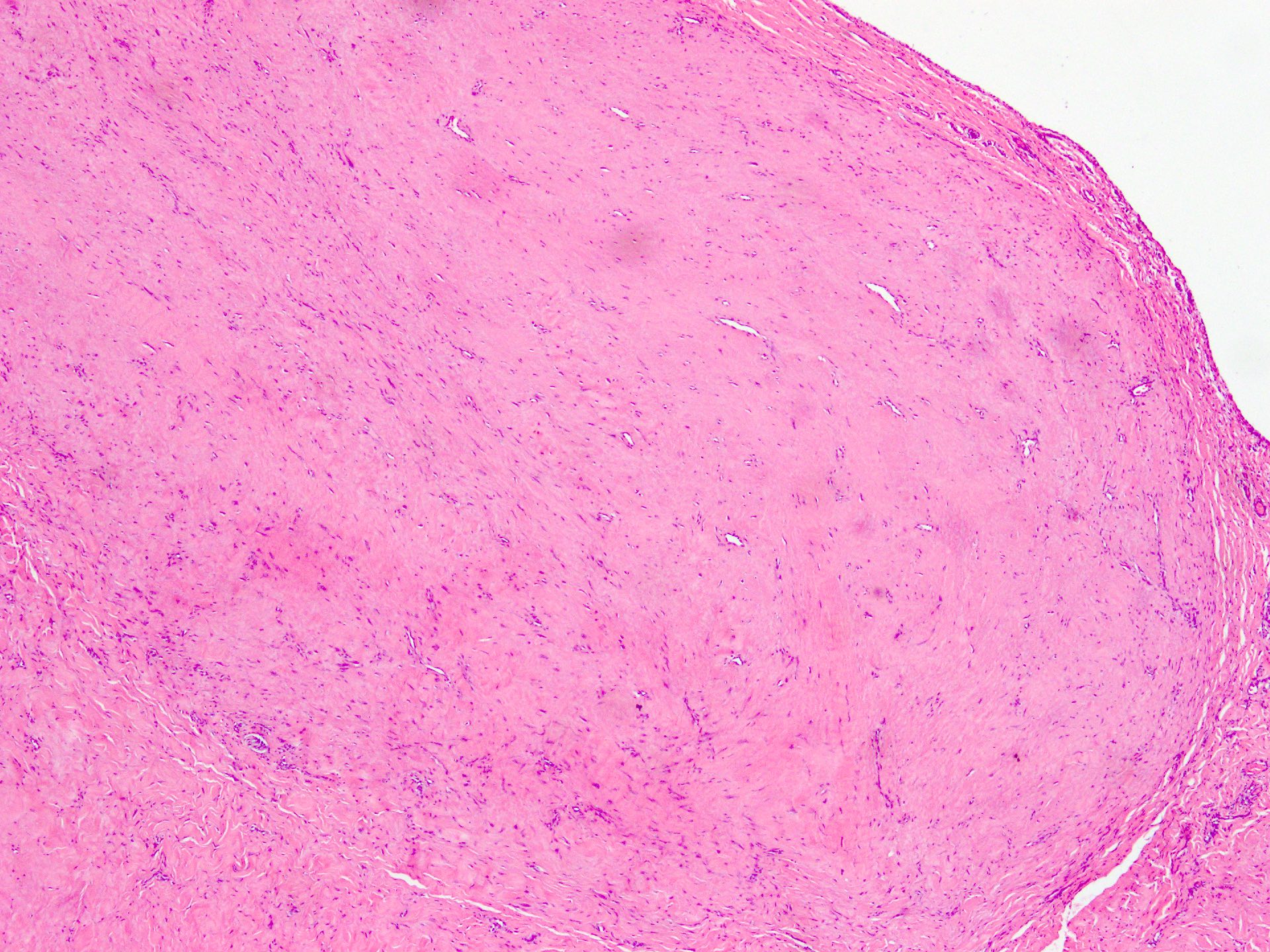
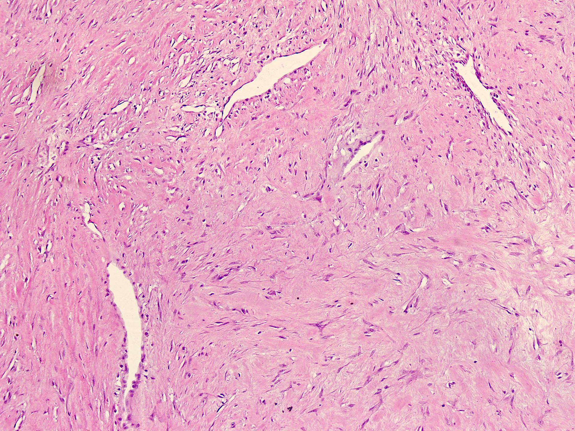
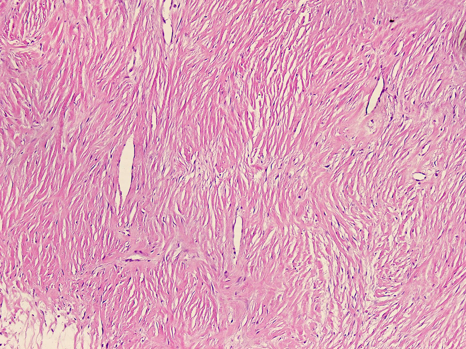
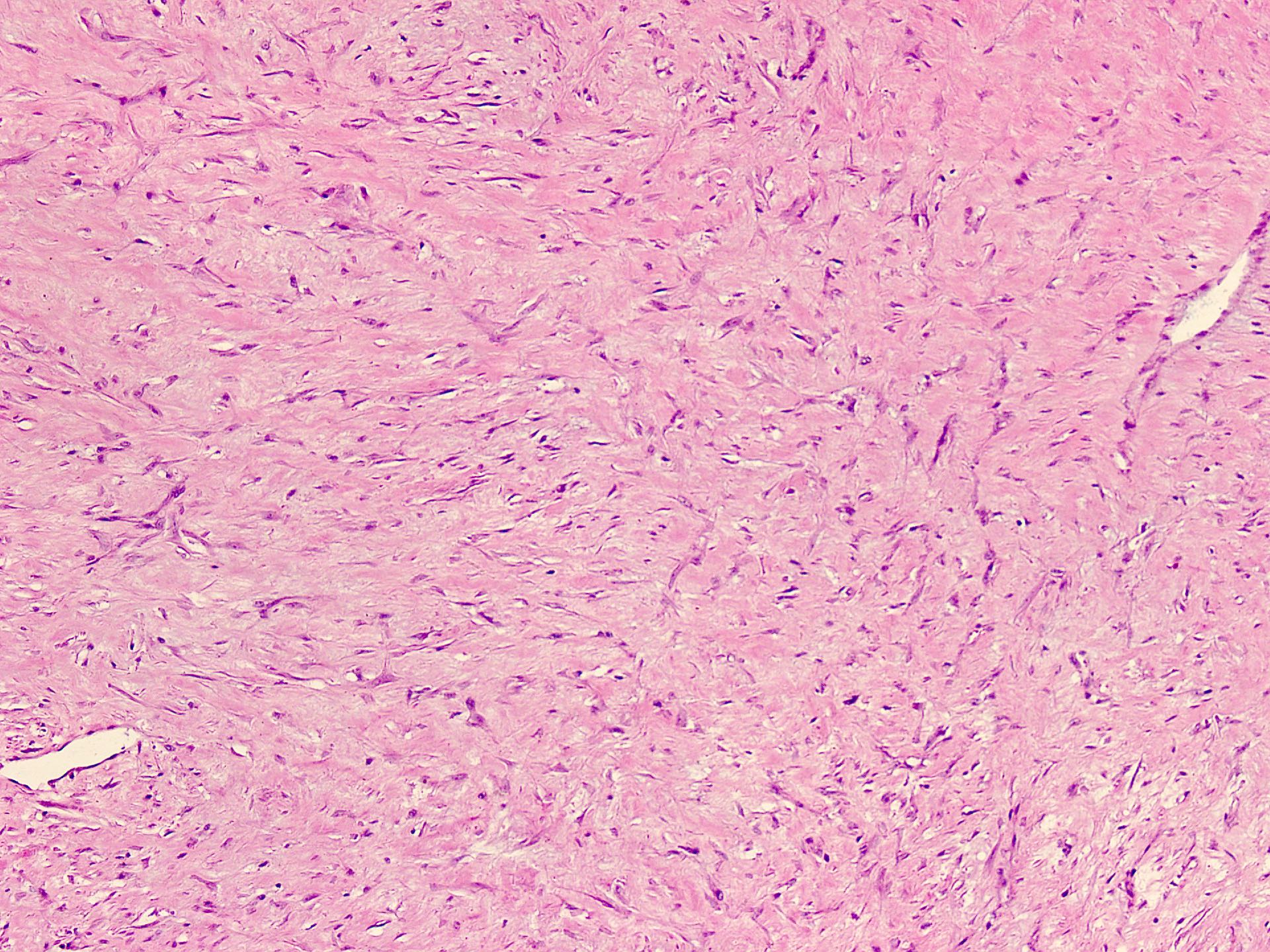
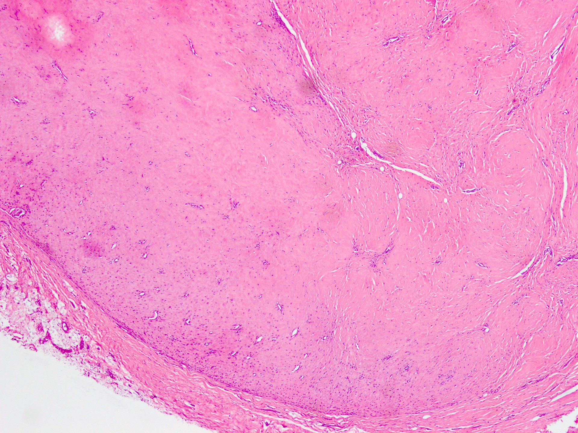
Microscopic (histologic) description
- Well circumscribed tumor of variable cellularity
- Cellularity mostly higher at tumor edges
- Bland spindle cells in a collagenous background
- Tumor has characteristic thin walled slit-like vessels
- Degenerative changes like myxoid / cystic change, osseous / chondroid metaplasia can be seen
- Bizarre pleomorphic cells can also be present
- Mitotically inactive
- Necrosis not present
- Cellular variant of fibroma of tendon sheath also exists; it overlaps morphologically with nodular fasciitis and fibrous histiocytoma (Cancer 1979;44:1945)
- Reference: Geschickter: Tumors of Bone, 1949
Microscopic (histologic) images
Contributed by Nasir Ud Din, M.B.B.S.
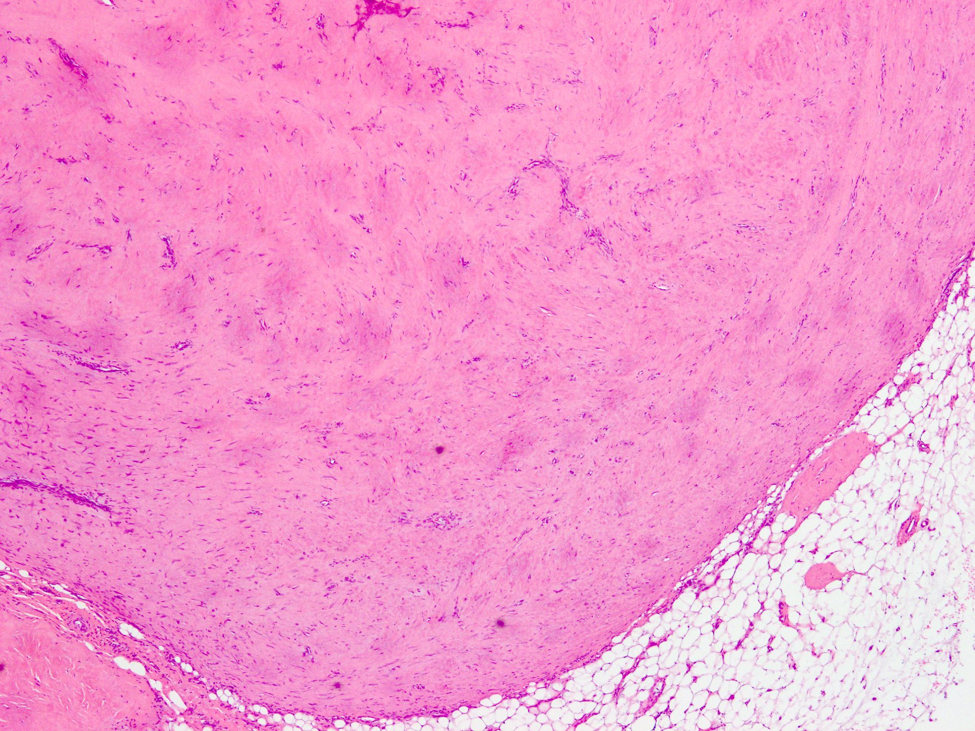
Circumscribed nodular growth
Circumscribed spindle cell nodule
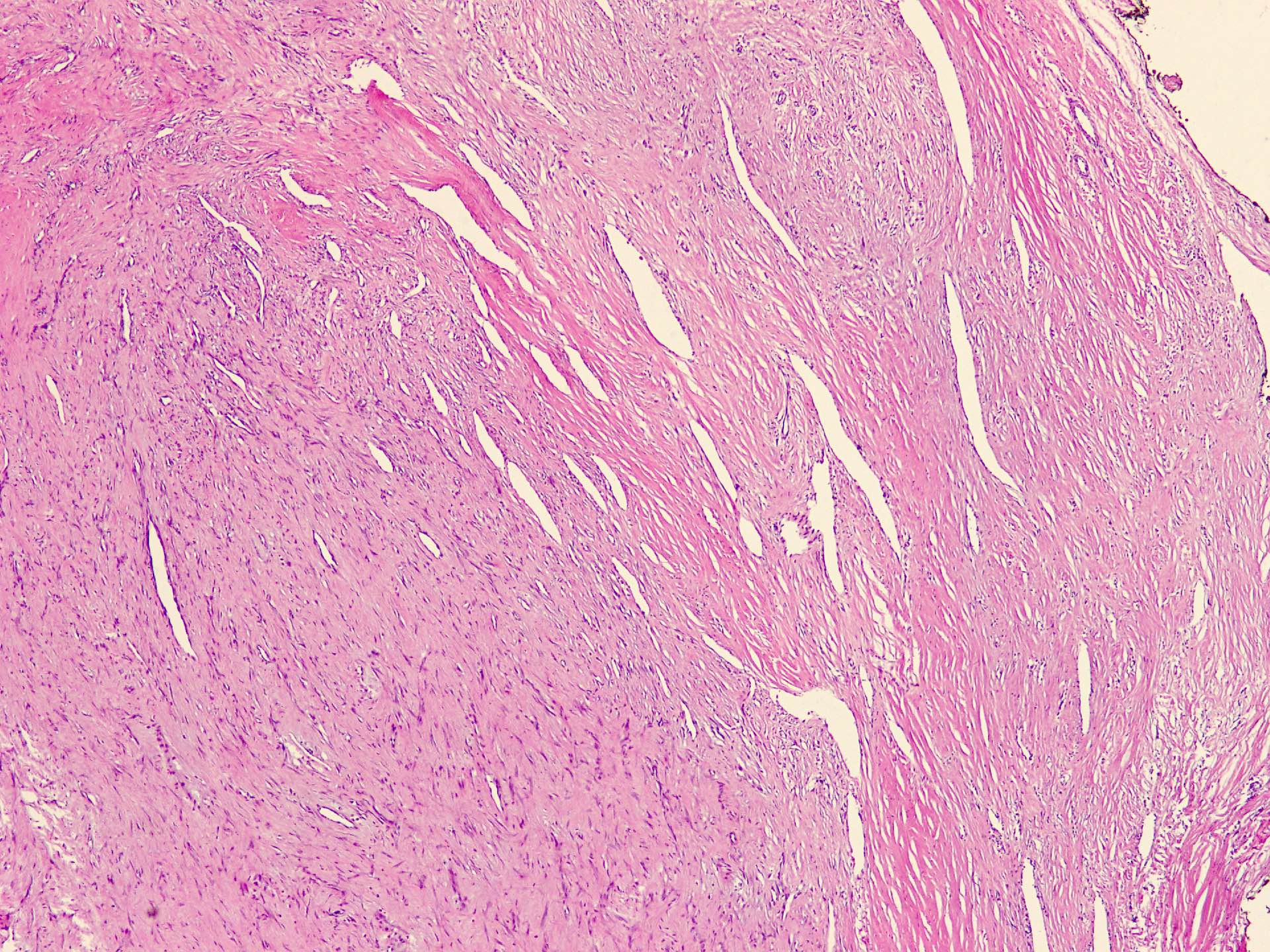
Slit-like vessels
Slit-like vascular spaces
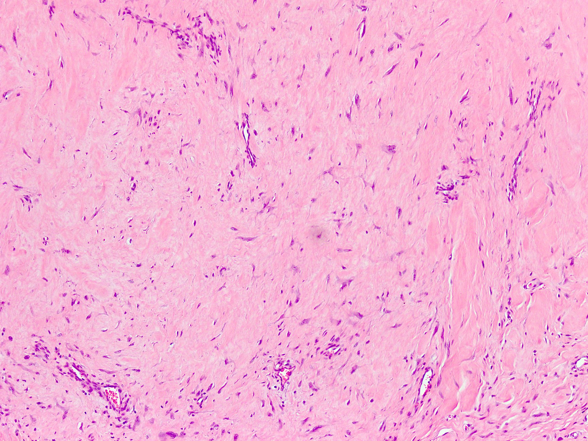
Paucicellular lesion with collagenous stroma
Markedly collagenized stroma
Spindle cell proliferation
Spindle cell lesion
AFIP images
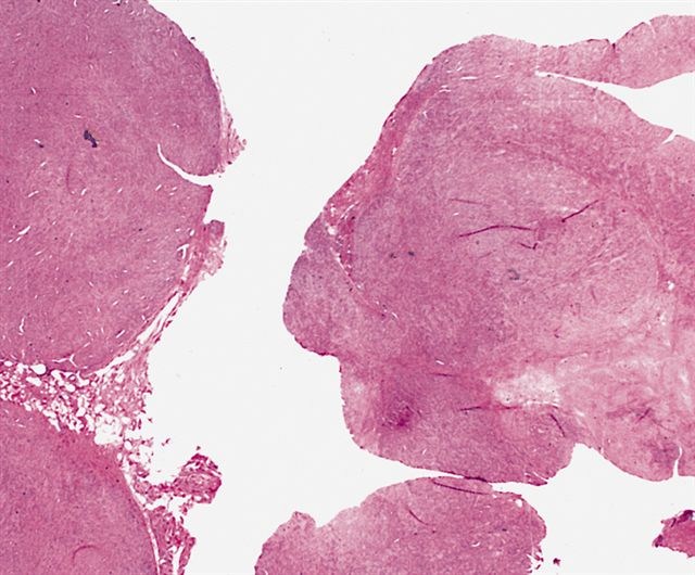
Multinodular proliferation
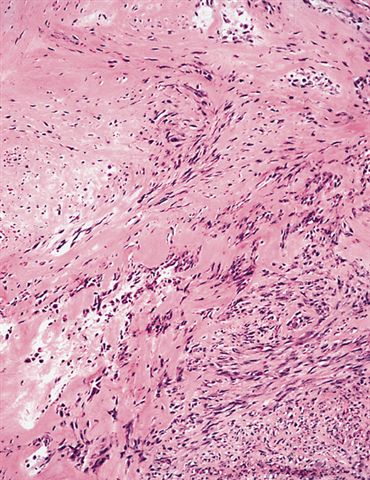
Transition from collagenous to cellular area

Fibroblasts are bland and separated by collagen

Cellular area
resembles
leiomyosarcoma
or fibrosarcoma
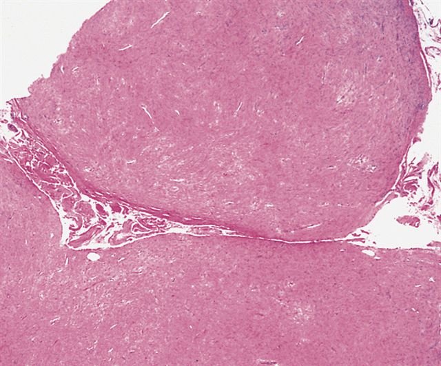
Extensive
collagenization
of nodules
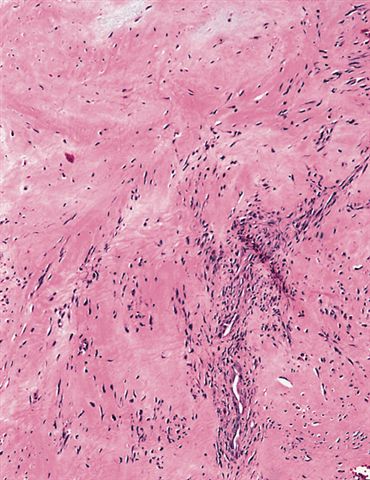
Most cases are
paucicellular
with scattered
spindled fibroblasts
Cytology description
- H&E stained slides (J Cytol 2015;32:207):
- Low cellularity
- Few loose clusters and singly dispersed bland appearing fibrotic spindle cells and stellate cells admixed with hyalinized fibrocollagenous matrix
- Necrosis and atypical mitoses not seen
Cytology images
Images hosted on other servers:

FNA
Positive stains
- May be focally positive for CD34, SMA, vimentin
- Rare cells can demonstrate calponin
- Special stain Masson trichrome highlights collagen
- References: Ann Dermatol 2019;31:110, Ophthalmic Plast Reconstr Surg 2013;29:e1, Anticancer Res 2014;34:5159
Negative stains
- CD31, CD34, CD117, beta catenin, FLI1, CD68, muscle specific actin (HHF35) and desmin
- < 1% of the cells demonstrated proliferation via Ki67
- References: Ann Dermatol 2019;31:110, Ophthalmic Plast Reconstr Surg 2013;29:e1, Anticancer Res 2014;34:5159
Electron microscopy images
Images hosted on other servers:

Organelles within a spindle cell

Densities of myofilament bundles

Cytoplasmic organelles

Spindled myofibrofibroblasts
Molecular / cytogenetics description
- t(9;11)(p24;q13-14) (Case Rep Orthop 2019;2019:3514013, Anticancer Res 2014;34:5159)
- t(4;10)(p16;q24) (Case Rep Orthop 2019;2019:3514013)
- t(2;11)(q31-32;q12) (Case Rep Orthop 2019;2019:3514013)
- USP6 gene rearrangement (Mod Pathol 1999;12:565)
- Cellular variant of fibroma of tendon sheath harbors gene rearrangements in USP6 with different partners (Mod Pathol 2021;34:13)
Sample pathology report
- Finger nodule, excision:
- Fibroma of tendon sheath (see comment)
- Comment: Histology showed a well circumscribed, variably cellular lesion composed of bland spindle cells having regular nuclei arranged in sheets and fascicles. Thin walled vessels are present. The background is collagenous.
- It is a benign condition with recurrence in 5 - 10% cases.
Differential diagnosis
- Deep benign fibrous histiocytoma:
- Involves extremities and head and neck region
- Affects wide age range (i.e. 6 - 84 years)
- Slight male predominance
- Histologically well circumscribed and cellular lesion, prominent histiocyte-like cells, foam cells, giant cells and hemosiderin
- IHC: CD34 positive
- Tenosynovial cell tumor, localized type:
- Present in hands; located in close proximity to synovium of tendon sheath or interphalangeal joint
- Age range of 30 - 35 years
- Female predominance
- Histologically cellular tumor exhibiting variable composition of mononuclear cells, multinucleated giant cells, foamy macrophages, inflammatory cells and hemosiderin
- IHC: positive for CD68, CD163 and CD45
- Inclusion body fibromatosis:
- Affects toes of children < 5 years of age
- Ill defined, paucicellular, plump spindle cells exhibiting intracytoplasmic inclusion and bland nucleus with low mitotic activity
- IHC: expresses SMA
- Recurrence rate is high
Additional references
Practice question #1
What is the most common location for fibroma of tendon sheath?
- Face
- Fingers
- Oral cavity
- Pelvis
- Toes
Practice answer #1
B. Fingers. The most common location for fibroma of tendon sheath is finger tendons (i.e. thumb, index finger and middle finger).
Comment Here
Reference: Fibroma of tendon sheath
Comment Here
Reference: Fibroma of tendon sheath
Practice question #2
A 32 year old man has had painless swelling in the palm of his hand for 6 months. It was excised and the histology is shown in the above image. What is the most likely diagnosis?
- Benign fibrous histiocytoma
- Fibroma of tendon sheath
- Nodular fasciitis
- Palmar fibromatosis
- Tenosynovial giant cell tumor, localized type
Practice answer #2
