

Soft tissue
Fibroblastic / myofibroblastic
Fibromatosis
Fibromatosis colli
Author: Komal Arora, M.D.

Last author update: 1 July 2012
Last staff update: 3 September 2021
Copyright: 2002-2024, PathologyOutlines.com, Inc.
PubMed Search: Fibromatosis colli
Table of Contents
Definition / general | Epidemiology | Treatment | Gross description | Microscopic (histologic) description | Microscopic (histologic) images | Cytology description | Positive stains | Differential diagnosisCite this page: Arora K. Fibromatosis colli. PathologyOutlines.com website. https://www.pathologyoutlines.com/topic/softtissuefibromatosiscolli.html. Accessed April 25th, 2024.
Definition / general
- Fibromatosis that appears at birth, often bilateral, affecting lower 1/3 of sternocleidomastoid muscle, causing thickened muscle
- Also called congenital torticollis (torticollis: twisting of neck causing unnatural position of head, usually caused by spasm of neck muscles, Am Fam Physician 1996;54:1965)
Epidemiology
- Associated with congenital anomalies (14% have congenital dislocations of hip, also breech deliveries)
- May be due to birth injury (breech presentation, forceps)
- Uncommon (0.4% of live births), usually diagnosed by age 6 months
- Recommended to diagnose by FNA since excision usually is not required
Treatment
- Early - stretching and physiotherapy, resolves in 70%
- Some cases require resection of affected muscle
- Does not recur
Gross description
- Tan gritty mass of muscle up to 3 cm, no hemorrhage or necrosis
Microscopic (histologic) description
- Diffuse proliferation of uniform plump fibroblasts and myofibroblasts and scar like collagen in muscle, with entrapped reactive and degenerating skeletal muscle fibers (loss of cross striations, nuclear enlargement and hypercellularity, multinucleation, atrophy)
- Surgical specimens are usually less cellular than FNA specimens because they are obtained later in time course of disease
Microscopic (histologic) images
AFIP images
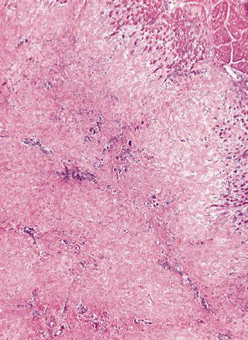
Multinodular
proliferation

Skeletal muscle
fibers are trapped
at advancing
edge of lesion
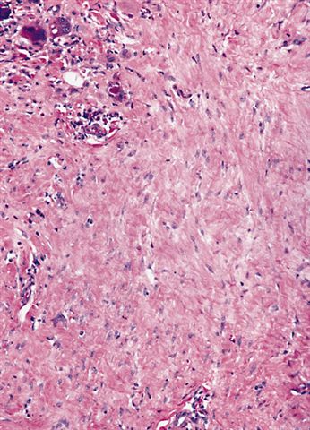
Scattered, bland
fibrocytes are widely
separated by
dense collagen
Cytology description
- Early - cellular specimen with clusters or parallel arrays of bland appearing spindle cells in fibromyxoid matrix
- Also atrophic skeletal muscle in clean background, frequent muscle giant cells, bland bare nuclei and collagen (Acta Cytol 2003;47:359)
- Usually no significant inflammation (Diagn Cytopathol 2000;23:338)
Differential diagnosis
- Fibromatosis: no muscle fibers which are replaced by fibrous tissue except at periphery, does not typically affect sternocleidomastoid muscle
- Proliferative myositis: doesn’t affect this site, stroma resembles granulation tissue and is not collagenous
- Fibrodysplasia ossificans progressiva: doesn’t affect this site, hand malformations are present, bone is present
