

Soft tissue
Fibroblastic / myofibroblastic
Proliferative myositis
Last author update: 1 June 2012
Last staff update: 31 July 2023
Copyright: 2002-2024, PathologyOutlines.com, Inc.
PubMed Search: Proliferative myositis


Table of Contents
Definition / general | Epidemiology | Sites | Pathophysiology | Etiology | Treatment | Gross description | Microscopic (histologic) description | Microscopic (histologic) images | Cytology description | Positive stains | Negative stains | Electron microscopy description | Differential diagnosisCite this page: Walsh M. Proliferative myositis. PathologyOutlines.com website. https://www.pathologyoutlines.com/topic/softtissueproliferativemyositis.html. Accessed April 20th, 2024.
Definition / general
- Infiltrative poorly demarcated intramuscular mass resembling nodular fasciitis but with large basophilic cells resembling ganglion cells; histologically almost identical to proliferative fasciitis except located in muscle
Epidemiology
- Mean age 50 years, rare in children
Sites
- Muscles of trunk, shoulder, chest or thigh
Pathophysiology
- Painless mass that grows rapidly in 1 to 6 weeks
Etiology
- May be related to a reactive process, occasional history of trauma noted
Treatment
- Conservative surgery is curative, may have spontaneous resolution (Head Neck 2007;29:416), recurrence suggests diagnostic error
Gross description
- Poorly circumscribed, scar-like induration of muscle, usually 3 - 4 cm, can occur under fascia, decreases the central portion of muscle in a wedge fashion
Microscopic (histologic) description
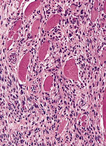
- Cellular with plump fibroblasts and myofibroblasts surrounding individual muscle fibers creating a checkerboard pattern (proliferative fibroblasts alternating with atrophic muscle)
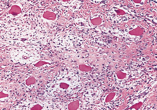
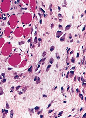
- Also large ganglion-like cells with abundant amphophilic to basophilic cytoplasm, vesicular nuclei and prominent nucleoli
- Stroma is collagenous or myxoid
- Variable mitotic figures but no atypical ones
- Ill defined margins
- May have metaplastic bone
Microscopic (histologic) images
AFIP images
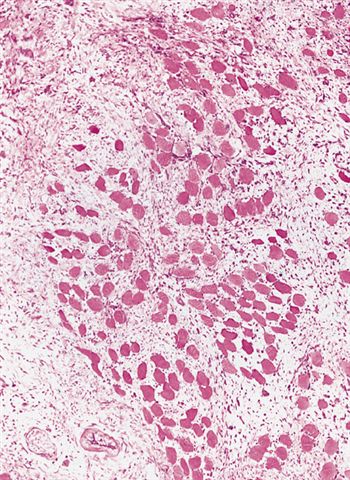
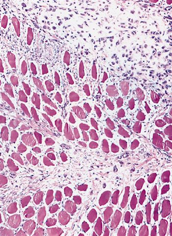
Characteristic checkerboard pattern
Metaplastic bone
Large ganglion cells
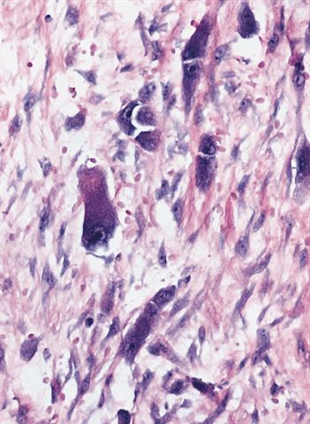
Abundant amphophilic cytoplasm

Spindle cell sarcoma
Images hosted on other servers:


Ganglion cells
Cytology description
- Loose clusters of uniform fibroblast-like spindle cells and large, ganglion-like cells with eccentric nuclei, prominent nucleoli and abundant cytoplasm (Acta Cytol 1995;39:535)
- Cytologic diagnosis not recommended due to limited sampling
Positive stains
Negative stains
Electron microscopy description
- Fibroblasts and myofibroblasts, ganglion-like cells are fibroblasts or myofibroblasts with abundant dilated rough endoplasmic reticulum but no neuronal characteristics (Am J Surg Pathol 1991;15:654)
Differential diagnosis
- Desmoid fibromatosis:
- 3 cm or larger, completely replaces muscle
- Spindle cells organized into broad sweeping fascicles, stroma is collagenous, skeletal muscle at periphery is often entrapped
- No ganglion type cells
- Ganglioneuroblastoma:
- S100+
- Similar histology but lacks the fibrillary background
- Nodular fasciitis:
- Completely obliterates muscle when extends deeper than fascia
- No or few ganglion type cells
- Proliferative fasciitis:
- Almost identical except subcutaneous rather than intramuscular location
- Rhabdomyosarcoma:
- Desmin+, myogenin+
- Ganglion-like cells of proliferative myositis lack cross striations and are more basophilic than rhabdomyoblasts
- Sarcoma:
- Large mass, marked atypia, atypical mitoses, necrosis
