
Table of Contents
Foamy macrophages | Follicular hyperplasia | Hypersplenism | Infarction | PerisplenitisCite this page: Mansouri J, Lynch D. Common nonspecific abnormal features. PathologyOutlines.com website. https://www.pathologyoutlines.com/topic/spleennonspecificabnormalfeatures.html. Accessed April 18th, 2024.
Foamy macrophages
Definition / general
Essential features
Terminology
Case reports
Microscopic (histologic) description
Microscopic (histologic) images
Contributed by David Lynch, M.D.
Positive stains
Negative stains
Differential diagnosis
Additional references
- Increased foamy histiocytes / macrophages, typically in the red pulp
Essential features
- Associated with a wide variety of conditions both benign and malignant
- Conditions with high cell turnover may produce foamy histiocytes
- Diagnosis typically requires clinicopathologic correlation
Terminology
- Ceroid histiocytosis - histiocytes filled with phospholipids; not specific to a single disease entity
Case reports
- 7 year old boy diagnosed with Gaucher disease by FNA of spleen (J Clin Diagn Res 2016;10:ED13)
- 52 year old man with splenomegaly (BMJ Case Rep 2015 Feb 5;2015)
Microscopic (histologic) description
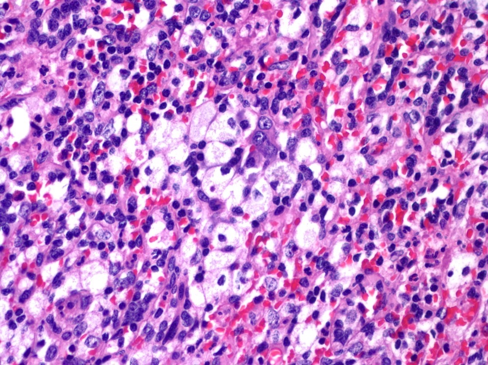
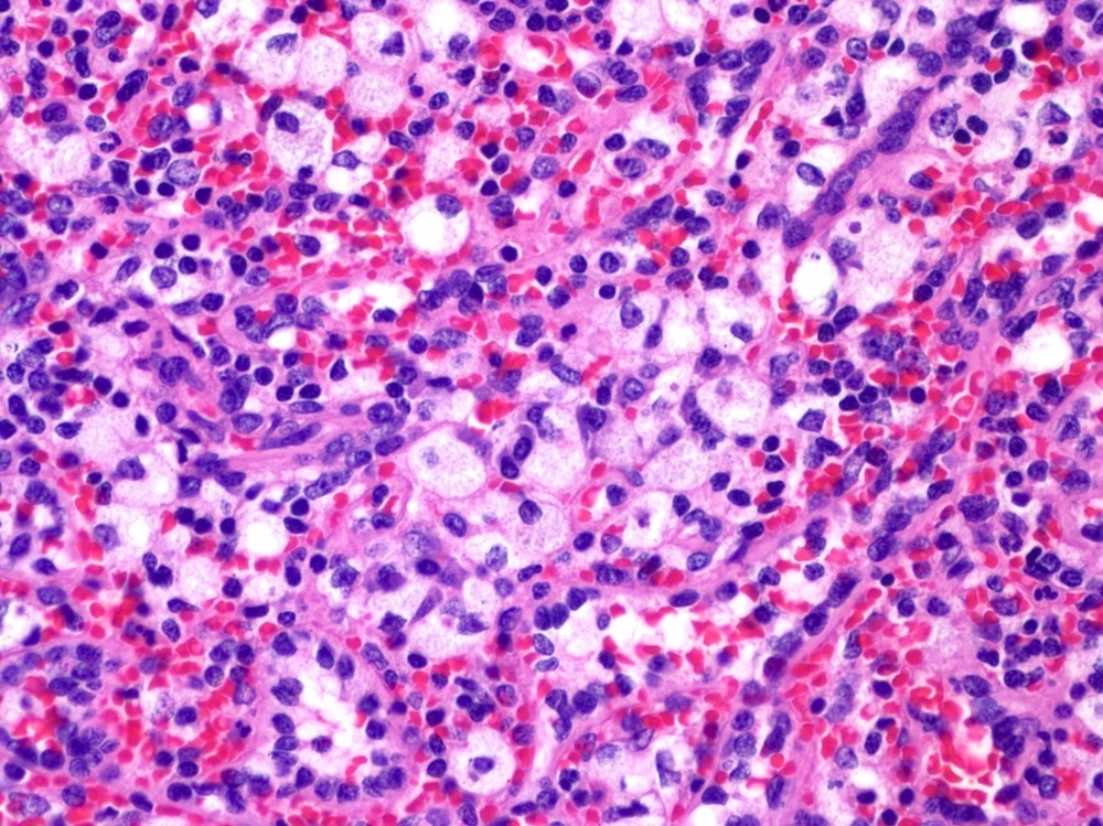
- Red pulp expanded by histiocytes with abundant foamy cytoplasm
- Histiocytes scattered without forming a discrete mass
- May have basophilic cytoplasm from ingestion of platelets in idiopathic thrombocytopenic purpura (ITP)
- Finely fibrillary cytoplasm suggests underlying storage disease
- White pulp is normal in size
Microscopic (histologic) images
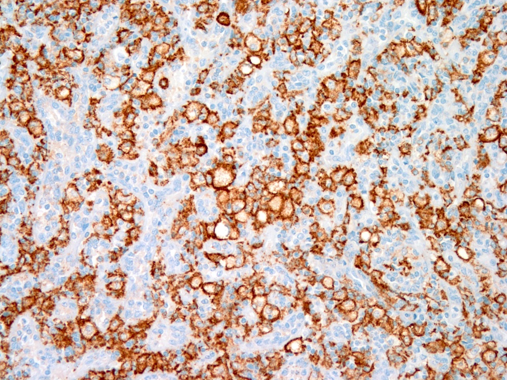
Contributed by David Lynch, M.D.
Foamy histiocytes
CD163
Positive stains
Negative stains
Differential diagnosis
- Autoimmune disease
- Chronic myelogenous leukemia: extramedullary hematopoiesis with hypolobated megakaryocytes, foamy macrophages
- Crystal storing histiocytosis: eosinophilic rhomboid crystals within histiocytes
- Hemophagocytic lymphohistiocytosis: histiocytes have ingested RBCs
- Hyperlipidemia
- Idiopathic thrombocytopenic purpura: follicular hyperplasia, increased foamy histiocytes, increased plasma cells, lipogranulomas in some cases
- Langerhans cell histiocytosis: cleaved nuclei; S100, CD1a and langerin are positive
- Mycobacterial or fungal infections: positive AFB or GMS stains
- Postchemotherapy histiocyte rich pseudotumor (Am J Clin Pathol 2009;132:342)
- Rosai-Dorfman disease: emperipolesis is present; histiocytes are S100 positive
- Storage disease (Gaucher, Niemann-Pick): histiocytes have fibrillary cytoplasm in Gaucher disease
Additional references
Follicular hyperplasia
Definition / general
Gross description
Microscopic (histologic) description
- Also called reactive follicular hyperplasia
- Focal or diffuse
- Normal in children
- In adults, due to systemic infection (malaria, measles, typhoid fever, virus, other), immune mediate disorders (hemolytic anemia, immune thrombocytopenic purpura, rheumatoid arthritis)
- Often associated with congestion and plasmacytic proliferation
- Associated with hypersplenism in Zaire, Nigeria and New Guinea, where spleens also show extramedullary hematopoiesis and marked sinusoidal dilation; may be related to malaria
- Note: graft rejection and AIDS are associated with reactive nonfollicular hyperplasia, which may resemble lymphoma but has heterogeneous lymphocytic population without atypia and without clonality
- Felty syndrome (rheumatoid arthritis): no granulocytic phagocytosis but expansion of red pulp cords and sinuses with macrophages
Gross description
- May have enlarged spleen with multiple small, pale tan nodules or solitary large nodules resembling lymphoma (Am J Surg Pathol 1983;7:373)
Microscopic (histologic) description
- Resemble nodal reactive follicles, with mixed follicular center population and tingible body macrophages
- Usually mature lymphocytes and plasma cells in red pulp
Hypersplenism
- Also called dysplenism
- Enlarged spleen leads to removal of cellular blood components (some or all), causing thrombocytopenia, neutropenia, hemolytic anemia or pancytopenia
- Due to disorders (congestive splenomegaly, Gaucher disease, hamartoma, hemangioma, Langerhans cell histiocytosis, leukemia / lymphoma, other conditions diffusely involving red pulp) causing widening of splenic cords with increase in macrophages or connective tissue, causing premature destruction of normal blood components
- Reactive follicular hyperplasia of white pulp may be present in hypersplenism associated with cytopenias
- Infectious causes include brucellosis, CMV, Echinococcus, histoplasmosis, infectious mononucleosis, leishmaniasis, malaria, schistosomiasis, syphilis, toxoplasmosis, trypanosomiasis, tuberculosis, typhoid
- May be due to abnormal cellular blood components (hereditary spherocytosis)
Infarction
Definition / general
Gross description
- Due to thrombosis of splenic vein, usually secondary to cardiac emboli
- Also associated with granulomatosis with polyangiitis (Wegener) (may cause splenic rupture), massive splenomegaly, idiopathic
Gross description
- Wedge shaped white-gray infarct involving capsule; infarcts heal as large, depressed scars
Perisplenitis
- Thick fibrous plaques coating splenic surface
- Incidental finding at autopsy or associated with portal hypertension
