
Stains & CD markers
SOX10

Copyright: 2014-2024, PathologyOutlines.com, Inc.
PubMed Search: SOX10
SOX10
Author: Pamela Wirth, Ph.D.
Editorial Board Members: Christian M. Schürch, M.D., Ph.D., Brandon Umphress, M.D.
Last author update: 27 February 2024
Last staff update: 27 February 2024
Copyright: 2014-2024, PathologyOutlines.com, Inc.
PubMed Search: SOX10

Table of Contents
Definition / general | Essential features | Terminology | Pathophysiology | Clinical features | Interpretation | Uses by pathologists | Prognostic factors | Microscopic (histologic) description | Microscopic (histologic) images | Positive staining - normal | Positive staining - disease | Negative staining | Molecular / cytogenetics description | Sample pathology report | Board review style question #1 | Board review style answer #1 | Board review style question #2 | Board review style answer #2Cite this page: Wirth P. SOX10. PathologyOutlines.com website. https://www.pathologyoutlines.com/topic/stainsSOX10.html. Accessed April 23rd, 2024.
Definition / general
- SRY related HMG box 10 (SOX10) protein
- Transcription factor known to be crucial in the specification of the neural crest and maintenance of Schwann cells and melanocytes
- Expressed in nuclei of melanocytes, peripheral nerves, breast myoepithelial cells, luminal cells of sweat and salivary glands (Am J Surg Pathol 2015;39:826)
Essential features
- Positive marker for most melanomas (Am J Surg Pathol 2015;39:826)
- Also expressed in benign nevi and benign tumors of Schwann cell lineage
- May be more sensitive than S100 for identifying metastatic melanoma (Cancer Cytopathol 2018;126:179)
- Encoded by SOX10 gene located on chromosome 22q13.1
- May be a useful marker for primary and metastatic triple negative breast cancer (TNBC) (Mod Pathol 2021;34:62)
Terminology
- SRY box 10
- SRY (sex determining region Y) box 10
- SRY related HMG box gene 10
Pathophysiology
- Encoded by the SOX10 gene on the long arm of chromosome 22 at position 22q13.1
- Important in the development of cells including neural crest cells, glial cells, neurons, osteoblasts, smooth muscle cells and melanocytes (J Otol 2022;17:247)
- Mutations in the SOX10 gene are associated with pigment abnormalities, disorders of gastrointestinal motility, loss of smell and severe hearing loss
Clinical features
- Mutations in SOX10 gene cause Waardenburg syndrome type 4 (auditory pigmentary abnormalities and Hirschsprung disease) (Gene 2014;538:36)
- Mutations within SOX10 enhancers may contribute to isolated Hirschsprung disease (Hum Mutat 2014;35:303)
- Mutations cause Kallman syndrome (anosmia and hypogonadotropic hypogonadism) with deafness (Am J Hum Genet 2013;92:707)
Interpretation
- Strong, distinct nuclear stain
Uses by pathologists
- Melanoma marker (Appl Immunohistochem Mol Morphol 2014;22:142)
- Nevi are positive; desmoplastic melanomas are often SOX10 reactive with some cases being only focally positive (Nat Cell Biol 2012;14:882, Appl Immunohistochem Mol Morphol 2013;21:506)
- SOX10 shows an increased specificity for soft tissue tumors of neural crest origin compared with S100 (Appl Immunohistochem Mol Morphol 2012;20:445)
- MITF1 and SOX10 can be used to differentiate melanoma in situ from actinic keratosis with melanocytic hyperplasia (Am J Dermatopathol 2014;36:124)
- SOX10 expression can serve as a marker for TNBC in conjunction with GATA3
- 58 - 74% of TNBC cases are positive for SOX10 (Mod Pathol 2021;34:62, Transl Cancer Res 2020;9:5603)
- SOX10 is useful for capturing GATA3 negative metastases (Hum Pathol 2019:85;221)
Prognostic factors
- SOX10 can help differentiate melanoma from other types of skin cancers
- Strong and widespread staining may indicate a more aggressive disease (Onco Targets Ther 2016;9:1671, PLoS One 2018;13:e0190834)
- Higher SOX10 expression may be associated with more aggressive tumor characteristics (late stage, higher grade and increased lymph node involvement) in TNBC (Transl Cancer Res 2020;9:5603)
Microscopic (histologic) description
- Normal finding: nuclear expression in some normal cells like melanocytes, peripheral nerves, breast myoepithelial cells, luminal cells of sweat and salivary glands (see Positive staining - normal)
- Nuclear expression in malignant melanoma and soft tissue tumors
- Any other pattern could represent abnormality or background staining and should be further assessed (some cytoplasmic staining has been noted with certain polyclonal antibodies) (Nowotwory J Oncol 2019;69:58)
Microscopic (histologic) images
Contributed by Pamela Wirth, Ph.D. (source: University of Toronto)
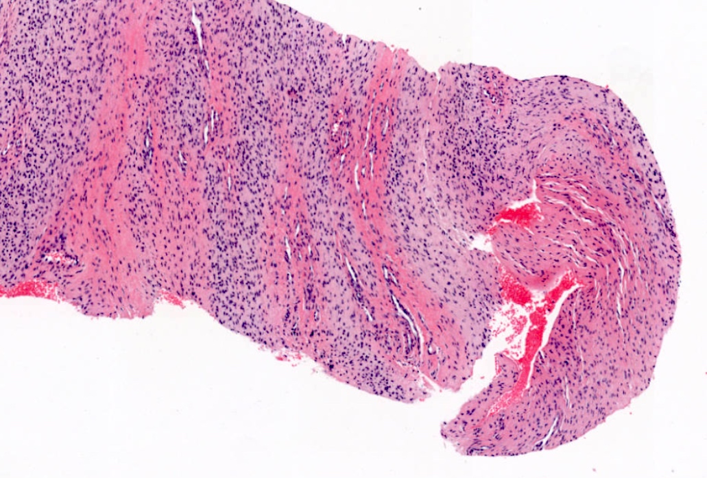
Nerve sheath tumor

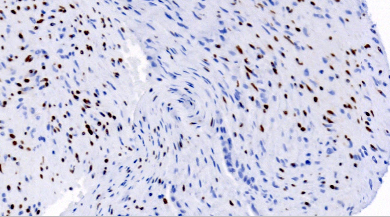
Nerve sheath tumor, SOX10
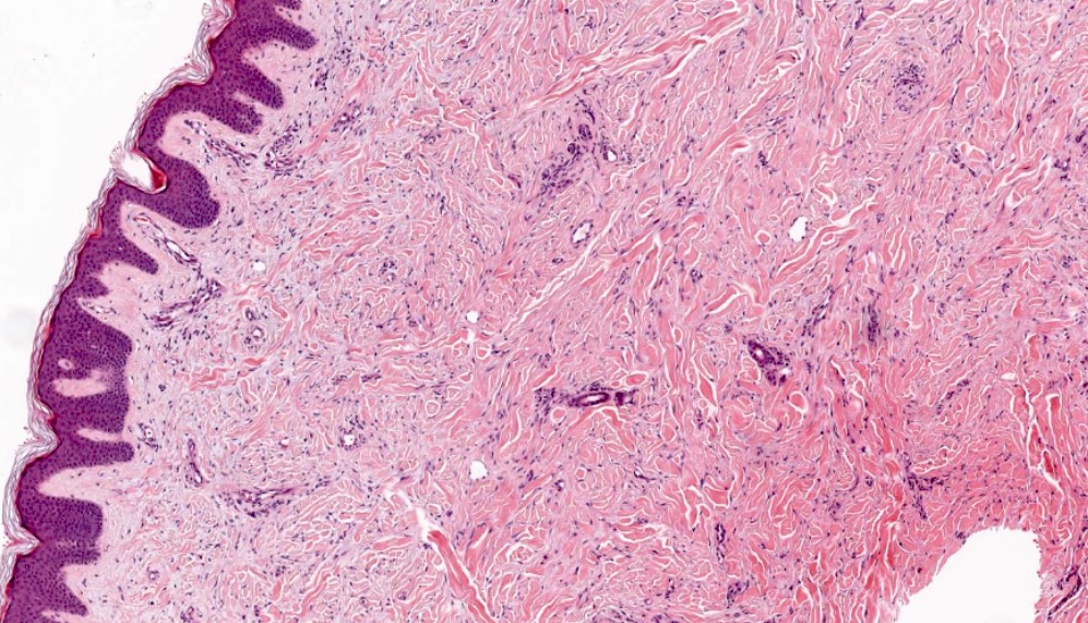
Desmoplastic melanoma
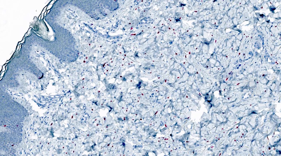
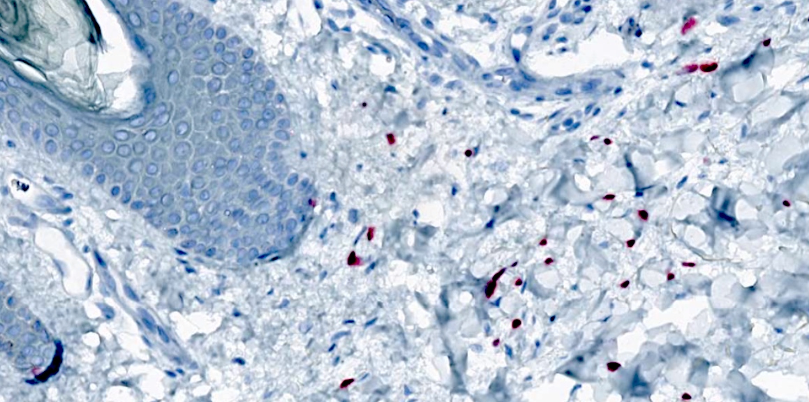
Desmoplastic melanoma, SOX10
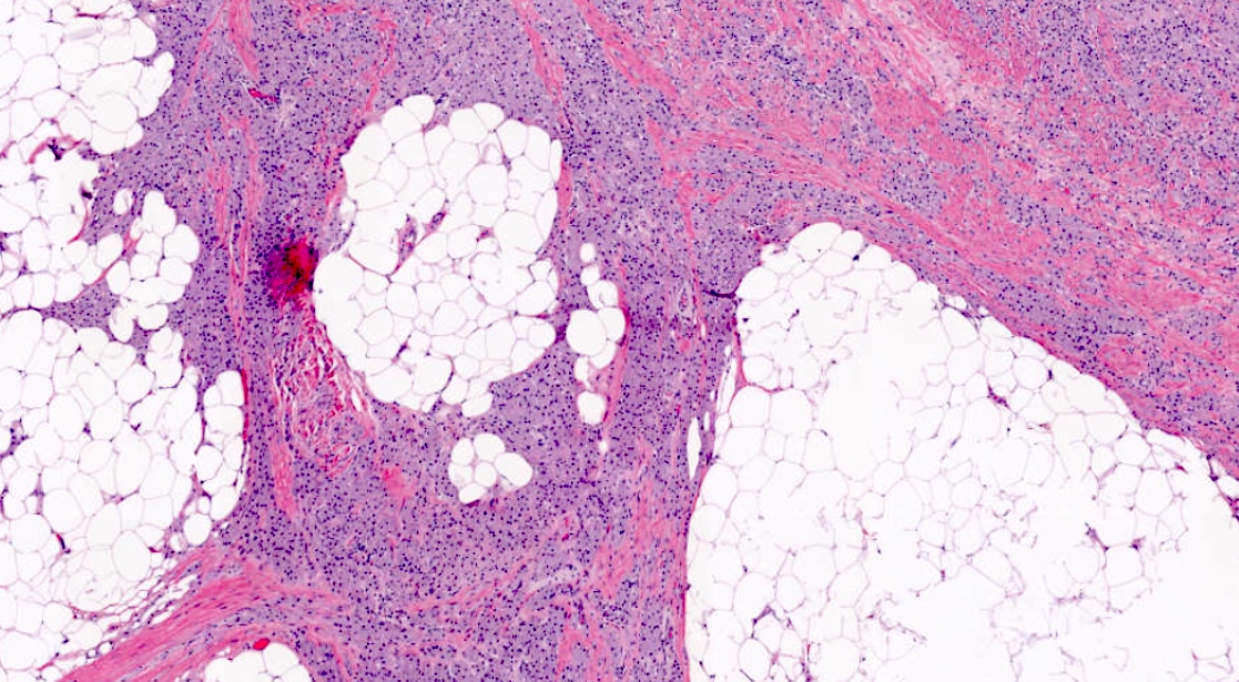
Soft tissue granular cell tumor
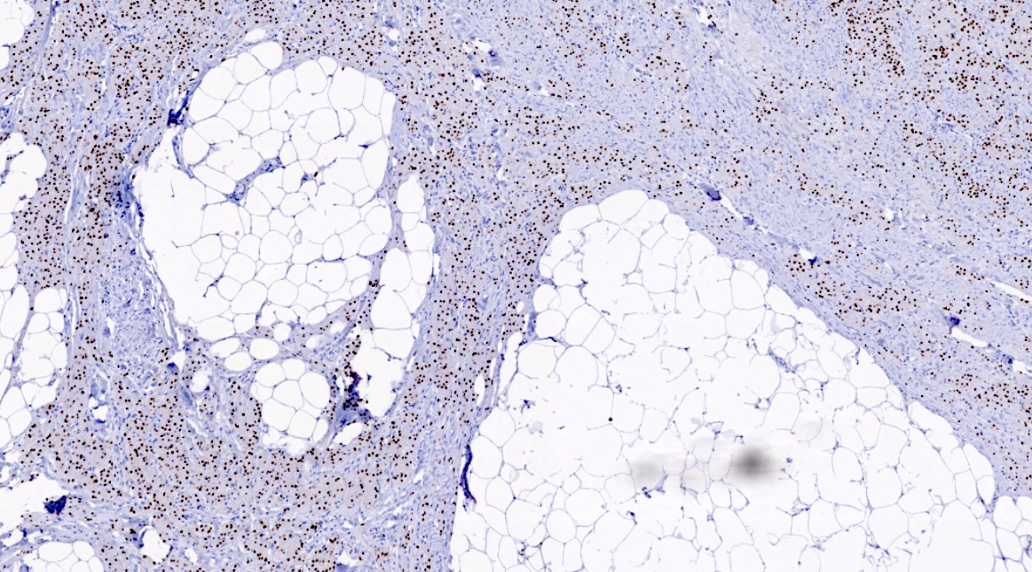
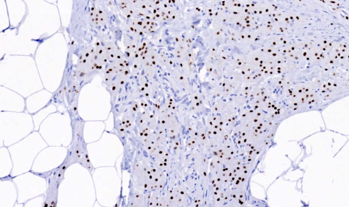
Soft tissue granular cell tumor, SOX10
Positive staining - normal
- Epidermal melanocytes, peripheral nerves, Schwann cells, neurofibromas and myoepithelial cells of the salivary and mammary glands (Am J Surg Pathol 2015;39:826)
- In major salivary glands, expressed in nuclei of acini and both luminal and abluminal cells of intercalated ducts but not in other sites (Mod Pathol 2013;26:1041)
Positive staining - disease
- Melanoma: almost all spindle cell melanomas and most desmoplastic melanomas (Actas Dermosifiliogr 2017;108:17)
- SOX10 has demonstrated positive staining in spiradenomas, cylindromas, hidradenoma papilliferum, syringocystadenoma papilliferum and apocrine adenomas (J Cutan Pathol 2017;44:544)
- SOX10+ carcinomas include acinic cell, adenoid cystic, epithelial myoepithelial, myoepithelial; also pleomorphic adenomas, including the pleomorphic adenoma component of carcinoma (Br J Cancer 2013;109:444)
- Soft tissue tumors including Schwannoma, neurofibroma, granular cell tumor, malignant peripheral nerve sheath tumor (Am J Surg Pathol 2015;39:826)
- Usually positive nuclear staining in breast basal-like / unclassified triple negative / metaplastic carcinoma (Hum Pathol 2013;44:959)
- Specific (93%) but not very sensitive (67%) for malignant peripheral nerve sheath tumor over synovial sarcoma (Mod Pathol 2014;27:55)
- Must interpret with caution in intraneural tumors
Negative staining
- Fibrohistiocytic and histiocytic proliferations (J Am Acad Dermatol 2012;67:717)
- Normal squamous, glandular and parenchymal epithelia (Am J Surg Pathol 2015;39:826)
- Lymphoid tissue and histiocytes (Am J Surg Pathol 2015;39:826)
- Usually negative / rarely positive staining in breast luminal A / B or HER2+ carcinoma (5% positive), ER- / PR- ductal carcinoma in situ (DCIS) (4% positive), phyllodes tumor, fibroadenoma
Molecular / cytogenetics description
- Mutations in SOX10 gene can lead to genetic disorders such as Waardenburg syndrome types II and IV and Hirschsprung disease (Am J Hum Genet 2007;81:1169)
- SOX10 gene deletions (15%) found in Waardenburg syndrome type II in one study of 30 patients
- PCR is more commonly used to screen for SOX10 gene deletions
- Some Waardenburg syndrome type IV cases involve multiple mutations and are not easily characterized by SOX10 mutations alone
Sample pathology report
- Skin biopsy, scalp:
- Spindle cell melanoma (see comment)
- Comment: Microscopic examination of the biopsy revealed atypical spindle cells along with areas of pleomorphism and high mitotic activity. Diagnosis of spindle cell melanoma was confirmed with an immunostaining panel that included S100 (positive), SOX10 (positive), AE1 / AE3 (negative) and p63 (negative) to confirm the diagnosis of spindle cell melanoma versus spindle cell squamous cell carcinoma.
Board review style question #1
SOX10 can be used to help diagnose which of the following conditions?
- Benign nevi versus melanoma
- HER2 positive breast tumors
- Soft tissue tumors of neural crest origin
- Squamous cell carcinomas of the skin
Board review style answer #1
C. Soft tissue tumors of neural crest origin. SOX10 is commonly expressed in peripheral nerves and melanomas with nerve cell involvement including desmoplastic melanoma. Answer A is incorrect because both benign nevi and melanomas can express SOX10, making it an insensitive marker for distinguishing between benign neoplasms and malignant melanomas. Answer B is incorrect because SOX10 is typically negative or expressed in very low levels in HER2 positive breast tissue. Answer D is incorrect because SOX10 expression is minimal to absent in squamous cell carcinoma.
Comment Here
Reference: SOX10
Comment Here
Reference: SOX10
Board review style question #2
A biopsy from an 86 year old man reveals a lesion with atypical spindle cell melanocytes infiltrating the dermis. IHC stain results were positive for S100 and SOX10 (shown) and negative for HMB45 and MelanA. What is the likely diagnosis?
- Cutaneous melanoma
- Dermatofibrosarcoma protuberans
- Desmoplastic melanoma
- Spindle cell squamous cell carcinoma
Board review style answer #2
C. Desmoplastic melanoma. The absence of HMB45 and MelanA staining is characteristic of desmoplastic melanomas in addition to positive staining for SOX10 and S100. Answer A is incorrect because melanomas are typically HMB45 positive. Answer D is incorrect as spindle cell squamous cell carcinomas are typically negative for S100 and HMB45. Answer B is incorrect because dermatofibrosarcoma protuberans is typically negative for SOX10, S100 and HMB45.
Comment Here
Reference: SOX10
Comment Here
Reference: SOX10

