Stains & CD markers
TRPS1


Copyright: 2022, PathologyOutlines.com, Inc.
PubMed Search: TRPS1
TRPS1
Editorial Board Member: Christian M. Schürch, M.D., Ph.D.
Deputy Editor-in-Chief: Gary Tozbikian, M.D.
Last author update: 9 August 2022
Last staff update: 10 August 2022
Copyright: 2022, PathologyOutlines.com, Inc.
PubMed Search: TRPS1
Table of Contents
Definition / general | Essential features | Terminology | Pathophysiology | Diagrams / tables | Interpretation | Uses by pathologists | Microscopic (histologic) images | Positive staining - normal | Positive staining - disease | Negative staining | Sample pathology report | Practice question #1 | Practice answer #1Cite this page: Ai D, Ding Q. TRPS1. PathologyOutlines.com website. https://www.pathologyoutlines.com/topic/stainstrps1.html. Accessed September 18th, 2025.
Definition / general
- TRPS1 (trichorhinophalangeal syndrome type 1) - a GATA family of zinc finger transcription factor
- Involved in breast cancer carcinogenesis and required for breast cancer cell survival
- Plays a critical role in the development of cartilage, bone and hair follicle
Essential features
- Involved in breast cancer carcinogenesis and required for breast cancer cell survival
- Strong nuclear staining
- Positive in invasive breast carcinoma (> 95%), including ER+ / HER2- (> 95%), HER2+ (> 90%), ER- / HER2- (> 90%)
- Negative in urothelial carcinoma, lung adenocarcinoma and thyroid carcinoma
Terminology
- This protein is also called zinc finger protein GC79
Pathophysiology
- Plays a critical role in the development of cartilage, bone and hair follicle; loss of the TRPS1 gene leads to trichorhinophalangeal syndrome type 1 with defects in hair, facial and bone / joint malformations
- Functions in the growth and differentiation of normal mammary epithelial cells; it is also involved in the development of breast cancer and is required for breast cancer cell survival (Cell Rep 2018;25:1255)
Diagrams / tables
Images hosted on other servers:

TRPS1 regulatory network
Interpretation
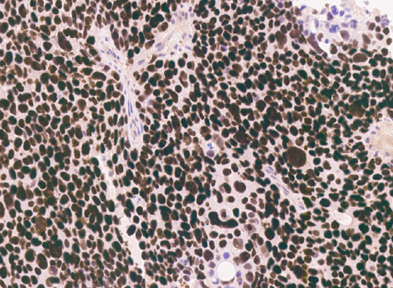
- Nuclear staining (majority of cases shows diffuse and strong nuclear stain)
Uses by pathologists
- Diagnosing ER positive breast cancer (> 95%), HER2 positive breast cancer (> 90%), triple negative breast cancer (> 90%, including metaplastic and nonmetaplastic triple negative breast cancer) (Mod Pathol 2021;34:710, Am J Surg Pathol 2022;46:415, Hum Pathol 2022;125:97)
- In a panel along with pancytokeratin, ER and GATA3 to diagnose metastatic carcinoma of breast origin (Hum Pathol 2022 Apr 14 [Epub ahead of print])
- Differentiating GATA3 positive invasive breast cancer from GATA3 positive urothelial carcinoma
- Differentiating invasive breast carcinoma from adenocarcinomas of nonbreast origin (such as adenocarcinoma from lung, GI, pancreas, etc.)
- Differentiating triple negative breast cancer from melanoma
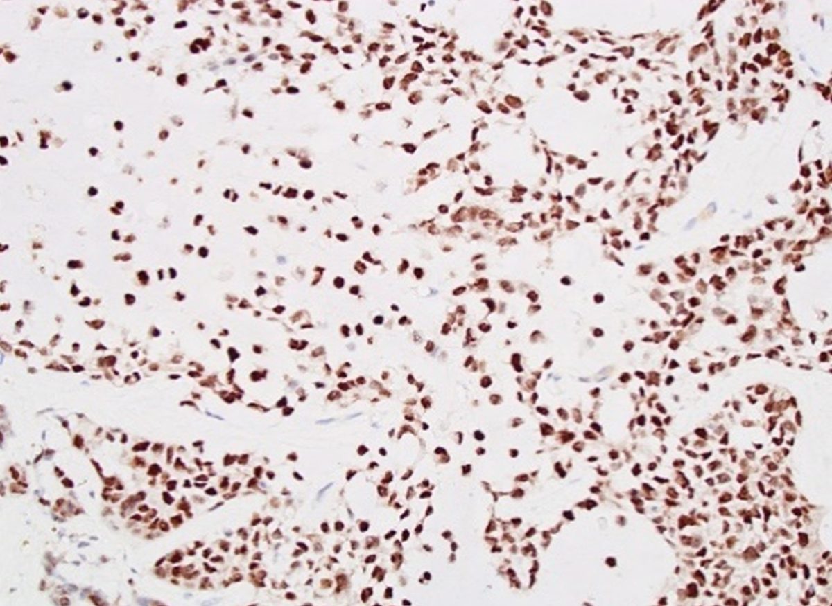
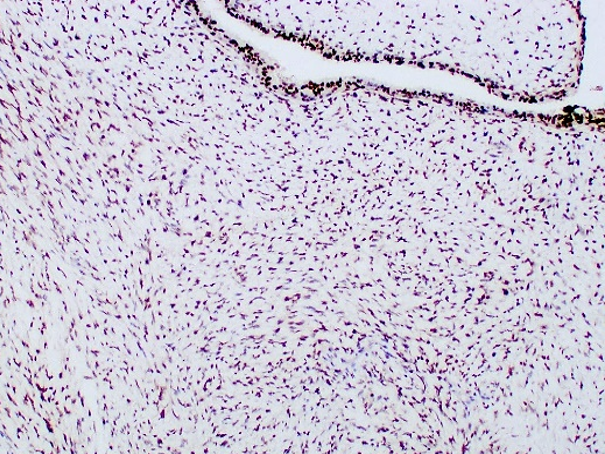
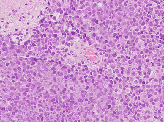
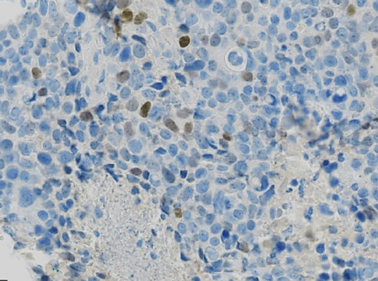
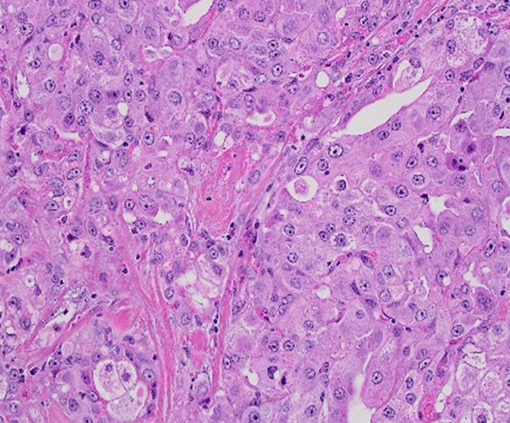
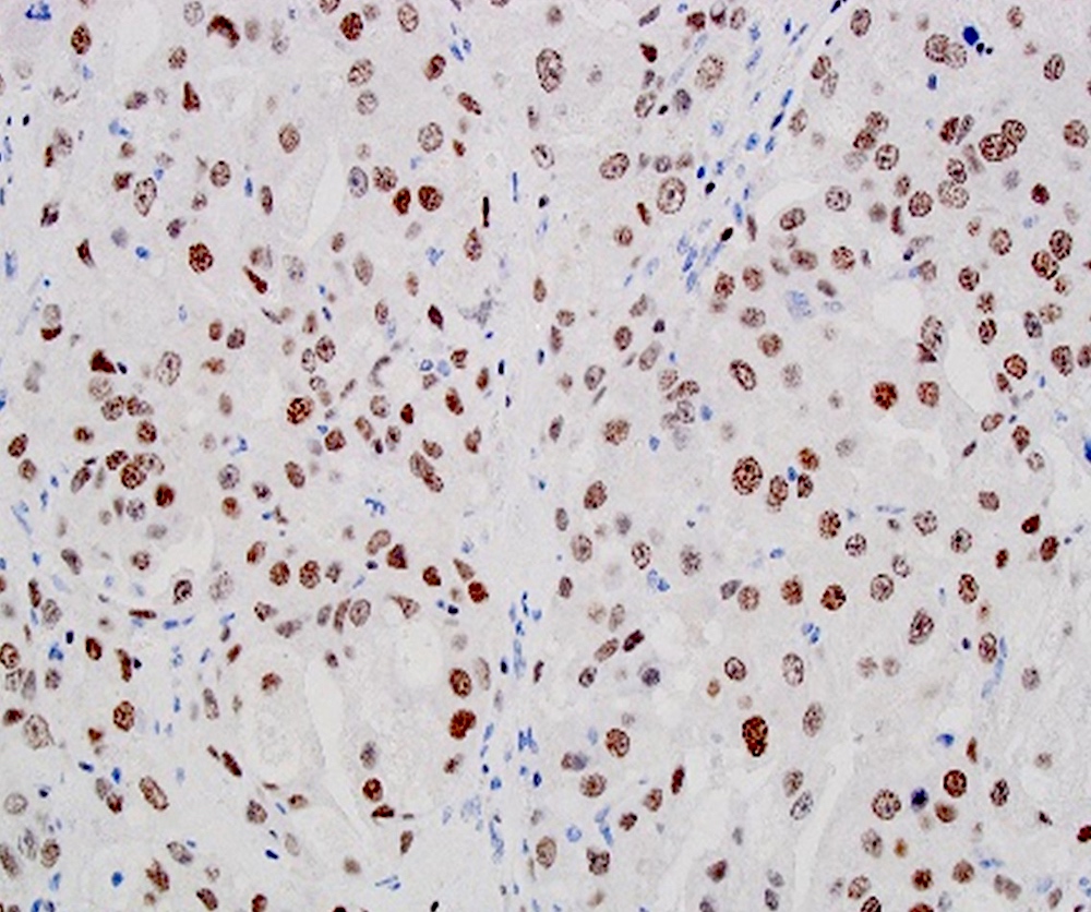
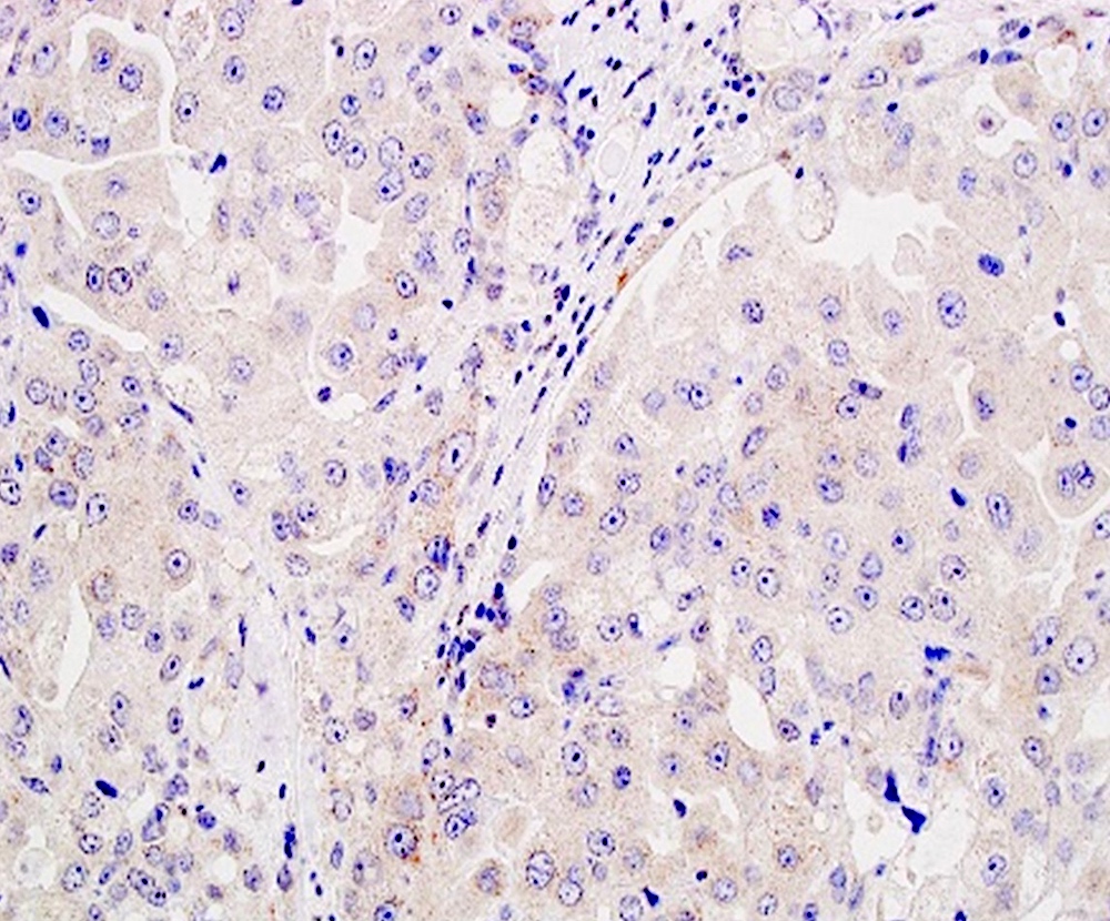
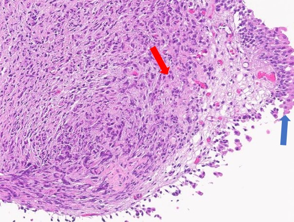
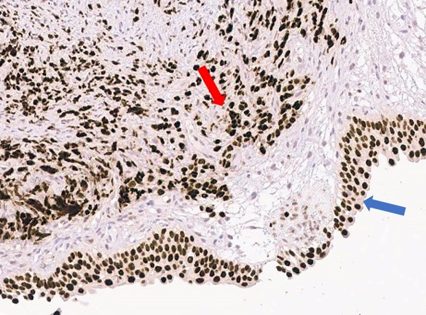
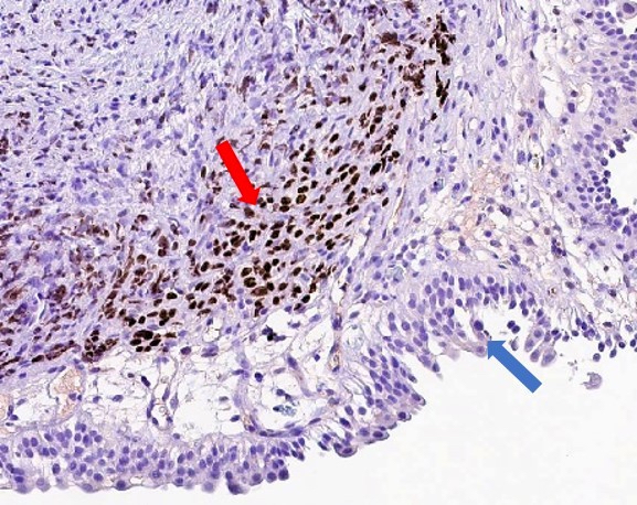
Microscopic (histologic) images
Contributed by Qingqing Ding, M.D., Ph.D.


Positive in triple negative breast cancer

Positive in metaplastic breast carcinoma

Positive in borderline phyllodes tumor
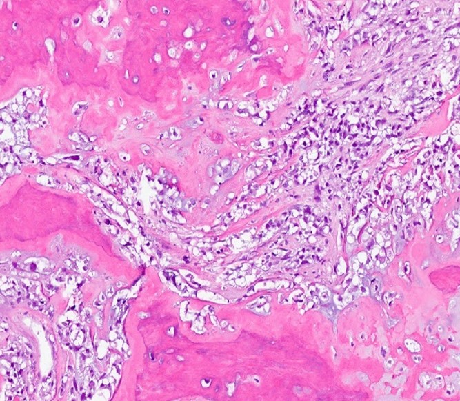
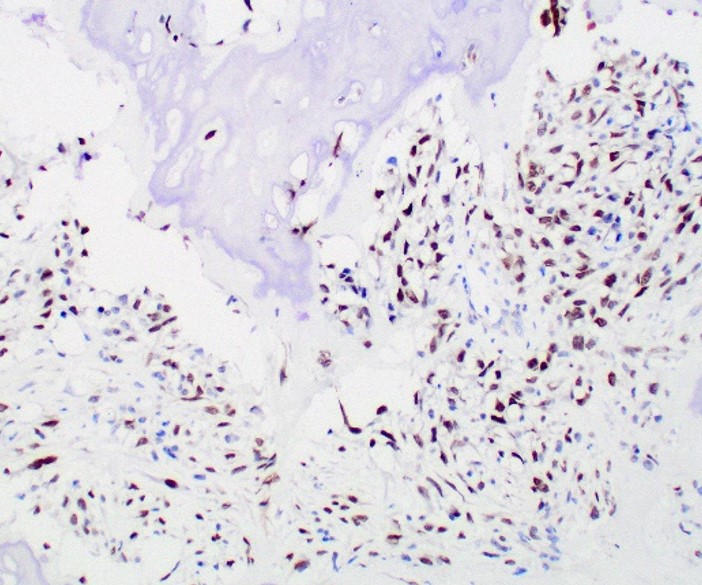
Positive in osteosarcoma
Positive in brain metastasis
Negative in apocrine breast cancer
Negative in urothelial epithelium
Positive staining - normal
- Benign breast luminal cells (Proc Natl Acad Sci U S A 2005;102:11005)
- Benign skin eccrine duct
Positive staining - disease
- Invasive breast carcinoma (> 95%), including ER+ / HER2- (> 95%), HER2+ (> 90%), ER- / HER2- (> 90%) (Mod Pathol 2021;34:710)
- Phyllodes tumor (> 95%), including benign, borderline and malignant
- Osteosarcoma (56%) and chondrosarcoma (28%) of the bone
- Salivary duct carcinoma (24%)
- Ovarian serous carcinoma (10 - 15%)
Negative staining
- Usually negative in a few special types of triple negative breast cancer, such as apocrine carcinoma (in which GATA3 can be positive) and neuroendocrine (small cell and large cell) carcinoma
- Urothelial carcinoma (Mod Pathol 2021;34:710)
- Gastrointestinal adenocarcinoma
- Hepatocellular carcinoma
- Cholangiocarcinoma
- Lung adenocarcinoma
- Kidney tumor
- Thyroid tumor
- Melanoma
- Undifferentiated pleomorphic sarcoma (UPS)
- Liposarcoma
- Angiosarcoma
Sample pathology report
- Left lobe of liver, core biopsy:
- Poorly differentiated carcinoma, consistent with breast primary (see comment)
- Comment: The patient's past history of triple negative breast cancer is noted. Immunostains performed on the biopsy block show that the tumor cells are positive for pancytokeratin, TRPS1 and CK7, while negative for CK20, HepPar1, ER, PR, HER2, GATA3, TTF1 and PAX8. The immunoprofile is consistent with metastatic carcinoma with breast primary.
Practice question #1
A 50 year old woman, with a history of triple negative breast cancer of the left breast 5 years ago, presents with a 2 cm lung nodule in the left lower lobe. Which of the following markers is the most helpful marker to rule in and rule out the breast origin?
- CK7
- GATA3
- GCDFP-15
- Mammaglobin
- TRPS1
Practice answer #1