Stains & CD markers
Cytokeratin 34 beta E12


Copyright: 2003-2025, PathologyOutlines.com, Inc.
PubMed Search: Cytokeratin 34 beta E12
Cytokeratin 34 beta E12
Editorial Board Members: Christian M. Schürch, M.D., Ph.D., Kristen E. Muller, D.O.
Last author update: 15 November 2024
Last staff update: 19 August 2025
Copyright: 2003-2025, PathologyOutlines.com, Inc.
PubMed Search: Cytokeratin 34 beta E12
Table of Contents
Definition / general | Essential features | Terminology | Pathophysiology | Diagrams / tables | Clinical features | Interpretation | Uses by pathologists | Prognostic factors | Microscopic (histologic) images | Virtual slides | Positive staining - normal | Positive staining - disease | Negative staining | Pitfalls | Sample pathology report | Additional references | Practice question #1 | Practice answer #1Cite this page: Bantumilli S, Trembath DG. Cytokeratin 34 beta E12. PathologyOutlines.com website. https://www.pathologyoutlines.com/topic/stainsck34be12.html. Accessed October 5th, 2025.
Definition / general
- Broad spectrum high molecular weight cytokeratins
- Antibody specific for high molecular weight cytokeratins 1, 5, 10 and 14, which are found in complex epithelia
Essential features
- Basic (type 2) cytokeratin
- High molecular weight protein groups of 48 - 67 kD
- Useful in the detection of epithelial basal cells
- Highlights intermediate filaments in basal cell cytoplasm
- Specific for prostatic basal cells and stratified squamous epithelium
Terminology
- High molecular weight keratin (HMWK) or high molecular weight cytokeratin (HMWCK)
- CK903 / K903
- 34βE12 / 34betaE12
- Cytokeratin 34 beta E12
Pathophysiology
- Intermediate filaments composed by keratins play a role in modulation of cell proliferation, migration and cancer invasion (Acta Oncol 2016;55:167)
- Keratins play a pivotal role in the integrity and mechanical stability of single epithelial cells via cell-cell contacts (Histochem Cell Biol 2008;129:705)
- Role in regulating the formation of epithelial tumors
- Influences tumor response to treatment and affects patient prognosis (J Cancer 2022;13:1713)
Diagrams / tables
Images hosted on other servers:

Chromosome localization of human cytokeratins

Basic information about the anti-CK antibodies
Clinical features
- Point mutations in human CK genes linked to epithelial disorders in multiple tissue types (J Cancer 2022;13:1713)
Interpretation
- Granular cytoplasmic positivity in basal cell of glands (Am J Clin Pathol 1997;107:219)
Uses by pathologists
- Prostate
- Triple cocktail (34βE12 + p63 + AMACR) as ancillary method to diagnose prostate cancer in biopsies with small foci of atypical glands (J Clin Pathol 2012;65:437, Urol Oncol 2011;29:614)
- Useful in diagnosis of prostatic adenocarcinoma (Am J Surg Pathol 1989;13:389, Am J Surg Pathol 1995;19:251)
- Helps in identifying benign atypia by highlighting the basal cells postradiation (Semin Diagn Pathol 2005;22:88)
- Stains prostatic basal cells, even after destaining H&E slides, whose presence rules out usual type prostatic adenocarcinoma (Hum Pathol 2000;31:1155)
- Useful in biopsies and even after hormonal therapy (Am J Clin Pathol 2004;121:99, Hum Pathol 2007;38:332)
- Note that patchy staining may rarely occur in carcinoma and negative staining does not predict carcinoma in small foci of atypical glands (Am J Surg Pathol 2002;26:1151, Hum Pathol 2004;35:43)
- Triple cocktail with p63 and AMACR is more sensitive / specific than 34βE12 alone (Am J Clin Pathol 2007;127:248)
- Distinguishes high grade prostatic intraepithelial neoplasia (PIN) (intact or fragmented basal cell layer) from adenocarcinoma (Mod Pathol 2004;17:360)
- Distinguishes cribriform basal cell hyperplasia from cribriform PIN (Am J Surg Pathol 2002;26:237)
- Typically does not stain prostatic adenocarcinoma cells but does stain prostatic adenoid cystic / basal carcinoma and tumors with squamous differentiation (Am J Surg Pathol 2003;27:1523, Am J Surg Pathol 2004;28:651)
- Breast intraductal lesions
- 90% of flat epithelial atypia, atypical ductal hyperplasia and ductal carcinoma in situ are negative to focally positive for 34βE12
- In contrast, all cases of usual ductal hyperplasia and 90% of lobular carcinoma in situ (LCIS) are diffusely positive with perinuclear cytoplasmic staining with 100% of classic lobular intraepithelial neoplasia exhibiting this pattern (Am J Clin Pathol 2018;149:S30, Arch Pathol Lab Med 2014;138:1629)
- Florid ductal hyperplasia without atypia (intense staining) (Am J Surg Pathol 1999;23:1048)
- Urinary bladder
- Sensitive positive marker for high grade invasive urothelial carcinoma (Histopathology 2003;42:167)
- Distinguishes dysplasia (basal staining only) from carcinoma in situ (stains all urothelial layers) (Hum Pathol 2000;31:745)
Prognostic factors
- Reduced reactivity to CK903 in intrahepatic cholangiocarcinoma: favorable survival (Mod Pathol 2002;15:1181)
- Specific keratin 34βE12 / K7 patterns in lung carcinomas are prognostic factors for cancer specific and overall survival in patients with early stage non-small cell lung cancer (Acta Oncol 2016;55:167)
- Independent predictor of disease free survival in patients with triple negative breast cancer associated with good prognosis (J Clin Pathol 2010;63:744)
- Expression is a good indicator of tumor progression in esophageal squamous cell carcinoma (Biomed Res 2012;33:183)
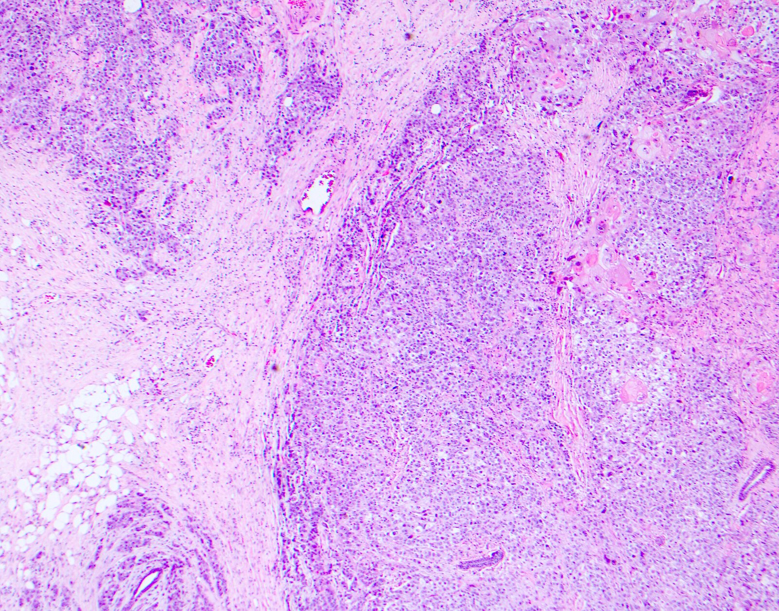
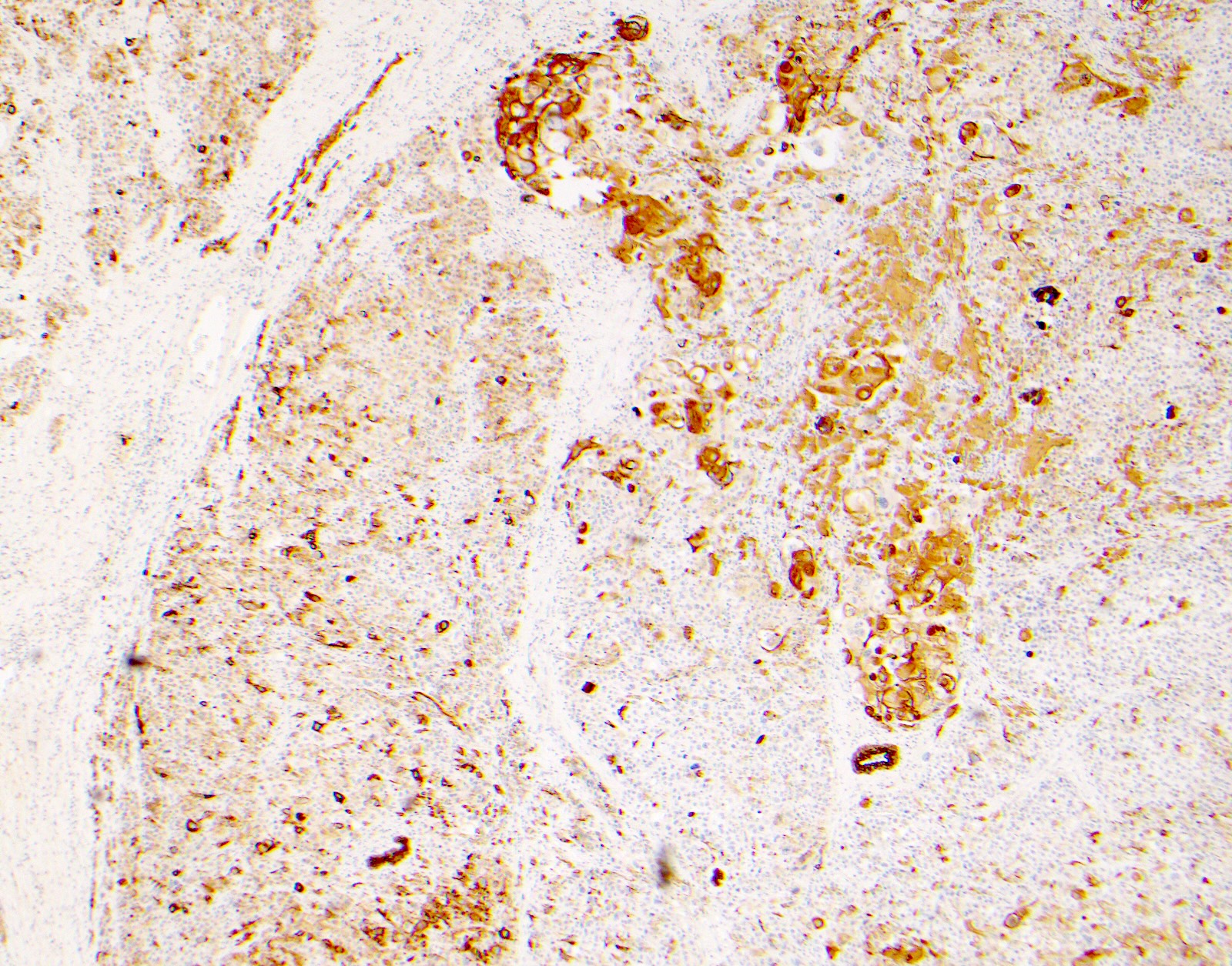
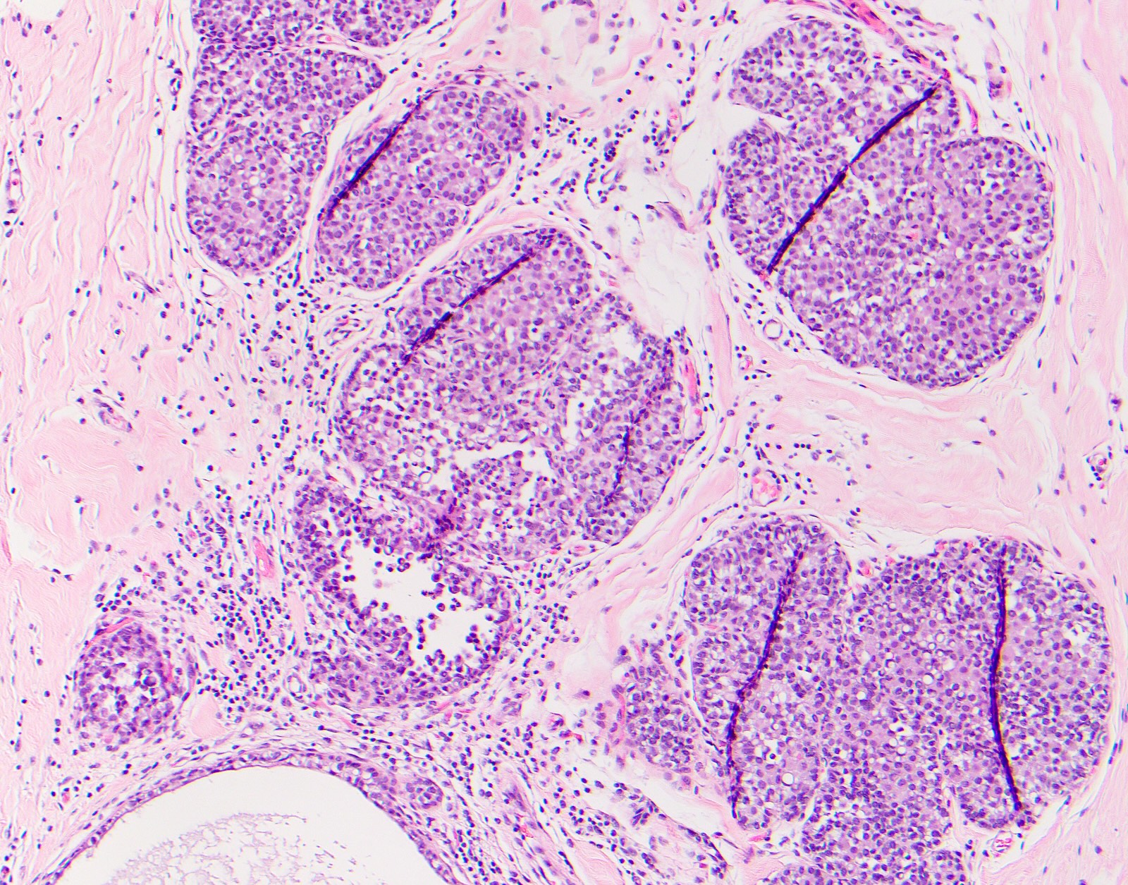
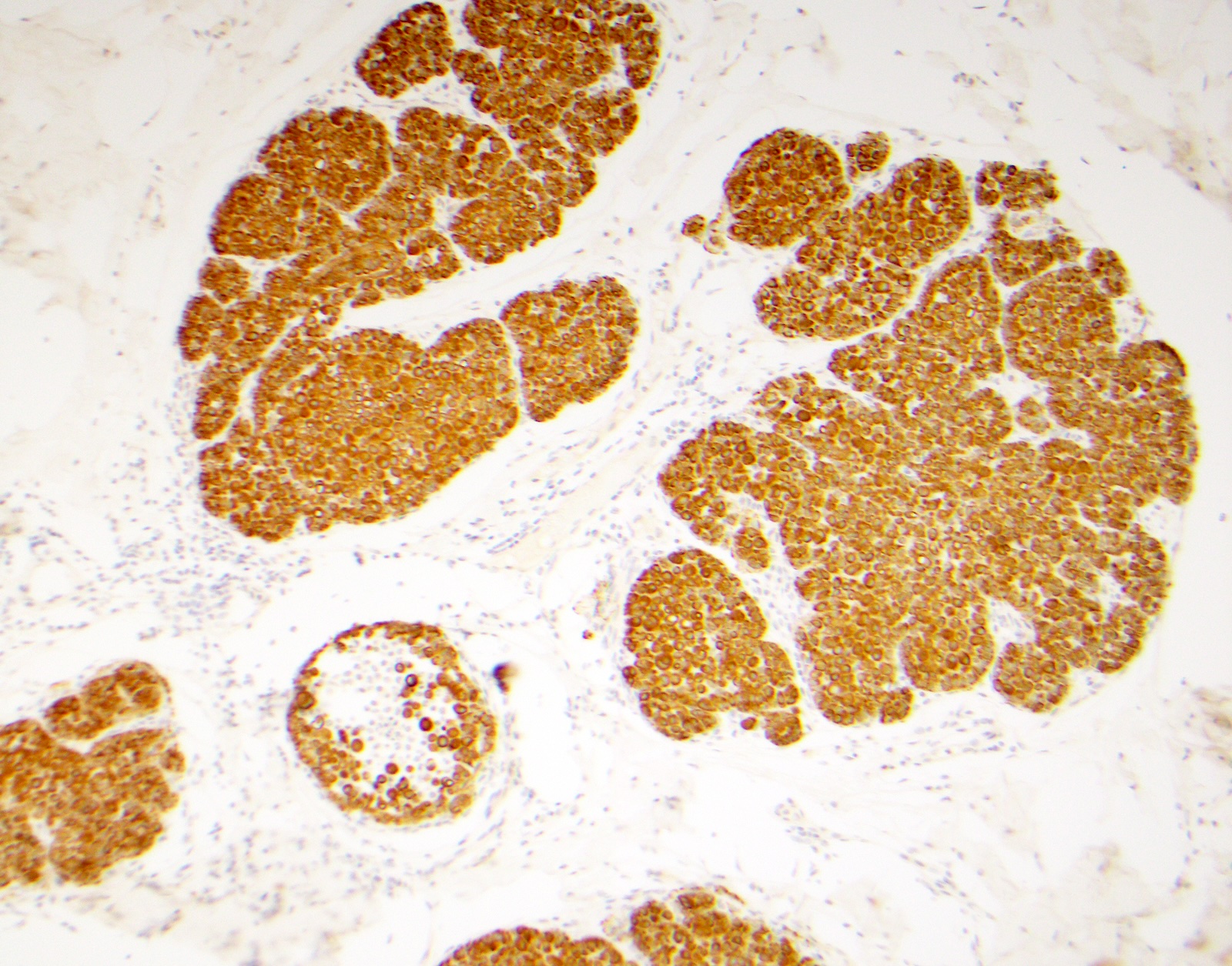
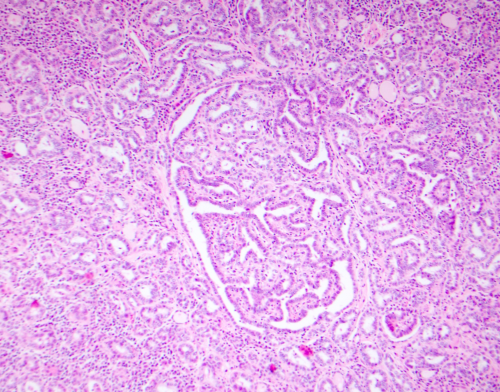
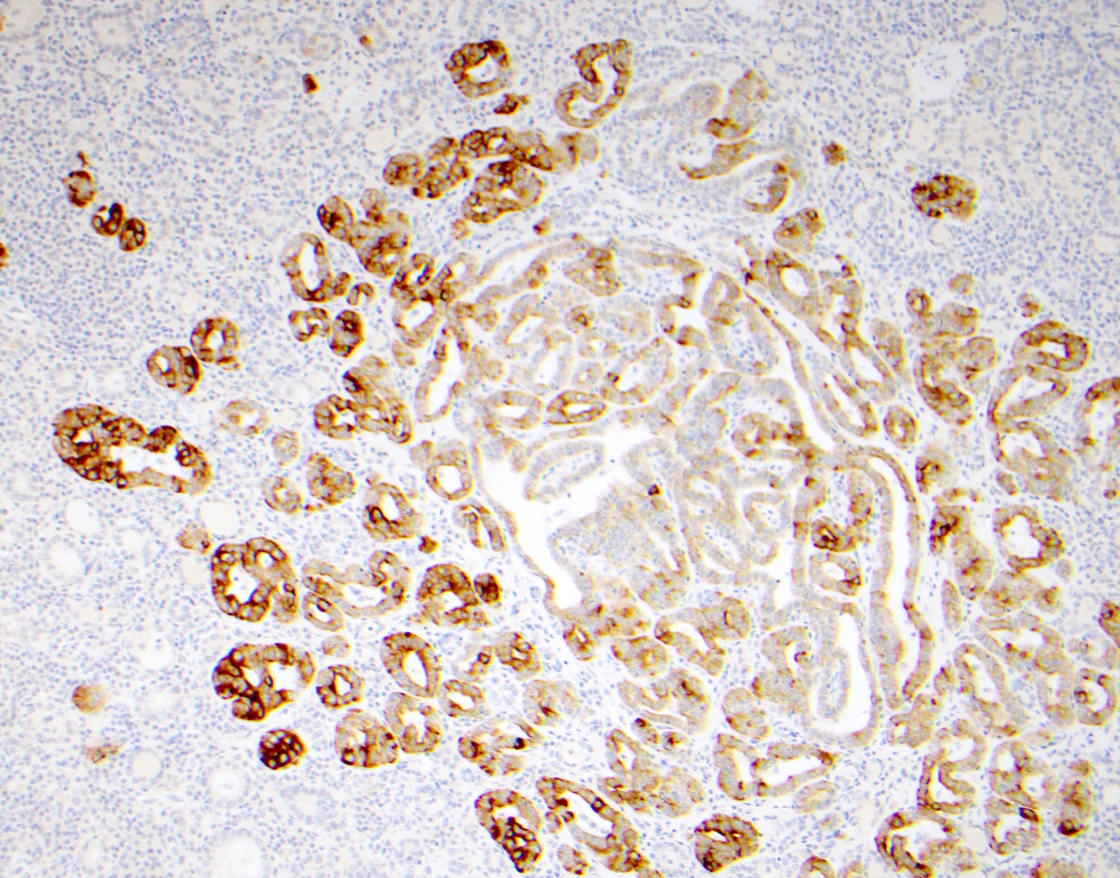
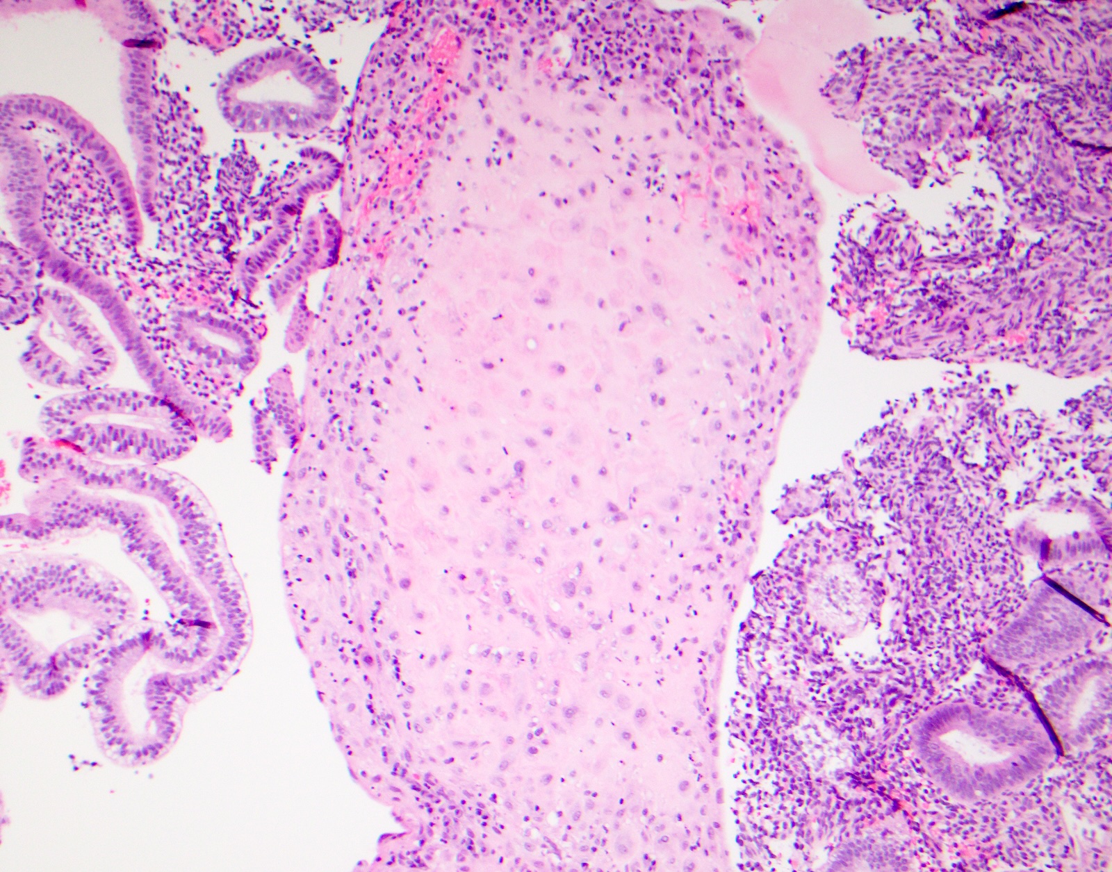
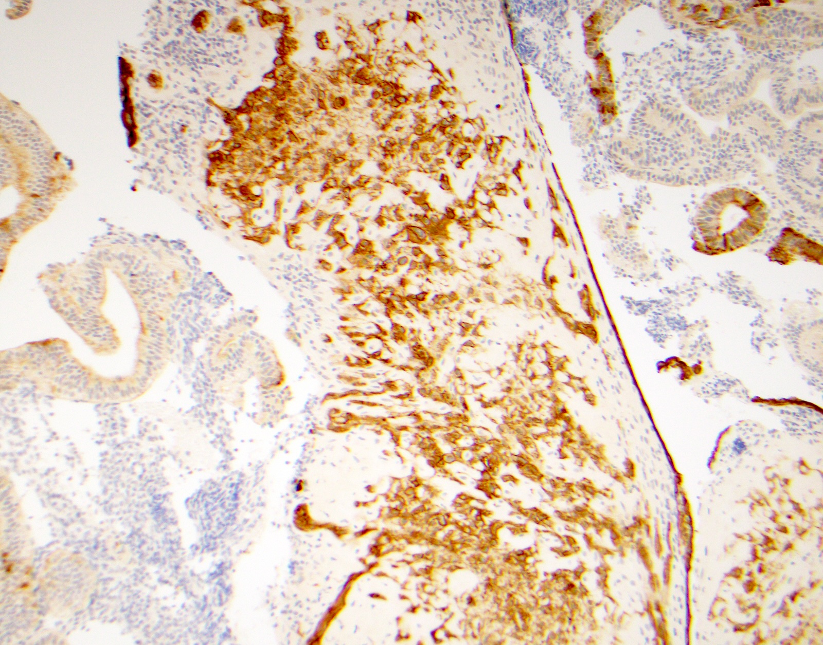
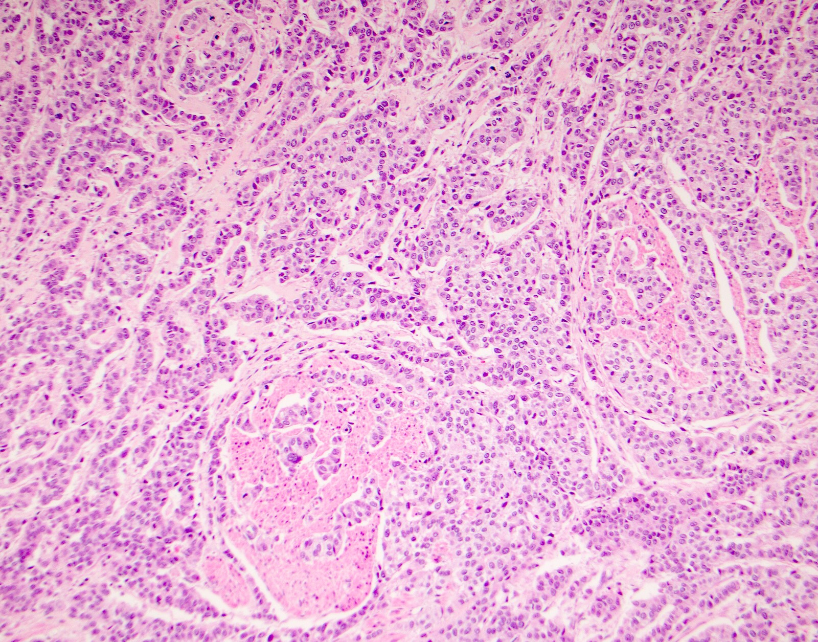
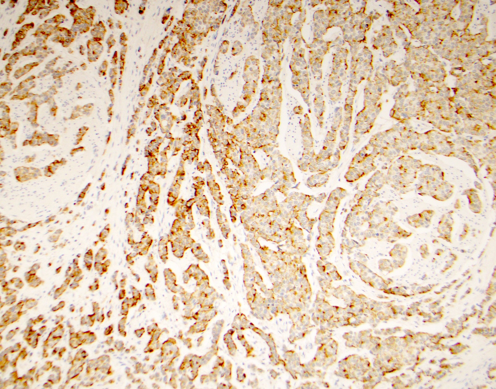
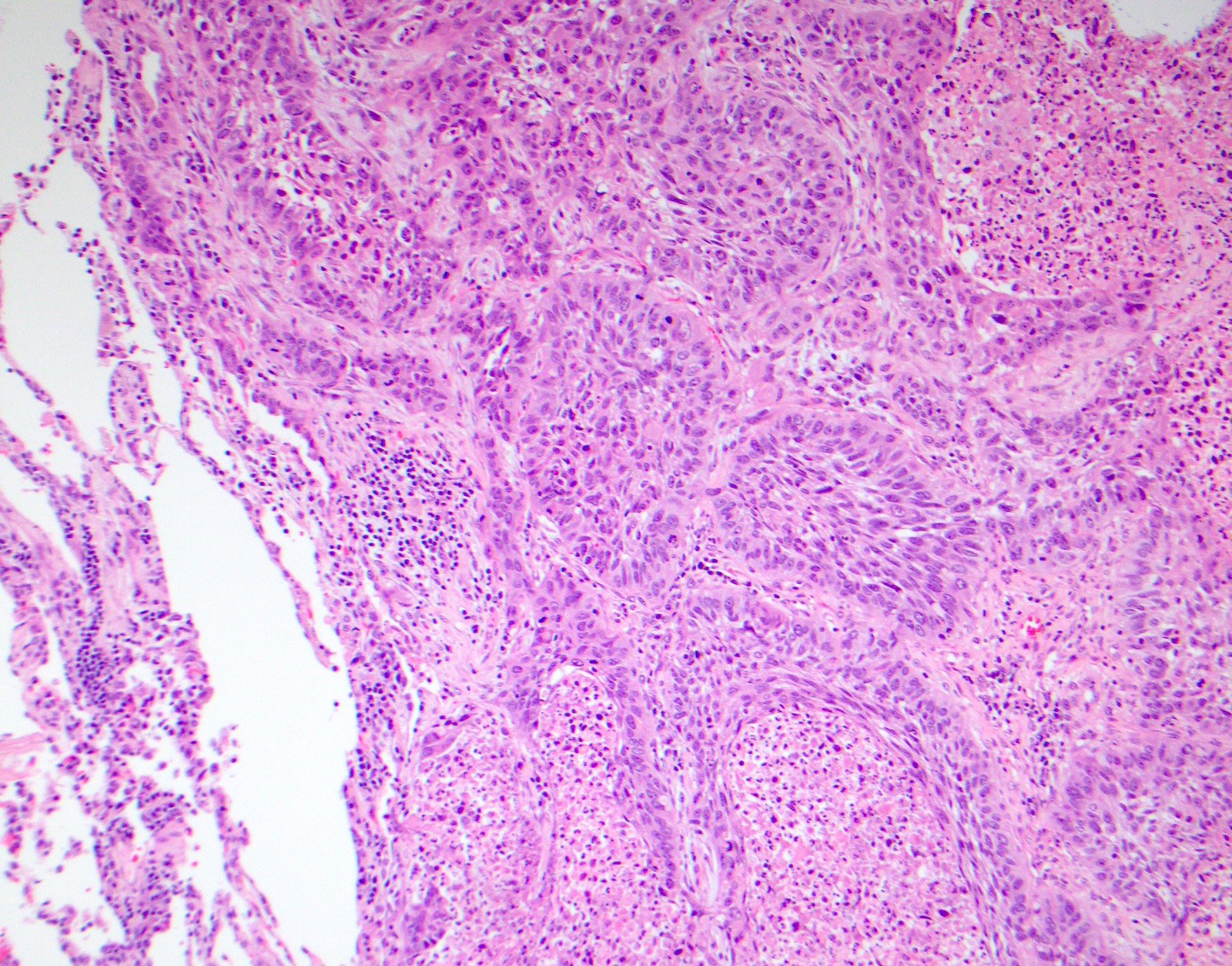
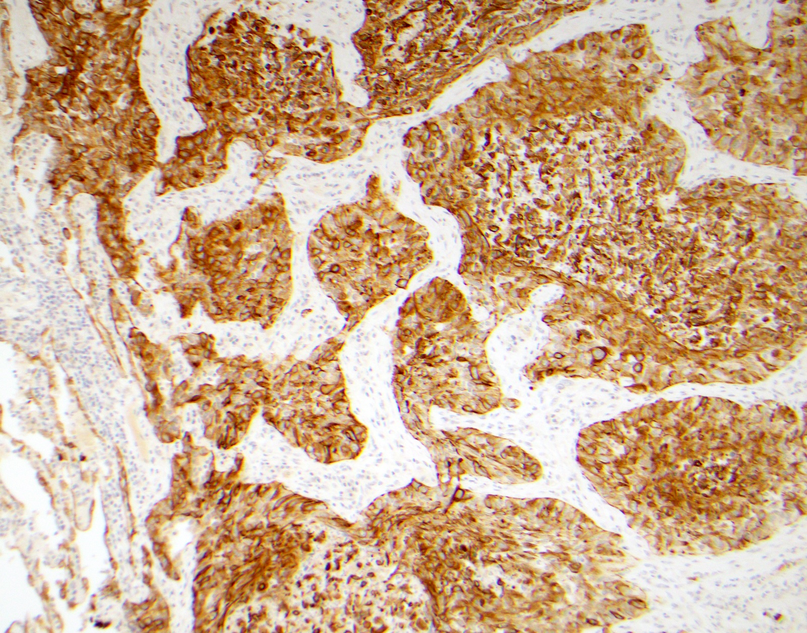
Microscopic (histologic) images
Contributed by Surekha Bantumilli, M.D. and Dimitri G. Trembath, M.D., Ph.D.
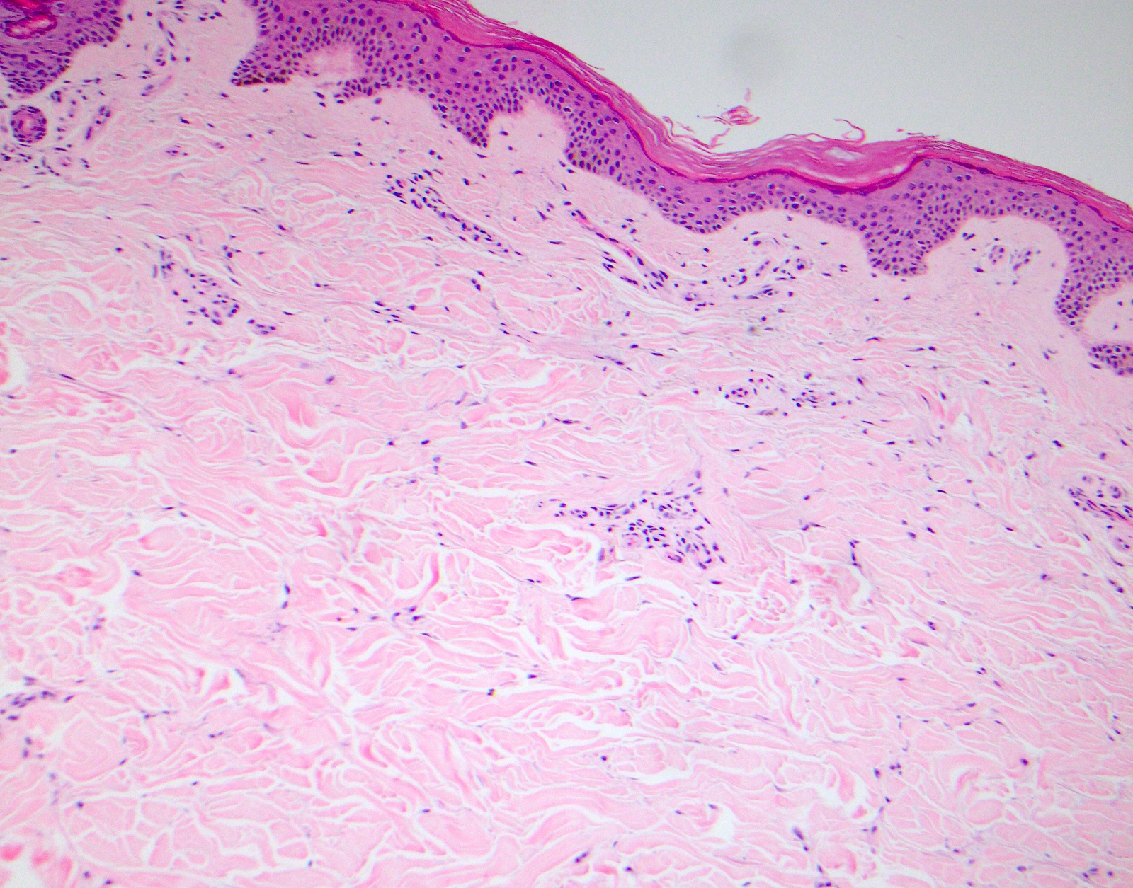
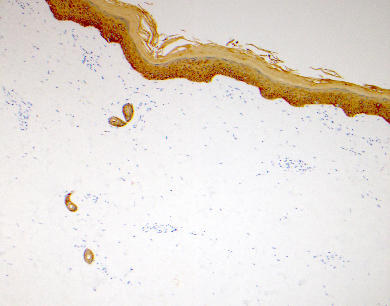
Normal skin
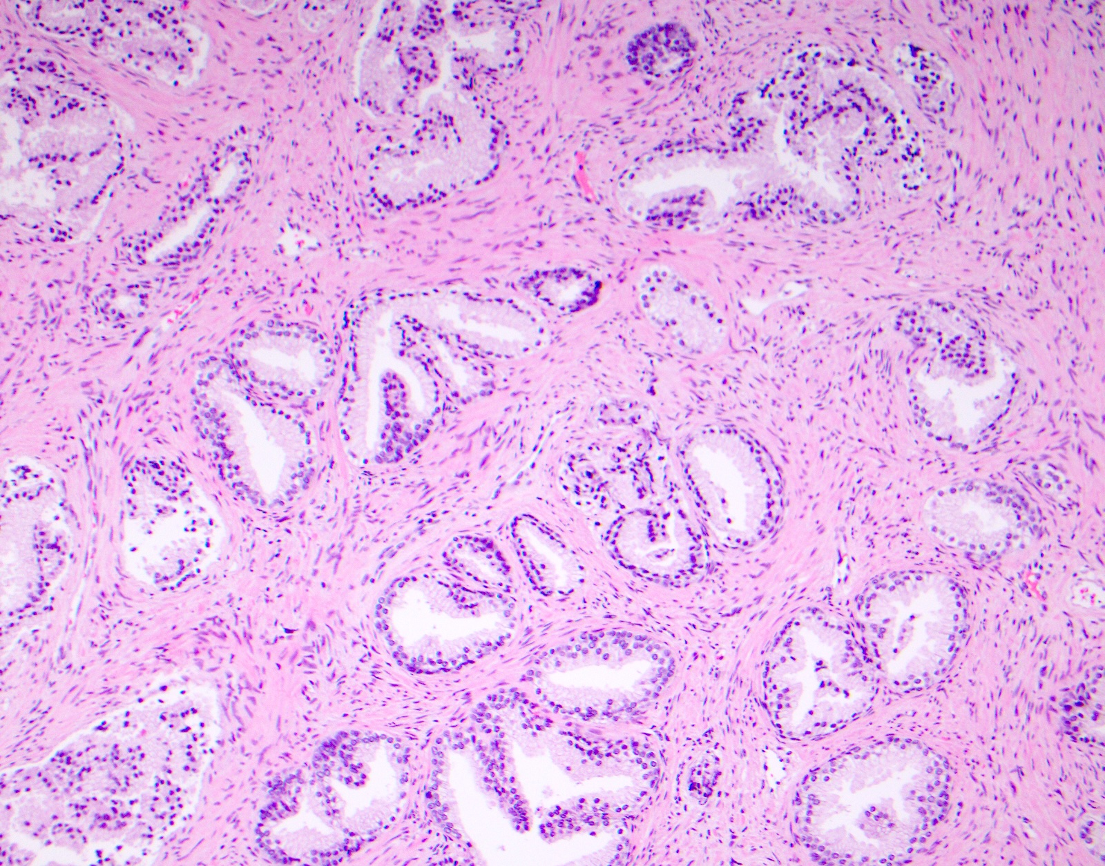

Benign prostate gland
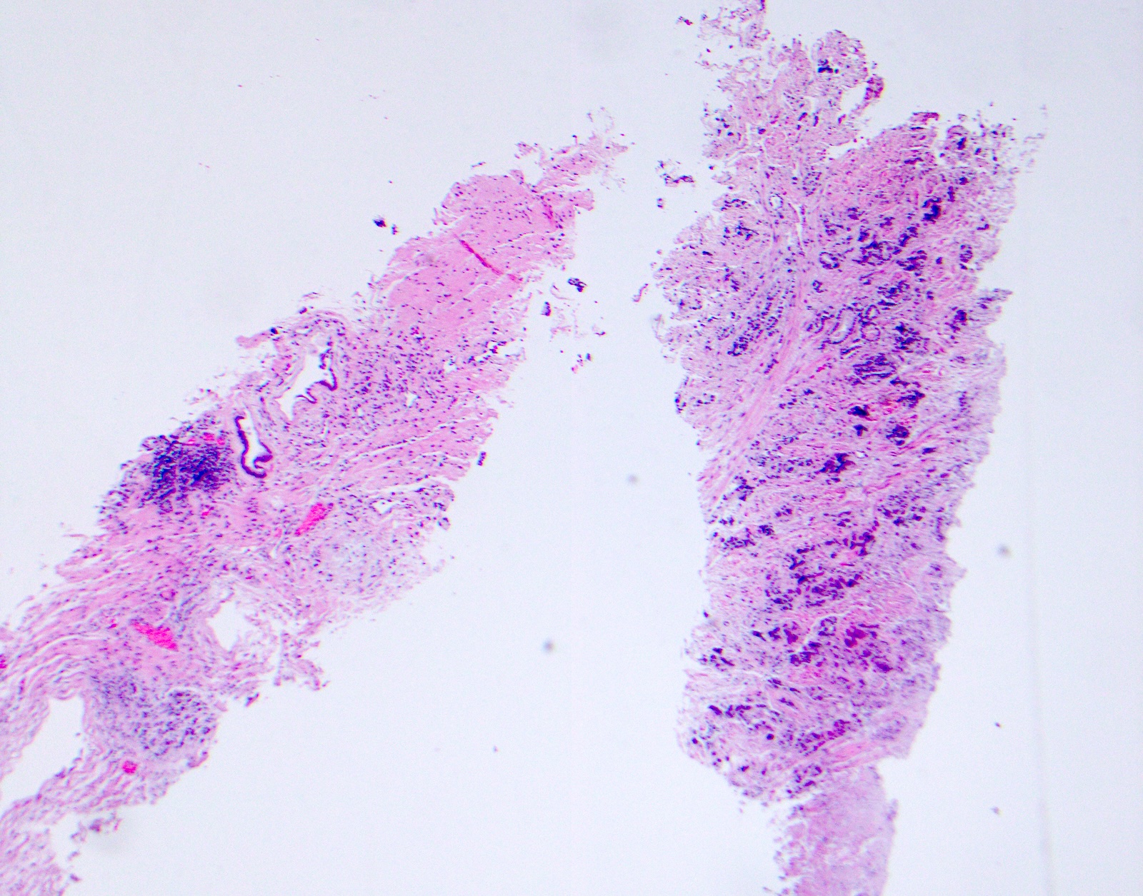
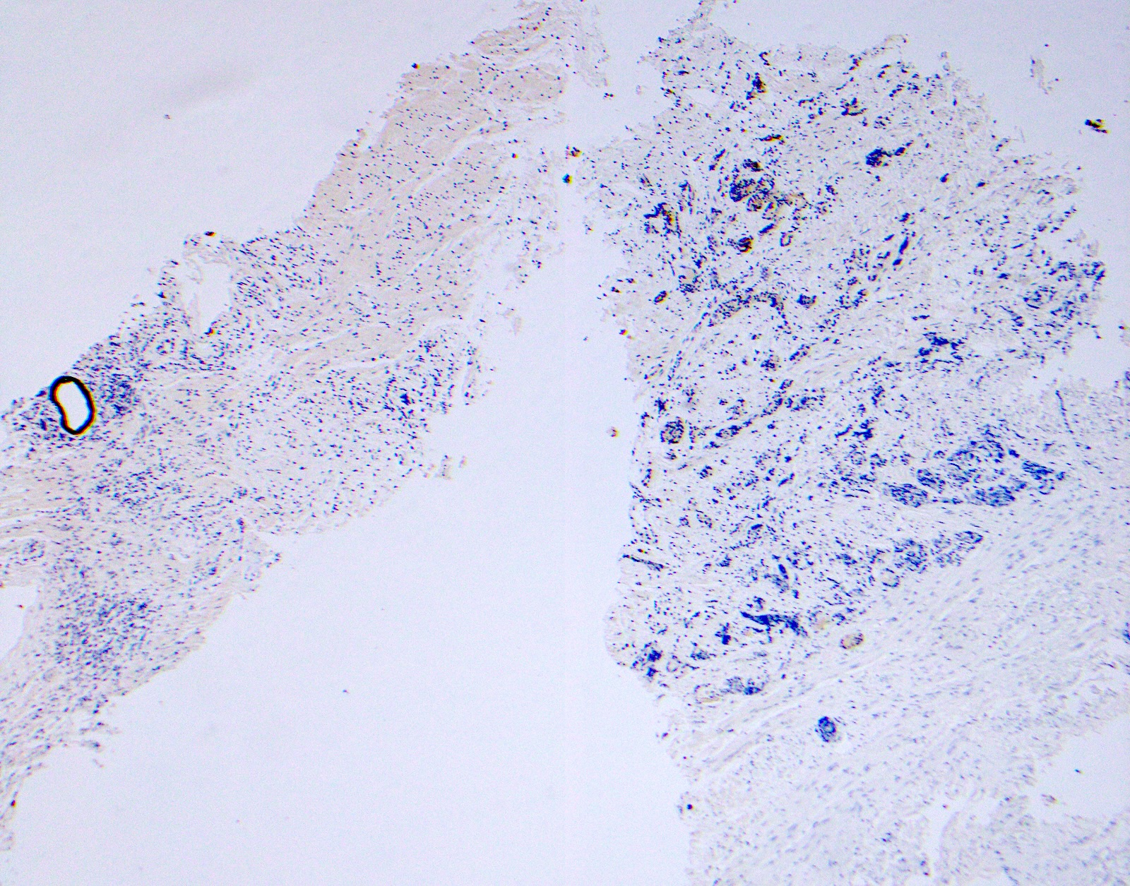
Prostate adenocarcinoma
Metaplastic carcinoma, breast
Lobular carcinoma in situ
Papillary carcinoma of thyroid
Placental site nodule
Invasive urothelial carcinoma
Basaloid squamous cell carcinoma of lung
Virtual slides
Images hosted on other servers:

Prostate adenocarcinoma, Gleason 4+3=7, 34BE stain
Positive staining - normal
- Prostate basal cells, skin (J Exp Clin Cancer Res 2003;22:441)
- Thyroid solid cell nests (Am J Surg Pathol 2006;30:994)
- Biliary tracts of liver (large, septal, interlobular bile ducts and bile ductules) (Mod Pathol 2002;15:1181)
Positive staining - disease
- Demonstrates the epithelial origin of amyloid deposits in dermal papillae in macular and lichenoid amyloidosis (Actas Dermosifiliogr 2013;104:99)
- Helps to visualize colloid bodies and diagnose lichen planopilaris (Am J Dermatopathol 2016;38:353)
- Breast ductal hyperplasia and lobular intraductal neoplasia (J Histochem Cytochem 2003;51:1527, Am J Surg Pathol 1999;23:1048)
- Placental site nodules (Pathology 1999;31:328)
- Prostatic basal cell hyperplasia (Hum Pathol 2003;34:462)
- Thymoma (high grade) (Rom J Morphol Embryol 1999;45:153)
- Identification of the basal cell layer in prostate tissue in the determination of carcinoma
- Squamous cell carcinoma (100%) with cytoplasmic staining (Diagn Cytopathol 2008;36:20)
- Cholangiocarcinoma, cytoplasmic and membranous staining (Mod Pathol 2002;15:1181)
- Diagnostic tool for differentiating between atypical squamous cell carcinoma and atypical fibroxanthoma (Arch Pathol Lab Med 2001;125:799)
- Diffuse staining in both intraductal and invasive urothelial cancer (Am J Surg Pathol 2022;46:454)
- Urachal adenocarcinoma (60%) (Cancers (Basel) 2018;10:108)
- Clear cell adenocarcinoma of urethra (Int J Surg Oncol 2015;2015:790235)
- Squamous cell carcinoma and basaloid carcinoma in lung
- Distinguishes spindle cell squamous cell carcinoma (positive staining) from histologic mimickers like atypical fibroxanthoma (AFX), spindle cell melanoma, scar and leiomyosarcoma (Am J Dermatopathol 2008;30:228, Arch Pathol Lab Med 2001;125:799)
- Stains the epithelial component in primary epithelial myoepithelial carcinoma of lung (Case Rep Pathol 2012;2012:319434)
- Primary cutaneous extramammary Paget disease (Case Rep Oncol 2021;14:430)
- Collecting duct carcinoma (Zhonghua Zhong Liu Za Zhi 2001;23:162)
- Nasopharyngeal carcinoma and thymoma (J Cancer 2022;13:1713)
- Holocrine poroma (100%) (Am J Dermatopathol 2018;40:401)
- Cutaneous squamous and adnexal tumor (Actas Dermosifiliogr 2013;104:99)
- Squamoid component of high grade serous carcinoma of ovary (50%) and squamous differentiation in endometrial carcinoma (100%) (Am J Surg Pathol 2023;47:967)
- Cutaneous carcinosarcoma of hand (carcinoma component) (Dermatol Online J 2019;25:13030)
- Thyroid papillary carcinoma (Exp Mol Pathol 2007;82:91)
- Paratesticular serous papillary adenocarcinoma (Int J Surg Pathol 2011;19:692)
- Pagetoid Bowen disease (pagetoid SCIS) (Ann Dermatol 2016;28:497)
- Thyroid carcinoma showing thymus-like differentiation (CASTLE) tumor (Am J Surg Pathol 2006;30:994)
- Ovary (Int J Gynecol Pathol 2001;20:155)
- Squamous cell carcinoma (classic and basaloid) (Hum Pathol 1998;29:609)
- Breast metaplastic carcinoma, clear cell carcinoma of gynecologic tract (Int J Gynecol Pathol 2001;20:252)
- Endocervical and endometrial carcinoma (Int J Gynecol Pathol 2002;21:11)
Negative staining
- Prostatic adenocarcinoma, usual type
- Benign mimickers of prostate carcinoma, including atrophy (23%), atypical adenomatous hyperplasia (AAH) (50%), nephrogenic adenoma (75%) and mesonephric hyperplasia (66%) (Ann Diagn Pathol 2013;17:41, Semin Diagn Pathol 2005;22:88)
- Negative staining in neuroendocrine proliferations of lung (Histopathology 2003;42:156)
- No reactivity with cells derived from simple epithelia, mesenchymal tumors, lymphomas, melanomas, neural tumors and neuroendocrine tumors
- Clear cell papillary renal cell carcinoma (Pathol Oncol Res 2018;24:447, Pathology 2017;49:10)
- Follicular thyroid carcinoma and hepatocellular carcinoma (J Cancer 2022;13:1713)
- Endometrial carcinoma (38.1%) (J Cancer 2022;13:1713)
- Clear cell adenocarcinoma of urinary tract (Hum Pathol 1998;29:1451)
- Lymphoepithelioma-like carcinoma (LELC) of ureter (Ann Diagn Pathol 2010;14:209)
- Mucinous tubular and spindle cell carcinoma (MTSCC) (Diagn Cytopathol 2010;38:51)
- Paget disease of vulva (Ann Dermatol 2016;28:497)
- Renal clear cell carcinoma (Int J Gynecol Pathol 2001;20:155)
- Prostatic secretory and stromal cells
- Adenocarcinoma (40%) (Diagn Cytopathol 2008;36:20)
- Colorectal adenocarcinoma (10%) (Cancers (Basel) 2018;10:108)
Pitfalls
- Prostate: negative staining of CK34βE12 in some glands is not definitive evidence of malignancy because benign glands may show discontinuous staining (Iran J Pathol 2020;15:232)
- Prolonged formalin fixation has been shown to have a negative effect on detection of basal cell specific keratins, giving rise to false negative staining (J Clin Pathol 2003;56:892, Mod Pathol 1999;12:472)
- Prostatic ductal adenocarcinoma, ductal type may show focally positive basal cell staining (J Clin Pathol 2003;56:892)
- Focal staining in rhabdomyosarcoma: pitfall in differentiating from carcinoma (J Cutan Pathol 2014;41:588)
Sample pathology report
- Prostate, core biopsy:
- Benign prostatic hyperplasia, no evidence of malignancy (see comment)
- Comment: Immunohistochemical stain for CK34βE12 (HMWK) highlights the basal cells in all the cores and confirms the diagnosis.
Additional references
Practice question #1
A 65 year old man presents with elevated prostate specific antigen (PSA) and undergoes a prostate biopsy, stained with high molecular weight cytokeratin (HMWCK) (shown above). What is the most likely interpretation of this staining pattern?
- Benign prostate tissue
- Benign seminal vesicle
- High grade prostatic intraepithelial neoplasia
- Prostatic adenocarcinoma
- Urothelial carcinoma
Practice answer #1
A. Benign prostate tissue. The basal layer of benign prostate glands will stain with high molecular weight cytokeratin. Answer D is incorrect because staining will be lost on prostatic adenocarcinoma. Answer C is incorrect because high molecular weight cytokeratin staining can be present in high grade prostatic intraepithelial neoplasia but is not diagnostic by itself for this entity. Answers B and E are incorrect because the glandular appearance outlined by the high molecular weight cytokeratin staining is not consistent with either seminal vesicle or urothelial carcinoma.
Comment Here
Reference: Cytokeratin 34 beta E12
Comment Here
Reference: Cytokeratin 34 beta E12