
Stains & CD markers
NUTM1

Copyright: 2003-2024, PathologyOutlines.com, Inc.
PubMed Search: NUTM1
NUTM1
Author: Brendan C. Dickson, M.D., M.Sc.
Editorial Board Members: Brandon Umphress, M.D., Christian M. Schürch, M.D., Ph.D.
Editor-in-Chief: Debra L. Zynger, M.D.
Last author update: 20 March 2023
Last staff update: 20 March 2023
Copyright: 2003-2024, PathologyOutlines.com, Inc.
PubMed Search: NUTM1

Table of Contents
Definition / general | Essential features | Pathophysiology | Interpretation | Uses by pathologists | Microscopic (histologic) images | Positive staining - normal | Positive staining - disease | Negative staining | Sample pathology report | Board review style question #1 | Board review style answer #1Cite this page: Dickson BC. NUTM1. PathologyOutlines.com website. https://www.pathologyoutlines.com/topic/stainsnutm1.html. Accessed April 20th, 2024.
Definition / general
- NUTM1 (NUT midline carcinoma family member 1) encodes the NUT family member 1 protein (HGNC: Symbol report for NUTM1 [Accessed 30 November 2022], NIH: NUTM1 [Accessed 30 November 2022])
- Several types of neoplasms are characterized by NUT rearrangement; this predominantly includes NUT carcinoma but rearrangement has also recently been reported in a subset of soft tissue, bone and visceral tumors (Am J Surg Pathol 2018;42:636, Am J Surg Pathol 2018;42:1360, Am J Surg Pathol 2019;43:268, Genes Chromosomes Cancer 2019;58:809)
- NUT rearranged neoplasms are characterized by the fusion of NUTM1 with another gene (e.g., BRD4); the resulting oncoprotein causes tumor growth (Genes Dev 2015;29:1507, Proc Natl Acad Sci U S A 2017;114:E4184)
- Translocations involving NUTM1 can be detected molecularly and by immunohistochemistry for NUT
Essential features
- Diagnosis of NUT carcinoma and other NUT rearranged neoplasms is predicated on immunohistochemical demonstration of nuclear NUT staining (or molecular confirmation of NUTM1 rearrangement)
- NUT is expressed in a subset of germ cell tumors (Am J Surg Pathol 2009;33:984)
- Normal overexpression of this biomarker is limited to testicular and ovarian germ cells (Cancer Res 2003;63:304, Am J Surg Pathol 2009;33:984)
Pathophysiology
- Function of the NUT protein may relate to promoting histone H4 hyperacetylation during spermatogenesis (Cell Rep 2018;24:3477)
- NUT carcinoma is characterized by the fusion of NUTM1 with another gene (e.g., BRD4, BRD3 or NSD3, among others); these proteins bind acetylated chromatin
- These domains result in dysregulated transcription of genes, including c-MYC and TP63, which promote tumor growth (Genes Dev 2015;29:1507, Proc Natl Acad Sci U S A 2017;114:E4184)
Interpretation
- In NUT carcinoma, staining is nuclear with a speckled distribution (> 90% of cells are generally positive); the reported sensitivity is 87% and the specificity is 100% (clone: C52) (Am J Surg Pathol 2009;33:984)
- In germ cell tumors, staining is nuclear with an even / uniform distribution and often only focal (Am J Surg Pathol 2009;33:984, Histopathology 2014;65:35, Pathology 2015;47:118)
Uses by pathologists
- NUT is a biomarker for NUT carcinoma and neoplasms harboring NUTM1 fusion genes; the presence of staining confirms the diagnosis and excludes potential mimics (Am J Surg Pathol 2009;33:984, Cell 2016;164:1060, Am J Surg Pathol 2018;42:636)
- NUT is variably expressed among germ cell tumors, including dysgerminoma (64 - 93%), embryonal carcinoma (0 - 9%), immature teratoma (75%), seminoma (6 - 74%), spermatocytic seminoma (100%) and yolk sac tumor (7 - 10%); however, in this context and in the absence of a broader panel of immunohistochemical markers, NUT alone is of limited diagnostic value (Am J Surg Pathol 2009;33:984, Histopathology 2014;65:35, Pathology 2015;47:118)
Microscopic (histologic) images
Contributed by Brendan C. Dickson, M.D., M.Sc.
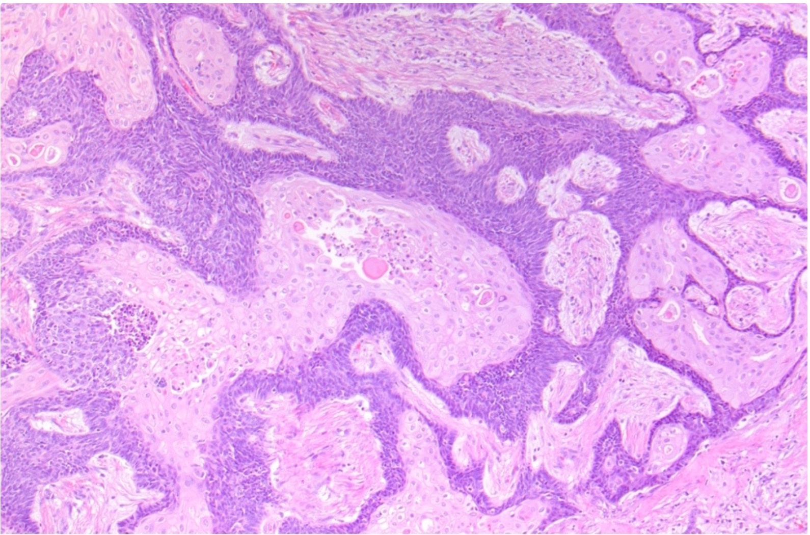
Abrupt keratinization
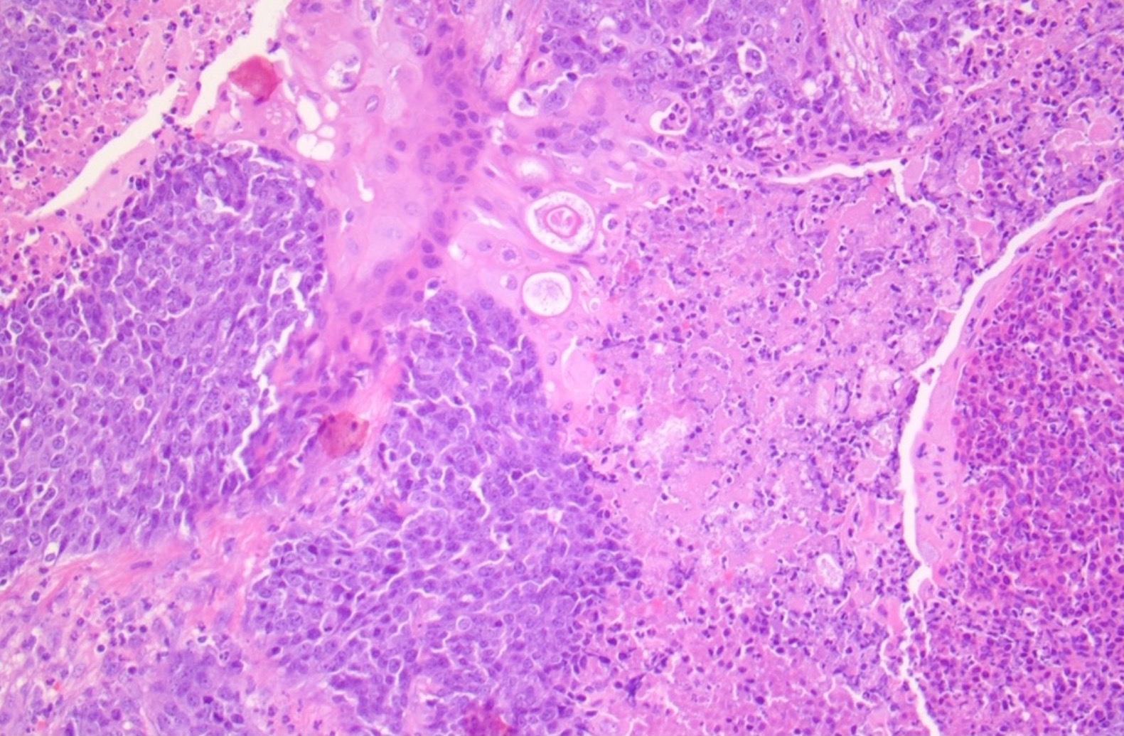
Necrosis
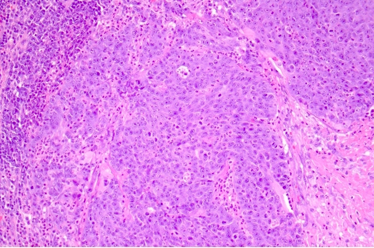
Morphologically undifferentiated
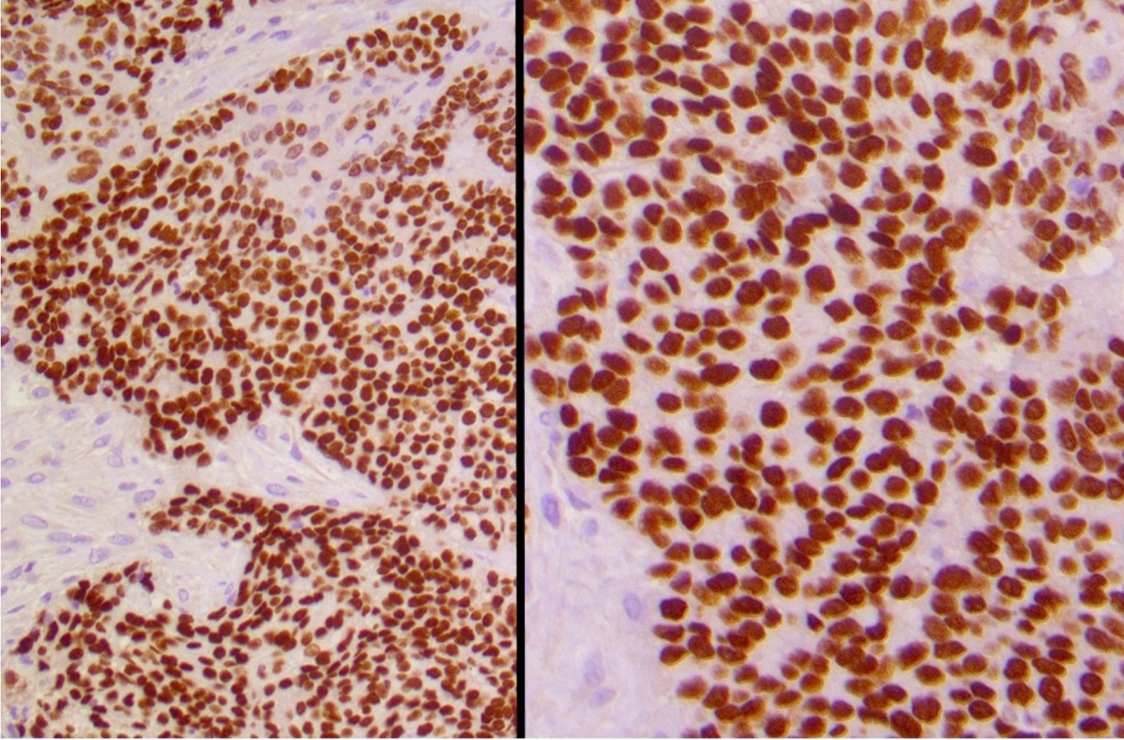
Diffuse p63 expression with even / uniform pattern
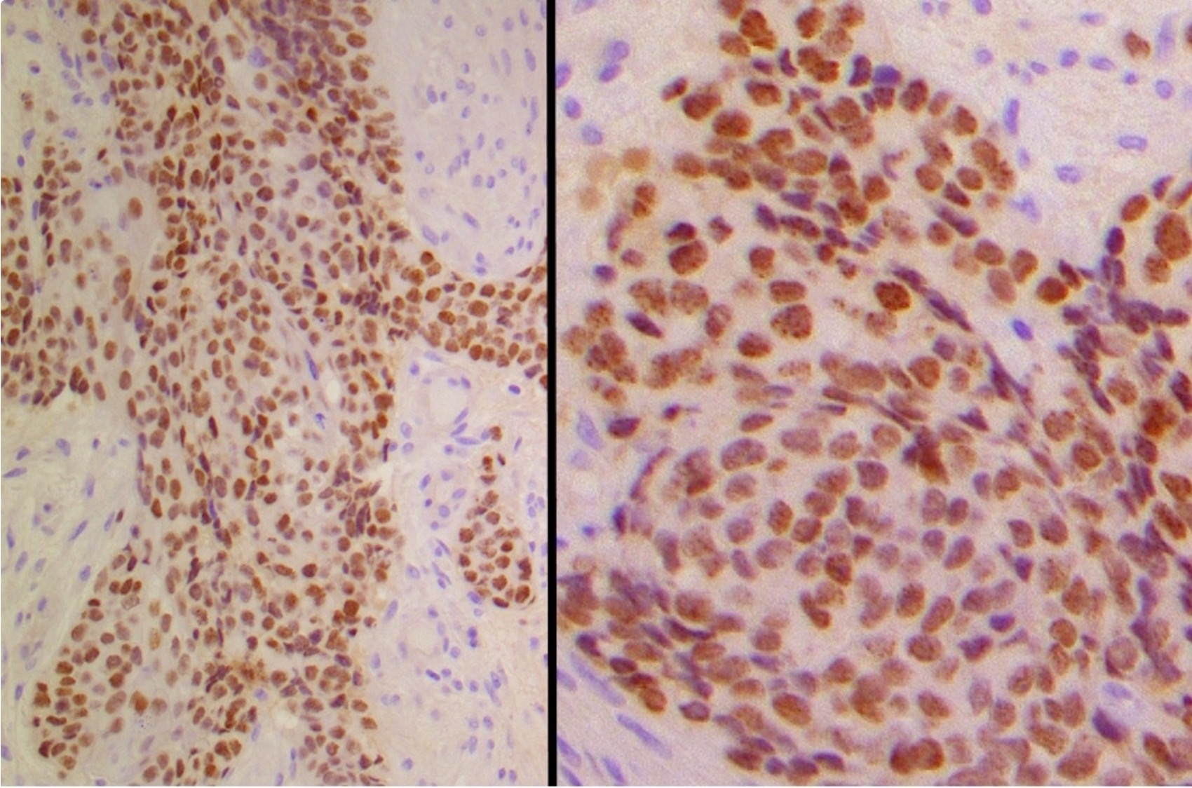
Diffuse NUT with speckled pattern
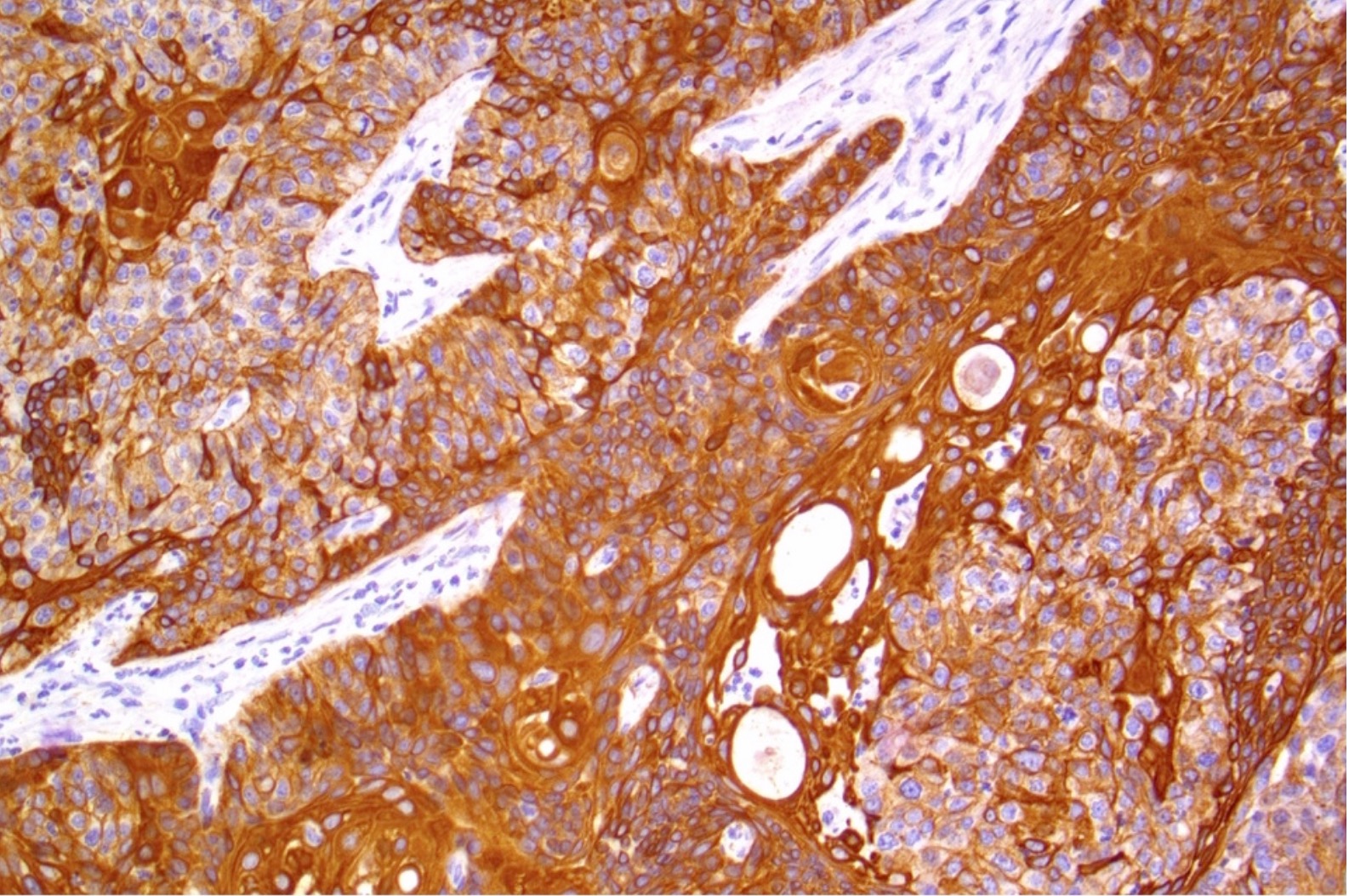
AE1 / AE3
Positive staining - normal
- Testis: germ cells (Am J Surg Pathol 2009;33:984)
- Ovary: oocytes (weak) (Am J Surg Pathol 2009;33:984)
Positive staining - disease
- NUT carcinoma (Am J Surg Pathol 2009;33:984)
- CNS: primitive neuroectodermal tumor (PNET) with CIC::NUTM1 fusions (Cell 2016;164:1060)
- NUT associated mesenchymal tumors arising in the soft tissue and viscera (Am J Surg Pathol 2018;42:636, Virchows Arch 2020;476:317, Histopathology 2022;81:131, Genes Chromosomes Cancer 2022;61:542)
- Germ cell tumors: spermatocytic tumor, other germ cell tumors often have focal or weak staining (Am J Surg Pathol 2009;33:984, Histopathology 2014;65:35, Pathology 2015;47:118)
- Poroma and porocarcinoma with NUTM1 rearrangement (J Cutan Pathol 2021;48:403, Am J Surg Pathol 2021;45:1221, J Cutan Pathol 2022;49:850)
Negative staining
- Normal:
- Breast
- Large intestine
- Liver (minority of cases may contain cytoplasmic and nuclear blush)
- Lung
- Prostate
- Thymus
- Tonsil (Am J Surg Pathol 2009;33:984)
- Disease:
- Adenocarcinoma, NOS
- Ewing sarcoma
- Nasopharyngeal carcinoma
- Neuroblastoma
- Renal cell carcinoma
- Rhabdomyosarcoma
- Rhabdoid tumor
- Serous carcinoma
- Sinonasal undifferentiated carcinoma
- Small cell carcinoma
- Squamous cell carcinoma, NOS
- Urothelial carcinoma (Am J Surg Pathol 2009;33:984)
Sample pathology report
- Immunohistochemical analysis of NUTM1 expression (clone, dilution, automated staining platform*):
- The tissue staining pattern is positive nuclear / negative.*
- Positive and negative controls stain appropriately.
- * Details to be specified / confirmed upon reporting stain
Board review style question #1
A mediastinal mass is biopsied and shows a morphologically undifferentiated carcinoma. Which of the following staining patterns would be most consistent with classification as NUT carcinoma?
- Cytoplasmic expression in < 90% of cells with a speckled pattern
- Cytoplasmic expression in < 90% of cells with an even / uniform pattern
- Nuclear expression in > 90% of cells with a speckled pattern
- Nuclear expression in > 90% of cells with an even / uniform pattern
Board review style answer #1
C. Nuclear expression in > 90% of cells with a speckled pattern. In NUT carcinoma, most tumor cells are positive for this biomarker, which is expressed in the nucleus with a speckled distribution. In contrast, germ cell tumors often have only focal tumor staining, which is expressed in the nucleus with an even / uniform distribution.
Comment Here
Reference: NUTM1
Comment Here
Reference: NUTM1

