Stains & CD markers
p63


Copyright: 2002-2025, PathologyOutlines.com, Inc.
PubMed Search: p63
p63
Editorial Board Members: Christian M. Schürch, M.D., Ph.D., Brandon Umphress, M.D.
Last author update: 20 February 2024
Last staff update: 20 February 2024
Copyright: 2002-2025, PathologyOutlines.com, Inc.
PubMed Search: p63
Table of Contents
Definition / general | Essential features | Terminology | Pathophysiology | Diagrams / tables | Clinical features | Interpretation | Uses by pathologists | Prognostic factors | Microscopic (histologic) description | Microscopic (histologic) images | Positive staining - normal | Positive staining - disease | Negative staining | Molecular / cytogenetics description | Molecular / cytogenetics images | Sample pathology report | Practice question #1 | Practice answer #1Cite this page: Ismail S, Elshimali JYI. p63. PathologyOutlines.com website. https://www.pathologyoutlines.com/topic/stainsp63.html. Accessed October 1st, 2025.
Definition / general
- Transcription factor that is a member of the p53 gene family
- Master regulator of epidermal keratinocyte proliferation and embryonic epidermal growth
Essential features
- Encoded by the gene TP63 located on 3q27-29 (Mol Cell 1998;2:305, Development 2012;139:772)
- Has more than 6 isoforms
- Marker for myoepithelial cells
Terminology
- Transcription factor
- Known as the guardian of reproduction (Development 2012;139:772)
- Formerly known as keratinocyte transcription factor (KET) in 1997 (Oncogene 1997;15:1363)
Pathophysiology
- Regulates embryonic stem cells development and commitment to epithelial tissue (Genes Dev 2006;20:3185)
- Functions through epigenetic regulation of Satb1, Brg1, Cbx4 and tissue specific chromatin factors (Development 2014;141:101, J Cell Biol 2011;194:825, J Cell Biol 2016;212:77)
- Limb malformation, ectodermal dysplasia and orofacial clefting are associated with p63 germline mutations (Am J Med Genet A 2006;140:1419)
- Multiple isoforms that regulate epithelial growth and development were generated from alternative splicing, including
- TA isoform: the full length and most predominant isoform (BMC Genomics 2015;16:584)
- Has 3 specific exons
- Plays a role in protein interactions through a sterile alpha motif (SAM) domain
- Includes a transactivation domain
- Acts as a transcription factor and a tumor suppressor gene
- Activates BAX, p21 and other p53 target genes
- ΔN isoform: also known as p40 (Mol Cell Biol 2002;22:8601, Development 2012;139:772)
- Was considered an inactive transcription factor
- Inhibits TAP63 isoform
- Essential for epithelial development
- Lacks the transactivation domain TA1
- Shorter than TAP63
- (β) isoform: lacks exon 13, the transcription inhibitory (TID) and sterile alpha motif domains (Nucleic Acids Res 2009;37:6092)
- (γ) isoform: lacks the exons 11 - 14, mainly expressed in muscle cells and cancer-like stem cells (Nucleic Acids Res 2009;37:6092, Mol Cell 1998;2:305)
- ΔΔN isoform: a recently generated isoform, distinct from the ΔN isoform as it lacks its first 26 amino acids in epidermal keratinocytes (Nucleic Acids Res 2009;37:6092, Hum Mol Genet 2008;17:1968)
- (δ) isoform: lacks exon 13
- (ε) isoform: detected first though generation from a stop codon in exon 10 (Nucleic Acids Res 2009;37:6092, Hum Mol Genet 2008;17:1968)
- TA isoform: the full length and most predominant isoform (BMC Genomics 2015;16:584)
Diagrams / tables
Images hosted on other servers:

Structure of p63 molecule
Clinical features
- Investigate the primary origin of malignant tumors
- In most cases, p63 can help exclude the diagnosis of invasive adenocarcinomas in the prostate and breast; however, there are exceptions
- Case of prostatic adenocarcinoma in 65 year old man with diffuse, nonbasal p63 staining (Arch Pathol Lab Med 2013;137:1179)
- Low level of p63 staining in tumor cells in 2 cases of metaplastic breast carcinoma (Hamdan Medical Journal 2023;16:278)
Interpretation
- Mainly nuclear
- Could demonstrate cytoplasmic expression in Z bands in skeletal muscle cells (Mod Pathol 2011;24:1320)
Uses by pathologists
- In lung: detects squamous cell carcinoma and differentiates it from adenocarcinomas (J Cytol 2021;38:151)
- In the kidneys: distinguishes urothelial carcinoma from renal cell carcinoma (Hum Pathol 2014;45:1824)
- In the prostate: distinguishes prostatic hyperplasia from well differentiated prostatic adenocarcinoma (Am J Surg Pathol 2002;26:1161)
- In the bladder: distinguishes poorly differentiated urothelial carcinoma from metastatic prostatic adenocarcinoma (J Pathol Transl Med 2016;50:345)
- Favors the diagnosis of sarcomatoid carcinoma over sarcomas in the bladder (Mod Pathol 2005;18:1471)
- In most cases, p63 can help exclude the diagnosis of invasive adenocarcinomas in the prostate and breast; however, there are exceptions
- Case of prostatic adenocarcinoma in 65 year old man with diffuse, nonbasal p63 staining (Arch Pathol Lab Med 2013;137:1179)
- Low level of p63 staining in tumor cells in 2 cases of metaplastic breast carcinoma (Hamdan Medical Journal 2023;16:278)
- Differentiates cutaneous sweat gland carcinoma (p63+) from metastatic breast adenocarcinoma to skin (p63-) (Arch Pathol Lab Med 2011;135:975)
- Differentiates olfactory neuroblastoma (p63- / calretinin+) from other small round blue cell tumors of sinonasal tract (Am J Surg Pathol 2011;35:1786)
- Differentiates renal collecting duct carcinoma (p63- / PAX8+) from upper tract urothelial carcinoma (Am J Surg Pathol 2011;35:757)
- p63 expression can be helpful in identifying primary adnexal neoplasms versus other metastatic processes (J Cutan Pathol 2007;34:474)
Prognostic factors
- Loss of p63 expression correlates with poor prognosis in urothelial carcinoma, squamous cell carcinoma of the esophagus and larynx (Hum Pathol 2014;45:1824, Oncol Rep 2006;15:323, ORL J Otorhinolaryngol Relat Spec 2010;72:319)
- p63 expression is associated with poorer prognosis for Merkel cell carcinoma (Mod Pathol 2011;24:1451)
- Identifies false lymphatic invasion (tumor cells surrounded by D2-40+ / p63+ ducts), which has a good prognosis (Mod Pathol 2011;24:502)
Microscopic (histologic) description
- Nuclear marker for myoepithelial cells in benign tumors and noninvasive malignancies
Microscopic (histologic) images
Contributed by John Yahya I. Elshimali, M.D., Andrey Bychkov, M.D., Ph.D.,
Jijgee Munkhdelger, M.D., Ph.D., Hind Nassar, M.D. and Cases #117, 191 and 209
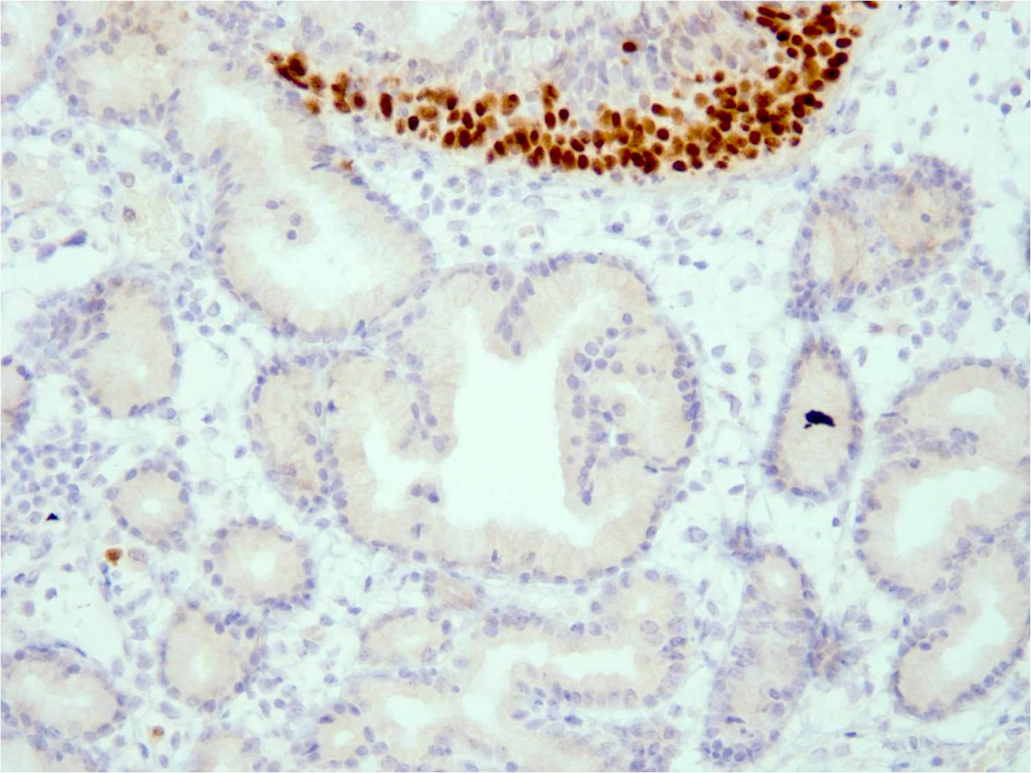
Prostatic glands
Myoepithelial cells staining positive for p63
Prostatic adenocarcinoma
Endometrial adenocarcinoma
Mammary glands and ducts
Myoepithelial cells
Elongated tubular structures with chondroid fibrous stroma
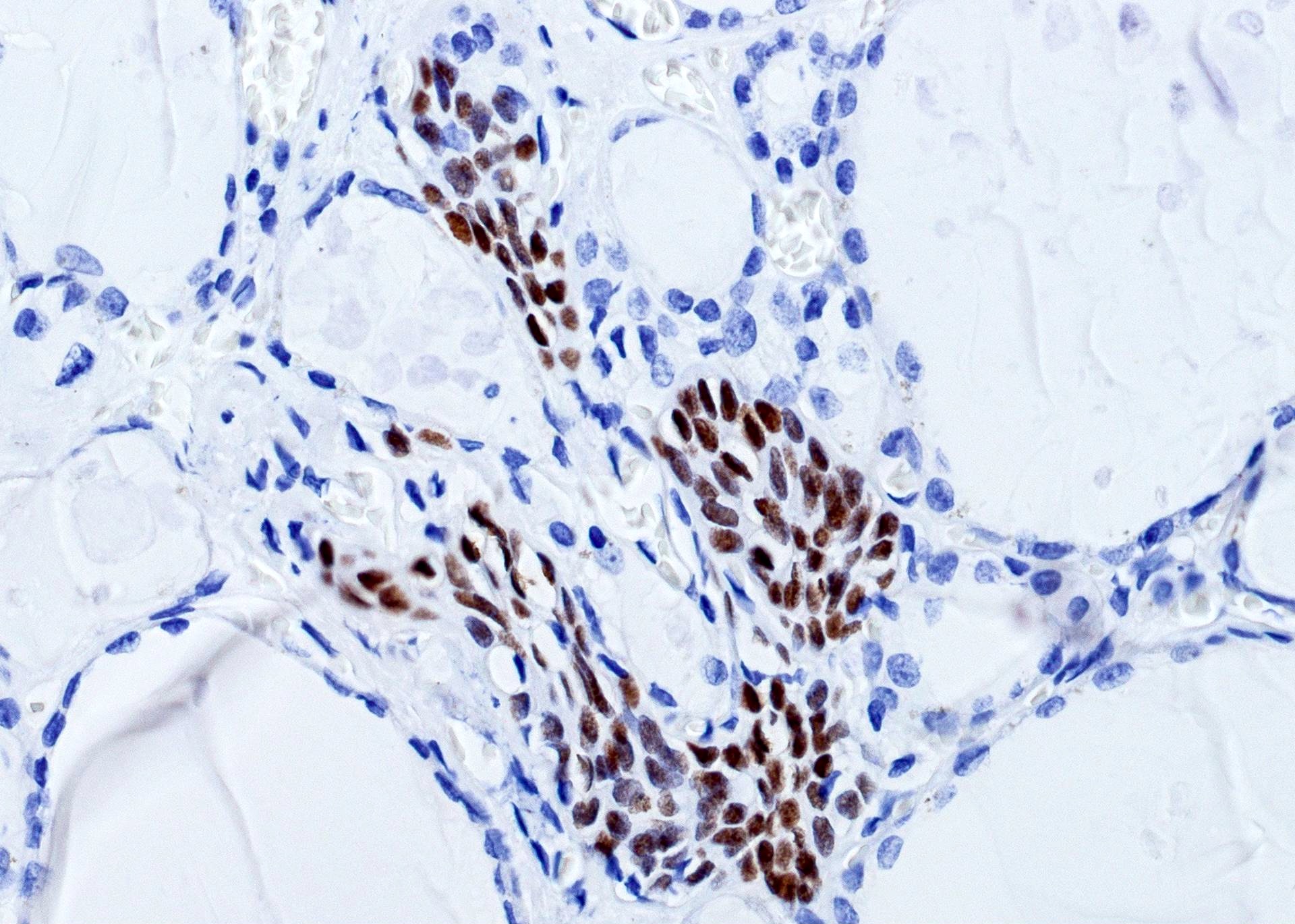
Solid cell nests

p63+ in many follicles
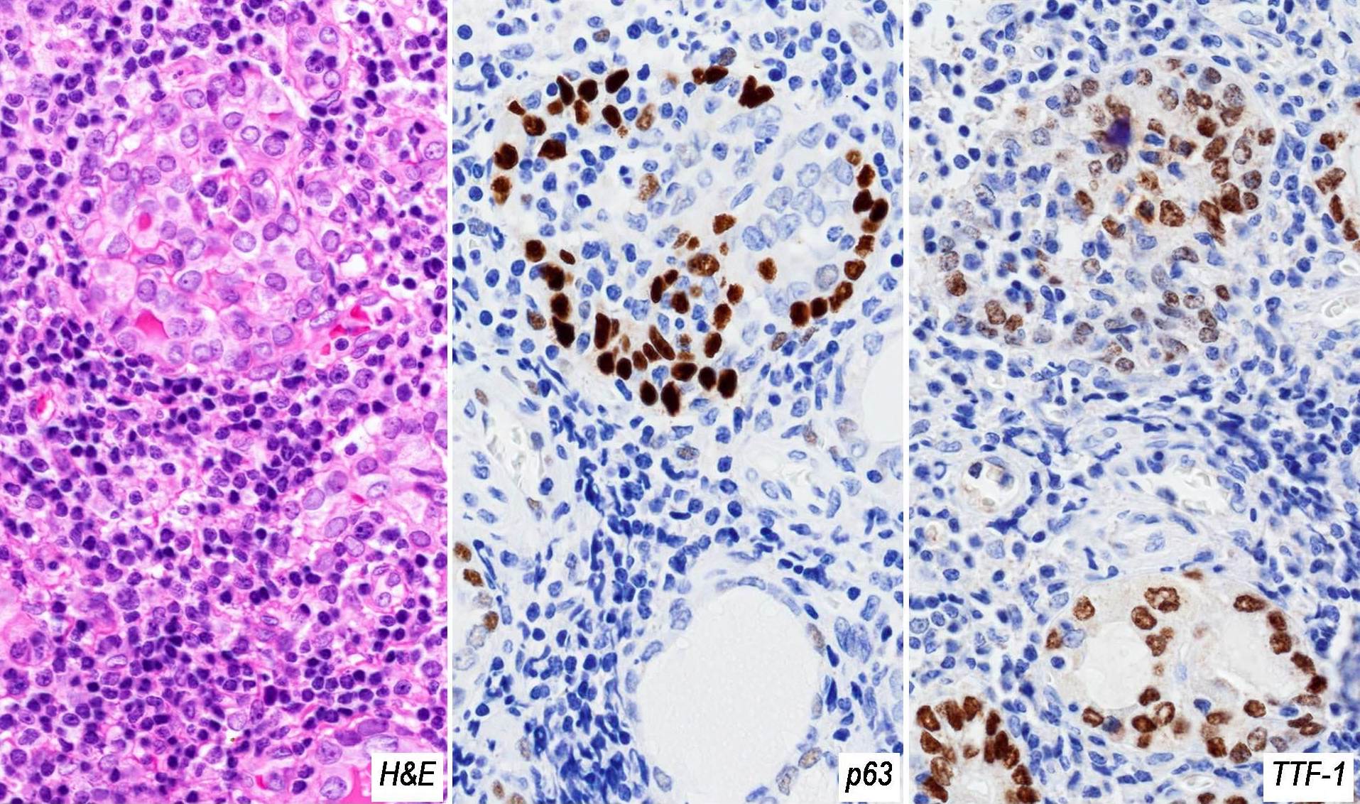
Correlates with a loss of TTF1
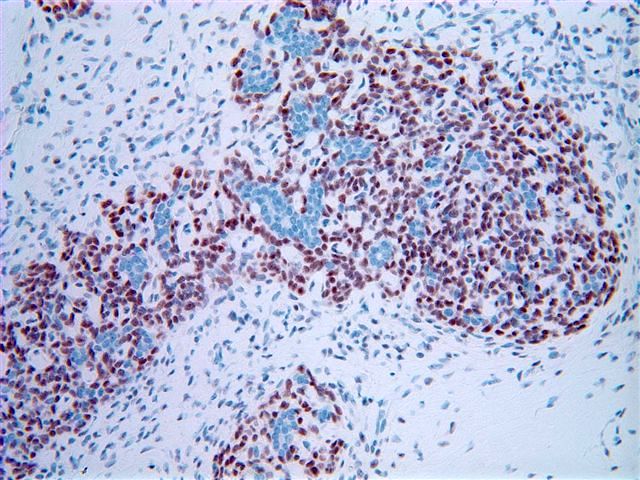
Adenoid cystic carcinoma, solid
Seromucinous hamartoma p63-, no myoepithelial cells, positive internal control
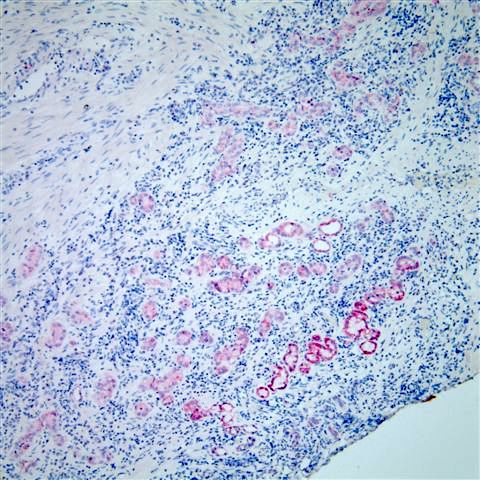
Nephrogenic adenoma (p63- / AMACR+)
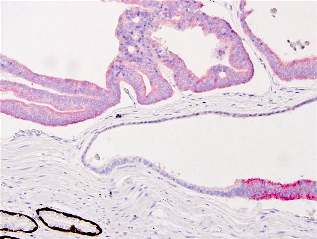
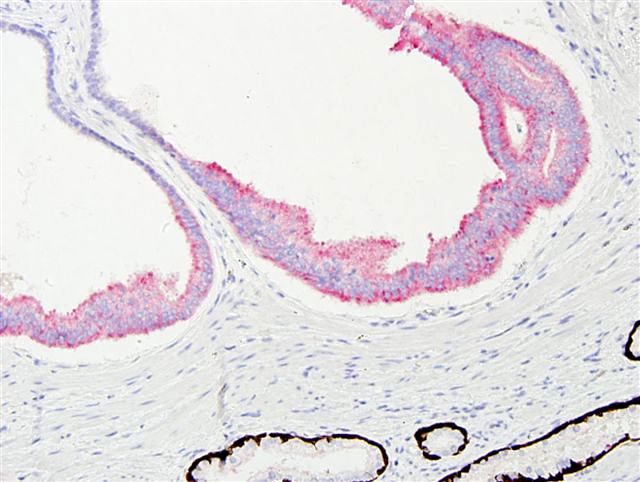
Adenocarcinoma (upper, p63- / AMACR+) and benign (lower, p63+ / AMACR-)
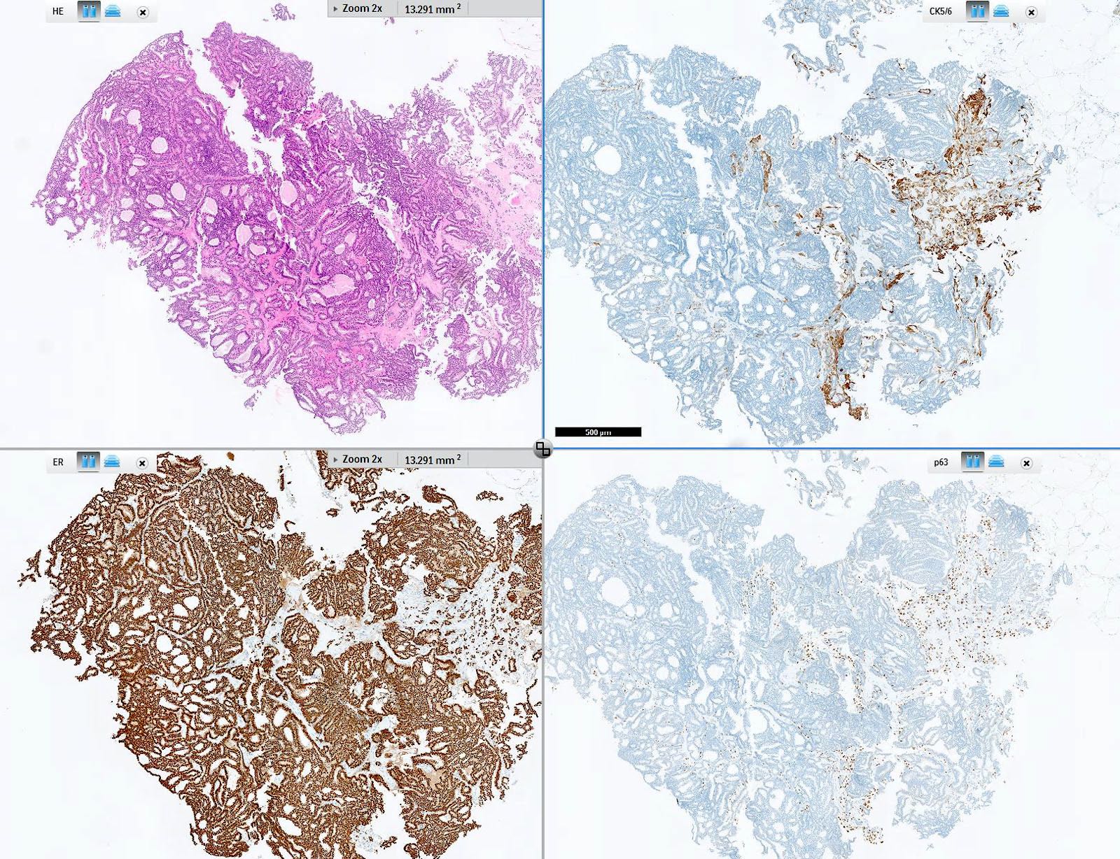
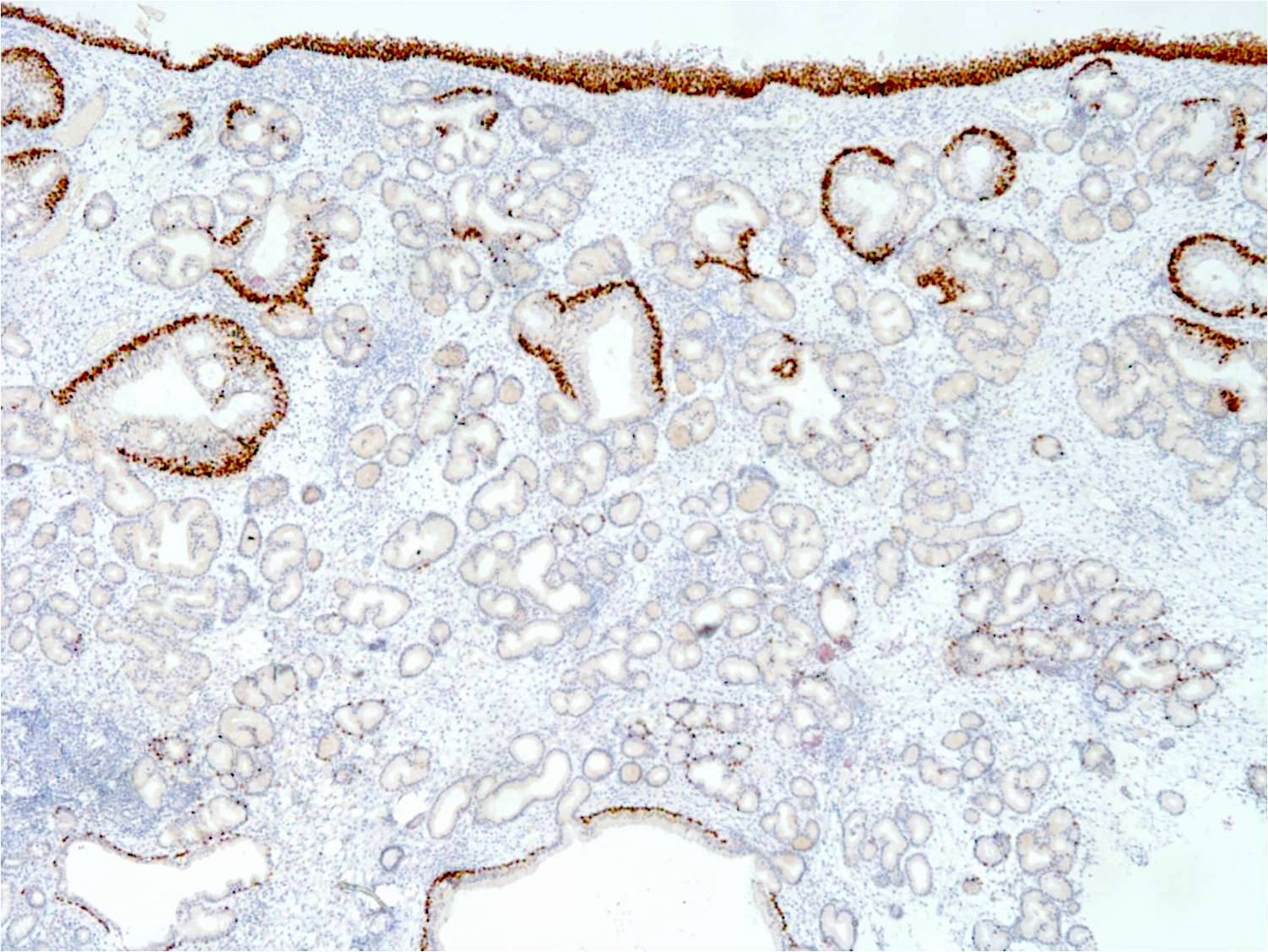
Intraductal papilloma with DCIS immunoprofile
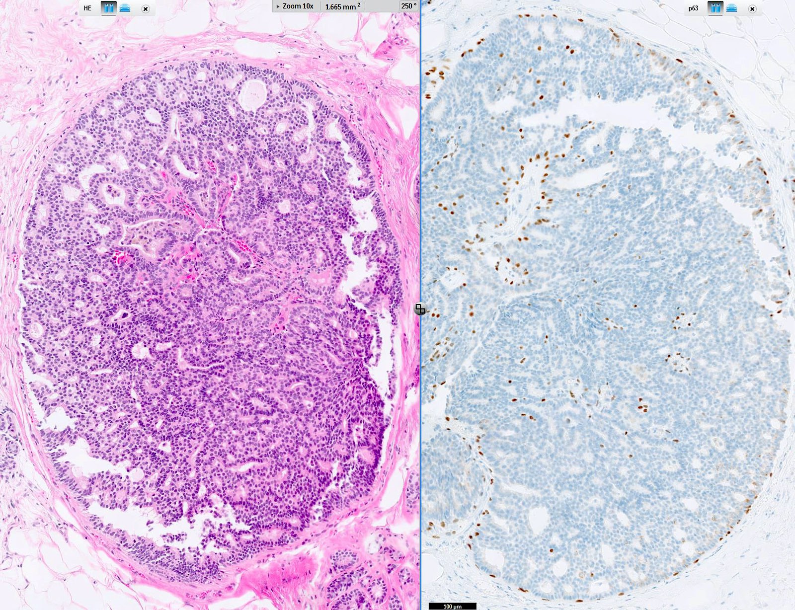
Intraductal papilloma with DCIS, p63
Positive staining - normal
- Squamous epithelial cells (Biomark Res 2021;9:7)
- Thymic epithelial cells (Biomark Res 2021;9:7)
- Urothelium (Biomark Res 2021;9:7)
- Basal cells of the respiratory tract (Biomark Res 2021;9:7)
- Basal cells of prostate (Biomark Res 2021;9:7)
- Basal cells of seminal vesicle (Biomark Res 2021;9:7)
- Gynecologic tract: basal and parabasal cells of mature cervical, vaginal and vulval squamous epithelium; cervical reserve cells at transformation zone, immature metaplastic and atrophic cervical squamous epithelium (J Pathol Transl Med 2015;49:450)
- Myoepithelial cells in breast (Am J Surg Pathol 2001;25:1054)
- Myoepithelial cells in parotid, submandibular and sublingual glands (Biomark Res 2021;9:7, Clin Cancer Res 2002;8:494)
Positive staining - disease
- In the skin: squamous cell carcinomas, basal cell carcinomas (Biomark Res 2021;9:7)
- Primary cutaneous adnexal neoplasms (with the exception of primary mucinous carcinoma in most cases)
- Tumors of the head and neck: squamous cell carcinoma of the larynx / oral cavity, pleomorphic adenoma of the parotid gland, Warthin tumor (Biomark Res 2021;9:7)
- Tumors of the mediastinum: thymoma (in 65%) (Biomark Res 2021;9:7, Clin Cancer Res 2002;8:494)
- In the lung: identification of squamous cell carcinoma, mainly the specific isoform p40 ΔNp63 (PLoS One 2010;5:e12209, Mod Pathol 2012;25:405)
- Tumors of the female genital tract: squamous cell carcinoma of the vagina / vulva / cervix, Brenner tumor (Biomark Res 2021;9:7)
- Sarcomatoid carcinoma of the breast (Histopathology 2003;42:94)
- Tumors of the digestive system: squamous cell carcinoma of the esophagus / anal canal
- Tumors of the urinary system: urothelial carcinoma (Hum Pathol 2014;45:1824)
- Mild expression in diffuse large B cell lymphoma, follicular lymphoma and chronic lymphocytic leukemia (Appl Immunohistochem Mol Morphol 2005;13:237, Cancer Sci 2006;97:1050, J Clin Pathol 2009;62:77)
- ALK negative ALCLs can express p63, which is associated with TP63 rearrangements (Hum Pathol 2017;64:19)
Negative staining
- Anus: anal gland carcinoma (Arch Pathol Lab Med 2007;131:1304)
- Breast: normal epithelium, stromal cells, myofibroblasts; may be reduced or occasionally absent in benign apocrine lesions, benign sclerosing lesions (Am J Surg Pathol 2011;35:202, Histopathology 2016;68:1030)
- Malignant mixed Müllerian tumors, endometrioid carcinoma, pancreatic, colorectal and cholangiocellular carcinoma (10 - 25%) (Proc Natl Acad Sci U S A 2013;110:8105, Oncotarget 2017;8:22741)
- Melanoma (Am J Surg Pathol 2012;36:1216)
- Mesothelioma
- Prostate: adenocarcinoma, nephrogenic adenoma, partial atrophy (Am J Surg Pathol 2012;36:1216)
- Most soft tissue tumors (Am J Clin Pathol 2011;136:762)
Molecular / cytogenetics description
- Encoded by the gene TP63 located on 3q27-29
- Multidomain protein with more than 6 isoforms
- Isoforms are generated through alternative splicing at the 3 site
- Tetramer that has a DNA binding domain (DBD), an N terminal transcactivation domain (TA), a tetramerization domain (TD) and a transcription inhibitory domain following C terminal sterile alpha motif (SAM) (Mol Cell Biol 2002;22:8601, Mol Cell 1998;2:305)
Molecular / cytogenetics images
Images hosted on other servers:

Characterization of p63 isoform specific antibodies
Sample pathology report
- Prostate, core needle biopsy, measuring 2 x 1 x 1 cm and tan-brown in color:
- Benign prostatic hyperplasia (see comment)
- Comment: Microscopic examination of the biopsy revealed the proliferation of prostatic glands lined by epithelial cells with no signs of atypia in the lining of epithelial cells. Immunohistochemistry demonstrated a strong nuclear expression of p63 in the basal cells of the hyperplastic glands. These features correlate with benign prostatic hyperplasia.
Practice question #1
Which of the statements below regarding p63 is true?
- Has only 3 isoforms
- Loss of p63 expression correlates with poor prognosis in urothelial carcinoma
- p63 is absent in squamous epithelial cells
- p63 is a marker for smooth muscle cells
Practice answer #1
B. Loss of p63 expression correlates with poor prognosis in urothelial carcinoma. Answer A is incorrect because p63 has more than 6 isoforms. Answer C is incorrect because p63 is a marker for squamous epithelial cells. Answer D is incorrect because p63 is a marker for basal epithelial cells.
Comment Here
Reference: p63
Comment Here
Reference: p63