
Stains & CD markers
Uroplakin II


Copyright: 2022-2024, PathologyOutlines.com, Inc.
PubMed Search: Uroplakin II
Uroplakin II
Editorial Board Member: Brandon Umphress, M.D.
Deputy Editor-in-Chief: Maria Tretiakova, M.D., Ph.D.
Last author update: 22 September 2022
Last staff update: 5 September 2023
Copyright: 2022-2024, PathologyOutlines.com, Inc.
PubMed Search: Uroplakin II

Table of Contents
Definition / general | Essential features | Terminology | Clinical features | Interpretation | Uses by pathologists | Prognostic factors | Microscopic (histologic) images | Positive staining - normal | Positive staining - disease | Negative staining | Molecular / cytogenetics description | Sample pathology report | Board review style question #1 | Board review style answer #1 | Board review style question #2 | Board review style answer #2 | Board review style question #3 | Board review style answer #3 | Board review style question #4 | Board review style answer #4Cite this page: Batra H, Parwani A. Uroplakin II. PathologyOutlines.com website. https://www.pathologyoutlines.com/topic/stainsuroplakinii.html. Accessed April 19th, 2024.
Definition / general
- Uroplakin II, a 15kDa protein that is part of the uroplakin family (UP1a, UP1b, UPII and UPIII), which forms the urothelial plaque covering the apical surface of the urothelium
Essential features
- Uroplakins are terminal differentiation products that are exclusively expressed in urothelial cells (Kidney Int 2009;75:1153)
- 15 kDa transmembrane protein involved in terminal differentiation of urothelium
- Exists as a heterodimer along with UP1a (Kidney Int 2009;75:1153)
- UPII IHC has significantly higher sensitivity (57% versus 50%) than UPIII and the same specificity (~99%) for urothelial carcinomas (Histopathology 2014;65:132)
- Helpful in pinpointing the origin where secondary involvement by prostate adenocarcinoma or metastatic carcinoma may be positive for GATA3
Terminology
- UPII, UPK2, UROII
Clinical features
- Highly specific and moderately sensitive for urothelial carcinomas
- Helpful in pinpointing the primary origin as urothelium in cases where GATA3 is inconclusive
Interpretation
- Moderate to strong, predominantly membranous and cytoplasmic staining reaction in virtually all umbrella cells in the urethra (Appl Immunohistochem Mol Morphol 2022;30:326)
- At least weak to moderate cytoplasmic and membranous staining reaction of the majority of intermediate urothelial cells (Hum Pathol 2007;38:1703)
- Diffuse membranous staining in urothelial carcinoma cells (Appl Immunohistochem Mol Morphol 2022;30:326)
Uses by pathologists
- Highly specific and moderately sensitive for urothelial carcinomas
- Helpful in ascertaining the primary origin as urothelium, especially in cases where GATA3 is inconclusive
- Currently recommended by ISUP as a second line additional marker to confirm urothelial primary (Am J Surg Pathol 2014;38:e20)
Prognostic factors
- Loss of UPII seen more frequently in higher grade lesions (Arch Pathol Lab Med 2014;138:943)
Microscopic (histologic) images
Contributed by Charles C. Guo, M.D., Varsha Nair, M.D., Prih Rohra, M.D., Priya Rao, M.D. and Bogdan A. Czerniak, M.D., Ph.D.
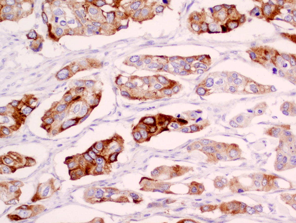
Bladder, invasive urothelial carcinoma
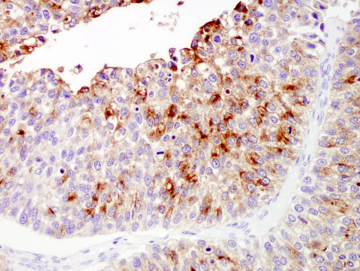
Bladder, papillary urothelial carcinoma
Positive staining - normal
- Apical expression in normal bladder epithelium (umbrella cells)
- Intermediate cells in normal bladder epithelium
Positive staining - disease
- Urothelial carcinoma
- Ovarian Brenner tumor (Am J Surg Pathol 2003;27:1434)
- Pleomorphic invasive lobular carcinoma (single study) (Dis Markers 2016;2016:2940496)
- Apocrine metaplasia of breast (single study) (Dis Markers 2016;2016:2940496)
- Apocrine carcinoma of breast (single study) (Dis Markers 2016;2016:2940496)
- Adenocarcinoma of the colon (< 5% of cases) (Anticancer Res 2018;38:4759)
- Squamous cell carcinoma of the lung (< 5% of cases) (Anticancer Res 2018;38:4759)
- Endometrioid carcinoma of the uterine corpus (< 5% of cases) (Anticancer Res 2018;38:4759)
- Classical hepatocellular carcinoma of the liver (10%) (seen only in intracytoplasmic hyaline bodies) (Anticancer Res 2018;38:4759)
Negative staining
- Prostatic carcinoma (Anticancer Res 2018;38:4759)
- Squamous cell carcinoma of bladder (Hum Pathol 2013;44:164)
- Renal cell carcinoma (Histopathology 2014;65:132)
- Classic invasive lobular carcinoma (Dis Markers 2016;2016:2940496)
Molecular / cytogenetics description
- UPII mRNA expression in tissue and peripheral blood is associated with lymph node metastasis (J Pathol 2003;199:41)
Sample pathology report
- Lymph node, pelvic node dissection:
- Metastatic urothelial carcinoma (see comment)
- Comment: Histologic sections show a lymph node with deposits of tumor cells in diffuse sheets and few with a trabecular pattern. The cells are round to polygonal, demonstrating moderate pleomorphism, inconspicuous nucleoli and high N:C ratios. Immunohistochemistry is positive for CK20, p63, GATA3, S100, uroplakin III and uroplakin II and negative for PSA, PSAP, NKX3.1 and prostein, suggesting a urothelial origin. Please correlate clinicoradiologically. Follow up is advised.
Board review style question #1
Uroplakin II forms a heterodimer with which other uroplakin?
- UPIa
- UPIb
- UPIII
- UPV
Board review style answer #1
Board review style question #2
Uroplakin II is expressed normally in which of the following?
- Apical region (umbrella cells) of urothelium
- Muscularis propria of bladder
- Normal breast tissue
- Prostate
Board review style answer #2
Board review style question #3
Uroplakin II is highly specific for which of the following?
- Infiltrating ductal carcinoma, NOS, breast
- Lung adenocarcinoma
- Prostate carcinoma
- Urothelial carcinoma
Board review style answer #3
Board review style question #4
Which of the following is true regarding uroplakin II, as compared to uroplakin III?
- Equally sensitive IHC marker for urothelial carcinoma
- Less sensitive IHC marker for urothelial carcinoma
- Less specific IHC marker for urothelial carcinoma
- More sensitive IHC marker for urothelial carcinoma
Board review style answer #4

