

Testis & paratestis
Sex cord stromal tumors
Myoid gonadal stromal tumor
Editorial Board Member: Debra L. Zynger, M.D.
Deputy Editor-in-Chief: Maria Tretiakova, M.D., Ph.D.


Last author update: 25 April 2023
Last staff update: 25 April 2023
Copyright: 2022-2024, PathologyOutlines.com, Inc.
PubMed Search: Myoid gonadal stromal tumor
Table of Contents
Definition / general | Essential features | Terminology | ICD coding | Epidemiology | Sites | Pathophysiology | Etiology | Clinical features | Diagnosis | Laboratory | Radiology description | Radiology images | Prognostic factors | Case reports | Treatment | Gross description | Frozen section description | Microscopic (histologic) description | Microscopic (histologic) images | Positive stains | Negative stains | Electron microscopy description | Molecular / cytogenetics description | Sample pathology report | Differential diagnosis | Board review style question #1 | Board review style answer #1Cite this page: Gupta P, Mehta V. Myoid gonadal stromal tumor. PathologyOutlines.com website. https://www.pathologyoutlines.com/topic/testismyoidgonadalstromaltumor.html. Accessed April 25th, 2024.
Definition / general
- A rare testicular neoplasm composed of spindle shaped cells that share features of smooth muscle and gonadal stroma (Virchows Arch 2021;478:727)
- Hypothesized to arise from peritubular myoid cells or intertubular primitive mesenchymal cells (Am J Clin Pathol 2014;142:675)
- Proposed as an emerging entity in the 2016 WHO classification because of the limited number of cases and the overlapping morphology and immunoprofile with the fibroma thecoma group; distinct entity in the WHO 5th edition
Essential features
- A pure spindle cell tumor that lacks sex cord differentiation; positive for both actin and S100
- Rare testicular tumor that may originate from intertubular primitive mesenchymal cells that undergo myogenic differentiation
- Benign tumor with favorable prognosis
- Small, circumscribed and nonencapsulated tumors that contain medium to large sized ectatic blood vessels with spindled tumor cells arranged in short intersecting fascicles
- Differential diagnosis includes leiomyoma, testicular fibrothecoma and unclassified spindle cell predominant sex cord stromal tumor
Terminology
- Historically, nomenclature includes
- Unusual gonadal stromal tumor
- Testicular stromal tumor with myofilaments
- Unclassified sex cord stromal tumor with predominance of spindle cells
ICD coding
- ICD-O: 8590/0 - myoid gonadal stromal tumor
- ICD-11: 2F77 & XH9G57 - neoplasms of uncertain behavior of male genital organs & sex cord - gonadal stromal tumor, NOS
Epidemiology
- Can occur in any age but typically seen in young to middle aged men between 4 and 59 years (median is 37 years, mean is 43 years) (Am J Clin Pathol 2014;142:675)
Sites
- Affects the testis unilaterally
Pathophysiology
- Hypothesized to arise from peritubular myoid cells which originate from intertubular primitive mesenchymal cells that undergo myogenic differentiation
- Peritubular myoid cells play key roles in the development of the fetal testis and maintenance of testicular function (Hum Pathol 2012;43:144, Arch Histol Cytol 1996;59:1)
Etiology
- Unknown
Clinical features
- Asymptomatic
- Incidental testicular mass
- Testicular pain
- Infertility
- Reference: Am J Clin Pathol 2014;142:675
Diagnosis
- Surgical specimen evaluated by H&E and immunohistochemical staining
Laboratory
- No abnormalities seen
Radiology description
- Ultrasound of testis
- Nonhomogenous: hypoechoeic nodule with hyperechoic spots (Indian J Pathol Microbiol 2022;65:444)
Radiology images
Images hosted on other servers:

Ultrasound features
Prognostic factors
- Small tumors (< 4 cm) with low mitotic activity have an extremely favorable outcome
- Recurrence or metastasis is rarely seen
- Reference: Am J Clin Pathol 2014;142:675
Case reports
- 25 year old man with right testicular mass (Hum Pathol 2012;43:144)
- 38, 43 and 59 year old men with singular testicular masses (Am J Clin Pathol 2014;142:675)
- 41 year old man with infertility (Indian J Pathol Microbiol 2022;65:444)
- 46 year old man with cytologically bland testicular tumor (Ultrastruct Pathol 1991;15:409)
Treatment
- Orchiectomy
- Adjuvant therapy generally not required
Gross description
- Unifocal and well circumscribed with a yellow / tan appearance
- Small sized tumors that range from < 1 cm to 4 cm (Am J Clin Pathol 2014;142:675)
Frozen section description
- Spindle cell neoplasm, low grade
Microscopic (histologic) description
- Circumscribed and nonencapsulated
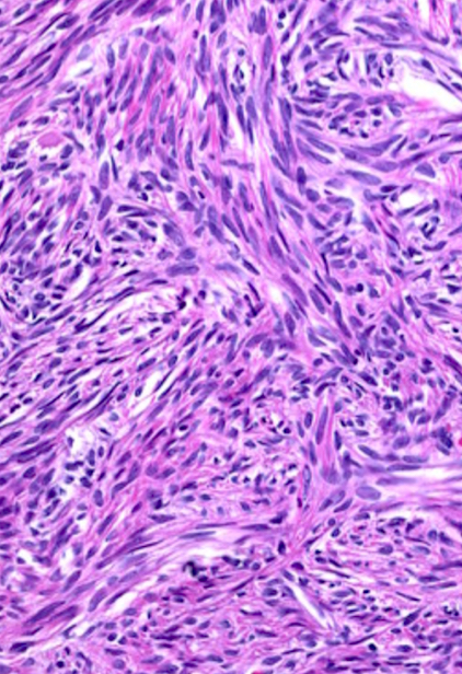
- Focally contain medium to large sized ectatic blood vessels and can be seen adjacent to the rete testis
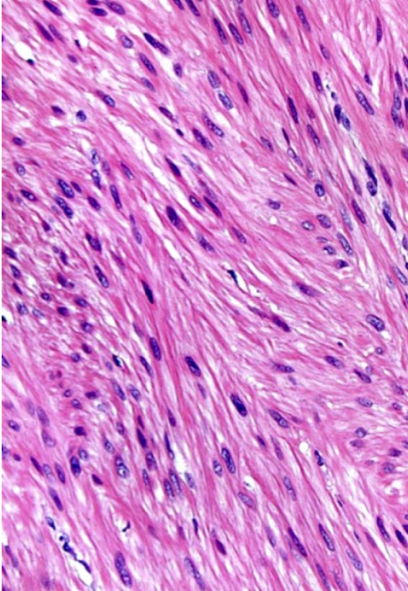
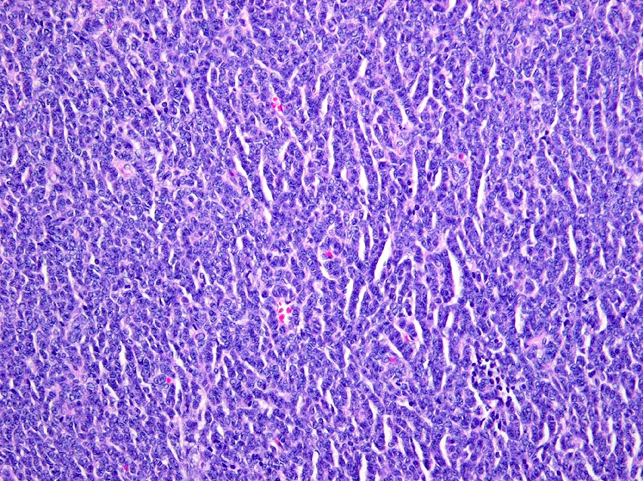
- Tumor cells are spindled and arranged in short intersecting fascicles, sometimes with a storiform pattern
- Scant collagen with thin bands in the background
- Nuclei range from elongated to fusiform with inconspicuous to small nucleoli and occasional grooves
- Cytoplasm is scant to moderate and ill defined, ranging from pale to lightly eosinophilic
- No significant cytologic atypia is seen
- Mitotic figures vary from 0 to 5 per 10 high power fields
- No necrosis or lymphovascular invasion
- No differentiated gonadal stromal cells (Sertoli, Leydig or granulosa cells) are seen
- Reference: Am J Clin Pathol 2014;142:675
Microscopic (histologic) images
Contributed by Vikas Mehta, M.D.
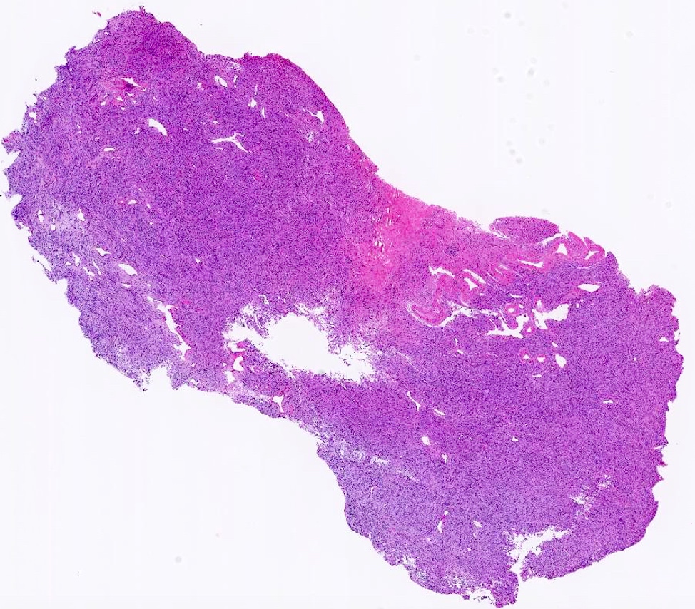
Ectatic blood vessels

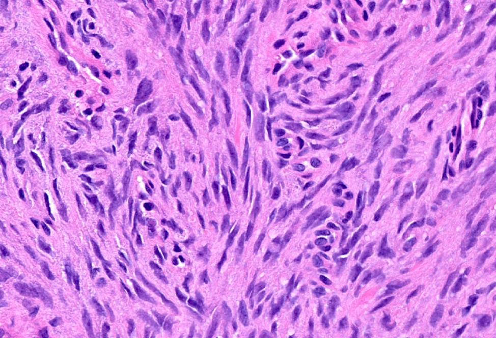
Spindle arrangement of tumor cells

SF1 positivity
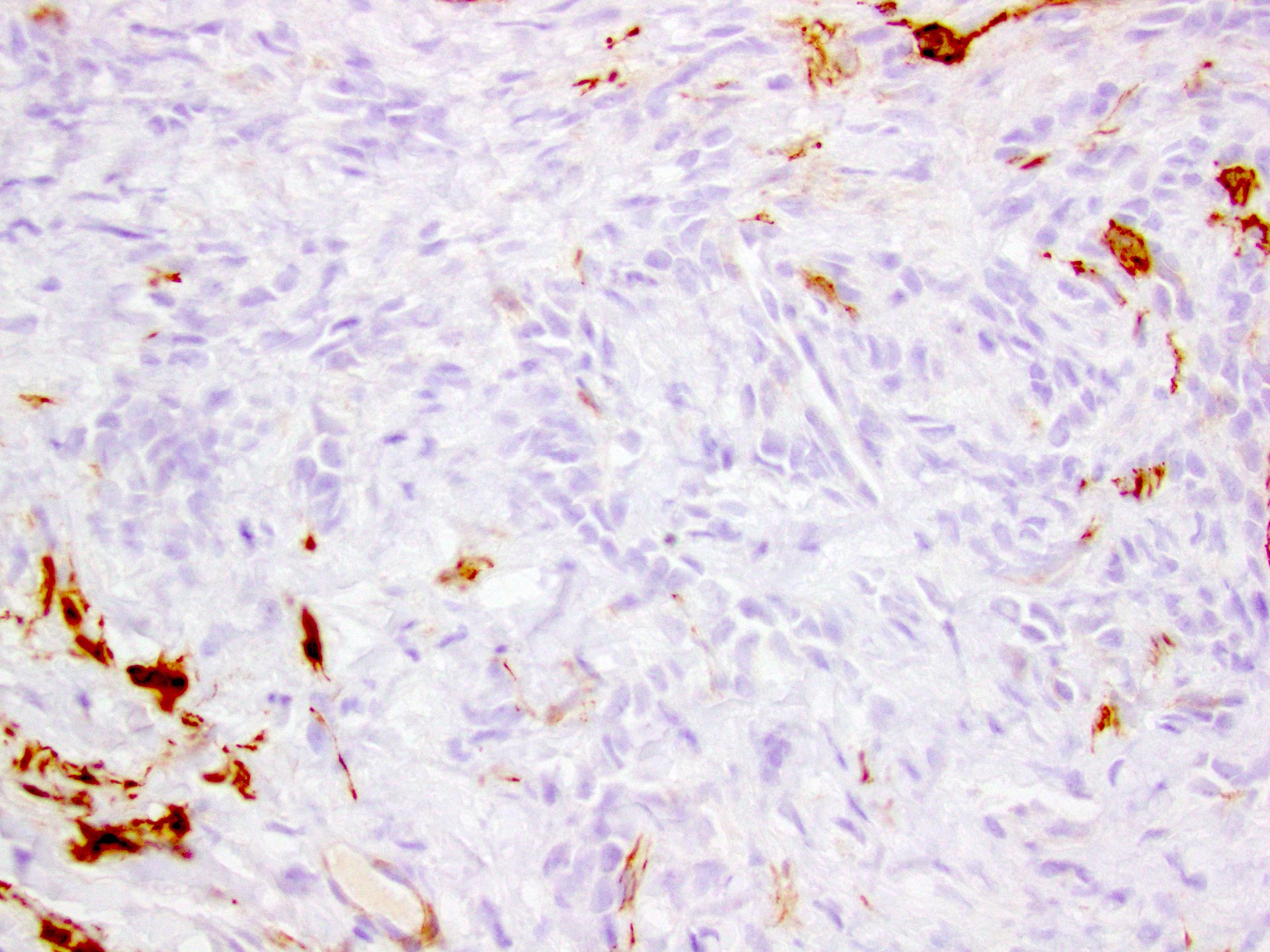
h-caldesmon negativity
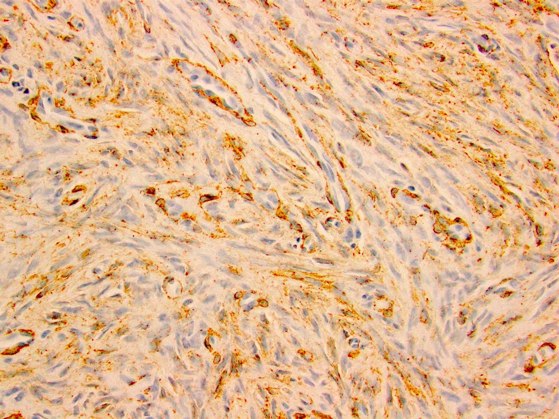
SMA positivity

S100 positivity
Positive stains
- S100 and SMA strong and diffuse positivity is considered a defining criteria (Hum Pathol 2012;43:144, Arch Histol Cytol 1996;59:1)
- FOXL2: strong and diffuse positive
- SF1: diffuse positive
- Muscle specific actin
- Vimentin
- Inhibin: focal, weak and variable positive
- Desmin: variable positive
- Calponin: focal and weak positive
- WT1: variable positive
Negative stains
- h-caldesmon
- Calretinin
- SOX9
- CD34
- Peritubular myoid cells in the adjacent seminiferous tubules can be positive for SMA, desmin, h-caldesmon and calponin but negative for S100 protein, inhibin, calretinin, WT1, SOX9, FOXL2 and SF1
- References: Hum Pathol 2012;43:144, Am J Clin Pathol 2014;142:675
Electron microscopy description
- Myofilaments and desmosomes
- Spindle cells showing lipid vacuoles
- Elongated cell with spindle nucleus and small cytoskeletal filaments (Virchows Arch 2021;478:727)
Molecular / cytogenetics description
- Recurrent chromosome arm level and whole chromosome level copy number gains indicative of ploidy shifts
- Chromosome gains of 3, 6 or 6p, 7, 8, 9 or 9q, 11, 12, 14q, 15q, 17 or 17p, 18q, 20 and 21q and copy number loss 22q (Histopathology 2023;82:431)
Sample pathology report
- Testis, biopsy:
- Myoid gonadal stromal tumor (see comment)
- Comment: The biopsy shows a spindle cell tumor with fascicular and storiform architecture. The tumor cells show varying amounts of eosinophilic to amphophilic cytoplasm, round to oval regular nuclei and inconspicuous or small nucleoli. No vascular invasion, foci of necrosis or atypical mitosis are identified. The tumor cells express SMA, S100 protein, inhibin, FOXL2, calretinin and SF1. In contrast, SOX9, WT1, AE1 / AE3 and EMA expression were not detected. The overall morphologic features and coexpression of S100 protein and SMA support the diagnosis above.
Differential diagnosis
- Leiomyoma:
- Diffuse broad fascicles
- Abundant eosinophilic cytoplasm
- Cigar shaped nuclei
- Negative S100 reactivity
- Positive h-caldesmon reactivity
- Testicular fibrothecoma:
- Collagen bundles are typically thicker and more diffuse, sometimes forming hyaline plaques
- Variable and patchy S100 reactivity
- Positive SOX9 reactivity
- Unclassified spindle cell predominant sex cord stromal tumor:
- Subtle sex cord elements identified on H&E
- Darker, tight groupings of cells with rounded nuclei and indistinct, typically pale, often scant cytoplasm; may be arranged in nests or short cords
- Immunohistochemical stain reactivity may show significant overlap
Board review style question #1
Myoid gonadal stromal tumor is a new entity described in the latest WHO classification of testicular tumors. A representative histologic image is shown above. Which of the following is true for these tumors?
- Consistent pattern of recurrent chromosome arm level and whole chromosome level copy number gains are indicative of ploidy shifts
- Intermixed primitive germ cells and diffuse broad fascicles are present
- Mitotic count of > 5/10 high power fields is common
- Vascular invasion is a defining characteristic of these tumors
Board review style answer #1
A. Consistent pattern of recurrent chromosome arm level and whole chromosome level copy number gains are indicative of ploidy shifts
Comment Here
Reference: Myoid gonadal stromal tumor
Comment Here
Reference: Myoid gonadal stromal tumor
