

Thyroid & parathyroid
Other thyroid malignancies
Angiosarcoma
Author: Sheren Younes, M.D., Ph.D.

Last author update: 1 August 2015
Last staff update: 15 August 2023
Copyright: 2003-2024, PathologyOutlines.com, Inc.
PubMed Search: angiosarcoma thyroid

Table of Contents
Definition / general | Clinical features | Prognostic factors | Case reports | Gross description | Gross images | Microscopic (histologic) description | Microscopic (histologic) images | Cytology description | Positive stains | Negative stains | Differential diagnosisCite this page: Younes S. Angiosarcoma. PathologyOutlines.com website. https://www.pathologyoutlines.com/topic/thyroidangiosarcoma.html. Accessed April 19th, 2024.
Definition / general
- Seen in elderly in Alpine regions of Europe, where tumor may comprise 16% of thyroid malignancies, due to high prevalence of iodine deficient goiter
- Non-Alpine tumors are rare
Clinical features
- Thyroid mass
- Compression symptoms
- Symptoms related to distant metastasis
Prognostic factors
- Often poor prognosis due to persistent local disease and distant metastases (Am J Clin Pathol 1994;102:322)
- May have favorable prognosis if confined to thyroid at surgery (Virchows Arch 1996;429:131)
Case reports
- 38 year old woman with juxtathyroidal neck soft tissue angiosarcoma (Thyroid 2002;12:427)
- 64 year old man with coexistent angiosarcoma and follicular carcinoma of the thyroid (J Korean Med Sci 2003;18:908)
- 73 year old woman from an non-Alpine area with epithelioid angiosarcoma of thyroid (Endocr Pathol 2015;26:152)
- 74 and 86 year old men with epithelioid angiosarcoma involving the thyroid (Arch Pathol Lab Med 2003;127:E70)
- Two cases of epithelioid angiosarcoma of the thyroid gland (Arch Pathol Lab Med 1994;118:642)
- Metastatic angiosarcoma to the thyroid (Rev Laryngol Otol Rhinol (Bord) 2005;126:111)
Gross description
- Single nodule commonly filled with bloody fluid, compressing thyroid
Gross images
AFIP images
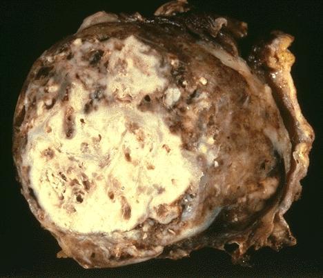
Necrotic and hemorrhagic tumor
Microscopic (histologic) description
- Pleomorphic tumor, usually poorly differentiated, with irregular slit vascular spaces with anastomosing channels or discrete cytoplasmic vacuoles
- Epithelioid variant has poorly circumscribed growth in sheets / cords, intracytoplasmic lumina filled with RBCs; composed of polygonal epithelioid cells with abundant eosinophilic cytoplasm, vesicular nuclei, prominent amphophilic-basophilic nucleoli
Microscopic (histologic) images
AFIP images

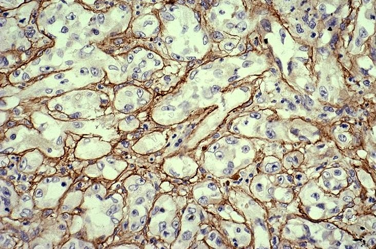
Solid and cellular areas

Abortive vascular lumina
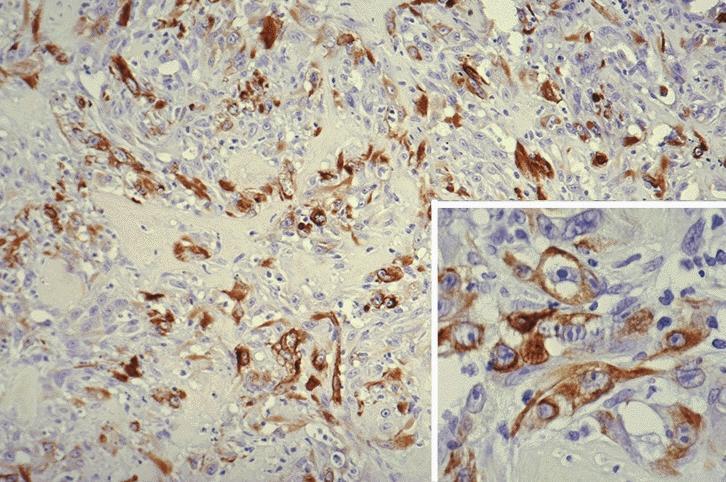
Keratin+ tumor cells
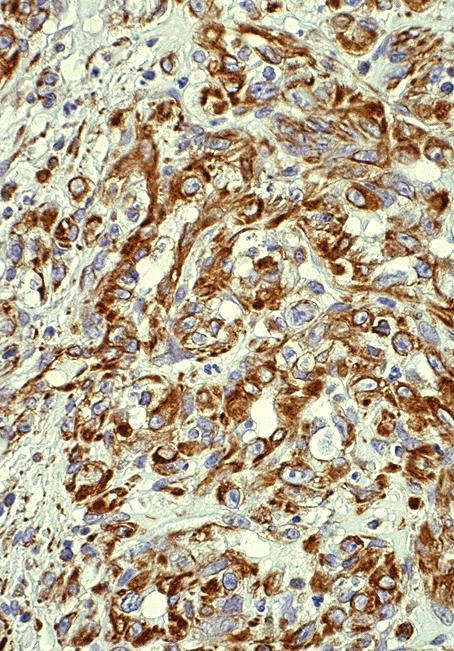
Vimentin+
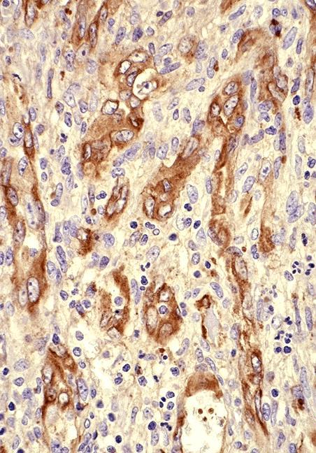
Plump endothelial, Factor VIII+
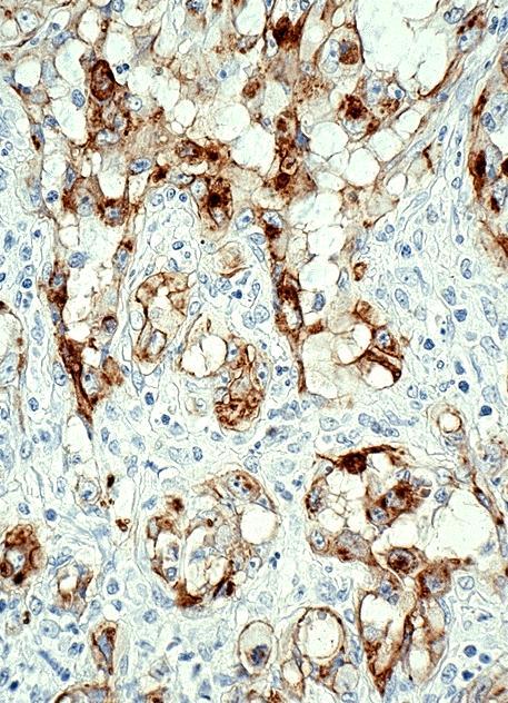
Ulex europaeus I lectin+
Type IV collagen stains basal lamina
Cytology description
- Cellular smear with single cells and small clusters of oval and round tumor cells
- Cell borders are indistinct and cytoplasm is vacuolated
- Nuclei are eccentric with coarse chromatin, irregular membranes and a single, prominent nucleoli
- Also features suggestive of intracytoplasmic lumens (Acta Cytol 2002;46:767)
Positive stains
- Vimentin, factor VIII, CD31, CD34, variable cytokeratin (Am J Surg Pathol 1990;14:737) and Ulex europaeus agglutinin I
Negative stains
Differential diagnosis
- Anaplastic carcinoma or other carcinoma with angiosarcomatous foci: negative for endothelial markers (Am J Surg Pathol 1990;14:69, Am J Clin Pathol 1986;86:674, Diagn Cytopathol 2007;35:424)
- Metastatic angiosarcoma
- Reactive endothelial hyperplasia (Masson Tumor)

