

Thyroid & parathyroid
Other thyroid nonneoplastic
Black / pigmented thyroid
Author: Andrey Bychkov, M.D., Ph.D.

Last author update: 1 June 2015
Last staff update: 15 August 2023
Copyright: 2015-2024, PathologyOutlines.com, Inc.
PubMed Search: black thyroid

Table of Contents
Definition / general | Terminology | Pathophysiology | Clinical features | Diagnosis | Case reports | Gross description | Gross images | Microscopic (histologic) description | Microscopic (histologic) images | Cytology description | Positive stains | Negative stains | Electron microscopy description | Electron microscopy images | Differential diagnosisCite this page: Bychkov A. Black / pigmented thyroid. PathologyOutlines.com website. https://www.pathologyoutlines.com/topic/thyroidblack.html. Accessed April 20th, 2024.
Definition / general
- Rare side effect of minocycline therapy, ~ 100 cases reported
- Pigment deposition in thyroid gland in patients on minocycline (often prescribed for chronic acne) or related tetracyclines
- Pigment also deposited in bone, teeth, skin, nails and oral mucosa (Diagn Cytopathol 1991;7:640)
- Pigment may be lipofuscin, melanin / neuromelanin or an oxidation product of minocycline; however, lipofuscin is a predominant fraction (Diagn Cytopathol 2006;34:106)
- Thyroid discoloration (dark red tissue) may also be due to psychotropic drugs such as doxepin, lithium carbonate or tricyclic antidepressants (Arch Pathol Lab Med 1994;118:79)
Terminology
- Black thyroid "syndrome" is a controversial term (Thyroid 2007;17:905)
Pathophysiology
- Proposed mechanisms include (Otolaryngol Head Neck Surg 1999;121:293):
- Degradation products of drug combined with lipofuscin and melanin-type pigments; minocycline accelerates and accentuates the normal process of lipofuscin accumulation (Biochem Pharmacol 1978;27:1103)
- Oxidative degradation of the drug itself (Isr J Med Sci 1988;24:51)
- Drug interaction and alteration of tyrosine metabolism
- Lysosomal dysfunction resulting in concentration of drug metabolites rather than their elimination (Am J Clin Pathol 1983;79:738)
- Thyroid pigmentation by psychotropic medications is a result of lysosomal accumulation of the drugs (Arch Pathol Lab Med 1994;118:79)
Clinical features
- Typically does not affect thyroid function; only a few cases reported of minocycline induced hyperthyroidism (Thyroid 2008;18:795)
- Although several reports claim high rate (30 - 65%) of thyroid cancer in black thyroid, no casual relationship has been established (ORL J Otorhinolaryngol Relat Spec 2015;77:33)
Diagnosis
- Incidental discovery at neck surgery or autopsy
- Hypopigmented foci in gross specimens that are otherwise pigmented should be thoroughly examined to rule out papillary carcinoma (Mod Pathol 1999;12:1181, Arch Pathol Lab Med 2004;128:355)
Case reports
- 18 year old man with cystic fibrosis and Burkholderia dolosa infection (Case of the Week #98)
- 18 year old man with tumor in pigmented thyroid gland (Arch Pathol Lab Med 2004;128:355)
- 21 year old man with black thyroid (Postgrad Med J 1989;65:34)
- 35 year old woman with papillary thyroid carcinoma (Neth J Med 2007;65:185)
- 42 year old woman with black thyroid associated with hyalinizing trabecular tumor (Endocr J 2008;55:1109)
- 69 year old man and 72 year old woman (Br Med J 1976;2:1109, Intern Med 2010;49:1835)
- Black thyroid resulting from short term doxycycline use (Head Neck 2006;28:373)
- Follicular carcinoma associated with minocycline induced black thyroid (Endocr Pathol 1996;7:345)
- Trabecular adenoma of the thyroid with black pigmentation (Thyroid 2007;17:593)
Gross description
- Normal size, uniformly dark brown to jet black on surface and on cut section
- Coexisting tumor tissue, if present, is frequently hypopigmented compared to rest of the gland, due to altered function of thyroid peroxidase in neoplastic cells
Gross images
Case #98
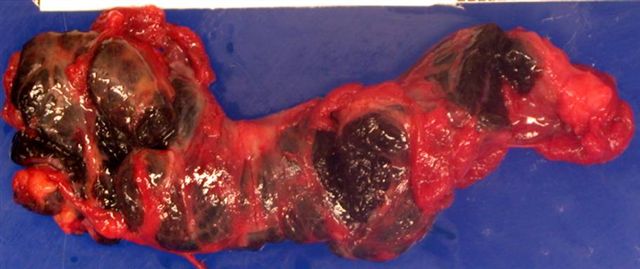
Thyroid gland with black pigment
Images hosted on other servers:

Black thyroid and follicular adenoma

Cut surface

Coal black coloration
Microscopic (histologic) description
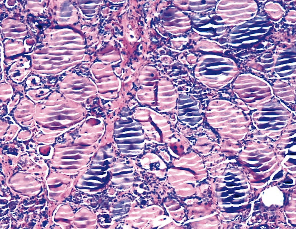
- Dark brown, granular, dust-like cytoplasmic pigment abundant in apical portion of follicular cells, colloid and stromal macrophages (Am J Pathol 1984;117:98)
- Pigment is nonfluorescent and nonbirefringent (Arch Pathol Lab Med 2004;128:355)
Microscopic (histologic) images
Contributed by Andrey Bychkov, M.D., Ph.D. and Mark R. Wick M.D.
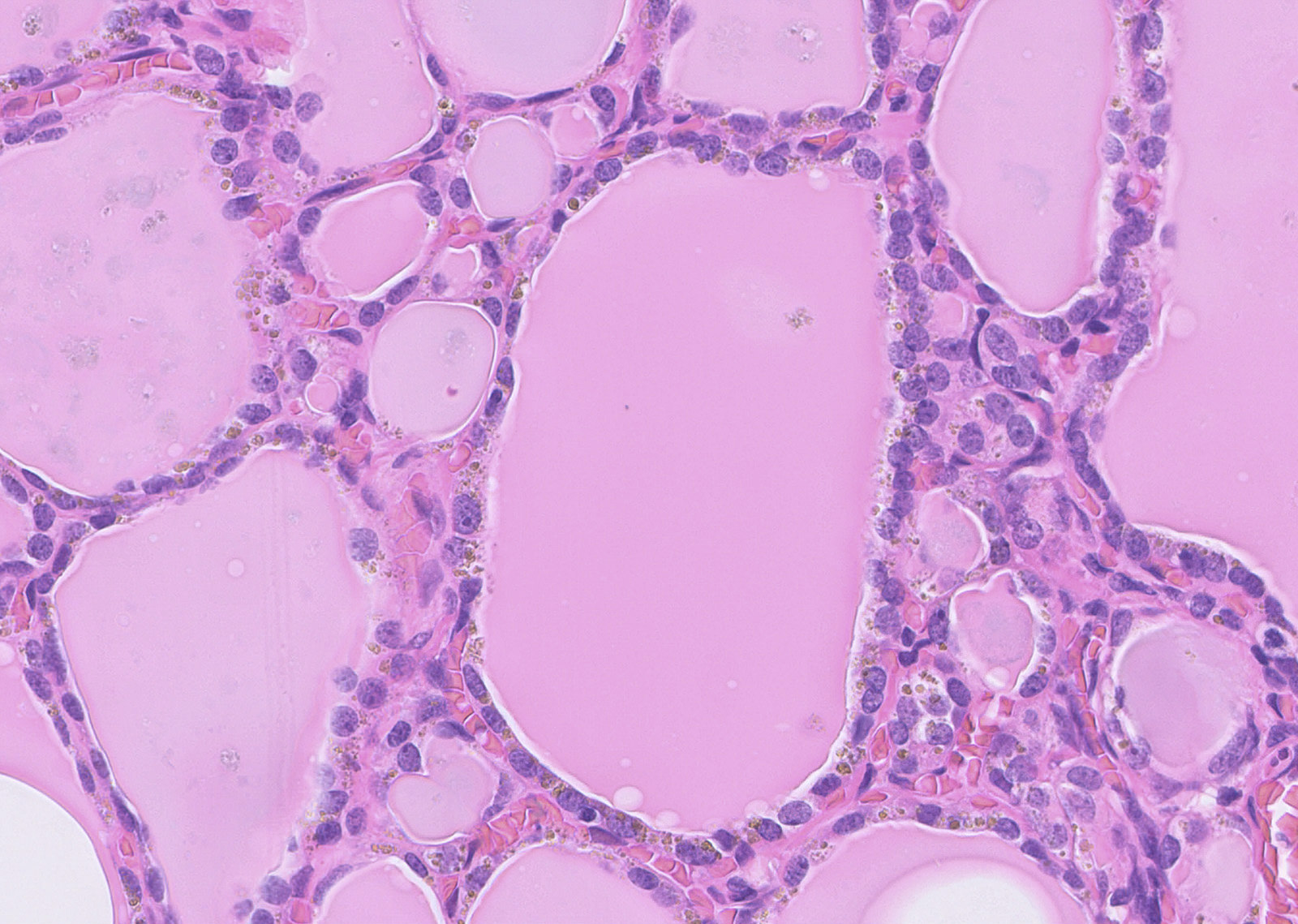
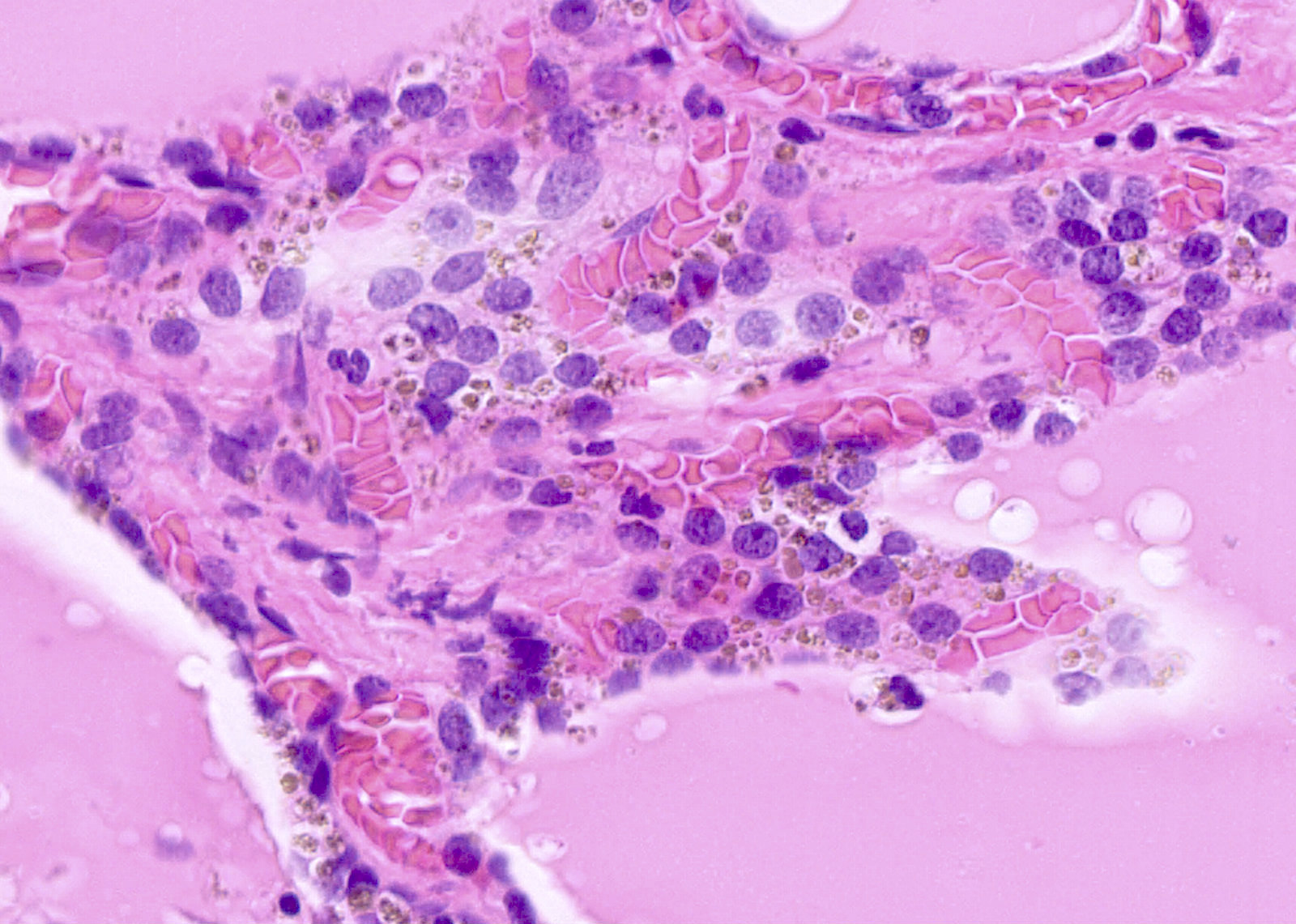
Perinuclear brown pigment
Minocycline induced pigmentation
Images hosted on other servers:

Pigmented cells and colloid

Fontana-Masson
Cytology description
- Dark brown, finely granular intracytoplasmic pigment in follicular cells and macrophages on Papanicolaou stain
- Small round granules are darker and more regular in size and shape than hemosiderin
- On Diff-Quik stain, granules are dark blue (Diagn Cytopathol 2010;38:579)
- Often no specific findings when FNA is done for a thyroid nodule - neoplasms likely do not contain pigment (Diagn Cytopathol 2007;35:135)
Positive stains
- Fontana-Masson and Schmorl for melanin component
- PAS and Ziehl-Neelsen for lipofuscin component
Negative stains
Electron microscopy description
- Numerous electron dense granular structures in lysosomes, with leakage into cytoplasm and exocytosis into follicular lumina
Electron microscopy images
Images hosted on other servers:

Pigment granules in cytoplasm and colloid space

Pigment deposits and Xray spectrogram

Experimental black thyroid, dog

Experimental black thyroid, monkey
Differential diagnosis
- Cystic fibrosis
- Hemochromatosis
- Ochronosis
- Pigmented / melanotic medullary thyroid carcinoma (Diagn Pathol 2008;3:2)

