Thyroid & parathyroid
Papillary thyroid carcinoma
Other subtypes
Follicular variant
Author: Bin Xu, M.D., Ph.D.
Editorial Board Member: Andrey Bychkov, M.D., Ph.D.
Editor-in-Chief: Debra L. Zynger, M.D.

Last author update: 20 April 2021
Last staff update: 17 August 2023
Copyright: 2003-2025, PathologyOutlines.com, Inc.
PubMed Search: Papillary thyroid carcinoma follicular variant
See also: Diffuse follicular variant, Encapsulated follicular variant
Table of Contents
Definition / general | Essential features | Terminology | ICD coding | Epidemiology | Sites | Etiology | Diagrams / tables | Clinical features | Diagnosis | Radiology description | Prognostic factors | Case reports | Treatment | Gross description | Gross images | Frozen section description | Microscopic (histologic) description | Microscopic (histologic) images | Cytology description | Cytology images | Positive stains | Molecular / cytogenetics description | Sample pathology report | Differential diagnosis | Additional references | Practice question #1 | Practice answer #1 | Practice question #2 | Practice answer #2Cite this page: Xu B. Follicular variant. PathologyOutlines.com website. https://www.pathologyoutlines.com/topic/thyroidfollicularvariant.html. Accessed September 17th, 2025.
Definition / general
- Follicular variant of papillary thyroid carcinoma (FV-PTC) is defined by 2 features:
- Architecturally, it is composed exclusively or almost exclusively of follicles
- Cytologically, it shows nuclear features of papillary thyroid carcinoma, such as nuclear enlargement, nuclear membrane irregularity and chromatin clearing
Essential features
- Exclusive or near exclusive follicular architecture
- Nuclear features of papillary thyroid carcinoma
- Can be divided into 2 subtypes (Eur J Surg Oncol 2018;44:338, Cancer 2006;107:1255, J Clin Endocrinol Metab 2017;102:15)
- Encapsulated follicular variant is characterized by the presence of a complete capsule or well circumscribed border and usually RAS mutation
- Infiltrative follicular variant is associated with infiltrative growth, risk of nodal metastasis and BRAF V600E mutation, akin to classic type of papillary thyroid carcinoma
Terminology
- Two subtypes:
- Encapsulated FV-PTC
- Infiltrative FV-PTC (old terminology: multinodular FV-PTC, diffuse FV-PTC)
- Noninvasive encapsulated FV-PTC has been reclassified to noninvasive follicular thyroid neoplasm with papillary-like nuclear features (NIFTP) (JAMA Oncol 2016;2:1023)
ICD coding
Epidemiology
- In Western countries, frequency of follicular variant among all papillary carcinoma increased over the past few decades from 3.4% to 25.2% (J Clin Endocrinol Metab 2014;99:E276)
- In Asia, incidence is much lower (< 10%) due to certain biological differences and historically higher diagnostic threshold for PTC nuclei (Endocr Pathol 2018;29:276, Thyroid 2017;27:983)
- While classic (papillary) pattern used to be the dominant (59% - 76%) architectural pattern of papillary carcinoma diagnosed in the '70s - '90s, follicular patterned papillary carcinoma (regardless of tumor size and cytologic features) became the most prevalent (57%) architecture in papillary carcinoma in 2009 (J Clin Endocrinol Metab 2014;99:E276)
- Above shift of diagnosis is in part due to widespread use of follicular variant terminology and decreased threshold for nuclear alterations required to diagnose papillary carcinoma (J Clin Endocrinol Metab 2017;102:15)
- Lowering of PTC nuclei threshold was particularly noted in USA, partially driven by malpractice fears (Am J Clin Pathol 2002;117:19, Arch Pathol Lab Med 2019;143:1171)
- Among all papillary thyroid carcinomas, the rate of encapsulated follicular variant without invasion, encapsulated follicular variant with invasion and infiltrative follicular variant were 13.6%, 7.1% and 1.7%, respectively (J Clin Endocrinol Metab 2017;102:15)
- Above mentioned data represent epidemiology prior to the creation of NIFTP; since the reclassification of encapsulated follicular variant without invasion as NIFTP in 2016, the epidemiologic data of follicular variant need to be reassessed (JAMA Oncol 2016;2:1023)
Sites
- Most commonly affects the thyroid gland
- May also occur in thyroglossal duct, lingual thyroid, ectopic thyroid and struma ovarii (Int Surg 2006;91:141, J Oral Maxillofac Surg 2013;71:644)
Etiology
- Same etiology as papillary thyroid carcinoma
Diagrams / tables
Images hosted on other servers:

Follicular variant
Clinical features
- Painless thyroid mass detected by imaging or palpation
- More aggressive invasive subtype may manifest with cervical nodal metastasis or obstructive symptoms
Diagnosis
- Diagnostic workup is similar to any thyroid mass / nodule
- Ultrasound with fine needle aspiration cytology
- CT scan may be useful to evaluate extrathyroidal extension and lymph node metastases
- Final diagnosis is rendered on histopathological examination of resected tumor, using a combination of nuclear features and follicular architecture
- FV-PTC can be suspected on cytology, but accuracy of preoperative diagnosis in this variant is lower than in classic PTC
Radiology description
- Larger size, more benign sonographic features and lower ultrasound imaging score compared to classic PTC (J Ultrasound Med 2009;28:1685)
Prognostic factors
- Overall prognosis of follicular variant is excellent (Cancer 2006;107:1255)
- Prognosis differs according to the status of encapsulation and invasion
- Encapsulated follicular variant without capsular and vascular invasion is highly indolent, with negligible (zero or near zero) risk of recurrence or metastasis: the majority of such tumors have been revised as NIFTP (JAMA Oncol 2016;2:1023, Cancer 2006;107:1255)
- Risk of nodal metastasis is 65% for infiltrative follicular variant and 7% for encapsulated follicular variant with capsular or vascular invasion (Cancer 2006;107:1255)
- Risk stratification as per American Thyroid Association 2015 (Thyroid 2016;26:1)
Case reports
- 22 year old woman with extension to parapharyngeal space (BMC Ear Nose Throat Disord 2006;6:3)
- 46 year old man presenting with pleural effusion (Acta Cytol 2007;51:911)
- 47 year old man with invasive FV-PTC harboring NRAS mutation and presenting with bone metastasis (Int J Surg Case Rep 2017;38:180)
- 49 year old woman with aggressive metastatic FV-PTC harboring BRAF K601E and BCORL1 mutations (BMJ Case Rep 2020;13:e234208)
- 56 year old man with glomeruloid growth pattern (Hum Pathol 2008;39:1540)
- 70 year old man with FV-PTC presented as autonomous functioning thyroid nodule (Cureus 2018;10:e3014)
- 83 year old woman with diffuse FV-PTC (Rare Tumors 2016;8:6536)
Treatment
- Most of the intrathyroidal follicular variant is considered ATA (American Thyroid Association) low risk and can be treated successfully by surgical resection (lobectomy / hemithyroidectomy) alone (Thyroid 2016;26:1)
- Total thyroidectomy and postoperative radioactive iodine treatment may be considered if the tumor exhibits additional aggressive features, e.g. extensive vascular invasion, gross extrathyroidal extension or large lymph node metastasis (Thyroid 2016;26:1)
Gross description
- Encapsulated follicular variant: well circumscribed solid beige to tan mass with or without a grossly appreciable tumoral capsule
- Infiltrative follicular variant has a multinodular to infiltrative tumor border
Gross images
Contributed by Bin Xu, M.D., Ph.D.
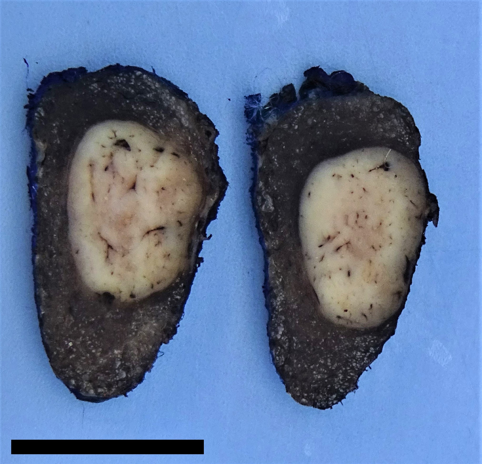
Encapsulated follicular variant
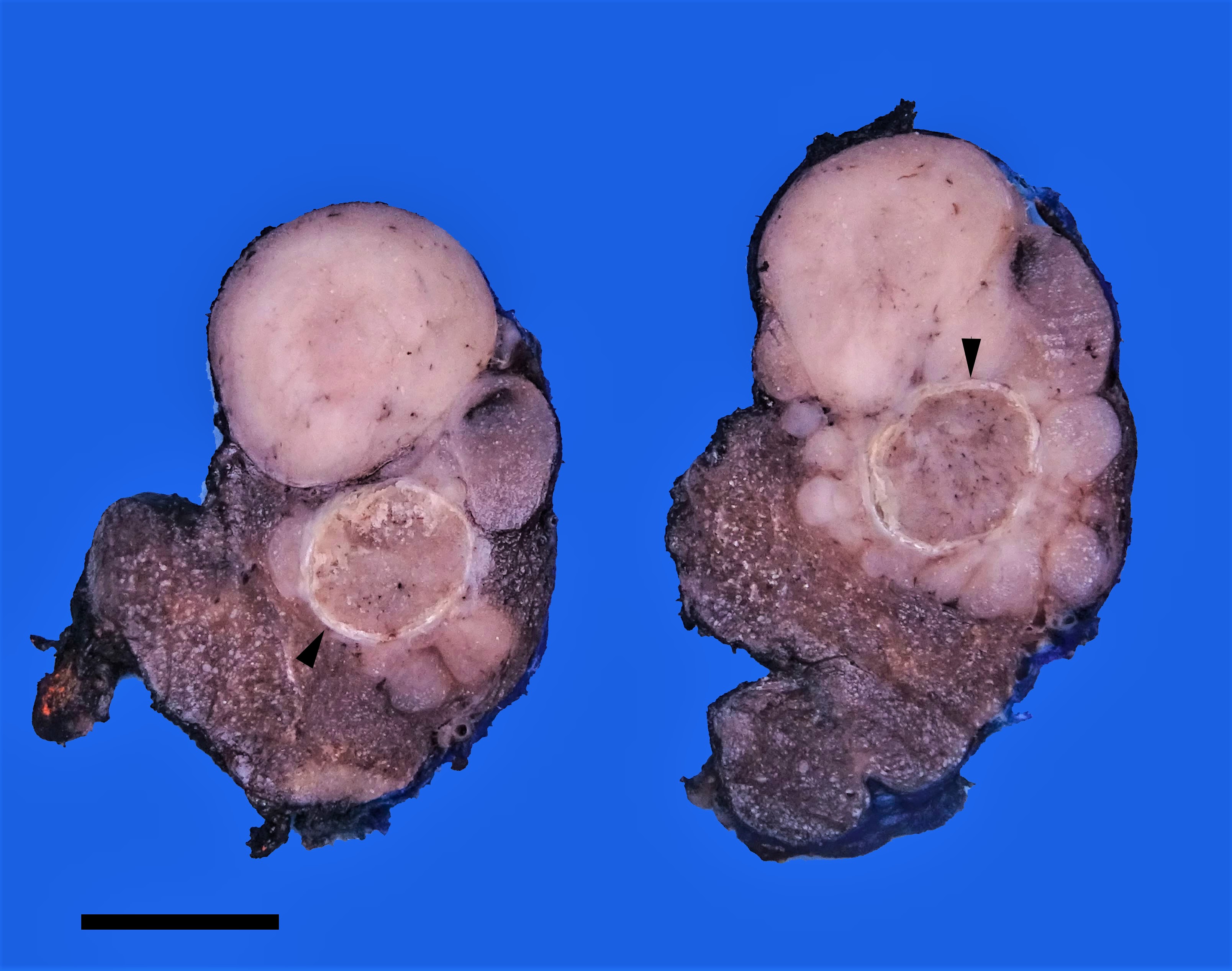
Infiltrative follicular variant
Frozen section description
- Frozen section is strongly discouraged as frozen artifacts distort the nuclear features necessary to diagnose papillary thyroid carcinoma
- Invasion (capsular or vascular) may not be present in the representative section(s) sampled at the time of intraoperative consultation
- Standard care is to perform preoperative fine needle aspiration to establish the diagnosis
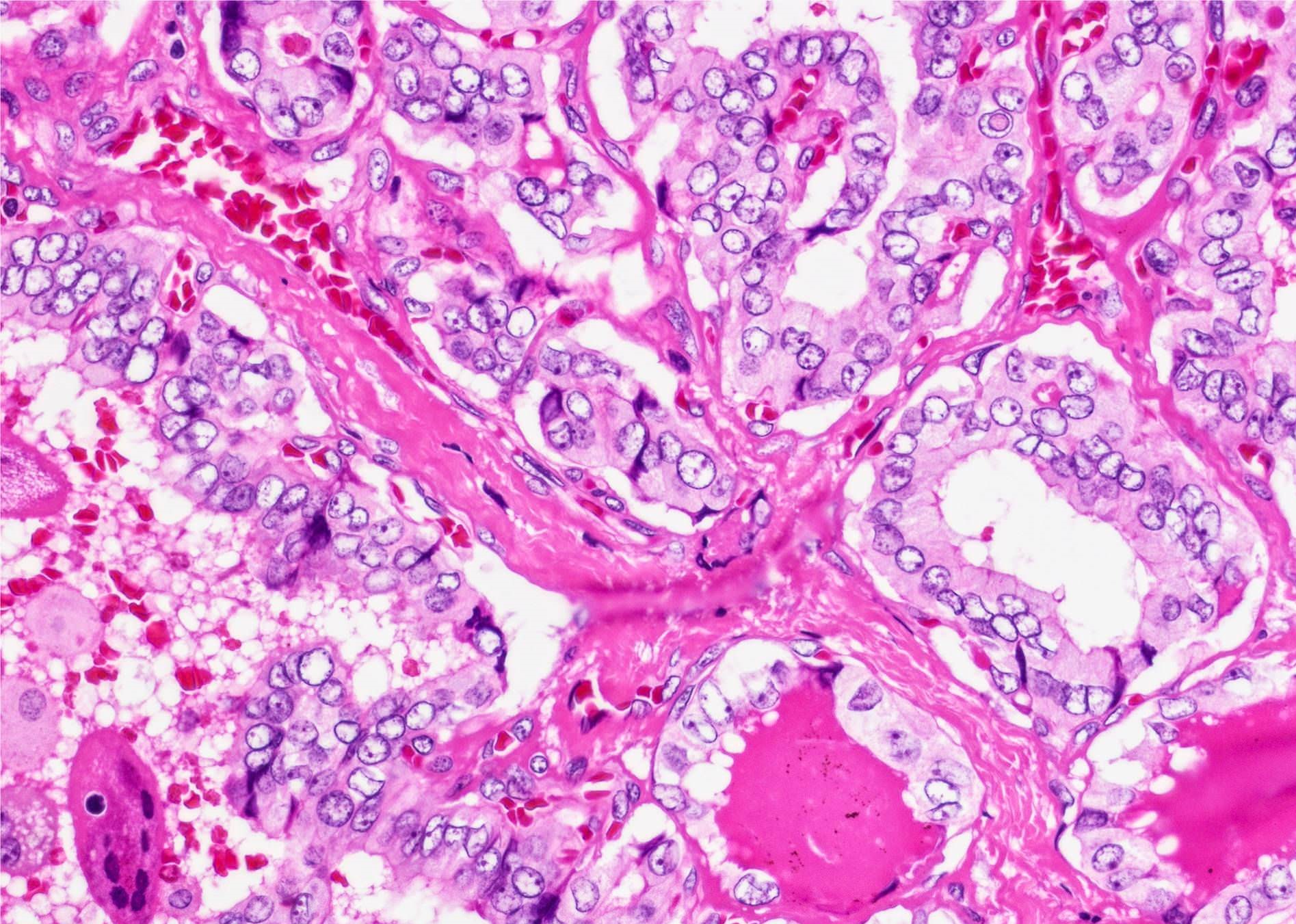
Microscopic (histologic) description
- Nuclear features of papillary thyroid carcinoma should be seen multifocally (at least 2 foci) or diffusely within the tumor; such features include nuclear enlargement, nuclear overlapping, chromatin clearing, nuclear membrane irregularity and nuclear grooves (J Clin Endocrinol Metab 2017;102:15)
- Nuclear score 2 - 3 (JAMA Oncol 2016;2:1023)
- Nuclear pseudoinclusion, a feature commonly seen in classic and tall cell variant, is rarely present in follicular variant
- Architecture: exclusively or nearly exclusively follicular
- True papillae with fibrovascular core are in general absent in follicular variant
- Tumors with mixed papillary (≥ 1% of total tumor volume) and follicular architecture should be classified as classic PTC with predominant follicular pattern, given the associated risk of lymph node metastasis (Thyroid 2019;29:1792)
- Solid architecture may be present: tumors with mixed follicular and solid architecture should be classified as follicular variant, whereas those with (almost) exclusive solid growth are classified as solid variant
- Encapsulated follicular variant has a complete fibrous tumor capsule or a well circumscribed tumor border
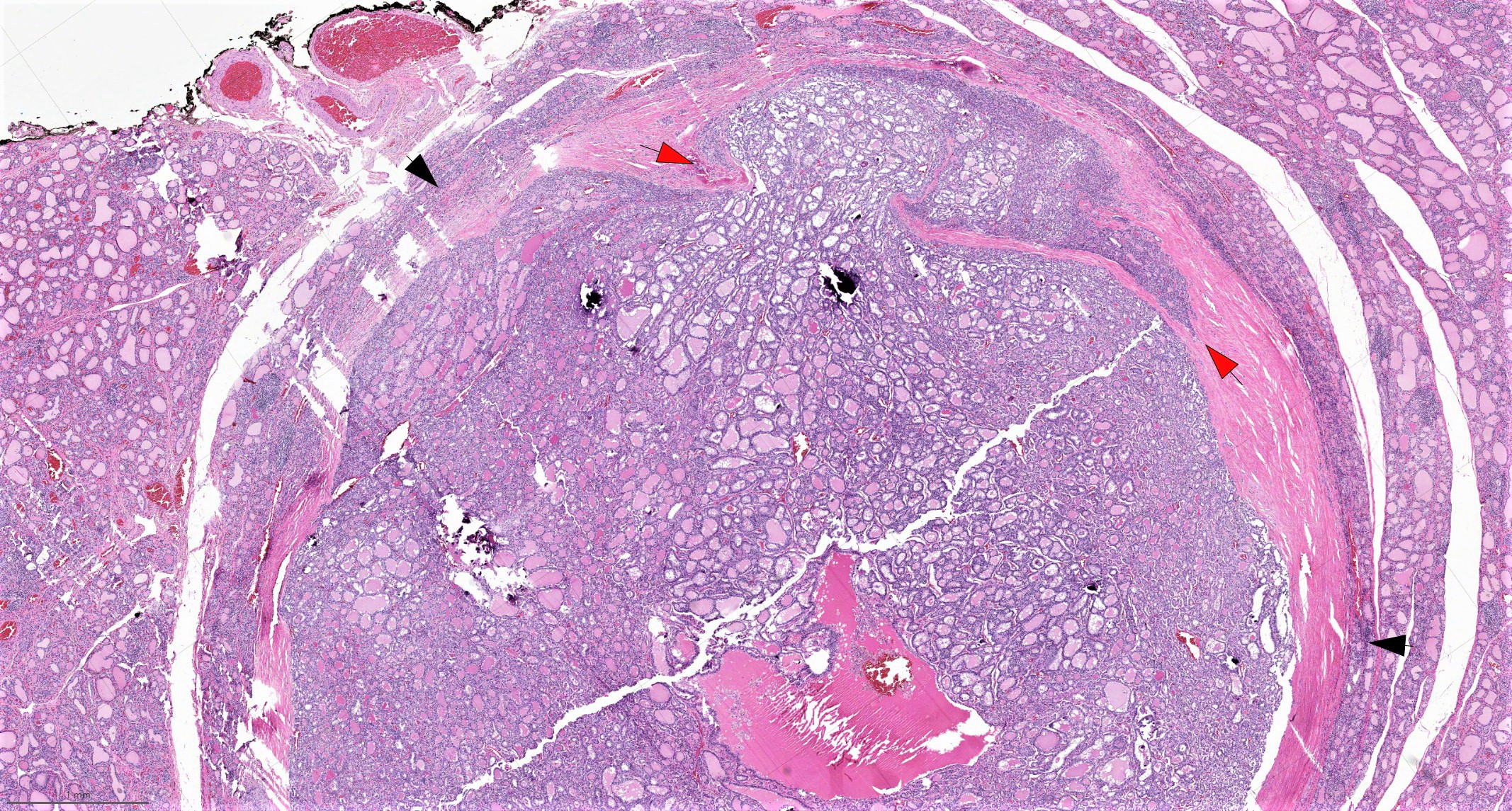
- Infiltrative follicular variant shows infiltrative or multinodular growth
- Sprinkling sign refers to the phenomenon that neoplastic follicles are seen scattered within the background of normal follicles
- "Bubble gum colloid", i.e. dense homogenous hypereosinophilic colloid, may be seen in the lumen of neoplastic follicles; scalloping of colloid may be seen
- Psammoma bodies are exceedingly rare in follicular variant; the identification of psammoma body should promote a search for true papillary (classic) area within the tumor
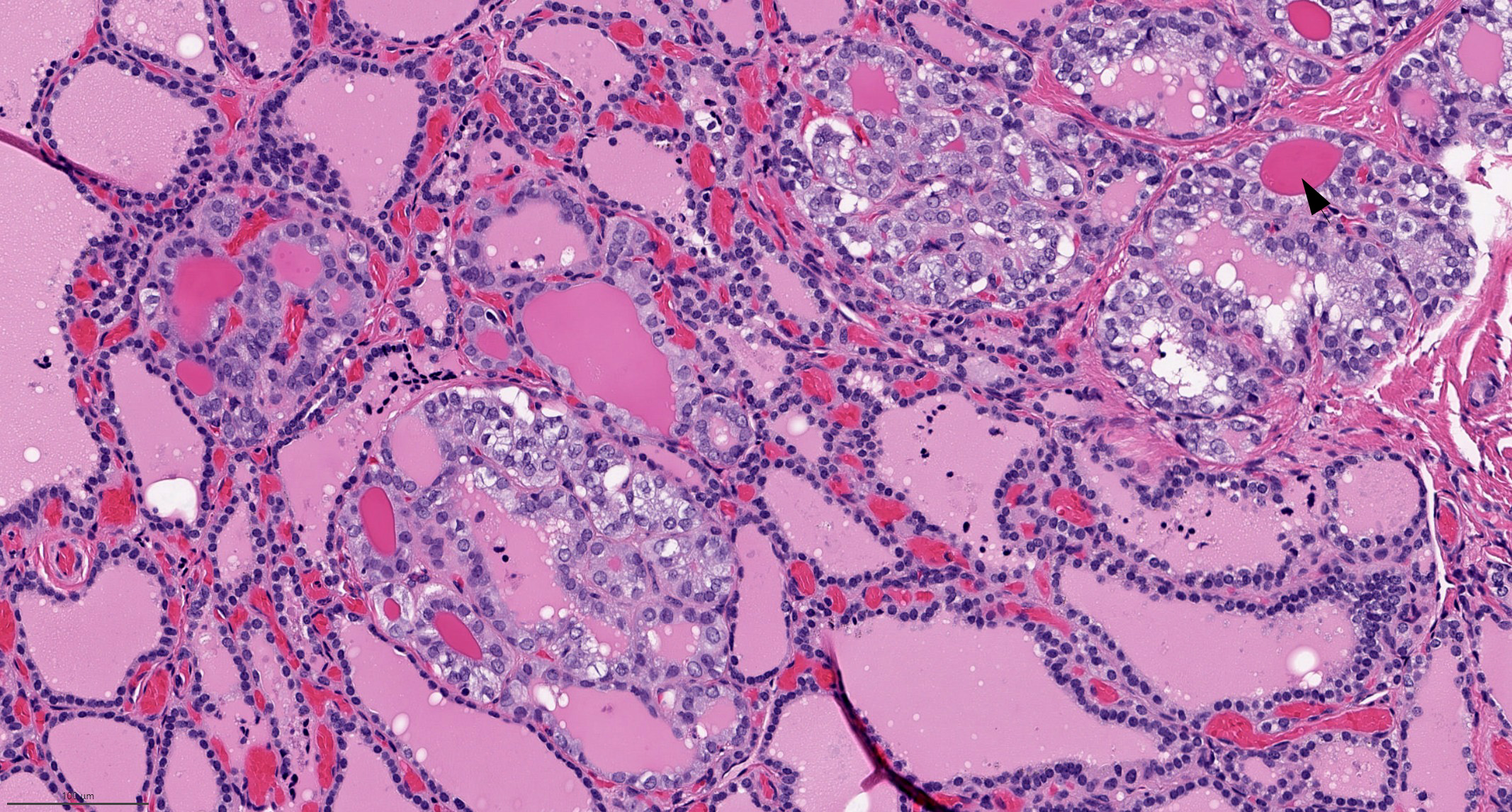
Microscopic (histologic) images
Contributed by Bin Xu, M.D., Ph.D. and Andrey Bychkov, M.D., Ph.D.
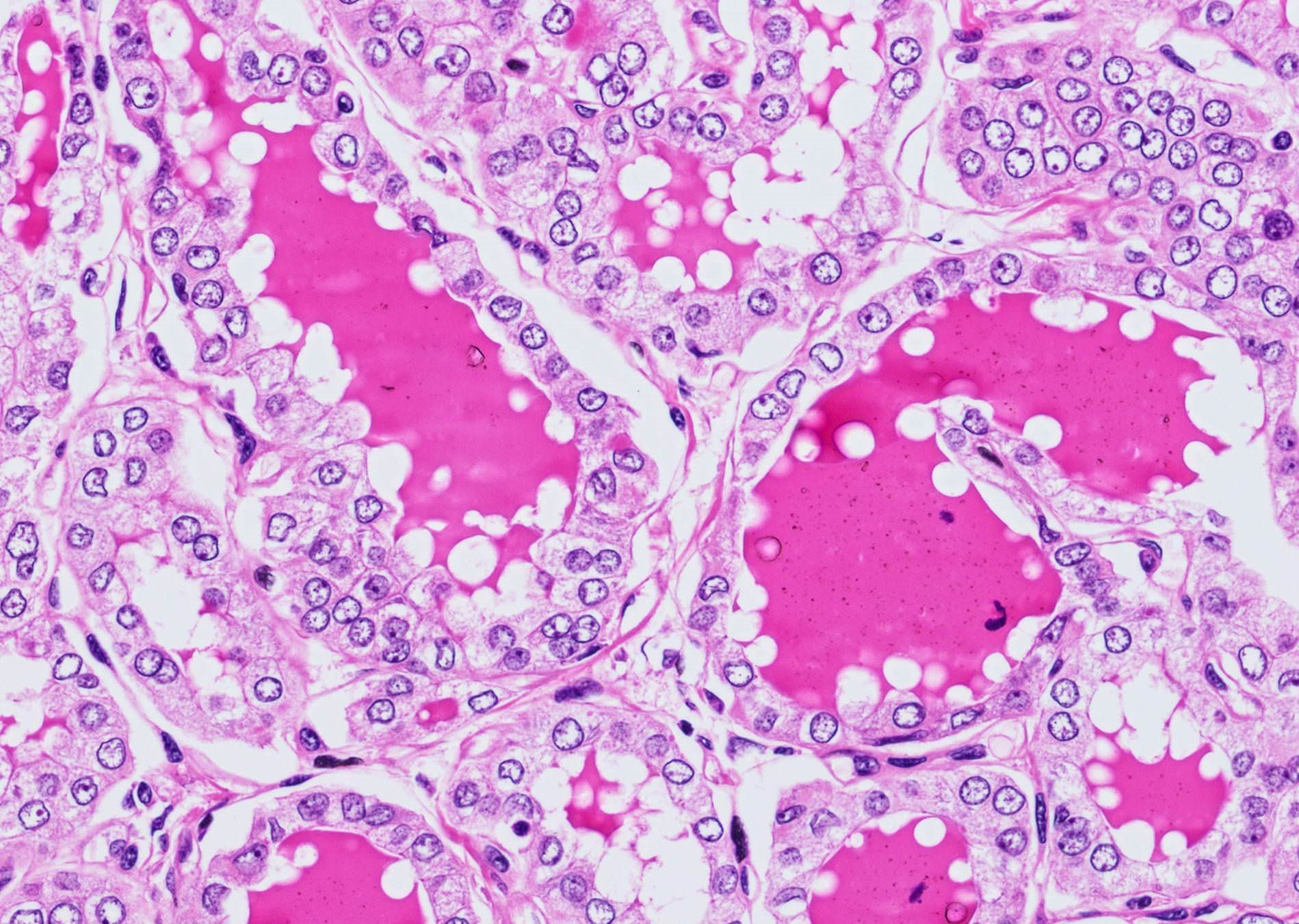
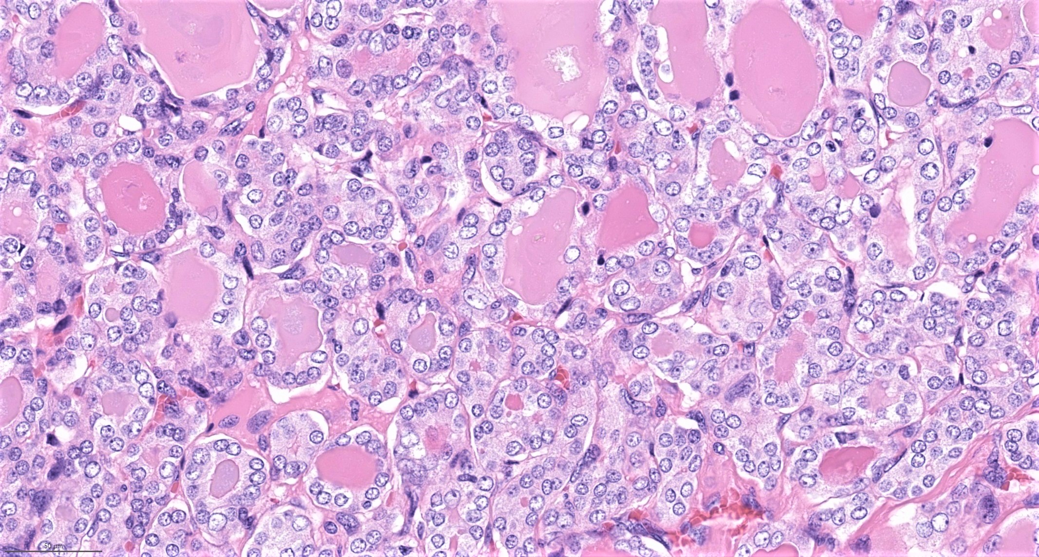
Follicular architecture & nuclear features
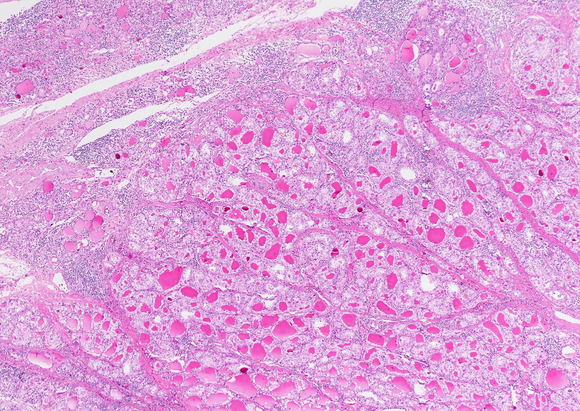
Infiltrative follicular variant
Encapsulated follicular variant
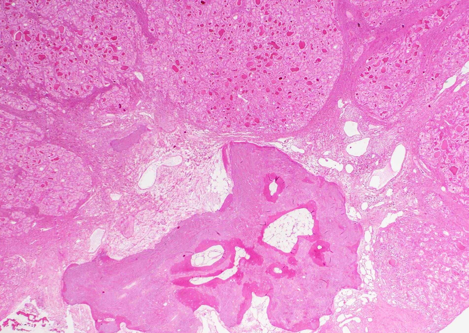
Ossification
Cytology description
- Hypercellularity
- Small, round and dense colloid (hyaline colloid) may be present, sometimes within follicles
- Cells arranged in microfollicles or trabecular pattern
- Nuclear enlargement but may lack prominent nuclear features of papillary carcinoma (Am J Clin Pathol 1999;111:216)
- Highly suggestive of syncytial clusters, microfollicular architecture, chromatin clearing and nuclear grooves (Acta Cytol 2006;50:663)
- Classified by Bethesda system as categories III to VI
- Cytologically unable to distinguish between noninvasive and invasive
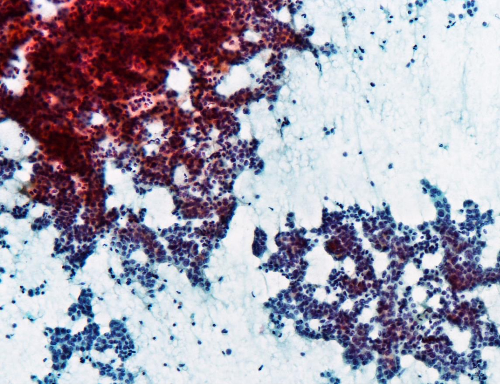
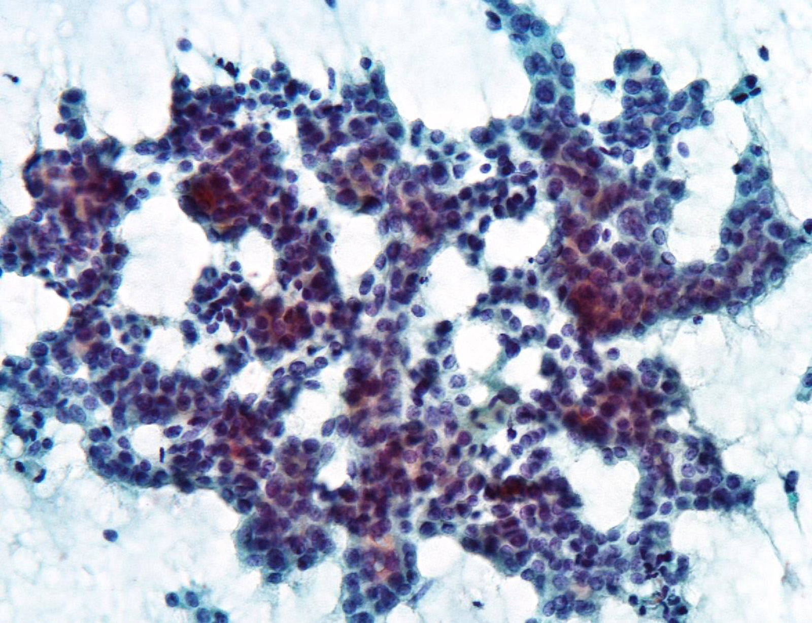
Cytology images
Contributed by Ayana Suzuki, C. T.
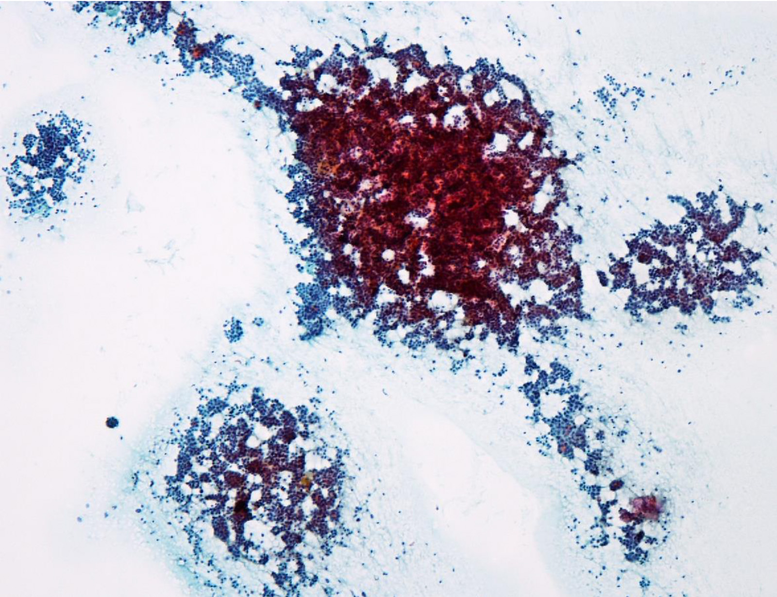
Hypercellular aspirate
Microfollicular and trabecular pattern
Microfollicles
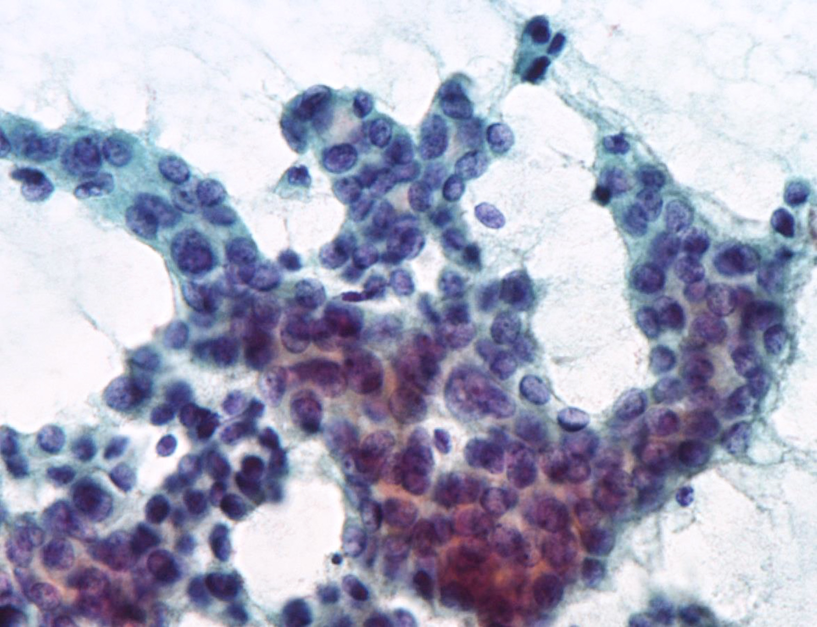
Irregular nuclei
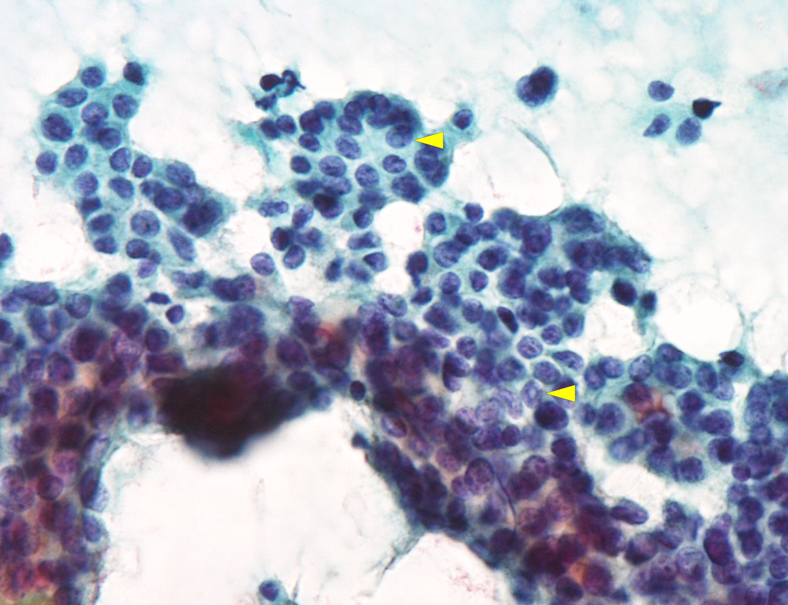
Nuclear inclusions
Images hosted on other servers:

Cellular aspirates;
some microfollicular
aggregates, no colloid

Acinar arrangement with intranuclear inclusion

Grape-like nuclei of papillary thyroid carcinoma

Microfollicles in thyroid FNA smears

Comparison of a
tissue proven follicular
adenoma and variant
of papillary carcinoma
Positive stains
- Mutated proteins, e.g. BRAF V600E and NRAS Q61R, can be positive in a proportion of cases (see Molecular / cytogenetics description) (Thyroid 2019;29:1792)
- Thyroid specific markers: TTF1, thyroglobulin, PAX8
- CK19, galectin3, HBME1 produce focal staining, which cannot reliably distinguish FV-PTC from other follicular patterned lesions
Molecular / cytogenetics description
- The Cancer Genome Atlas (TCGA) has shown that follicular variant as a group is associated with RAS mutation, high thyroid differentiation score and a RAS-like molecular signature (Cell 2014;159:676)
- Overall, encapsulated follicular variant has a genomic profile akin to follicular adenoma / follicular carcinoma, whereas infiltrative follicular variant has molecular alterations similar to classic papillary thyroid carcinoma (Eur J Surg Oncol 2018;44:338, Am J Clin Pathol 2003;120:71, Mod Pathol 2010;23:1191, Thyroid 2013;23:1256)
- Encapsulated follicular variant is associated with high frequency (36% - 40%) of RAS mutation and absence of BRAF V600E mutation
- PAX8-PPARgamma fusion may be seen in encapsulated follicular variant
- Infiltrative follicular variant has high frequency of BRAF V600E mutation (26%) and low rate of RAS mutation (10%)
- RET / PTC rearrangement may be seen in infiltrative follicular variant
Sample pathology report
- Thyroid, right lobe, lobectomy:
- Papillary thyroid carcinoma, infiltrative follicular variant, 3.2 cm (see synoptic report)
Differential diagnosis
- Papillary carcinoma, classic type:
- May contain various amounts of follicles
- Tumor with ≥ 1% of papillae should be classified as classic type
- NIFTP:
- In 2016, most (but not all) noninvasive encapsulated follicular variant has been reclassified as NIFTP (JAMA Oncol 2016;2:1023)
- See NIFTP for diagnostic criteria of NIFTP
- Follicular adenoma and follicular carcinoma:
- Lack nuclear features of papillary thyroid carcinoma
- Macrofollicular PTC:
- Macrofollicles (> 200 µm) constitute > 50% of tumor
Additional references
Practice question #1
What is the most common molecular alteration seen in follicular variant of papillary thyroid carcinoma?
- BRAF V600E mutation
- RAS mutation
- RET / PTC fusion
- TP53 mutation
Practice answer #1
Practice question #2
A 45 year old woman underwent thyroid lobectomy for a 2.2 cm thyroid mass. The histology of the tumor is shown above. What is the diagnosis of this tumor?
- Adenomatoid nodule
- Follicular adenoma
- Follicular carcinoma
- Papillary thyroid carcinoma, follicular variant
Practice answer #2
