

Thyroid & parathyroid
Thyroiditis
Riedel thyroiditis
Author: Shahid Islam, M.D., Ph.D.

Last author update: 1 March 2009
Last staff update: 12 December 2023
Copyright: 2003-2024, PathologyOutlines.com, Inc.
PubMed Search: Riedel's thyroiditis

Table of Contents
Definition / general | Terminology | Epidemiology | Etiology | Clinical features | Case reports | Treatment | Gross description | Microscopic (histologic) description | Microscopic (histologic) images | Virtual slides | Cytology description | Differential diagnosis | Additional referencesCite this page: Islam S. Riedel thyroiditis. PathologyOutlines.com website. https://www.pathologyoutlines.com/topic/thyroidriedel.html. Accessed April 19th, 2024.
Definition / general
- Densely fibrotic inflammatory process involving thyroid gland and adjacent neck tissue
- Described in 1896 by German surgeon Bernhard Moritz Carl Ludwig Riedel (Wikipedia)
- Rare (0.05% of thyroidectomy specimens)
Terminology
- Also called Riedel struma, fibrous thyroiditis
Epidemiology
- Slight female predominance, usually age 40 - 60 years
Etiology
- Etiology unclear; may be part of generalized fibroinflammatory process also involving other organs (Am J Clin Pathol 2004;121:S50)
Clinical features
- Associated with inflammatory fibrosclerosis / multifocal systemic fibrosclerosis (mediastinal or retroperitoneal fibrosis, sclerosing cholangitis, inflammatory pseudotumor of orbit)
- 65% have antithyroid antibodies
- Clinically resembles carcinoma
- In one study, 67% had antithyroid antibodies, supporting an autoimmune mechanism of injury (Am J Clin Pathol 1988;90:715)
Case reports
- 41 year old woman with case resembling anaplastic carcinoma (Int J Surg 2008;6:e24)
- 44 year old woman with bilateral orbital pseudotumors (J Fr Ophtalmol 2008;31:715.e1)
- 51 year old woman with prior subacute thyroiditis (Endocr J 2007;54:559)
- Two cases of Riedel thyroiditis, one had coexisting retroperitoneal fibrosis (Am J Clin Pathol 1976;65:274)
Treatment
- Surgery to decompress, steroids or tamoxifen (Endocr Pract 2004;10:483)
Gross description
- Extensive stony hard fibrosis involving a goitrous thyroid gland and infiltration into adjacent muscle and other structures, obliterating tissue planes at surgery
- Binds soft tissues of neck in an "iron collar," may compress trachea
- Tan / gray, woody and avascular, no lobules apparent
Microscopic (histologic) description
- No normal lobular pattern
- Follicles are obliterated or compressed by extensive dense fibrous tissue, which also infiltrates adjacent skeletal muscle
- Patchy lymphocytes (B & T cells), plasma cells (IgA, lambda) and eosinophils, inflammation in walls of trapped veins
- 25% have adenoma centrally in fibrous mass
- No oncocytic cells, no giant cells
Microscopic (histologic) images
AFIP images
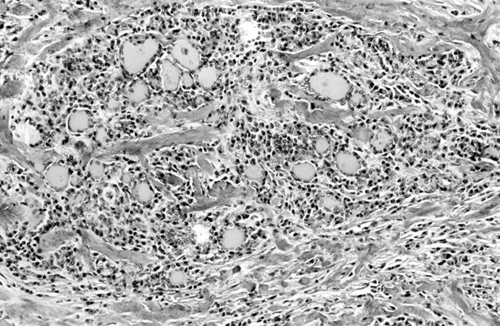
Atrophic thyroid follicles

Resembles papillary microcarcinoma

Follicles within scar irregular
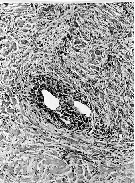
Heavy inflammatory infiltrate, venous wall
Images hosted on other servers:


Massive scarring and lymphohistiocytic infiltrate


Obliterating phlebitis
Virtual slides
Images hosted on other servers:


Invasive fibrous thyroiditis
Cytology description
- Moderate cellularity with fragments of fibrous tissue containing bland spindle cells and myofibroblasts (Diagn Cytopathol 2004;30:193)
Differential diagnosis
- Fibrous variant of Hashimoto thyroiditis: limited to thyroid, noninfiltrative, less abundant fibrous reaction, oncocytic cells present, more plasma cells, no granulocytes, no monocytes, no eosinophils (J Endocrinol Invest 2003;26:444)
- Sarcoma: atypical spindle cells (J Clin Pathol 2001;54:570)
- Subacute thyroiditis: late phase (Ann Pathol 2008;28:263)
Additional references

