

Thyroid & parathyroid
Other uncommon thyroid carcinomas
Sclerosing mucoepidermoid carcinoma with eosinophilia
Editorial Board Member: Andrey Bychkov, M.D., Ph.D.


Last author update: 1 March 2017
Last staff update: 17 August 2023
Copyright: 2003-2024, PathologyOutlines.com, Inc.
PubMed Search: Sclerosing mucoepidermoid carcinoma with eosinophilia

Table of Contents
Definition / general | Essential features | Epidemiology | Etiology | Clinical features | Radiology description | Case reports | Treatment | Gross description | Gross images | Microscopic (histologic) description | Microscopic (histologic) images | Cytology description | Cytology images | Positive stains | Negative stains | Differential diagnosis | Board review style question #1 | Board review style answer #1Cite this page: Wei S. Sclerosing mucoepidermoid carcinoma with eosinophilia. PathologyOutlines.com website. https://www.pathologyoutlines.com/topic/thyroidsclerosing.html. Accessed April 16th, 2024.
Definition / general
- Indolent mucoepidermoid carcinoma with fibrosis and eosinophil infiltrate in a background of sclerosing Hashimoto thyroiditis (Am J Surg Pathol 1991;15:438)
- Distinct from mucoepidermoid carcinoma of salivary gland
(Mod Pathol 2000;13:802, Mod Pathol 2017;30:329)
- May derive from squamous metaplasia or ultimobranchial body rests (Mod Pathol 2004;17:526)
- Death due to disease is uncommon, although lymph nodes metastases, extracapsular spread, vascular invasion and perineural invasion are common
- Distinct from thyroid mucoepidermoid carcinoma
Essential features
- Squamous and mucus secreting cells with fibrosis and eosinophilic infiltrate in a background of sclerosing Hashimoto thyroiditis
Epidemiology
- Almost always women (M:F = 1:16)
- Mean age: 55 years
- Associated with Hashimoto thyroiditis and solid cell nest hyperplasia
Etiology
- Associated with sclerosing Hashimoto thyroiditis
- Possibly derives from metaplastic squamous epithelium or solid cell nests
- Negative for MAML2 rearrangements, typically seen in salivary mucoepidermoid carcinoma (Mod Pathol 2017;30:329)
Clinical features
- Slowly growing thyroid mass in patients with sclerosing Hashimoto thyroiditis
- Death due to disease is uncommon, although lymph nodes metastases, extracapsular spread with extensive tumor invasion into the adjacent soft tissues and organs, vascular invasion and perineural invasion are common (Hum Pathol 2015;46:725)
Radiology description
- Ultrasound scan: ill defined, heterogeneous and hypoechoic nodule
- Cold on radionuclide scan
Case reports
- 35 year old woman with thyroid swelling and lymph node enlargement (J Cytol 2016;33:37)
- 38 year old woman diagnosed with sclerosing mucoepidermoid carcinoma (Am J Otolaryngol 2004;25:48)
- 39 year old woman with a goiter for many years (Am J Clin Pathol 1998;109:294)
- 44 year old woman with 4 month history of a painless swelling (Int Surg J 2017;4:1780)
- 45 year old man with a painless mass (Int J Clin Exp Pathol 2015;8:5947)
- 57 year old woman with right neck mass for 20 years (J Korean Med Sci 1999;14:338)
- 61 year old man with thyromegaly and vocal cord paralysis (Am J Clin Pathol 1998;109:294)
- 65 year old woman with thyroid swelling (Indian J Pathol Microbiol 2008;51:34)
- 65 year old man presented a diffuse neck swelling of rapid onset (Hematology/Oncology and Stem Cell Therapy 2008;1:62)
- 69 year old woman who had undergone thyroidectomy and 70 year old woman with distant metastasis (Hum Pathol 1997;28:1091)
- Middle aged woman with locally recurrent thyroid tumor (Case #230)
Treatment
- Total thyroidectomy with/without neck dissection
Gross description
- White, homogenous, firm, usually ill defined border
Gross images
Contributed by Mark R. Wick, M.D. and AFIP
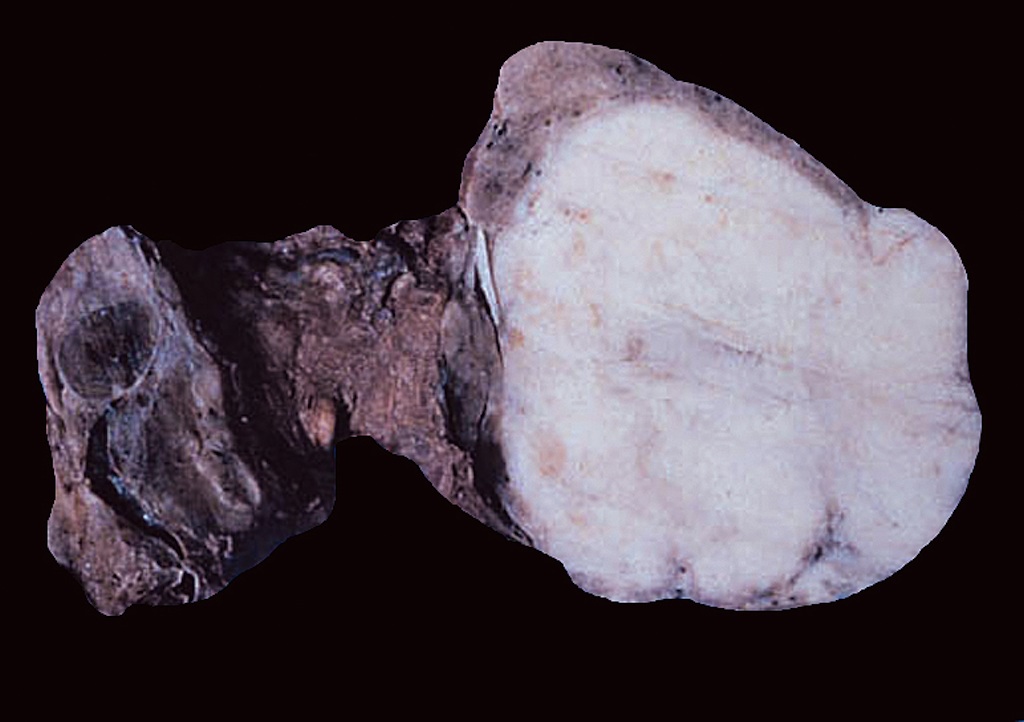
Mucoepidermoid
carcinoma,
sclerosing type

Well circumscribed
tumor (not typical)
with dense fibrosis
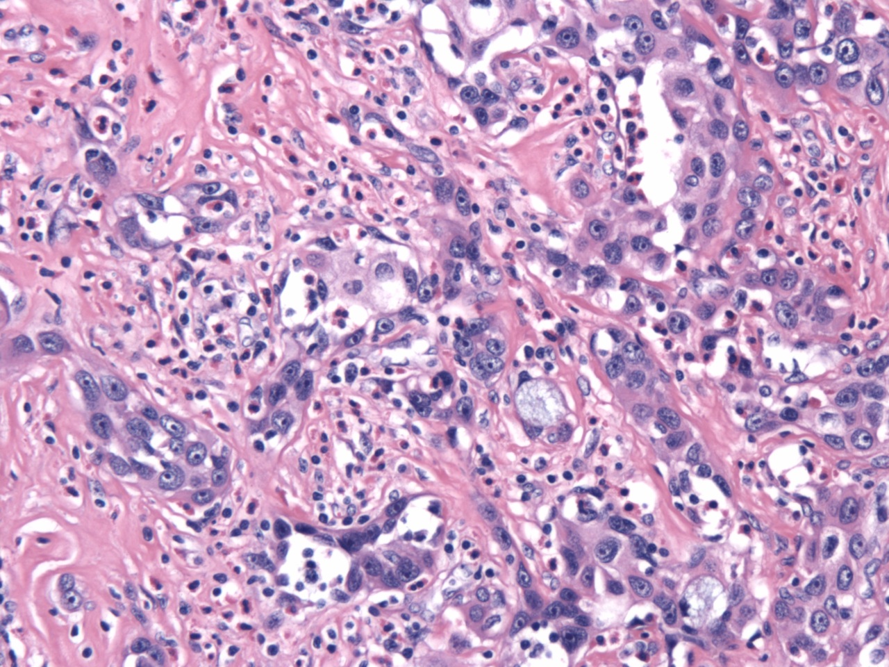
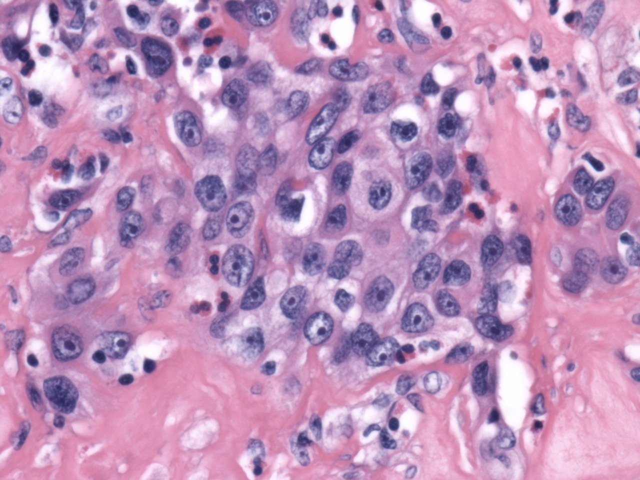
Microscopic (histologic) description
- Infiltrating solid / nested squamous tumor cells with mild to moderate atypia in dense fibrohyaline stroma with marked eosinophil infiltration; keratin pearls and keratin debris can be identified
- Mucus secreting cells, small mucin pools present; background of chronic lymphocytic thyroiditis
- Lymph nodes metastasis, extracapsular spread, vascular invasion and perineural invasion are common
- May have focal clear cells (Ann Diagn Pathol 2003;7:348)
Microscopic (histologic) images
Case #230 and AFIP
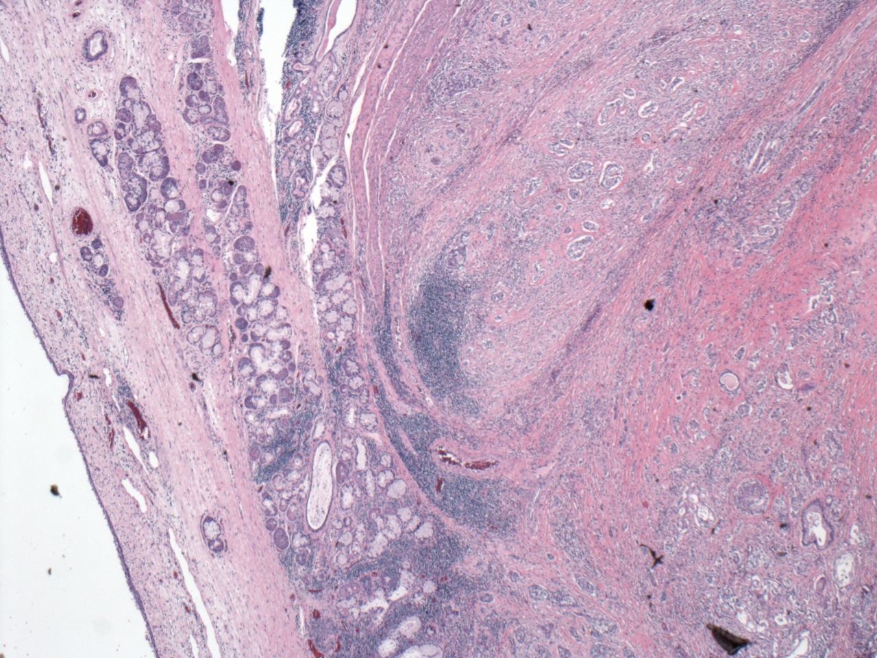
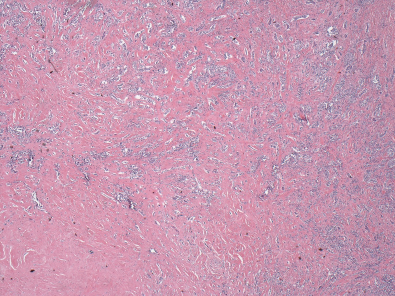
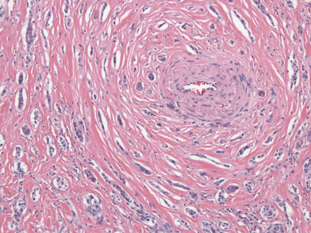
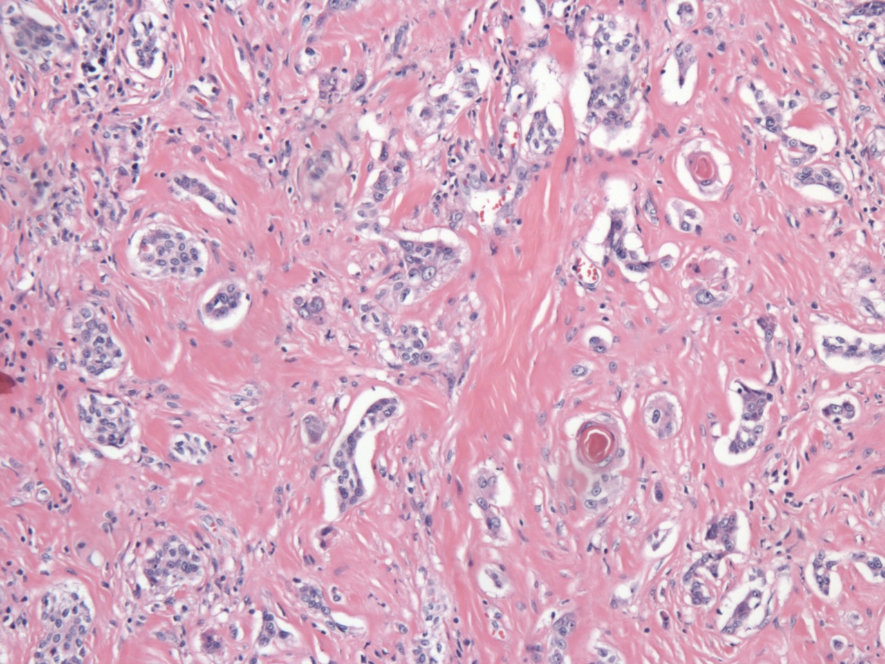


Small irregular strands of tumor cells
Images hosted on other servers:


Various images
Cytology description
- Atypical squamous cells, mucocytes, mucin and eosinophils; may mimic squamous cell / anaplastic carcinoma (Diagn Cytopathol 1996;15:30, Am J Clin Pathol 1998;109:294)
Cytology images
Images hosted on other servers:

Epithelial pearl-like structure

Cell block

Cell block, p40
Positive stains
- Keratin, p63, variable TTF1, CEA (mucus containing cells) (Hum Pathol 2015;46:725)
Negative stains
Differential diagnosis
- Anaplastic carcinoma: highly malignant carcinoma with necrosis, can have squamous component
- CASTLE: squamous cells with variable lymphocytes without eosinophils, CD5+, CD117+, can have mucin
- Direct extension or metastasis of salivary mucoepidermoid carcinoma: keratinization is rare
- Nodular sclerosing Hodgkin lymphoma: Reed-Sternberg cells without mucin (Arch Pathol Lab Med 2000;124:446)
- Solid cell nest and squamous metaplasia: no atypia, no invasion, no mucin, no eosinophils
- Squamous cell carcinoma: sheets of highly atypical squamous cells with necrosis, no mucin, no eosinophils
- Papillary carcinoma with squamous metaplasia
- Rare neoplastic (medullary carcinoma with squamous differentiation, invading adenosquamous carcinoma) and non-neoplastic (Hashimoto thyroiditis with cyst formation and prominent eosinophilic infiltration, branchial cleft-derived cysts) lesions (Wenig, Bruce: Atlas of Head and Neck Pathology)
- Differential diagnosis of FNA cytology includes other primary and metastatic thyroid malignancies, and Hashimoto’s thyroiditis (Diagn Cytopathol 1996;15:301)
Board review style question #1
Which item is correct regarding sclerosing mucoepidermoid carcinoma with eosinophilia?
- Sclerosing mucoepidermoid carcinoma with eosinophilia is not associated with Hashimoto thyroiditis
- Lymph nodes metastases, extracapsular spread, vascular invasion and perineural invasion are common
- Death due to sclerosing mucoepidermoid carcinoma with eosinophilia is common
- Eosinophil infiltration is common, and no keratin pearls and keratin debris are seen
- The squamous component is highly malignant
Board review style answer #1
B. Lymph nodes metastases, extracapsular spread, vascular invasion and perineural invasion are common. Death due to disease is uncommon, although lymph nodes metastases, extracapsular spread, vascular invasion and perineural invasion are common.
Comment Here
Reference: Sclerosing mucoepidermoid carcinoma with eosinophilia
Comment Here
Reference: Sclerosing mucoepidermoid carcinoma with eosinophilia

