

Vulva & vagina
Other carcinomas
Clear cell carcinoma-vagina
Authors: Shweta Gera, M.D., Arzu Buyuk, M.D.


Last author update: 1 March 2014
Last staff update: 2 April 2024
Copyright: 2002-2024, PathologyOutlines.com, Inc.
PubMed Search: Clear cell adenocarcinoma [title] vagina


Table of Contents
Definition / general | Terminology | Epidemiology | Sites | Pathophysiology | Etiology | Clinical features | Prognostic factors | Case reports | Treatment | Clinical images | Gross description | Microscopic (histologic) description | Microscopic (histologic) images | Cytology description | Cytology images | Positive stains | Negative stains | Electron microscopy description | Molecular / cytogenetics description | Videos | Differential diagnosis | Additional referencesCite this page: Gera S, Buyuk A. Clear cell carcinoma-vagina. PathologyOutlines.com website. https://www.pathologyoutlines.com/topic/vaginaclearcelladeno.html. Accessed April 25th, 2024.
Definition / general
- Most common subtype of vaginal adenocarcinoma associated with DES exposure in young females; can also occur in postmenopausal women without exposure to DES
Terminology
- Mesonephroid carcinoma, mesonephric carcinoma (Cancer 1970;25:745)
Epidemiology
- Rare vaginal cancer, accounting for 5% to 10% of primary vaginal malignancies (J Minim Invasive Gynecol 2006;13:237)
- Bimodal age distribution, mean age 22 years (Gynecol Oncol 1996;60:339) in women with exposure to DES and 55 years in postmenopausal women with no history of DES, but who may have pelvic endometriosis (J Minim Invasive Gynecol 2006;13:237, Gynecol Oncol 2006;103:1130)
Sites
- Most common - anterior vaginal wall (Gynecol Oncol 1993;51:266, Gynecol Oncol 2007;105:273); can also occur in elsewhere in vagina
Pathophysiology
- DES causes persistence of Müllerian epithelium while inducing contact between epithelium and the vaginal mesenchyme
- Unopposed estrogen and obesity causes increase in the peripheral conversion of steroid hormones to estrone by the enzyme aromatase leading to a hyperestrogenic environment (Gynecol Oncol 2006;103:1130)
Etiology
- Most cases occur among women born 1947 through 1971, when pregnant women were most frequently prescribed DES in the United States (Cancer Causes Control 2012;23:207, Gynecol Oncol 1996;60:339)
- 70% occur in women having intrauterine exposure to DES - also known as DES daughters (Gynecol Oncol 1996;60:339, Gynecol Oncol 2007;105:273, Gynecol Oncol 1993;51:266, Cancer Causes Control 2010;21:999)
- Endometriosis of vagina and perivaginal area may be a precursor in non-DES exposed females; the frequency of vaginal tumors arising in endometriosis ranges from 4% - 11% (Gynecol Oncol 2006;103:1130, J Minim Invasive Gynecol 2006;13:237)
- Vaginal adenosis (especially tuboendometrial adenosis) is associated with CCA (clear cell adenocarcinoma) in 90% of cases, suggesting adenosis is a precursor lesion (J Obstet Gynaecol Res 2010;36:681, Adv Anat Pathol 2012;19:296, Gynecol Oncol 1993;51:266)
Clinical features
- Abnormal vaginal bleeding or discharge, although 16 - 25% are asymptomatic (Gynecol Oncol 2007;105:273)
- Postmenopausal bleeding (Gynecol Oncol 2006;103:1130, J Minim Invasive Gynecol 2006;13:237)
- Congenital anomalies of GU tract without DES exposure: associated with metanephric and mesonephric remnants and Mü¸llerian duct anomalies (J Obstet Gynaecol Res 2010;36:681, J Pak Med Assoc 2009;59:568)
- Lymphatic and vascular spread can occur (J Minim Invasive Gynecol 2006;13:237)
- Can metastasize to regional lymph nodes (J Minim Invasive Gynecol 2006;13:237), lungs (Gynecol Oncol 1993;51:266, Gynecol Oncol 2007;105:273, J Minim Invasive Gynecol 2006;13:237), kidney (Gynecol Oncol 1993;51:266), peritoneum, omentum, ovary, liver and brain (Gynecol Oncol 2007;105:273)
- Can present with malignant pericardial effusion and cardiac tamponade (Int J Gynecol Cancer 2006;16:1458)
- May recur in distant sites even in absence of pelvic disease (Gynecol Oncol 1993;51:266)
- Women exposed to DES in utero (DES daughters) also have increased risk of clear cell adenocarcinoma persisting at older ages and an increased risk of melanoma at young ages, no increased risk of other cancers (Cancer Causes Control 2010;21:999)
Prognostic factors
- Biologic behavior and prognosis differ from squamous cell carcinoma:
- Better 5 year survival of localized vaginal CCA but greater risk of developing late recurrences after disease free interval; recurrences can occur 2 years to 8 years later (Gynecol Oncol 1993;51:266, Gynecol Oncol 2007;105:273)
- Non-DES exposed patients may have poorer prognosis compared with DES exposed individuals (J Minim Invasive Gynecol 2006;13:237)
Case reports
- 5 year old girl with clear cell adenocarcinoma of vagina (Clin Radiol 1998;53:69)
- 14 year old girl with vaginal bleeding had a mass involving the vagina and cervix (Case of the Week #363)
- 22 year old woman with fertility sparing radical abdominal trachelectomy for clear cell adenocarcinoma of the upper vagina (Gynecol Oncol 2007;105:820)
- 43 year old woman with diethylstilbestrol (DES) induced clear cell adenocarcinoma of the vagina metastasizing to the brain (Gynecol Oncol 2007;105:273)
- 55 year old woman with clear cell adenocarcinoma of the vagina and vaginal endometriosis (Gynecol Oncol 2006;103:1130)
- Late recurrence of clear cell adenocarcinoma of the cervix (Obstet Gynecol 1990;76:525)
- Clear cell adenocarcinoma of the vagina: MR features (Br J Radiol 1993;66:168)
- Clear cell adenocarcinoma of the cervix and vagina in a woman with mixed gonadal dysgenesis (J Reprod Med 1989;34:981)
- Primary clear cell adenocarcinoma of the vagina (Hiroshima J Med Sci 1976;25:141)
- Clear cell (mesonephric) adenocarcinoma of the vagina (Acta Cytol 1973;17:493)
Treatment
- Surgery is primary therapy for low stage vaginal CCA tumors
- Small tumors: local excision, evaluation of retroperitoneal lymph nodes followed by local irradiation to the bed of tumor (J Minim Invasive Gynecol 2006;13:237)
- Large tumors: neoadjuvant chemoradiation therapy (J Minim Invasive Gynecol 2006;13:237)
- Stage I or early stage II CCA:
- Surgical treatment includes radical hysterectomy with partial or complete vaginectomy, pelvic lymphadenectomy, vaginal reconstruction if needed
- Ovarian function is usually preserved (J Minim Invasive Gynecol 2006;13:237)
- Following surgery, combination chemotherapy paclitaxel and carboplatin may be given (J Minim Invasive Gynecol 2006;13:237)
- To preserve fertility, selective trachelocolpectomy may be chosen (J Obstet Gynaecol 2010;30:420)
- Constant close longterm followup required (Gynecol Oncol 1993;51:266, Gynecol Oncol 2007;105:273) including continued surveillance in postmenopausal women with cytological smears (both cervical and vaginal) and biopsies (Gynecol Oncol 1999;75:338)
Clinical images
Images hosted on other servers:
Fig 1: not involving urethra or clitoris
Fig 2: not originating from cervix
Gross description
- Superficially located polypoid, exophytic mass that typically originates from the anterior wall of the upper two thirds of the vagina (Gynecol Oncol 2007;105:273)
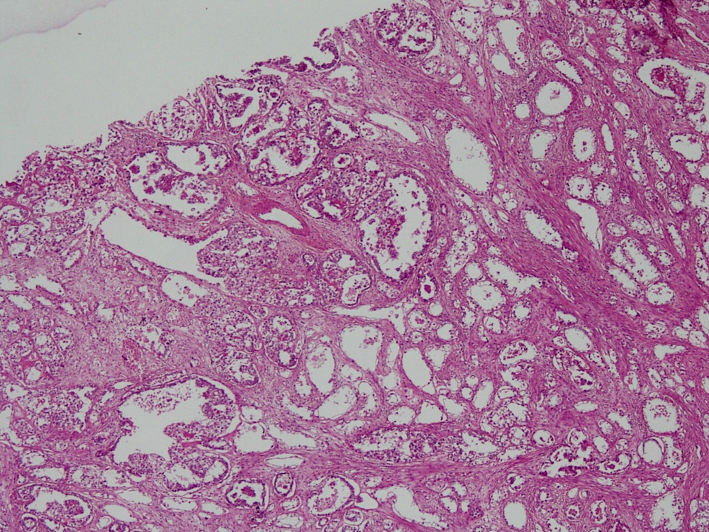
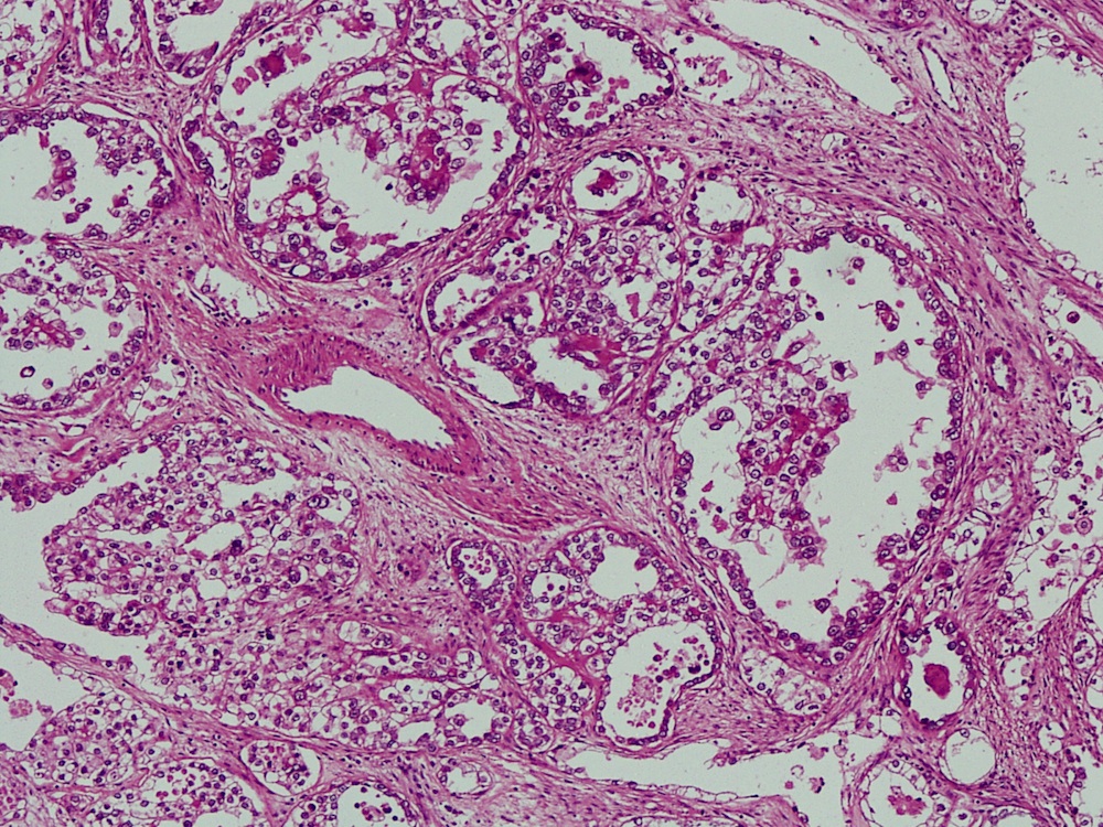
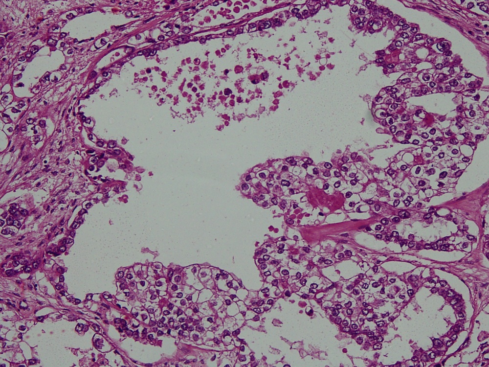
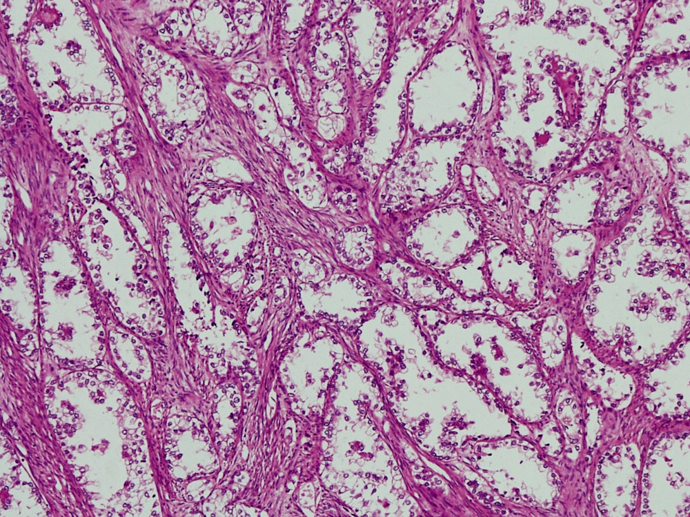
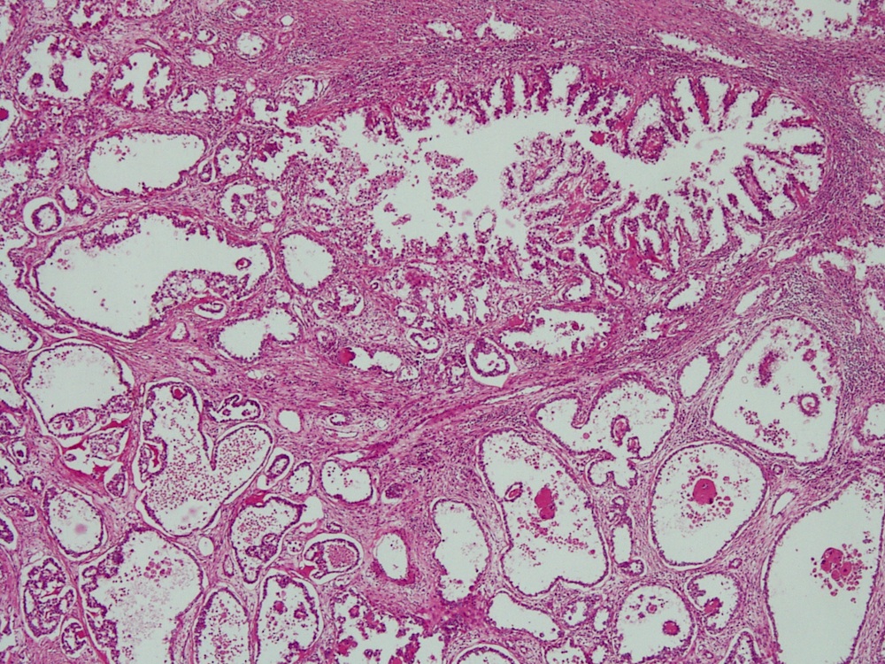
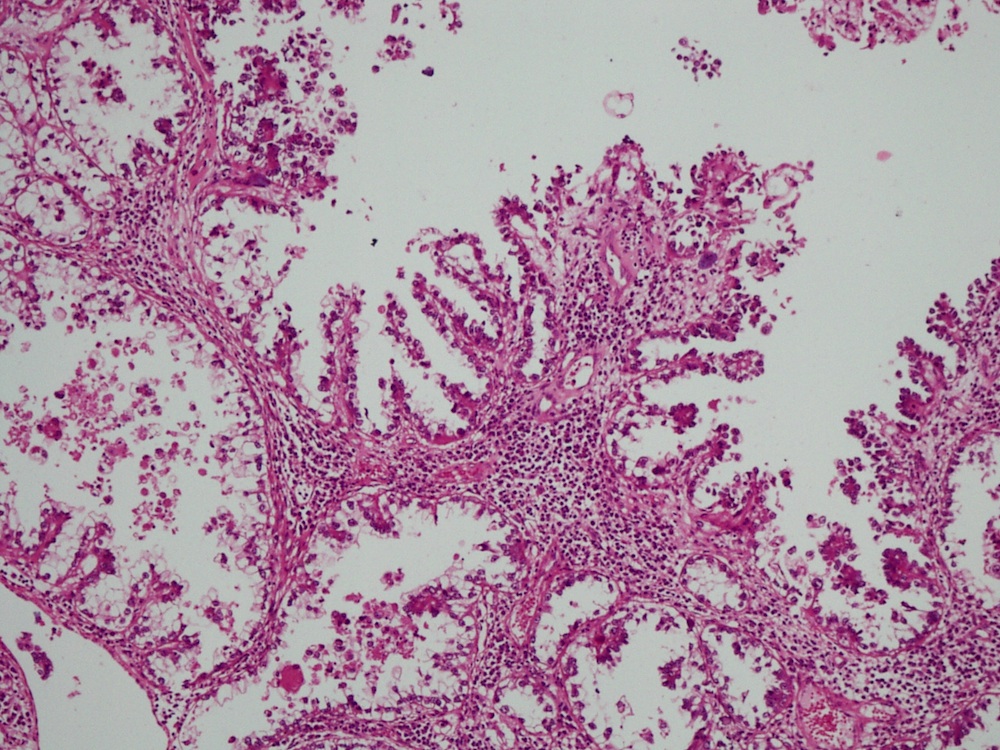
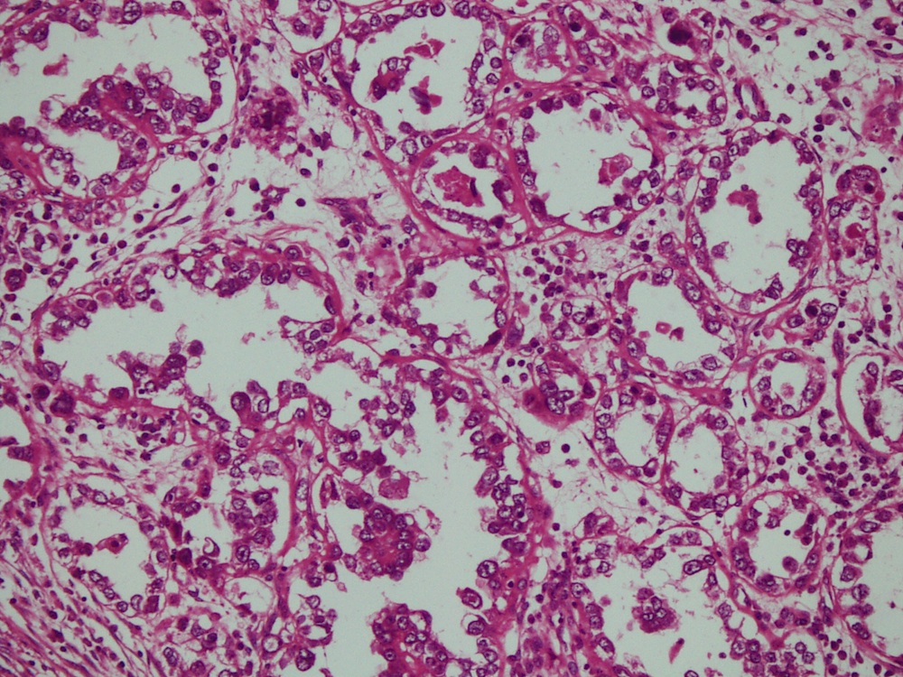
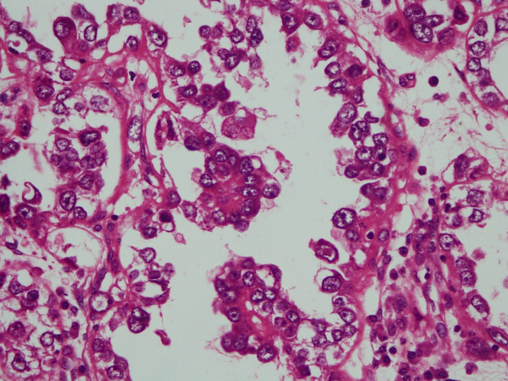
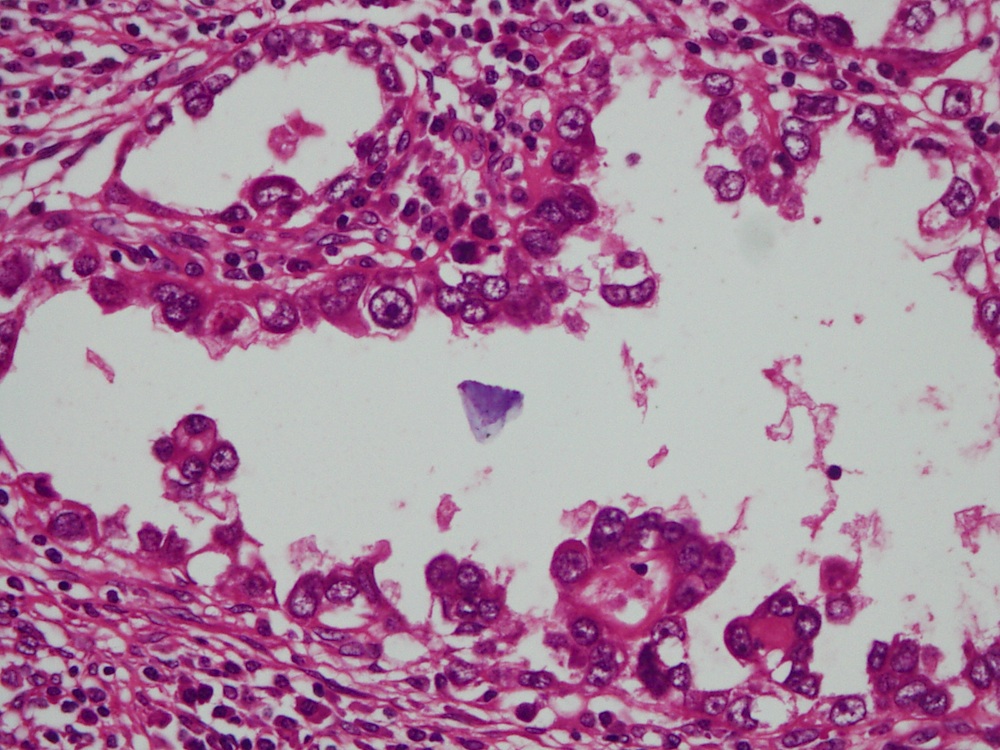
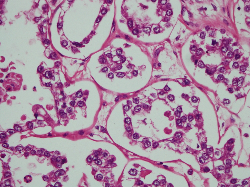
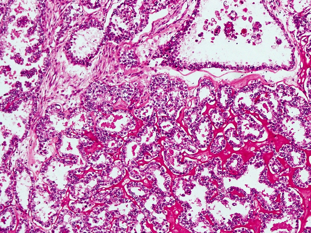
Microscopic (histologic) description
- Tumor has cystic, papillary, tubular / glandular and solid architectural patterns with focal necrosis
- Cells have distinct cell membranes, are large with moderate to abundant clear cytoplasm, occasionally may be oxyphilic
- Cells are usually cuboidal and sometimes hobnail type with nuclei protruding into the lumen
- Nuclei are round to irregular, hyperchromatic with conspicuous nucleoli (Int J Gynecol Pathol 2001;20:252)
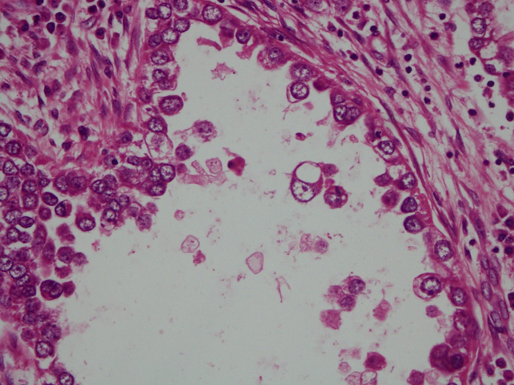
Microscopic (histologic) images
Case #363


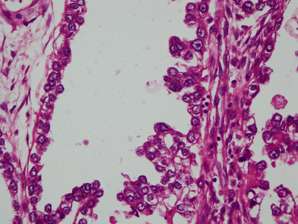
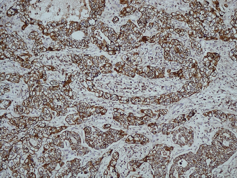
CK7
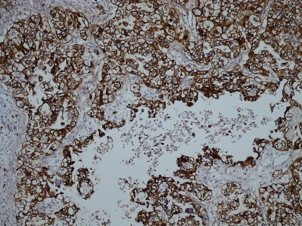
EMA
Images hosted on other servers:
Fig 3
Fig 4: papillae and acini in tubulocystic pattern
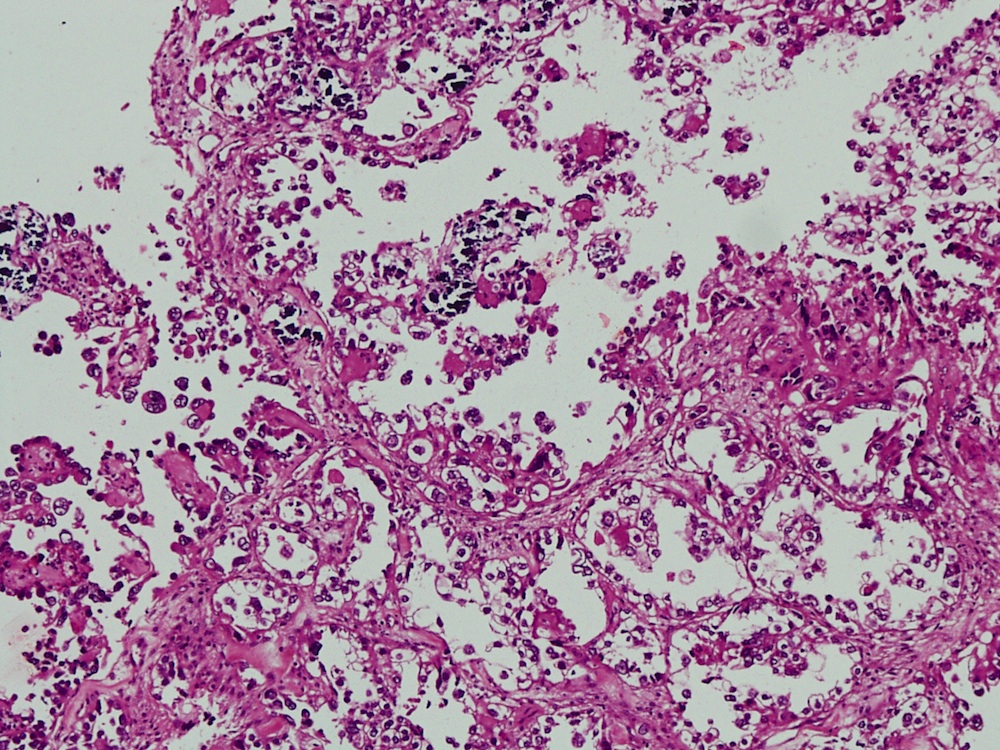
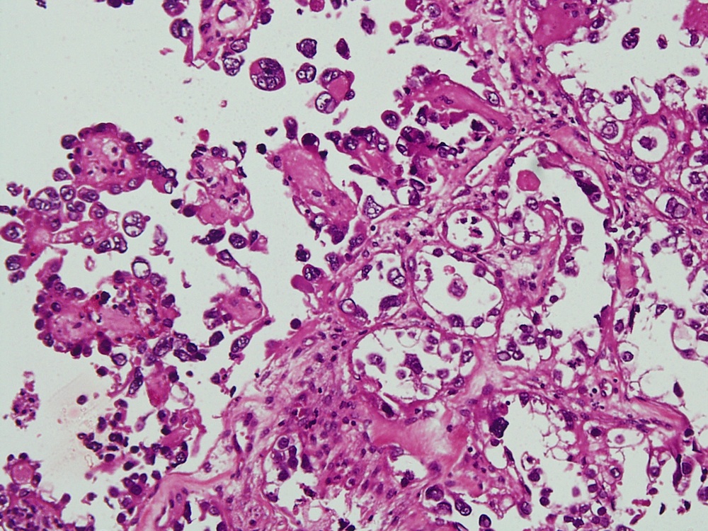
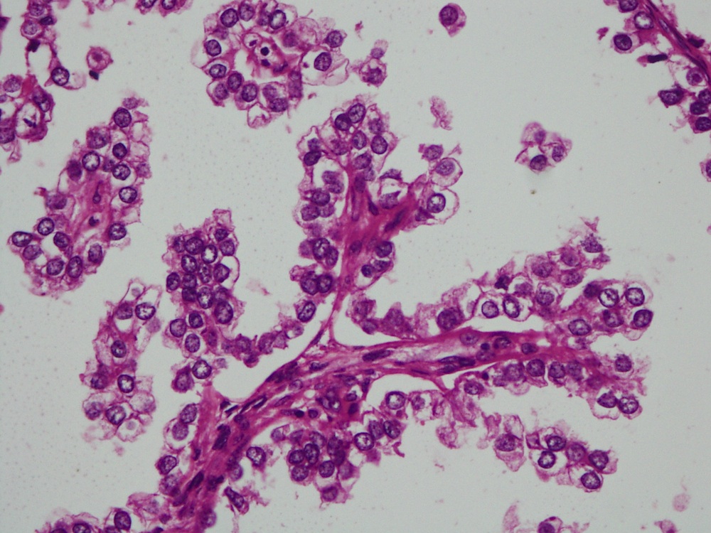
Cytology description
- Provides diagnostic information in 41% of cases (Gynecol Oncol 2007;105:273)
- Cells are arranged in sheets, clusters or papillae; cells have delicate vacuolated glycogen rich cytoplasm
- May have naked nuclei and a tigroid background, similar to other glycogen containing tumor cells such as seminoma and Ewing sarcoma
- Nuclei are large, pale and round with prominent nucleoli (Cytojournal 2013;10:17)
Cytology images
Images hosted on other servers:
Pap stain
Positive stains
- CK7, CAM 5.2, 34 beta E12, CEA, LeuM1, vimentin, bcl2, p53 and CA125
- Variable positivity for ER and HER2 / neu (Int J Gynecol Pathol 2001;20:252)
Negative stains
Electron microscopy description
- Similar ultrastructural features as CCA of ovary, cervix or endometrium
- Glands have short, thick microvilli in the lumina; cells are attached by desmosomes and interdigitating cytoplasmic processes
- Cells contain abundant glycogen granules, also many small, uniform mitochondria and "stacked" parallel rows of granular endoplasmic reticulum (Cancer 1972;29:1680, Cancer 1977;40:3019)
Molecular / cytogenetics description
- p53 nuclear staining patterns is heterogeneous in both proportion and intensity of tumor cells stained (Gynecol Oncol 1996;60:339)
- Overexpression of p53 protein is not due to mutational inactivation of the p53 gene but probably due to persistent DNA damage or genetic instability
- Intrauterine exposure to DES, therefore, is unlikely to directly alter the p53 gene as has been suggested for other mutagens
- Persistence of wild type p53 in these rare tumors may correlate with their typically favorable prognosis and radiosensitivity (Gynecol Oncol 1996;60:339)
Videos
Histopathology vagina - clear cell carcinoma
Differential diagnosis
- Arias-Stella reaction: cells are either not mitotically active or have only rare mitotic figures and the nuclei show degenerative features with pseudoinclusions and no mass lesion present (Adv Anat Pathol 2012;19:296)
- Mesonephric remnants: may also mimic clear cell carcinoma at low power (Adv Anat Pathol 2012;19:296)
- Metastatic tumor: rare; rule out kidney primary (Indian J Urol 2004;20:169)
- History, imaging, immunohistochemical stains and EM may be useful
- Microglandular hyperplasia: occasionally occurs in vagina (Adv Anat Pathol 2012;19:296)
- Serous carcinoma, yolk sac tumor and alveolar soft part sarcoma (Adv Anat Pathol 2012;19:296)
Additional references
