

Table 1: APLIS components (Adv Anat Pathol 2012;19:81)
| Layer | Description | Examples |
| APLIS application | The software interface to the end user; usually programmed for a specific operating system and almost always programmed for a specific DBMS; has user interfaces for data entry and manipulation | Cerner, CoPath, Cerner, PathNet, Orchard, Harvest, LIS, SSC SoftPath |
| Database management systems | A specialized software package for the persistent storage and manipulation of data; currently the vast majority of these use the relational model and implement an SQL interface; in the LIS, high performance is not as important as high reliability, requiring certain tradeoffs to be made | Microsoft SQL Server, mySQL, PostgreSQL, Oracle Database, MUMPS |
| Operating system | The fundamental control program through which the end user interacts with a computer; there are different operating systems that are suitable for different niches (e.g., it is far more common for Linux to be the operating system of choice for a server than for a personal computer) | Microsoft, Windows, Mac OS X, Linux |
| Hardware | Any physical device that either hosts or interfaces with the APLIS; requires both a hardware and a software interface for the operating system (and APLIS) to function | Server computer, client computer, barcode scanner, label printer, slide printer, H&E autostainer |

Google and ARM
Augmentiqs and ARM
AutoParis-X Demo Tutorial
Images hosted on other servers:

Master algorithm template for biochemical tests


Traditional image analysis versus computational pathology
(J Pathol 2019;249:286) |
(J Pathol Inform 2019;10:9) |
|
| Typical tasks | Detection of a morphological pattern (Lab Invest 2021;101:412) | Integration of all aspects of clinical workflow for more accurate diagnosis, prognosis and personalized treatment (Lab Invest 2021;101:412) |
| Parameter tuning | Image features / parameters are manually tuned |
Algorithm learns and extracts a large number of features automatically |
| Typical algorithm testing | Often on a few regions of the slide | Usually whole slide |
| Computer unit best suited for task | CPU (central processing unit) | GPU (graphics processing unit) |
| Number of training images required | Depending on application, may be low | Usually remarkably high |
Contributed by David F. Steiner, M.D., Ph.D.

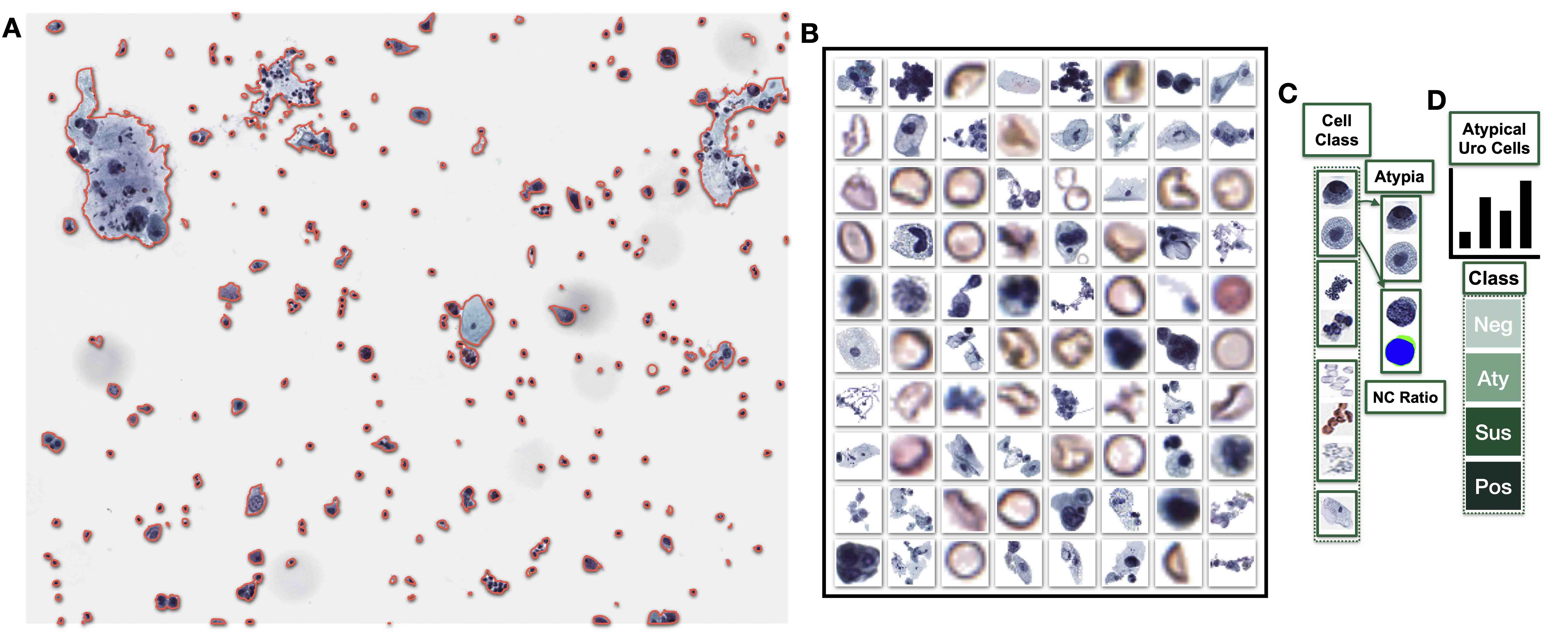
Detection of metastatic tumor in lymph nodes
- The figure below illustrates the type of calculations that image data goes through in convolution and pooling operations
- Convolution operations involve an elementwise product between the filter and different segments of equal dimensions from the input matrix
- Pooling operations perform an aggregate operation (e.g., maximum or average) on a region
- In the example below, the maximum value was returned from 2 x 2 regions of the input matrix
Contributed by Jerome Cheng, M.D.
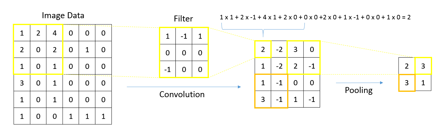
Convolution and pooling
Images hosted on other servers:

Typical CNN architecture

CNN layers in 3D

Neurons of convolutional layer connected to receptive field


Data management playlist - IBM Technology
Data storage essentials playlist - IBM Technology
Contributed by Chris Williams, M.D.
Highlighting pixels

PNG (lossless)
JPG (lossy) high quality
JPG (lossy) low quality
Images hosted on other servers:

Tagged diagnostic data within CCD

Untidy dataset and tidy equivalent

Relative pixel density and size of display resolution
Healthcare data standards
HL7
Images hosted on other servers:

Comparison of digital pathology and traditional workflow
Benefits of digital pathology
Digital pathology advances medical education and training
Social media in pathology education
Images hosted on other servers:
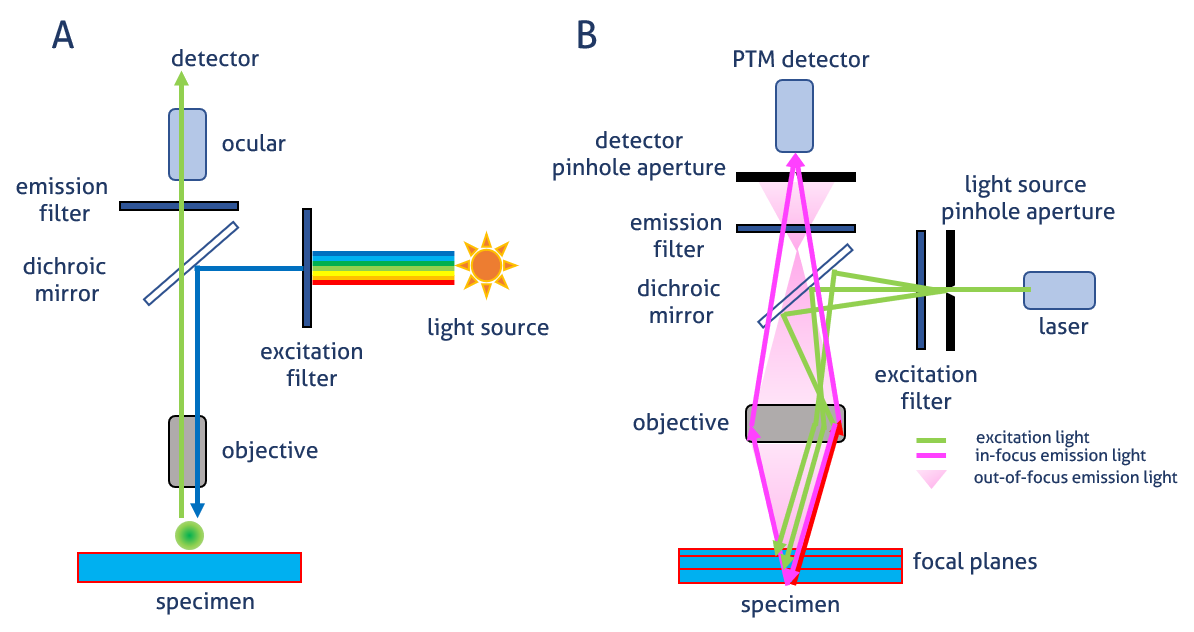
Image generation
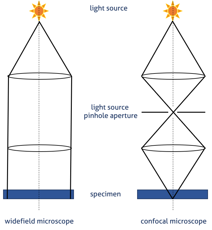
Visualization of pinhole principle
Fluorescence microscopy animation
Intro to fluorescence microscopy


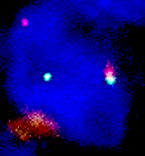
Contributed by Charles Caldwell, Ph.D., Vitria Adisetiyo, Ph.D., Cris Luengo, Ph.D., Roberto Gianani, M.D.
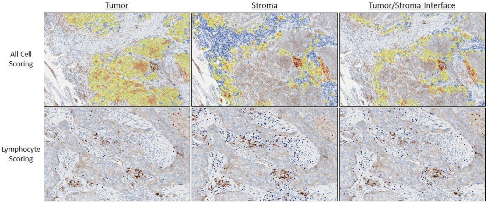
Image analysis of PDL1 in NSCLC
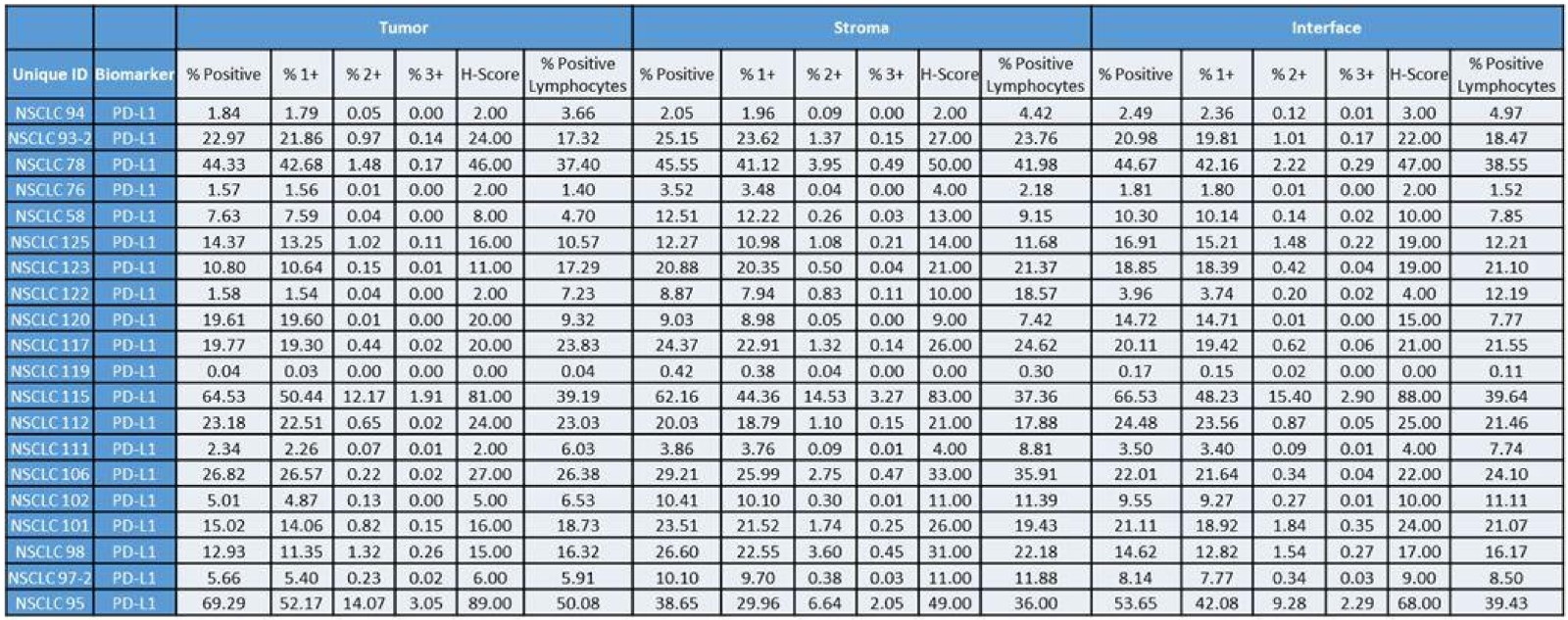
Image analysis pathology endpoint measures
Example of image analysis workflow: evaluation of PDL1 IHC staining in non small cell lung cancer
Images hosted on other servers:

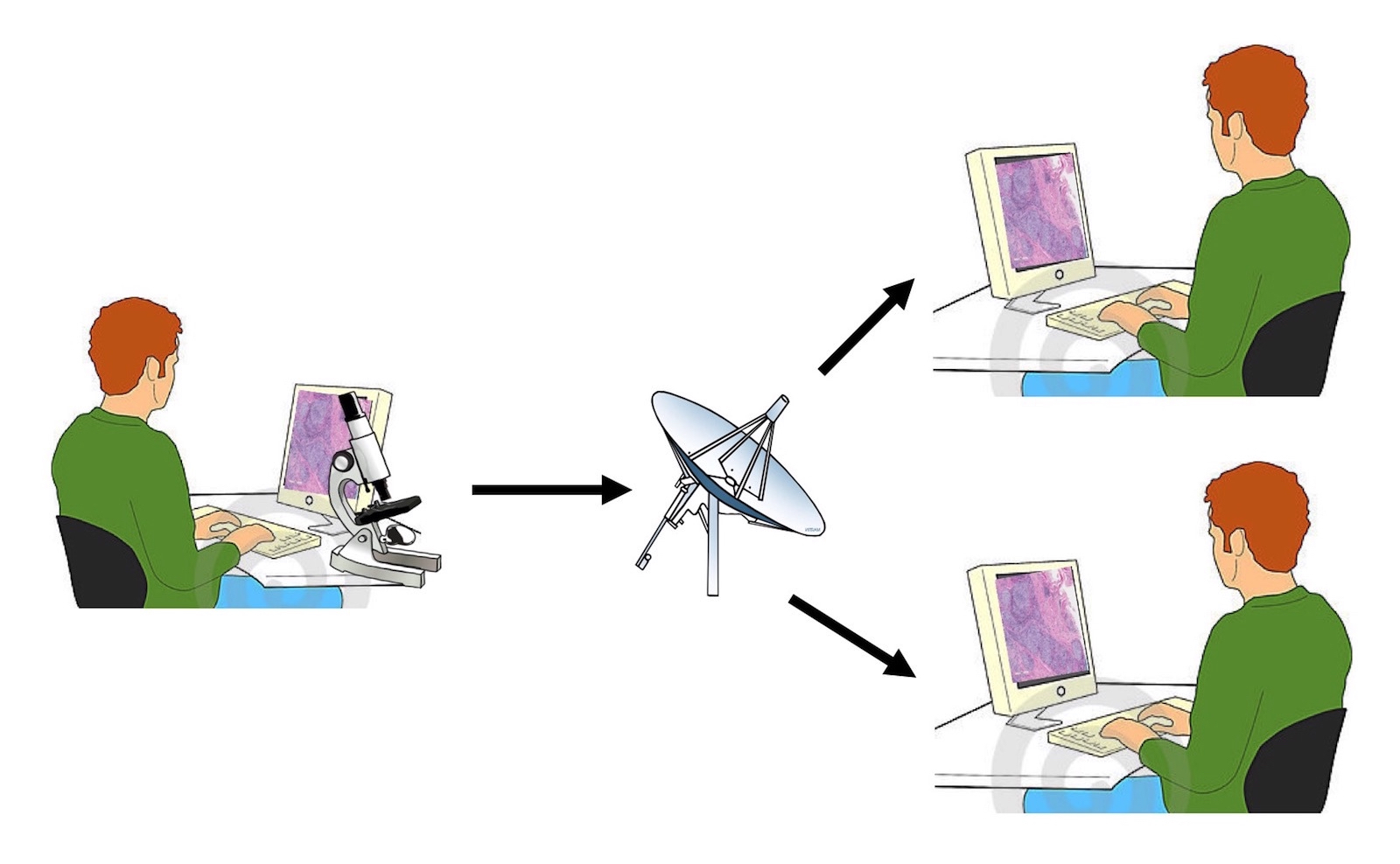
Network topology
for interinstitutional
frozen section
telepathology
Images hosted on other servers:

Most popular programming languages
What a computer program is
Programming languages
Integrated development environment (IDE)
Debugging
Source code


Contributed by Harsh Batra, M.B.B.S., D.C.P., D.N.B. and Anil Parwani, M.D., Ph.D., M.B.A.
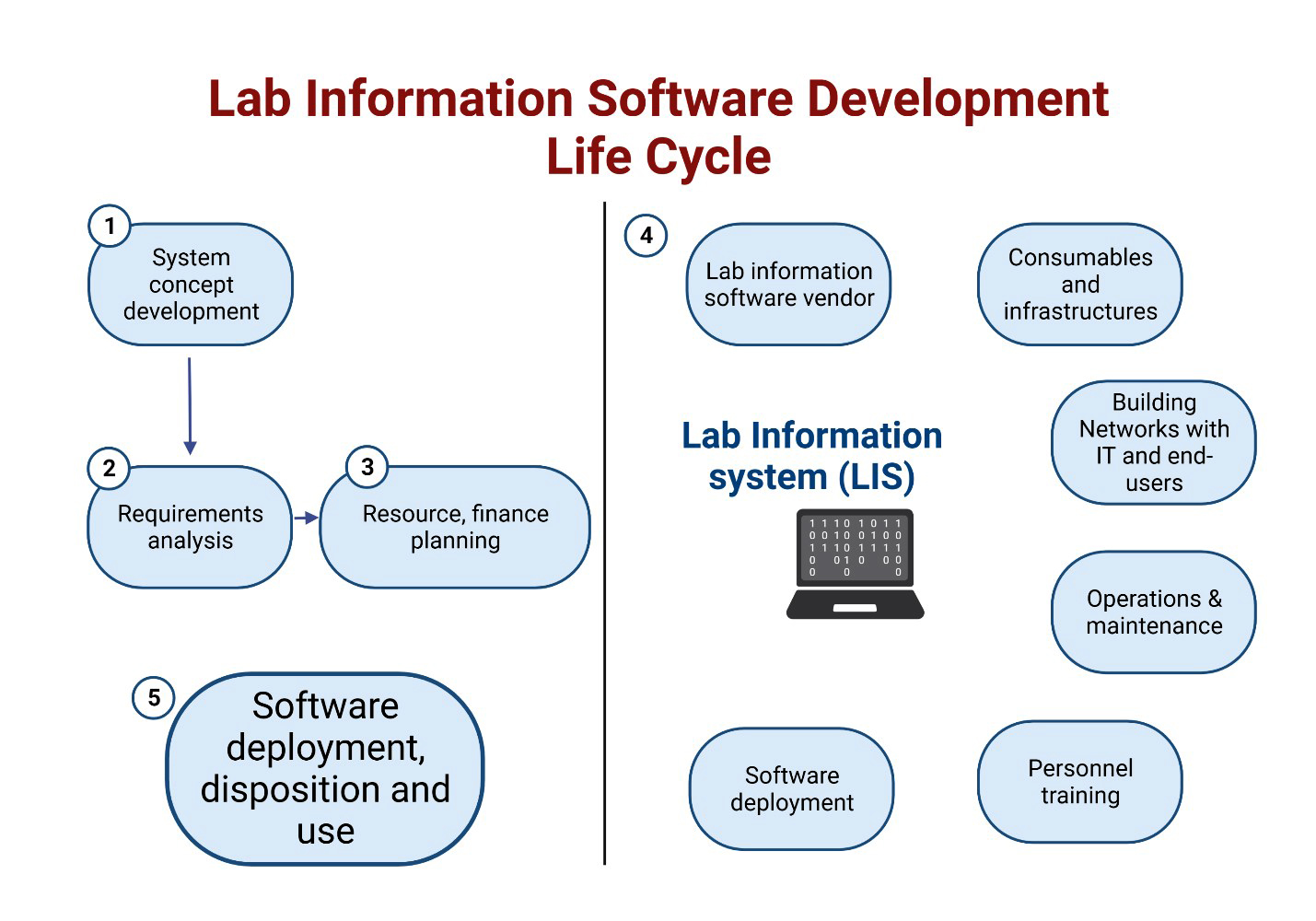
Lifecycle of LIS
Contributed by Harsh Batra, M.B.B.S., D.C.P., D.N.B., Maria Gabriela Raso, M.D. and Edwin Roger Parra Cuentas, M.D., Ph.D.
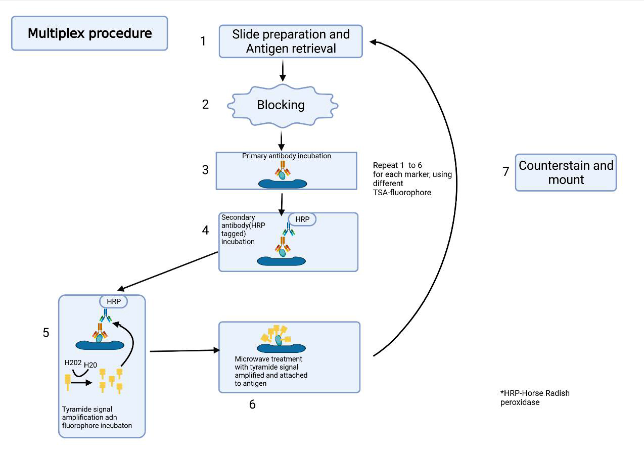
Procedure
Images hosted on other servers:

Architecture of deep neural network

Bag of words model

Word embedding


Spectral imaging laboratory
Medical applications of hyperspectral imaging
Images hosted on other servers:


Applications / incorporation of informatics into surgical / anatomical pathology
Contributed by Jasmine Saleh, M.D.
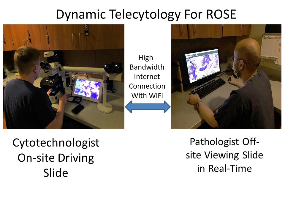
Real time
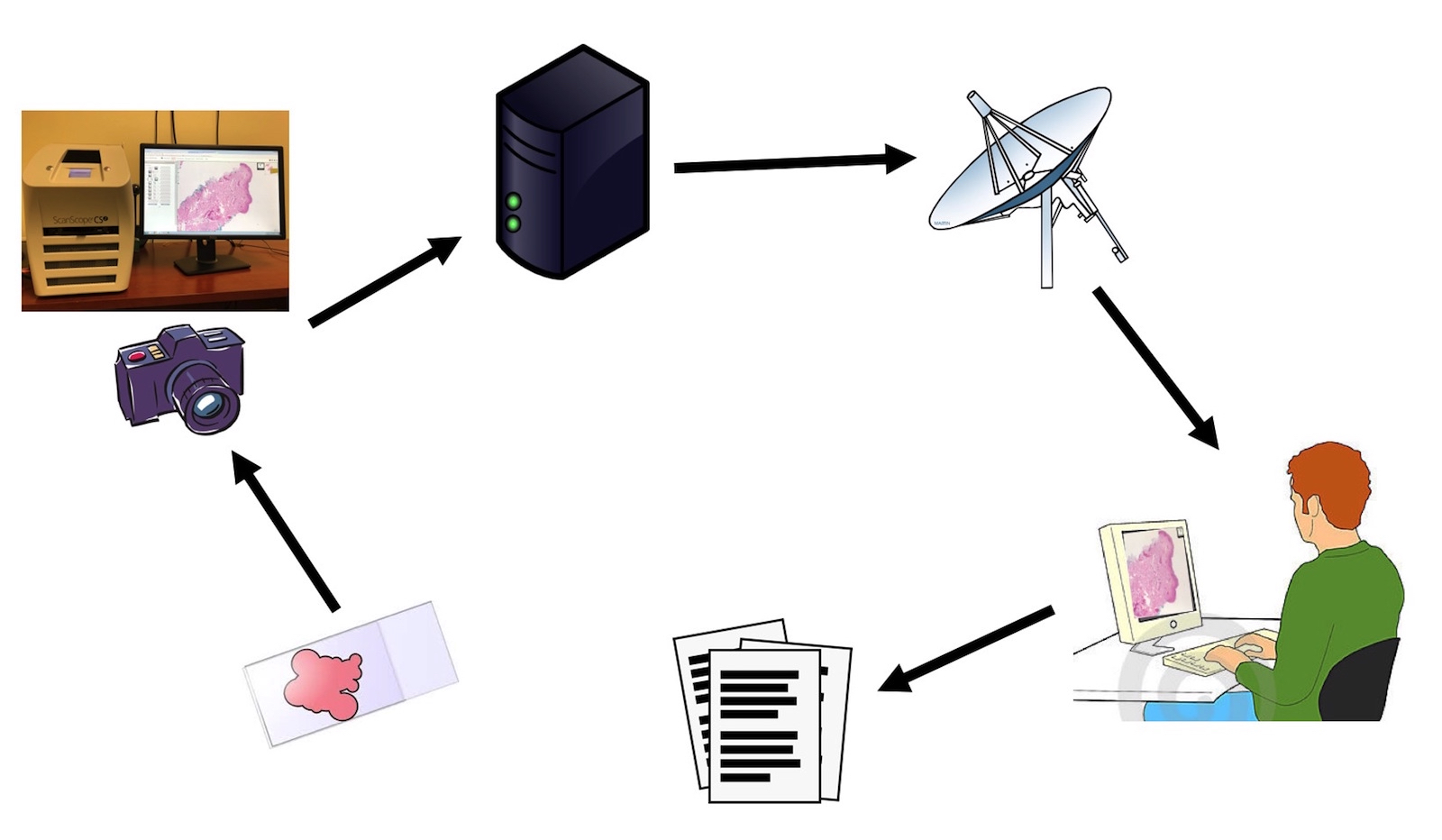
Store and forward
Images hosted on other servers:

Remote consultation

Use for frozen sections at remote site
Real time high definition video demo

Contributed by Rebecca Rojansky, M.D., Ph.D.
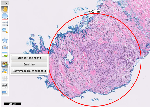
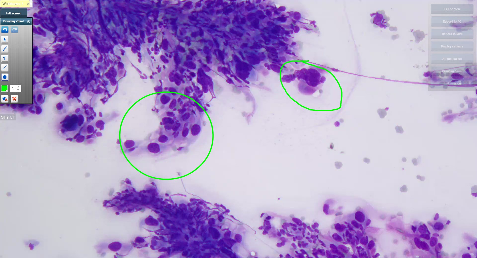
WSI annotation
Creating whole slide images with a robotic microscope stage
Whole slide images for diagnostic pathology
Whole slide imaging overview
Scanning (MikroScan D2)
Baron: 2023
Cohen: 2020
Holzinger: 2020
Huss: 2022
Pantanowitz: 2016
Pantanowitz: 2012
Parwani: 2016
Parwani: 2022
Zuraw: 2023
Find related Pathology books: informatics