
Author Instructions at PathologyOutlines.com
Revised: 12 April 2024
Copyright: 2012-2024, PathologyOutlines.com, Inc.
Author Instructions
Revised: 12 April 2024
Copyright: 2012-2024, PathologyOutlines.com, Inc.
Author Instructions
- Surgical pathology topic template
- Chemistry, toxicology & urinalysis topic template
- Cytology topic template
- Grossing topic template
- Hematopathology topic template
- Histology topic template
- Informatics, digital & computational pathology topic template
- Medical renal topic template
- Microbiology & infectious diseases topic template
- Stains, CD markers & molecular markers topic template
- Staging topic template
- Transfusion medicine topic template
- WHO classification topic template
- Other topic template
- Please note that to write for us, we need you to follow our format. This means writing in bullet points rather than complete sentences, adding references as PMIDs next to the relevant text, providing captions and legends for images and following specific format requirements for sections such as Sample pathology report and Board review questions. As a free website, we are not able to rewrite significant portions of text and we cannot accept submissions until they meet our requirements.
- We estimate it takes 10-15 hours to write a topic and obtain images.
- Authors typically work on one topic at a time and only those confirmed by us. After confirmation, we will email you a Word document of the topic as it currently exists along with our procedures. Please email the completed topic to us for review. A comprehensive writeup is 1,000-2,000 words (award winners are typically longer). There is no word limit, but we prefer concise writing. We will return it with a list of necessary changes, Our Editorial Board members and Deputy Editor for the subspecialty will then review. If you do send us more than one topic, we will hold the other submissions until the first topic has gone through the editing process and is published online. This way, you can apply any formatting or style changes that are needed to all further topics prior to submission.
- We need authors to be responsible and reliable. We expect the first draft within 4-6 weeks from assignment. Limited extensions may be granted. However, if we do not hear from you, we may cancel the assignment.
- Write using bullet points and concise phrases. If you use sub bullets, be consistent in the indentation of the sub bullets so it is clear for the editorial staff. Please limit use of bullets to two or rarely three levels of bullets.
- Do not quote directly from a publication; instead, paraphrase the text and include the reference at the end of the bullet.
- Special formatting such as bold, italics, underline or quotation marks should be removed whenever possible. We prefer plain text formatting.
- Text will be modified to conform to our publication format. For example, hyphens are mostly eliminated from text as they can be confused with a dash that means negative.
- Eliminate excess words and use a separate bullet for each fact, see example:
- Incorrect format:
- This disease affects males and females equally. Approximately 10% of patients with this disease have clinical symptoms of fever and pain.
- Correct format:
- M=F
- 10% have fever and pain (PMID XXXXXXXX)
- Incorrect format:
- Abbreviations are acceptable only if widely used, such as CT, DNA, EBV, FISH, H&E, HIV, IHC, N:C ratio, PET, PSA, RNA or RT-PCR. Otherwise, please write out the term for less common abbreviations, such as CR, DLBCL, LGL, ORR or PTCL.
- The use of tables is allowed; see examples: Diagnostic testing of SARS-CoV-2 (COVID-19) and Nephroblastoma
- Ensure that the topic contains material necessary for, or of interest to practicing pathologists.
- Images:
- You must provide your own micro images (typically 6 or more). We no longer allow links to outside micro images. H&E images should be JPG, GIF, PNG or TIFF format, 600 KB+, 1000 to 1600 pixels in height or width, demonstrating all important histologic features including low and high power and relevant immunostains. Please do not send images much larger than 2 MB.
- Generally, we prefer that authors send no more than 18 micro images.
- Click here to see Dr. Gonzalez's guide on how to take excellent histopathology images.
- Images should be original and in color. Authors should clean the slide, white balance the camera, focus the microscope, take the image and use imaging software to adjust contrast, color and brightness as needed. Do not edit images to change pathologic findings. If you edit images to clean up minor artifacts (which is acceptable), please indicate "Edits made to clean up minor artifacts" or something similar in the image legend. Examples of four high quality images are below:
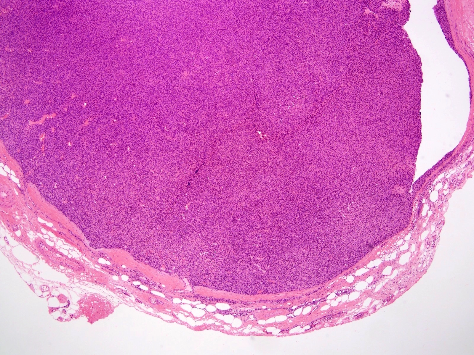
Well circumscribed
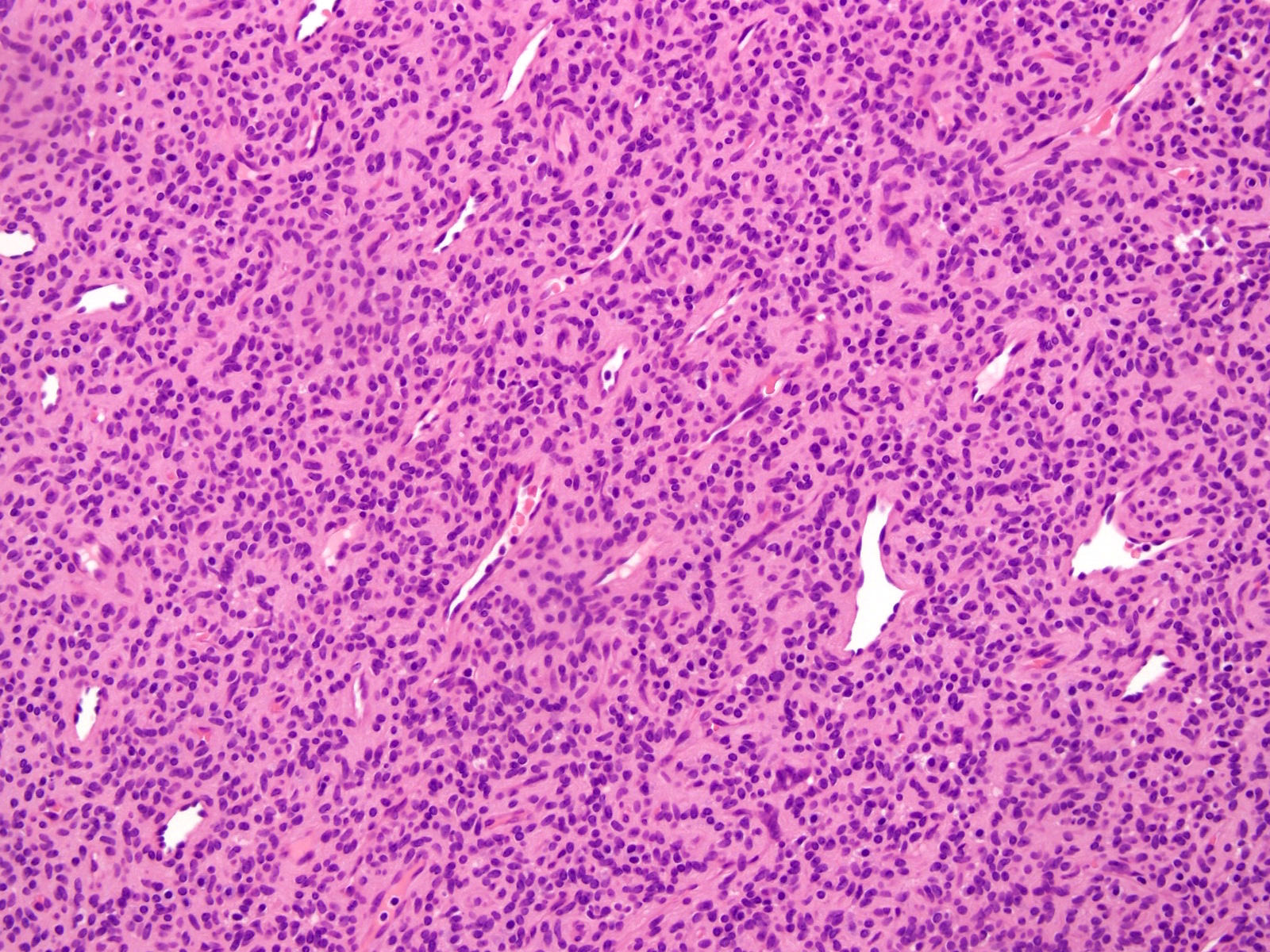
Sheets of cells

Spindling
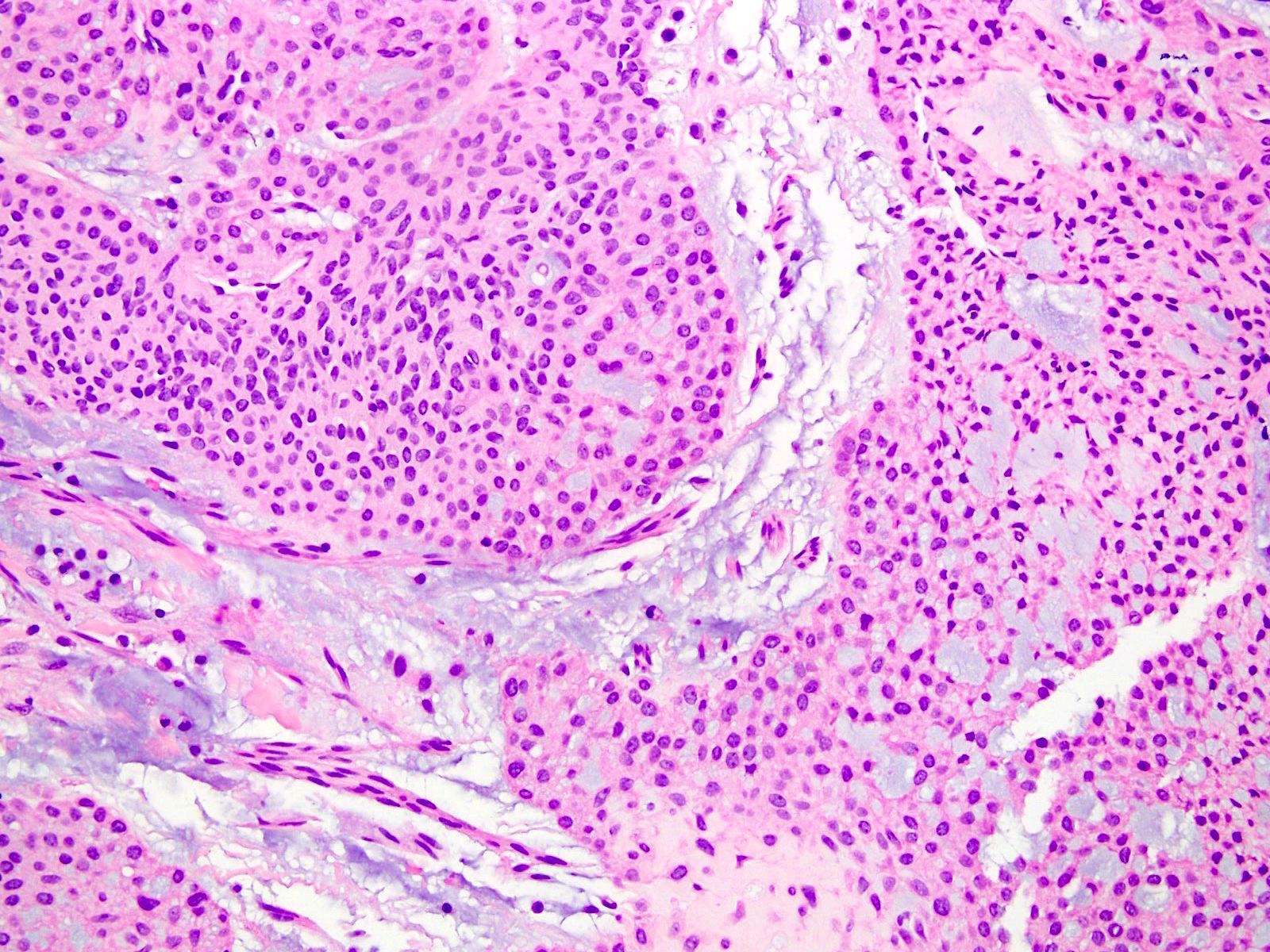
Myxoid stroma
- Each image should be 1 individual picture, not a composite, so it can be easily viewed and used.
- Images should be sent as attachments, either to the email or as part of a Google Drive or Dropbox file.
- Images should be submitted as you would for a textbook, in which pertinent clinical history is a part of the figure legend. Please name and number the image file so it can be clearly matched with the legend. Do not submit groups of pictures as interesting cases or unknown cases.
- For each image, submit the image file name, caption and a figure legend under the appropriate section (Gross images, Clinical images, etc). Please also indicate who owns the image and who contributed the image. If you own and contributed all of the images, you can make a general statement. Do not include your name, twitter handle, institution or logo on the image.
- The caption should be 1-4 words which describe the image findings, such as "cytologic atypia", "chronic inflammation" or "epithelioid cells." Do not use captions such as "Case 1", "Various images", "H&E", "100x" or the diagnosis.
- The figure legend will appear when the image is open and must contain all relevant information including site, procedure, diagnosis, key histologic findings and stain if other than H&E. It can include magnification (objective, excluding eyepiece) but should include more than that. Do not use figure legends such as "Case 1" or "Representative images".
- We will remove all existing outside micro images.
- To make the topic complete, include relevant clinical images, radiologic images, gross images, intraoperative images, electron microscope images and molecular / cytogenetic images which can be provided by you or with links to PathoPic, Radiopaedia, WebPath, Wikipedia or free full text journals accessed via PubMed. To keep an existing image, the author must provide a reason why it should be kept.
- Authors are responsible for their own illustrations but we can refer you to an illustrator upon request.
- Click here to view our Image Guide showing the different types of images.
- All external images must be accessible without having to register or pay a fee to see them or we cannot use them. This includes most PubMed images listed as "free full text" or "open access", but you need to double check. Since your institution may pay to give you access, you should verify that images are free on your home computer or by using the “incognito” setting on your browser.
- For external images, please provide a link for the image or specify which image from the article you would like to use, along with a 1 - 4 word caption. (e.g. PMID: 21084962, figure 1, IAPN gross / histologic correlation)
- Please check these websites for relevant virtual slides: University of Toronto, World Tumor Registry (click on Tumor Collections). Please advise if you cannot find any virtual slides. Contact us for instructions on how to submit virtual slides.
- Click here for a great resource by Dr. Eric Glassy on creating photomicrographs from whole slide images or other sources on the web that does not violate copyright law. If an image is captured from a virtual slide, please let us know so that we can list both you and the owner of the virtual slide as contributors.
- References: topics should have 5+ journal references and preferably 10. The most important case series and other references should be included. Authors should do a literature search of PubMed for articles. References should be from journals published in English and most should be from within the past 5 years. References should refer to the primary report that describes the finding, not to a case report that cites this primary report. Examples of preferred pathology journals are Am J Surg Pathol, Am J Clin Pathol, Arch Pathol Lab Med, Hum Pathol, Histopath, Mod Pathol and Virchows Archiv. Please insert the PMID number (at the bottom of the PubMed page) for each reference after the text that it is supporting. Example: 30% present with advanced disease. PMID 27053535. For rare topics we prefer some citations, even if older or in a foreign language. Results from a study or anything not general knowledge should have a reference. WHO books, textbooks, UpToDate, StatPearl or websites should be used sparingly as references and only if they define histologic variants, grading, a newly recognized entity or contain information not in a journal.
- For case report citations, we prefer including the best case reports as well as those in journals with free full text in English from the past 5 years.
- We provide you the option of writing a separate short paragraph to appear in our blog that describes this topic, to appear with your picture.
- Author names appear on topics and chapters with links to our Authors page, which lists topics completed per author.
- We consider a topic for an author award if it is thoroughly written with submission of several categories of supplemental material such as personally created or obtained diagrams or tables and radiology, cytology, frozen section, gross and molecular images. Click here for an example of a prior winning topic.
- Videos are optional but may be helpful to explain histology or other parts of your topic. They can be links to existing videos or to videos that you create. We suggest they be 5 minutes or less if you create your own.
- Contributing images to existing topics: We only accept images to add to an existing topic if they are high quality, have individual captions and figure legends for each image and if we currently lack high quality internal images for this topic.
If you are interested in being an author, please send your CV and a list of topics or chapters you are interested in writing / reviewing to our author coordinators at Authors@PathologyOutlines.com with any questions.
