
Stains & CD Markers
CD38


Copyright: 2002-2024, PathologyOutlines.com, Inc.
PubMed Search: CD38 stain
CD38
Editorial Board Member: Brandon Umphress, M.D.
Deputy Editor-in-Chief: Genevieve M. Crane, M.D., Ph.D.
Last author update: 16 June 2023
Last staff update: 16 June 2023
Copyright: 2002-2024, PathologyOutlines.com, Inc.
PubMed Search: CD38 stain

Table of Contents
Definition / general | Essential features | Terminology | Pathophysiology | Clinical features | Interpretation | Uses by pathologists | Prognostic factors | Microscopic (histologic) description | Microscopic (histologic) images | Positive staining - normal | Positive staining - disease | Negative staining | Flow cytometry images | Sample pathology report | Additional references | Board review style question #1 | Board review style answer #1Cite this page: Bruehl F, Schürch CM. CD38. PathologyOutlines.com website. https://www.pathologyoutlines.com/topic/cdmarkerscd38.html. Accessed May 13th, 2024.
Definition / general
- Marker of cellular activation expressed by plasma cells, T cells, NK cells and other hematopoietic cell types during various stages of maturation
Essential features
- Marker of activation that is present on many hematopoietic cells, especially plasma cells
- Used clinically as a prognostic marker in chronic lymphocytic leukemia (CLL) as evaluated by flow cytometry
- CD38 expression in lymphoid neoplasms is not specific for any discrete disease entity
- Can be aberrantly expressed in carcinoma and melanoma
- Absence of CD38 (in conjunction with CD34 positivity) is used as a marker for bone marrow hematopoietic stem cells
Terminology
- Also known as ADP ribosyl cyclase 1 (Cytometry B Clin Cytom 2013;84:207)
- First described in 1980 as T cell differentiation antigen (T10) (Proc Natl Acad Sci U S A 1980;77:1588)
Pathophysiology
- CD38 is a single chain, 45 kDa transmembrane molecule with its coding gene sequence located on chromosome 4 (Cytogenet Cell Genet 1995;69:38)
- CD38 has multifunctional activity; it has enzymatic and adhesion molecule features, for example, participating in the NAD+ cell cycle, regulating intracellular calcium levels and playing a role as receptor for the platelet and endothelial cell adhesion molecule CD31 (Physiol Rev 2008;88:841, J Immunol 1998;160:395, Curr Pharm Des 2009;15:57)
- Binding of CD38 by agonistic antibodies has been shown to prevent apoptosis (Eur J Immunol 1994;24:1218)
- CD38 has been shown to be highly expressed on myeloid derived suppressor cells (JCI Insight 2018;3:97022)
Clinical features
- CD38 is highly expressed on most multiple myeloma cells; the anti-CD38 monoclonal antibody daratumumab is used therapeutically in multiple myeloma patients and the response to therapy was shown to be correlated with CD38 expression as measured by flow cytometry (Br J Haematol 1999;105:441, Leukemia 2002;16:30)
- Several other anti-CD38 antibodies, including bispecific anti-CD3 / CD38 antibodies, are in development or have recently been approved for treatment in multiple myeloma (e.g., isatuximab, approved March 2020) (Front Immunol 2018;9:2722, Front Immunol 2020;11:501)
- CD38 expression on CD8+ T cells is associated with risk of progression of HIV infection to AIDS, is associated with poor prognosis and may be used as surrogate marker for HIV RNA viral load (J Acquir Immune Defic Syndr Hum Retrovirol 1997;16:83, Cytometry B Clin Cytom 2009;76:375, J Acquir Immune Defic Syndr 2006;41:416)
- Autoantibodies to CD38 have been implicated in autoimmune related diabetes mellitus type 1 in children (J Pediatr Endocrinol Metab 2005;18:1417)
Interpretation
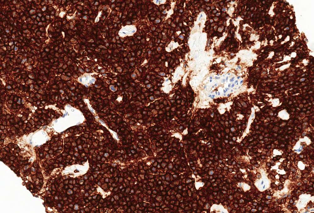
- CD38 expression is considered positive when the cell membrane shows strong and diffuse staining; the cytoplasm and nucleus should not stain with CD38 (Blood 2008;111:5173)
- Few studies have reported on the use and interpretation of anti-CD38 antibodies in tissue sections for diagnostic purposes; interpretation is difficult due to the prevalence of CD38 in many cell types and the necessity for quantitative assessment more amenable to flow cytometric studies (Am J Surg Pathol 2006;30:585)
Uses by pathologists
- Flow cytometry is the primary use of anti-CD38 antibodies in the pathology laboratory
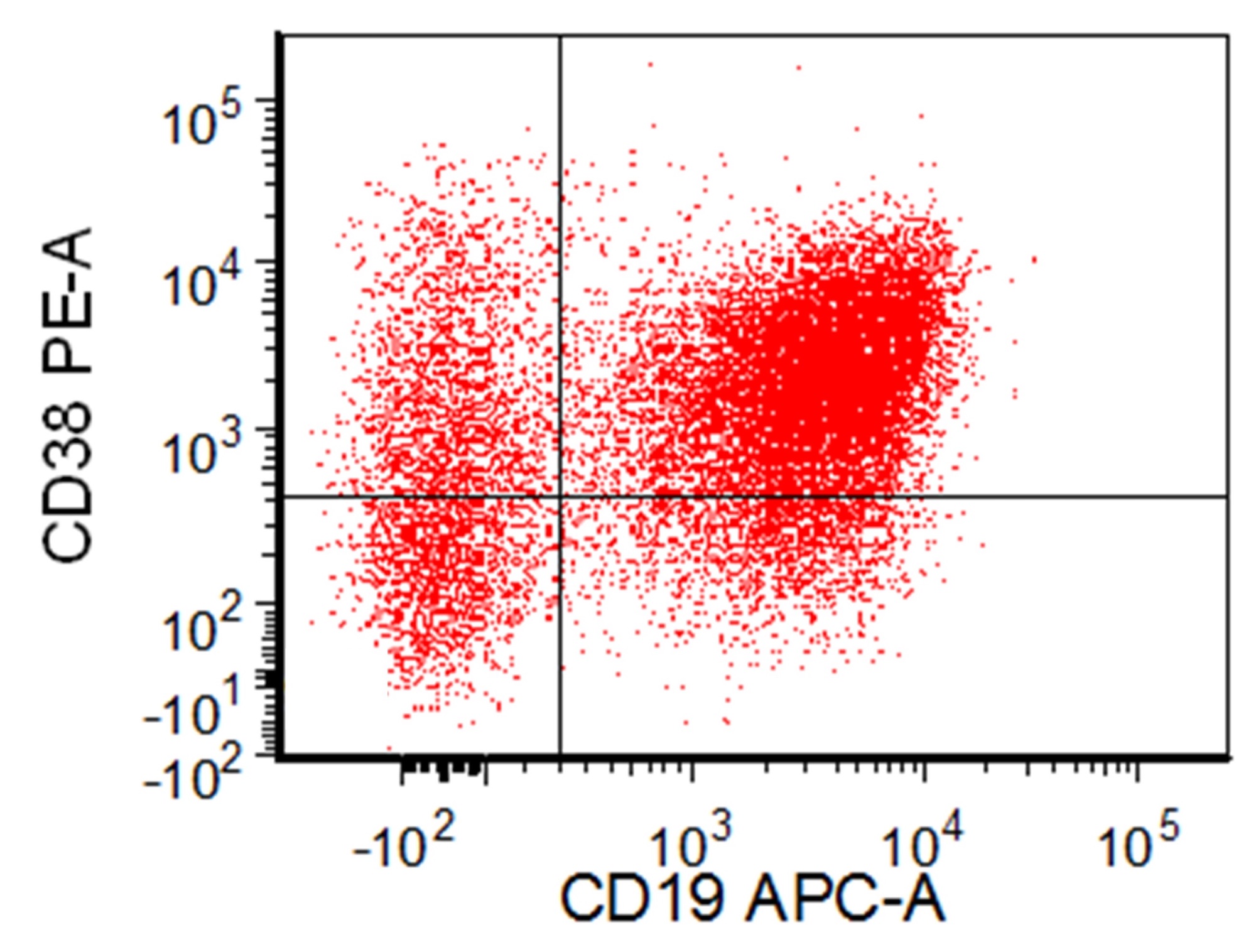
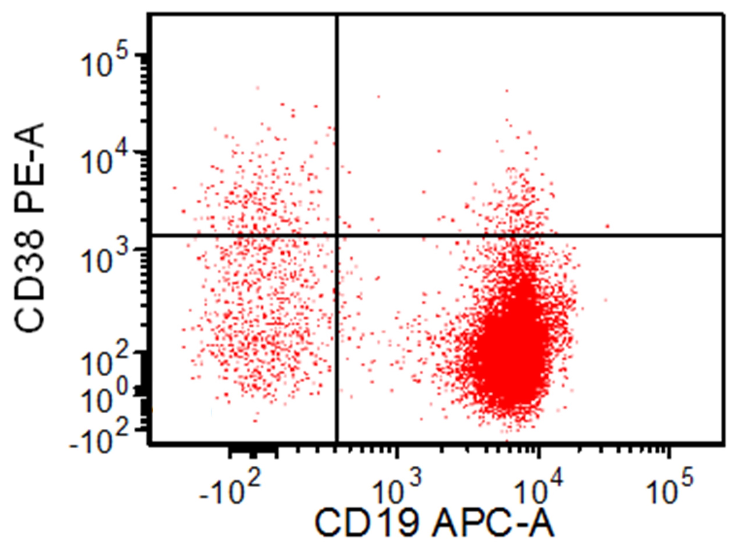
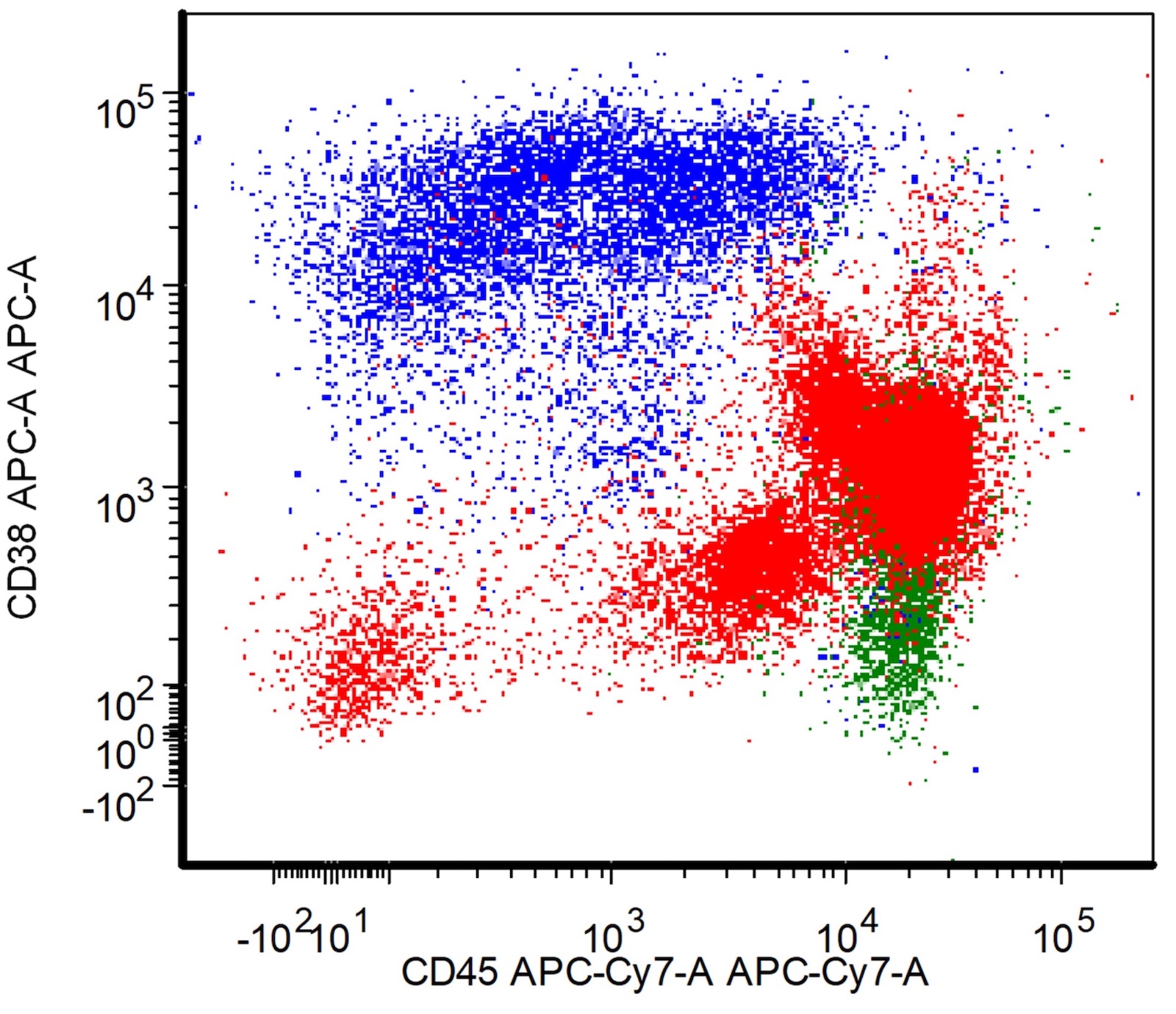
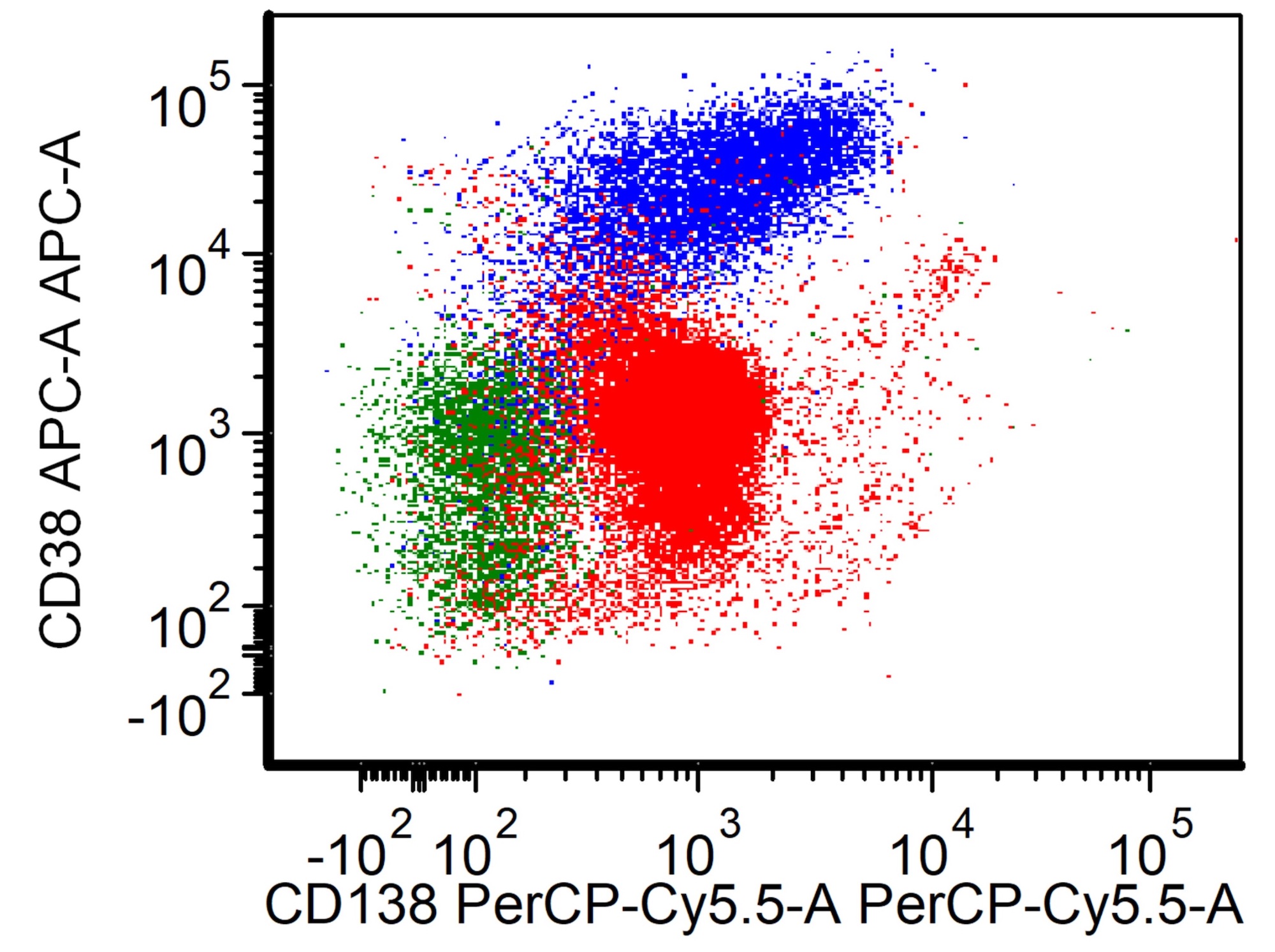
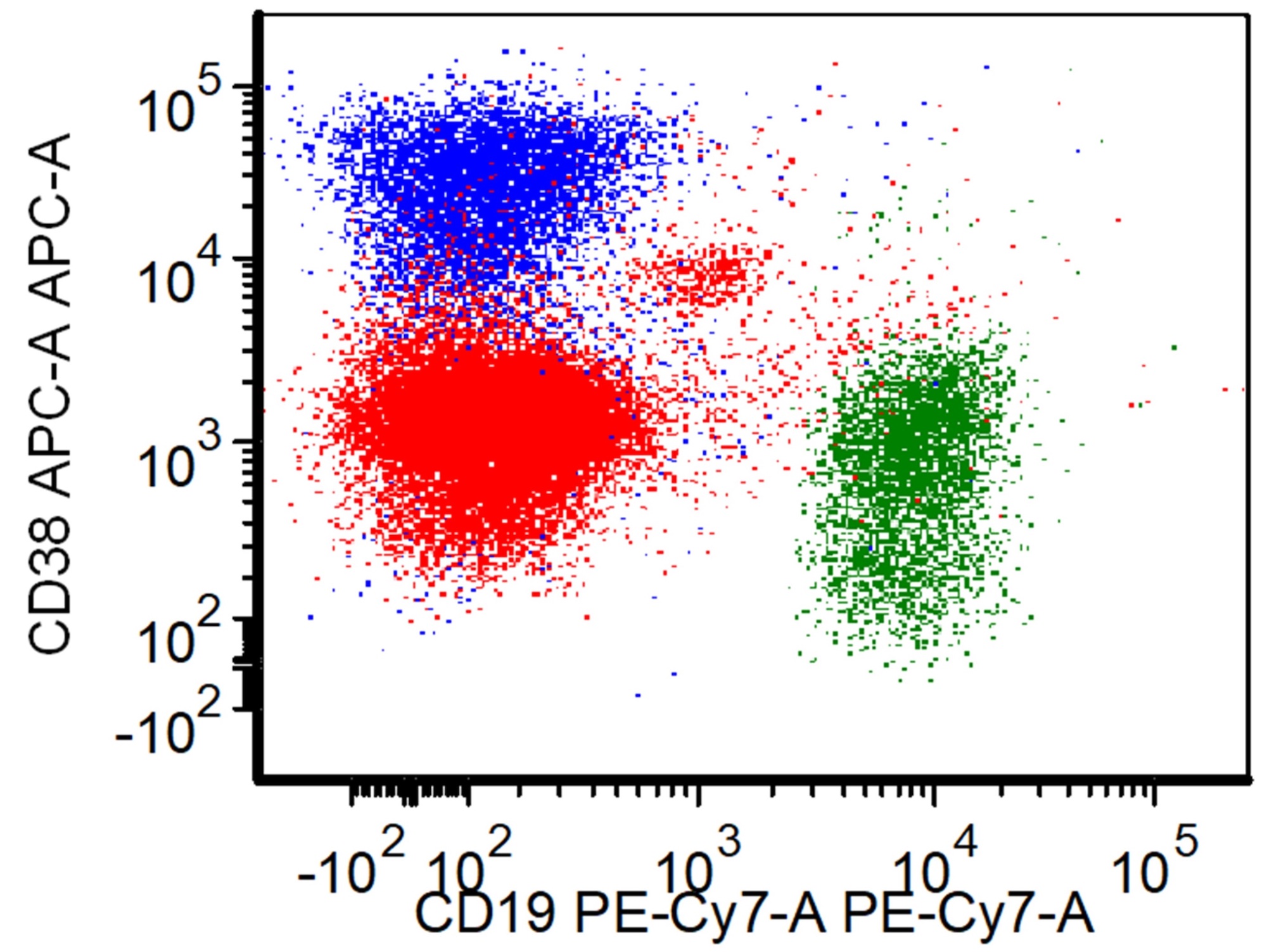
- CD38 is used in conjunction with CD138 to identify plasma cells (see Flow cytometry images below) and with CD20 to differentiate germinal center B cells from mantle zone B cells (Am J Clin Pathol 2003;119:130)
- CD38 expression by plasma cells is used in minimal residual disease monitoring (Cytometry B Clin Cytom 2016;90:61)
- Bright expression of CD38 aids in the identification of hematogones (Leuk Lymphoma 2004;45:277)
- There is considerable confusion in the literature regarding CD38 and the unrelated antibody VS38 which targets the p63 antigen and is also used for detection of plasma cells in tissue sections and flow cytometry (Blood Cancer J 2018;8:117)
Prognostic factors
- Chronic lymphocytic leukemia with CD38 expression is associated with a more aggressive clinical course and shorter overall survival (J Clin Pathol 2002;55:180, Br J Haematol 2003;120:1017)
- Hairy cell leukemia with CD38 expression is associated with a more aggressive clinical course (Cancer Res 2015;75:3902)
- Acute myeloid leukemia with CD38 expression was shown to be associated with a favorable prognosis and high numbers of immature CD34+ / CD38- blasts in myeloid leukemia are associated with unfavorable prognosis (Leuk Res 2000;24:153, Leukemia 2019;33:1102)
- CD38 expression on multiple myeloma cells has been correlated to anti-CD38 treatment response (Blood 2016;128:959)
- In the future, CD38 expression in tissue sections could be used to monitor anti-CD38 therapy with (bispecific) antibodies for early detection of treatment resistance (loss of CD38 expression)
Microscopic (histologic) description
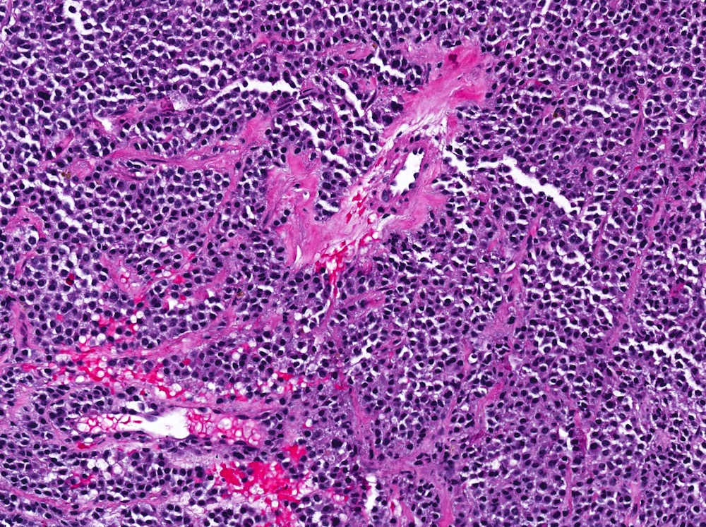
- Plasmacytoma: see Microscopic (histologic) images
Positive staining - normal
- Plasma cells, hematopoietic progenitor cells, NK cells, B and T cells, monocytes and basophils (Chem Immunol 2000;75:169)
- Neurons, small lymph vessels in intestinal tract and pancreatic islets, perivascular autonomic nerve terminals, double positive thymocytes, erythrocytes (Brain Res 1995;697:235, Virchows Arch 2002;441:605, J Biol Regul Homeost Agents 1998;12:81, Int Immunol 2003;15:1105, Hematology 2007;12:409)
Positive staining - disease
- B cell lymphoid neoplasms
- Multiple myeloma and other plasma cell neoplasms (Br J Haematol 1999;105:441, Best Pract Res Clin Haematol 2010;23:433)
- Plasmablastic lymphoma and plasmablastic plasma cell myeloma (Mod Pathol 2005;18:806, Mod Pathol 2010;23:991, Am J Surg Pathol 2004;28:736)
- High grade B cell lymphoma with MYC and BCL2 or BCL6 rearrangement (Am J Clin Pathol 2022;158:338, Mod Pathol 2022;35:419)
- Burkitt lymphoma (Diagn Pathol 2019;14:100)
- Primary effusion diffuse large B cell lymphoma (Am J Clin Path 1996;105:221, Mod Pathol 2010;23:773)
- Chronic lymphocytic leukemia / small lymphocytic lymphoma and Richter transformed large cell lymphoma (Am J Surg Pathol 2002;26:624, Am J Clin Path 2001;115:385)
- Atypical chronic lymphocytic leukemia only rarely expresses CD38 (Am J Clin Path 2001;116:655)
- Nodal marginal zone lymphoma (10/24) (Am J Surg Pathol 2003;27:762)
- Lymphoplasmacytic lymphoma (Am J Clin Path 2005;124:414)
- Potential use in distinguishing follicular lymphoma and follicular hyperplasia (Int J Clin Exp Pathol 2018;11:1046)
- T cell lymphoid neoplasms
- T lymphoblastic leukemia (Am J Surg Pathol 1992;16:1075)
- T cell lymphomas (peripheral T cell lymphoma, NOS [57% positive], angioimmunoblastic T cell lymphoma [80%], ALK negative [17%] and ALK positive [0%] anaplastic large cell lymphoma) (Am J Hematol 2017;92:E1, Cytometry B Clin Cytom 2006;70:142)
- NK / T cell leukemia (N Engl J Med 2016;375:1501)
- Myeloid neoplasms
- CD38 is heterogeneously expressed in acute myeloid leukemia (Haematologica 2019;104:e100)
- CD38 positivity is used as an inclusion criterion in clinical trials evaluating anti-CD38 therapies
- Other
- Various types of carcinoma and melanoma (Cancer Discov 2018;8:1156)
- Prostate carcinoma (Cancer Metab 2018;6:13)
- Glioma (J Immunol 2008;181:92)
- Transient myeloproliferative disorder (Am J Clin Path 2001;116:204)
- Various inflammatory lymphadenopathies: infectious mononucleosis, Kikuchi lymphadenitis (Am J Clin Path 2003;120:49, Am J Clin Path 1989;92:42)
Negative staining
- Absence of CD38 expression does not reliably exclude a given pathologic diagnosis based on currently available data
- CD38 surface expression may be reduced on multiple myeloma / plasma cells due to CD38 internalization induced by treatment with anti-CD38 antibodies (e.g., daratumumab) (Oncoimmunology 2018;7:e1486948)
- CD38 has been used in conjunction with CD117 in fluorescence activated cell sorting of mast cells from bone marrow samples (CD117 positive and CD38 negative cells) (Am J Pathol 1996;149:1493)
- CD4+ CD26- T cells in patients with a diagnosis of Sézary syndrome are typically negative for CD38 (Dis Markers 2022;2022:3424413)
Flow cytometry images
Sample pathology report
- Bone lesion, needle core biopsy:
- Monotypic kappa expressing plasma cell neoplasm, consistent with plasmacytoma (see comment)
- Comment: The needle core biopsy demonstrates a dense infiltration by a clonal plasma cell population with kappa light chain restriction. The neoplastic plasma cells are positive for CD38, CD138 and CD79a. Lambda light chains, CD20 and CD43 are not expressed. There are only scattered CD3 positive T cells. There is no amyloid deposition. In summary, the bone lesion represents a plasmacytoma; clinical, serological and imaging correlation is required.
Additional references
Board review style question #1

Which statement about CD38 is correct?
- CD38 is typically downregulated in neoplastic plasma cells
- CD38 is brightly expressed on hematogones
- CD38 expression is a specific marker for hairy cell leukemia
- In chronic lymphocytic leukemia / small lymphocytic lymphoma, CD38 expression is considered a favorable prognosis
Board review style answer #1
B. CD38 is brightly expressed on hematogones. Answer A is incorrect because plasma cells typically downregulate CD38 only after daratumumab therapy. Answer C is incorrect because CD38 expression in hematolymphoid cells is nonspecific but is typically brightly expressed by hematogones. Answer D is incorrect because in chronic lymphocytic leukemia, CD38 expression is a poor prognostic indicator.
Comment Here
Reference: CD38
Comment Here
Reference: CD38

