
CD Markers
CD4

Copyright: 2002-2024, PathologyOutlines.com, Inc.
PubMed Search: CD4[title]
CD4
Author: Nat Pernick, M.D.
Last author update: 1 August 2013
Last staff update: 14 May 2021
Copyright: 2002-2024, PathologyOutlines.com, Inc.
PubMed Search: CD4[title]

Table of Contents
Definition / general | Pathophysiology | Diagrams / tables | Uses by pathologists | Microscopic (histologic) images | Flow cytometry images | Positive staining - normal | Positive staining - disease | Negative stainingCite this page: Pernick N. CD4. PathologyOutlines.com website. https://www.pathologyoutlines.com/topic/cdmarkerscd4.html. Accessed May 2nd, 2024.
Definition / general
- Marker of T helper cells, important in T cell activation and receptor for HIV (Wikipedia, OMIM #186940)
- Also called OKT4
- At #12p13.31
Pathophysiology
- Nonpolymorphous glycoprotein belonging to immunoglobulin superfamily (Cell 1985;42:93)
- Expressed on surface of T helper cells; serves as coreceptor in MHC class II-restricted antigen induced T cell activation
- CD4+ CD25+ FOXP3+ regulatory T cells maintain peripheral tolerance and prevent autoimmunity (Curr Top Microbiol Immunol 2005;293:115), and may be prognostic factor in malignancies (Mod Pathol 2013;26:448, J Clin Invest 2013;123:2873)
- Serves as HIV receptor on T cells, macrophages and brain
- Downregulated by HIV proteins during AIDS progression (J Virol 2003;77:11536, J Biol Chem 2003;278:33912)
- Normally CD4 > CD8; in HIV patients, CD4 / CD8 ratio is inverted (i.e. CD4 < CD8) and patients are at risk for opportunistic infections, particularly if CD4 < 200
- Homologous to CD223
- Interpretation: staining is usually cytoplasmic and has membrane accentuation
Diagrams / tables
Images hosted on other servers:

CD4+ T cell and antigen presenting cell

CD4 acting as HIV receptor
Uses by pathologists
- Common marker for T cells; used to classify lymphomas and inflammatory conditions
- Serum levels are marker of HIV disease progression and response to HAART therapy (CD4+ cells are killed by HIV, Am J Clin Pathol 2010;134:556), although serum levels also increased by transient stress (Am J Clin Pathol 2002;117:819)
- Serum levels used to diagnose idiopathic CD4 lymphocytopenia (Obstet Gynecol 2013;122:455)
Microscopic (histologic) images
Images hosted on other servers:

CNS: acute demyelinating disease (fig 12)

Joint: rheumatoid arthritis (fig B)

Lymph node: atypical paracortical hyperplasia (fig B)

Salivary gland: Sjögren syndrome (fig B)


Small bowel: T cell lymphoma (left: fig D; middle: fig D); right - stomach (fig C)

Soft tissue: inflammatory myopathy
Flow cytometry images
Case #36
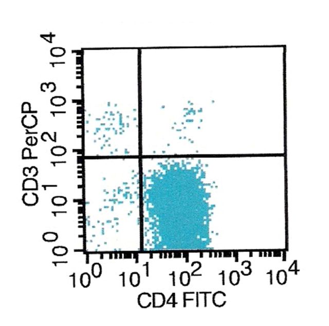
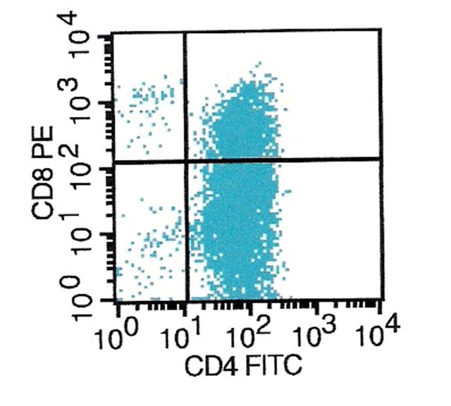
Soft tissue mass: anaplastic large cell lymphoma
Positive staining - normal
- T helper cells, thymocytes (80 - 90%), granulocytes, macrophages, Langerhans cells and dendritic cells
- Expressed in specific regions of brain
Positive staining - disease
- Many post-thymic T cell leukemia / lymphomas and T cell lymphoproliferative disorders
- Cutaneous lymphomatoid granulomatosis (Am J Surg Pathol 2001;25:1111)
- Eccrinotropic lymphomatoid papulosis (Am J Clin Pathol 2003;119:731)
- Indolent small intestinal CD4+ T-cell lymphoma (PLoS One 2013;8:e68343)
- Indolent T cell lymphoblastic proliferations
- Dendritic / histiocytic neoplasms
- Blastic plasmacytoid dendritic cell neoplasm
- Granulomatous histiocytic lymphoma / sarcoma
- Other hematologic / inflammatory lesions
- Acute myeloid leukemia (M4, M5, Am J Clin Pathol 1995;104:204)
- Florid antiviral inflammatory response (Mod Pathol 2003;16:166)
- Kikuchi-Fujimoto disease (decreased CD4:CD8 ratio in affected areas of lymph node, in contrast to lymphoma, Am J Clin Pathol 2004;122:141)
- Pityriasis lichenoides
- Pyothorax associated lymphoma (some)
Negative staining
- NK cells / NK leukemia / lymphoma
- B cell lymphoma (usually)
- Hodgkins lymphoma (usually)
- Non-hematopoietic neoplasms
- Some T cell lymphomas
- CD7+ stem cell lymphoma
- Enteropathy associated T cell lymphoma (Am J Surg Pathol 2011;35:1557)
- Epidermotropic aggressive CD8+ cutaneous T cell lymphoma (J Am Acad Dermatol 2010;62:300)
- Hepatosplenic alpha / beta and gamma / delta lymphoma (Leuk Lymphoma 2012;53:609)

