
CD Markers
CD8

Copyright: 2002-2024, PathologyOutlines.com, Inc.
PubMed Search: CD8
CD8
Author: Nat Pernick, M.D.
Last author update: 1 August 2013
Last staff update: 24 August 2023
Copyright: 2002-2024, PathologyOutlines.com, Inc.
PubMed Search: CD8

Table of Contents
Definition / general | Pathophysiology | Diagrams / tables | Clinical features | Uses by pathologists | Microscopic (histologic) images | Positive staining - normal | Positive staining - disease | Negative staining | Flow cytometry imagesCite this page: Pernick N. CD8. PathologyOutlines.com website. https://www.pathologyoutlines.com/topic/cdmarkerscd8.html. Accessed May 1st, 2024.
Definition / general
- Cytotoxic T cell marker (Wikipedia); CD8+ cells are called T cell suppressor cells, cytotoxic T cells
- Also called OKT8
Pathophysiology
- Cell surface glycoprotein, member of immunoglobulin superfamily; at 2p12
- Heterodimer of an alpha (CD8A, OMIM #186910 ) and a beta chain (CD8B, OMIM #186730 ) linked by two disulfide bonds; each chain has significant homology to immunoglobulin variable light chains; is present as a heterodimer on thymocytes and as homodimer on peripheral blood T cells
- Both the CD8 antigen (acting as a coreceptor) and the T cell receptor recognize antigens displayed by an antigen presenting cell (APC) in the context of class I MHC molecules; CD8 binds to non-polymorphic region of class I molecules; may increase avidity of interactions between cytotoxic T cell and target cell during antigen-specific activation
- Can kill target cells by recognizing peptide-MHC complexes on them or by secreting cytokines capable of signaling through death receptors on target cell surface
- CD8 alpha cells promote survival and differentiation of activated T cells into memory CD8+ T cells
- CD8 T cell clonal expansions occurr in normal young (PLoS One 2011;6:e21240) and elderly adults (Immunol Rev 2005;205:170)
- May contribute to initiation, progression and regulation of autoimmune responses (Curr Opin Immunol 2005;17:624)
Diagrams / tables
Images hosted on other servers:


CD8+ T cell activation requires 2 signals
Clinical features
- Lymphocytic infiltrate in lymphoepithelioma-like carcinomas is often CD8+ in cervix (Arch Gynecol Obstet 2009;280:725), esophagus (Hum Pathol 2003;34:407), liver (World J Gastroenterol 2008;14:4694) and lung (Am J Surg Pathol 2002;26:715)
- Mutations in alpha chain cause CD8 deficiency, with recurrent bacterial infections (J Clin Invest 2001;108:117)
- Accumulate in advanced atherosclerotic plaques (Hum Pathol 2008;39:1756)
Uses by pathologists
- Marker of T cells (normal and malignant)
- Marker of cytotoxic / suppressor T cells
- Classify lymphomas
- Differentiate splenic hamartoma (CD8+) from hemangioma or littoral cell angioma (CD8-)
- Note: use of CD3, CD8 does NOT improve detection of gluten-sensitive enteropathy in duodenal biopsies (Mod Pathol 2013 Apr 5 [Epub ahead of print])
Microscopic (histologic) images
Case #179
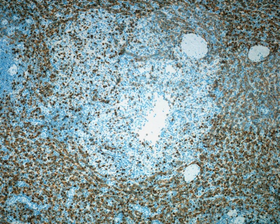
Splenic littoral cell angioma, sinus lining cells are CD8-
Images hosted on other servers:

Bladder: follicular cystitis (fig D)

Skin: cutaneous CD8+ epidermotropic T cell lymphoma

Skin: lymphomatoid papulosis (fig B, C)
Positive staining - normal
- Cortical thymocytes (70-80%), T cells (25-35% of mature peripheral T cells, mostly cytotoxic T cells), NK cells (30%) and dendritic cells
- Splenid littoral cells but not littoral cell angioma (Arch Pathol Lab Med 2019;143:1093)
Positive staining - disease
- T cell lymphoma, post-thymic (some)
- Heterotopic ovarian splenoma, Hodgkin lymphoma-nodular lymphocyte predominant (reactive T cells are CD4+ CD8+ in 58%, Am J Clin Pathol 2006;126:805)
- Melanoma (CD4+ CD8+ in 60%, PLoS One 2010;5:e8437)
- Mycosis fungoides - epidermotrophic lymphocytes, NK/T cell lymphoma (variable)
- Splenic hemartoma-sinus lining cells, subcutaneous panniculitis-like T cell lymphoma
- Rare: B-CLL (Am J Clin Pathol 2009;132:186), lymphomatoid papulosis, type "D" (Am J Clin Pathol 2006;125:490, Am J Surg Pathol 2010;34:1168), mantle cell lymphoma (Am J Clin Pathol 1998;109:689)
Negative staining
- Adult T cell leukemia / lymphoma, littoral cell hemangioma of spleen
Flow cytometry images
Case #36
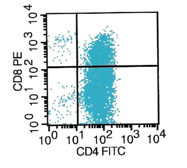
Anaplastic large
cell lymphoma:
partial CD4 / CD8
coexpression

