
Colon
Syndromes
Serrated polyposis
Authors: Michael Feely, D.O., Raul S. Gonzalez, M.D.


Last author update: 15 February 2022
Last staff update: 15 February 2022
Copyright: 2003-2024, PathologyOutlines.com, Inc.
PubMed Search: Serrated polyposis [title] colon tumor


Table of Contents
Definition / general | Essential features | Terminology | Sites | Pathophysiology | Diagrams / tables | Clinical features | Treatment | Gross images | Microscopic (histologic) description | Microscopic (histologic) images | Molecular / cytogenetics description | Sample pathology report | Differential diagnosis | Board review style question #1 | Board review style answer #1Cite this page: Feely M, Gonzalez RS. Serrated polyposis. PathologyOutlines.com website. https://www.pathologyoutlines.com/topic/colontumorserratedpolyposissx.html. Accessed April 26th, 2024.
Definition / general
- Polyposis syndrome defined by the development of numerous sessile serrated polyps in the colon
Essential features
- WHO diagnostic criteria: (a) at least 5 serrated polyps proximal to the rectum, all ≥ 5 mm in size, with 2 or more of these being ≥ 10 mm; or (b) > 20 serrated polyps of any size, distributed throughout the colon, with at least 5 proximal to the rectum (Gastroenterology 2020;158:1520)
- Significantly increased risk for colorectal carcinoma and possibly extracolonic malignancies (Gut 2010;59:1094, Dis Colon Rectum 2011;54:164)
- Family members also at increased risk for colorectal malignancies, suggesting an inherited component (Am J Gastroenterol 2012;107:770)
- Likely represents a heterogeneous group of patients that includes several phenotypes of serrated polyposis
Terminology
- Previously referred to as hyperplastic polyposis syndrome, although serrated polyposis is current preferred term by WHO
Sites
- Polyps found throughout the large intestine, including in the appendix (Pathology 2016;48:30)
Pathophysiology
- Likely consists of at least 2 groups: (type 1) patients with BRAF mutations and relatively few large right sided polyps and (type 2) patients with KRAS mutations and many small left sided polyps (J Pathol 2007;212:378)
- Awaiting more definitive molecular genetic studies
Diagrams / tables
Images hosted on other servers:

Diagnosis and management of serrated polyposis
Clinical features
- Mean age at diagnosis is 55 years, with overall equal distribution in males and females
- Type 1 patients (with BRAF mutations) typically female smokers (United European Gastroenterol J 2016;4:305)
- Typically asymptomatic and encountered on screening colonoscopy, although larger polyps may bleed
- Most cases appear de novo, although a few familial cases have been described
Treatment
- Colonoscopy for polyp removal every 1 - 2 years can help maintain polyp burden but advanced neoplasia may often be detected (Gastrointest Endosc 2020;92:1098, Endoscopy 2019;51:142)
- Colectomy with ileorectal anastomosis in cases of advanced lesions or those not amenable to colonoscopic management
- Some recommend screening colonoscopy in first degree family members (Am J Gastroenterol 2012;107:770)
Gross images
Contributed by Raul S. Gonzalez, M.D.
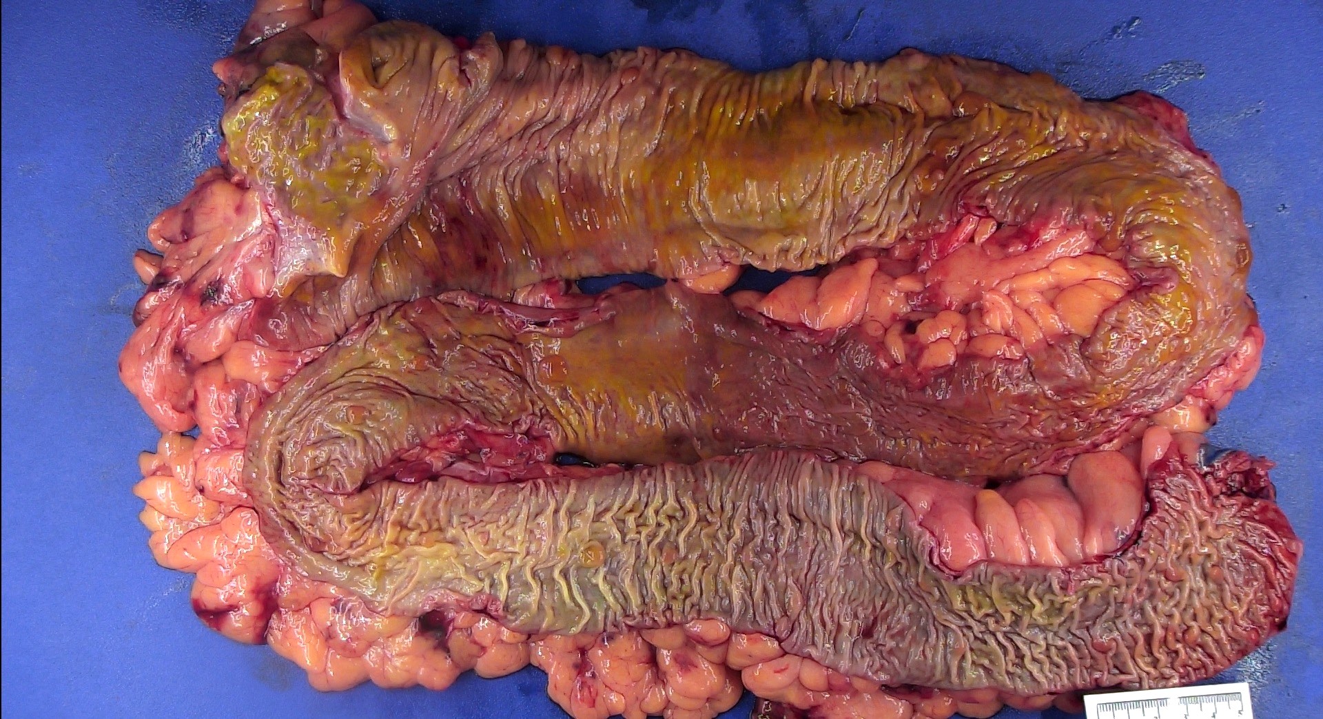
Serrated polyposis
Microscopic (histologic) description
- Lesions typically consist of sessile serrated adenomas / polyps that may have cytologic dysplasia
- Hyperplastic polyps, typically of the microvesicular type, are also encountered and may be large
- Conventional adenomas are occasionally present and may represent sessile serrated adenomas / polyps with cytologic dysplasia
Microscopic (histologic) images
Images hosted on other servers:

Sessile serrated
adenoma without
dysplasia (a) and with
low grade dysplasia (b)
Molecular / cytogenetics description
- In addition to BRAF and KRAS mutations (as above), RNF43 has recently been implicated in sporadic and familial sessile serrated polyps (Gut 2017;66:1645)
Sample pathology report
- Colon, total colectomy:
- Colon with approximately two dozen sessile serrated lesions, some with focal cytologic dysplasia (see comment)
- Negative for malignancy.
- Sessile serrated lesion focally present at proximal resection margin; distal resection margin unremarkable.
- Fourteen benign lymph nodes.
- Comment: The findings are consistent with the patient's reported history of serrated polyposis. All grossly identifiable polyps were submitted for microscopy.
Differential diagnosis
- MUTYH associated polyposis:
- Can also show serrated polyps but the predominant lesions are conventional adenomas
Board review style question #1
Which of the following is true about serrated polyposis syndrome?
- It has been linked to MLH1 germline mutations
- Patients only develop sessile serrated adenomas
- Patients usually present with lower gastrointestinal bleeding
- There appear to exist 2 distinct subgroups
Board review style answer #1
