

Fallopian tubes & broad ligament
Fallopian tube epithelial tumors
Endometrioid adenocarcinoma
Author: Nat Pernick, M.D.

Last author update: 1 September 2013
Last staff update: 16 June 2022
Copyright: 2002-2024, PathologyOutlines.com, Inc.
PubMed Search: Endometrioid adenocarcinoma


Table of Contents
Definition / general | Clinical features | Gross description | Gross images | Microscopic (histologic) description | Microscopic (histologic) imagesCite this page: Pernick N. Endometrioid adenocarcinoma. PathologyOutlines.com website. https://www.pathologyoutlines.com/topic/fallopiantubescarcinoma.html. Accessed May 12th, 2024.
Definition / general
- Rare, 0.3 to 1.0% of genital tract malignancies
- Mean age 57 years, rarely teenagers; usually incorrect preoperative diagnosis
- High stage with pelvic extension or positive peritoneal cytology
- To call primary in fallopian tube, should arise from mucosa (endosalpinx), have tubal histologic pattern, involve the lumen, uterus and ovaries must be normal or have foci of malignancy that resemble metastases or independent primaries; if tubal wall is involved, should detect a transition between benign and malignant tubal epithelium
Clinical features
- 5 year survival: stage 1 - 77%, stage 3 - 20%; usually recur intra-abdominally
- Associated with BRCA1 and BRCA2 mutations; for patients with known mutation or family history of breast or ovarian cancer, should submit entire fallopian tube and ovary for microscopic examination (Am J Surg Pathol 2002;26:171, Am J Surg Pathol 2001;25:1283)
- Carcinomas at this site: 50% serous, 25% endometrioid, 20% transitional or undifferentiated
- Symptoms: vaginal bleeding or discharge (2/3), pain, adnexal mass (triad in 50%); endometrial smear positive in 10%
Gross description
- Enlarged tube, with solid or papillary tumor filling the lumen
- Tumors occasionally are primary in the fimbriae
- 80 - 97% unilateral; hemorrhage, necrosis and cysts common
Gross images
AFIP images
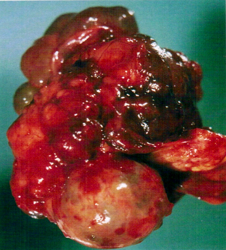
Diffusely enlarged tube
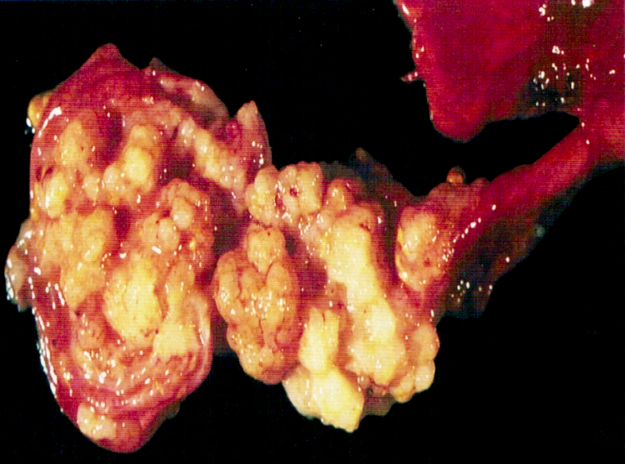
Papillary tumor attached to mucosa
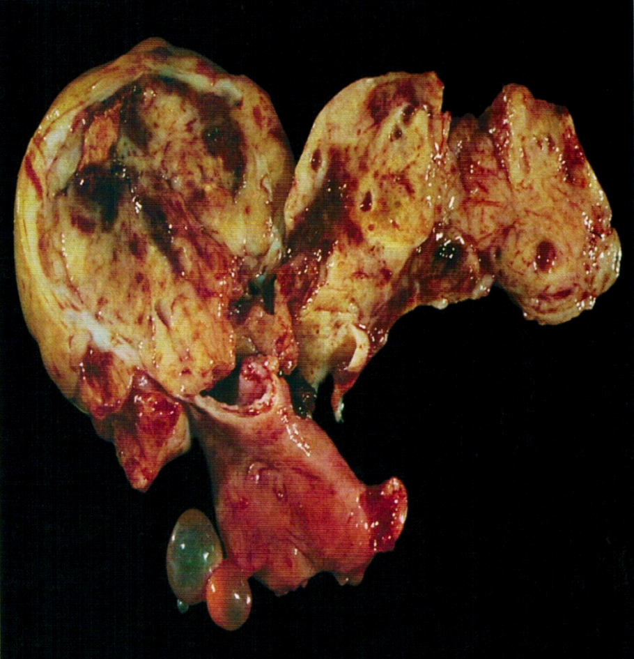
Lumen distended by solid tumor
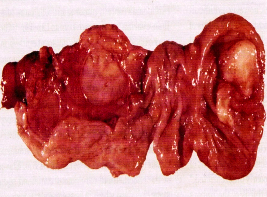
Distended, opened tube
Microscopic (histologic) description
- Endometrioid tumors may be noninvasive, have squamous metaplasia, be associated with endometriosis and contain spindled epithelial cells
- May have small, closely packed cells with numerous glandular spaces of varying sizes, containing PAS+ dense colloid-like secretion, resembling female adnexal tumor of probable Wolffian origin (FATWO), but usually intraluminal, typical endometrioid carcinoma elsewhere, more mitotic activity and atypia, mucin present
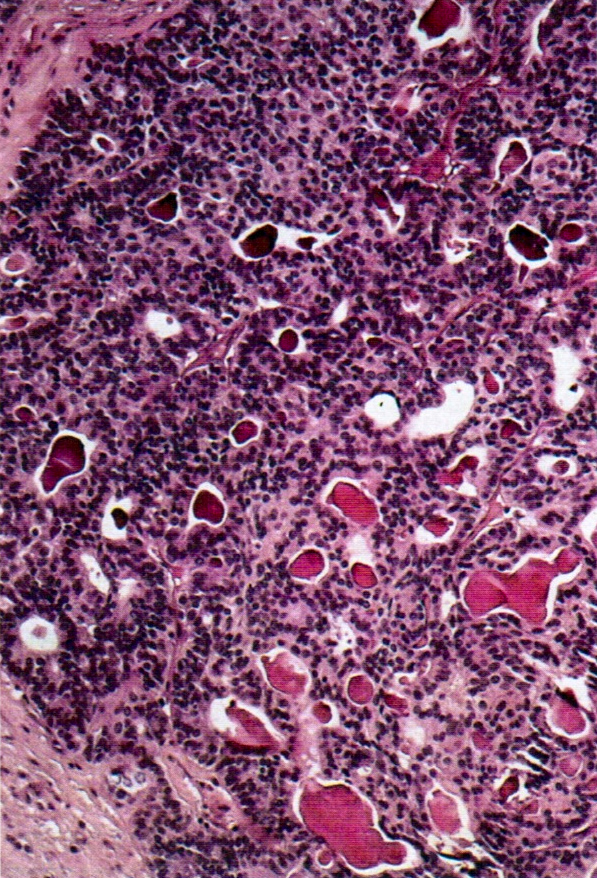
Microscopic (histologic) images
AFIP images
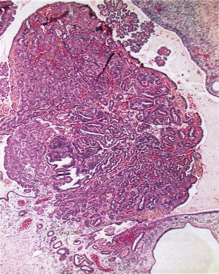
Tumor arising in endometriosis
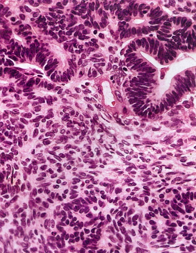
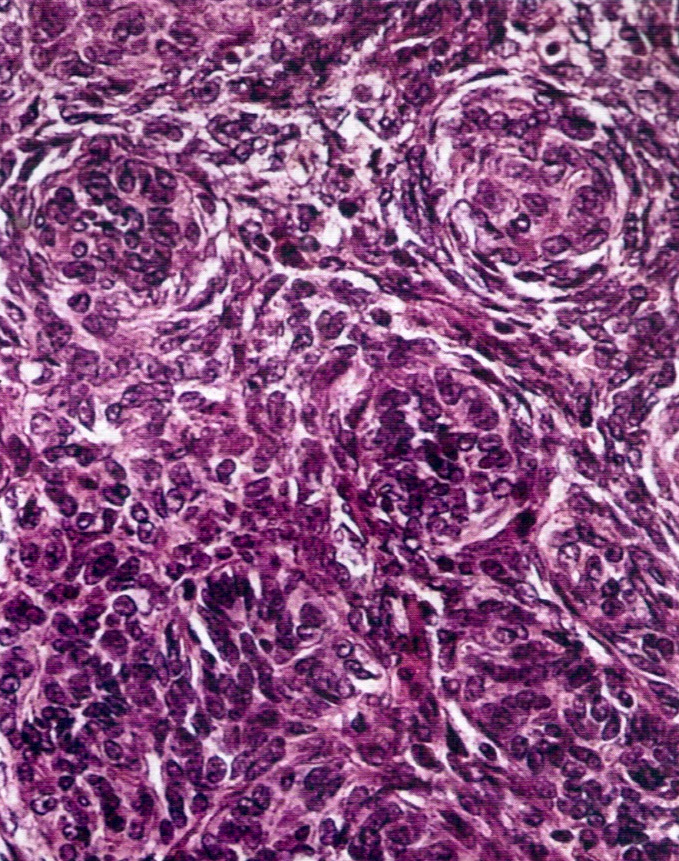
Tumor simulating Wolffian adnexal tumor
