

Skin melanocytic tumor
Blue nevi, dermal melanocytoses and associated neoplasms
Dermal melanocytosis
Author: Christopher S. Hale, M.D.

Last author update: 23 July 2021
Last staff update: 26 February 2024
Copyright: 2013-2024, PathologyOutlines.com, Inc.
PubMed Search: Nevus of Ito [title], nevus of ota, Mongolian spot
Cite this page: Hale CS. Dermal melanocytosis. PathologyOutlines.com website. https://www.pathologyoutlines.com/topic/skintumormelanocyticdermalmelanocytosis.html. Accessed April 26th, 2024.
Ito's nevus
Definition / general
Terminology
Epidemiology
Sites
Clinical features
Case reports
Treatment
Clinical images
Images hosted on other servers:

Microscopic (histologic) description
- First described by Minor Ito in 1954
Terminology
- More accurately grouped with dermal melanocytoses (nevus of Ito, nevus of Ota, Hori nevus, so called "Mongolian" spot) (eMedicine: Nevi of Ota and Ito [Accessed 23 July 2021])
- Although melanocytic, it is not a true nevus
- Different entity from Hypomelanosis of Ito, a neurocutaneous disorder (J Child Neurol 2000;15:635)
Epidemiology
- Usually individuals with pigmented skin, most commonly Asians; also Hispanics, blacks, Native Americans
- More common in women
- Often occurs with nevus of Ota in same patient but is much less common
Sites
- By definition, found in shawl or cloak distribution
Clinical features
- Macule with irregular blue-gray pigmentation
- Similar to nevus of Ota except for location
- Ito’s nevus is found in shoulder, side of neck and supraclavicular areas, within the distribution of the lateral (also called posterior) supraclavicular nerve and lateral cutaneous brachial nerves
- Nevus of Ota is found around the eyes
Case reports
- 38 year old man with nevus of Ota (Indian J Dermatol Venereol Leprol 2004;70:112)
- 72 year old woman with late onset Ito's nevus (J Cutan Pathol 2007;34:640)
- With transformation to melanoma (J Am Acad Dermatol 2010;62:869)
Treatment
- Laser to lighten pigment (cosmetic purposes)
- Cryotherapy, dermabrasion, chemical peeling and surgical excision may give variable results (Facial Plast Surg Clin North Am 2007;15:367)
Clinical images
Images hosted on other servers:
Nevus of Ota (face) and nevus of Ito (shoulder)
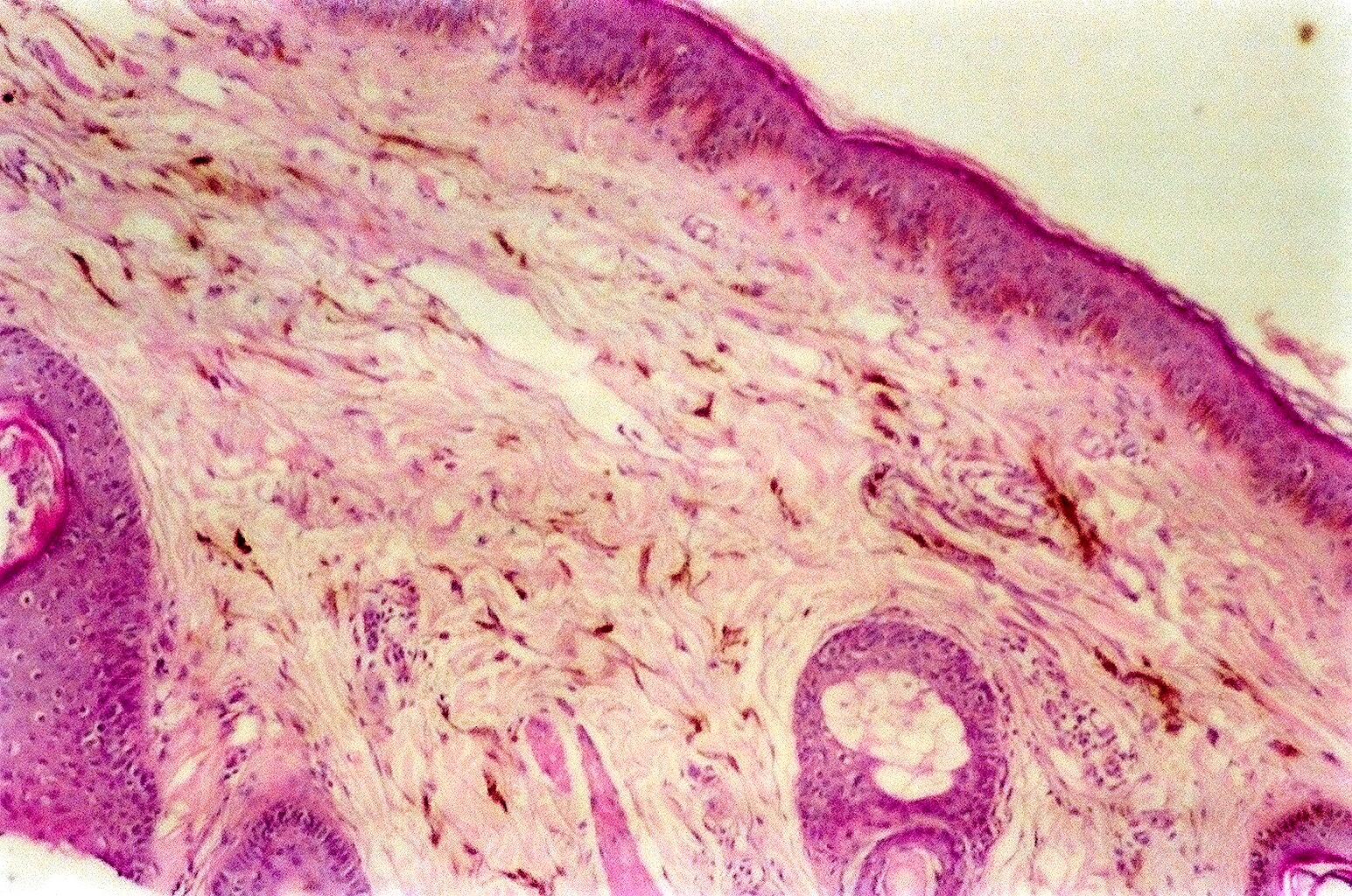
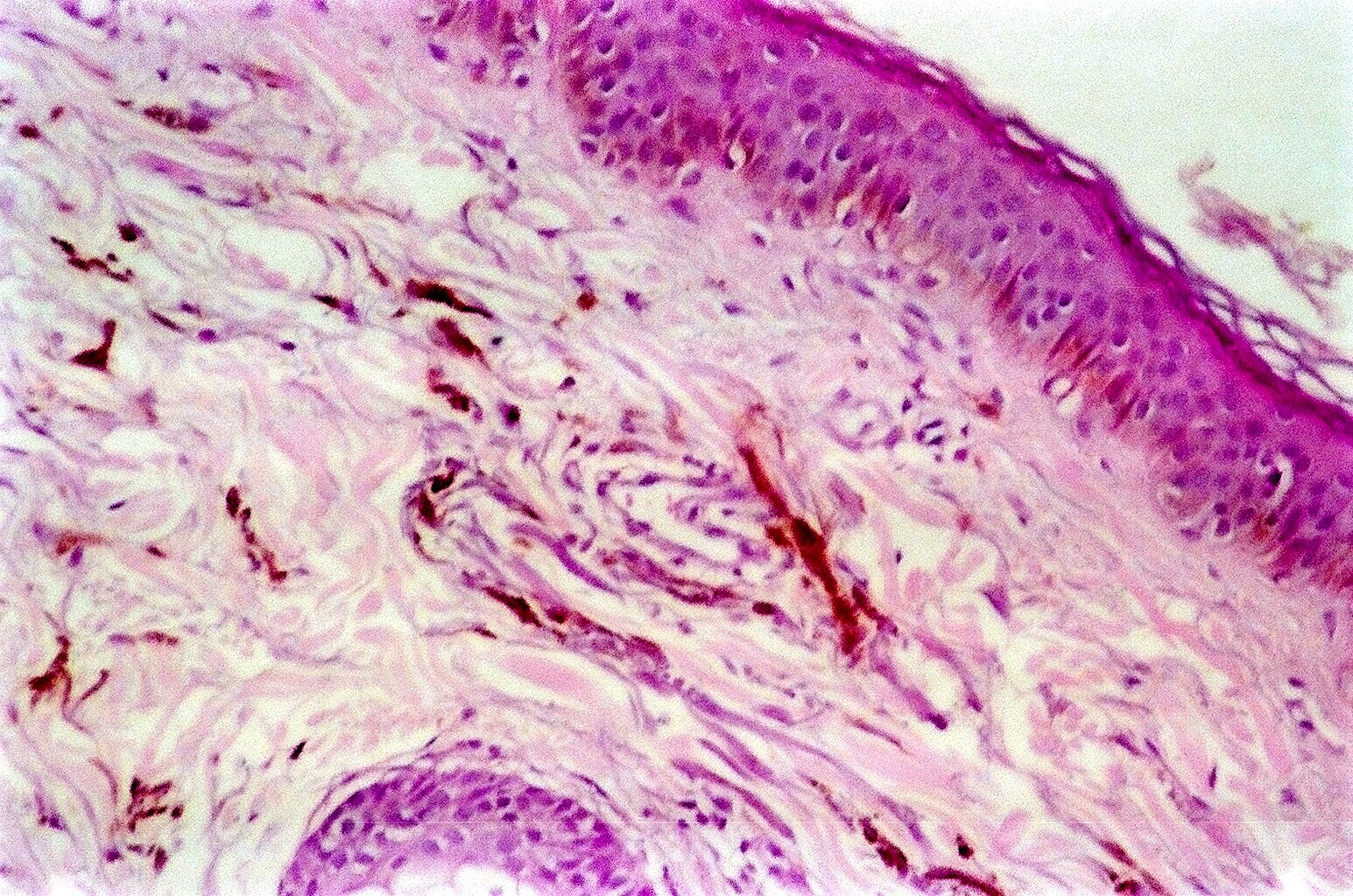
Microscopic (histologic) description
- Deeply pigmented dendritic melanocytes and melanophages dissecting bundles of dermal collagen in reticular dermis
- Overlying epidermis is normal
Nevus of Ota
Definition / general
Terminology
Epidemiology
Sites
Clinical features
Case reports
Treatment
Clinical images
Images hosted on other servers:


Microscopic (histologic) description
Microscopic (histologic) images
Contributed by Asmaa Gaber Abdou, M.D.
Images hosted on other servers:


Differential diagnosis
- First described by Ota in 1939
- Uncommon hamartoma in periorbital and temporal skin (eMedicine: Nevi of Ota and Ito [Accessed 23 July 2021])
- Types:
- Type IA : mild orbital type - distribution over upper and lower eyelids, periocular and temple region
- Type IB: mild zygomatic type - pigmentation in infrapalpebral fold, nasolabial fold and zygomatic region
- Type IC: mild forehead type - involvement of forehead alone
- Type ID: involvement of ala nasi alone
- Type II: moderate type - distribution over upper and lower eyelids, periocular, zygomatic, cheek and temple regions
- Type III: involves scalp, forehead, eyebrow and nose
- Type IV: bilateral
- Hori’s nevus: acquired bilateral nevus of Ota-like macules (J Am Acad Dermatol 1984;10:961, Dermatol Online J 2005;11:1); usually Chinese women with family history, becomes more confluent and gray over time (Br J Dermatol 2006;154:50)
- Sun’s nevus: acquired unilateral nevus of Ota
Terminology
- Also called oculodermal melanosis or nevus fuscoceruleus ophthalmomaxillaris
- Similar to nevus of Ito except for location (Ito in shoulder, side of neck and supraclavicular areas, within the distribution of the lateral supraclavicular nerve and lateral cutaneous brachial nerves)
Epidemiology
- Usually Asians; also Hispanics, blacks and Native Americans
- 60% have lesions at birth, 87% female, 60% have dermal and ocular involvement (Indian J Dermatol Venereol Leprol 2008;74:125)
Sites
- Periorbital, temporal
Clinical features
- Tends to persist and extend locally, becoming increasingly prominent with age, puberty and postmenopausal state
- Associated with ipsilateral glaucoma, intracranial melanocytosis; rarely with cutaneous, ocular or intracranial melanoma and vascular nevus (Cutis 2008;82:25, J Am Acad Dermatol 2008;58:88)
- Macule with irregular blue-gray pigmentation in distribution of 1st and 2nd division of trigeminal nerve
- May coexist with nevus of Ito
- Tanino classification system most common, others include Hirayama system and proposed Chan system (Lasers Surg Med 2001;28:267)
Case reports
- 30 year old woman with bilateral and oral mucosa involvement (Indian J Dermatol Venereol Leprol 2002;68:104)
- 41 year old woman with acquired bilateral nevus (Dermatol Online J 2005;11:1)
- 47 year old light-skinned non-Asian woman with bilateral manifestations (Dermatol Online J 2007;13:19)
- 50 year old man with coexisting iris melanoma (Surv Ophthalmol 2008;53:411)
- Elderly Caucasian woman with acquired Ito's nevus (J Cutan Pathol 2007;34:640)
Treatment
- Cosmetic coverup products
- Multiple sessions of laser photothermolysis to avoid darkening and extension, beginning early after diagnosis (Dermatol Surg 2007;33:455)
- Subtypes vary in response to laser therapy (Lasers Surg Med 2001;28:267)
- Combined skin abrasion and carbon dioxide snow method (Plast Reconstr Surg 1996;97:544)
- Cryosurgery and microsurgery not recommended due to scarring; chemical bleaching not recommended due to depigmentation
Clinical images
Images hosted on other servers:
Flat blue-gray pigmentation
Acquired dark brown spots below eyes
Episcleral involvement
With nevus of Ito
Microscopic (histologic) description
- Deeply pigmented dendritic melanocytes and melanophages dissecting bundles of dermal collagen in reticular dermis
Microscopic (histologic) images
Contributed by Asmaa Gaber Abdou, M.D.
Elongated dendritic melanocytes scattered with collagen bundles extending around hair follicles
Images hosted on other servers:
Bipolar dendritic
melanocytes in
papillary and
reticular dermis
Wavy dendritic
cells with evenly
dispersed
melanin granules
Differential diagnosis
- Acquired bilateral nevus of Ota-like macule
- Cafe-au-lait patch
- Speckled lentiginous nevus
Mongolian spot
Definition / general
Terminology
Epidemiology
Sites
Case reports
Treatment
Clinical images
Images hosted on other servers:


Microscopic (histologic) description
Differential diagnosis
- Ill defined area of blue discoloration, up to several centimeters and in lumbosacral region (eMedicine: Congenital Dermal Melanocytosis (Mongolian Spot) [Accessed 23 July 2021], Wikipedia: Mongolian spot [Accessed 23 July 2021])
- May also occur at other sites
- See also Ito’s nevus
Terminology
- Also known as congenital dermal melanocytosis
- Related to dermal melanocytoses (nevus of Ito, nevus of Ota, Hori nevus)
- Although melanocytic, is not a true nevus
Epidemiology
- Congenital disorder, present at birth in most neonates from Asia, East-Africa or Turkey; also Native Americans
- Incidence of 60 - 70% in Iran, Nigeria and Taiwan (Pediatr Dermatol 2006;23:61, Niger J Med 2001;10:121, Chang Gung Med J 2007;30:220)
- Usually regresses over several years, almost always by puberty but may persist (Int J Dermatol 2005;44:43)
- Usually not present in children with blond hair (Turk J Pediatr 2006;48:232)
- Extensive Mongolian spots may be associated with inborn errors of metabolism (Pediatr Neurol 2006;34:143, Br J Dermatol 2003;148:1173)
Sites
- Usually sacral region
Case reports
- 1 year old boy with facial lesion near mandibular area (J Dermatol 2007;34:381)
- 4 infants with darker pigmented Mongolian spot superimposed on another Mongolian spot (Pediatr Dermatol 2008;25:233)
Treatment
- Wait for regression
- Laser (Lasers Med Sci 2007;22:159)
Clinical images
Images hosted on other servers:

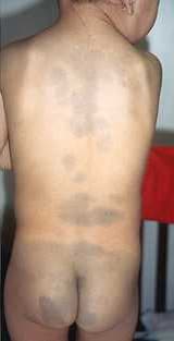
Mongolian spots
Microscopic (histologic) description
- Normal at low power
- High power shows occasional deep dendritic melanocytes, with melanin granules dissecting bundles of dermal collagen
- No associated melanophages
Differential diagnosis
- Bruises from child abuse
