
Small intestine & ampulla
Congenital anomalies
Meconium peritonitis
Author: Hanni Gulwani, M.B.B.S.

Last author update: 1 August 2012
Last staff update: 18 November 2020
Copyright: 2003-2024, PathologyOutlines.com, Inc.
PubMed Search: Meconium peritonitis[TI] small bowel

Table of Contents
Definition / general | Radiology description | Case reports | Treatment | Gross description | Gross images | Microscopic (histologic) description | Microscopic (histologic) images | Differential diagnosisCite this page: Gulwani H. Meconium peritonitis. PathologyOutlines.com website. https://www.pathologyoutlines.com/topic/smallbowelmeconiumperitonitis.html. Accessed May 14th, 2024.
Definition / general
- Rare prenatal complication in 1 per 30,000 live births
- GI perforation releases meconium into abdominal cavity, inducing sterile inflammatory reaction and calcium deposition
- Perforation may be due to anoxia leading to bowel ischemia, atresia, congenital bands, Hirschprung disease, internal hernia, meconium ileus, stenosis, volvulus or idiopathic
- Presents with fetal distress, maternal polyhydramnios, abdominal distention or a mass
- Newborns with perforation should be evaluated for cystic fibrosis (Pediatr Surg Int 2003;19:75)
Radiology description
- Prenatal ultrasound shows dilated bowel, ascites, polyhydramnios, intra-abdominal calcifications (Prenat Diagn 2005;25:676)
- Ultrasound findings have prognostic value (Fetal Diagn Ther 2003;18:255, Prenat Diagn 2007;27:960)
Case reports
- 35 week old girl with intrauterine distress (Case of the Week #106)
Treatment
- Surgical
- Gestational age at diagnosis does not predict postnatal outcome (J Pediatr Surg 1995;30:979)
Gross description
- Organized peritonitis with fibrosis, calcifications, dense intestinal adhesions
- Meconium pseudocyst (fibrous wall) may form
Gross images
Case #106
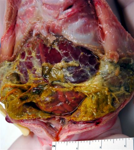
Abdominal cavity
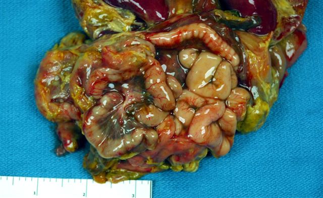
Small intestine
Microscopic (histologic) description
- Peritoneal surface shows fibrinous exudate with microcalcifications, bile pigment-like debris, histiocytes, chronic inflammatory cells
Microscopic (histologic) images
Case #106
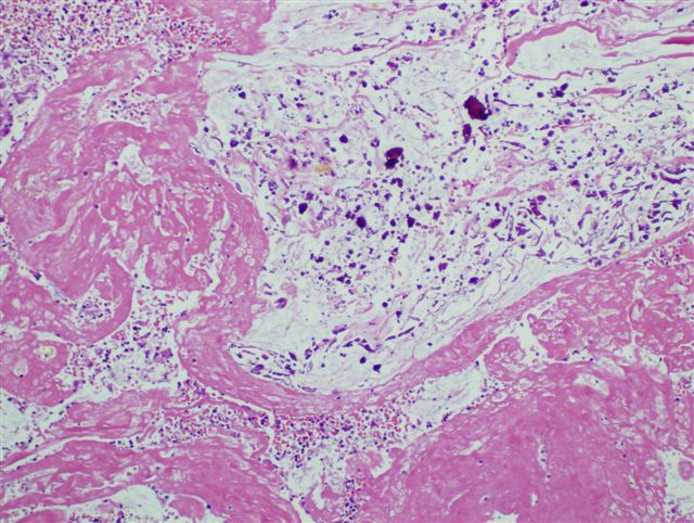
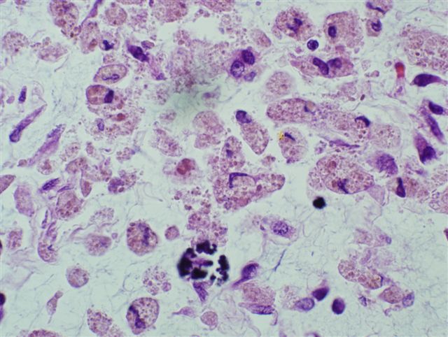
Peritoneal surface
Differential diagnosis
- Vernix caseosa peritonitis: cheesy white exudate coats the visceral organs after cesarean section (J Obstet Gynaecol 2007;27:660)
