
Stains & CD markers
MUM1 / IRF4


Copyright: 2002-2024, PathologyOutlines.com, Inc.
PubMed Search: MUM1
MUM1 / IRF4
Authors: Fatima Iqbal, M.D., Kirill A. Lyapichev, M.D.
Editorial Board Member: Christian M. Schürch, M.D., Ph.D.
Deputy Editor-in-Chief: Genevieve M. Crane, M.D., Ph.D.
Last author update: 19 April 2022
Last staff update: 19 April 2022
Copyright: 2002-2024, PathologyOutlines.com, Inc.
PubMed Search: MUM1

Table of Contents
Definition / general | Essential features | Terminology | Pathophysiology | Interpretation | Uses by pathologists | Prognostic factors | Microscopic (histologic) description | Microscopic (histologic) images | Positive staining - normal | Positive staining - disease | Negative staining | Molecular / cytogenetics description | Molecular / cytogenetics images | Sample pathology report | Board review style question #1 | Board review style answer #1 | Board review style question #2 | Board review style answer #2Cite this page: Iqbal F, Lyapichev KA. MUM1 / IRF4. PathologyOutlines.com website. https://www.pathologyoutlines.com/topic/stainsmum1.html. Accessed April 28th, 2024.
Definition / general
- MUM1 / IRF4 is the abbreviation of multiple myeloma 1 / interferon regulatory factor 4
- Normally expressed in plasma cells, melanocytes, activated B cells and activated T cells (Appl Immunohistochem Mol Morphol 2010;18:301, Int J Clin Exp Pathol 2015;8:11372)
Essential features
- MUM1 / IRF4 is a nuclear marker that is normally expressed in activated B and T cells, plasma cells and melanocytes
- Involved in the differentiation of B cells and T cells
- Marker is expressed in lymphoid and some nonlymphoid malignancies including classic Hodgkin lymphoma, diffuse large B cell lymphoma (nongerminal center B cell-like subtype), multiple myeloma and malignant melanoma
- Fluorescence in situ hybridization (FISH) for 6p25 (IRF4 / DUSP22) gene rearrangement can be used to diagnose different entities
Terminology
- PU.1 interaction partner (PIP)
- Lymphocyte specific interferon response factor (LSIRF)
- Interferon consensus sequence binding protein for activated T cells (ICSAT) (Appl Immunohistochem Mol Morphol 2010;18:301, Blood 2000;95:2084)
Pathophysiology
- MUM1 was originally found to be involved in the t(6;14)(p25;q32) translocation of multiple myeloma, which causes the juxtaposition of the MUM1 gene, mapping at 6p25, to the IgH locus on 14q32 (Leukemia 2000;14:563)
- MUM1+ cells are mainly located in the light zone of the germinal center but the highly proliferating, follicle colonizing B blasts (centroblasts) of the dark zone fail to express the protein; thus, it is unlikely that the MUM1 protein is involved in the process of clonal expansion known to occur in the dark zone of the germinal center (Blood 2000;95:2084)
- MUM1 expression may denote the final step of intragerminal center B cell differentiation (i.e., the stage known as centrocyte, as well as subsequent steps of B cell maturation toward plasma cells) (Leukemia 2000;14:563)
- Loss of MUM1 function results in the absence of activated lymphoid cells and Ig secreting plasma cells (Science 1997;275:540, Leukemia 2000;14:563)
Interpretation
- Nuclear stain
Uses by pathologists
- MUM1 is one of the markers used in Hans algorithm (requires immunostains CD10, BCL6 and MUM1); it is used in subclassification of diffuse large B cell lymphoma (DLBCL) into germinal center B cell-like subtype and nongerminal center B cell-like subtype (Blood 2004;103:275, Med J Malaysia 2020;75:98, Cancer Cytopathol 2016;124:135)
- May help differentiate classic Hodgkin lymphoma (cHL) (MUM1 positive) from nodular lymphocyte predominant Hodgkin lymphoma (MUM1 negative) (Rom J Morphol Embryol 2011;52:69)
- Helps differentiate the double hit lymphomas (MUM1 positive > 45%) from Burkitt and diffuse large B cell lymphoma (usually MUM1 negative if germinal center phenotype) (Am J Surg Pathol 2010;34:327)
- To differentiate angioimmunoblastic T cell lymphoma (AITL) with Hodgkin / Reed-Sternberg (HRS)-like cells from classic Hodgkin lymphoma (Int J Clin Exp Pathol 2015;8:11372):
- In AITL, MUM1 is expressed not only in HRS-like cells but also in the neoplastic T cells around the HRS-like cells, forming a rosette pattern (Int J Clin Exp Pathol 2015;8:11372)
- FISH for IRF4 favors cutaneous anaplastic large cell lymphoma versus other cutaneous T cell lymphoproliferative disorders (Mod Pathol 2011;24:596)
- MUM1 expression differentiates tumors in the PEComa family from clear cell sarcoma and melanoma (Int J Surg Pathol 2012;20:29)
- Can be used to differentiate primary effusion lymphoma among other lymphomatous effusions (Br J Haematol 2000;111:247)
Prognostic factors
- In diffuse large B cell lymphomas, MUM1 identifies cases of the nongerminal center B cell-like subtype and has been linked to poor survival (Leukemia 2008;22:441)
- MUM1 mRNA expression has been found to be an independent risk factor for poor survival in myeloma (Leukemia 2008;22:441)
- High expression of MUM1 was in addition to β2 microglobulin, the only myeloma risk factors analyzed in the study that were found to be independently associated with overall survival
- Combined score between these 2 parameters allowed a strong discrimination between high and low risk groups
- Lack of MUM1 expression was found to be associated with worse outcome in classic Hodgkin lymphoma (Haematologica 2007;92:1343)
- IRF4 / MUM1 expression was associated with poor survival outcomes in peripheral T cell lymphoma (PTCL) (J Cancer 2017;8:1018)
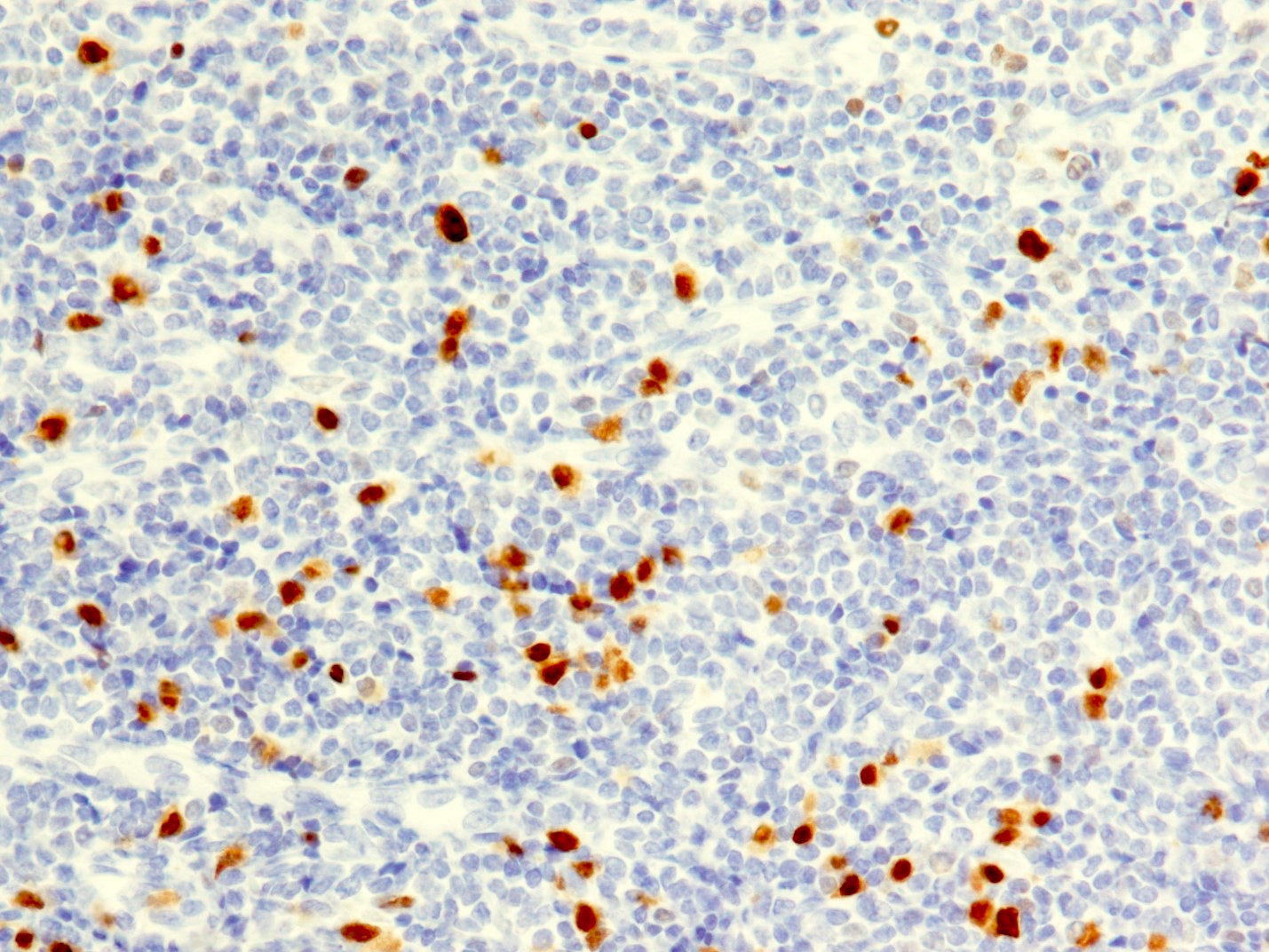
Microscopic (histologic) description
- Percentage of nuclei with positive staining in tumor cells is scored to subclassify diffuse large B cell lymphoma (DLBCL) into germinal center B cell-like subtype and nongerminal center B cell-like subtype
- > 30% - nongerminal center B cell-like subtype if CD10 is negative and BCL6 is positive
- < 30% - germinal center B cell-like subtype if CD10 is negative and BCL6 is positive
- Reference: Blood 2004;103:275
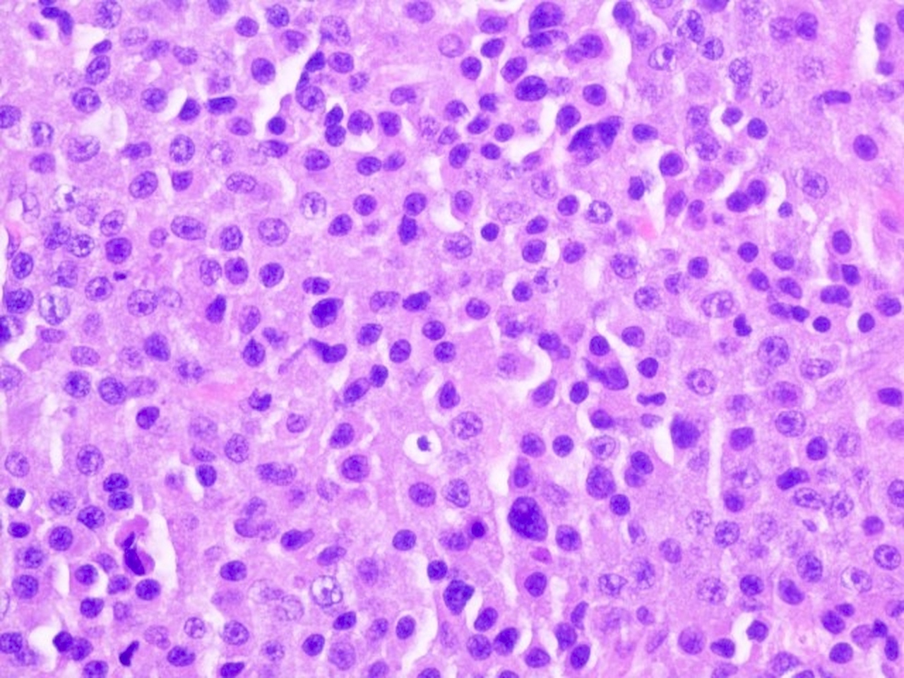
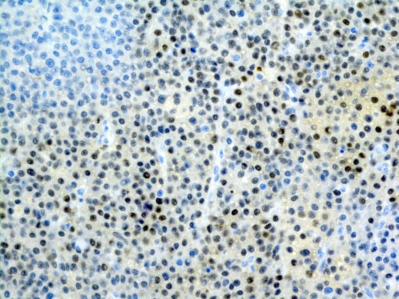
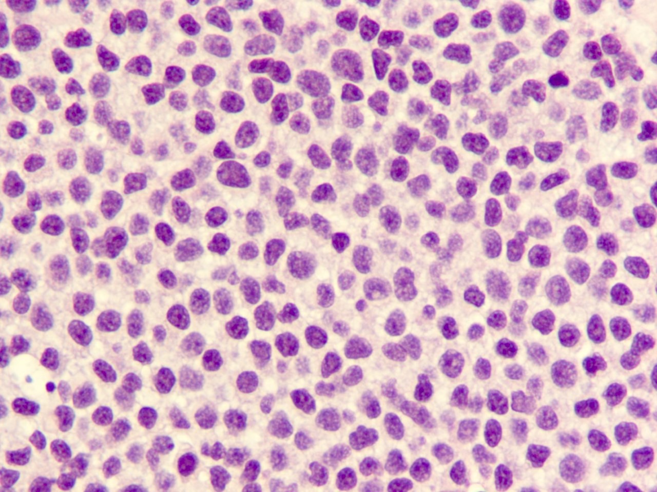
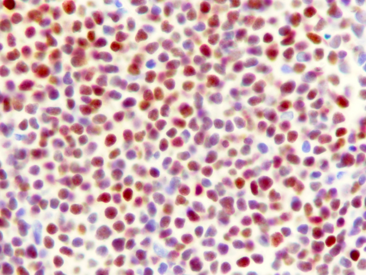
Microscopic (histologic) images
Contributed by Fatima Iqbal, M.D. and Kirill A. Lyapichev, M.D.
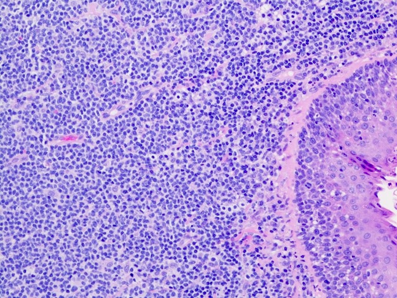
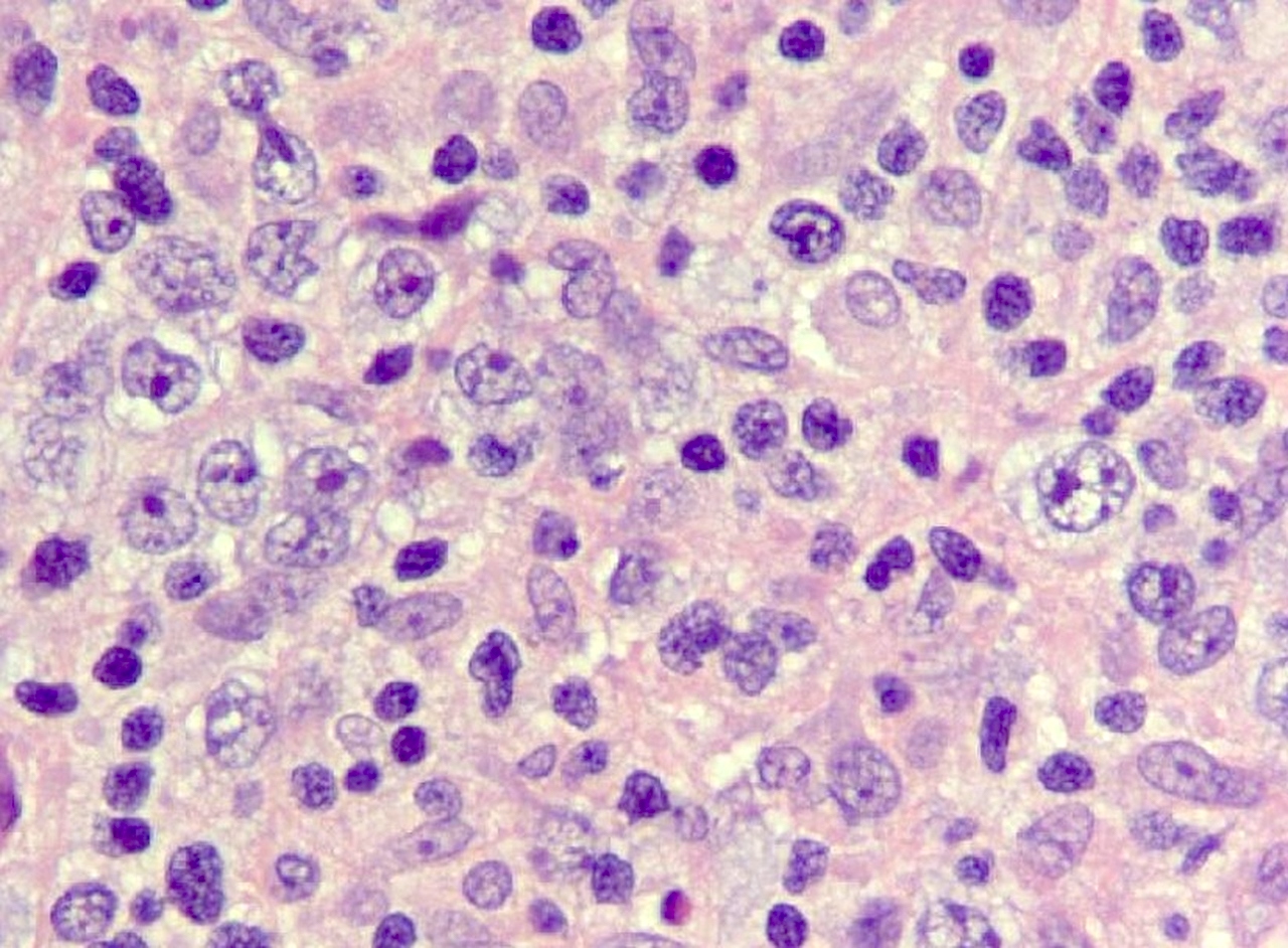
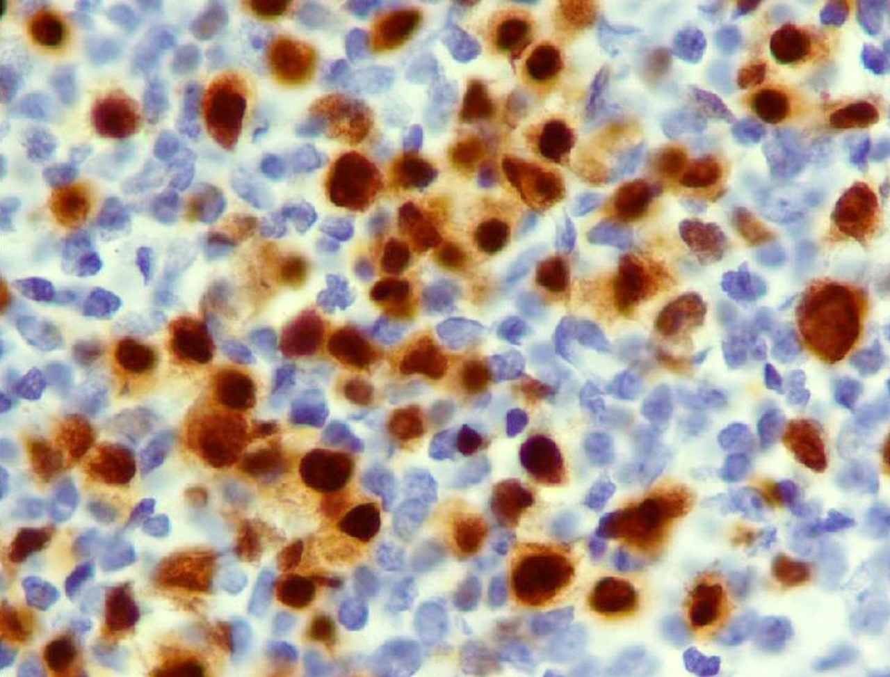
Mantle cell lymphoma
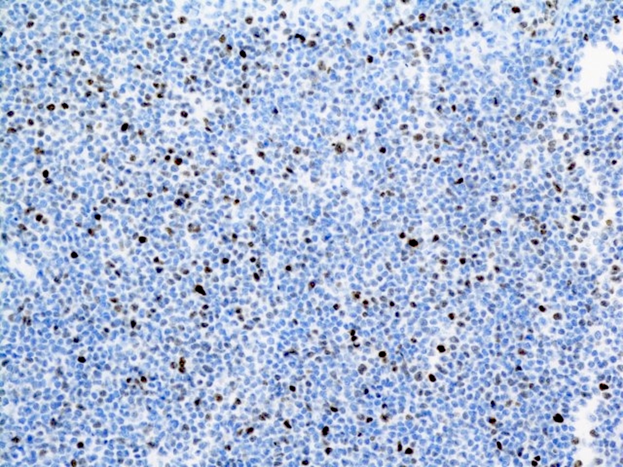
MUM1 in mantle cell lymphoma
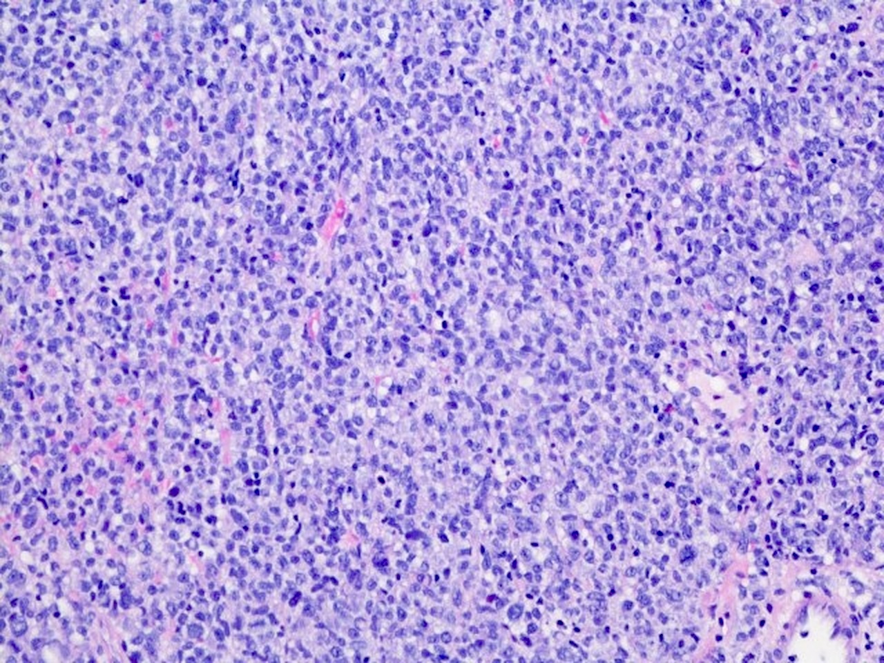
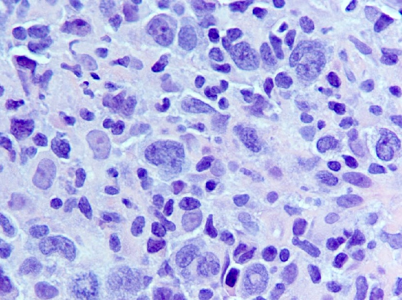
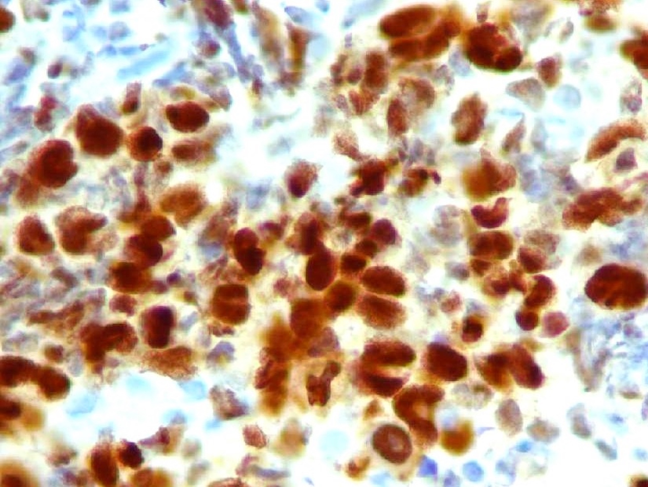
Plasmablastic lymphoma
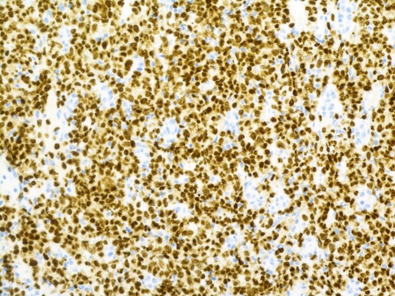
MUM1 in plasmablastic lymphoma
Plasmacytoma
MUM1 in plasmacytoma
Multiple myeloma
MUM1 in multiple myeloma
DLBCL with high content of histiocytes and T cells
MUM1 in DLBCL
Primary mediastinal large B cell lymphoma
MUM1 in primary mediastinal large B cell lymphoma
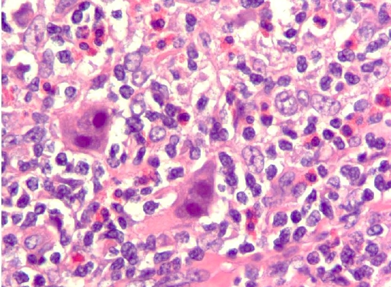
Classic Hodgkin lymphoma
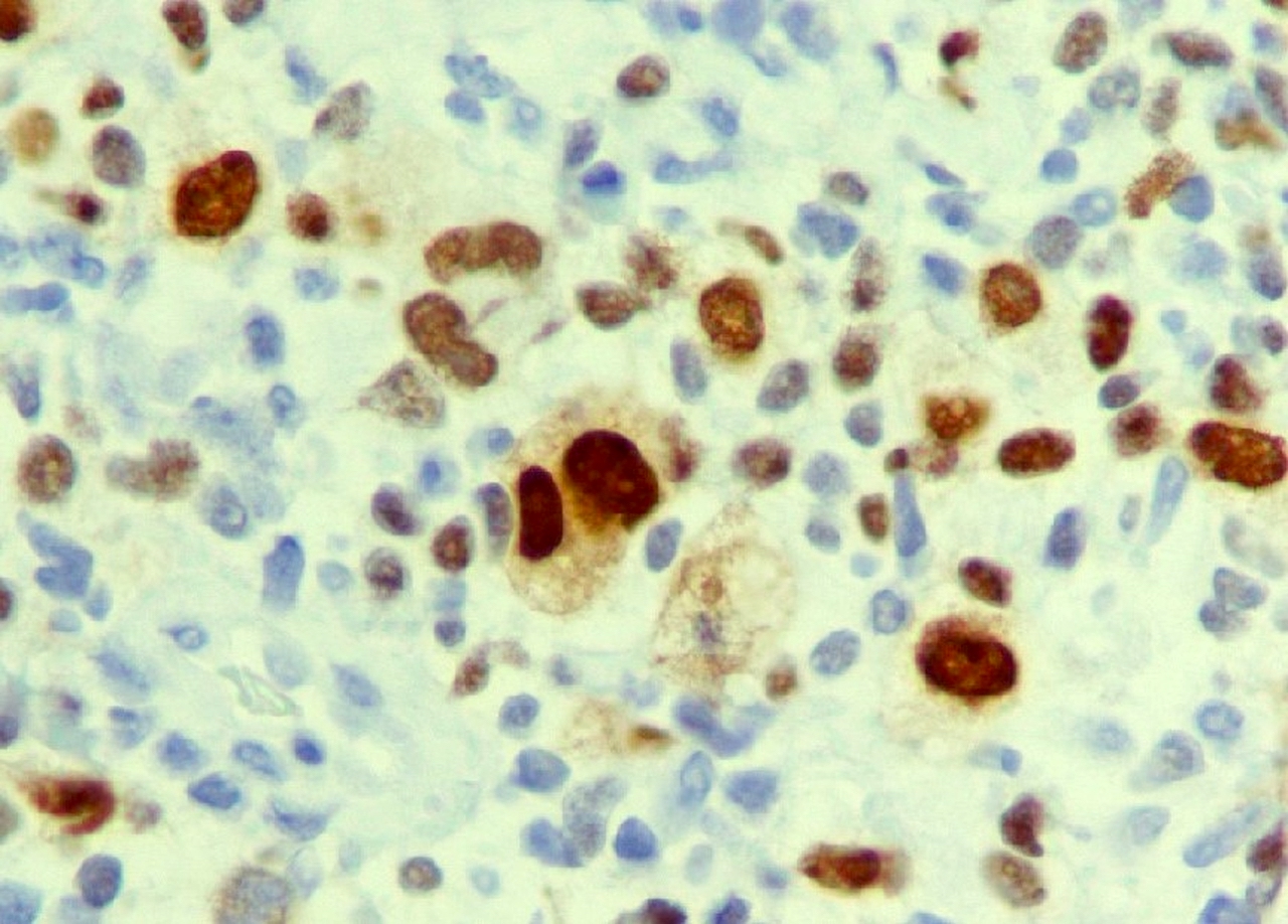
MUM1 in classic Hodgkin lymphoma
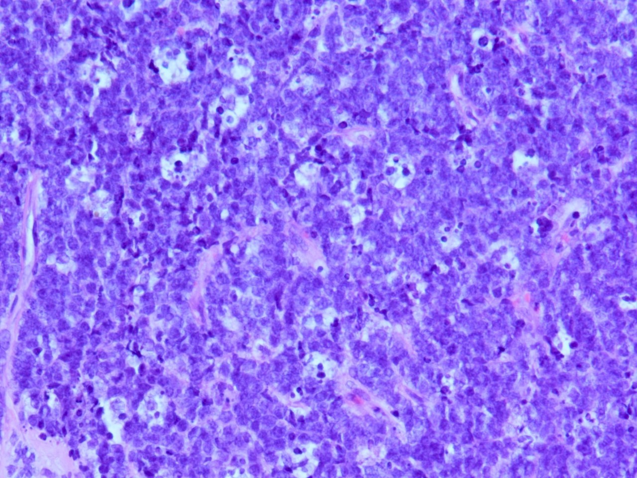
Burkitt lymphoma
MUM1 in Burkitt lymphoma
Positive staining - normal
- Germinal center B cells, activated T cells, plasma cells (Appl Immunohistochem Mol Morphol 2010;18:301, Mod Pathol 2001;14:686, Int J Clin Exp Pathol 2015;8:11372, Nat Immunol 2006;7:773)
- Melanocytes (Appl Immunohistochem Mol Morphol 2010;18:301, Int J Clin Exp Pathol 2015;8:11372)
Positive staining - disease
- Adult T cell leukemia / lymphoma (ATLL) (Appl Immunohistochem Mol Morphol 2010;18:301)
- Anaplastic large cell lymphoma (100%) (Blood 2000;95:2084)
- Angioimmunoblastic T cell lymphoma (Hodgkin / Reed-Sternberg [HRS]-like cells) (Int J Clin Exp Pathol 2015;8:11372)
- Burkitt lymphoma (40%) (Hum Pathol 2009;40:565, Appl Immunohistochem Mol Morphol 2010;18:301)
- Chronic lymphocytic leukemia (variable expression) (Leuk Lymphoma 2008;49:273, Appl Immunohistochem Mol Morphol 2010;18:301)
- Classic Hodgkin lymphoma (100%) (Appl Immunohistochem Mol Morphol 2010;18:301)
- Diffuse large B cell lymphoma (76% adult cases and 44% pediatric cases) (Appl Immunohistochem Mol Morphol 2010;18:301, Arch Pathol Lab Med 2010;134:759)
- Extranodal NK / T cell lymphoma, nasal type (Am J Surg Pathol 2012;36:481)
- Follicular lymphoma, high grade (Appl Immunohistochem Mol Morphol 2010;18:301, Haematologica 2007;92:267)
- Infectious mononucleosis (Mod Pathol 2012;25:1149)
- Intravascular large B cell lymphoma (95%) (Arch Pathol Lab Med 2012;136:333)
- Lymphomatoid papulosis (87%) (Br J Dermatol 2008;158:1280, Appl Immunohistochem Mol Morphol 2010;18:301)
- Malignant melanoma (92%) (Appl Immunohistochem Mol Morphol 2010;18:301)
- Mantle cell lymphoma (35%) (Appl Immunohistochem Mol Morphol 2010;18:103)
- Marginal zone lymphoma (30 - 50%) (Blood 2000;95:2084)
- Mycosis fungoides (40%) (Blood 2000;95:2084)
- Peripheral T cell lymphoma, NOS (7%) (Leukemia 2009;23:574, Appl Immunohistochem Mol Morphol 2010;18:301)
- Plasma cell myeloma (strong expression) (Appl Immunohistochem Mol Morphol 2010;18:301)
- Primary cutaneous diffuse large B cell lymphoma (70%) (Appl Immunohistochem Mol Morphol 2010;18:301)
- Primary effusion lymphoma (all positive cases) (Br J Haematol 2000;111:247, Appl Immunohistochem Mol Morphol 2010;18:301)
- Primary diffuse large B cell lymphoma of the CNS (45%) (Invest Ophthalmol Vis Sci 2005;46:3957, Appl Immunohistochem Mol Morphol 2010;18:301)
- Primary mediastinal (thymic) large B cell lymphoma (70%) (Appl Immunohistochem Mol Morphol 2010;18:301)
Negative staining
- B acute lymphoblastic lymphoma / leukemia (Leukemia 2000;14:563)
- Carcinoma (Adv Anat Pathol 2007;14:25, Appl Immunohistochem Mol Morphol 2010;18:301)
- Follicular lymphoma, low grade (Haematologica 2007;92:267)
- T acute lymphoblastic lymphoma / leukemia (Adv Anat Pathol 2007;14:25)
Molecular / cytogenetics description
- In the proper context, the presence of the 6p25 gene rearrangement supports a diagnosis of large B cell lymphoma with IRF4 rearrangement or ALK negative anaplastic large cell lymphoma with IRF4 / DUSP22 rearrangement (Blood 2014;124:1473, Blood 2011;118:139)
- FISH detection of IG-IRF4 fusion detects lymphomas associated with younger patient age and favorable outcome (Blood 2011;118:139)
- t(6;14)(p25;q32), causing MUM1 / IRF4-IgH, present in 20% of myeloma (Leukemia 1999;13:1812)
- Recurrent translocations involving the IRF4 oncogene locus were identified in peripheral T cell lymphomas (Leukemia 2009;23:574)
- FISH for IRF4 favors cutaneous anaplastic large cell lymphoma versus other cutaneous T cell lymphoproliferative disorders (Mod Pathol 2011;24:596)
Molecular / cytogenetics images
Images hosted on other servers:

IRF4 / MUM1 FISH abnormalities
Sample pathology report
- Lymph node, left groin, excision:
- Diffuse large B cell lymphoma, nongerminal center B cell-like immunophenotype
- CD10 negative in malignant cells < 30%
- BCL6 positive in malignant lymphoid cells > 30%
- MUM1 positive in malignant lymphoid cells > 30%
Board review style question #1
The above figure shows a MUM1 immunostain result. What is needed to confirm it as positive in diffuse large B cell lymphoma, nongerminal center B cell-like immunophenotype?
- Positive nuclear and cytoplasmic staining pattern
- Positive nuclear staining in > 30% malignant lymphoid cells
- Positive nuclear staining in > 50% malignant lymphoid cells
- Positive nuclear staining pattern
Board review style answer #1
B. Positive nuclear staining in > 30% malignant lymphoid cells. According to Han's algorithm, more than 30% malignant lymphoid cells (nuclear staining) is required to call it positive in diffuse large B cell lymphoma, nongerminal center B cell-like immunophenotype (Blood 2004;103:275).
Comment Here
Reference: MUM1 / IRF4
Comment Here
Reference: MUM1 / IRF4
Board review style question #2
Which of the following immunophenotypic findings is consistent with nongerminal center B cell-like immunophenotype of diffuse large B cell lymphoma (non-GCB, DLBCL)?
- CD10-, BCL6+, MUM1-
- CD10+, BCL6-, MUM1-
- CD10-, BCL6+, MUM1+
- CD10+, BCL6+, MUM1-
Board review style answer #2
C. CD10-, BCL6+, MUM1+. According to Hans algorithm, if CD10- (< 30% expression), BCL6+ (> 30% expression) and MUM1+ (> 30% expression), it represents nongerminal center B cell-like subtype of diffuse large B cell lymphoma (Blood 2004;103:275).
Comment Here
Reference: MUM1 / IRF4
Comment Here
Reference: MUM1 / IRF4

