
Stains & CD markers
NPM1


Copyright: 2022-2024, PathologyOutlines.com, Inc.
PubMed Search: NPM1
NPM1
Editorial Board Members: Brandon Umphress, M.D., Mario L. Marques-Piubelli, M.D.
Last author update: 6 July 2023
Last staff update: 29 September 2023
Copyright: 2022-2024, PathologyOutlines.com, Inc.
PubMed Search: NPM1

Table of Contents
Definition / general | Essential features | Terminology | Pathophysiology | Clinical features | Interpretation | Uses by pathologists | Prognostic factors | Microscopic (histologic) images | Positive staining - normal | Positive staining - disease | Negative staining | Molecular / cytogenetics description | Sample pathology report | Board review style question #1 | Board review style answer #1 | Board review style question #2 | Board review style answer #2Cite this page: Bosch-Schips J, Schürch CM. NPM1. PathologyOutlines.com website. https://www.pathologyoutlines.com/topic/stainsNPM1.html. Accessed May 14th, 2024.
Definition / general
- Nucleophosmin (NPM1) is located on chromosome 5q35.1 and encodes a multifunctional protein predominantly located in the nucleolus but with shuttling capability to the nucleus and cytoplasm
- Mutations in NPM1 are associated with myeloid neoplasms, namely acute myeloid leukemia (AML)
Essential features
- Multifunctional protein participating in multiple cellular processes
- NPM1 mutated AML is recognized as a genetically defined entity in the 2017 World Health Organization (WHO) classification of hematopoietic neoplasms
- Aberrant cytoplasmic staining by immunohistochemistry (IHC) is a surrogate that strongly supports underlying NPM1 mutation
- NPM1 is involved in the t(2;5)(p23;q35) NPM1::ALK translocation in anaplastic large cell lymphoma (ALCL)
Terminology
- NPM1, nucleophosmin 1, nucleophosmin, nucleolar phosphoprotein B23, numatrin
Pathophysiology
- NPM1 is a multifunctional protein taking part in (Biochem Res Int 2011;2011:195209, Int J Mol Sci 2021;22:10040)
- Ribosome biogenesis
- Histone chaperoning
- Centrosome duplication
- DNA repair
- Chromatin remodeling
- RNA helix destabilizing activity
- Regulation of alternative reading frame (ARF) p53 tumor suppressor pathway
- Inhibition of caspase activated DNase
- Prevents apoptosis when located in the nucleolus
- NPM1 is predominantly located in the nucleolus but with shuttling capability to the nucleus and cytoplasm (Blood 2020;136:1707, Haematologica 2007;92:519)
- NPM1 mutation is likely related to errors during DNA replication that are stimulated by improper activity of terminal deoxynucleotidyl transferase (Leuk Res 2020;99:106462)
- Exon 12 alterations can cause a change in the position of nucleophosmin within the cell (Leukemia 2017;31:798)
- More recently, novel non-exon 12 NPM1 mutations, such as exon 5 mutations, also reinforce nucleophosmin cytoplasmic relocation as critical for leukemogenesis (Blood 2021;138:2696)
- NPM1 mutation stimulates elevated HOXA and HOXB gene expression (Cancer Cell 2018;34:499)
- FLT3, DNMT3A and IDH1 / 2 genes are frequently mutated in conjunction with NPM1 (Am J Hematol 2019;94:913, J Hematol Oncol 2008;1:10)
- NPM1 mutation in a context of clonal hematopoiesis of undetermined significance harboring a DNMT3A mutation can progress to a myeloproliferative neoplasm (Leukemia 2019;33:1635)
- NPM1 can fuse with ALK in the t(2;5)(p23;q35) chromosome translocation in ALCL (Haematologica 2007;92:519)
Clinical features
- NPM1 mutations are frequently observed genetic abnormalities in AML, occurring in ~35% of all AML cases and in 50 - 60% of cases with normal karyotype (Blood 2020;136:1707, Leukemia 2022;36:2351, Blood Rev 2018;32:167)
- NPM1 mutated AML is strongly associated with myelomonocytic and monocytic morphology (Blood 2007;109:874)
- NPM1 mutations in AML are useful for measurable residual disease (MRD) monitoring due to frequency, stability at relapse and absence in subjects with clonal hematopoiesis (Int J Mol Sci 2018;19:3492, Blood 2021;138:2602)
- NPM1 mutations correlate with age, showing higher incidence as age increases (Hematol Oncol 2009;27:171)
- In pediatric AML cases, the frequency of NPM1 mutations (7.6%) is lower than observed in adult AML (25.4 - 41%)
- In the pediatric group, NPM1 mutations independently predicted better outcomes, including cases with FLT3 / ITD mutations (Blood Cancer J 2020;10:1)
Interpretation
- Nuclear expression only: normal / wild type pattern
- Cytoplasmic expression is aberrant and supports underlying mutation (Appl Immunohistochem Mol Morphol 2013;21:205)
- Currently, there is no cut off for percentage or intensity of NPM1 cytoplasmic expression to predict mutation (Blood 2006;108:1999)
Uses by pathologists
- Aberrant cytoplasmic staining by IHC can be used as a surrogate supporting NPM1 mutation
- IHC is typically performed in AML with monocytic and myelomonocytic phenotype, as well as in myeloid sarcoma
- NPM1 can be identified through FISH probes when detecting NPM1::ALK fusion / translocation in anaplastic large cell lymphoma (Cancers (Basel) 2021;13:144)
Prognostic factors
- In AML, mutated NPM1 in the context of normal karyotype and absence of FLT3 internal tandem duplication mutation has a more favorable prognosis (Dis Markers 2013;35:581)
- Multilineage dysplasia has no prognostic impact in NPM1 mutated AML (Blood 2010;115:3776)
- Prognostic biomarker in gastric cancer, in which NPM1 high expression groups are associated with a better overall survival rate (Neoplasma 2022;69:965)
Microscopic (histologic) images
Contributed by Leonie Frauenfeld, M.D.
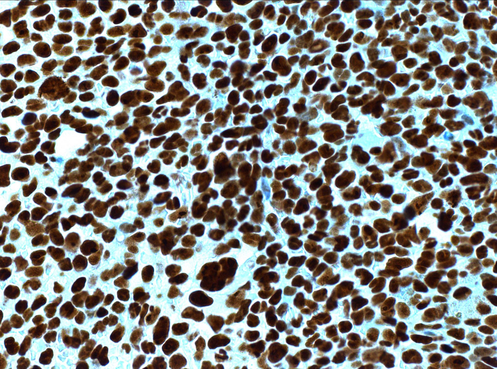
NPM1 wild type AML
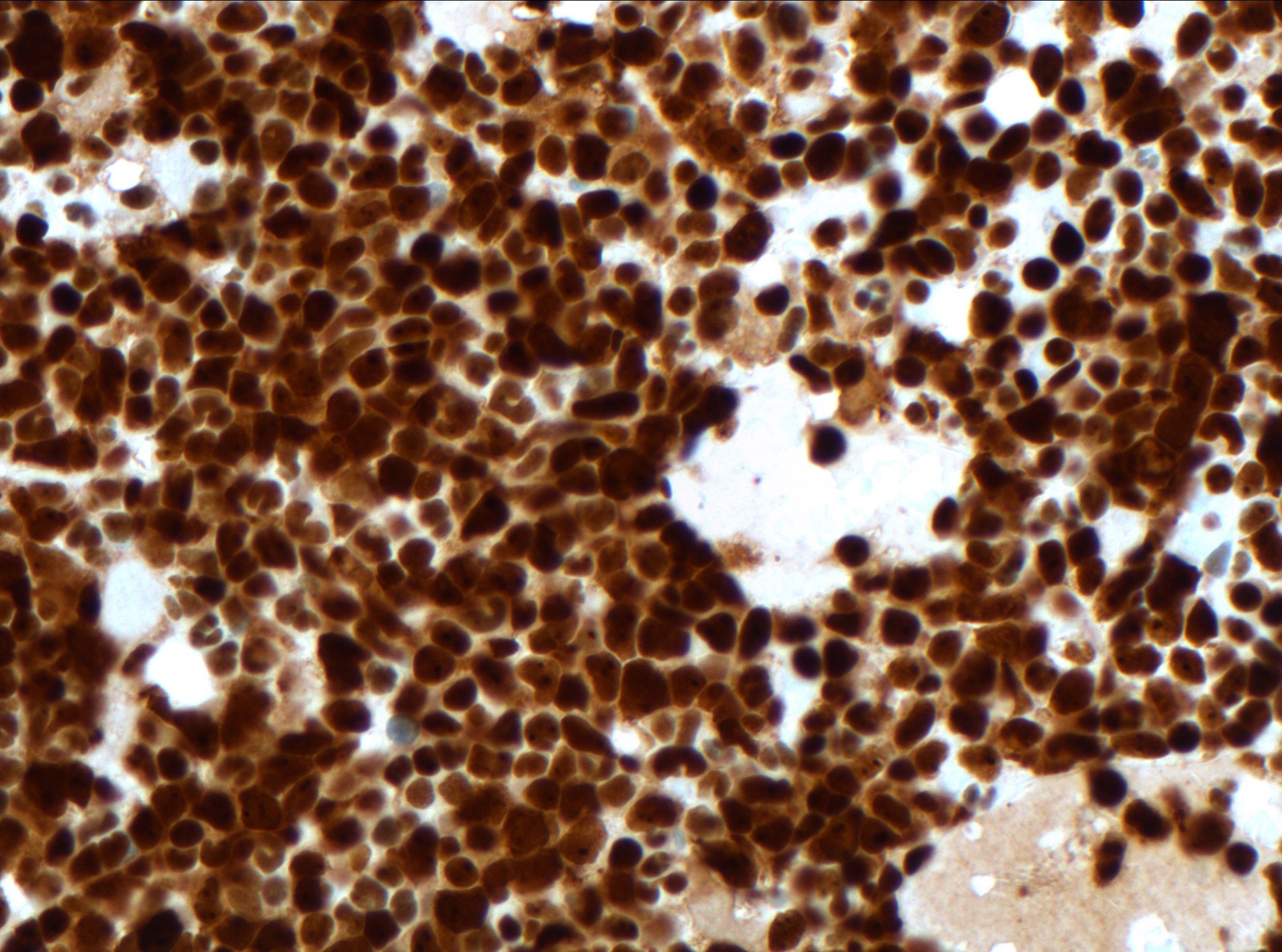
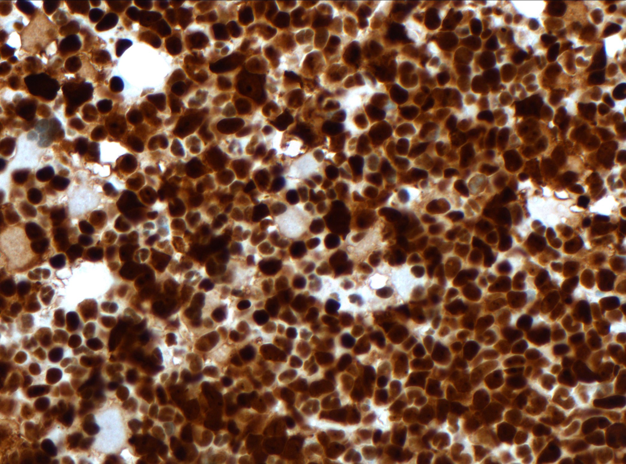
NPM1 mutated AML
Positive staining - normal
- Normal tissues (ubiquitous nuclear expression)
Positive staining - disease
- NPM1 mutated AML (nuclear and cytoplasmic)
- Myelodysplastic syndrome (MDS) (Blood Adv 2019;3:922)
- Myelodysplastic / myeloproliferative neoplasms (MDS / MPN), namely chronic myelomonocytic leukemia (Eur J Haematol 2016;96:65)
- Myeloma (Genes Chromosomes Cancer 2010;49:333)
- Diffuse large B cell lymphoma (Cancers (Basel) 2021;13:5900)
- Chronic lymphocytic leukemia (PLoS One 2022;17:e0276674)
Negative staining
- NPM1 expression is heterogeneous in gastric tumors (BMC Gastroenterol 2014;14:9)
- Downregulation may have a role in gastric carcinogenesis
Molecular / cytogenetics description
- NPM1 mutation is strongly linked to specific microRNA and long noncoding RNA patterns (Int J Lab Hematol 2021;43:664)
Sample pathology report
- Bone marrow, trephine biopsy:
- NPM1 mutated acute myeloid leukemia (see comment)
- Comment: Hypercellular bone marrow for age involved by acute myeloid leukemia. Increase of immature cells with myelomonocytic differentiation is seen. NPM1 immunohistochemistry shows aberrant nuclear and cytoplasmic positivity, supporting the notion of an underlying mutation ultimately confirmed by molecular analysis.
Board review style question #1
Board review style answer #1
D. Nuclear and cytoplasmic staining. NPM1 is predominantly located in the nucleolus but has shuttling capability to the nucleus and cytoplasm. Nuclear and aberrant cytoplasmic NPM1 expression in neoplastic cells supports an underlying NPM1 mutation. Answers A and C are incorrect because NPM1 mutation leads to enhanced cytoplasmic staining, not Golgi or membrane staining. Answer B is incorrect because NPM1 is still detectable even if mutated. Answer E is incorrect because strictly nuclear staining is considered normal.
Comment Here
Reference: NPM1
Comment Here
Reference: NPM1
Board review style question #2
Which of the following morphologies is NPM1 mutated AML most frequently associated with?
- Erythroblastic
- Megakaryoblastic
- Monocytic and myelomonocytic
- Myeloid
- Undifferentiated
Board review style answer #2
C. Monocytic and myelomonocytic. NPM1 mutated AML is strongly associated with myelomonocytic and monocytic morphology (Blood 2007;109:874).
Comment Here
Reference: NPM1
Comment Here
Reference: NPM1

