
All cases are archived on our website. To view them sorted by case number, diagnosis or category, visit our main Case of the Month page. To subscribe or unsubscribe to Case of the Month or our other email lists, click here.
This case was contributed by Dr. Semir Vranic, Clinical Center of the University of Sarajevo, Bosnia).
CancerTYPE ID is a standardized, objective molecular test based on the differential expression of 92 genes, that classifies tumors by matching the gene expression pattern of a patients tumor tissue to a database of known tumor types and histological subtypes.
CancerTYPE IDs database includes 2,206 tumors from multiple tumor banks, selected to provide broader and deeper representation of the heterogeneity of tumors. The 92-gene assay does not overlap with IHC markers, providing complementary data to standard tumor diagnosis.
CancerTYPE ID uses real-time reverse transcription polymerase chain reaction (RT-PCR). A very low copy number of RNA molecules can be detected, thus reducing the sample tissue required for testing. Testing is conducted and results are generated at bioTheranostics' CAP-accredited, CLIA-certified laboratory.
For more information, click here.
Advertisement
(1) We continue to have record website traffic. Please continue to let us know what we can do to make our website more useful to you.
(2) Dr. Pernick has posted a research paper, The Laws of Complexity and Self-organization: A Framework for Understanding Neoplasia. This is based on an exciting new view of biology developed by Dr. Stuart Kauffman, the winner of a MacArthur Fellowship. Your comments are welcome to Nat@PathologyOutlines.com.
Visit and follow our Blog to see recent updates to the website.
Case #227
Clinical history:
A 60 year old woman with no history of malignancy, kidney disease or other medical disorders presented with a tumor mass in the upper outer quadrant of her left breast, measuring 30 x 25 mm. Previous mammography revealed massive calcification with a benign appearance. A wide excision was performed. Frozen section revealed no definite diagnosis.
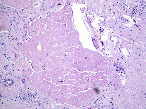
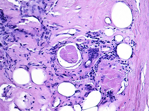
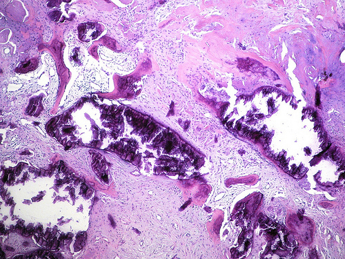
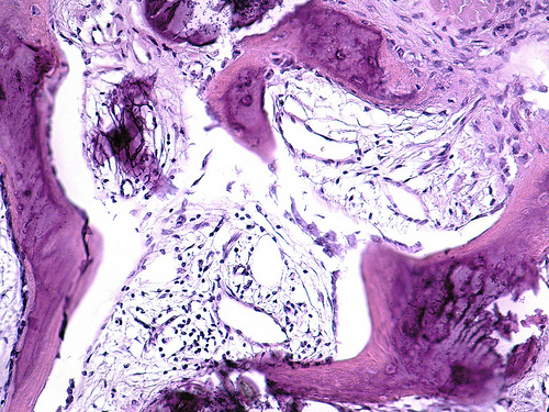
Microscopic images:


What is your diagnosis?
Diagnosis: Osseous metaplasia of the breast
Discussion:
The specimen contains well formed bone tissue without associated neoplasia.
Osseous metaplasia is uncommon. It is associated with benign lesions, including cholesterol granuloma (Pathol Res Pract 2008;204:353), fibroadenoma (Radiology 1989;172:671), fasciitis ossificans (Pathol Res Pract 2007;203:737, Arch Pathol Lab Med 2004;128:e29), lipogranuloma (Yonsei Med J 2011;52:373), radiation therapy (Br J Radiol 2002;75:460), saline implants (Plast Reconstr Surg 2001;107:356) and silicone implants (Ann Plast Surg 1998;41:348).
Osseous metaplasia is also associated with metaplastic carcinoma and other invasive subtypes (Breast 2008;17:314, Am J Surg Pathol 2009;33:534) as well as osteosarcoma of the breast (Cases J 2008 Aug 9;1(1):80).
This disorder is benign but it is important to exclude a possible associated malignancy, which may not be evident on a core biopsy.
