
Stains & CD markers
H3K27me3


Copyright: 2002-2024, PathologyOutlines.com, Inc.
PubMed Search: H3K27me3
H3K27me3
Editorial Board Member: Christian M. Schürch, M.D., Ph.D.
Editor-in-Chief: Debra L. Zynger, M.D.
Last author update: 21 March 2023
Last staff update: 21 March 2023
Copyright: 2002-2024, PathologyOutlines.com, Inc.
PubMed Search: H3K27me3

Table of Contents
Definition / general | Essential features | Pathophysiology | Interpretation | Uses by pathologists | Prognostic factors | Microscopic (histologic) images | Positive staining - normal | Positive staining - disease | Negative staining | Sample pathology report | Board review style question #1 | Board review style answer #1Cite this page: Nawar NAM, Dickson BC. H3K27me3. PathologyOutlines.com website. https://www.pathologyoutlines.com/topic/stainsH3k27me3.html. Accessed April 28th, 2024.
Definition / general
- Epigenetic modification of histone H3 results in H3K27me3 (trimethylation at lysine 27 of histone H3) (J Hum Genet 2013;58:439, Nat Rev Genet 2020;21:555)
- H3K27me3 is a downstream target of the polycomb repressive complex 2 (PRC2), which has histone methlyltransferase activity; PRC2 contains evolutionarily conserved proteins essential for regulating gene expression (Nature 2011;469:343, Nat Genet 2014;46:1170, Sci Adv 2015;1:e1500737)
- As a gene transcription repressor, H3K27me3 has diverse roles in embryogenesis and neoplasia (Cell Res 2018;28:593, Epigenetics 2014;9:658)
Essential features
- Loss of H3K27me3 occurs in a significant subset of malignant peripheral nerve sheath tumors (Nat Genet 2014;46:1227, Mod Pathol 2016;29:4, J Pathol 2020;252:151)
- Other neoplasms may show loss of H3K27me3 expression, such as embryonal rhabdomyosarcoma, meningioma, radiation associated unclassified sarcoma, radiation associated angiosarcoma, dedifferentiated chondrosarcoma, melanoma and Merkel cell carcinoma, among others (Ann Diagn Pathol 2021;52:151735, Pathol Res Pract 2018;214:417, Mod Pathol 2016;29:4, Virchows Arch 2018;472:361, Mod Pathol 2019;32:435, Mod Pathol 2017;30:1677, Mod Pathol 2017;30:877, Histopathology 2023;82:420, Histopathology 2017;70:385)
- Some tumors may exhibit heterogeneous H3K27me3 expression (mosaic pattern), the significance of which remains to be fully established (Mod Pathol 2016;29:4)
Pathophysiology
- Polycomb repressive complex 2 (PRC2) has diverse roles in stem cell pluripotency, somatic differentiation and proliferation (Nature 2011;469:343, Sci Adv 2015;1:e1500737)
- Enzymatic subunits EZH1 and EZH2 of PRC2 catalyze methylation of H3K27; trimethylation of H3K27 is associated with gene silencing (Nature 2011;469:343, Cell Rep 2016;17:1369)
- Alterations involving PRC2, accessory proteins or H3K27me3 may disrupt an epigenetic regulatory network, with complex and varied contributions in neoplasia (Mol Cancer Res 2014;12:639)
Interpretation
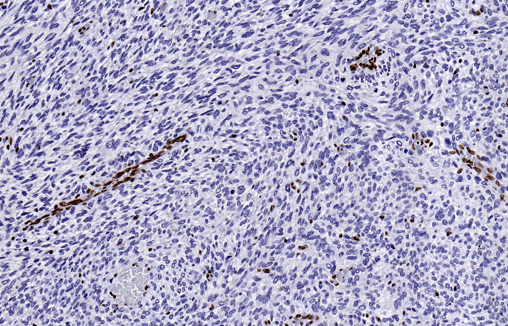
- H3K27me3 is constitutively expressed and intact expression is denoted by the presence of diffuse nuclear staining; loss of staining may be complete or heterogeneous (mosaic) (Mod Pathol 2016;29:4)
- Interpretation is context dependent
- For example, when presented with a peripheral nerve sheath tumor, loss of staining would favor malignant peripheral nerve sheath tumor over neurofibroma (Mod Pathol 2016;29:4, Mod Pathol 2016;29:582)
- An eccentric intranuclear dot reflects the inactivated X chromosome in normal tissues (Histopathology 2016;69:702)
Uses by pathologists
- Complete loss of H3K27me3 occurs in ~34% to 72% of malignant peripheral nerve sheath tumors (91 - 100% radiation associated, 41 - 71% neurofibromatosis type 1 associated, 32 - 90% sporadic and 0% epithelioid) (Mod Pathol 2016;29:4, Am J Surg Pathol 2016;40:479, Mod Pathol 2016;29:582, Mod Pathol 2017;30:1677)
- As a result, this may be used to differentiate from potential mimics (e.g., neurofibroma)
- It is important to note that H3K27me3 may be lost in a significant proportion of melanoma and rarely other malignant and benign neoplasms (Mod Pathol 2017;30:1677, Mod Pathol 2016;29:582)
- H3K27me3 immunohistochemistry highlights the inactivated X chromosome in female tissues; as a result, this may be used to predict sex in nonneoplastic tissues (Histopathology 2016;69:702)
Prognostic factors
- Loss of H3K27me3 in malignant peripheral nerve sheath tumors appears to be associated with higher grade and aggressive behavior (Histopathology 2016;69:702, Mod Pathol 2016;29:582)
- Loss of H3K27me3 may portend aggressive behavior in meningioma; conversely, increased expression has been associated with a worse prognosis in some tumors, such as oral squamous cell carcinoma (Acta Neuropathol 2018;135:955, Cancer 2013;119:4259)
Microscopic (histologic) images
Contributed by Brendan C. Dickson, M.D.
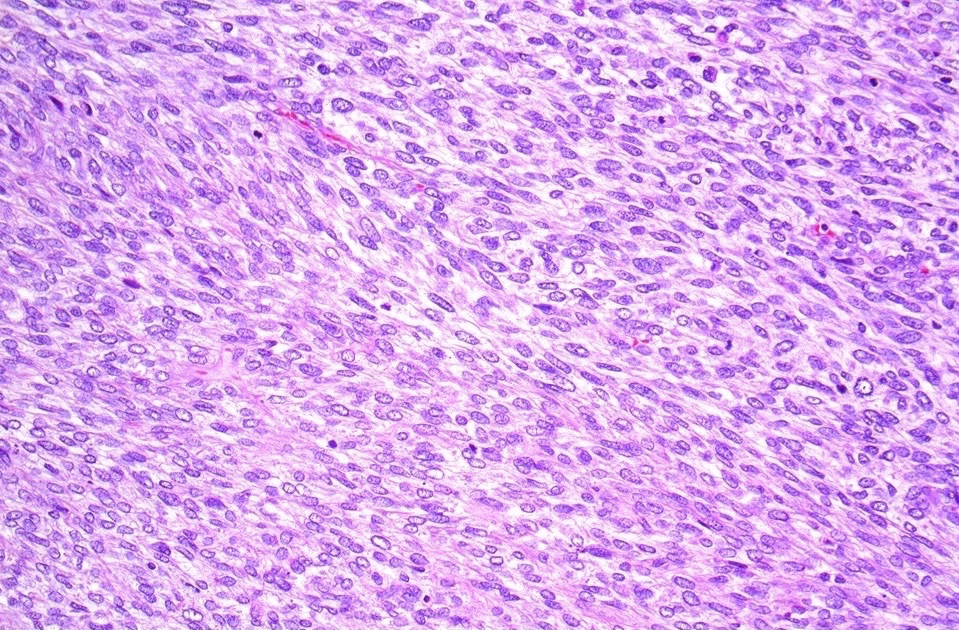
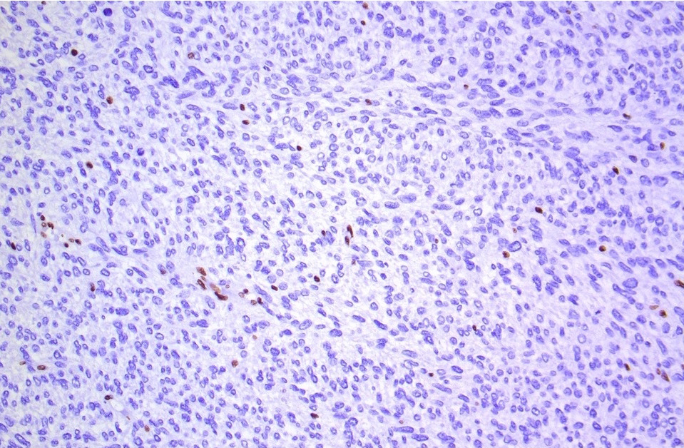
Malignant peripheral nerve sheath tumor
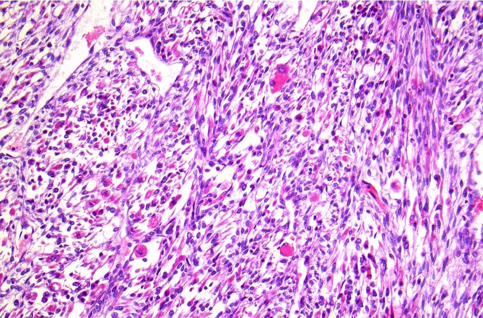
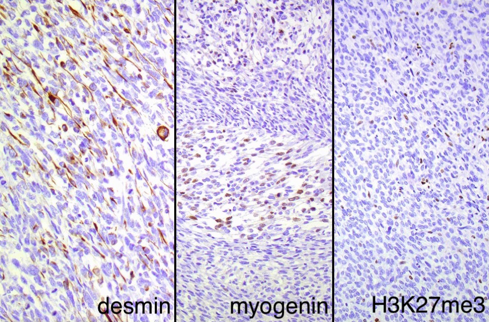
Malignant triton tumor with rhabdomyoblastic differentiation

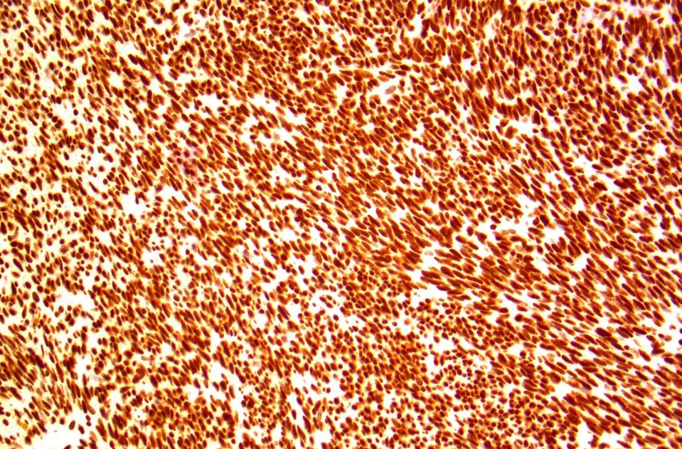
Schwannoma
Positive staining - normal
- Normal tissue contains intact nuclear H3K27me3 expression
- Presence of an eccentric intranuclear dot reflects the inactivated X chromosome in normal tissues (Histopathology 2016;69:702)
Positive staining - disease
- Most tumor tissues contain intact nuclear H3K27me3 expression
Negative staining
- To date, no normal tissues have been reported to consistently lack H3K27me3 expression
- Complete loss of H3K27me3 occurs in ~34% to 72% malignant peripheral nerve sheath tumors (91 - 100% radiation associated, 41 - 71% neurofibromatosis type 1 associated, 32 - 90% sporadic and 0% epithelioid) (Mod Pathol 2016;29:4, Am J Surg Pathol 2016;40:479, Mod Pathol 2016;29:582, Mod Pathol 2017;30:1677)
- Complete loss of H3K27me3 occurs in ~37% of melanomas (Mod Pathol 2017;30:1677)
- H3K27me3 is reported to rarely be lost in a subset of other tumors, such as meningioma, radiation associated unclassified sarcoma, radiation associated angiosarcoma, undifferentiated pleomorphic sarcoma, osteosarcoma, dedifferentiated chondrosarcoma, melanoma and Merkel cell carcinoma (Pathol Res Pract 2018;214:417, Mod Pathol 2016;29:4, Virchows Arch 2018;472:361, Histopathology 2017;70:385, Mod Pathol 2019;32:435, Mod Pathol 2017;30:1677, Mod Pathol 2017;30:877)
Sample pathology report
- Immunohistochemical analysis of H3K27me3 expression (clone, dilution, automated staining platform*):
- The tissue staining pattern is intact / mosaic / lost.*
- External (positive and negative) and internal (e.g., endothelial cells and lymphocytes) controls have intact staining pattern.*
- *Details to be specified / confirmed upon reporting stain
Board review style question #1
A tumor is biopsied in a patient with a remote history of cancer. It shows a cellular spindle cell neoplasm with a herringbone pattern. Which of the following neoplasms is most likely to exhibit loss of H3K27me3?
- Cellular schwannoma
- Epithelioid malignant peripheral nerve sheath tumor
- Neurofibroma
- Radiation associated malignant peripheral nerve sheath tumor
Board review style answer #1
D. Radiation associated malignant peripheral nerve sheath tumor. Complete loss of H3K27me3 occurs in > 90% of radiation associated malignant peripheral nerve sheath tumors. It is intact in the vast majority of cases of epithelioid malignant peripheral nerve sheath tumor, schwannoma and neurofibroma.
Comment Here
Reference: H3K27me3
Comment Here
Reference: H3K27me3

