

Contributed by Chunlai Zuo, M.D., M.S. and Huihui Ye, M.D., M.S.

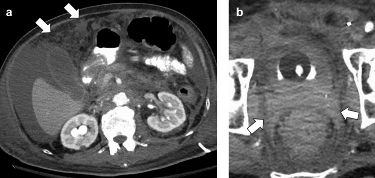
Bladder wall mass by CT
Images hosted on other servers:

Bladder neck mass by ultrasound
Images hosted on other servers:

Bladder neck tumor cystoscopy
Images hosted on other servers:

Primary signet ring
cell carcinoma of
upper urinary tract

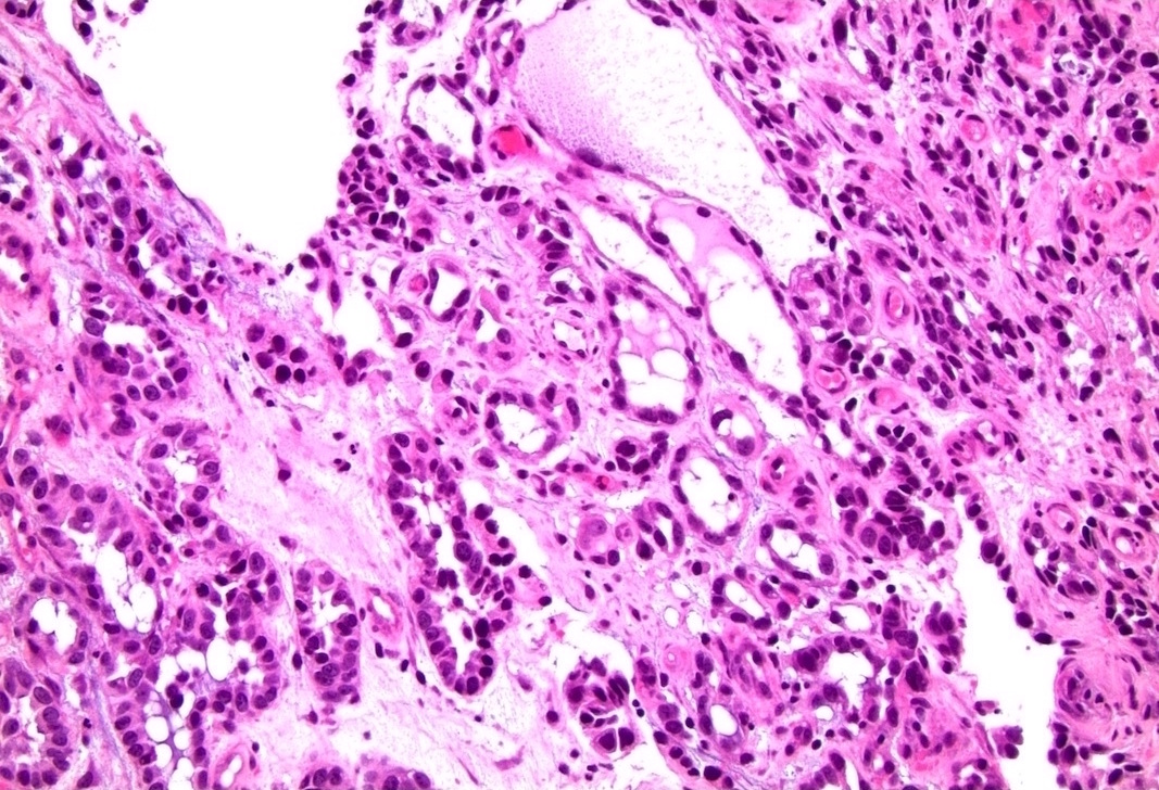
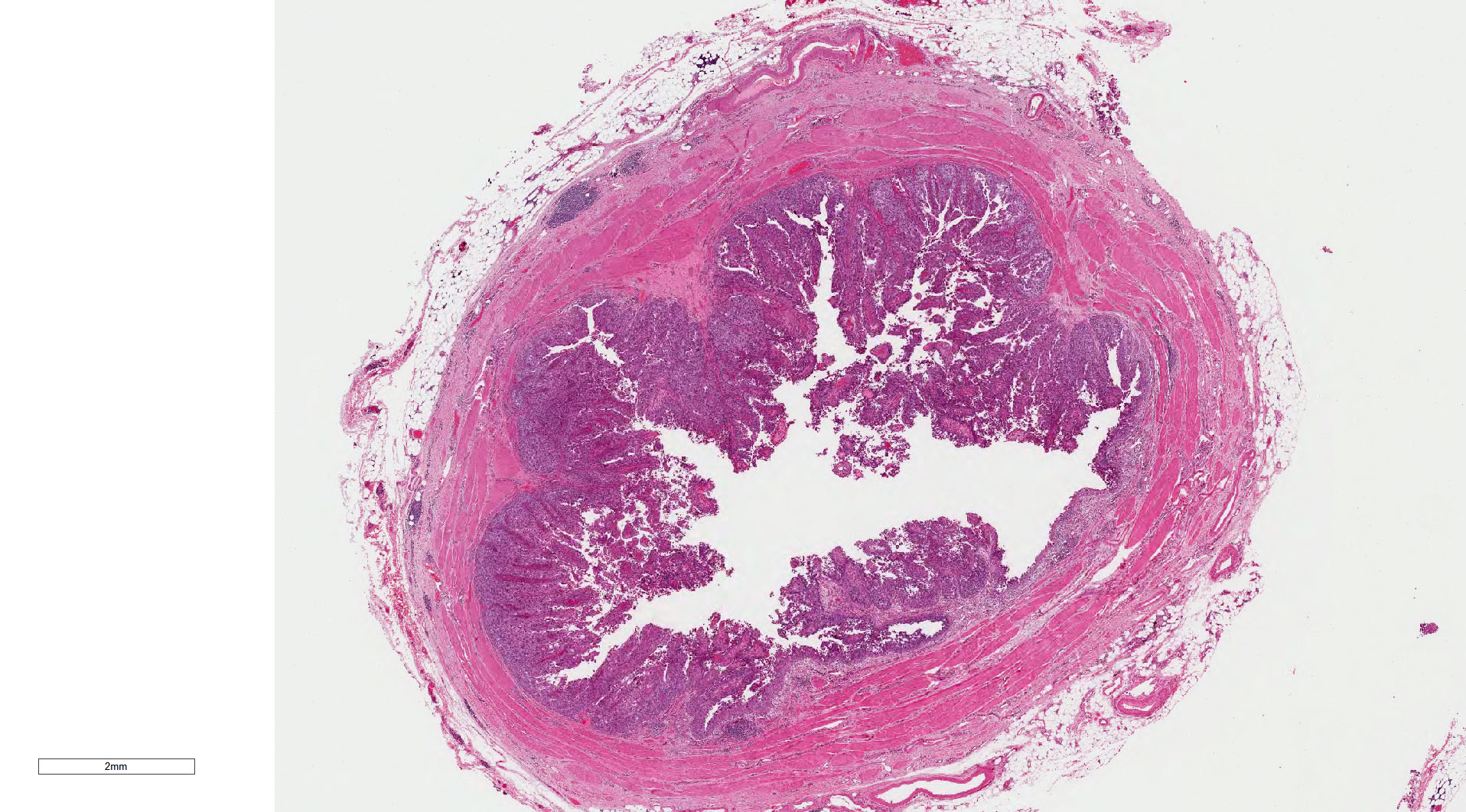
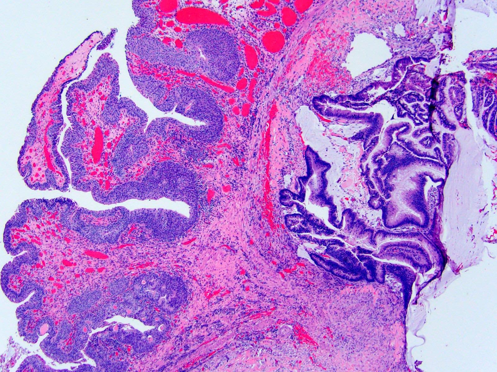
Adenocarcinoma of urinary bladder
Contributed by Chunlai Zuo, M.D., M.S. and Huihui Ye, M.D., M.S.
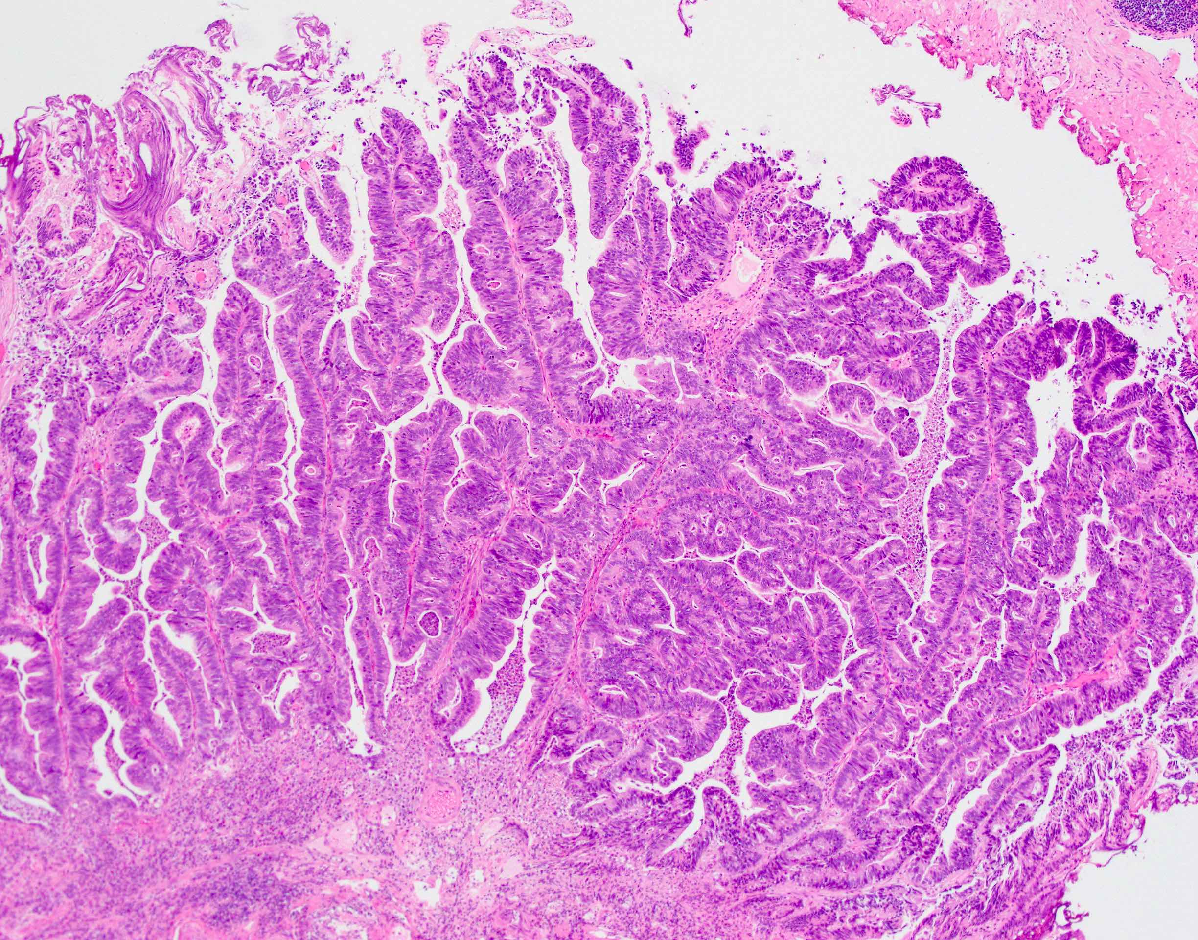
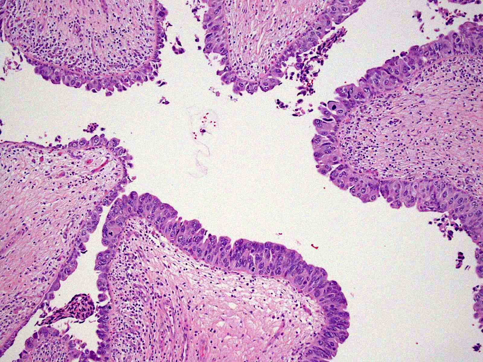
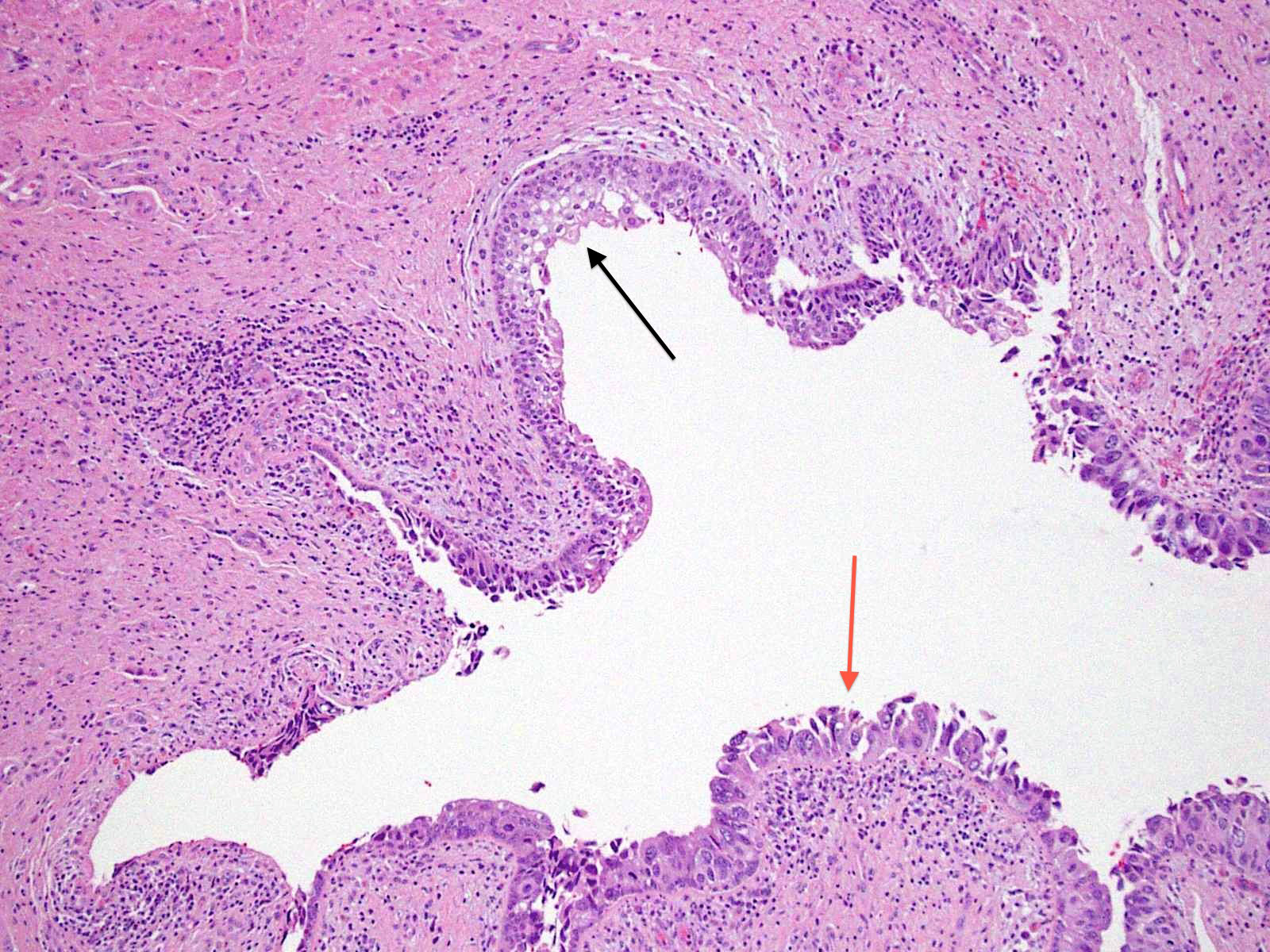
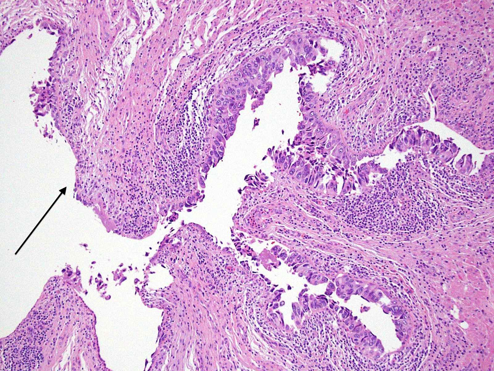
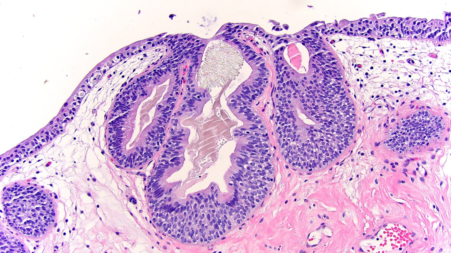
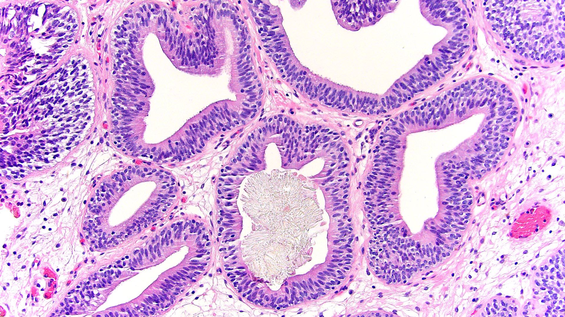
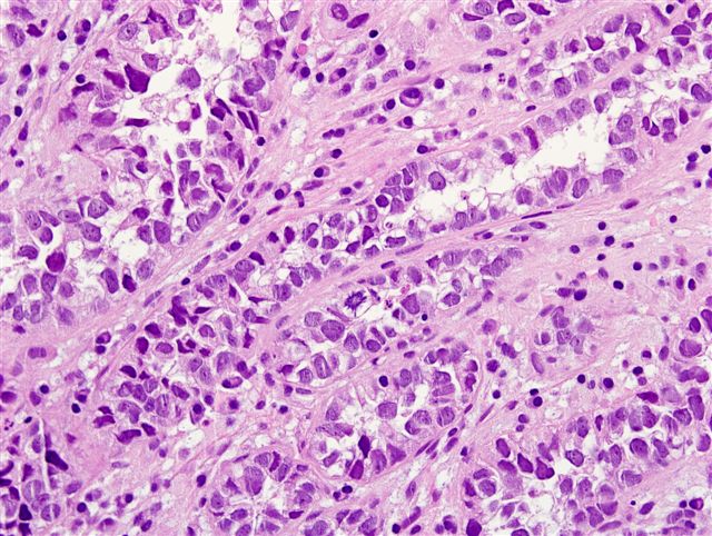
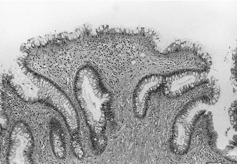
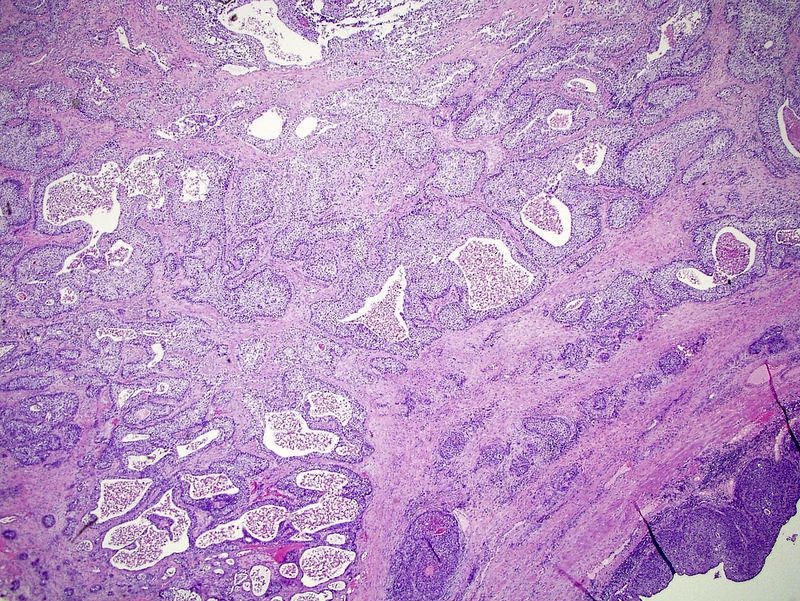
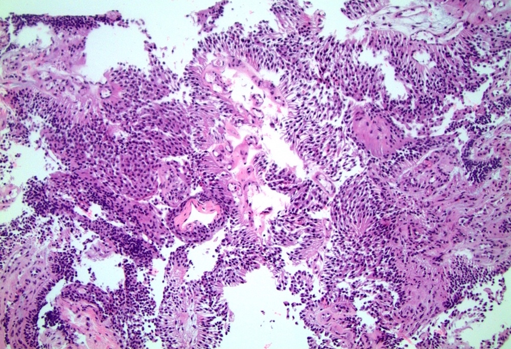
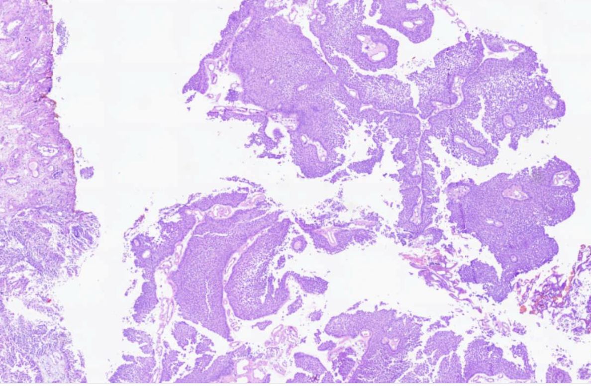
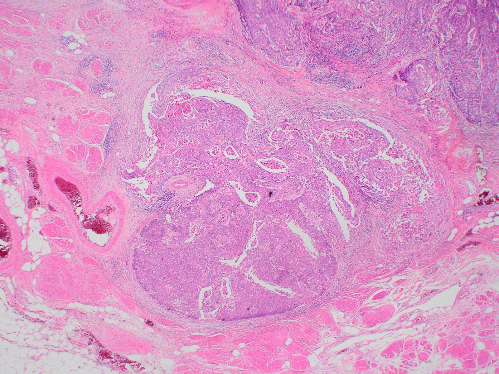
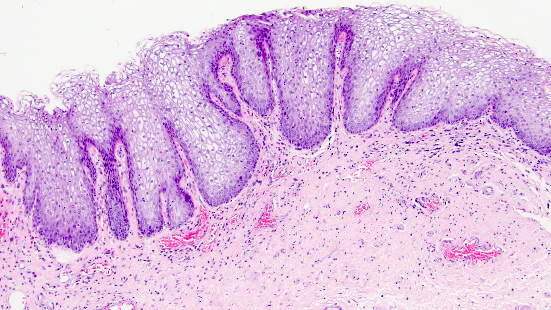
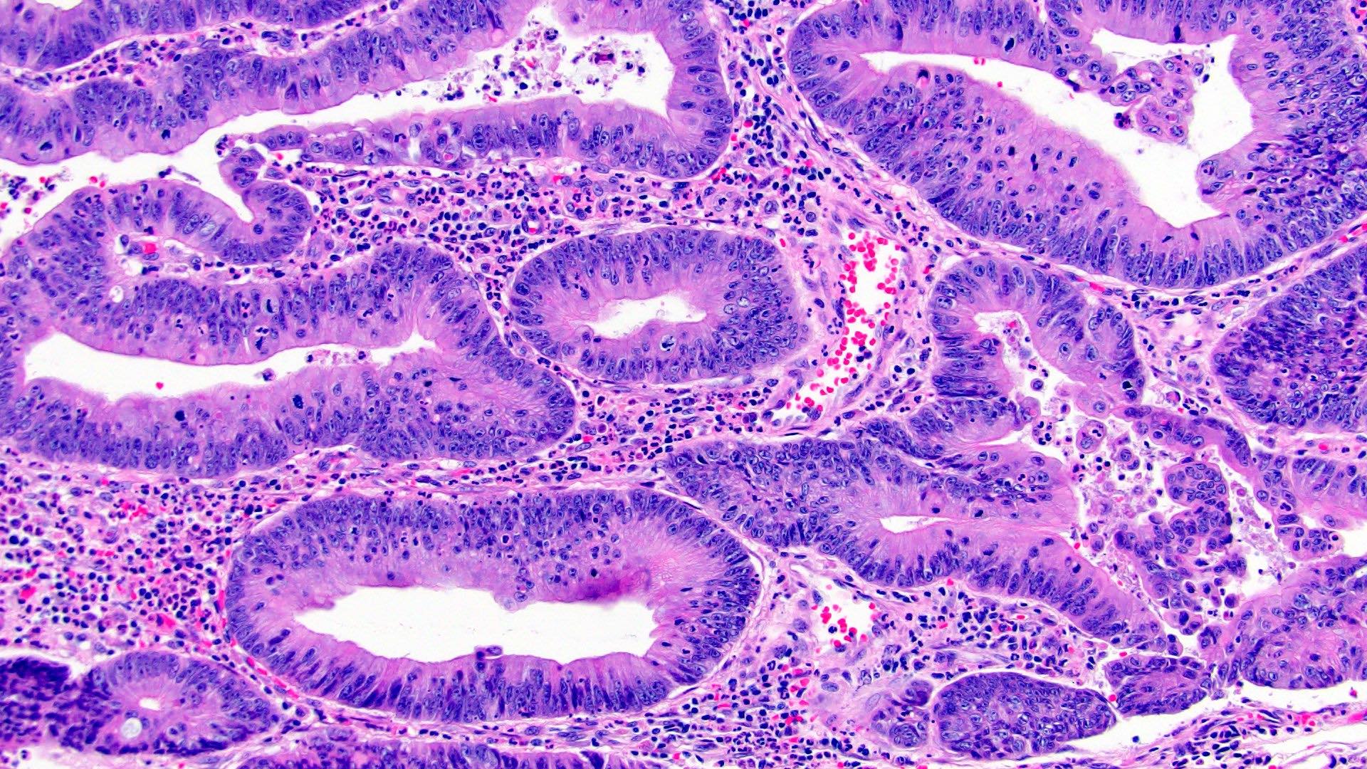
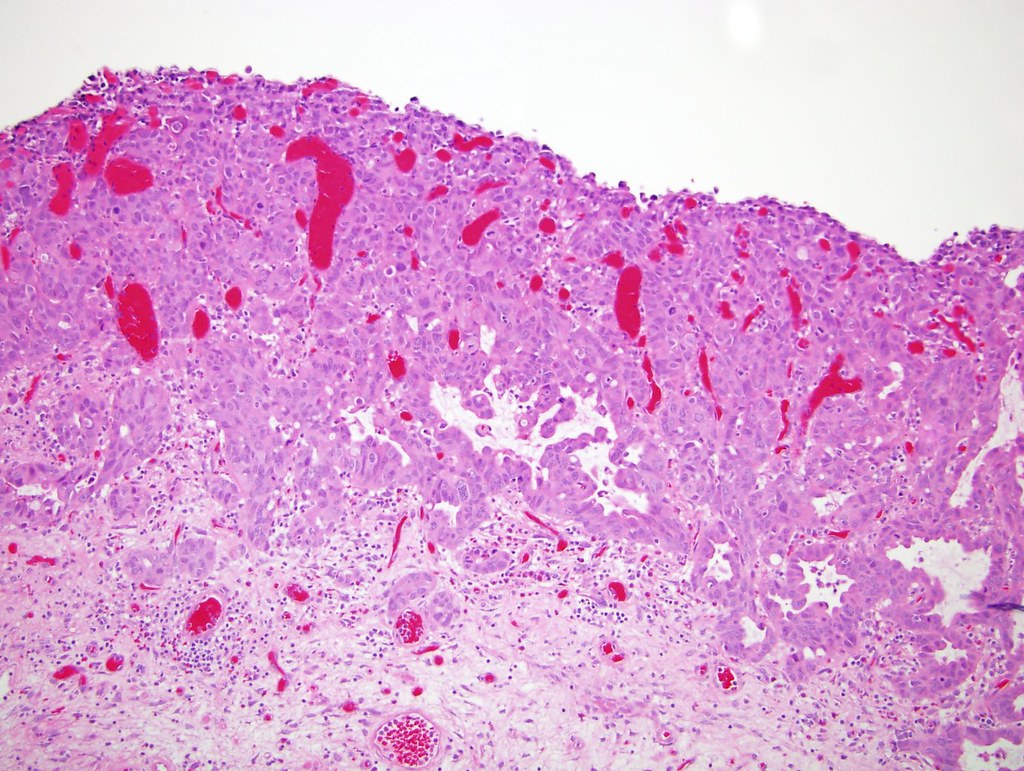
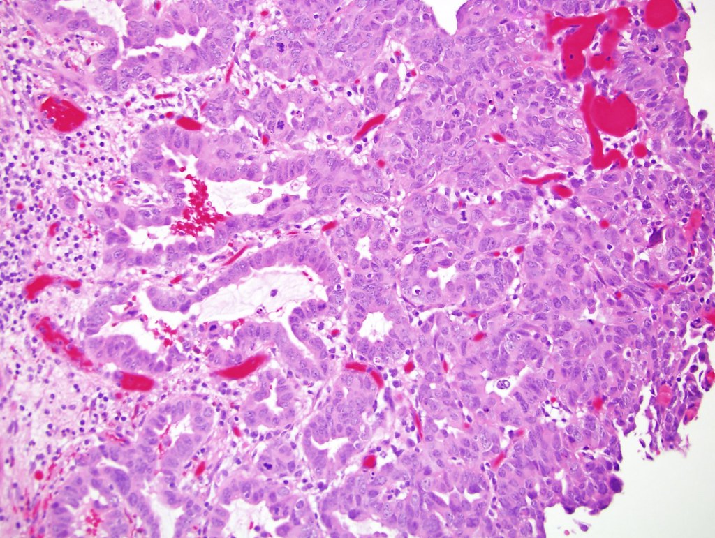
Villoglandular growth
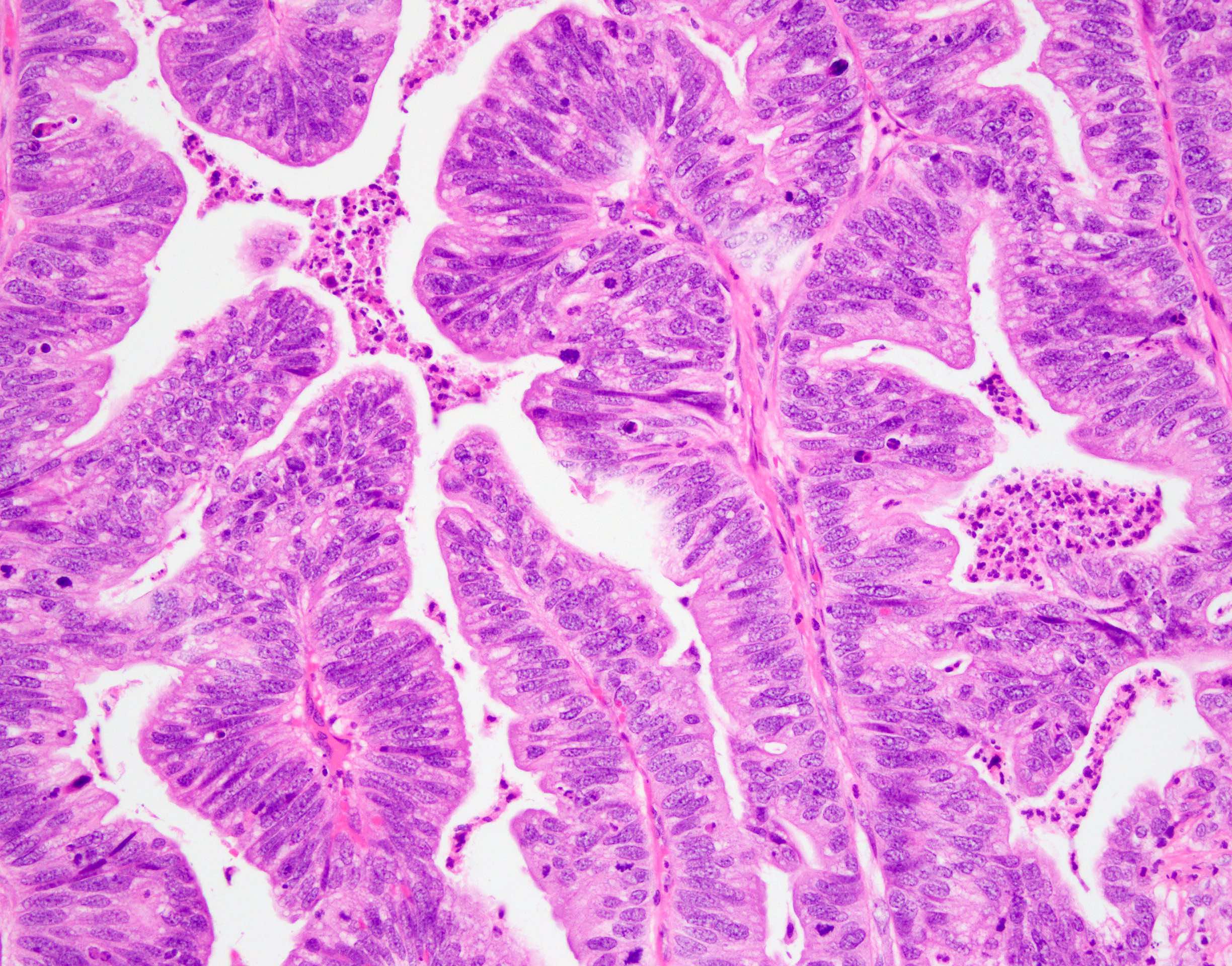
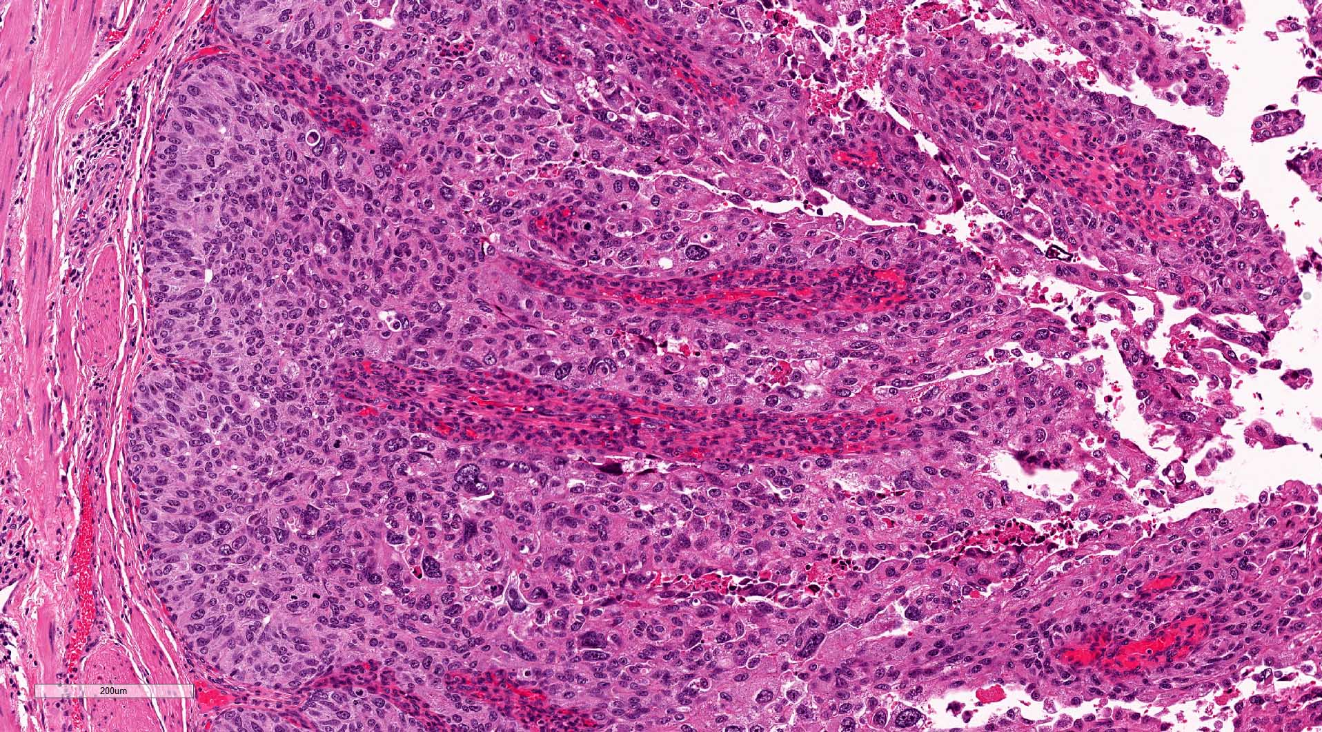
Cytologic atypia
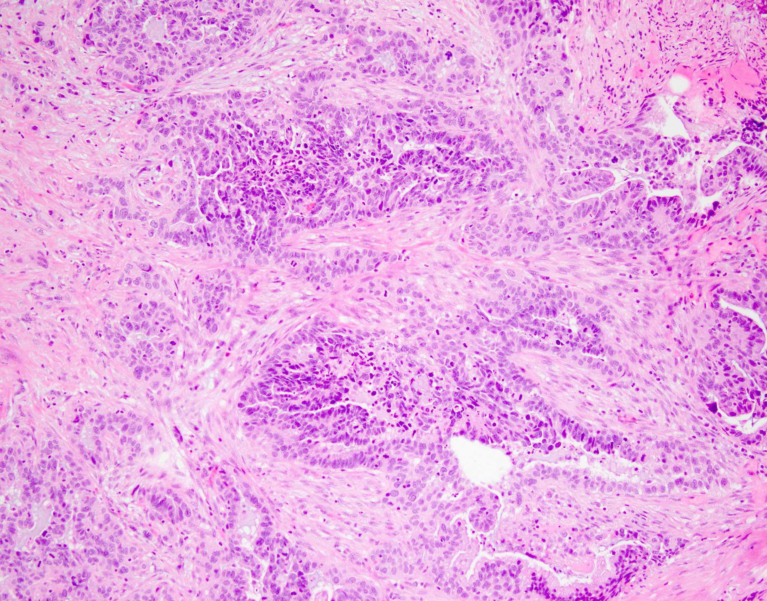
Desmoplastic reaction
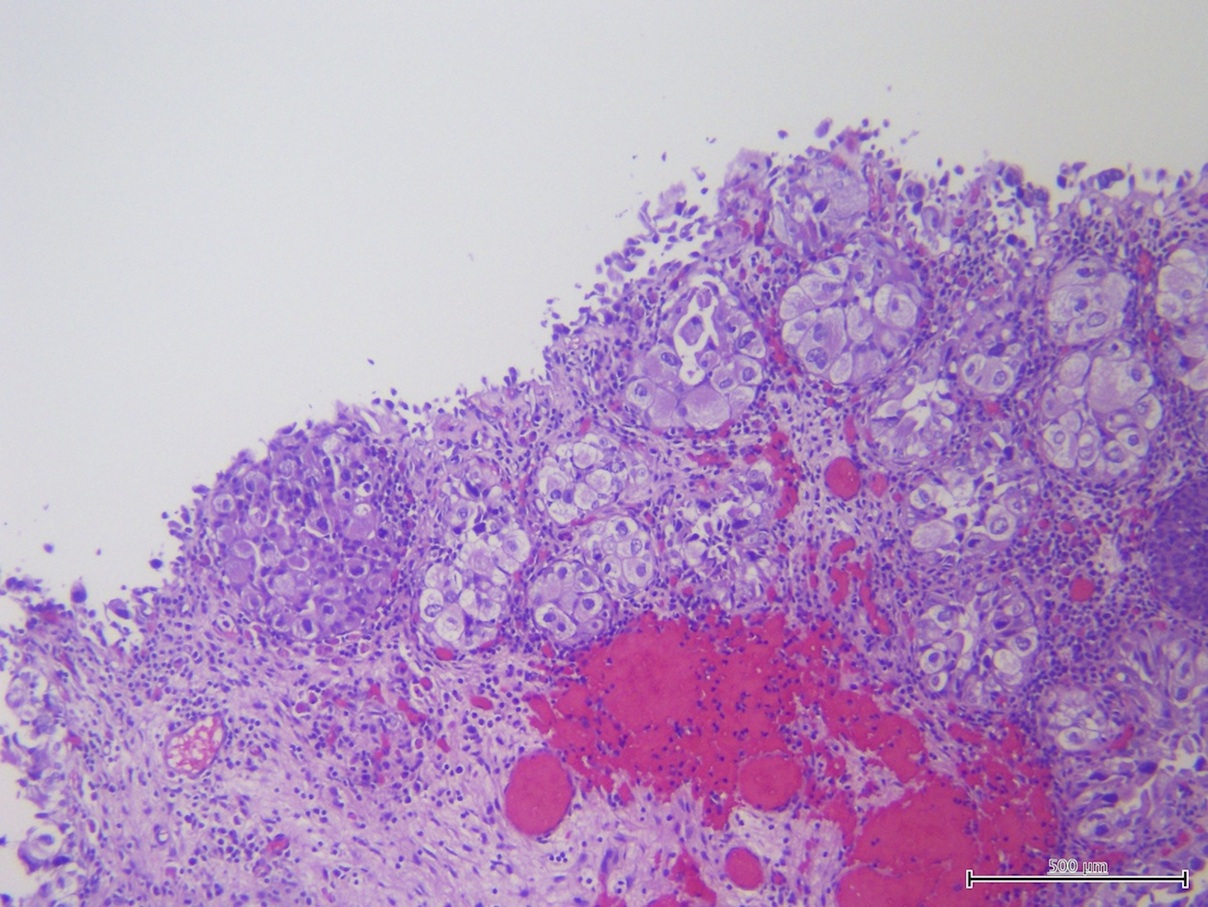
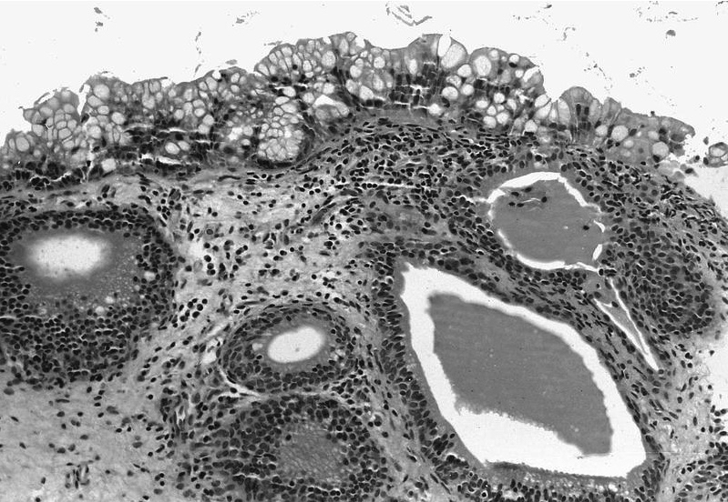
Intraluminal mucin
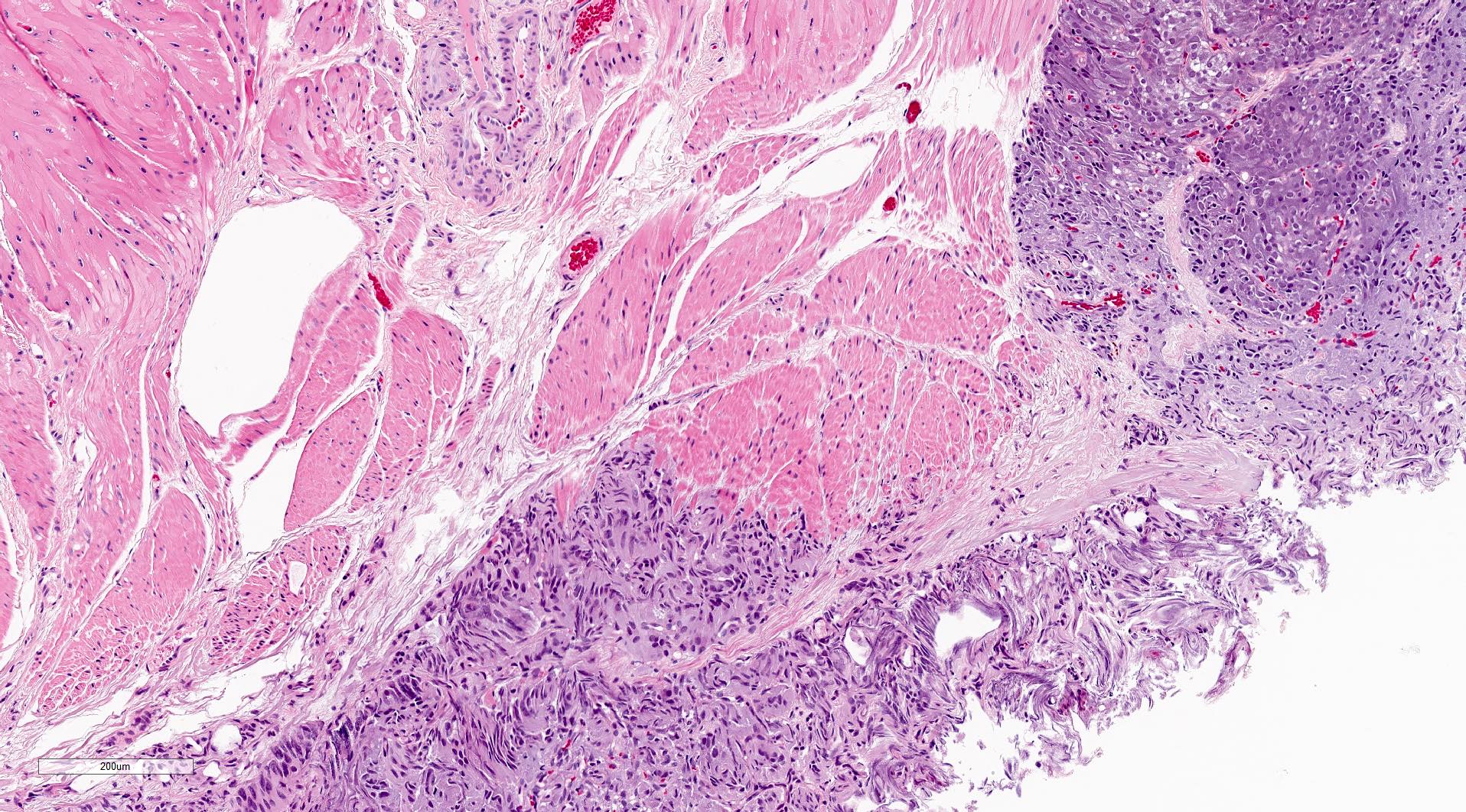
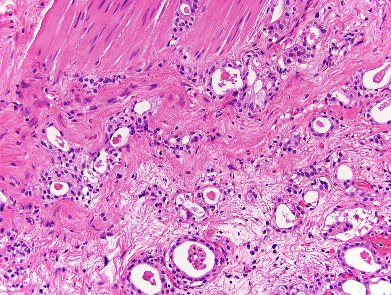
Muscularis propria invasion
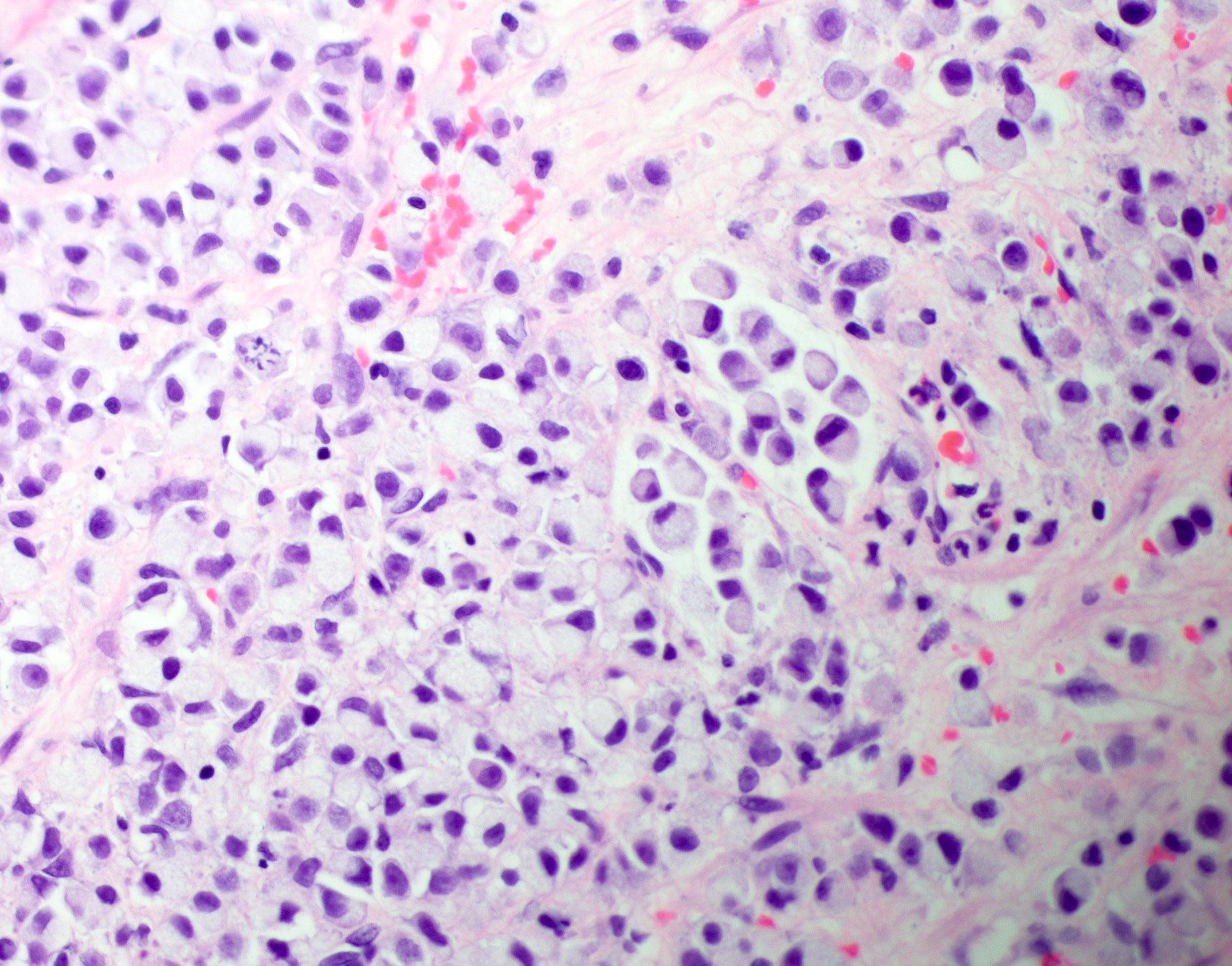
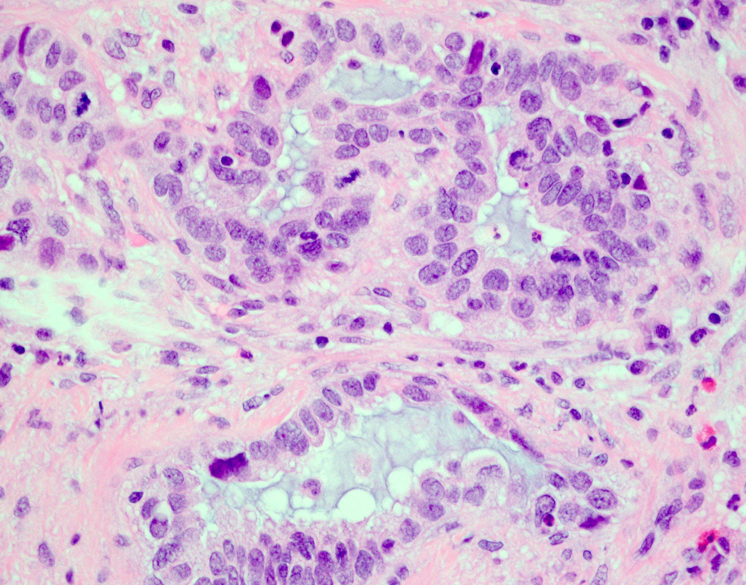
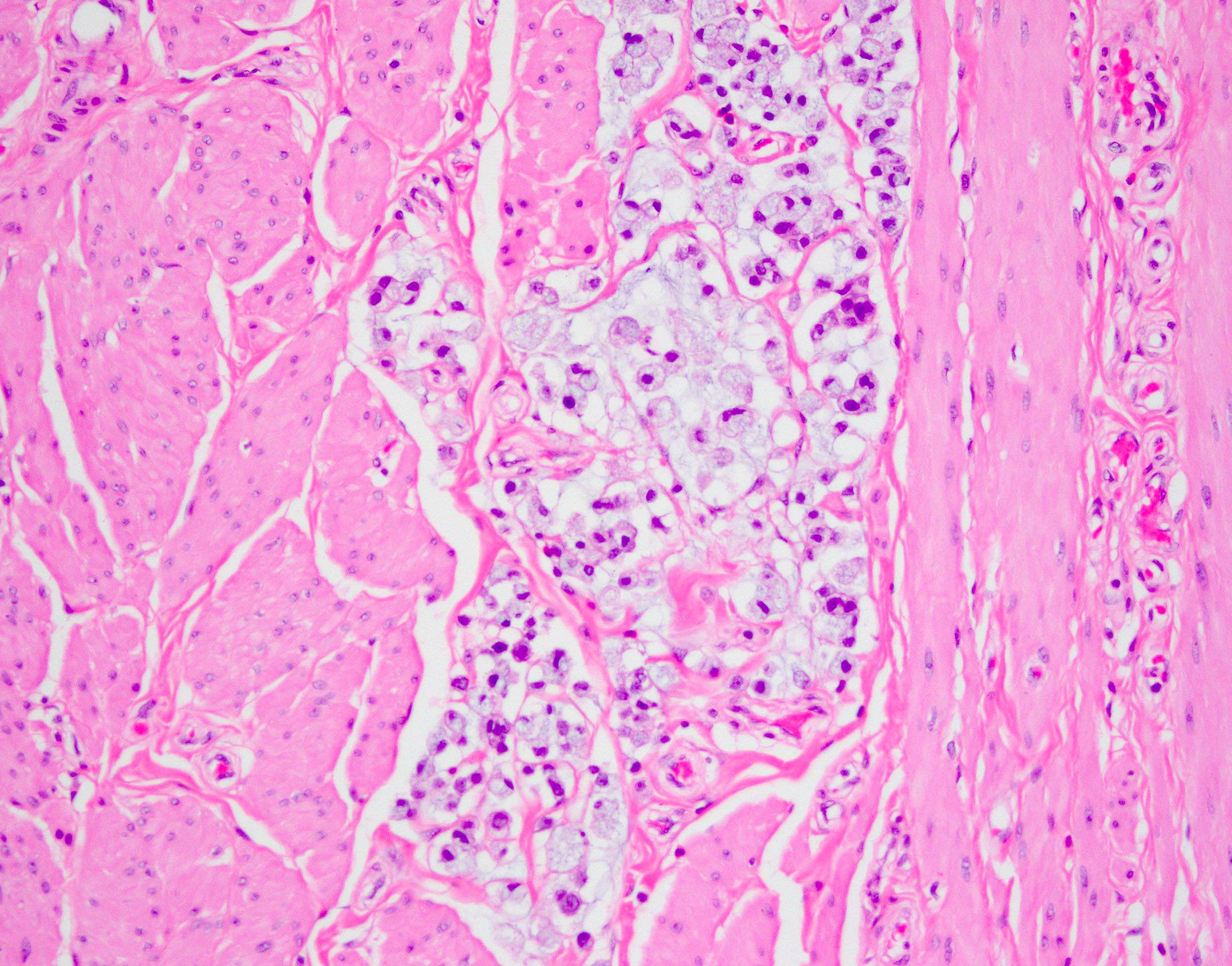
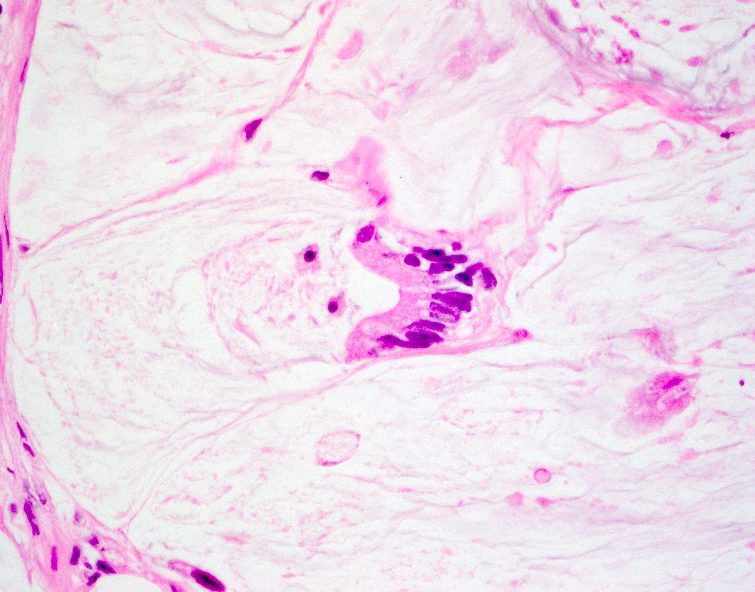
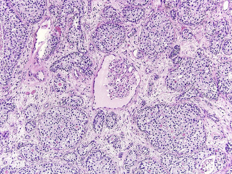
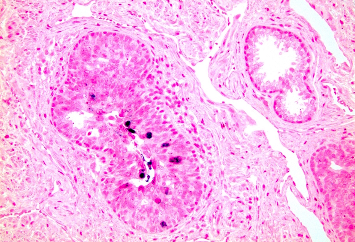
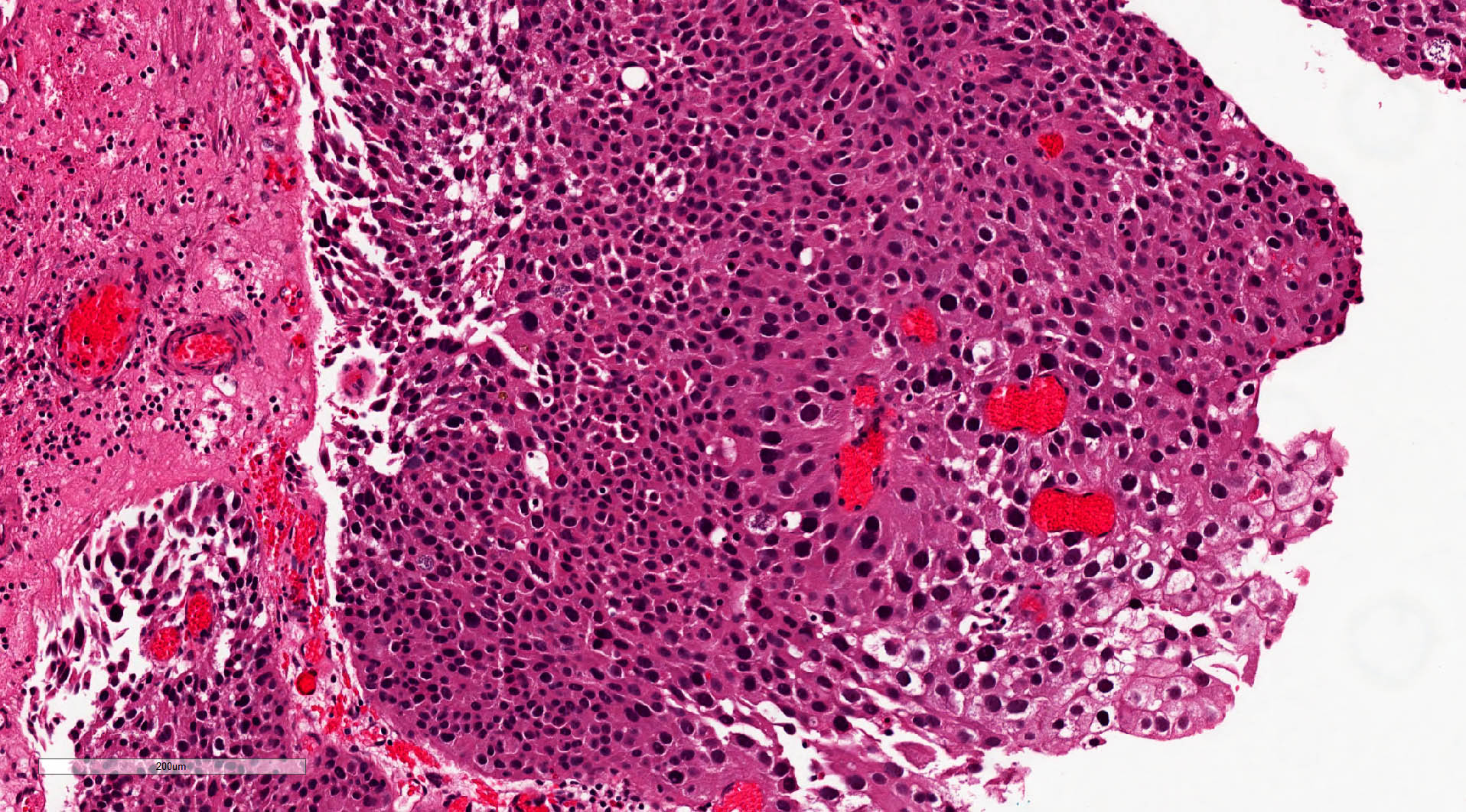
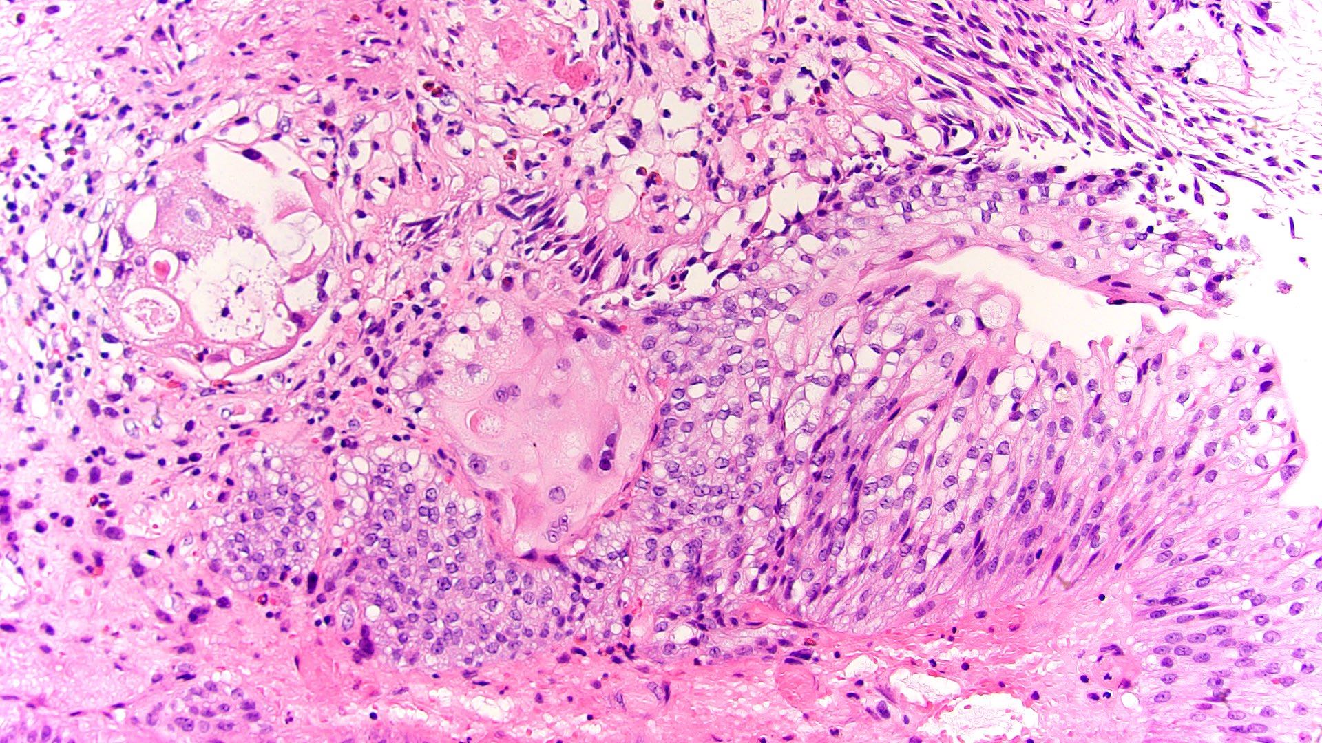
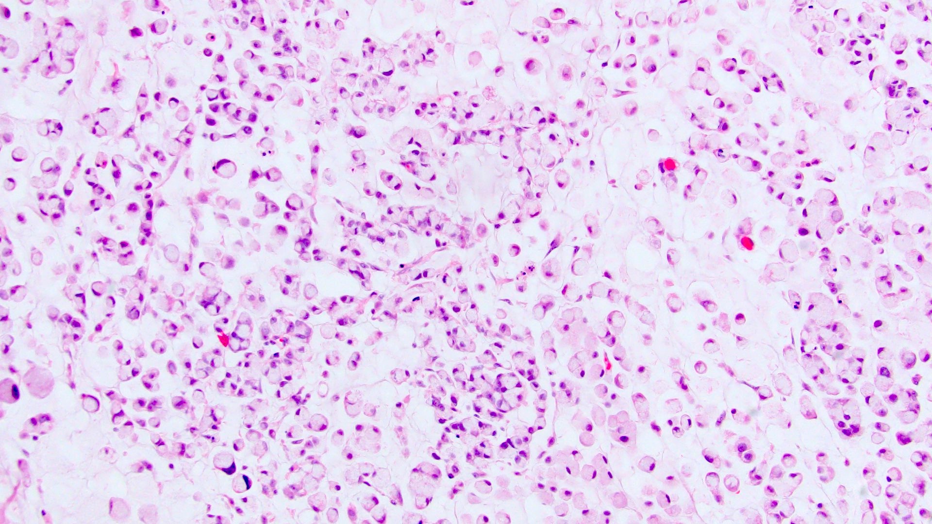
Signet ring cells
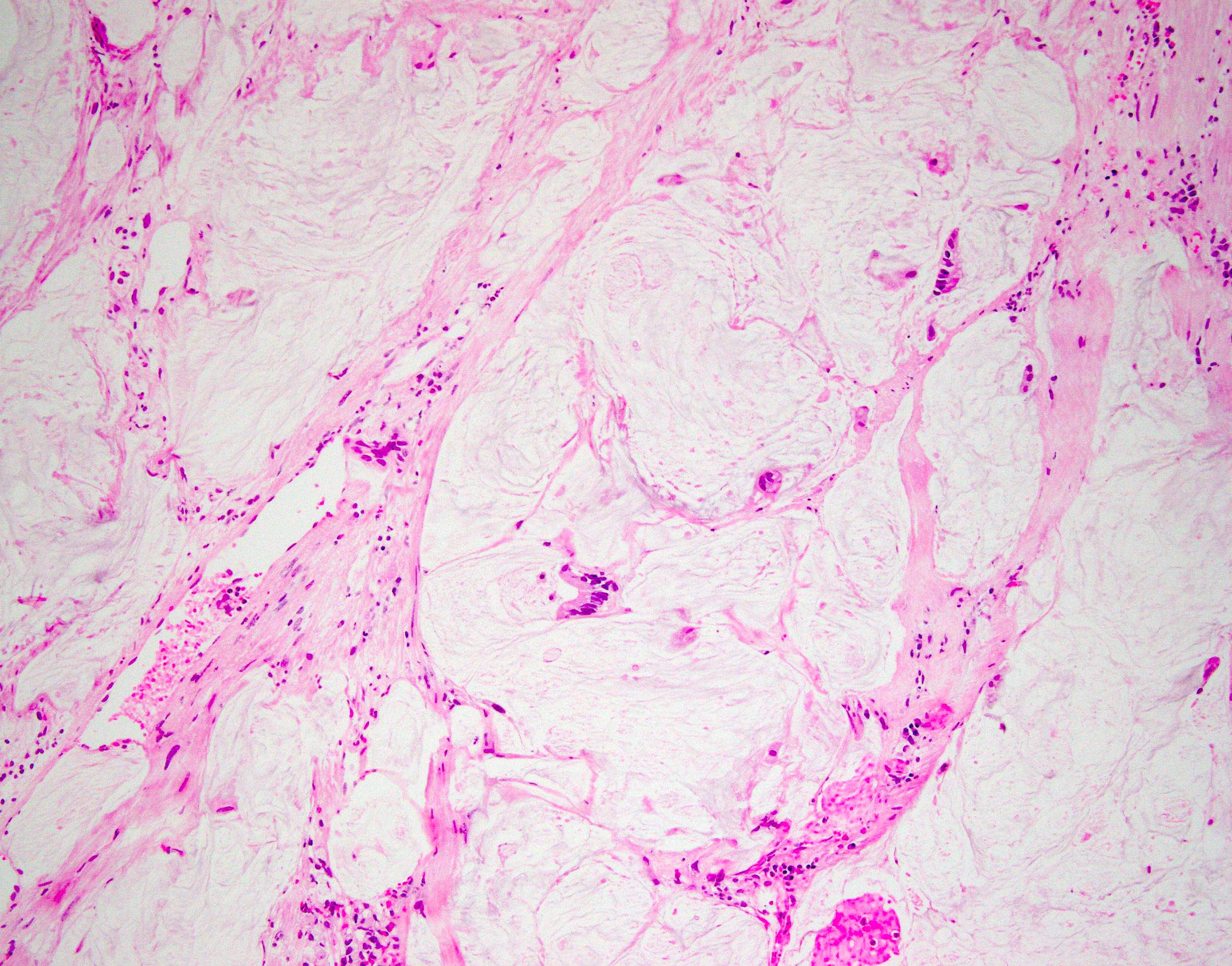
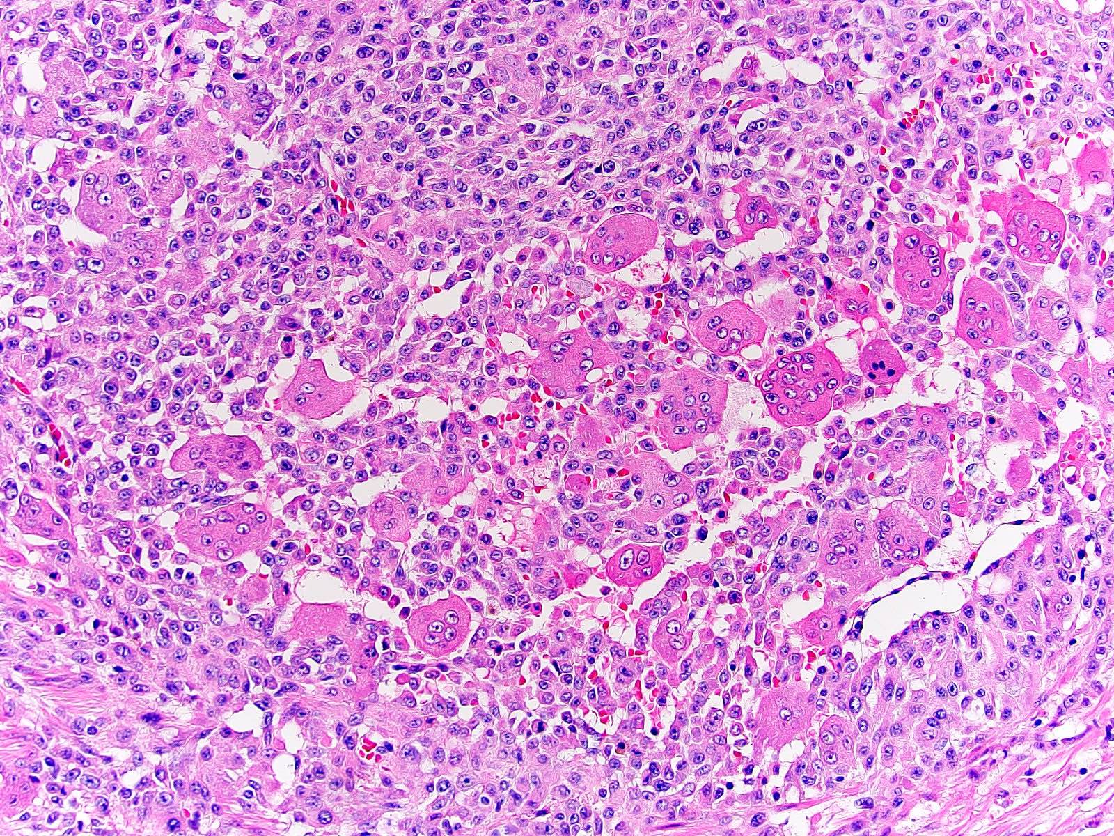
Mucin pool
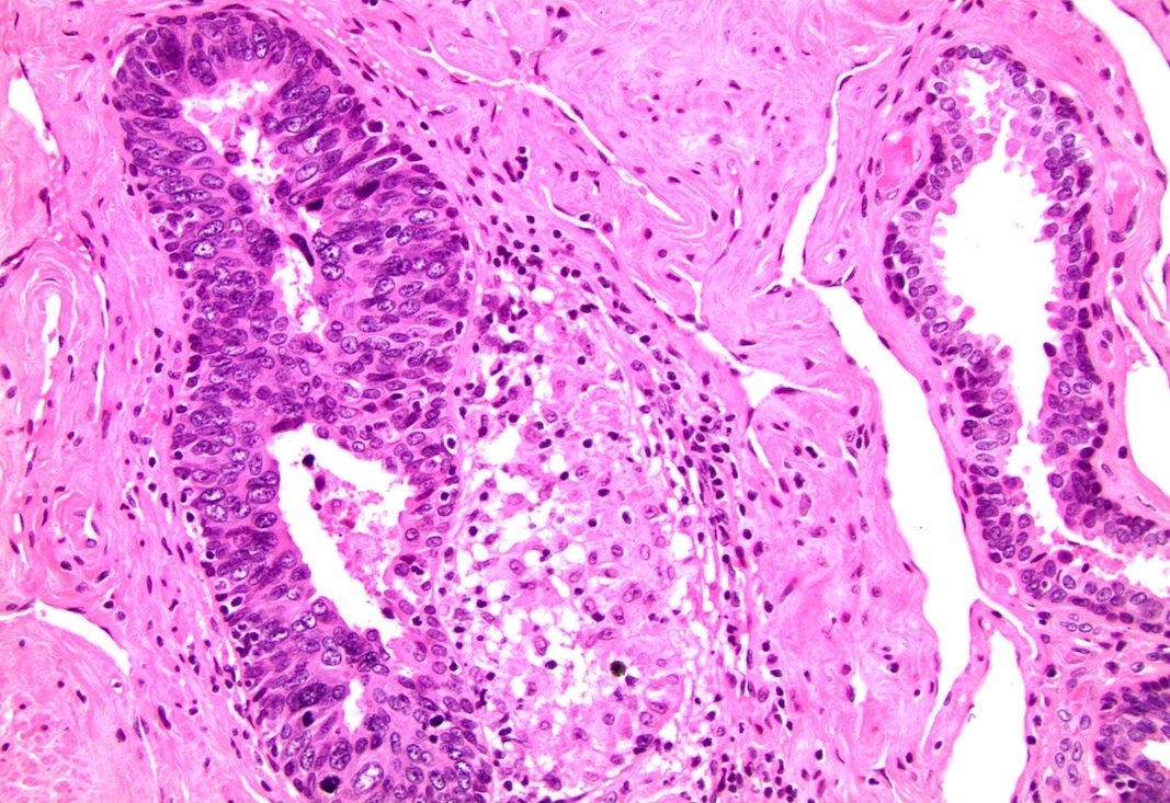
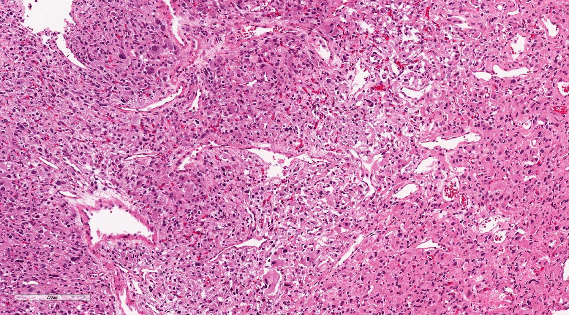
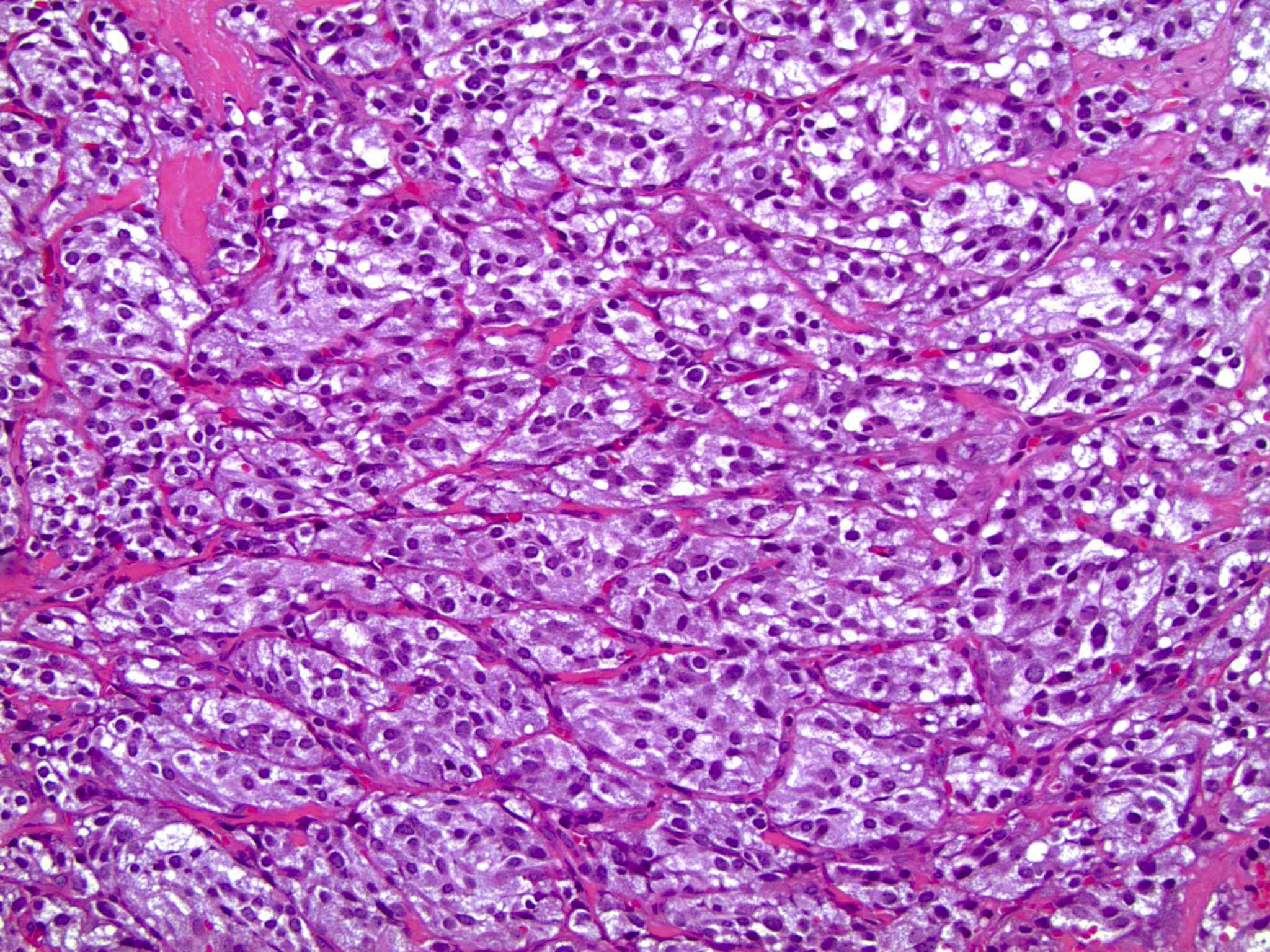
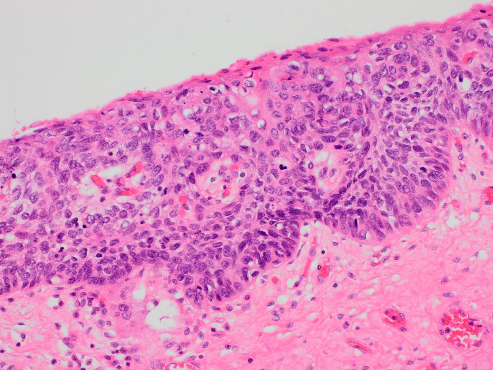
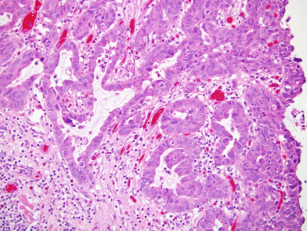
Malignant glandular epithelium
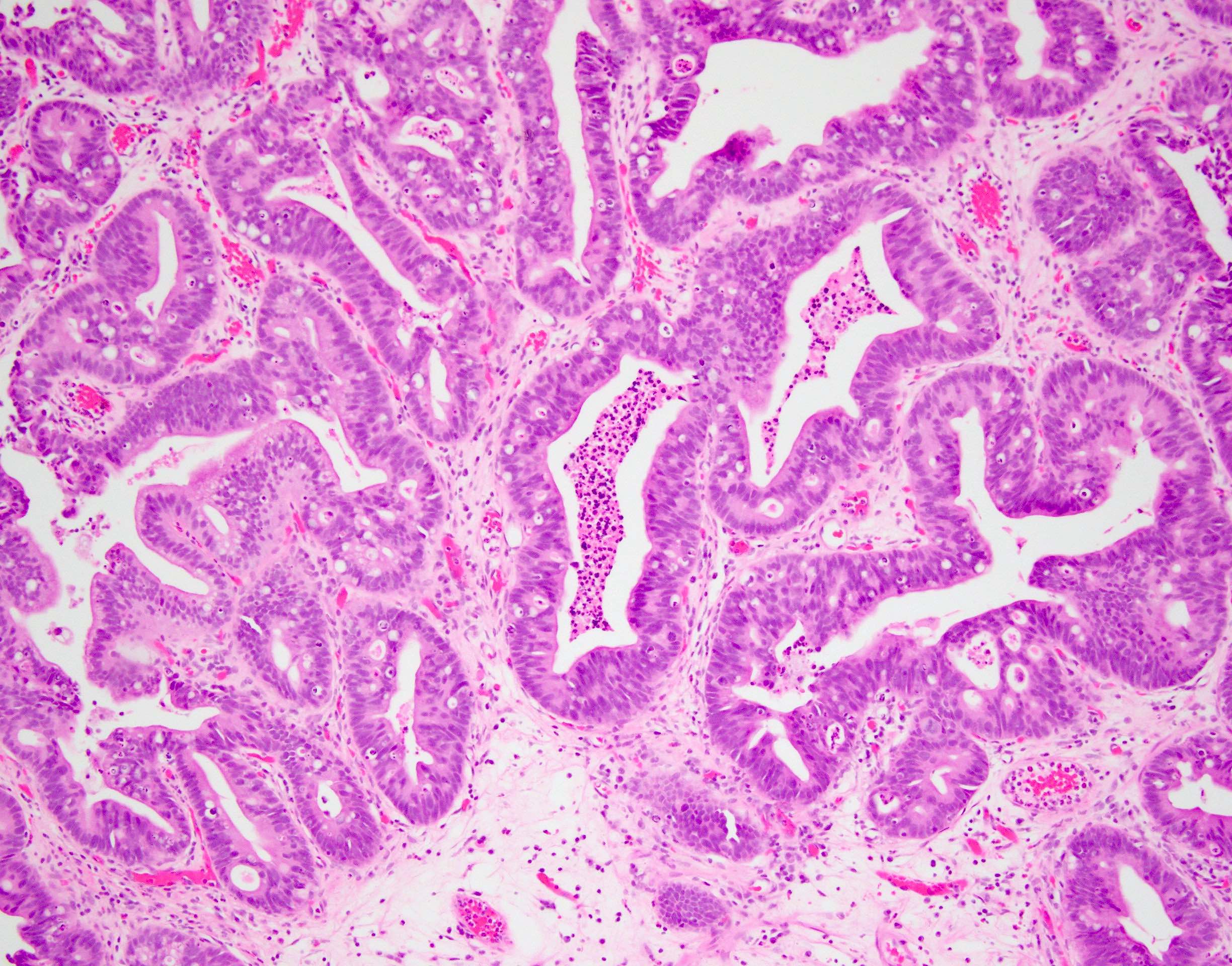

Intestinal / enteric type
Images hosted on other servers:

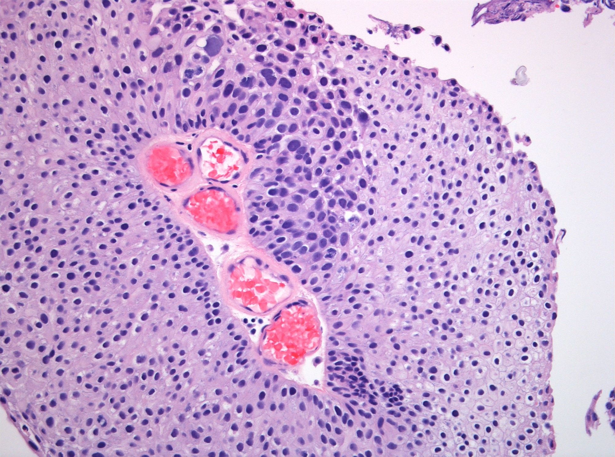
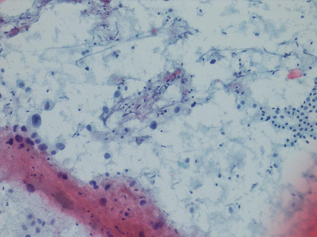
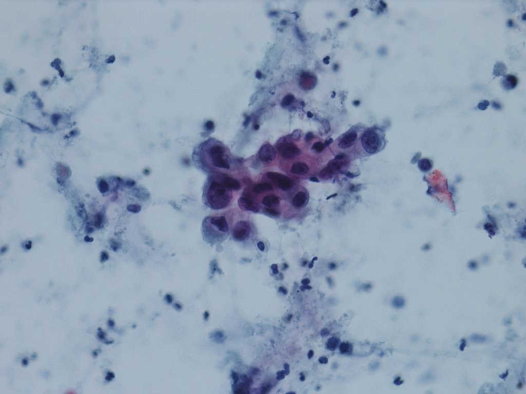
Vacuolated cells

Penile metastasis
Images hosted on other servers:


Abnormal FISH
Images hosted on other servers:

Development of bladder

Position of bladder in female pelvis

Interior


Position of bladder in male pelvis
Contributed by @Andrew_Fltv on Twitter
 Contributed by @Andrew_Fltv on Twitter (see original post here)">
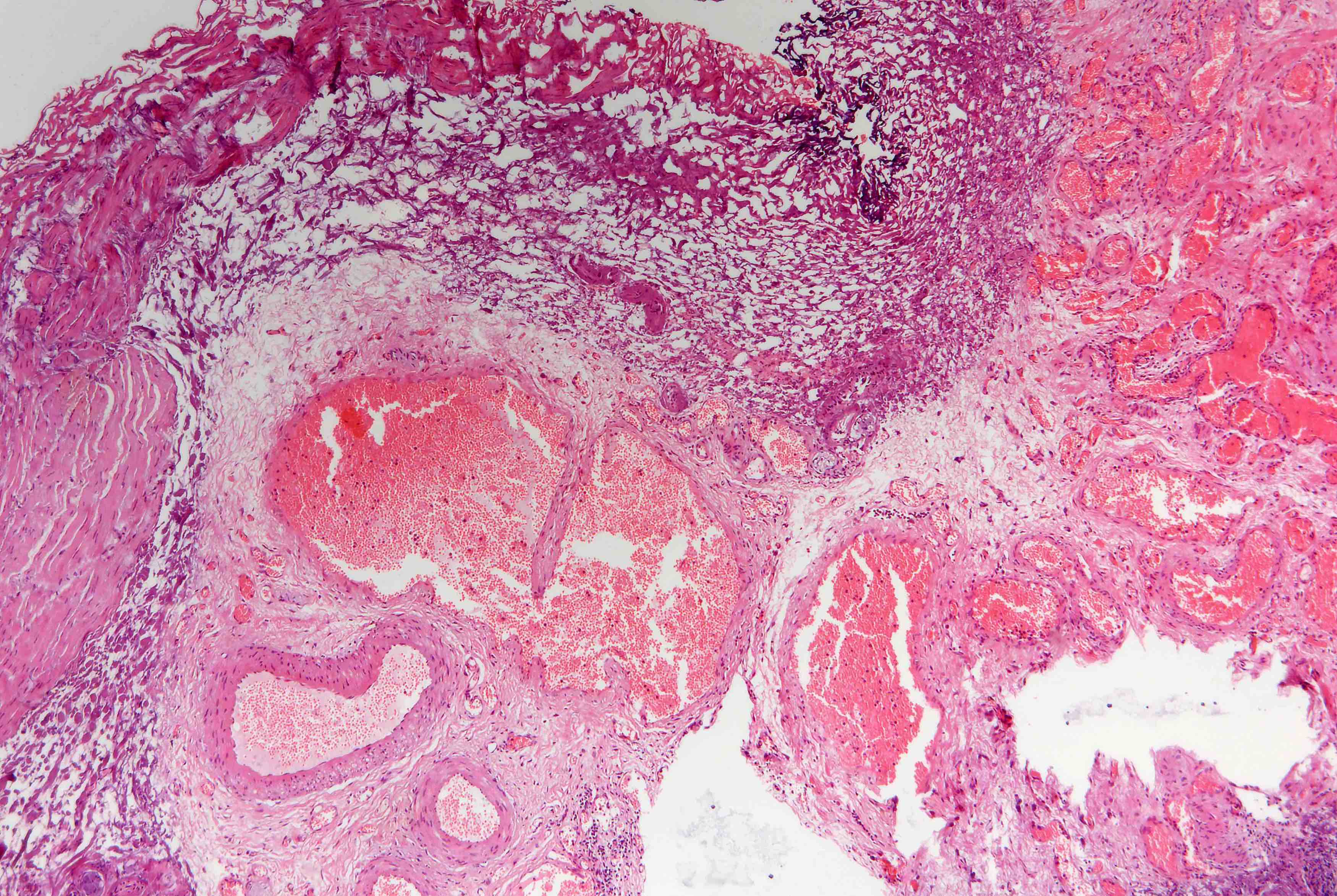
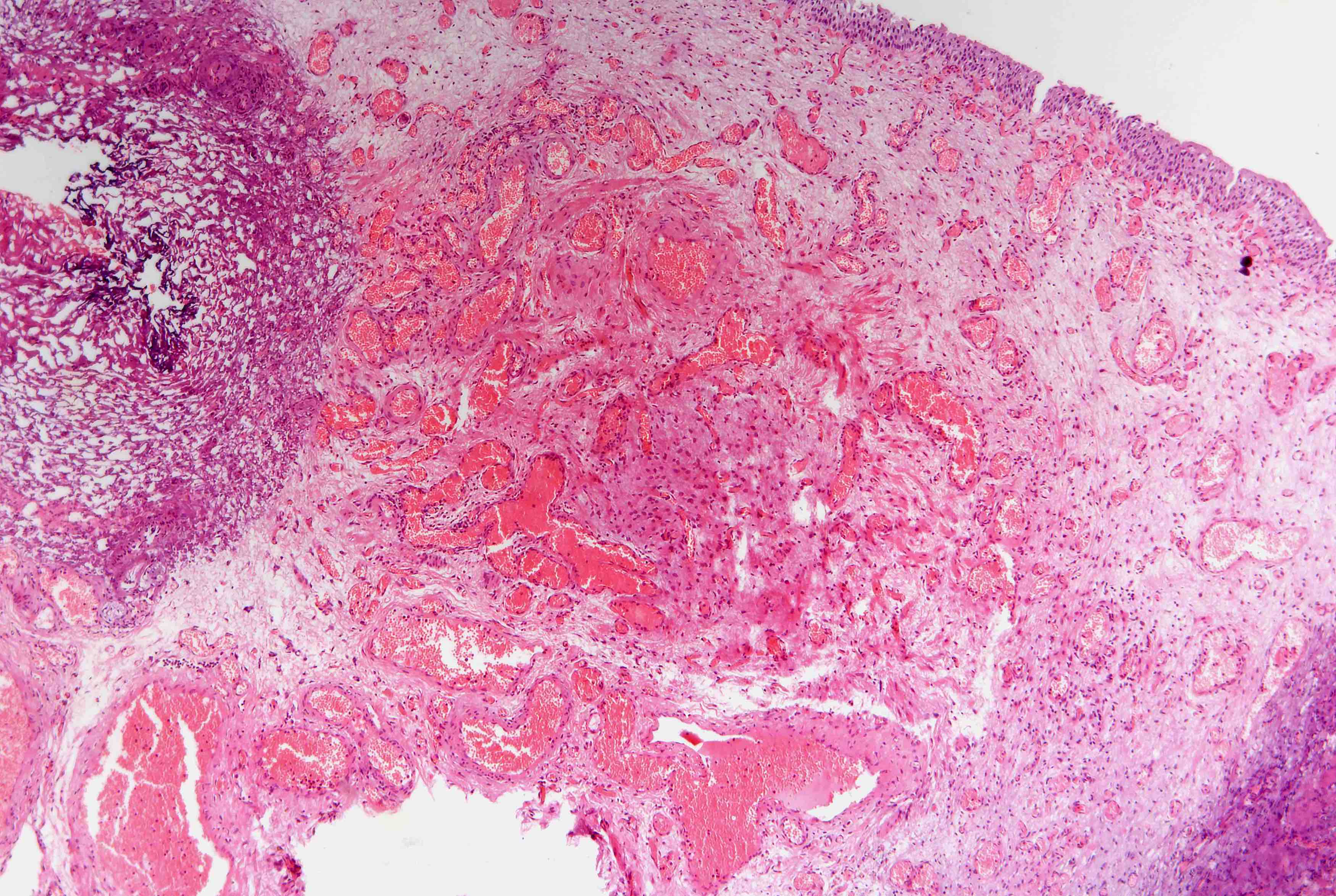
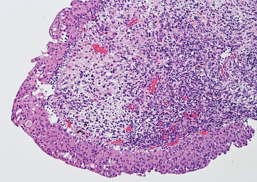
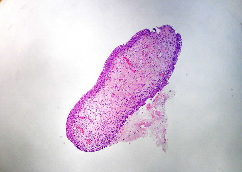
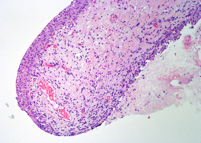
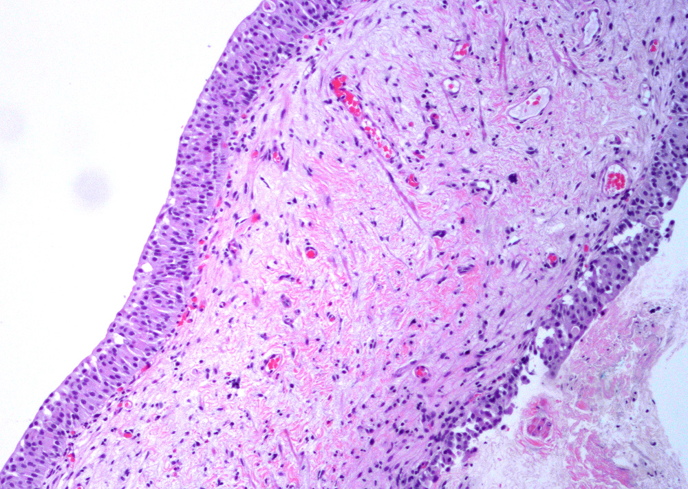
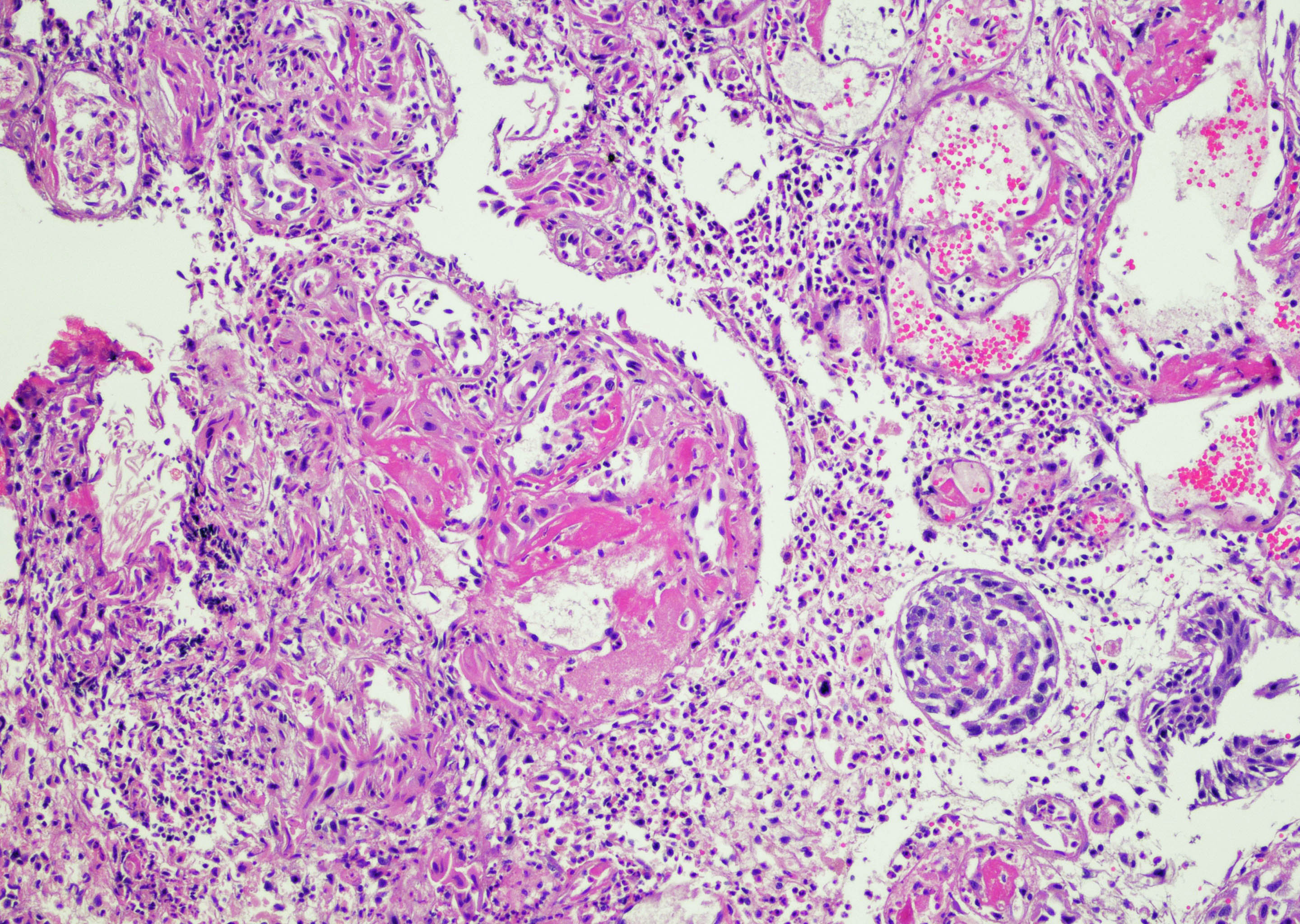
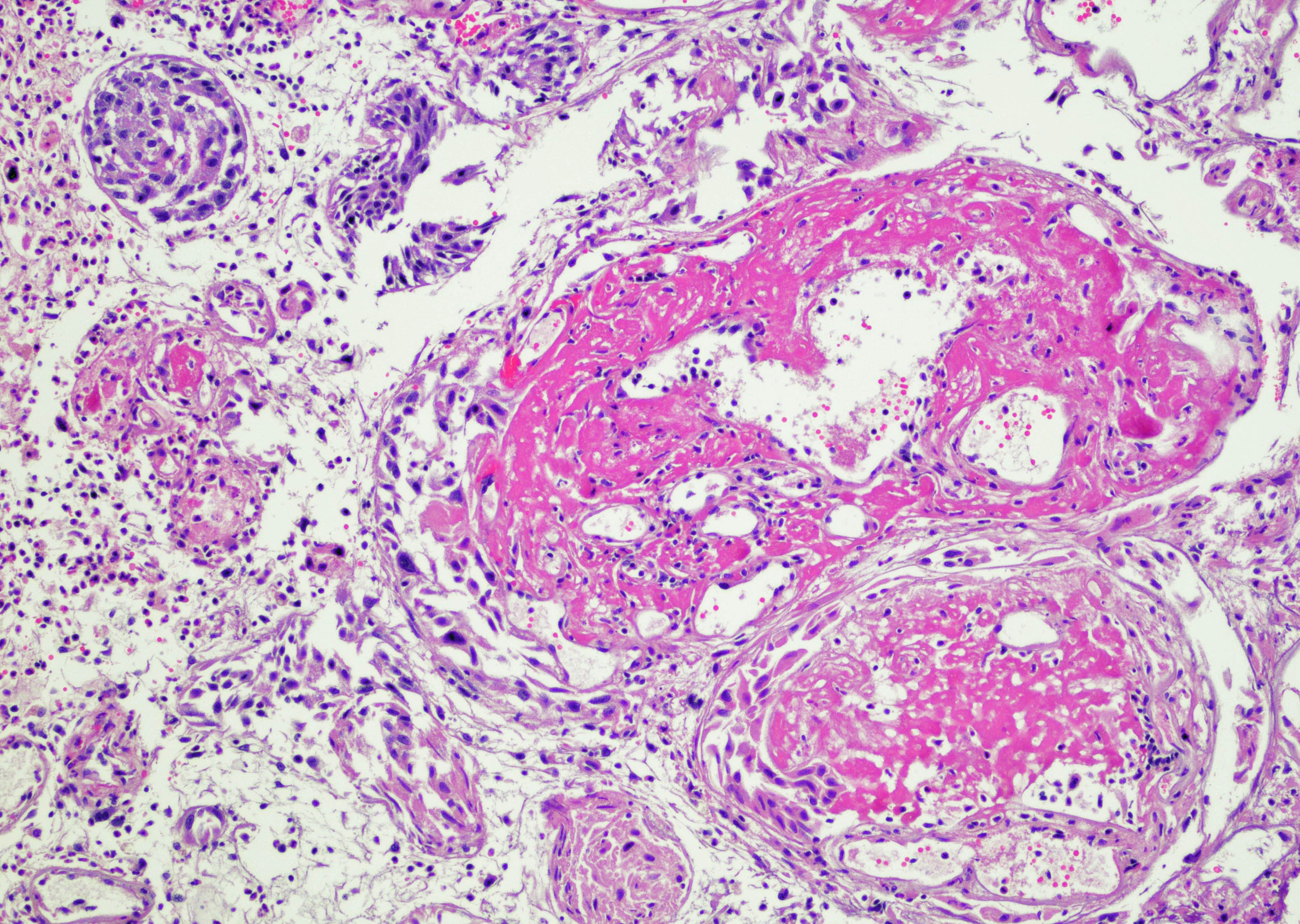
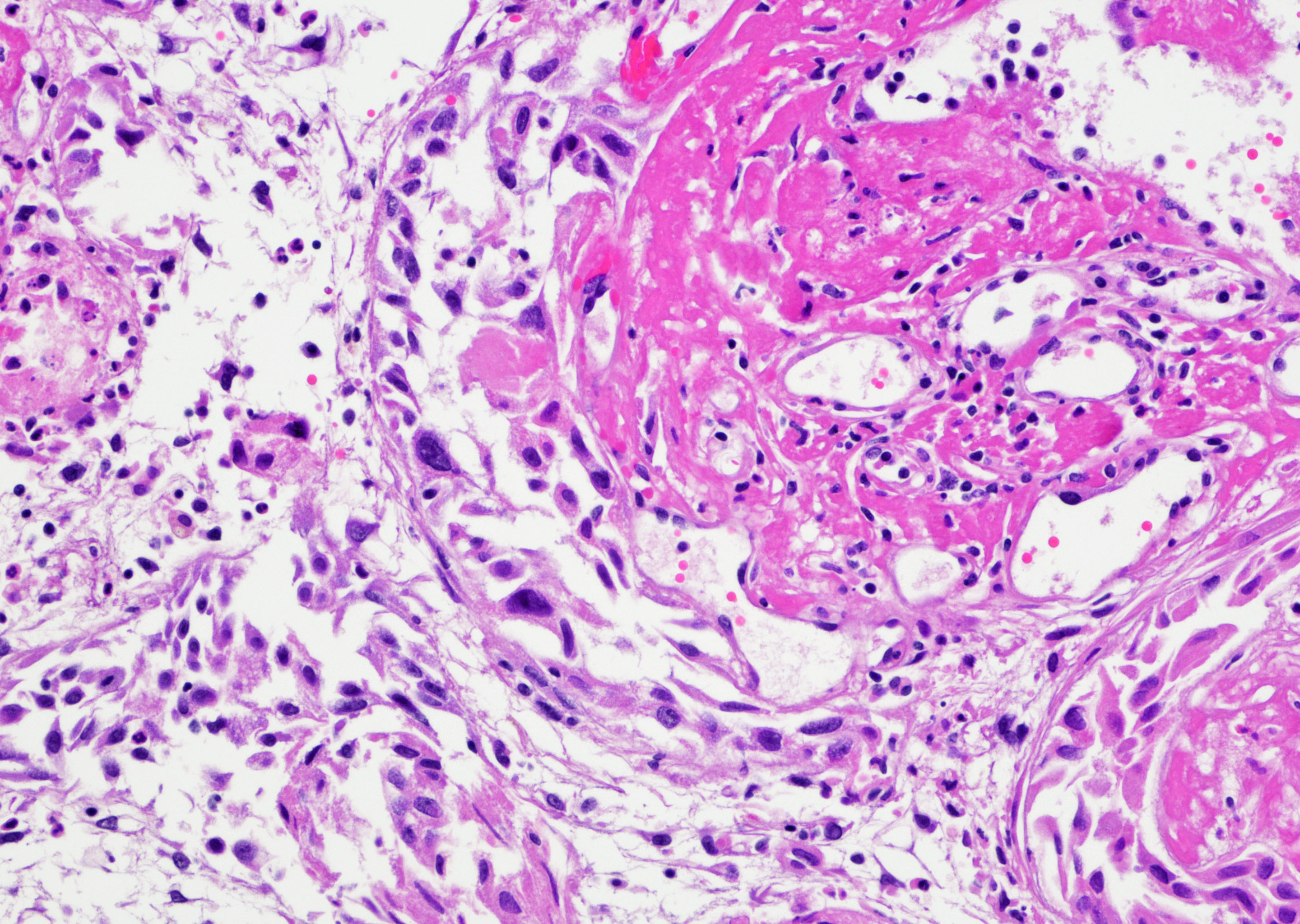
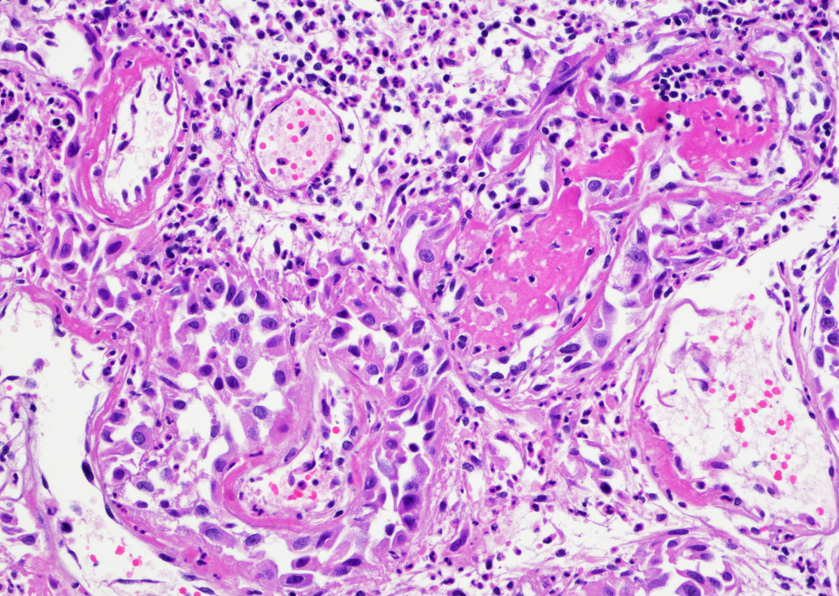
Arteriovenous malformation
Images hosted on other servers:

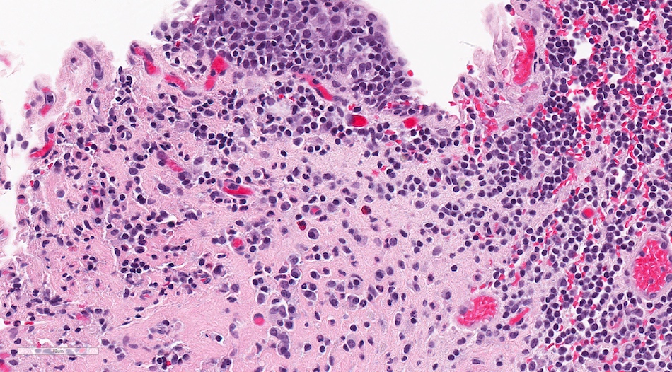
Many eosinophils
Contributed by Sean R. Williamson, M.D. and Bhavesh Papadi, M.D. (Case #331)
Ureter CIS
Denuded urothelium


Various images
Additional images
Contributed by @AnaPath10 on Twitter
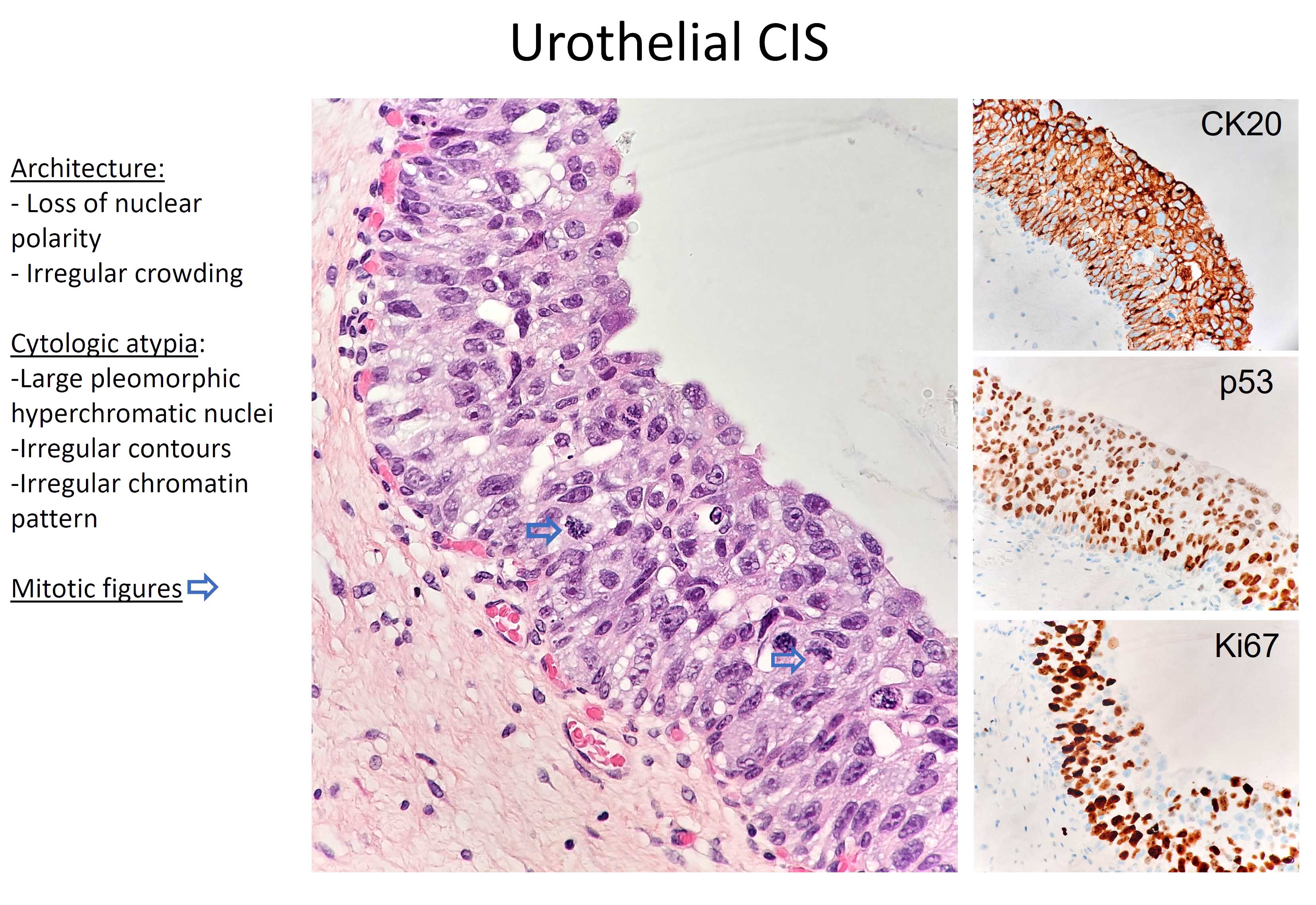
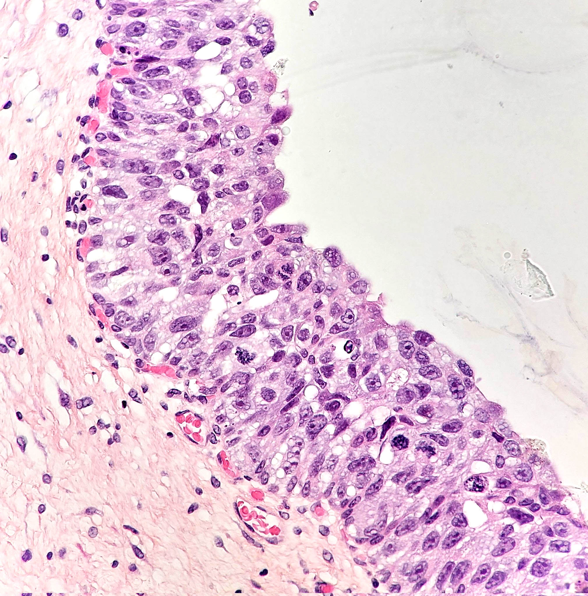
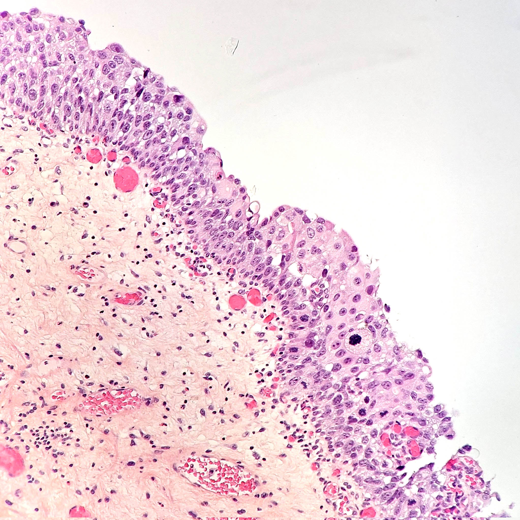
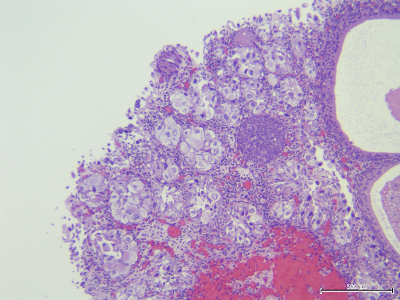
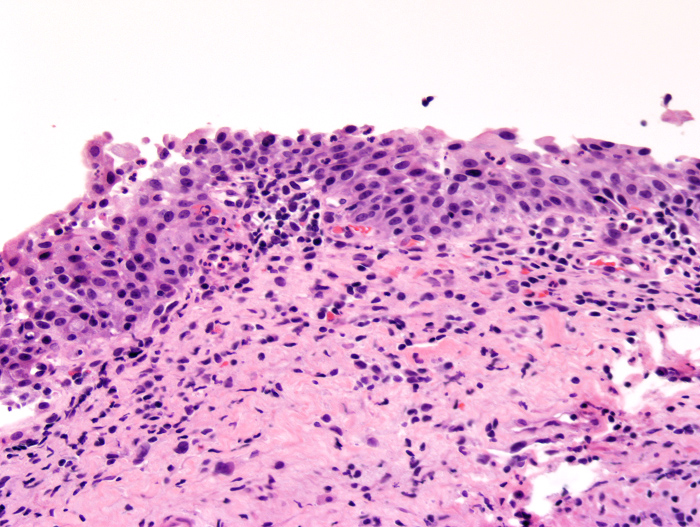
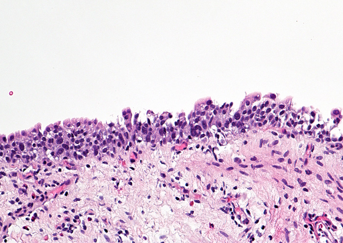
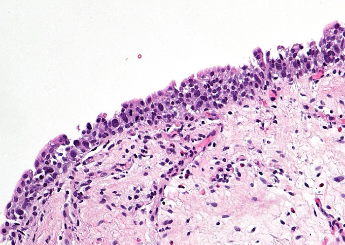
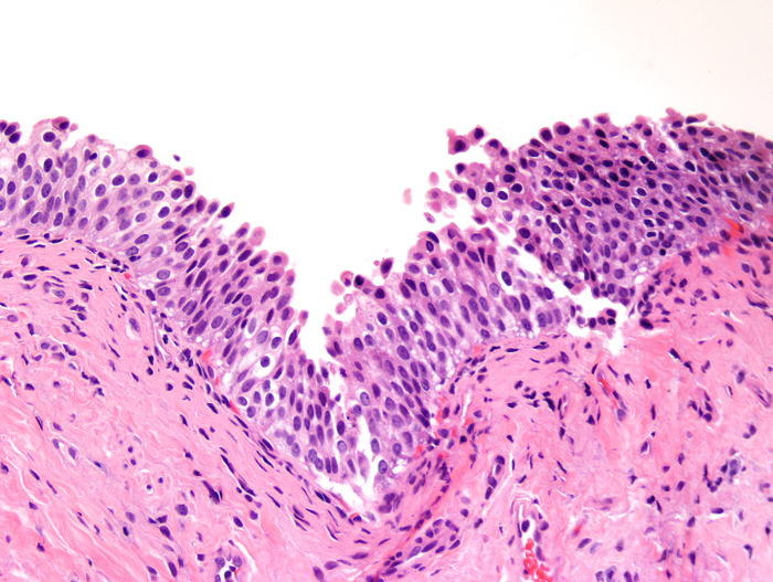
Bladder carcinoma in situ
Images hosted on other servers:

H&E

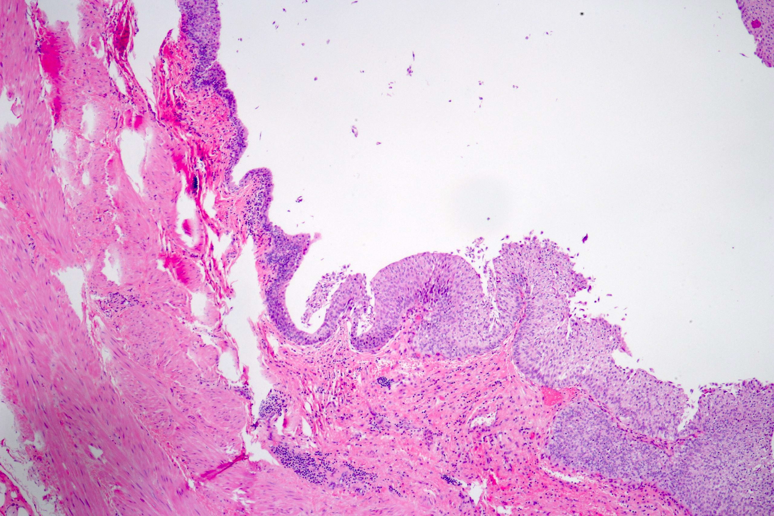
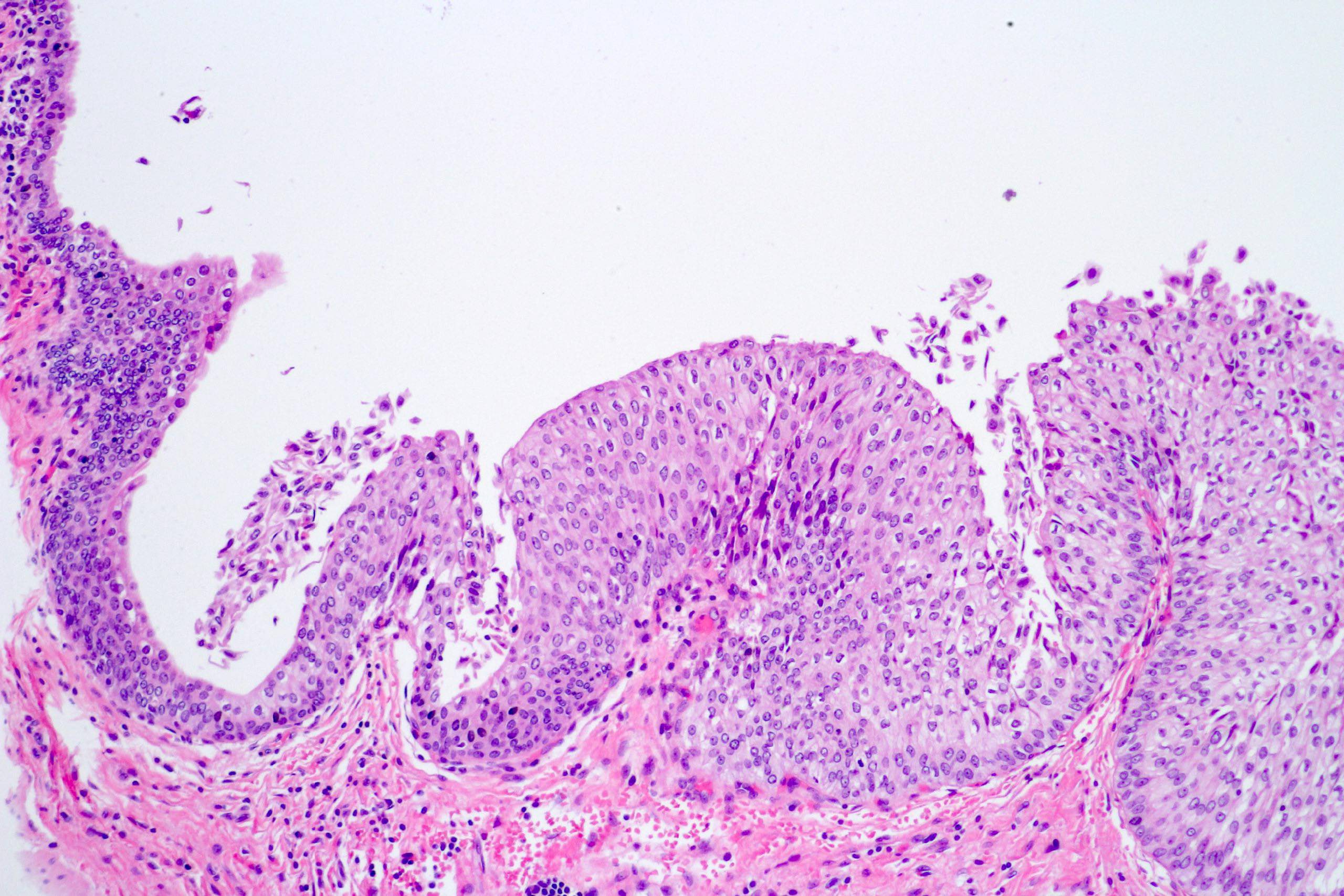
Loss of polarization

Broadened urothelium

Narrowed urothelium

Pagetoid pattern
Histopathology Bladder Transitional Carcinoma in situ
Images hosted on other servers:

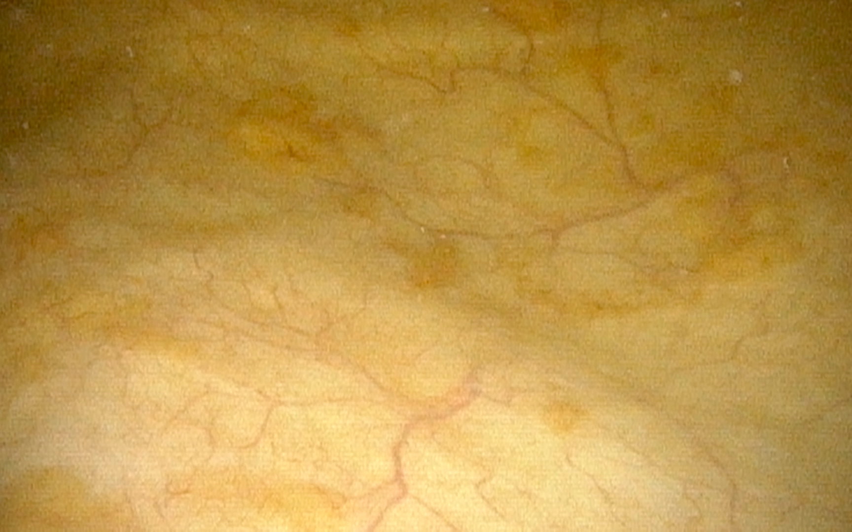
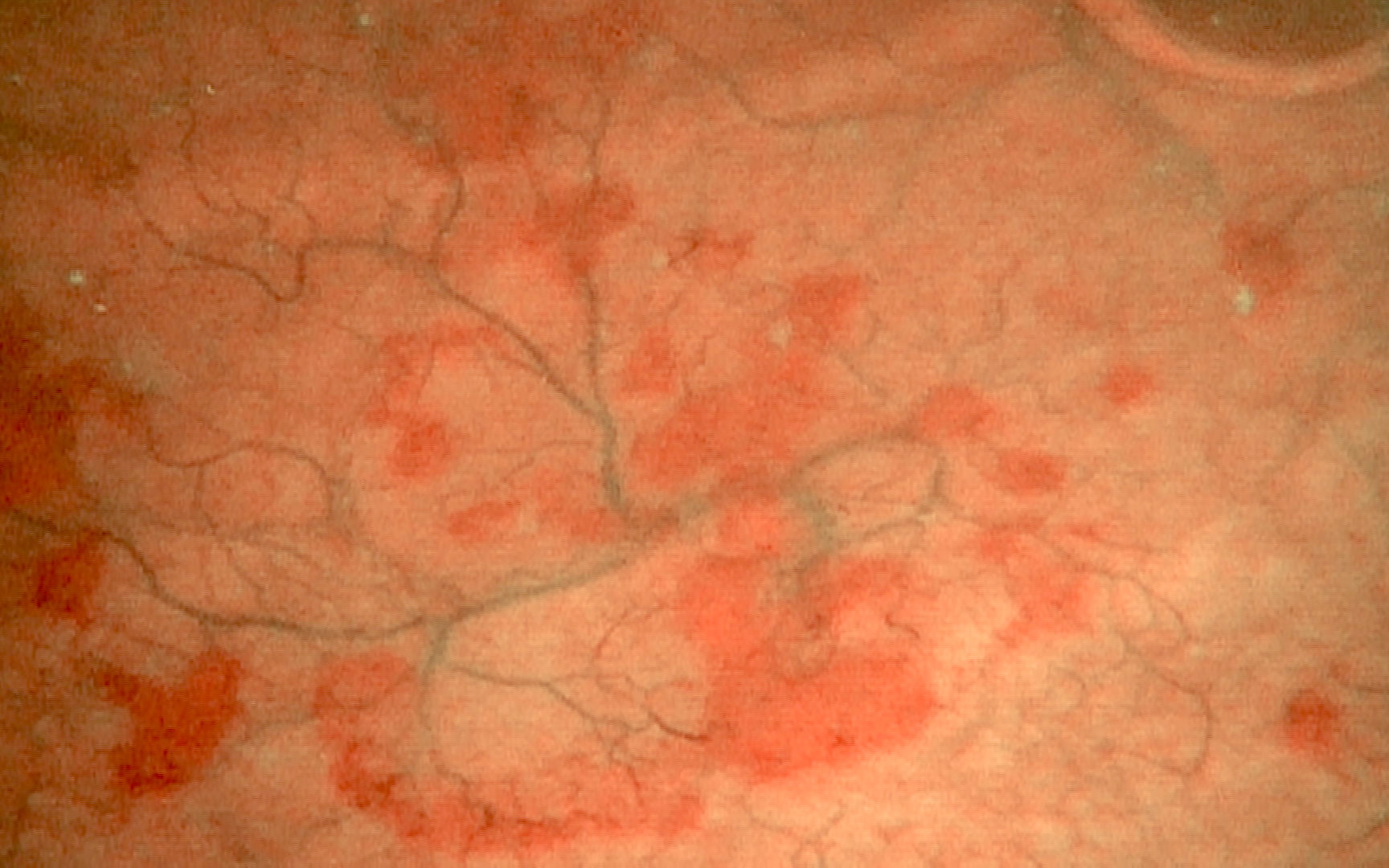
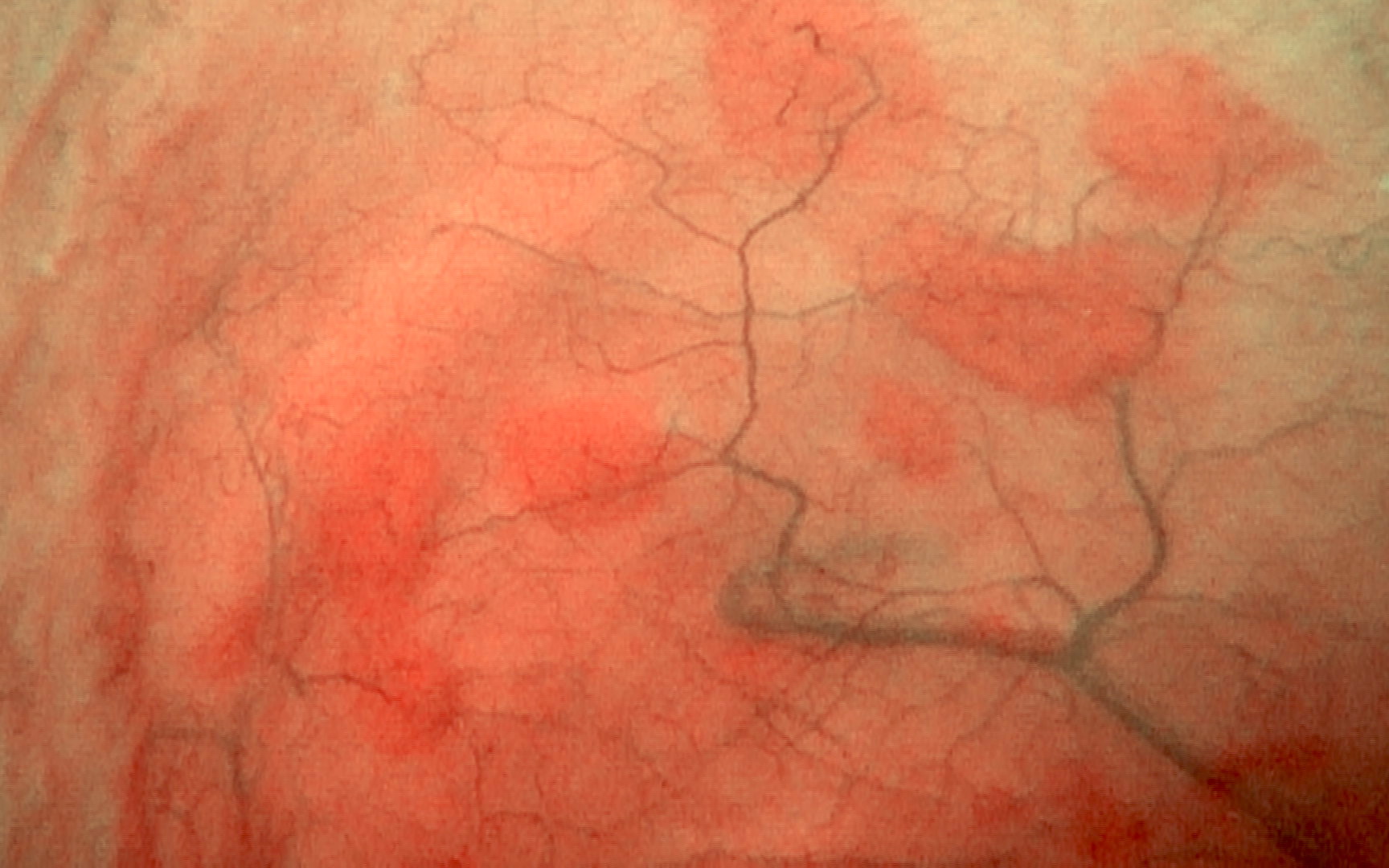
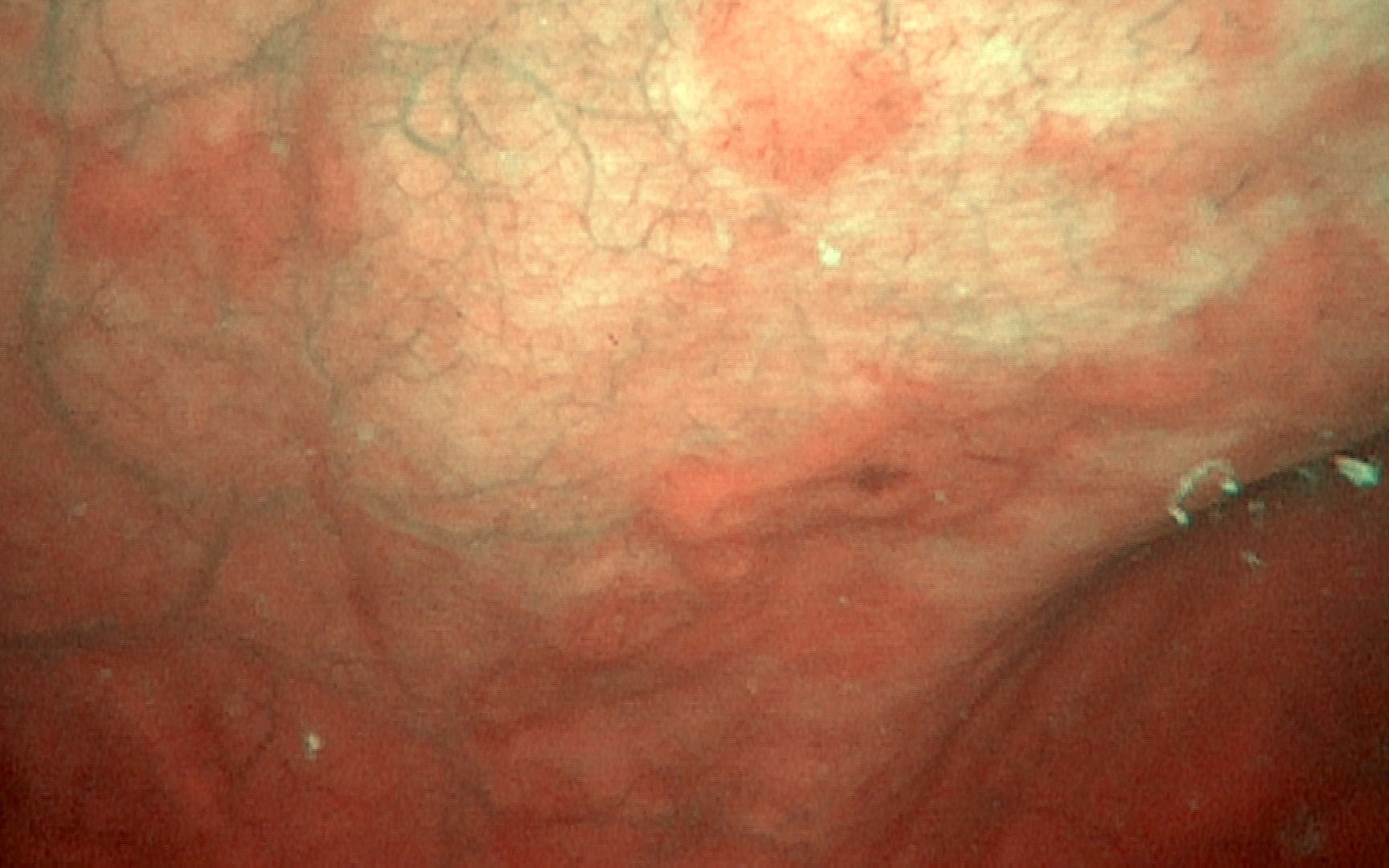
Cystoscopy
Contributed by Jonathan Epstein, M.D.
Prostatic urethra papillary lesion
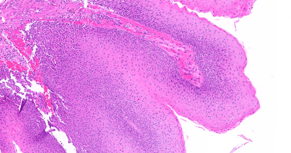
Urinary bladder hyperplastic lesion
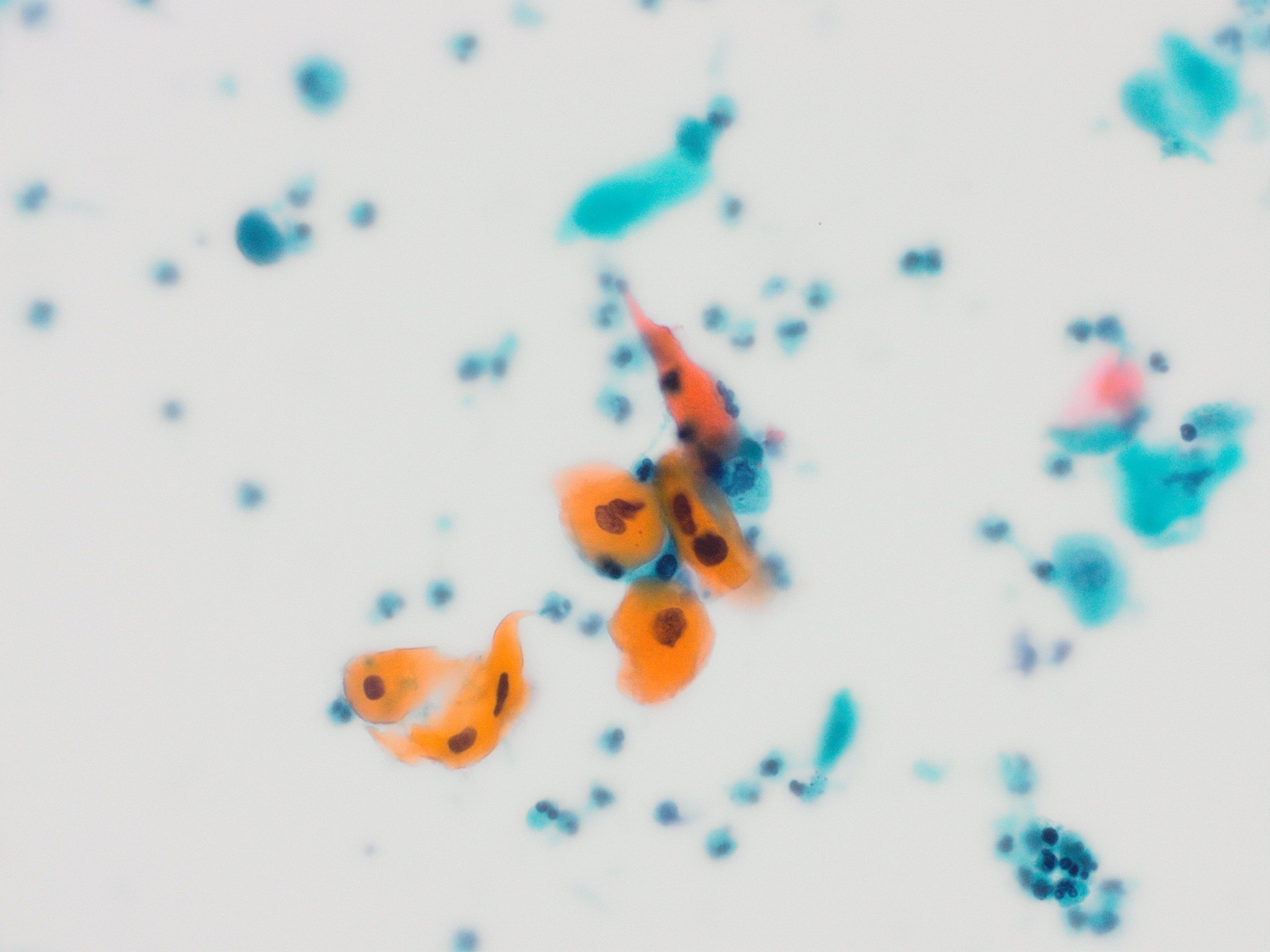
Urinary bladder koilocytosis
Images hosted on other servers:

T1 weighted MRI

Transabdominal ultrasound
Images hosted on other servers:

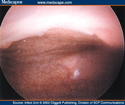
Cystoscopy
Contributed by Maria Tretiakova, M.D., Ph.D.
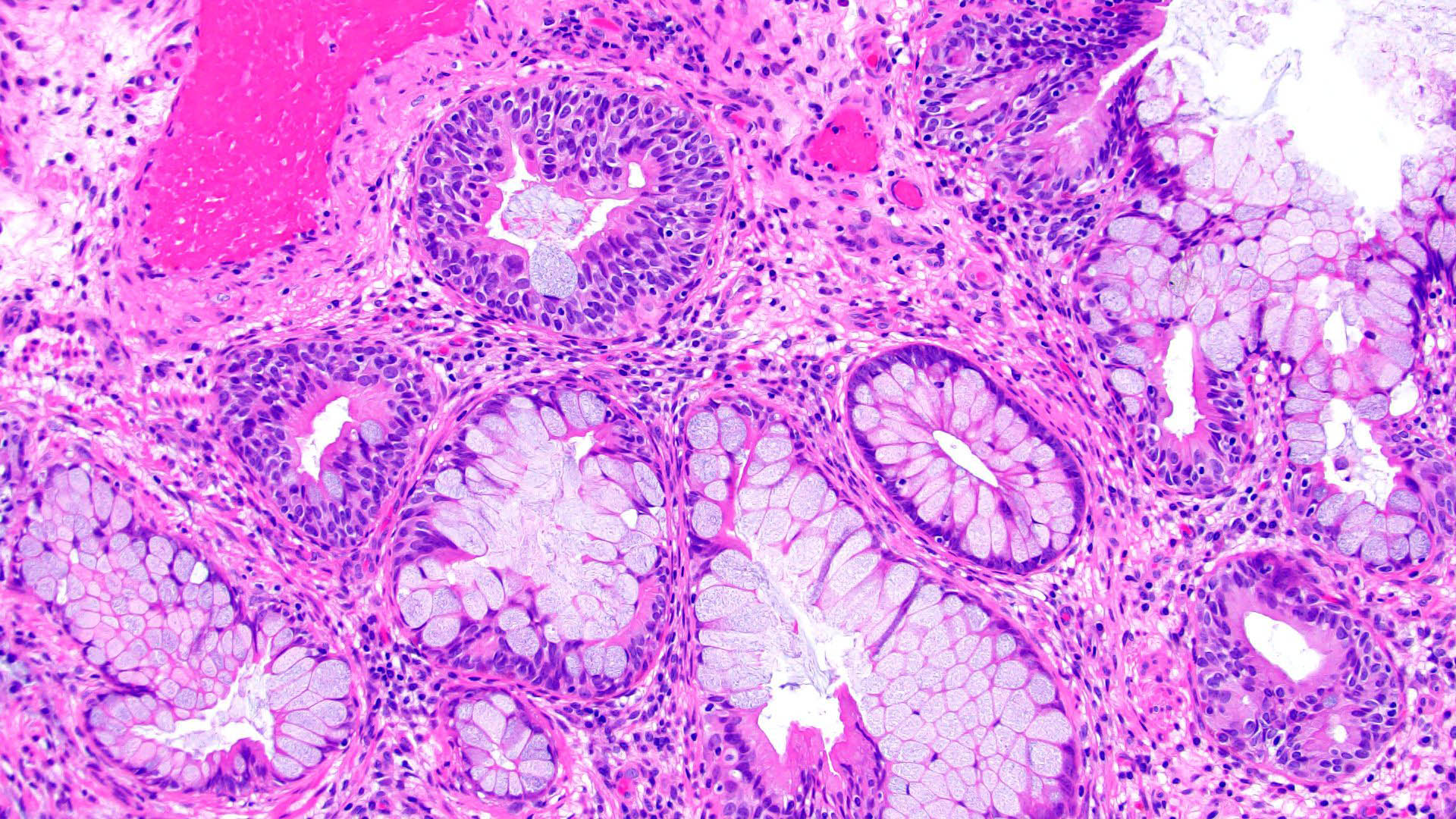
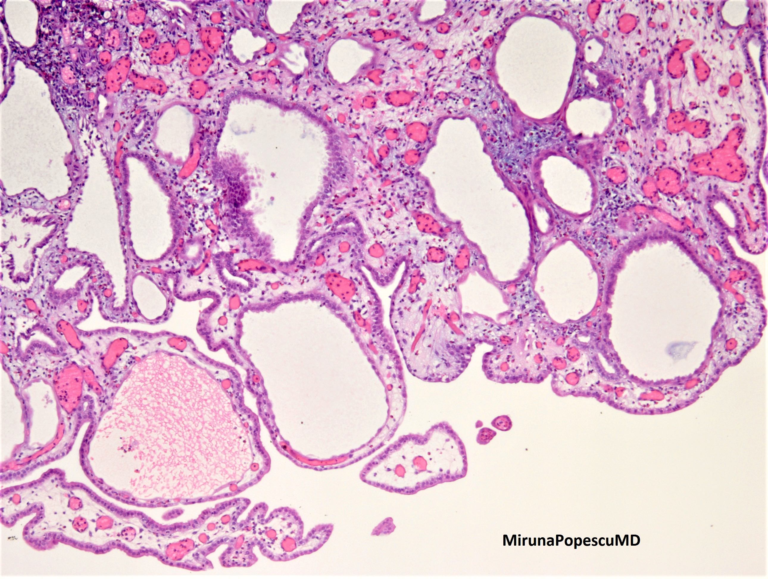
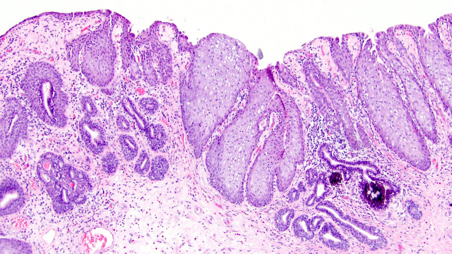
Cystitis cystica
Cystitis glandularis
Images hosted on other servers:

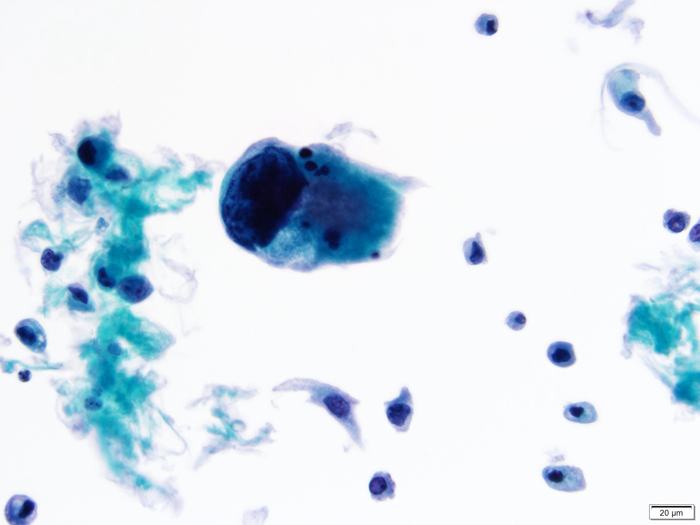
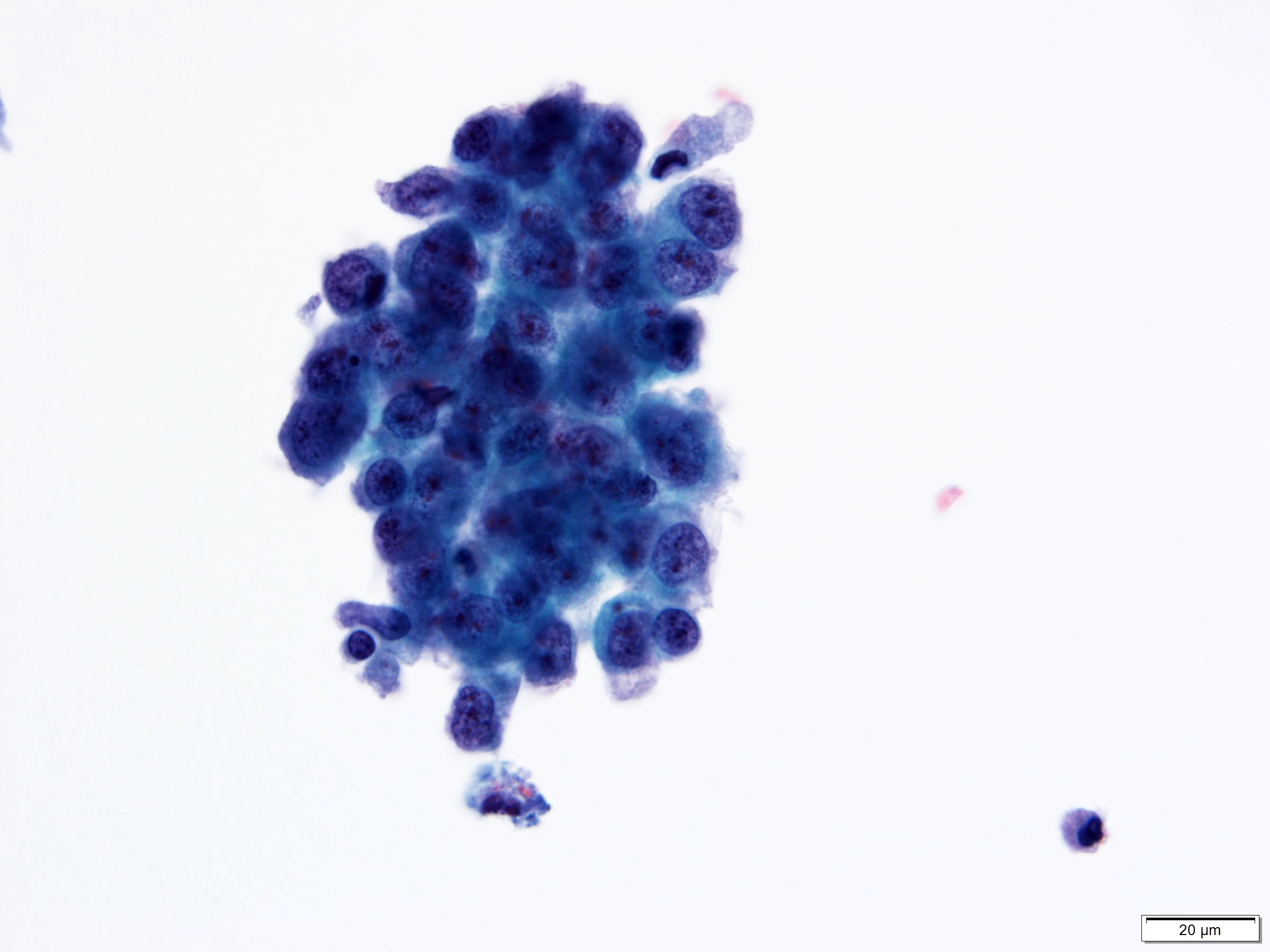
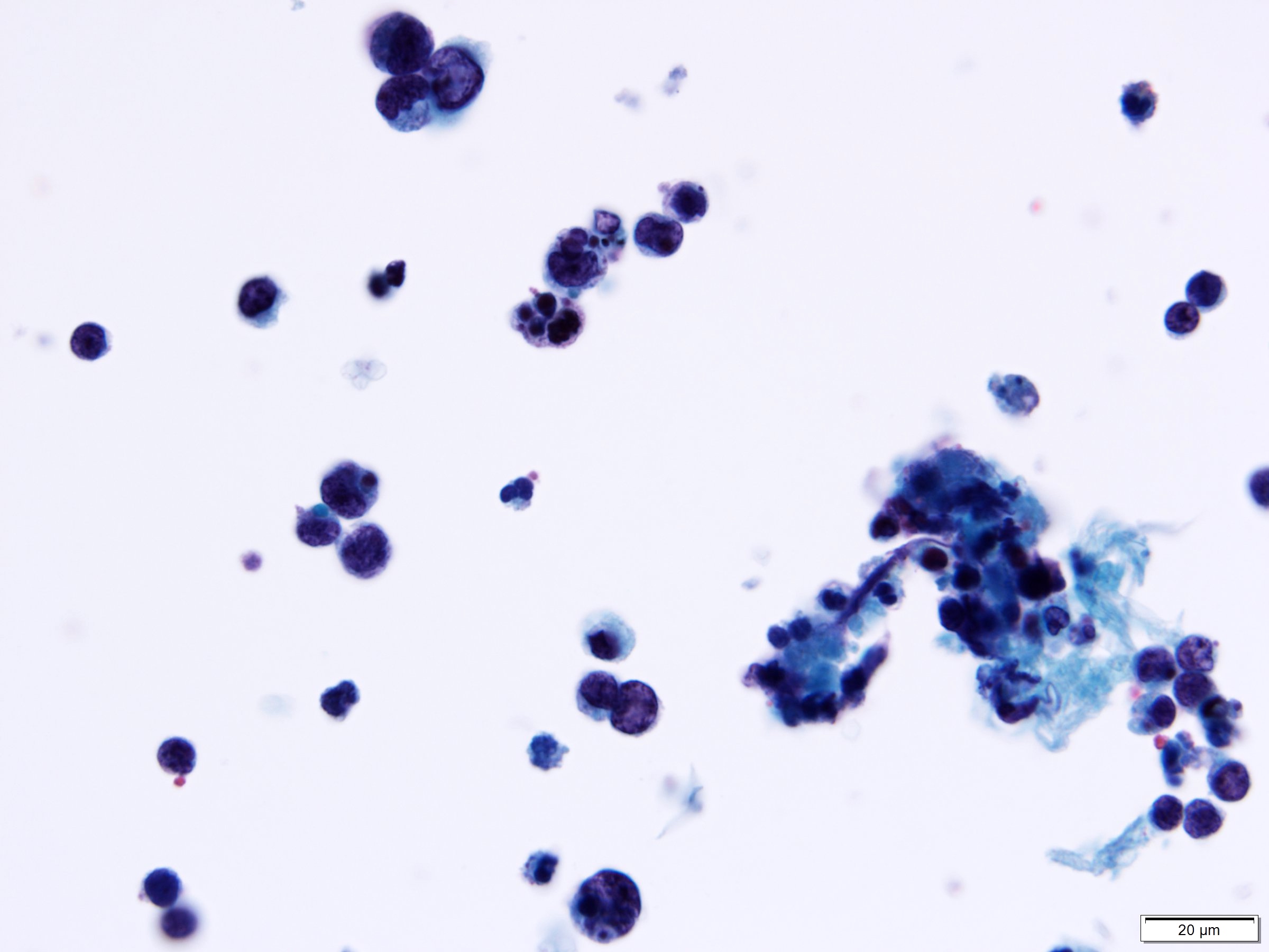
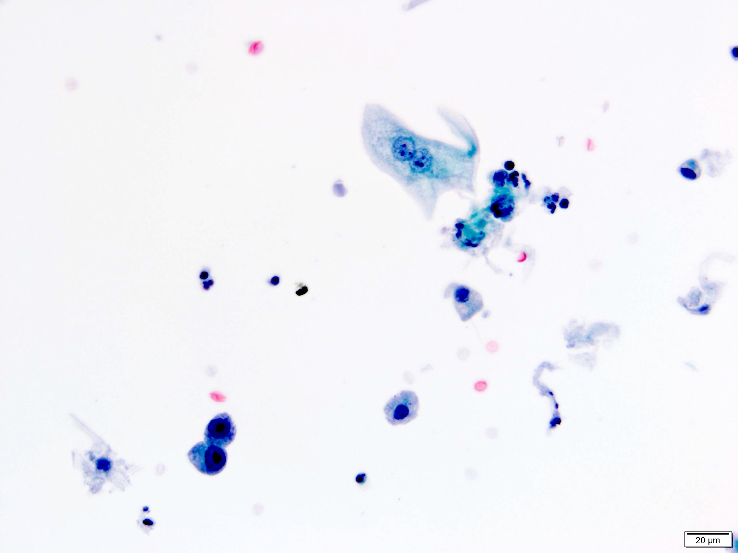
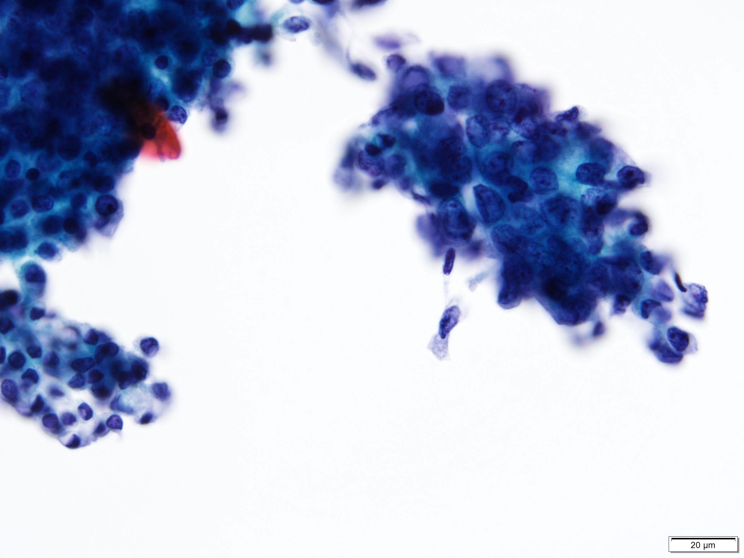
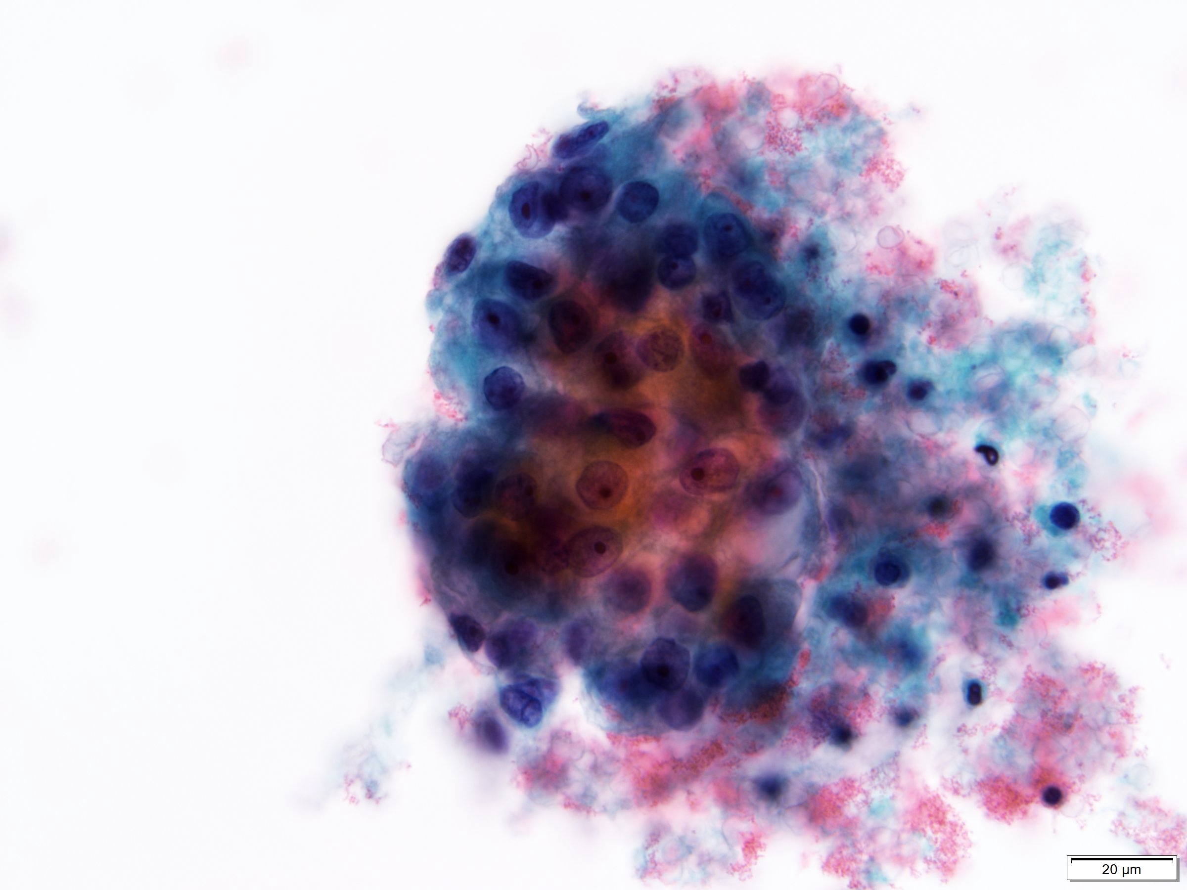
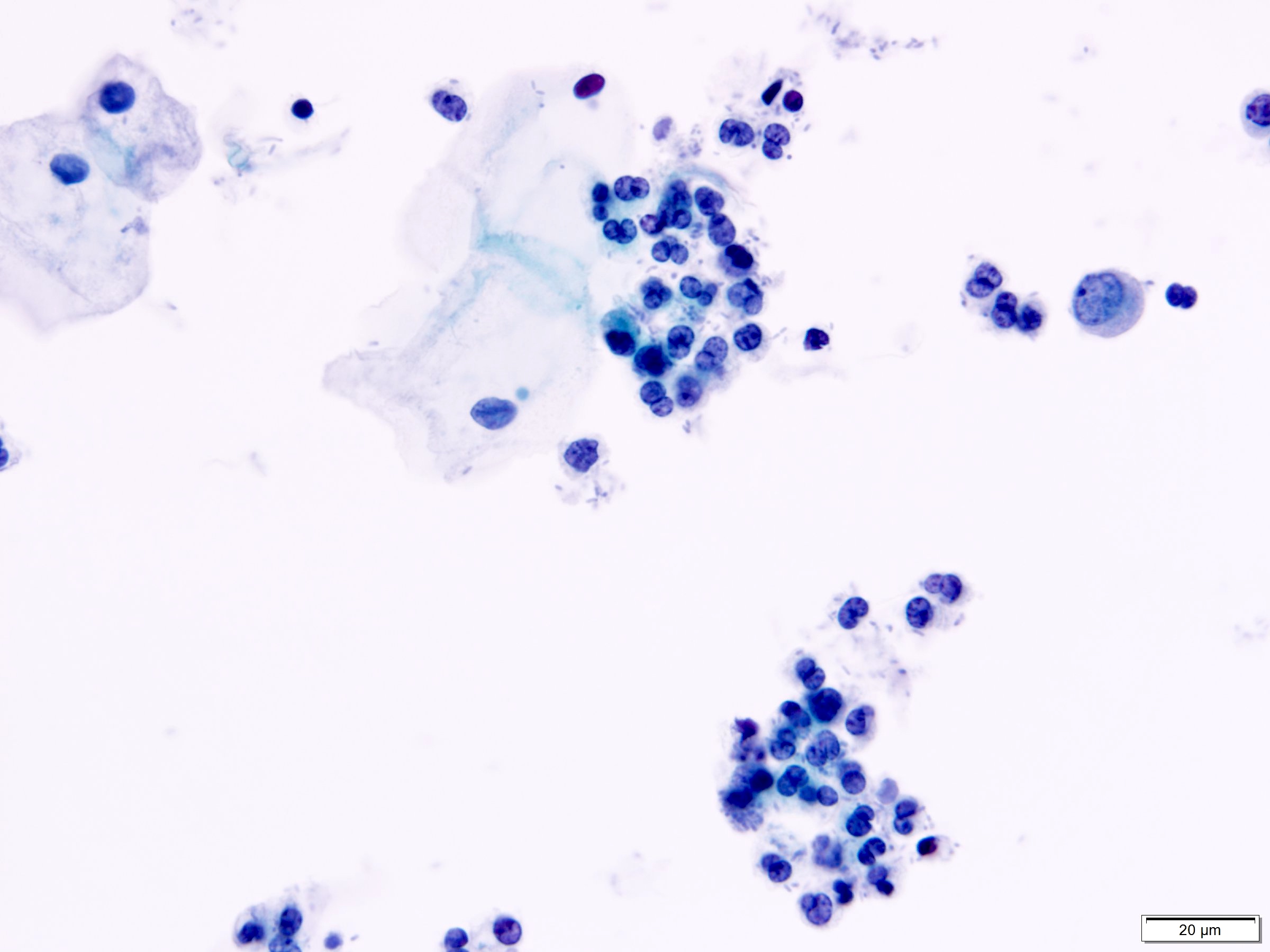
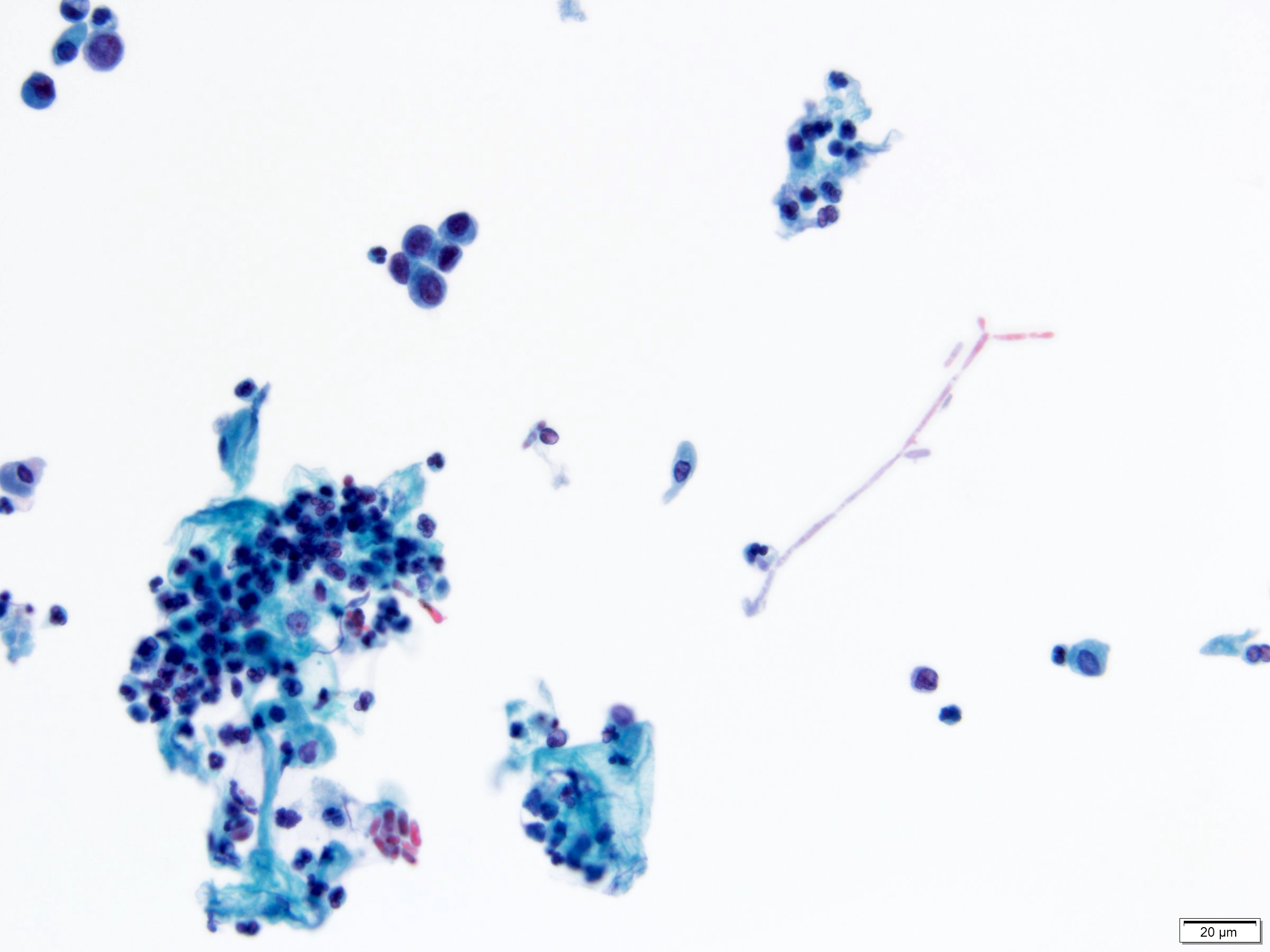
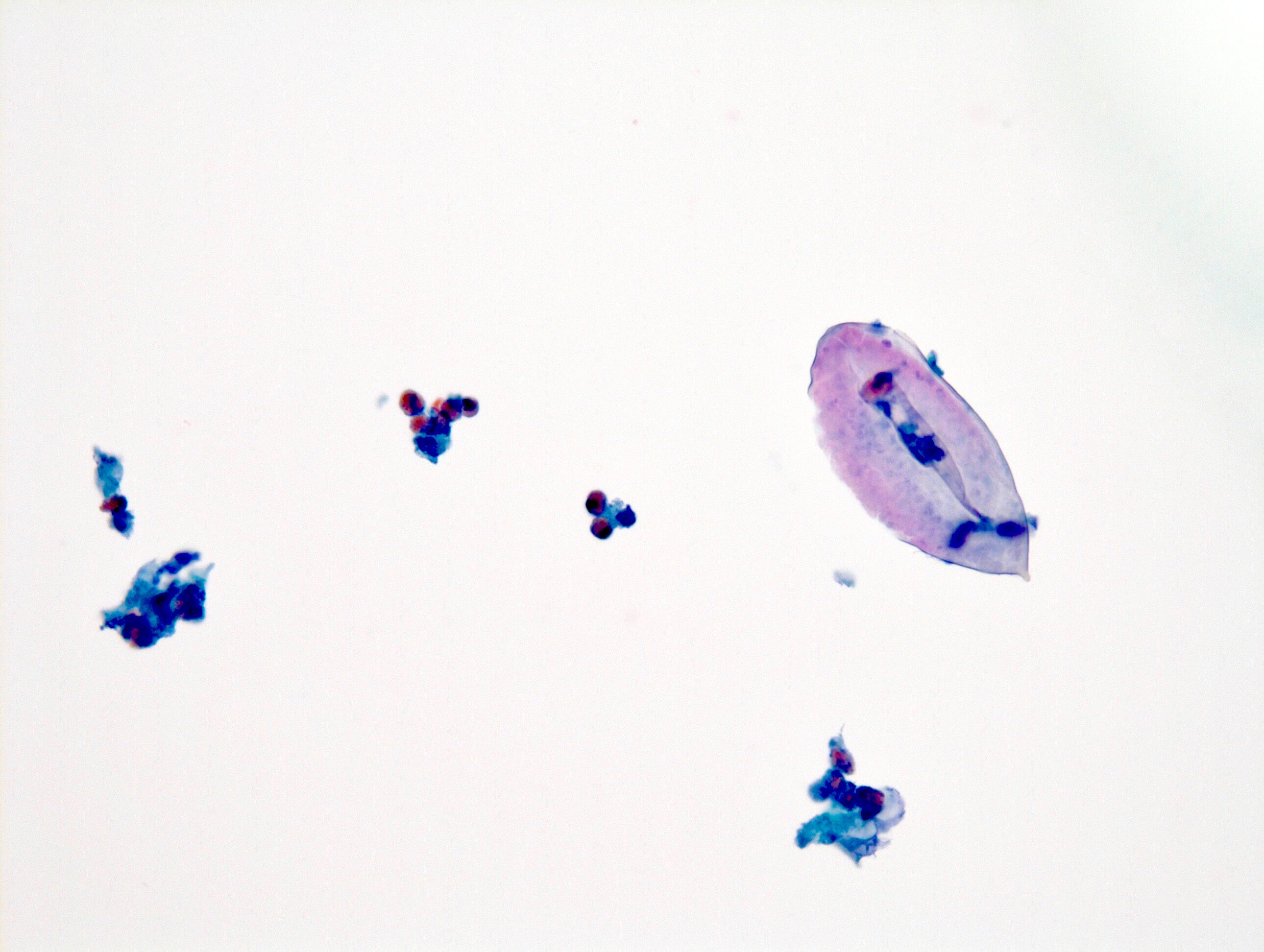
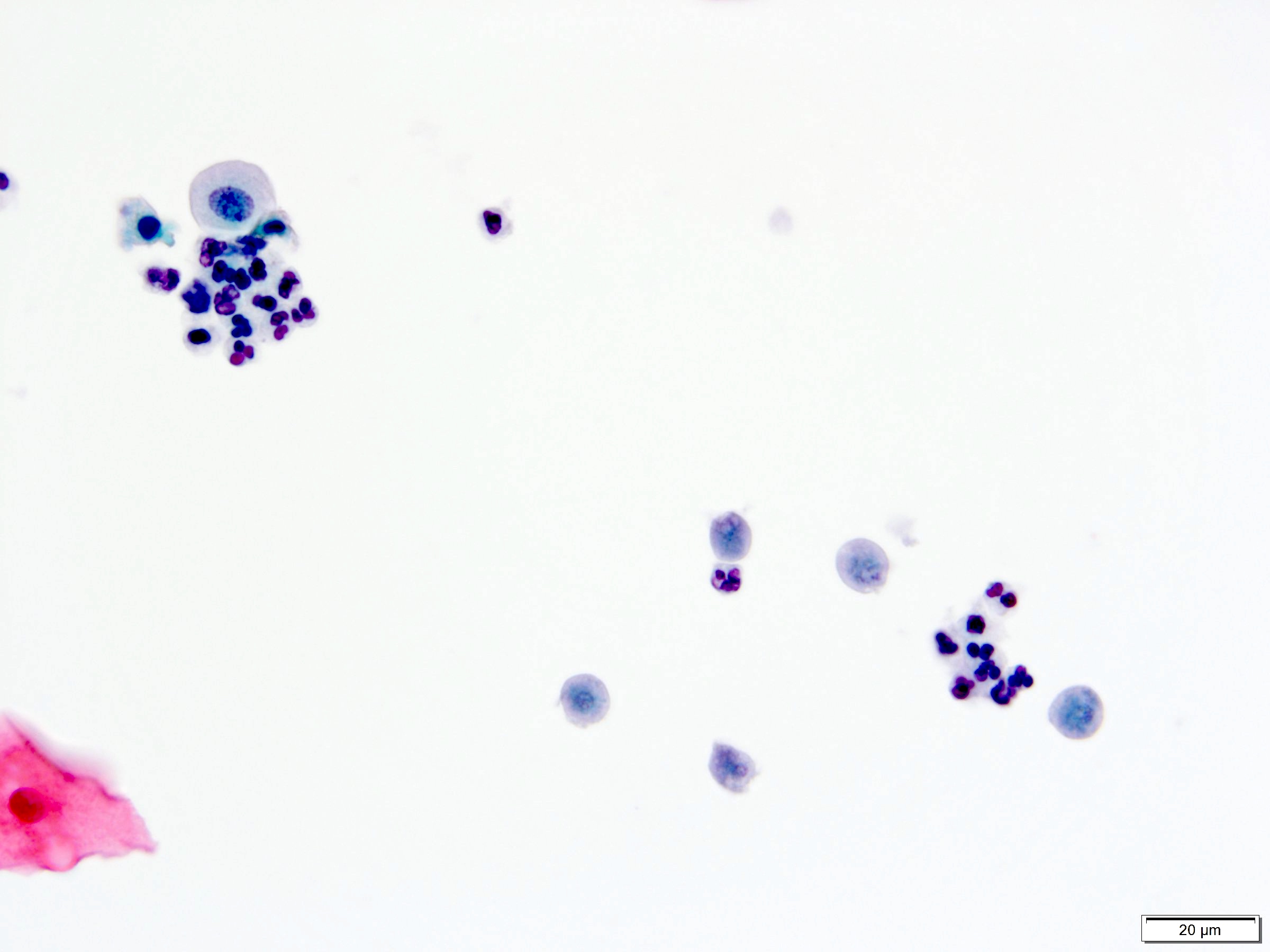
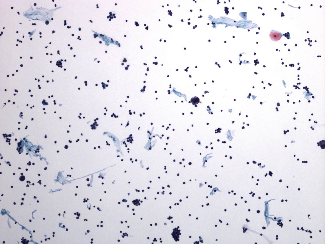
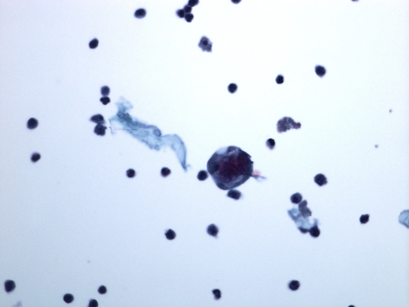
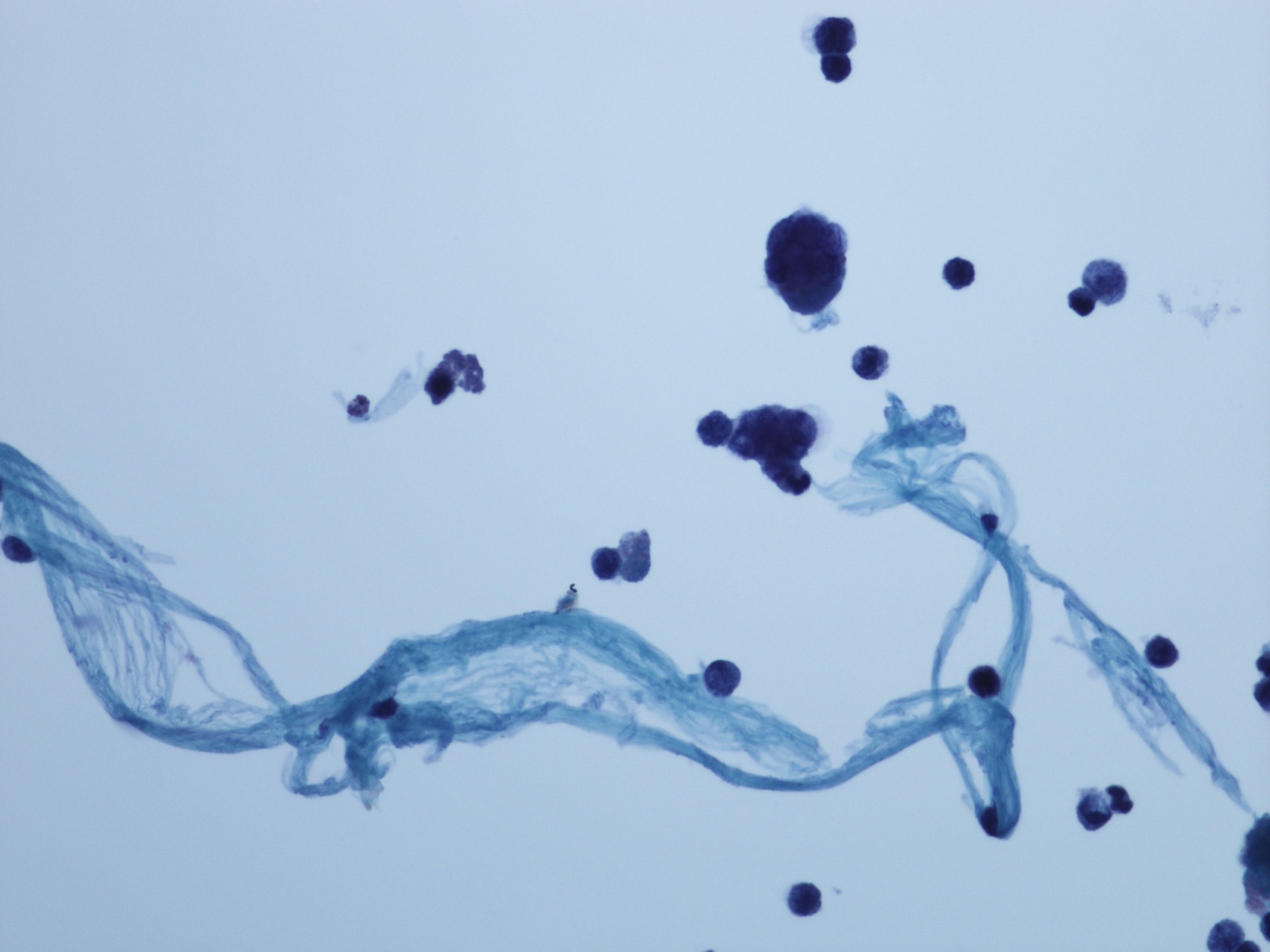
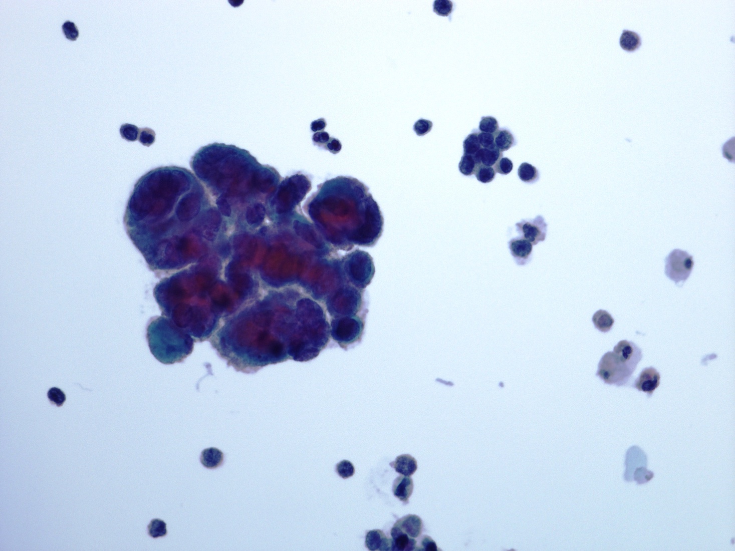
Urine cytology
Contributed by Bonnie Choy, M.D.
High grade urothelial carcinoma

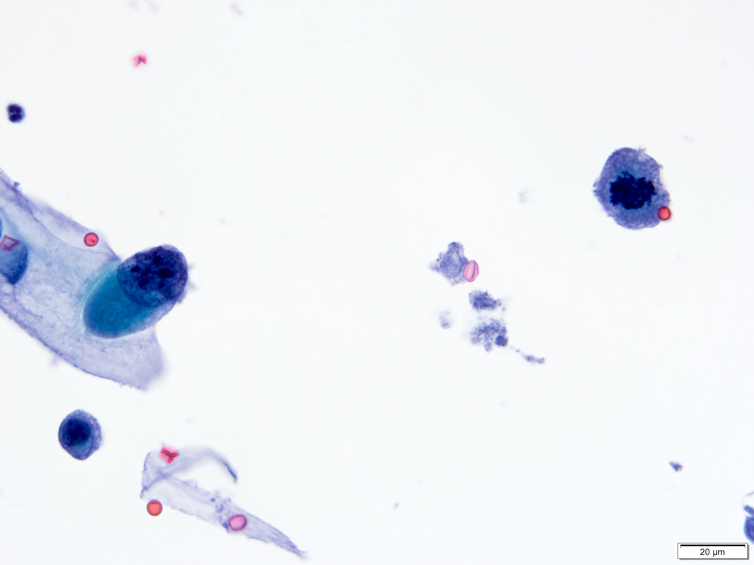
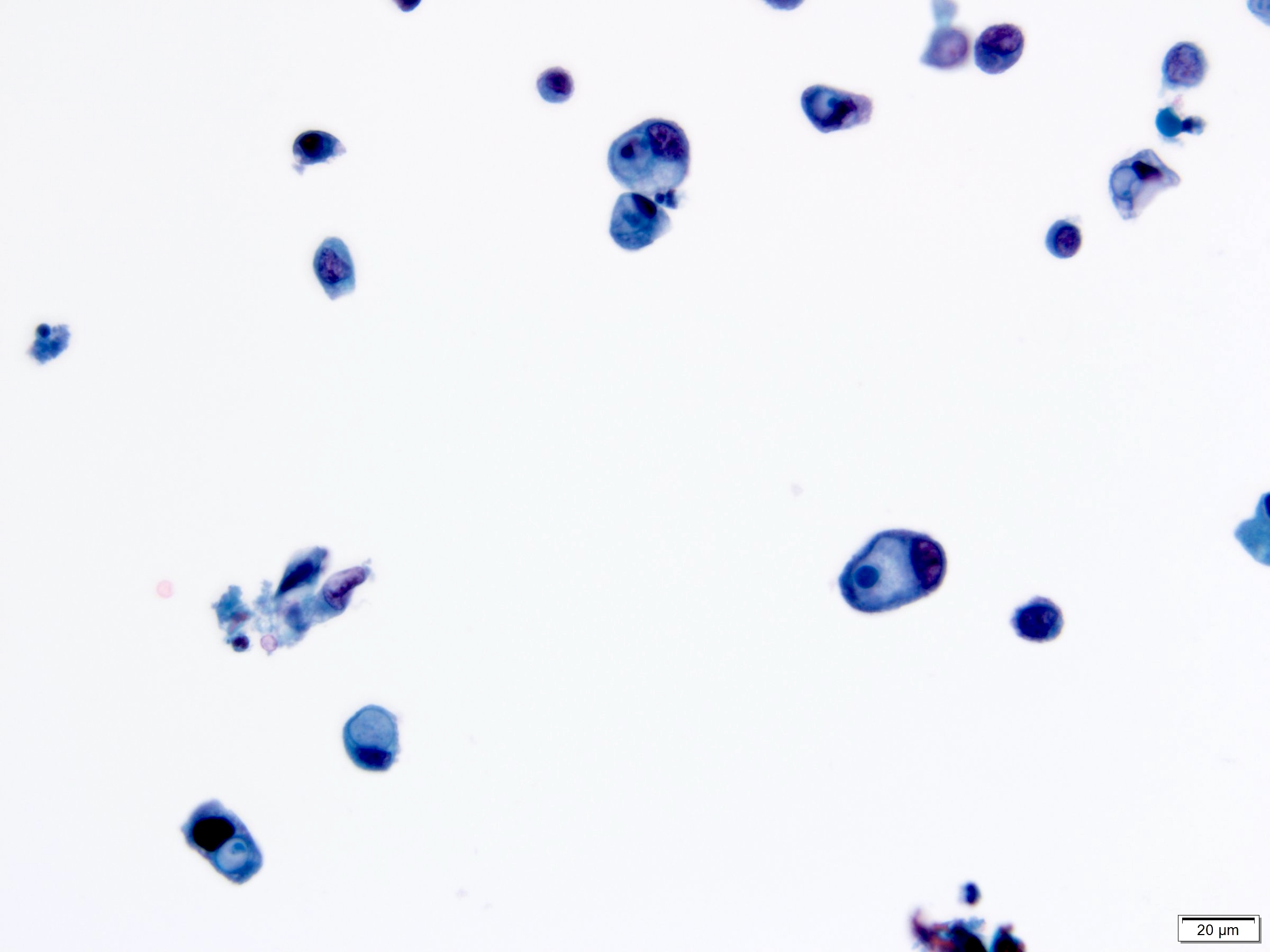
High grade urothelial carcinoma
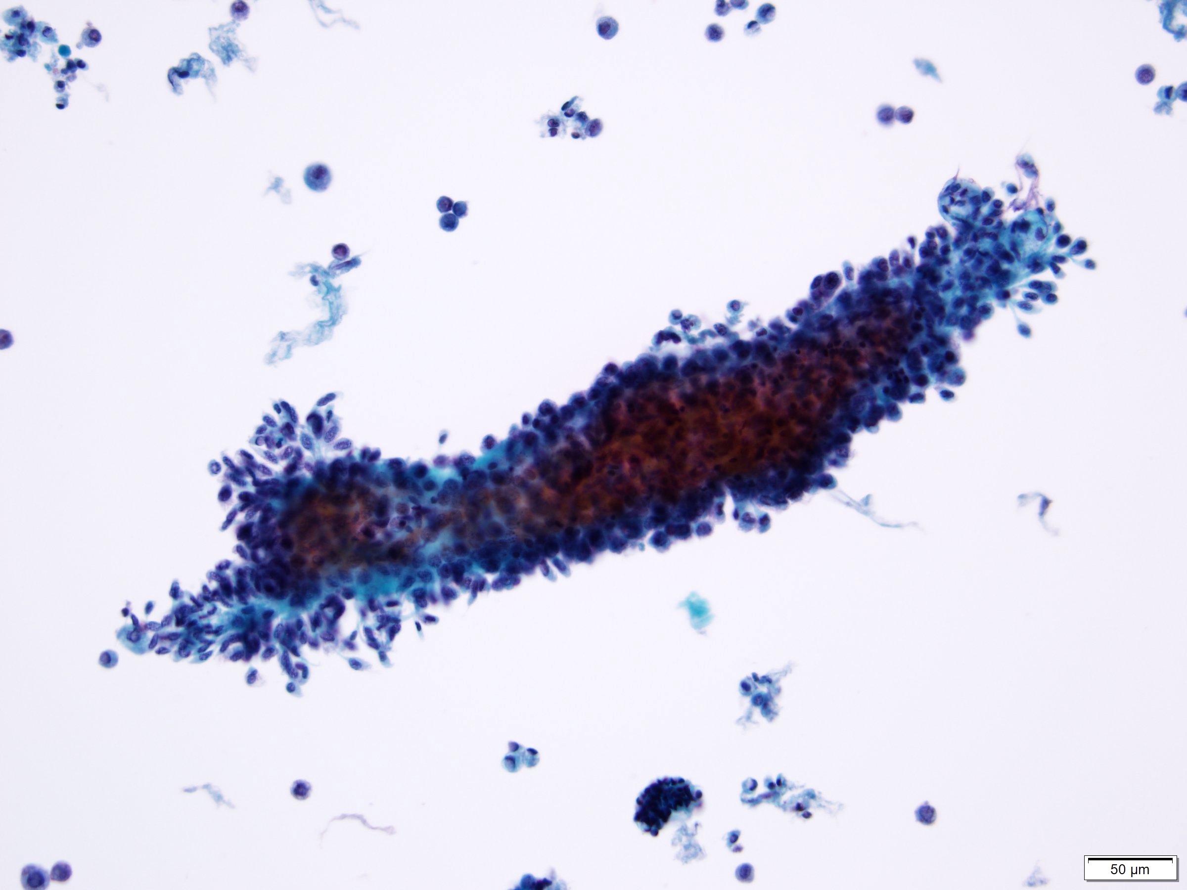
Low grade urothelial neoplasia

Squamous cell carcinoma
Small cell carcinoma
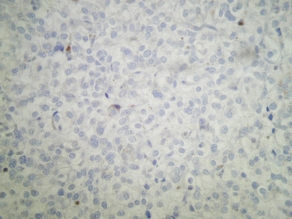
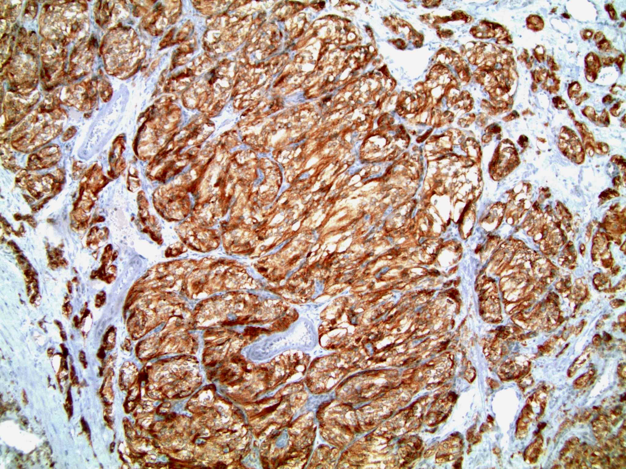
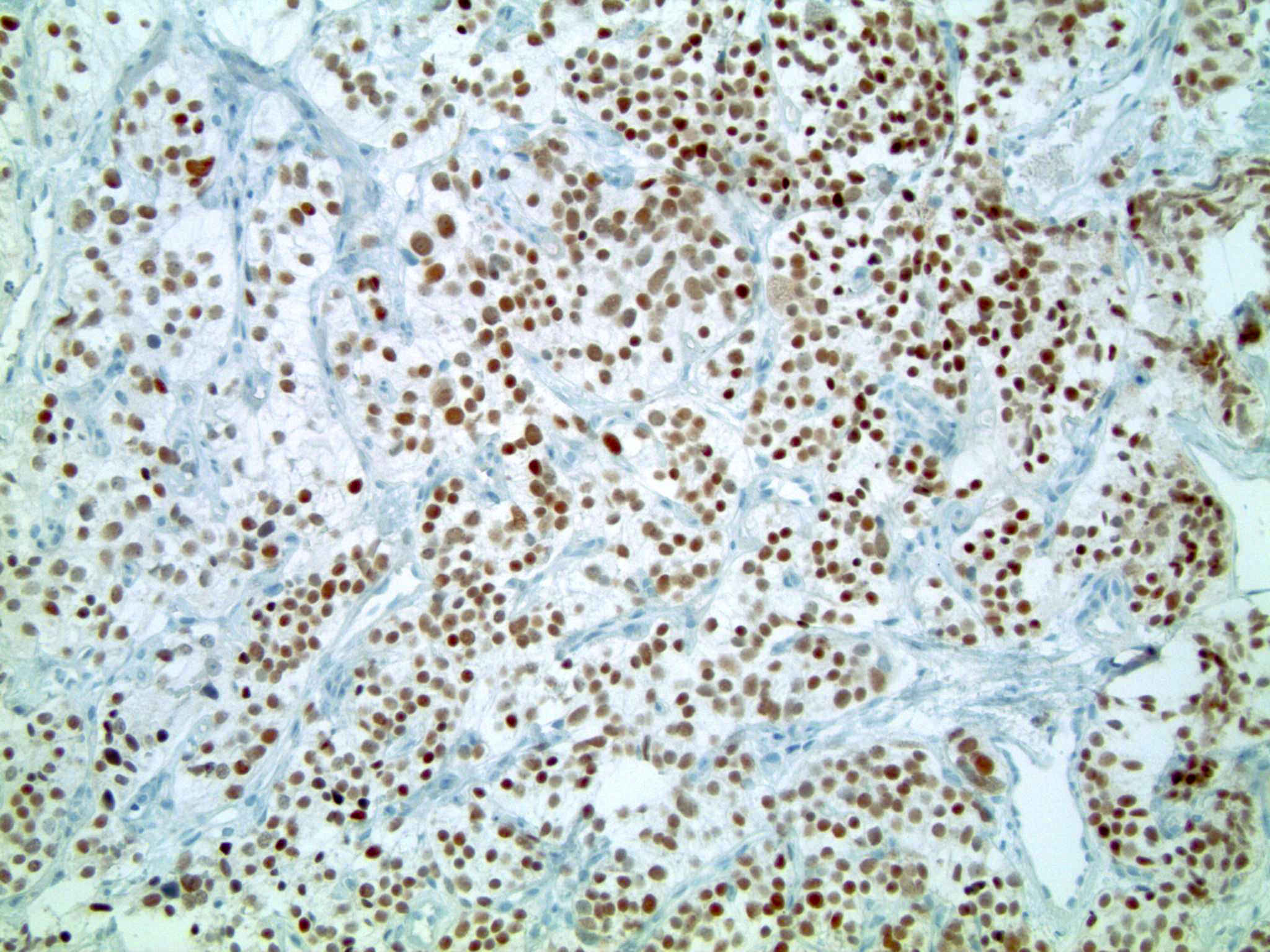
Prostatic adenocarcinoma
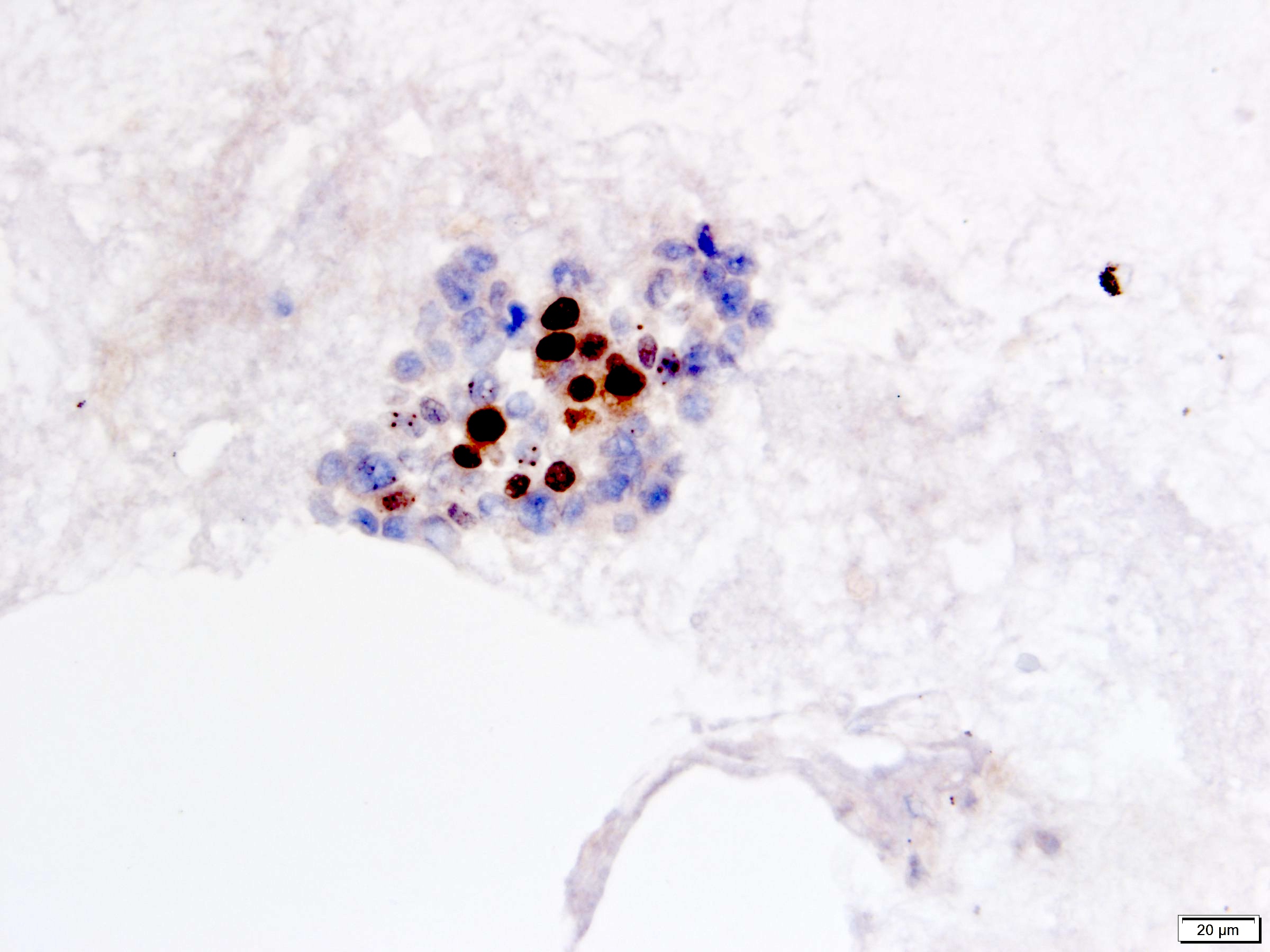
NKX3.1
Colorectal adenocarcinoma
Renal cell carcinoma
Contributed by Bonnie Choy, M.D.
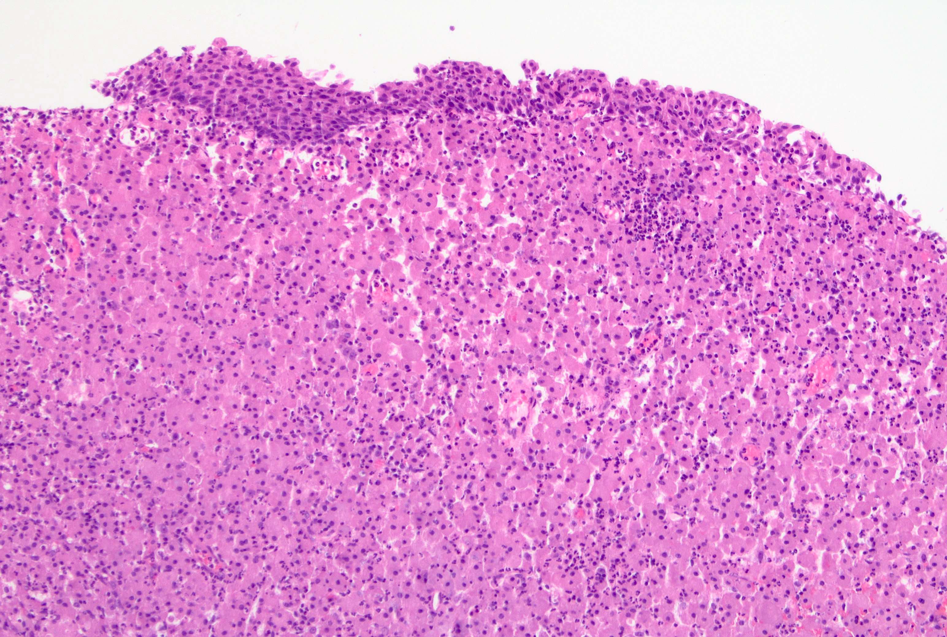
Acute bacterial infection
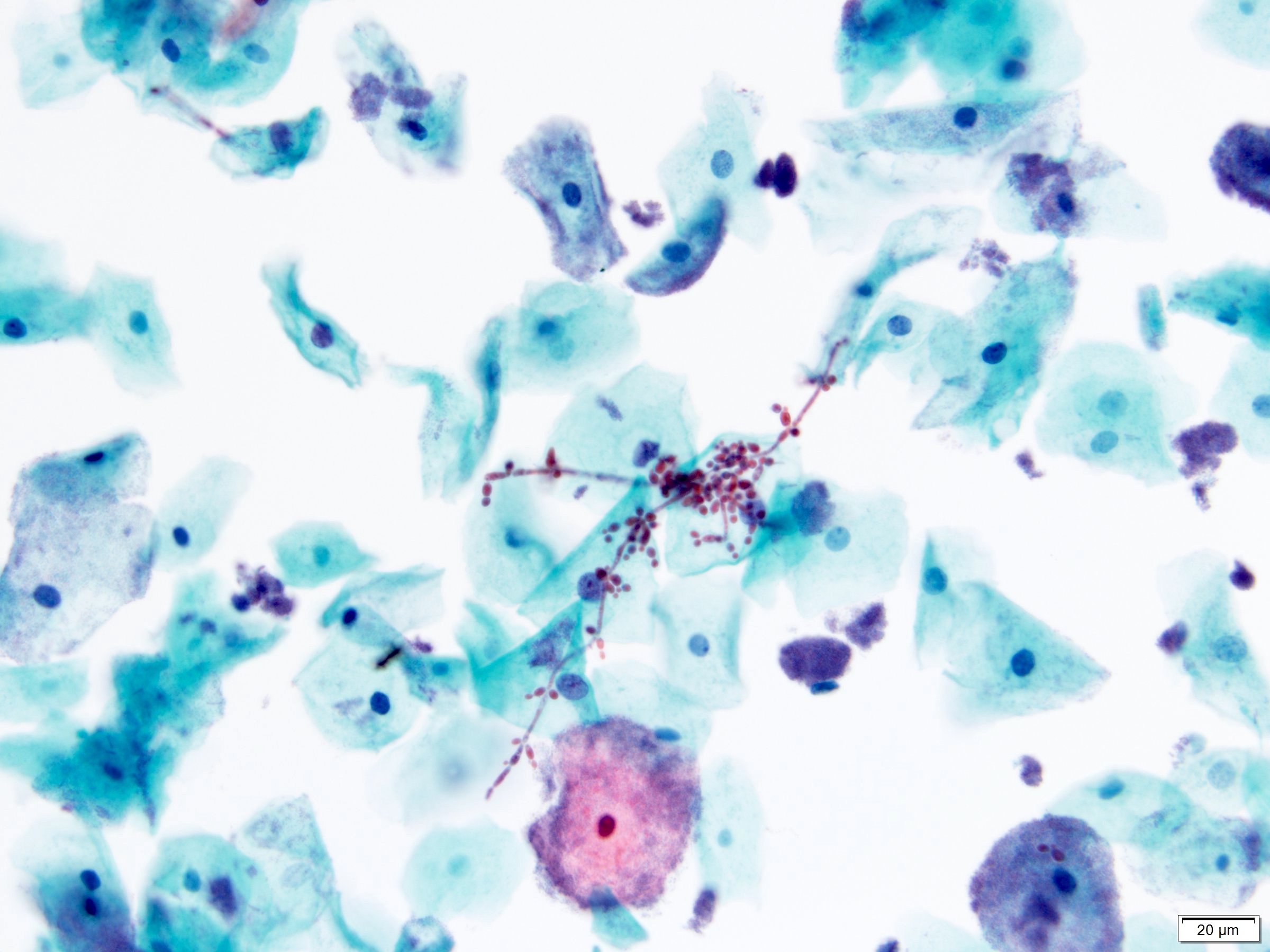
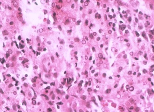
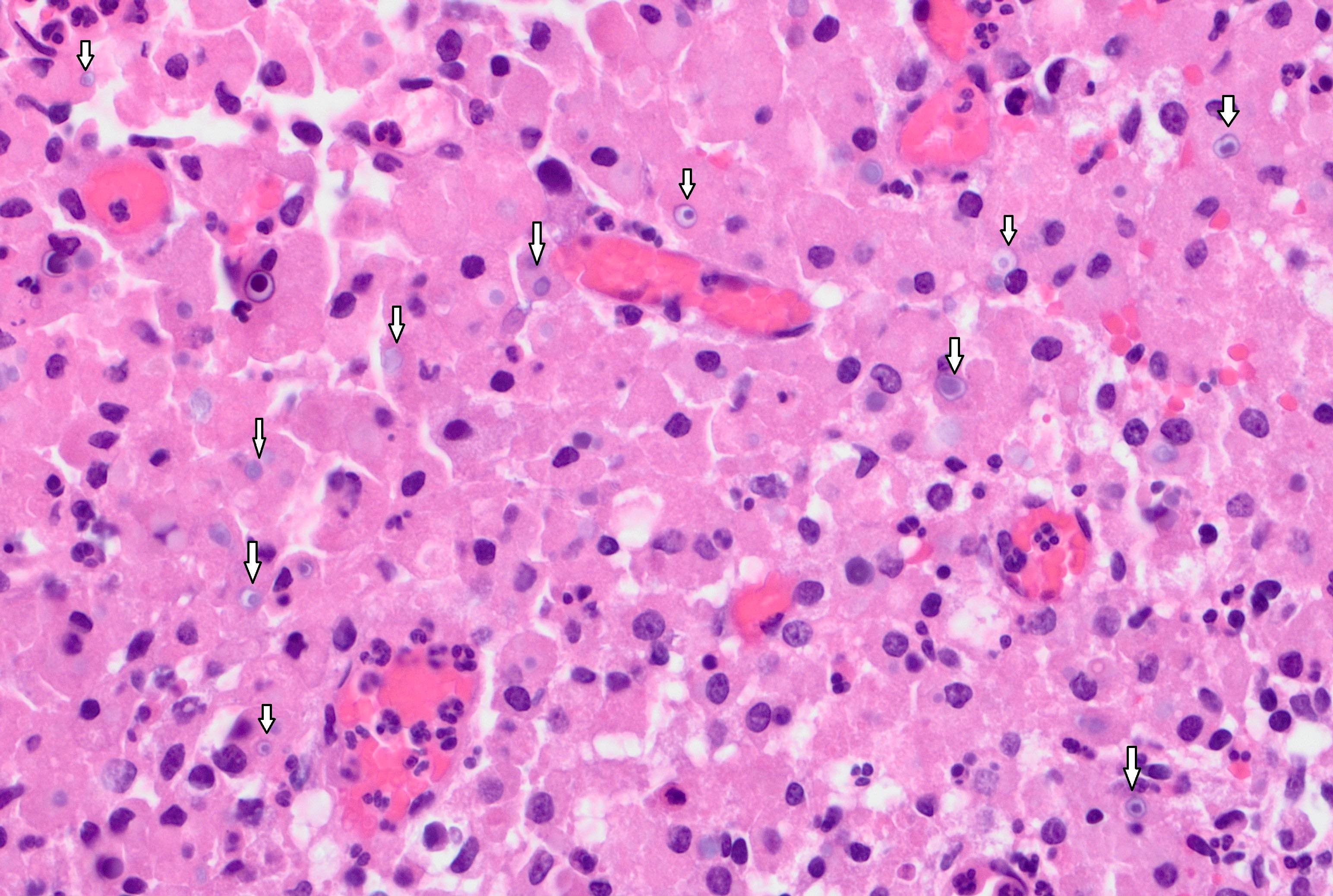
Candida
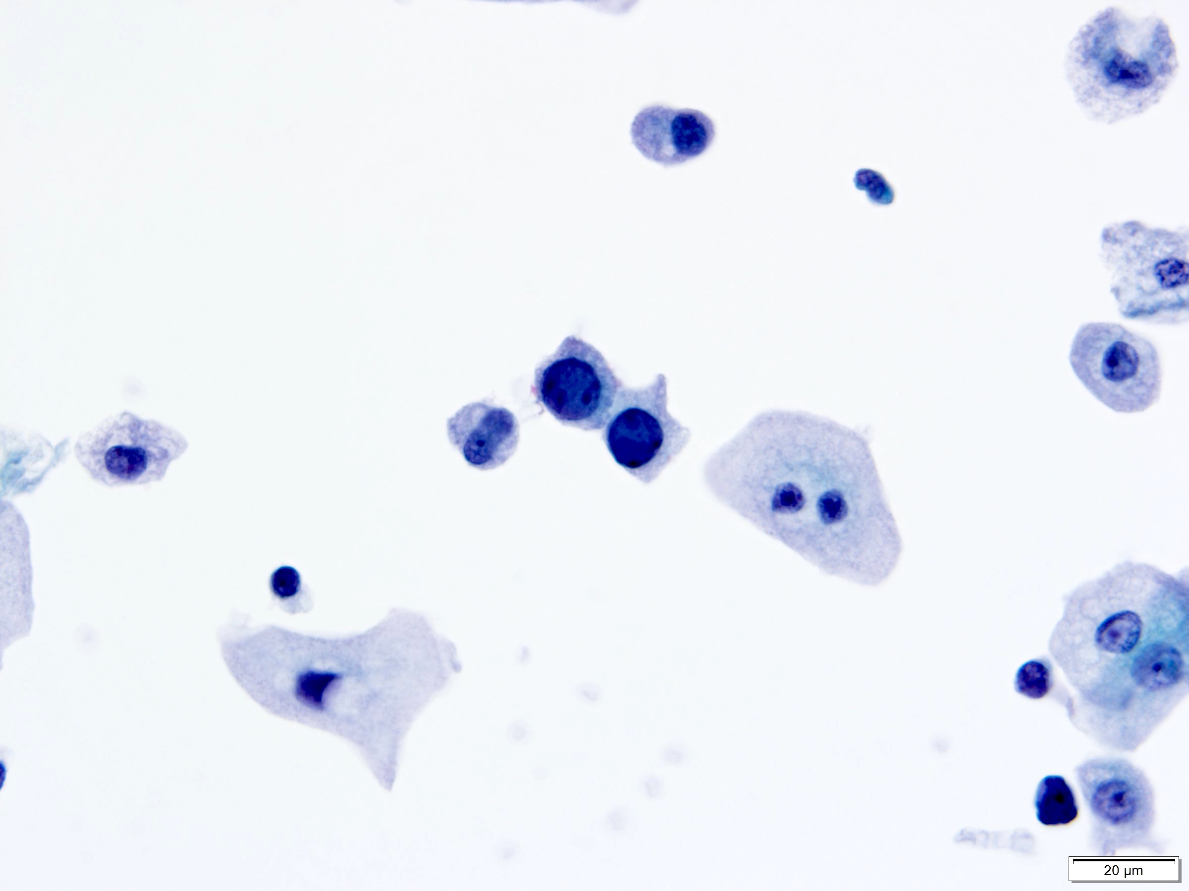
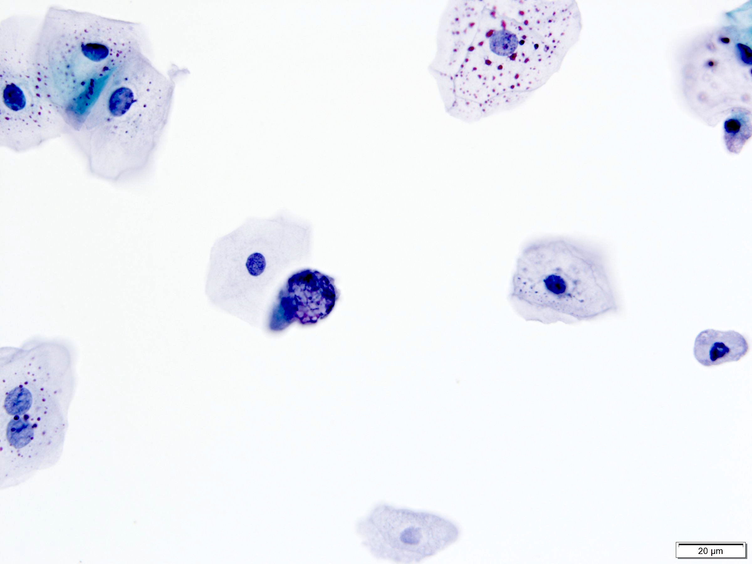
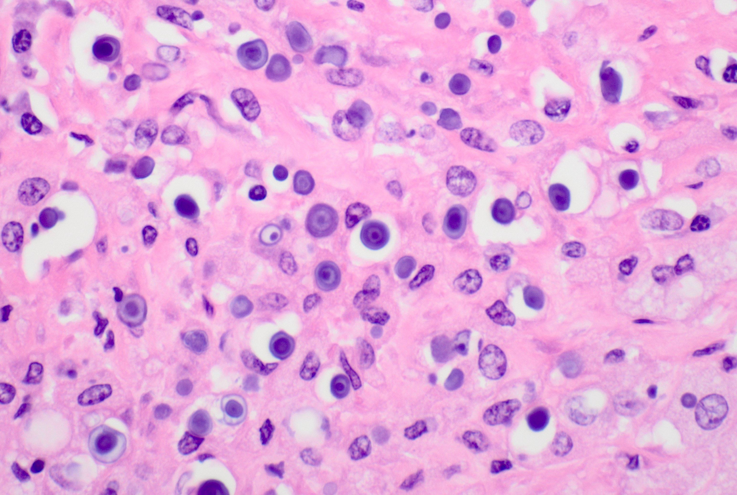
Polyomavirus
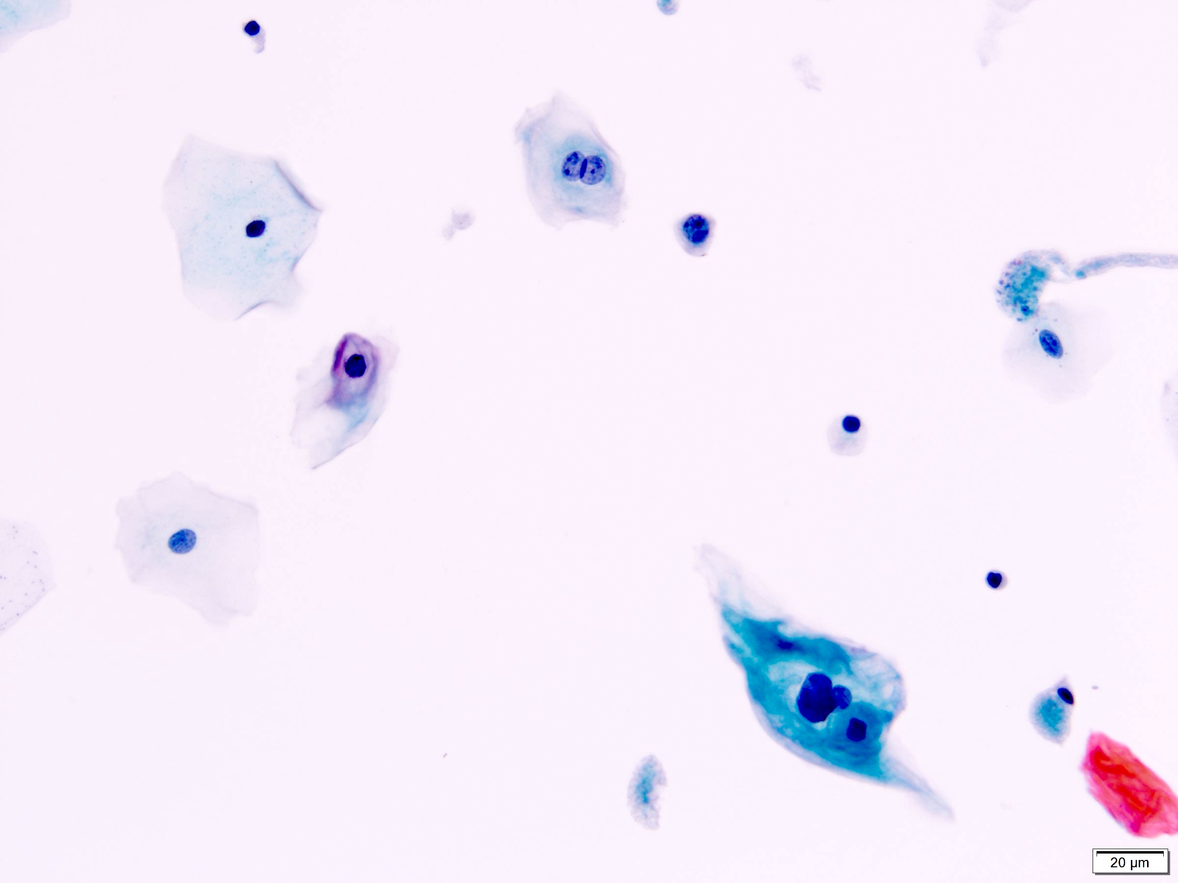
Human papilloma virus
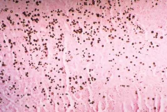
Schistosoma
Trichomonas
Images hosted on other servers:

Image with case history
Images hosted on other servers:

Cystourethrogram shows complete bladder duplication
Images hosted on other servers:

Pathogenesis of bladder cancer
Contributed by Moe Thuzar, M.D. and Y. Albert Yeh, M.D., Ph.D.
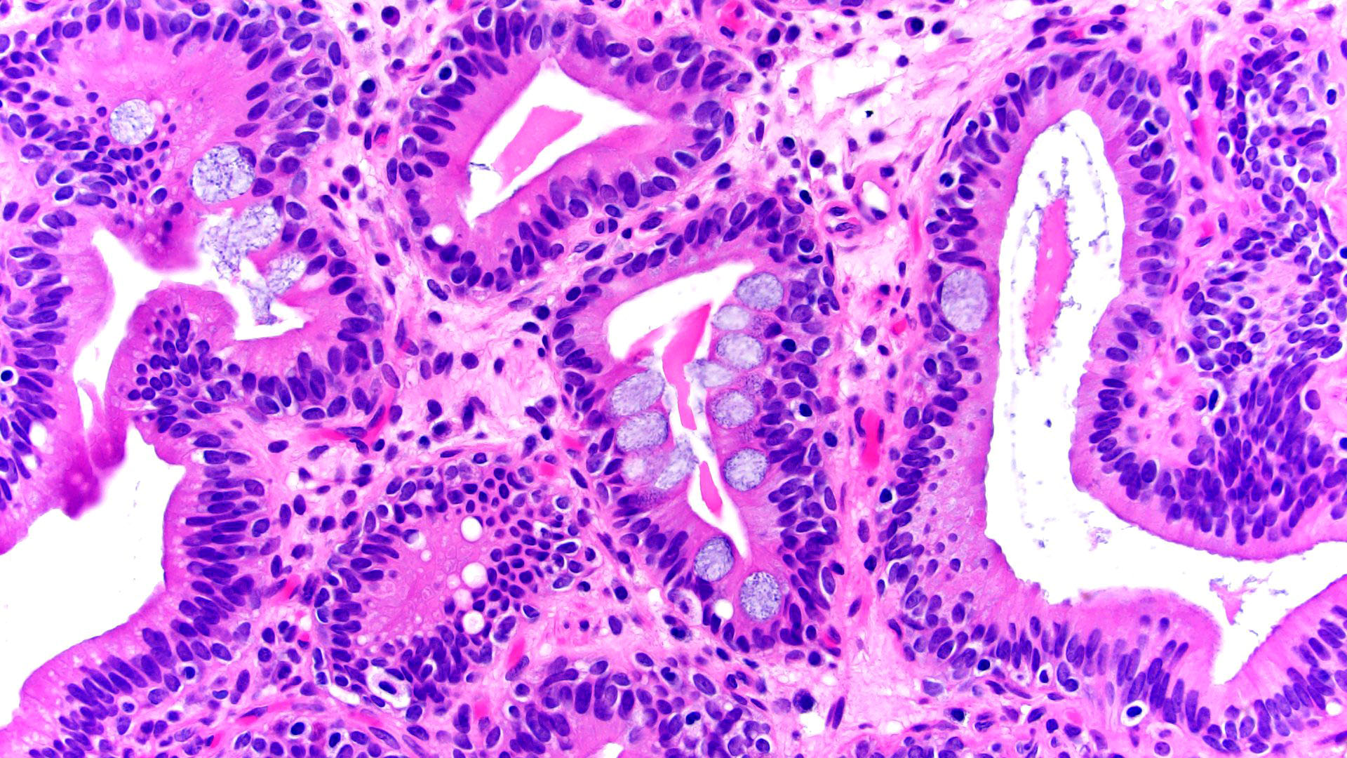
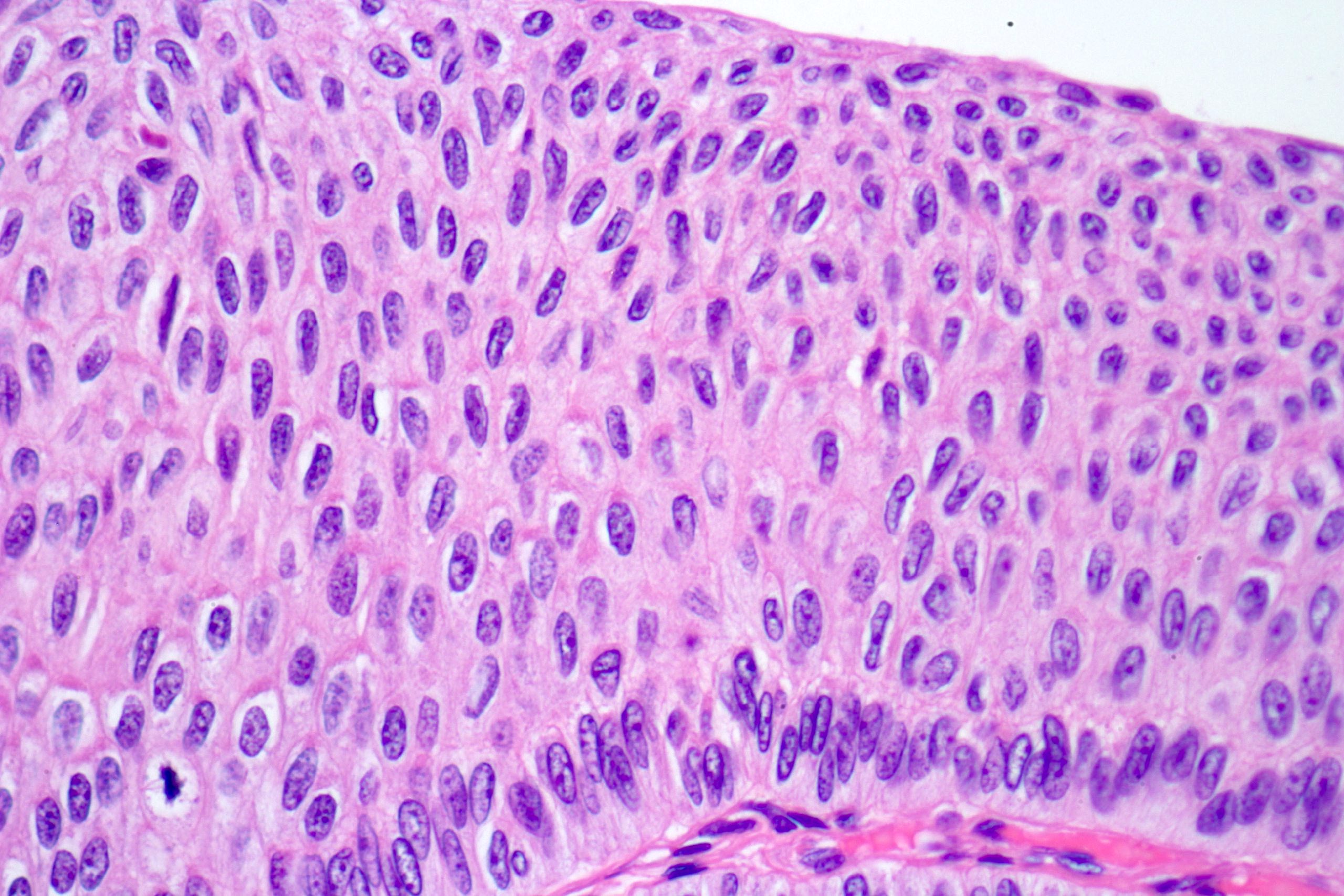
Loss of polarity
Nuclear enlargement and hyperchromasia
Decreased urothelial thickness
Hyperplastic atypical urothelium
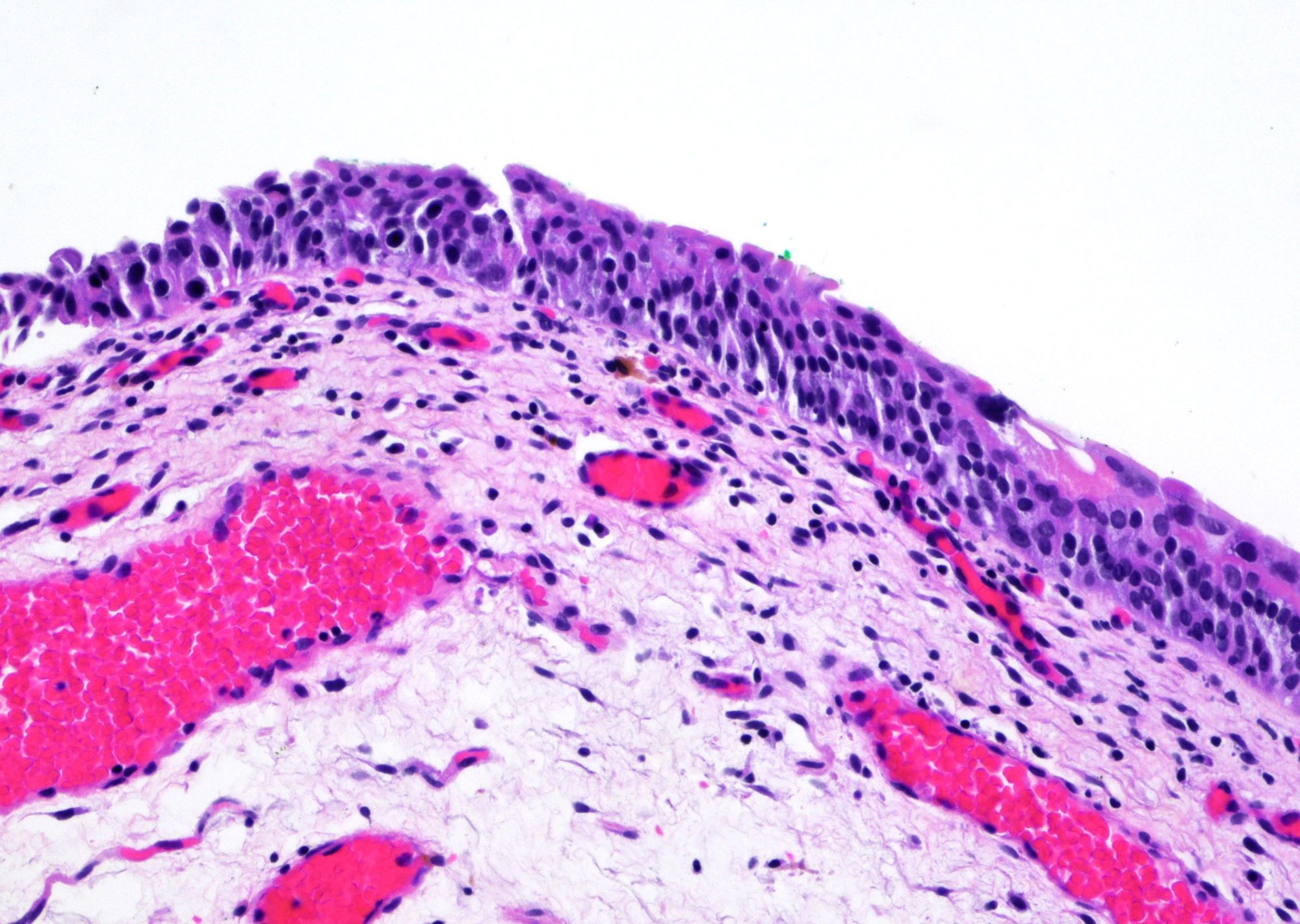
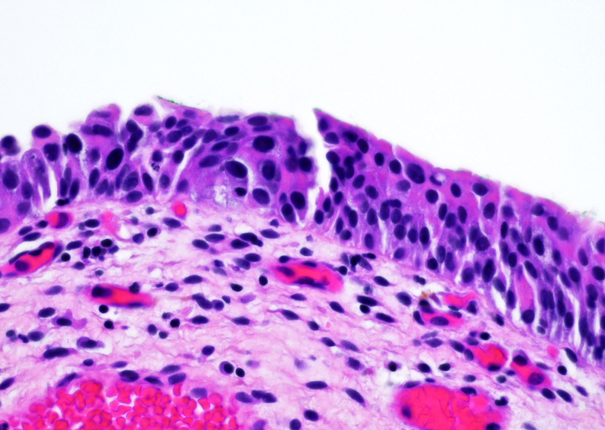
Urothelial atypia
Images hosted on other servers:


Prostatic glands with luminal secretion underlying urothelium (right: PSA+)
Images hosted on other servers:

CT scan shows gas in bladder and bladder wall

CT scan of 77 year old woman without diabetes


Images hosted on other servers:


Linear calcifications


Irregular calcifications
Images hosted on other servers:

Multiple thin calcifications
Contributed by Bohdan Zoshchuk, M.D. and Y. Albert Yeh, M.D., Ph.D.
Dystrophic calcifications
Encrusted calcifications
Encrusted necrotic debris
Encrusted calcified fragments
Calcified plaques
Calcifications and chronic inflammation
Encrustations and fibrin
Calcifying encrustations
Calcifications and giant cells
Calcifications in fibrotic stroma
Calcium salts
Images hosted on other servers:

Calcifying nanoparticles
Contributed by Jian-Hua Qiao, M.D.
Inflammatory infiltrate with marked eosinophils
Images hosted on other servers:

Diagrams of exstrophy
Images hosted on other servers:


Exstrophy in male infants
Contributed by Dr. Jesus Chavez and Dr. Debra Zynger
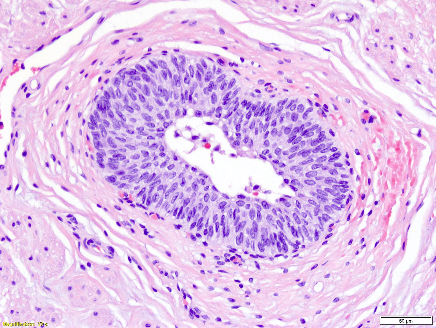
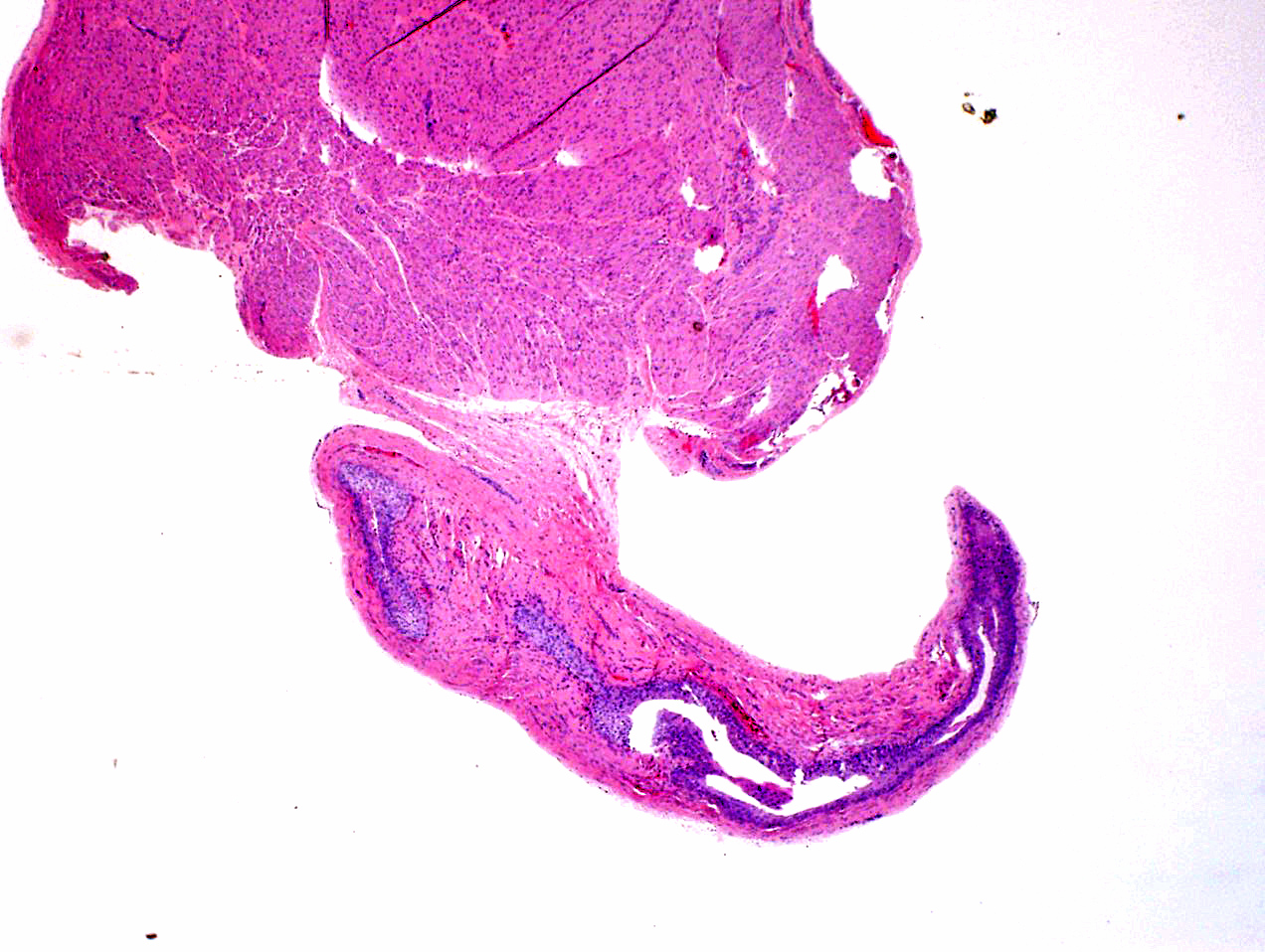
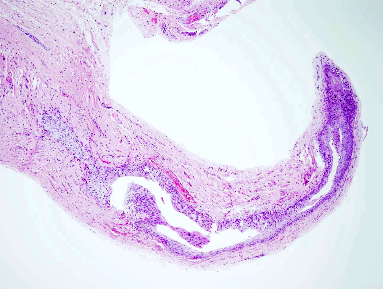
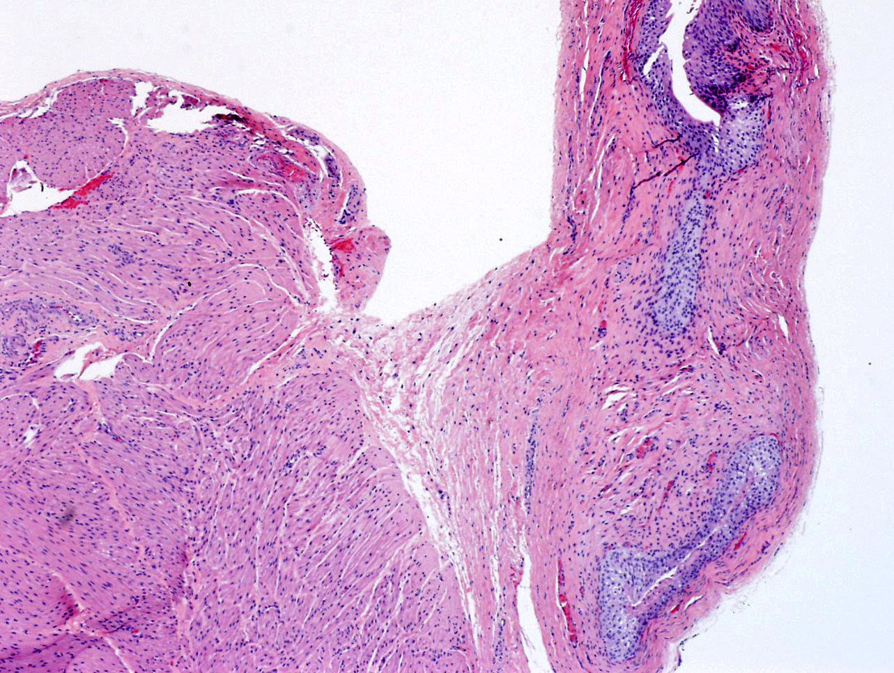
Urethra and periurethral tissue
Contributed by Dr. Jesus Chavez and Dr. Debra Zynger
High power, low grade
Noninvasive high grade
Invasive high grade

Necrosis and keratinization
Elderly woman: metastatic
Clear cell adenocarcinoma
Low power, prominent necrosis
Can mimic nephrogenic metaplasia
With hobnailing
With prominent clear cells and diffuse, sheet-like growth
Case #194

Various images
CK7
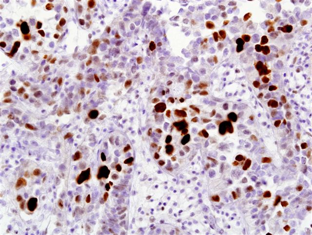
p53
Images hosted on other servers:

Cystoscopy
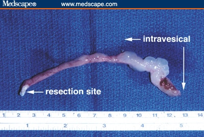
Ureteral polyp
Images hosted on other servers:

Polyp of ureter
Images hosted on other servers:


Bladder biopsies

Follicular cystitis
Images hosted on other servers:

Numerous lymphocytes with varying maturity
Images hosted on other servers:
Granulomatous cystitis due to bCG treatment
Granulomatous cystitis, postresection
Postoperative granulomas, postresection
Images hosted on other servers:

Inflammatory bleeding bladder mucosa
Images hosted on other servers:

Hemorrhagic and ulcerative cystitis after cyclophosphamide
Images hosted on other servers:

Edema, bleeding and granulocyte infiltration

Hemorrhage and edema in the lamina propria
Case of the Week #353
47 year old man with herpes viral cytopathic effects in urine cytology
Images hosted on other servers:

Staging of bladder carcinoma
Contributed by Anil Parwani, M.D., Ph.D., M.B.A.
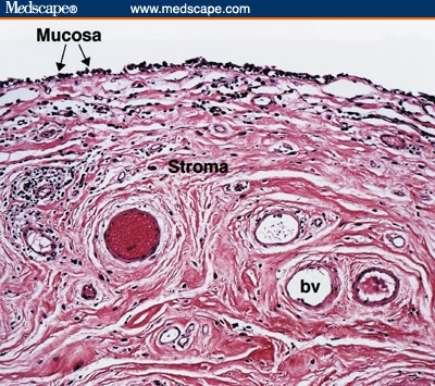
Bladder wall
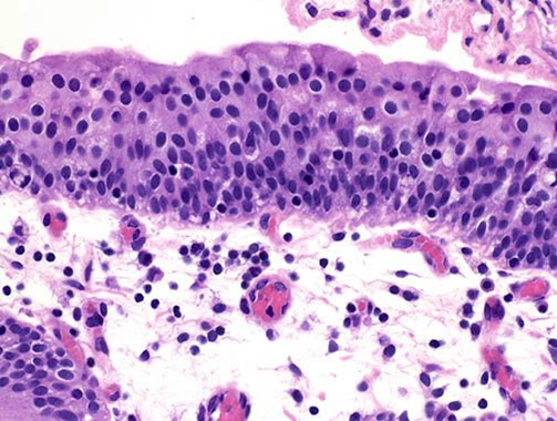
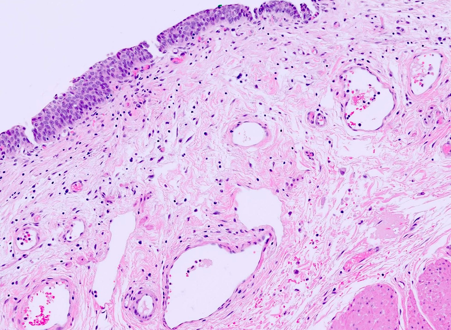
Urothelium
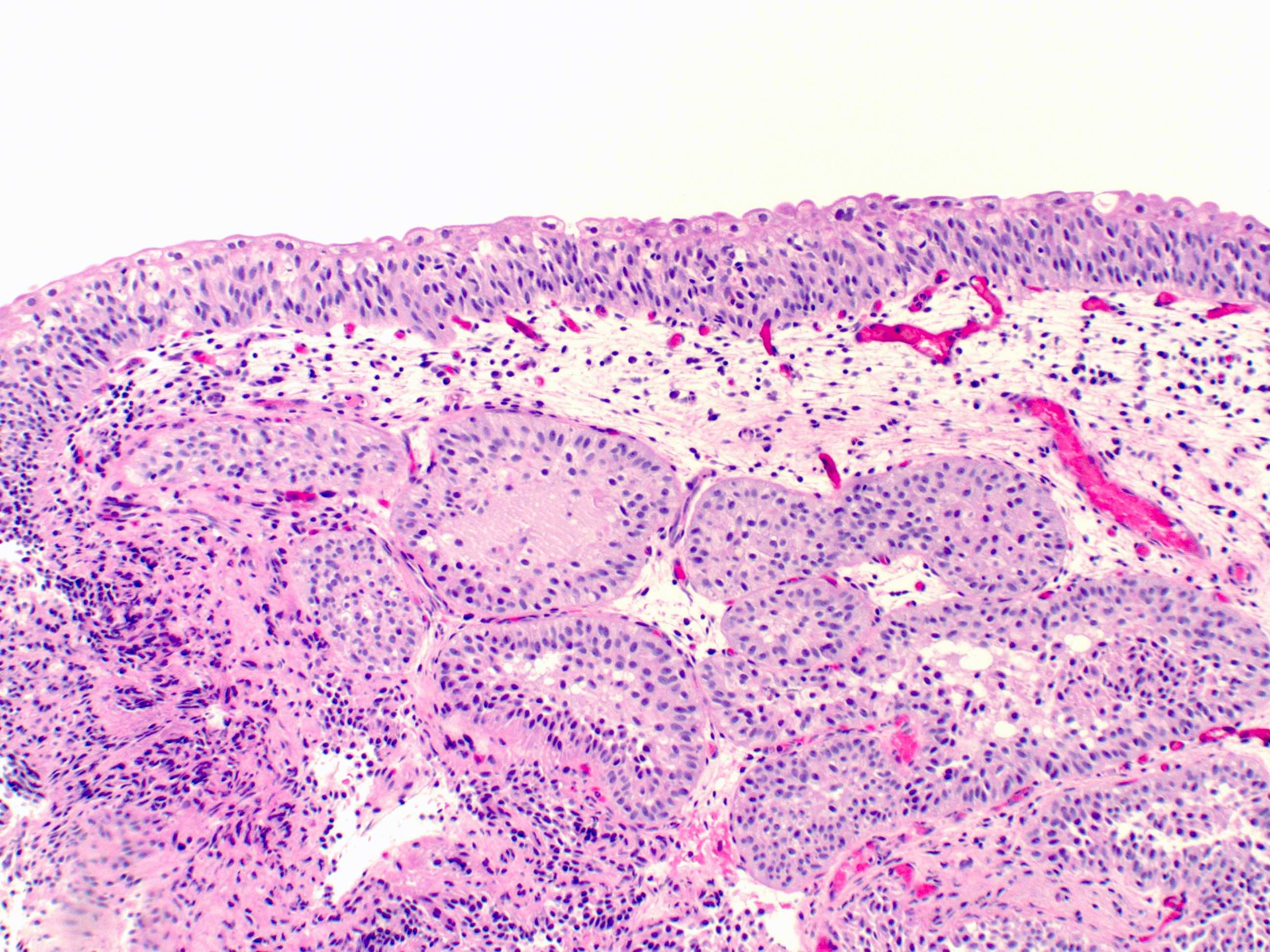
von Brunn nests
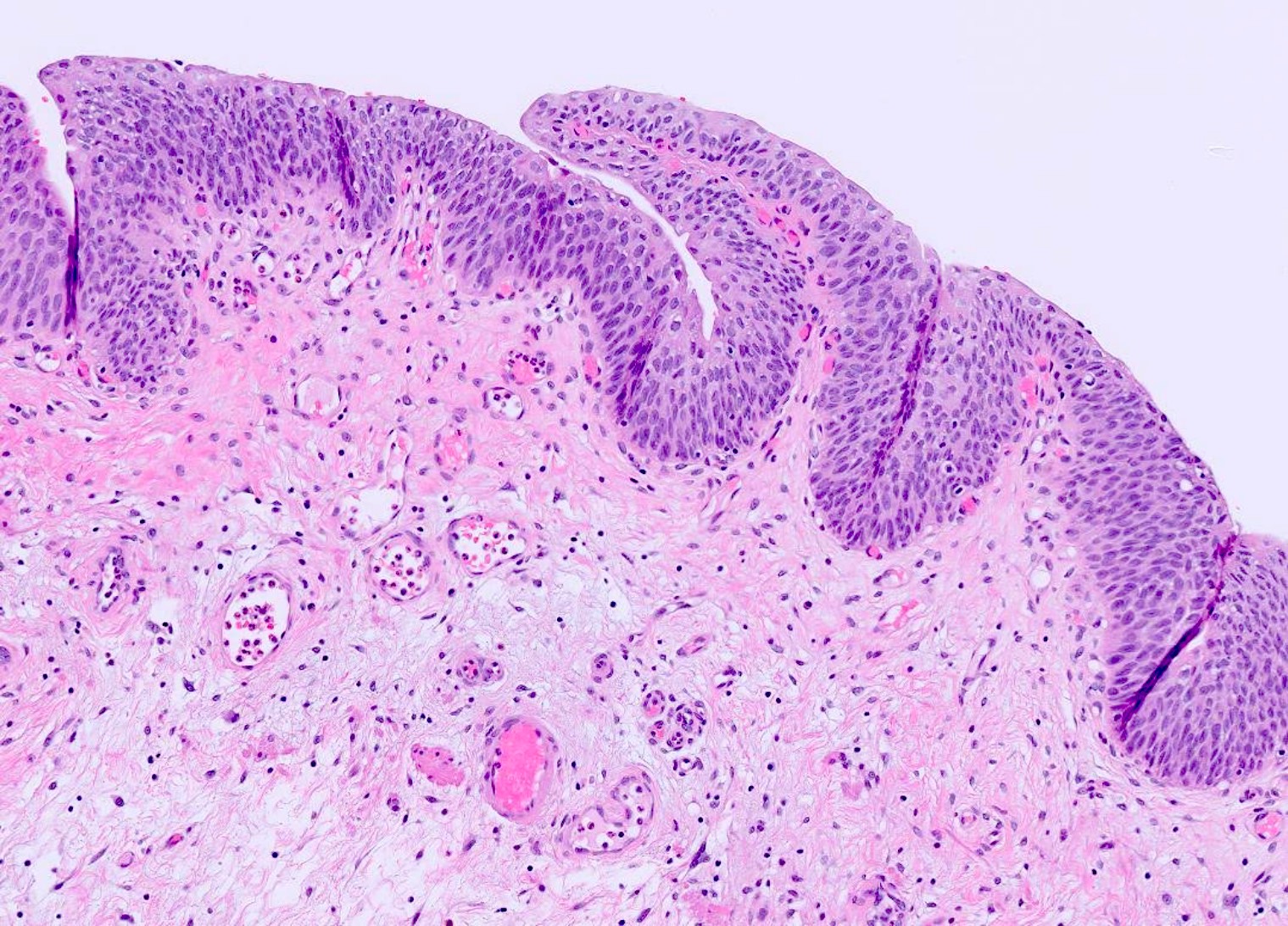
Urothelium and lamina propria
Muscularis mucosa
Muscularis propria
Shotgun histology bladder
Images hosted on other servers:

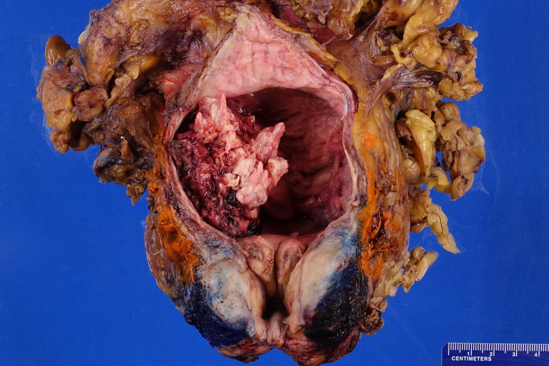
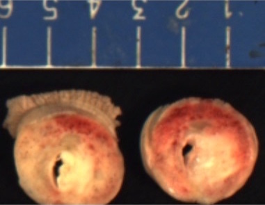
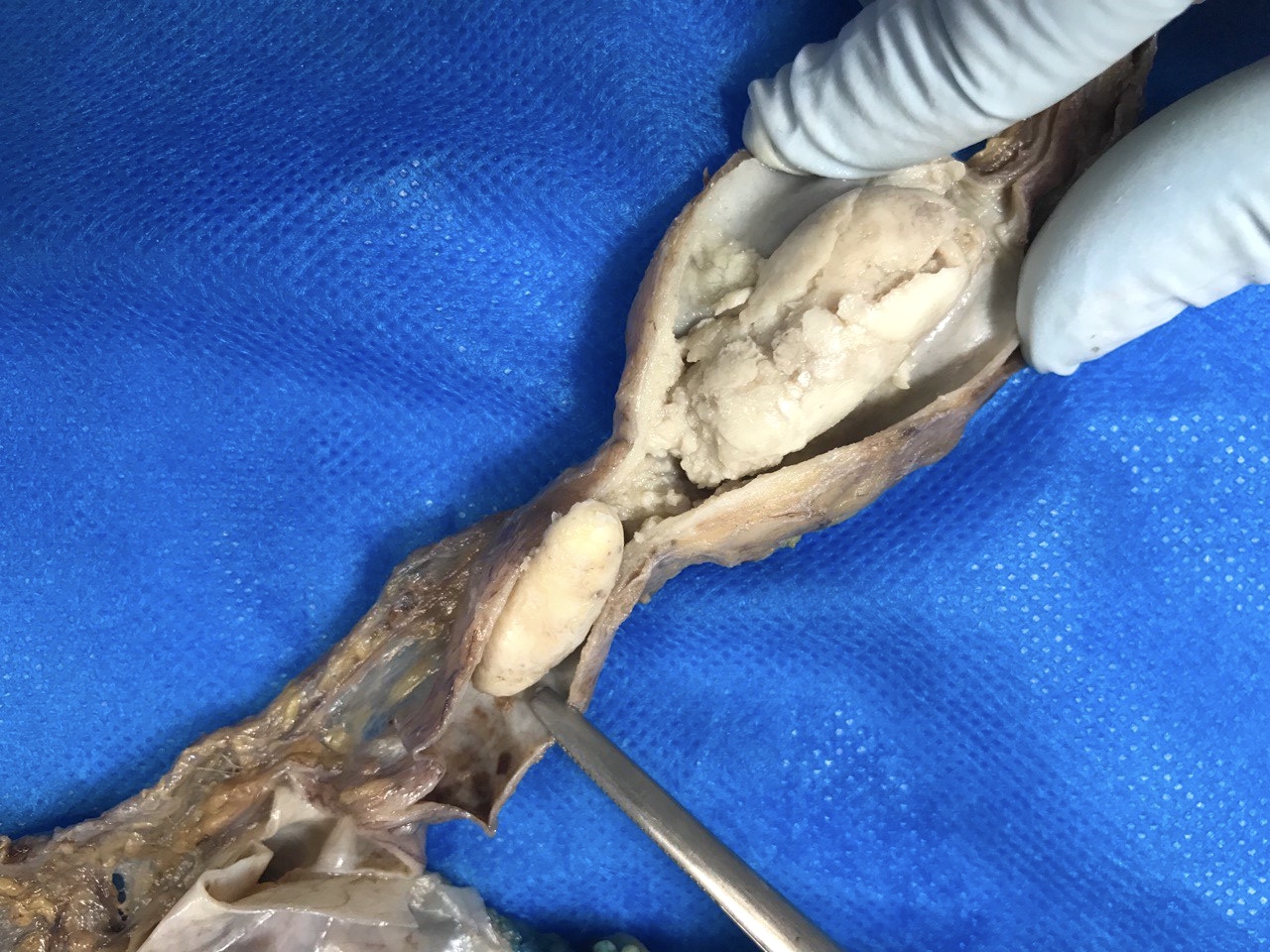
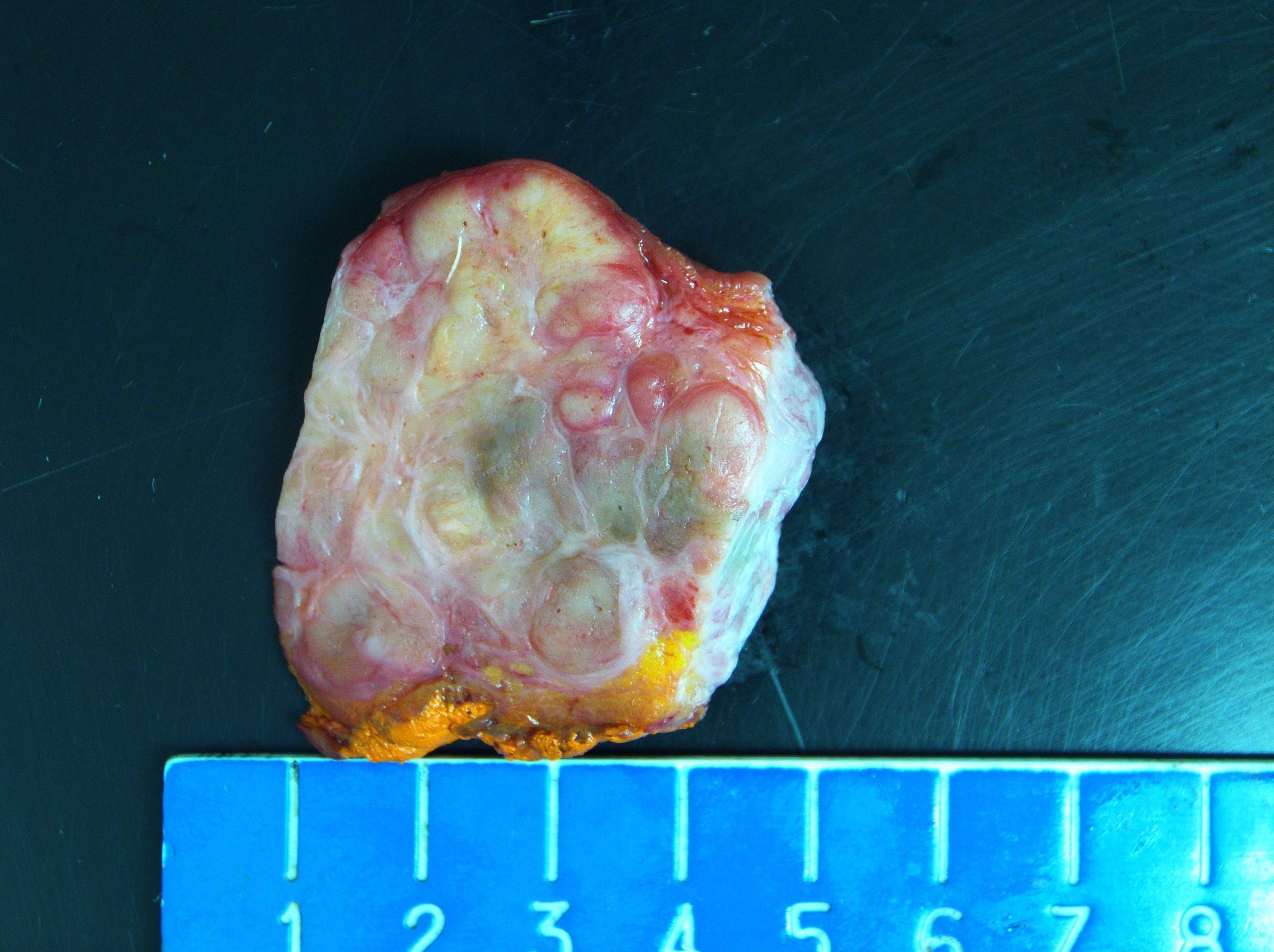
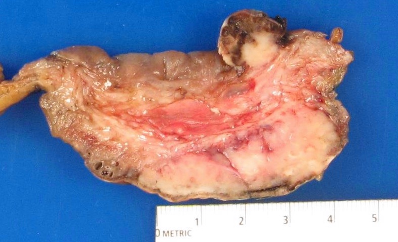
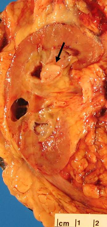
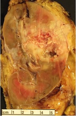
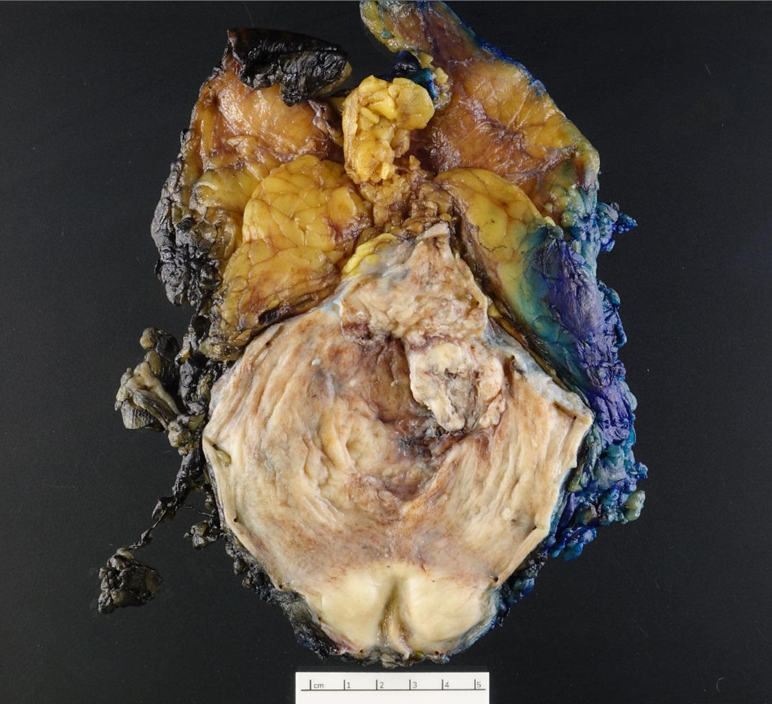
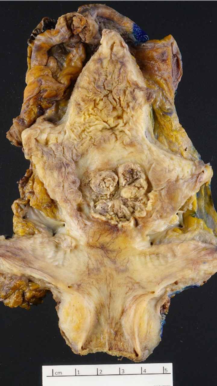
White, gelatinous
mass with cystic
change brand
hemorrhage
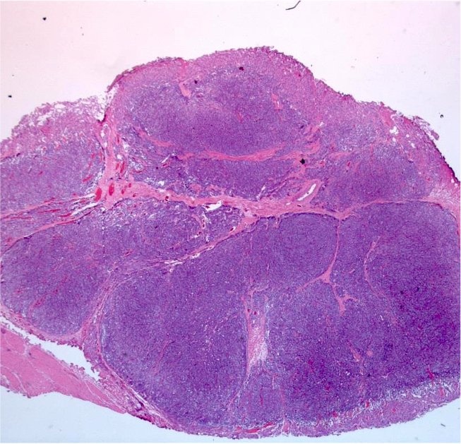
AFIP images

Abundant thin walled vessels
Images hosted on other servers:

ALK1 expression

Focal inflammatory cells

Myofibroblastic cells
and inflammatory
cells in edematous
stroma


Spindle cells within fibromyxoid matrix and inflammatory cells
Images hosted on other servers:

Bladder CT
Images hosted on other servers:

Cystoscopic findings
Images hosted on other servers:

Inflamed mucosa
Contributed by Michelle R. Downes, M.D.
Hemorrhage and edema
Urothelial denudation and inflammation
Mixed inflammatory infiltrate
Surface ulceration
Inflammation,
microhemorrhages
and congestion
Hemorrhage and fibrosis
Contributed by Maria Tretiakova, M.D., Ph.D.
Replacement of urothelium
Without dysplastic changes

Formation of cystic spaces
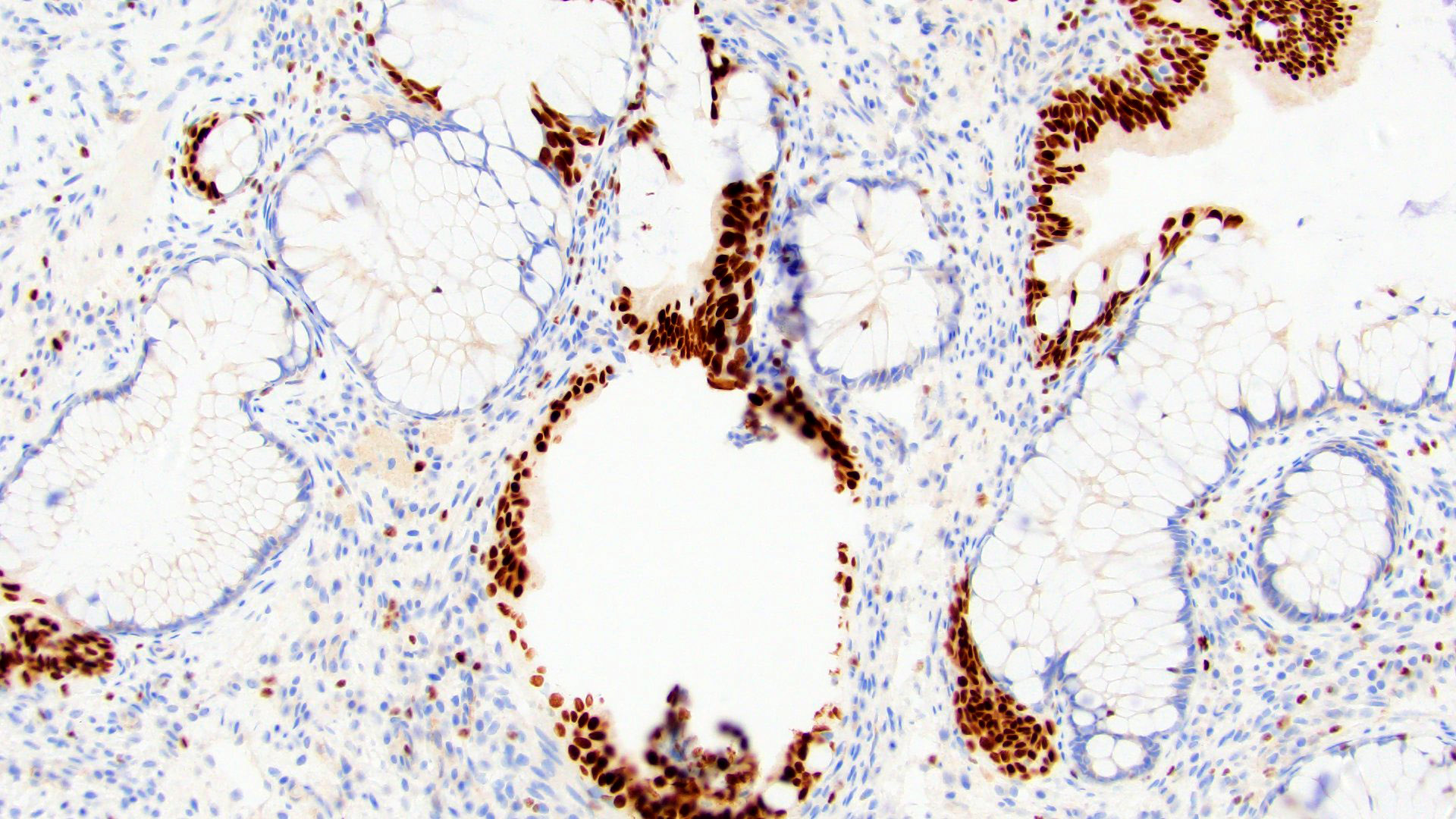
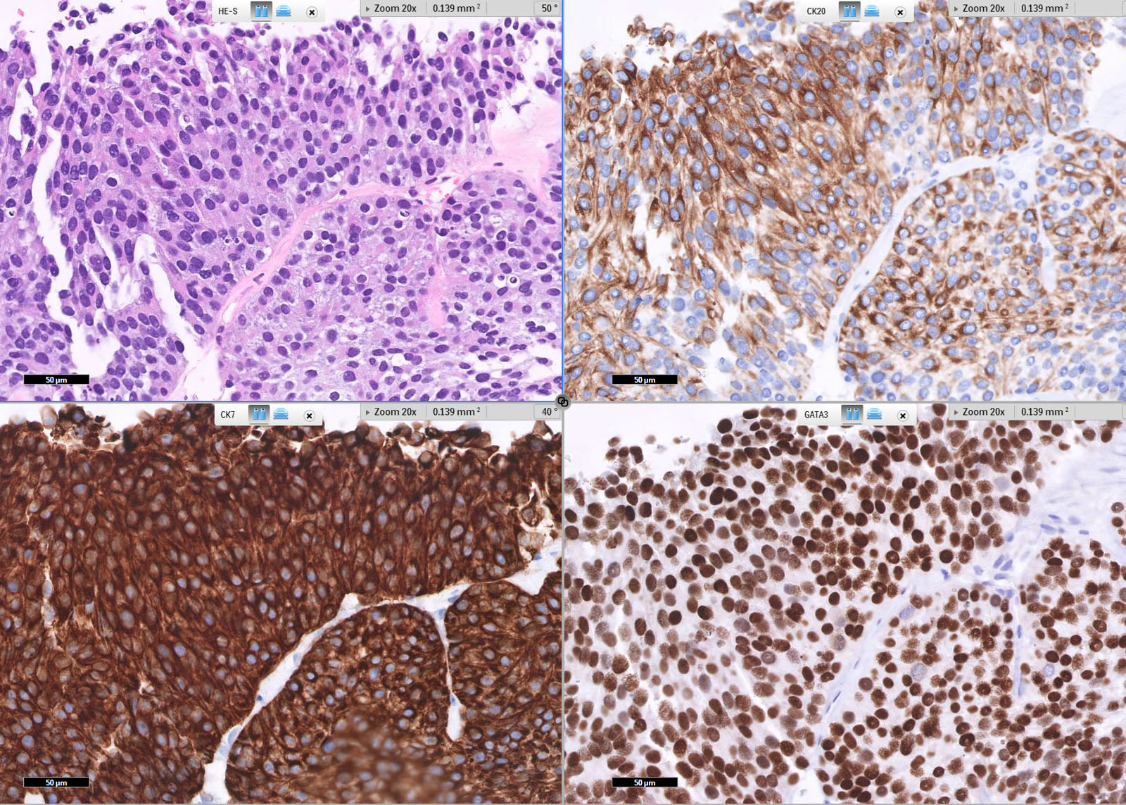
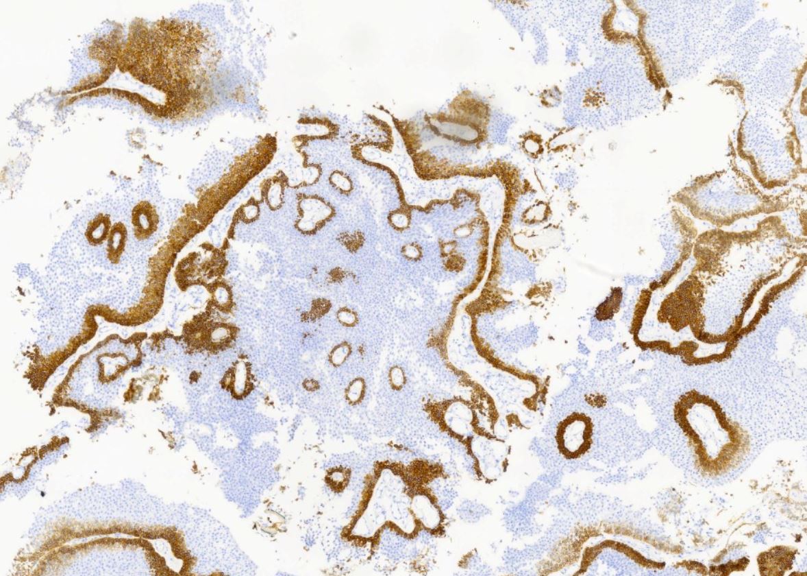
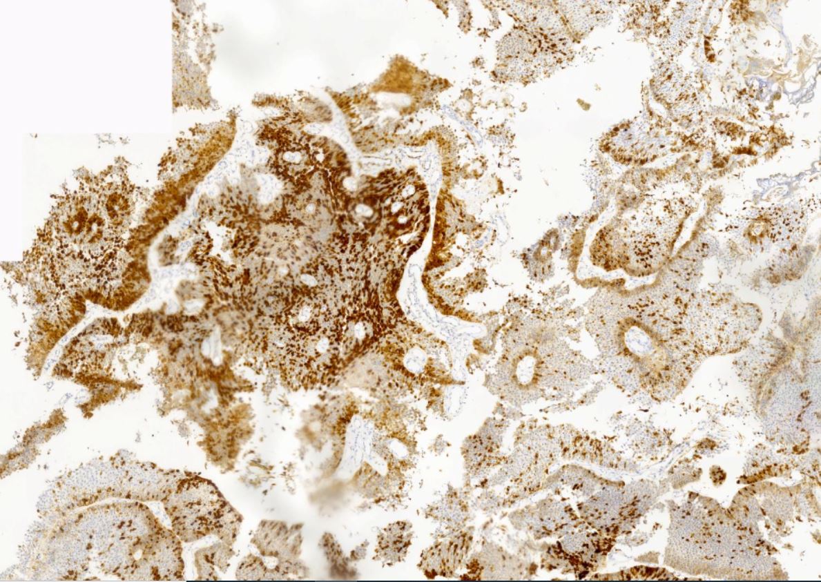
GATA3
CK20
CK20
CK20
HMWCK
Contributed by Rugvedita Parakh, M.D.
Replacing urothelium
Surface goblet cells
Images hosted on other servers:

CT with bladder thickening

Bladder tumor and lung metastasis

Retrograde pyelogram

Pyeloureteral transitional cell
Images hosted on other servers:

Cystoscopy: pedunculated tumor
Contributed by Debra L. Zynger, M.D., Nicole K. Andeen, M.D. and Maria Tretiakova, M.D.
Muscularis propria invasion (pT2b)
Prostatic invasion (pT4a)

Renal pelvic and peripelvic fat invasion (pT3)
Friable, exophytic
papillary mass,
renal pelvis
Contributed by Maria Tretiakova, M.D., Ph.D., Andrey Bychkov, M.D., Ph.D., Nicole K. Andeen, M.D. and @katcollmd on Twitter
Invasive high grade urothelial carcinoma
Extensive invasion into lamina propria
Invasive urothelial
carcinoma with
comedo necrosis
Lymphovascular invasion
Muscularis propria invasion
IHC profile
Subepithelial invasion (pT1)
Invasion into muscularis (pT2)
Invasion of renal parenchyma (pT3)


Invasive urothelial carcinoma


Invasive urothelial carcinoma
Contributed by Bonnie Choy, M.D.
High grade urothelial carcinoma
Contributed by Nicole K. Andeen, M.D. and Maria Tretiakova, M.D.
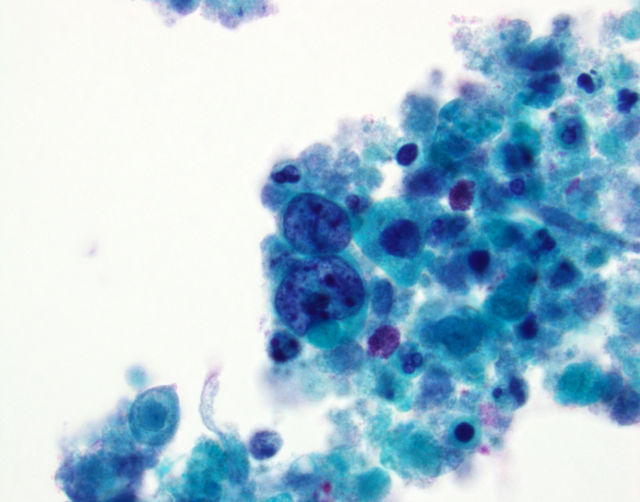
Highly atypical cells
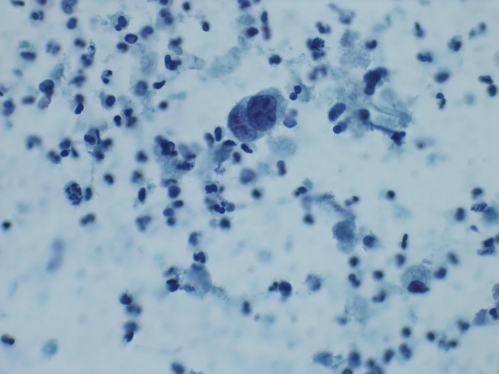
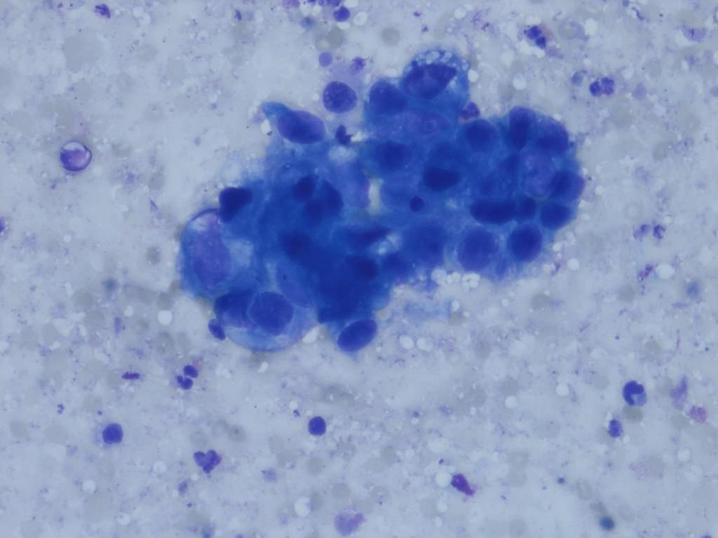
Large nuclei with high nuclear to cytoplasmic ratios, coarse chromatin and irregular nuclear contours (Papanicolaou and DiffQuik)
Contributed by Nicole K. Andeen, M.D. and Maria Tretiakova, M.D.
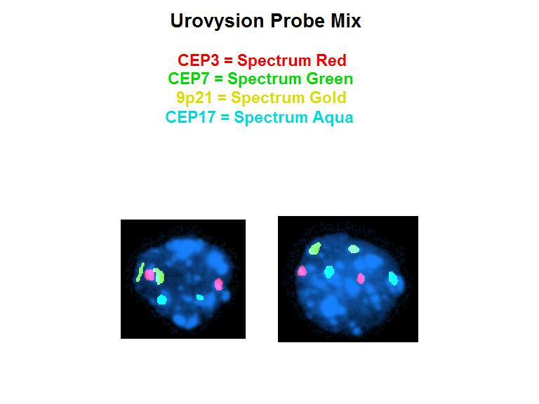
FISH, loss of 9p, normal disomy chromosomes 3, 7, 17

FISH, polyploidy chromosomes 3, 7, 17; 9p preserved
Images hosted on other servers:

Left ureter filling defect
Images hosted on other servers:

Tumor with smooth contour
Images hosted on other servers:

Polypoid mass
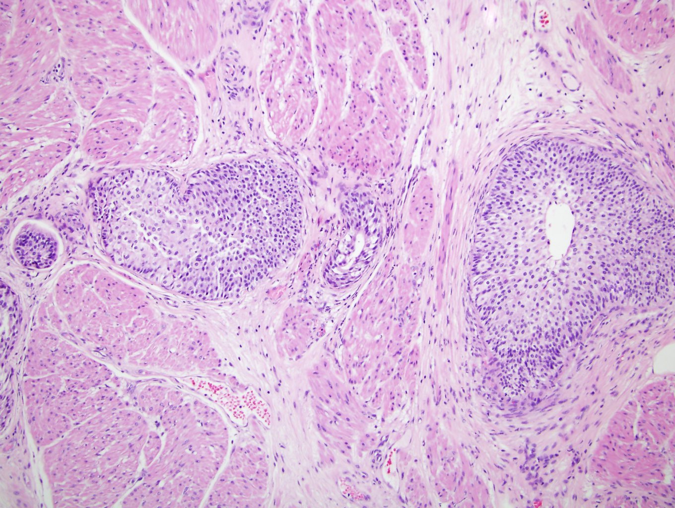
Contributed by Y. Albert Yeh, M.D., Ph.D., Daniel Athanazio, M.D., Ph.D. and Debra Zynger, M.D.
Smooth surface contour
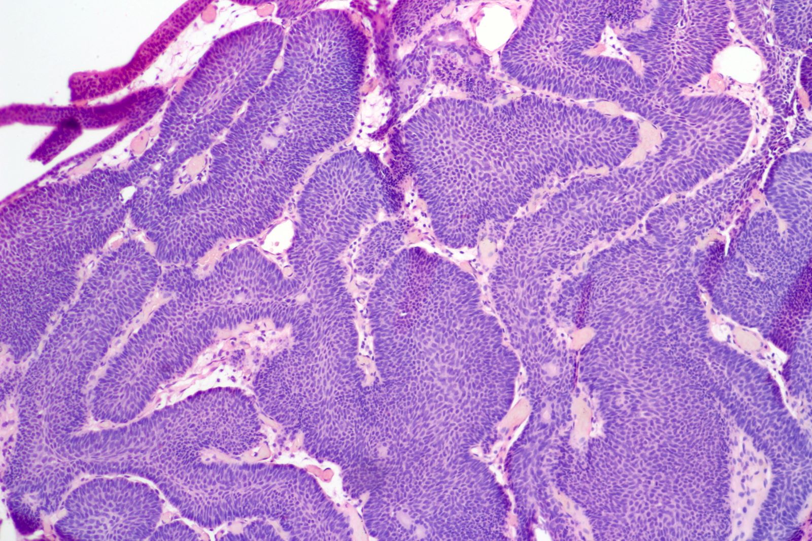
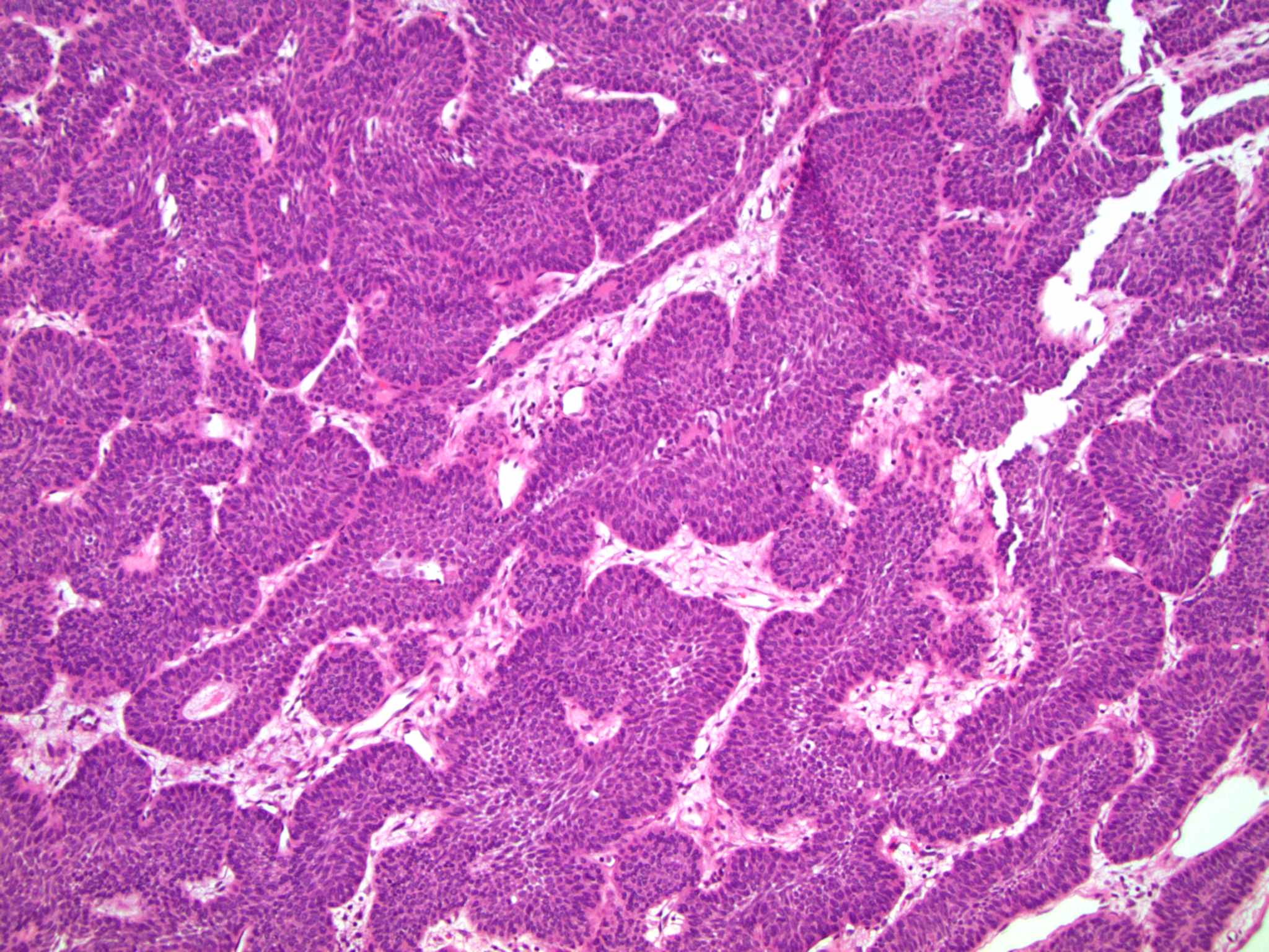
Invagination of trabeculae
Interconnecting trabeculae and cords
Endophytic growth
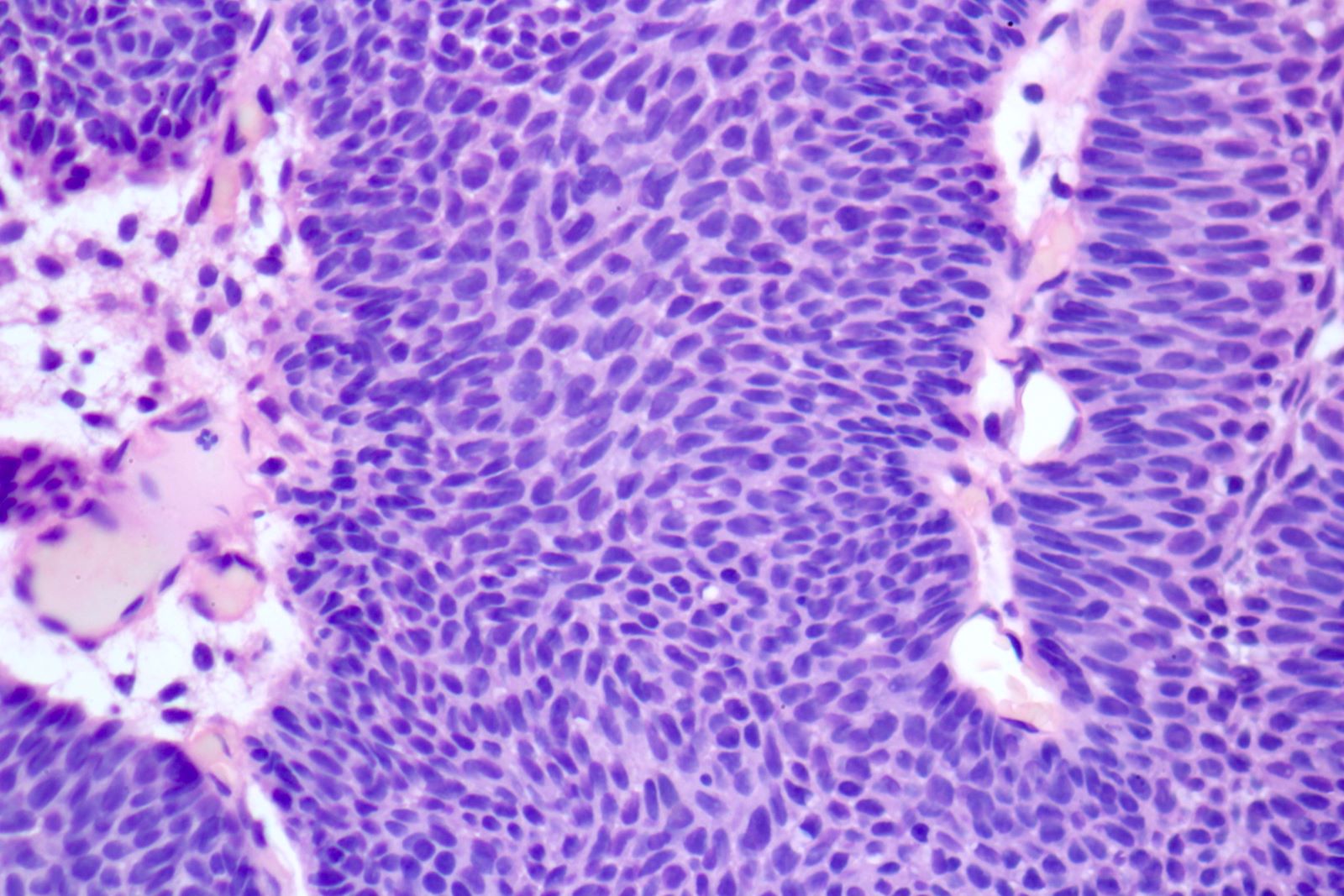
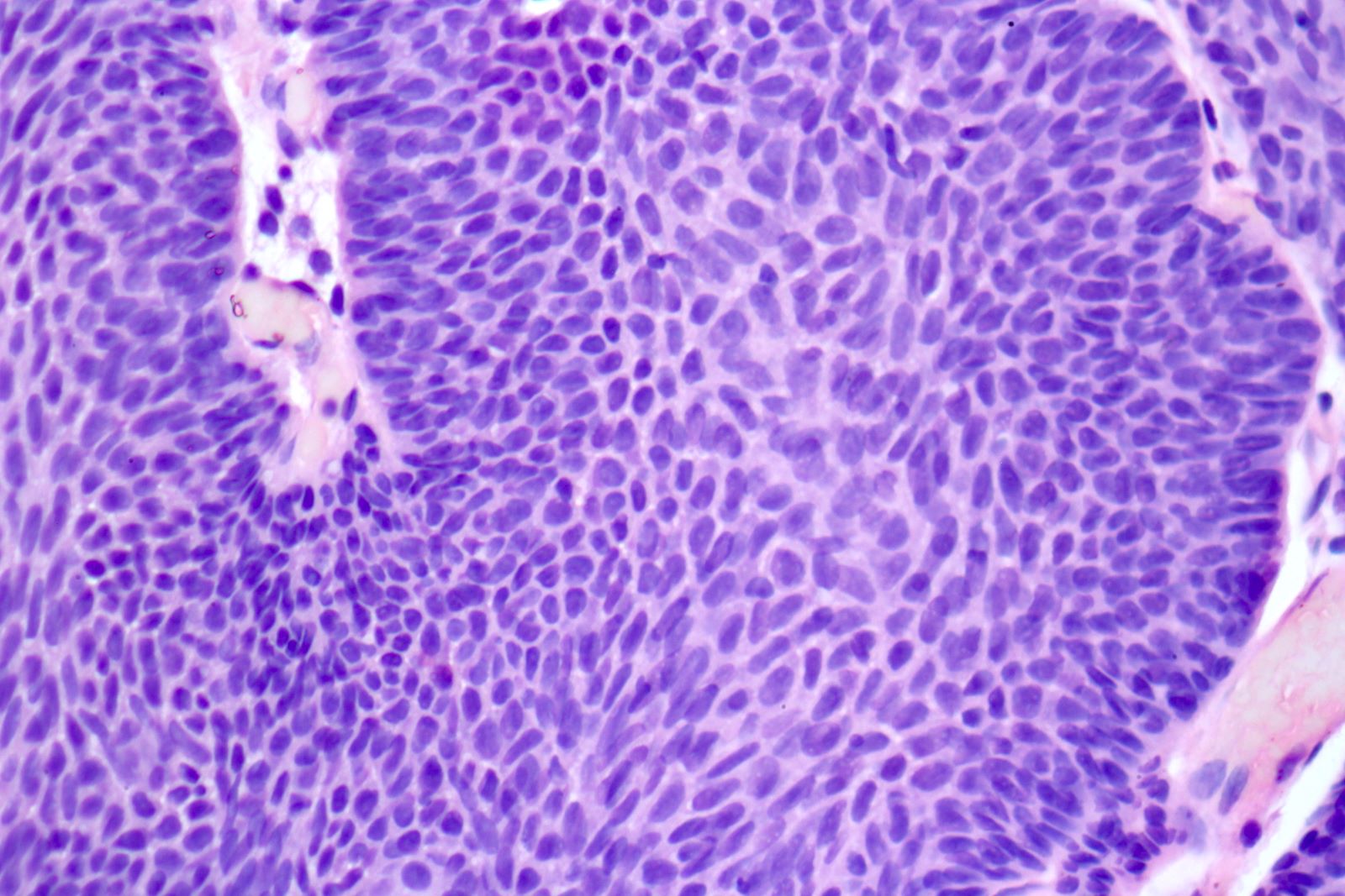
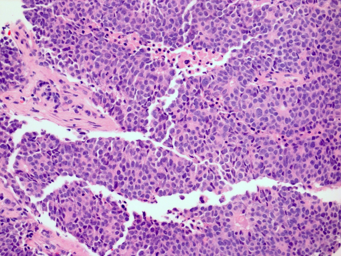
Bland urothelial cells
Spindle urothelial cells
Central streaming / peripheral palisading
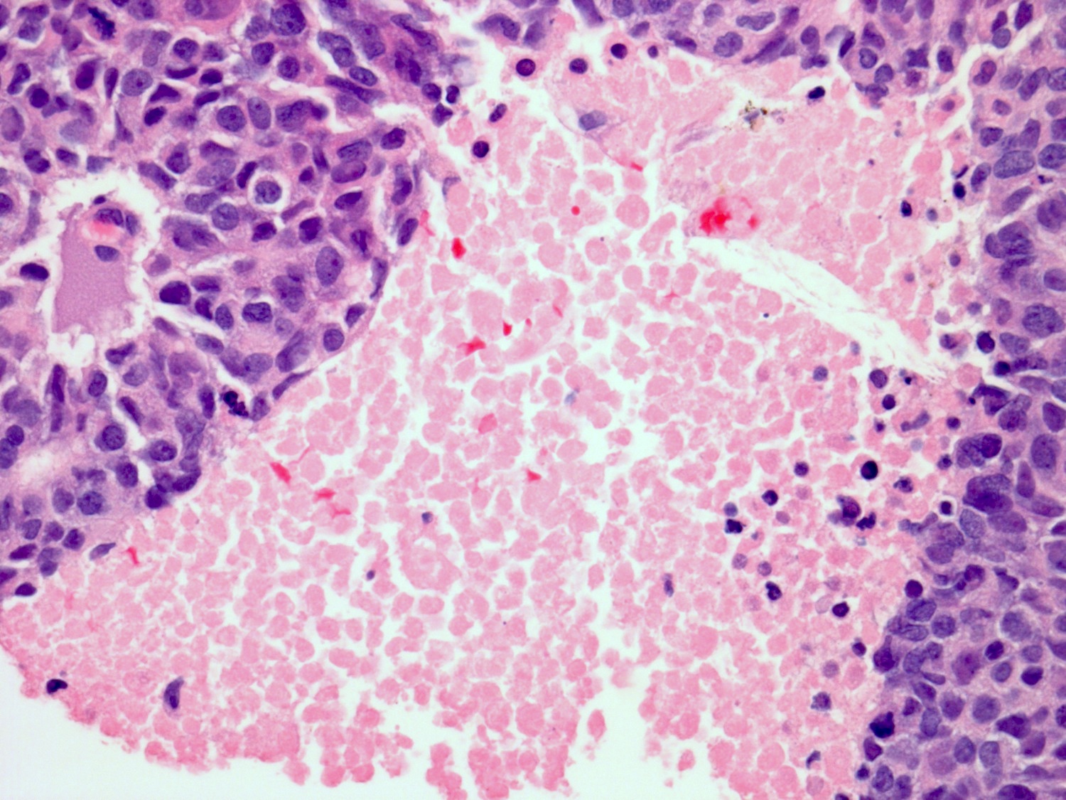
Focal mild reactive atypia
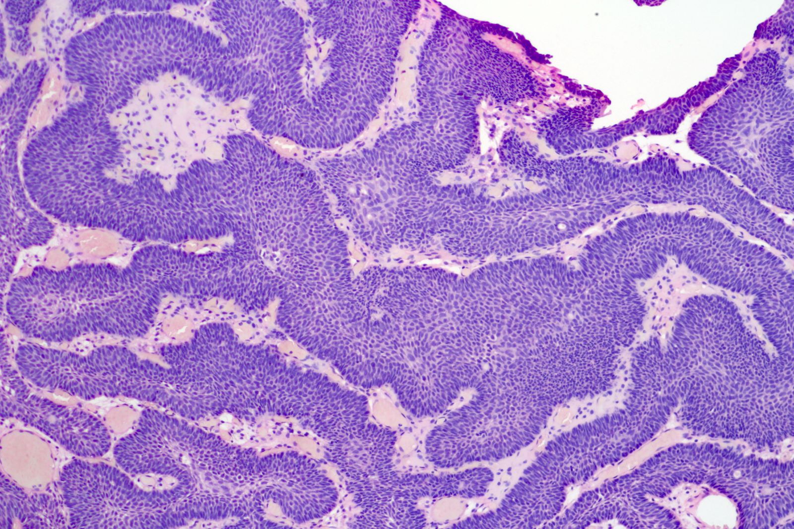
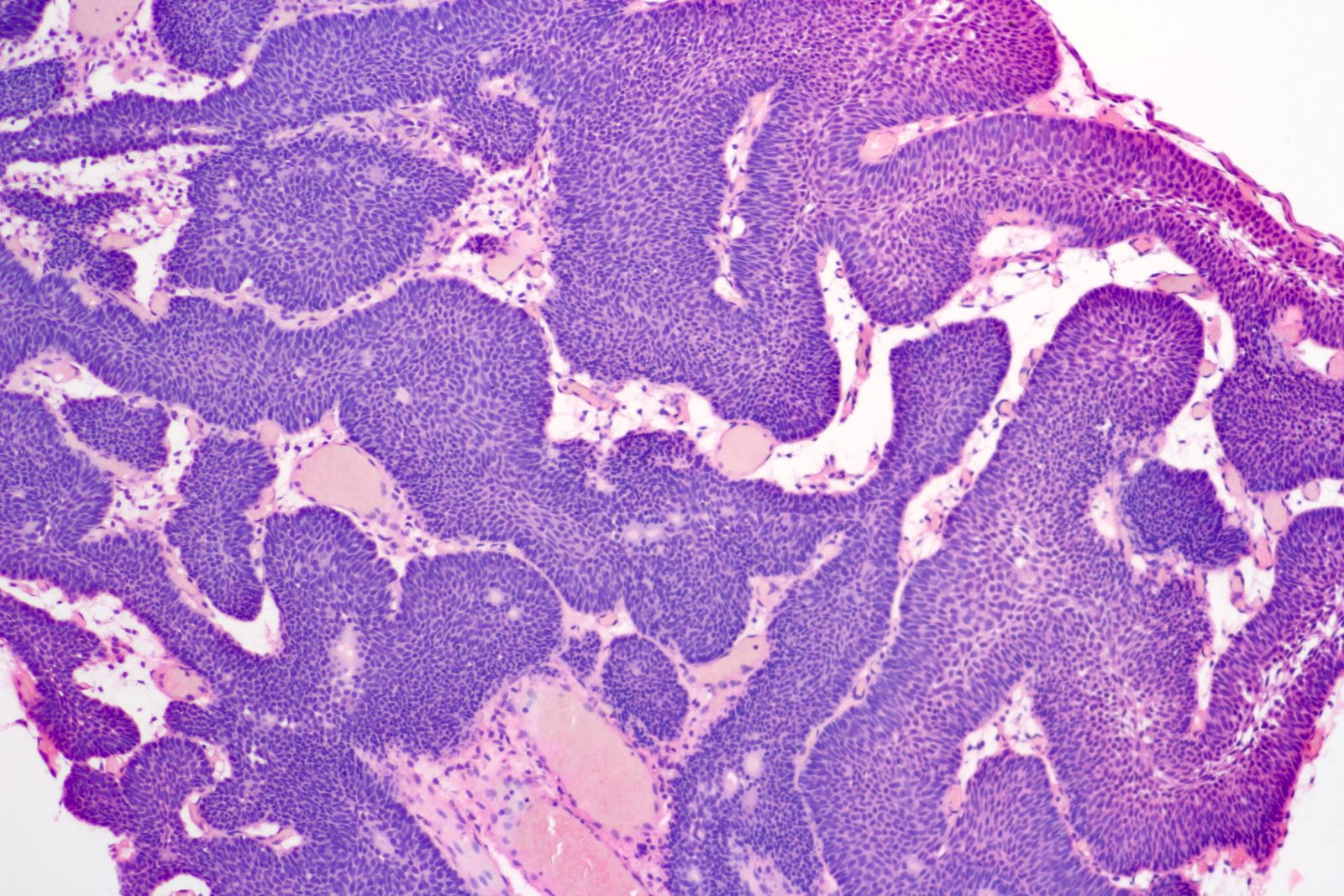
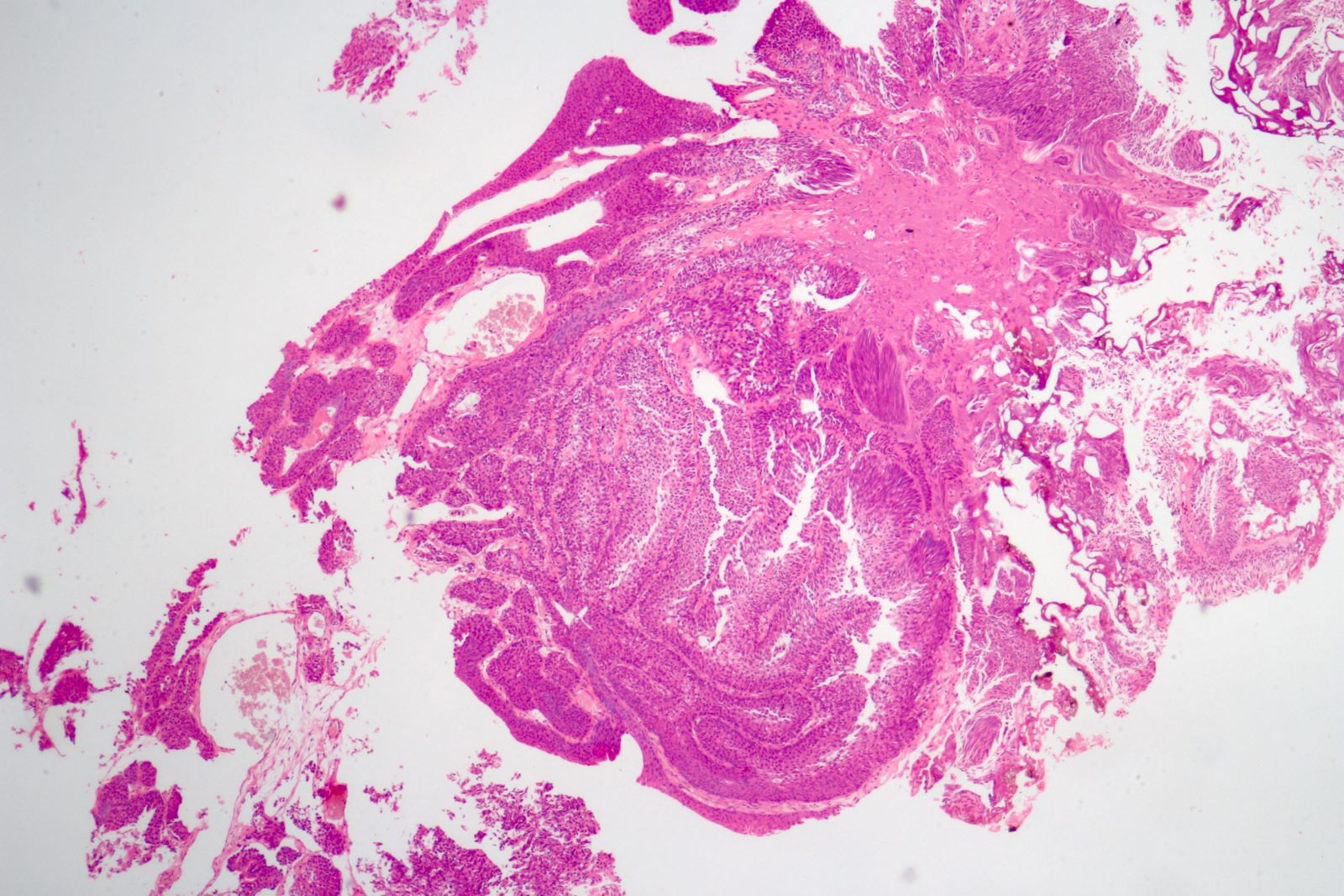
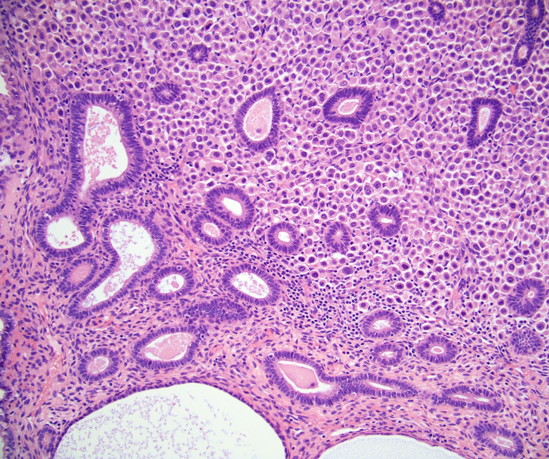
Inverted papilloma
Inverted papilloma - anastomosing cords
Thickened epithelium
Epithelium / no atypia
Inverted papilloma
Inverted papilloma - no atypia
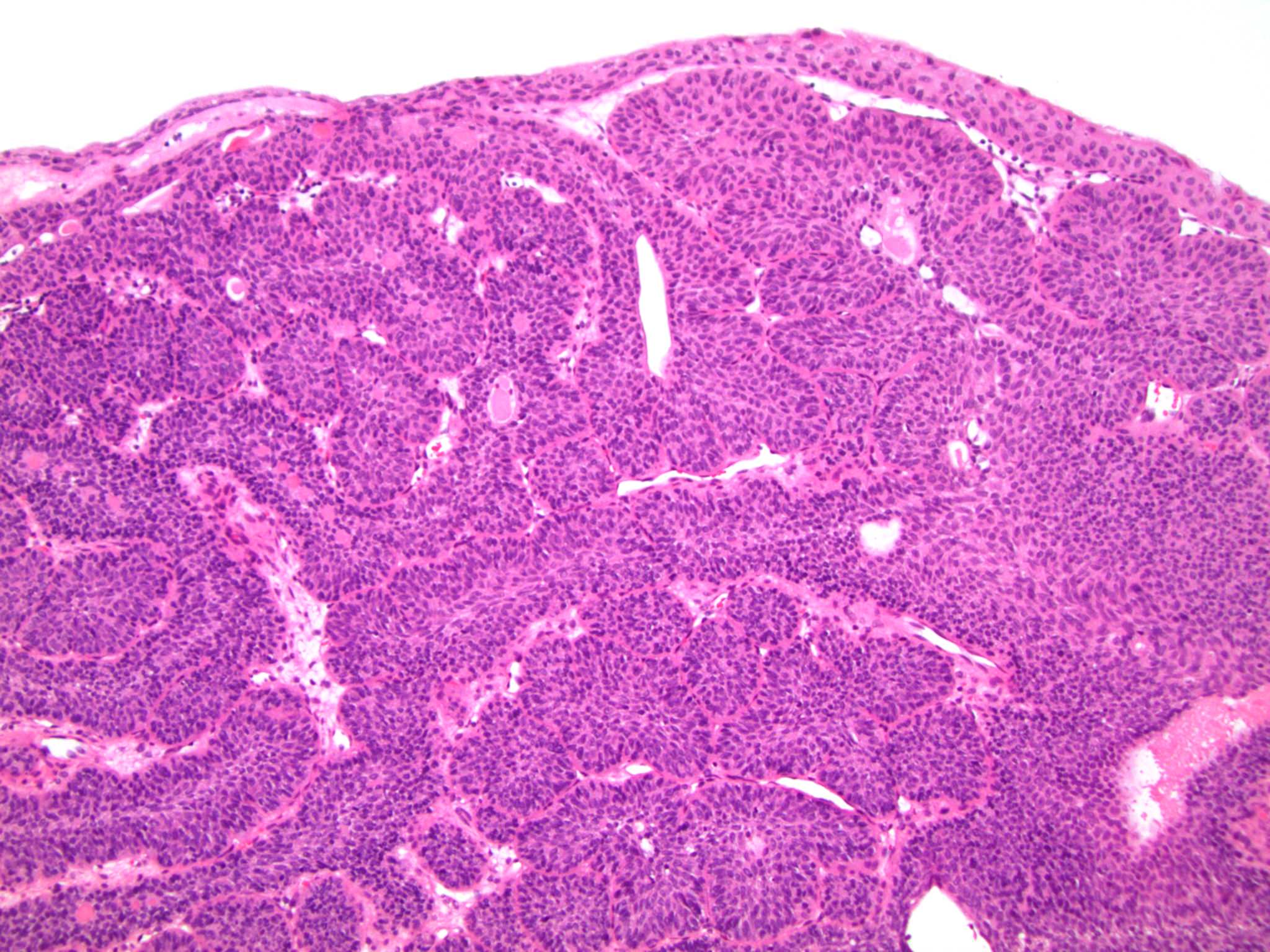
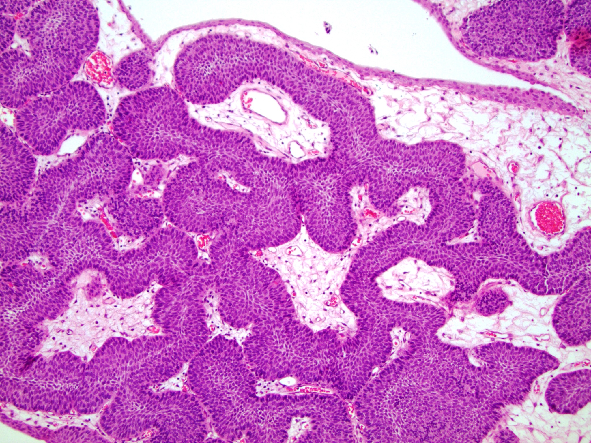
Surface with thin, flat urothelium
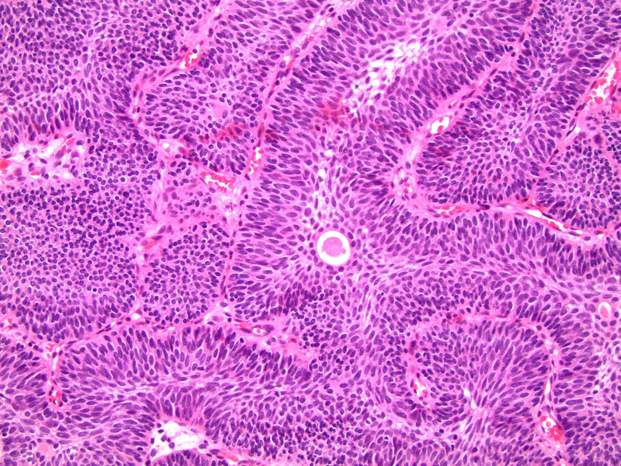
Anastomosing, basophilic cords
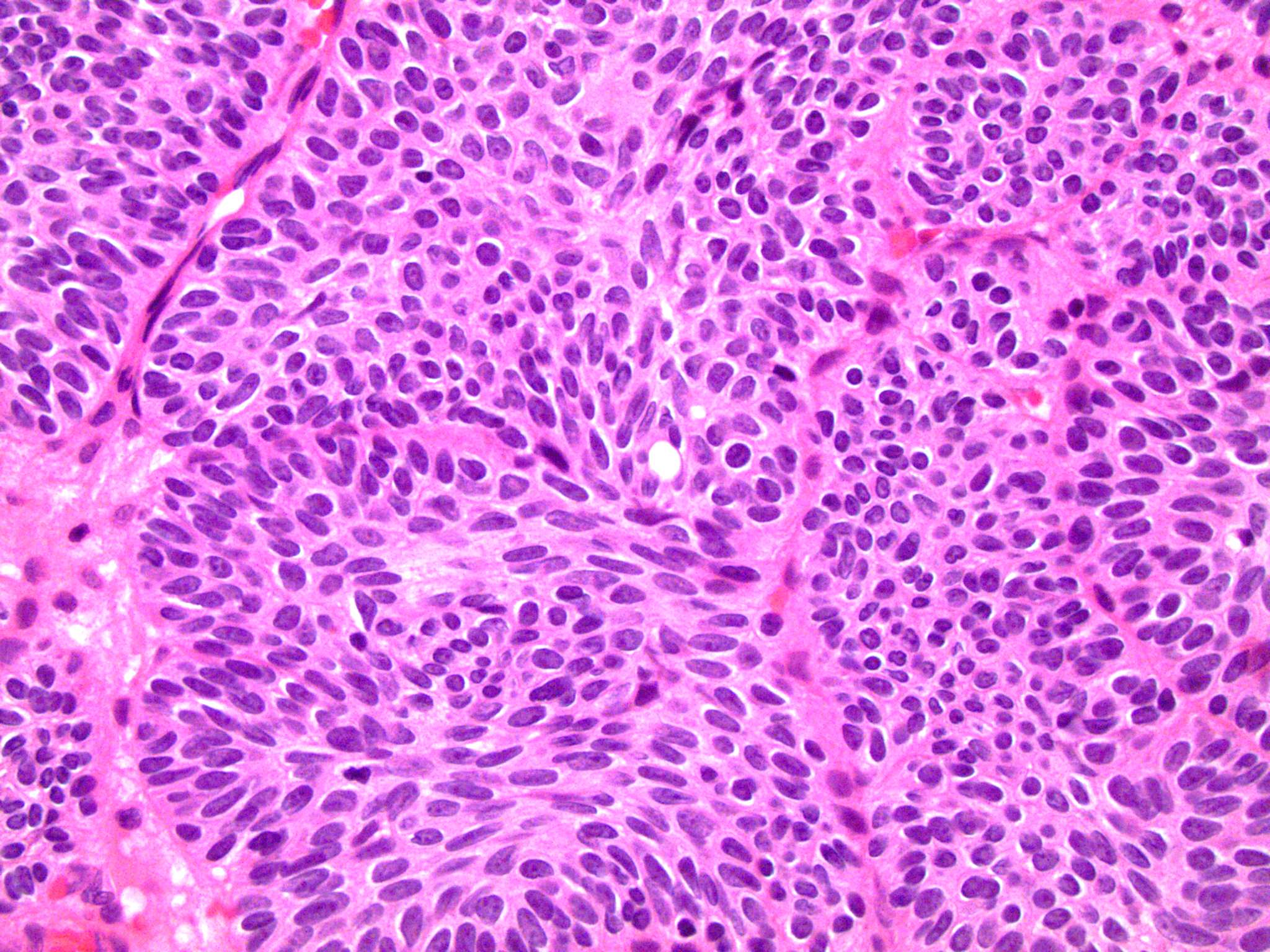
Oval nuclei
Contributed by Daniel Anderson, M.D., M.B.A.
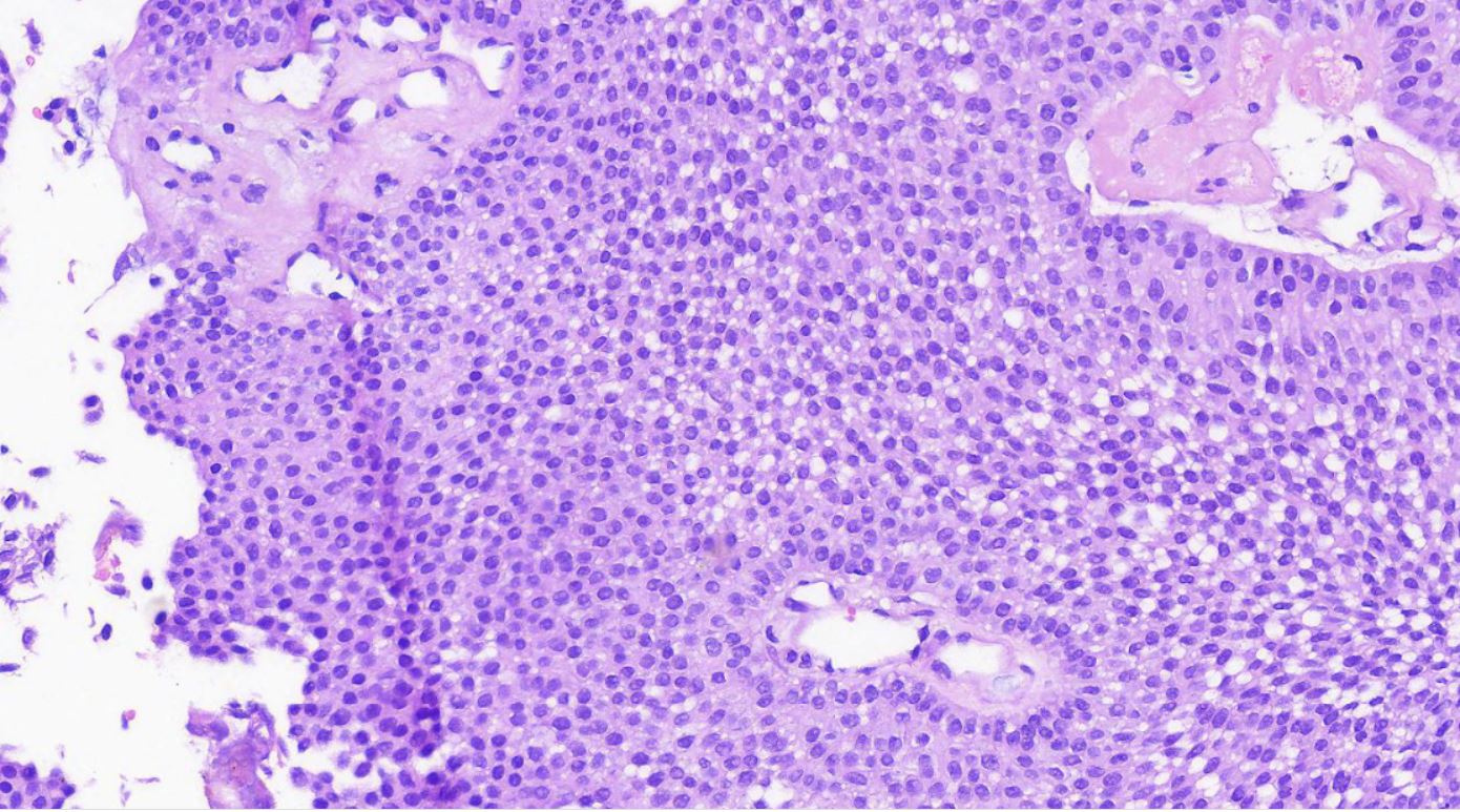
Solid growth
Necrosis and nuclear detail
Mitotic figures
CD56
Ki67
Synaptophysin
Images hosted on other servers:

Prominent nucleoli shown in pleural fluid
Contributed by Ankur Sangoi, M.D.


Images hosted on other servers:

Enhancing lesion from right renal pelvis
Images hosted on other servers:

Renal
pelvic tumor
invading parenchyma
and hilar fat
Contributed by Maria Tretiakova, M.D., Ph.D.
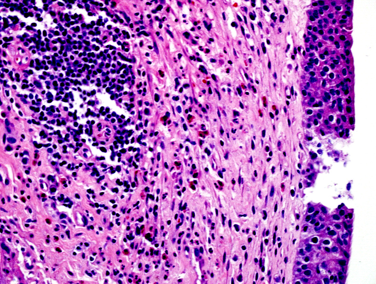
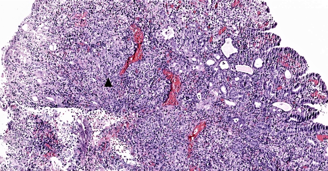
Syncytial growth, inflammation
Prominent nucleoli
Syncytial growth, prominent nucleoli
Syncytial growth, mitosis
Images hosted on other servers:
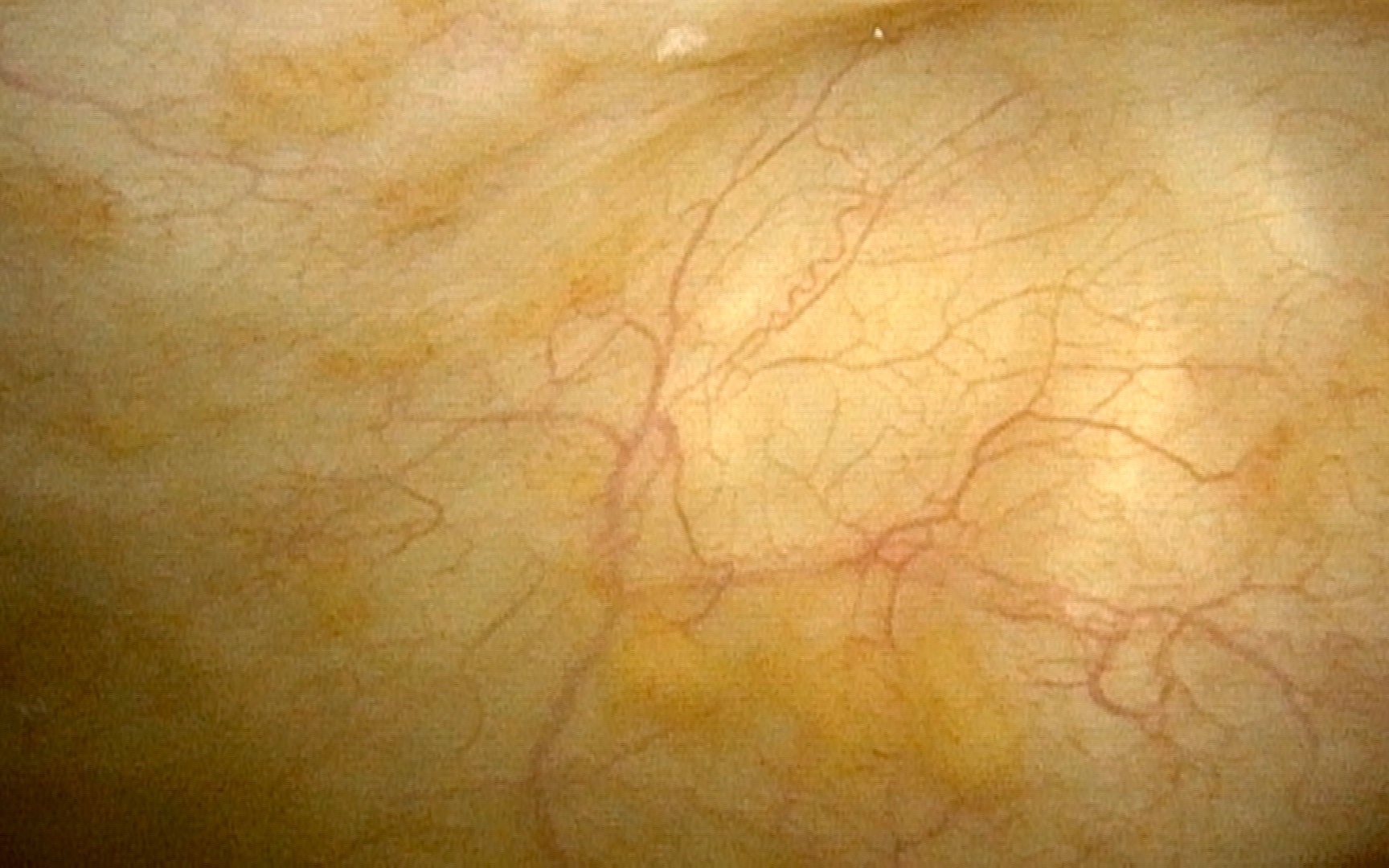
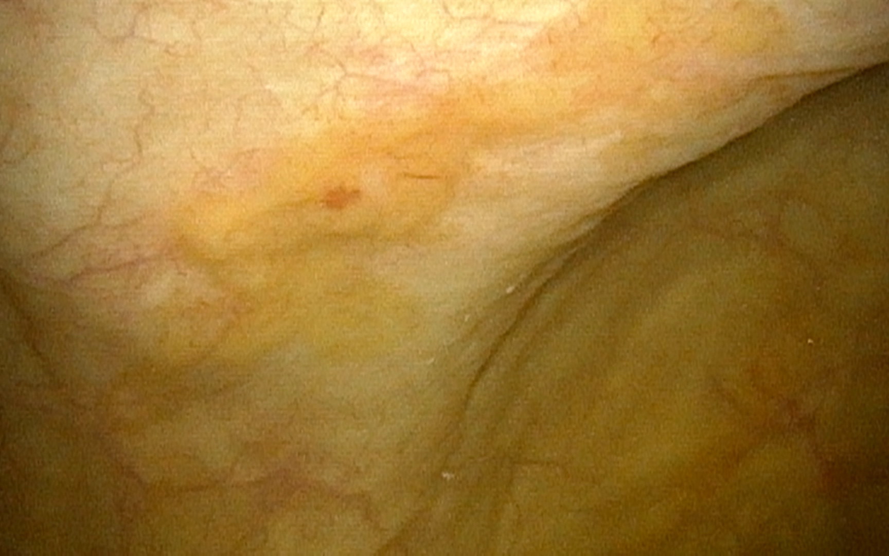
Endometriosis

Laparoscopic segmental cystectomy
AFIP images
Endosalpingiosis

Ovary: glands lined by ciliated epithelium lie in fibrous stroma
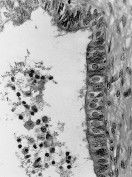
Ovary: ciliated,
secretory and
intercalated cells
line the cystic space
Images hosted on other servers:
Endocervicosis

Prominent
endocervical
glands in
muscularis propria

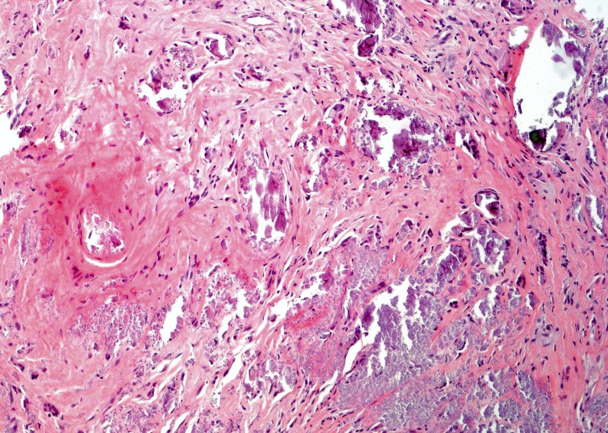
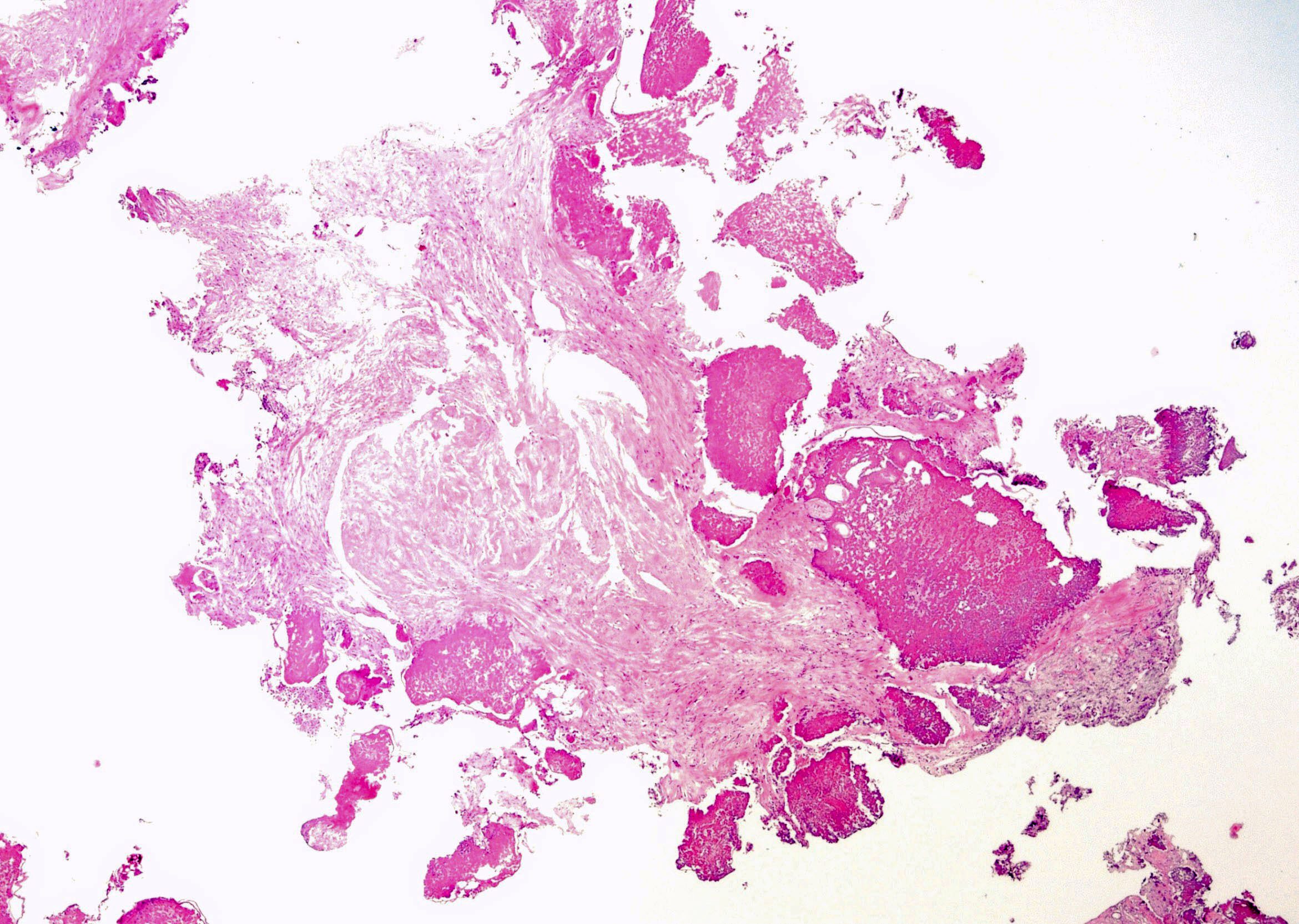
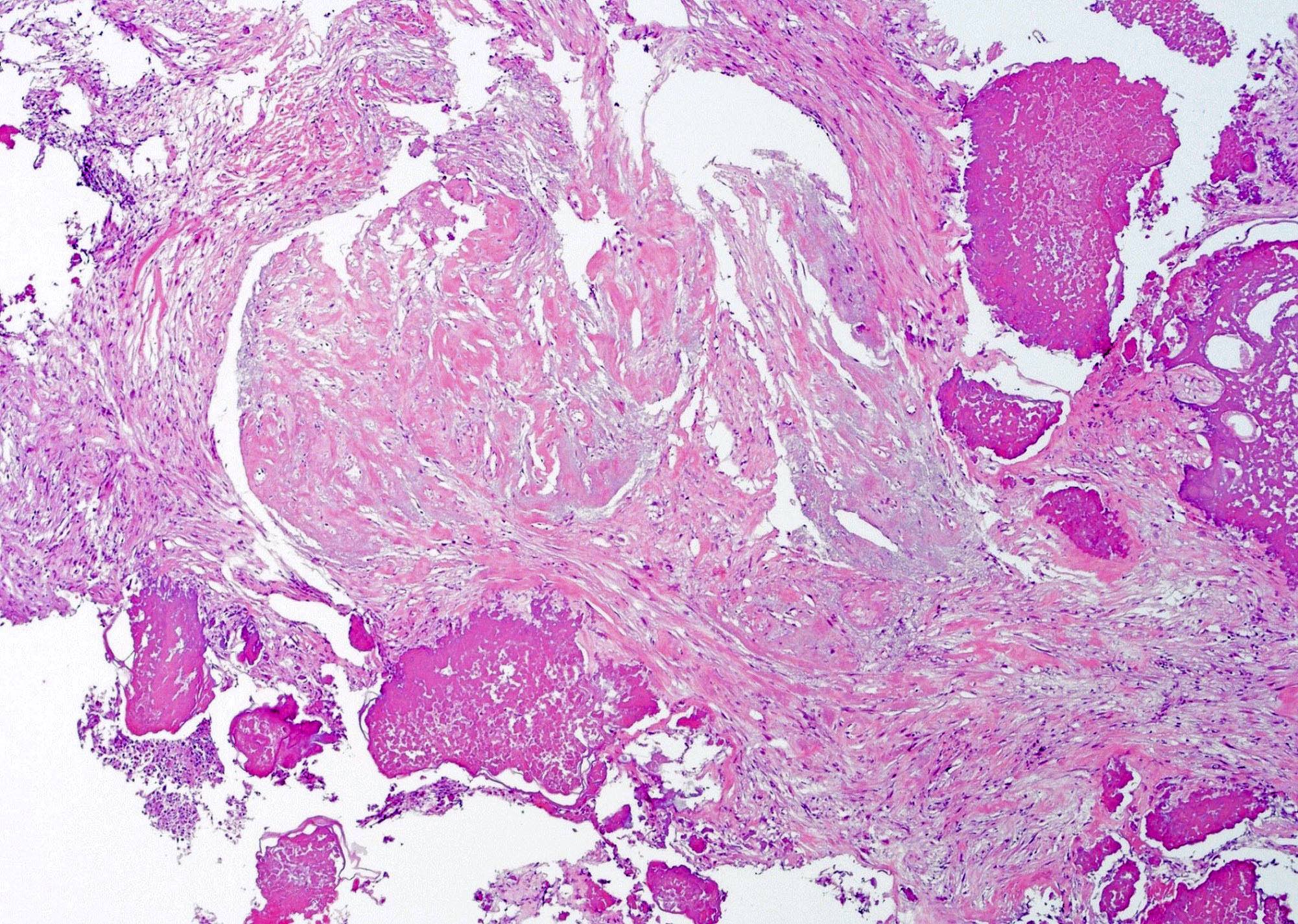
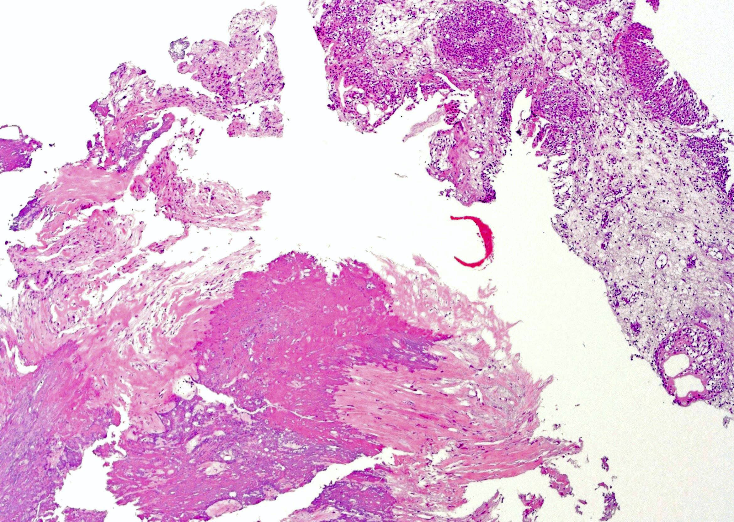
Complex cystic lesion

Columnar cells with granular mucinous apical cytoplasm
Endometriosis

Endometrial glands and
stroma involving the
peri-ureteral soft tissue
Contributed by Zachary Gordon, M.D., G. Bailey, M.D. and the Genitourinary Pathology Society (Case #496)

White light cystoscopy
Narrow band cystoscopy
Cystoscopic view of bladder lesions
Contributed by Cheng Wang, M.D. and the Genitourinary Pathology Society (Case #496)
Various images
PAS stain
Von Kossa stain
Images hosted on other servers:
Malakoplakia (arrows at Michaelis-Gutmann bodies)

von Kossa calcium stain

CD68
Contributed by Zachary Gordon, M.D.
White light cystoscopy
Narrow band cystoscopy
Contributed by Mustafa Goksel, M.D.
Sheets of eosinophilic histiocytes
Urinary bladder with histiocytic infiltration
Numerous intracytoplasmic inclusions
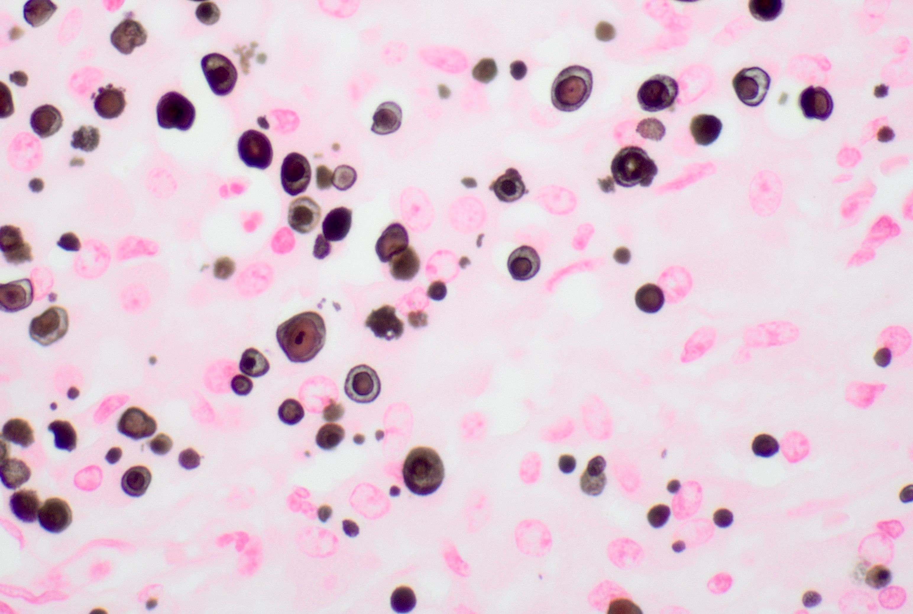
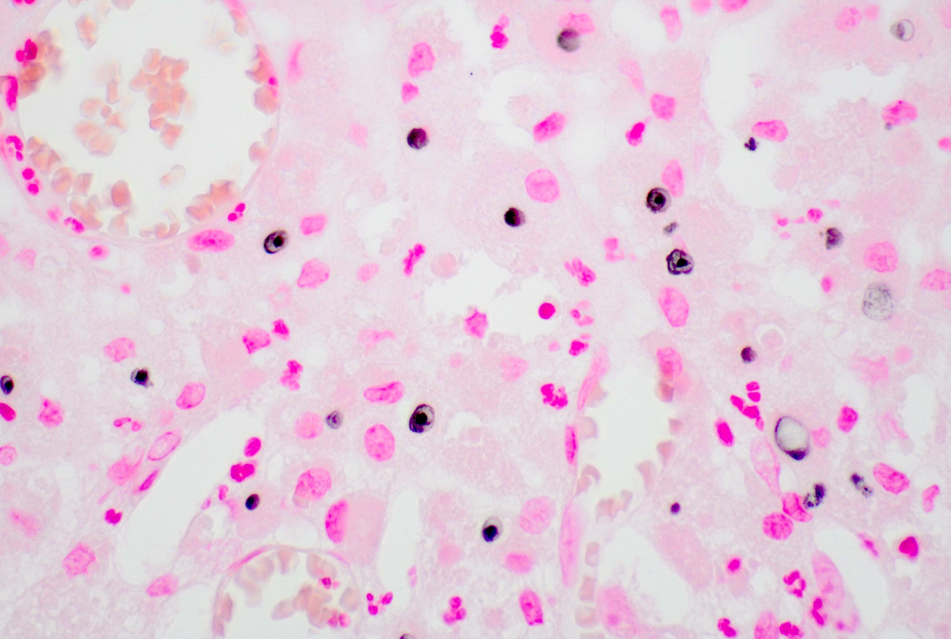
Von Kossa stain
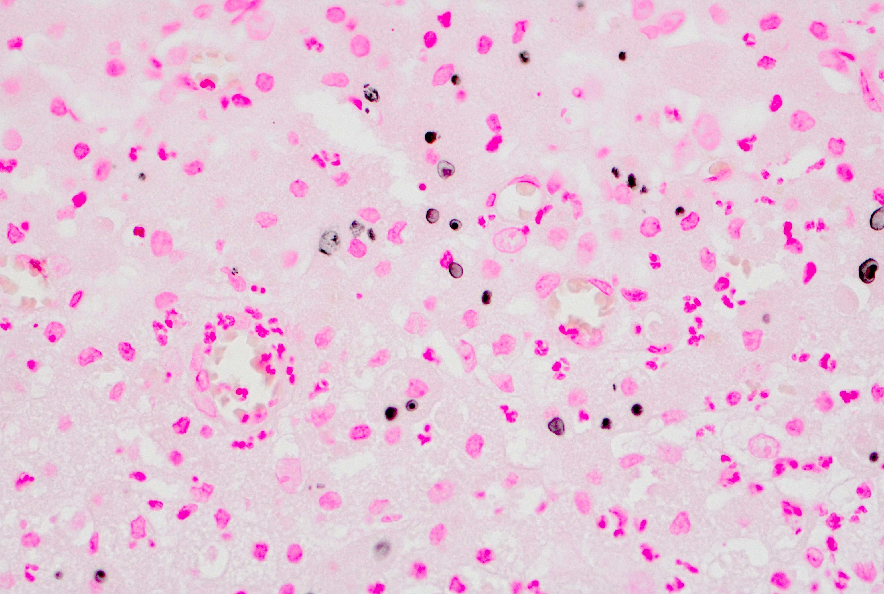
Von Kossa stain
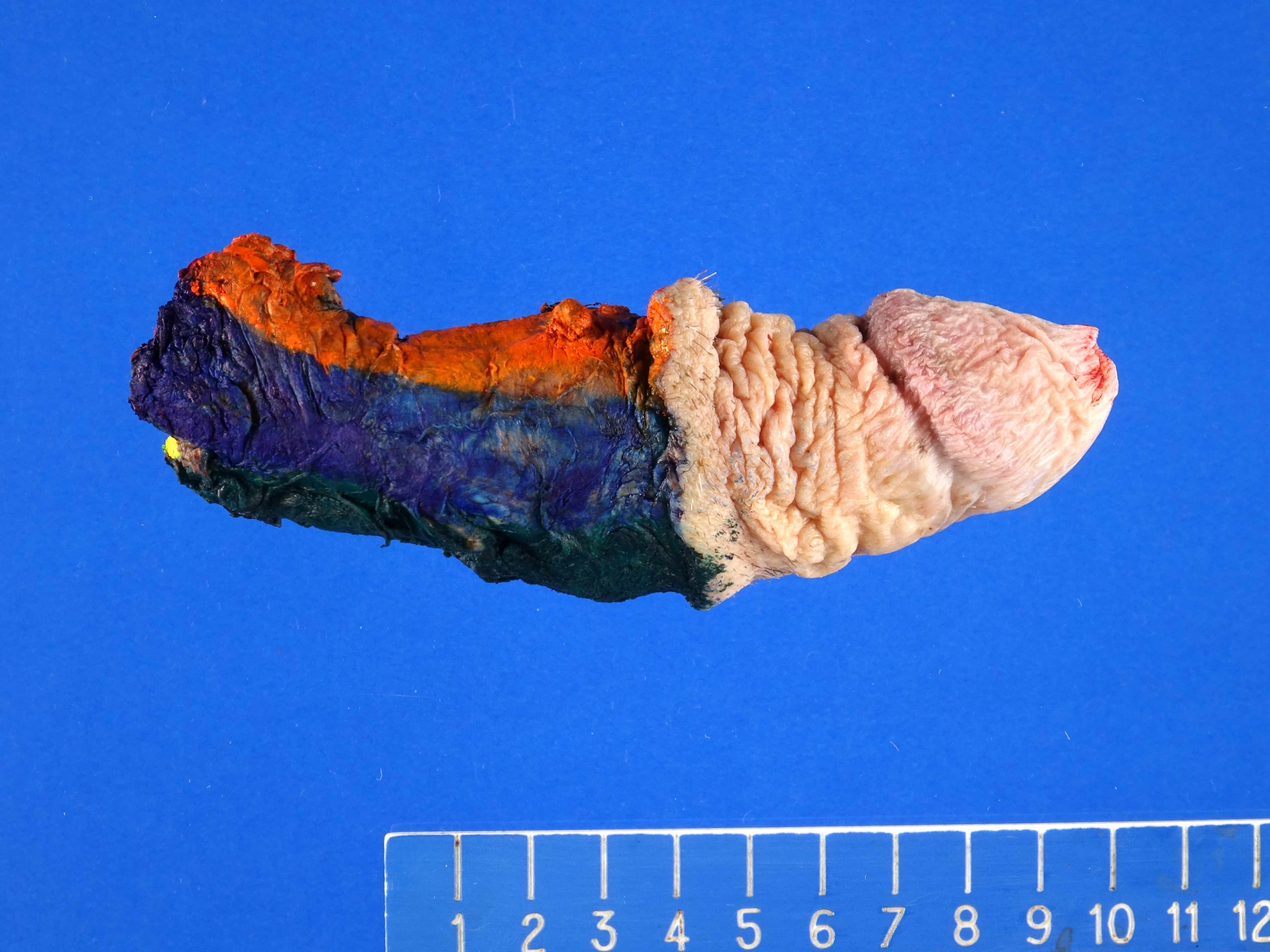
Contributed by Jesus Adrian Chavez, M.D. and Debra Zynger, M.D.
Penile urethra with periurethral involvement
Contributed by Jesus Adrian Chavez, M.D. and Debra Zynger, M.D.

Low grade noninvasive papillary urothelial carcinoma
Penile urethra, squamous cell carcinoma
Penile urethra, HPV+ high risk ISH
Necrosis and keratinization
Clear cell adenocarcinoma:

Can mimic nephrogenic metaplasia
With hobnailing
With prominent clear cells and diffuse, sheet-like growth
Images hosted on other servers:

Metastatic appendiceal
mucinous adenocarcinoma
- various images

Metastatic breast cancer: H&E, PR, HER2
Contributed by Bonnie Choy, M.D.
Prostatic adenocarcinoma
NKX3.1
Colorectal adenocarcinoma
Renal cell carcinoma
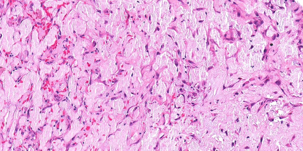
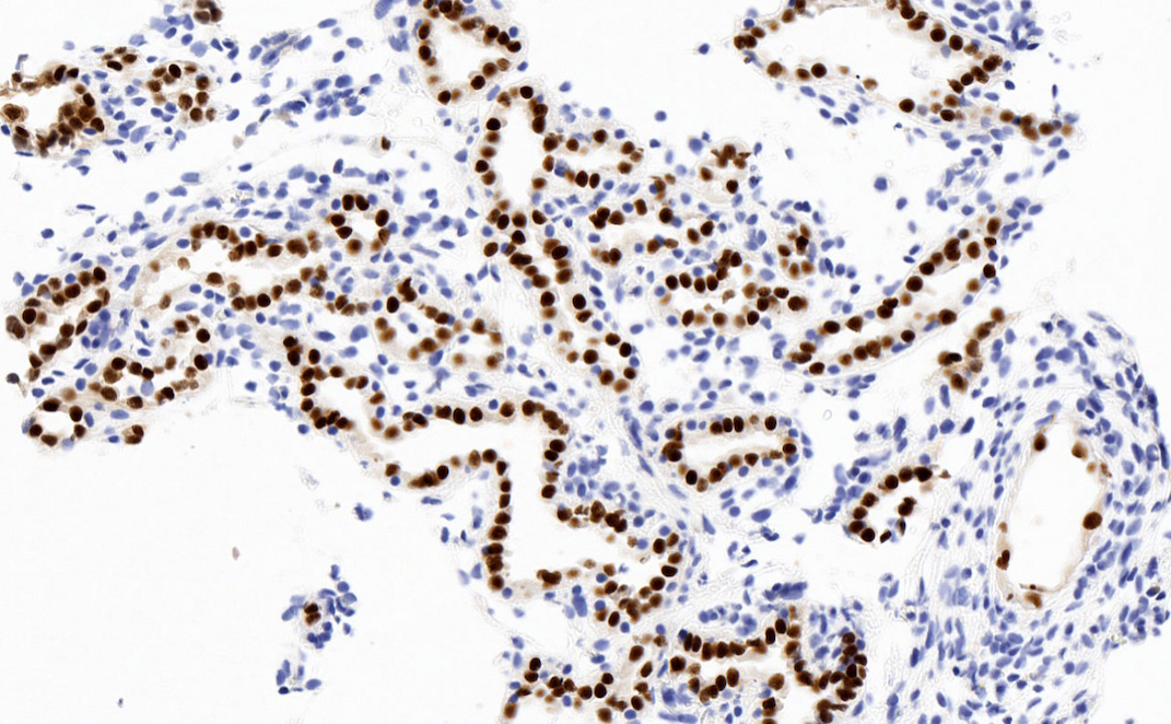
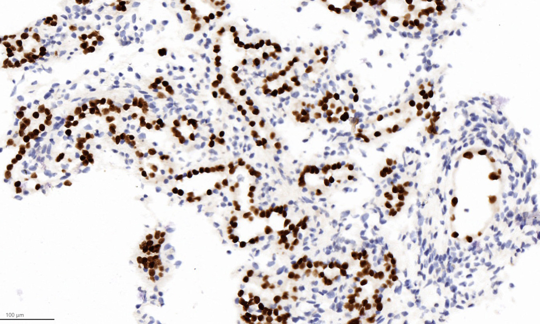
Contributed by Megan L. Brown, M.D. and Maria Tretiakova, M.D., Ph.D.
Microcysts, macrocysts and tubules
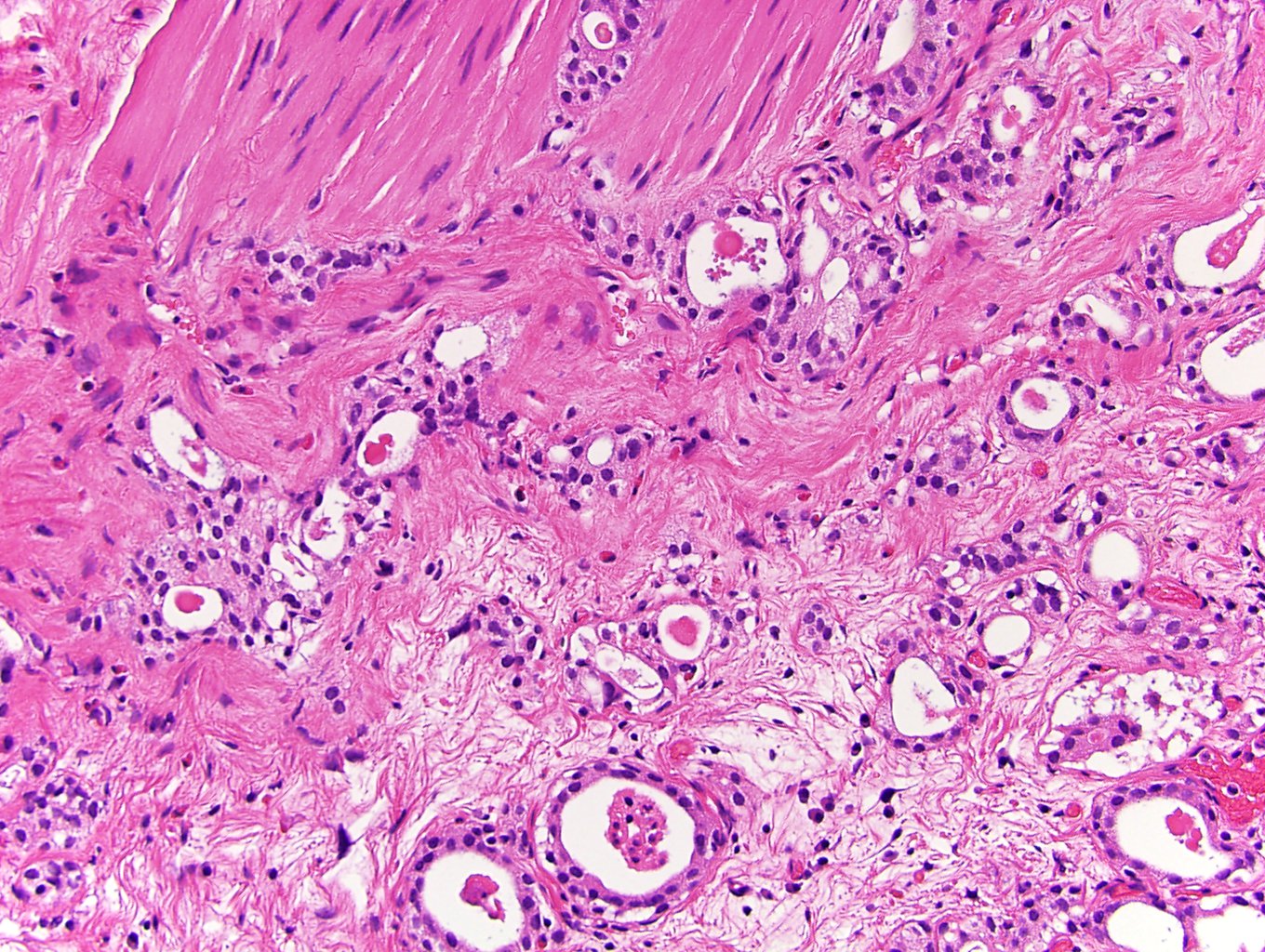
Invasion to muscularis propria
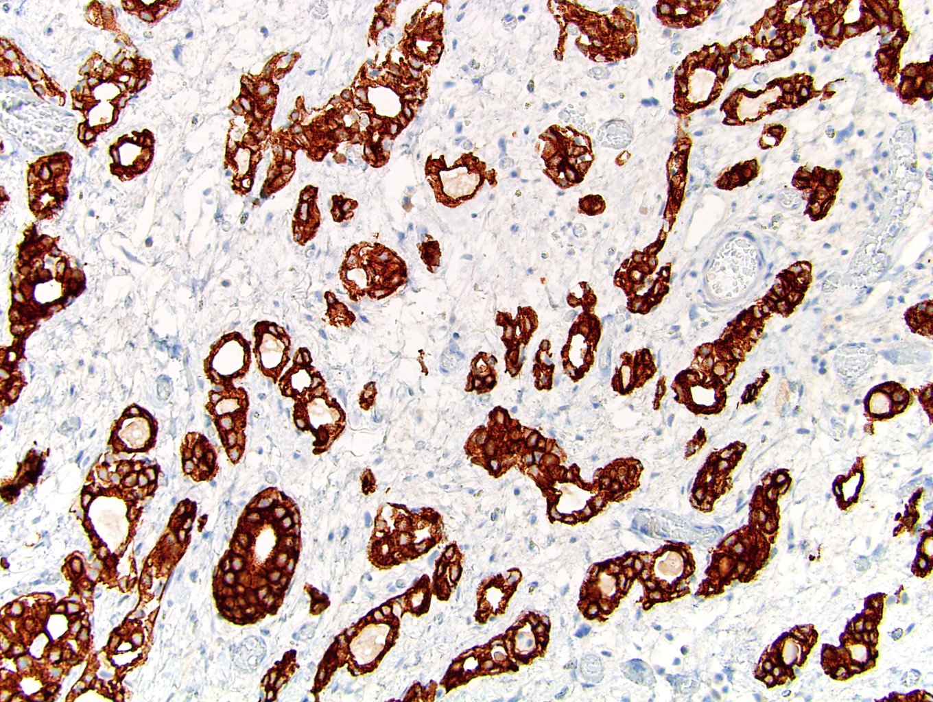
Cytokeratin 7
Images hosted on other servers:

CT with bladder tumor invading the left ureteral orifice

CT with tumor in right posterolateral bladder wall
Contributed by Timothy Isaac Miller, M.D., M.A., Maria Tretiakova, M.D., Ph.D. and @katcollmd on Twitter
Classic features
Micropapillary
and conventional
urothelial
carcinoma
Multiple nests throughout stroma

Epithelial ring morphology
Ring forms and intracytoplasmic vacuolization
Numerous nests in large lacunae
Micropapillary urothelial carcinoma
Micropapillary urothelial carcinoma
Images hosted on other servers:


Cohesive clusters in a 3 dimensional arrangement
Bladder urothelial carcinoma
Contributed by Jatin S. Gandhi, M.B.B.S., M.D., @MirunaPopescu13 on Twitter and @katcollmd on Twitter
With BCG granuloma
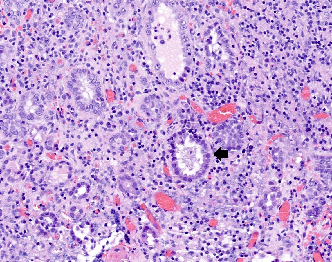
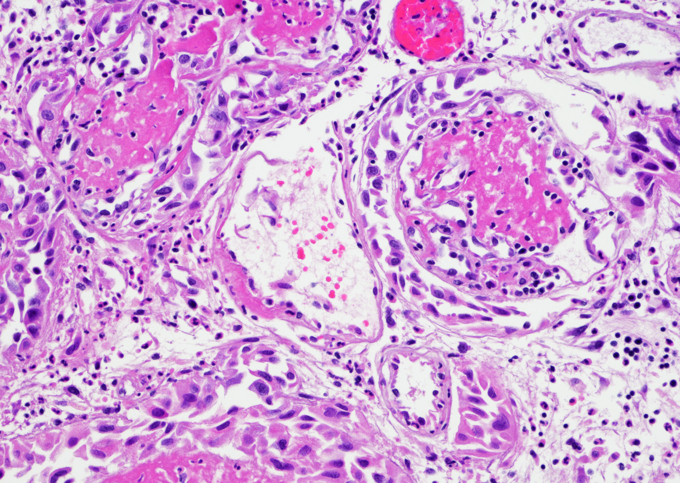
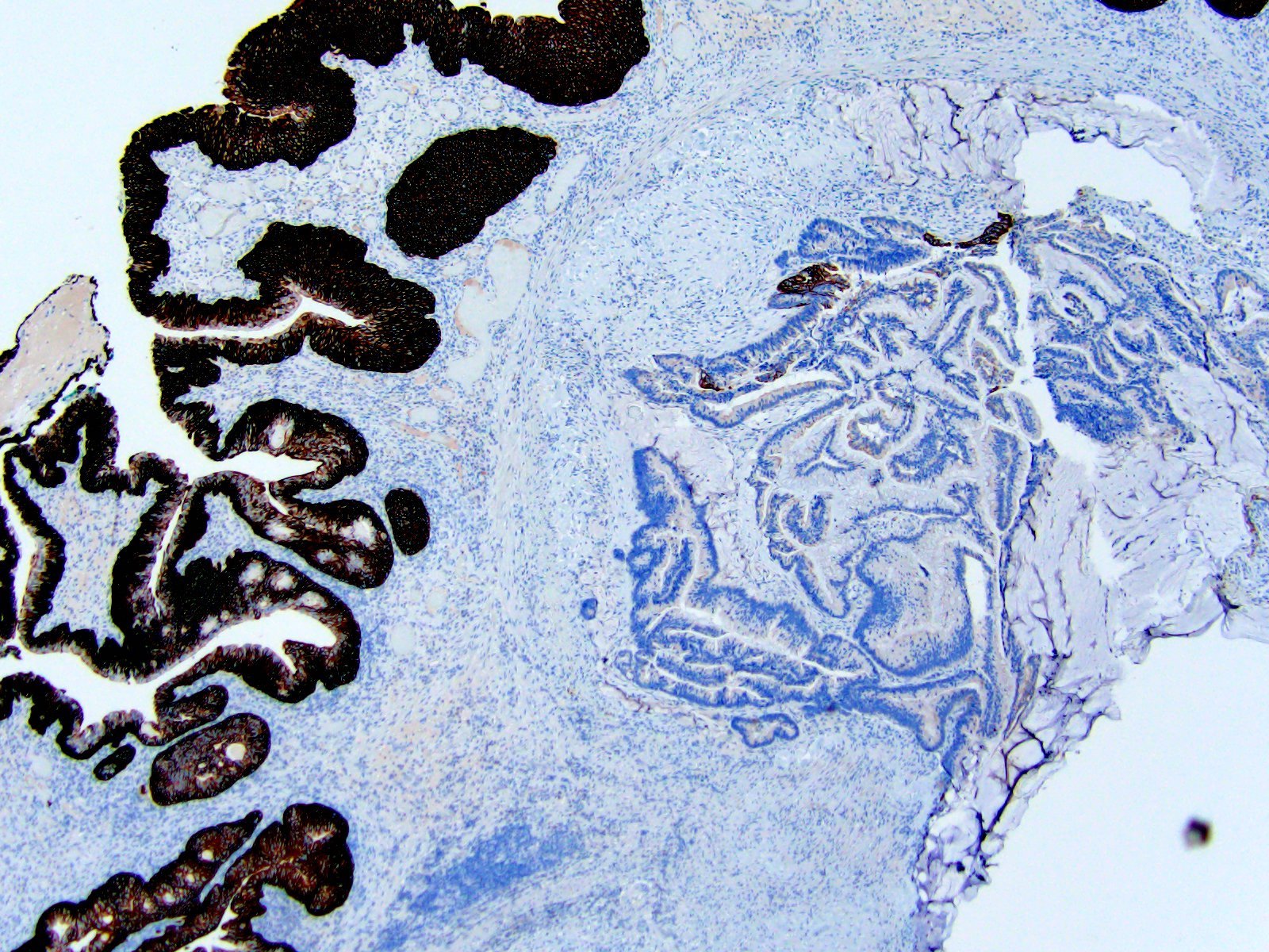
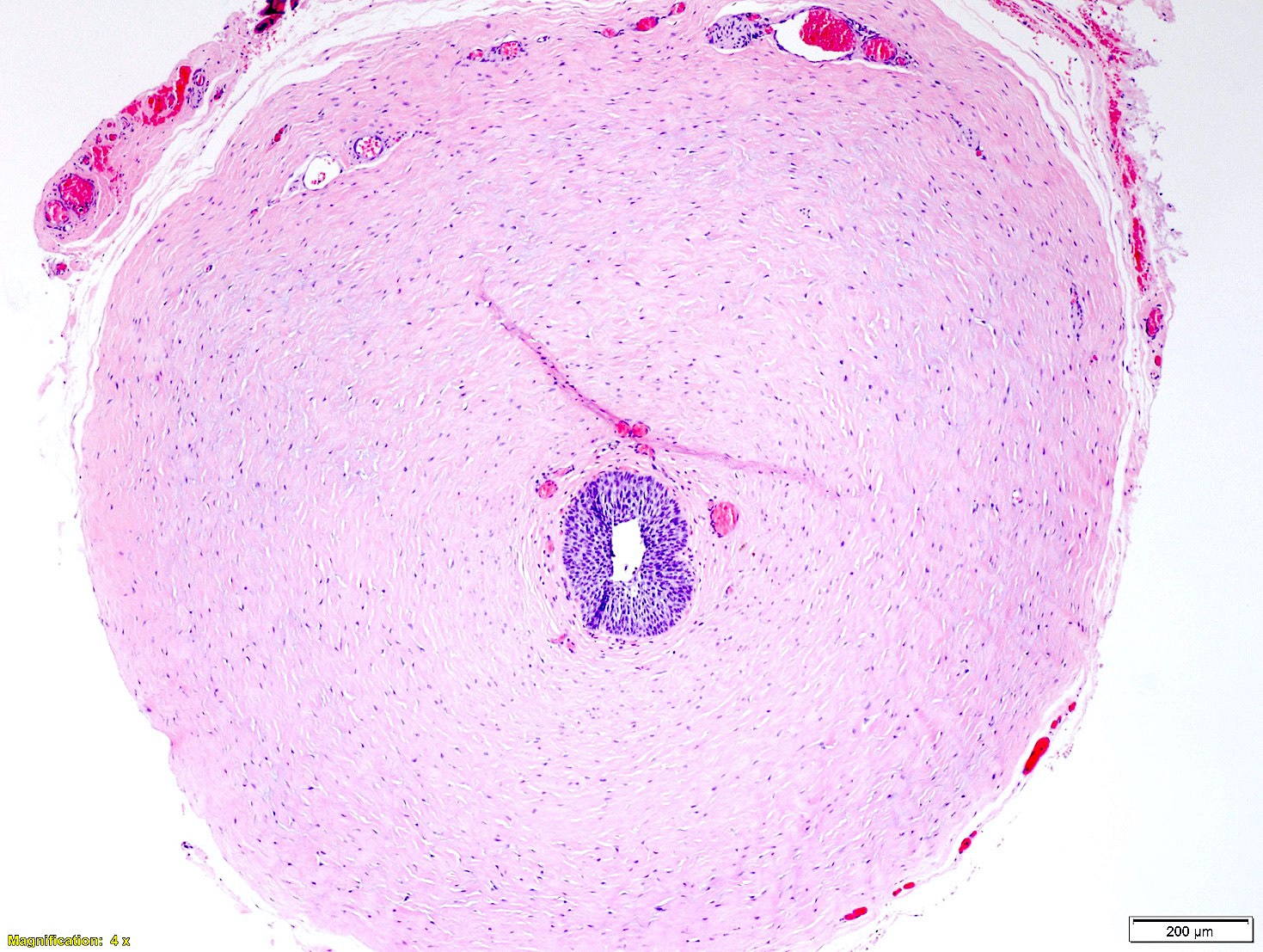
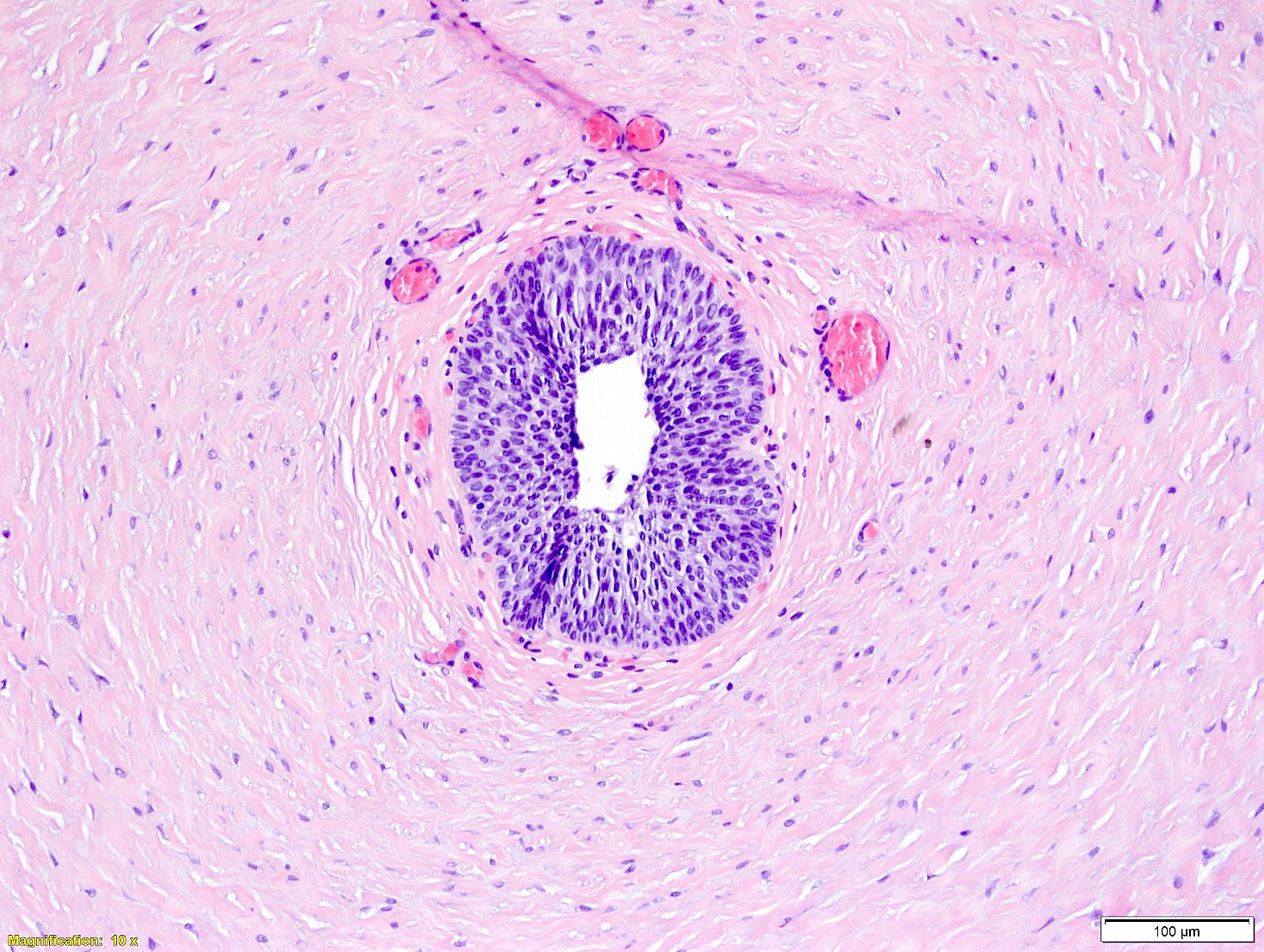
Peritubular basement membrane
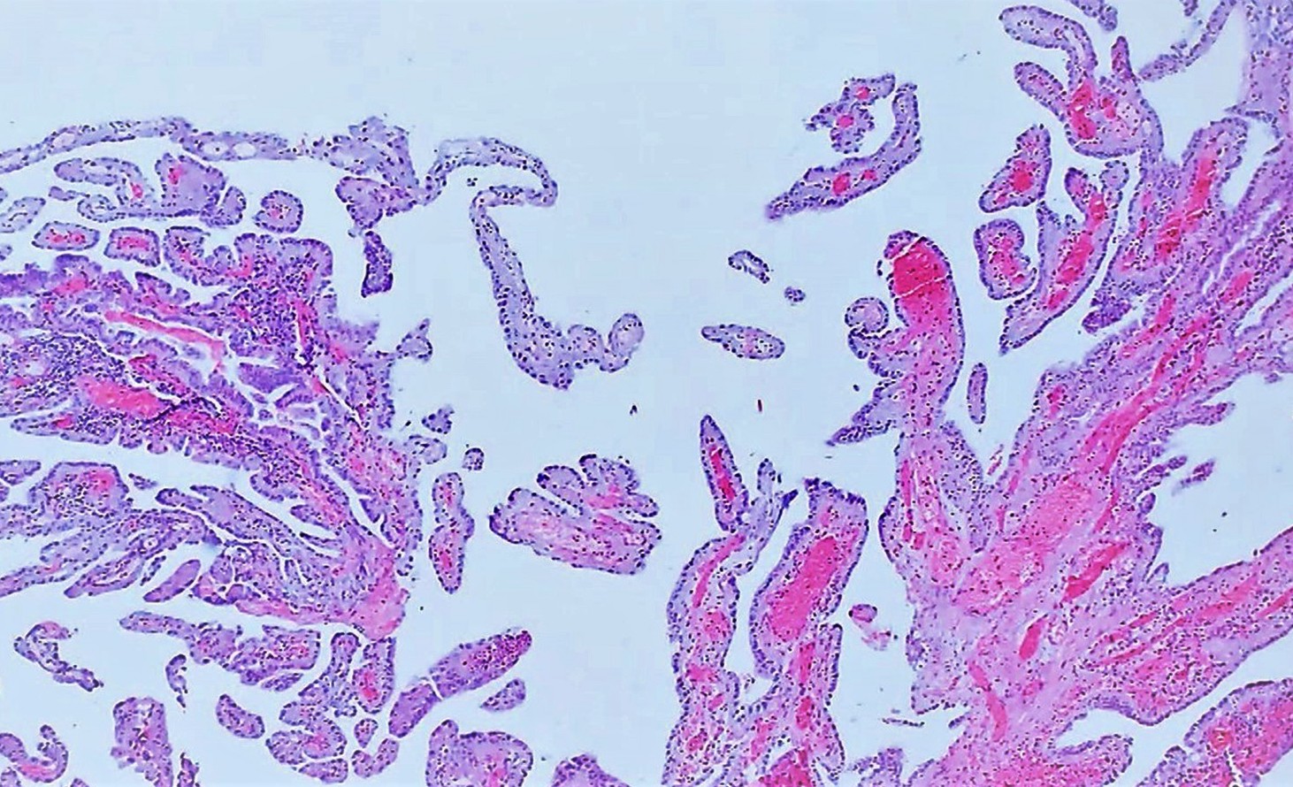
With papillary architecture
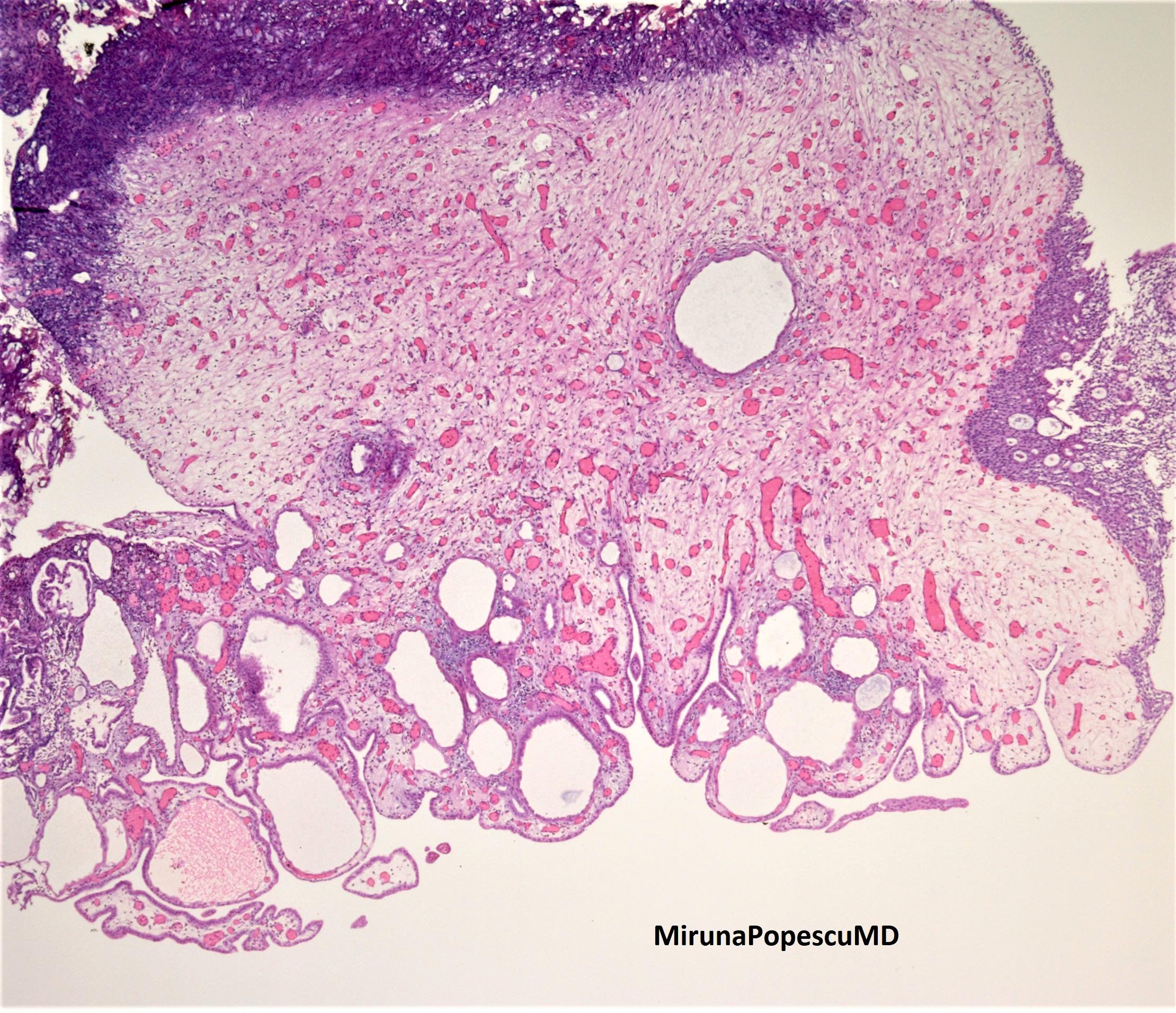
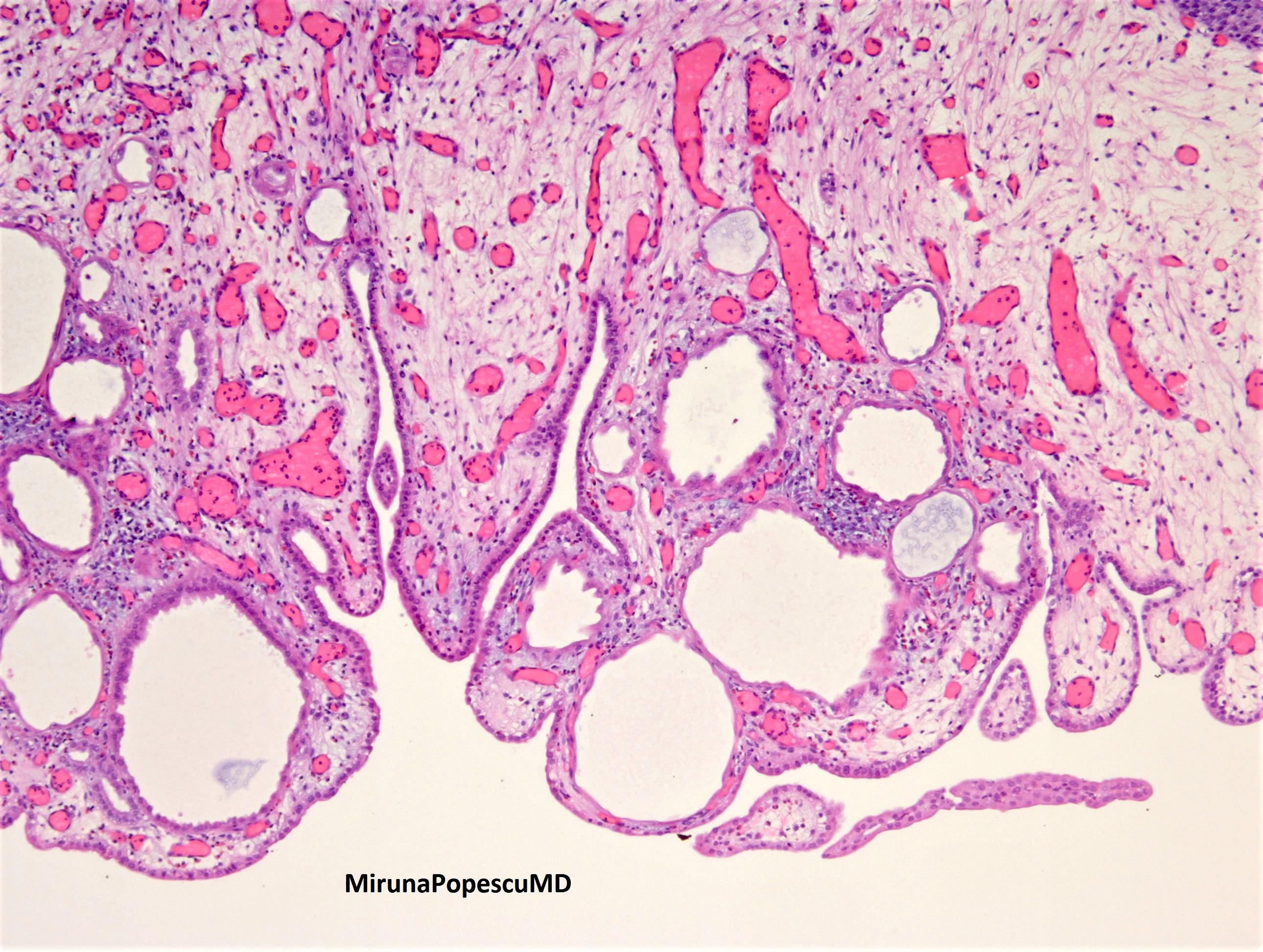
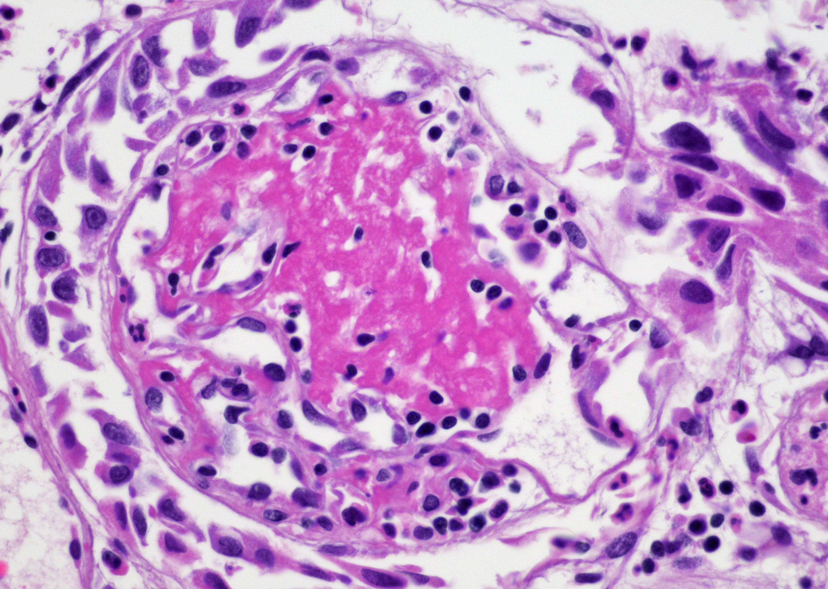
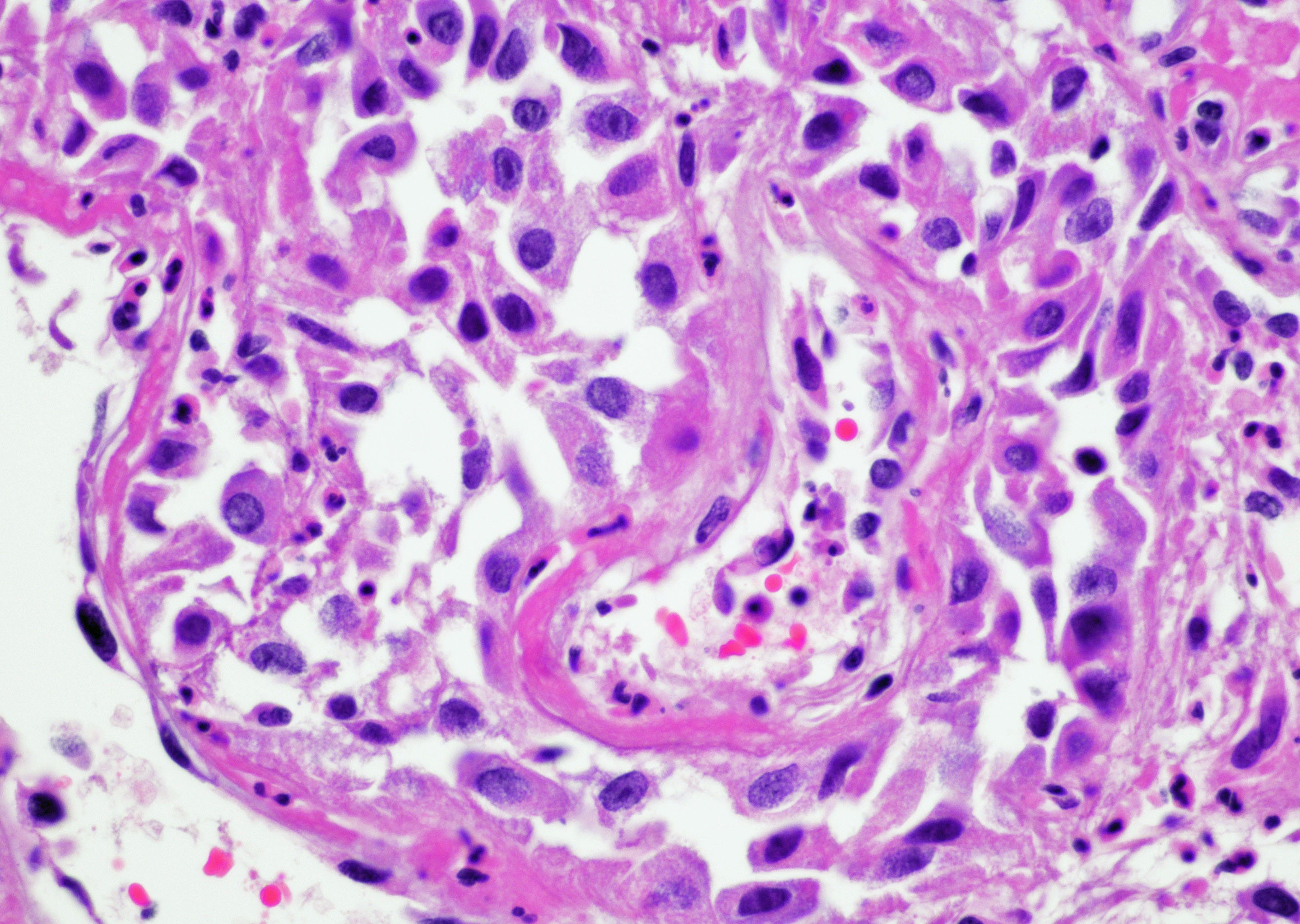
Nephrogenic metaplasia
Nephrogenic metaplasia
PAX8+
HNF1 beta+
Images hosted on other servers:

Gray-white tumor
Contributed by Megan L. Brown, M.D., Maria Tretiakova, M.D., Ph.D. and @SueEPig on Twitter
von Brunn-like appearance
Irregular projections and occasional lumens
Invasion into muscularis propria

Large nested carcinoma
Nested urothelial carcinoma
Contributed by Michelle R. Downes, M.D., Nicole K. Andeen, M.D. and Maria Tretiakova, M.D.
Cystectomy section
Transurethral bladder resection
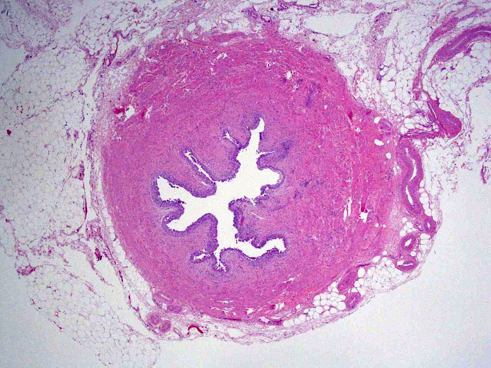
Cross section of ureter
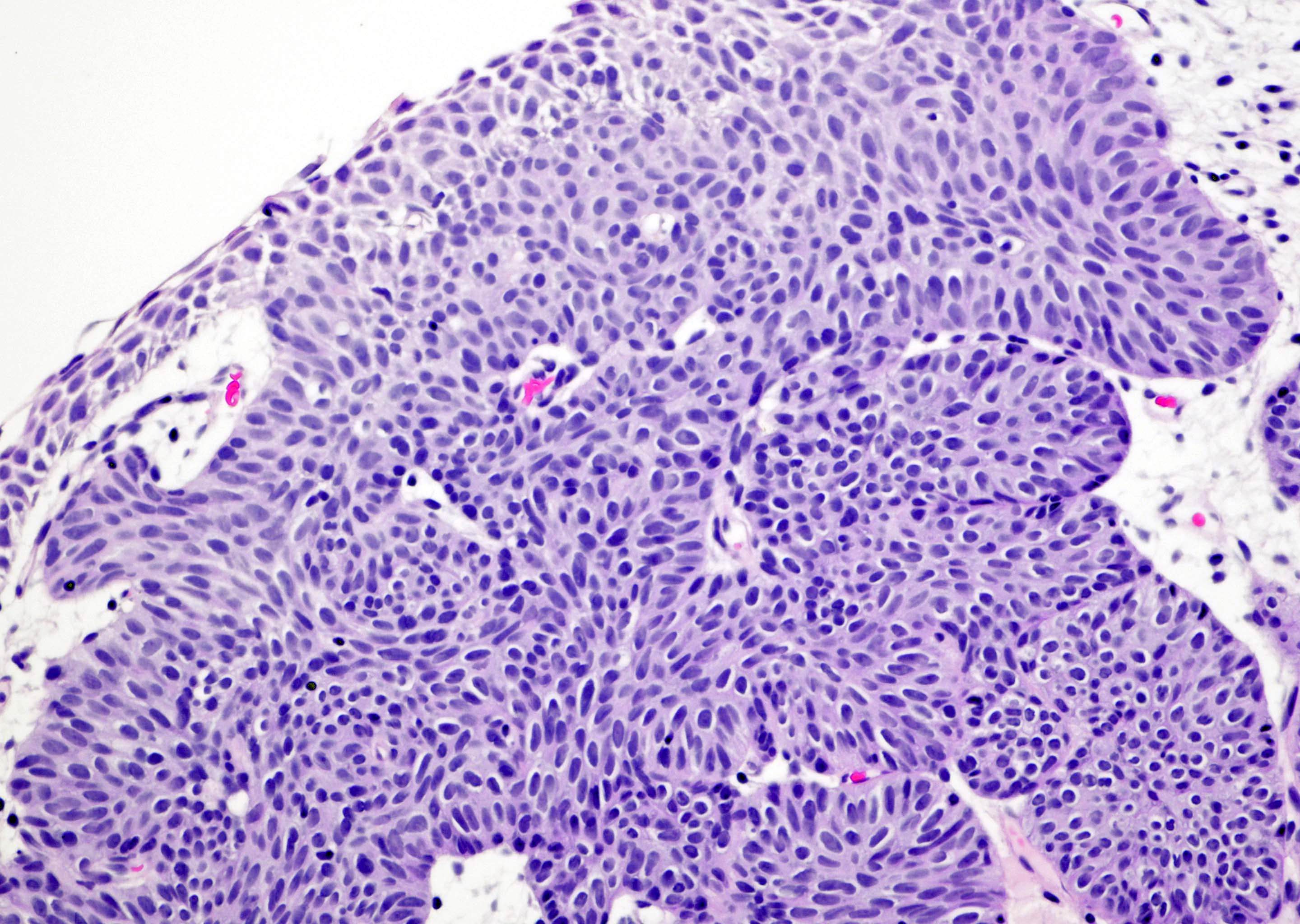
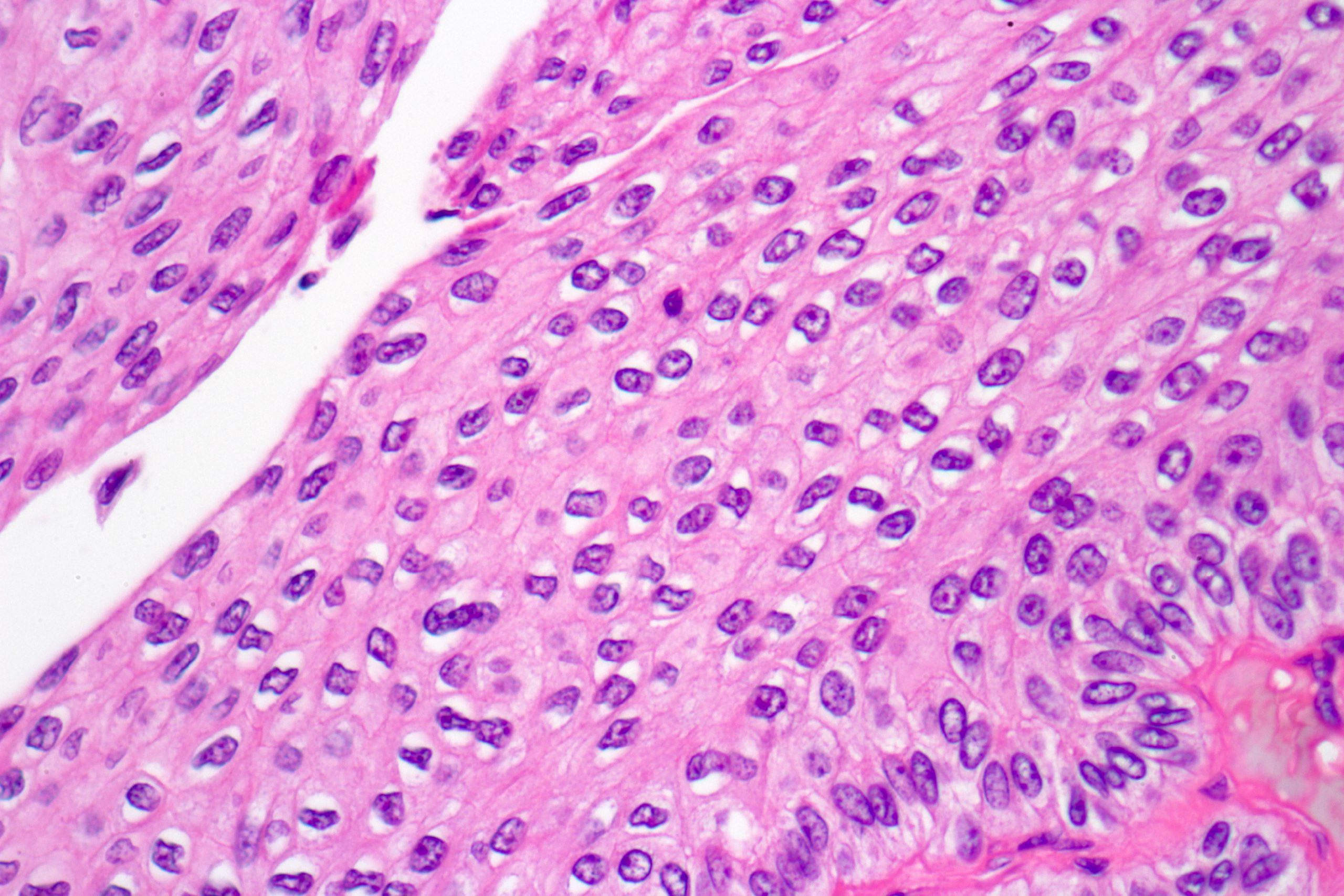
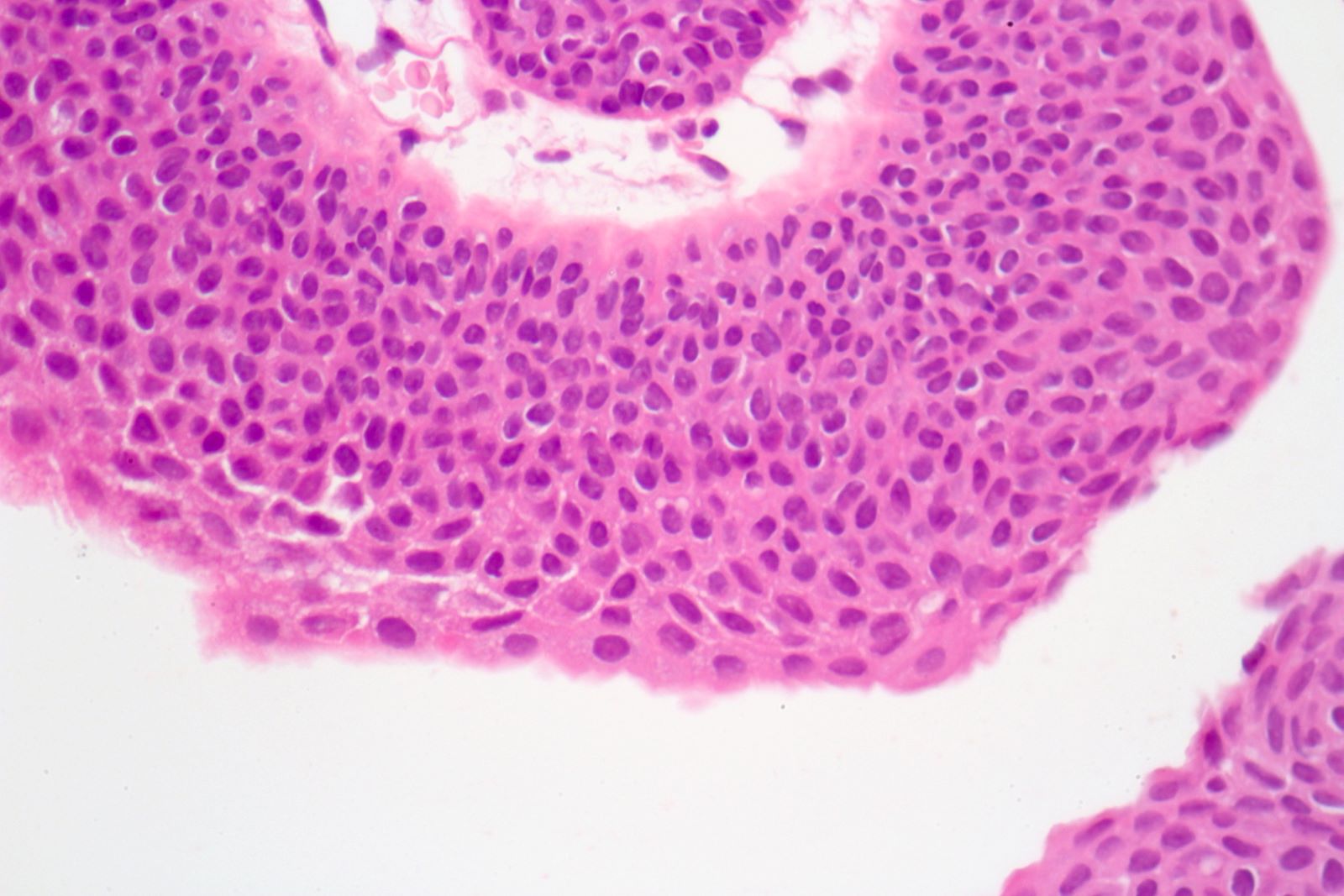
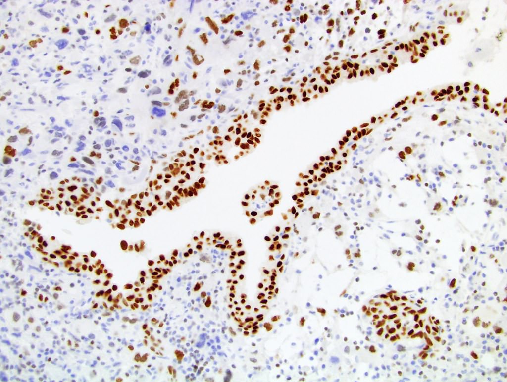
Architecture and cytology
Cytologic features
Noninvasive papillary urothelial carcinoma, high grade (pTa)
CK7 expression
GATA3 expression
CK5/6 loss
p53
Contributed by Zeina Ghorab, M.D. and Bonnie Choy, M.D.

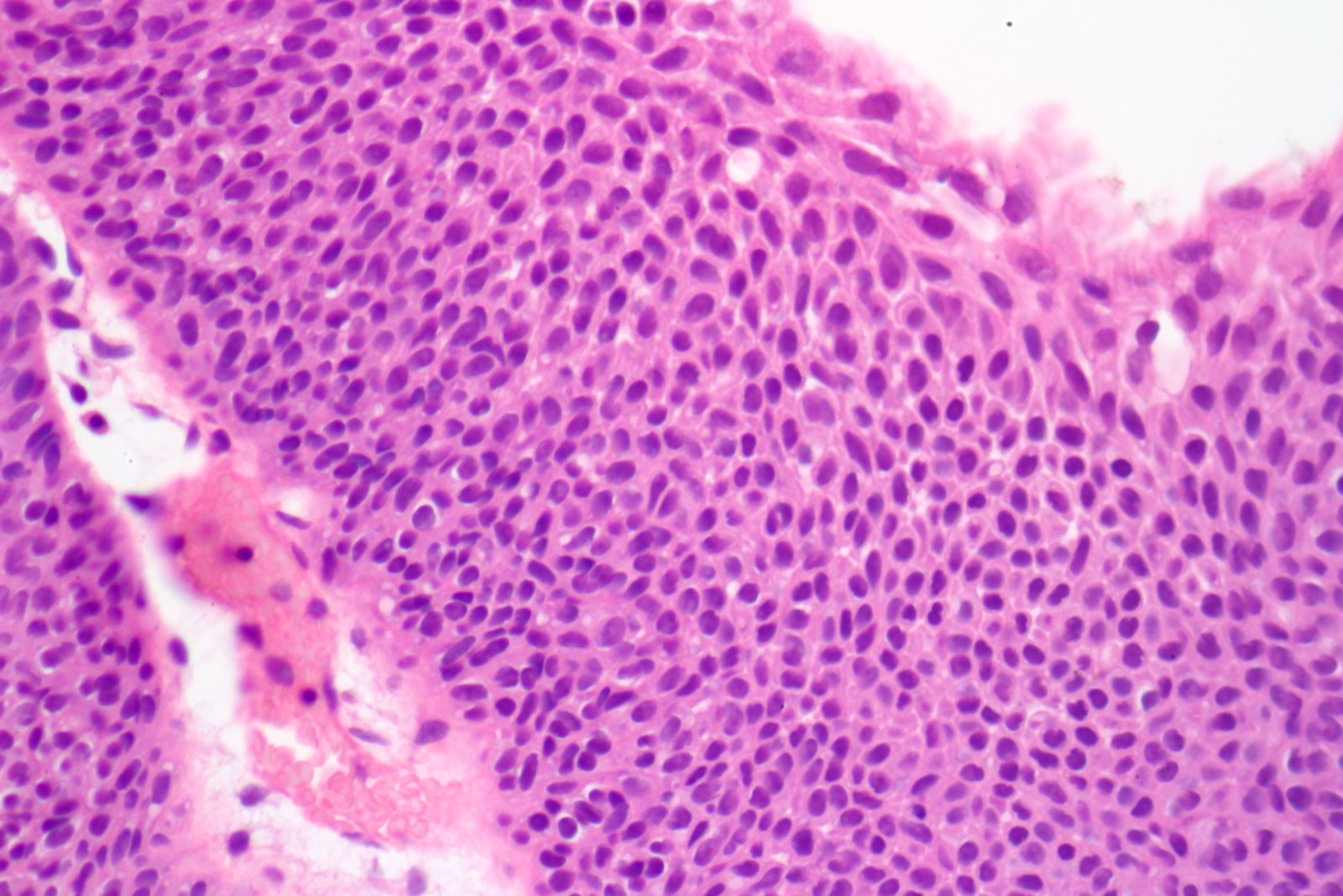
High grade cytology
High grade urothelial carcinoma
Urothelial carcinoma, papillary and invasive
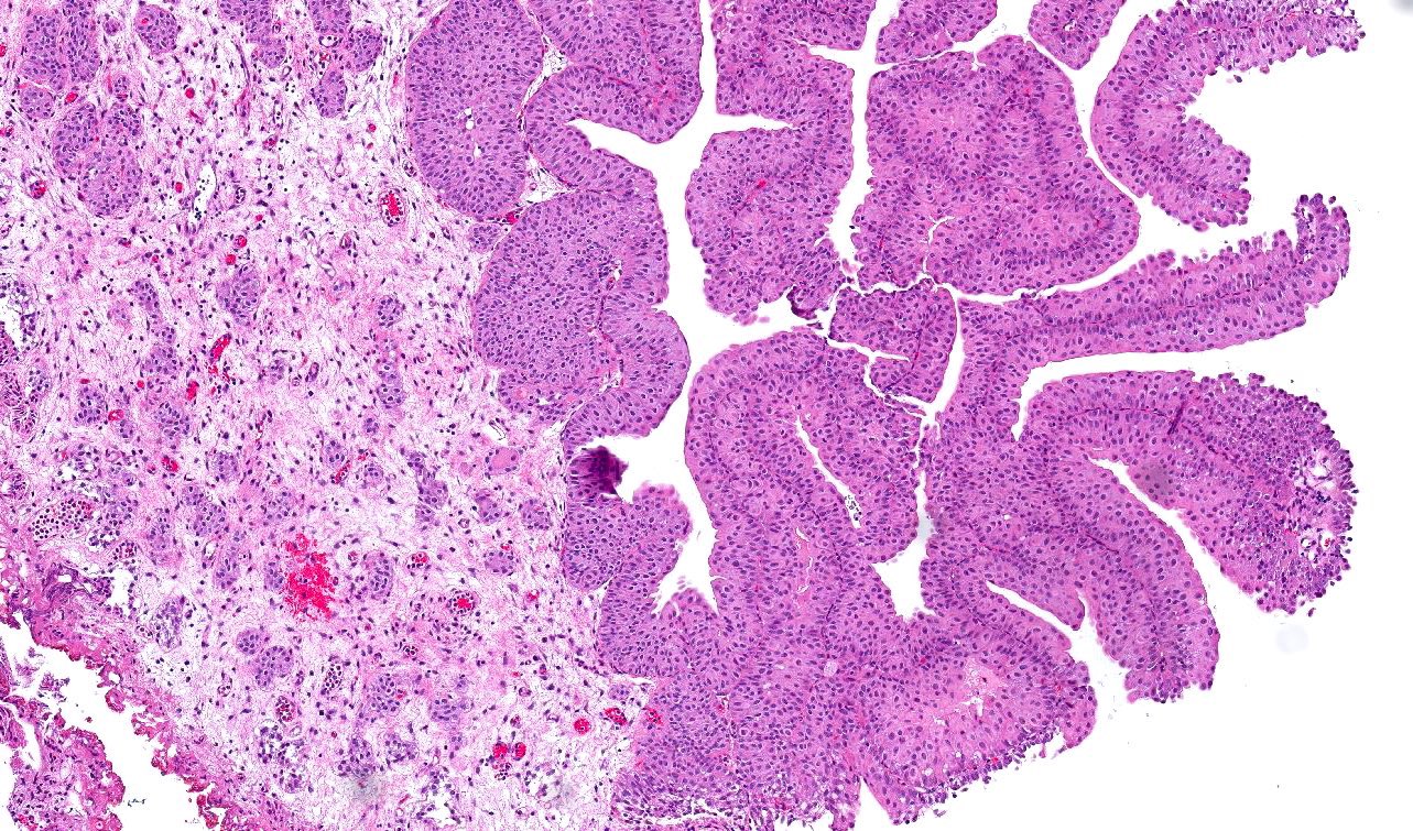
Contributed by Michelle R. Downes, M.D., Nicole K. Andeen, M.D. and Maria Tretiakova, M.D.
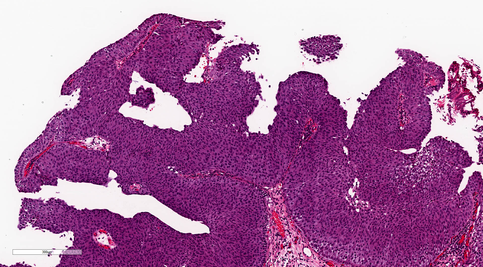
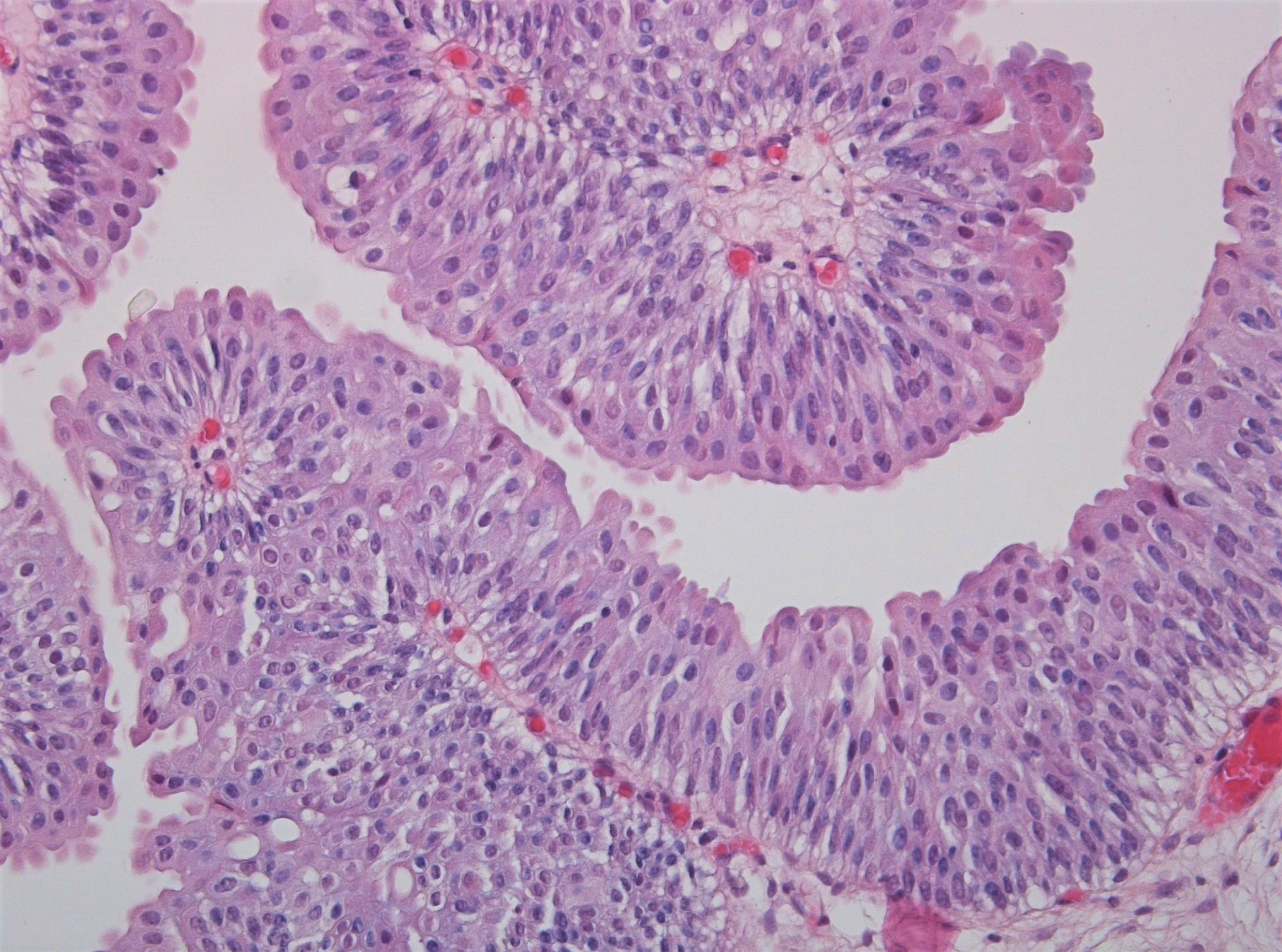
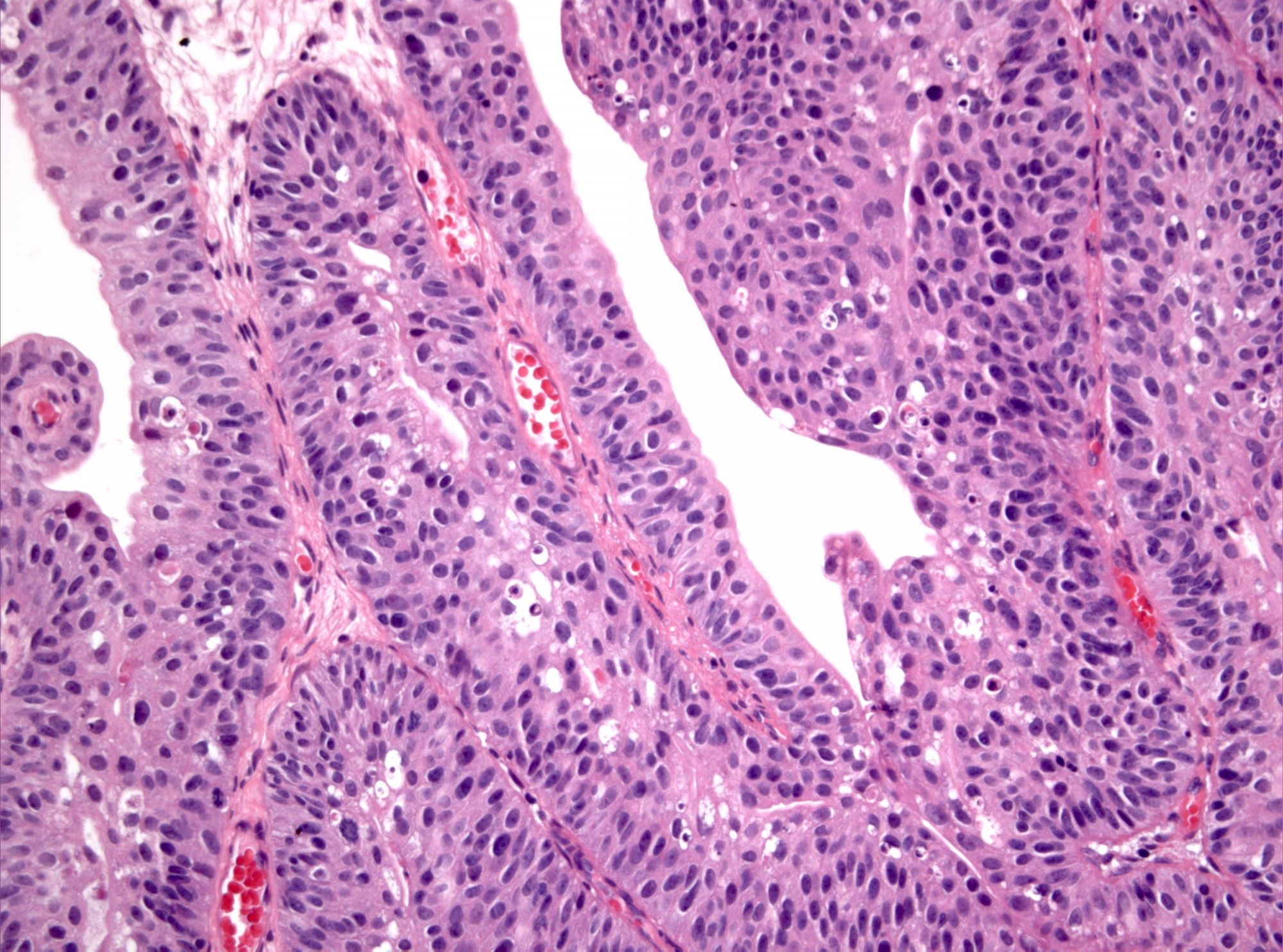
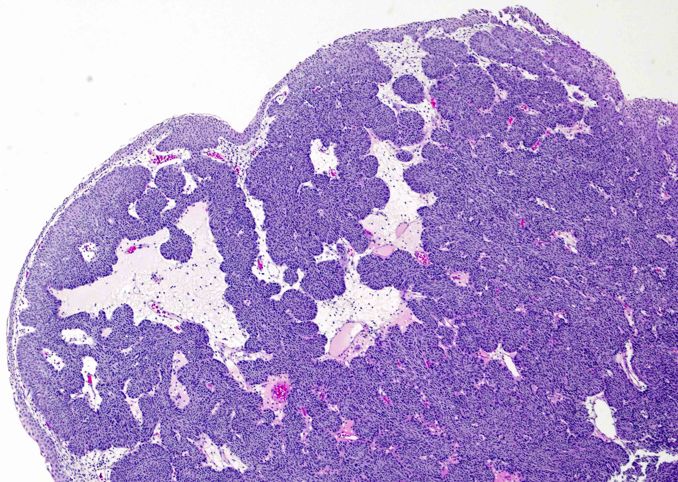
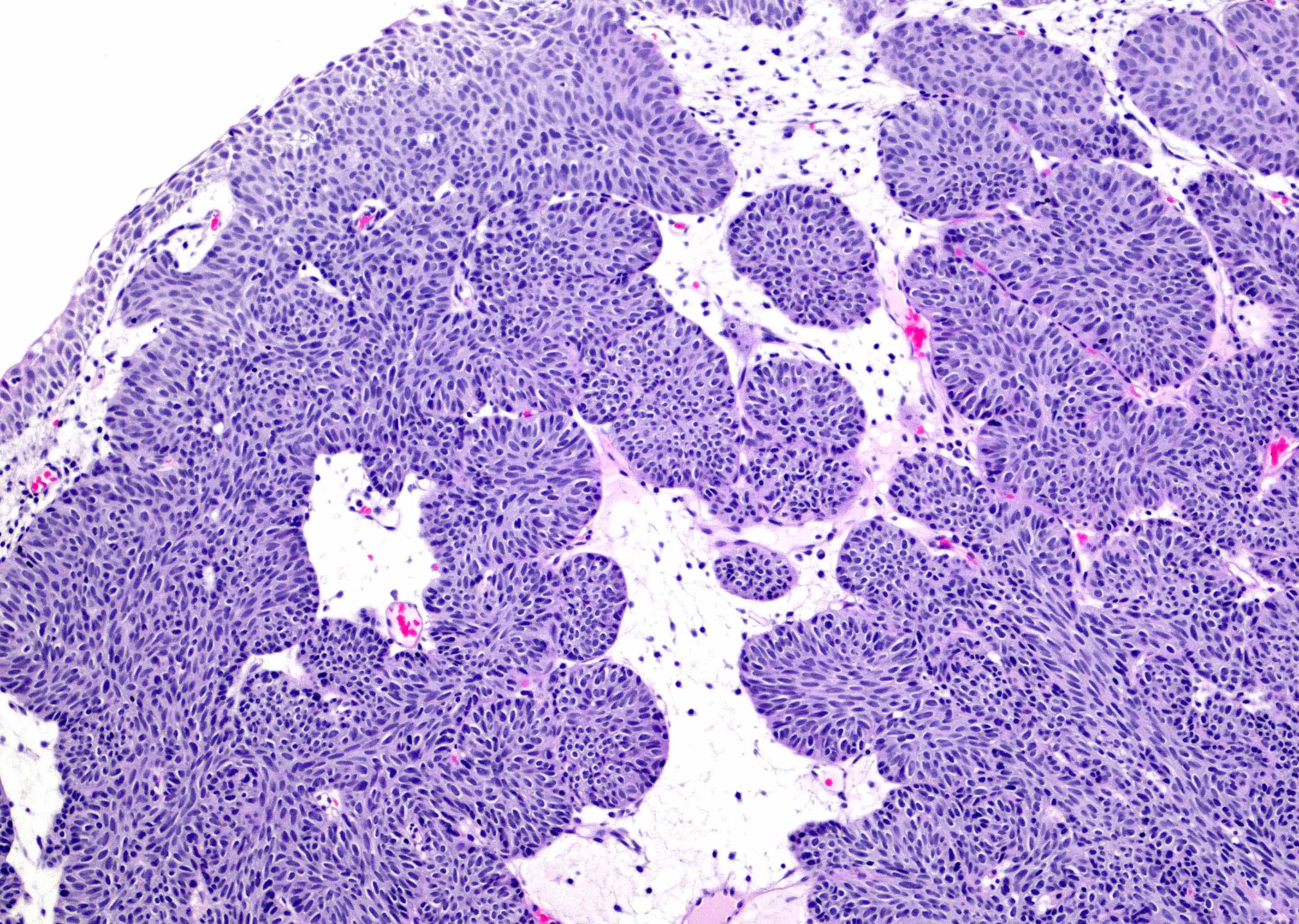
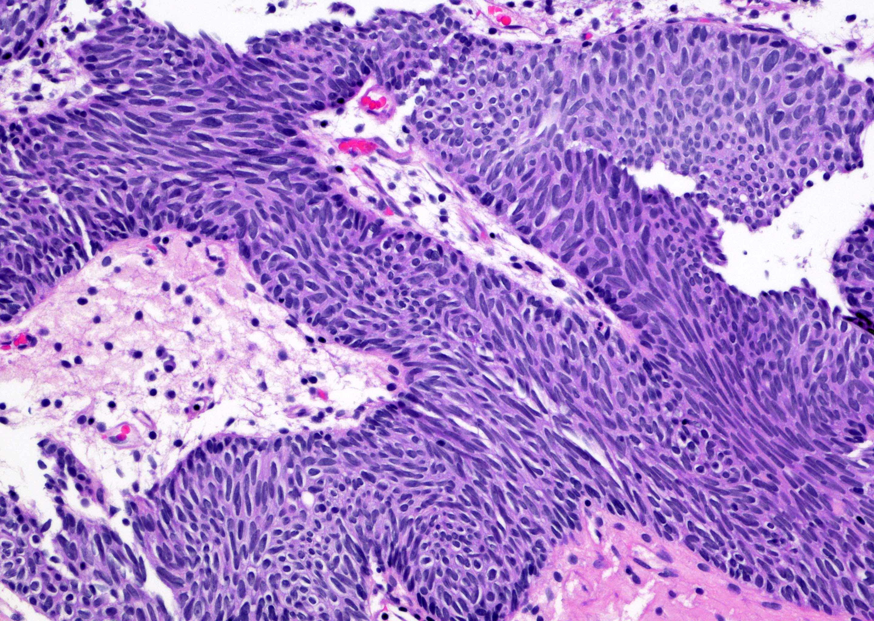
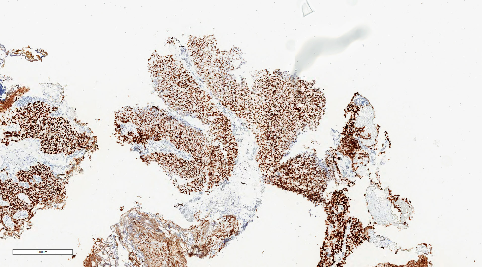
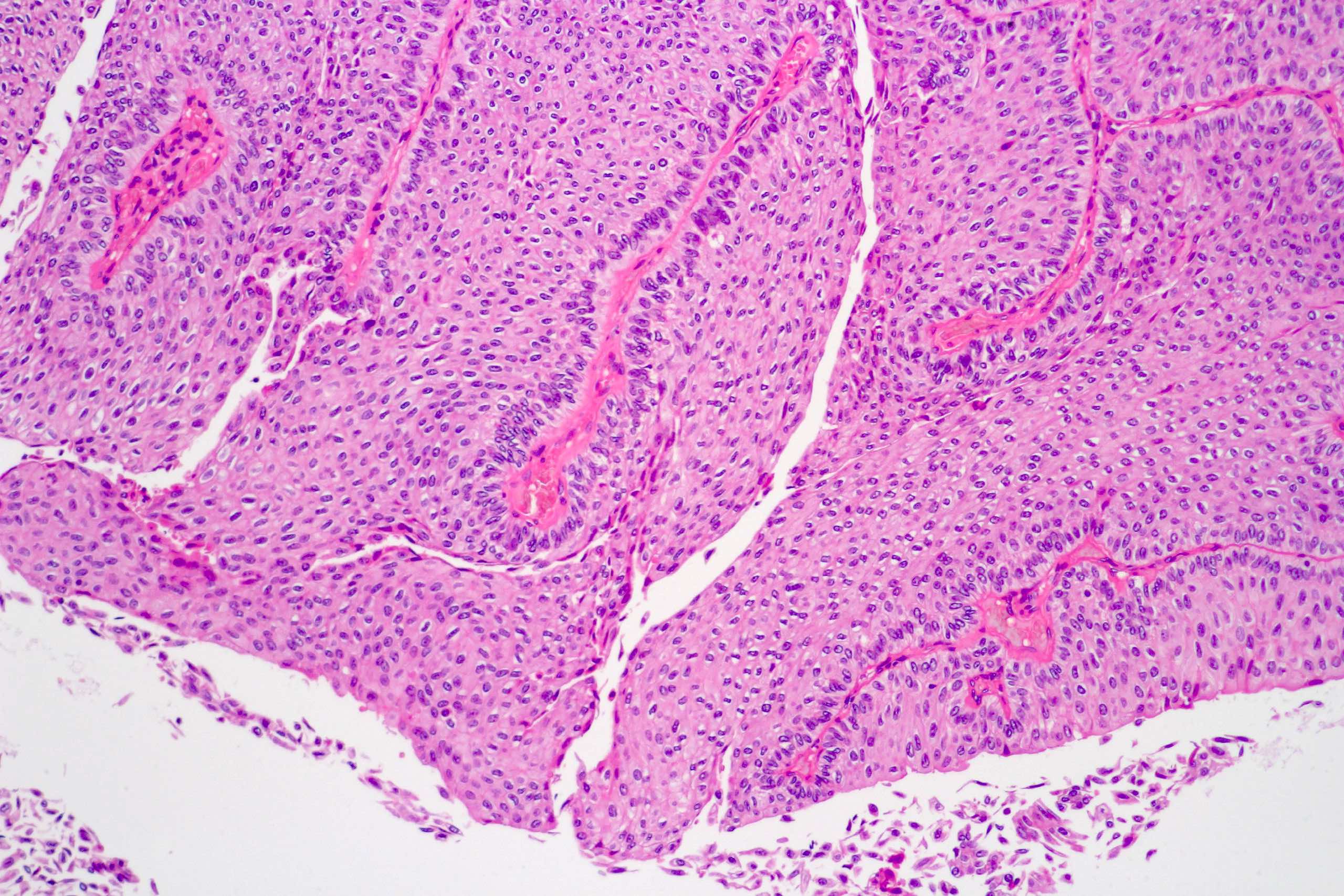
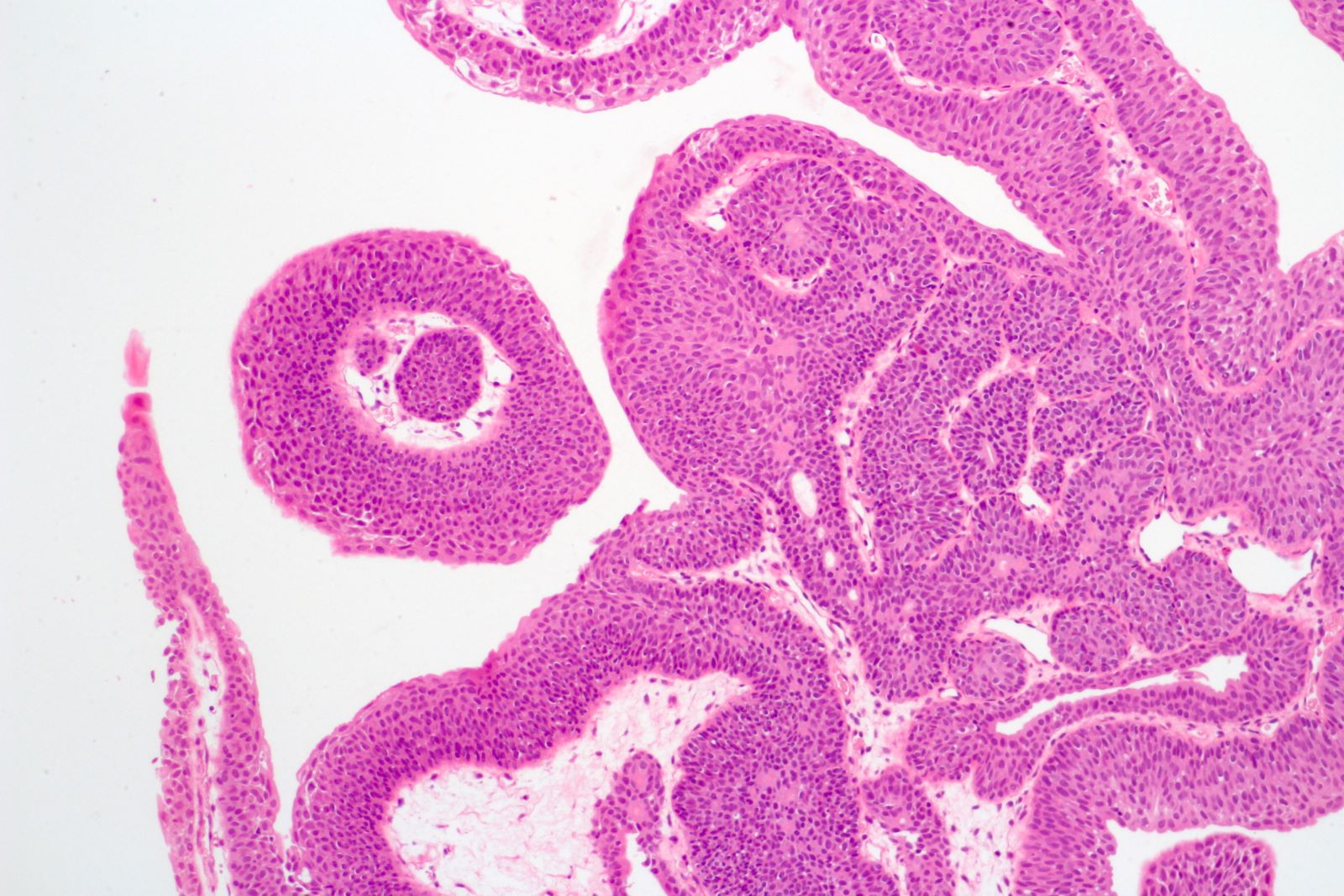
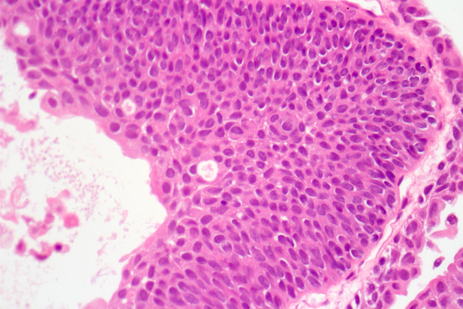
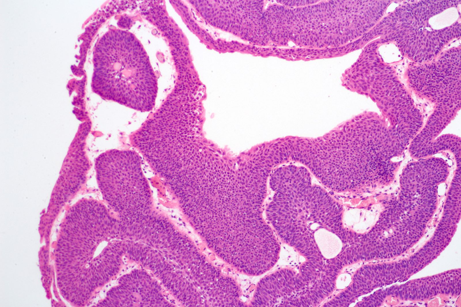
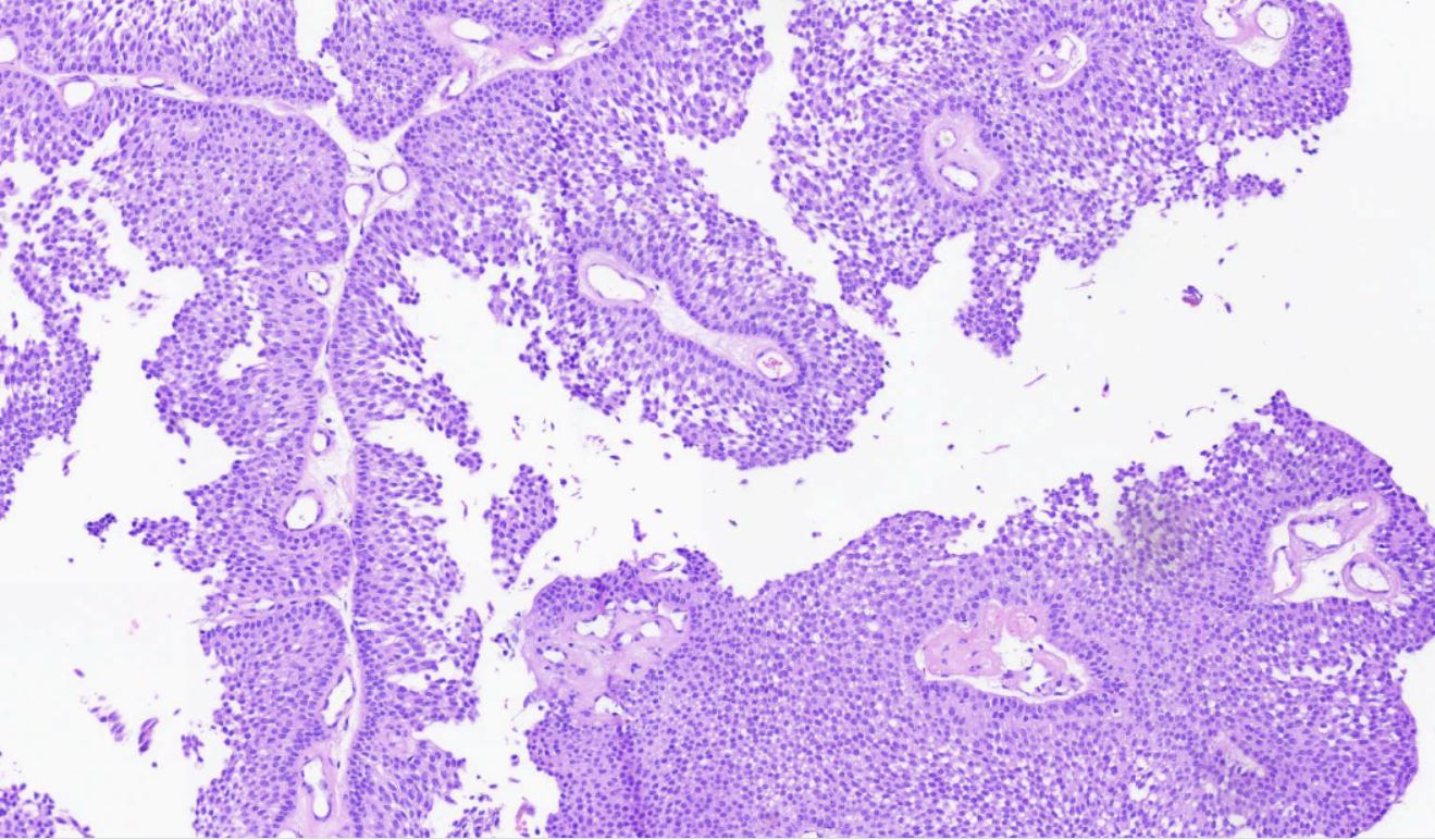
Fibrovascular cores lined by neoplastic urothelium
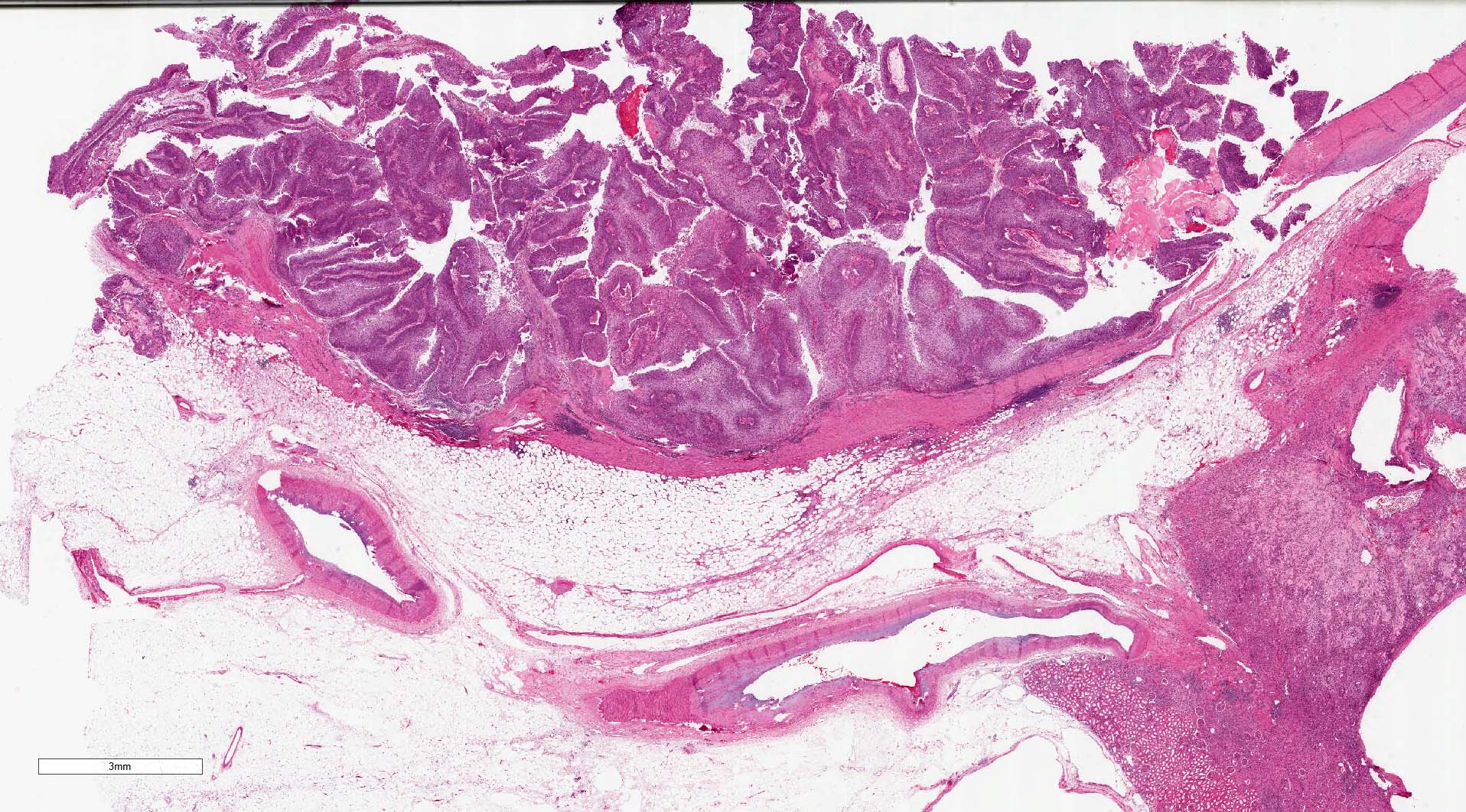
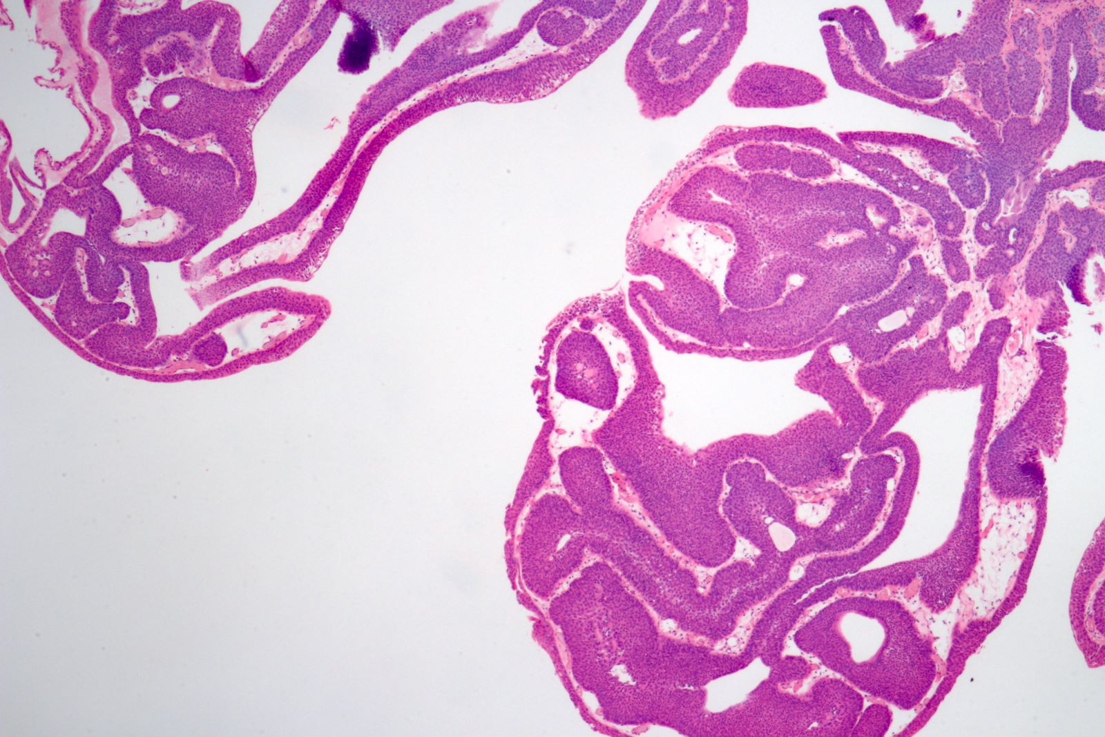
Noninvasive papillary, low grade urothelial carcinoma
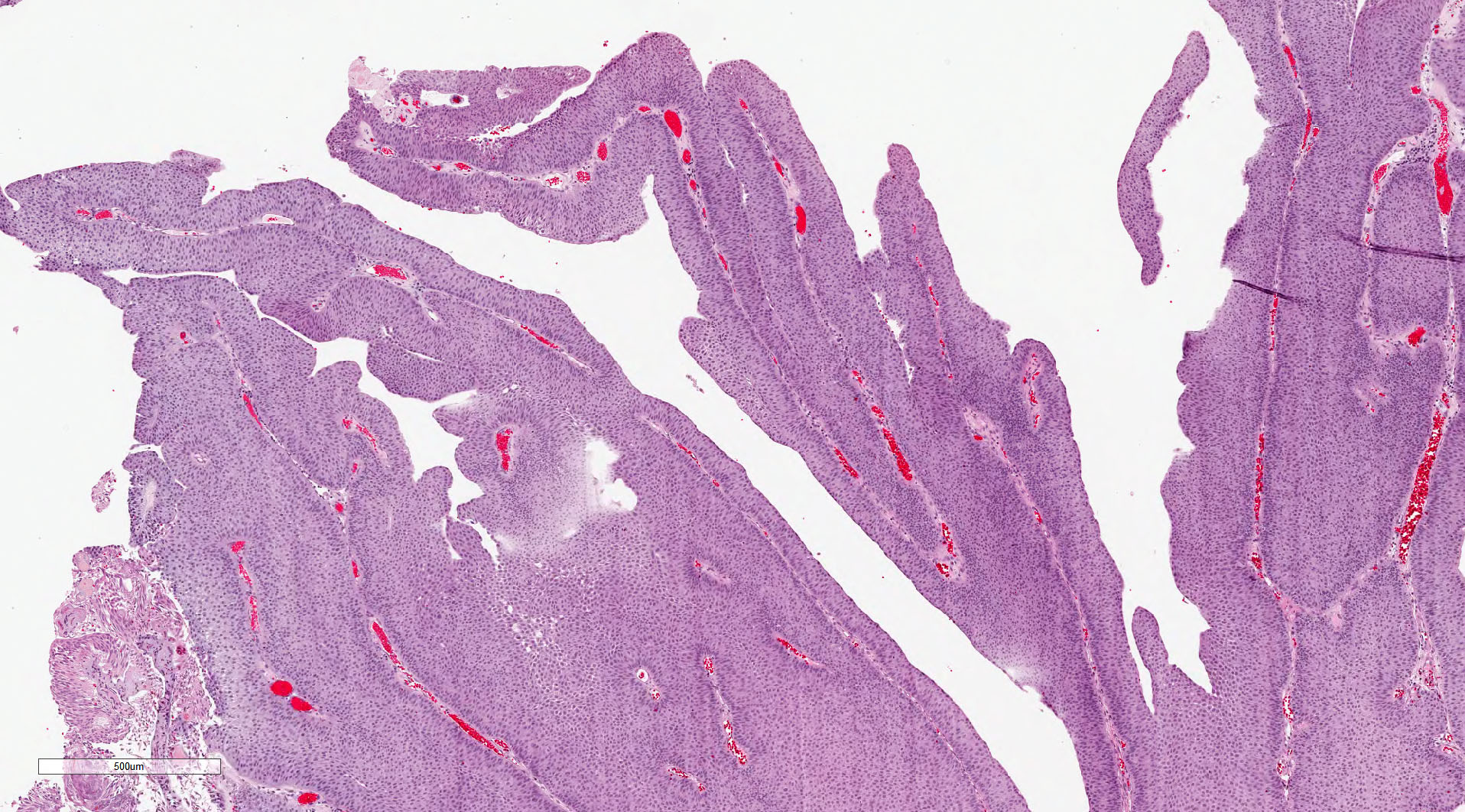
Papillary architecture

Architectural features
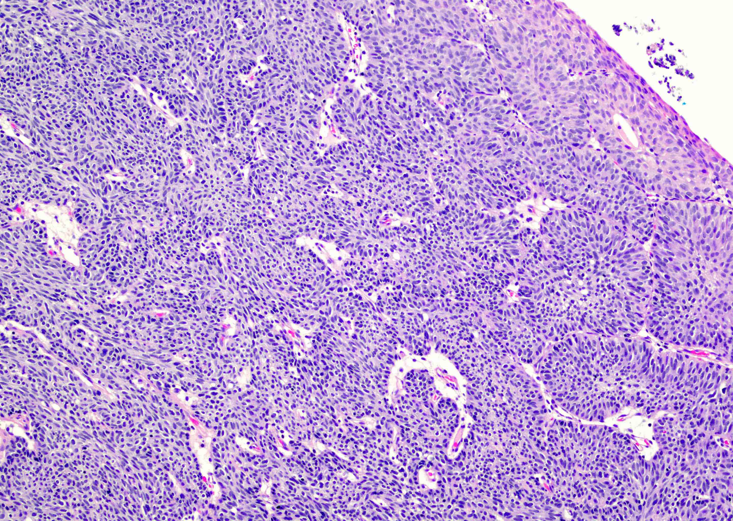
Endophytic growth pattern
GATA3
p53
Ki67
Noninvasive papillary urothelial carcinoma, low grade (pTa)
Papillary urothelial
carcinoma, absent
muscularis
Contributed by Zeina Ghorab M.D. and Bonnie Choy, M.D.
Cytology of
non high grade lesion
Low grade urothelial neoplasia
Urothelial carcinoma, papillary and invasive
Images hosted on other servers:

Exophytic lesion with internal vascularity
Images hosted on other servers:

Exophytic papillary - recurrent PUNLMP

Cystoscopy: exophytic papillary
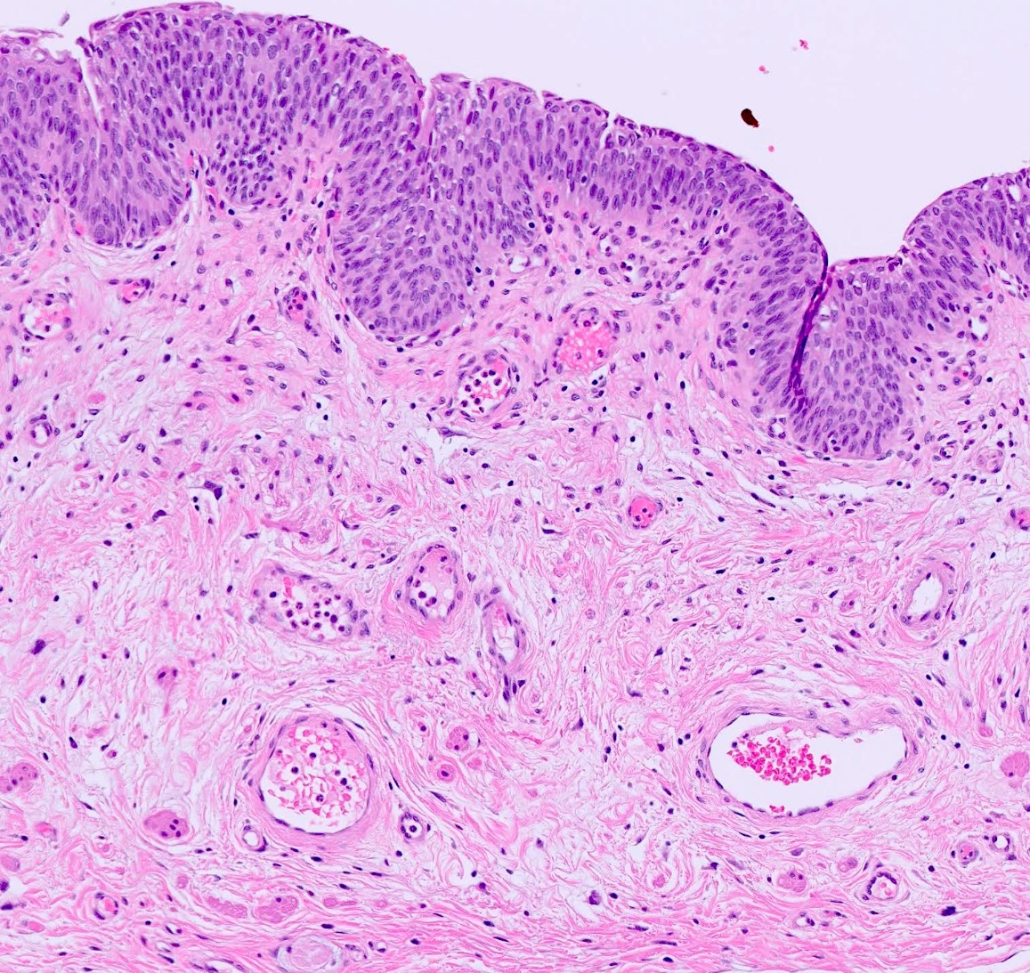
Contributed by Daniel Athanazio, M.D., Ph.D.
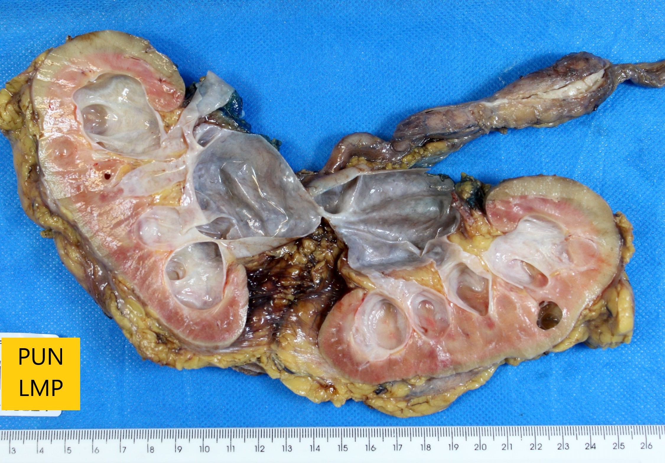
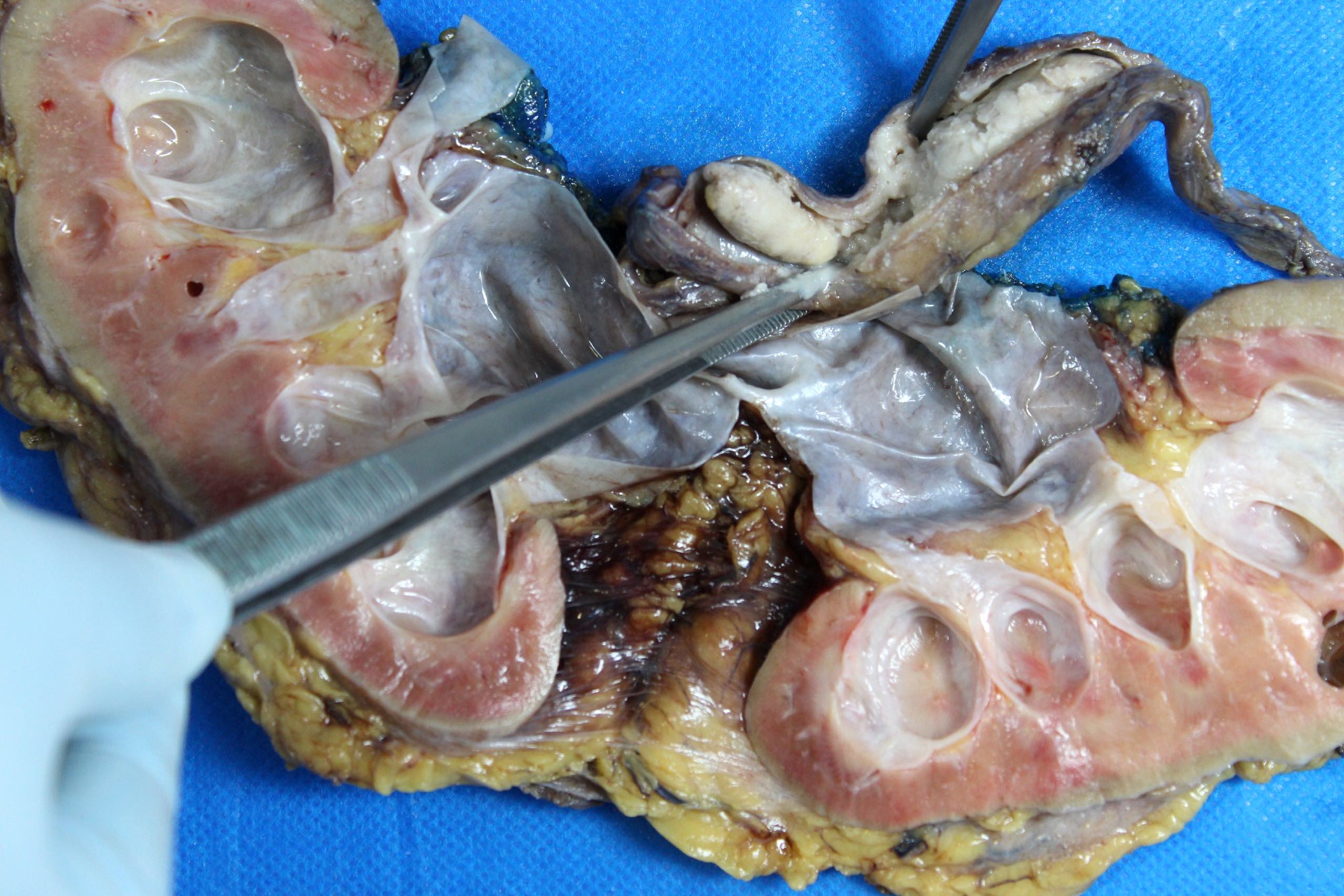
Fresh specimen
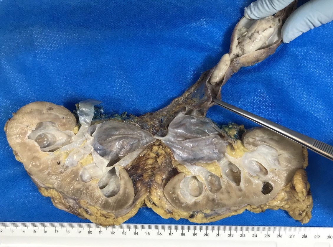
Formalin fixed specimen
Contributed by Daniel Athanazio, M.D., Ph.D. and Luciana Schultz, M.D., Ph.D. (source: Instituto de Anatomia Patológica)

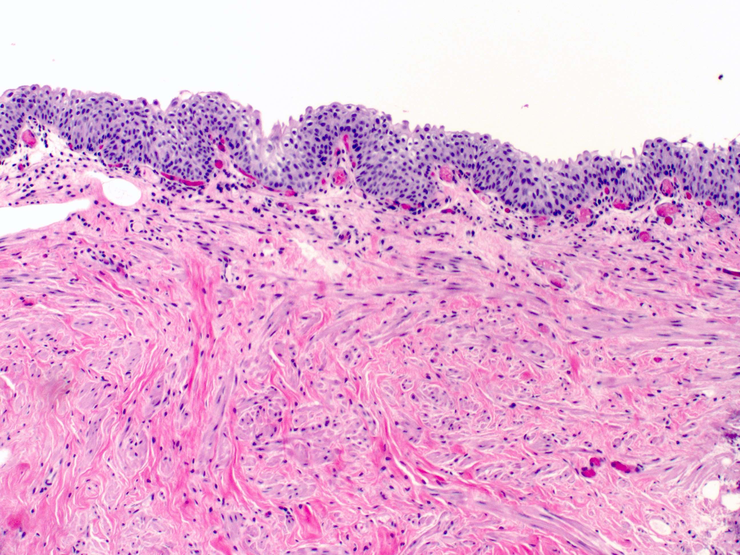
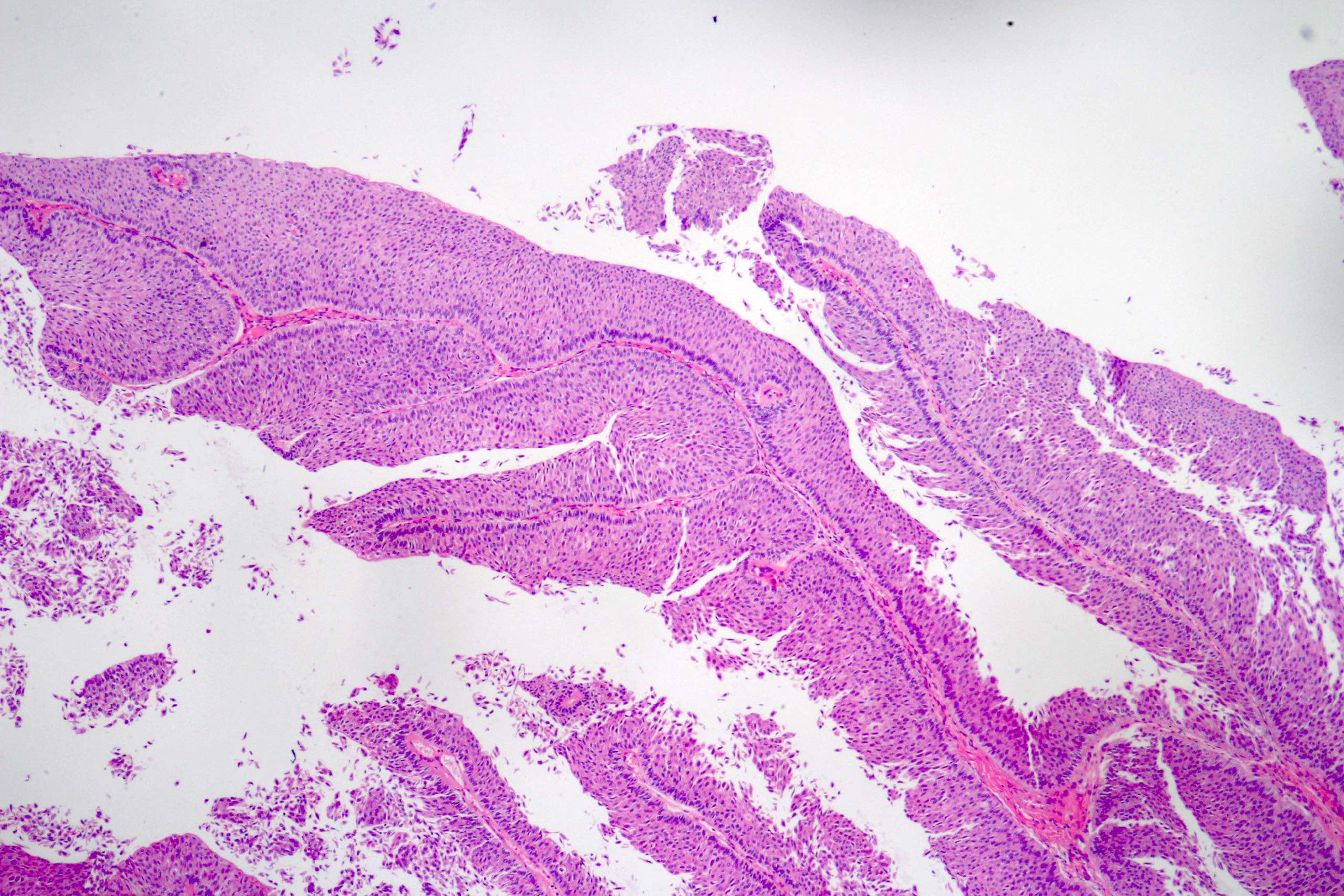
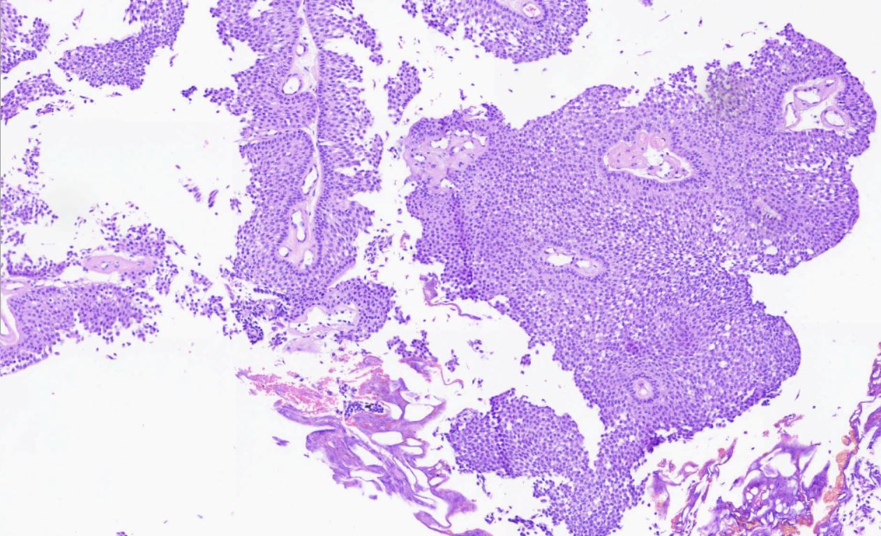
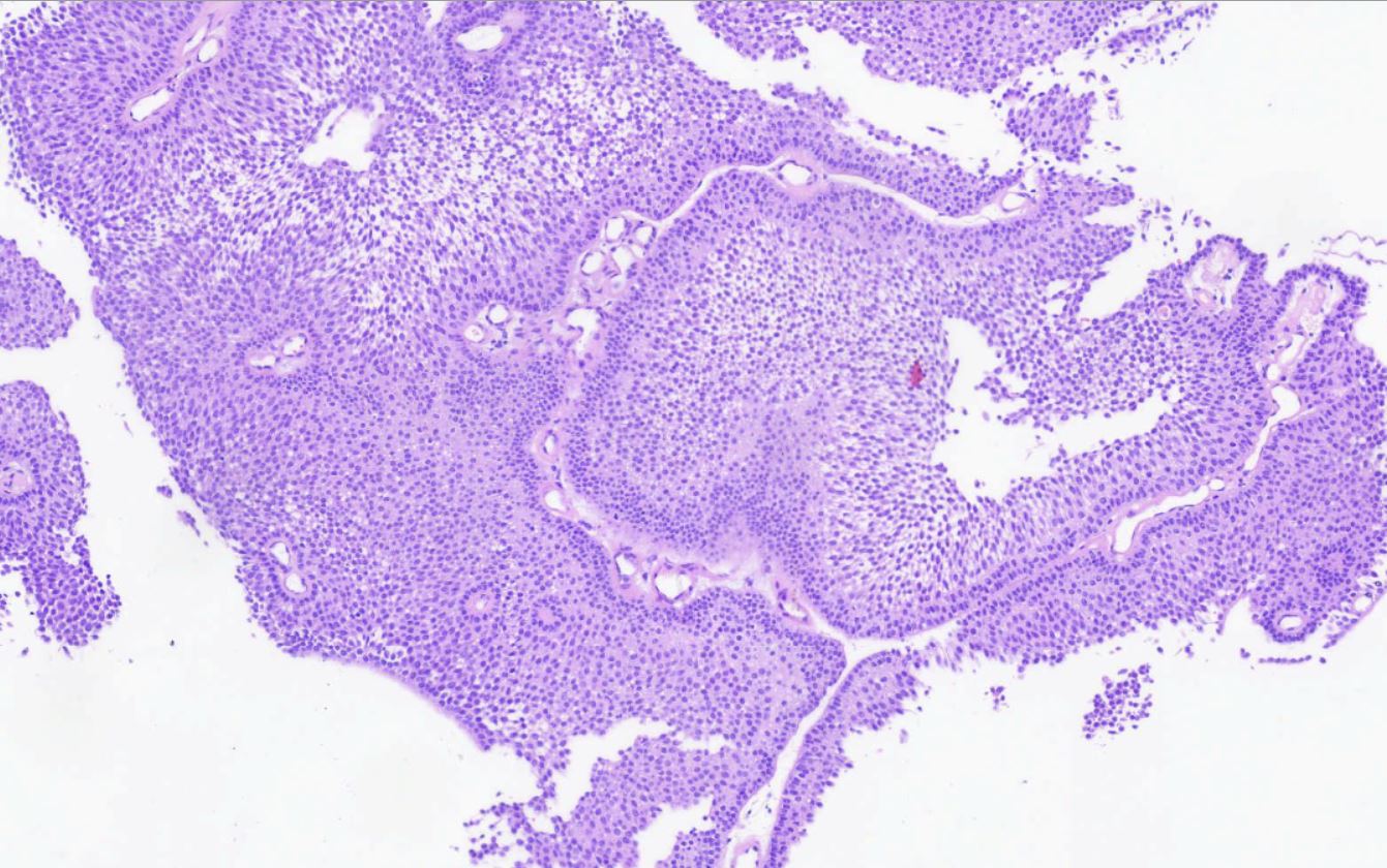
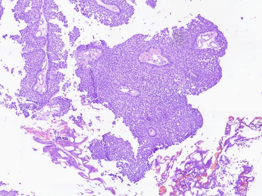
Exophytic intraluminal urothelial papillary neoplasm
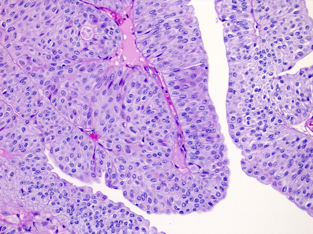
Preserved polarity and no atypia
Thickened urothelium and hypercellularity
Thickened urothelium / no atypia
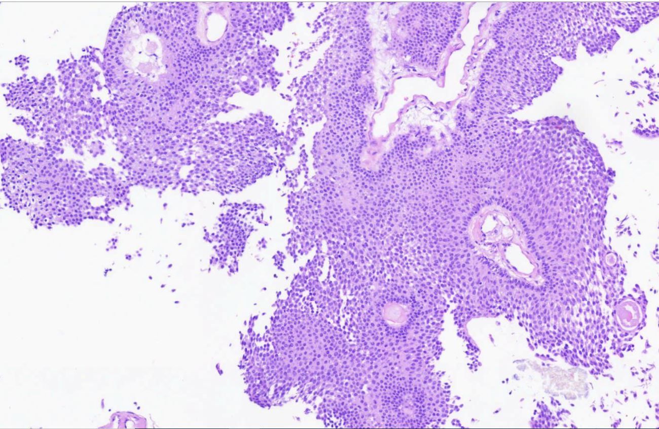
Papillary exophytic neoplasm
Preserved polarity and hypercellularity
Basally located mitosis
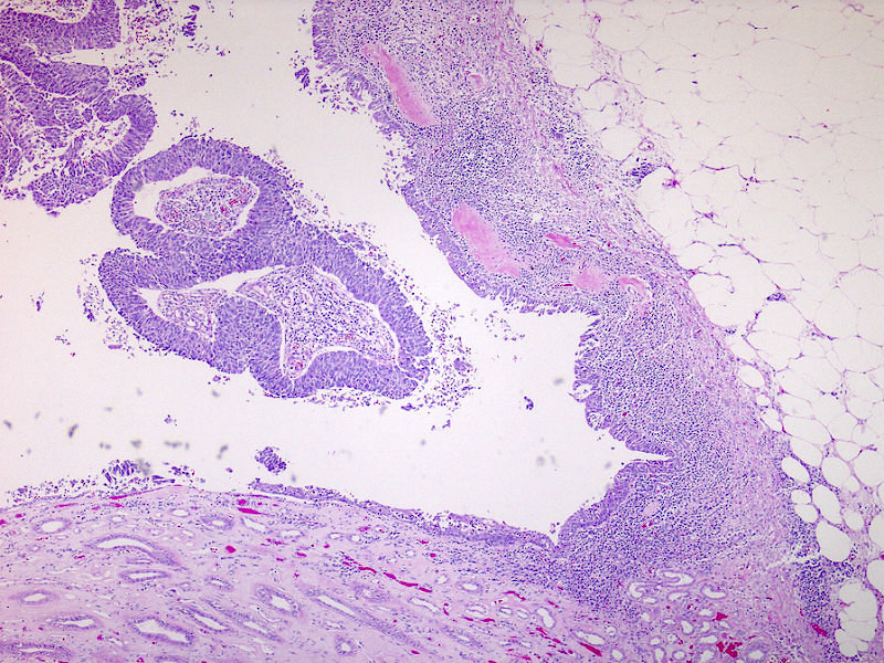
Transition zone between normal ureteral urothelium and a shoulder lesion

Endophytic / inverted growth
Exophytic and endophytic growth
Preserved polarity
Thickened epithelium / no atypia
Thickened epithelium / inverted growth
Thickened epithelium / inverted growth
Preserved polarity
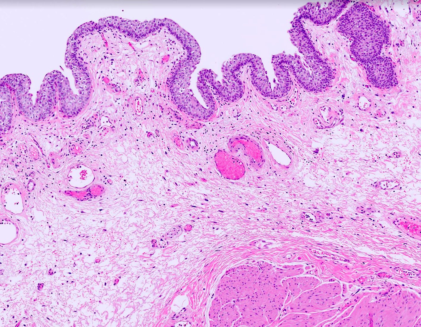
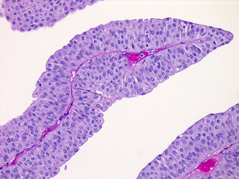
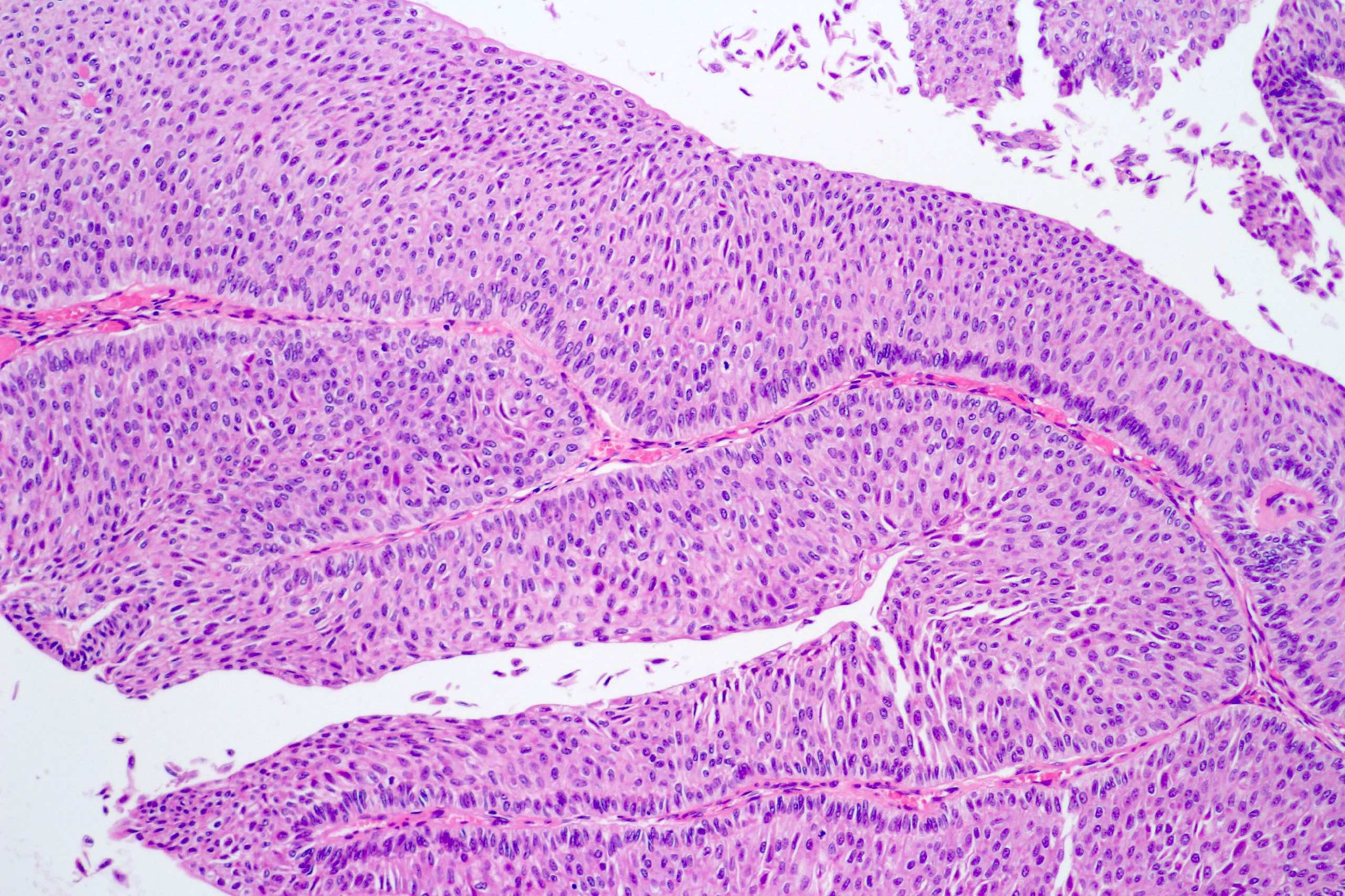
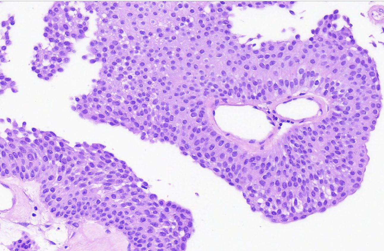
Papillary exophytic
Thickened epithelium
No atypia
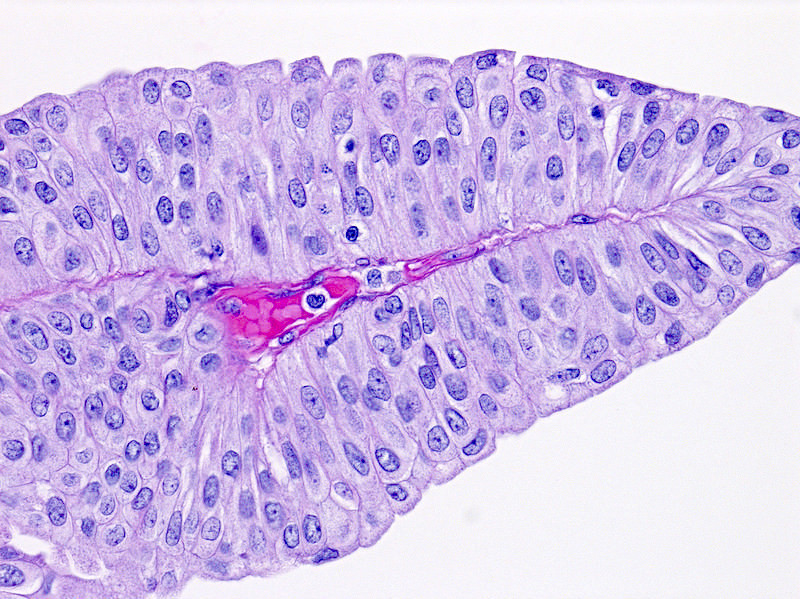
Papillary exophytic
Hypercellular monotonous urothelial lining
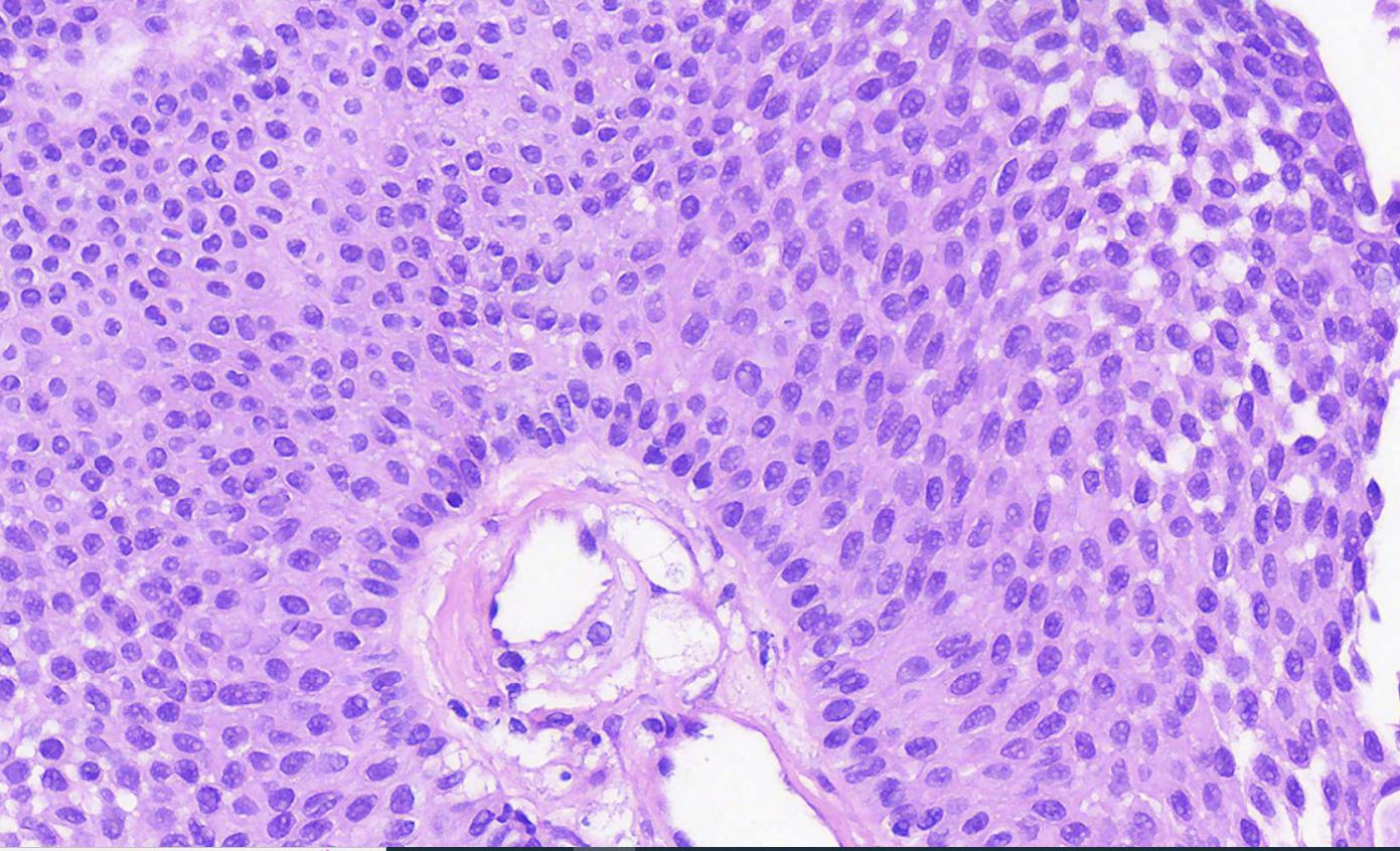
No atypia
Preserved polarity

GATA3
CK5
p16
Differential diagnosis of noninvasive endophytic urothelial neoplasms
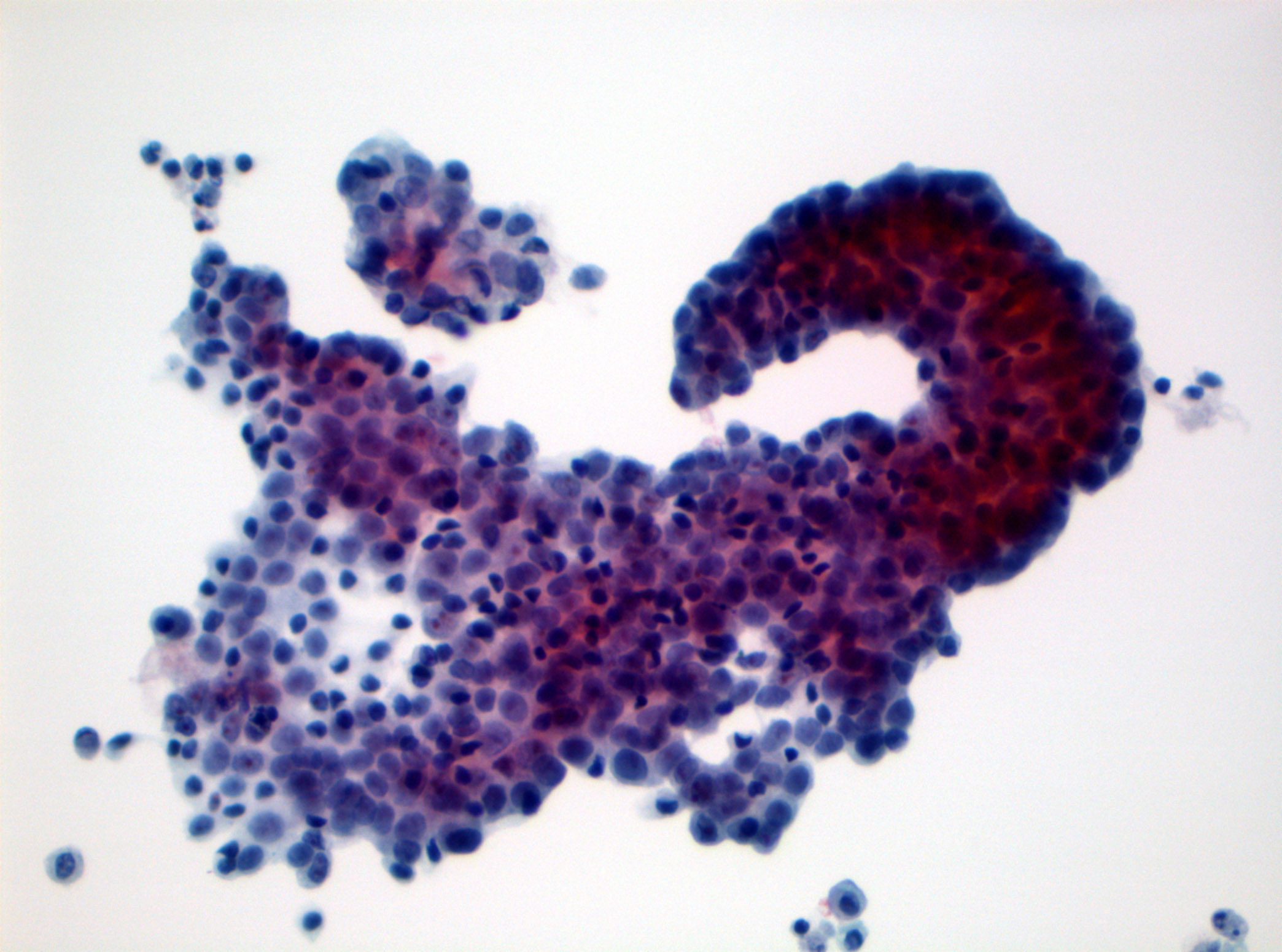
PUNLMP at cytology
Images hosted on other servers:

Axial nonenhanced CT, Doppler ultrasound, MRI, PET

Axial contrast enhanced CT

Coronal contrast enhanced CT, MIBG scan
Images hosted on other servers:

Cystoscopic findings

Laparoscopic findings

Robot assisted laparoscopic findings
Contributed by Debra L. Zynger, M.D.
Partial cystectomy
Images hosted on other servers:

Partial cystectomy

Nephroureterectomy
Contributed by Theodorus H. van der Kwast, M.D., Ph.D., Michelle R. Downes, M.D., Debra L. Zynger, M.D. and David Cohen, M.B.B.Ch., M.D.
Transurethral bladder resection
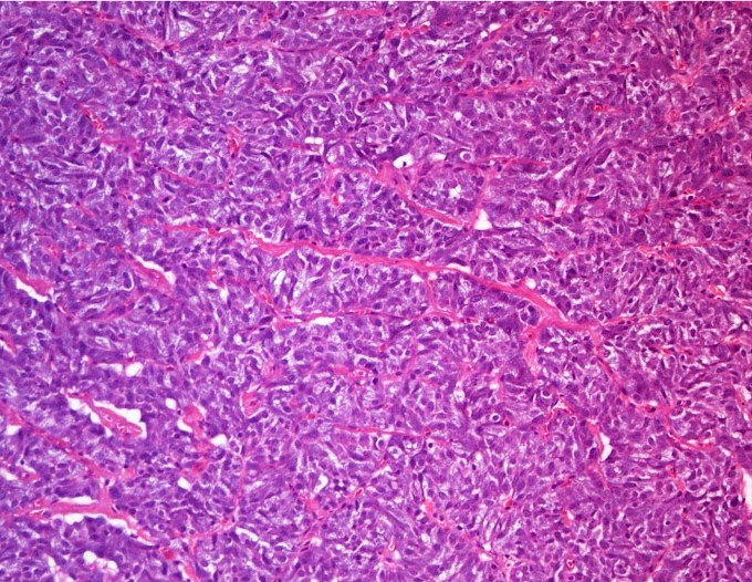
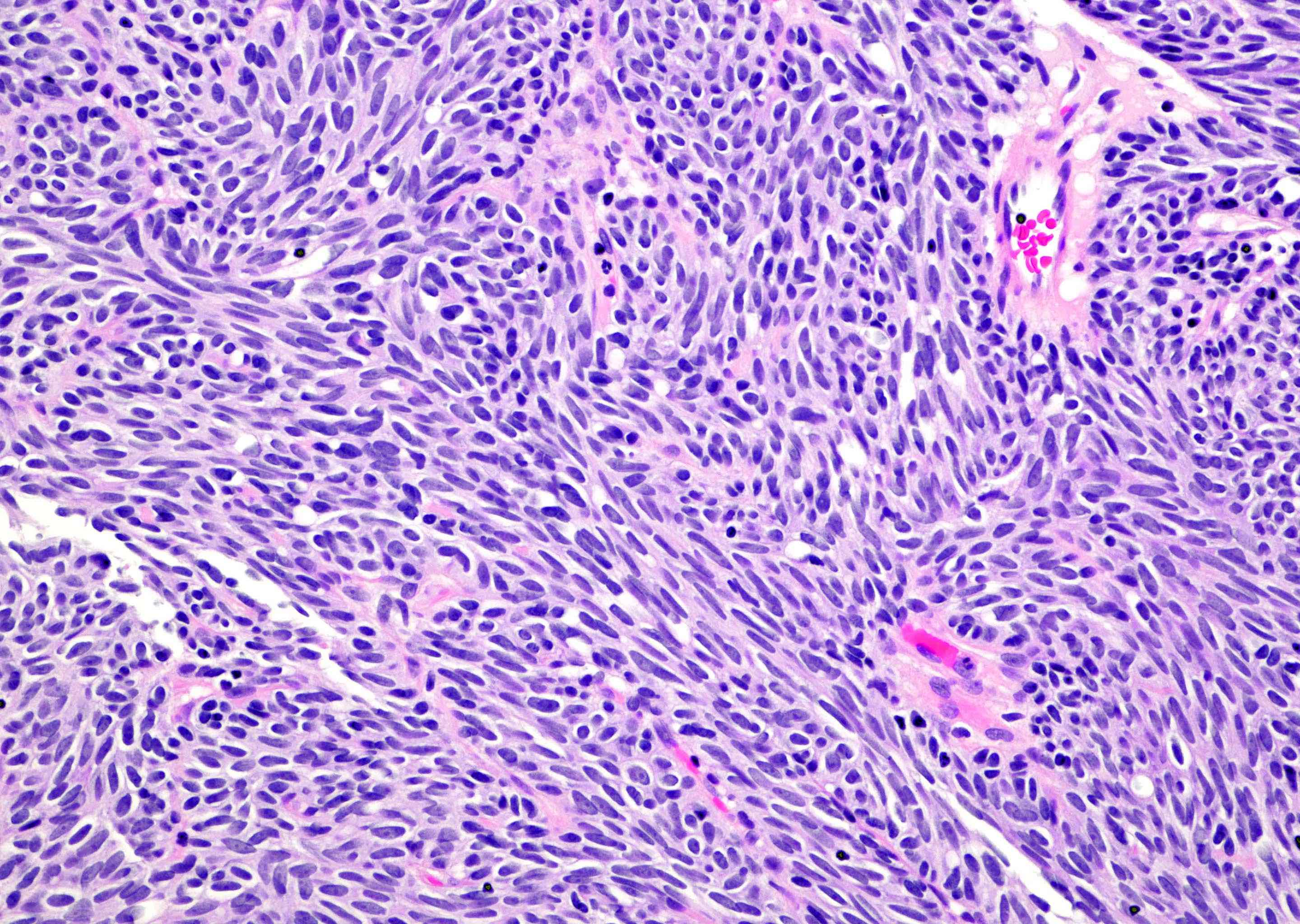
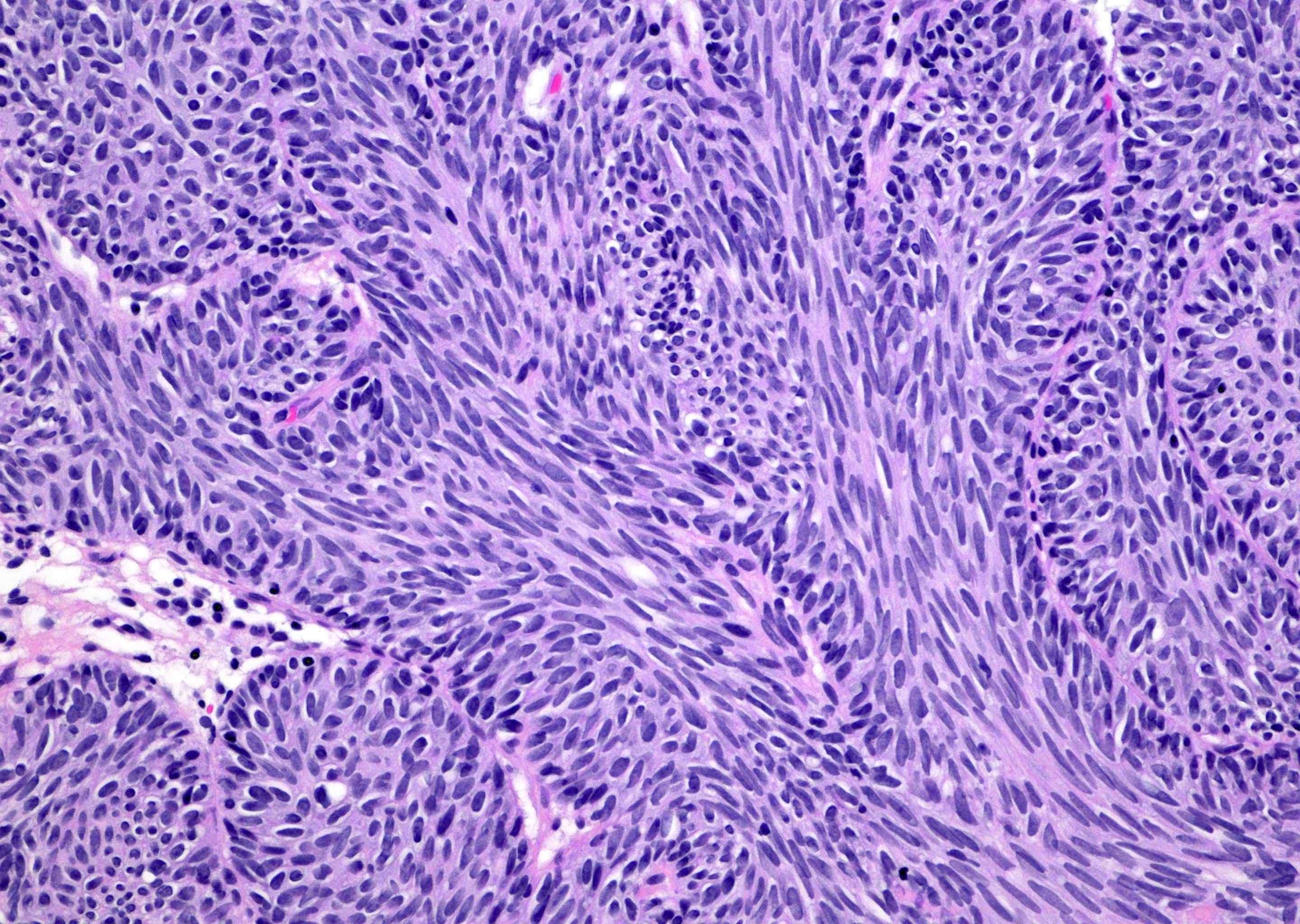
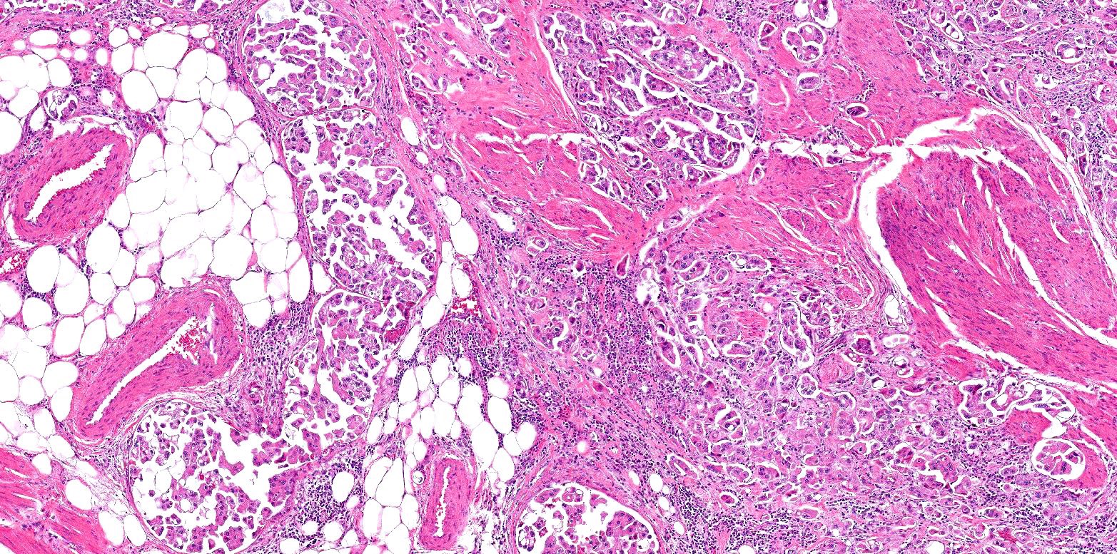
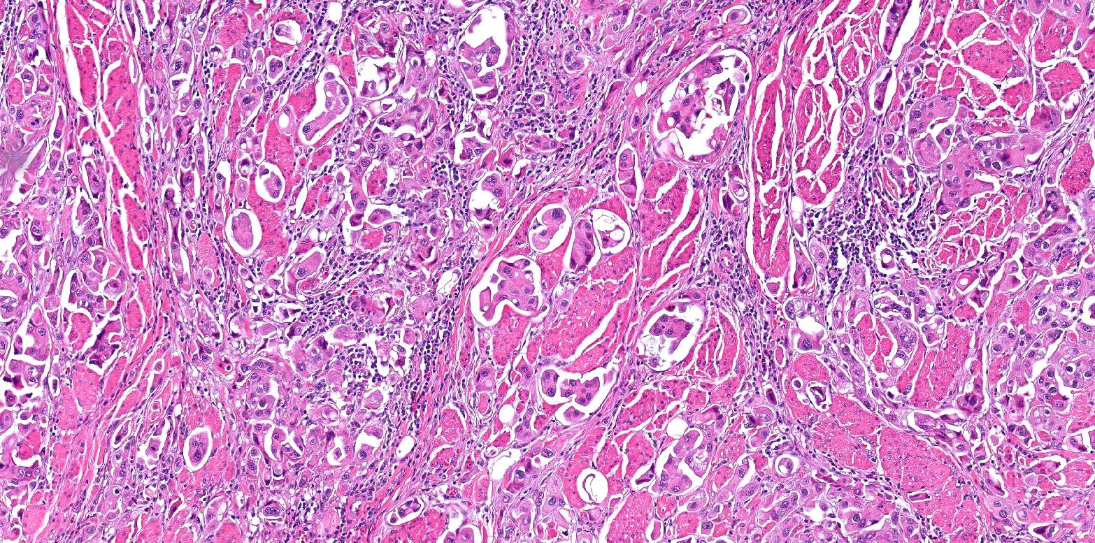
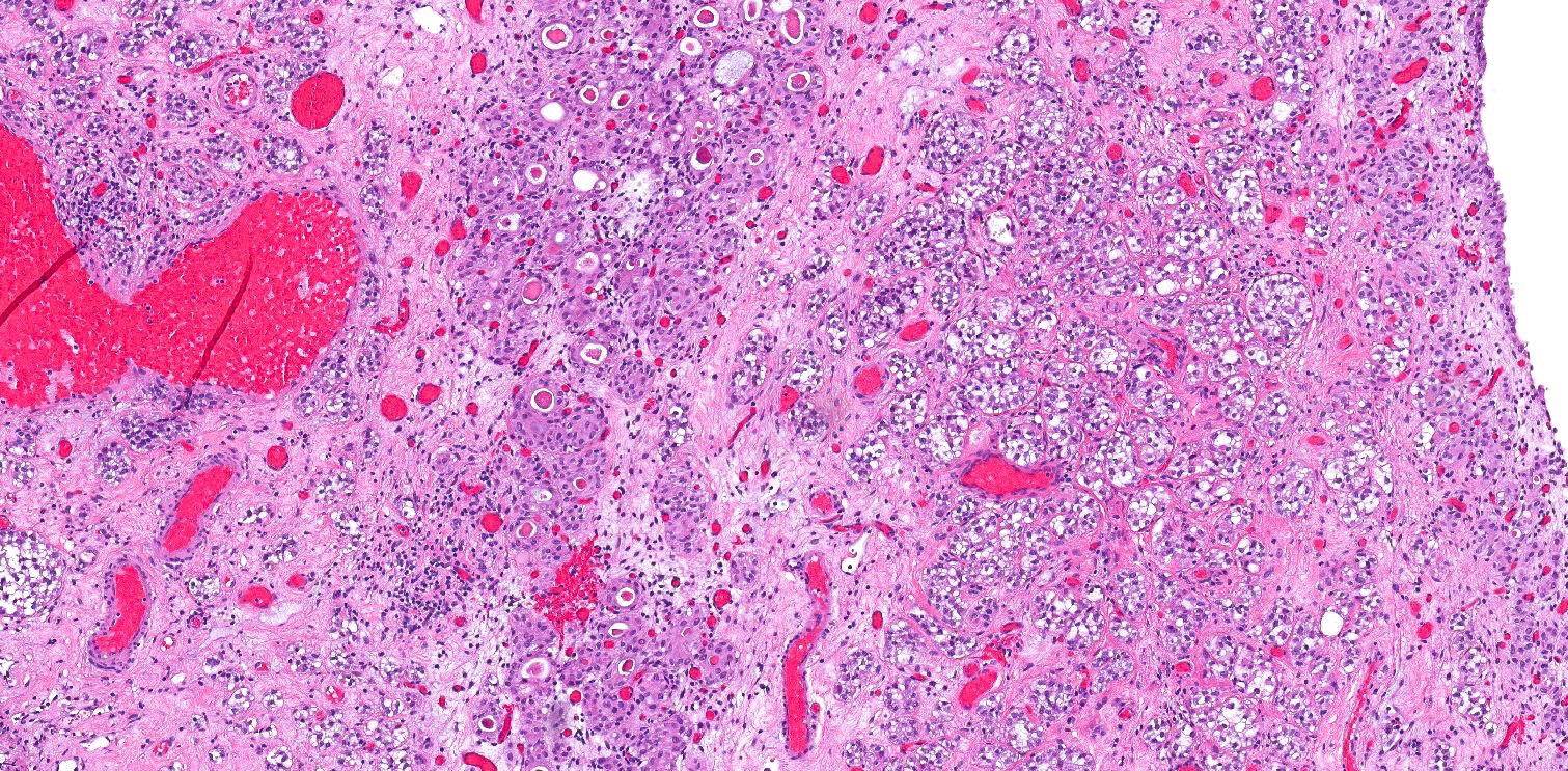
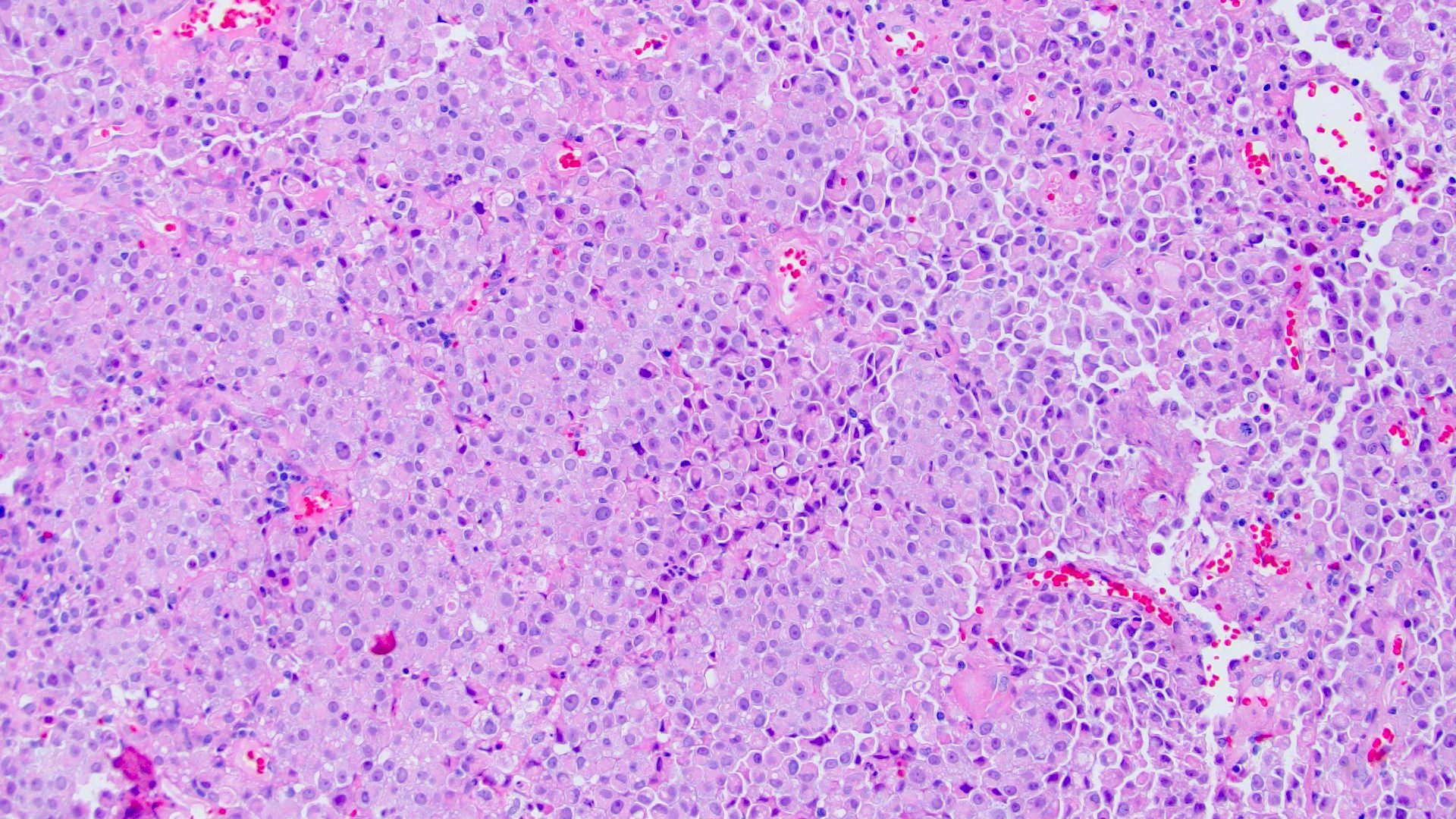
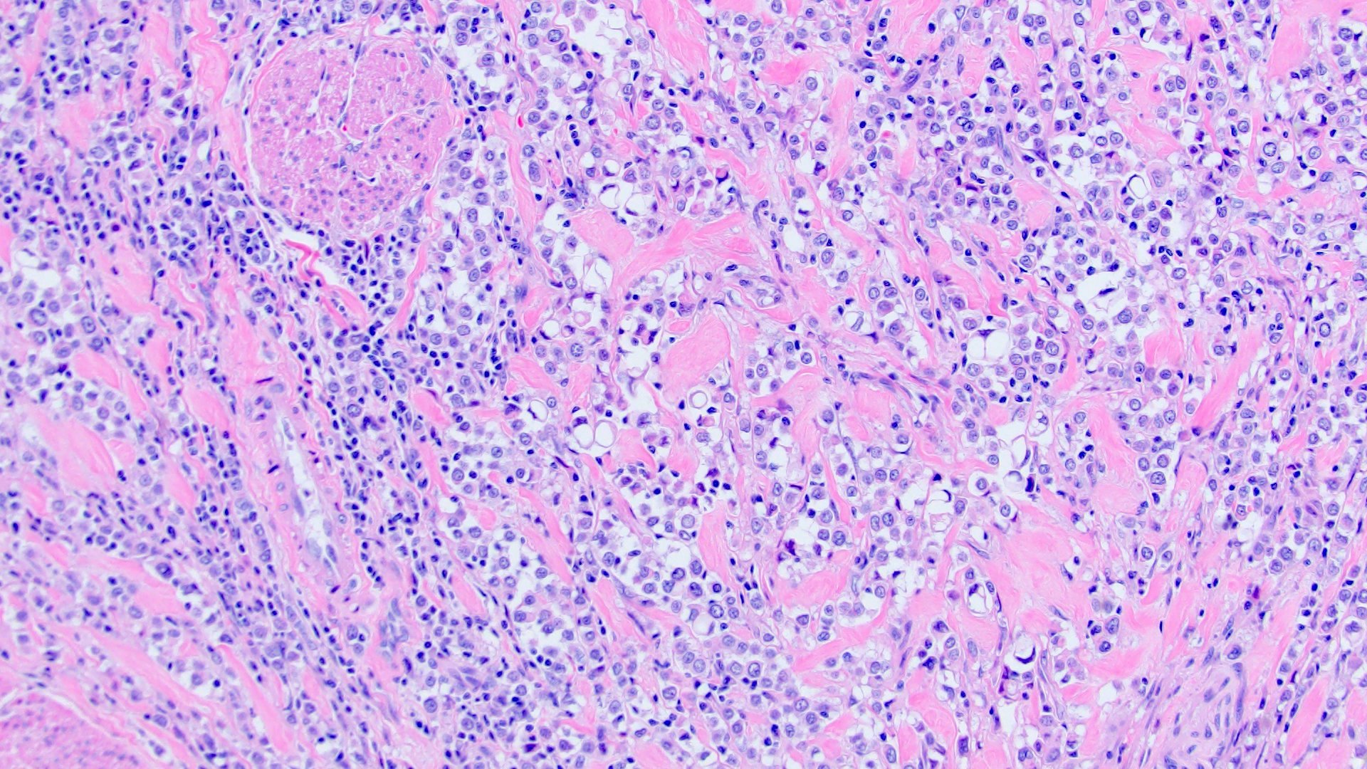
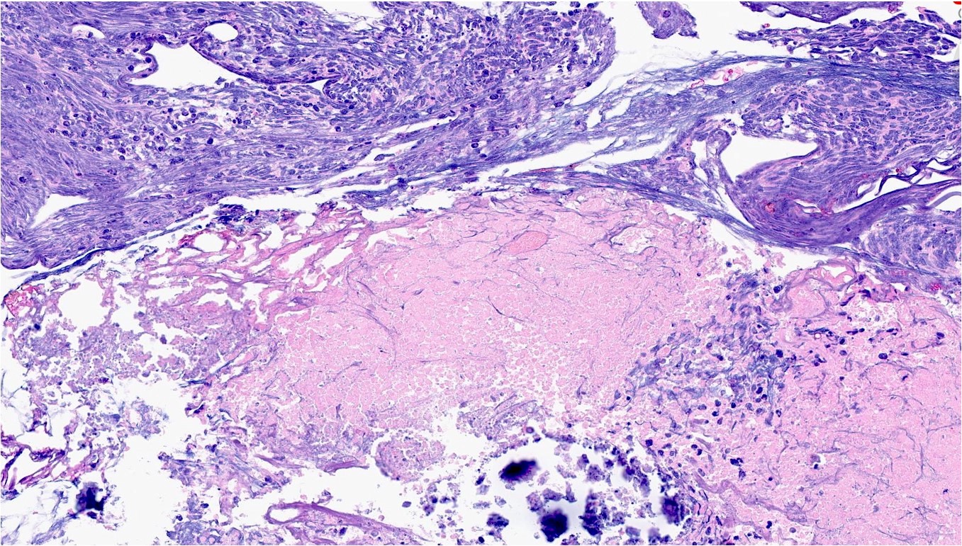
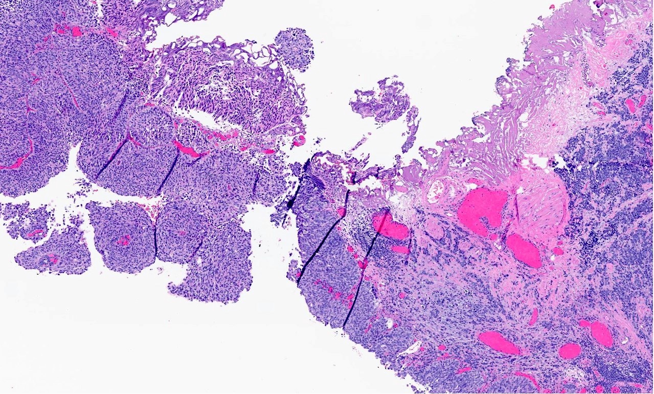
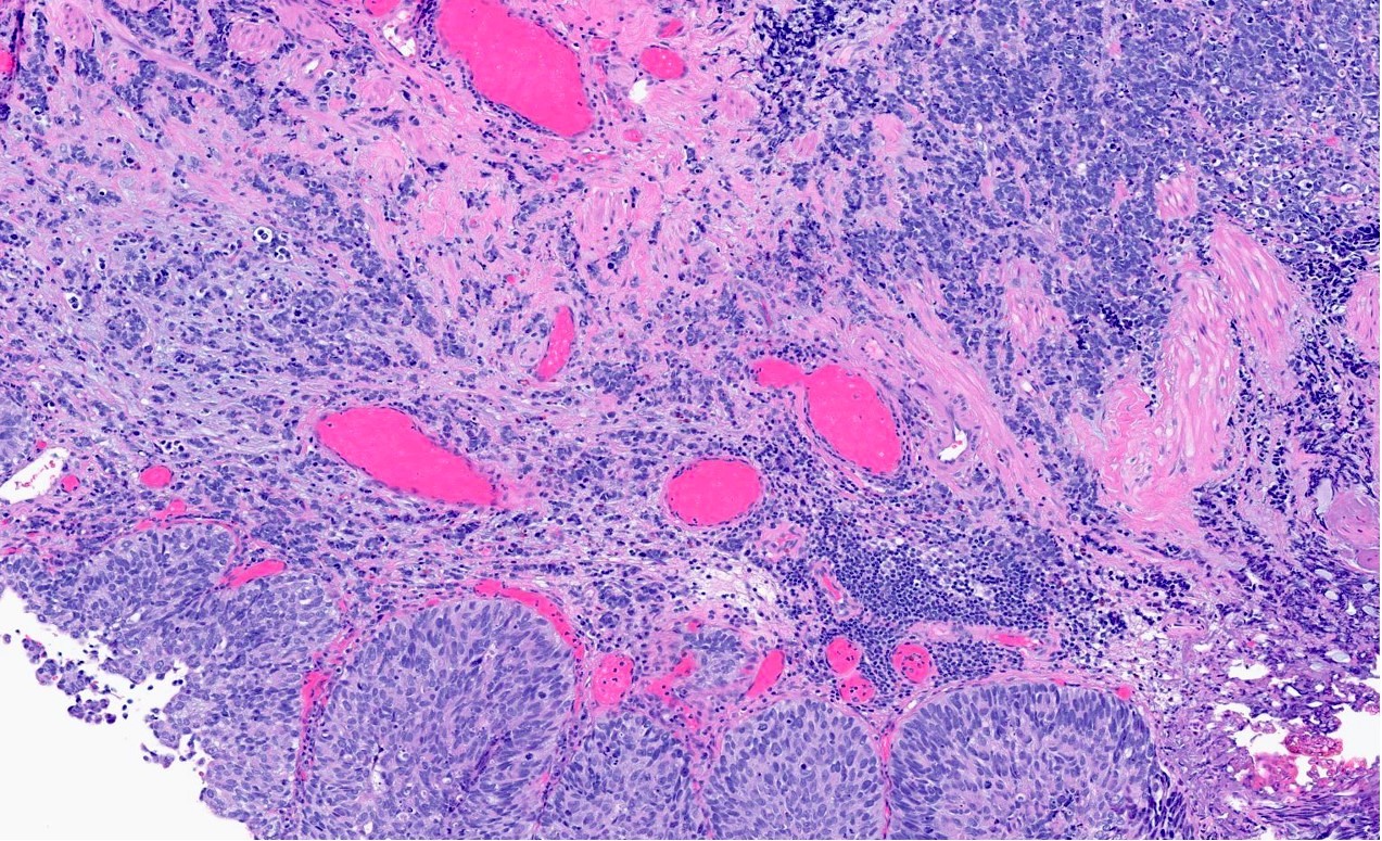
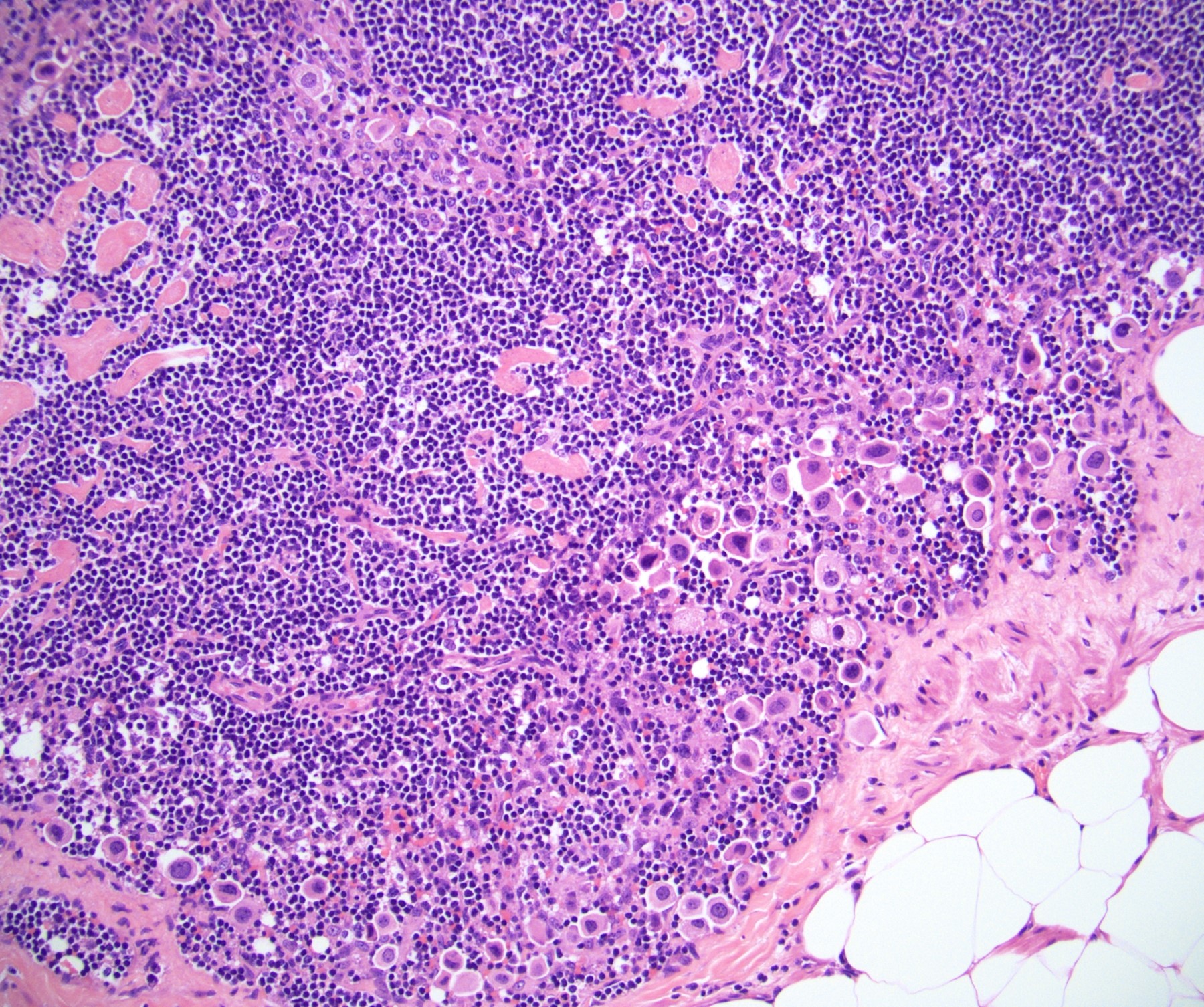
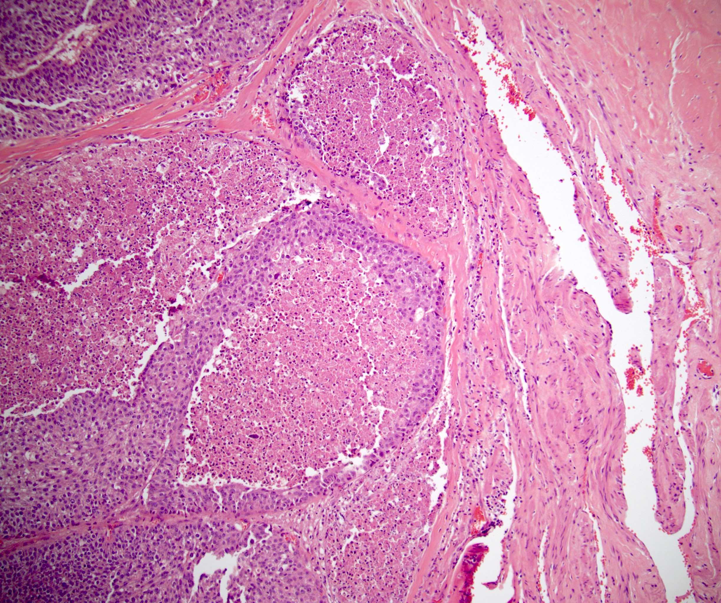
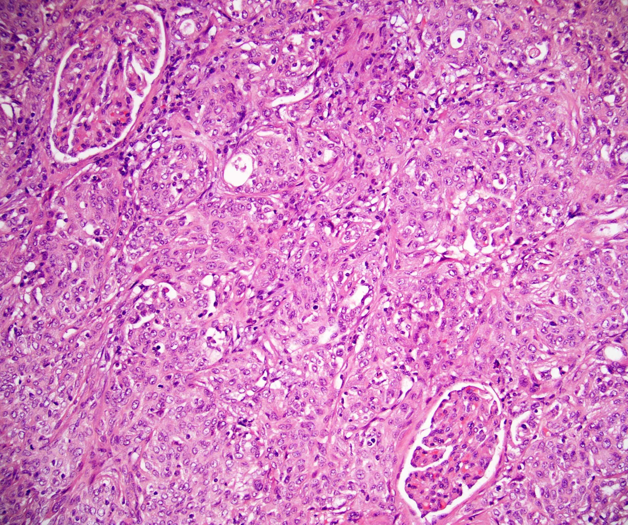
Zellballen pattern
Typical cytologic features
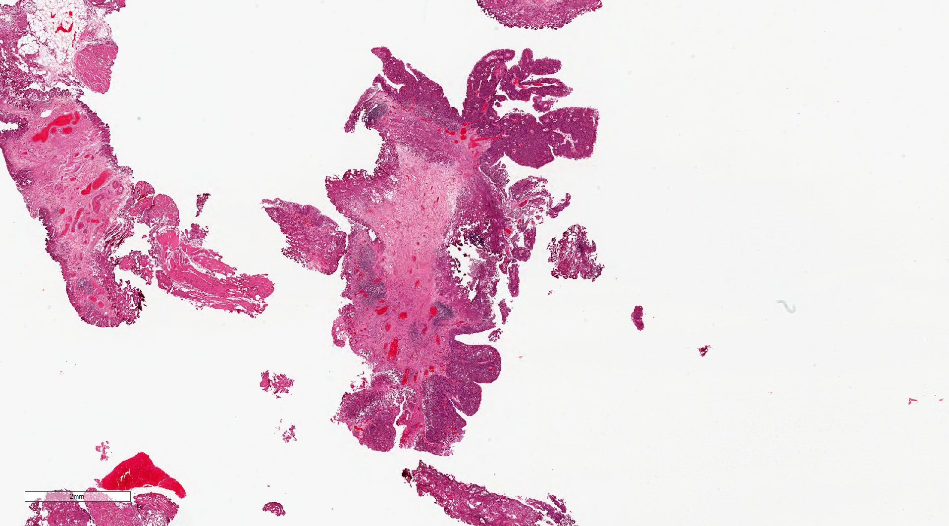
Transurethral bladder resection
Zellballen pattern
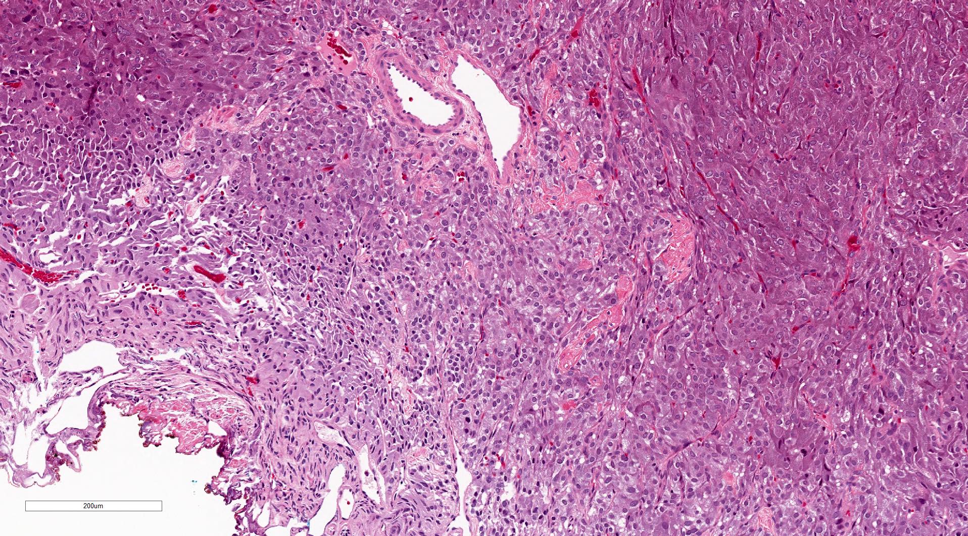
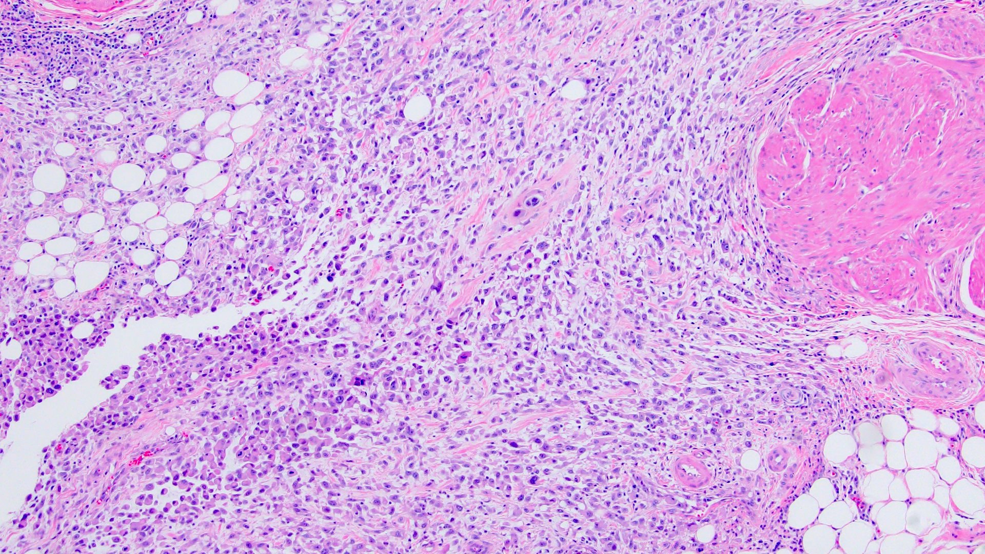
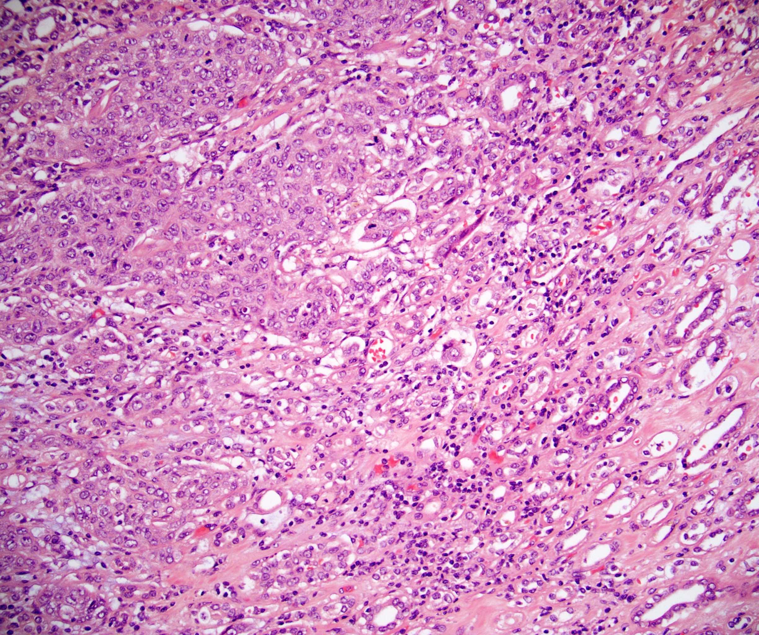
Muscularis propria invasion
Transurethral bladder resection
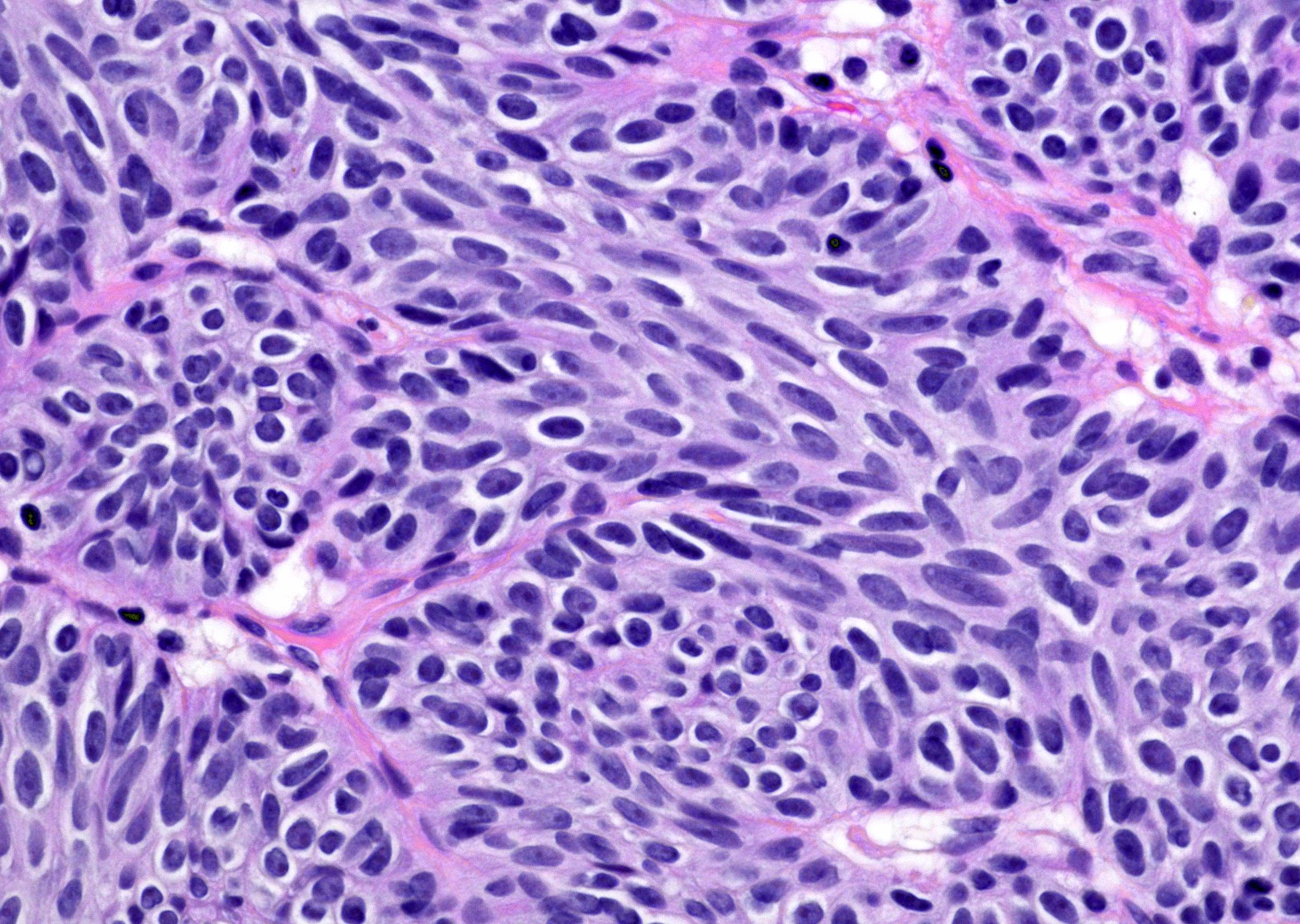
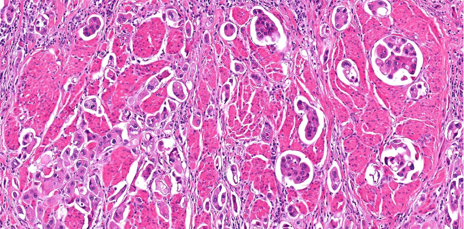
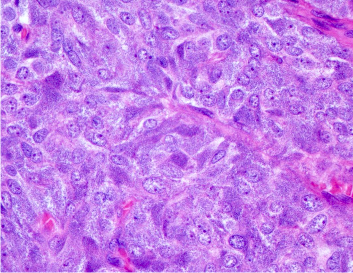
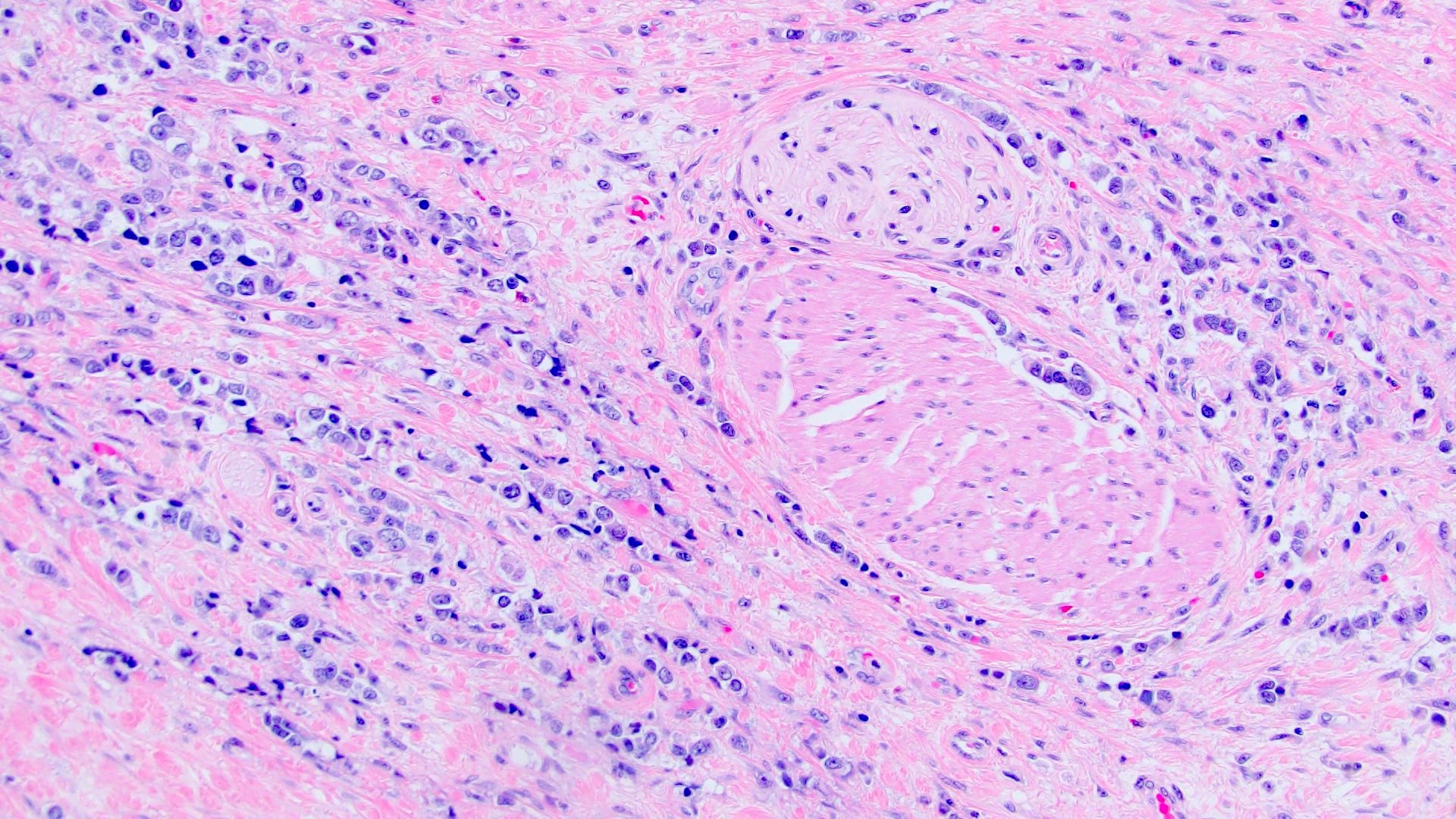
Nuclear atypia
Zellballen pattern

Amphophilic cytoplasm

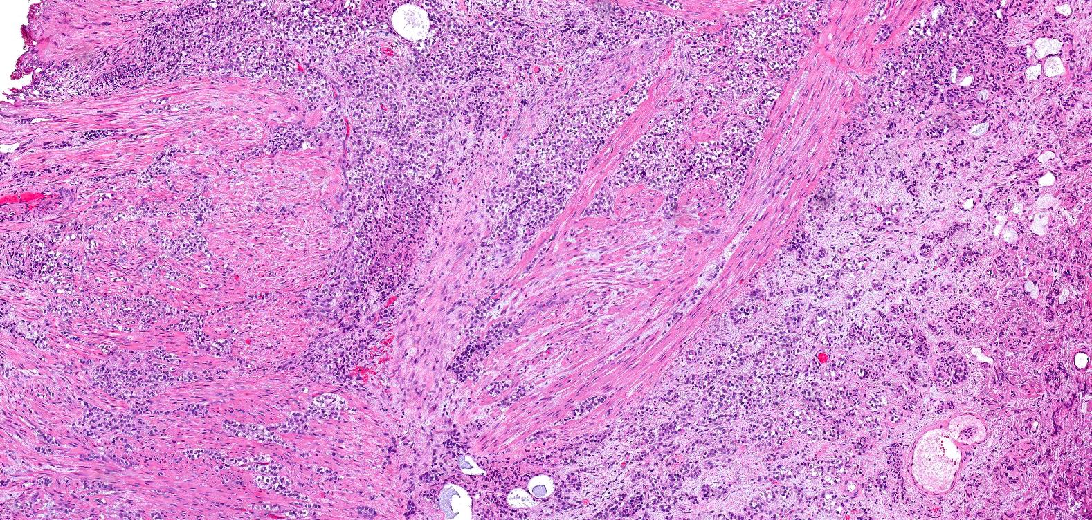
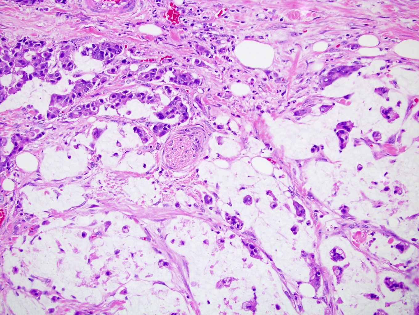
Pseudopapillary architecture
Cellular spindling
Mitotic activity
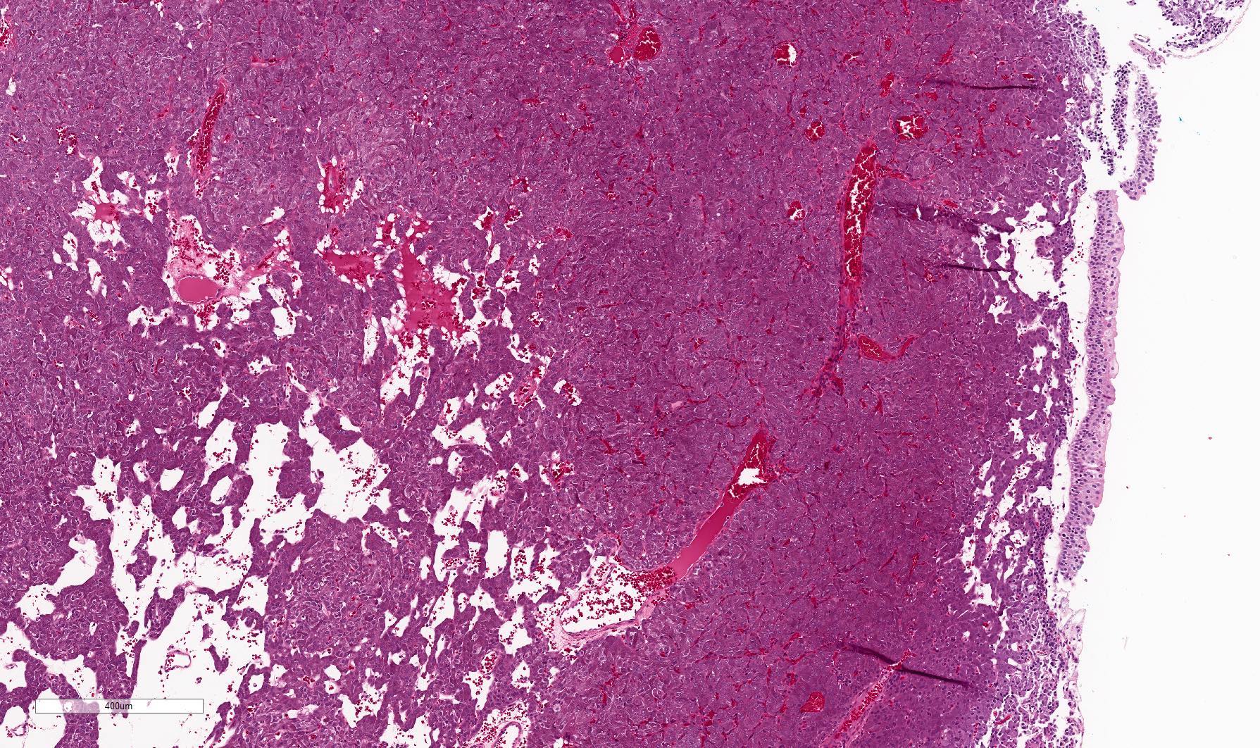
Muscularis propria invasion
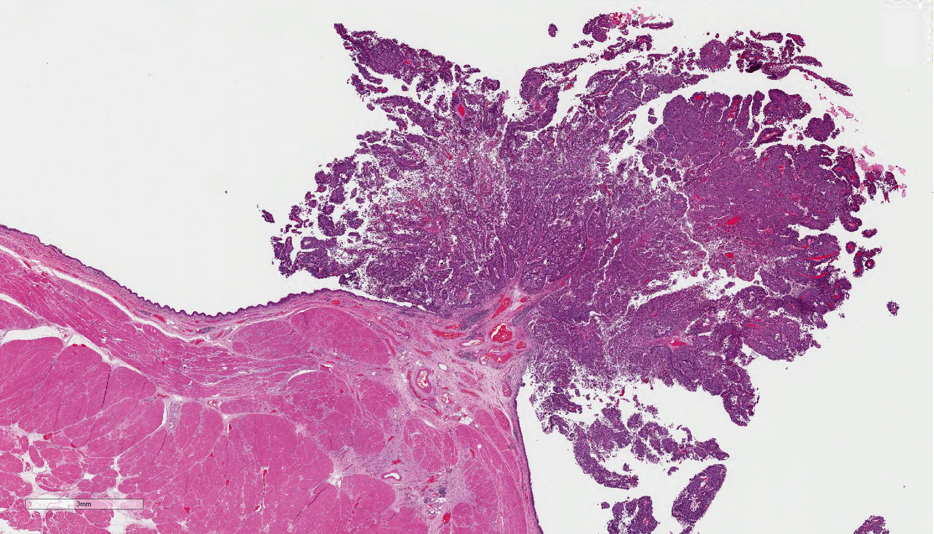
Deep lamina propria invasion
Pseudocapsule
Typical cytologic features
Typical cytologic features
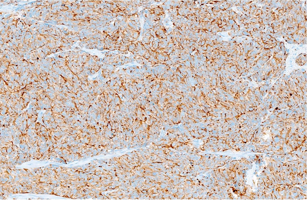
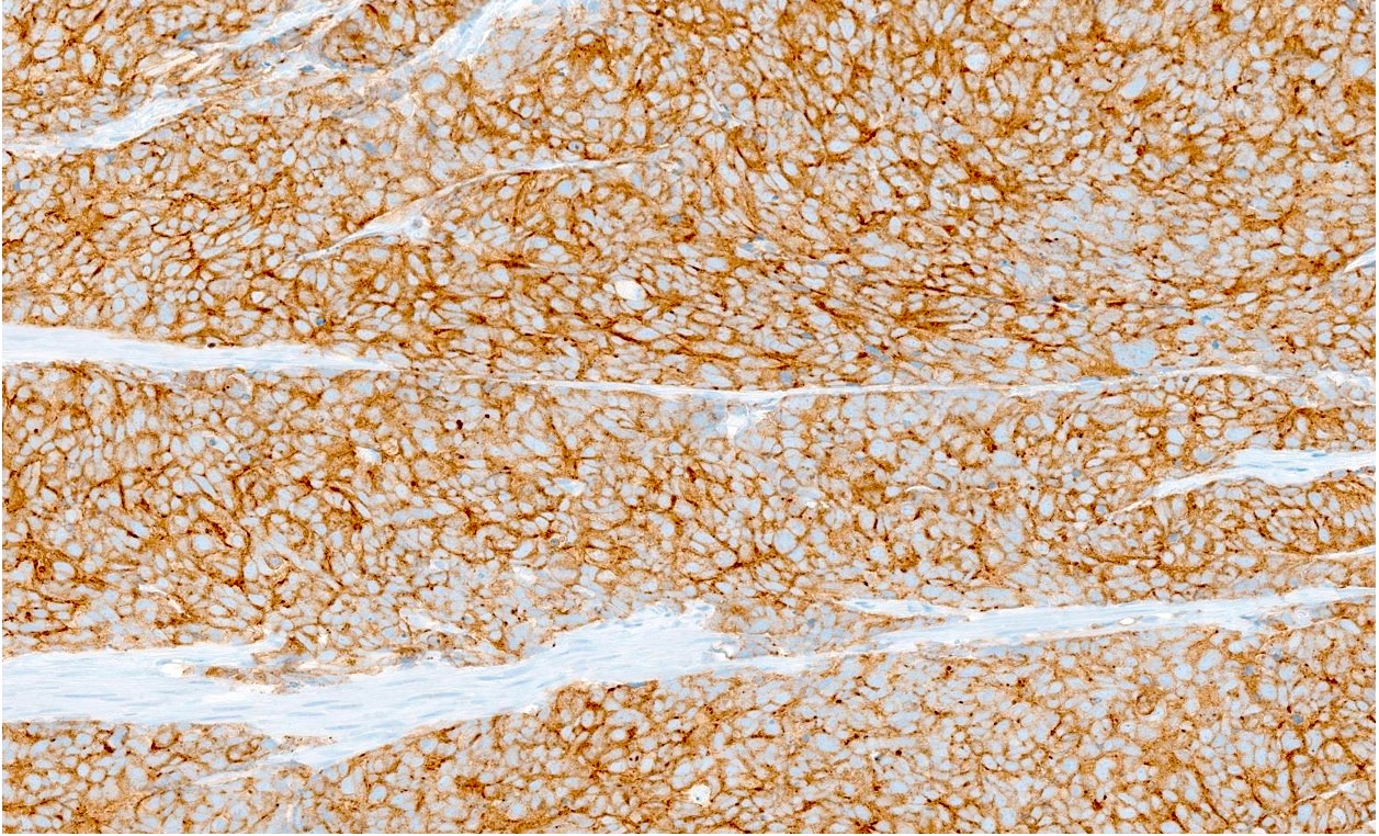
Synaptophysin
Chromogranin
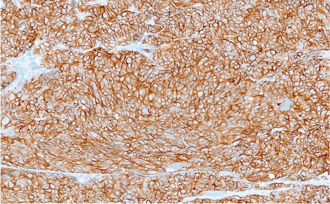
GATA3
S100
SDHB retained
Pankeratin
p63
Chromogranin

Synaptophysin
S100
HMB45
Synaptophysin
GATA3
Contributed by Vaishali Pansare, M.D.
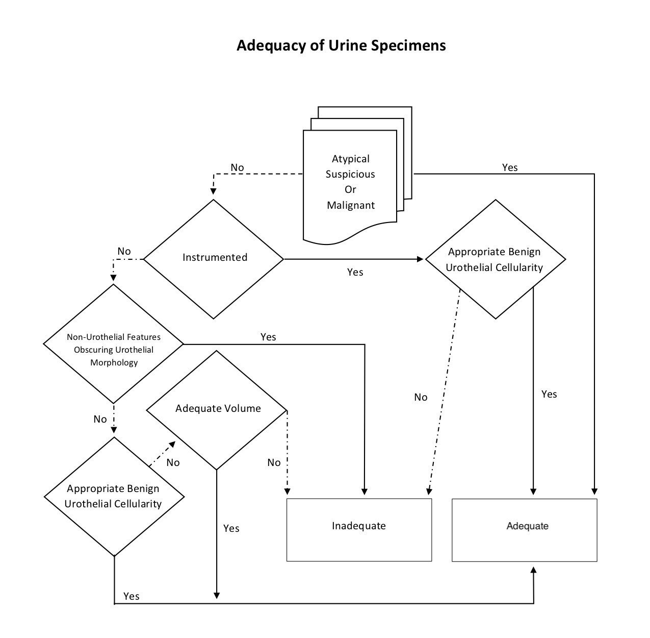
Adequacy of urine specimens
Images hosted on other servers:

Normal embryo at 25 - 27 days

Normal embryo at weeks 5 - 7

Diagram of persistent cloaca
Images hosted on other servers:

MRI of PUC with bladder muscle invasion

MRI and CT of PUC with peritoneal spread
CT of PUC with
peritoneal spread
and bladder wall
thickening
Contributed by Timothy Isaac Miller, M.D., M.A. and Maria Tretiakova, M.D., Ph.D.
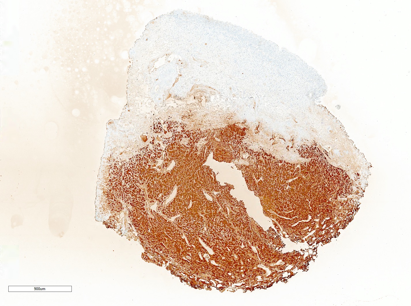
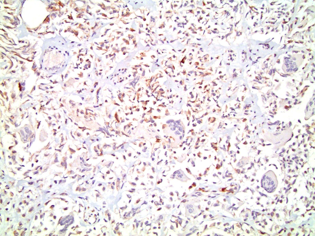
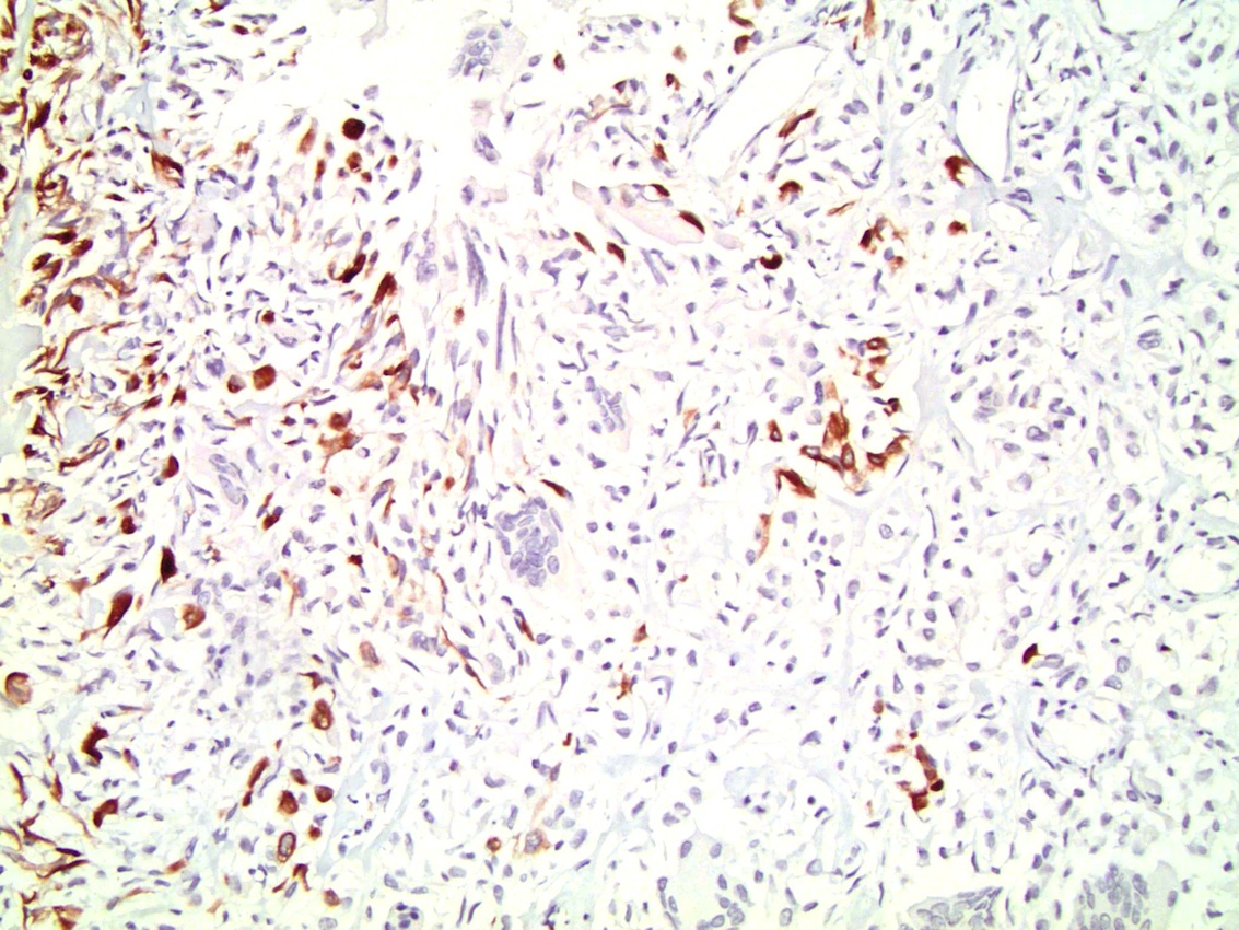
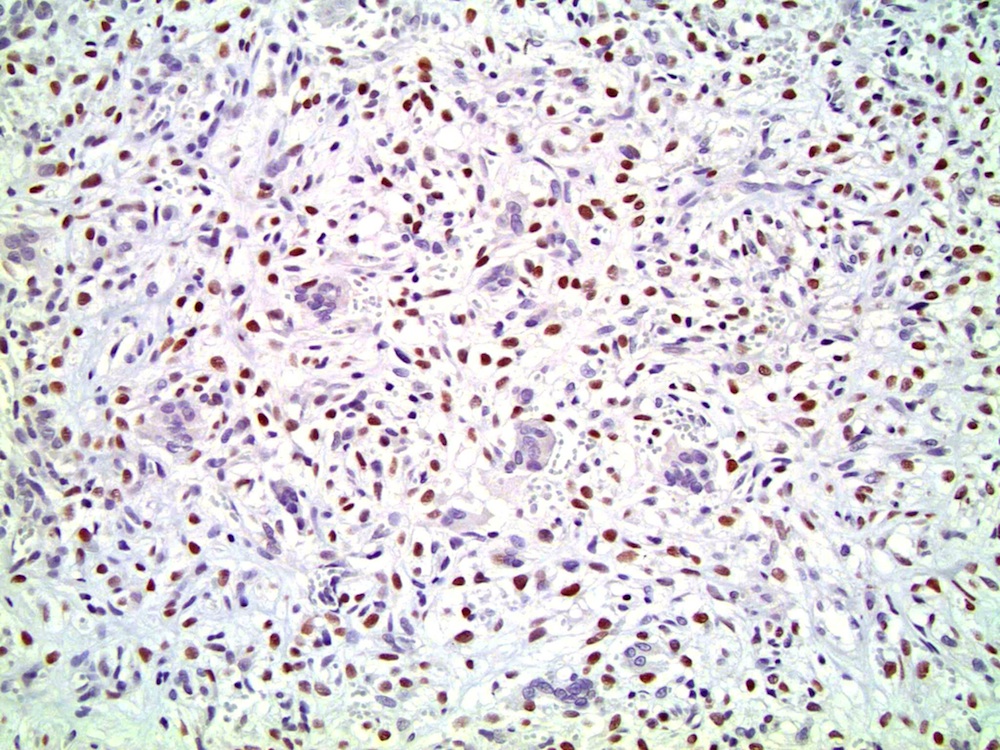
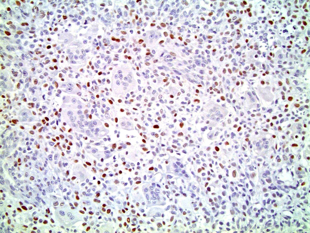
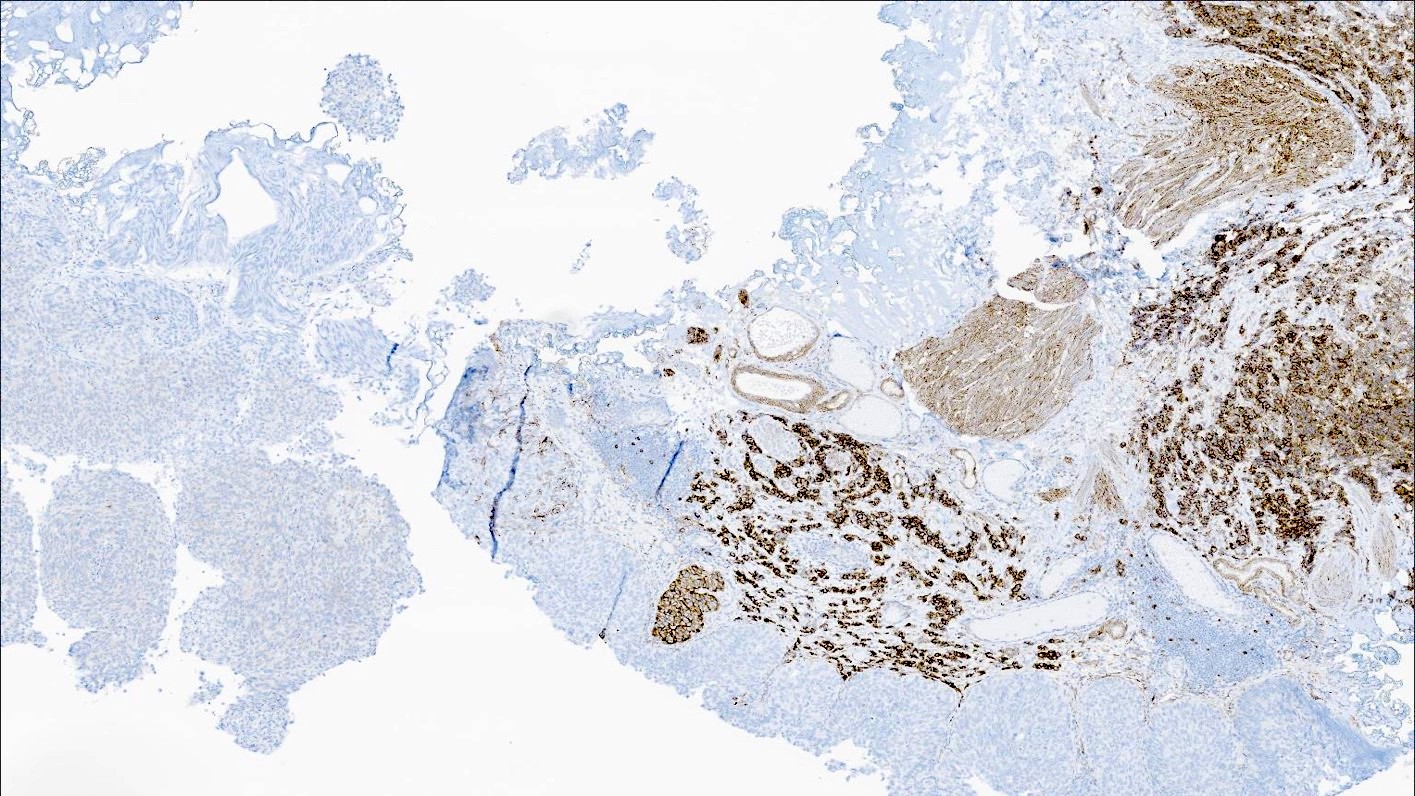
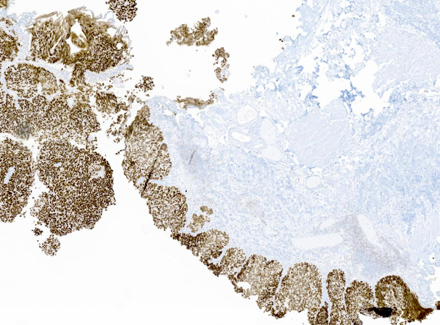
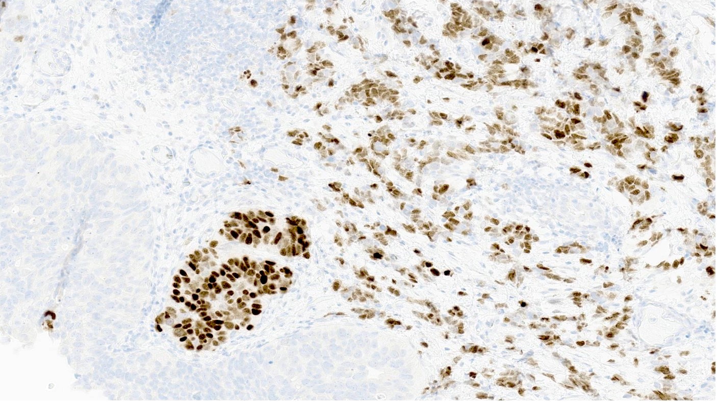
Bladder with plasmacytoid urothelial carcinoma
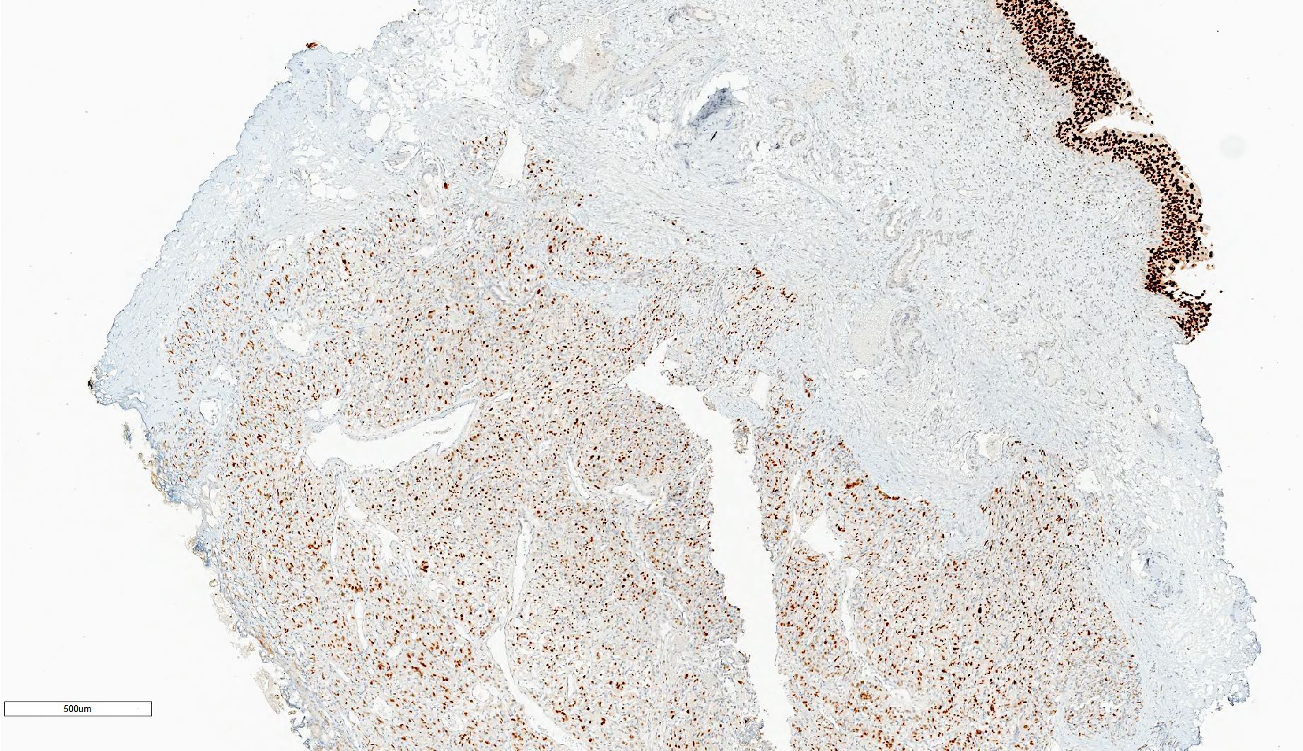
Contributed by Timothy Isaac Miller, M.D., M.A., Nicole K. Andeen, M.D. and Maria Tretiakova, M.D., Ph.D.
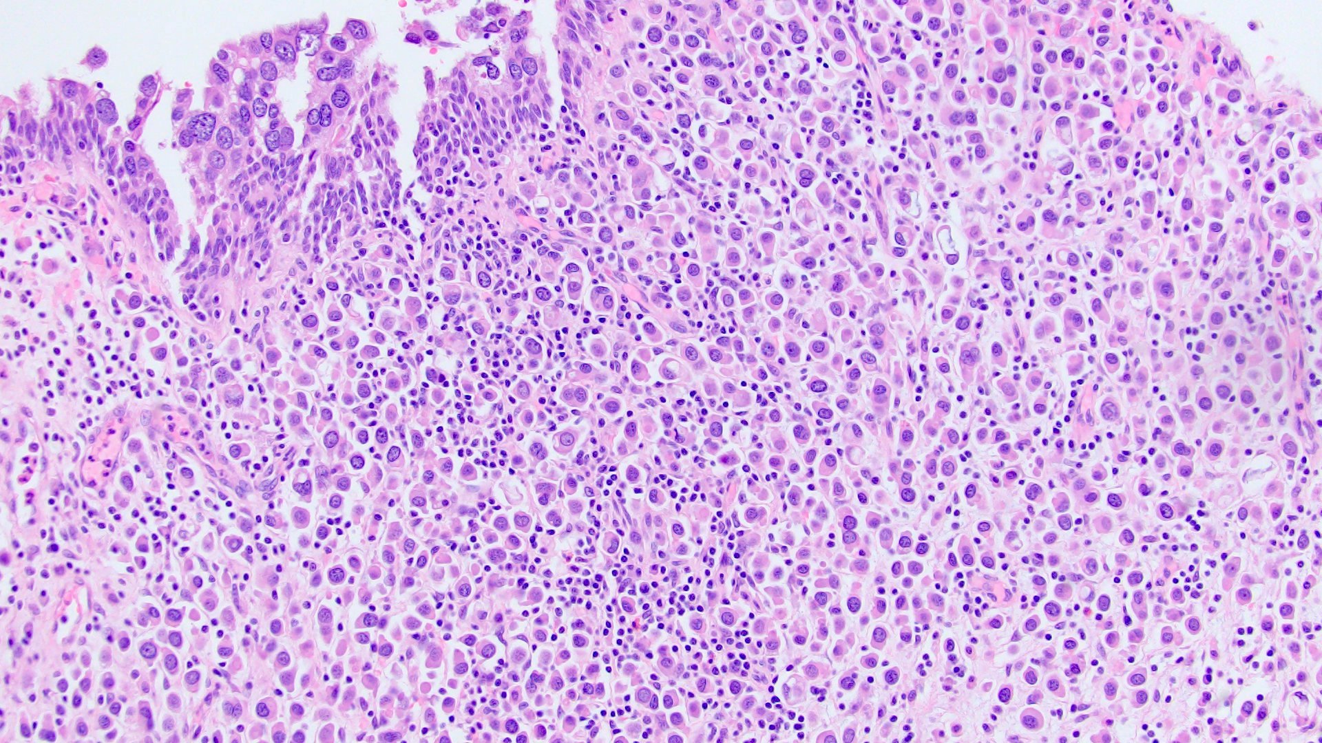
PUC with carcinoma in situ
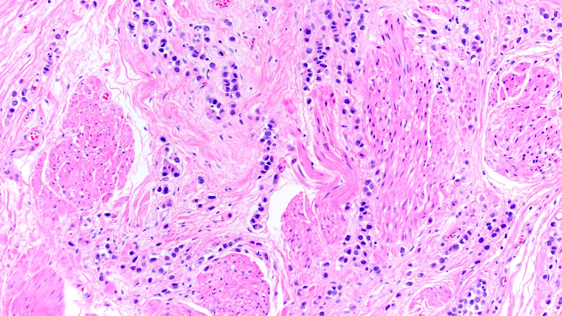
Muscularis propria involvement
Diffuse growth

Pleomorphic subvariant

Desmoplastic subvariant

Signet ring and classic morphologies

Lymphovascular invasion
Perineural invasion
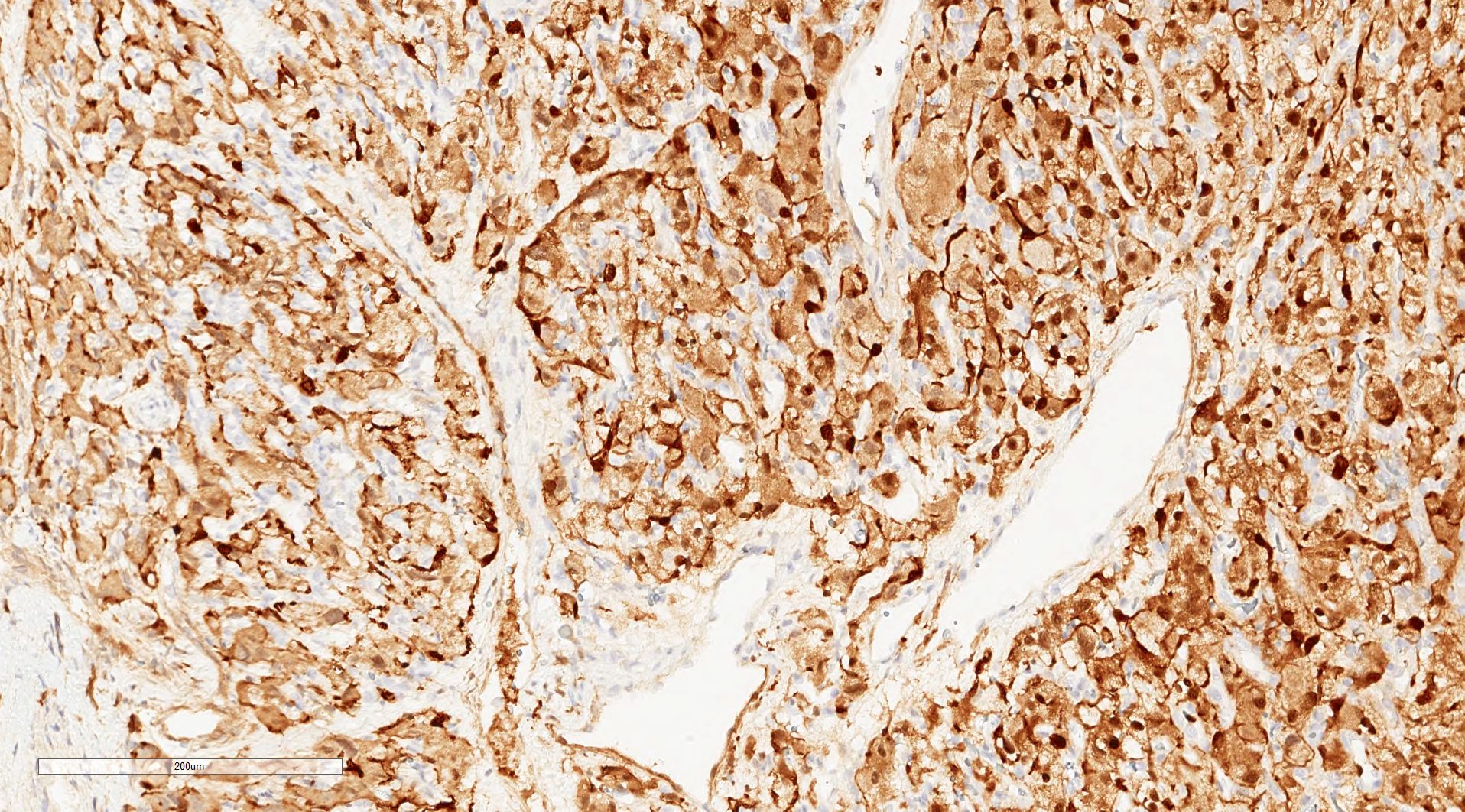
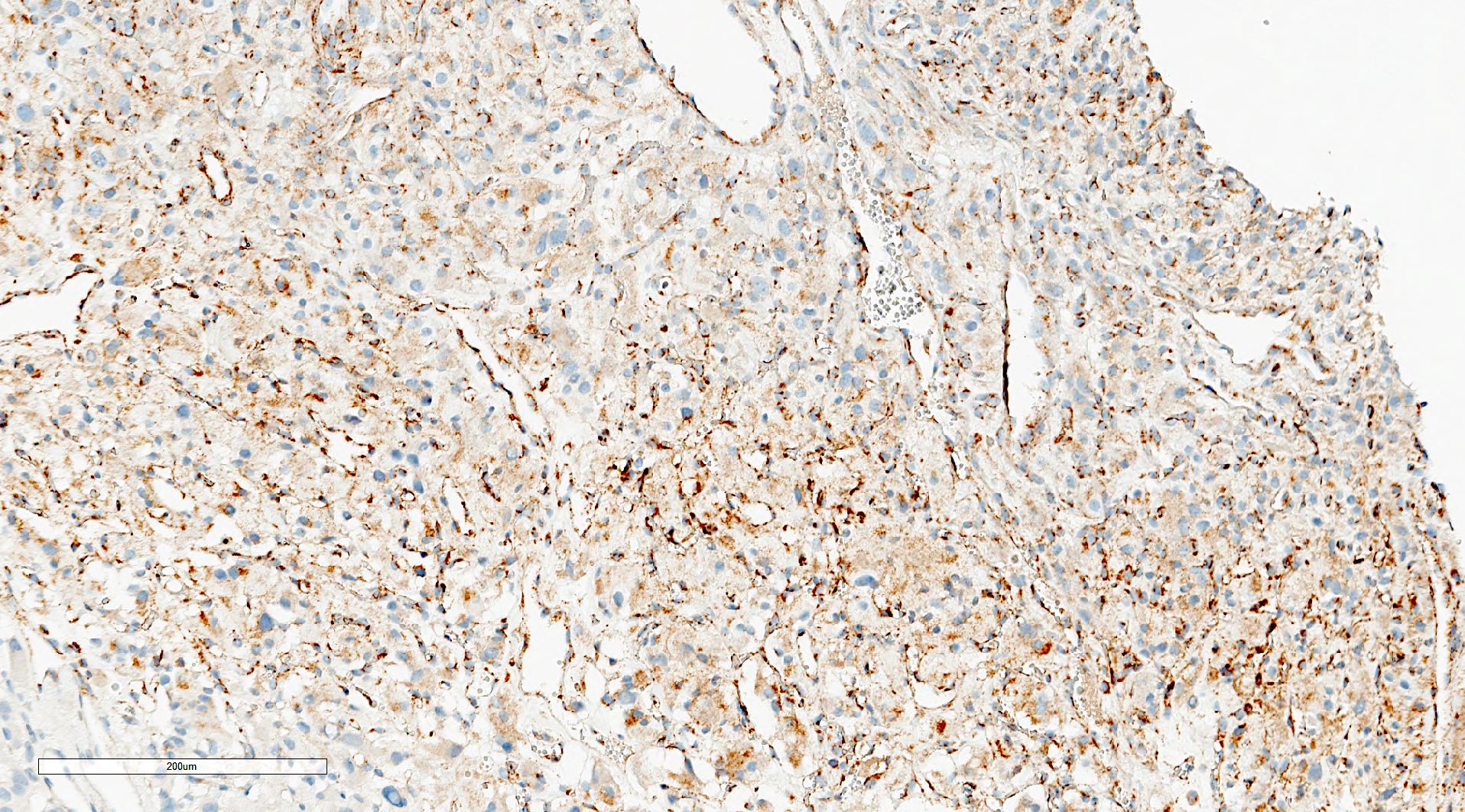
PUC in bladder wall
PUC at urethral margin
AE1 / AE3
CD138
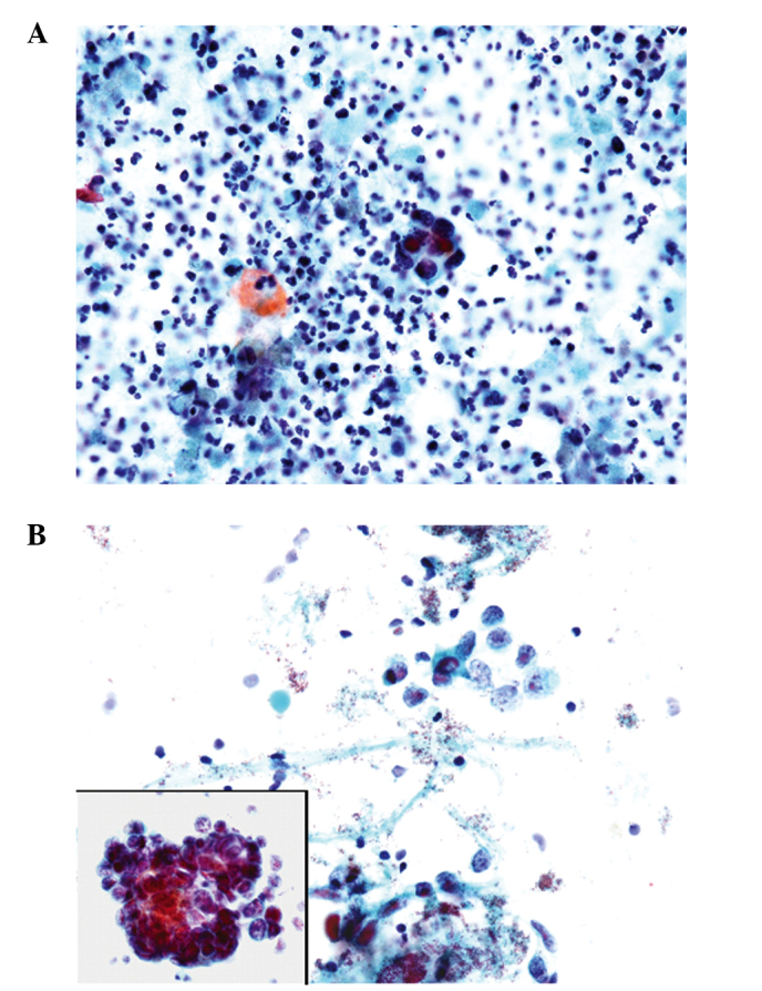
Contributed by Lisa Han, M.D. and Ricardo Lastra, M.D. (Case #510)

Urothelial carcinoma, plasmacytoid variant
Images hosted on other servers:

Bladder washing with PUC (Papanicolau stain, x400)
Plasmacytoid variant histology in bladder cancer
Images hosted on other servers:

Irregular wall thickening
Images hosted on other servers:

Tumor-like lesion
Contributed by Maria Carolina Beeter, M.D. and Y. Albert Yeh, M.D., Ph.D.
Polypoid mass
Inflammatory polypoid lesion
Nonneoplastic polypoid lesion
Polypoid lesion
Small cobblestone nodule
Small polypoid nodule
Fibrotic lamina propria
Somewhat fibrotic stroma
Polypoid papillary projections
Edematous polypoid lesion
Narrow to broad based papillae
Focal urothelial hyperplasia
Bullous lesion
Normal urothelial lining cells
Polypoid and papillary configuration
Images hosted on other servers:

Tumor of distal ureter
Case #339:
Various images
EMA
CK7

p53
GATA3
p63
SMA
S100
CD68
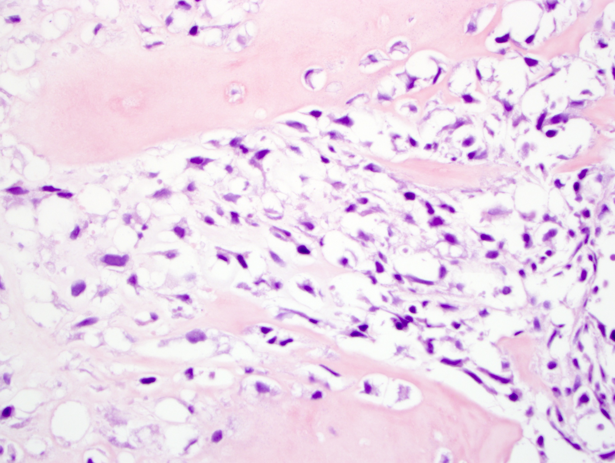
Images hosted on other servers:

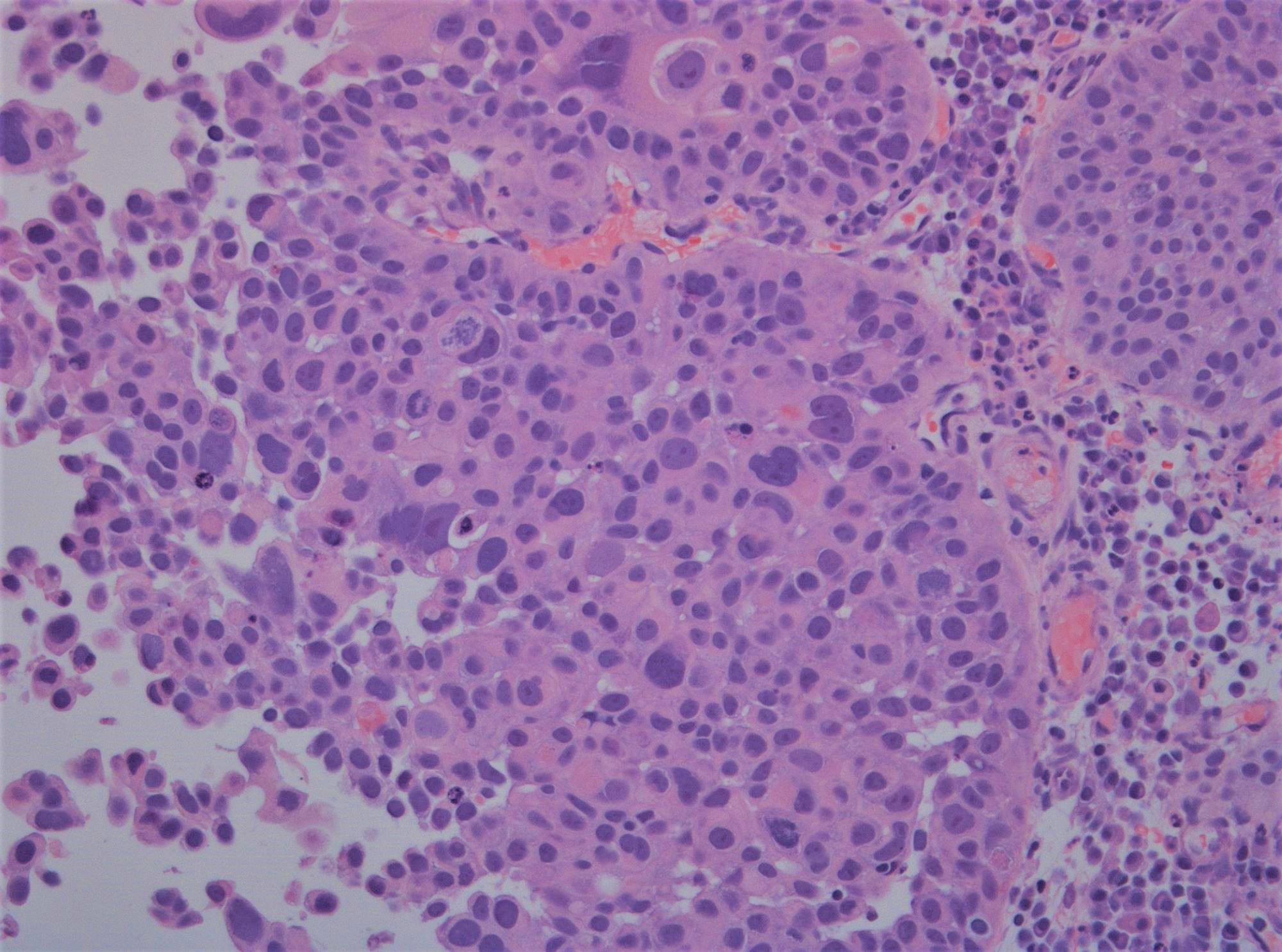
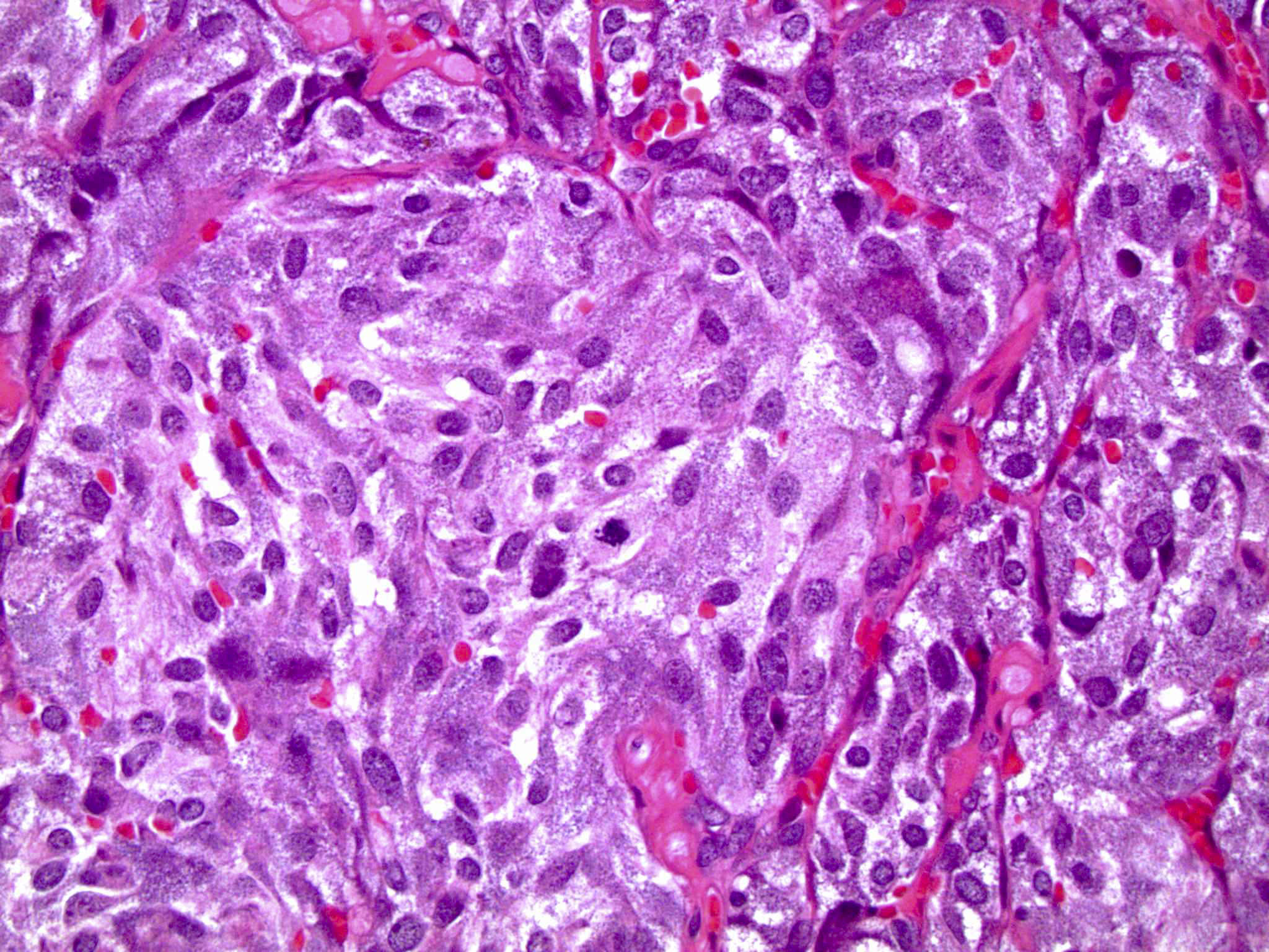
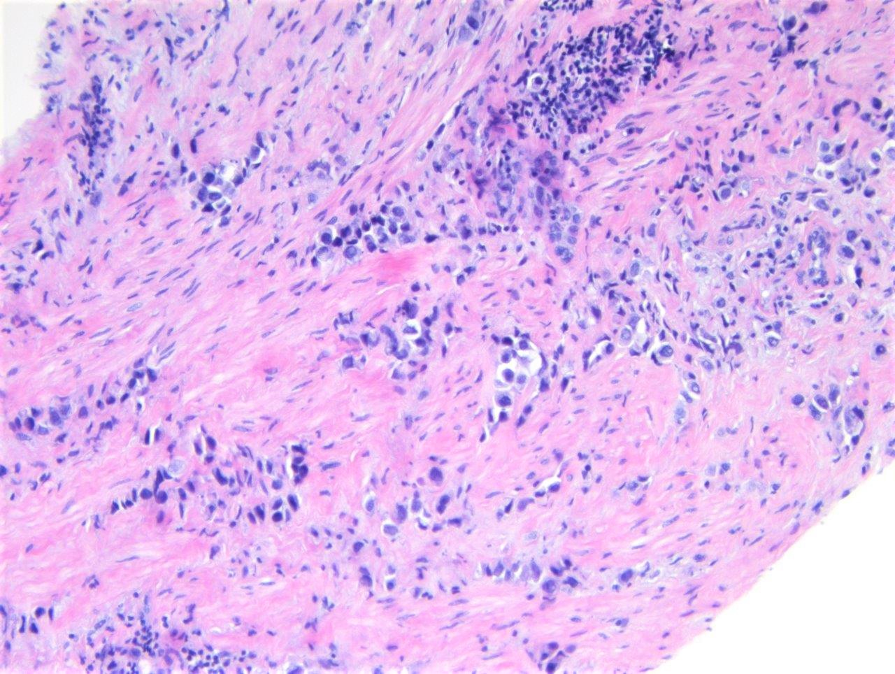
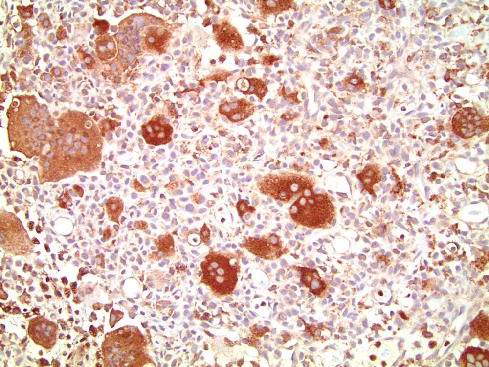
Background mononuclear tumor cells

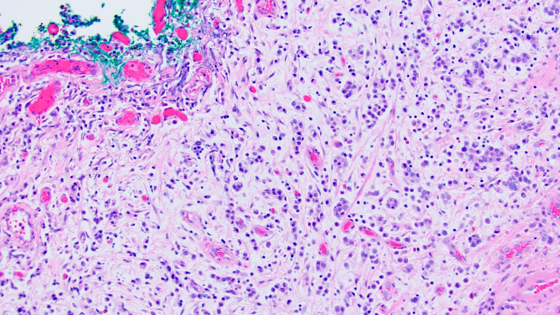
Osteoclast-like giant cell carcinoma

Osteoclast rich undifferentiated urothelial carcinoma

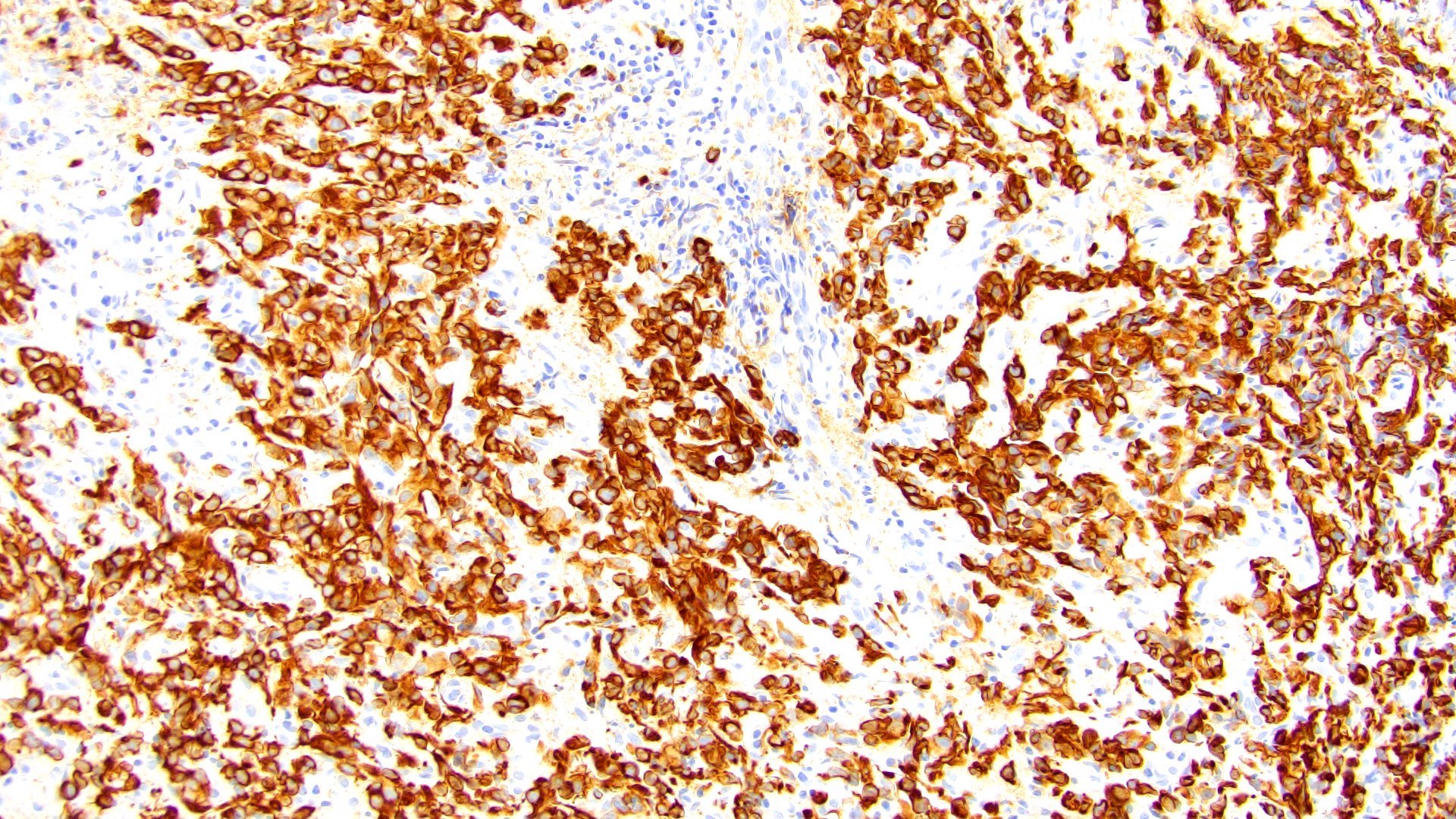
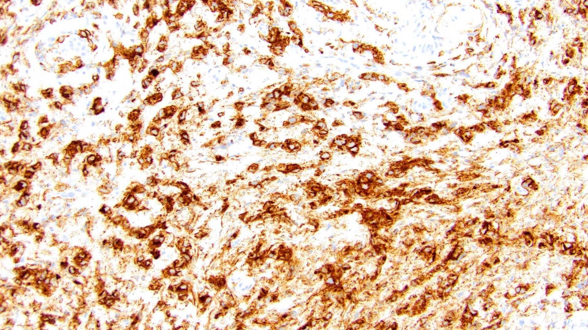
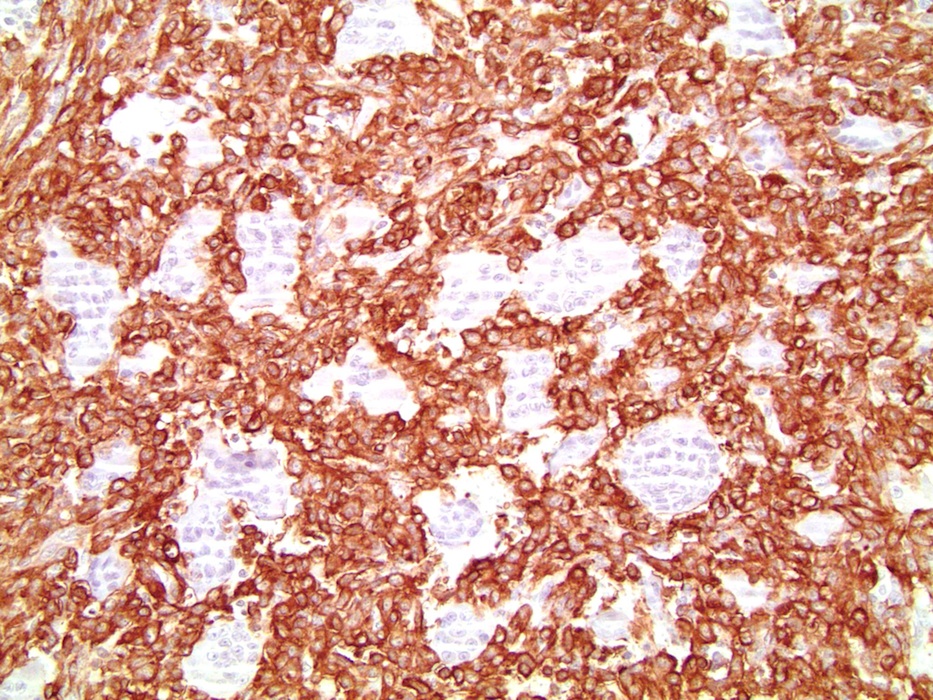
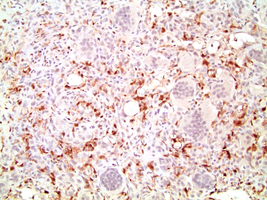
Cytoplasmic immunoreactivity with vimentin

Immunoreactivity with CD68

Focal immunoreactivity with AE1 / AE3

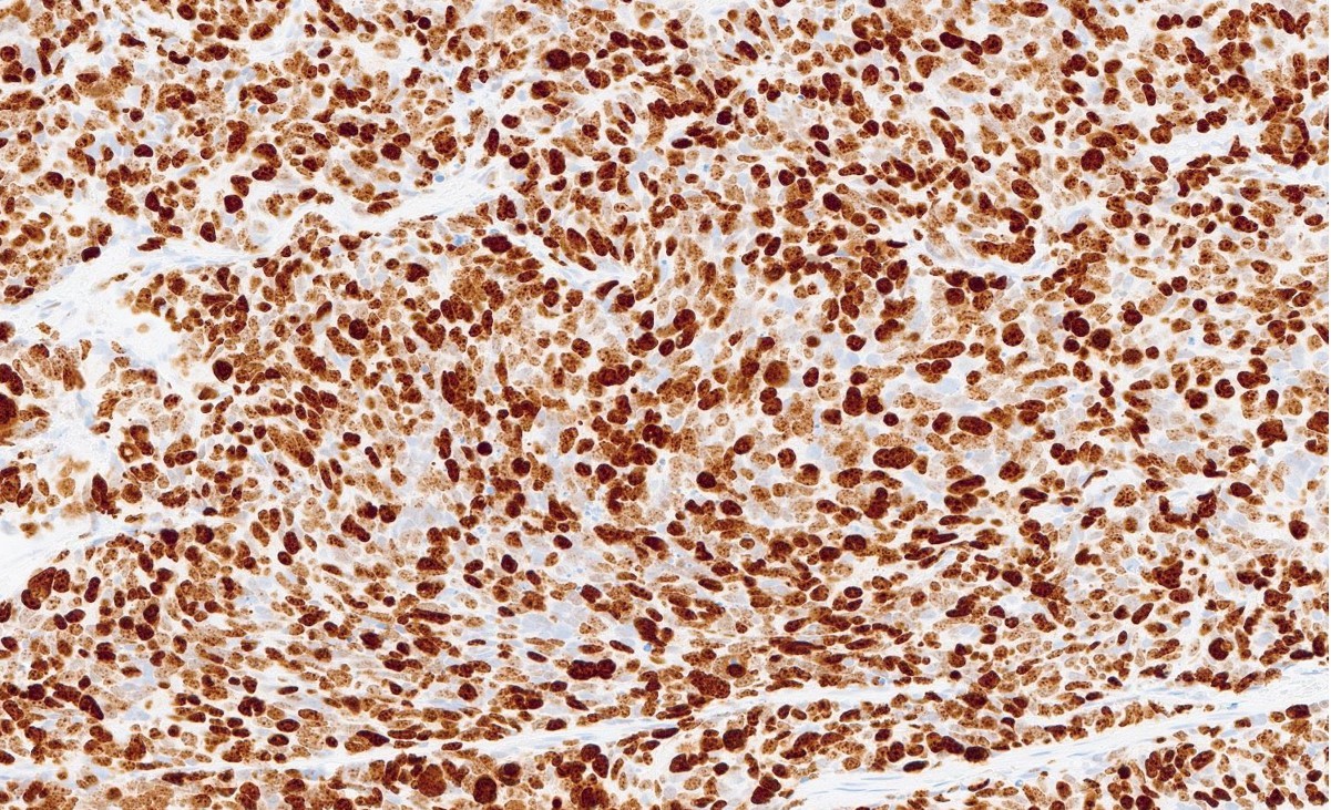
Nuclear immunoreactivity with Ki67

Nuclear immunoreactivity with p53

Osteoclast-like giant cells positive for CD68
Images hosted on other servers:

Diff Quick stain

Pap stain

Diff Quick stain
AFIP images
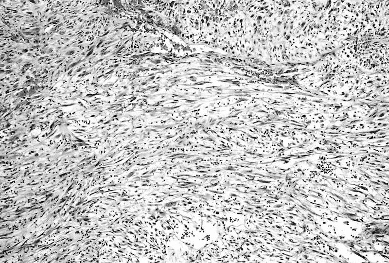
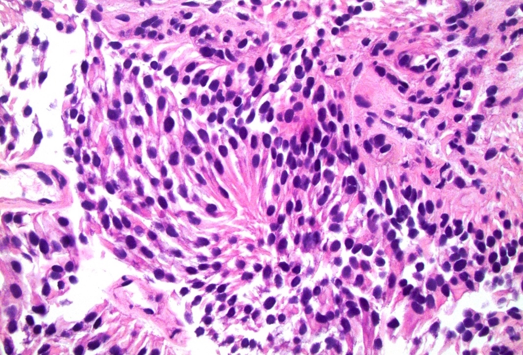
Interlacing fascicles of myofibroblasts
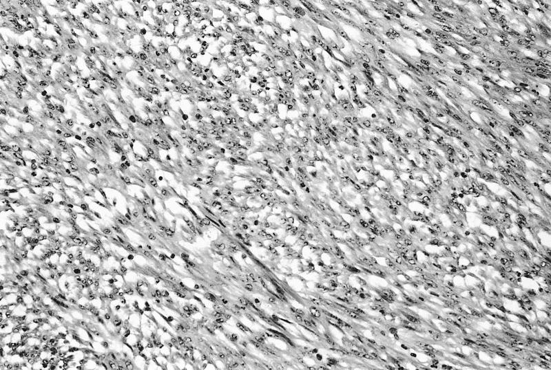
Uniform cells are mixed with inflammatory cells
Images hosted on other servers:
Left: muscularis
propria,
Right: spindle
cell nodule
Images hosted on other servers:

Smooth sessile mass on cystoscopy

Spherical mass on cystoscopy
Contributed by Bonnie Choy, M.D.
Polypoid
Admixture of urothelium and prostatic acini
Corpora amylacea
PSA positive
Images hosted on other servers:

Pathophysiology
Images hosted on other servers:

Bladder wall thickening
Images hosted on other servers:

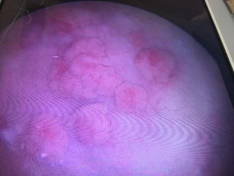
Cystoscopy
Contributed by Y. Albert Yeh, M.D., Ph.D. and Jennifer Lee, M.D.
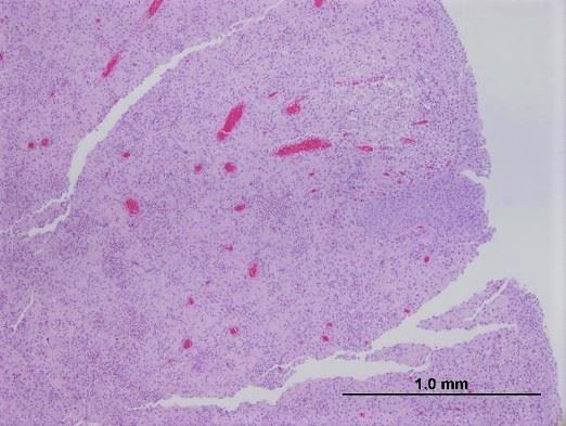
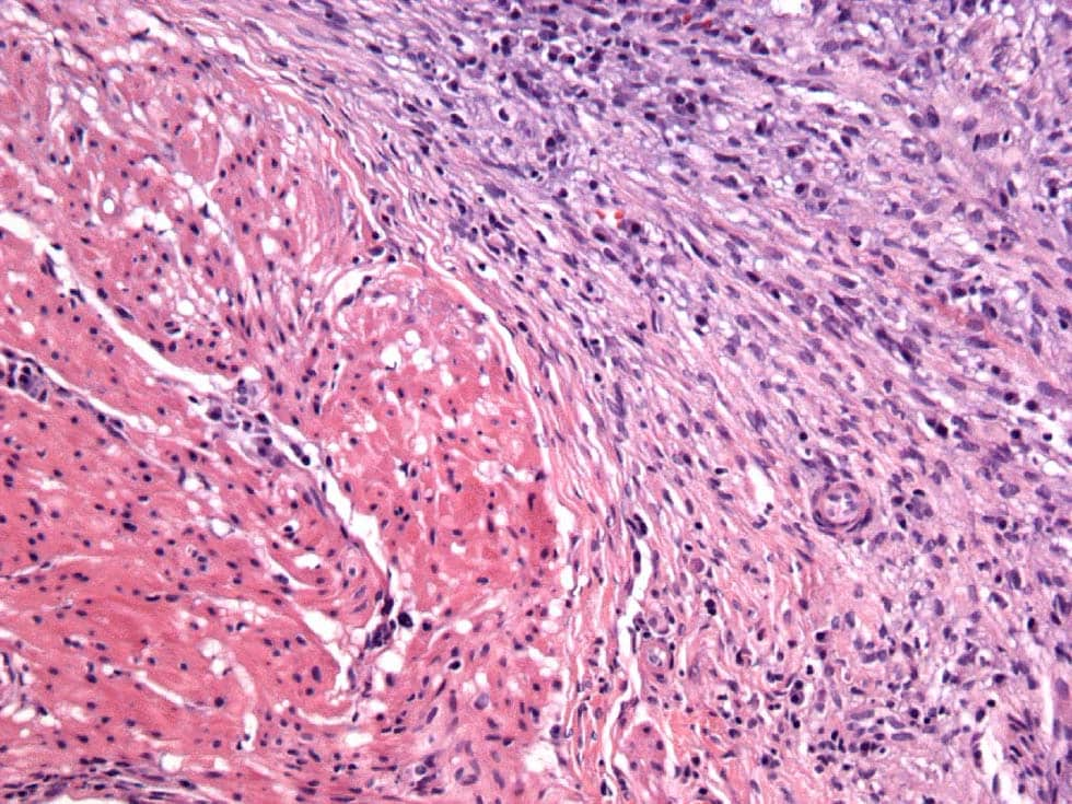
Telangiectatic vessels with fibrin
Dilated vessels with fibrin
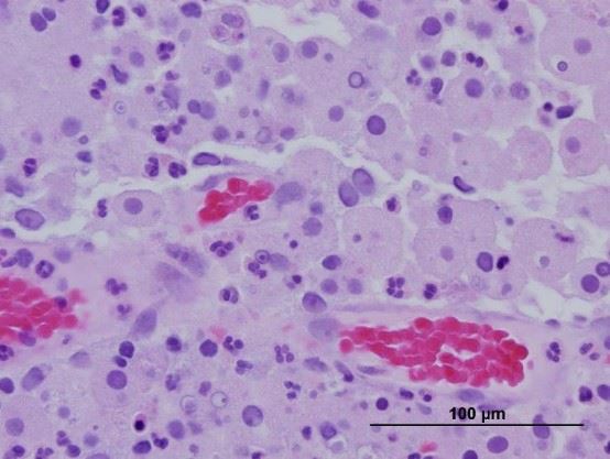
Epithelial cells encircling fibrin
Urothelial cells enclosing fibrin
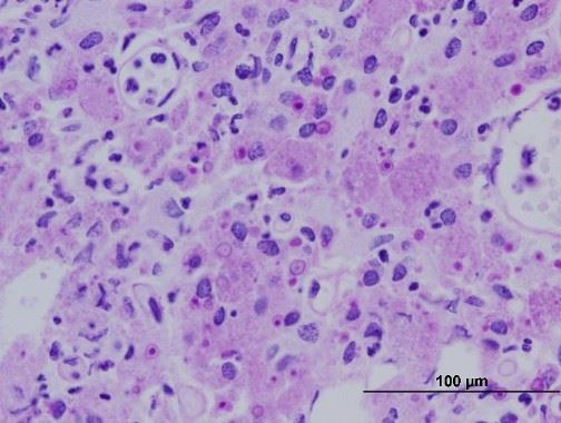
Atypical urothelial proliferation
Atypical urothelial proliferation
Fibrinoid vascular necrosis
Fibrin and proliferating cells
Fibrinoid necrosis of vessels
Mucosal hemorrhage and inflammation
Radiation induced urothelial changes
Atypical von Brunn nests
Stromal cell changes
Images hosted on other servers:

Radiation induced urothelial cell changes
Radiation induced urothelial reactive atypia
Contributed by Nicole K. Andeen, M.D. and Maria Tretiakova, M.D., Ph.D.
Mass involving hilum
Contributed by Megan L. Brown, M.D., Nicole K. Andeen, M.D., Maria Tretiakova, M.D., Ph.D. and Kenneth A. Iczkowski, M.D.
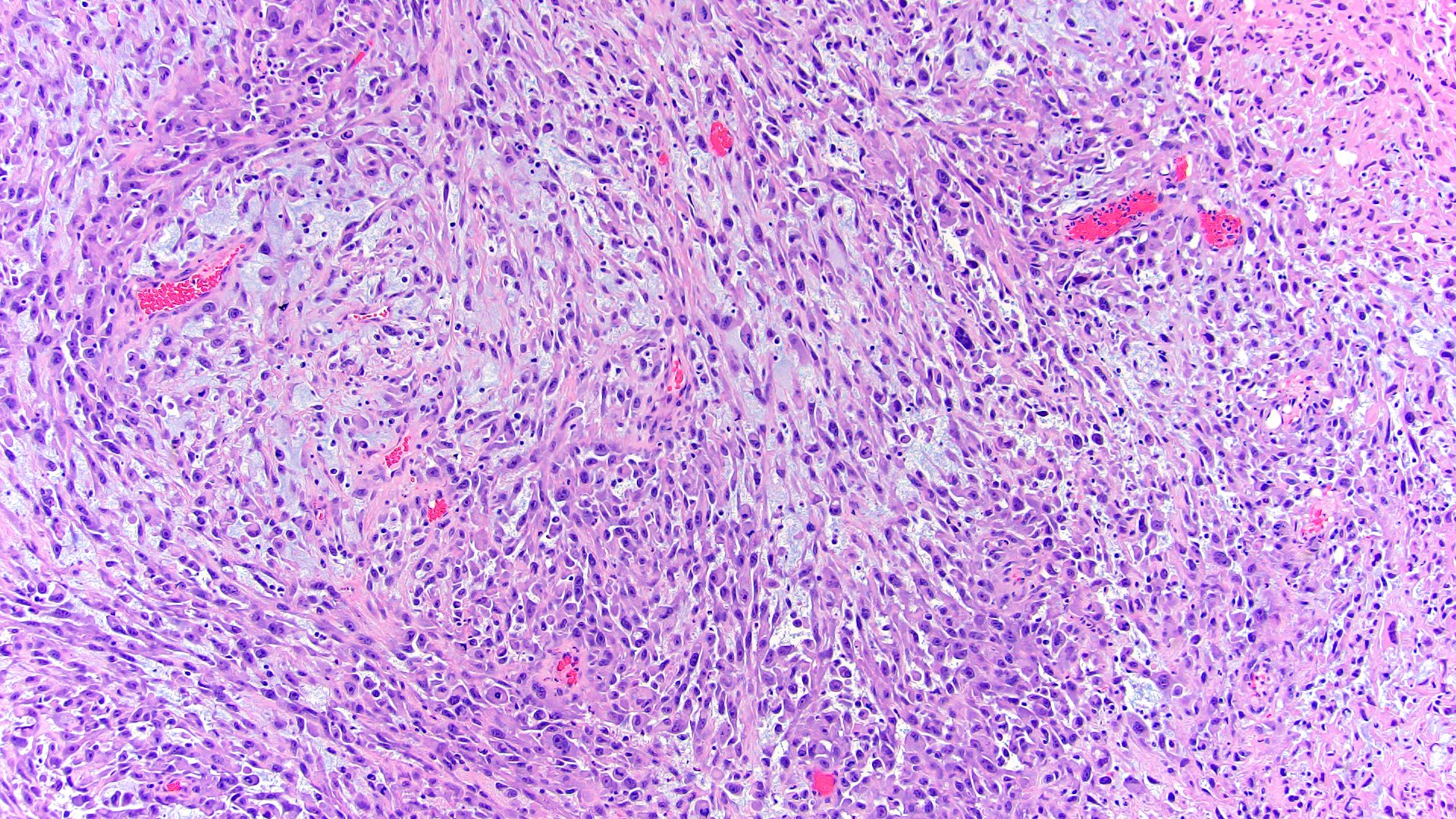
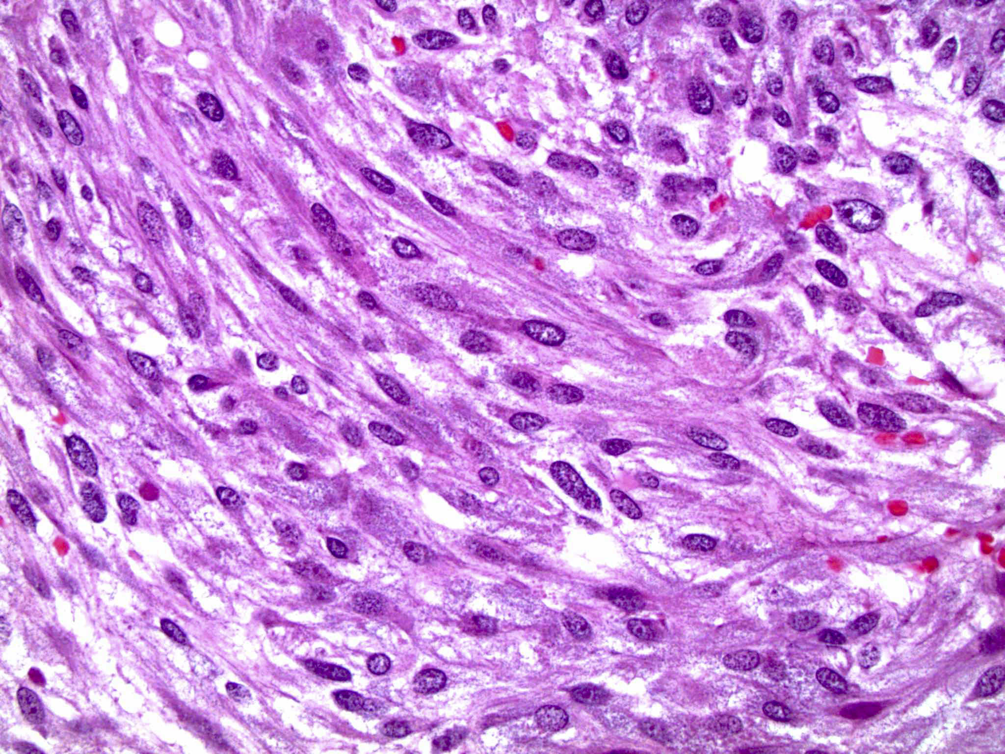
High grade spindled to epithelioid cells
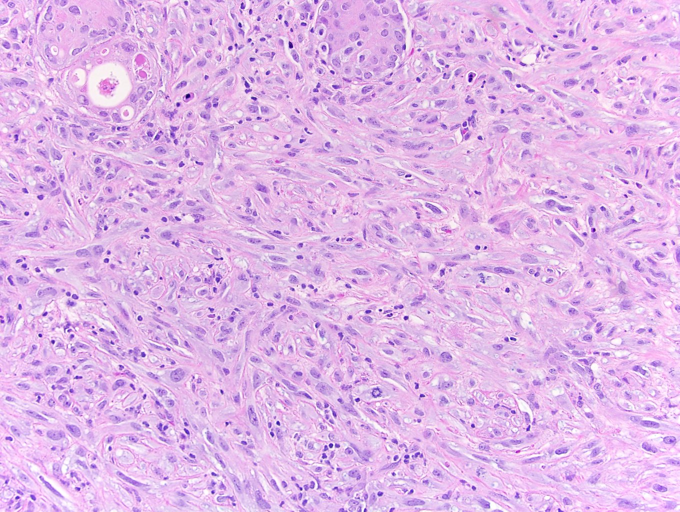
Elongated spindled cells
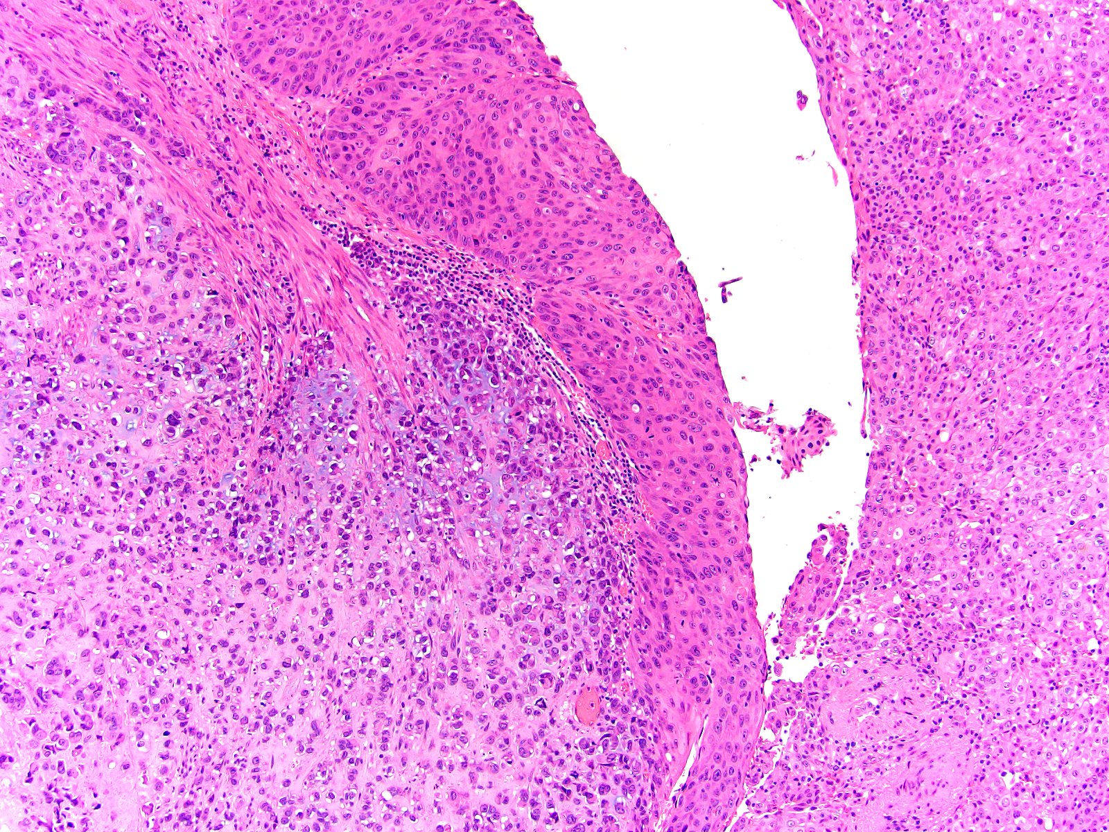
Classic urothelial carcinoma

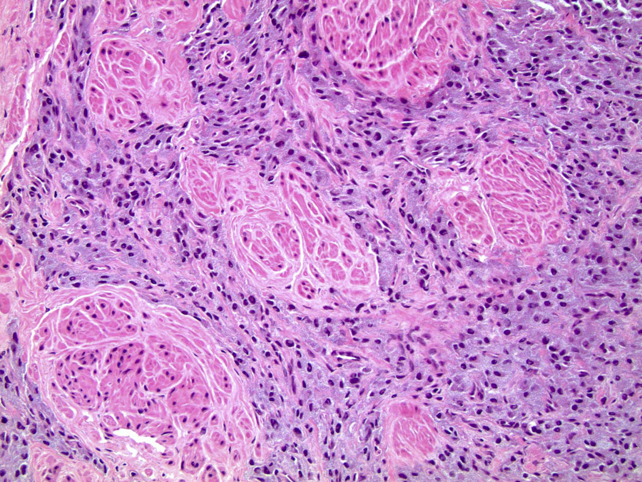
Neoplasm with admixed carcinomatous and sarcomatous elements
Osteosarcomatoid differentiation
GATA3 and CK7
Images hosted on other servers:

Life cycle
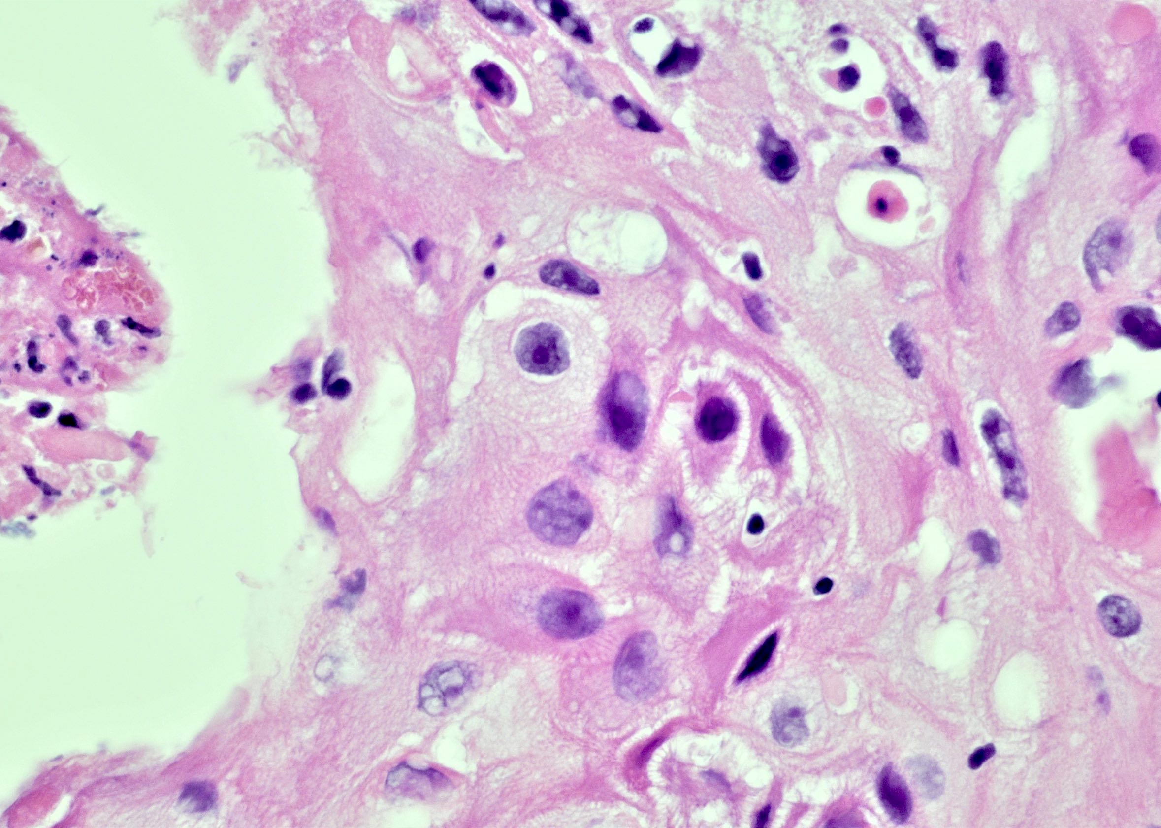
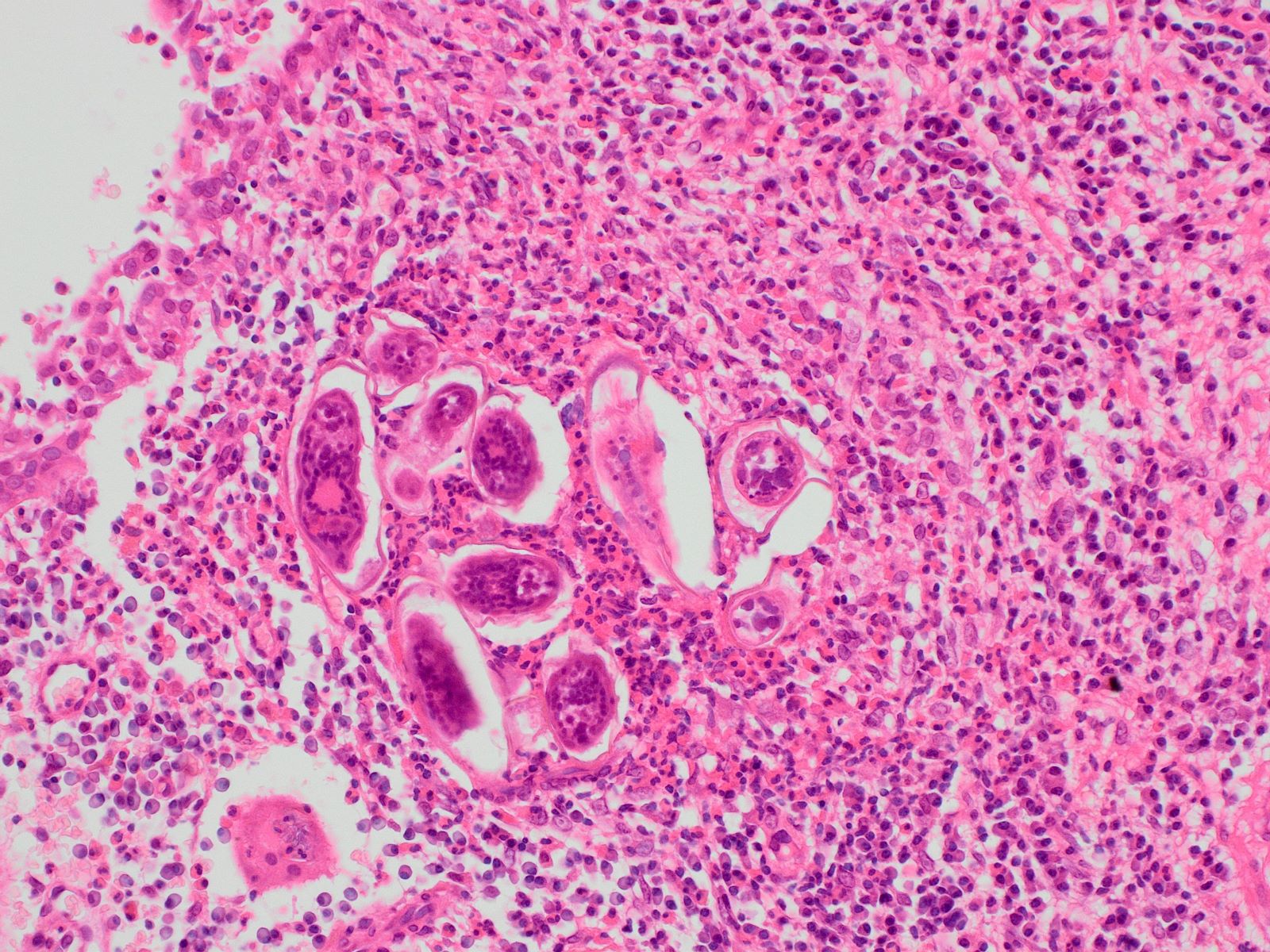
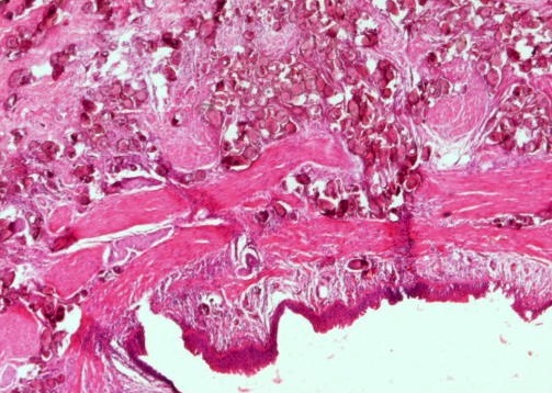
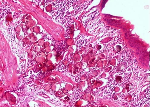
Contributed by Y. Albert Yeh, M.D., Ph.D., Susan Prendeville, M.D. and Kiran Alam, M.D., Anshu Jain, M.D.,
Veena Maheshwari, M.D., Farhan A. Siddiqui, M.B.B.S., Ershadul Haq, M.B.B.S., M.D. and @ThatGlassTho on Twitter
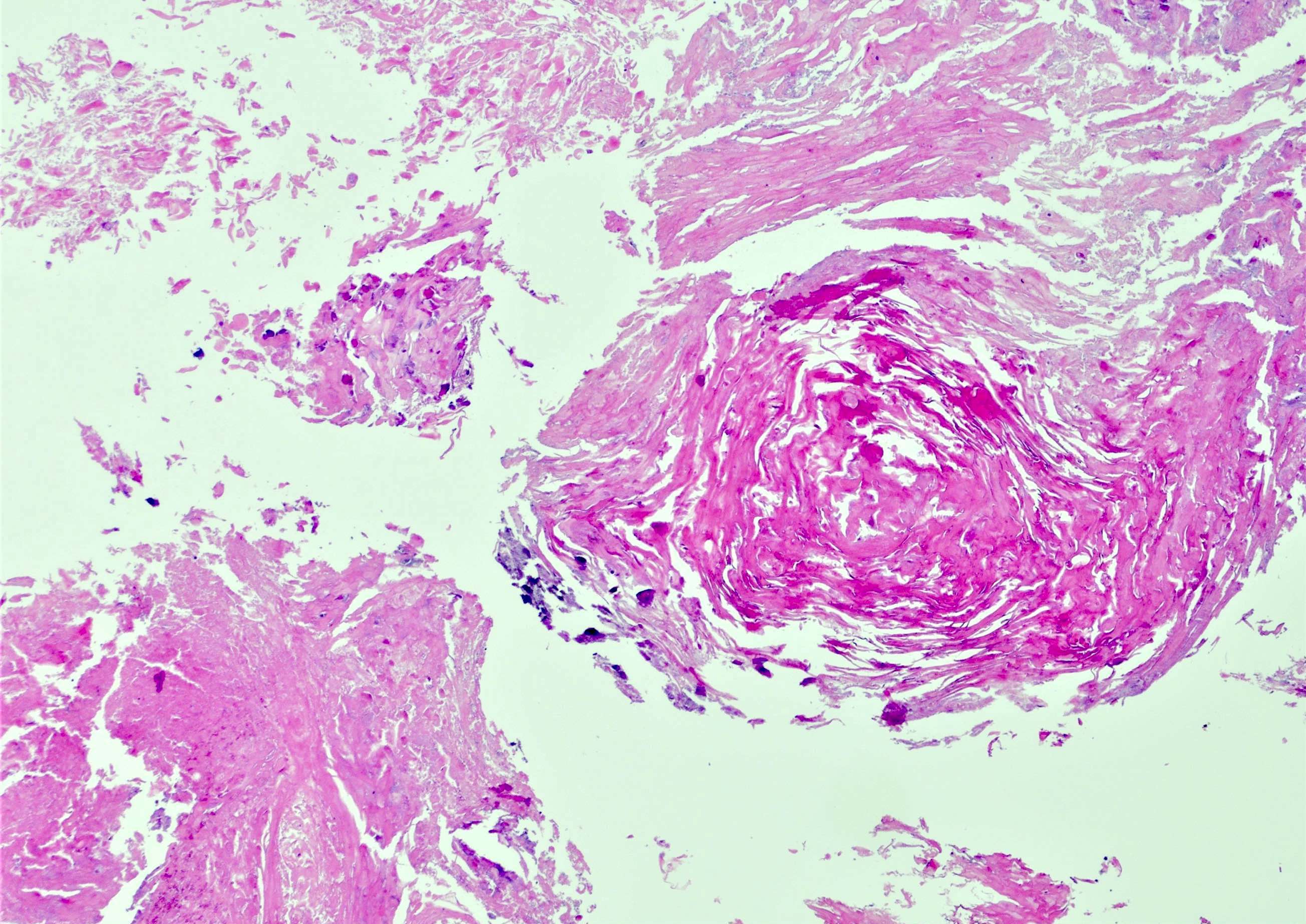
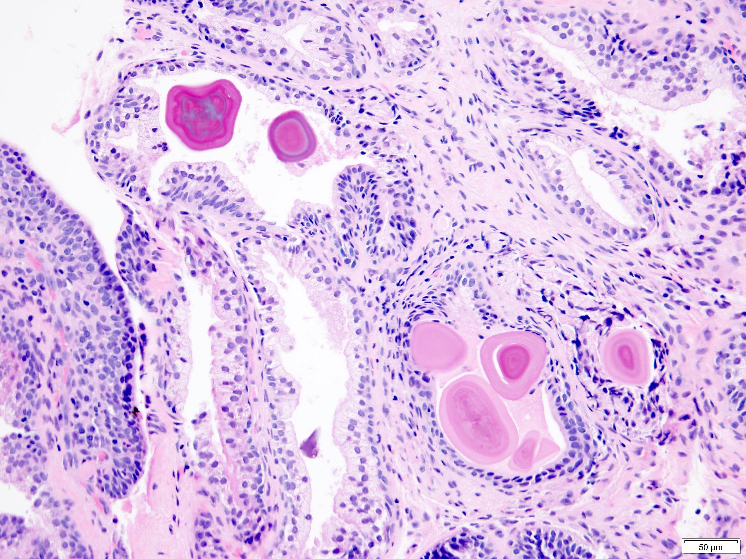
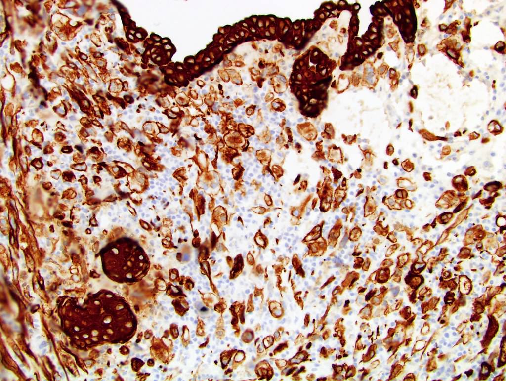
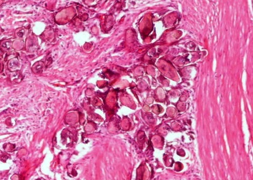
Calcified eggs and keratin
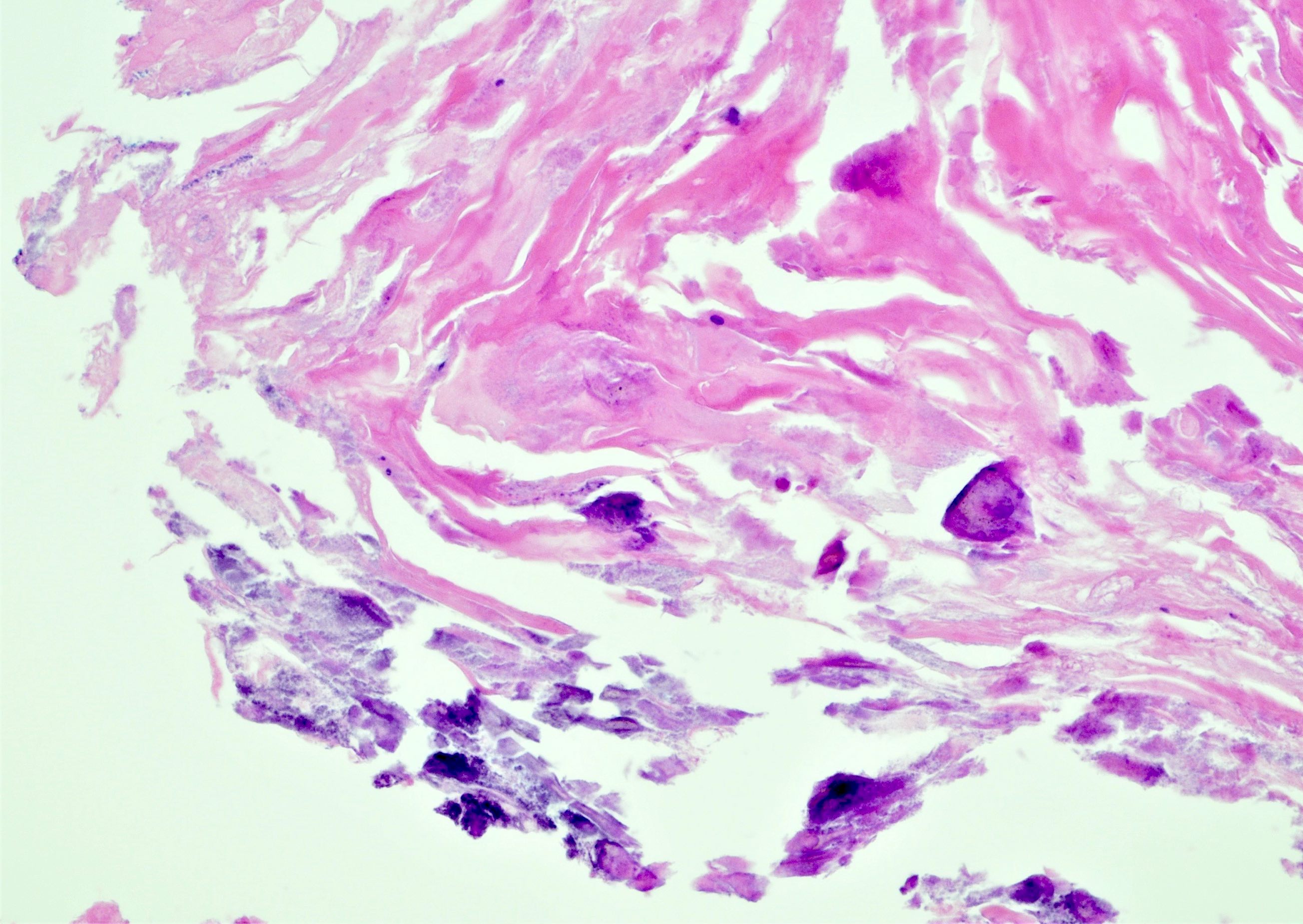
Schistosoma eggs and keratin
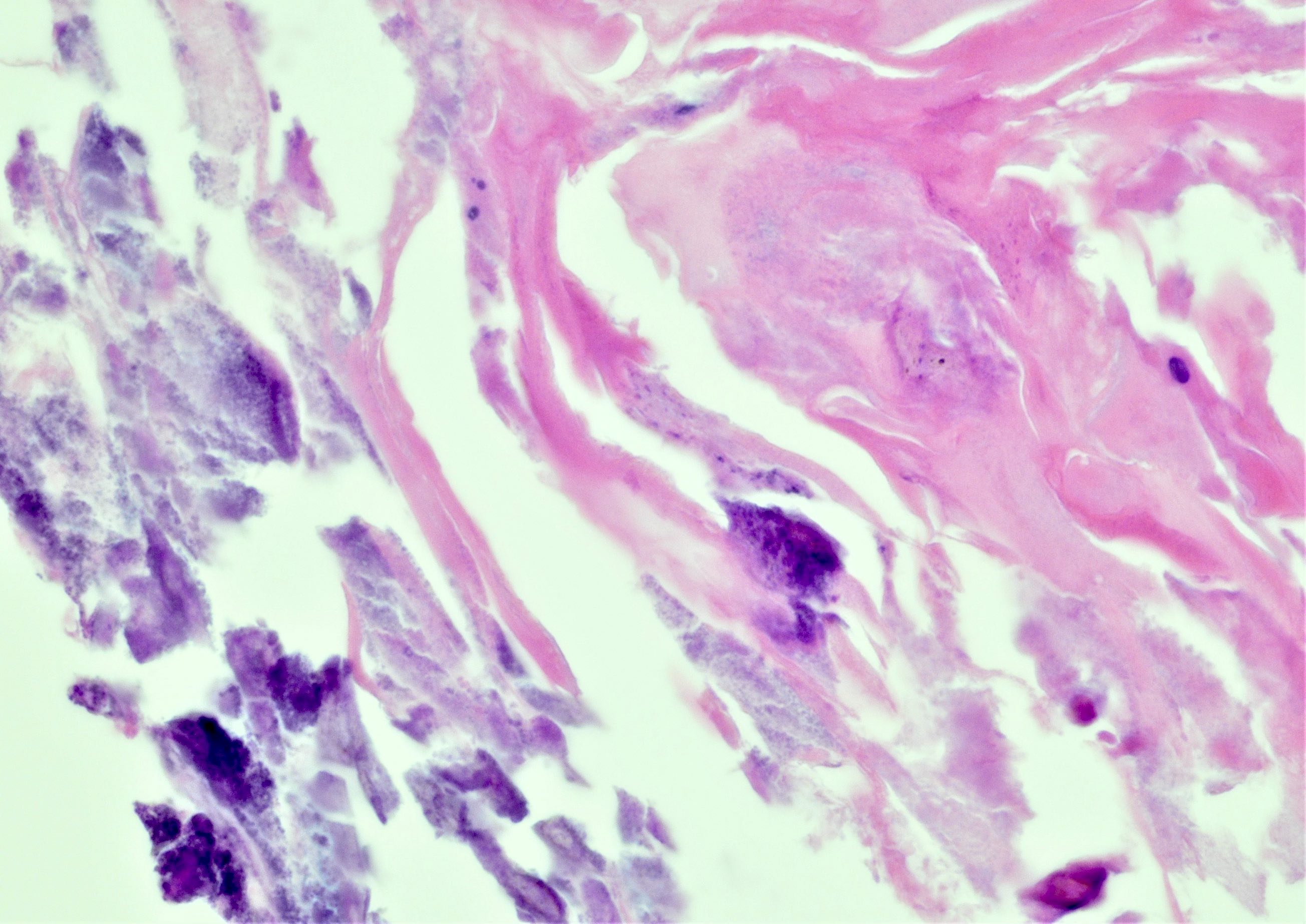
Schistosoma eggs with terminal spine
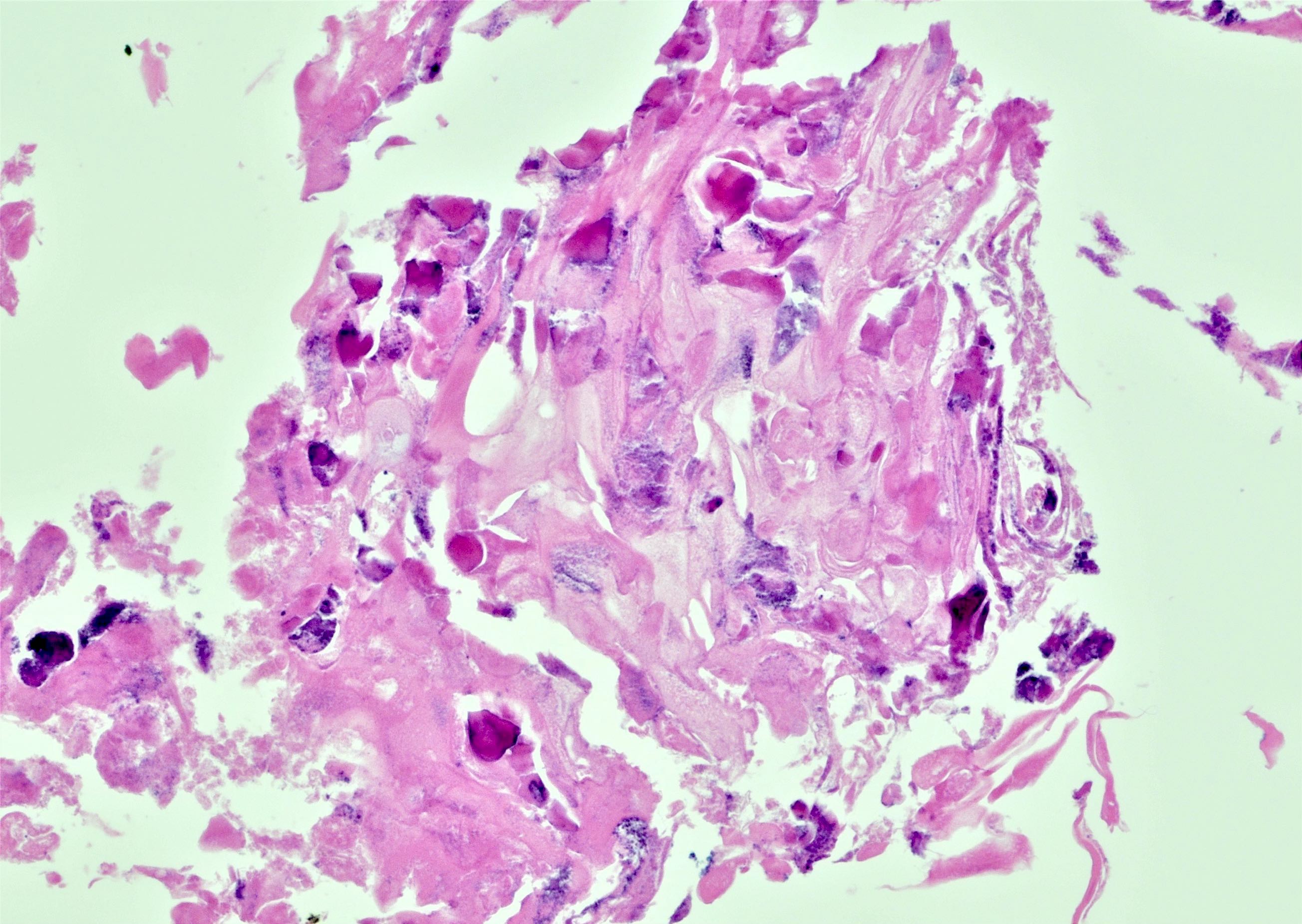
Bladder calcified eggs
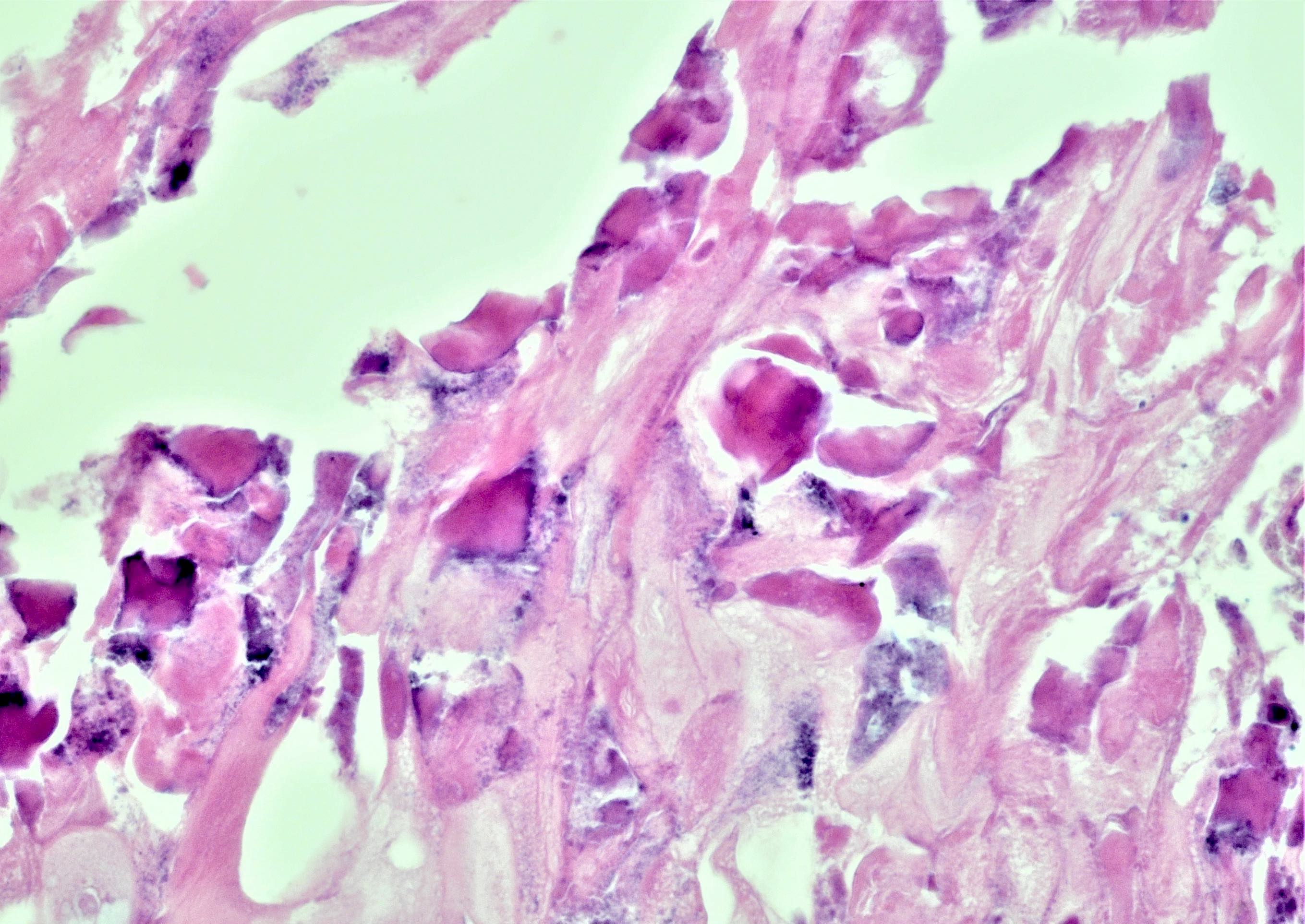
Calcified eggs with cracks
Parasitic calcified eggs

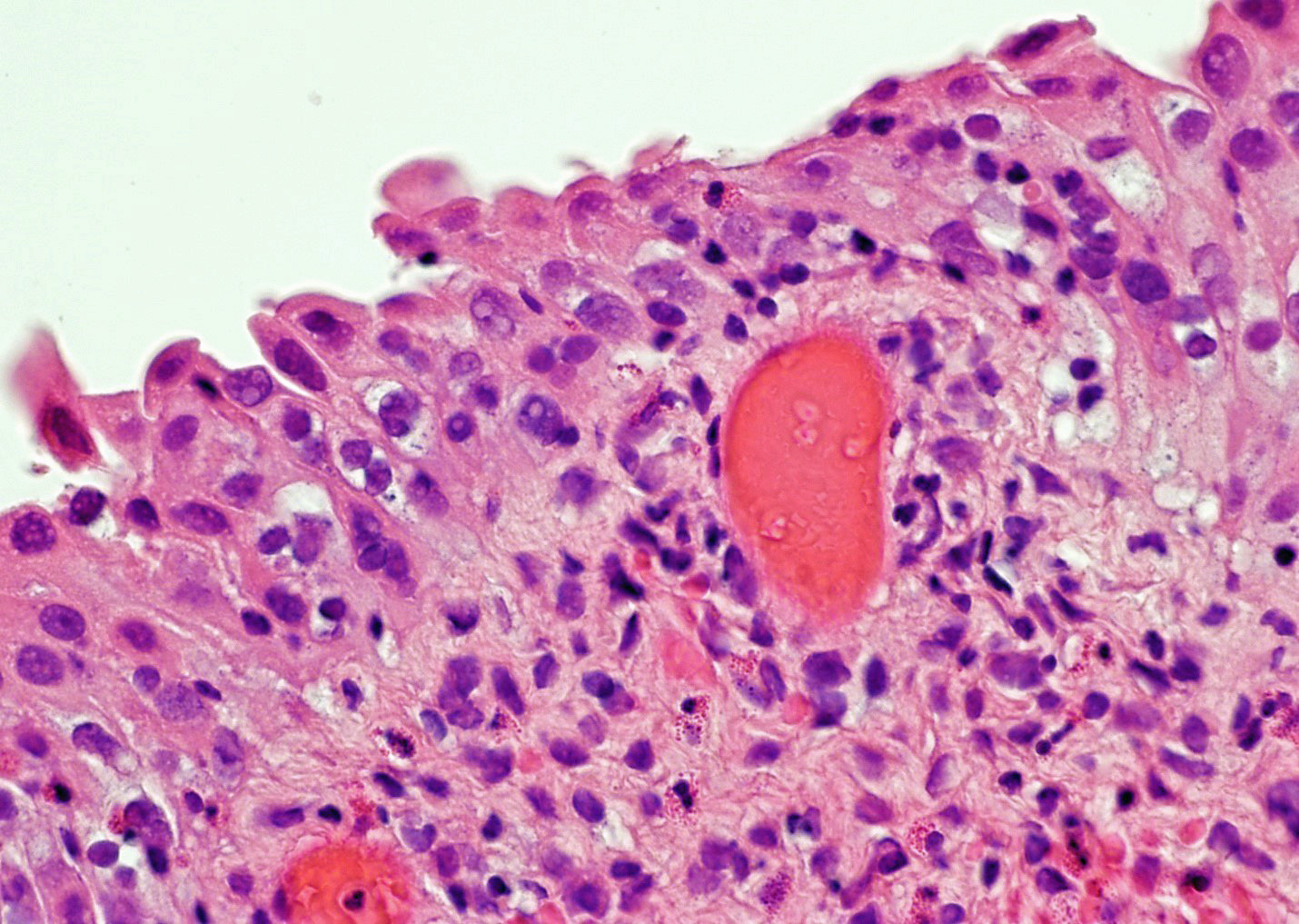
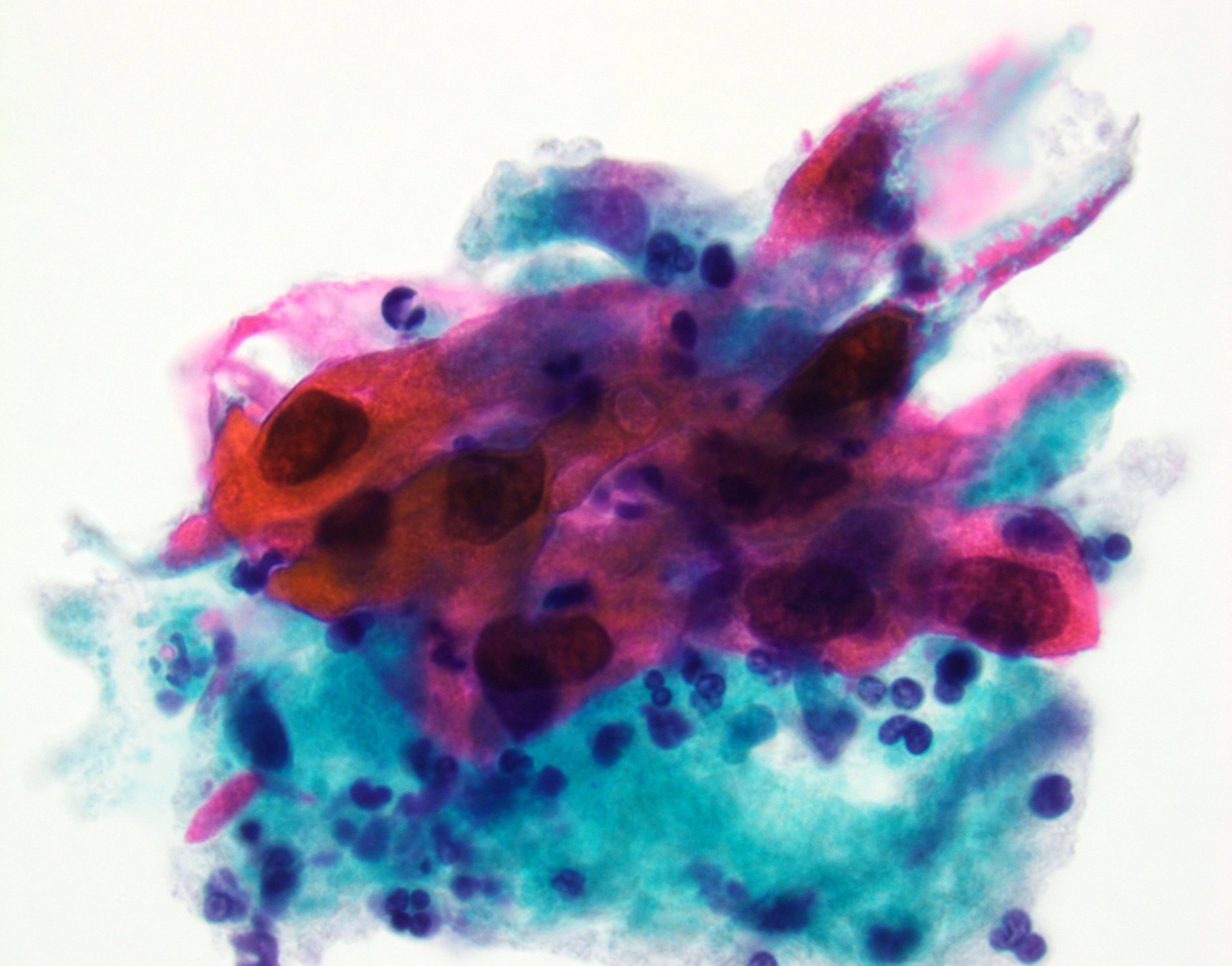
Dysplastic keratinizing squamous cells
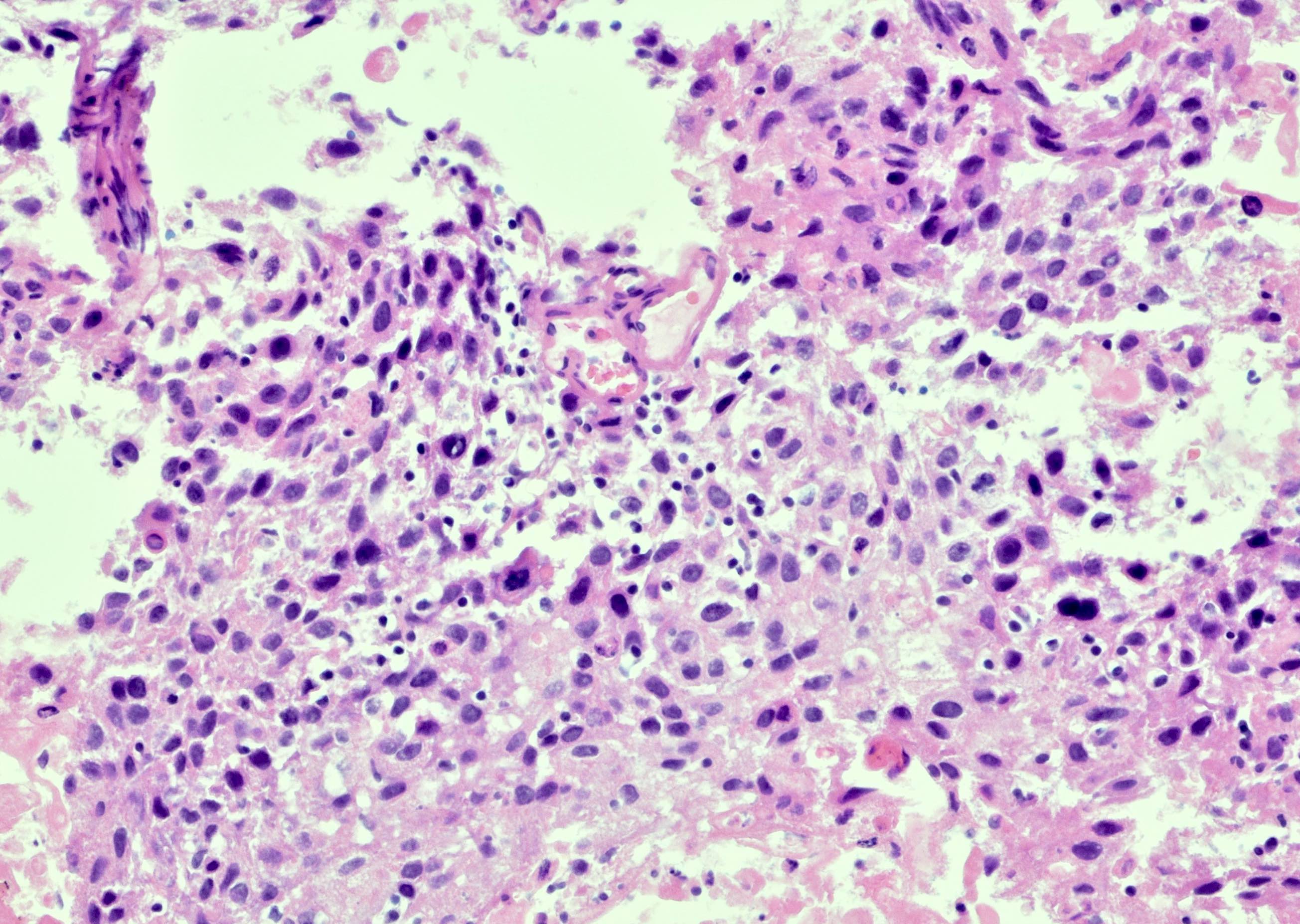
Malignant squamous cells
Malignant squamous cells with intercellular bridges
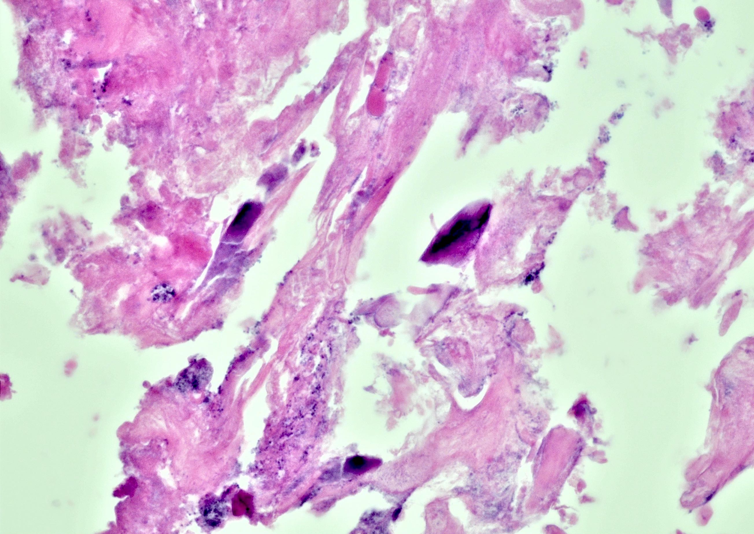
Schistosoma haematobium eggs
Schistosoma eggs in ureter
Ureter with Schistosoma eggs
Muscularis propria with Schistosoma eggs
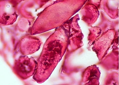
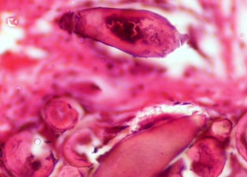
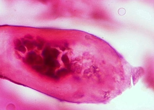
Schistosoma eggs
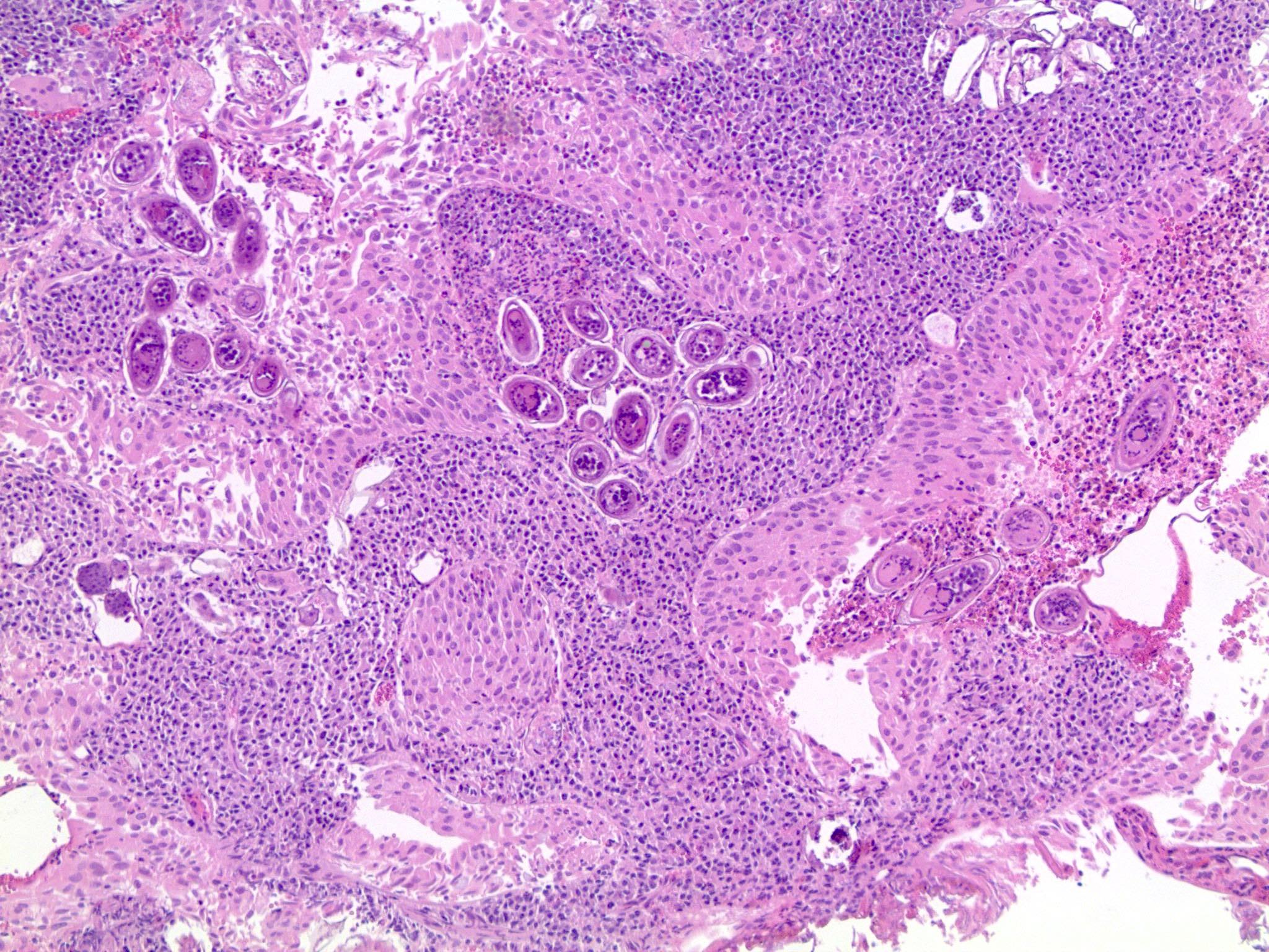
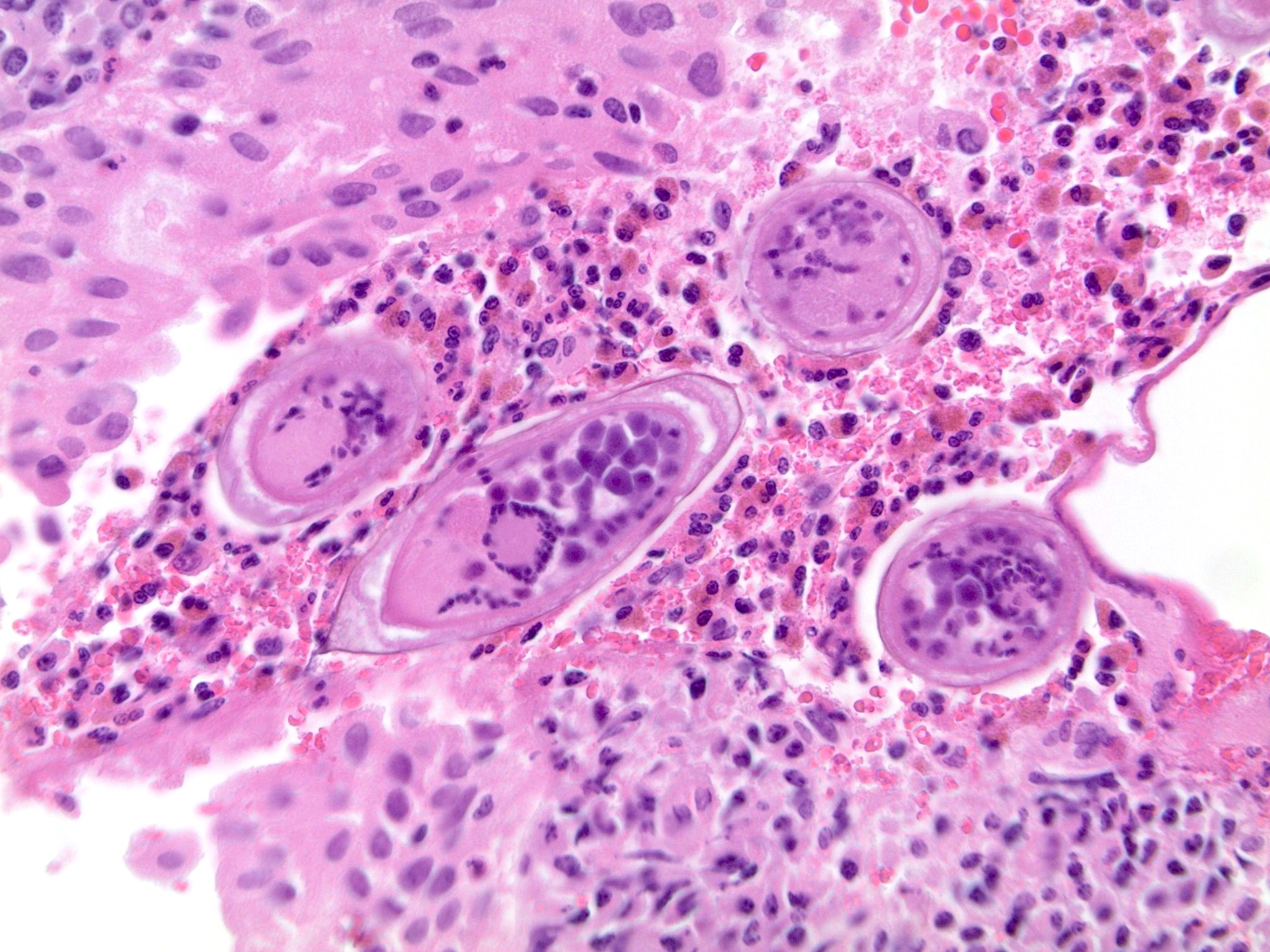
Schistosomiasis (bilharziasis)
Contributed by Y. Albert Yeh, M.D., Ph.D.
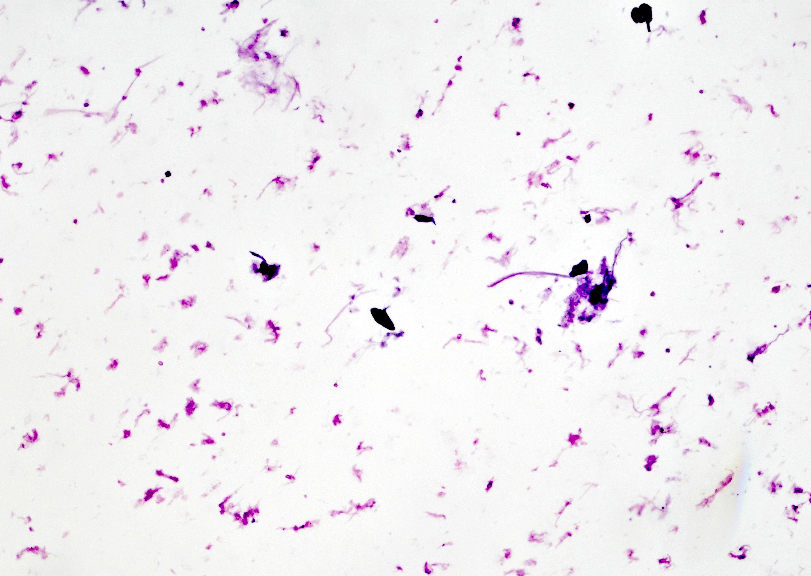
Calcified eggs in urine
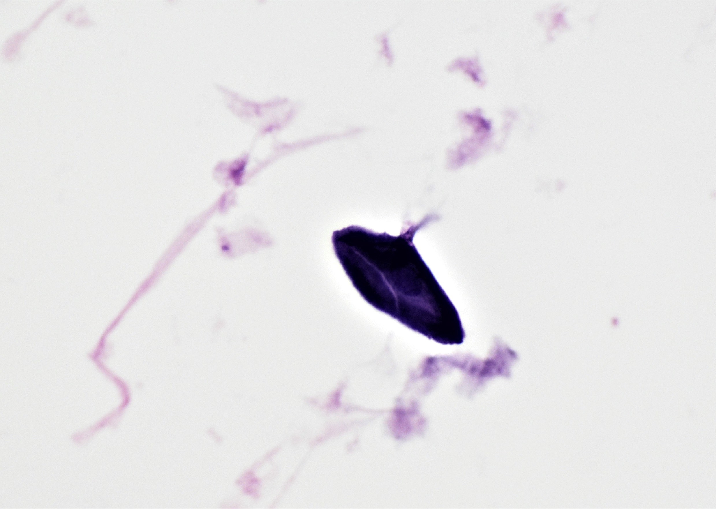
Schistosoma egg with lateral spine
Images hosted on other servers

CT of bladder

CT of ureter

US of bladder

MRI of bladder

Gadolinium enhanced MRI of renal pelvis
Images hosted on other servers:

Right ureter mass

Renal pelvic lesion
Contributed by Reima El Naili, M.D.
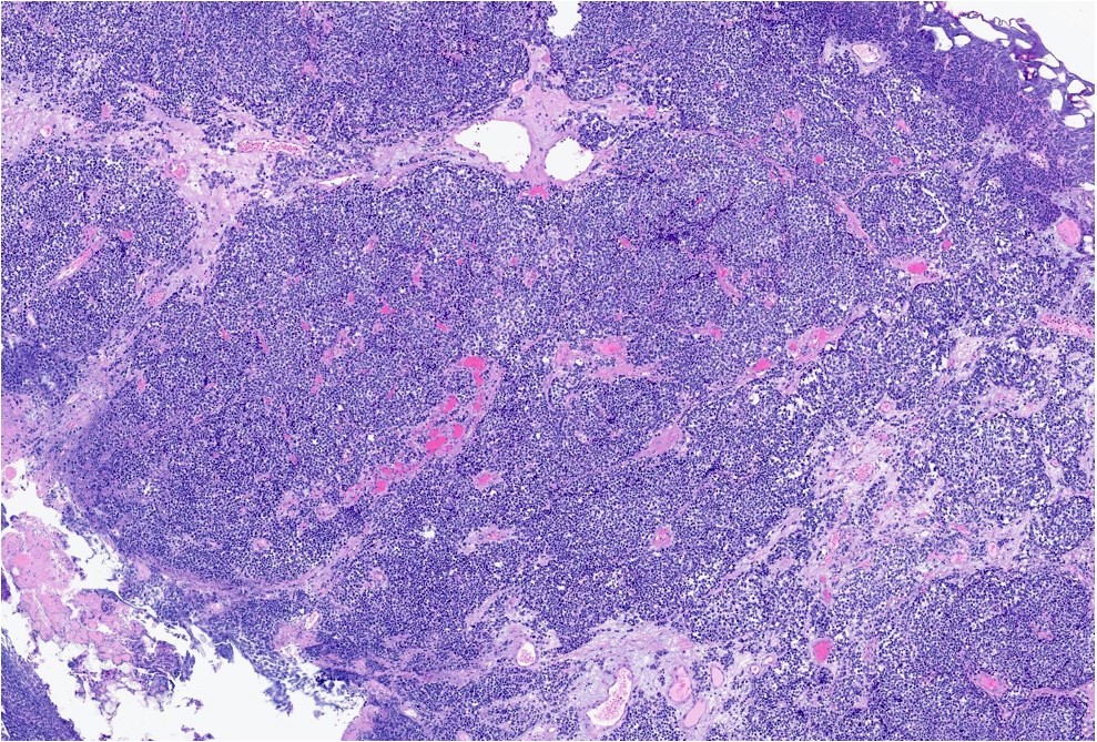
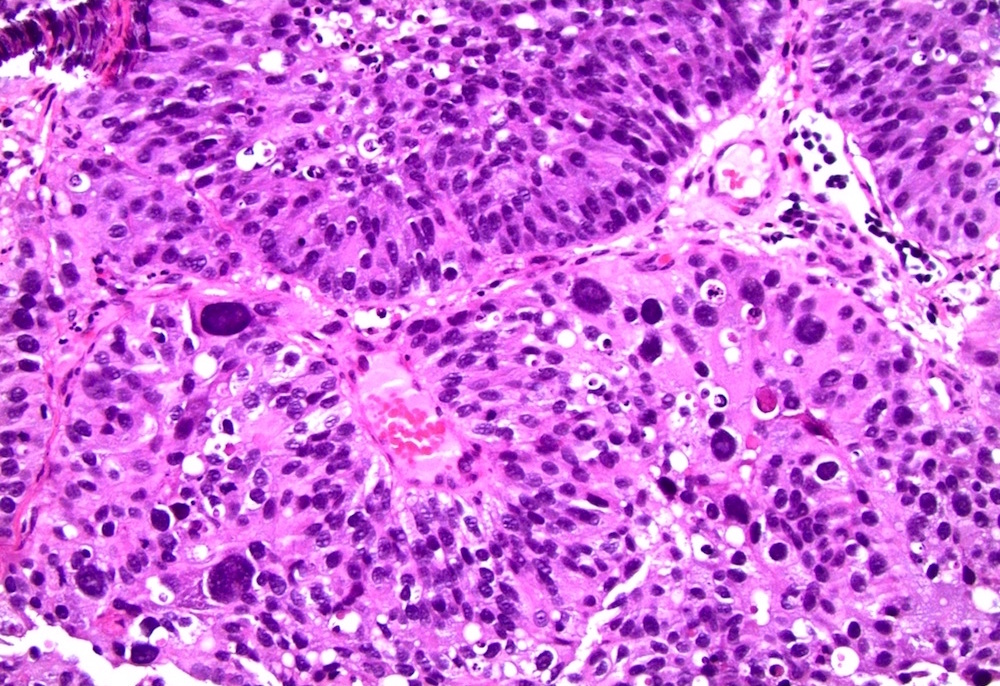
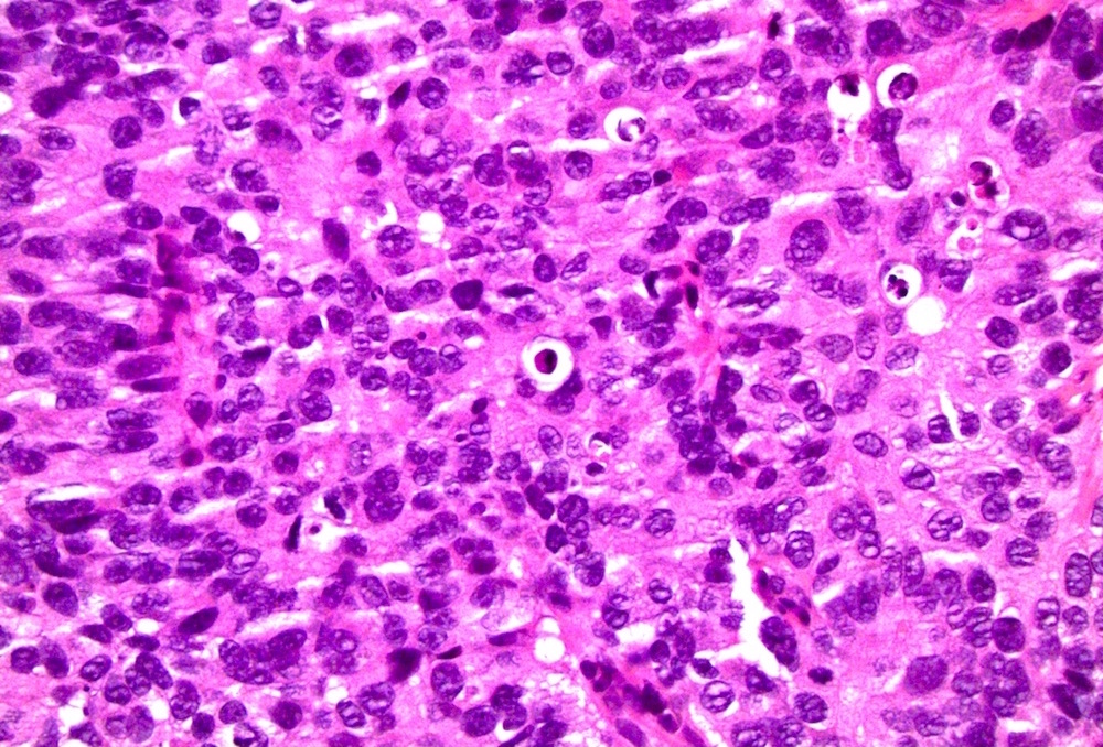
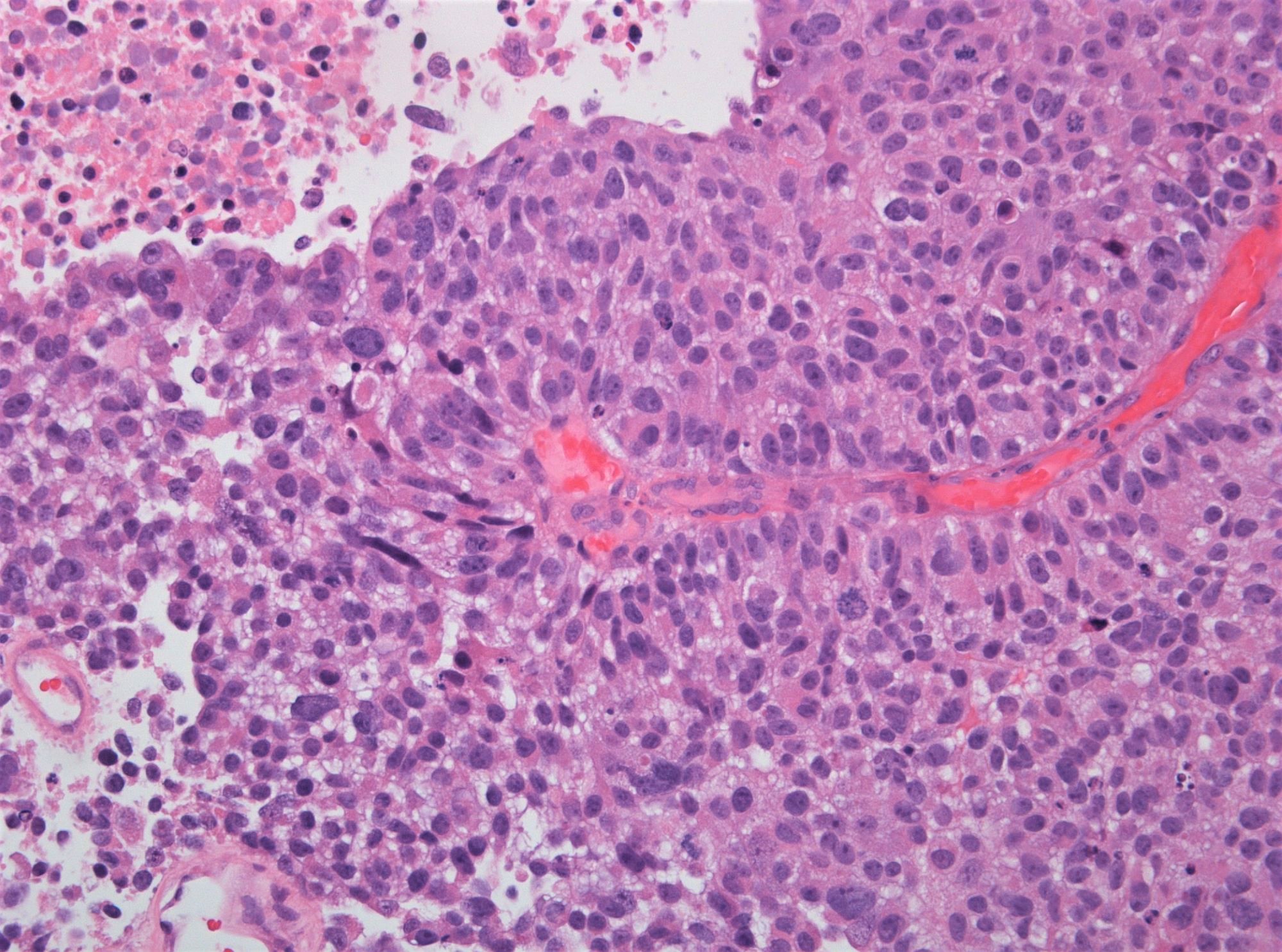
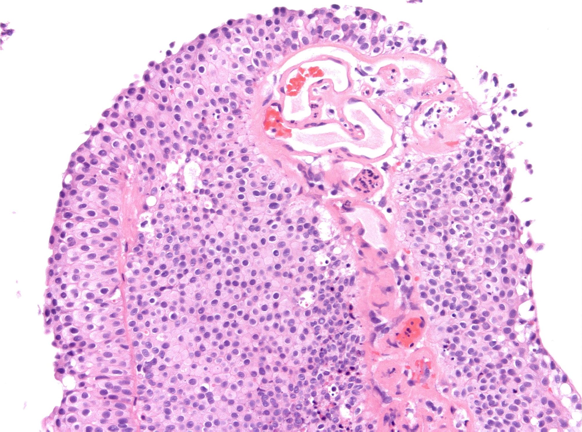
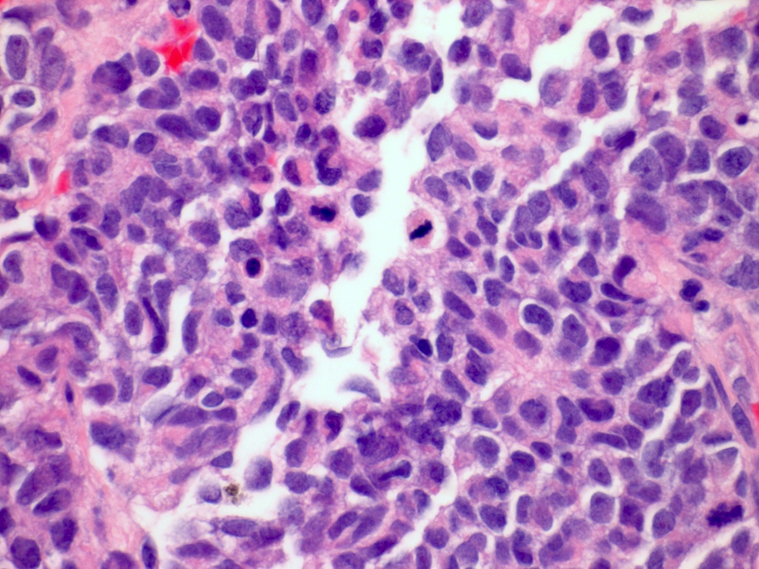
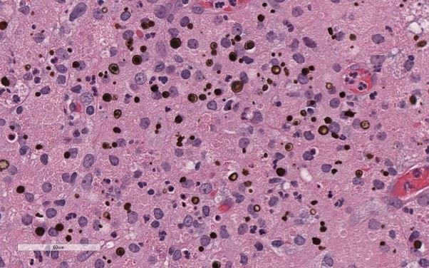
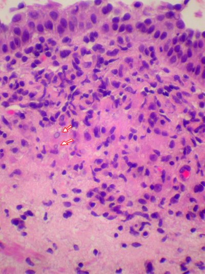
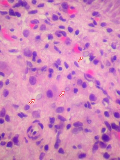
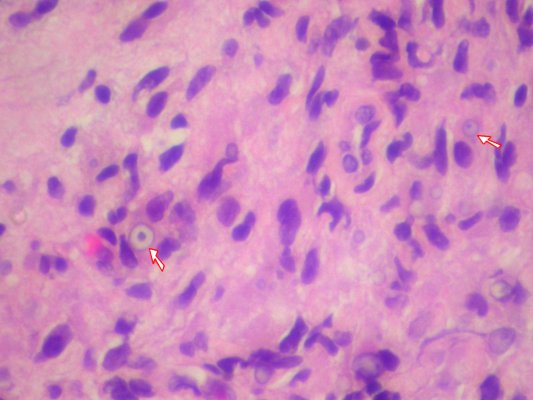
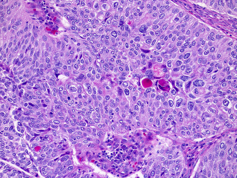
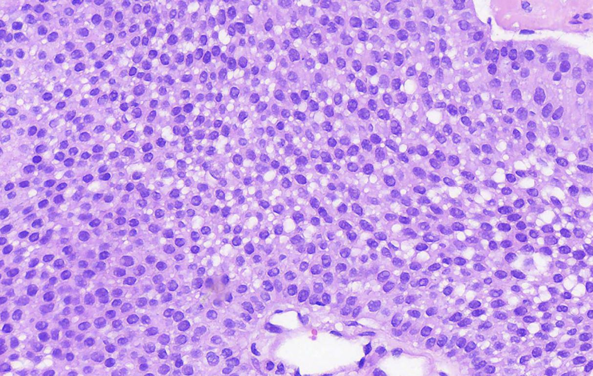
Sheets of basophilic cells
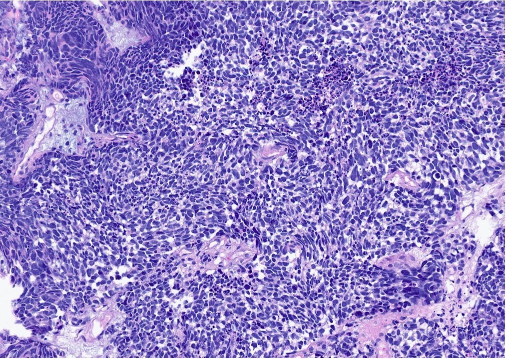
Pleomorphism and nuclear molding

High N:C ratio
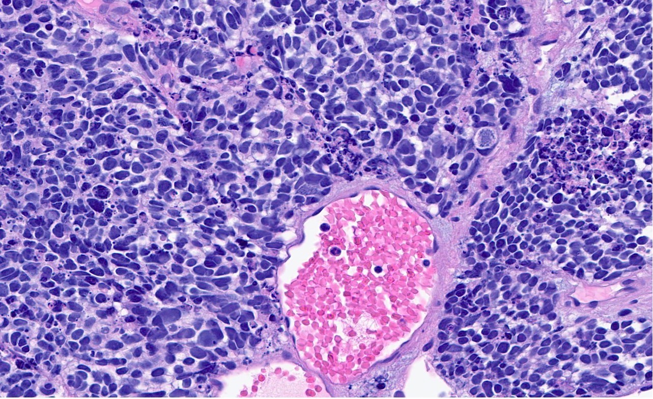
Brisk mitotic activity
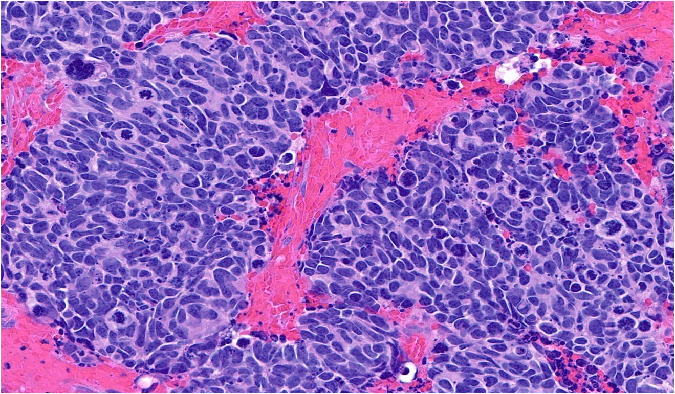
Hemorrhage and atypical mitoses
Necrosis and Azzopardi effect
Mixed urothelial neuroendocrine carcinoma
Carcinoma with normal urothelium
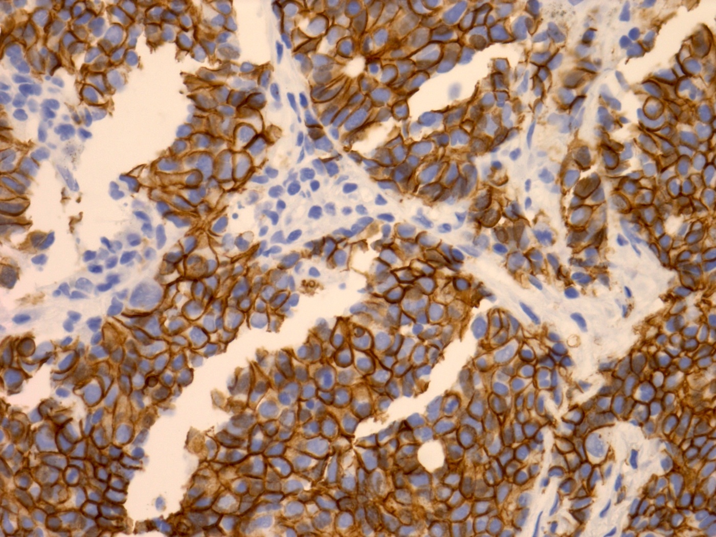
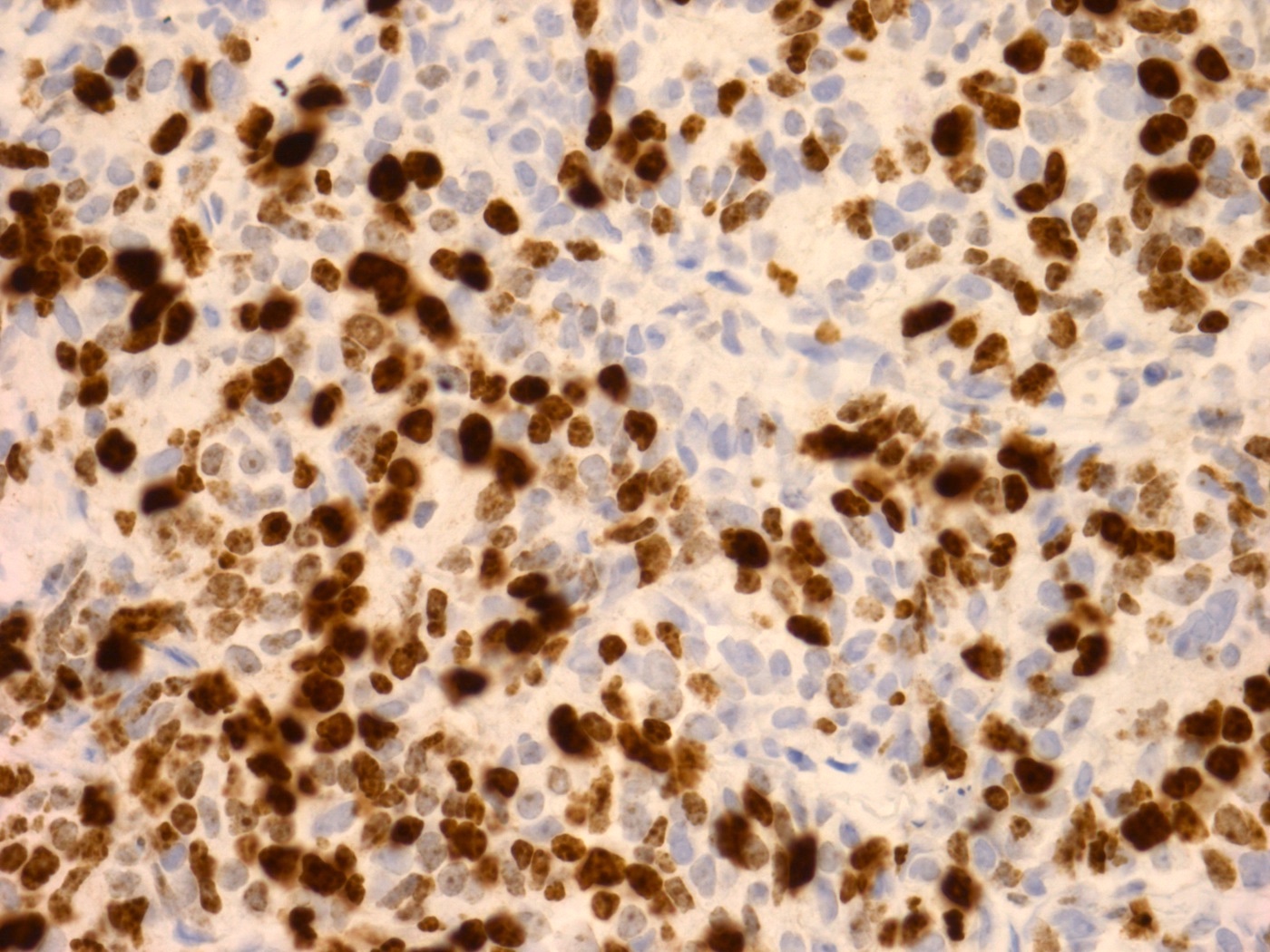
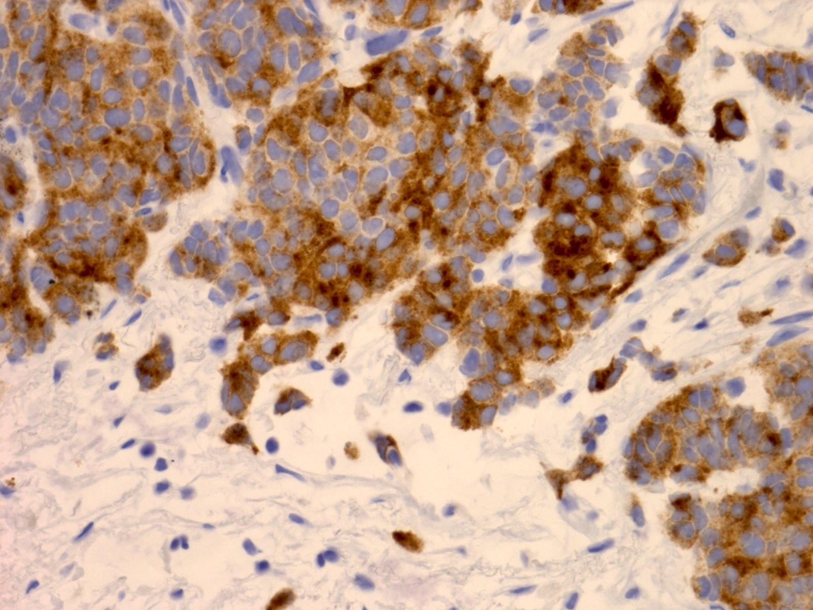
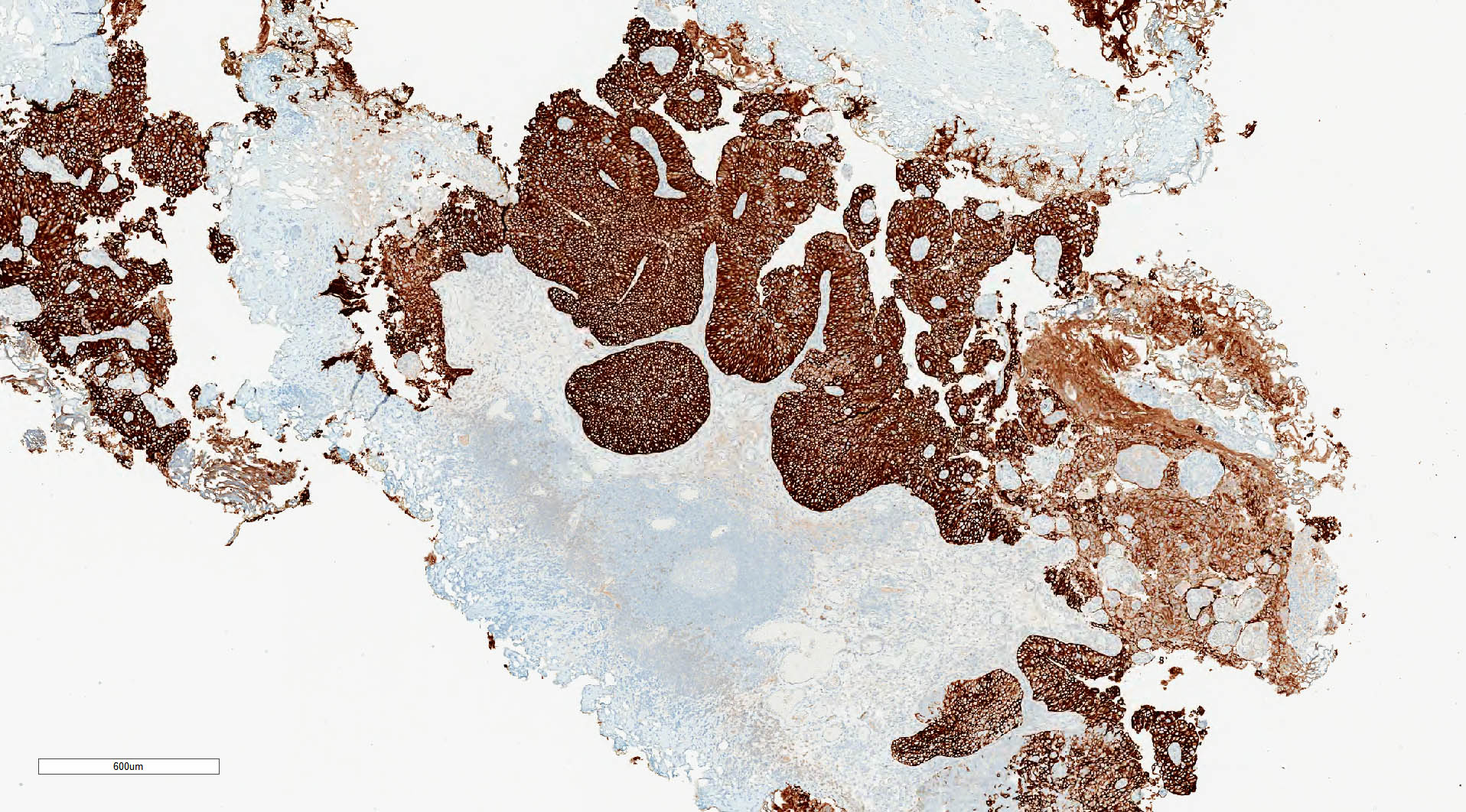
CK AE1 / AE3
Synaptophysin
CD56
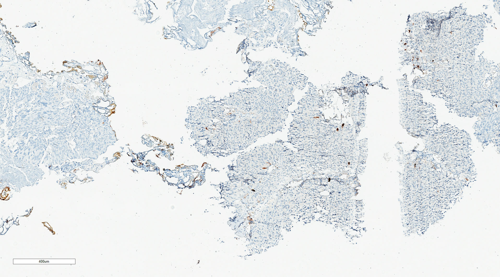
TTF1
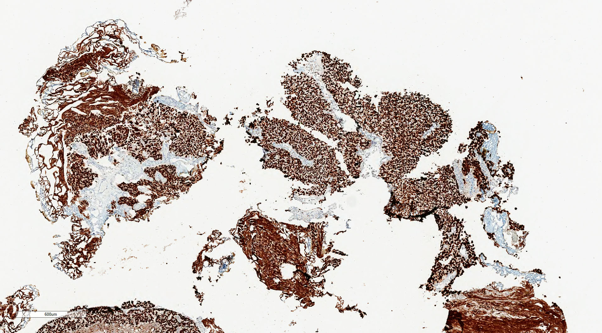
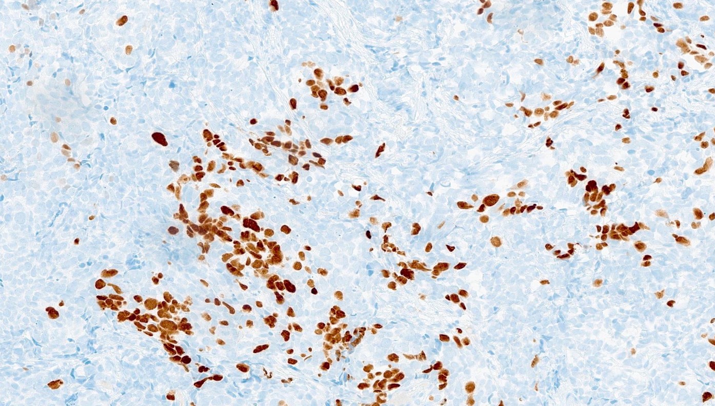
Ki67
CD56 in mixed tumor
GATA3 in mixed tumor
INSM1
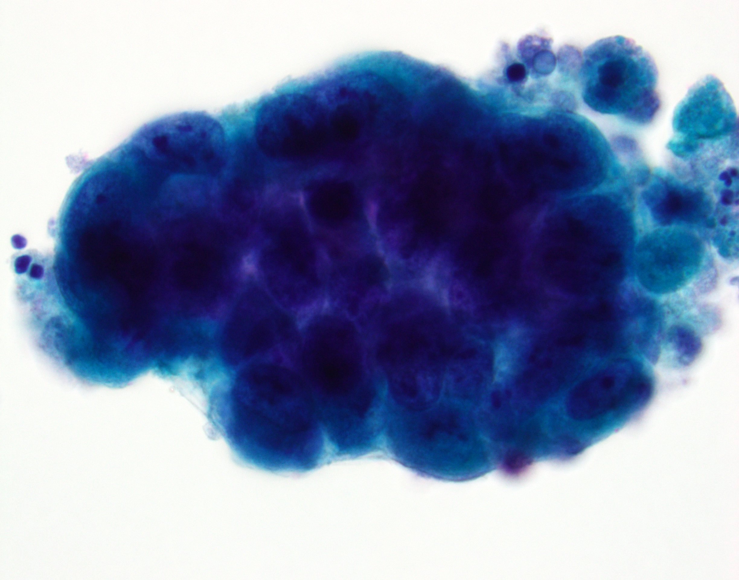
Contributed by Bonnie Choy, M.D.
Hypercellular specimen
Images hosted on other servers
Bladder, Pap
Bladder small cell neuroendocrine carcinoma
Images hosted on other servers:

MRI bladder

Solitary mass lower right kidney

Mass in right renal pelvis
Images hosted on other servers:

Bladder exstrophy
Bladder
Contributed by Susan Prendeville, M.D.
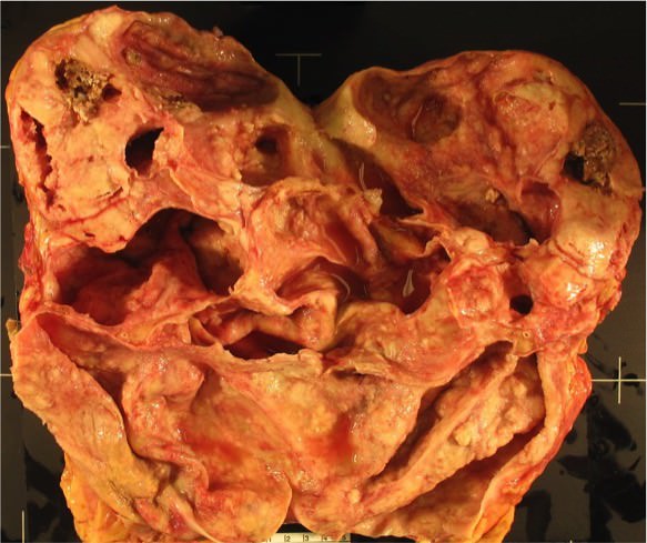
specimen
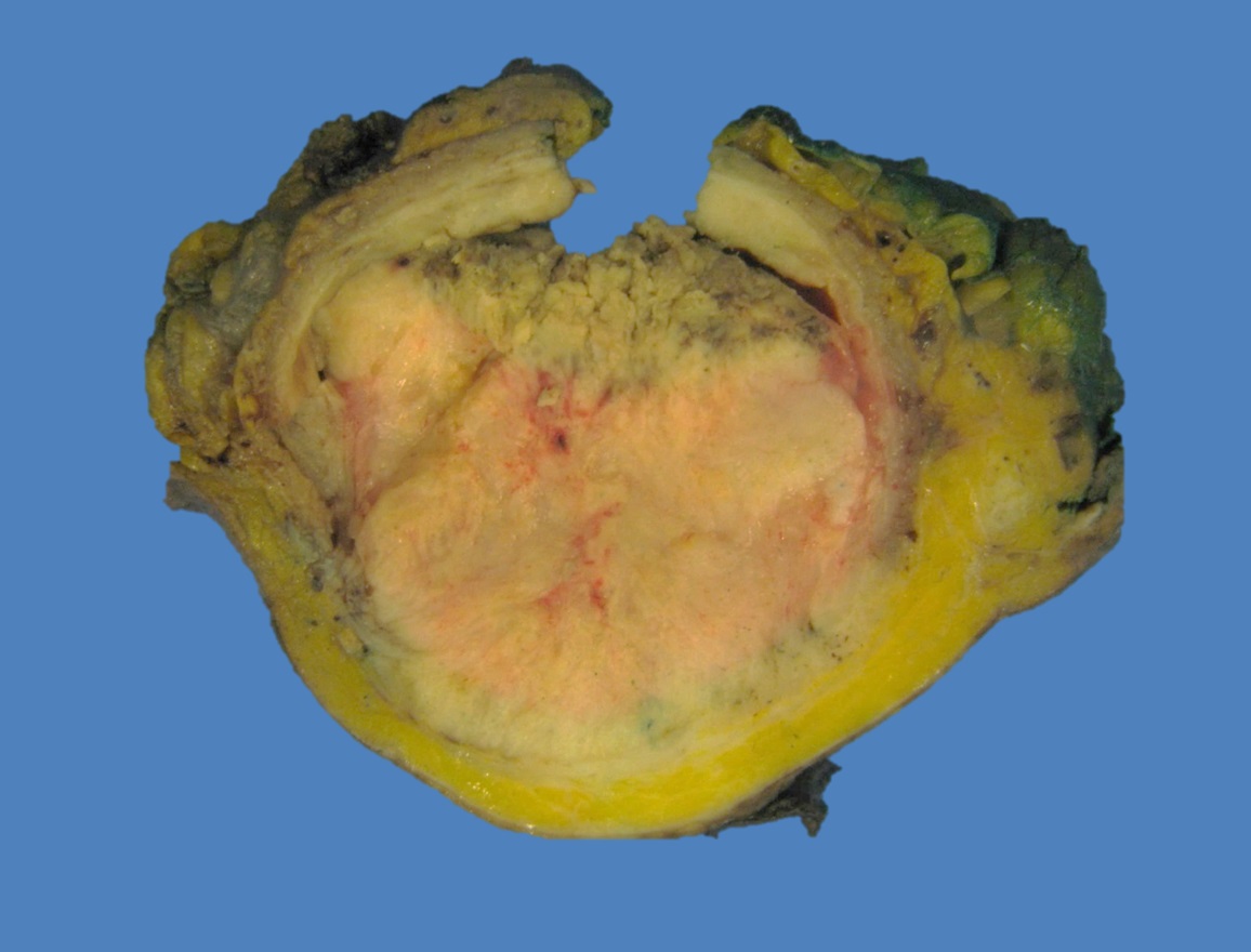
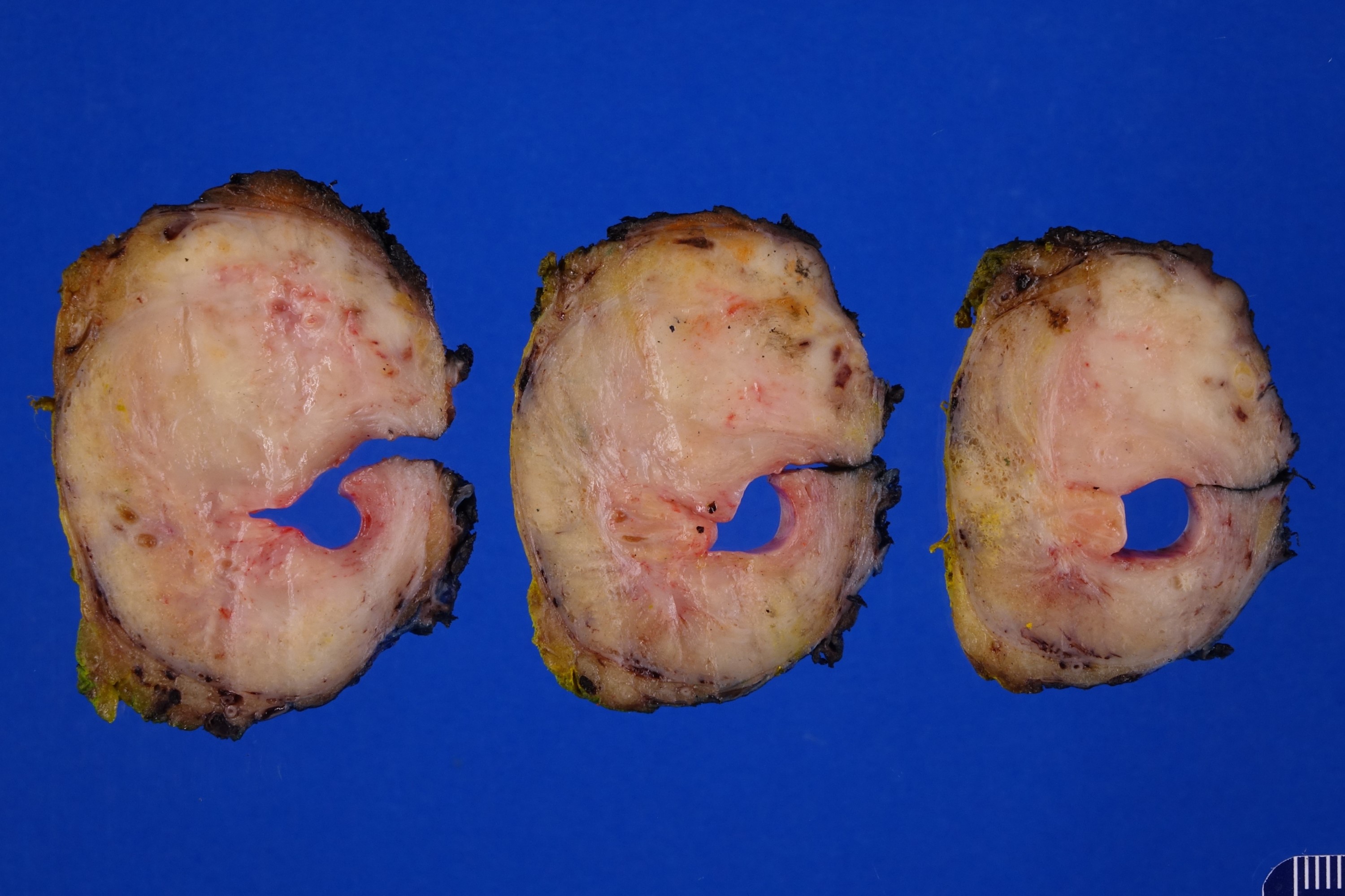
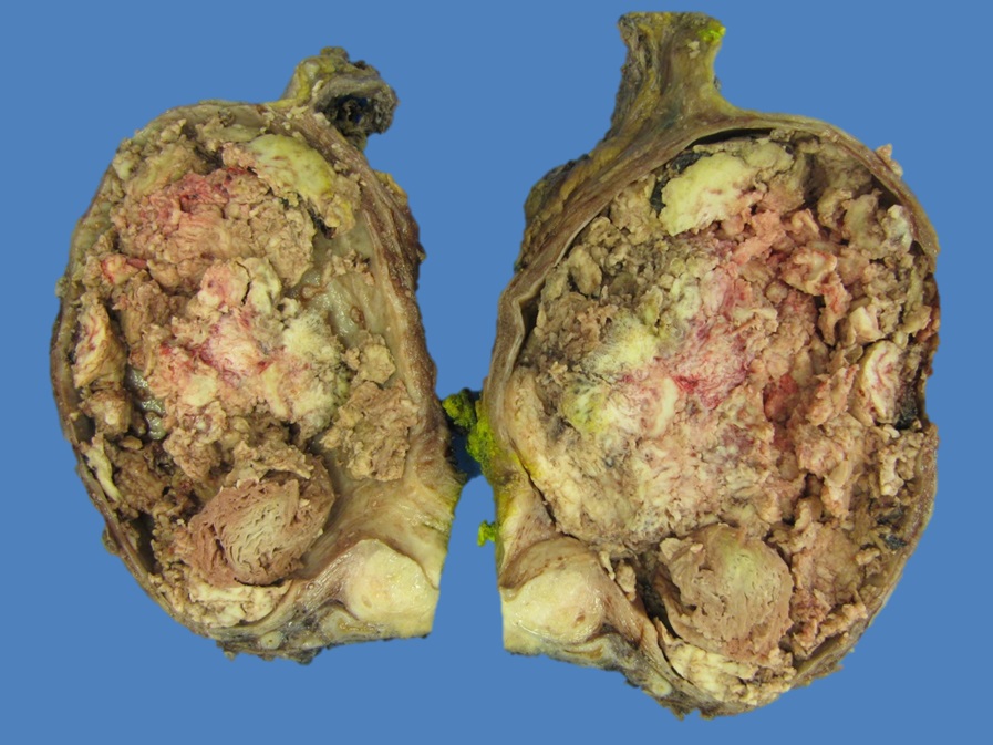
Cross section of tumor
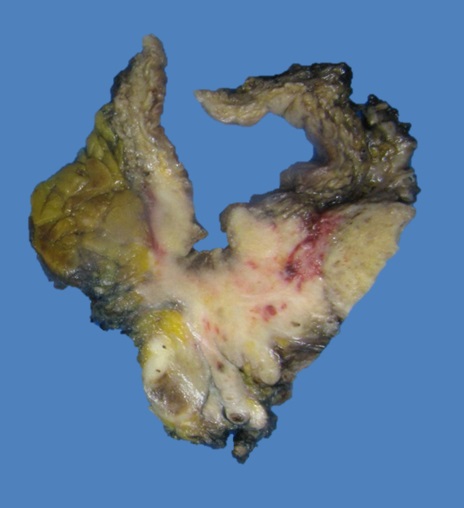
Ulcerating tumor
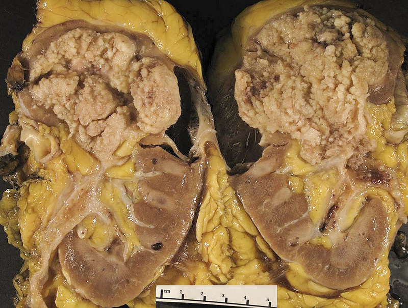
Renal pelvis
Contributed by Nicole K. Andeen, M.D.
Dilated renal calyces due to reflux
Solid, tannish white with central necrosis
Bladder
Contributed by Susan Prendeville, M.D.
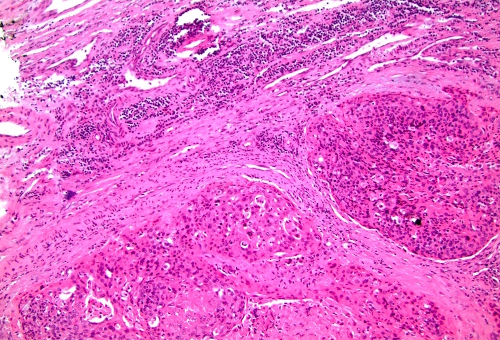
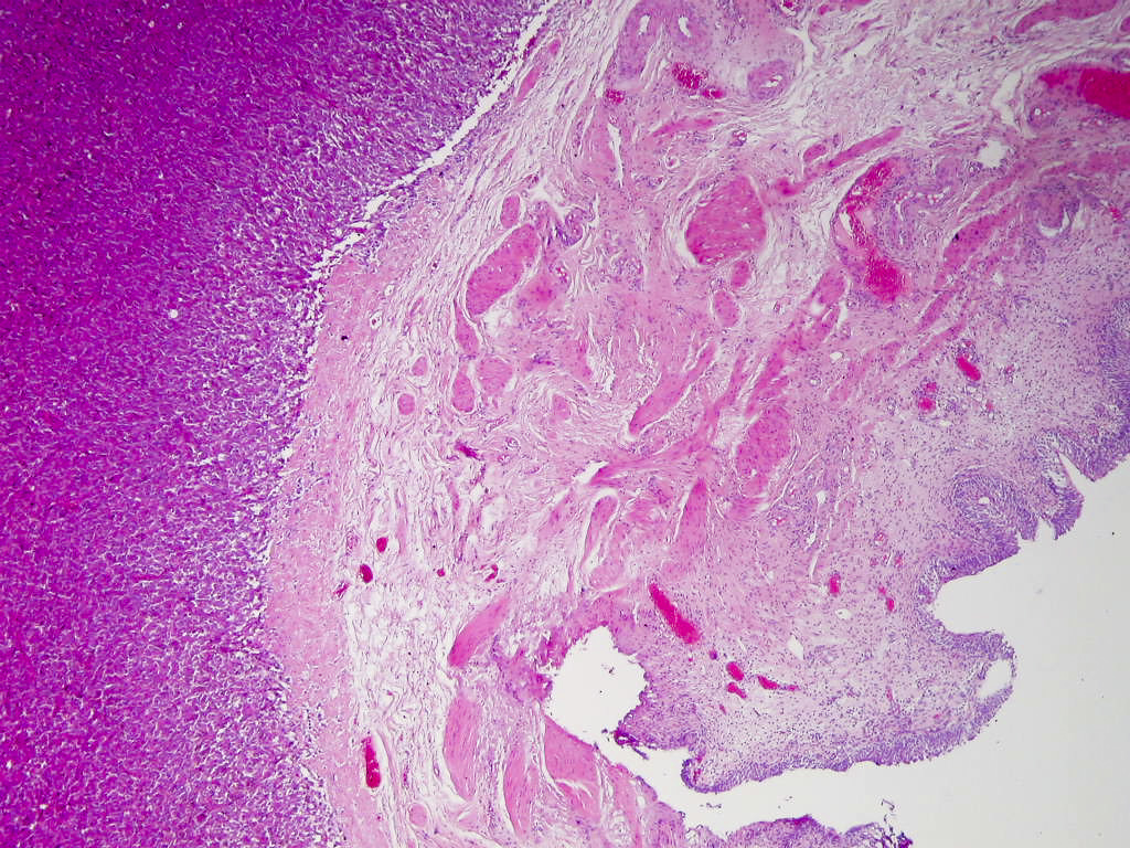
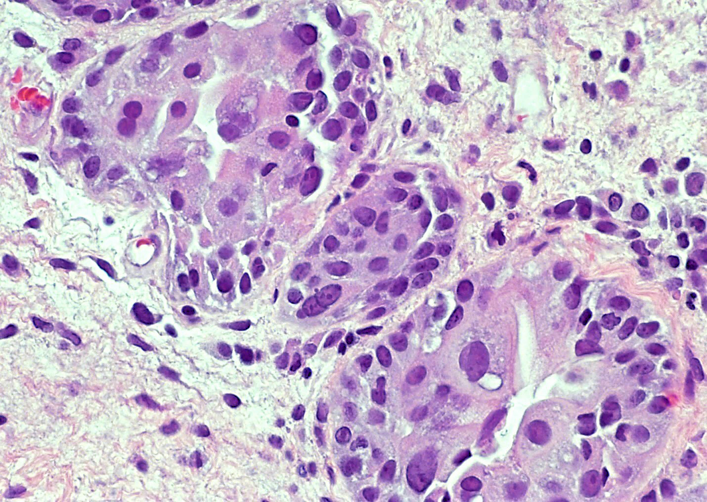
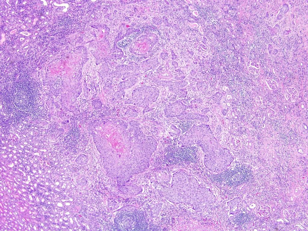
Destructive stromal invasion
Irregular squamous nests
Keratin pearls
Prominent keratinization
Intercellular bridges
Poorly differentiated tumor
Squamous dysplasia / carcinoma in situ
Invading muscularis propria
Keratinizing squamous dysplasia
Schistosoma haematobium eggs
Renal pelvis
Contributed by Nicole K. Andeen, M.D.
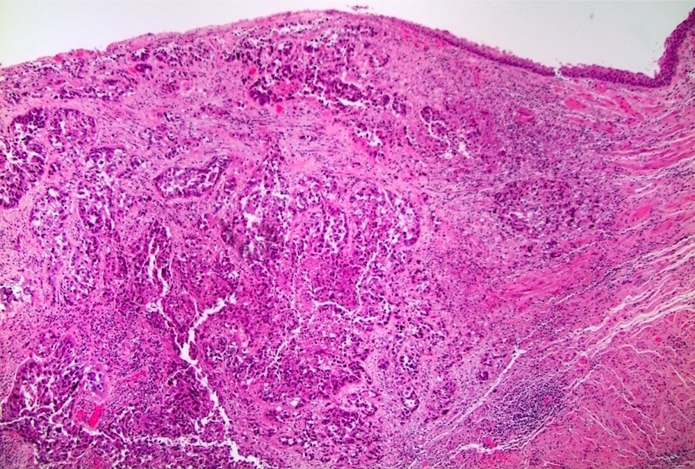
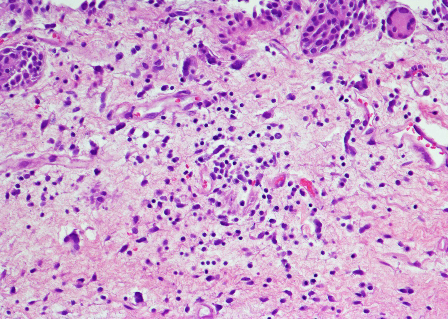
Invasive tumor
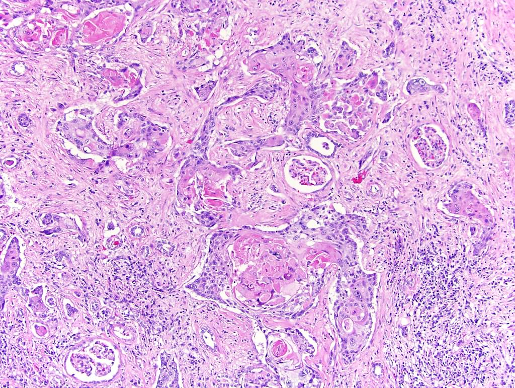
Entrapped glomeruli
No urothelial differentiation
Individual cell keratinization
Entrapped renal tubules
Contributed by Susan Prendeville, M.D. and Bonnie Choy, M.D.
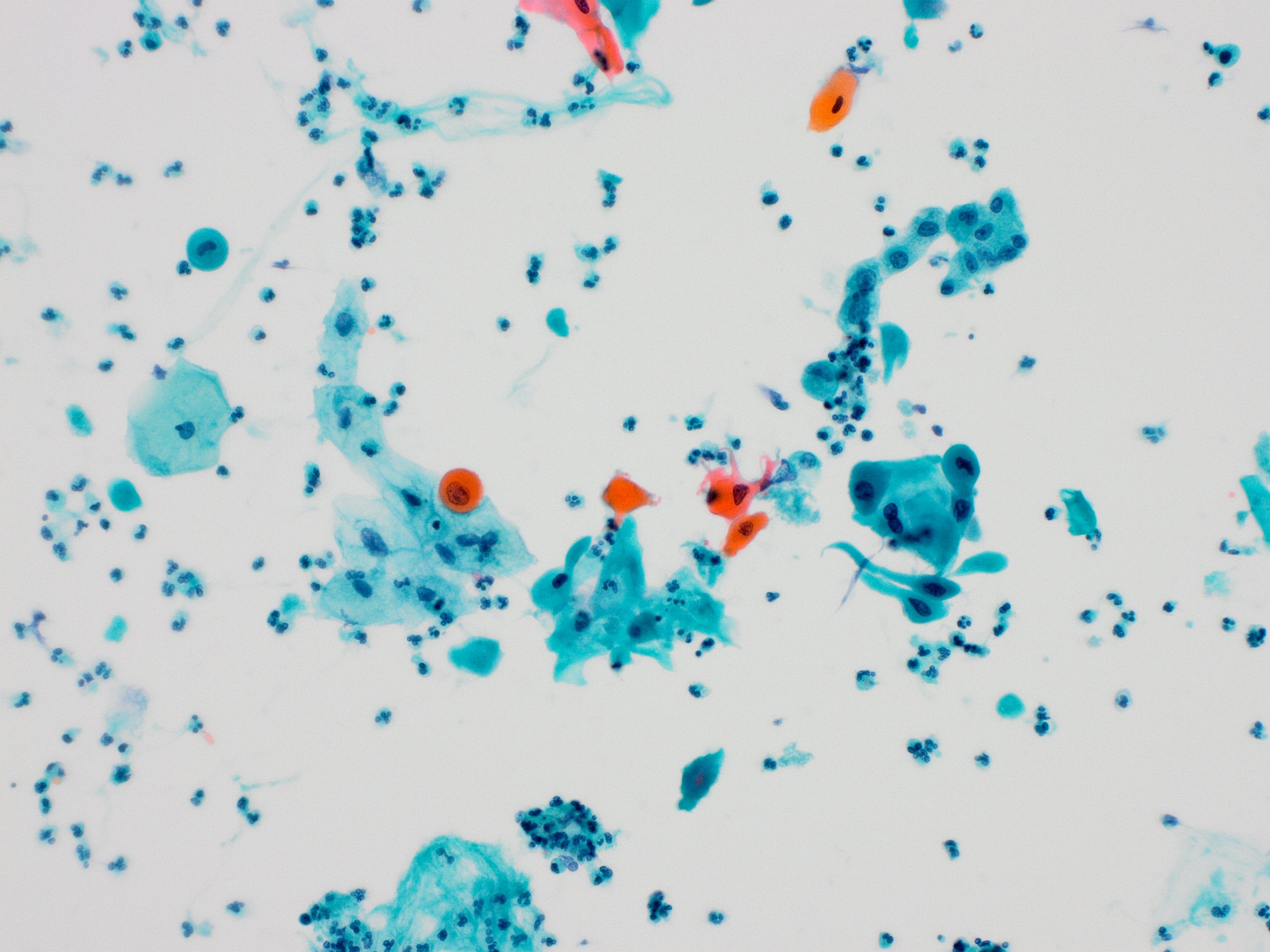
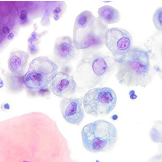
Malignant squamous cells
Keratinized malignant cells
Squamous cell carcinoma
Images hosted on other servers:

Cystoscopy finding: whitish exophytic lesion
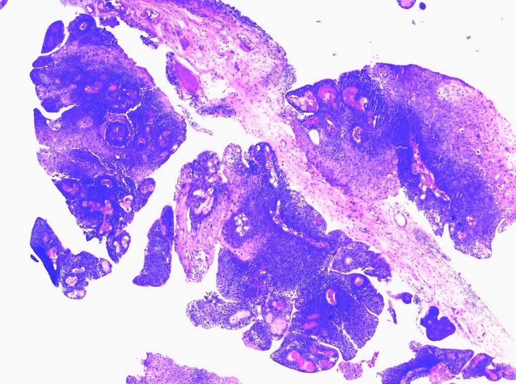
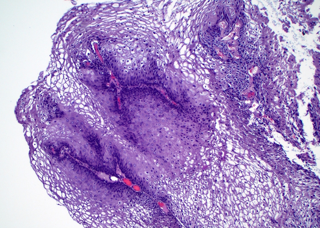
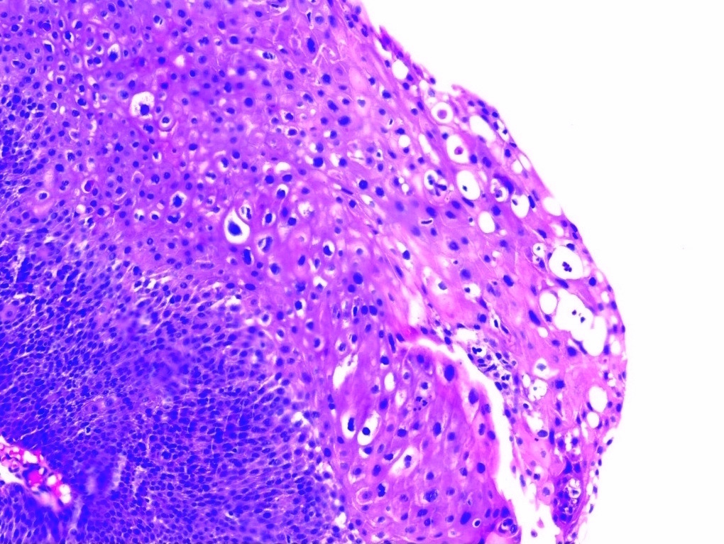
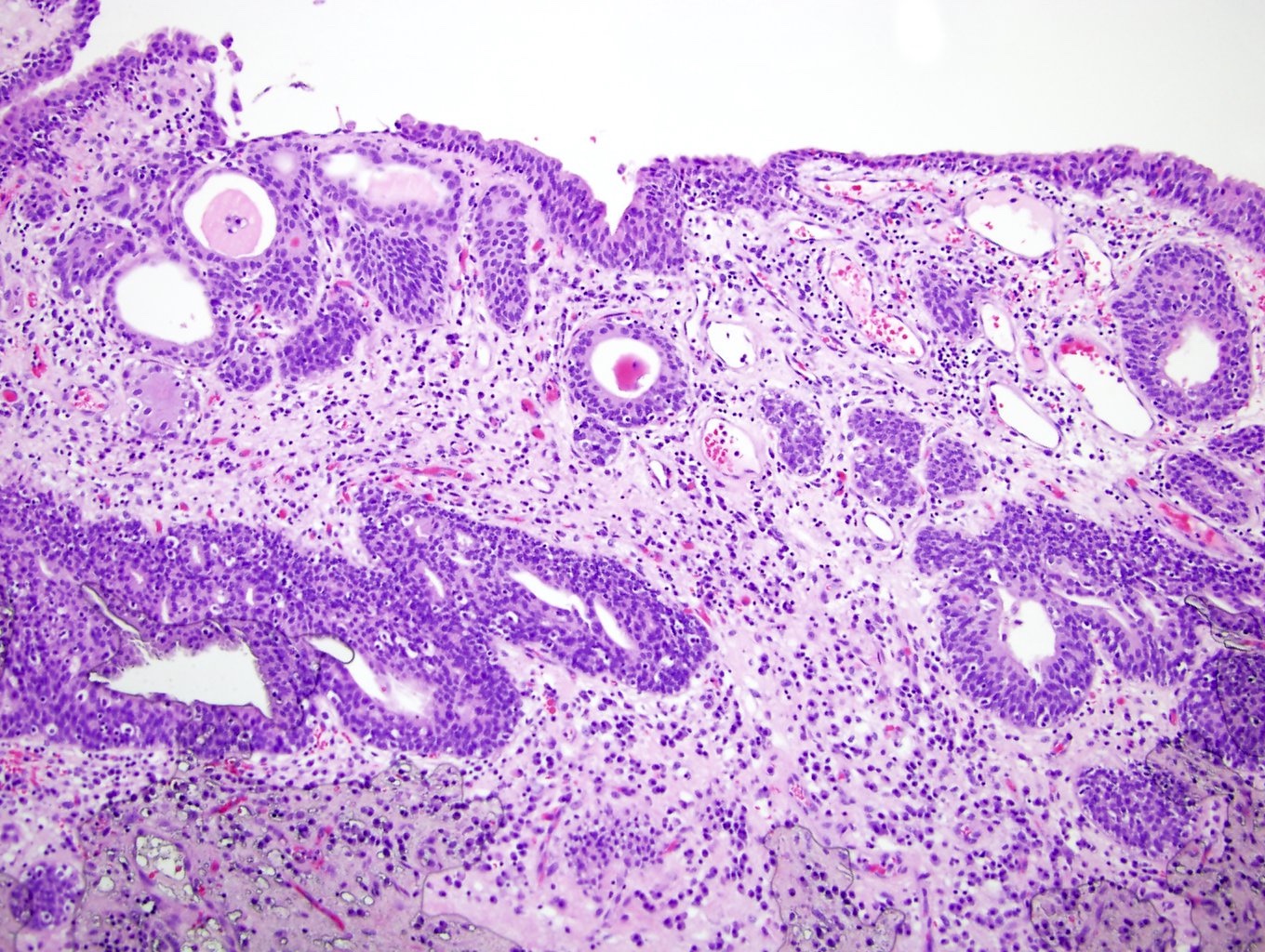
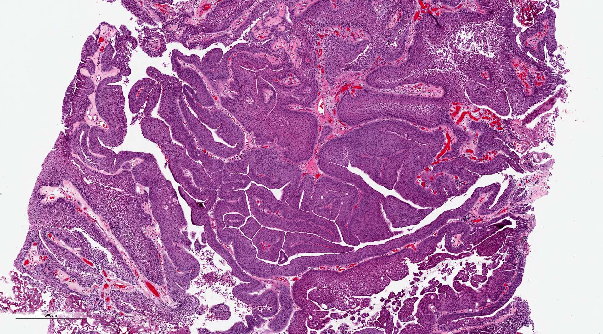
Contributed by Harsh Batra, M.B.B.S., D.C.P., D.N.B. and Anil Parwani, M.D., Ph.D., M.B.A.
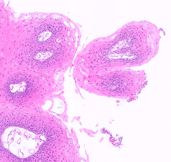
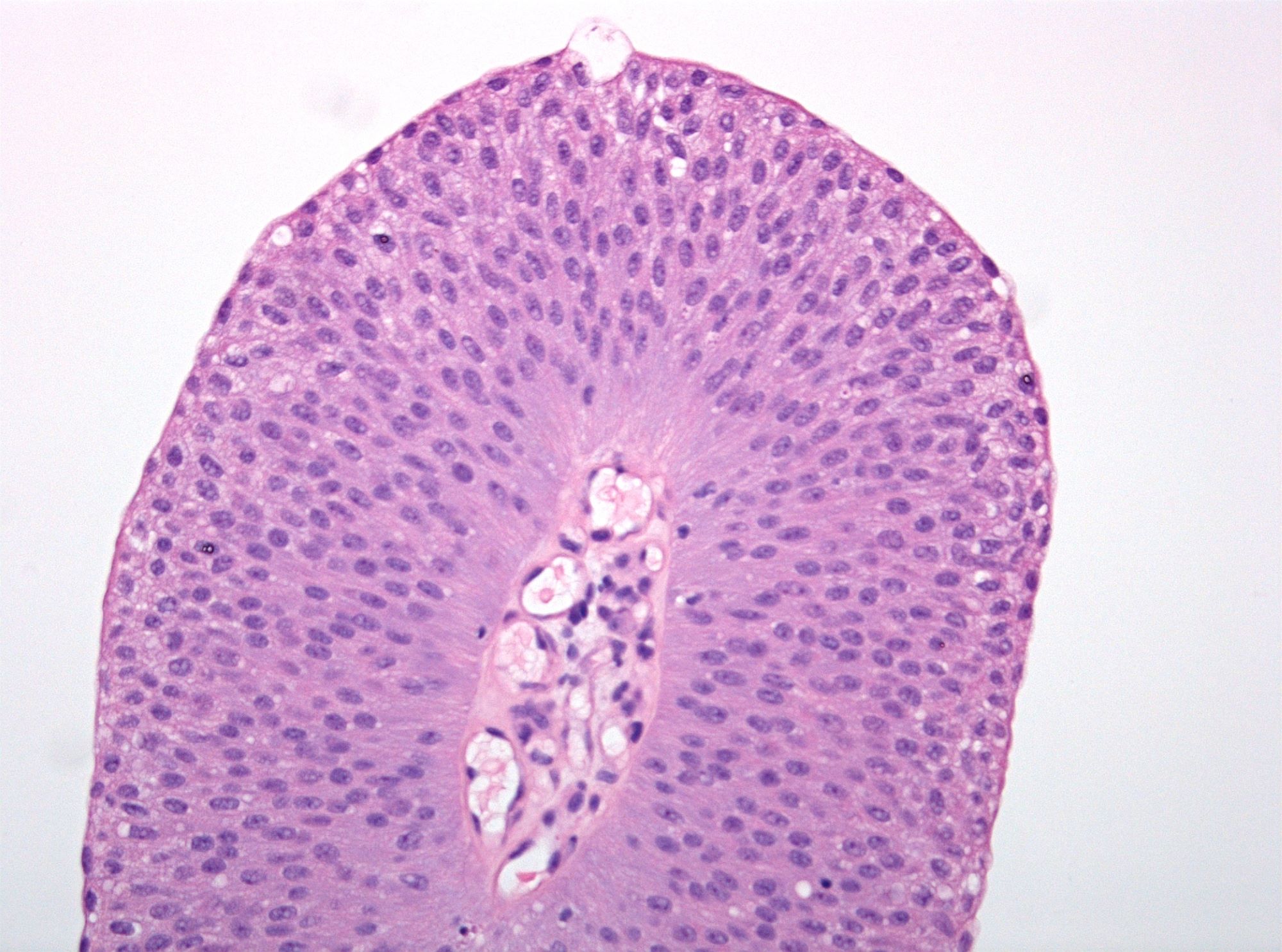
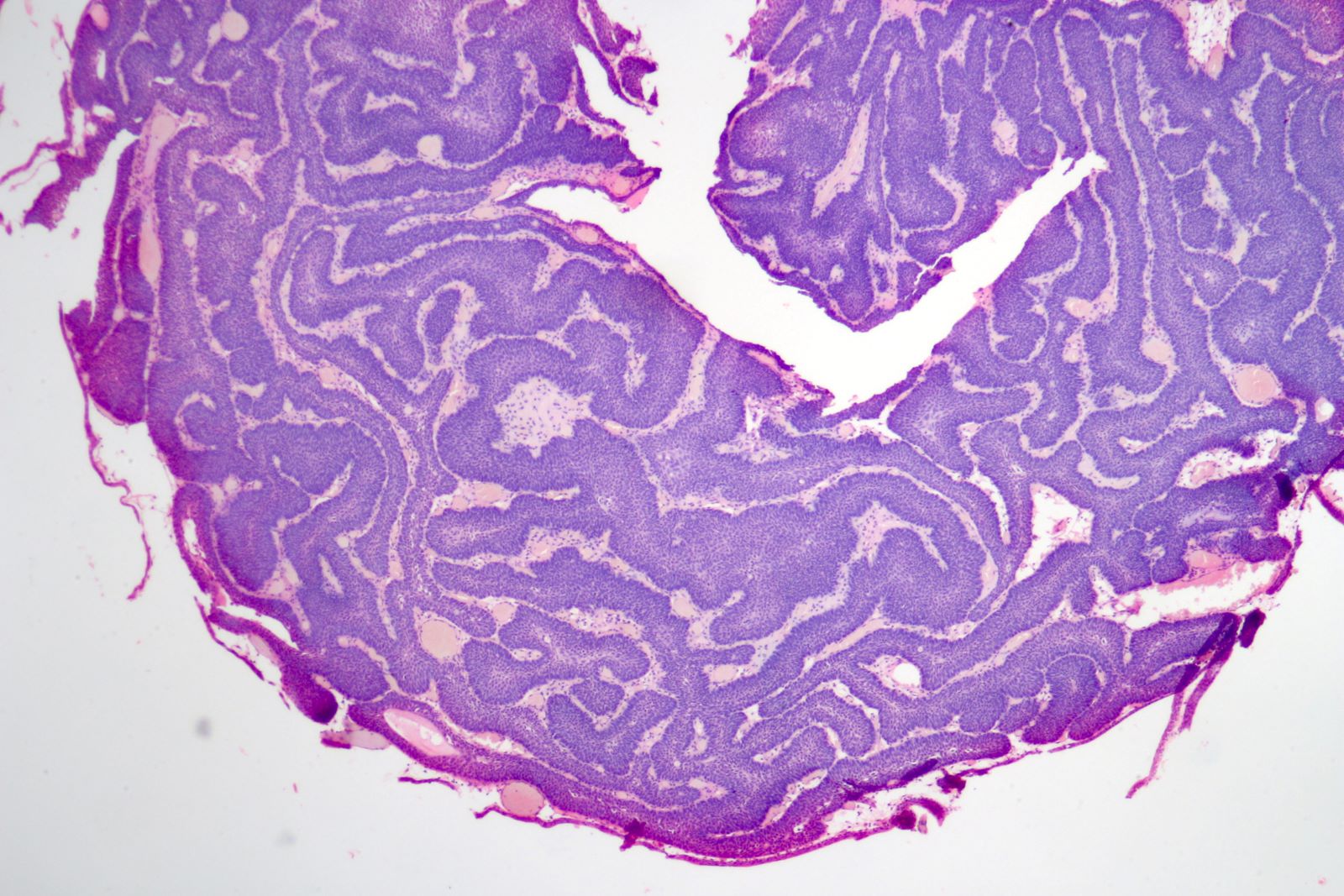
Transverse section of papillae

Transverse section through a single papilla
Fibrovascular cores
Images hosted on other servers:

Flaky, white, plaque-like lesions
Cystoscopy of trigone in 15 year old girl
Contributed by Maria Tretiakova, M.D., Ph.D.

Nonkeratinizing squamous metaplasia
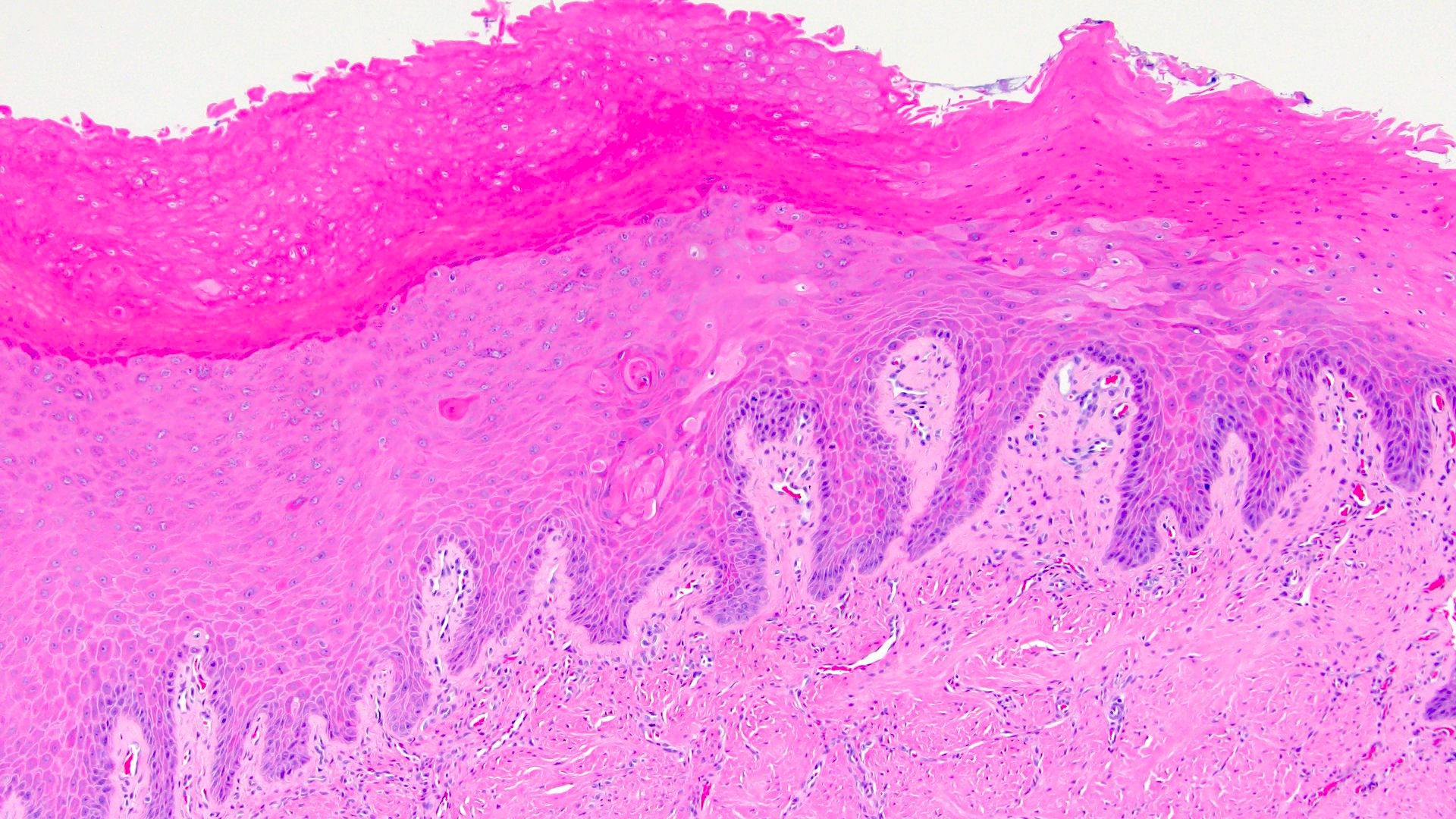
Keratinizing squamous metaplasia with parakeratosis
Squamous metaplasia with cystitis glandularis
Keratinizing squamous metaplasia
Images hosted on other servers:

Benign squamous cells
Contributed by Debra L. Zynger, M.D.
Muscularis propria invasion (pT2b)
Prostatic invasion (pT4a)
Residual noninvasive tumor (ypTis / a)
Contributed by Debra L. Zynger, M.D.
Noninvasive low grade (pTa)
Microinvasion (pT1)
Lamina propria invasion (pT1)
Within muscularis mucosae (pT1)

Within muscularis propria (pT2)
Within perivesicular adipose (pT3)
Within prostate (pT4a)
Within prostate and prostatic glands (pT4a)

Lung metastasis (pM1b)
Uterus metastasis (pM1b)
Common iliac lymph node metastasis (pN3)
Contributed by Debra L. Zynger, M.D.
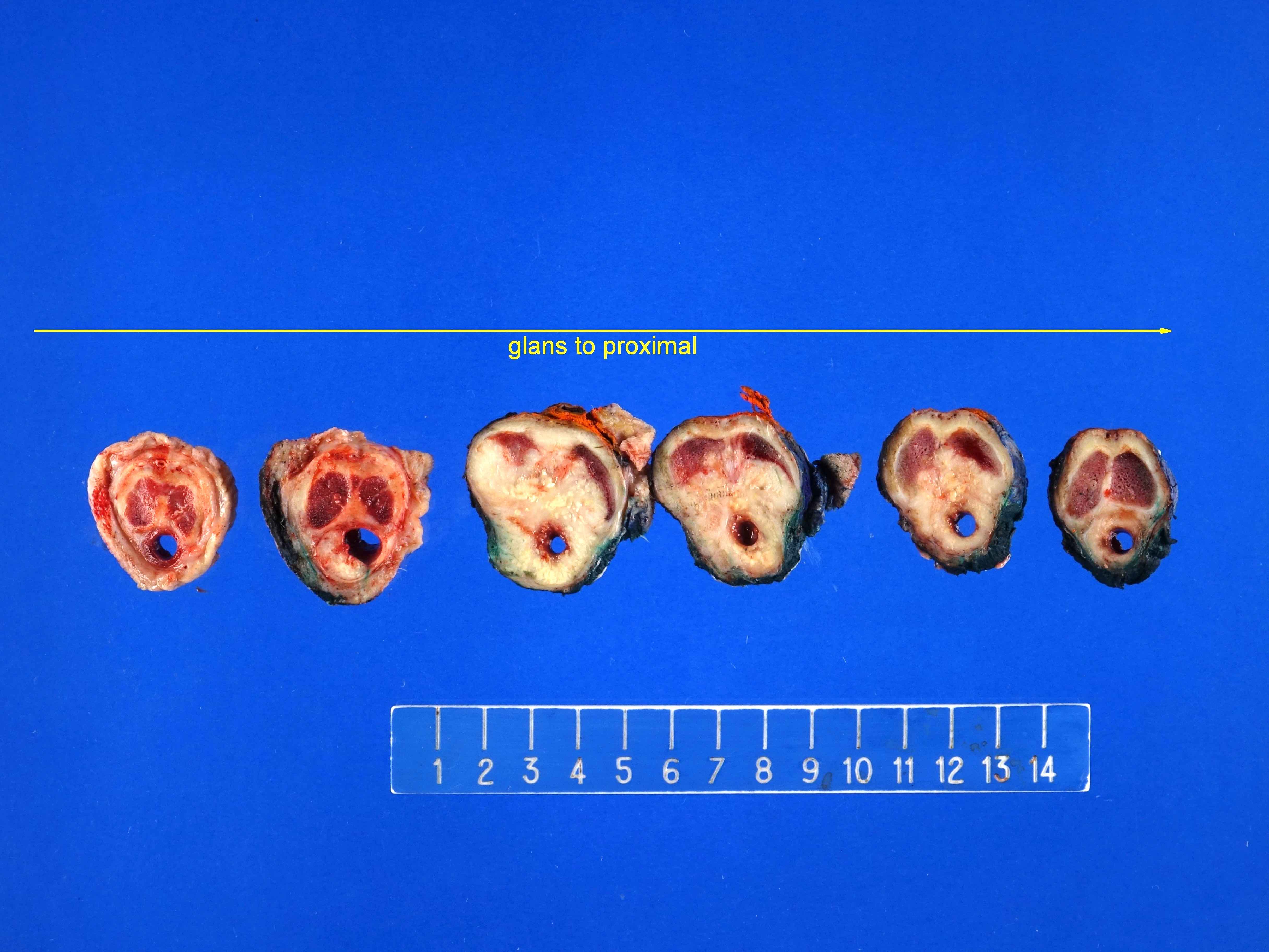
Penile urethral urothelial carcinoma (pT3)
Contributed by Debra L. Zynger, M.D.

Penile urethral urothelial carcinoma (pT3)
Contributed by Debra L. Zynger, M.D.
Renal pelvic and peripelvic fat invasion (pT3)
Contributed by Debra L. Zynger, M.D.
Renal parenchyma invasion (pT3)
Images hosted on other servers:

Various images

Postradiation changes mimic dysplasia
Images hosted on other servers:

Fetal tube connecting bladder with umbilicus
Images hosted on other servers:

Low attenuating cystic mass

Mass in the anterior abdominal wall
Images hosted on other servers:

Intraoperative image of cystic lesion at dome of bladder

Intraoperative image of urachal cyst

Intraoperative image of multilobulated cyst
Images hosted on other servers:

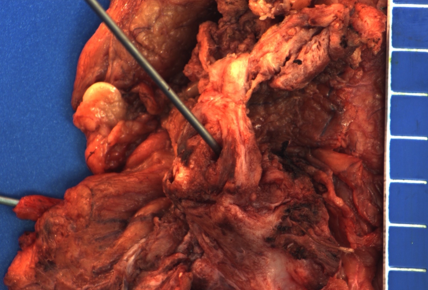
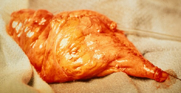
Excised specimen with umbilicus and neoplasm
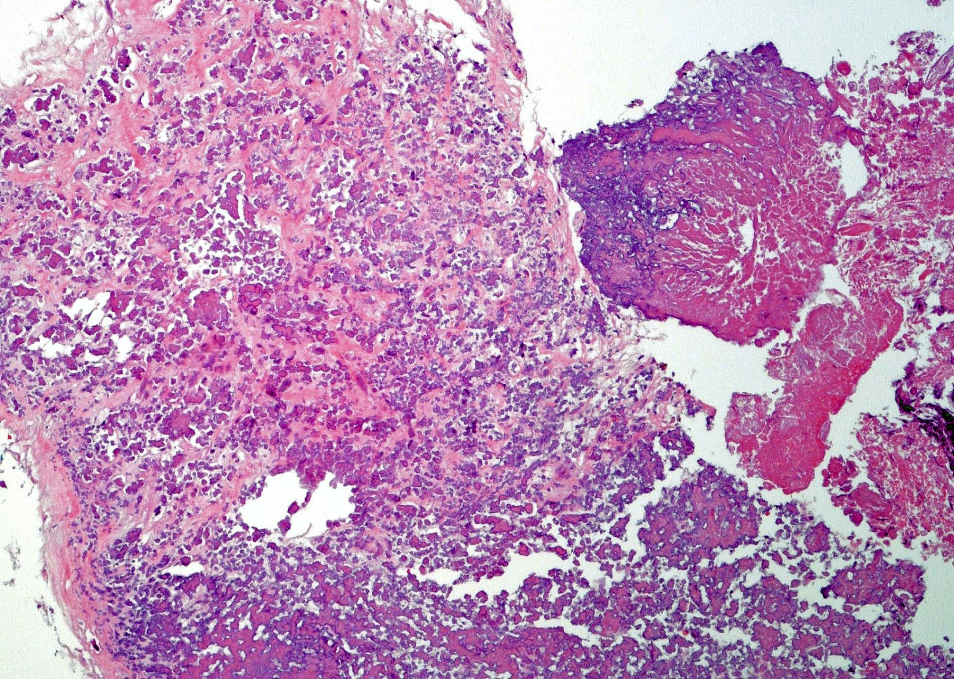
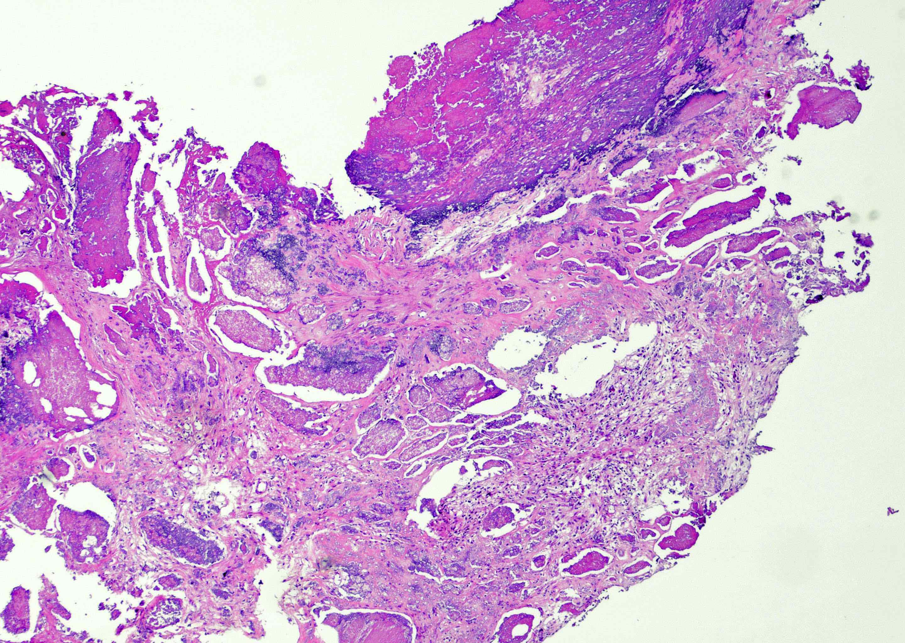
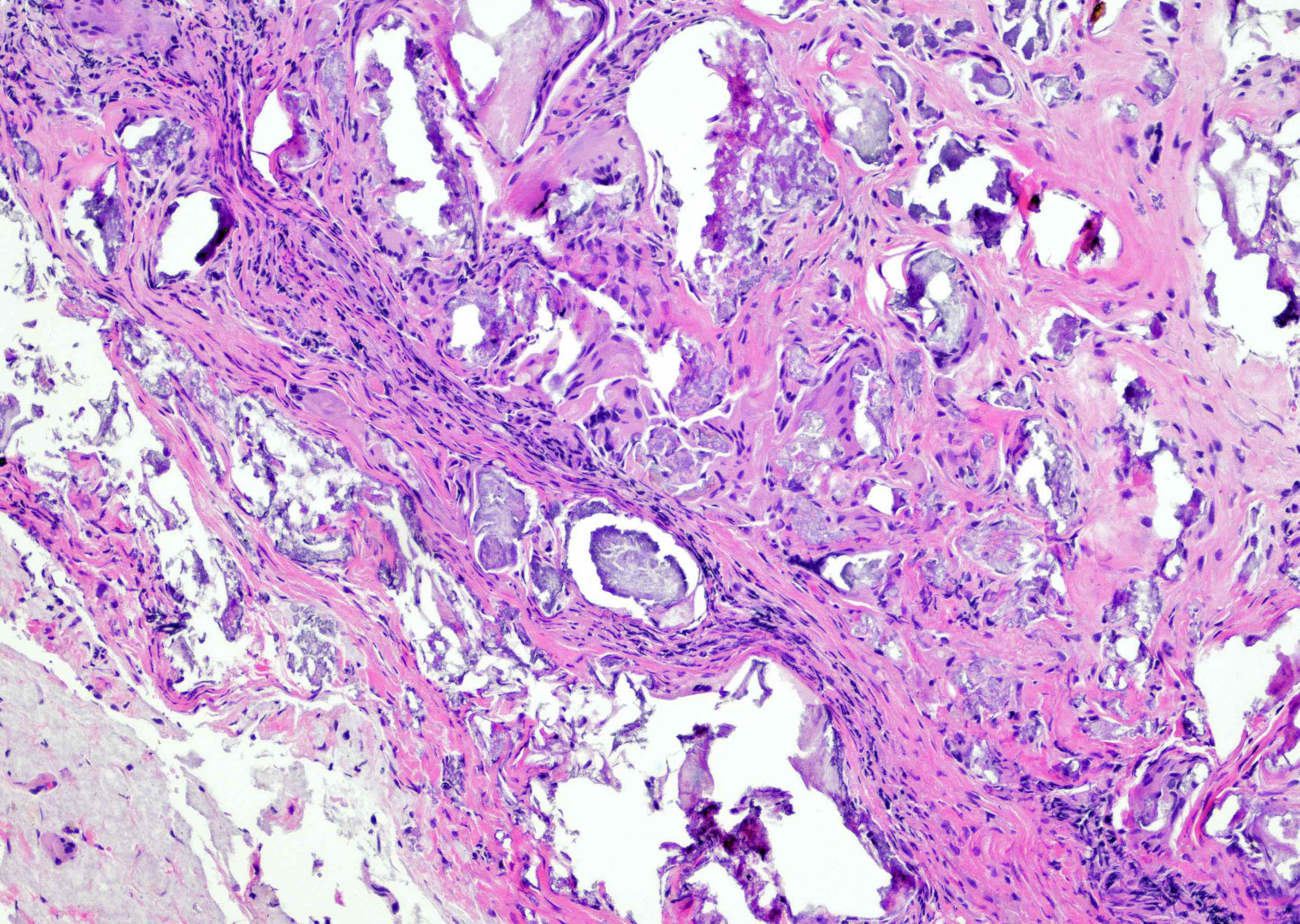
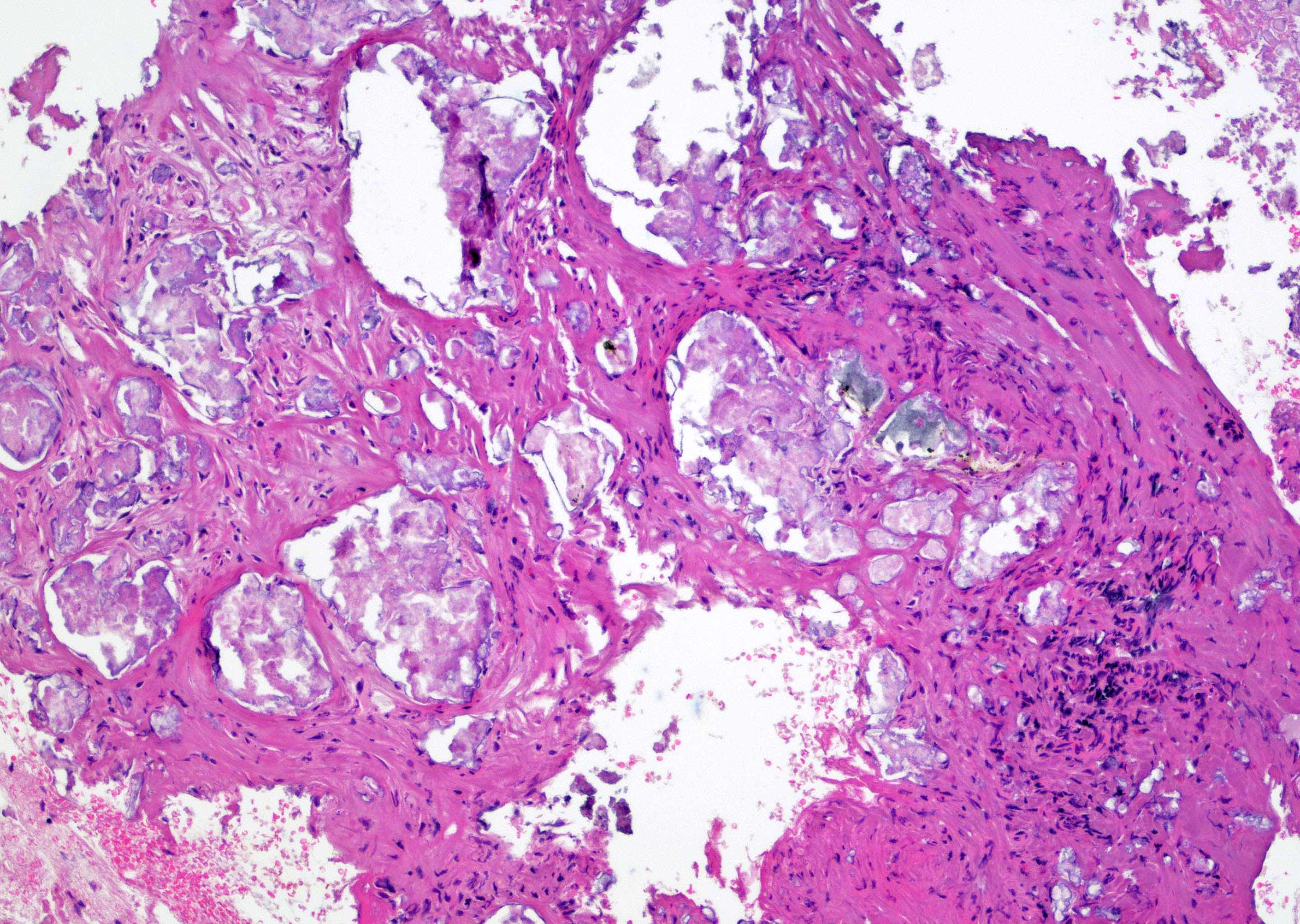
Contributed by Maria Tretiakova, M.D., Ph.D.
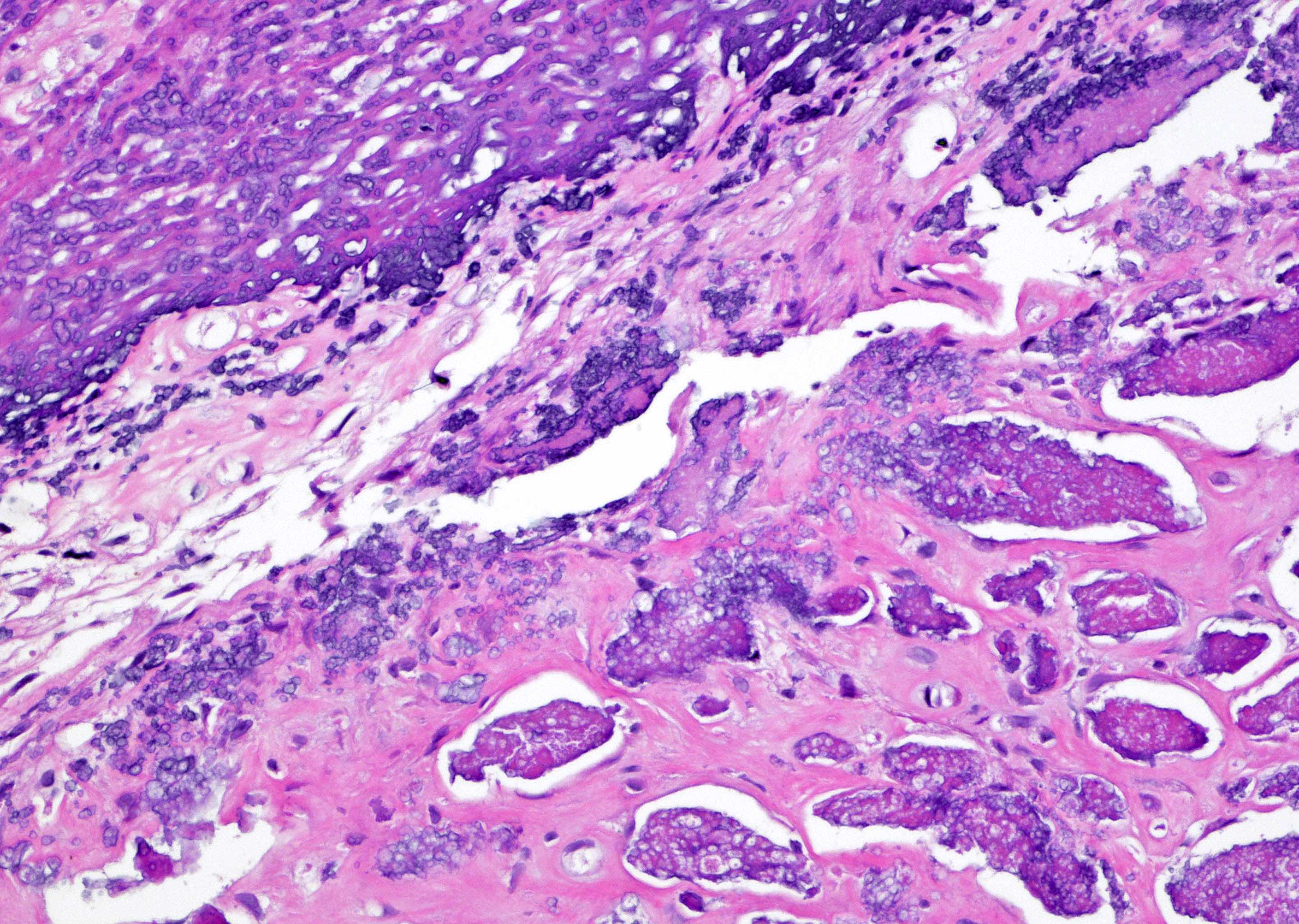
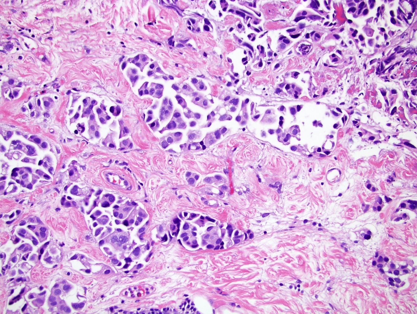
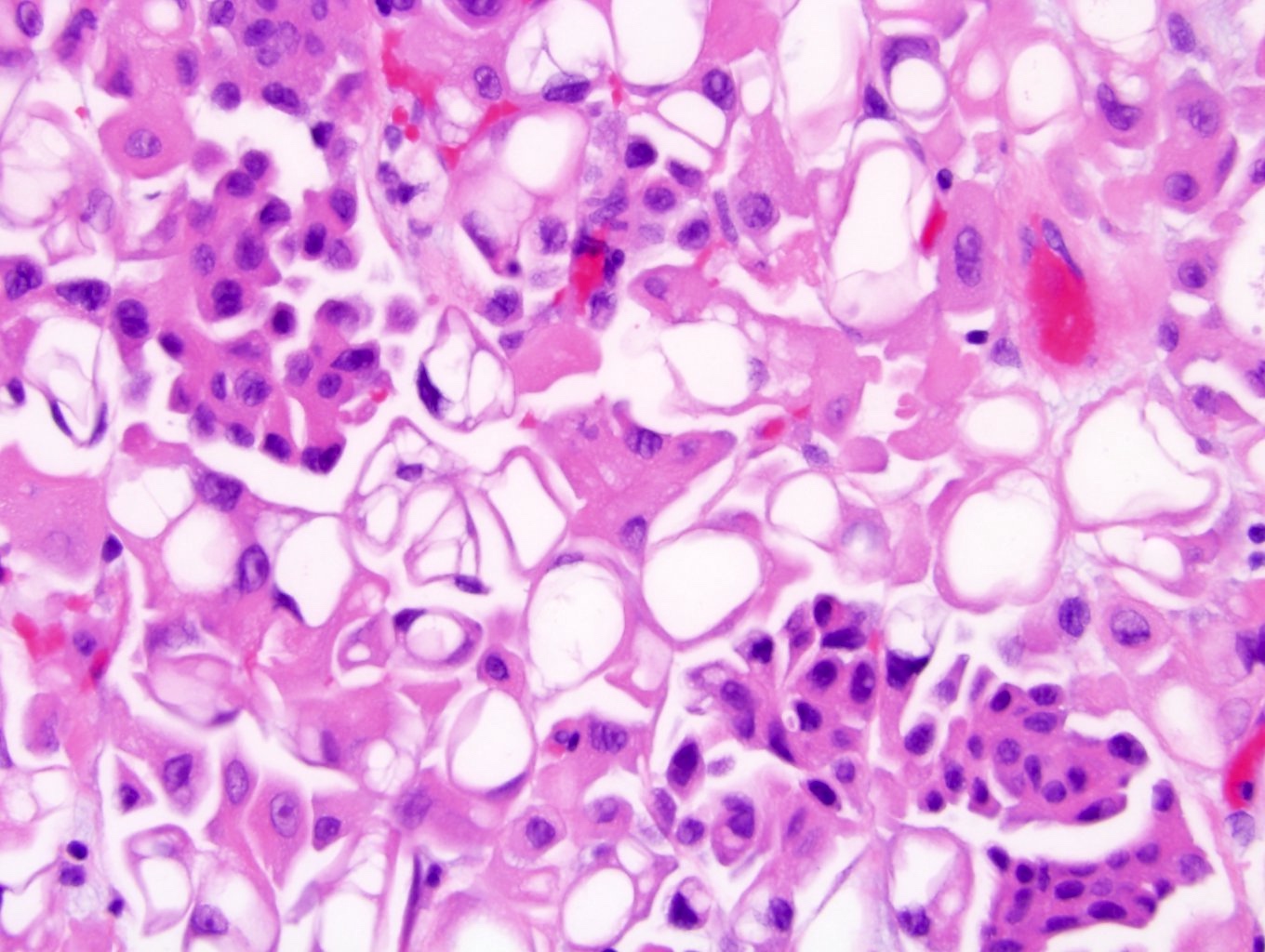
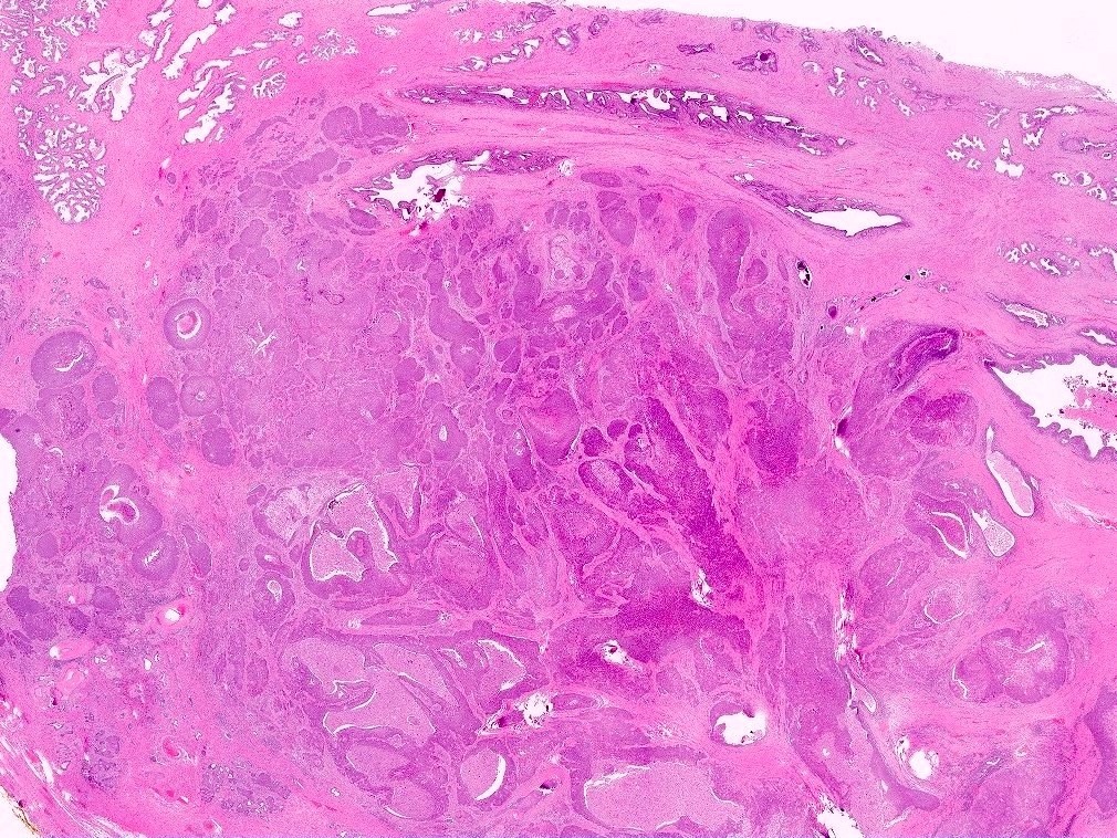
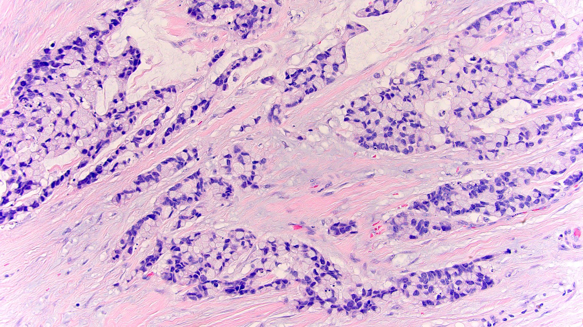
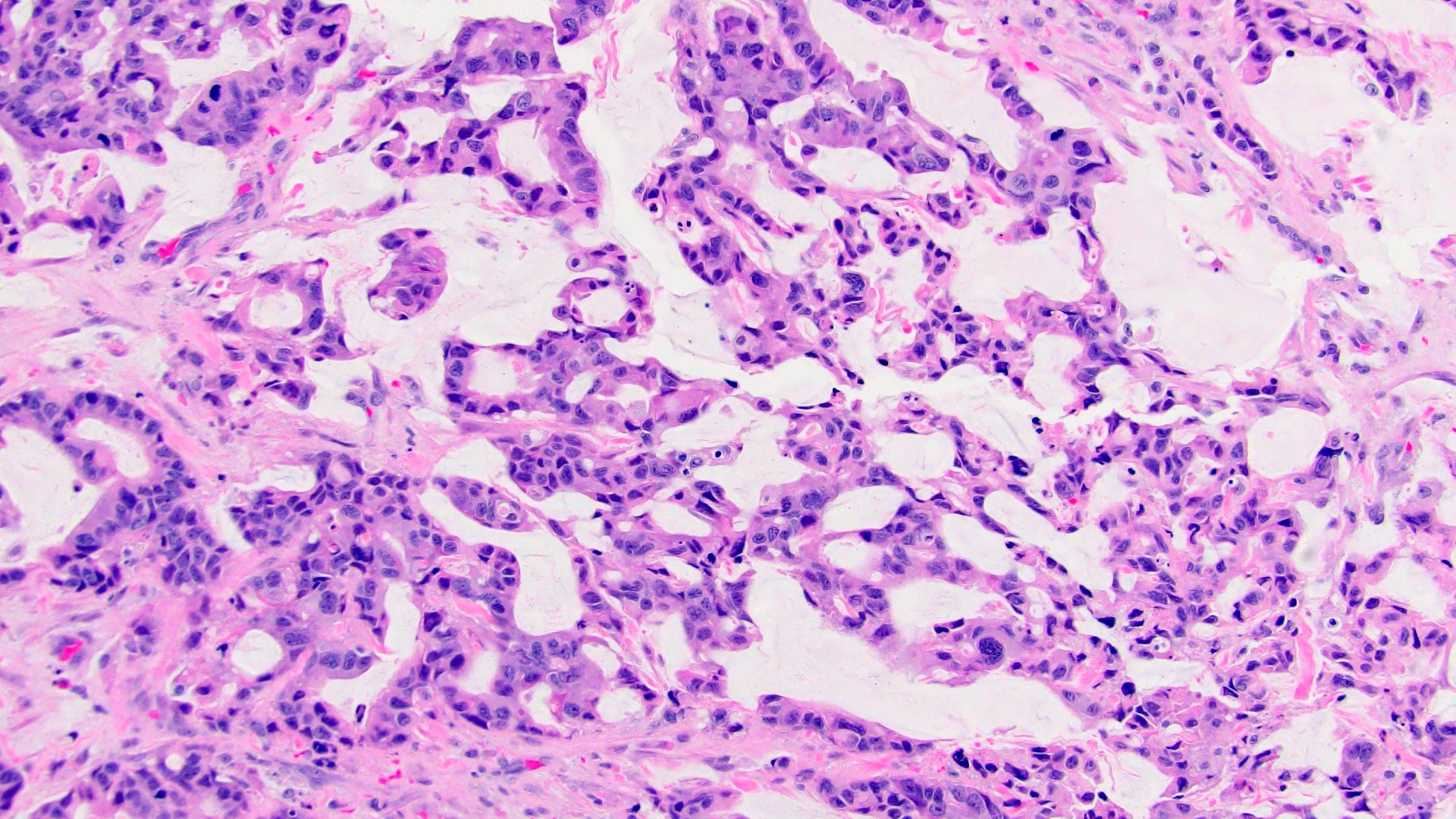
Mucinous / colloid subtype
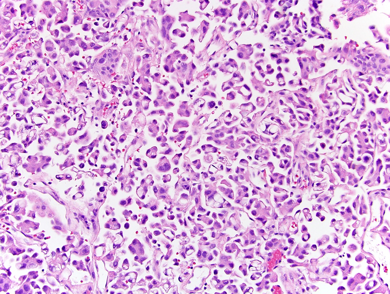
Enteric subtype
Signet ring subtype
Mixed subtype
Urachal carcinoma, NOS
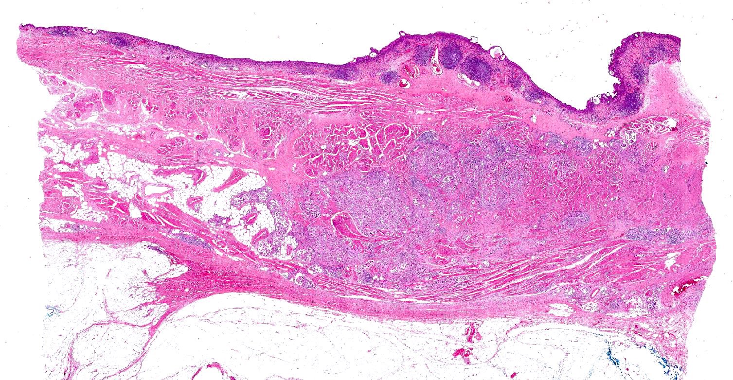
Mucinous adenocarcinoma, bladder dome
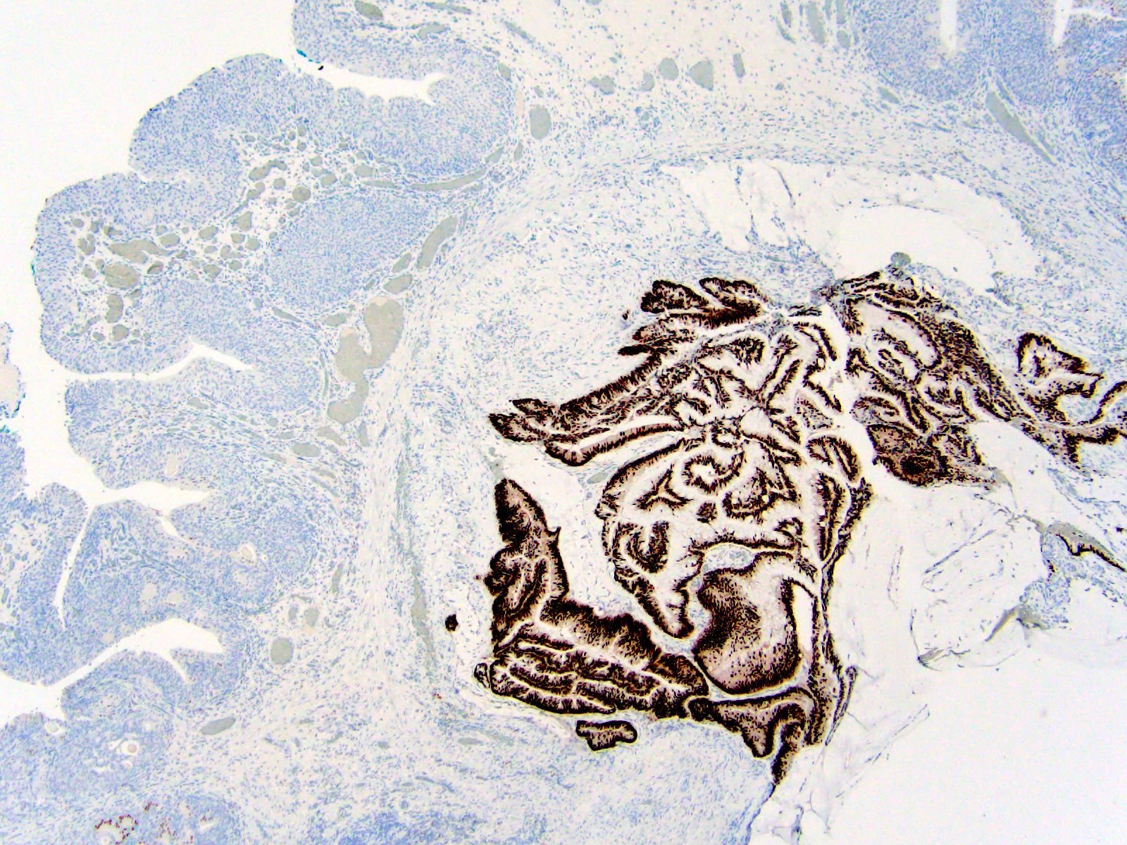
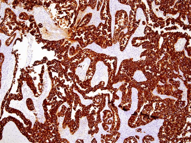
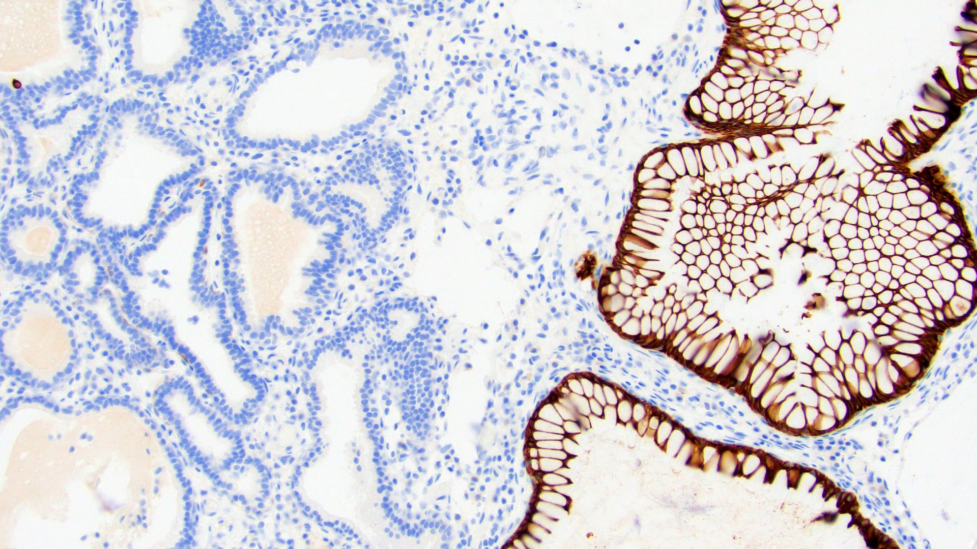
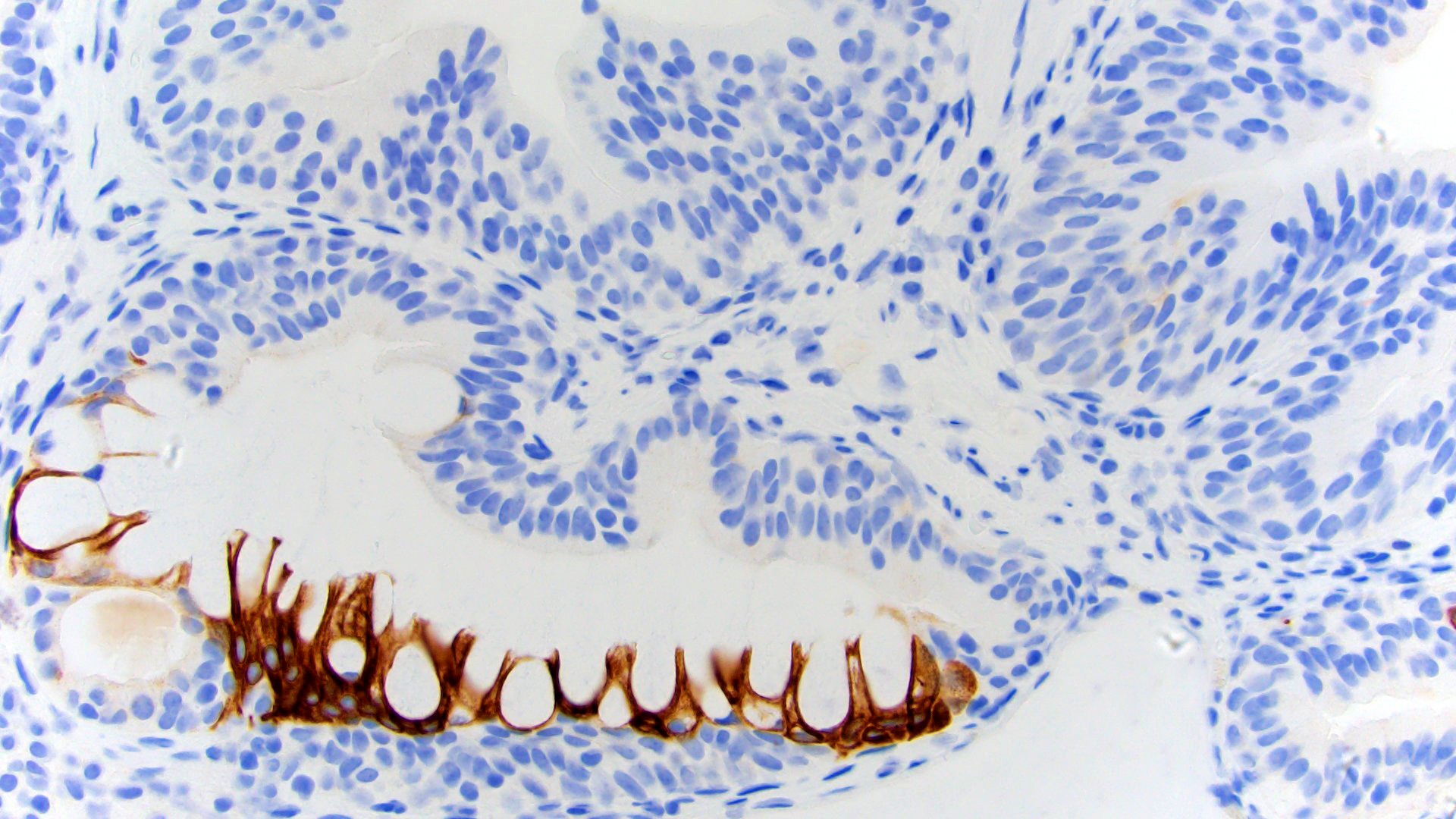
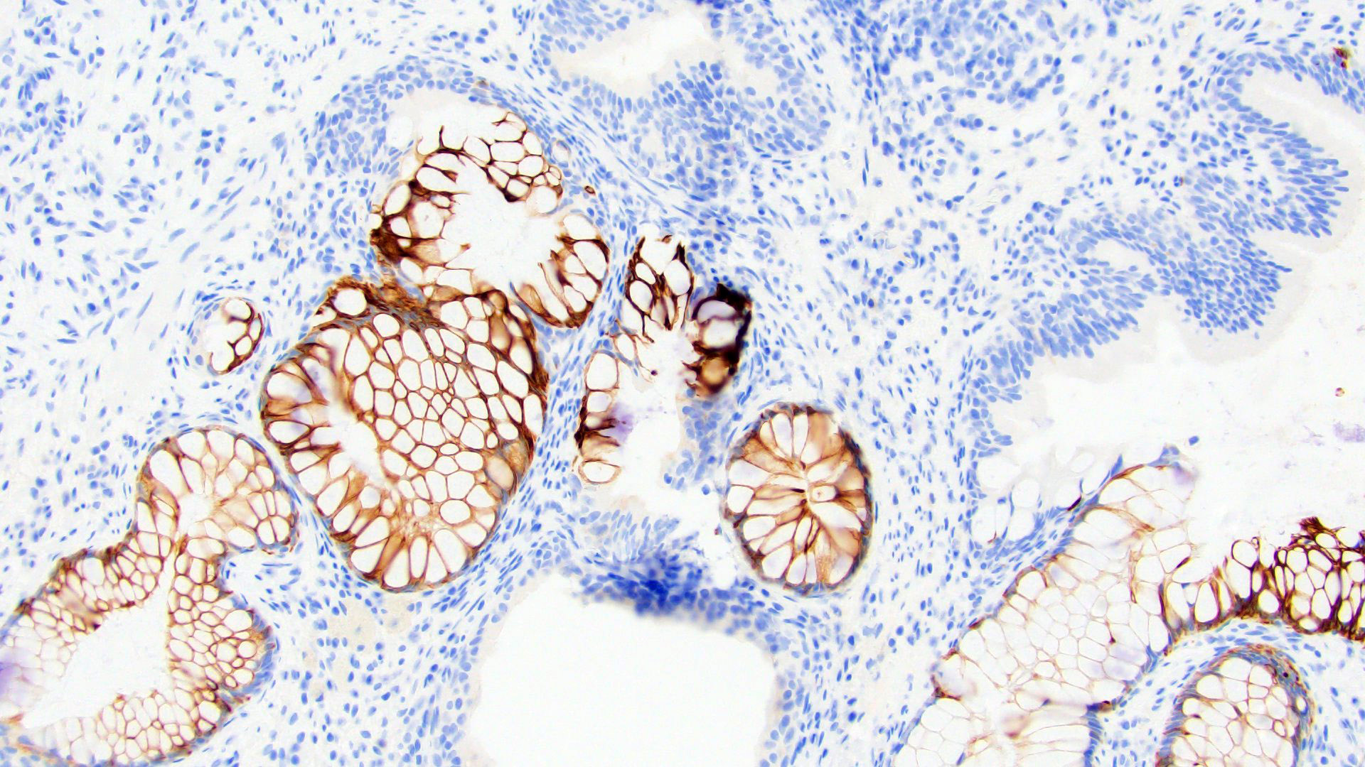
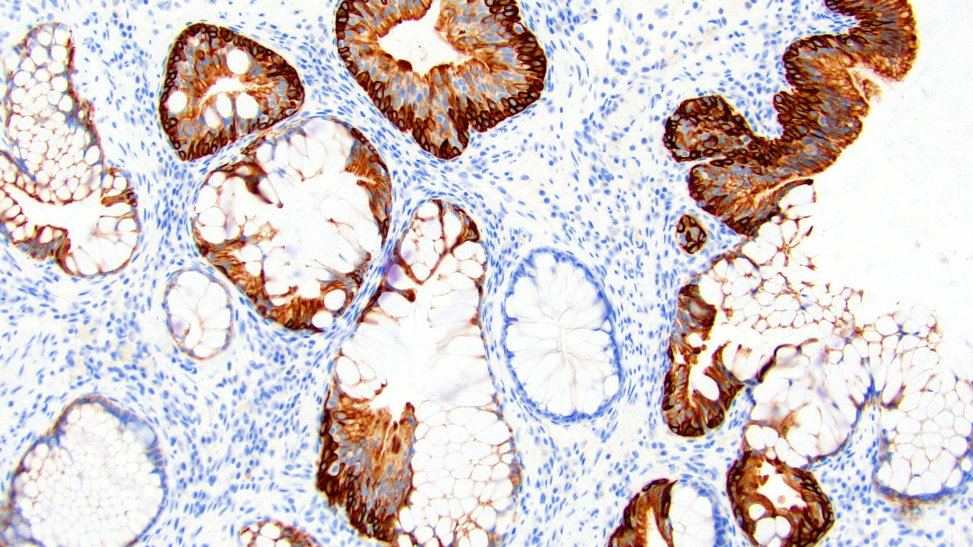
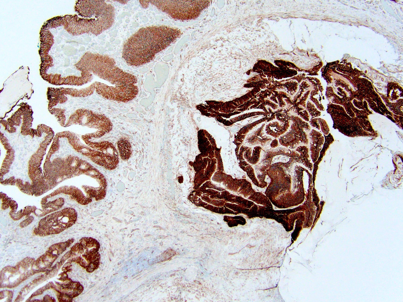
CDX2 in bladder dome adenocarcinoma
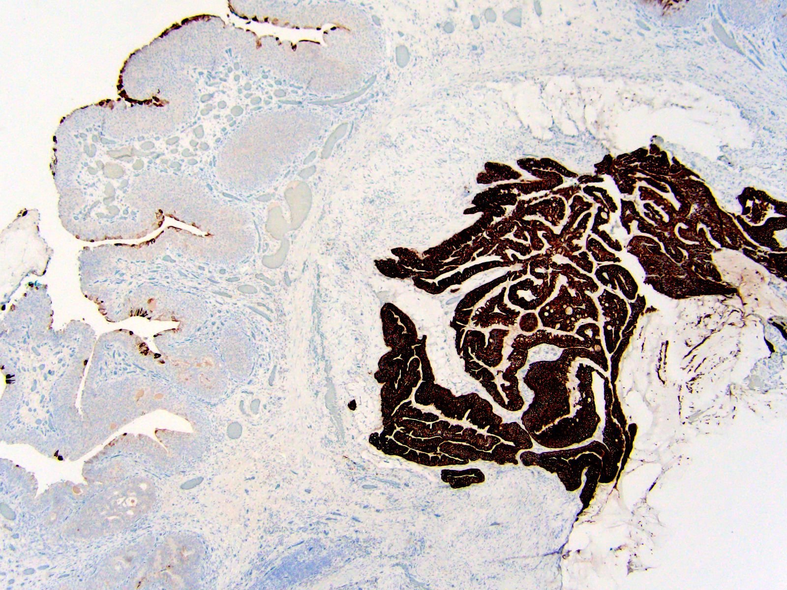
CK20 in bladder dome adenocarcinoma
Beta catenin expression
High molecular weight cytokeratin expression
Images hosted on other servers:

FNAC smear of urachal adenocarcinoma
Mini tutorial of urachal tumors
Urachal adenocarcinoma diagnosis and treatment


Images hosted on other servers:

Persistent urachus

Before and after surgery


Urachal cysts - sinus tract between bladder dome and umbilicus

Infected urachal cyst

Urachal cyst attached to Meckel diverticulum
Images hosted on other servers:

Patent urachus, autopsy specimen

Benign urachal lesion

Infected urachal cyst and fibrous tract

Urachal cyst containing stones

Urachal sigmoid fistula

Inflamed urachal cyst
Contributed by Bohdan Zoshchuk, M.D. and Manasa Morisetti, M.D.
Patent urachus
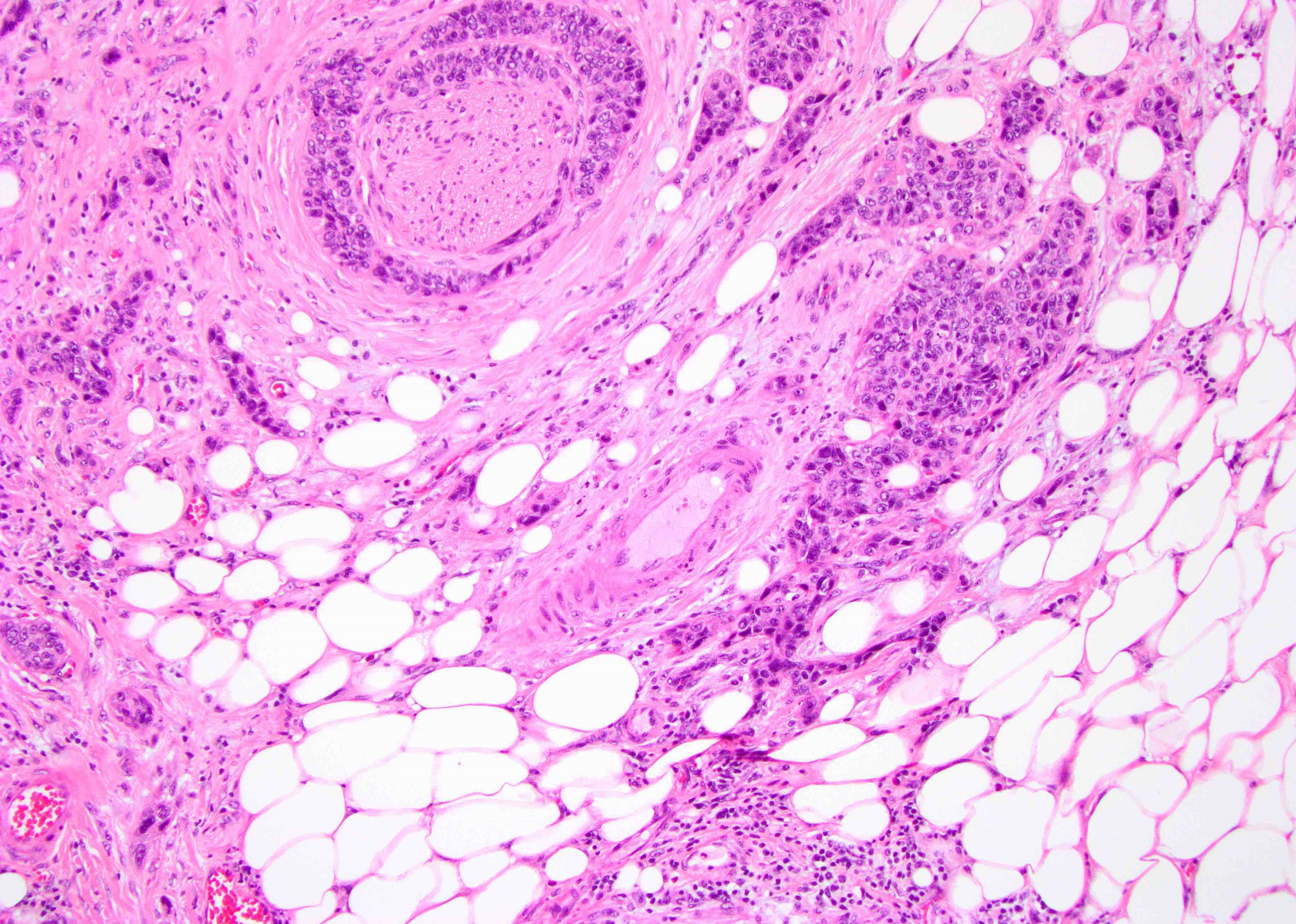
Urachal remnant in bladder wall
Urachal remnant in bladder wall
Umbilical urachal sinus
Images hosted on other servers:
Persistent urachus
Before and after surgery
Urachal cysts - sinus tract between bladder dome and umbilicus
Infected urachal cyst
Urachal cyst attached to Meckel diverticulum
Images hosted on other servers:
Patent urachus, autopsy specimen
Benign urachal lesion
Infected urachal cyst and fibrous tract
Urachal cyst containing stones
Urachal sigmoid fistula
Inflamed urachal cyst
Contributed by Bohdan Zoshchuk, M.D. and Manasa Morisetti, M.D.
Patent urachus
Urachal remnant in bladder wall
Urachal remnant in bladder wall
Umbilical urachal sinus
Images hosted on other servers:
Continent urinary diversion using ileum
Images hosted on other servers:

Tubular adenoma with
high grade dysplasia
after augmentation
ileocystoplasty
Images hosted on other servers:

USG finding - solitary polypoid lesion

Lobulated soft-tissue mass
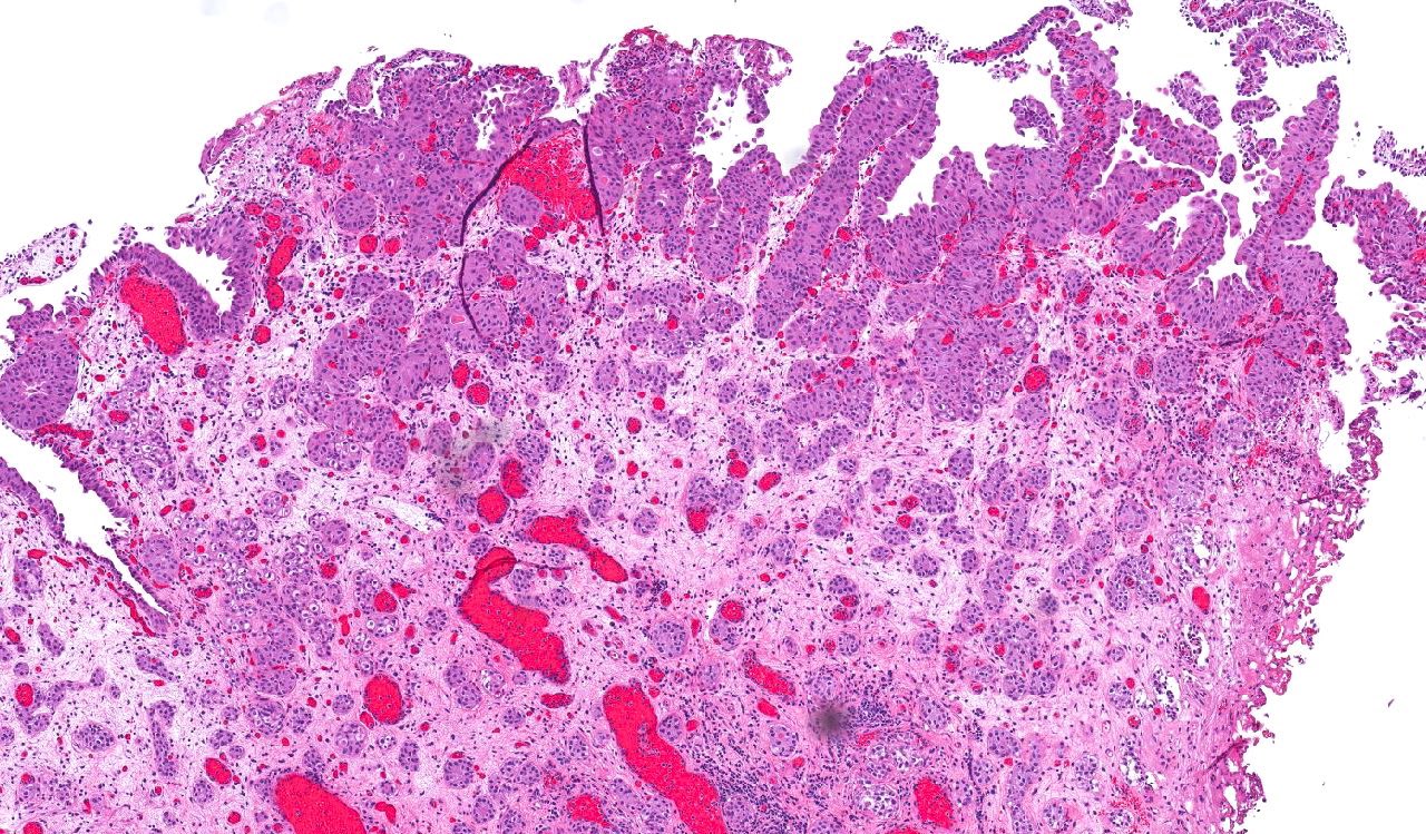
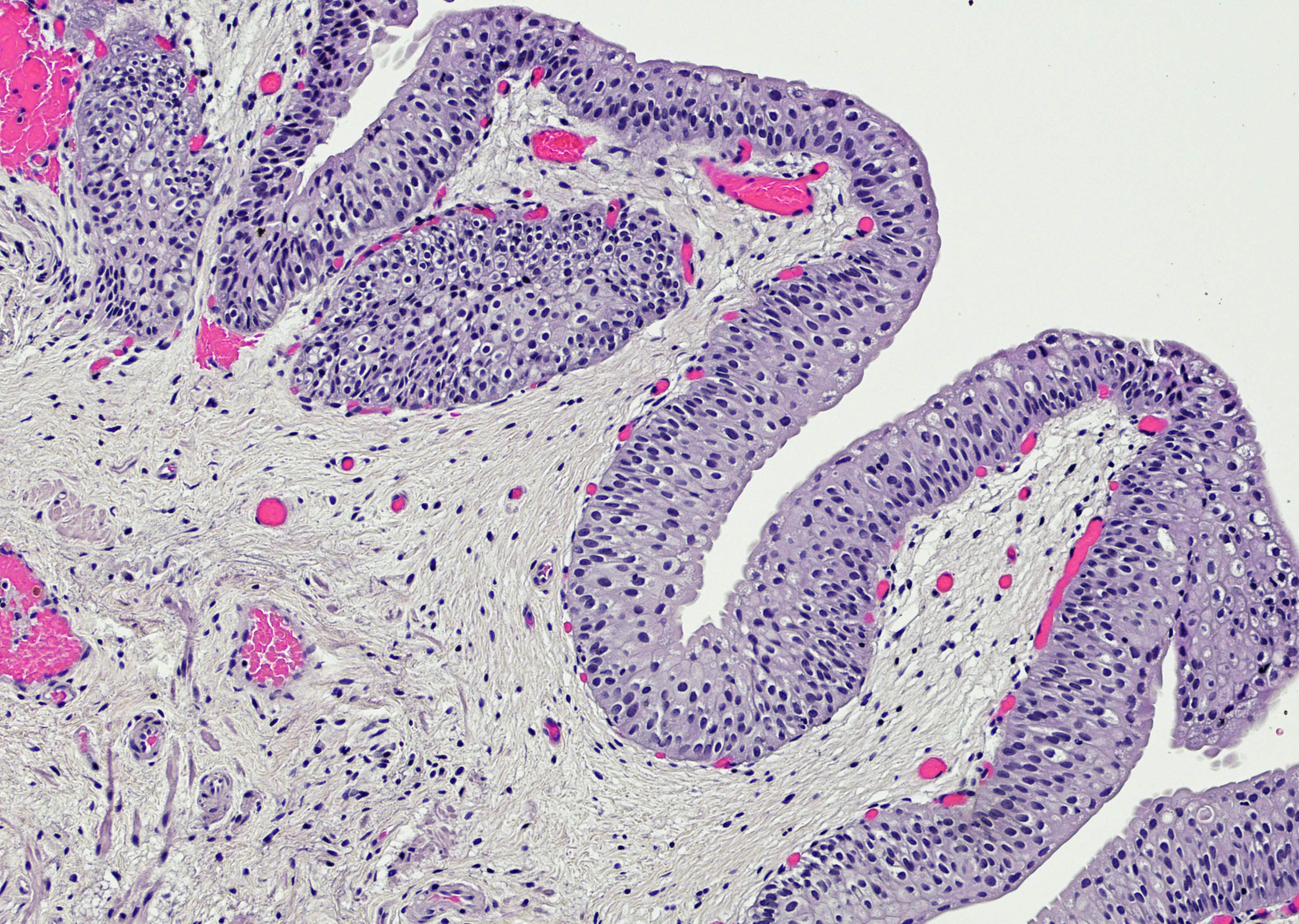
Contributed by Judy Sarungbam, M.D.
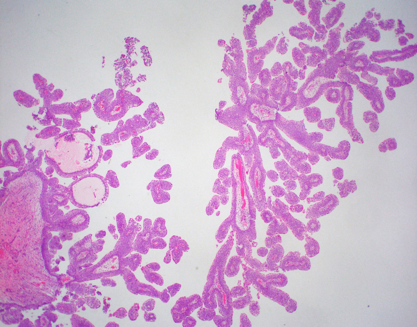
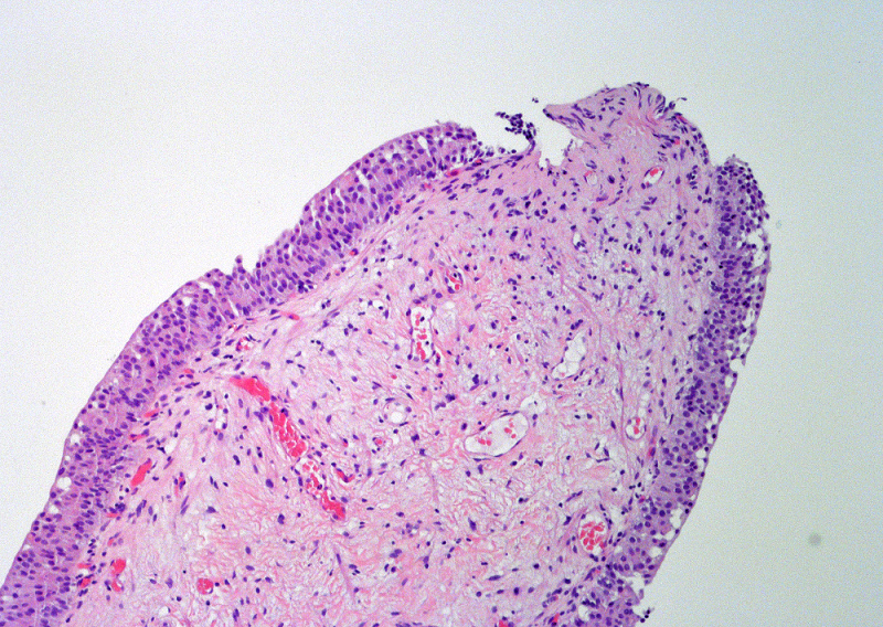
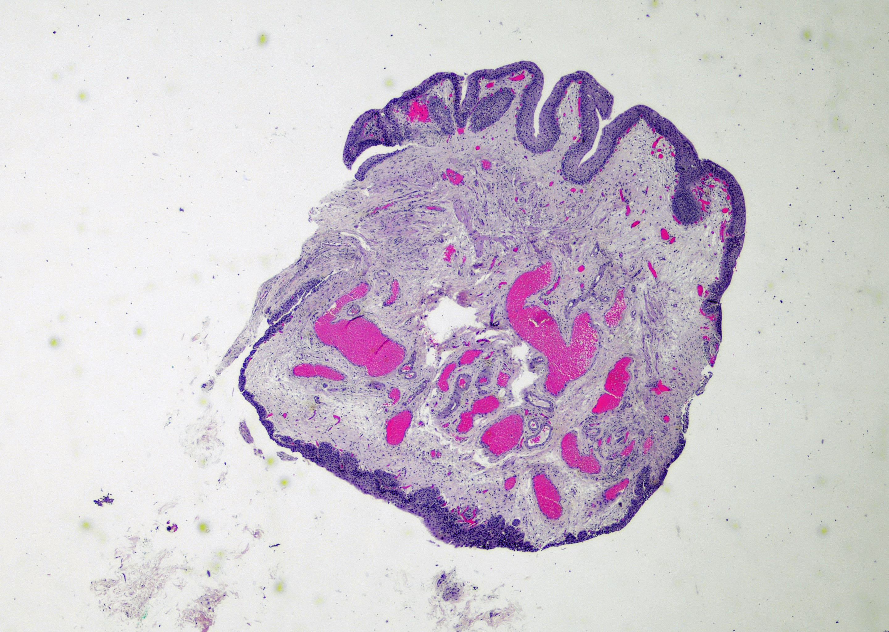
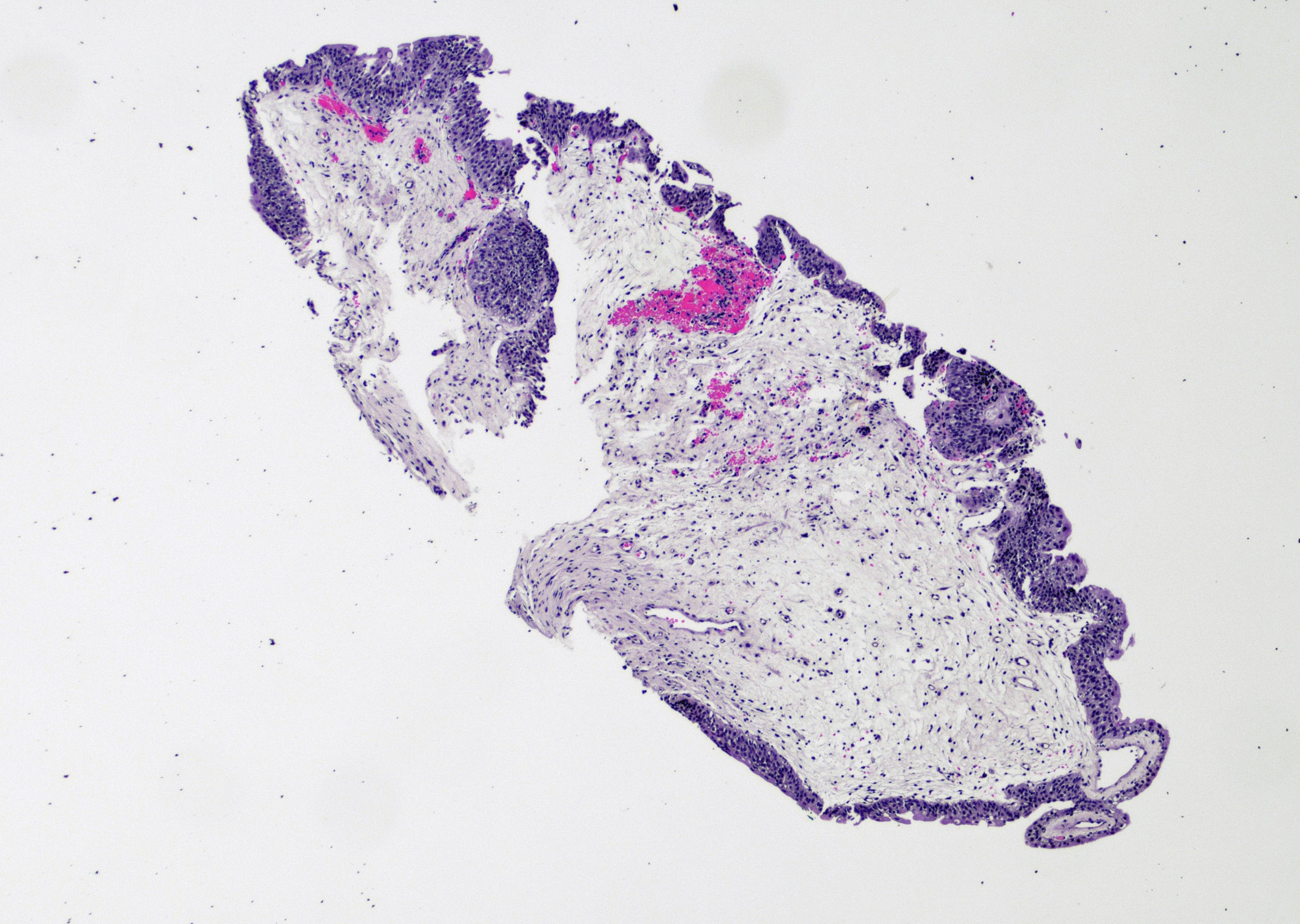
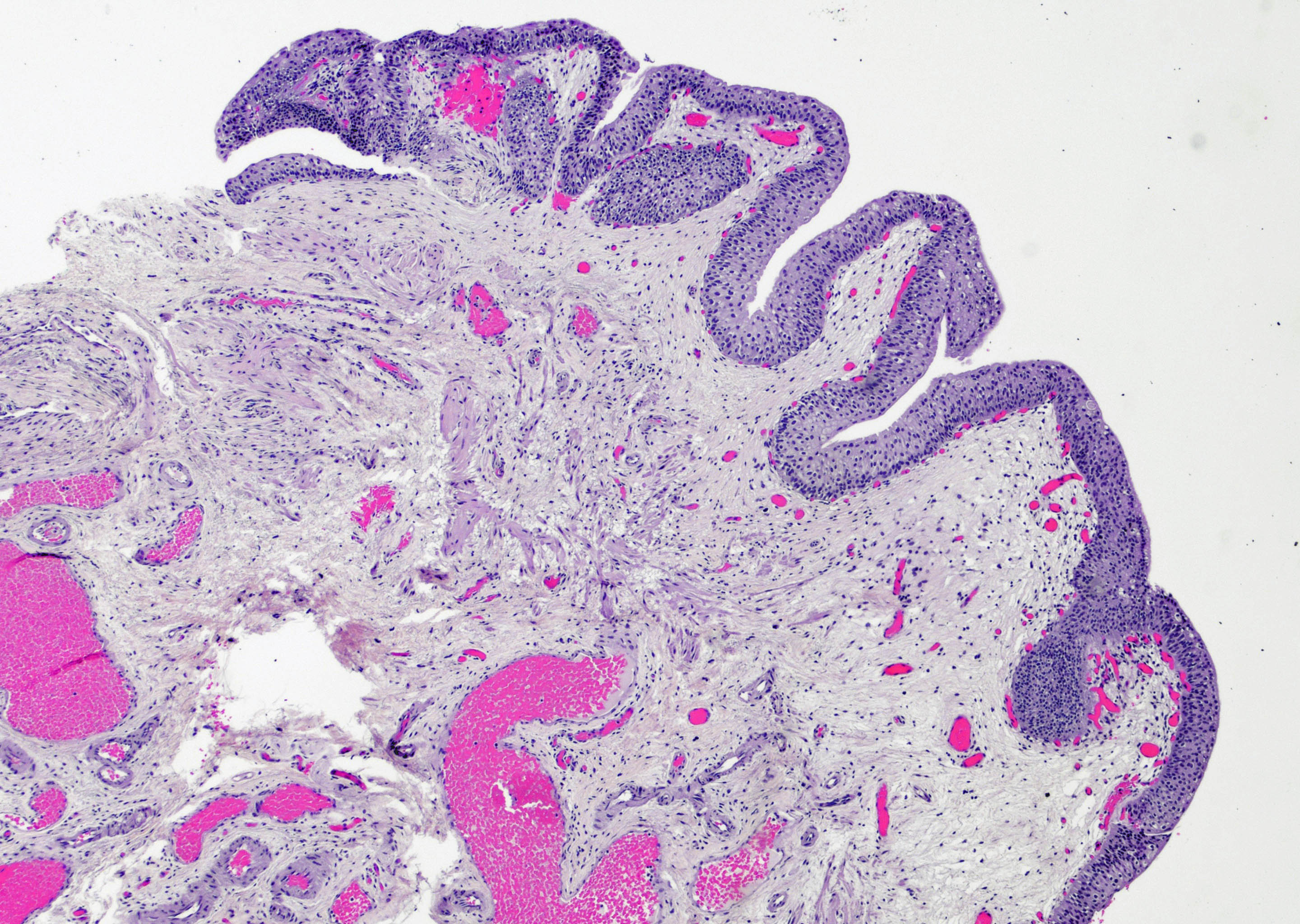
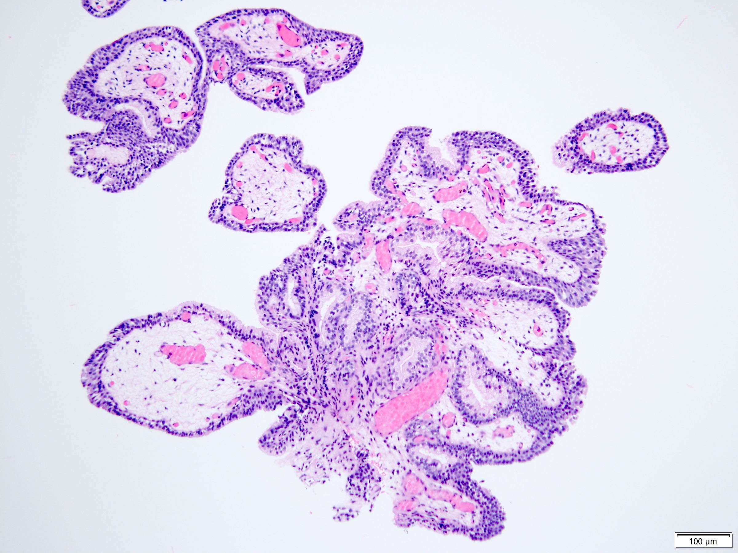
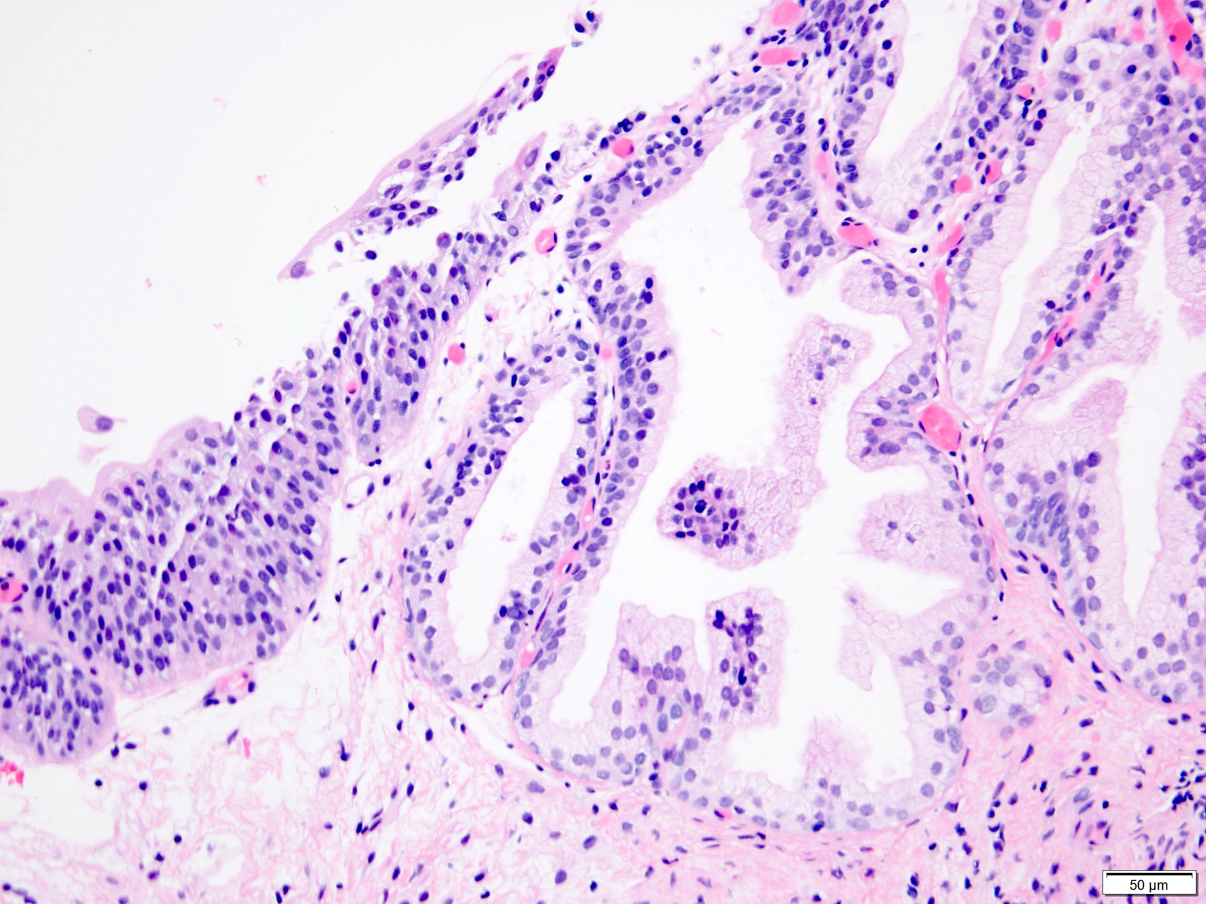
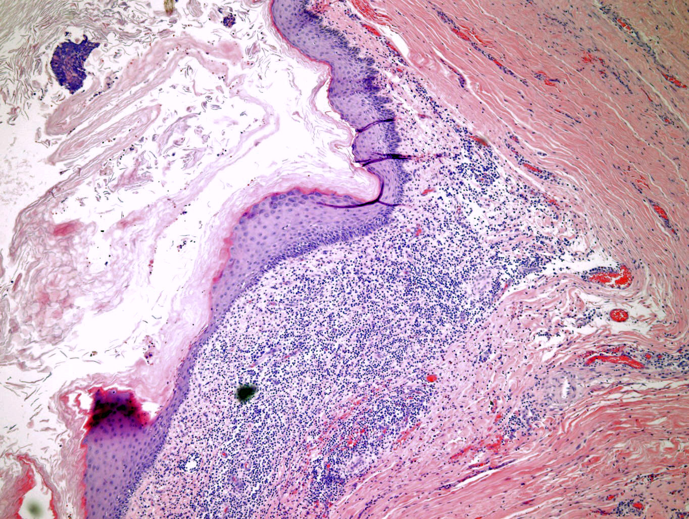
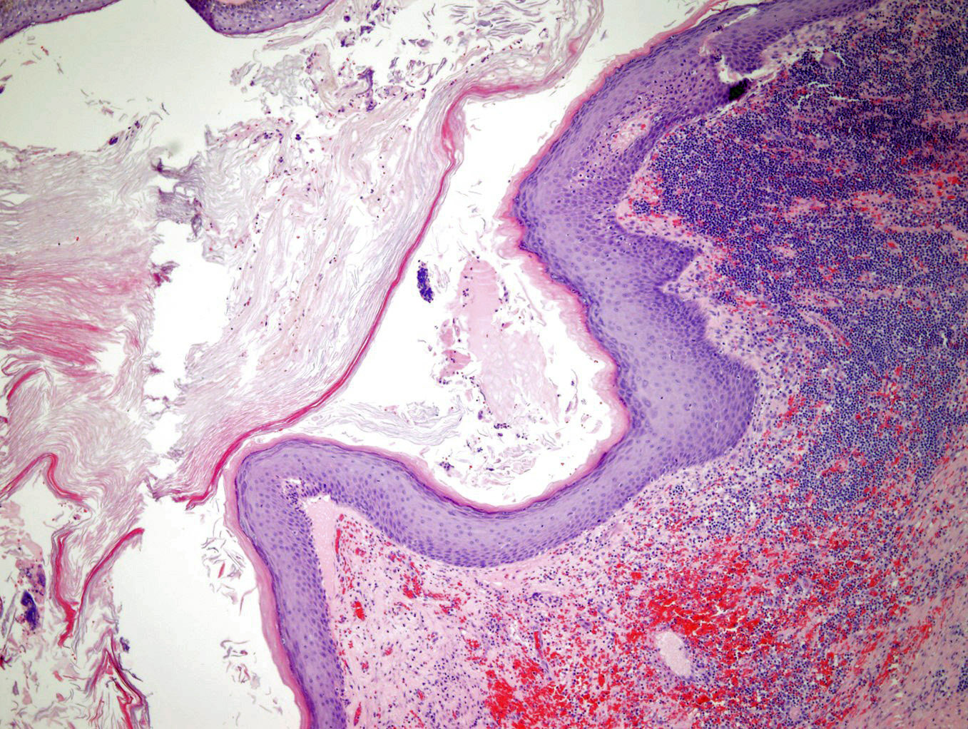
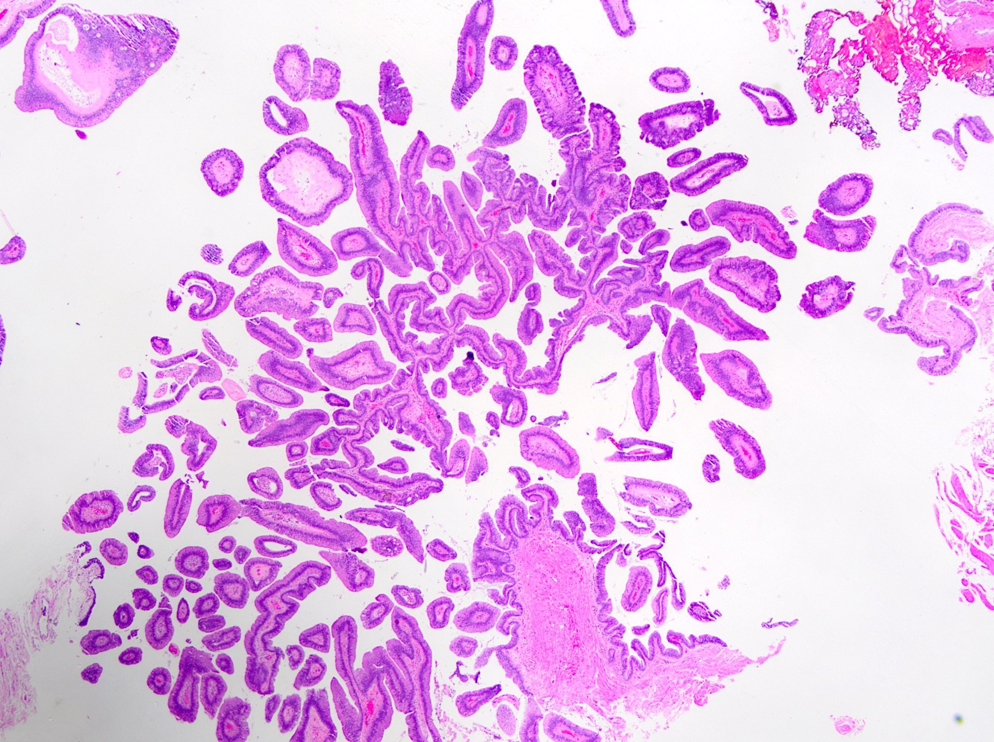
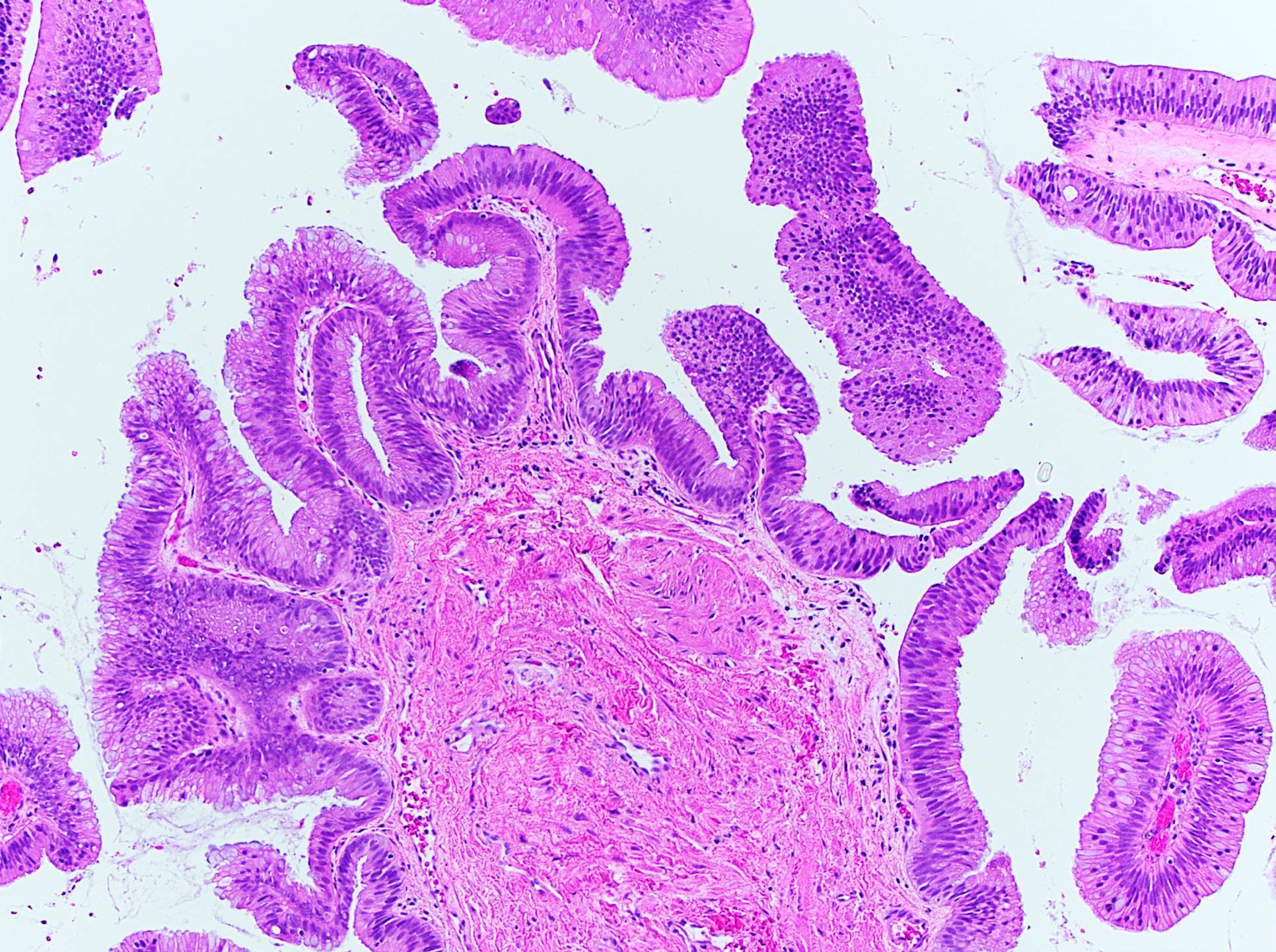
Discrete papillary structures

Slender fibrovascular core
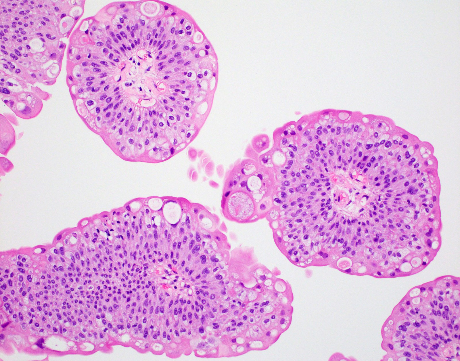
Prominent umbrella cells
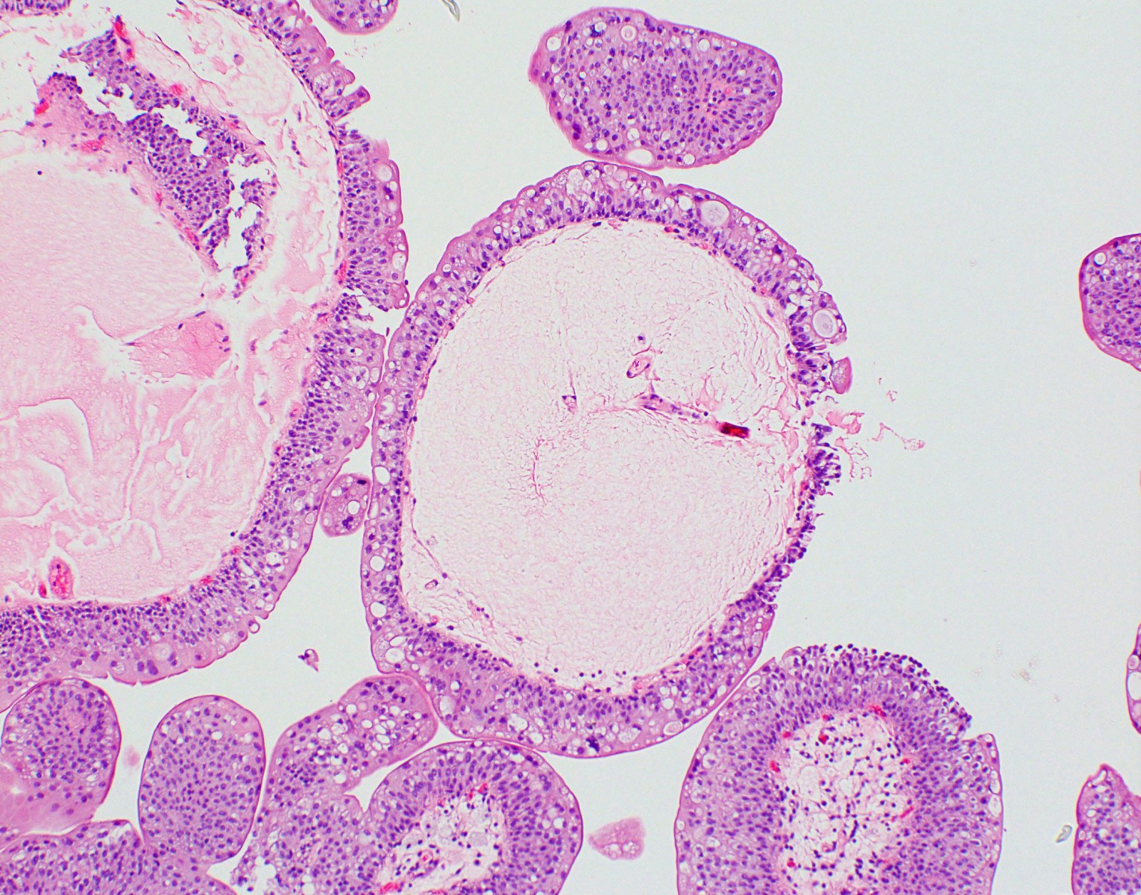
Edematous fibrovascular core
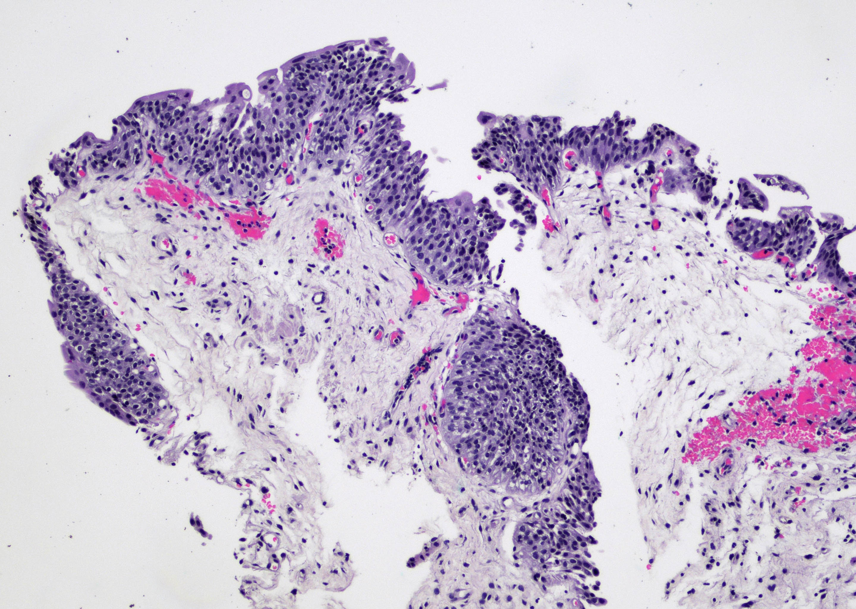
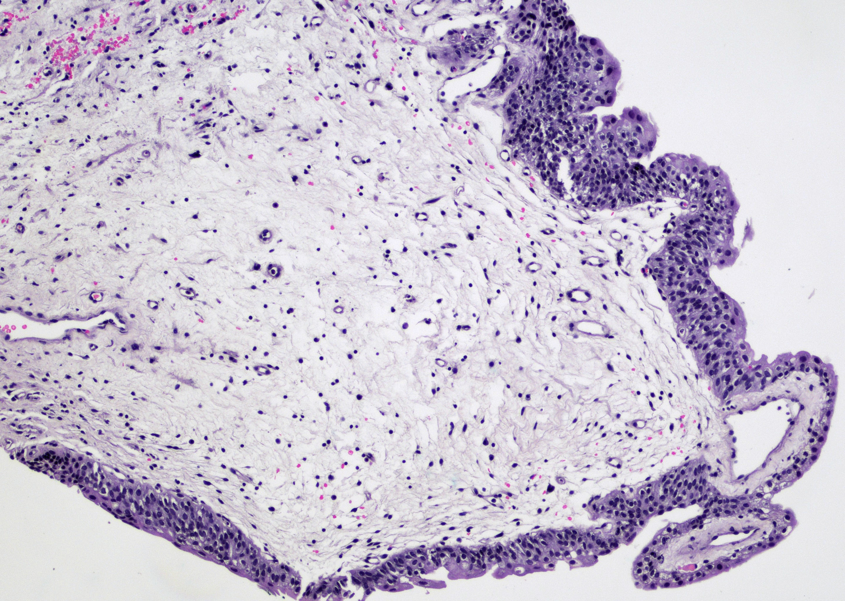
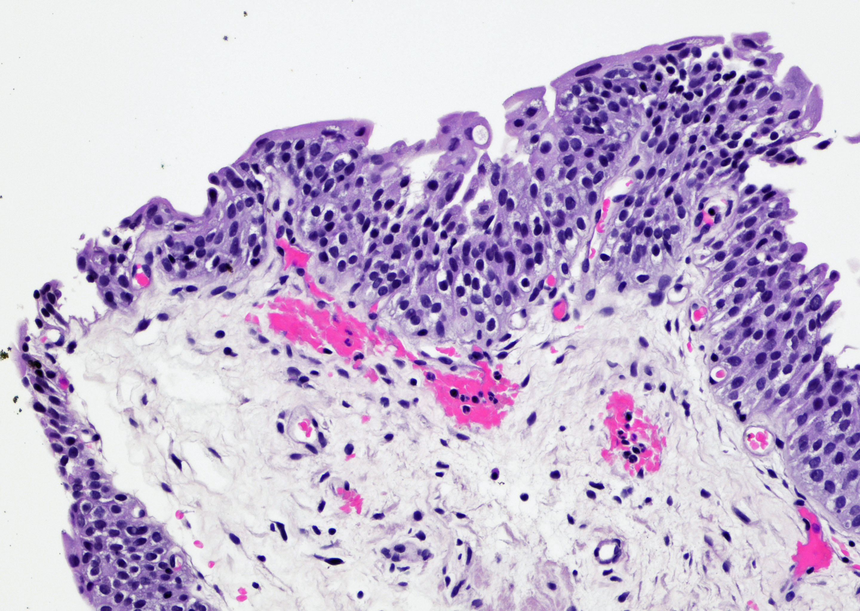
Contributed by Varsha Manucha, M.D.
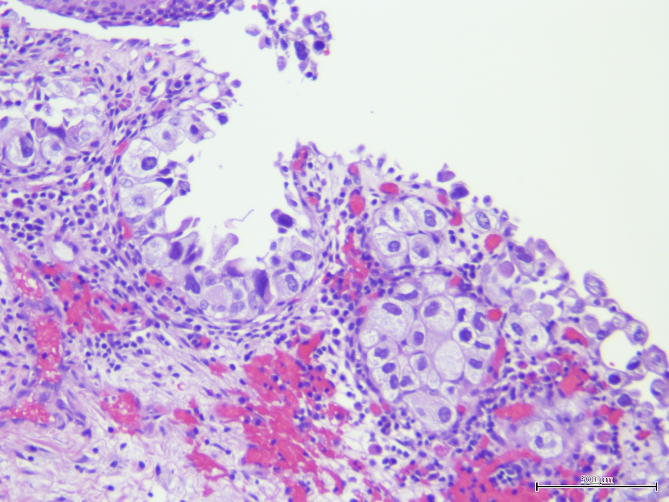
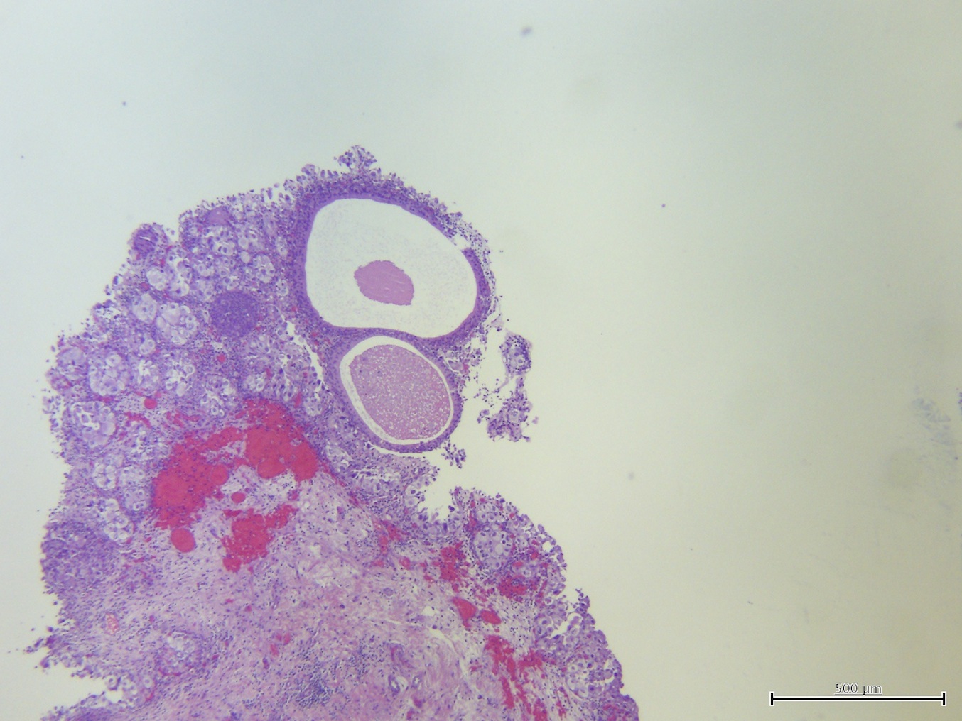
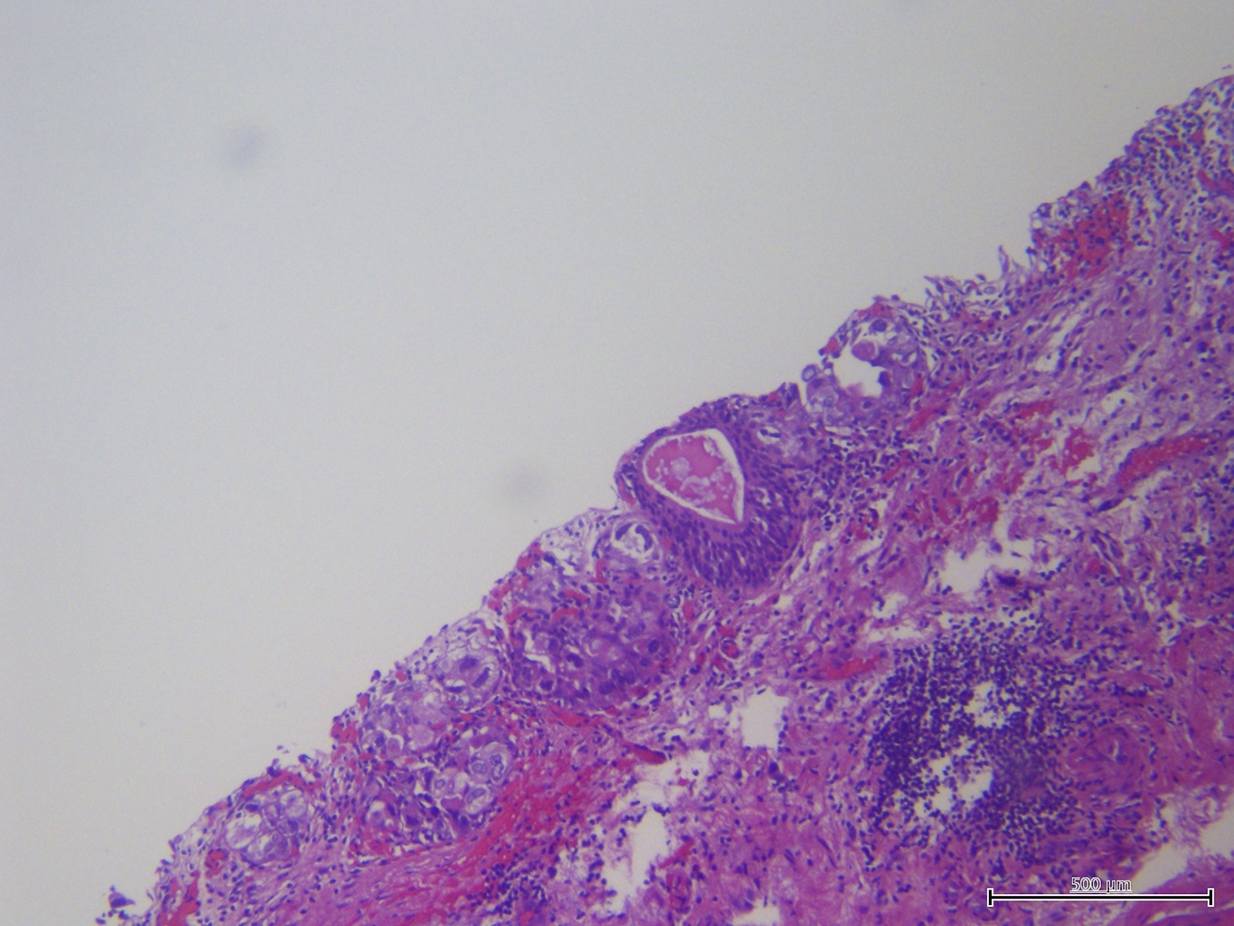
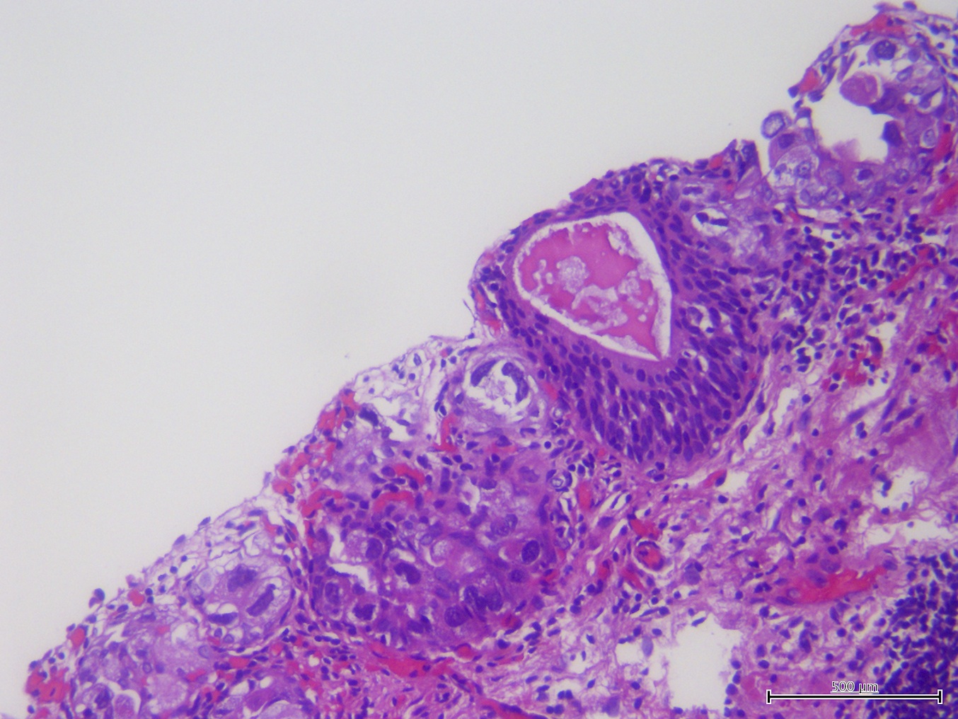
Villous adenoma
Villous pattern


Nuclear stratification
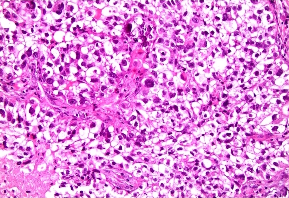
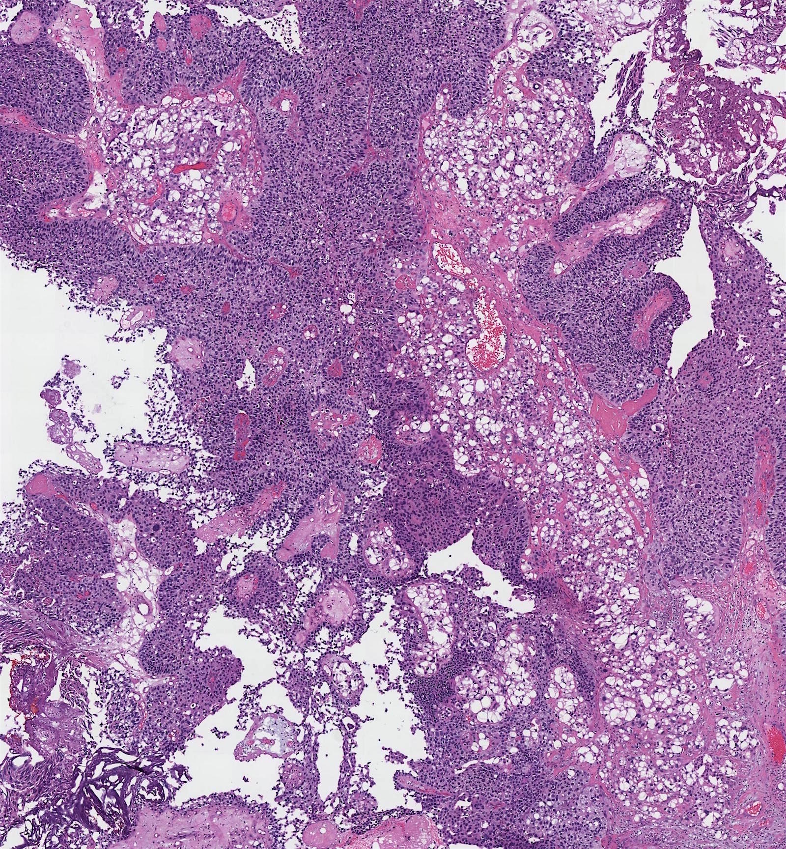
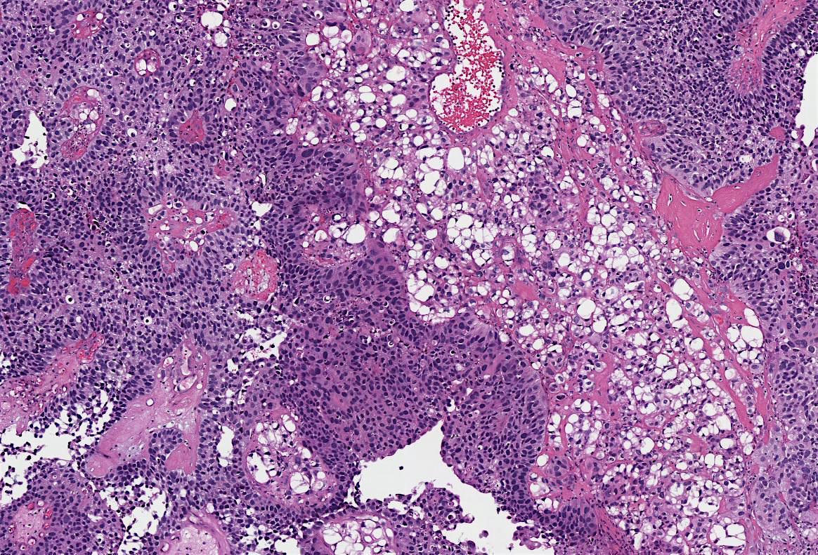
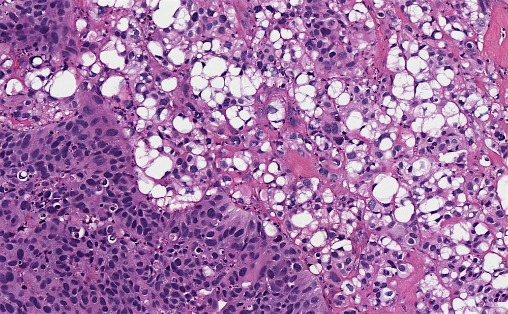
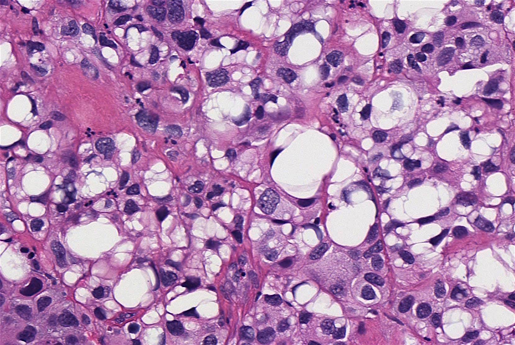
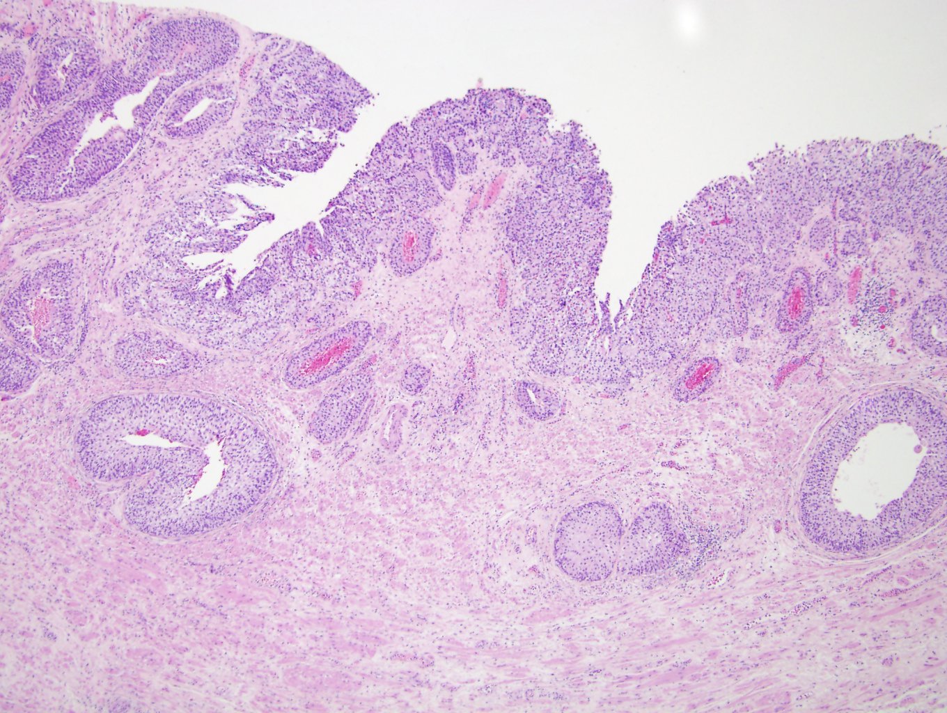
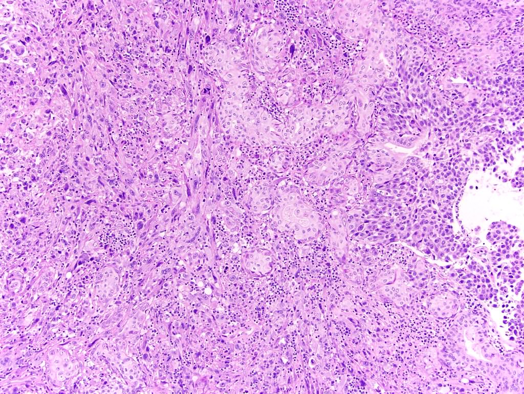
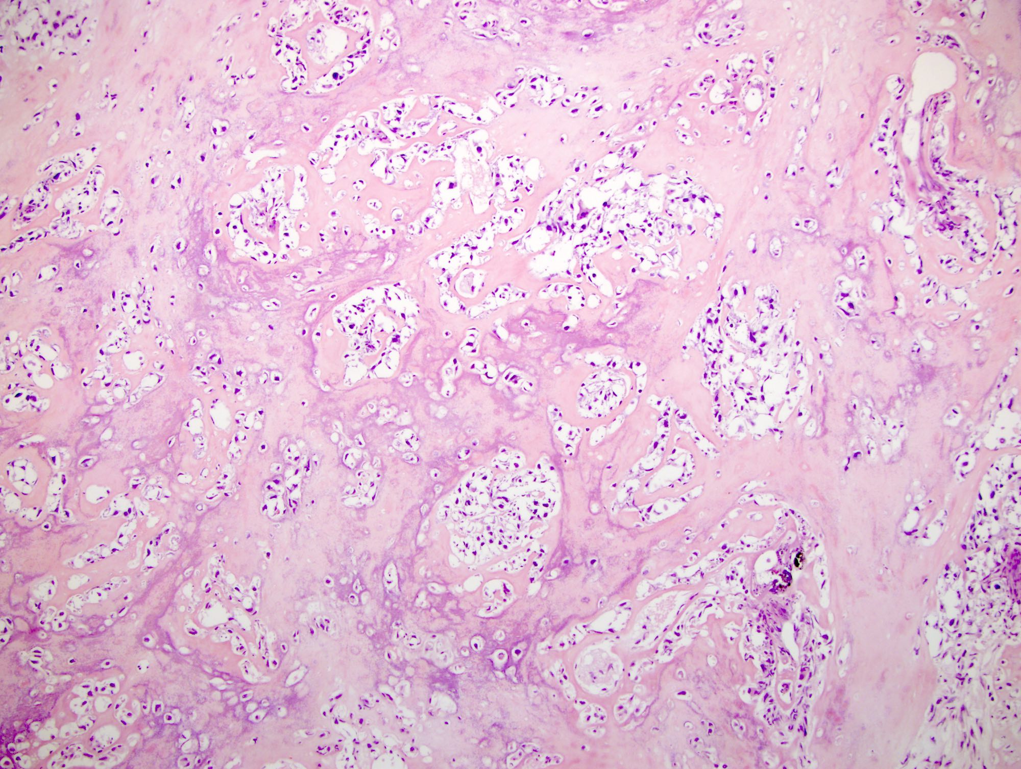
Contributed by Maria Tretiakova, M.D., Ph.D.
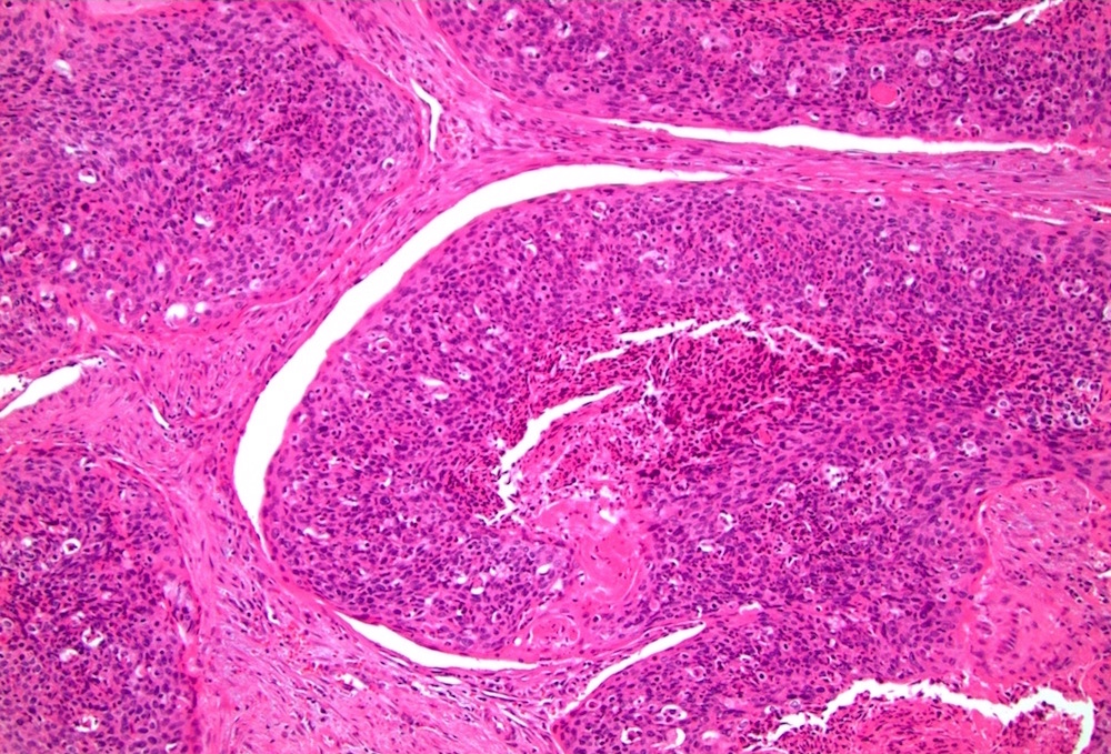
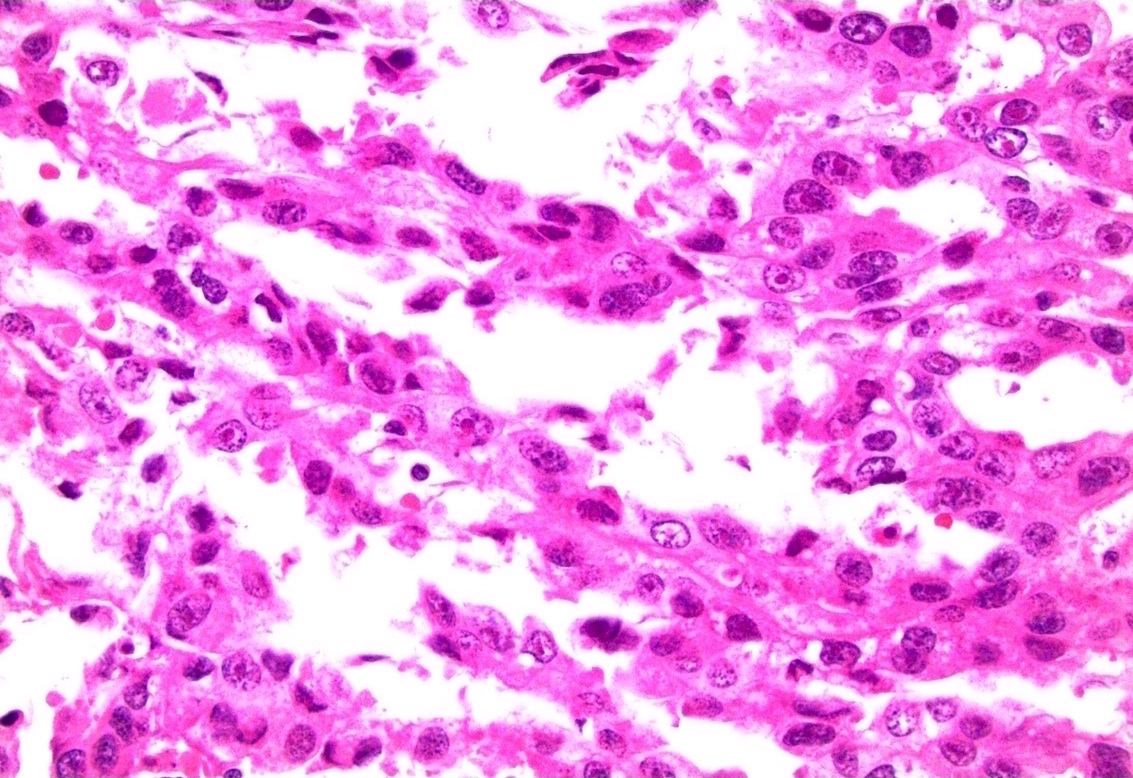
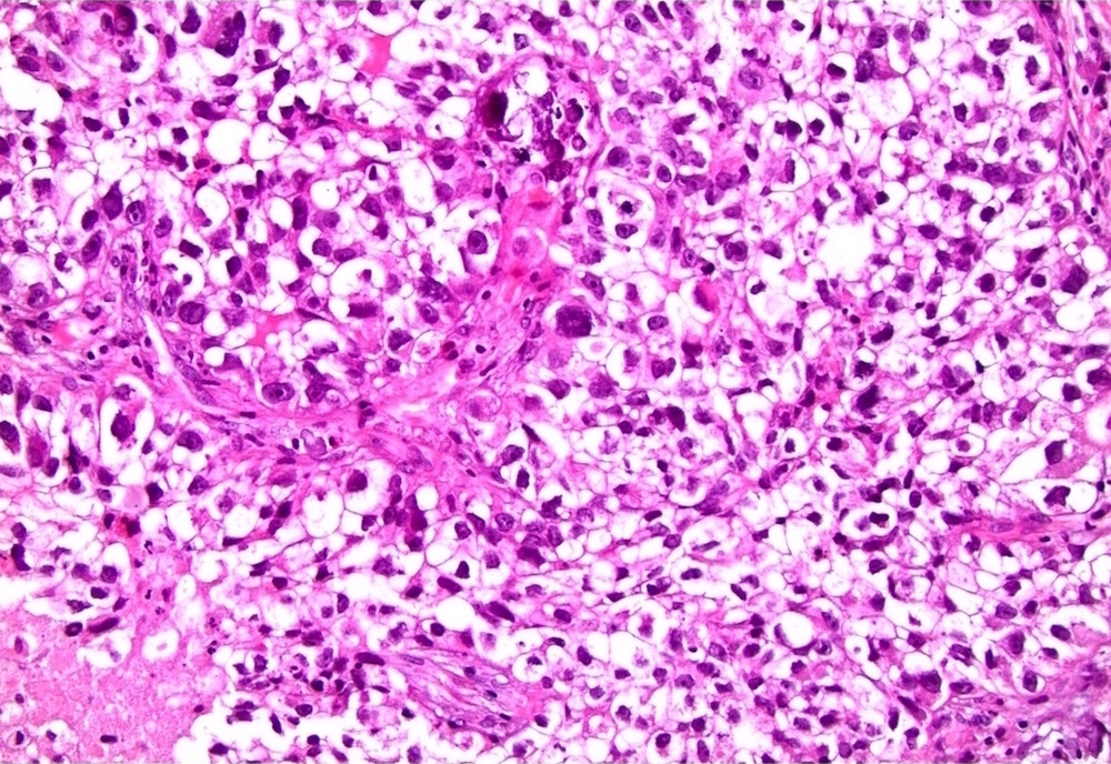
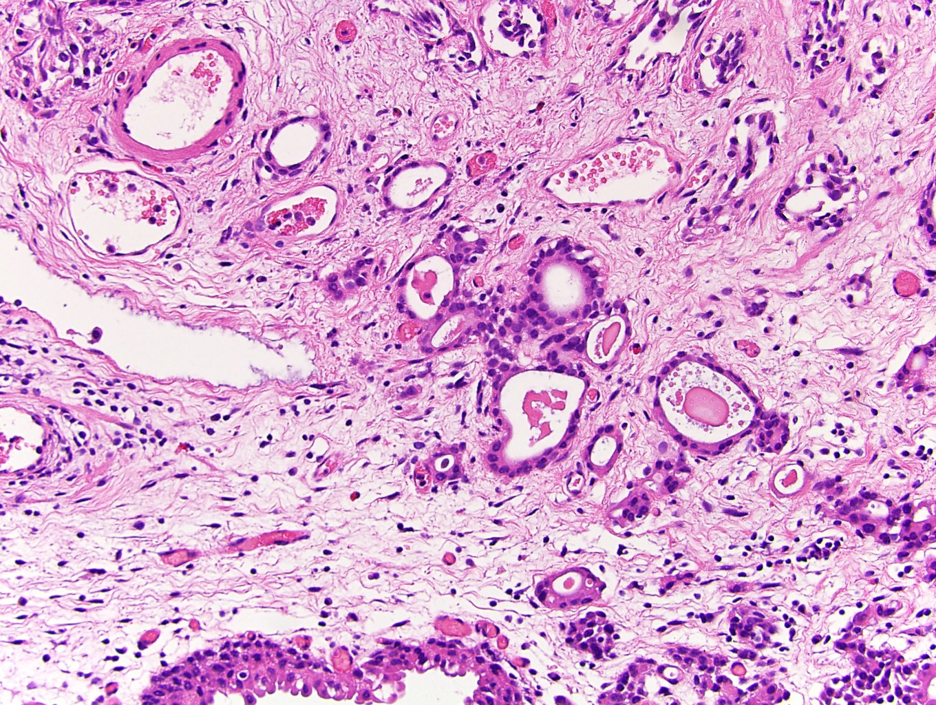
Urothelial carcinoma
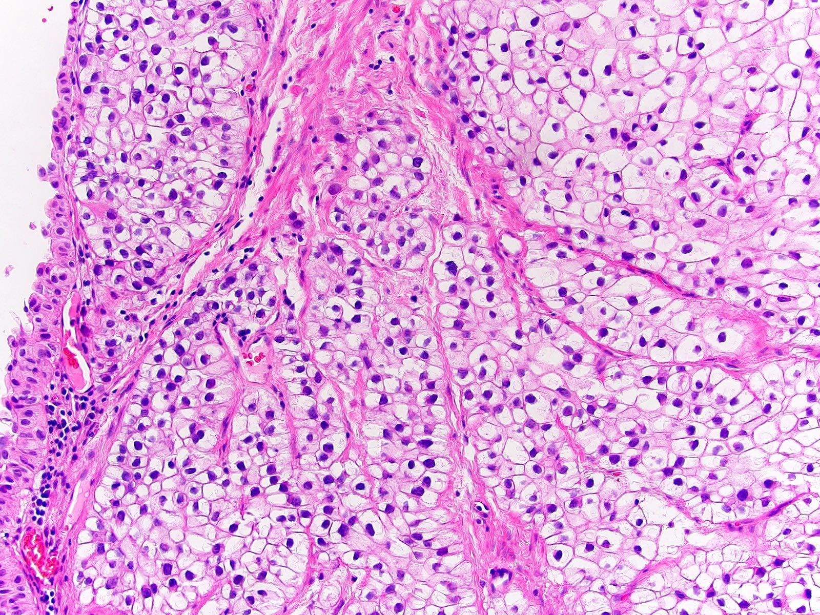
Clear cell (glycogen rich)
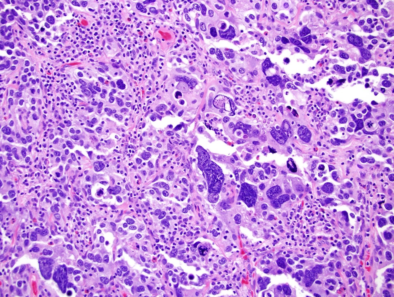
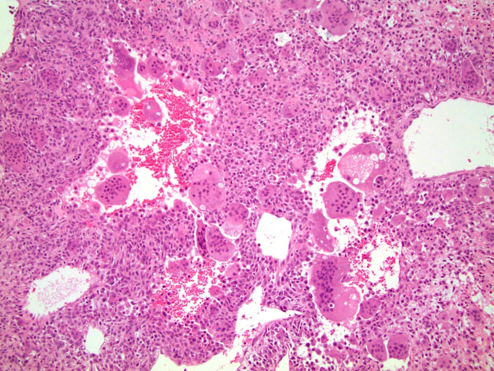
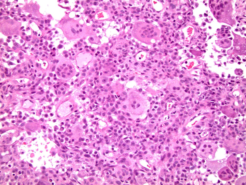
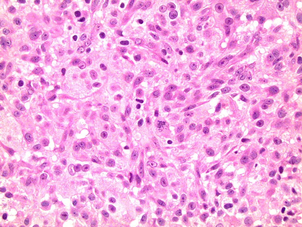
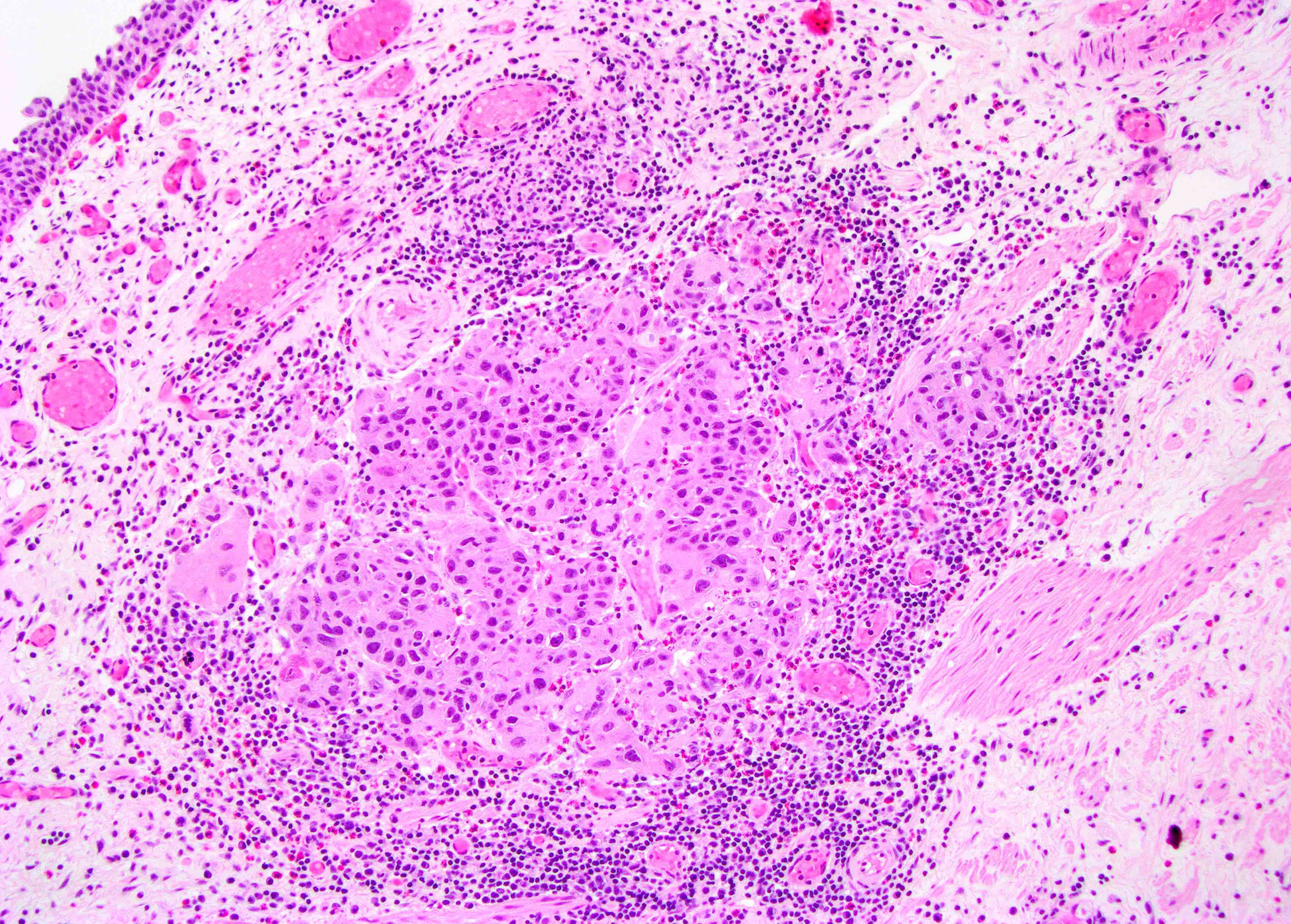
Giant cell
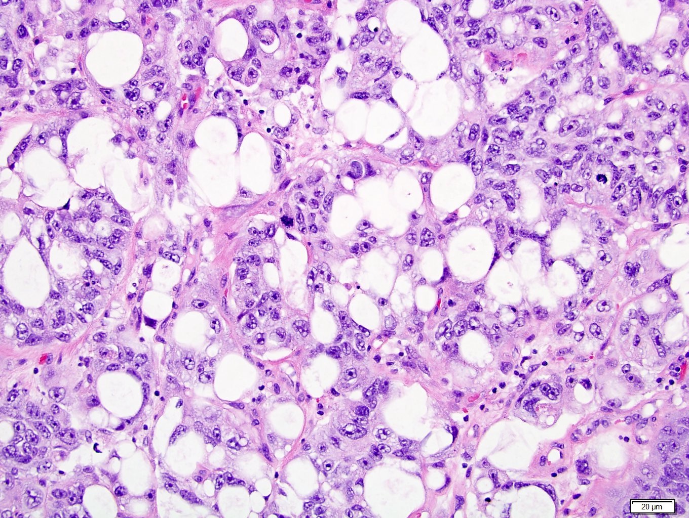
Lipid rich
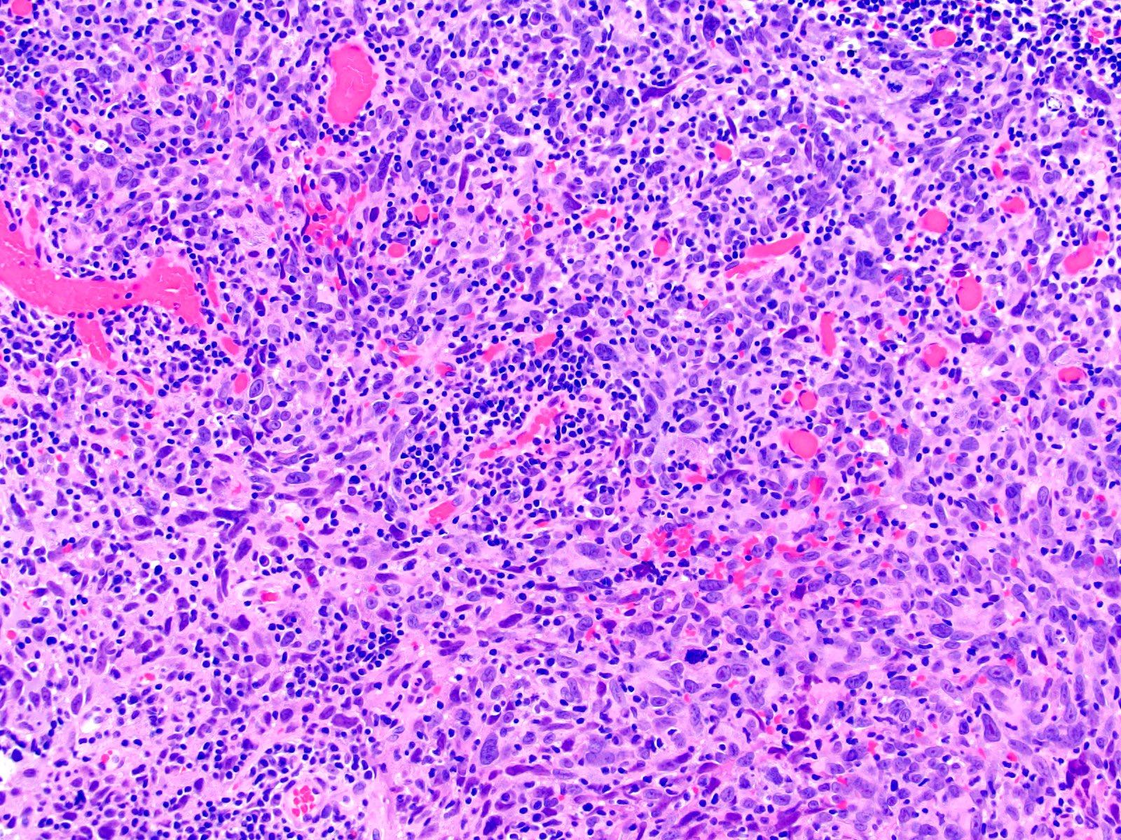
Lymphoepithelioma-like
Microcystic / tubular
Poorly differentiated with osteoclast-like
giant cells

Trophoblastic differentiation
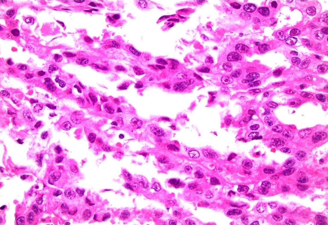
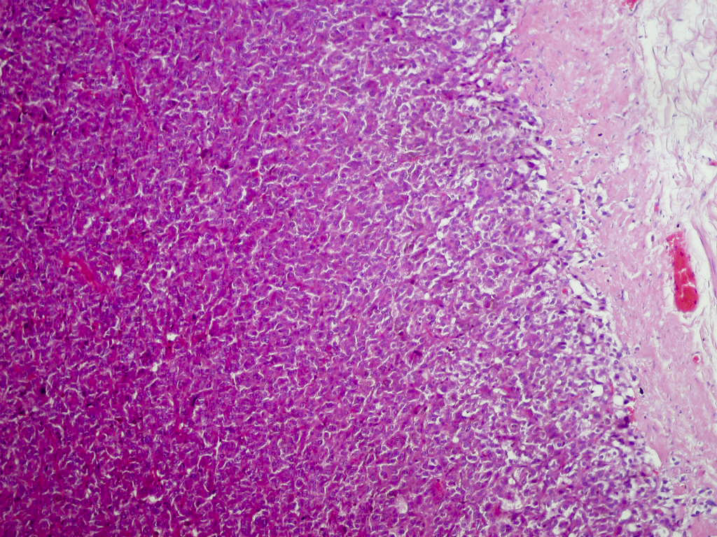
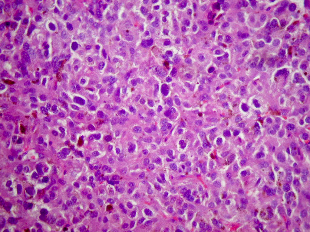
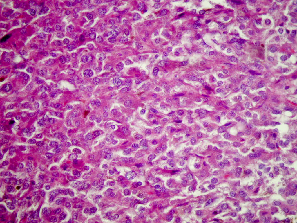
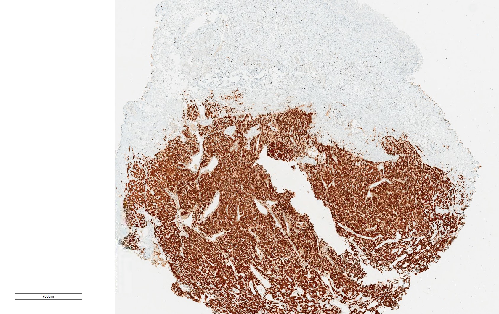
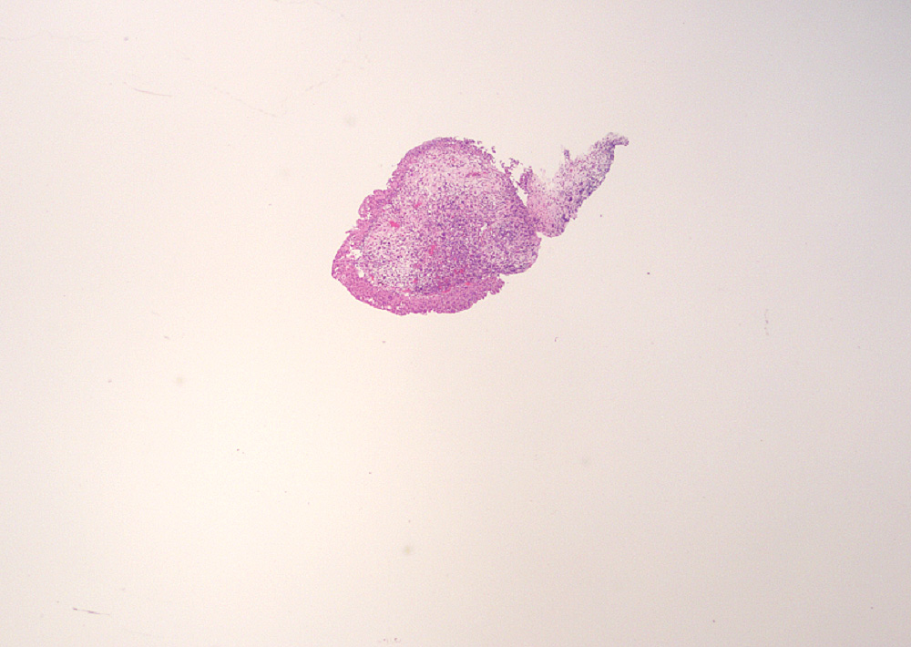
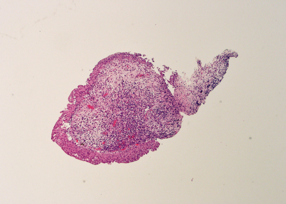
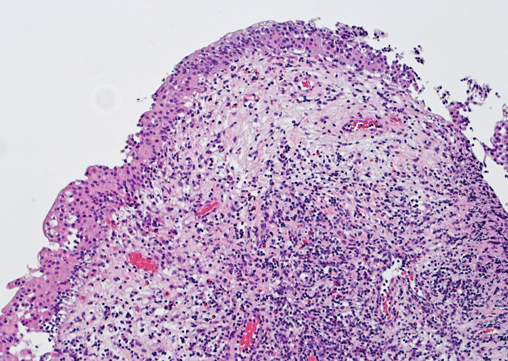
Contributed by Nicole K. Andeen, M.D. and Maria Tretiakova, M.D., Ph.D.
Various images
Images hosted on other servers:

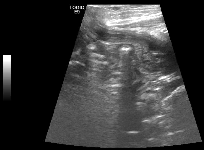
T1 precontrast demonstrates isoechoic mass

Ultrasound shows mass-like hyperechoic area
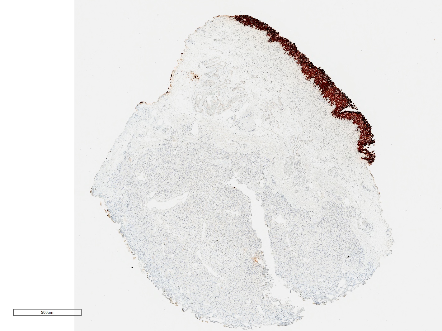
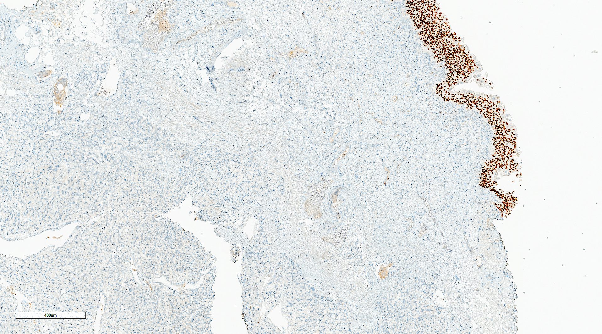
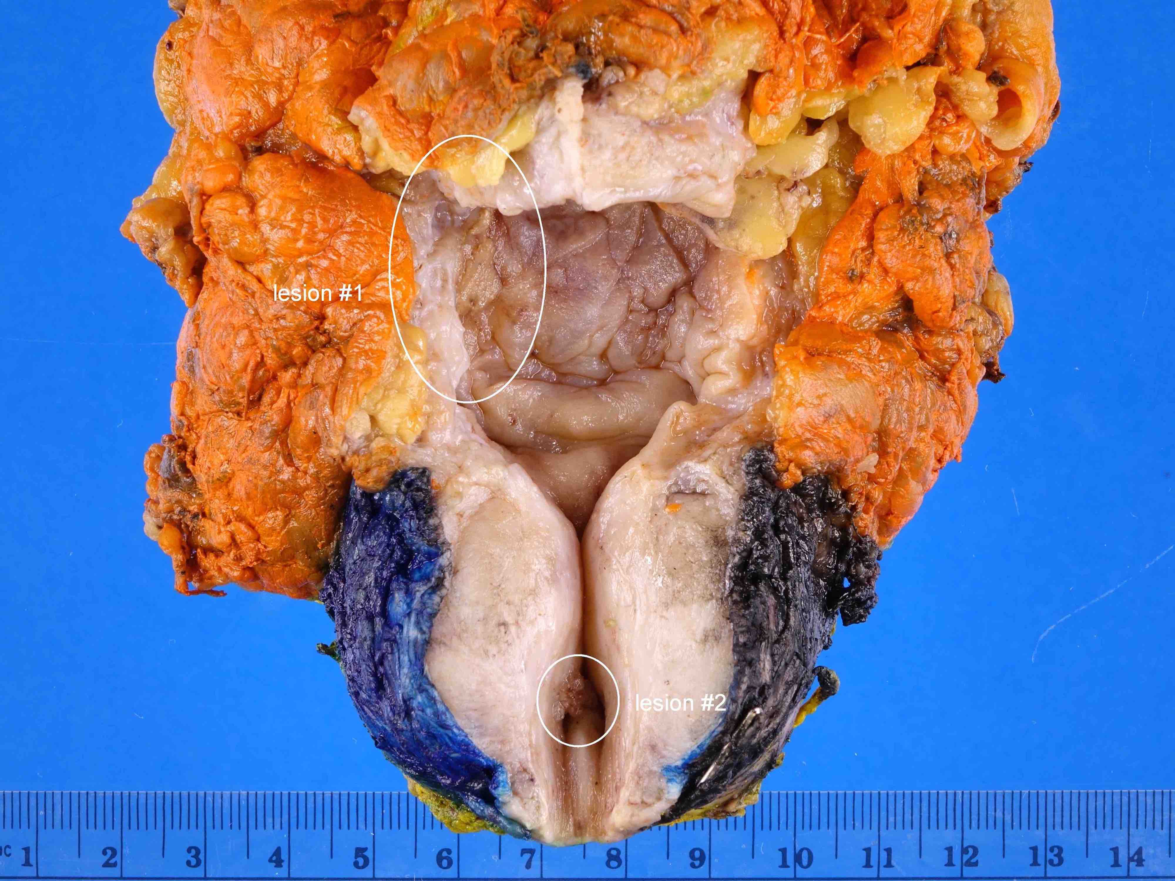
Contributed by Huihui Ye, M.D., M.S.
Dome tumor
Posterior wall tumor
Contributed by Huihui Ye, M.D., M.S.
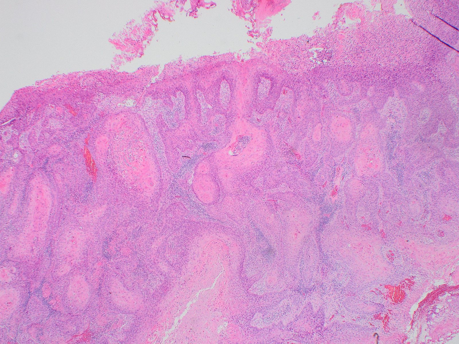
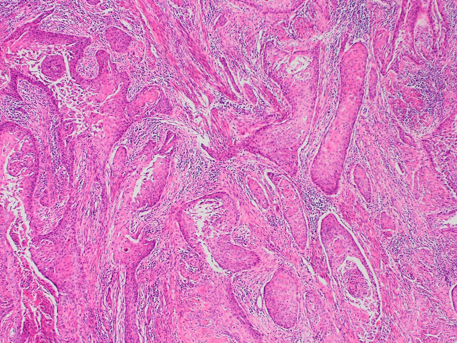
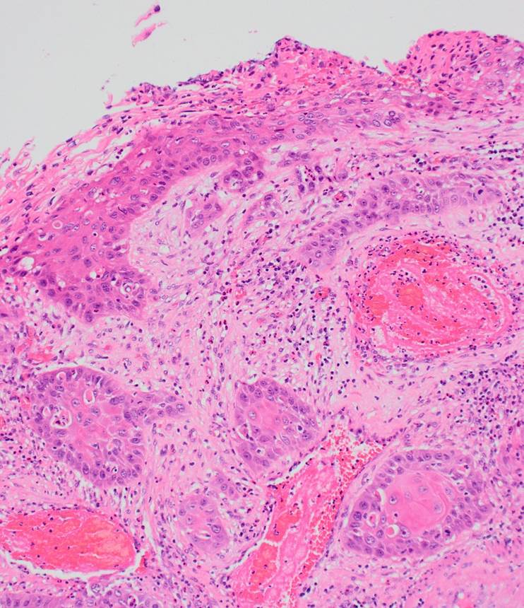
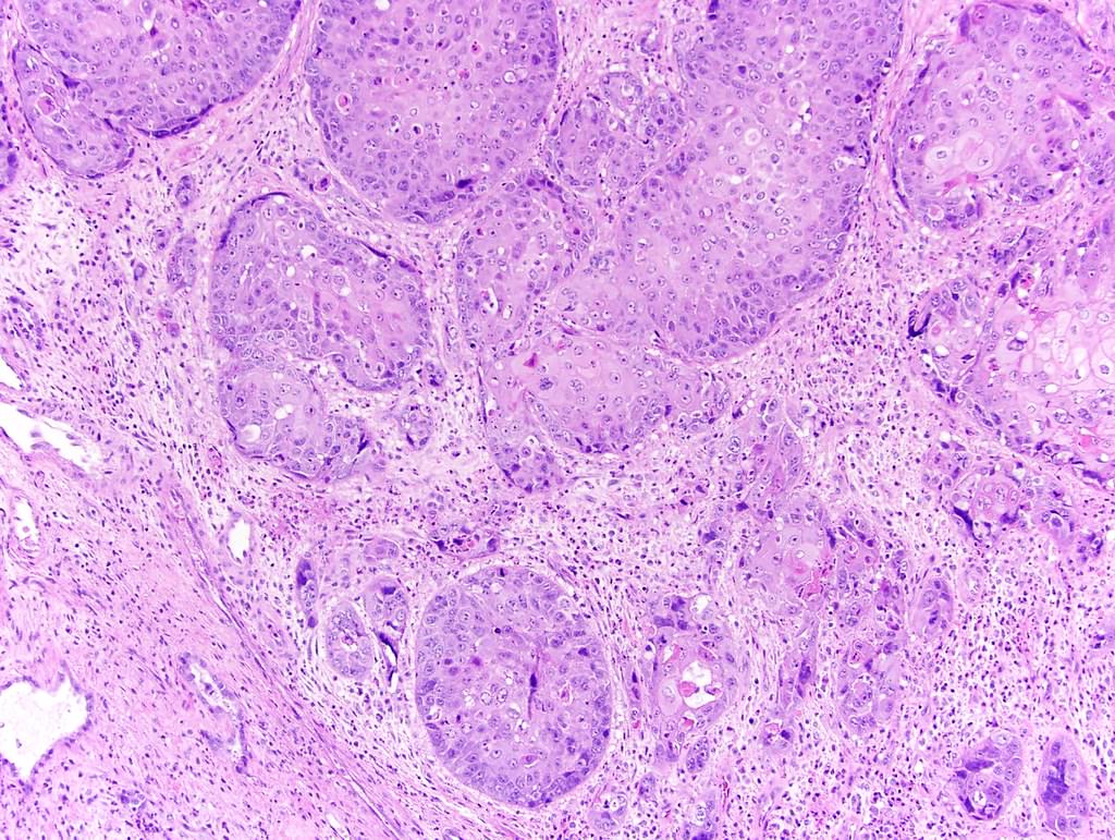
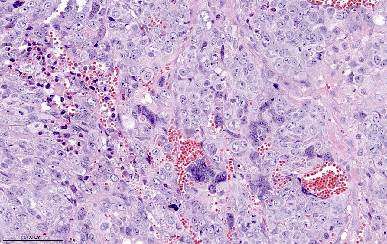
Basaloid features
Clear cell features
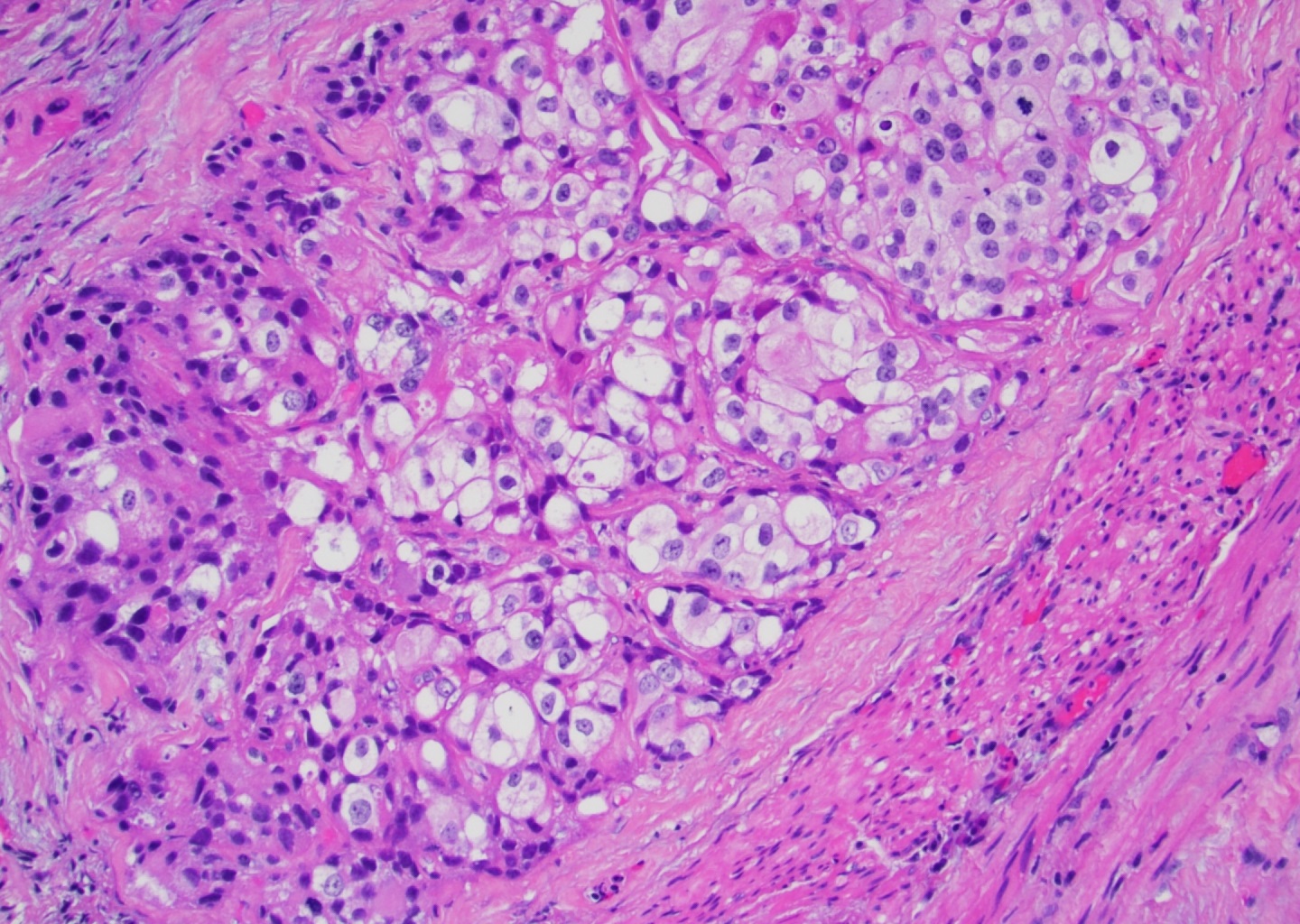
Nests of clear cells
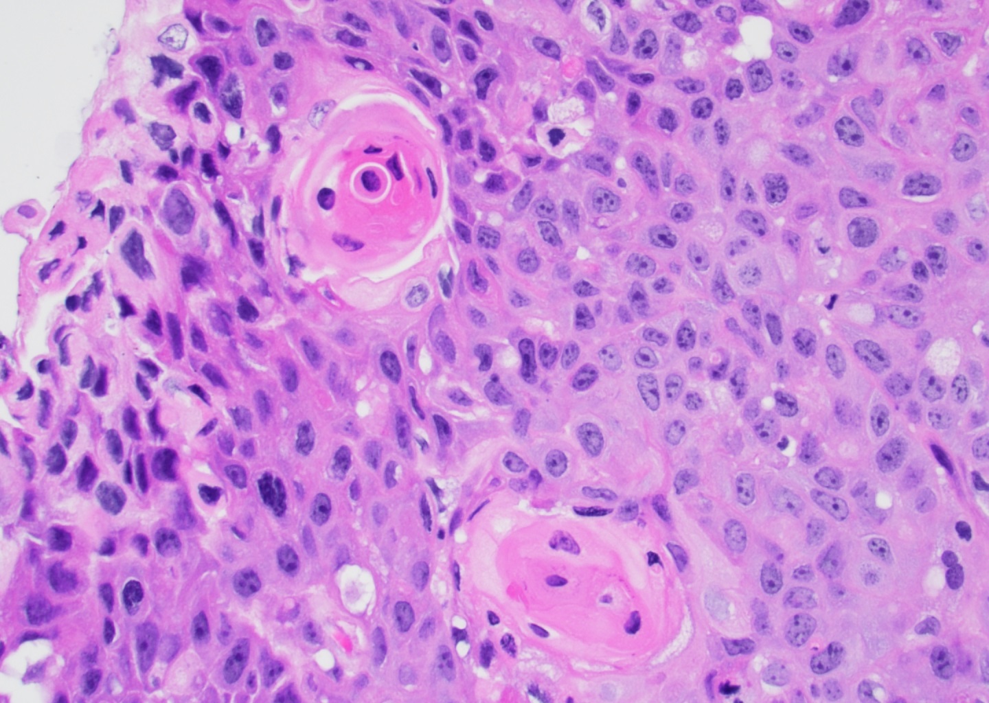
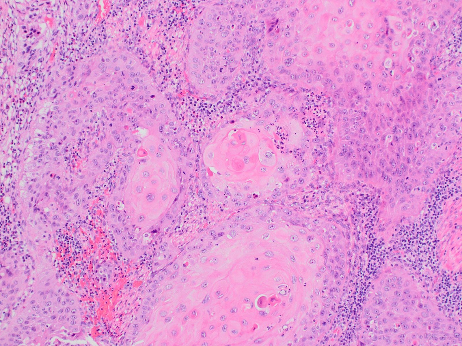
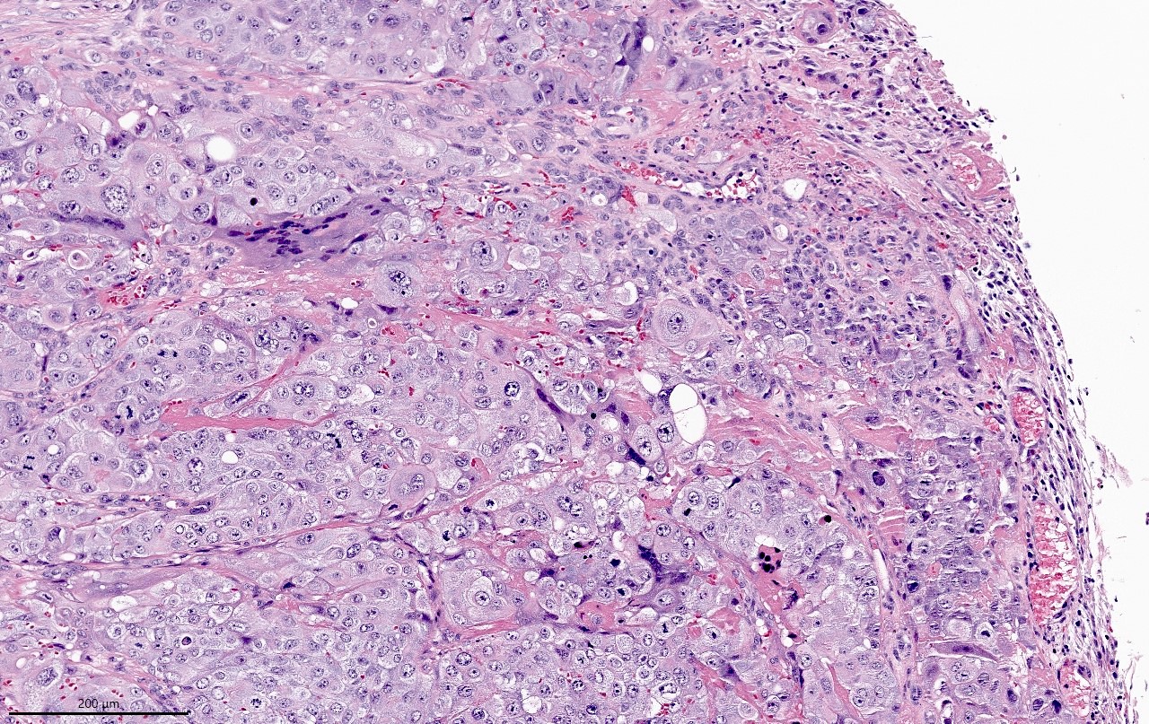
Keratin pearls
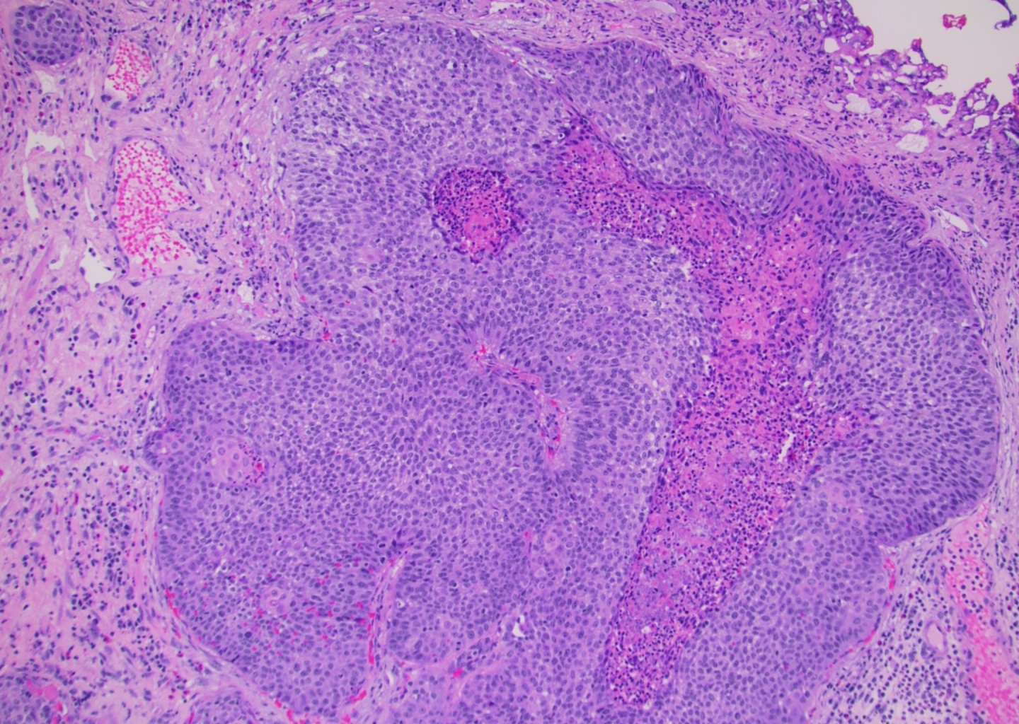
Basaloid with central necrosis
Surface keratinization
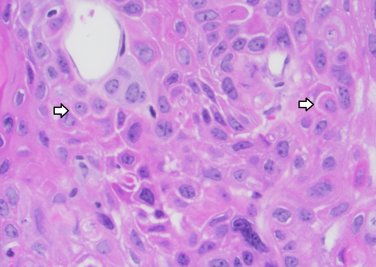
Intercellular bridges
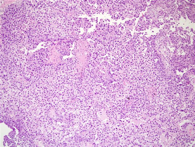
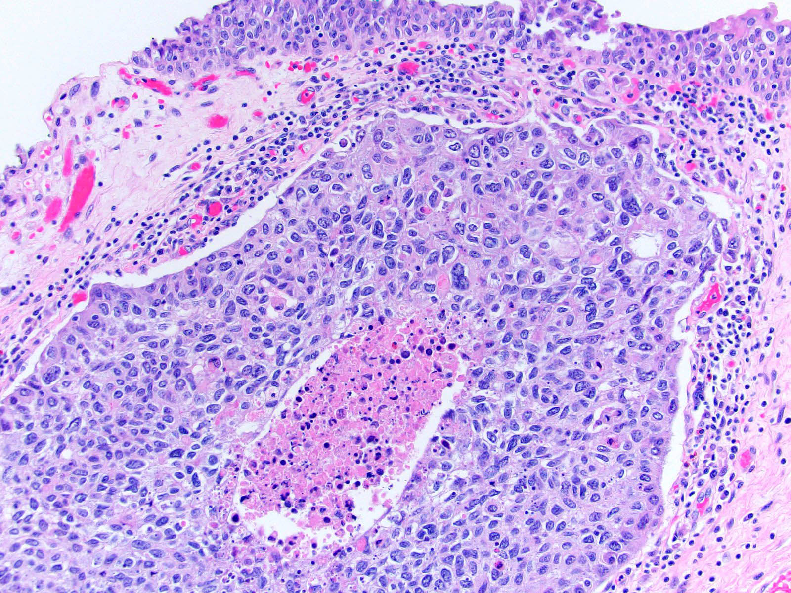
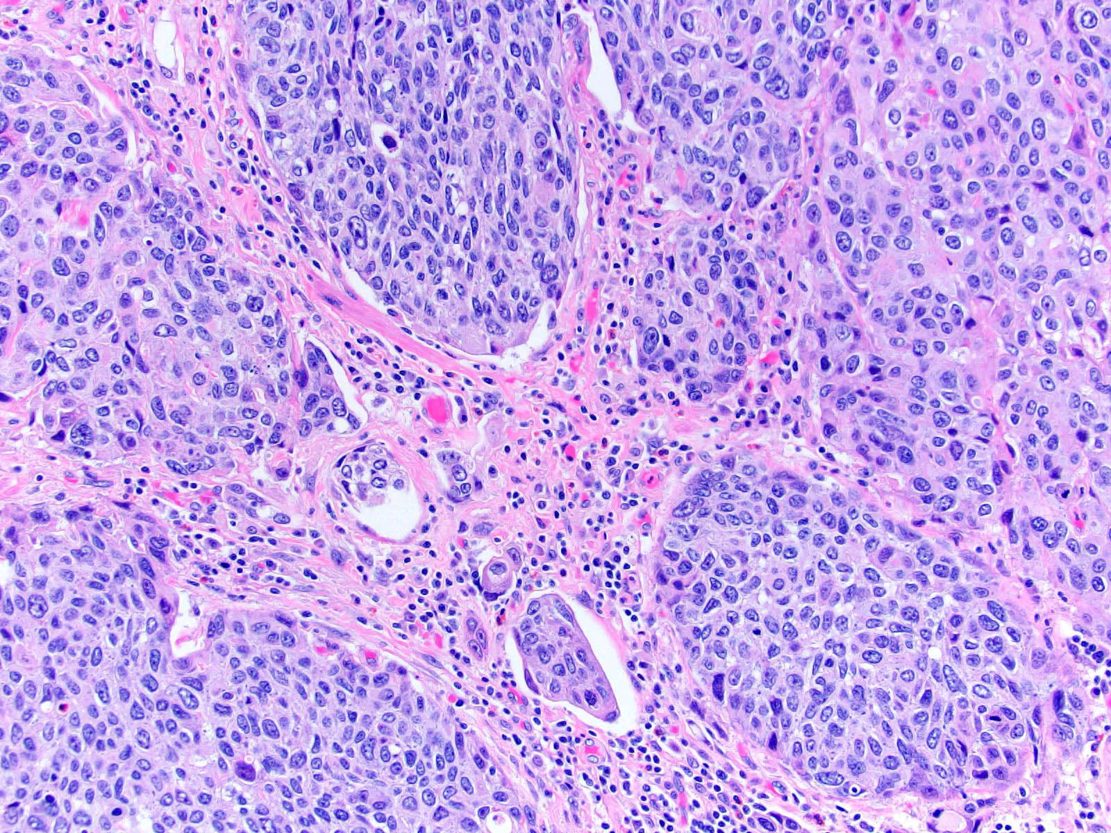
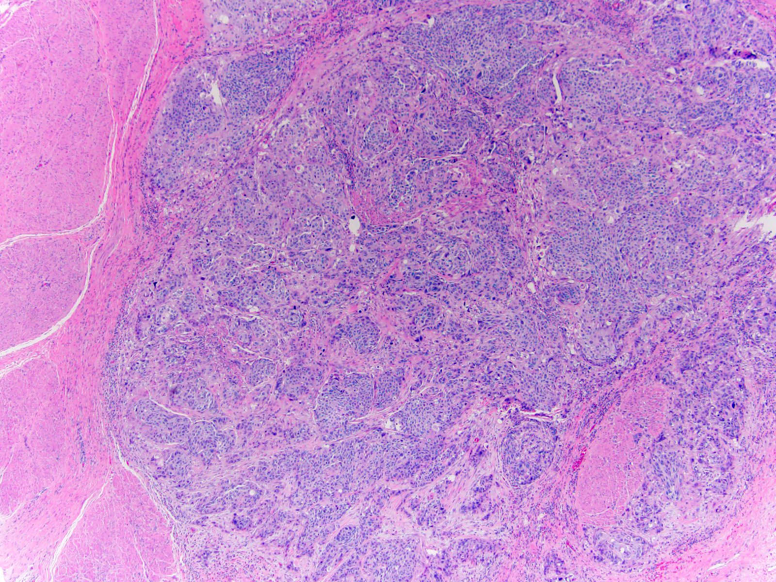
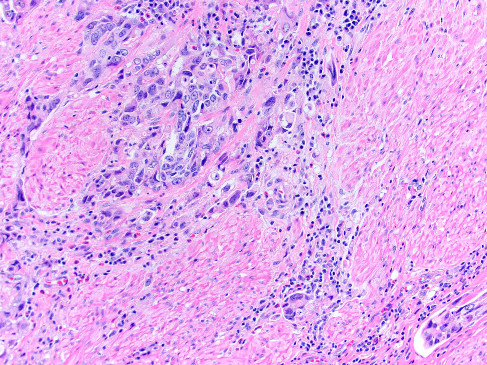
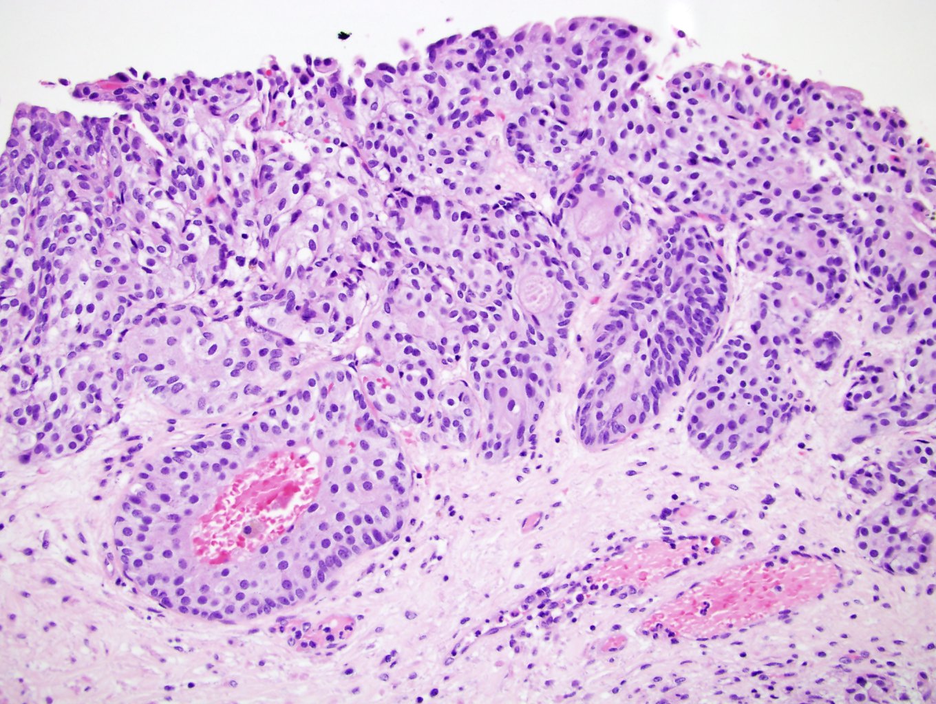
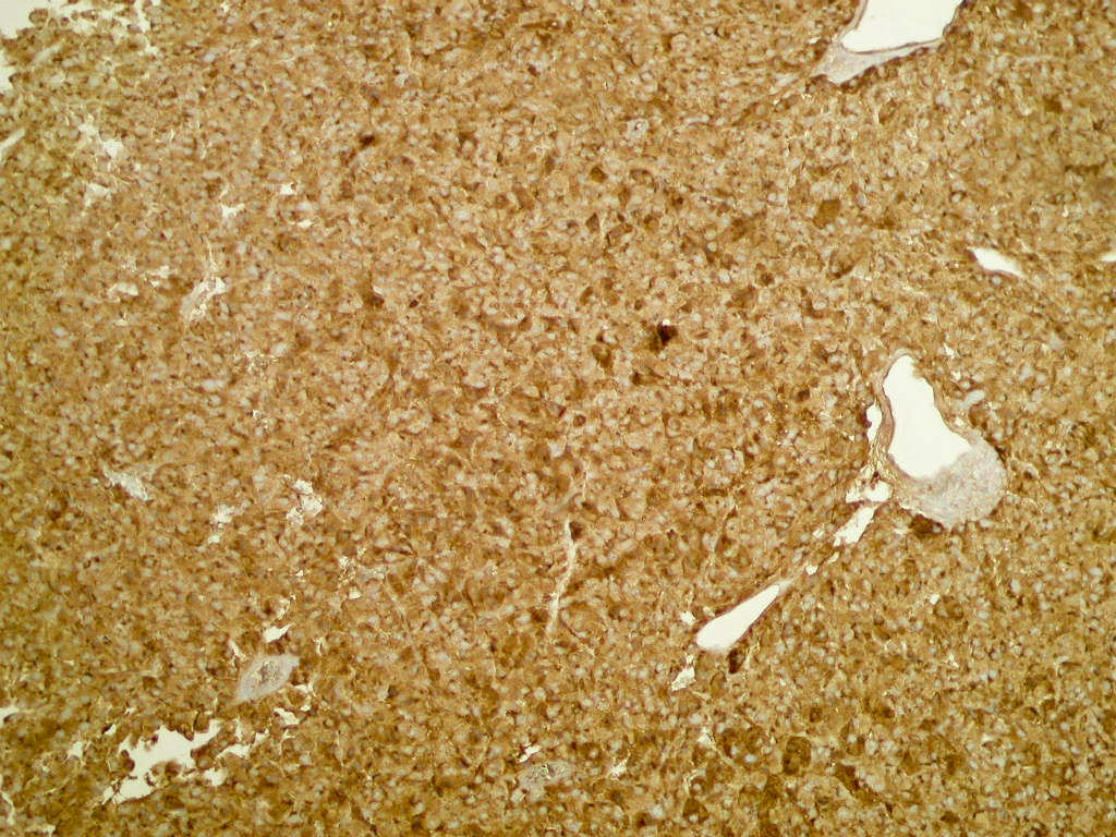
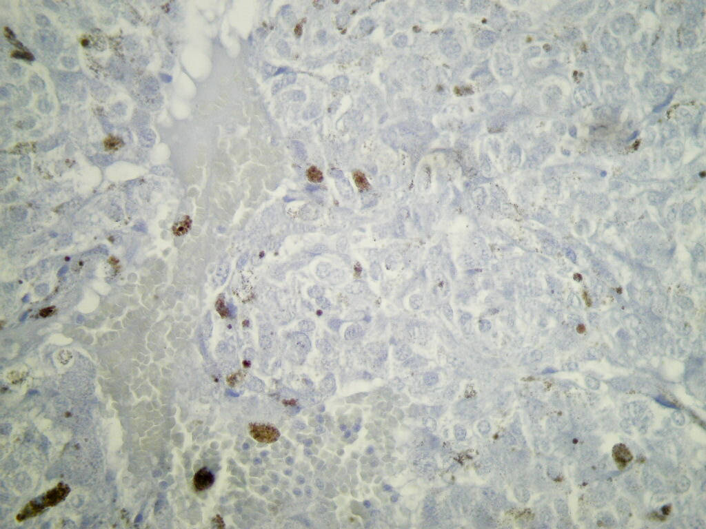
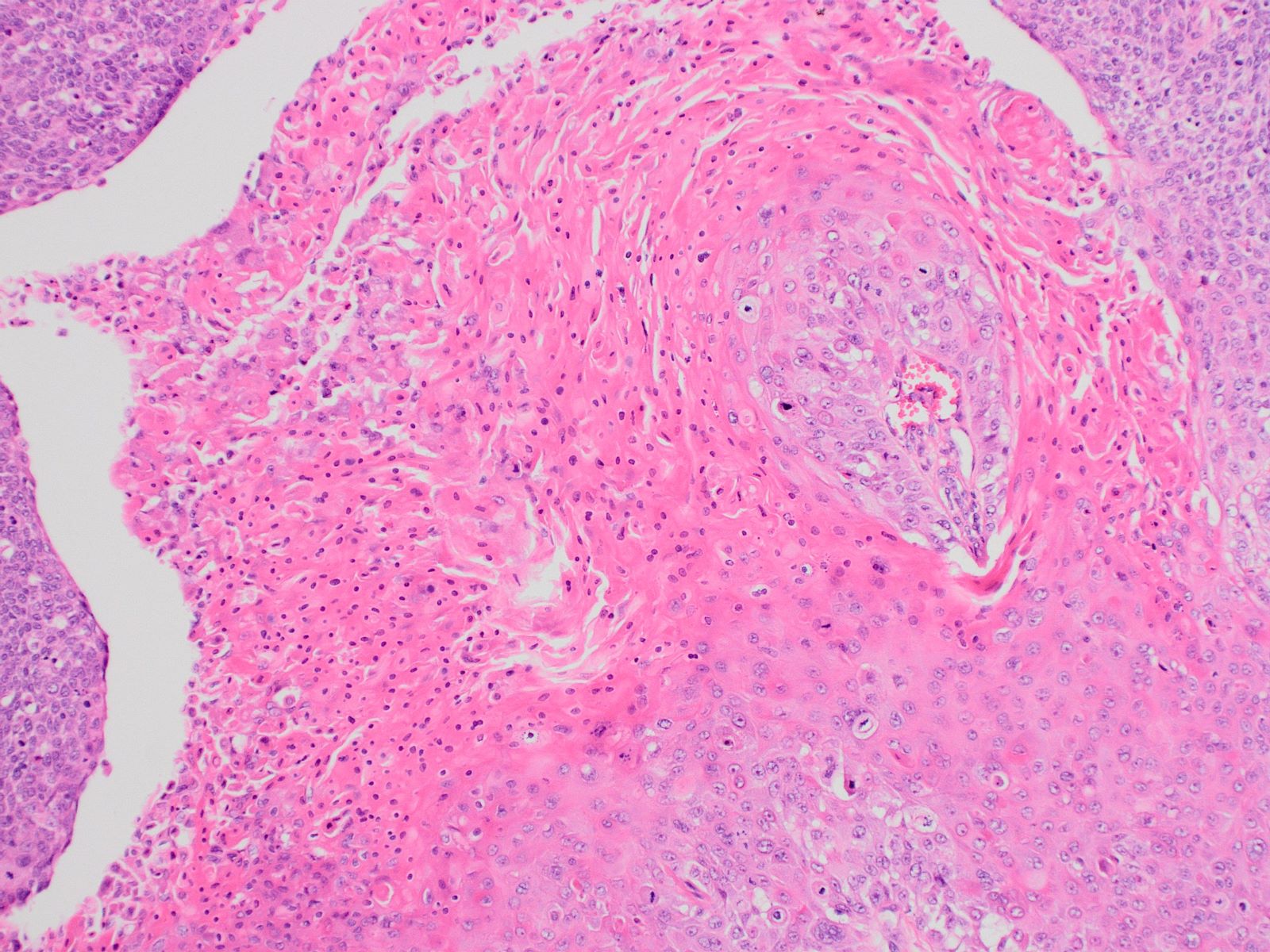
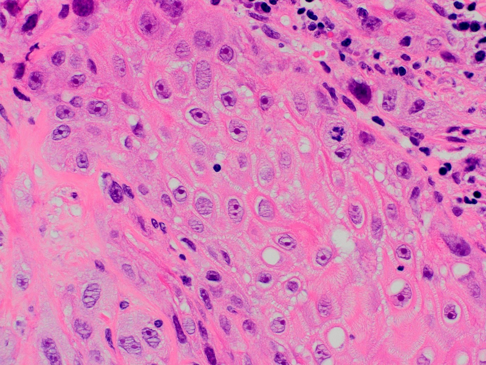
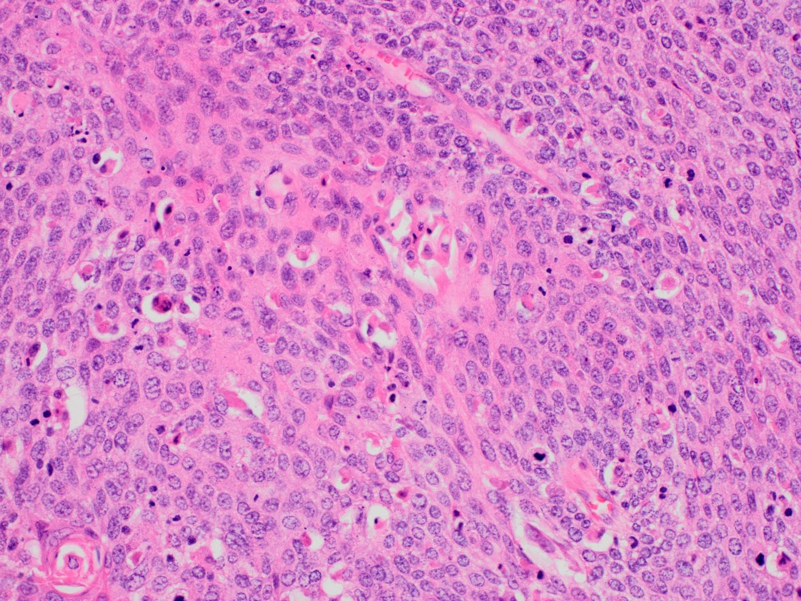
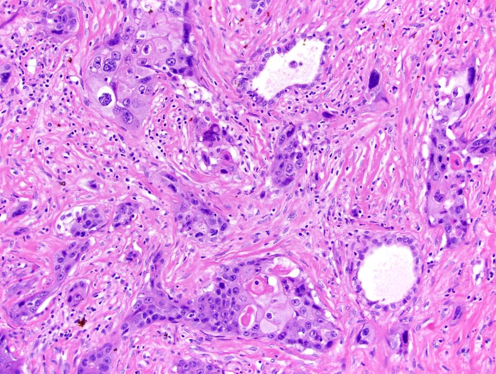
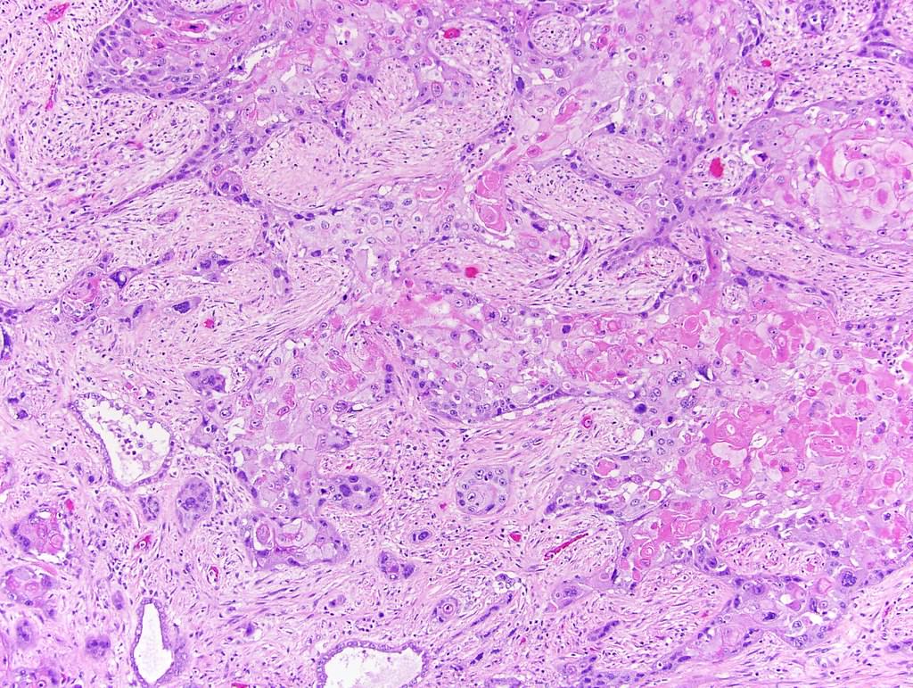
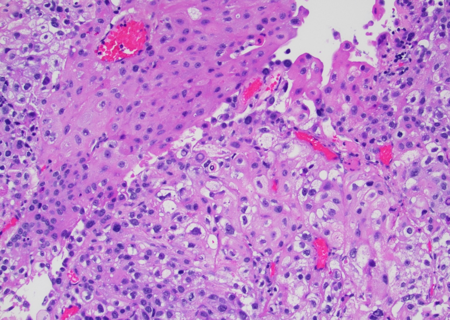
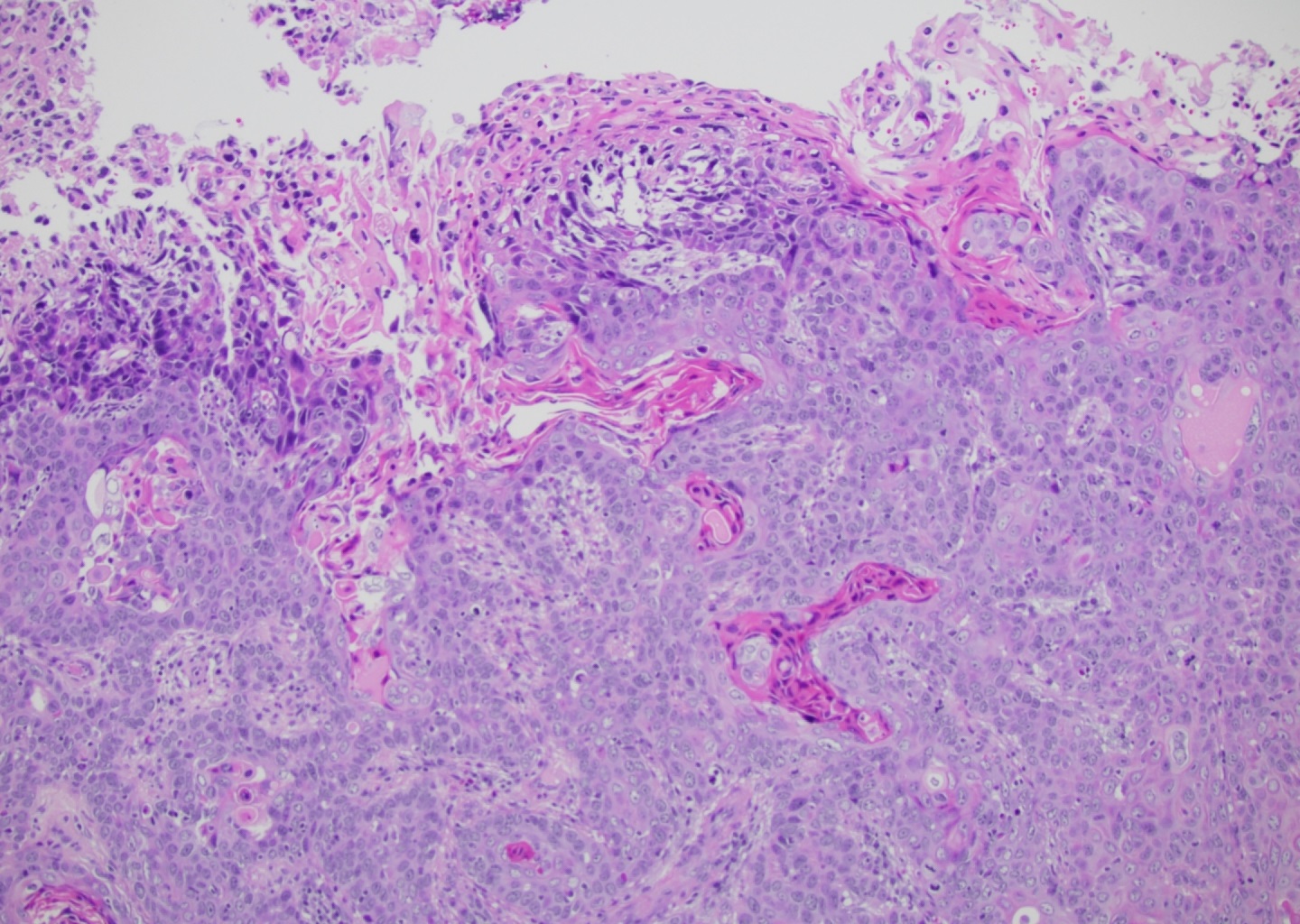
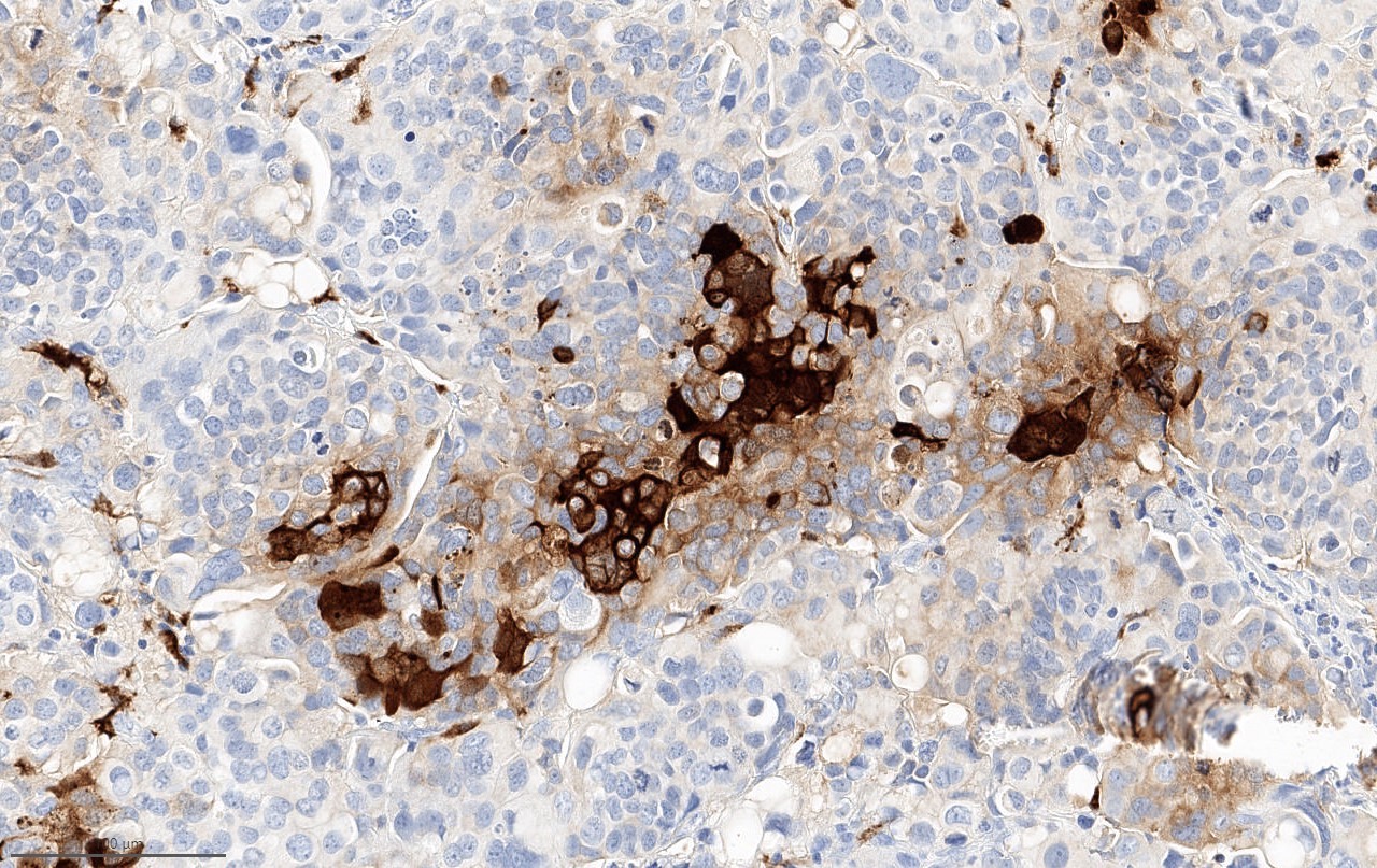
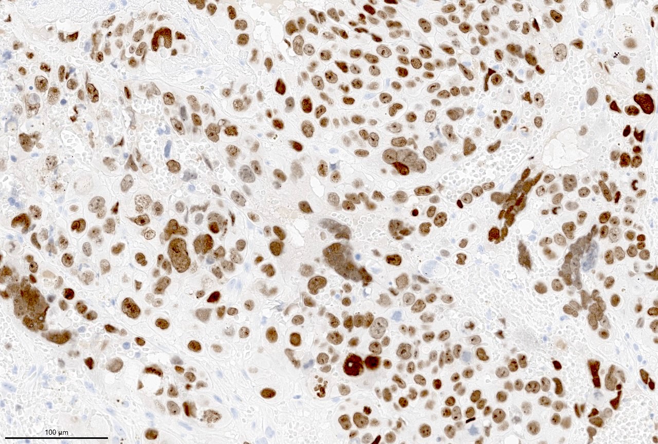
Contributed by Chunlai Zuo, M.D., M.S.
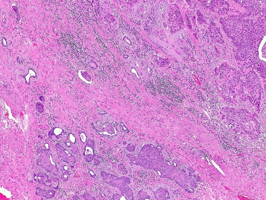
Squamous differentiation
Conventional urothelial carcinoma component
HGUC and necrosis
Images hosted on other servers:

High grade with squamous differentiation
Contributed by Anuradha Gopalan, M.D., Victor Reuter, M.D. and AFIP images
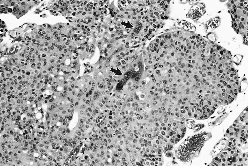
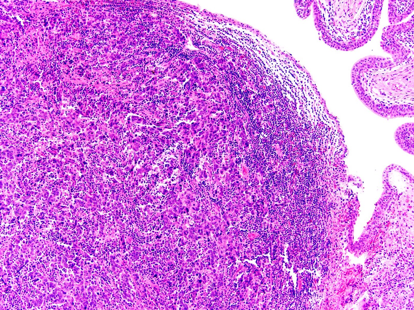
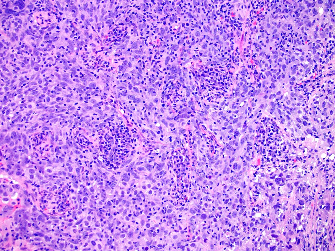
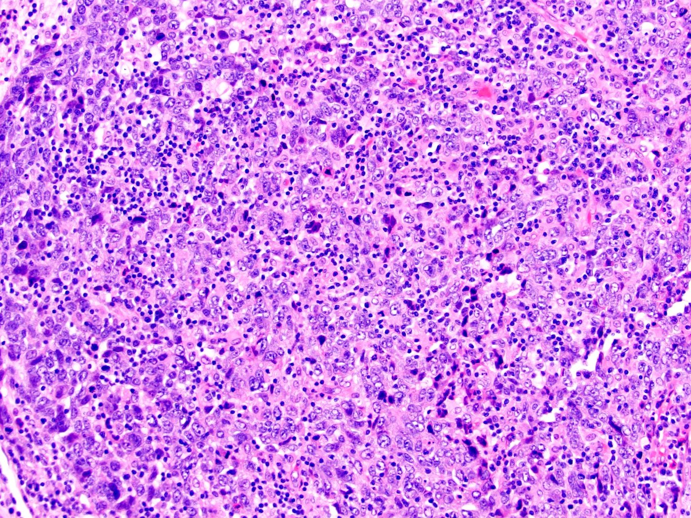
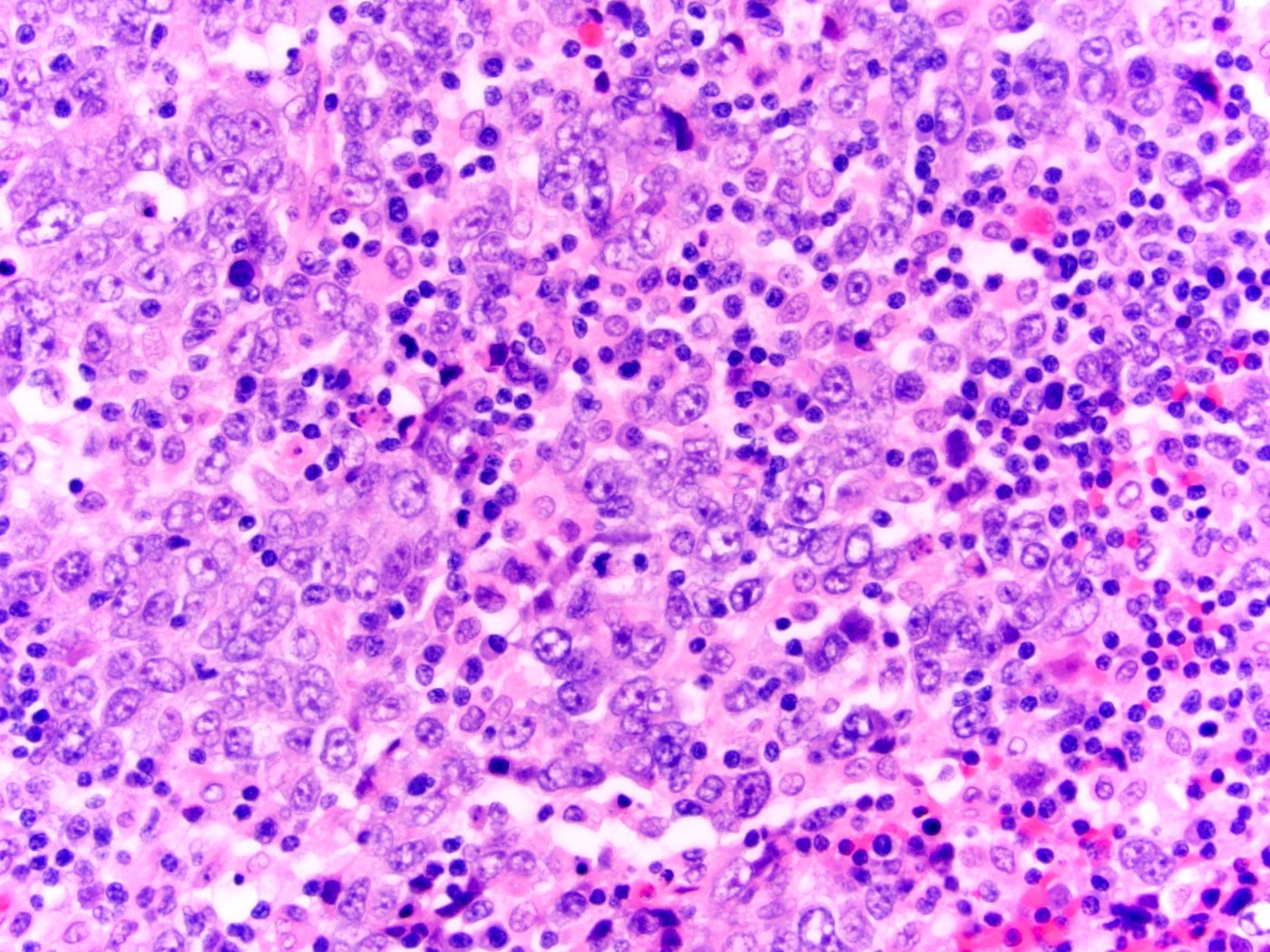
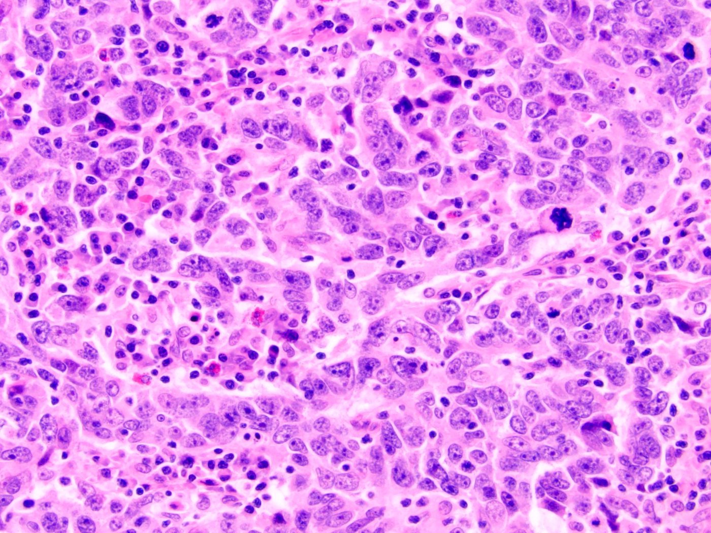
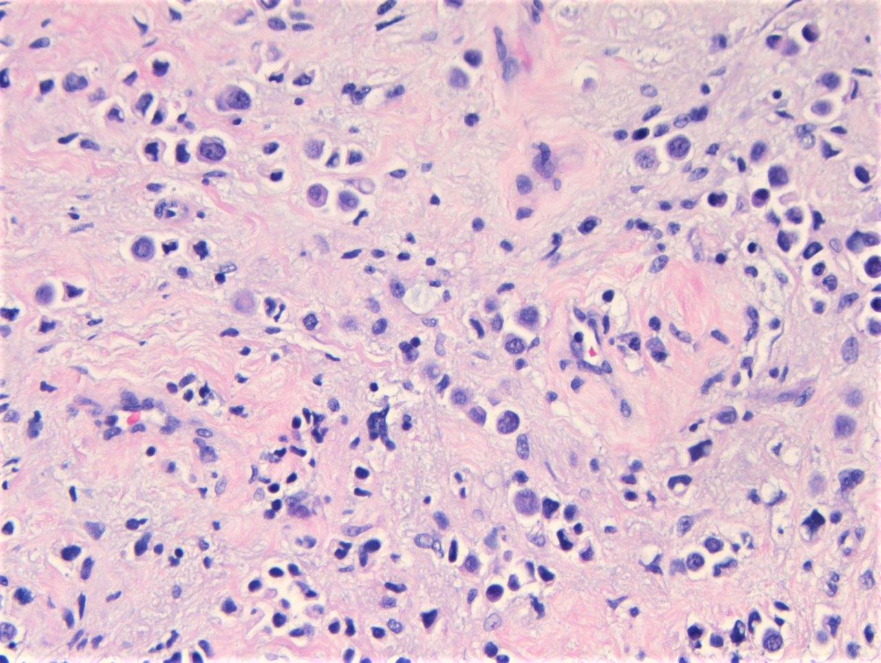
Multinucleated giant cells
hCG
GATA3
Amin: 2022

Cheng: 2019
Eble: 2022
Epstein: 2016
Haber: 2010
Hansel: 2018
IARC: 2022
Jim Zhai: 2015
Lopez-Beltran: 2016
Raspollini: 2019

VandenBussche: 2019
VandenBussche: 2022

Wobker: 2021
Wojcik: 2022

Yang: 2020
Zhou: 2022
Zhou: 2022
Find related Pathology books: GU/adrenal, renal, body fluid/urinalysis, cytopathology, gynecologic
