

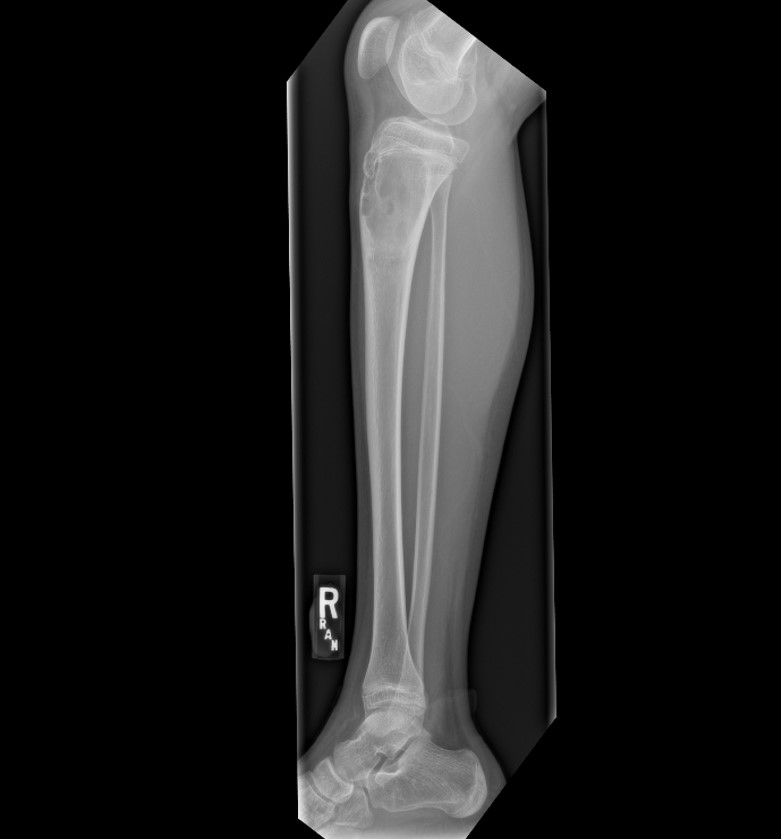
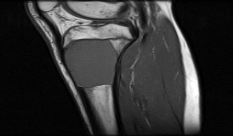
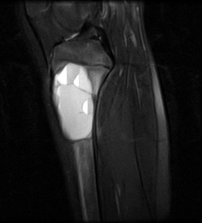
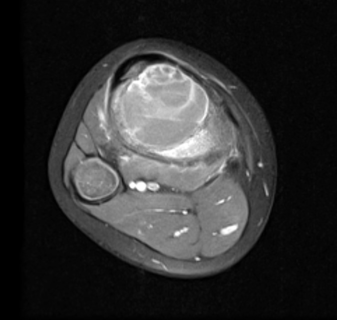
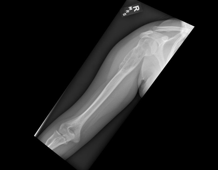
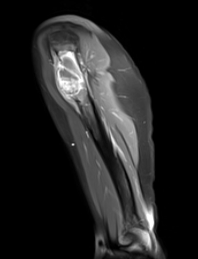
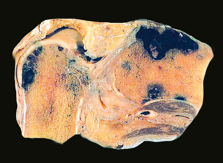
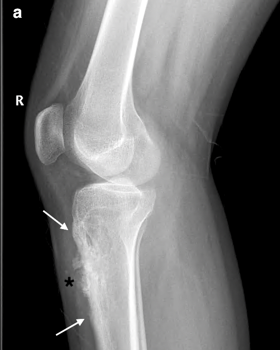
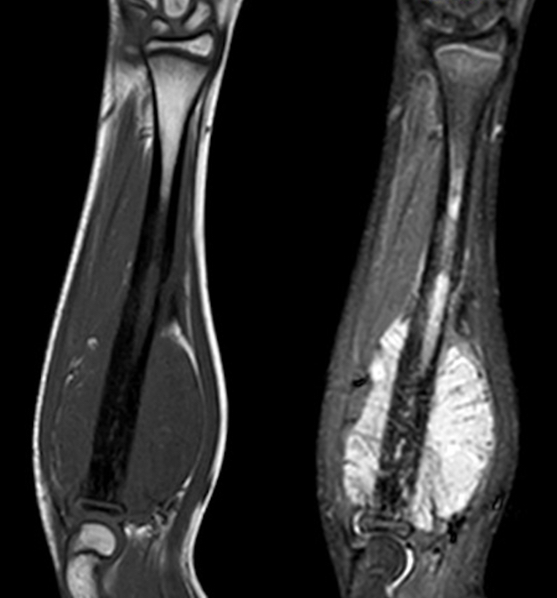
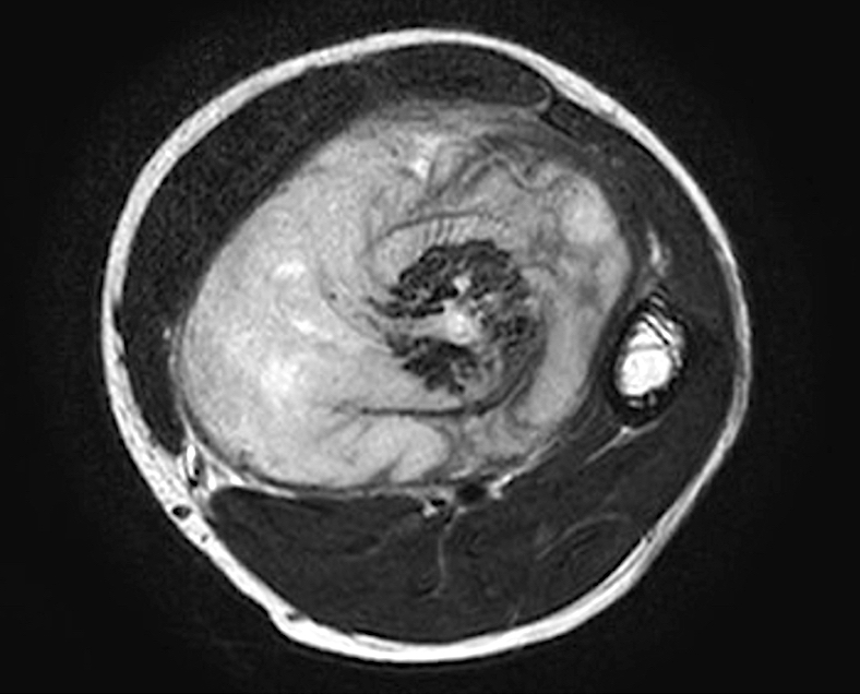
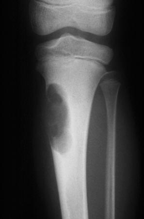
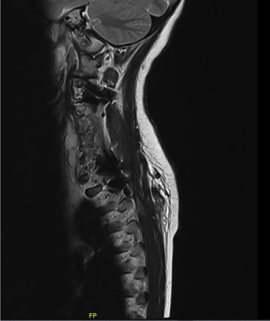
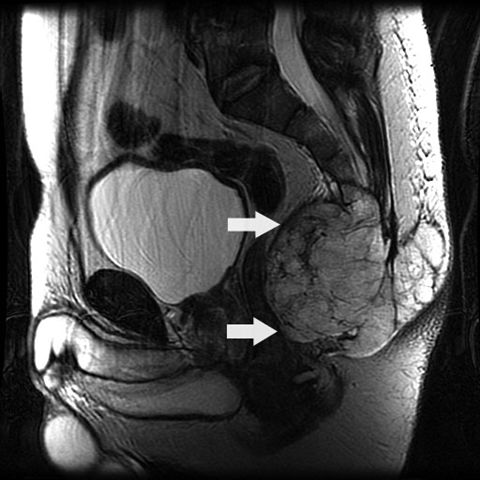
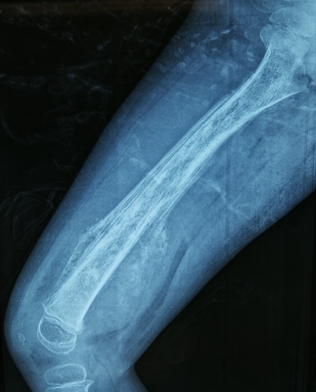
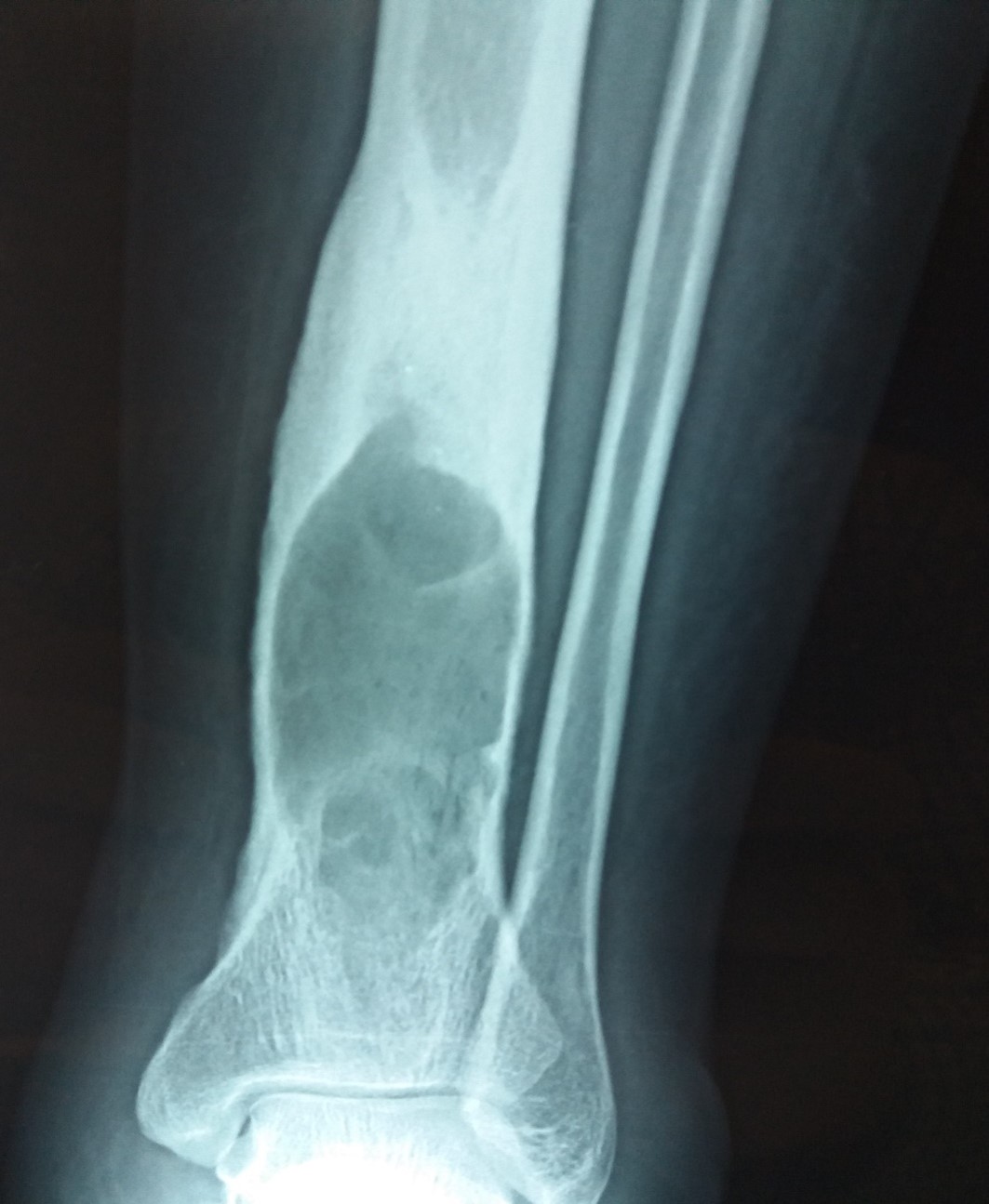
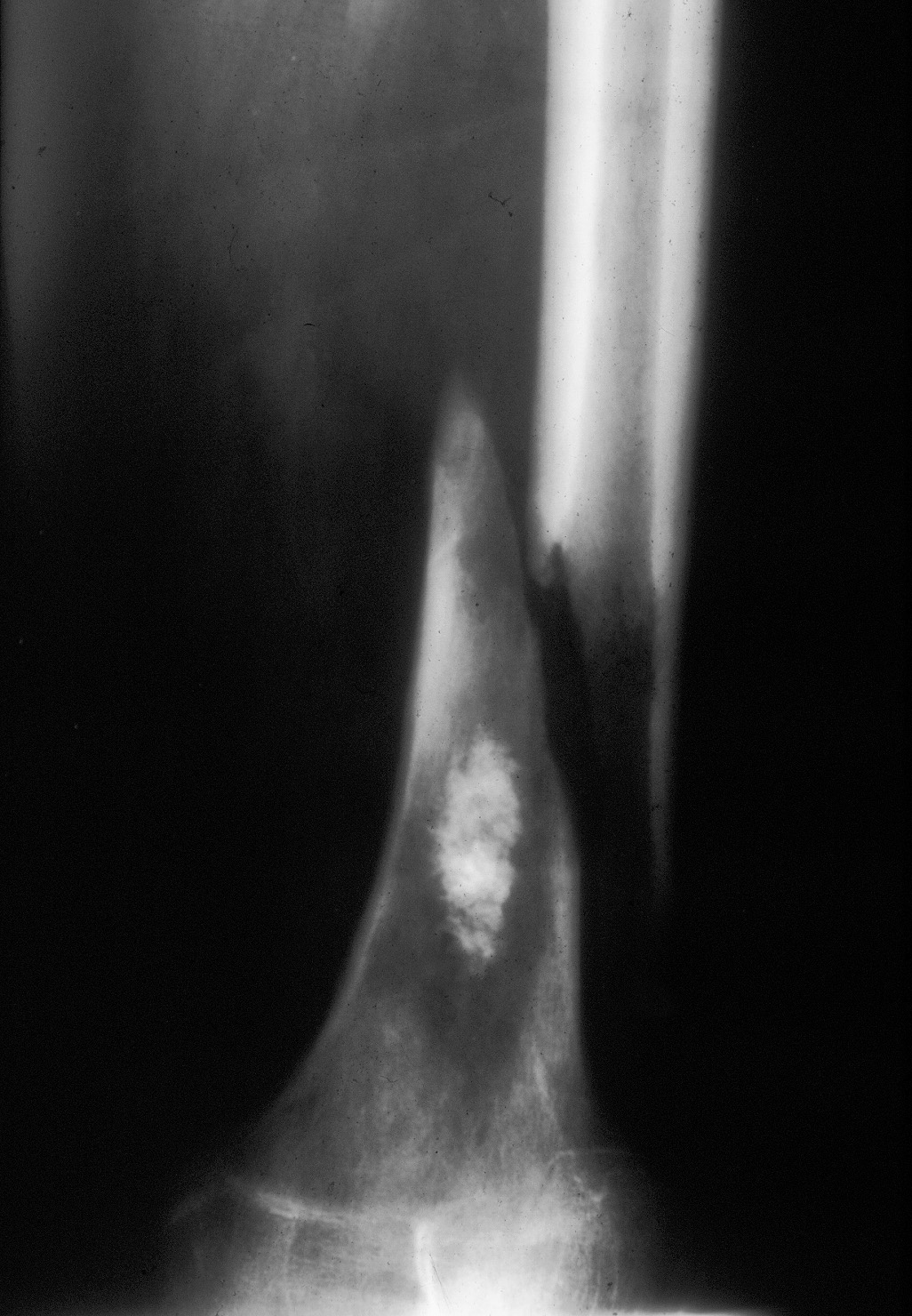
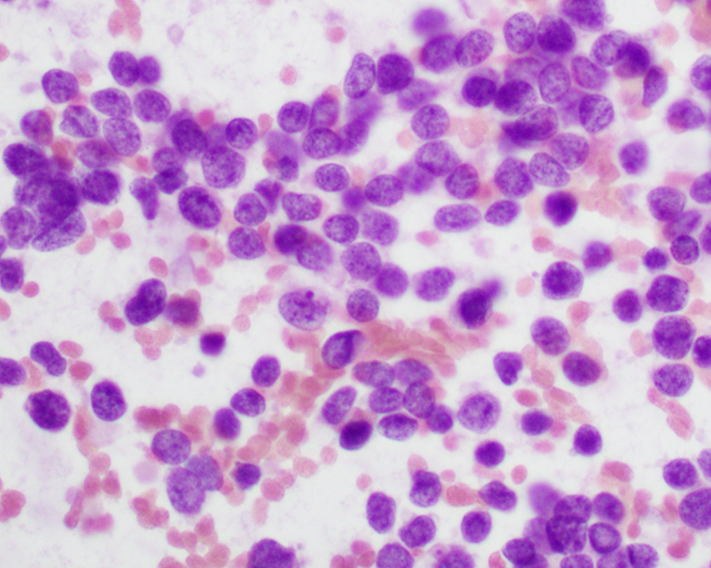
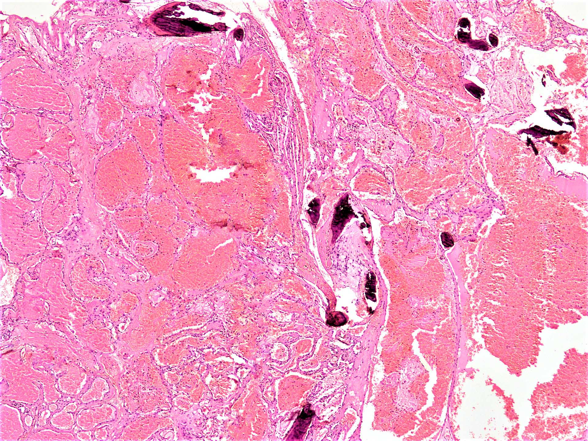
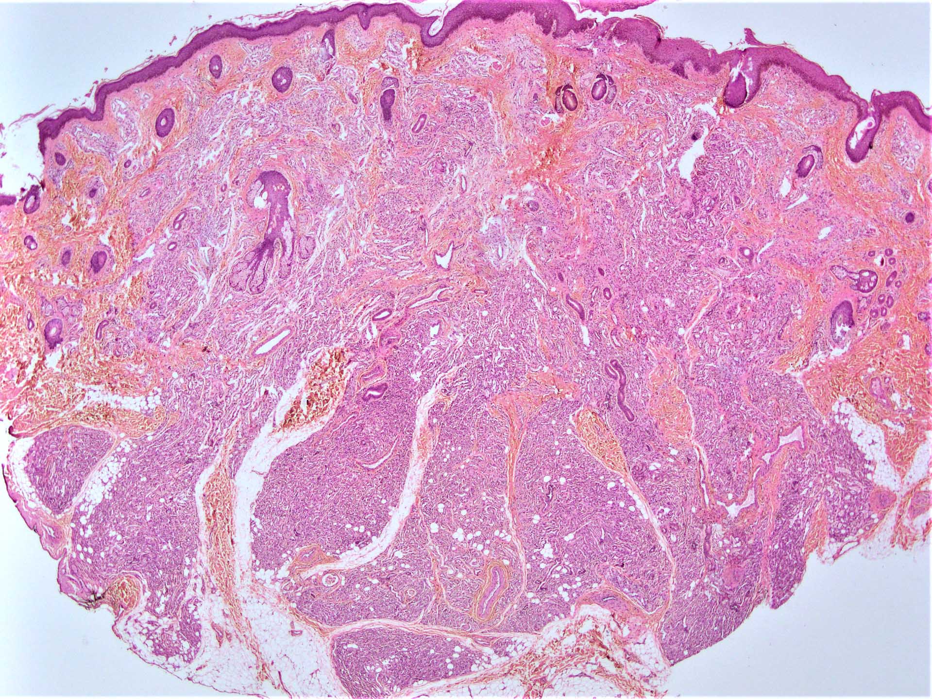
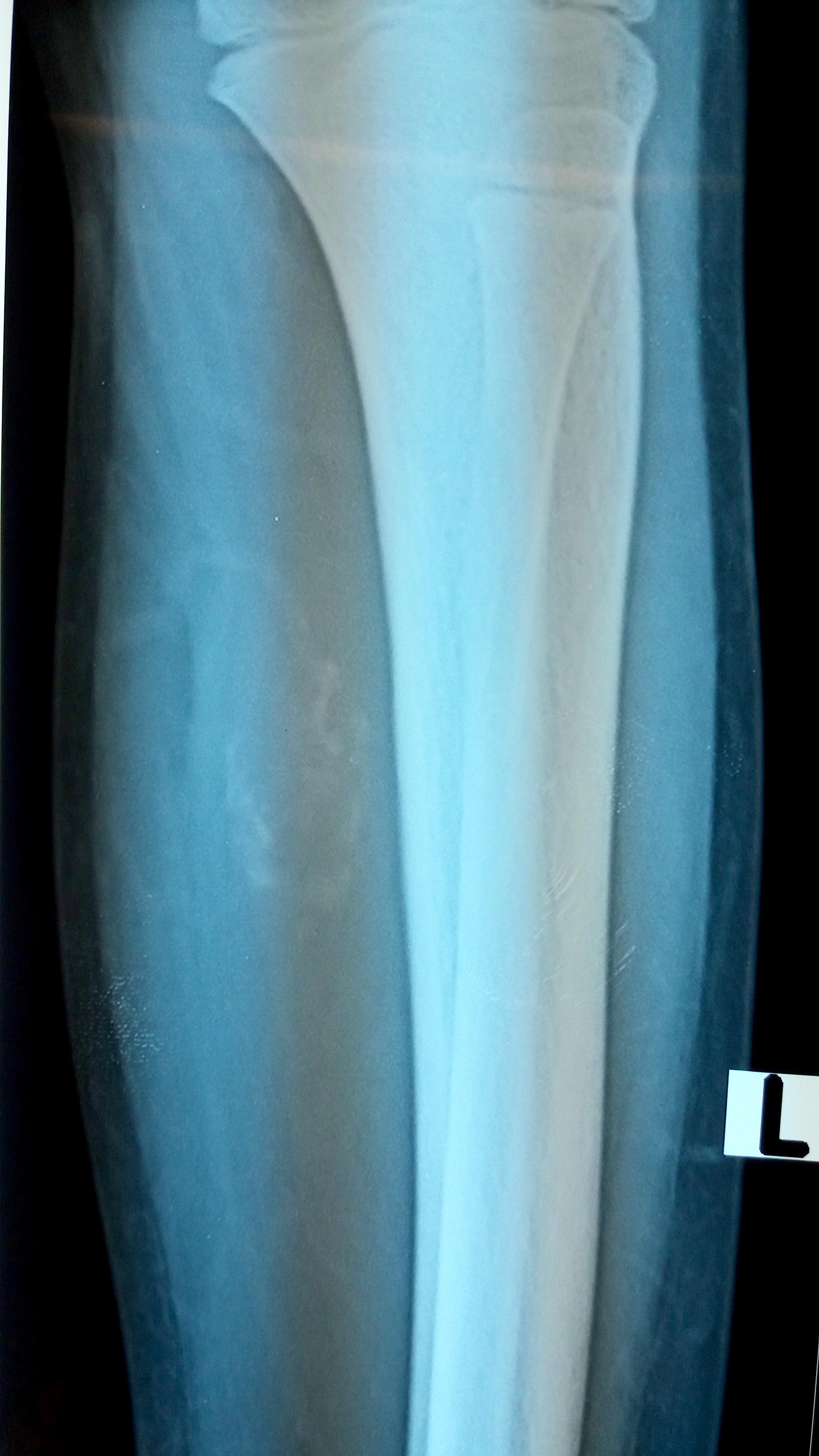
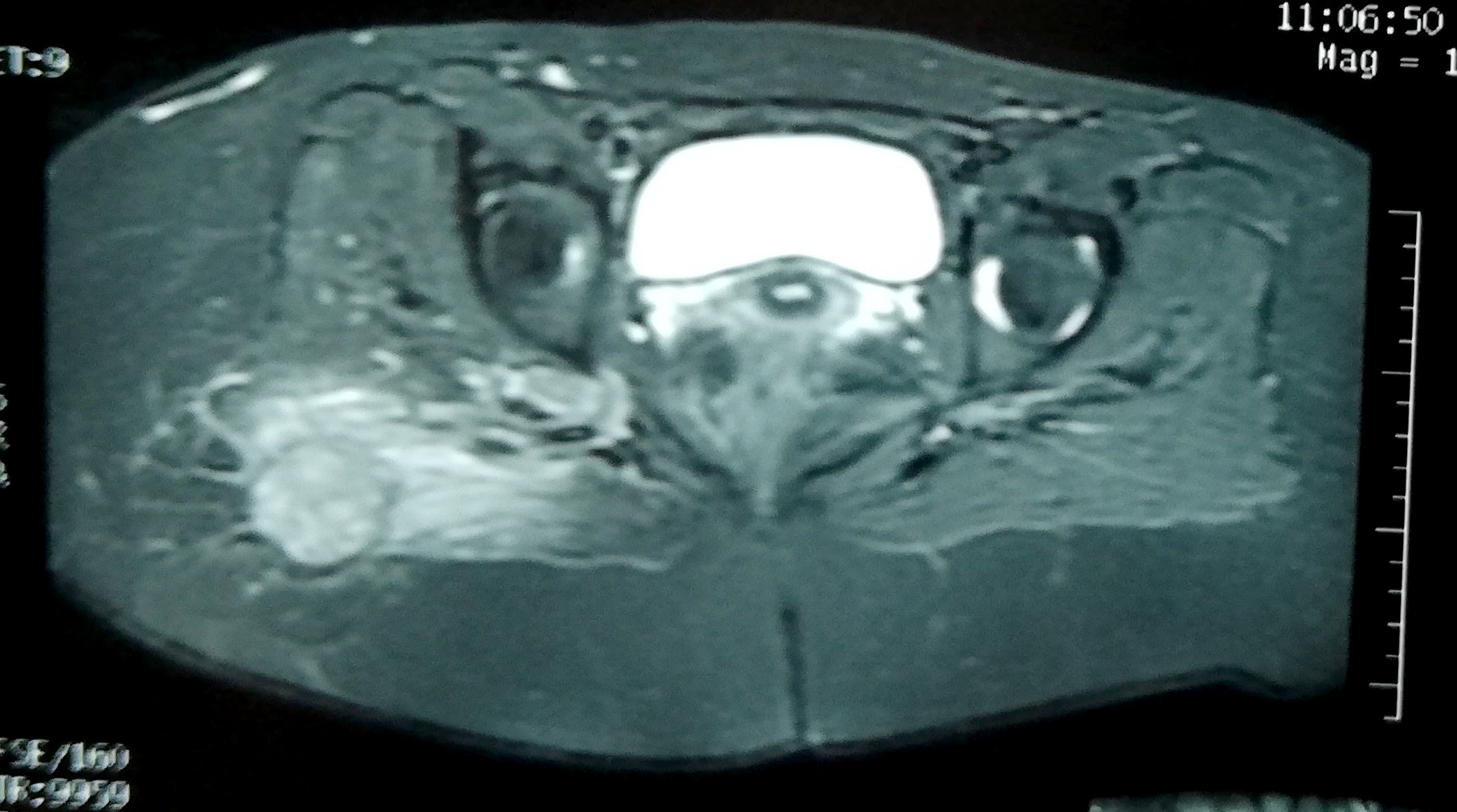
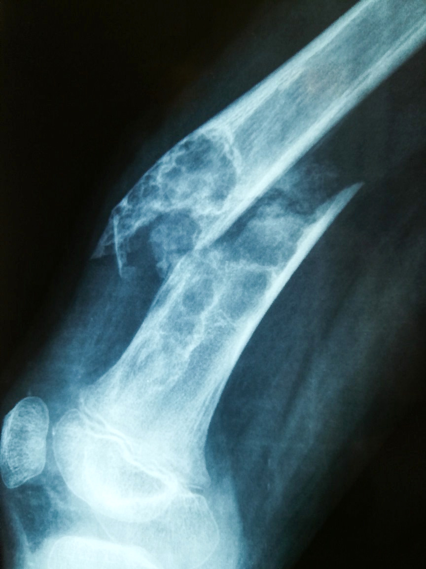
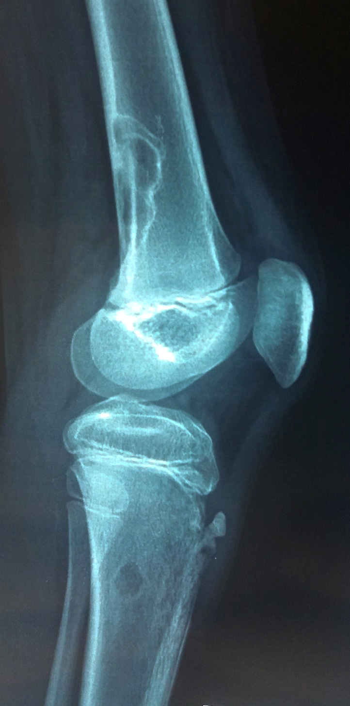
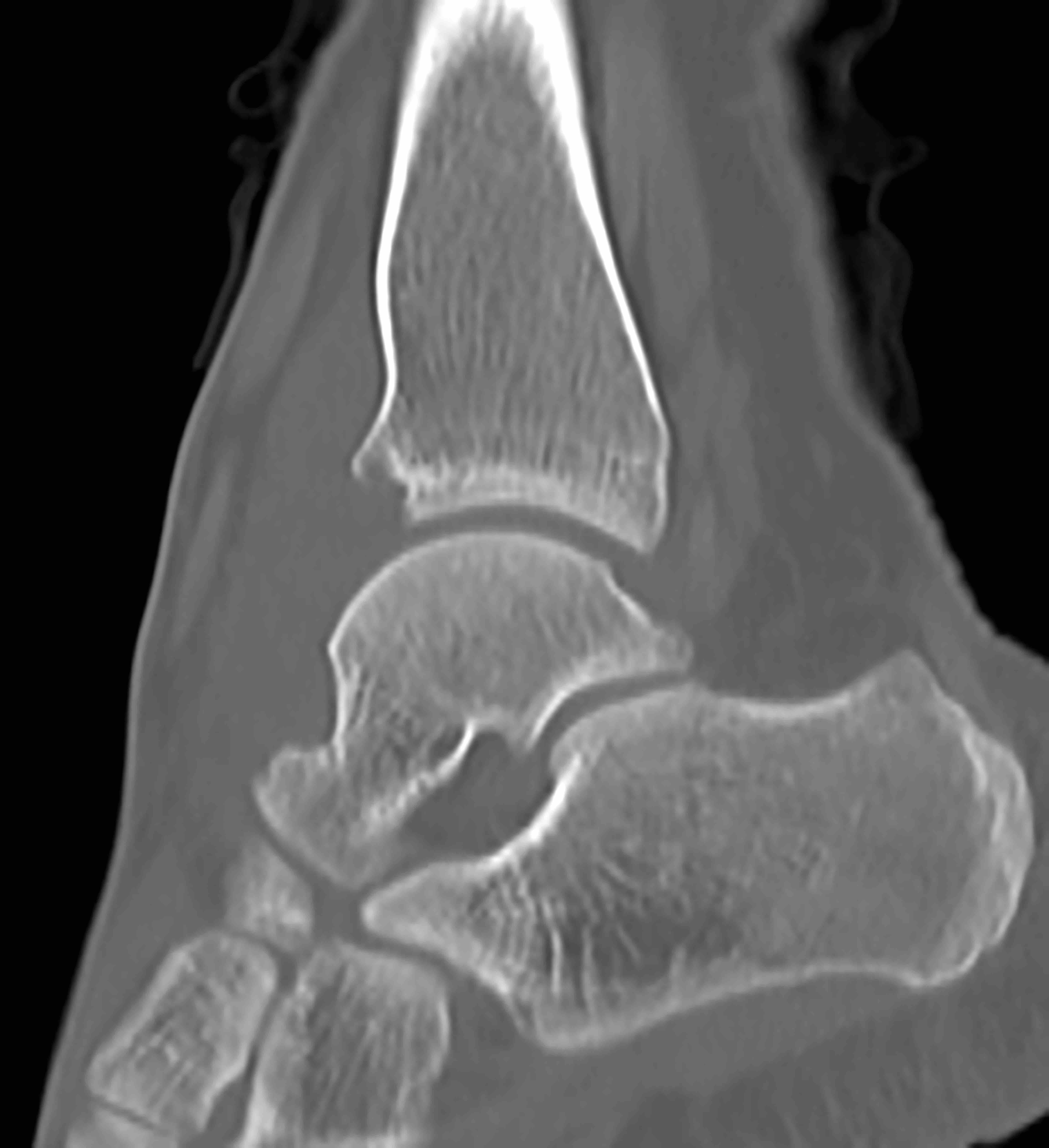
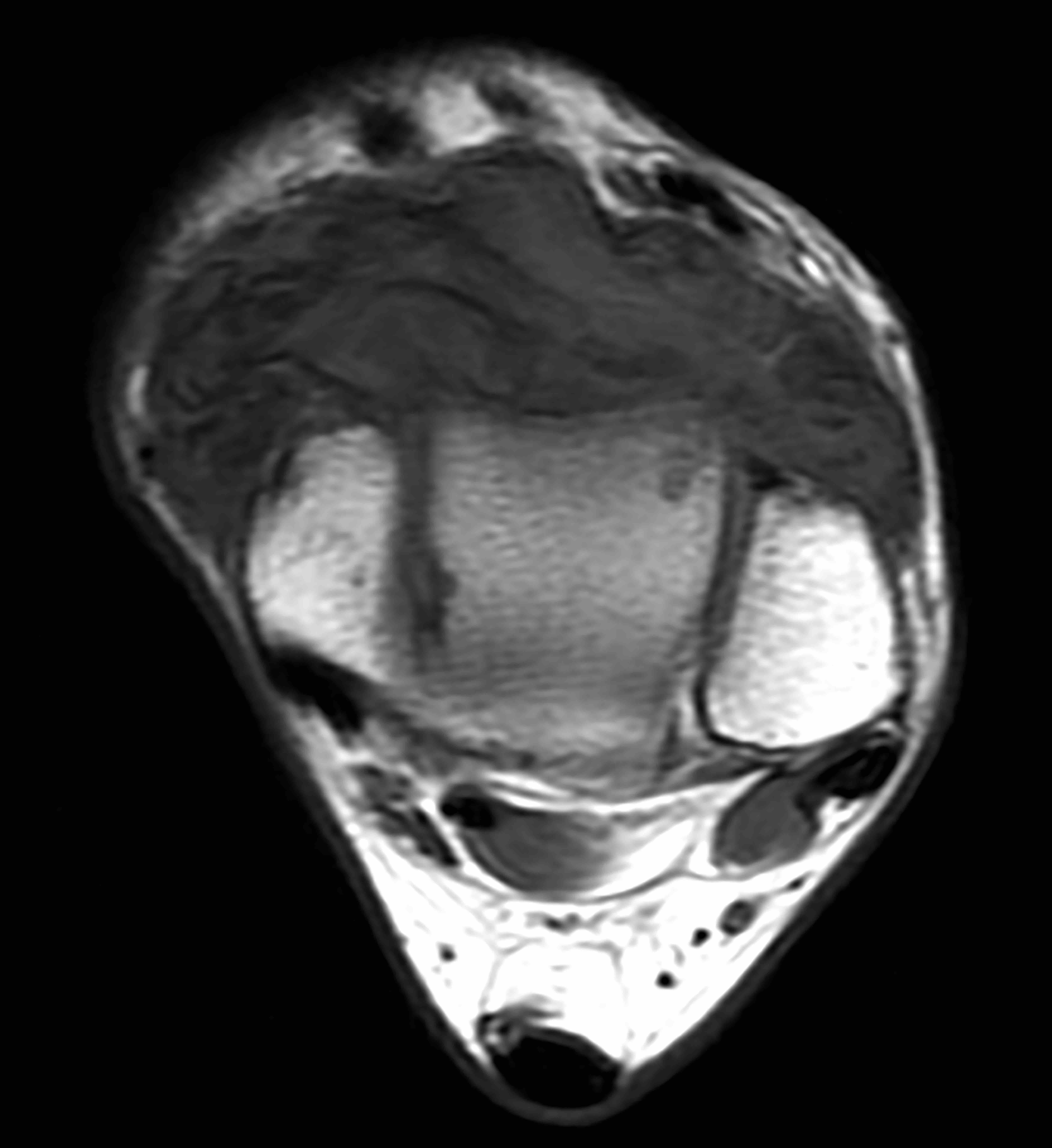
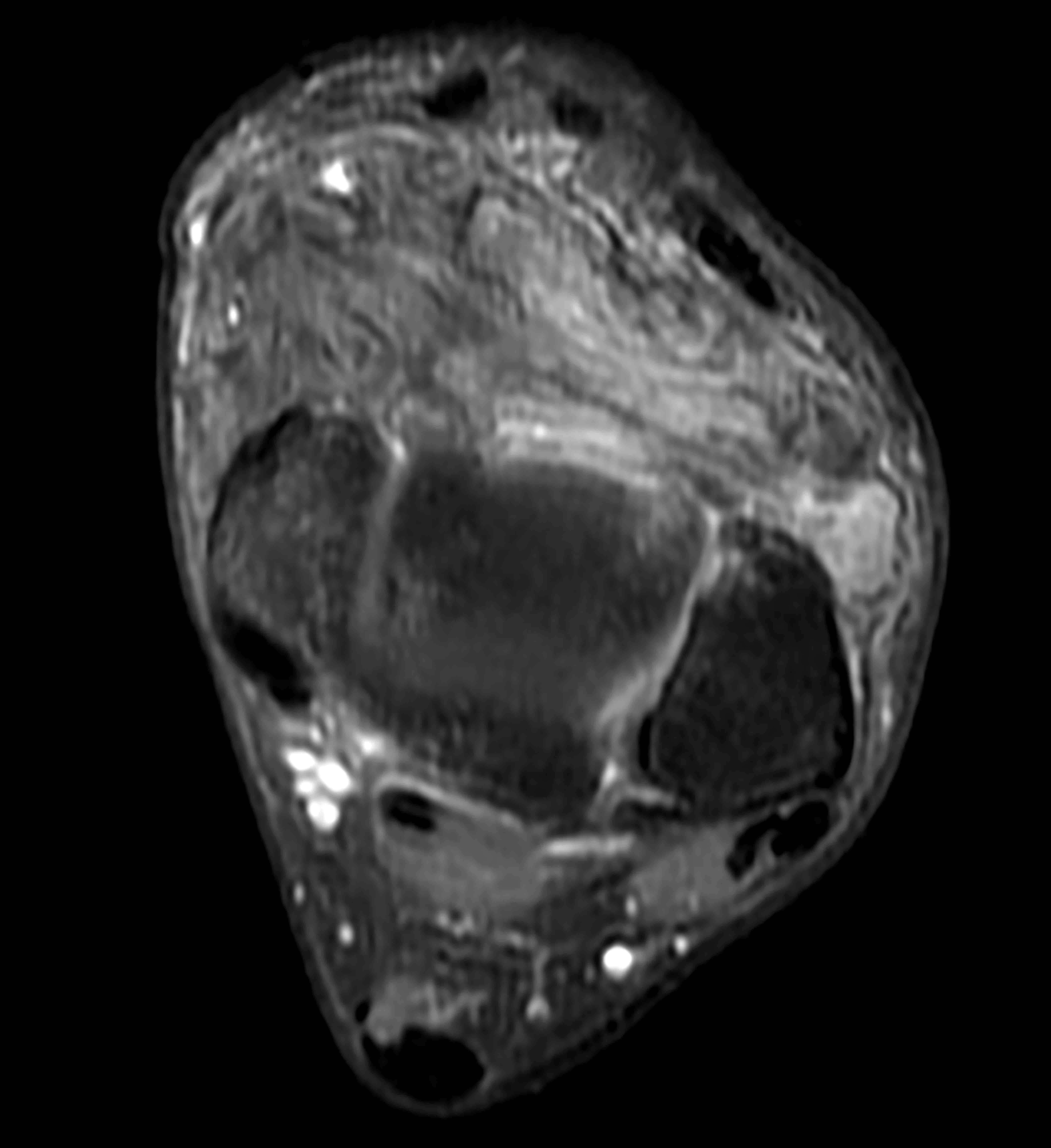
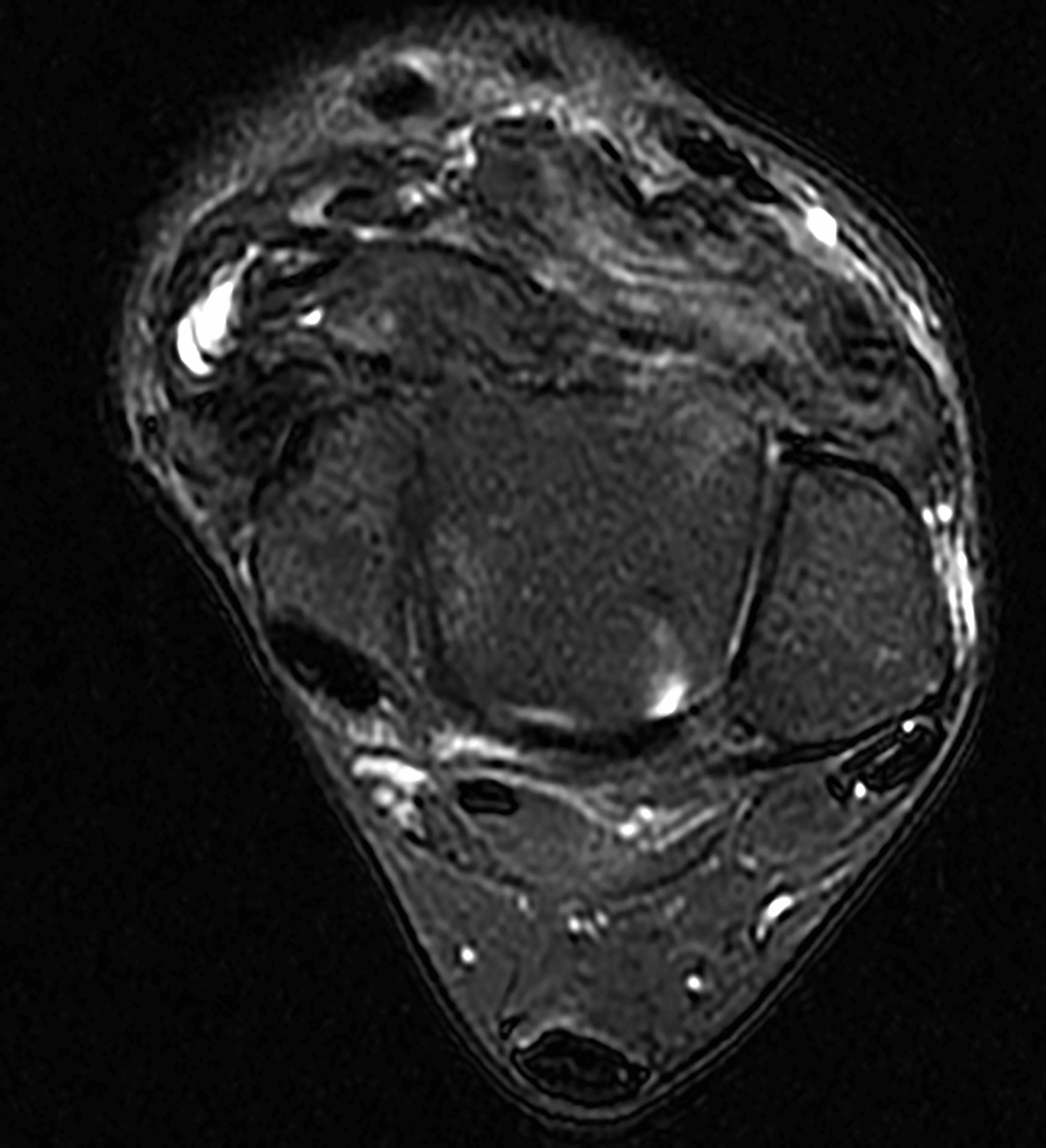
Contributed by Borislav A. Alexiev, M.D.
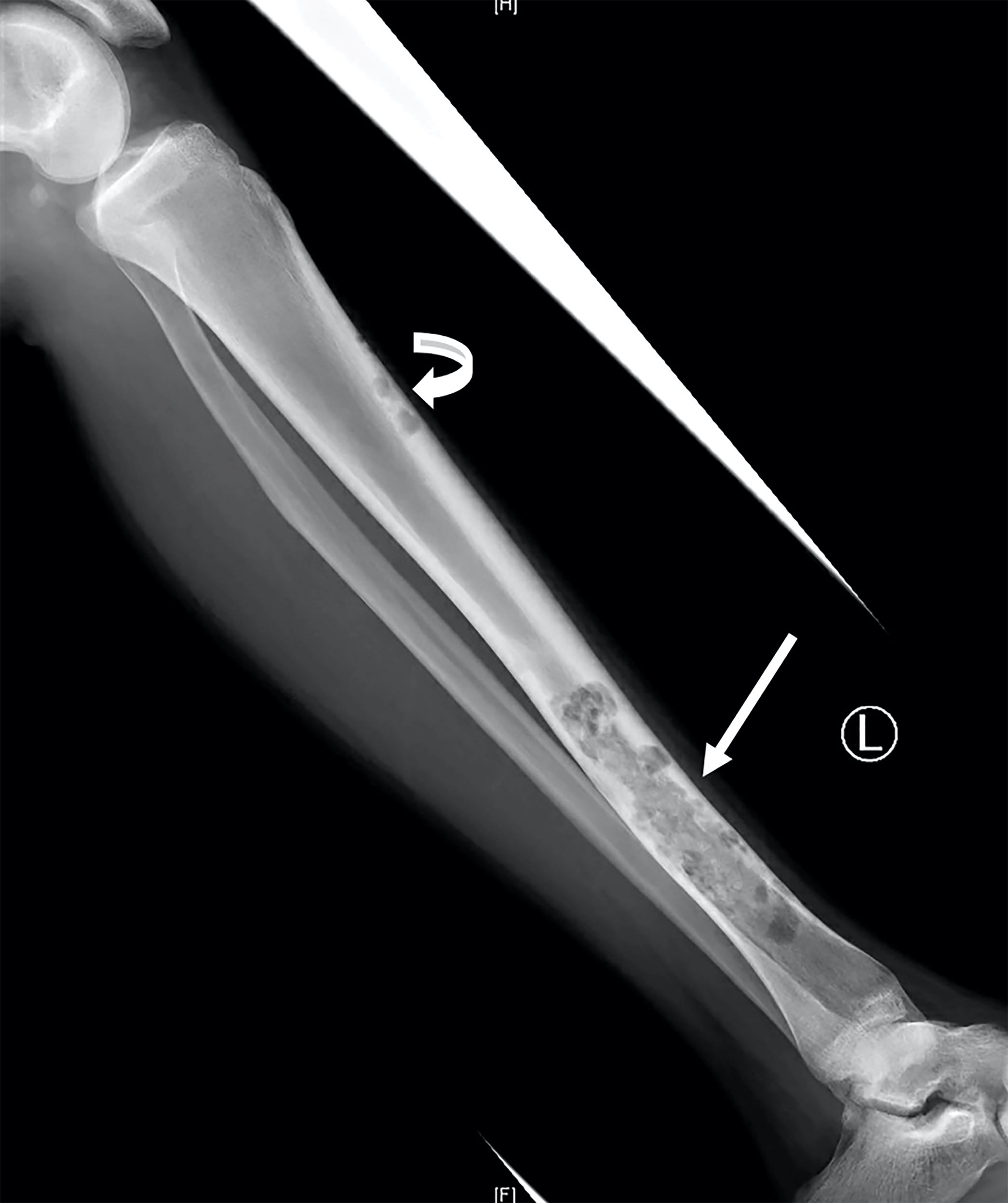
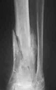
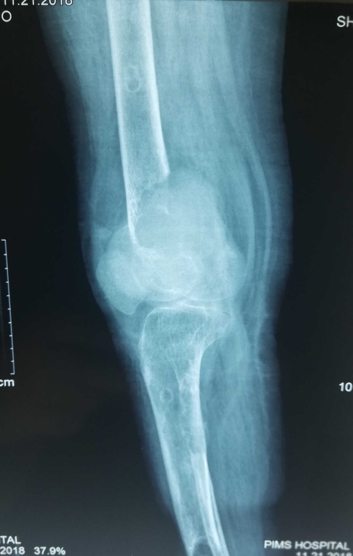
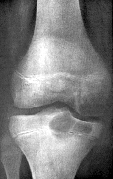
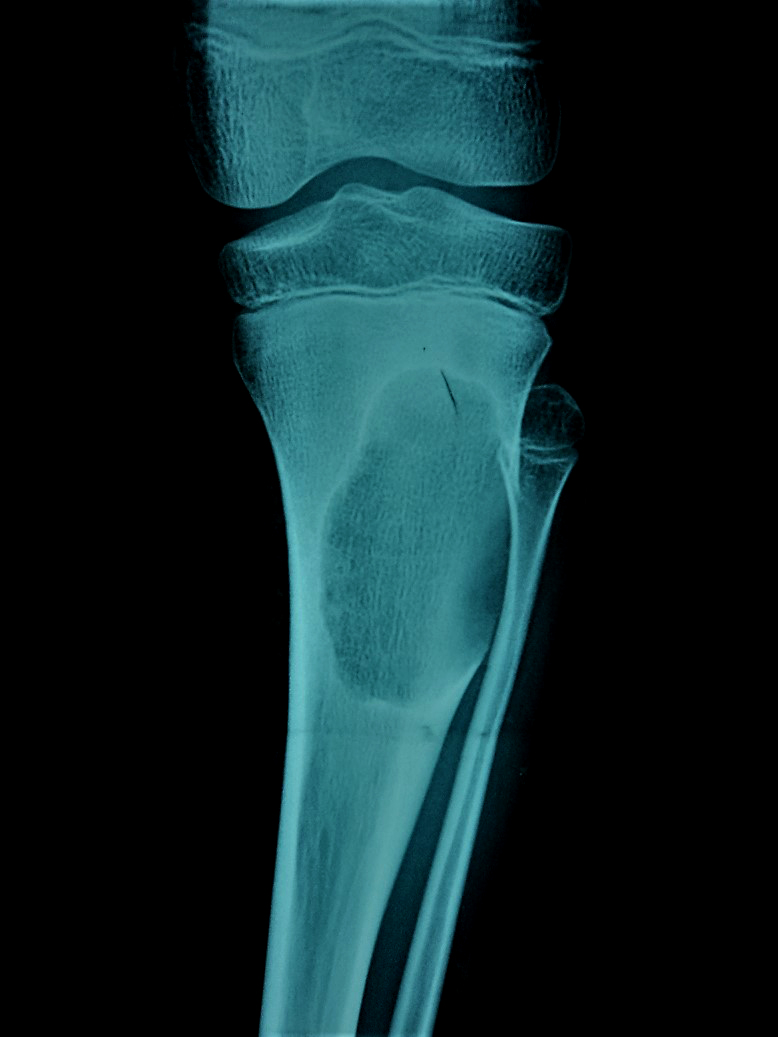
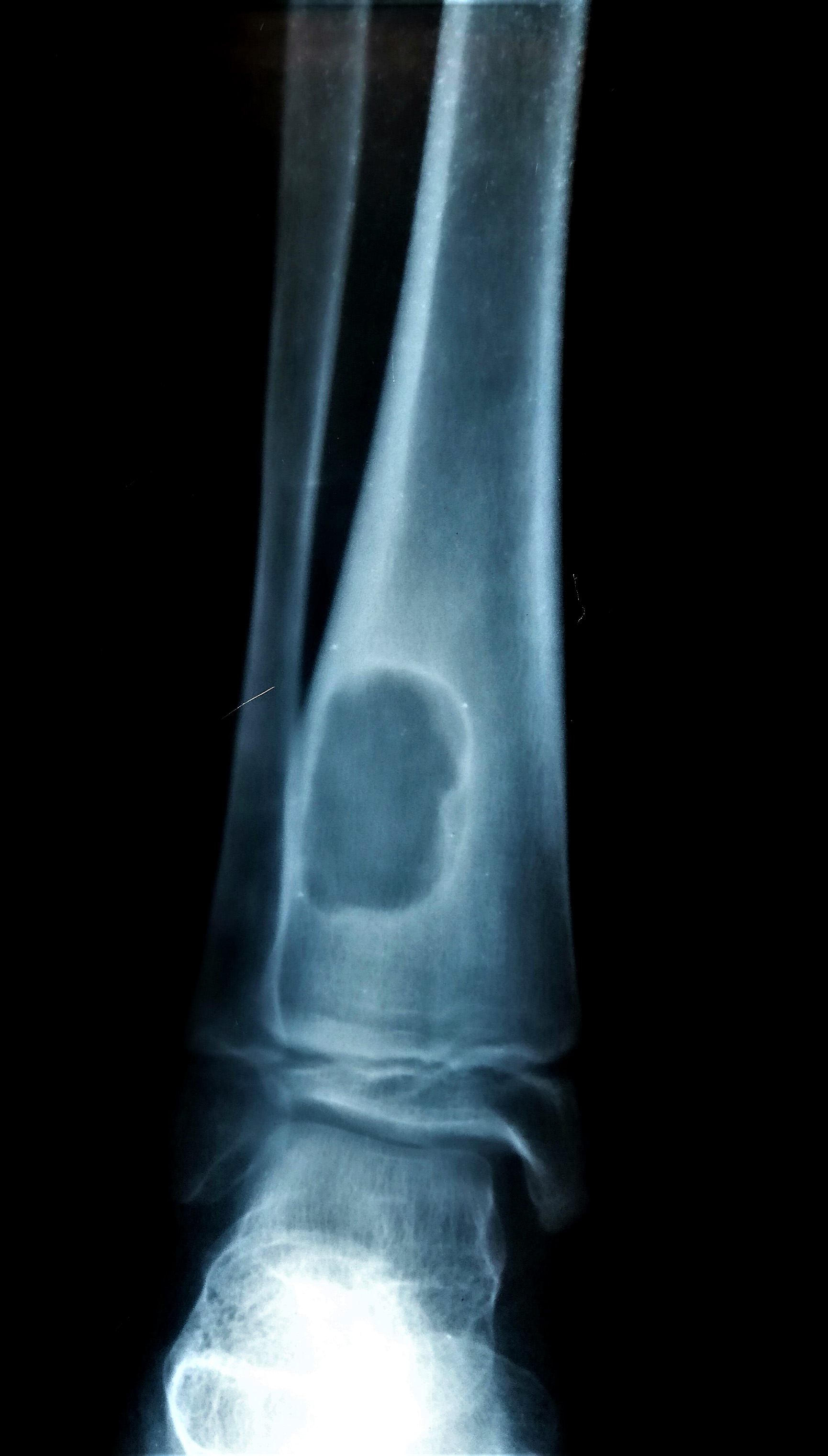
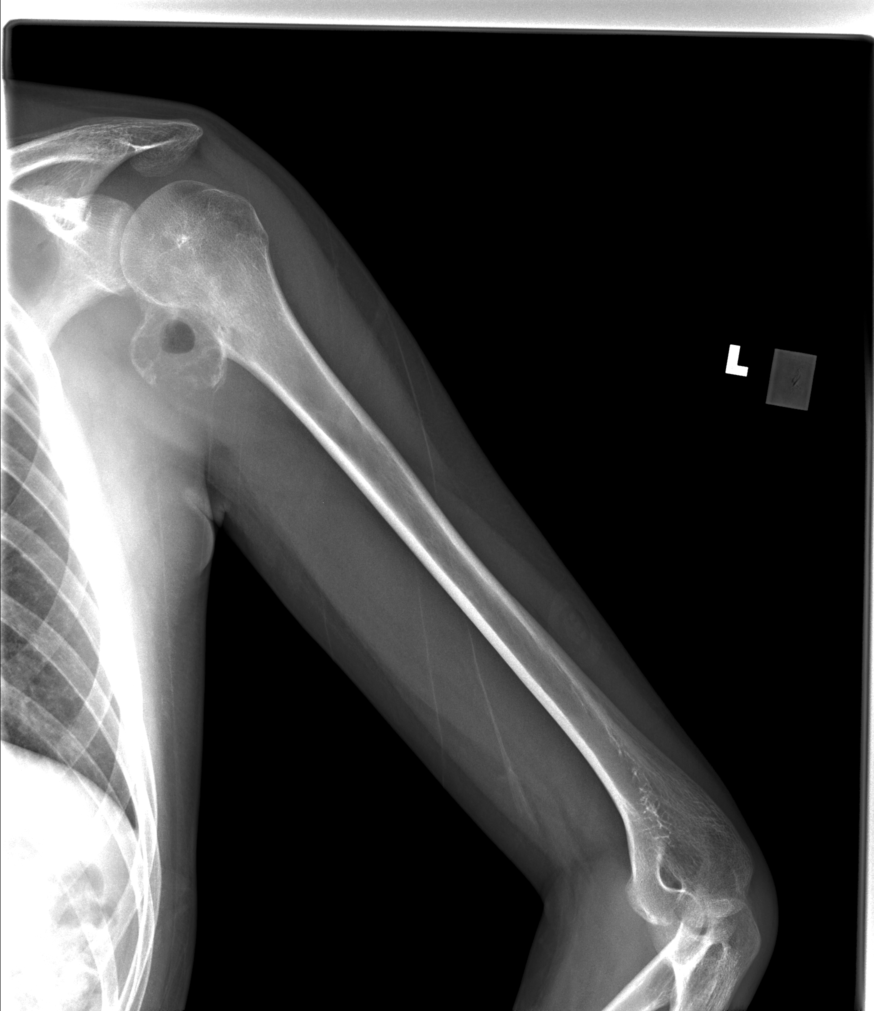
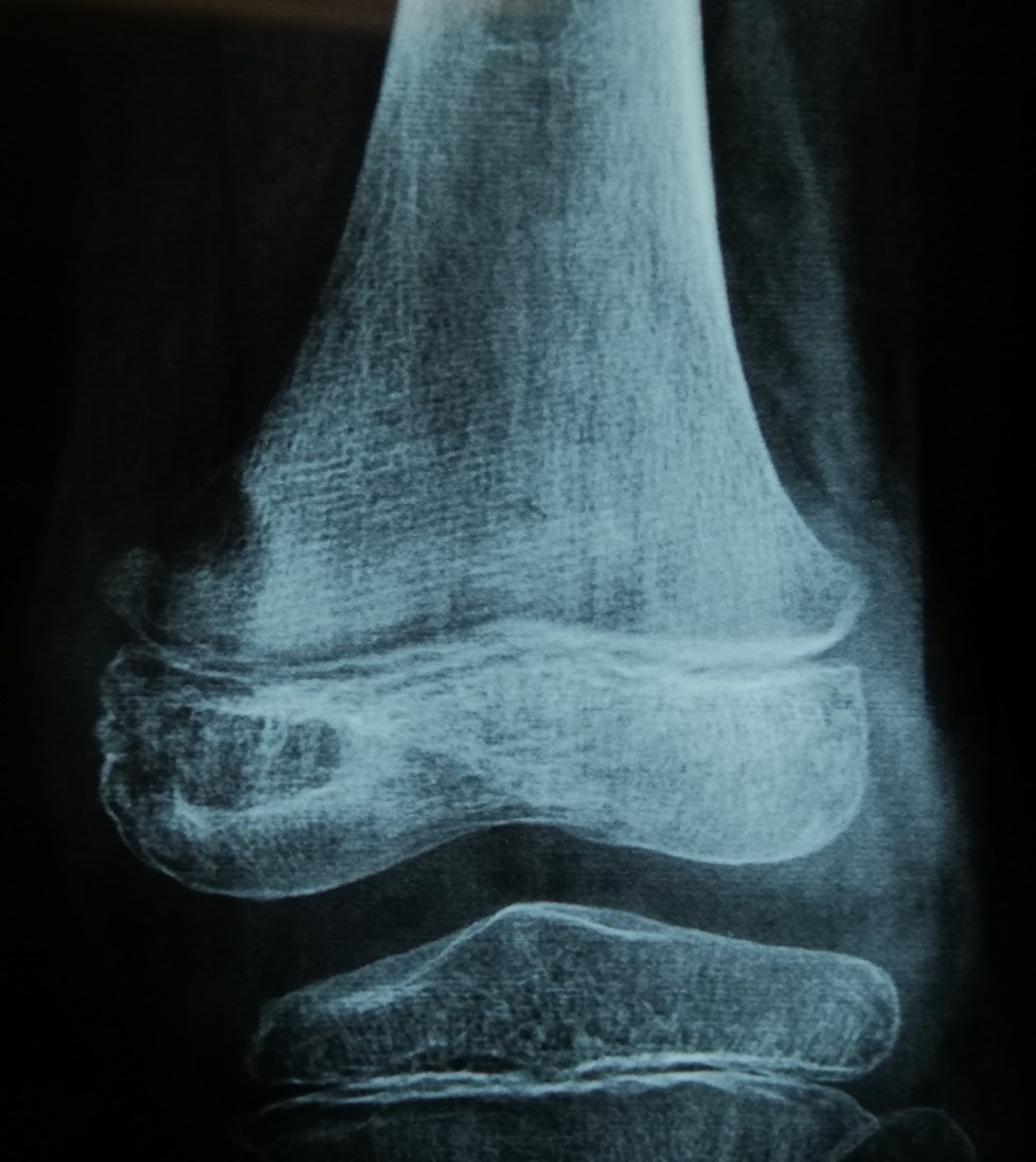
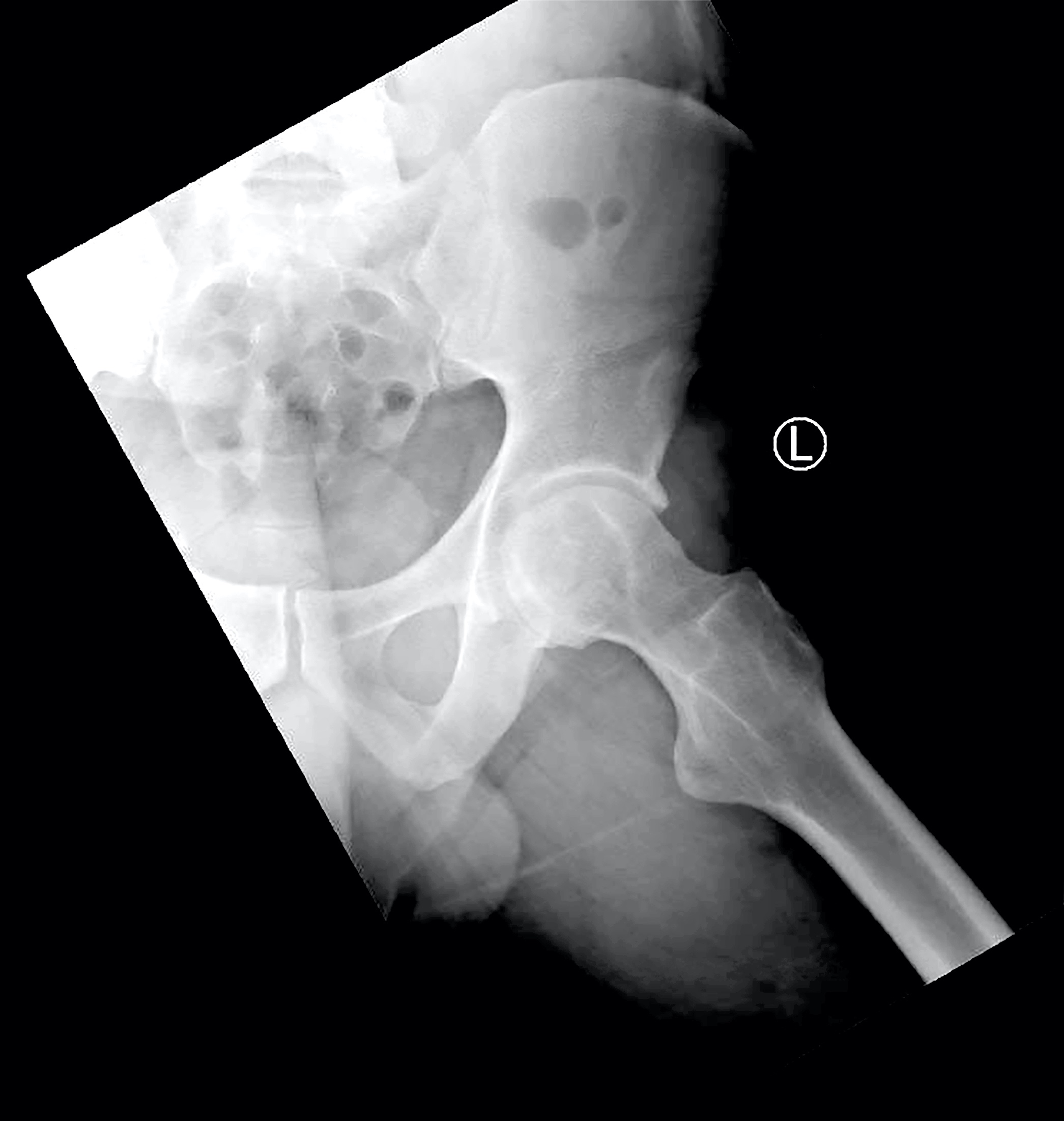
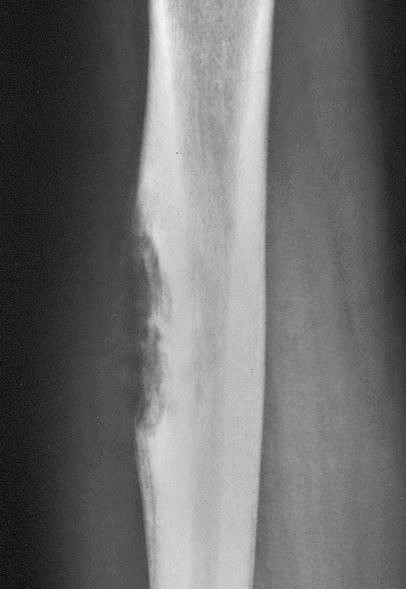
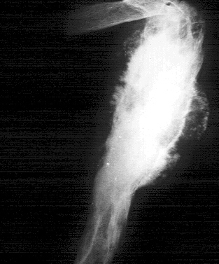
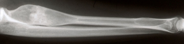
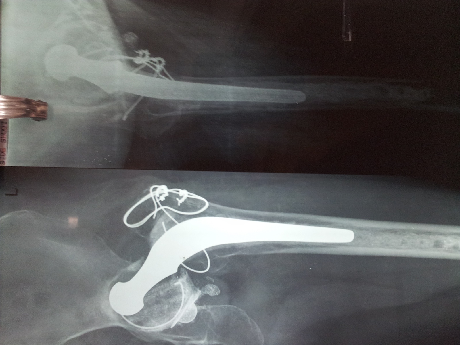
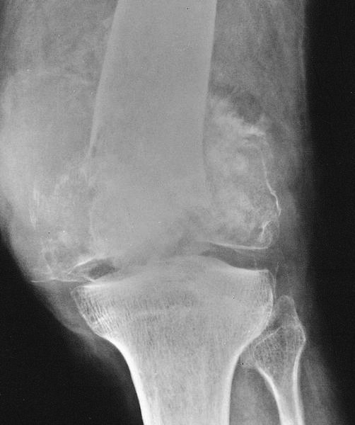
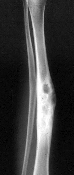
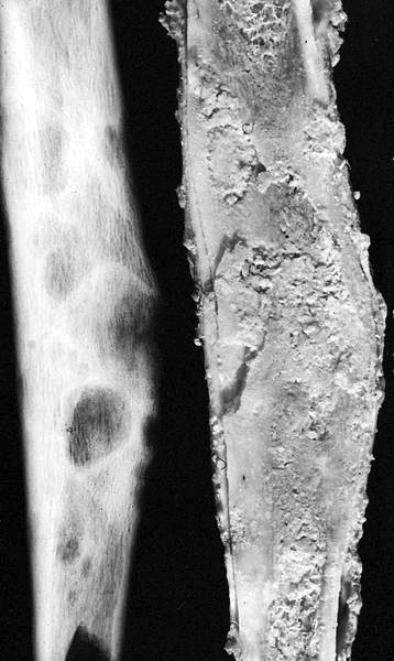
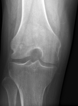
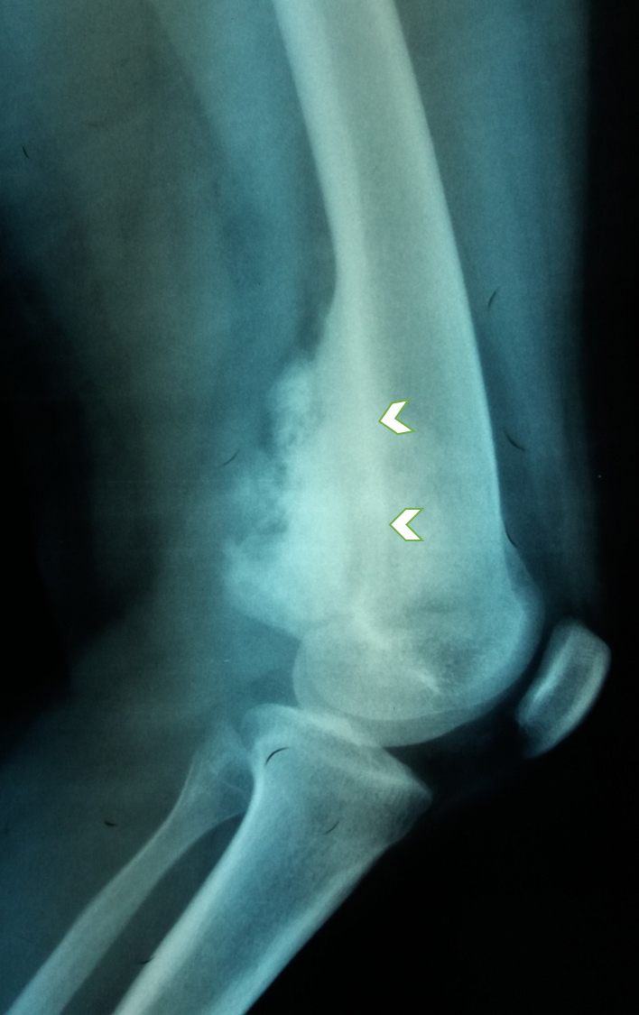
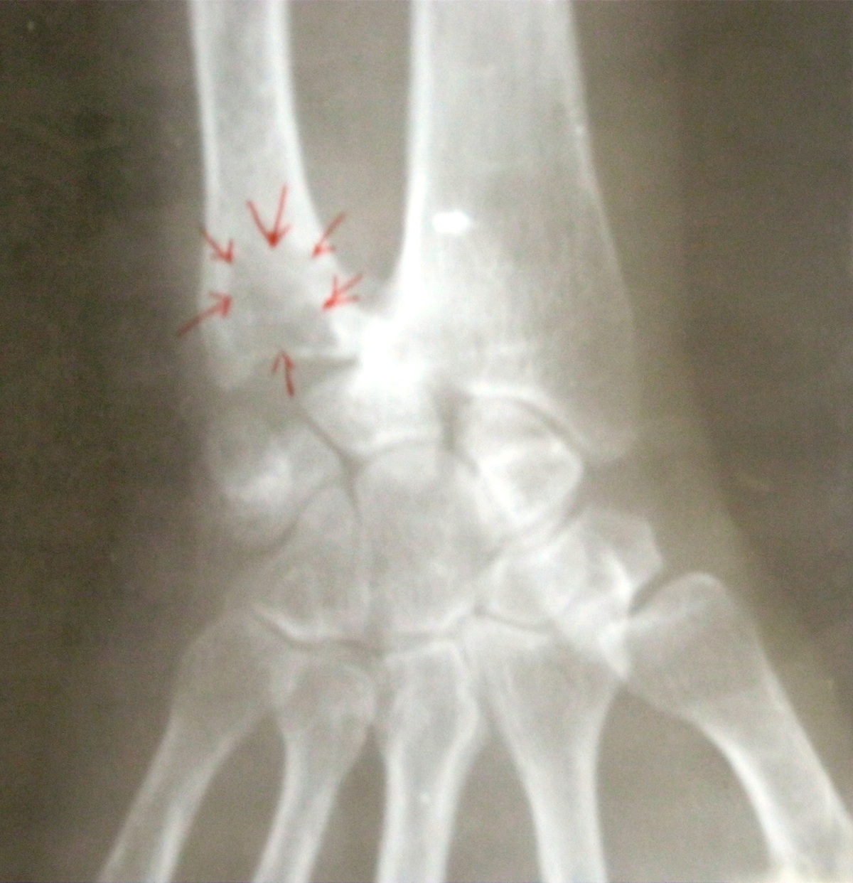
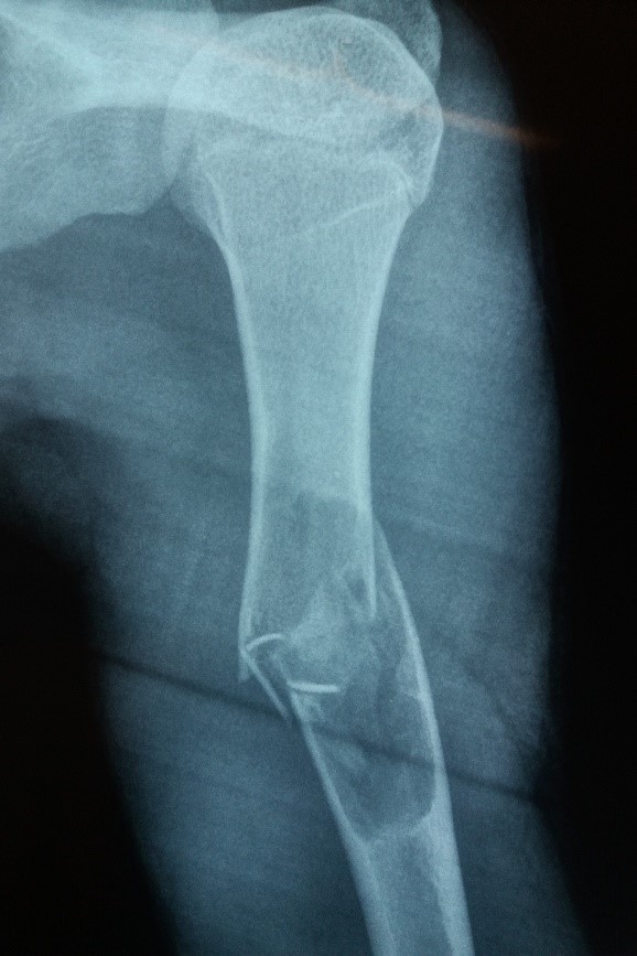
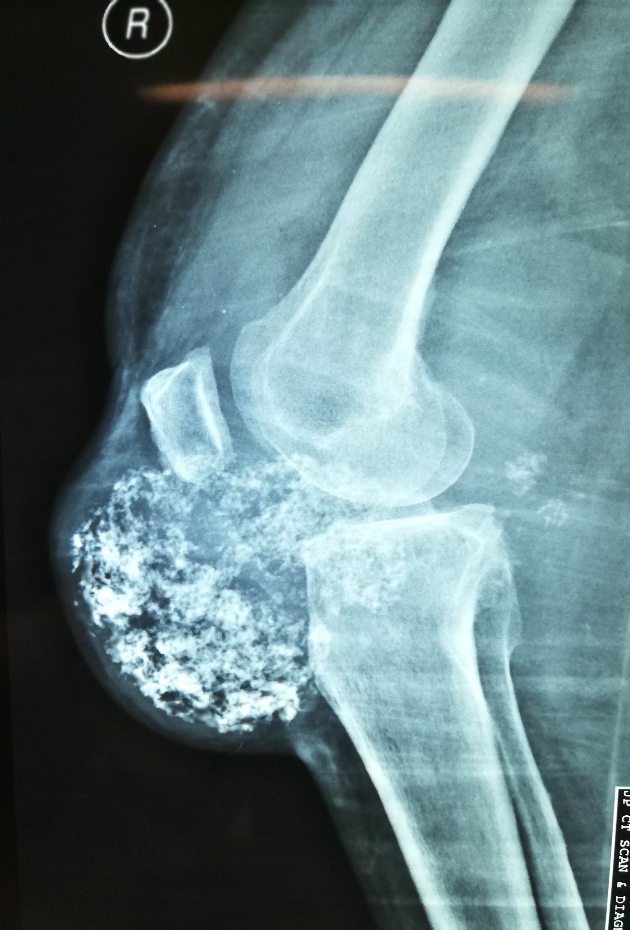
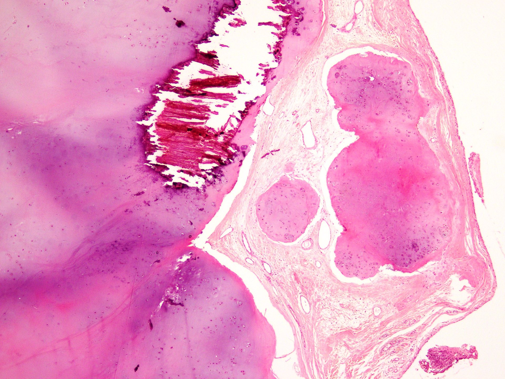
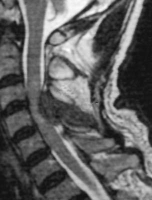
Tibia lesion, radiography
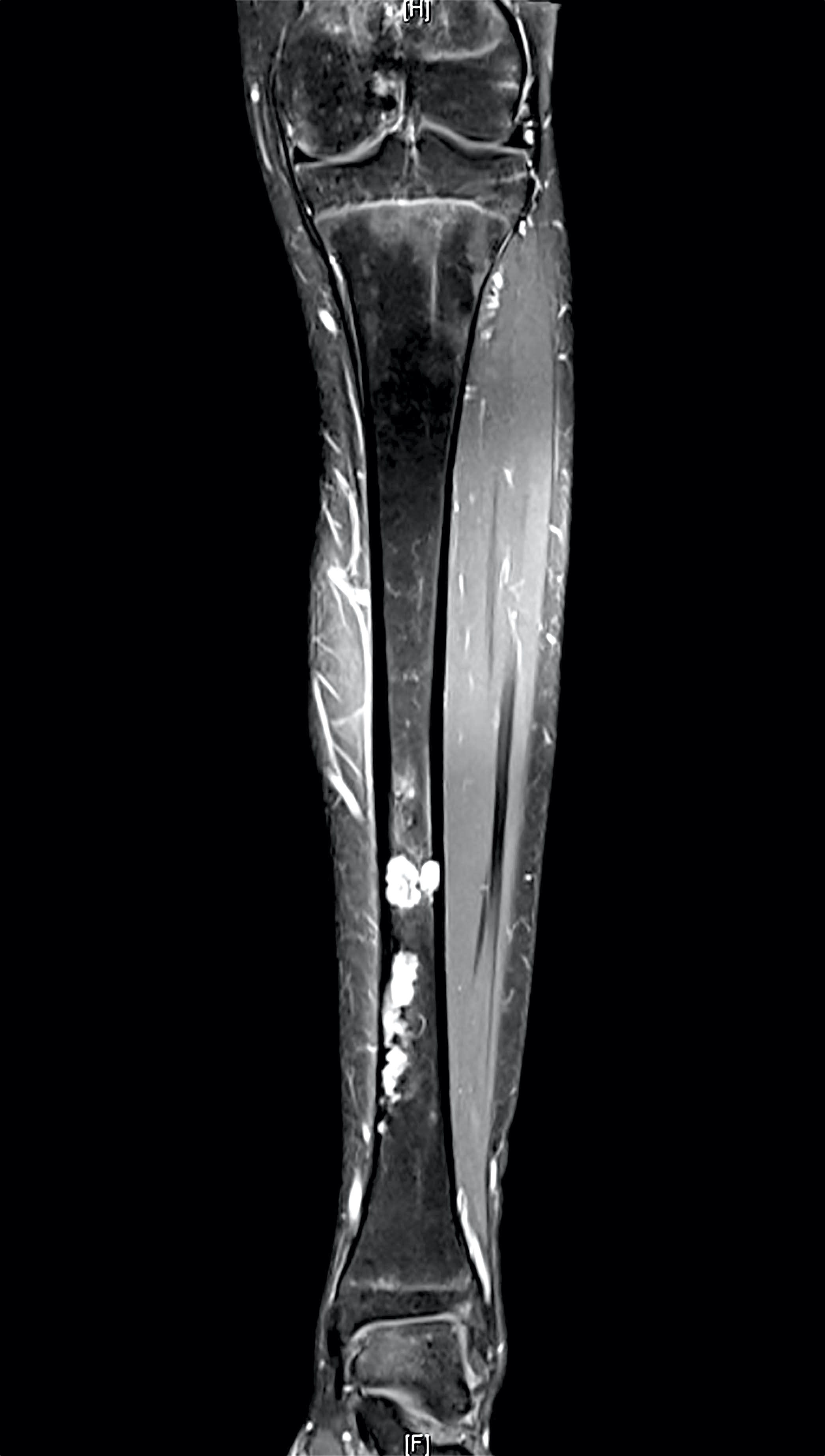
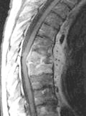
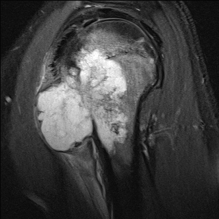
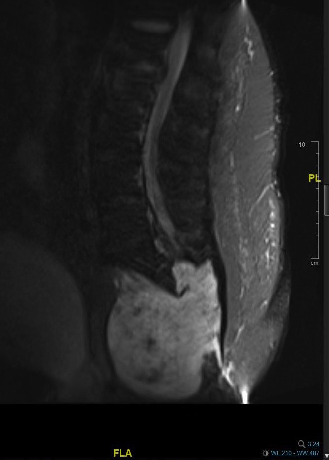
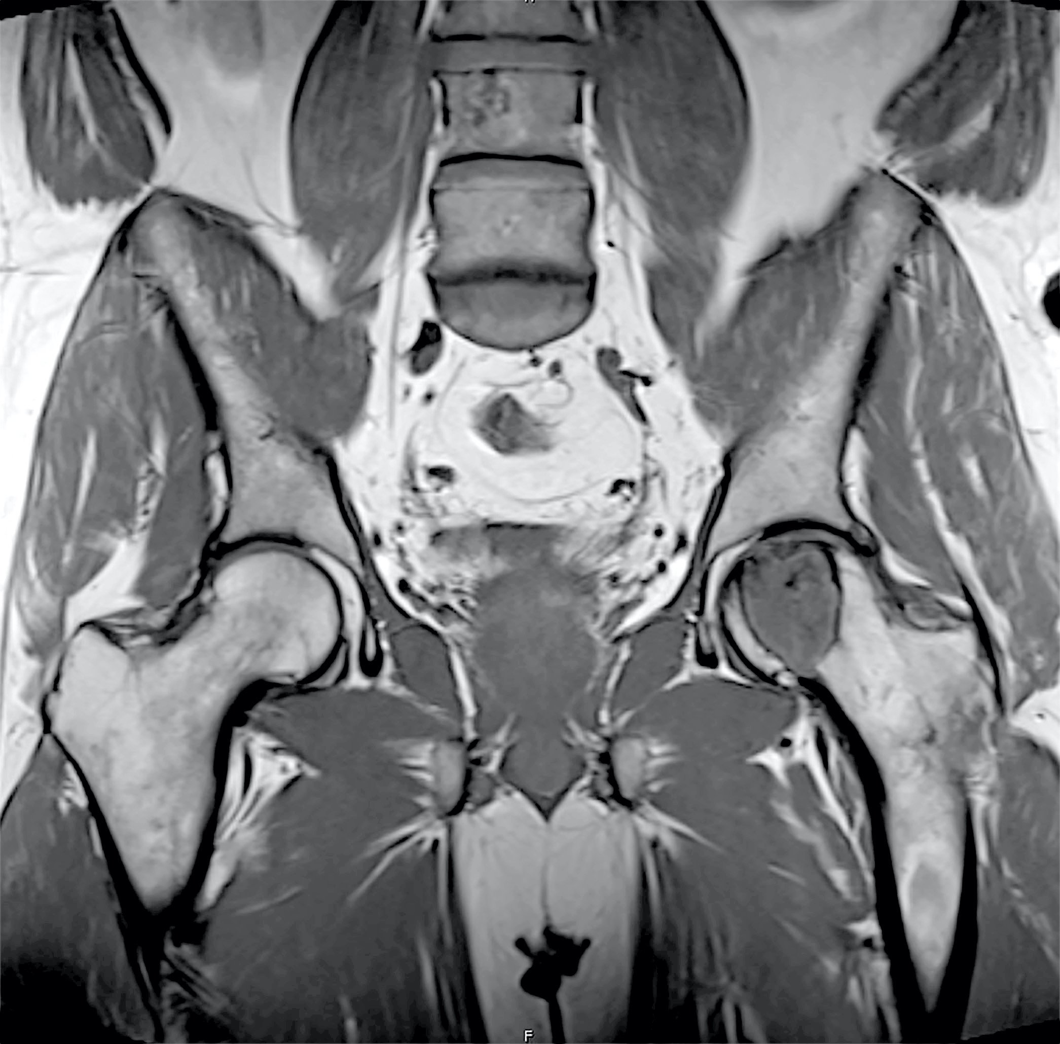
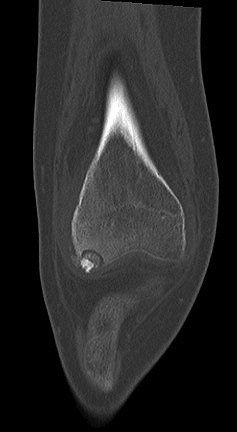
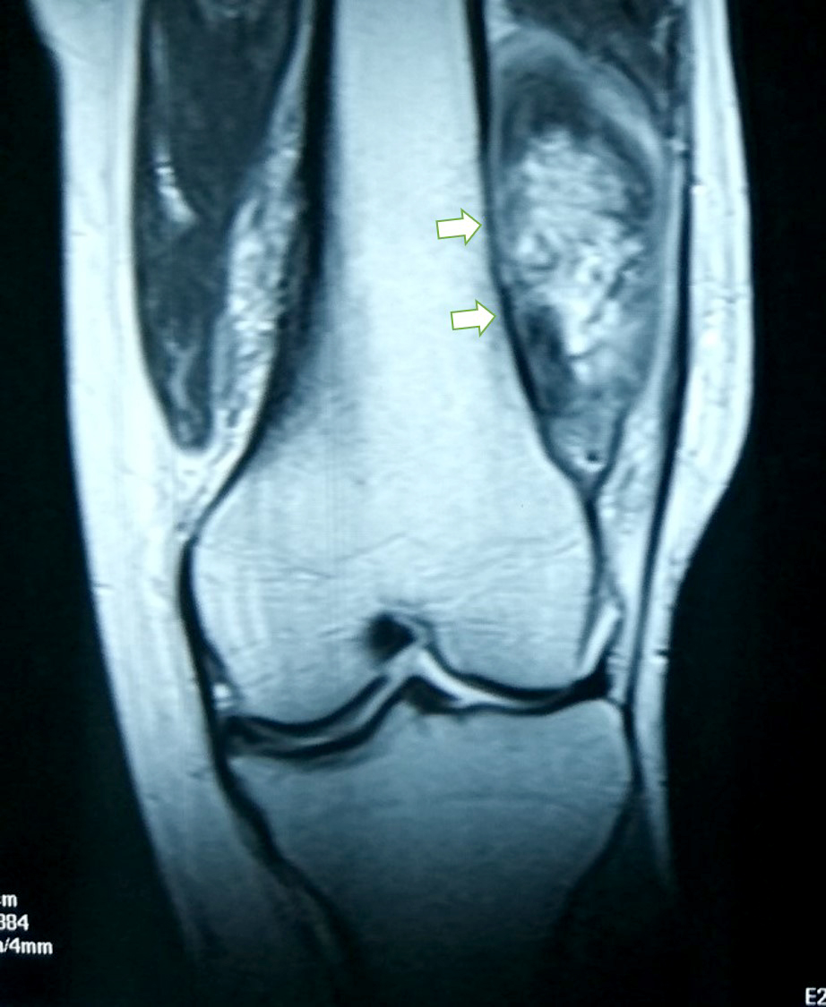
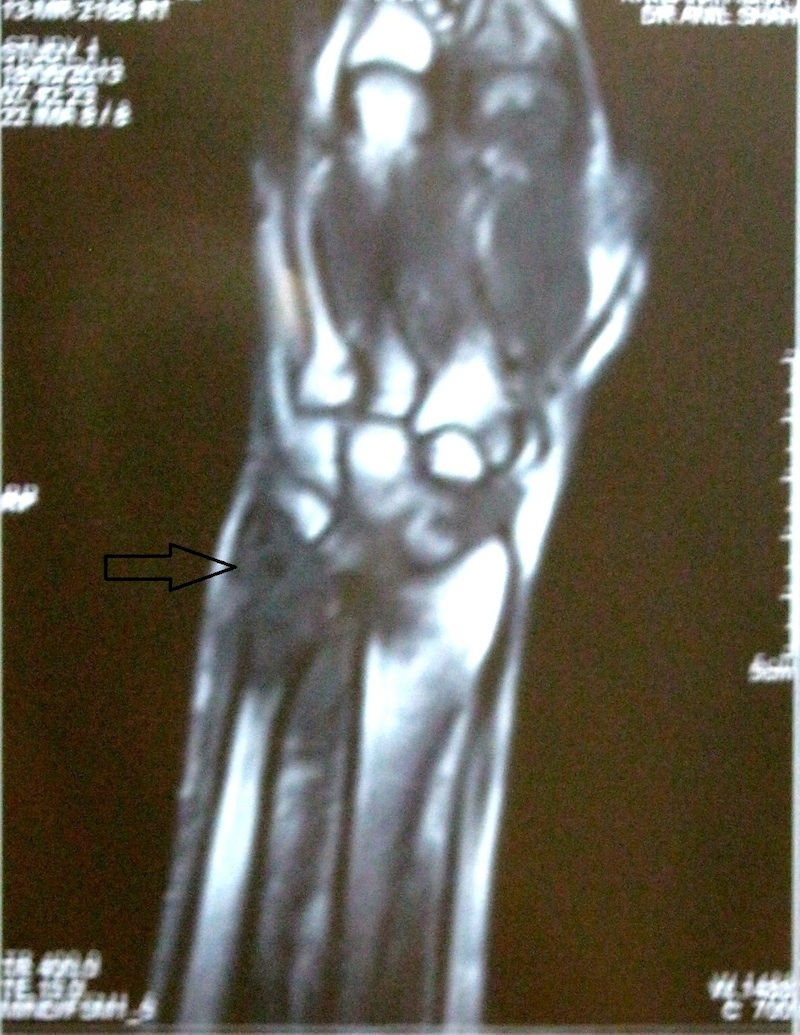
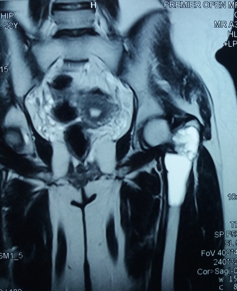
Tibia lesion, MRI
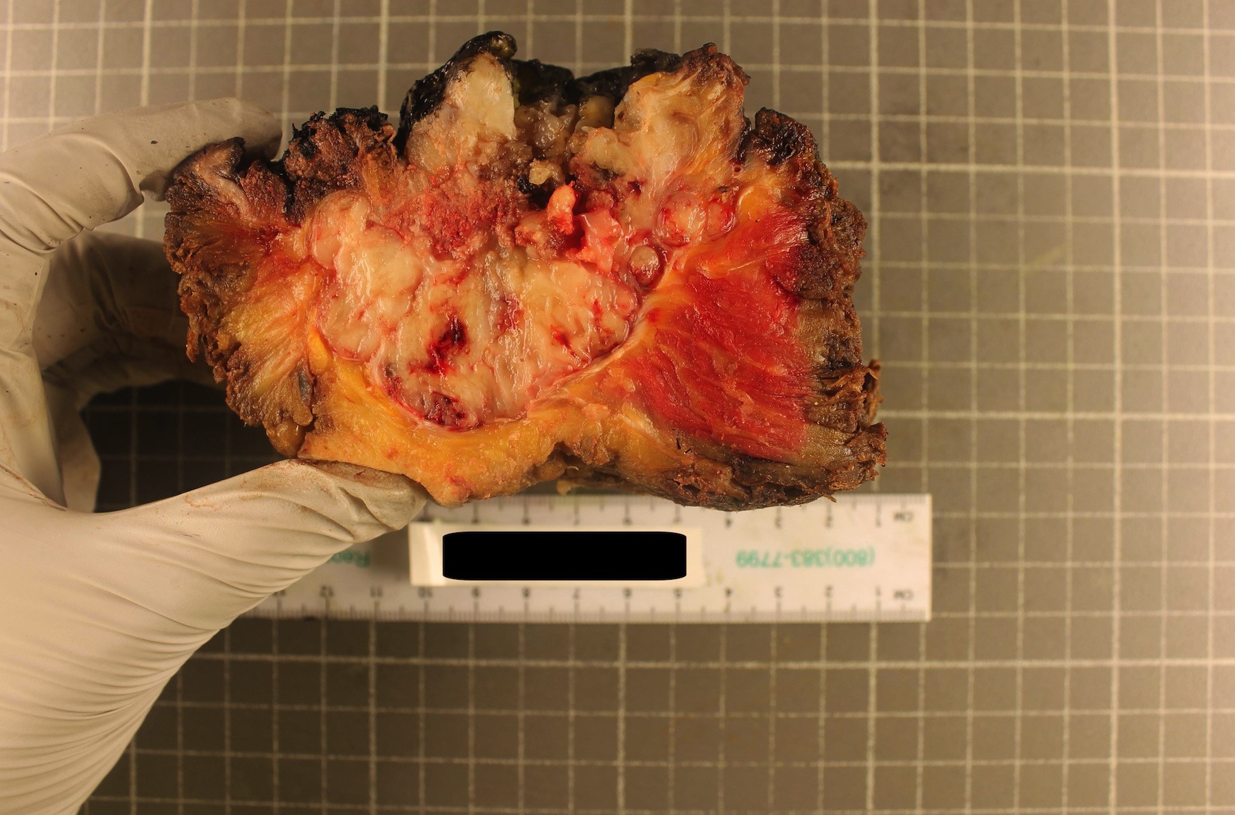
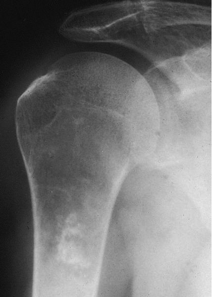
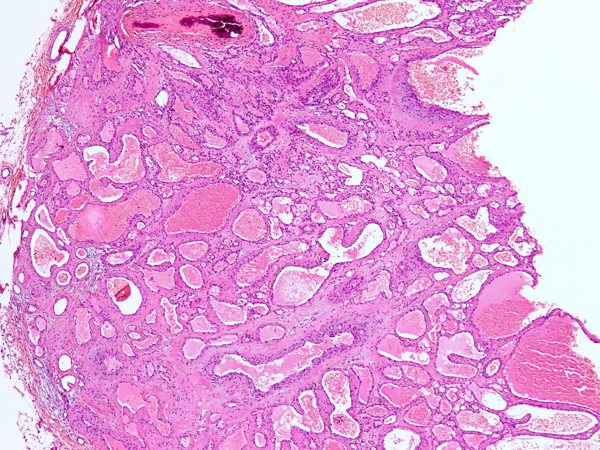
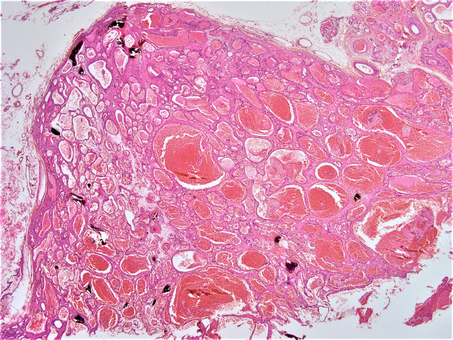
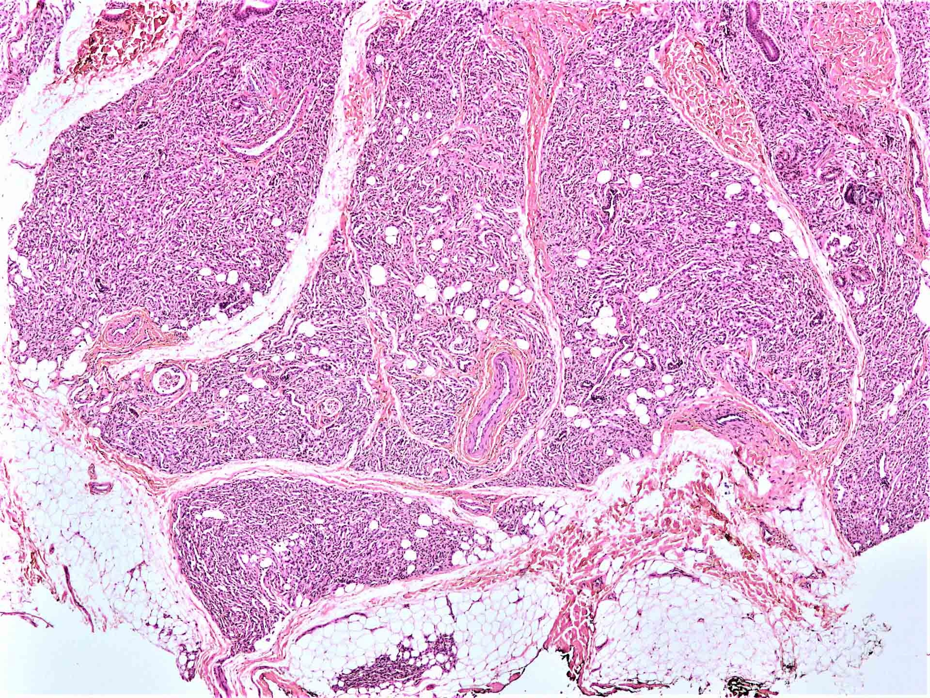
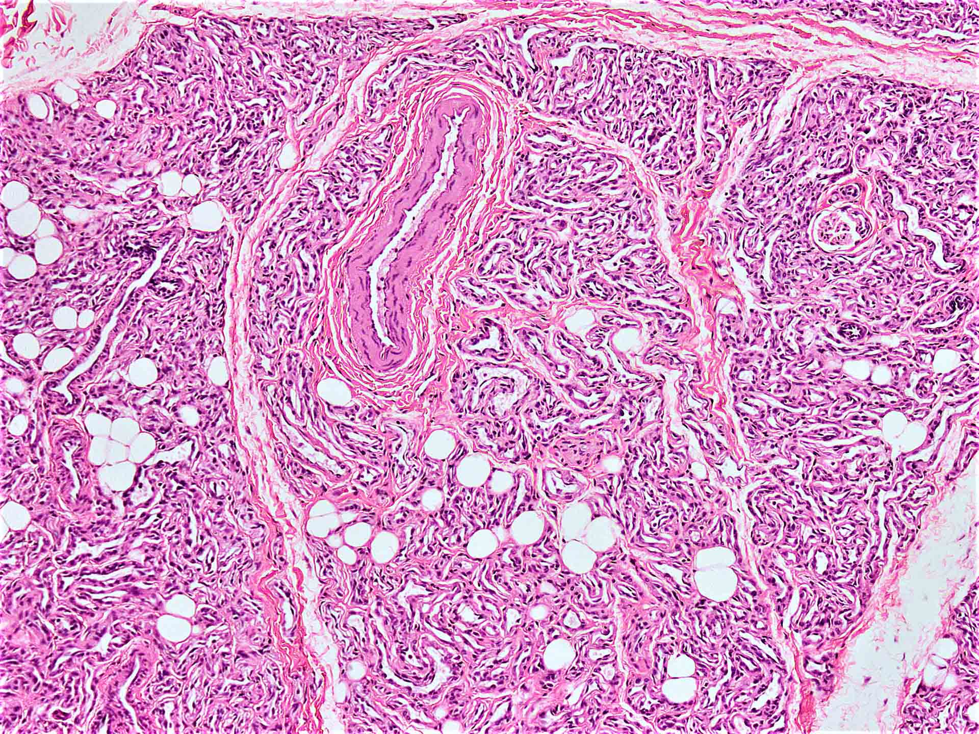
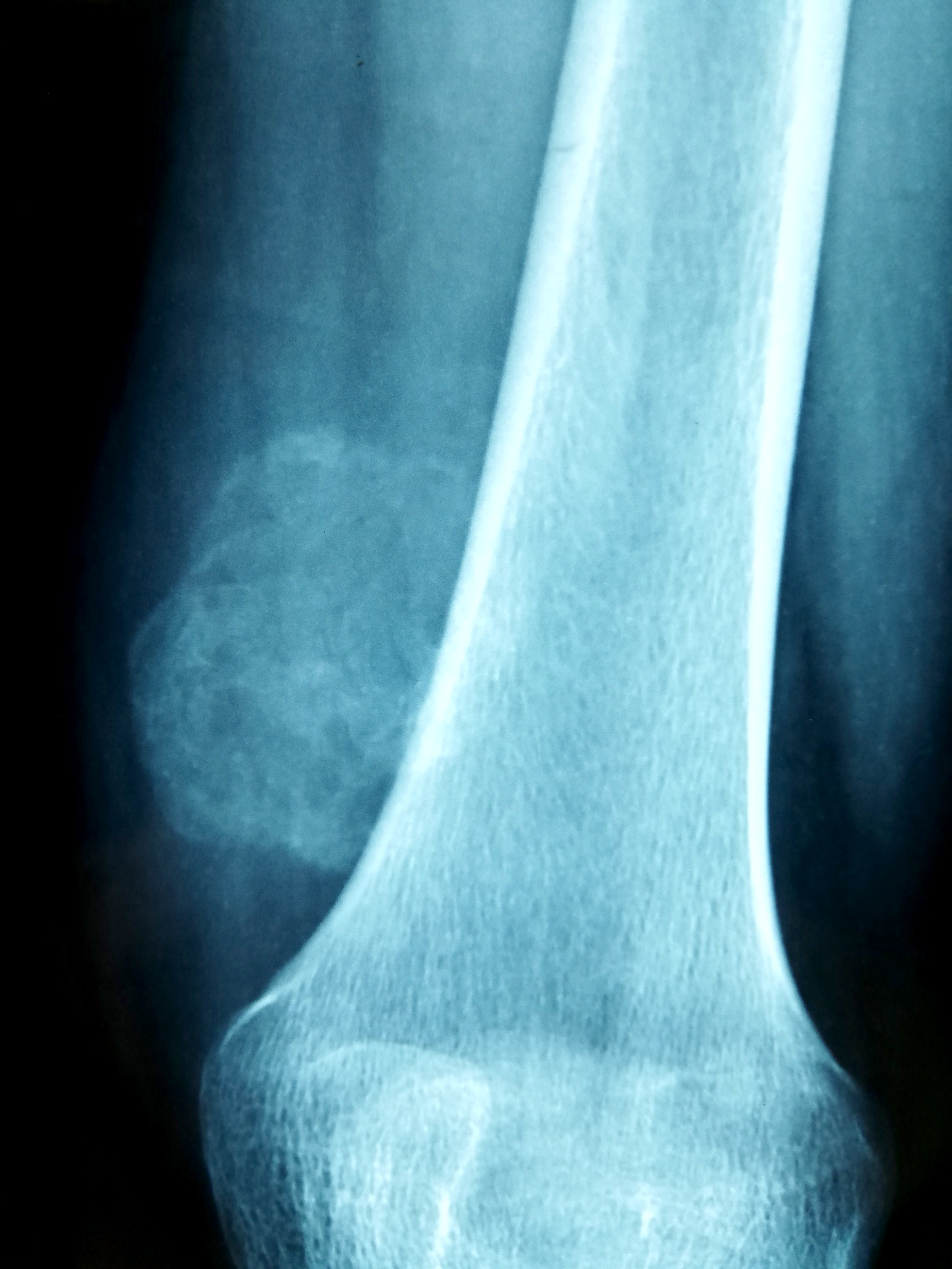
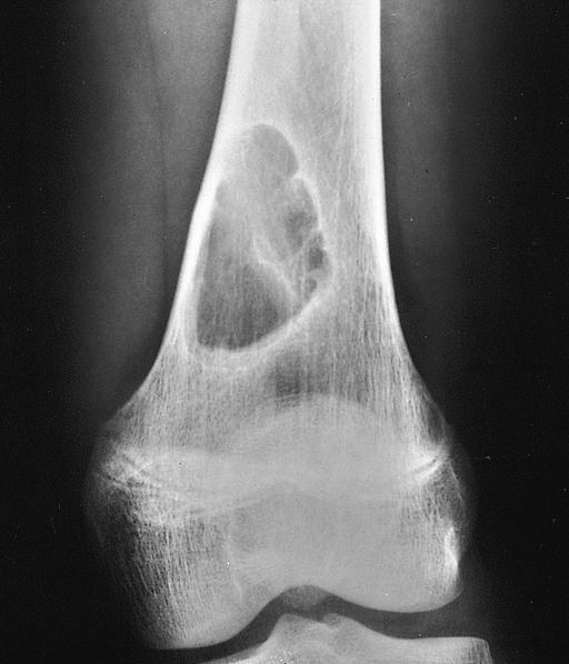
Contributed by Mark R. Wick, M.D.
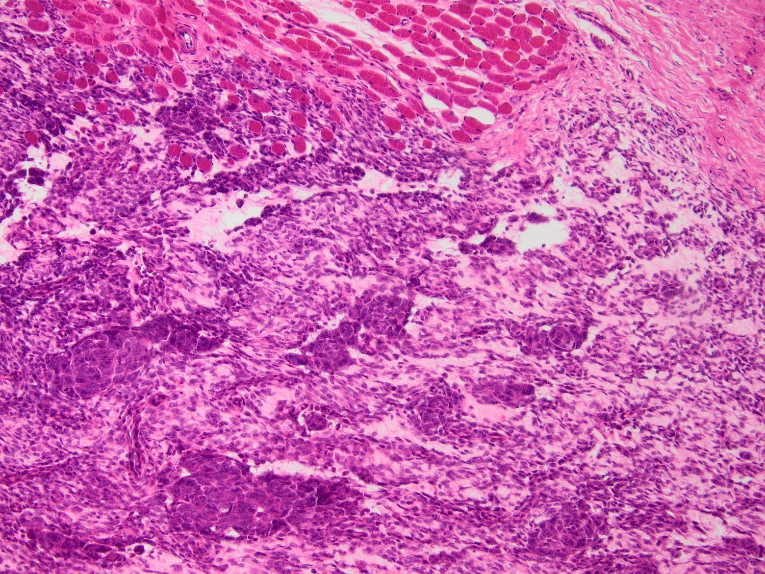
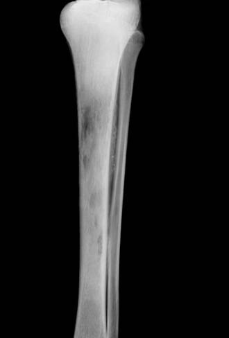
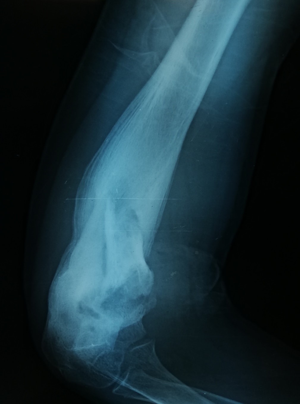
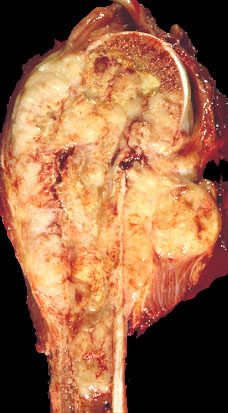
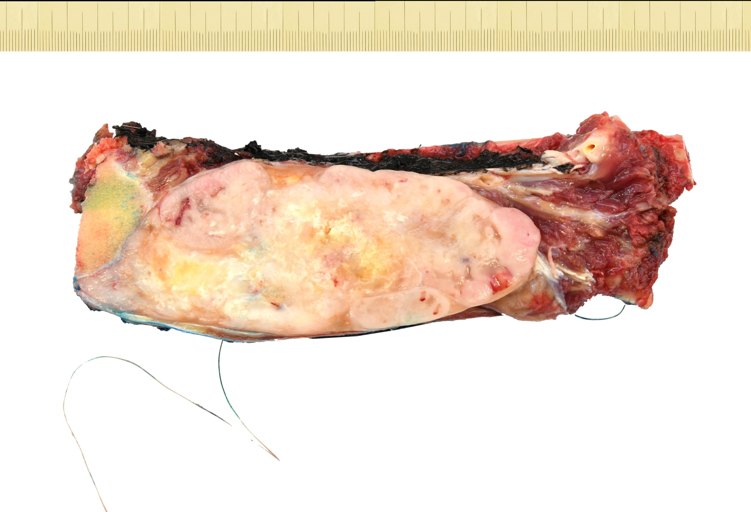
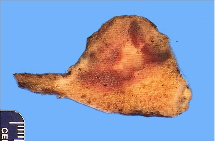
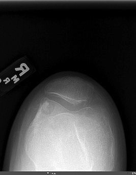
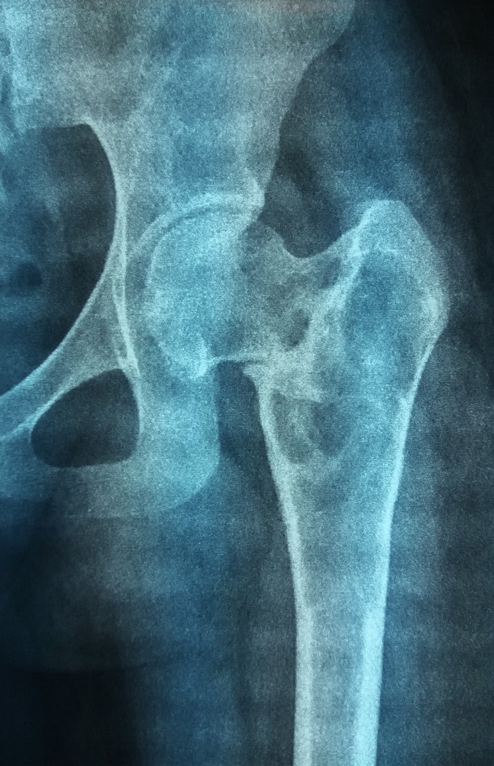
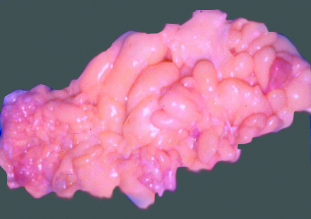
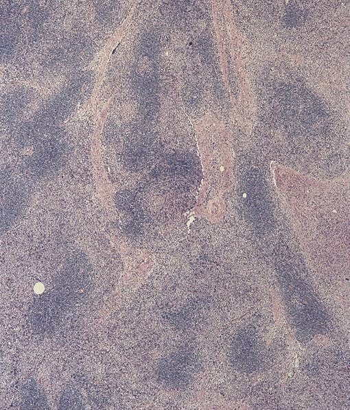
Tibial lesion
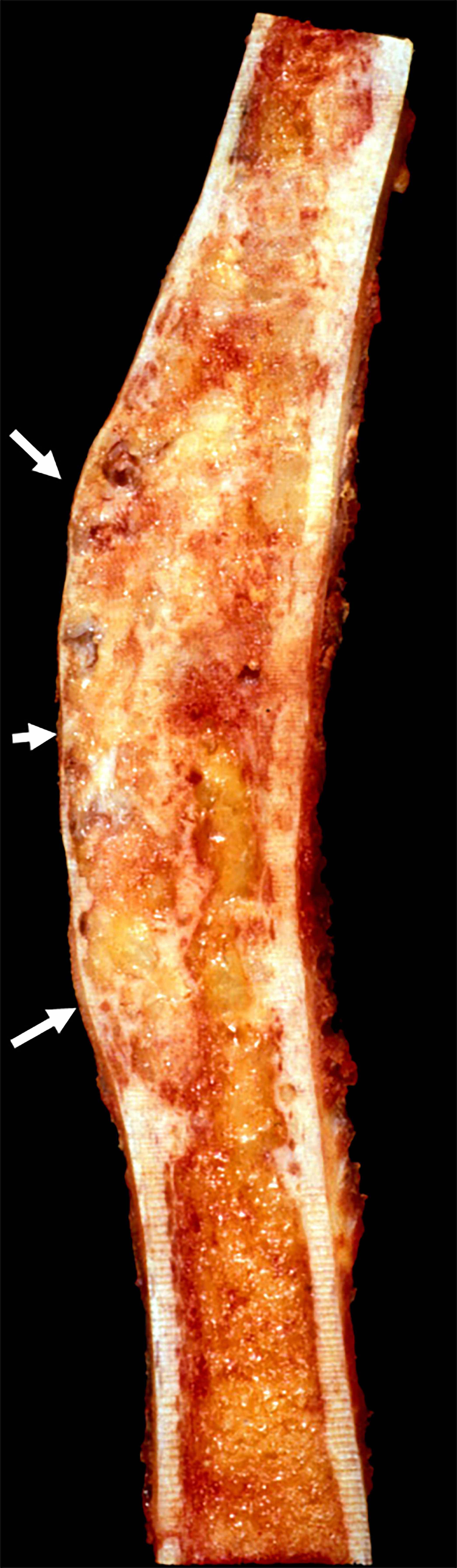
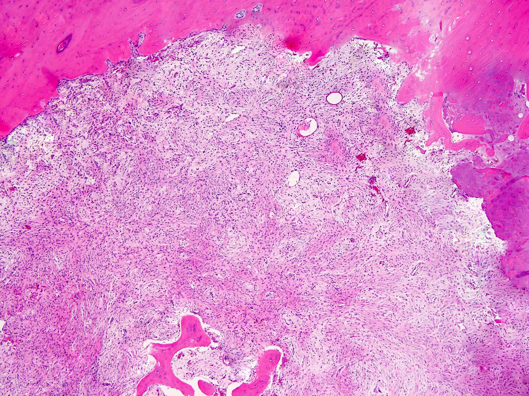
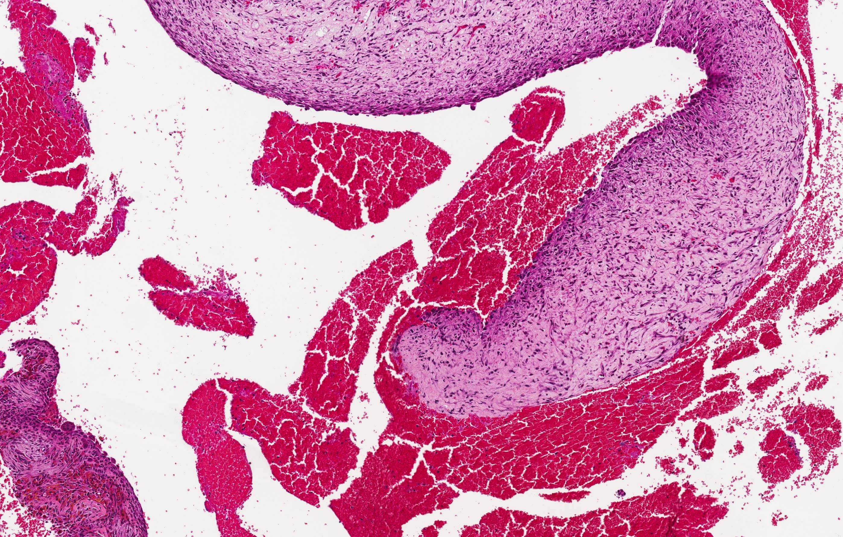
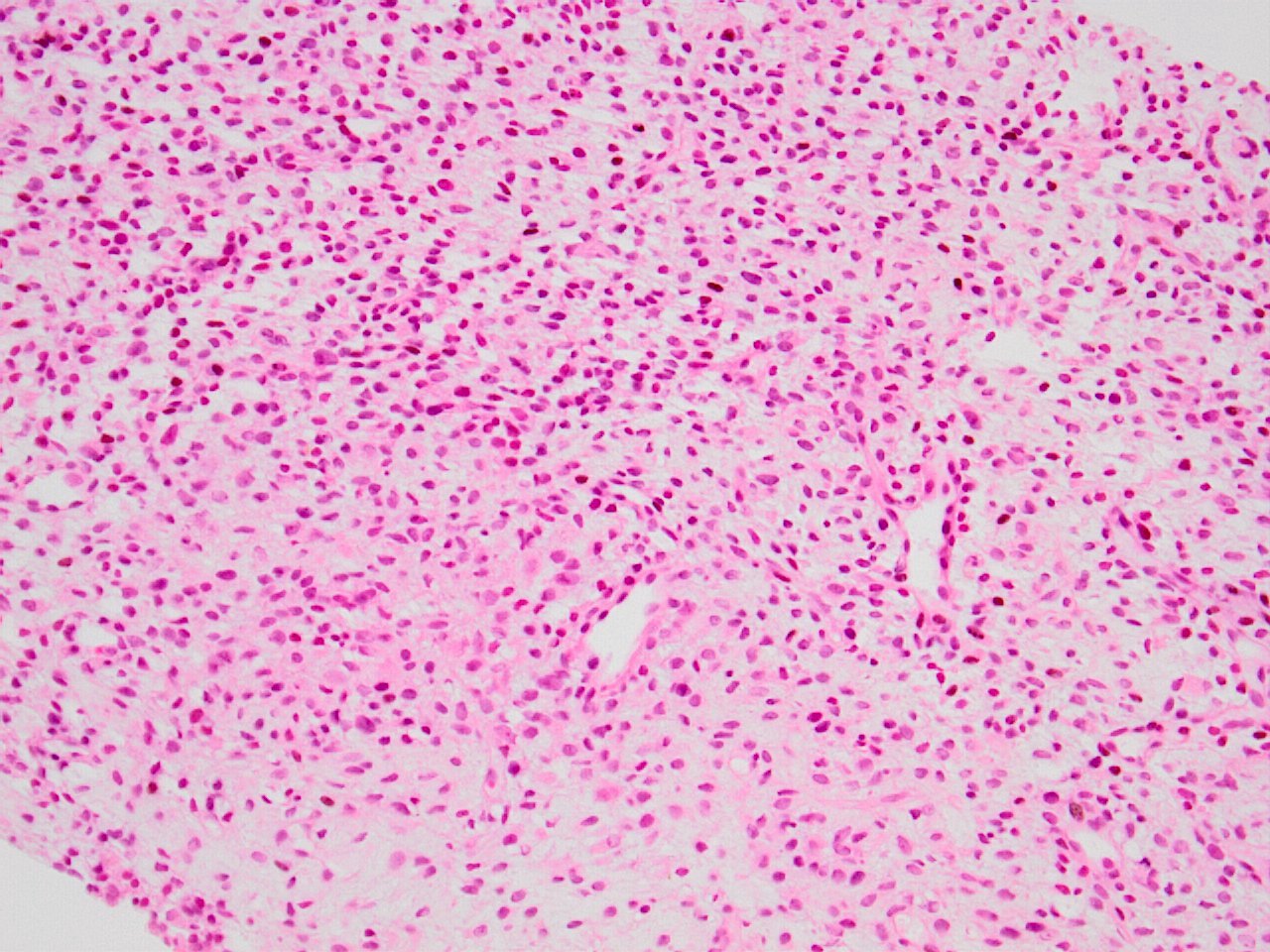
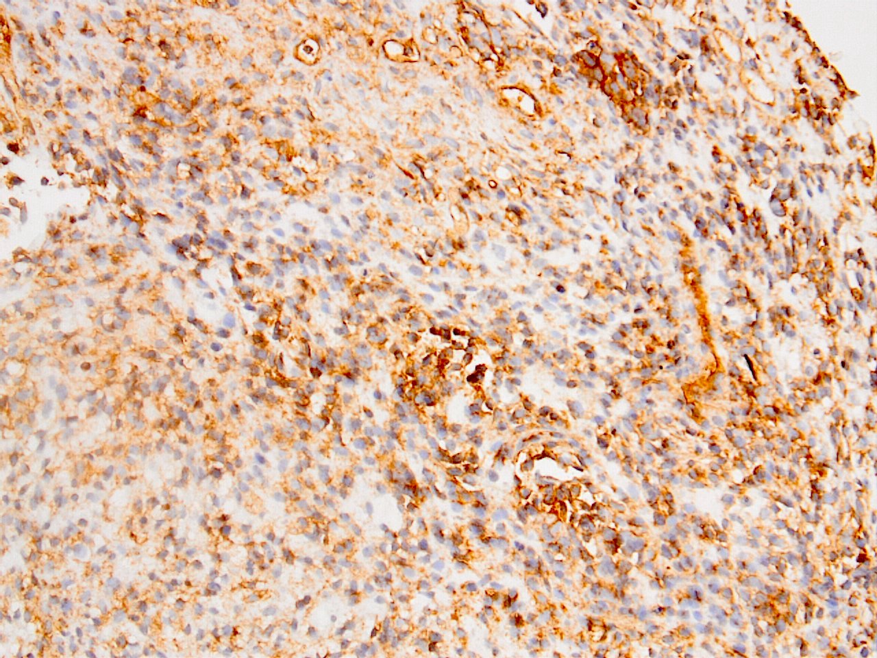
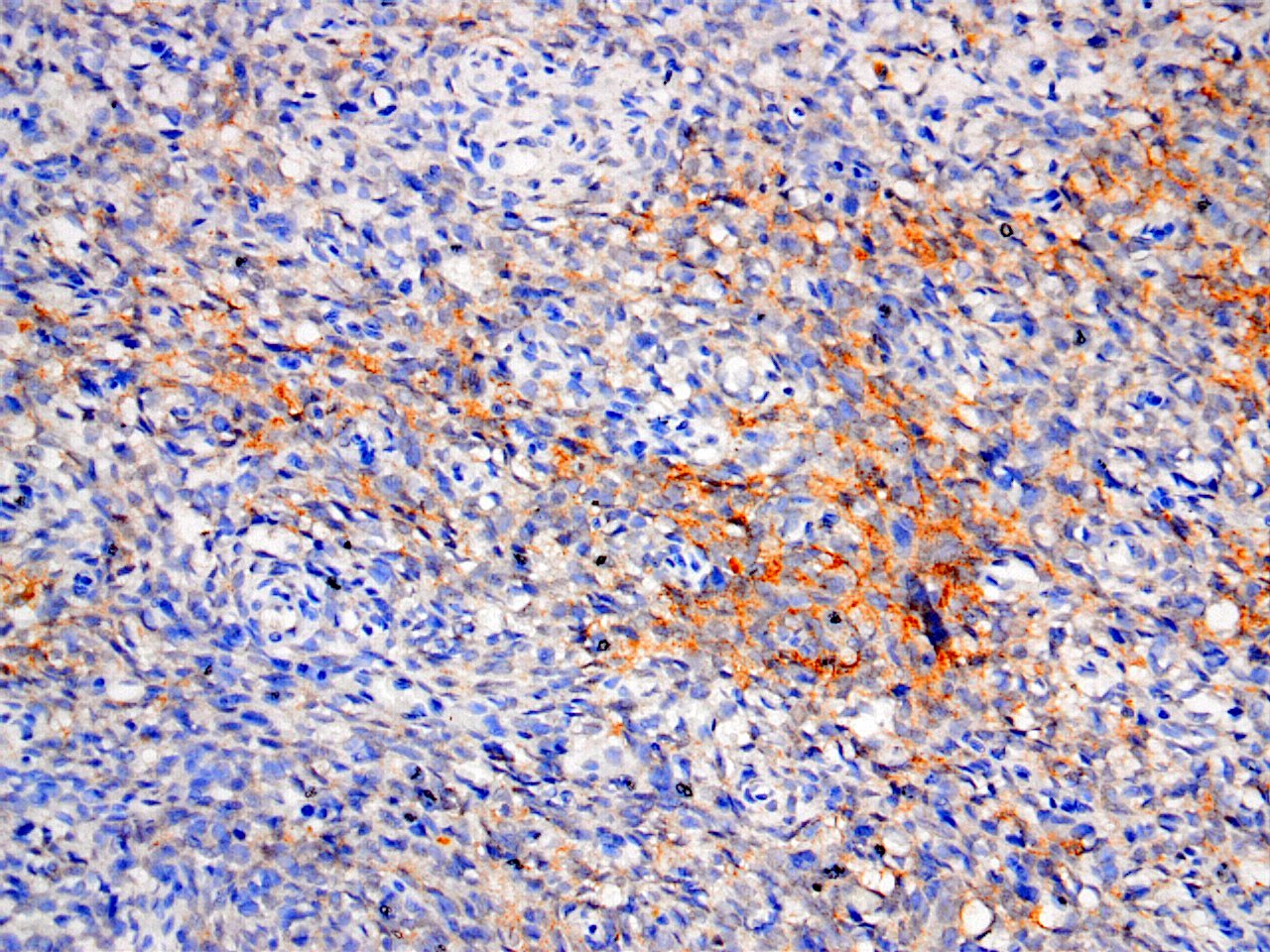
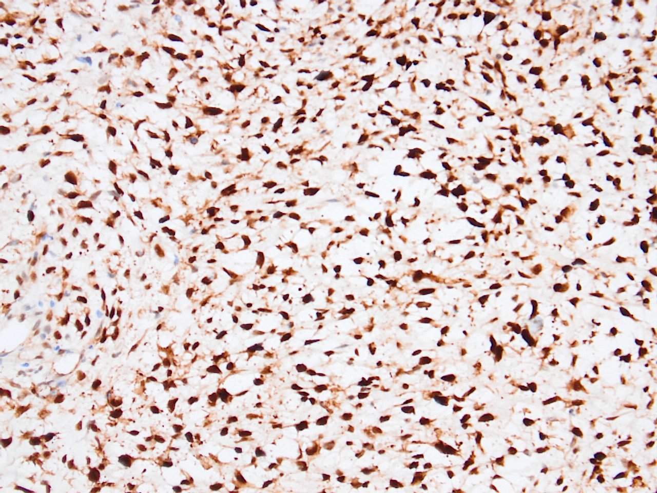
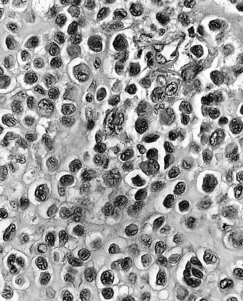
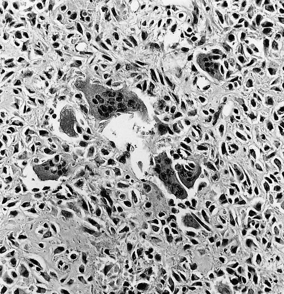
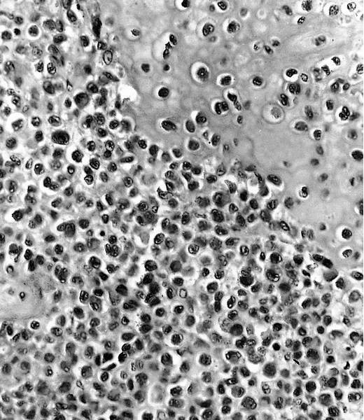
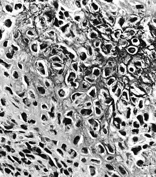
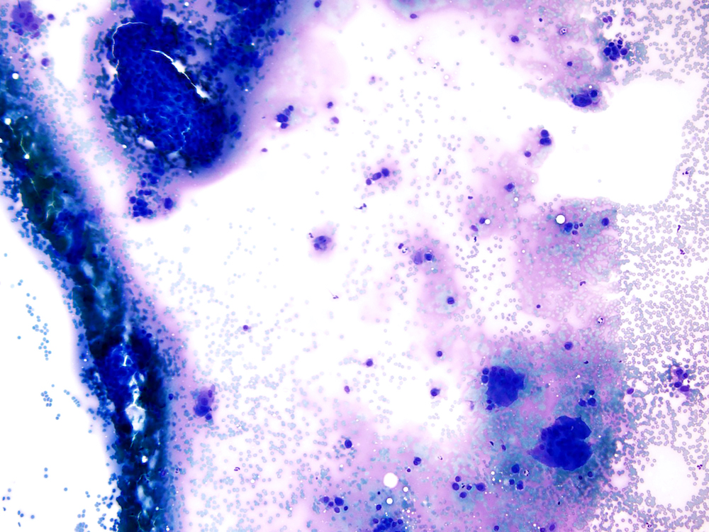
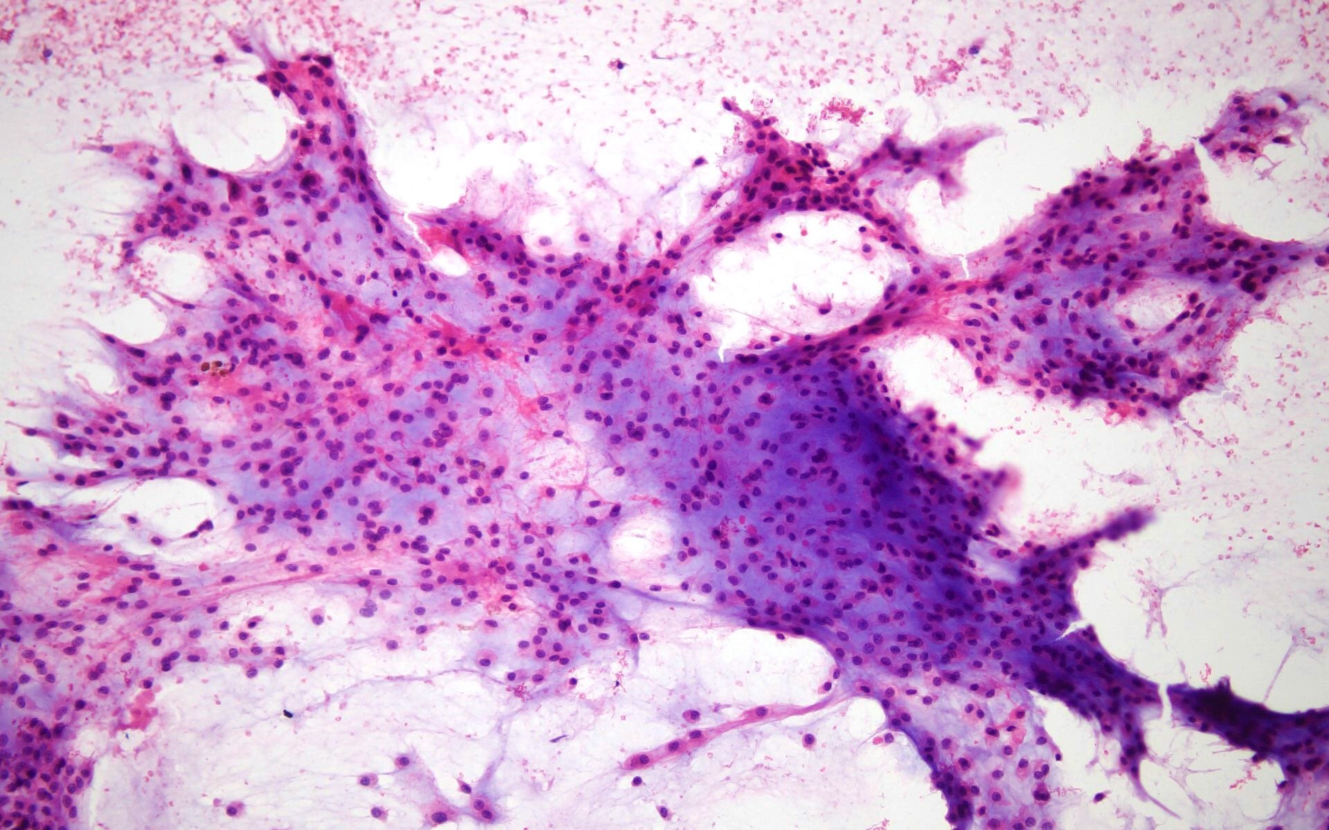
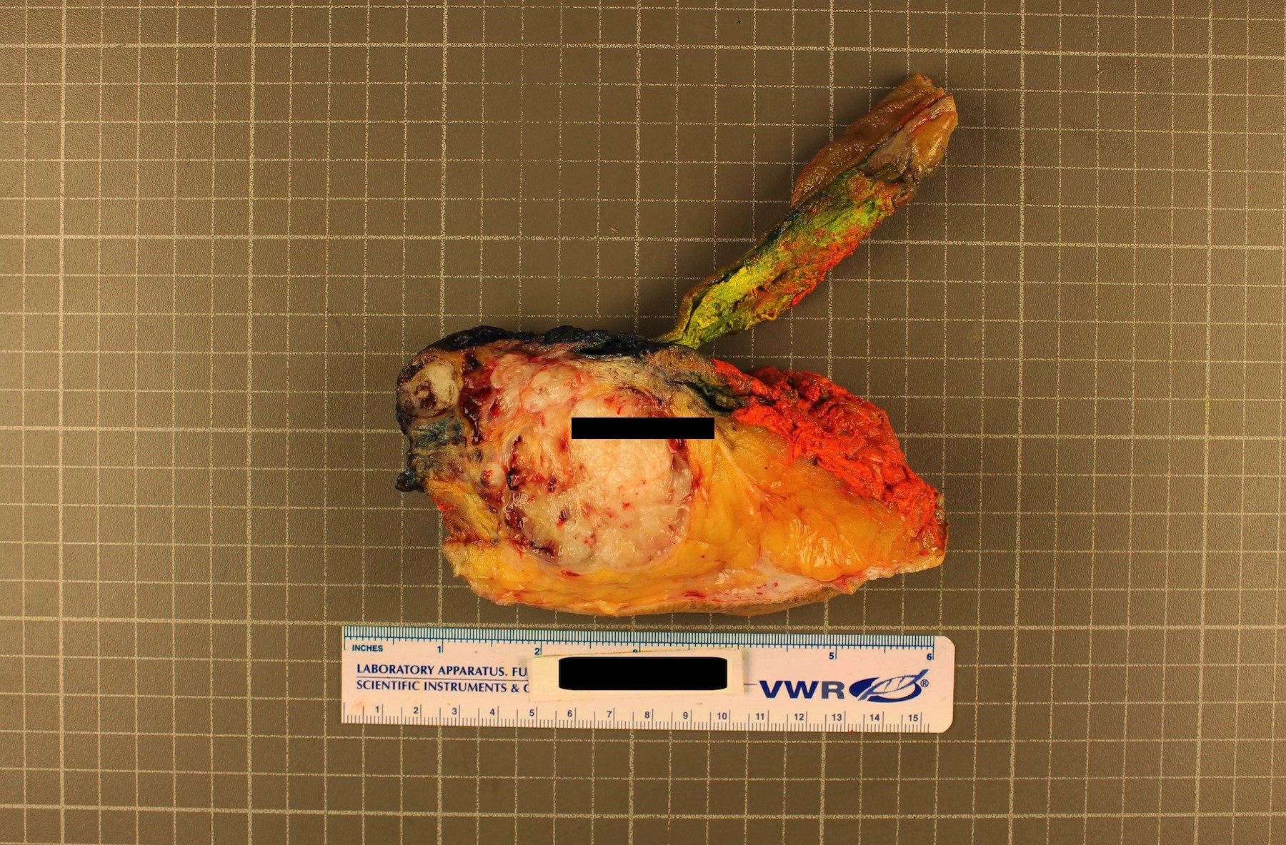
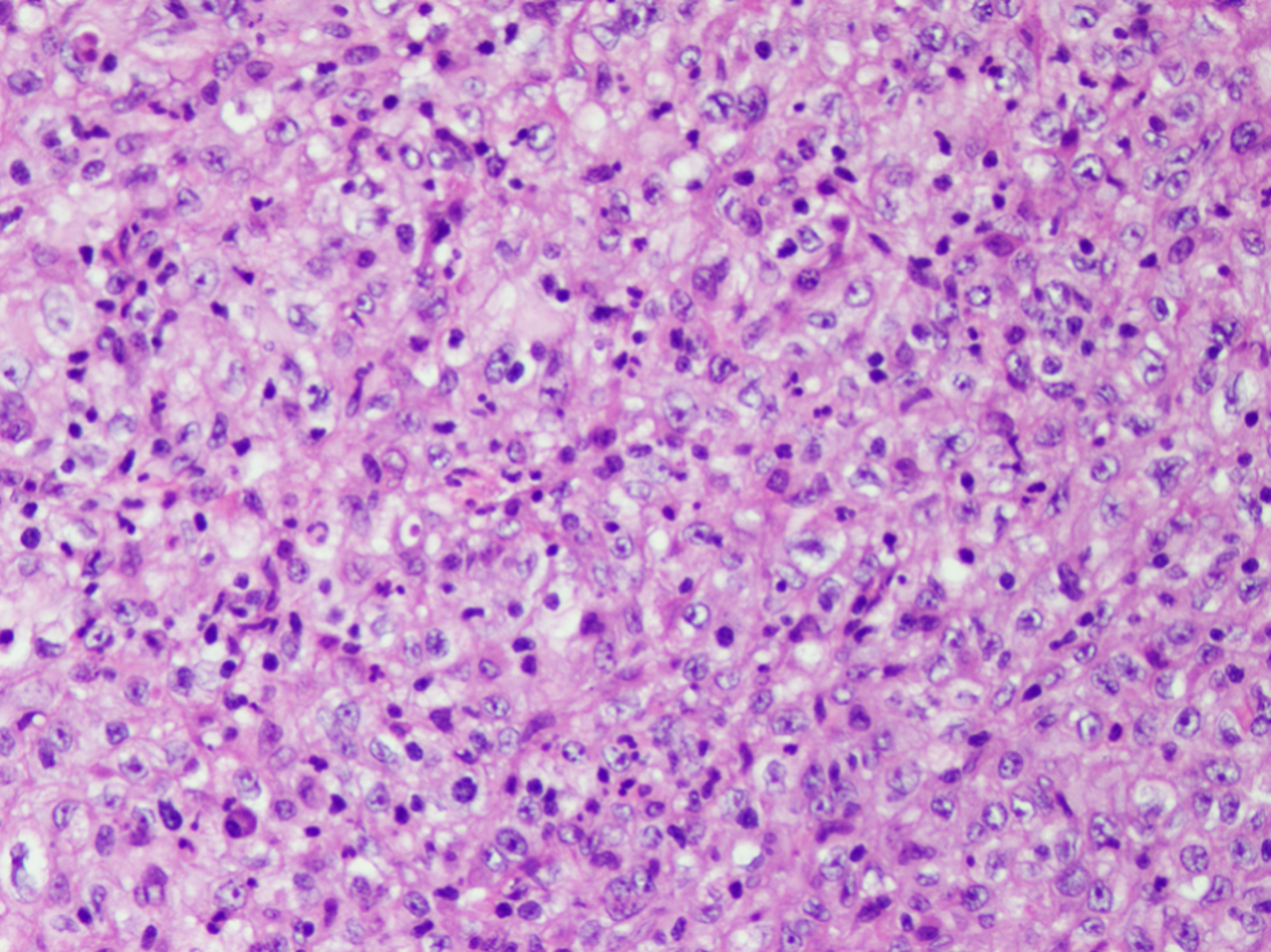
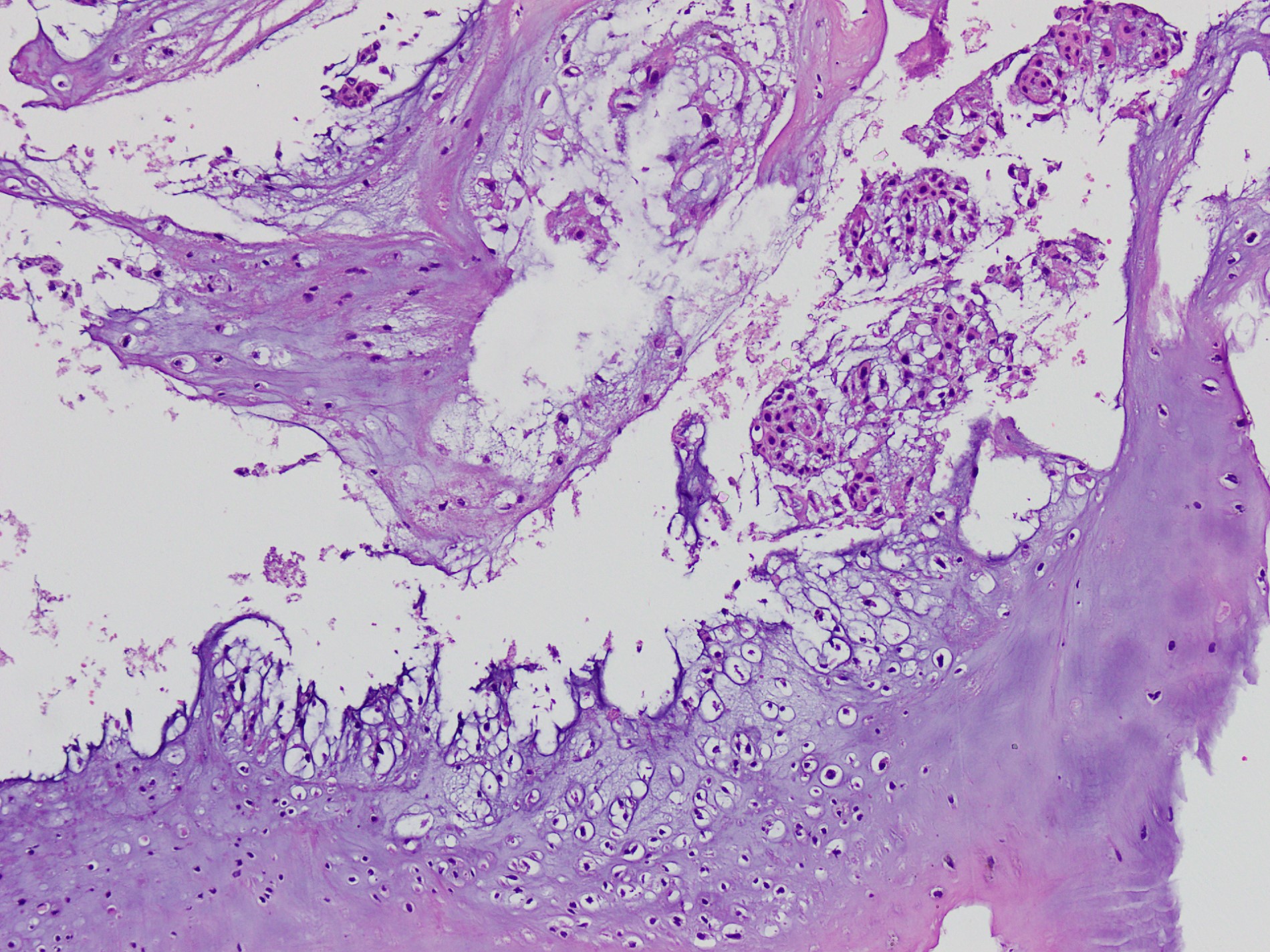
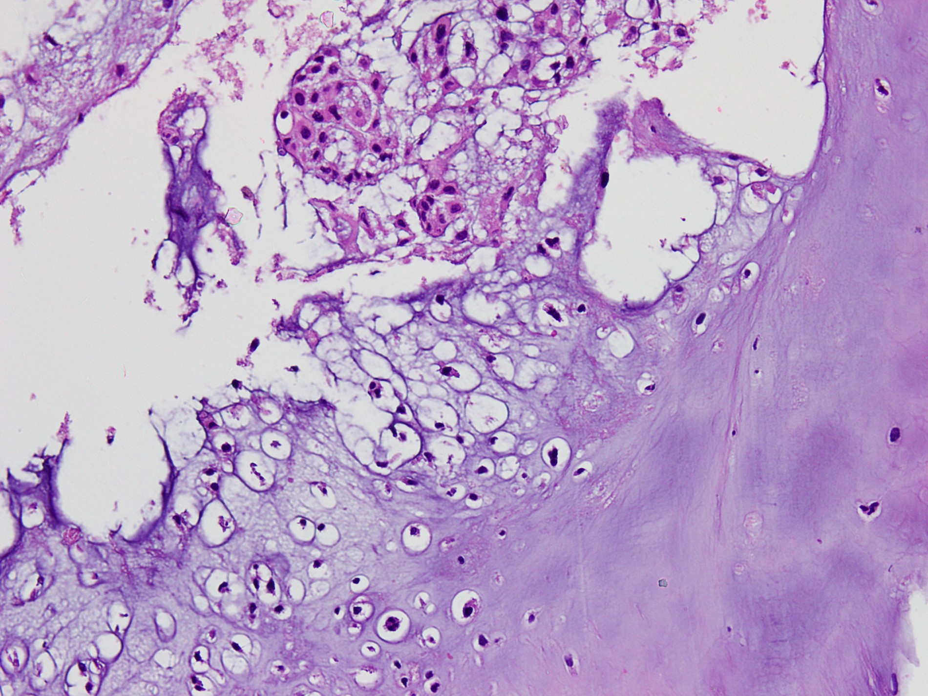
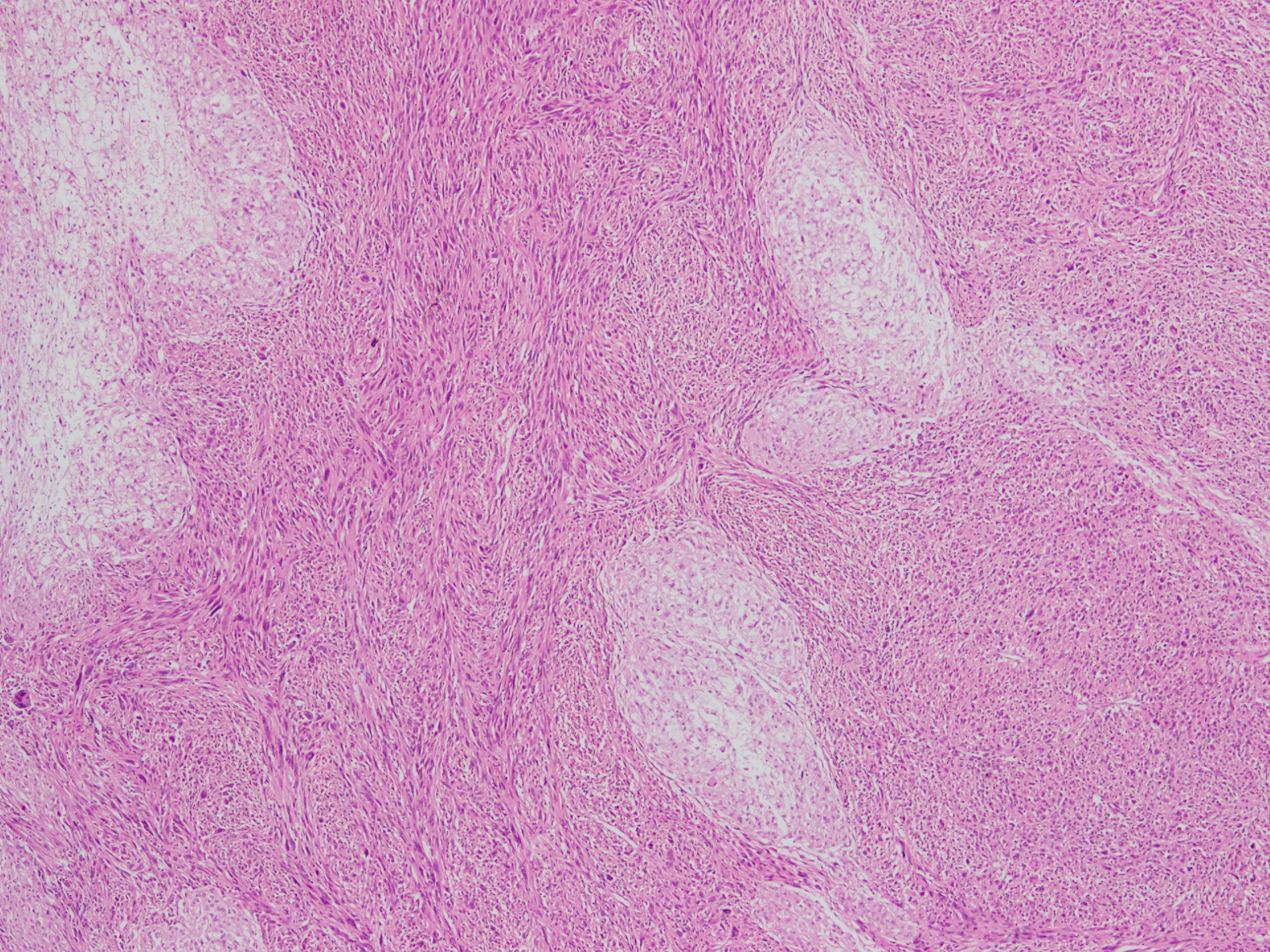
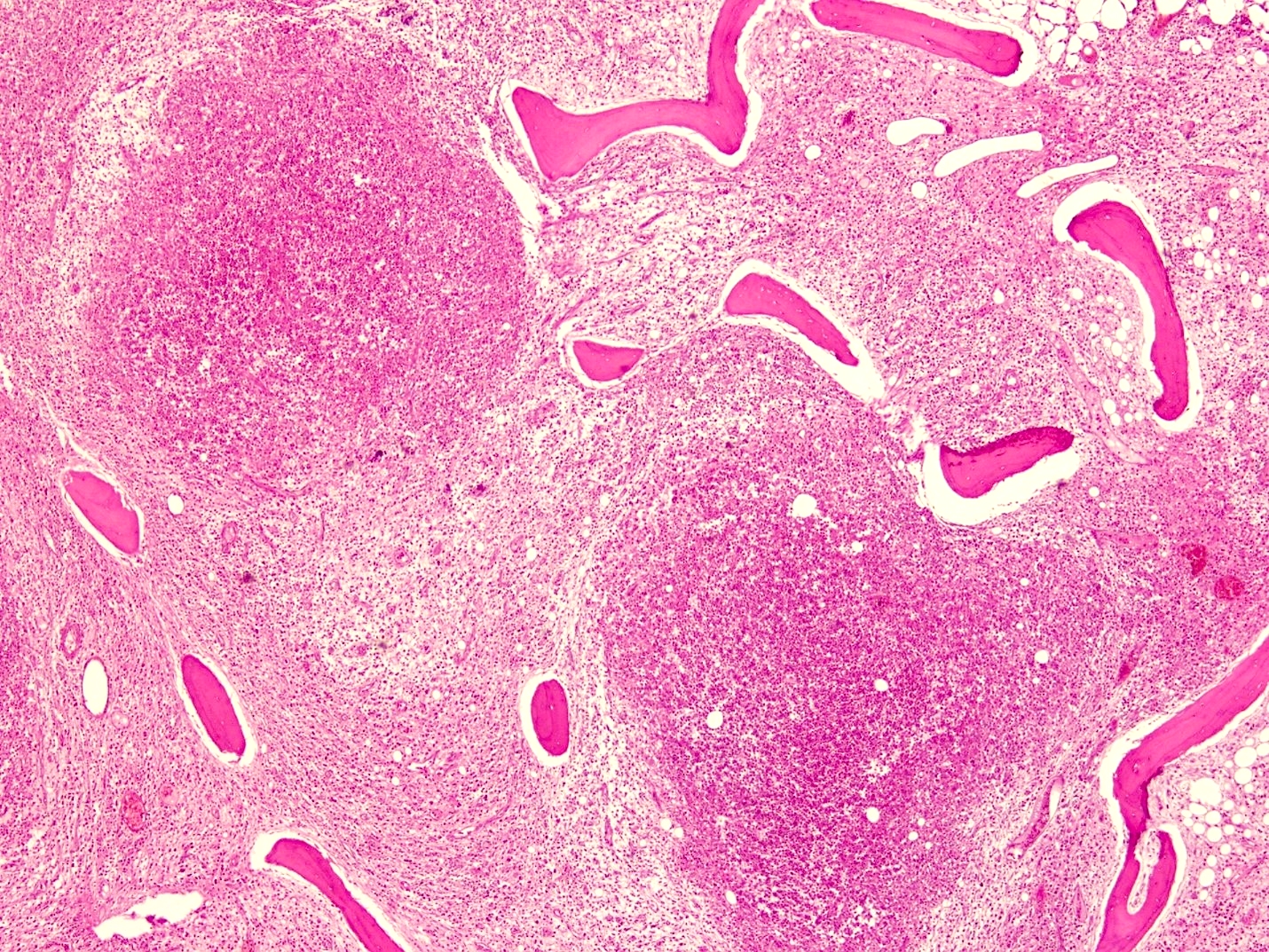
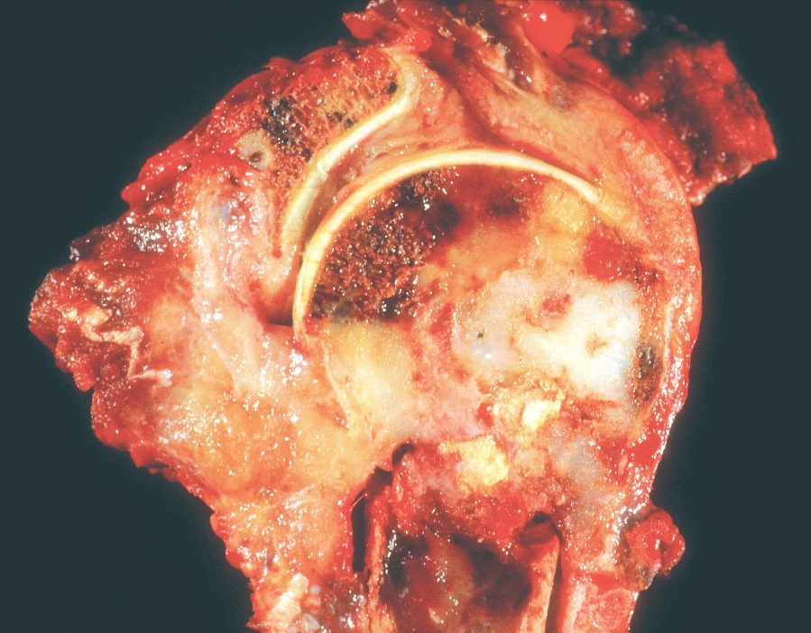
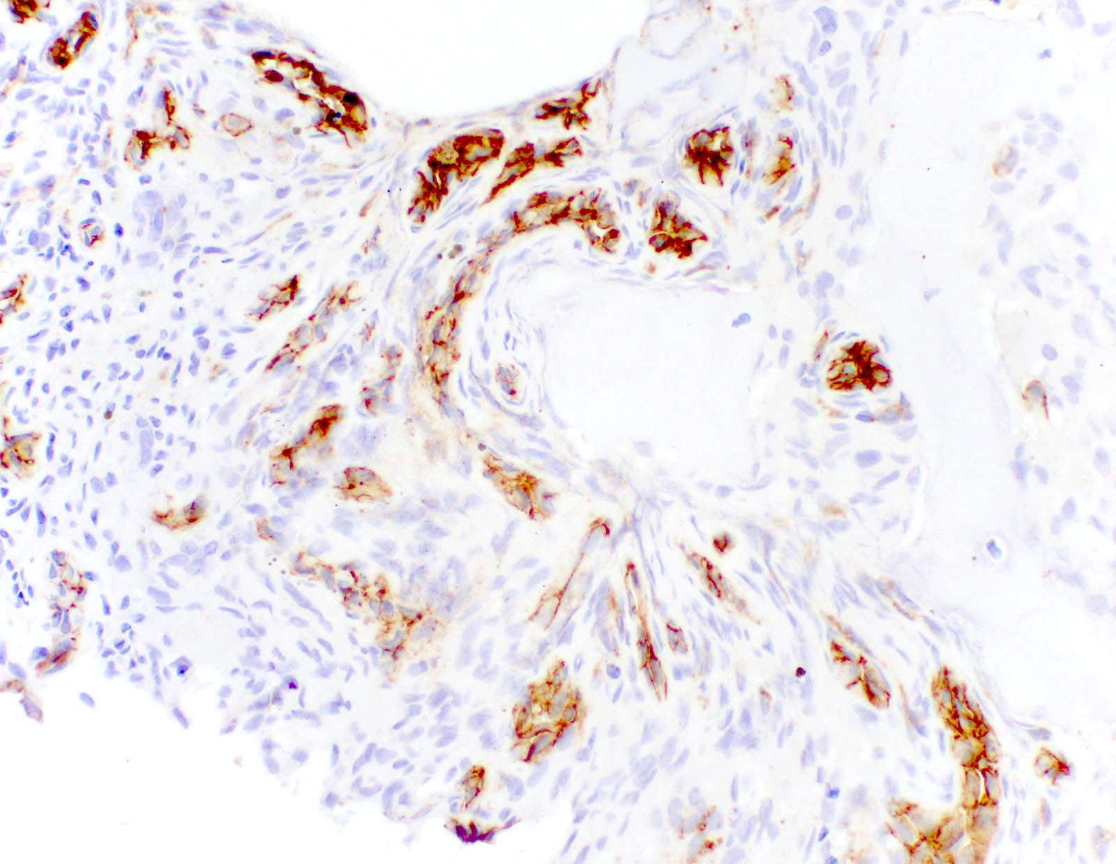
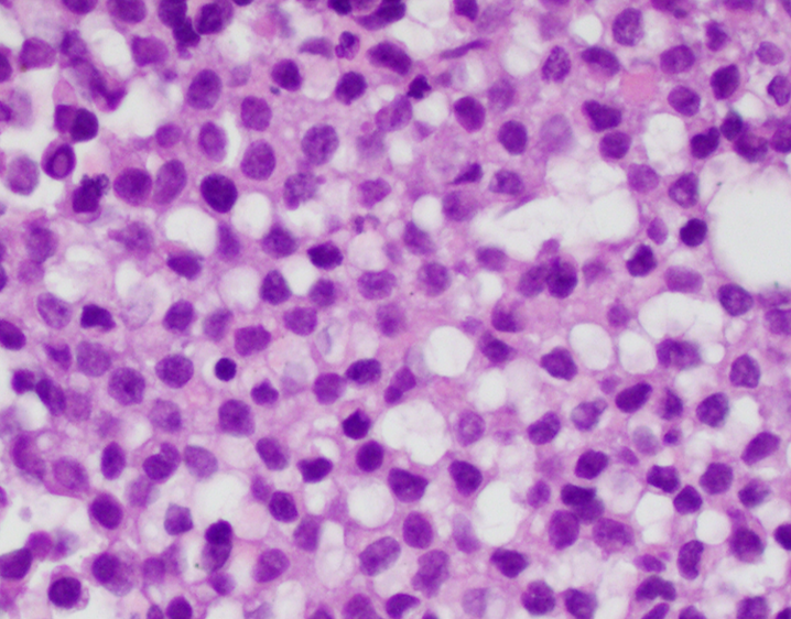
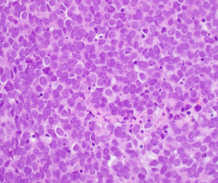
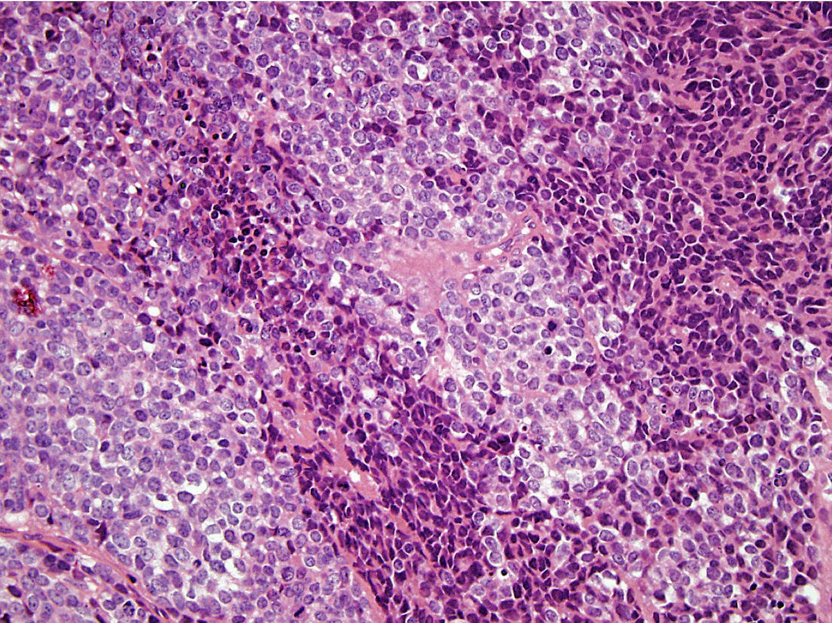
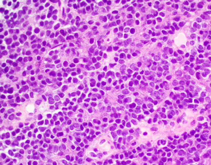
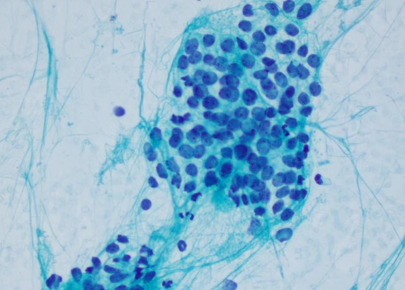
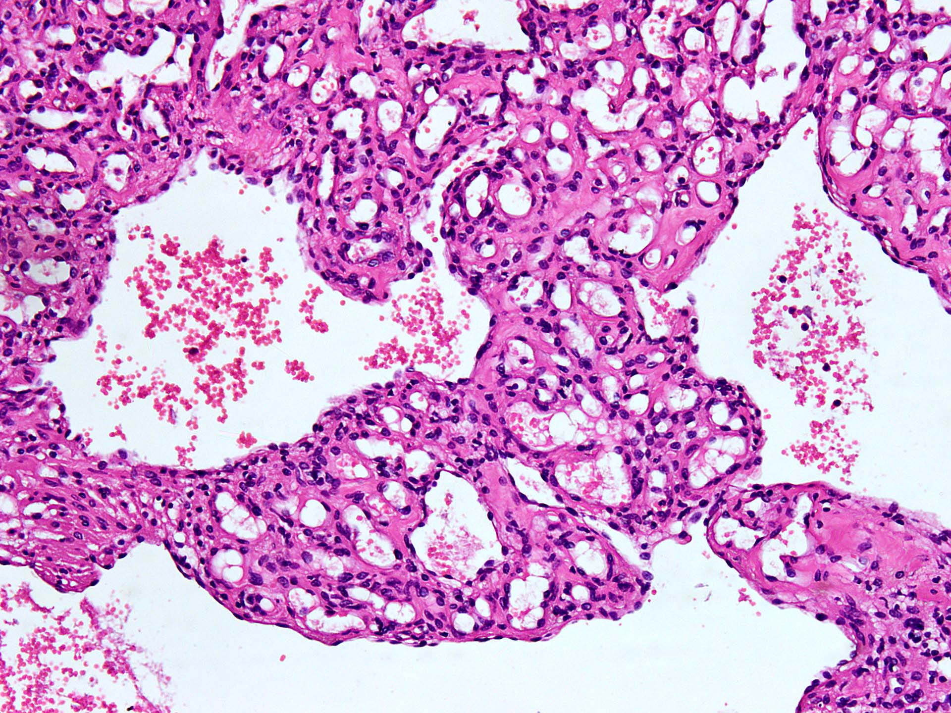
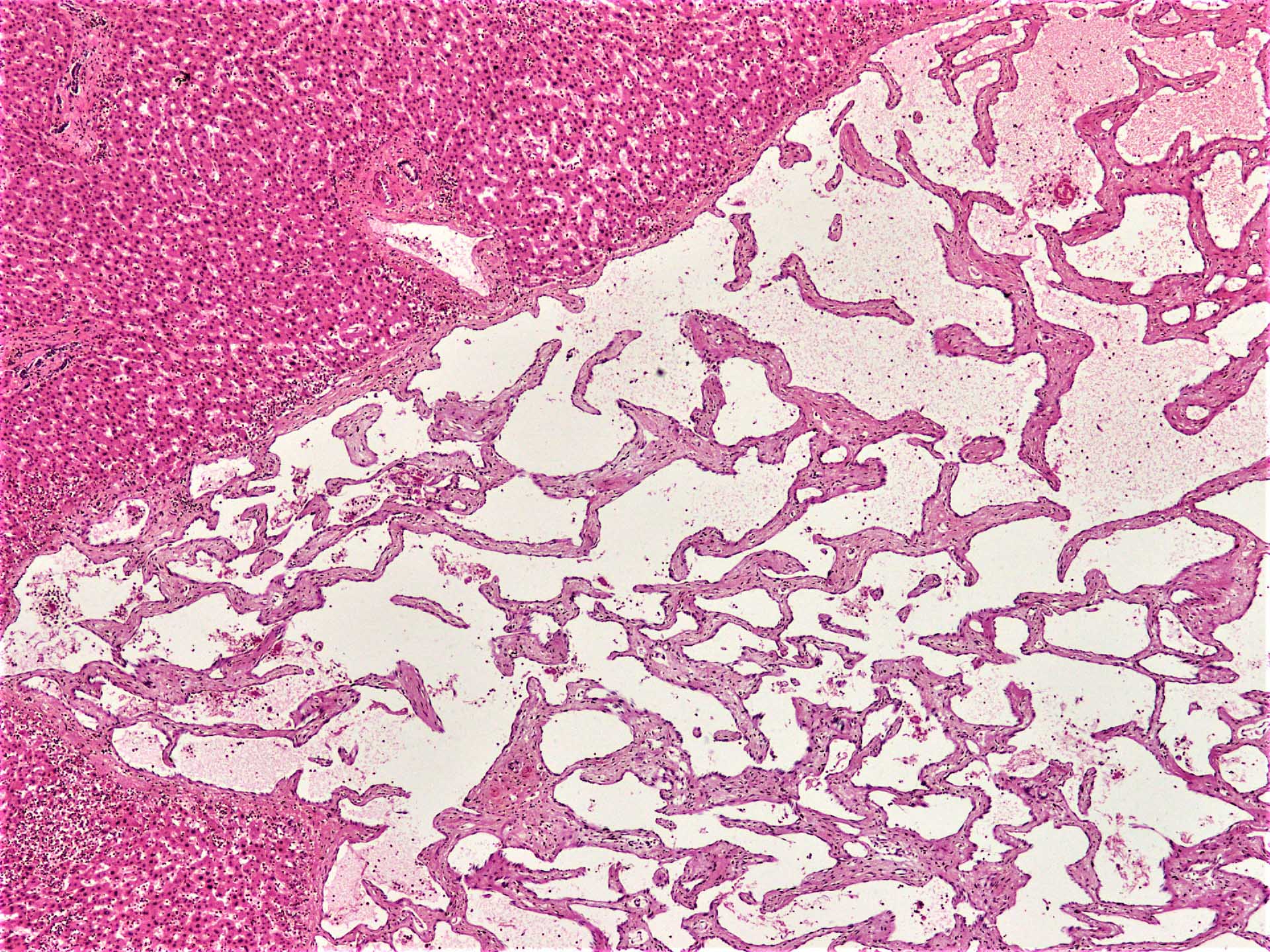
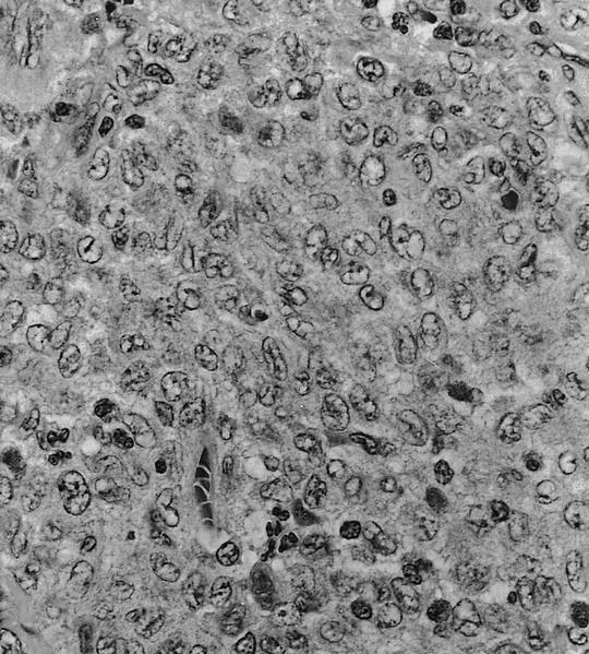
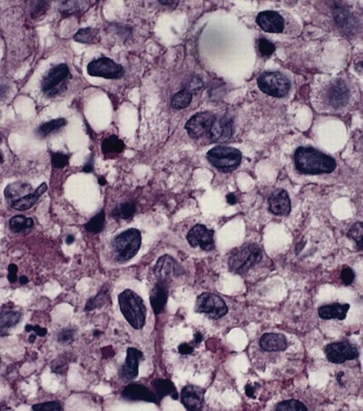
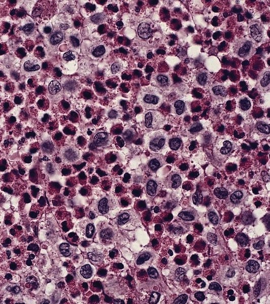
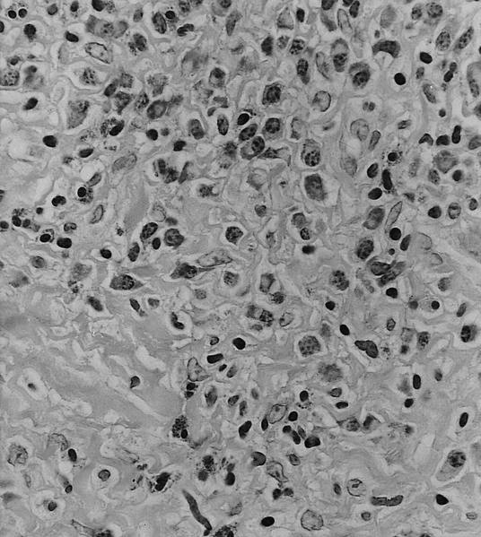
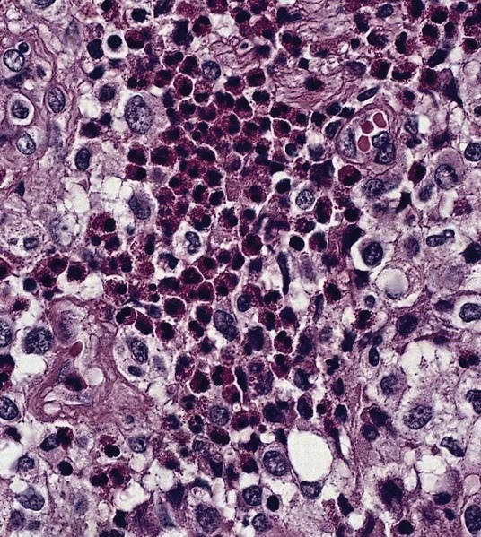
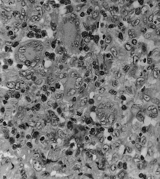
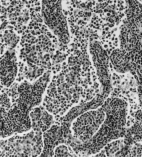
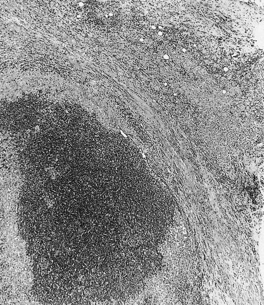
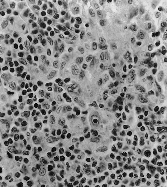
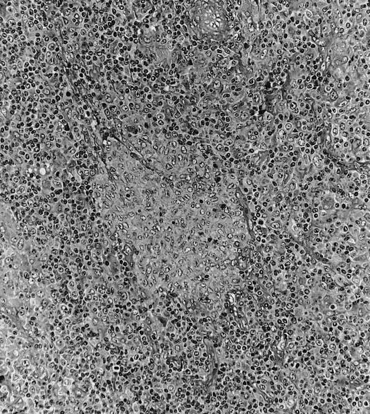
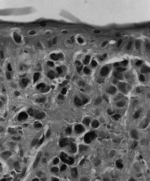
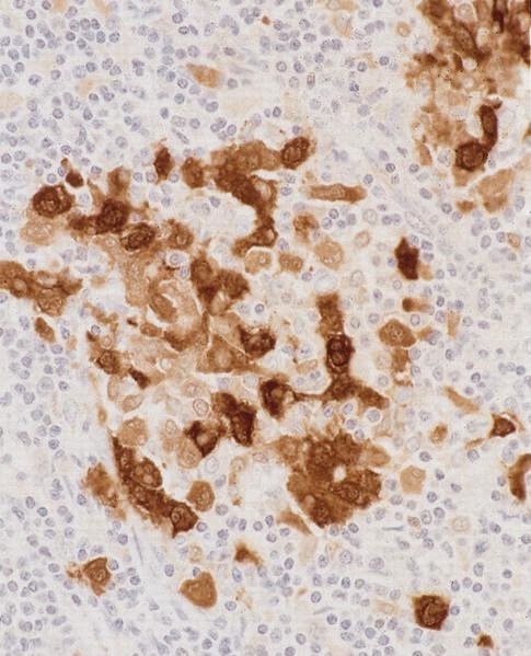
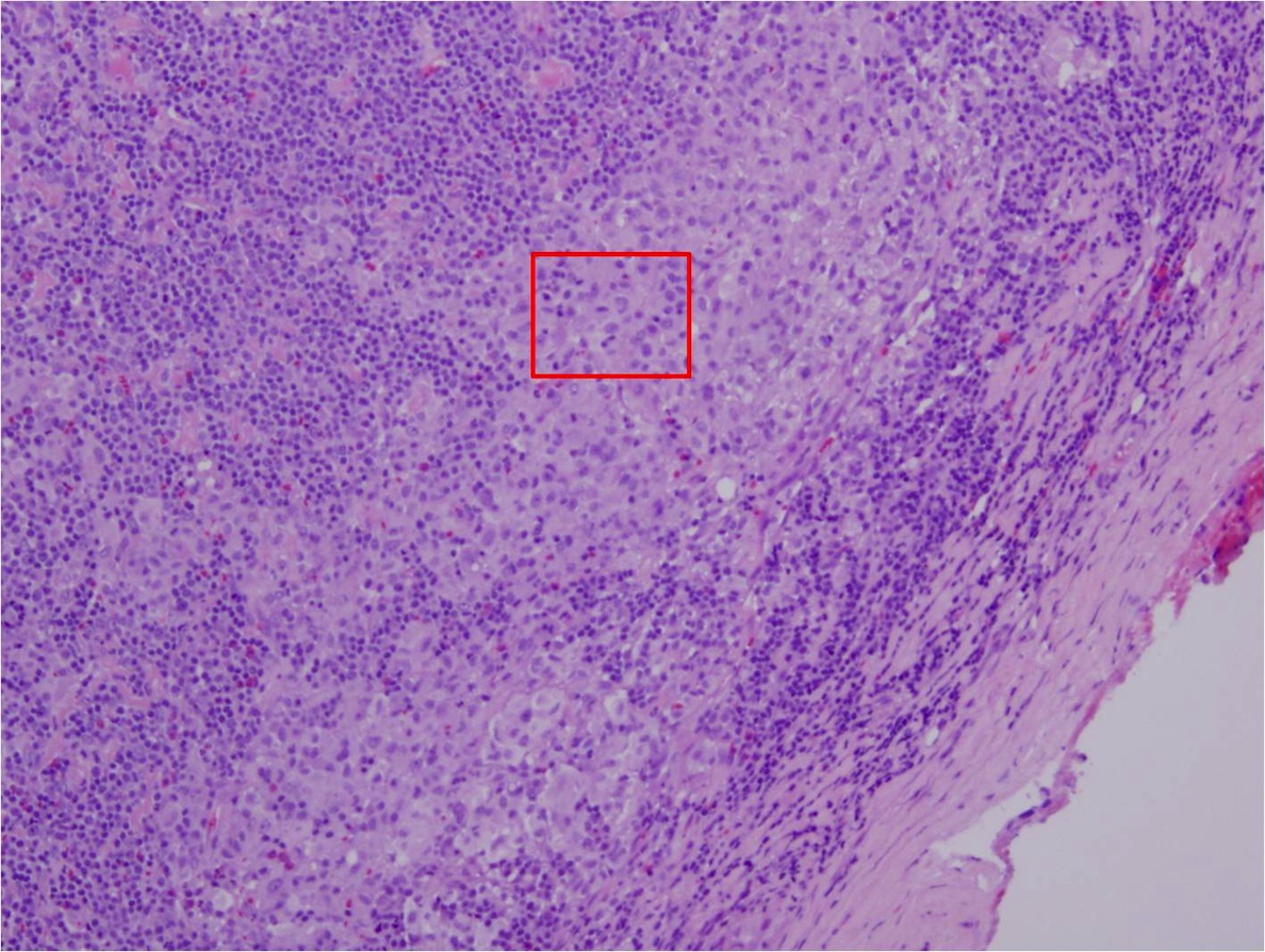
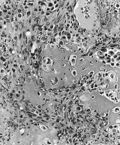
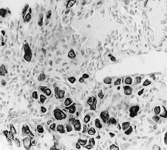
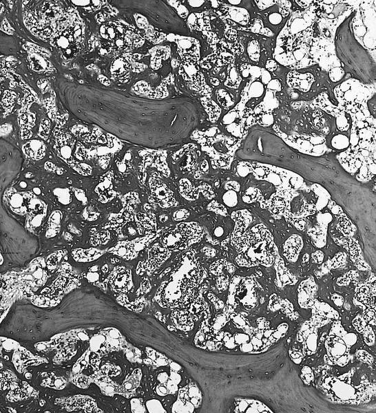
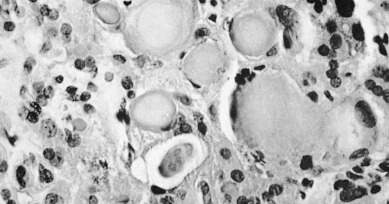
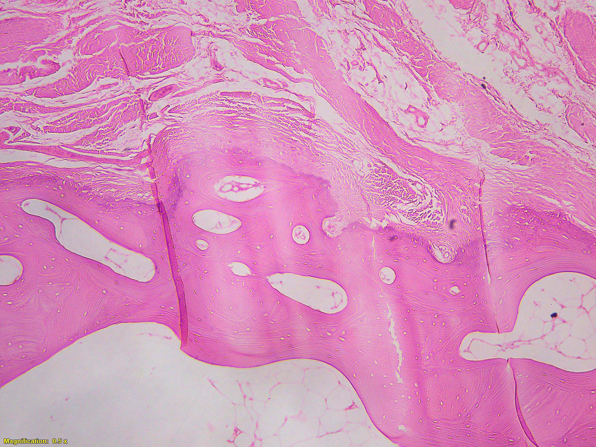
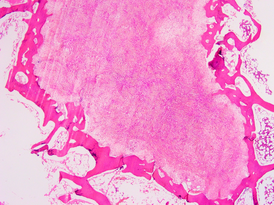
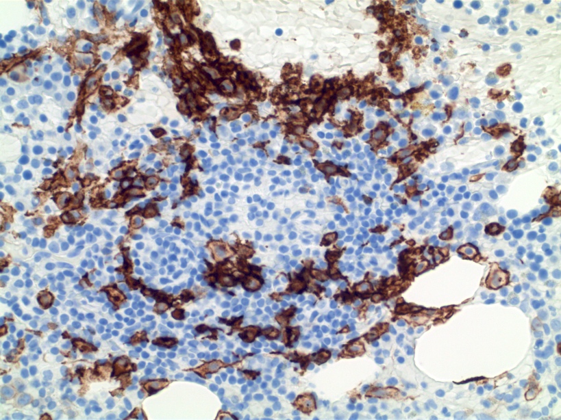
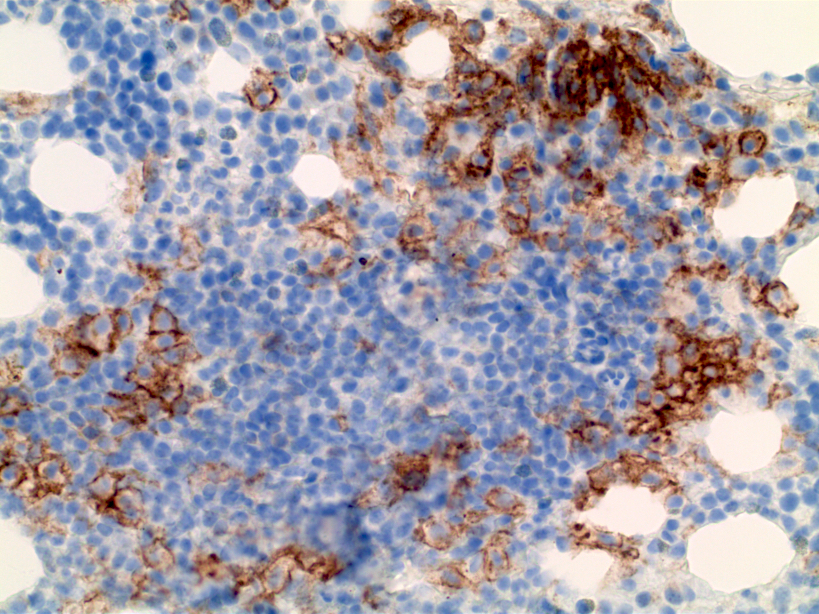
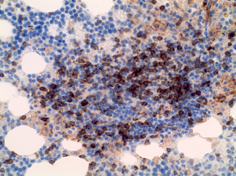
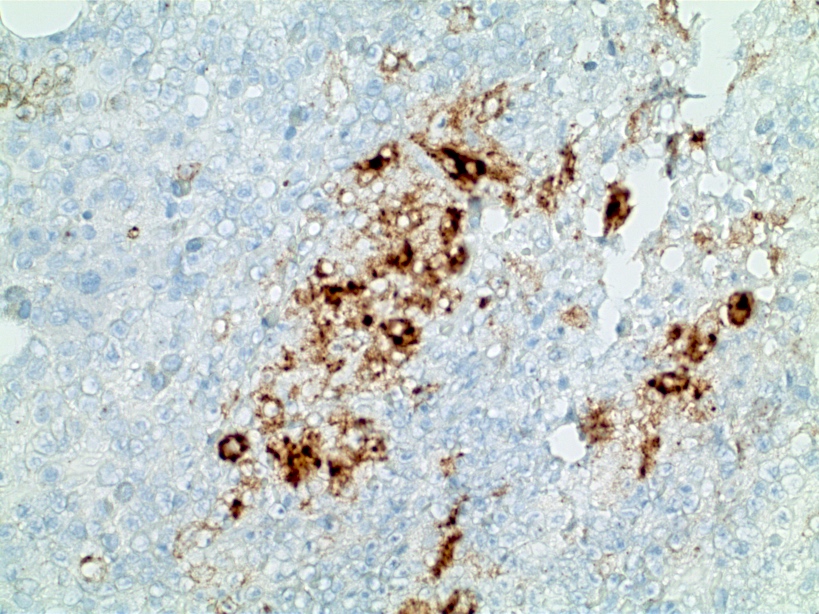
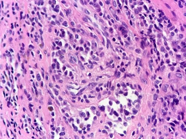
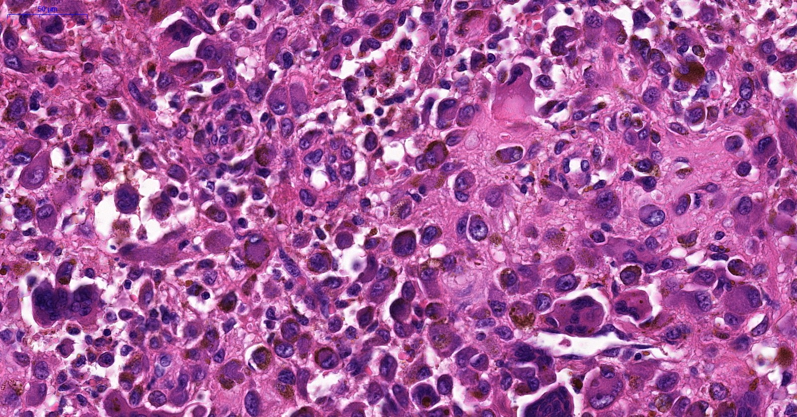
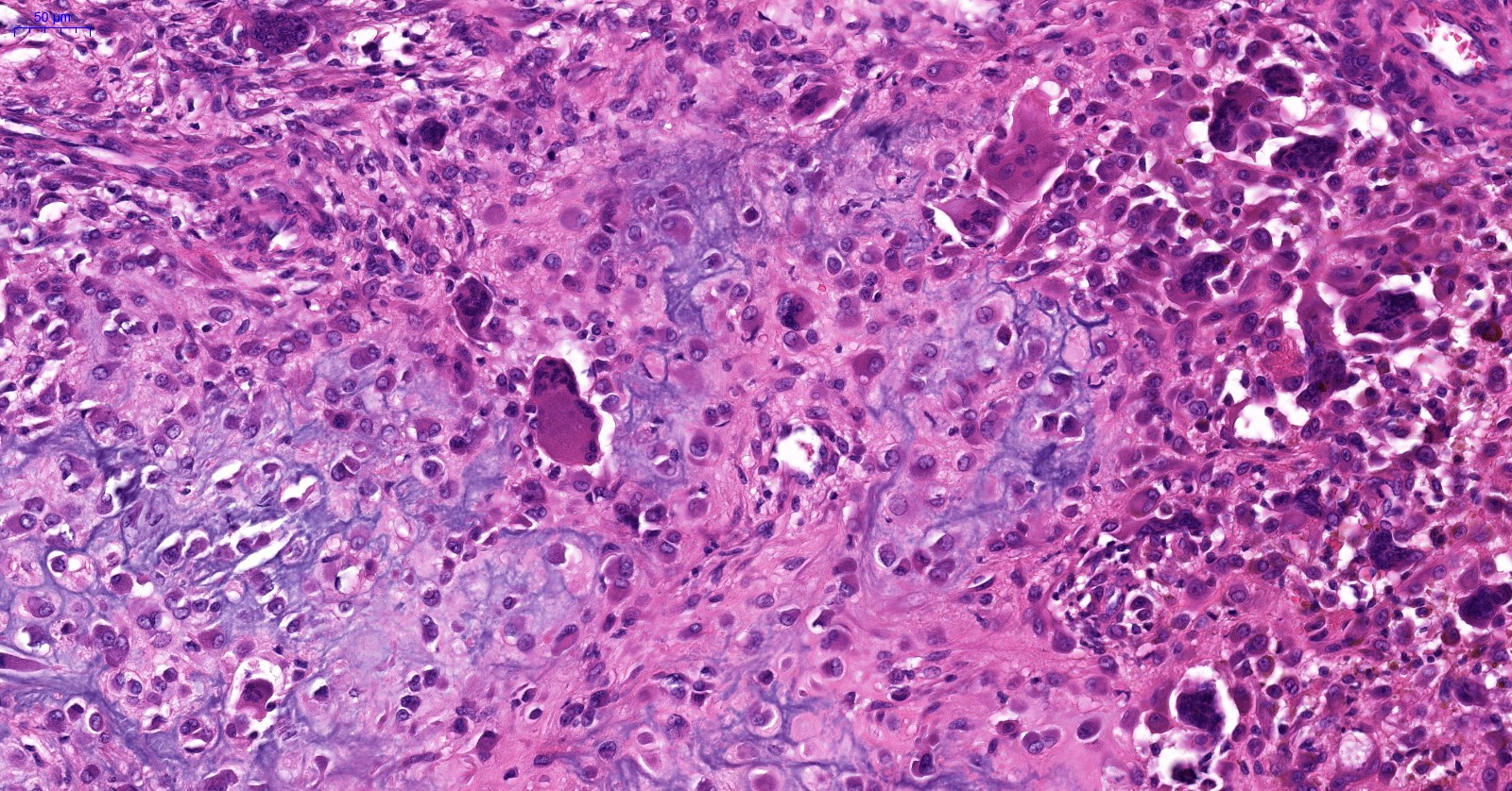
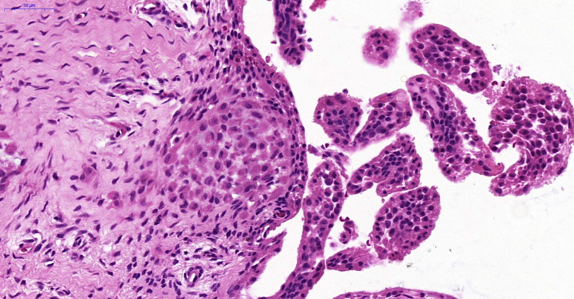
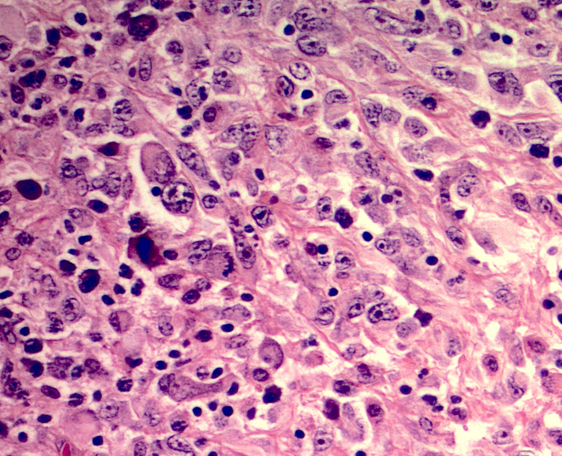
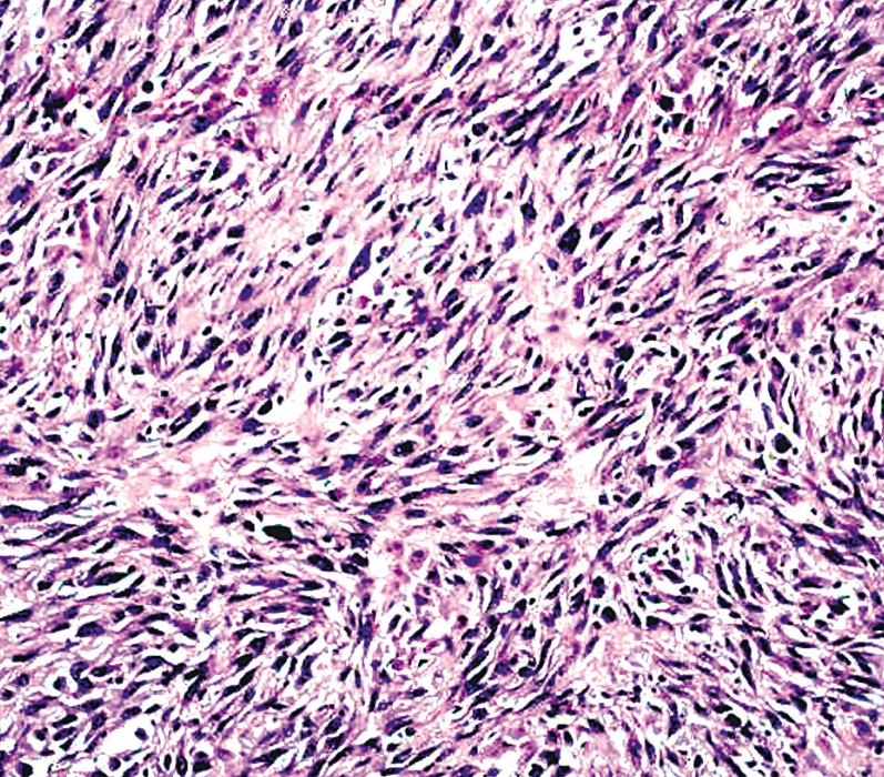
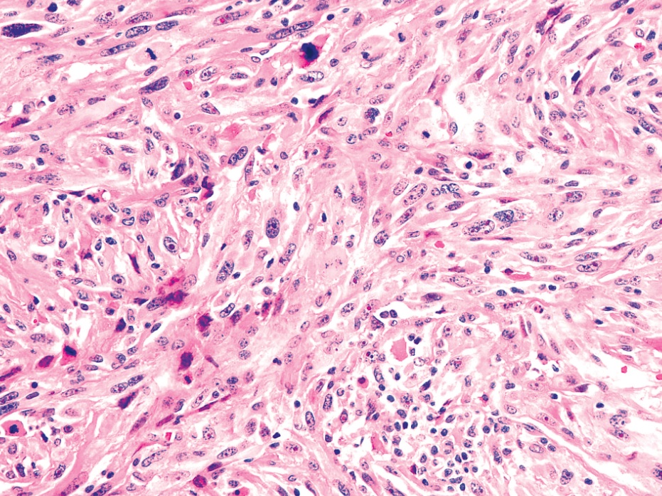
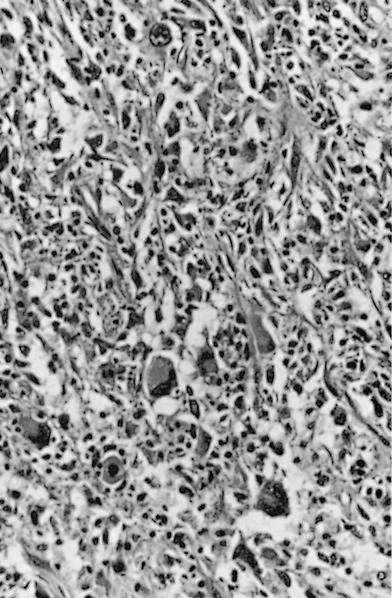
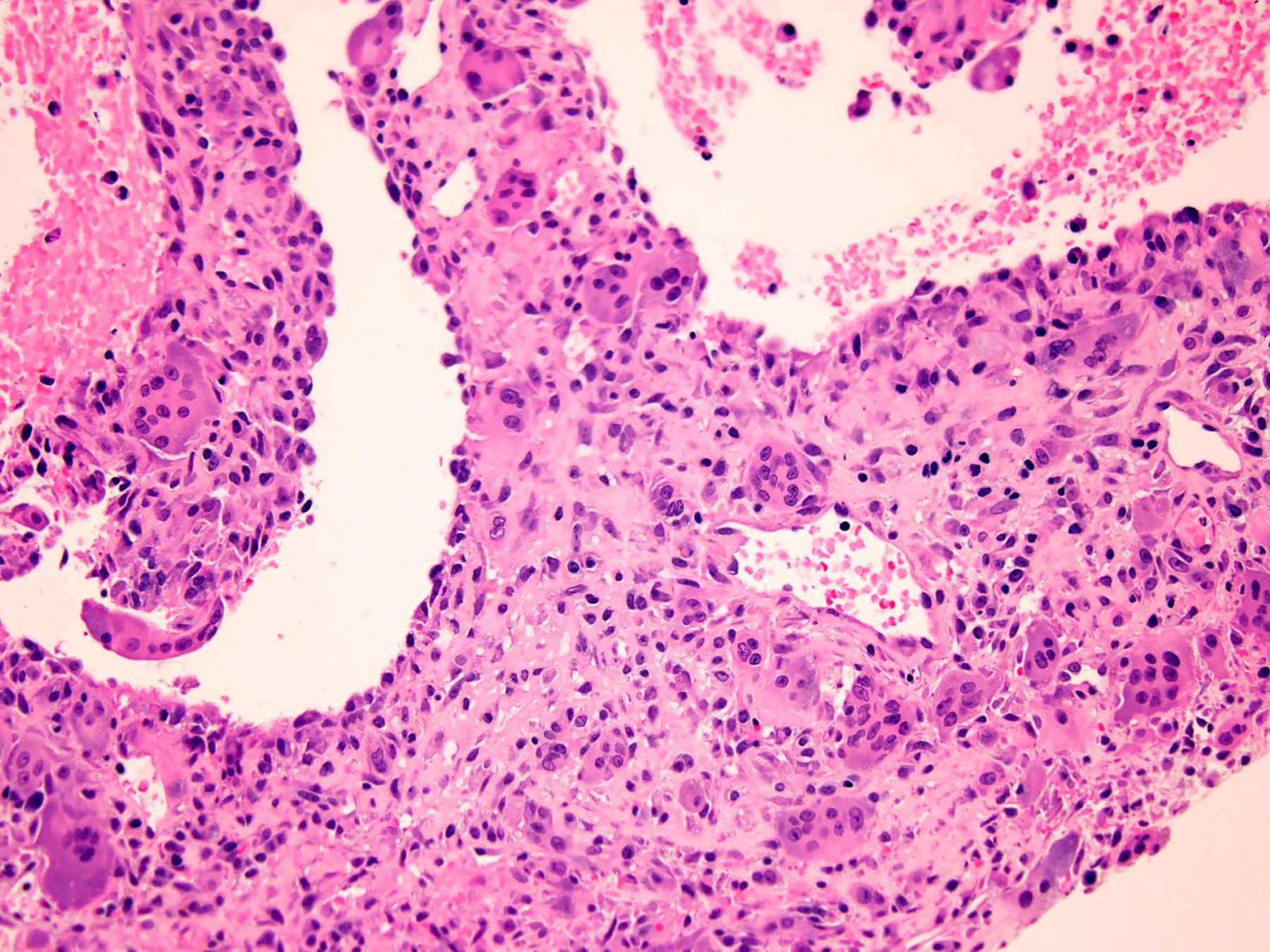
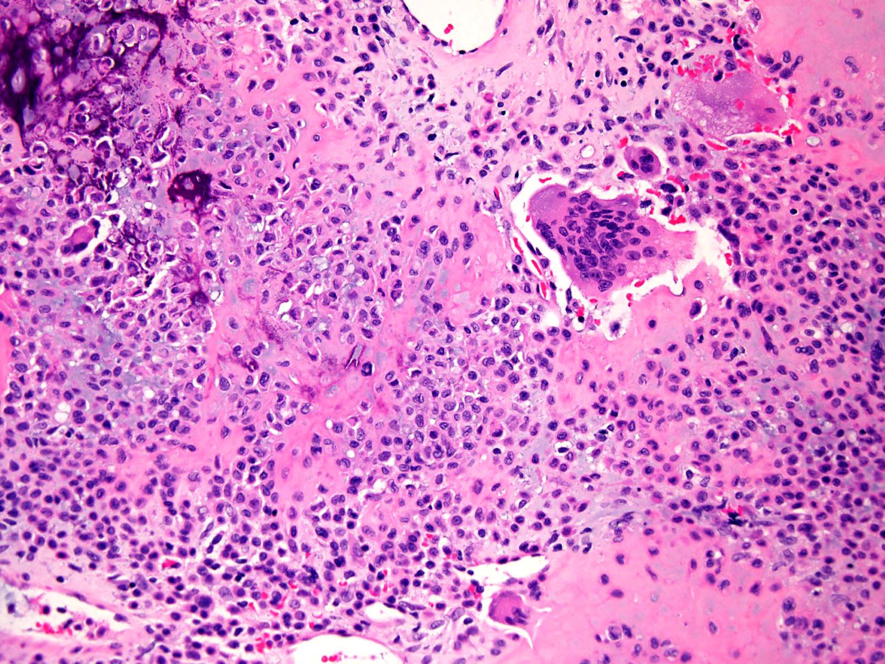
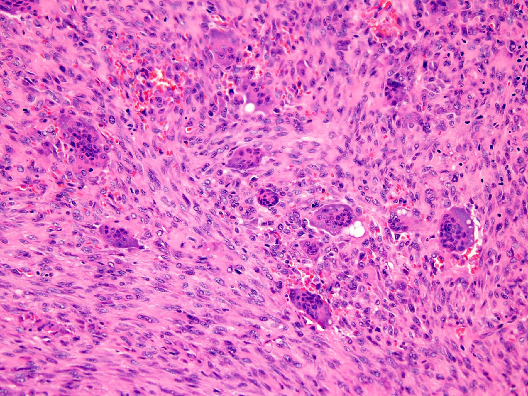
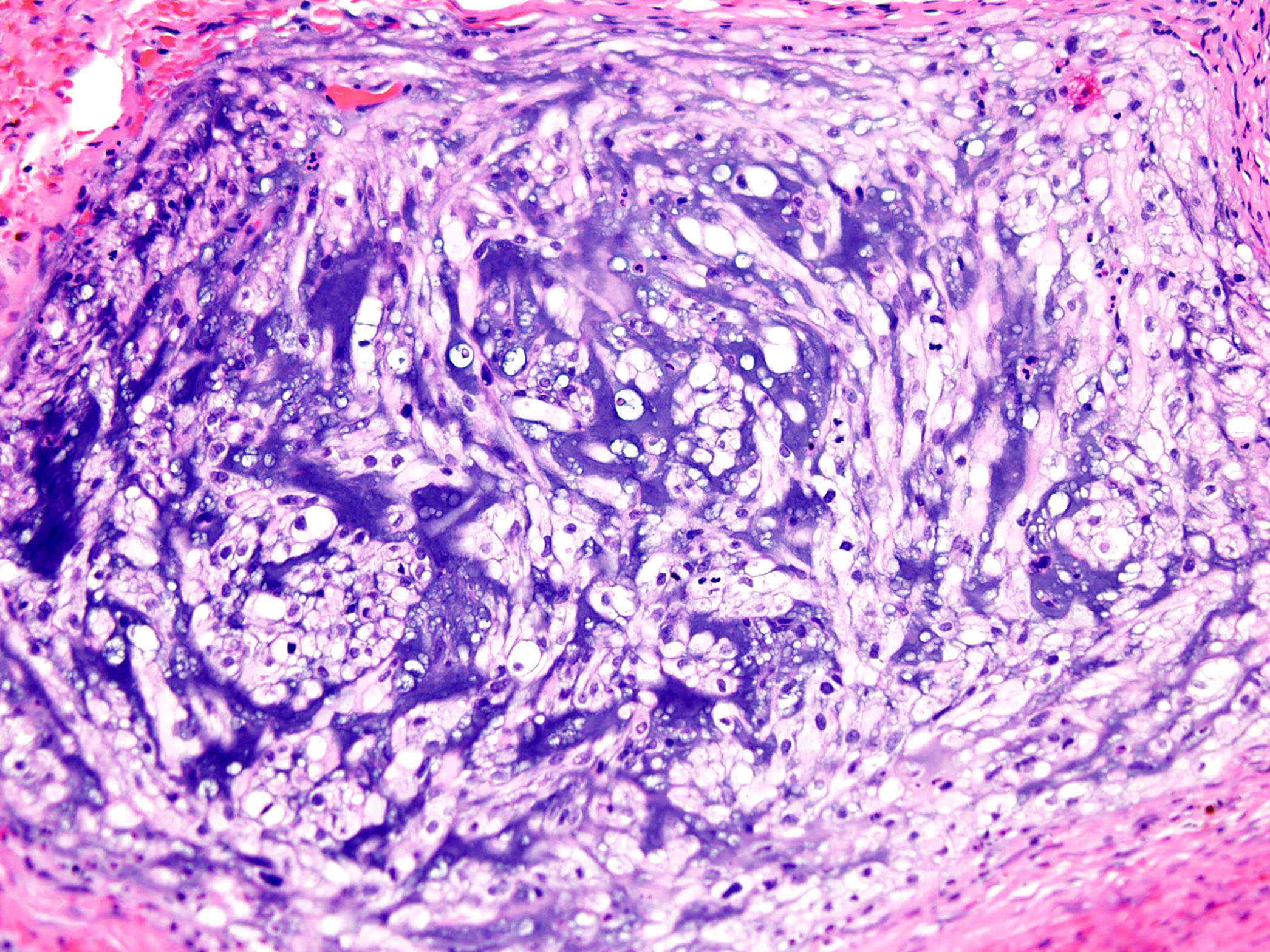
Contributed by Borislav A. Alexiev, M.D. and William B. Laskin, M.D.
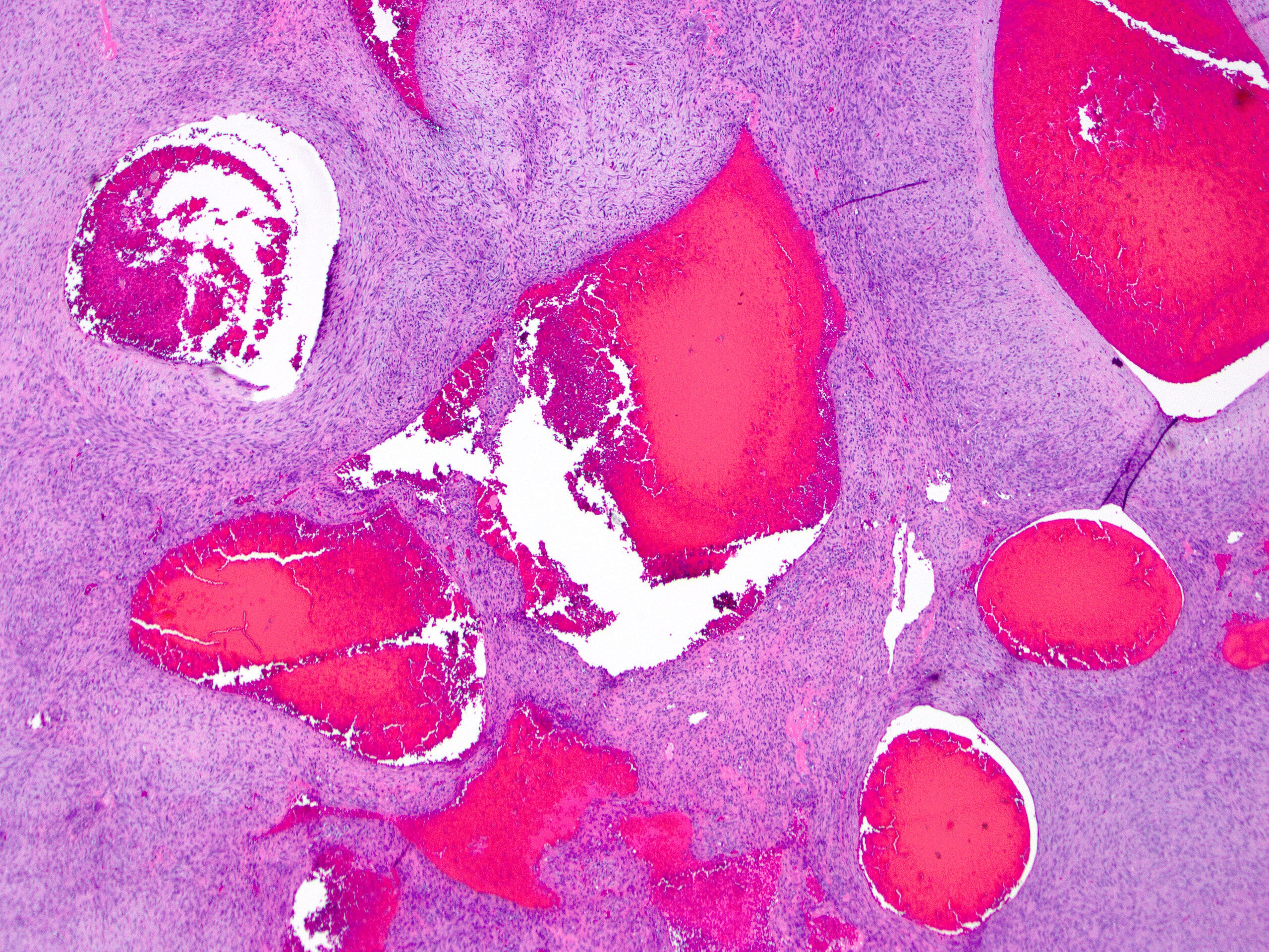
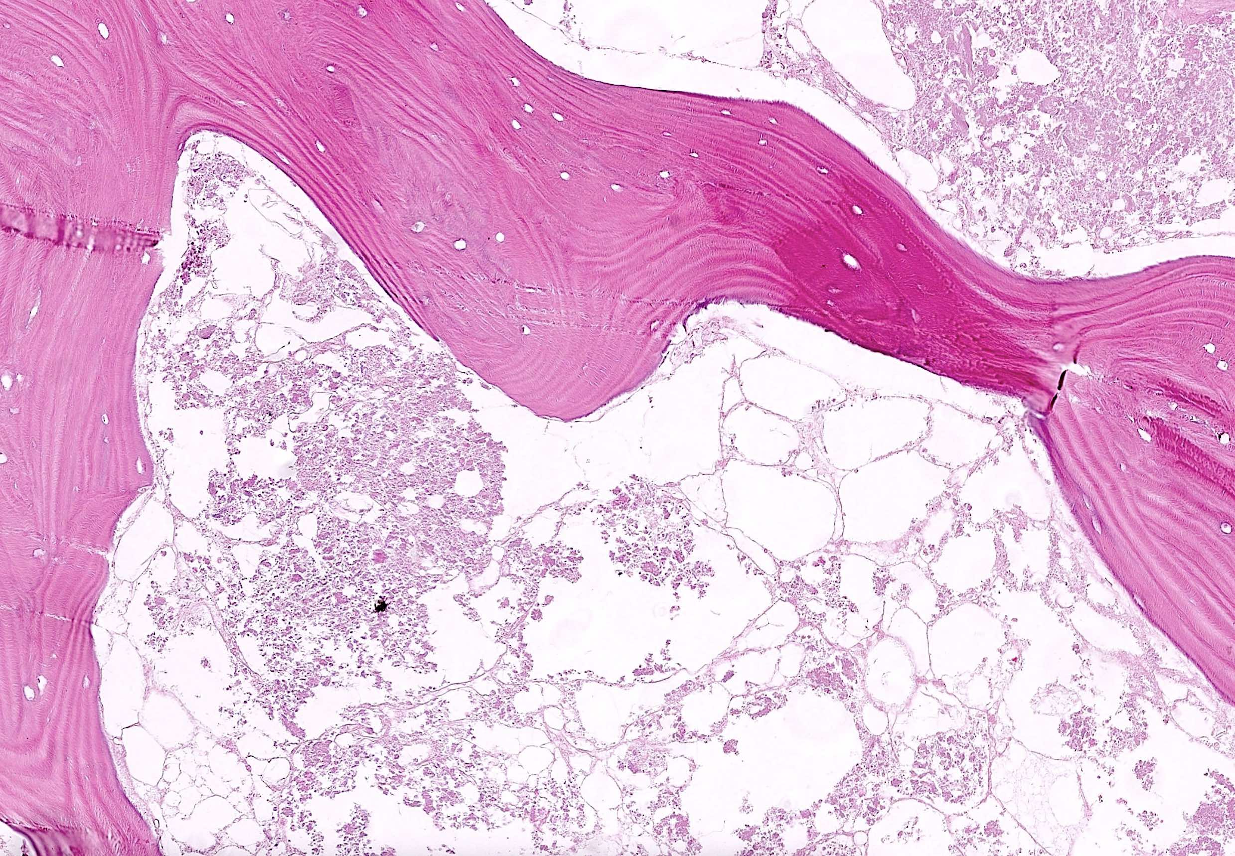
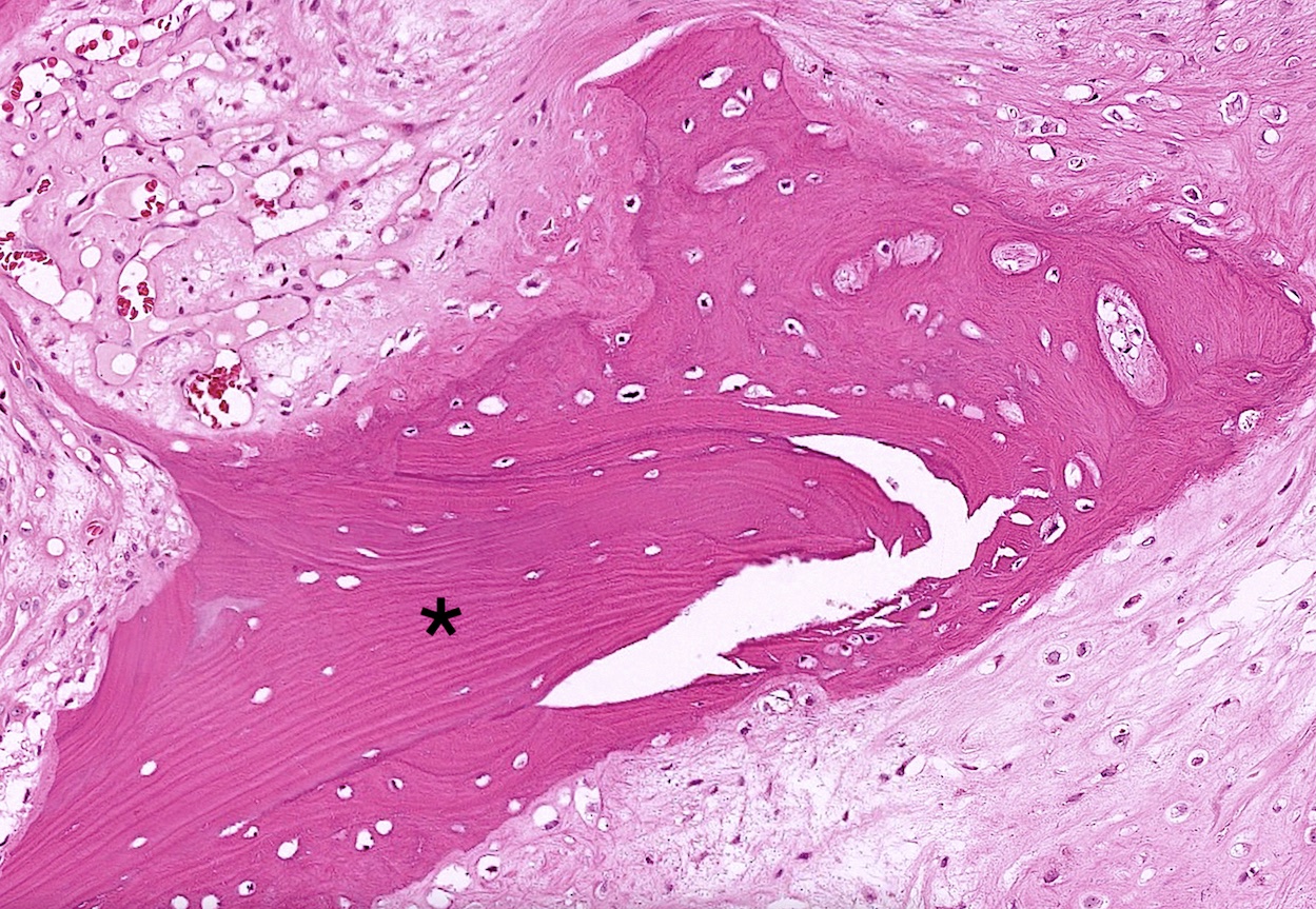
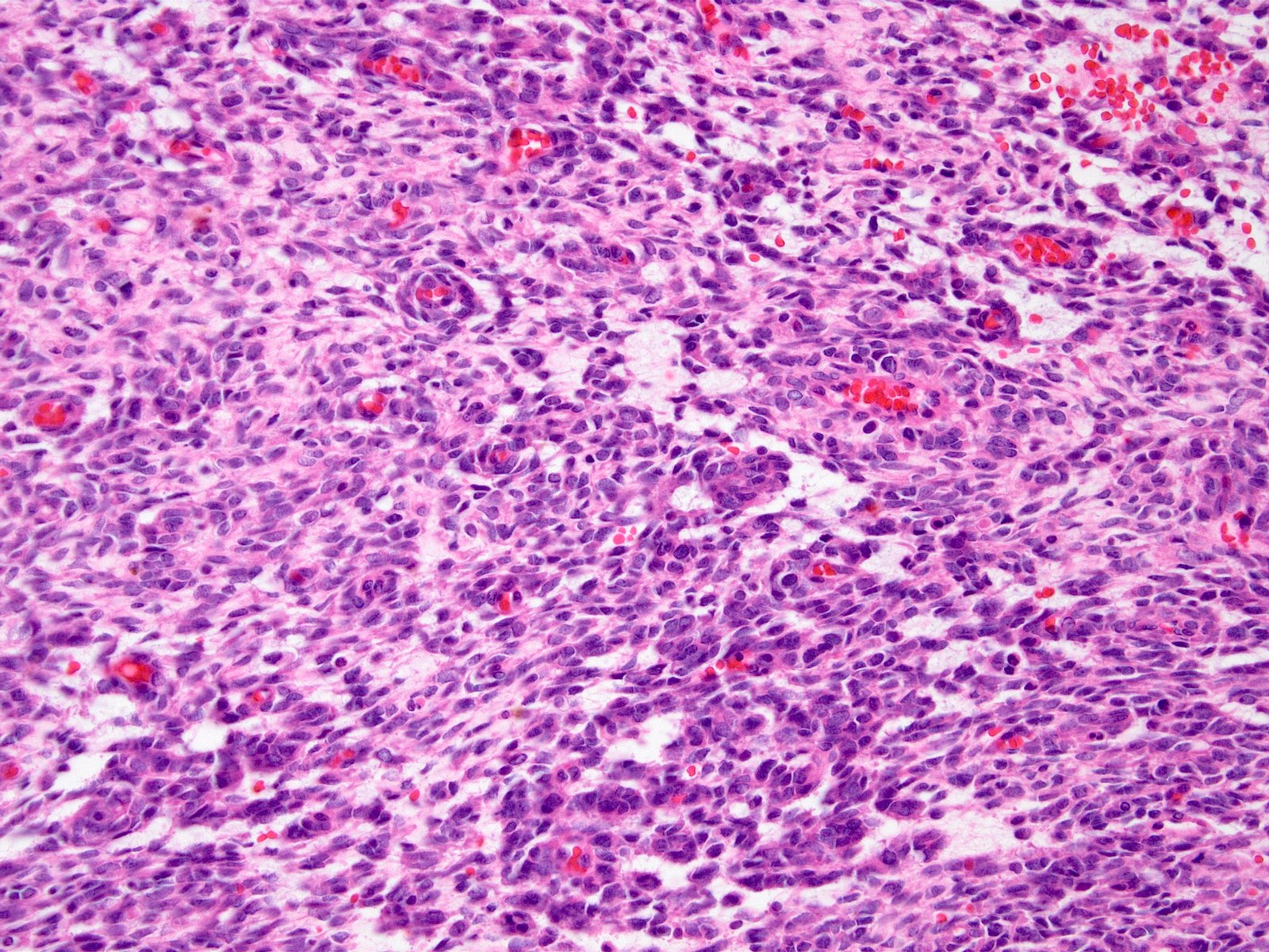
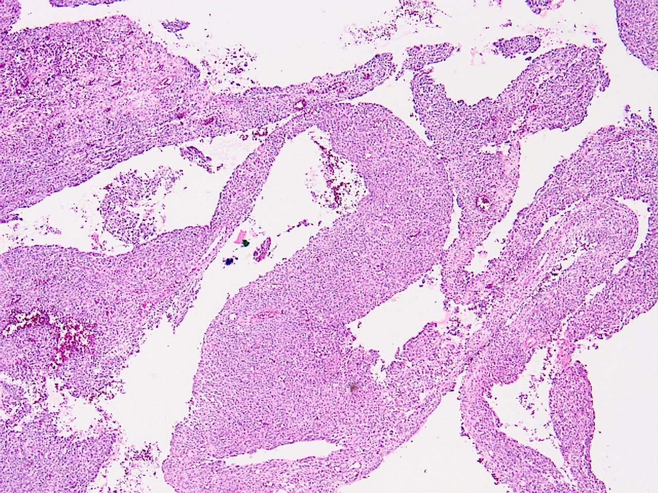
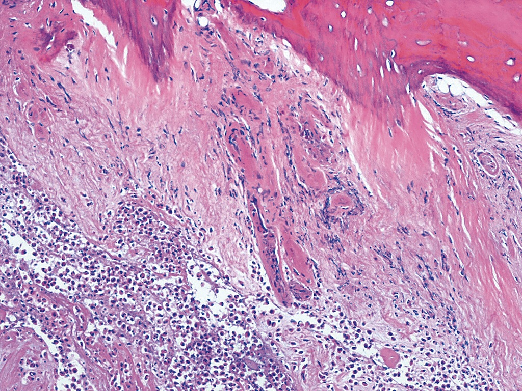
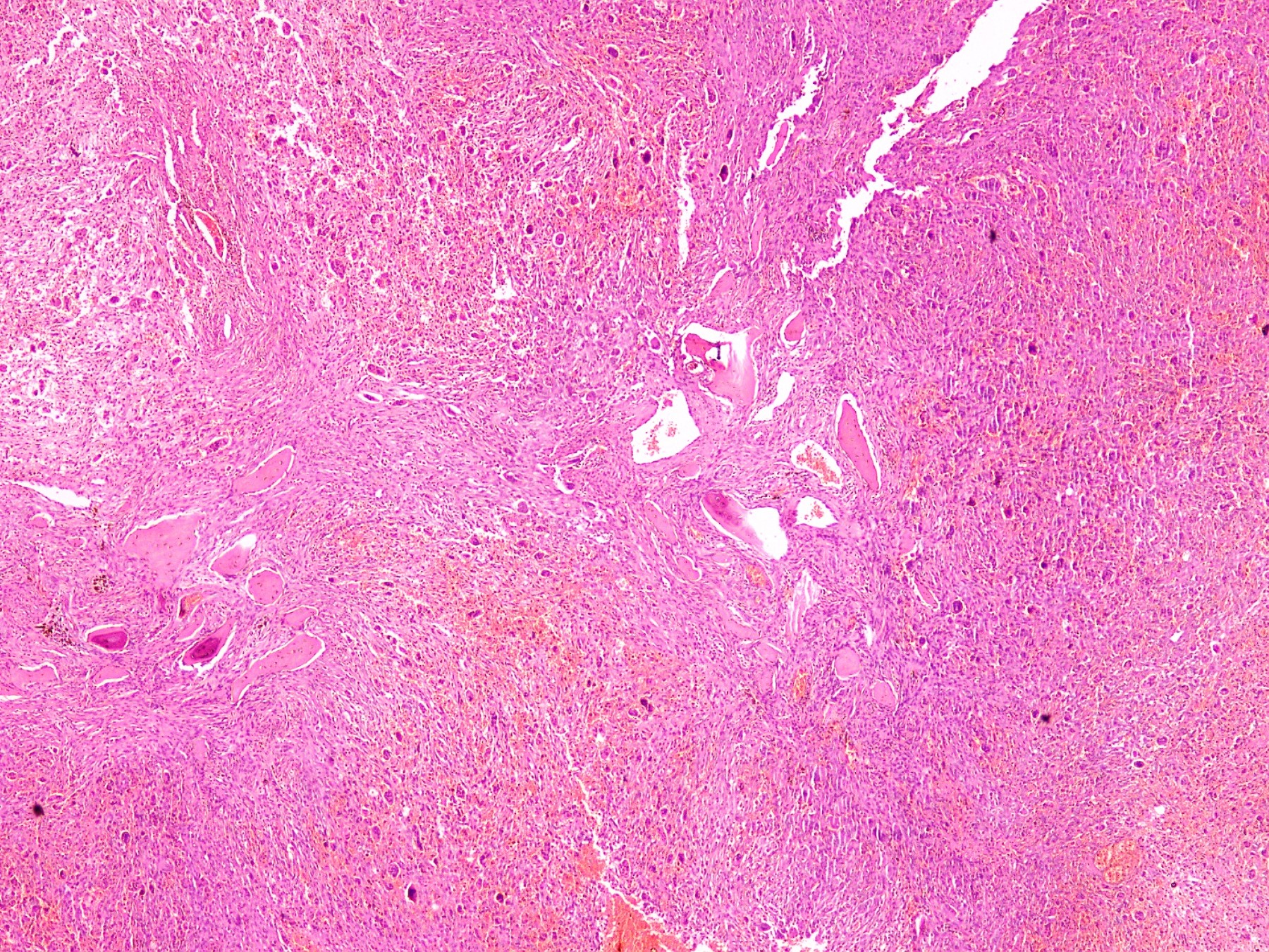
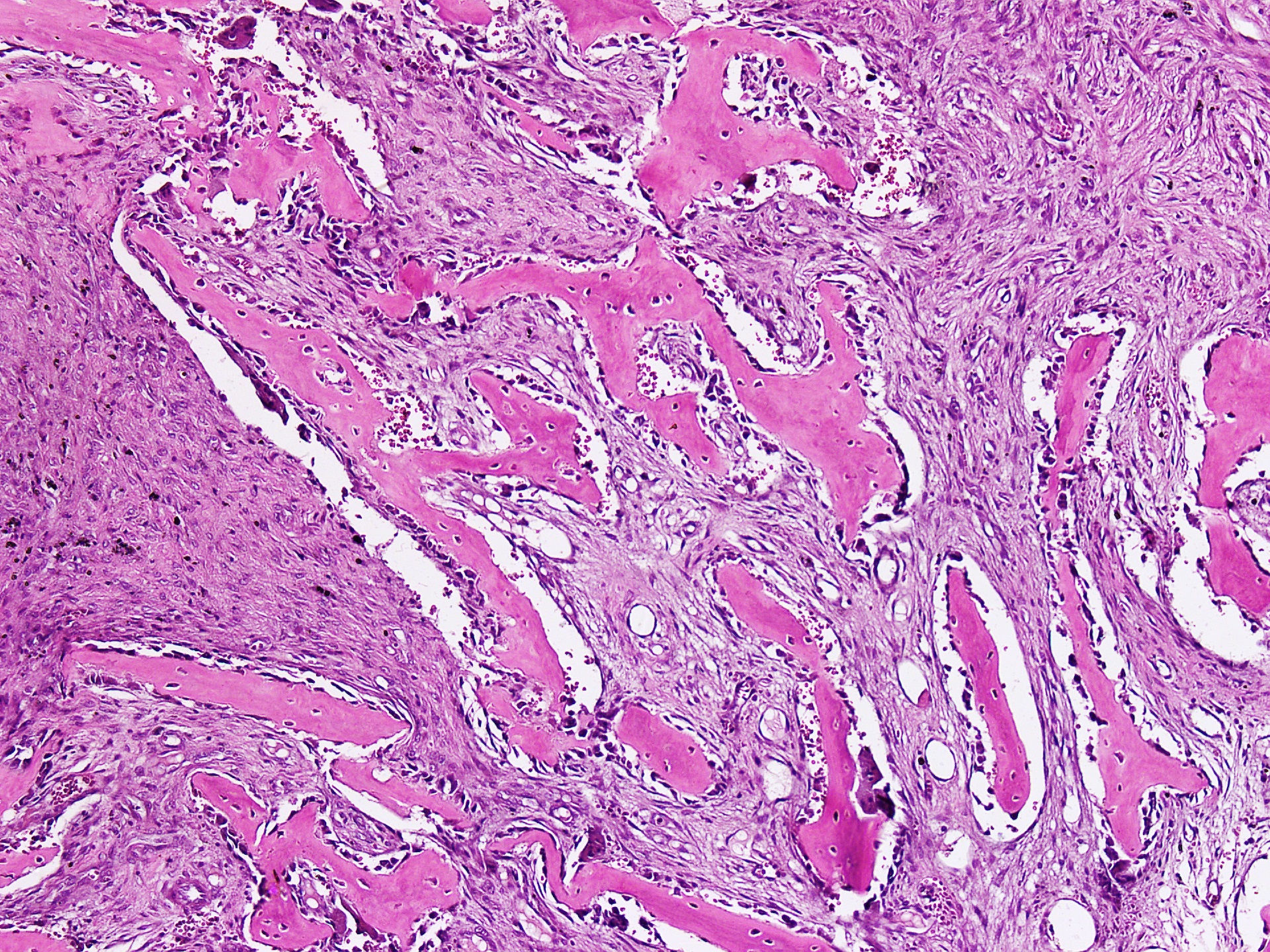
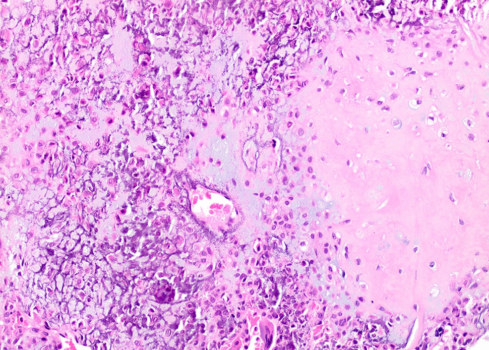
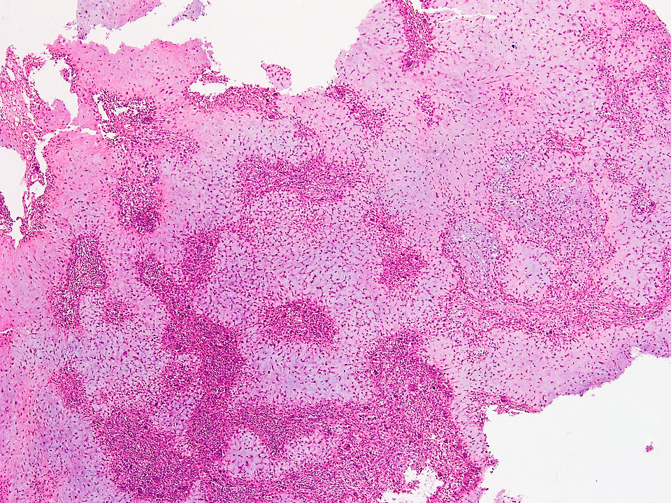
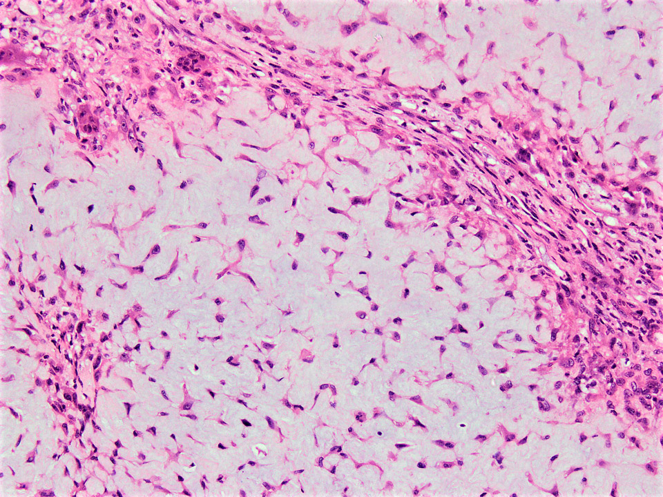
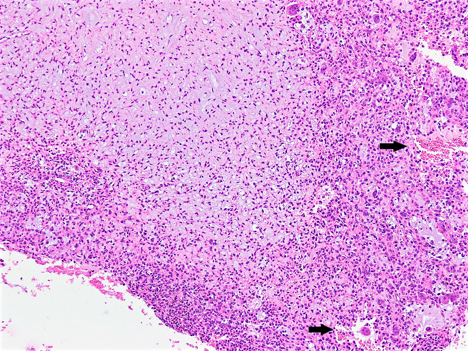
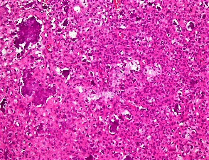
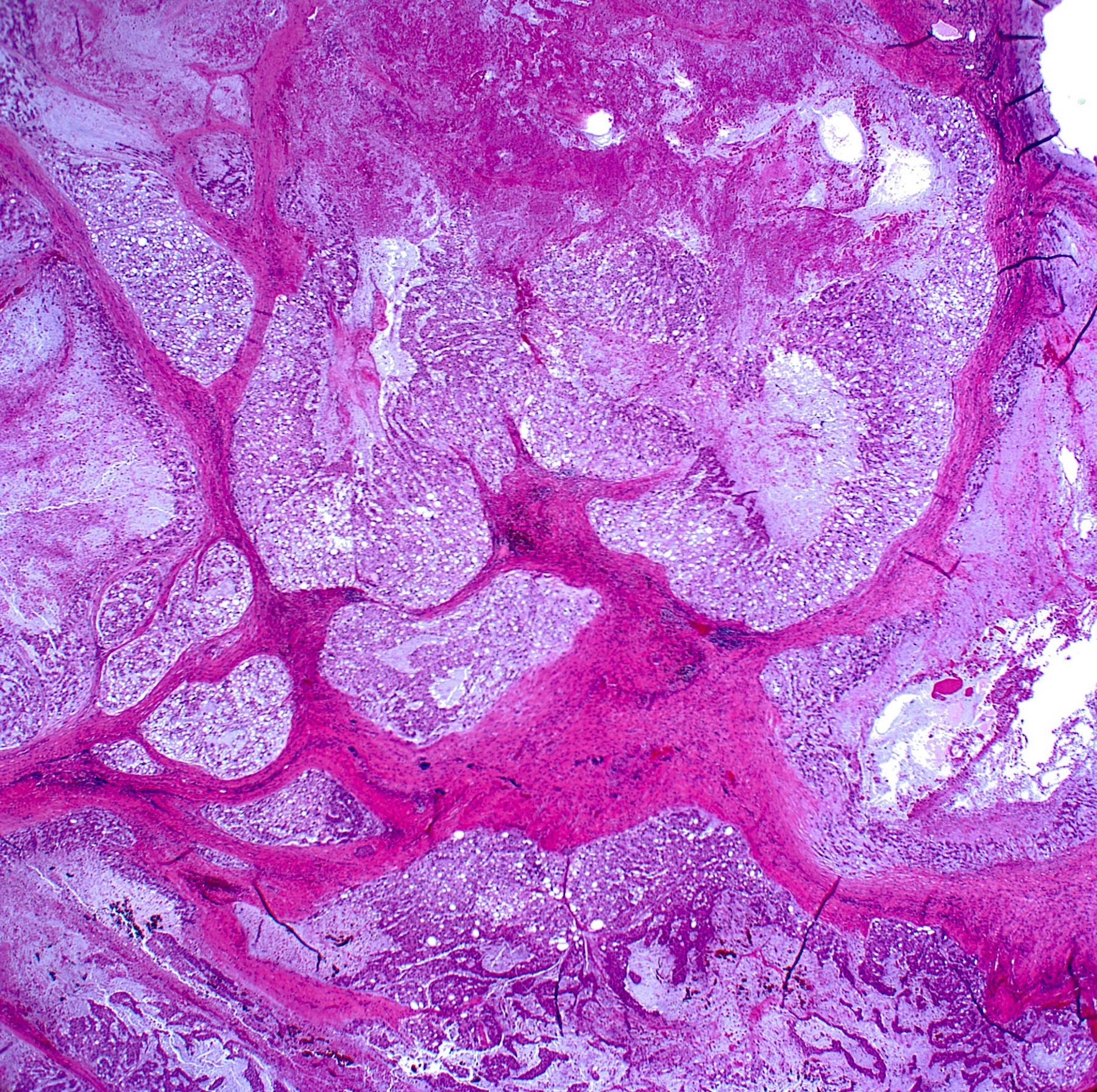
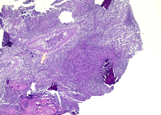
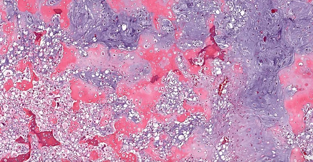
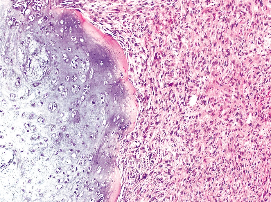
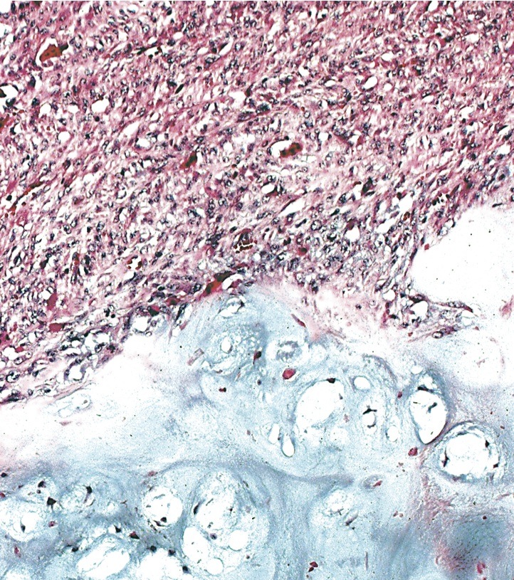
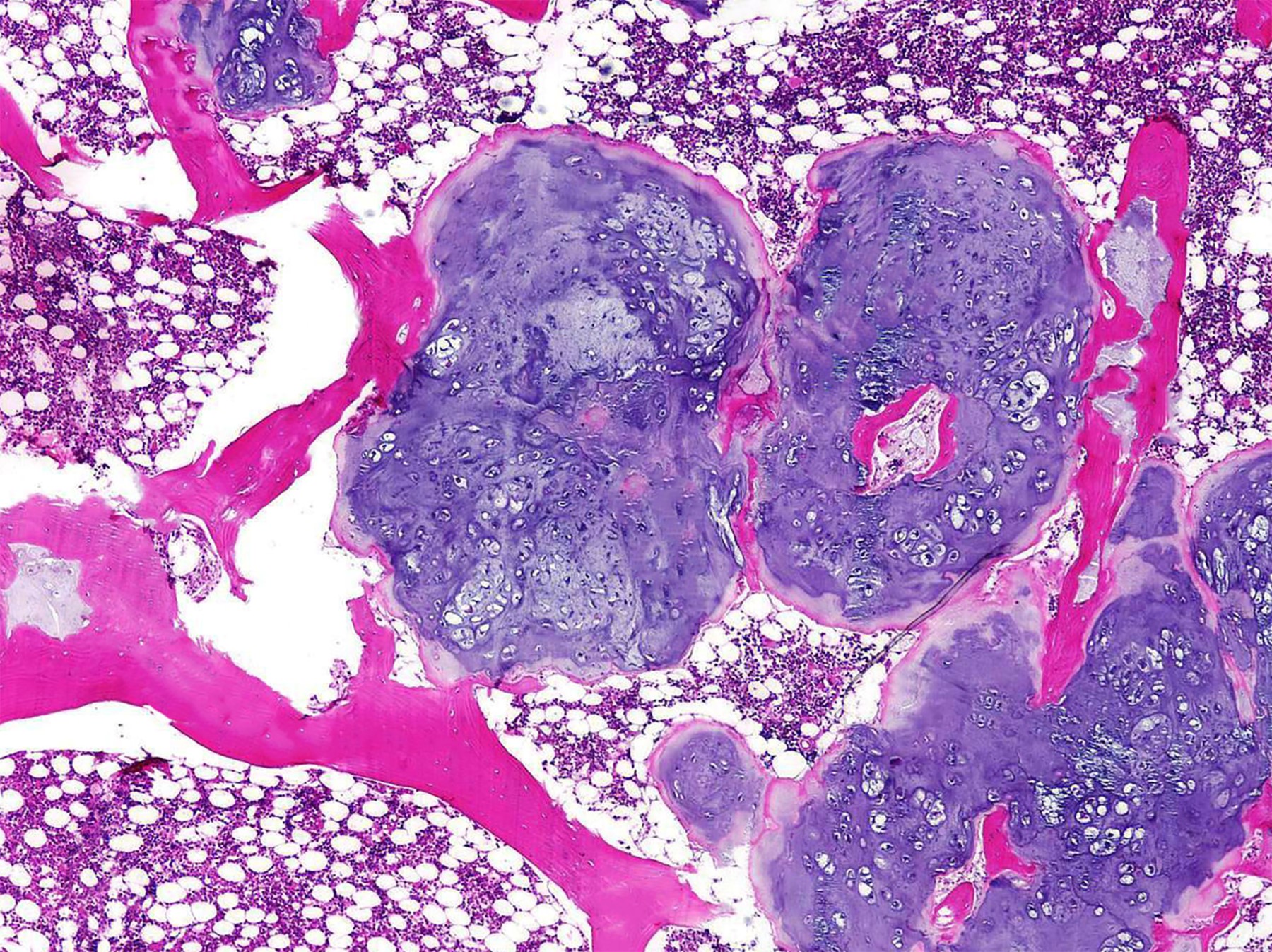
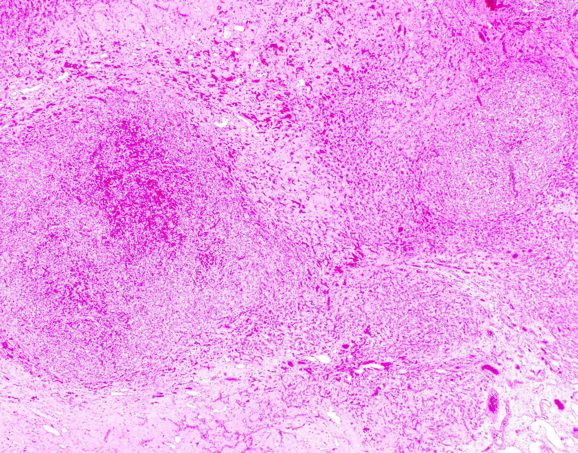
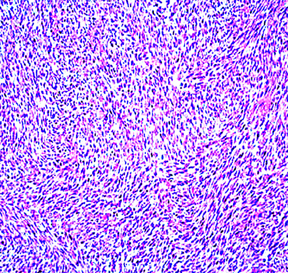
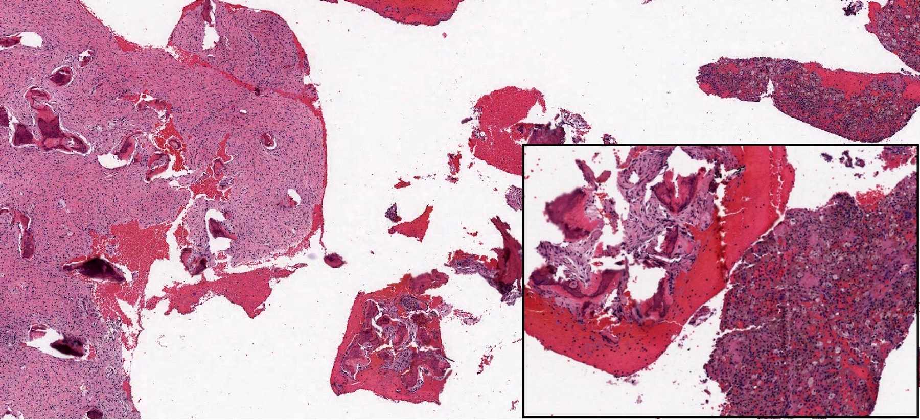
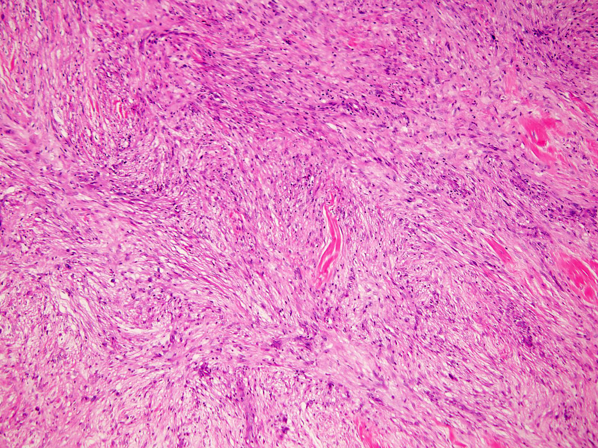
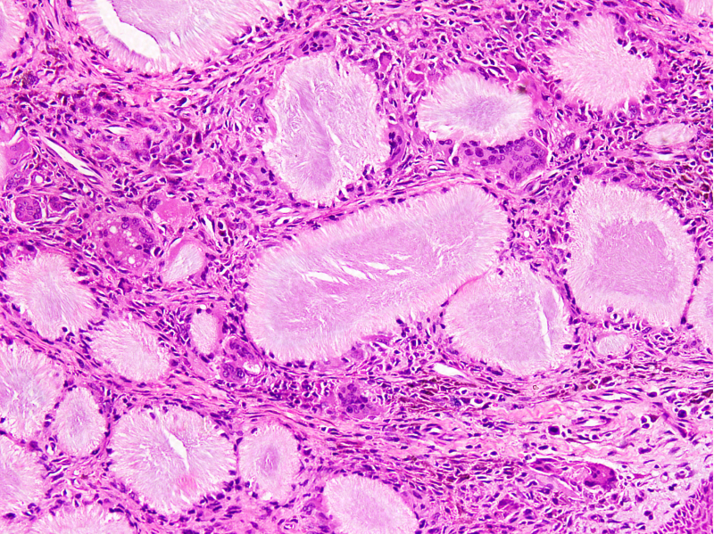
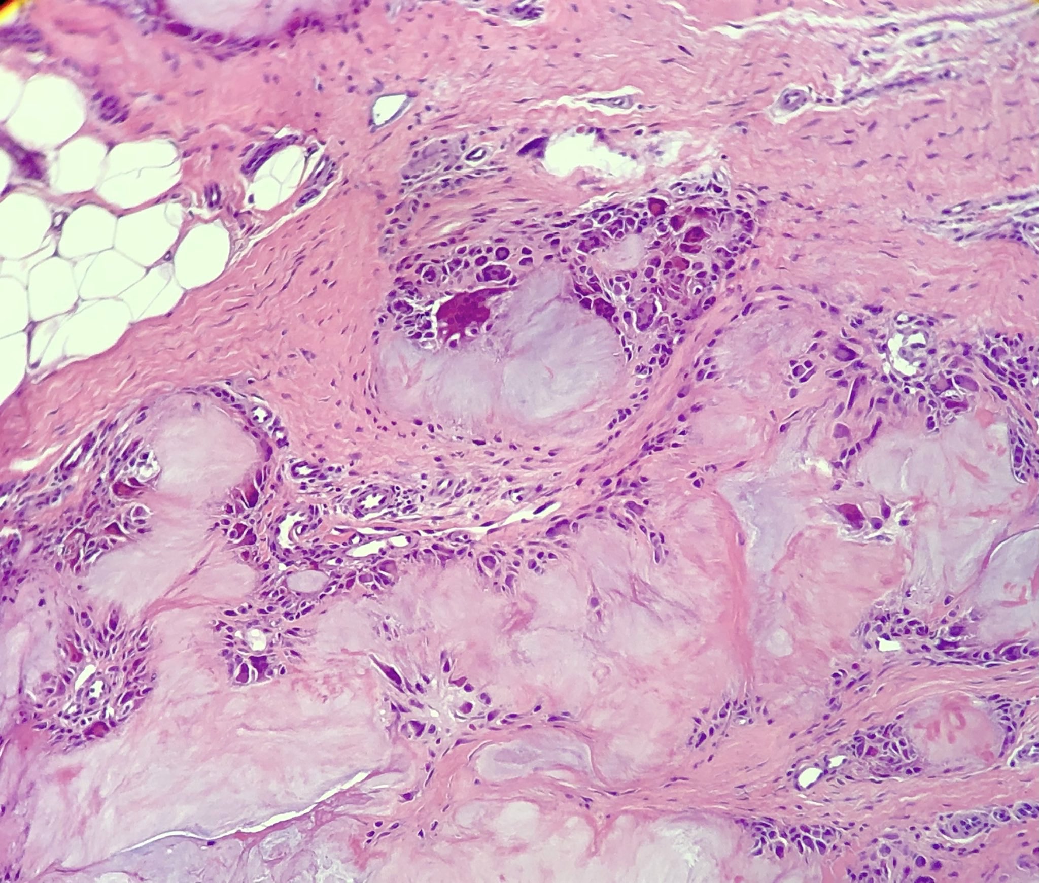
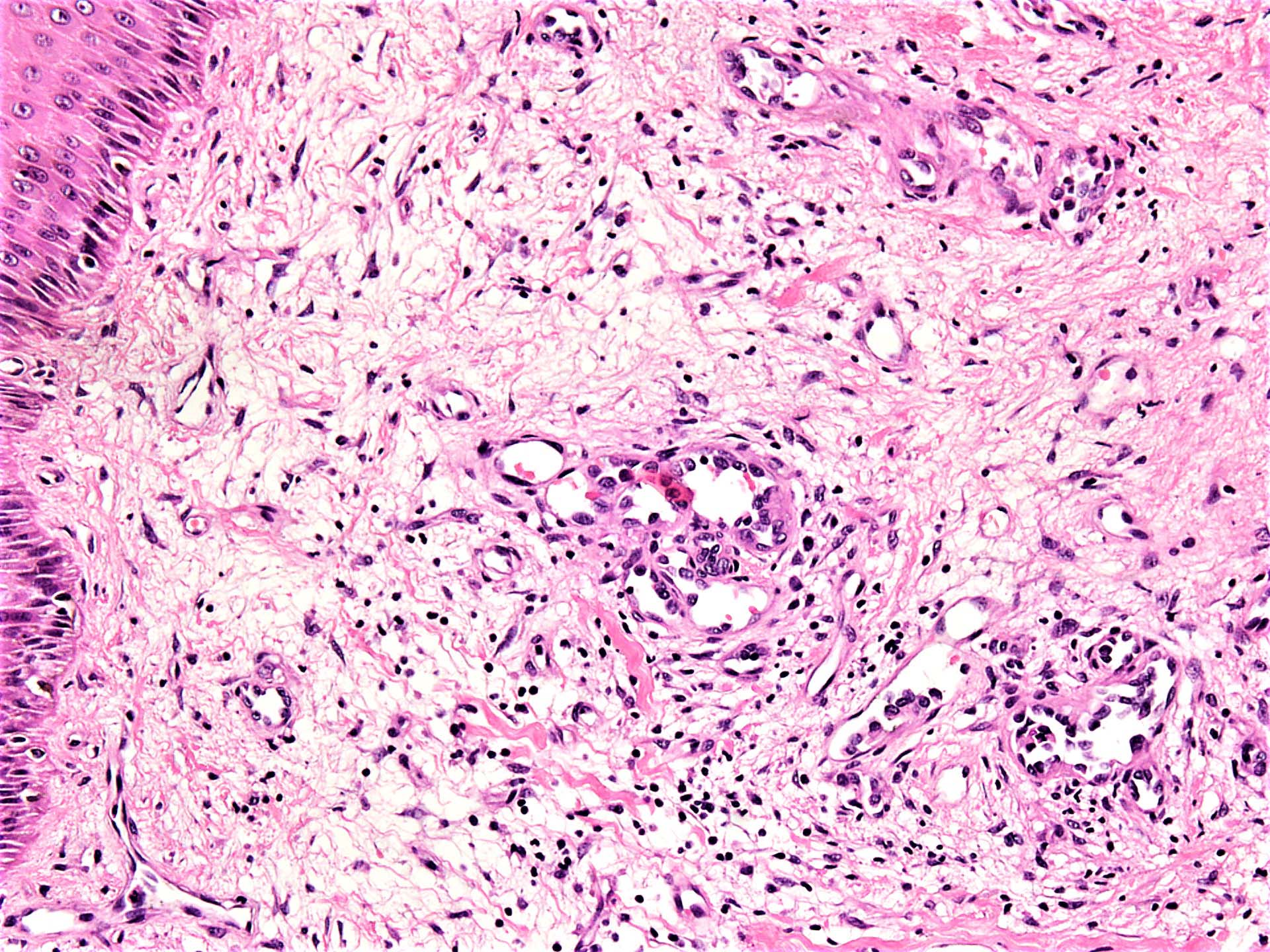
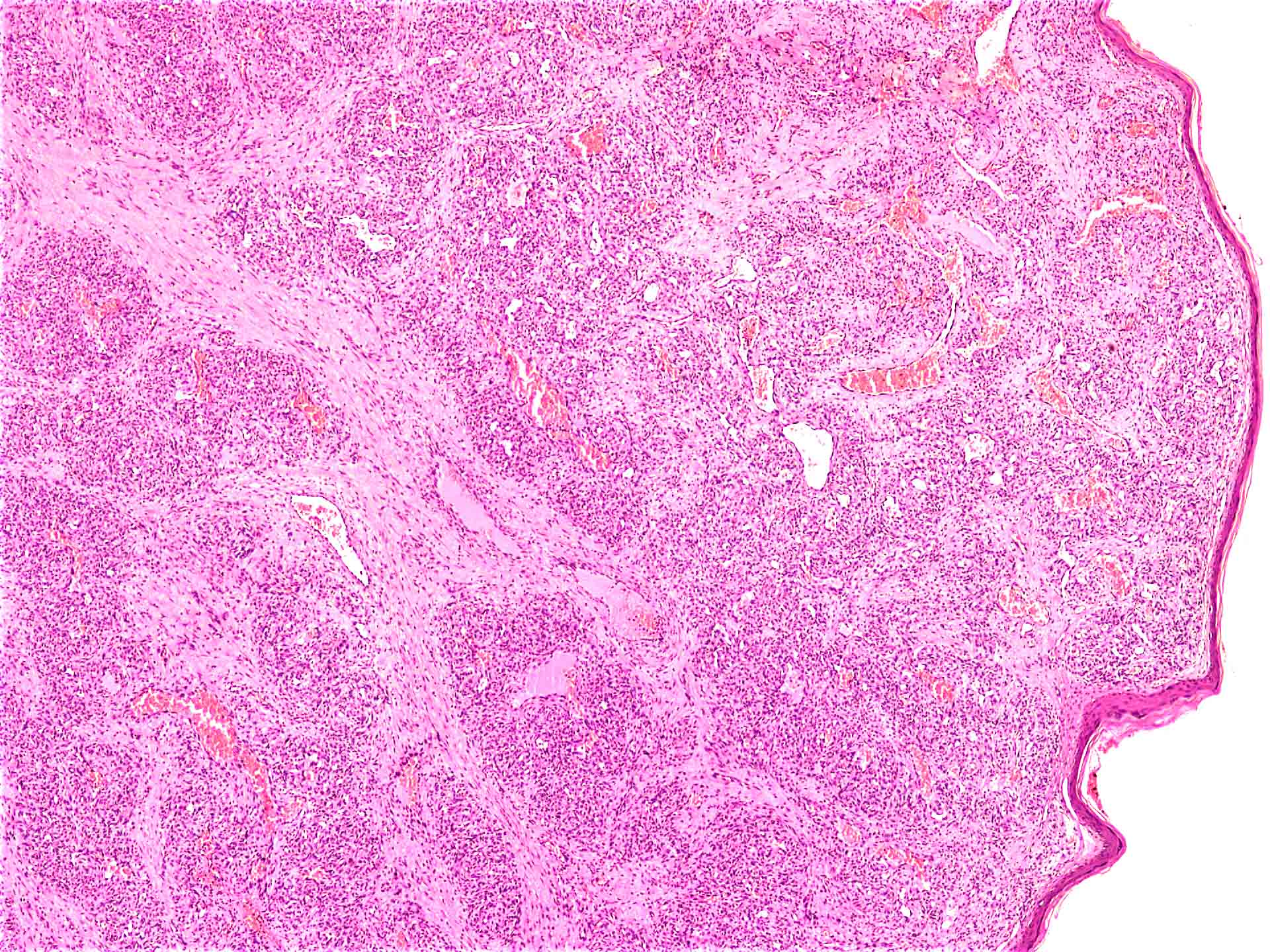
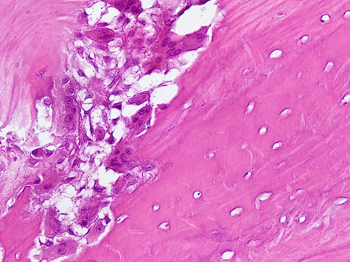
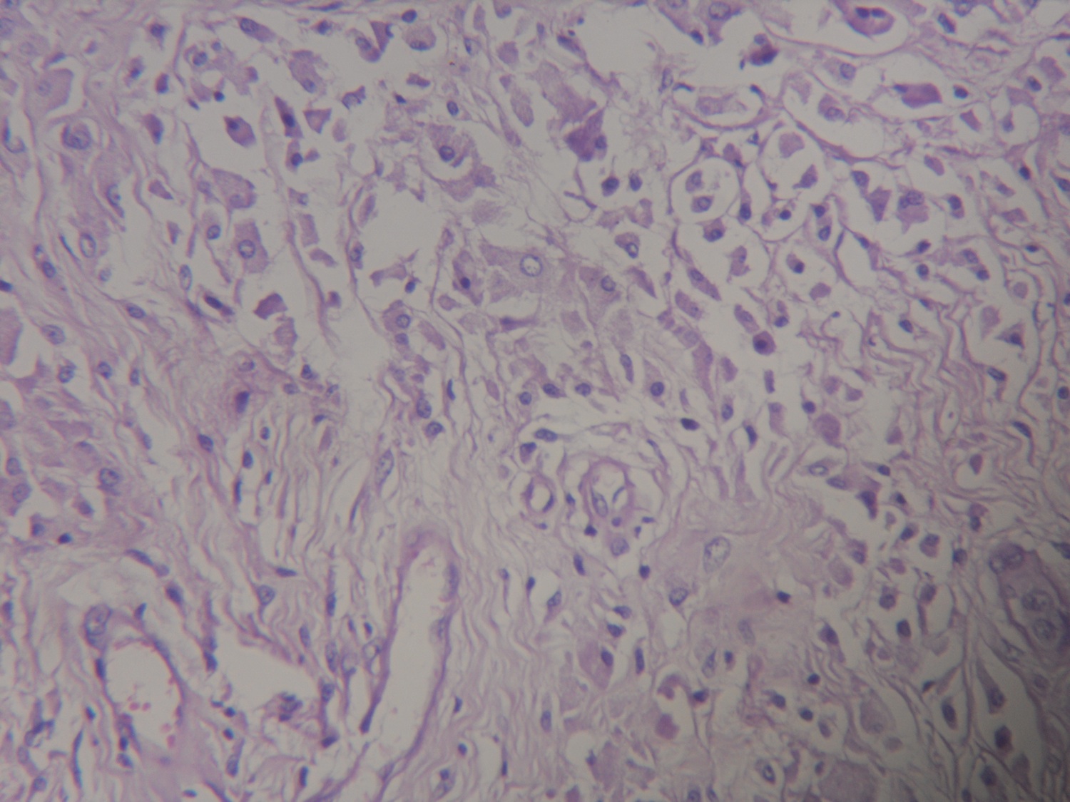
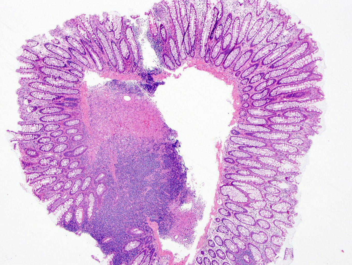
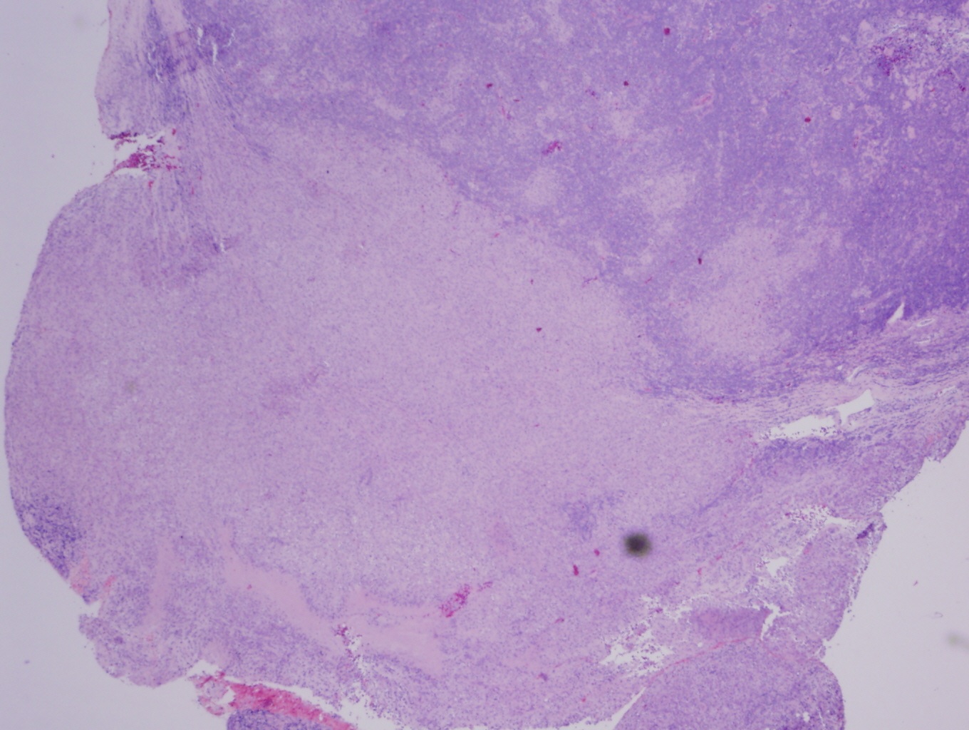
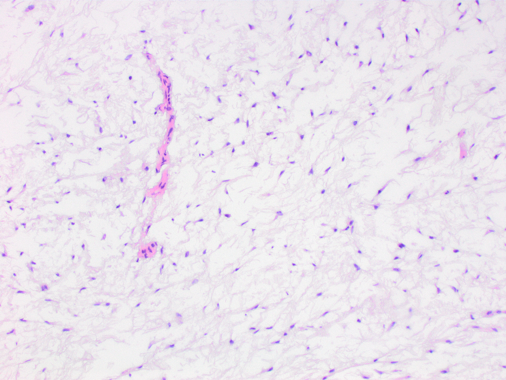
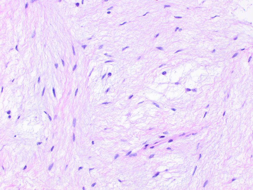
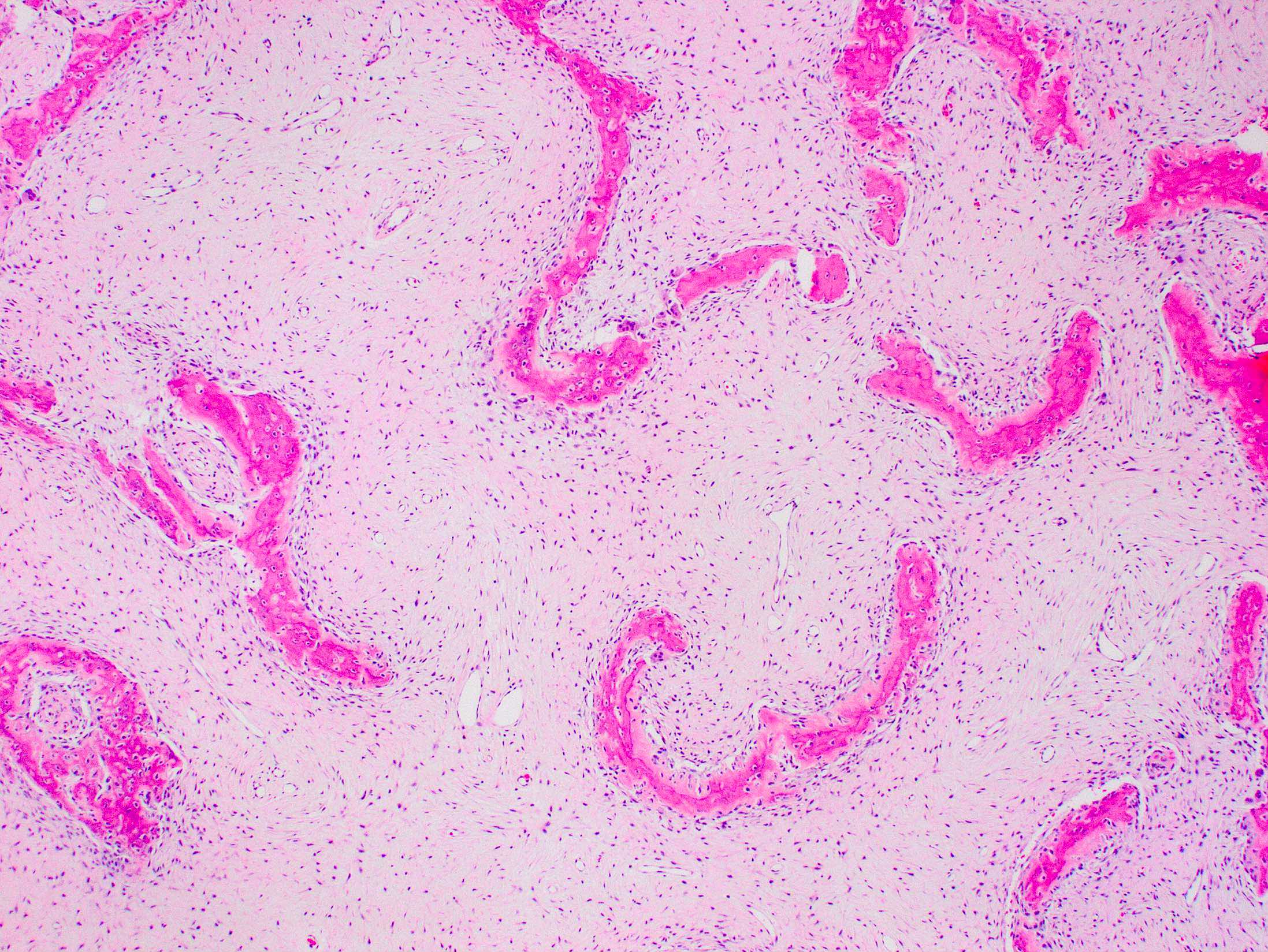
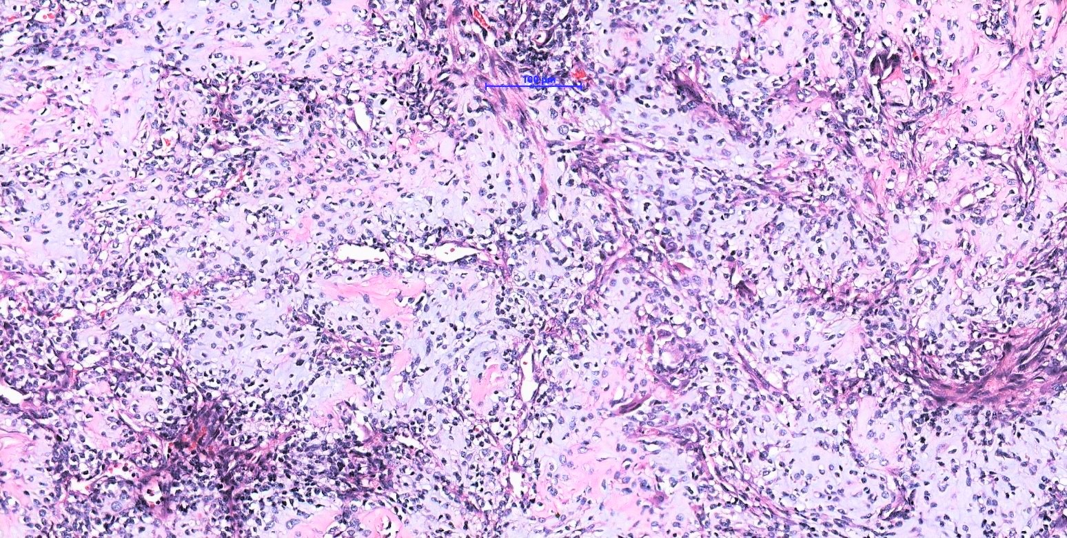
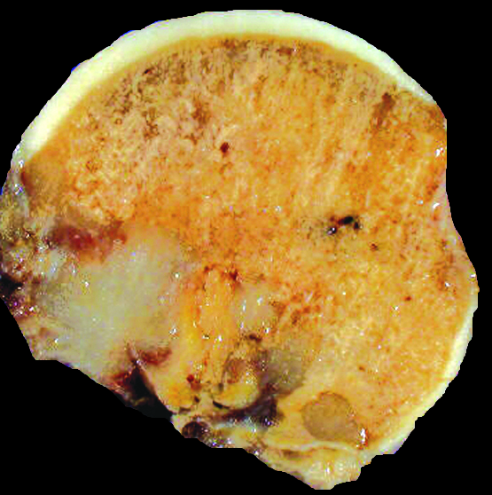
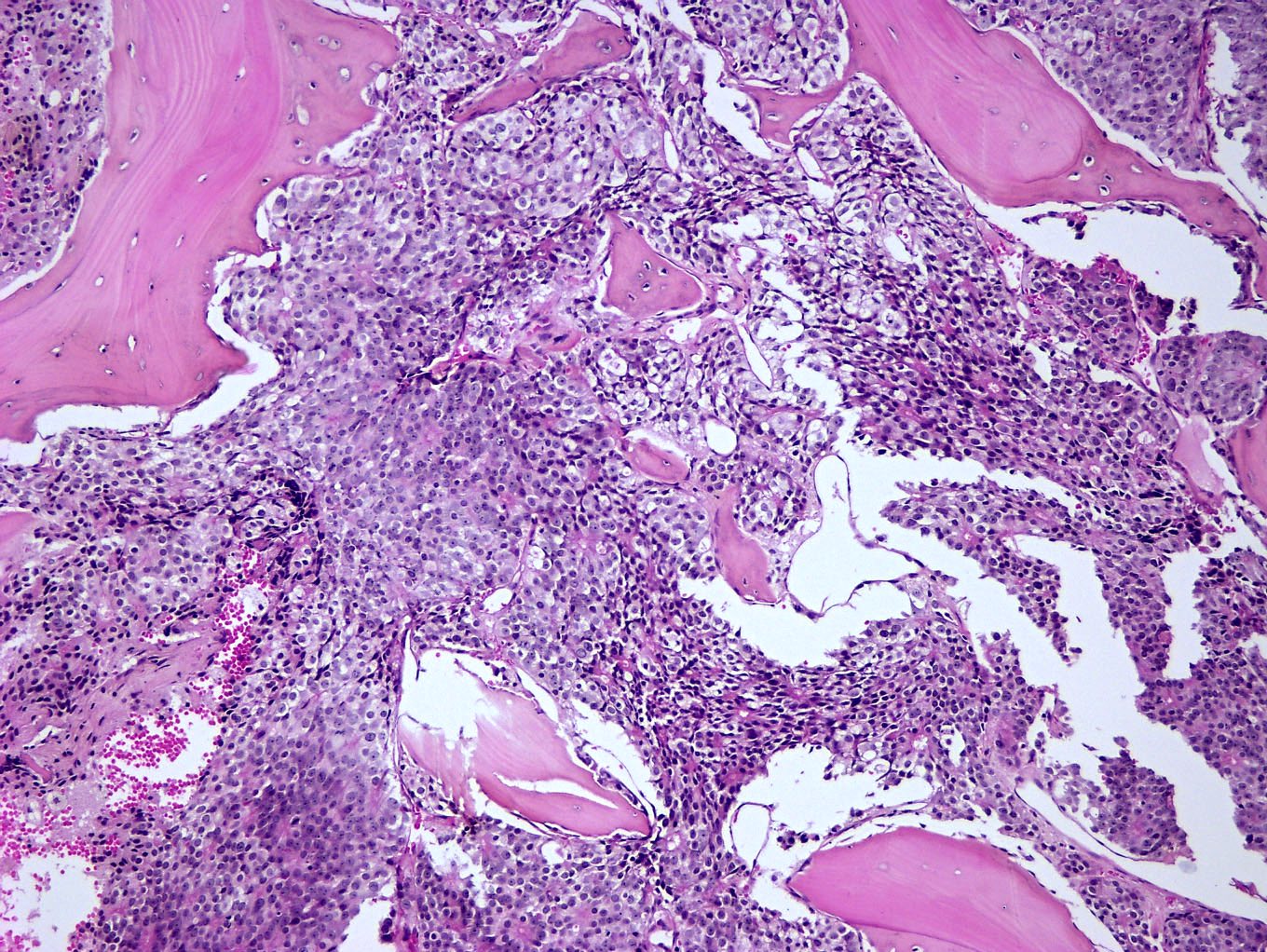
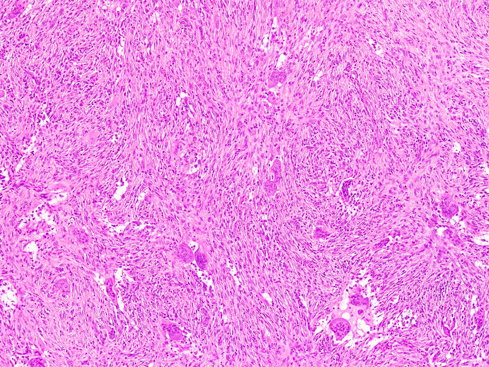
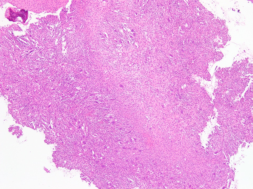
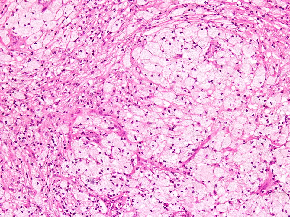
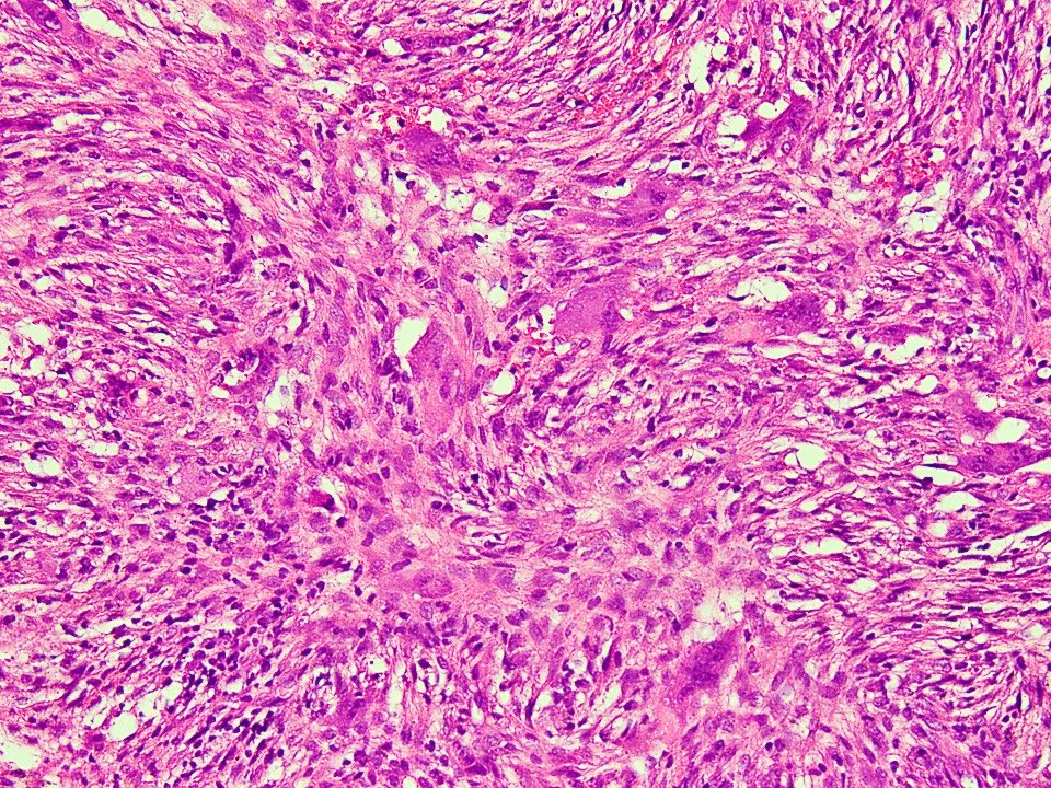
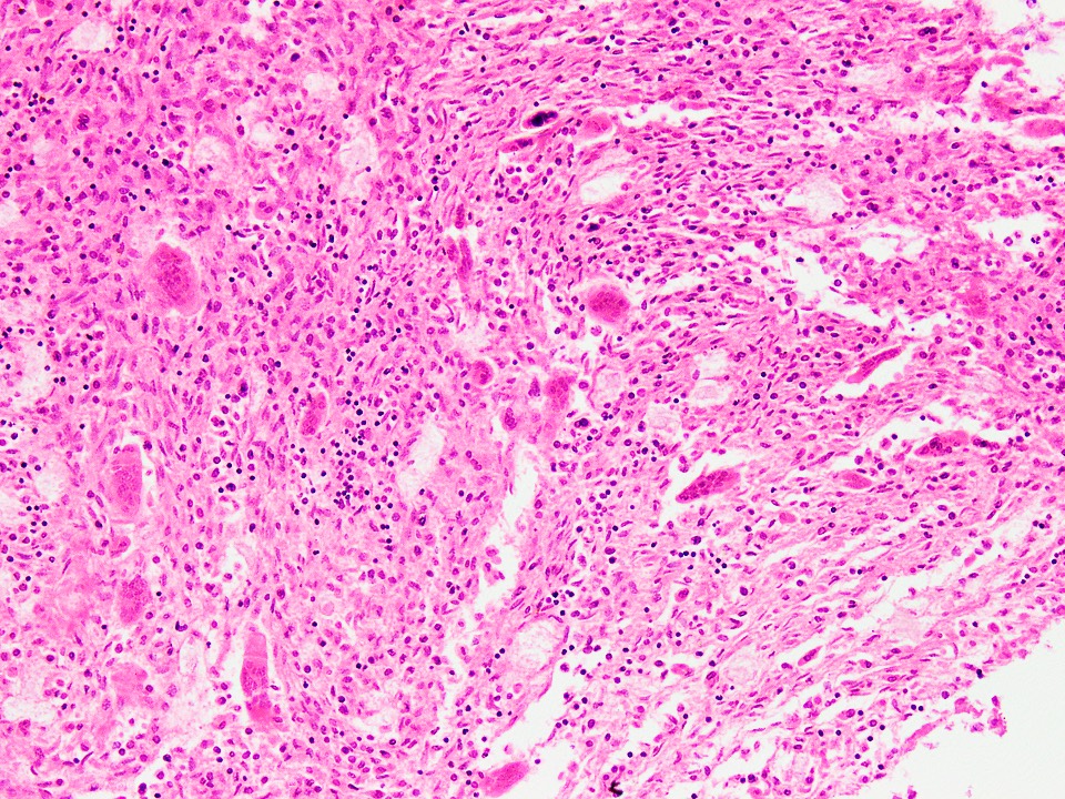
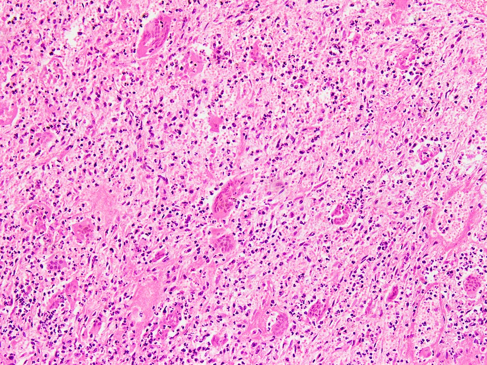
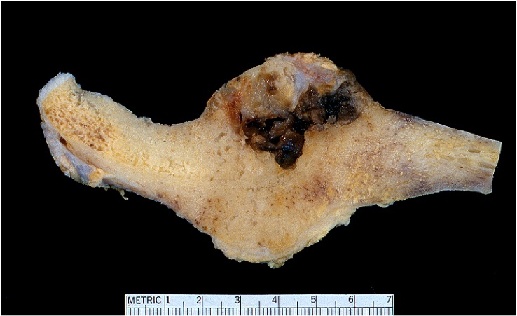
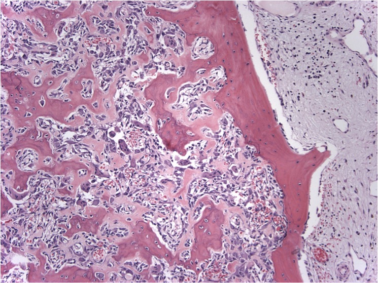
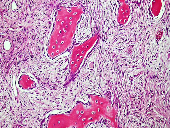
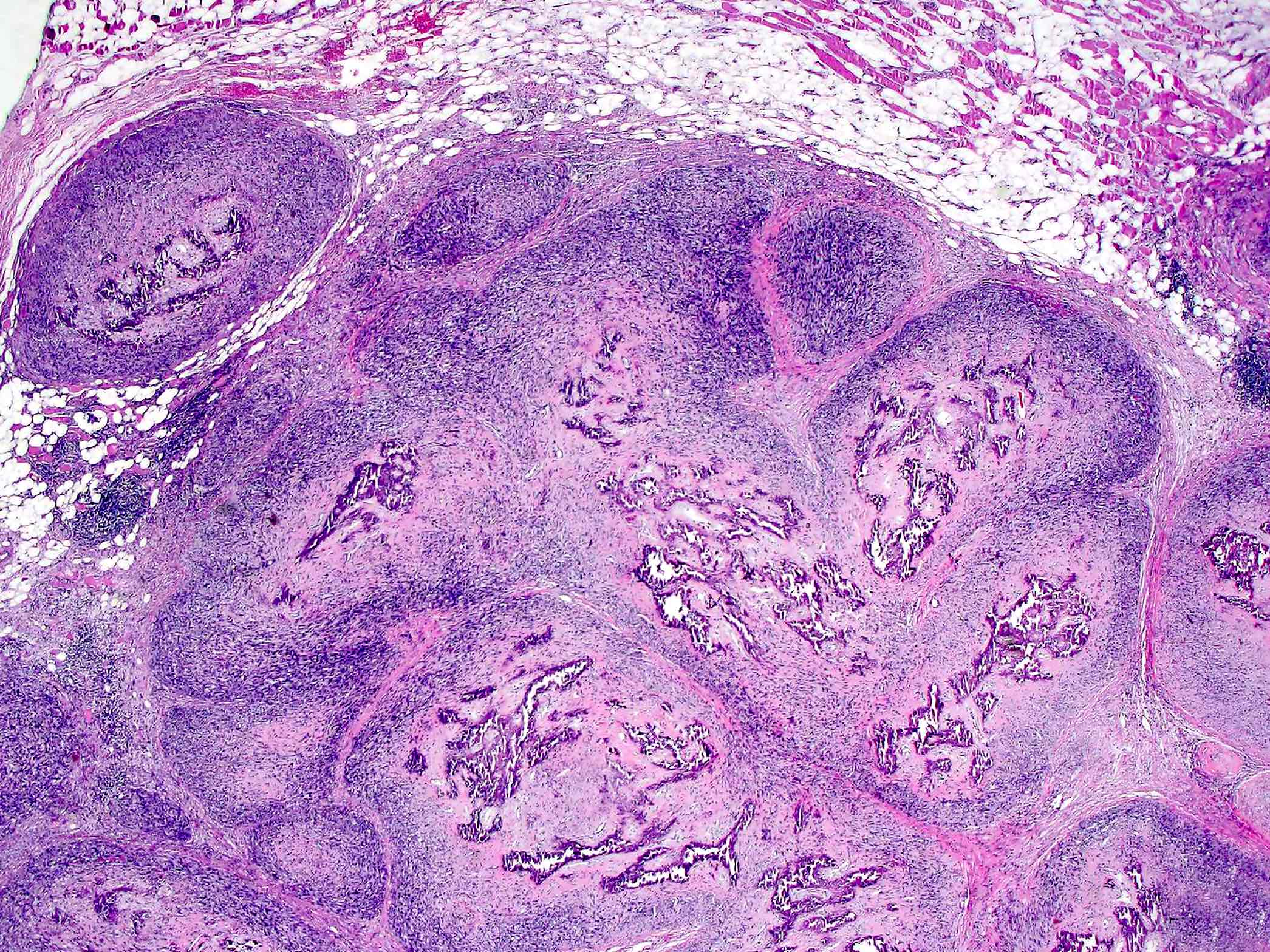
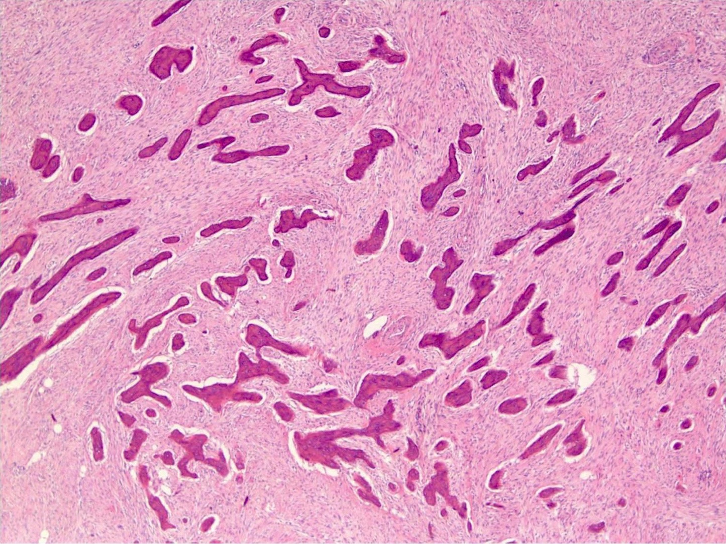
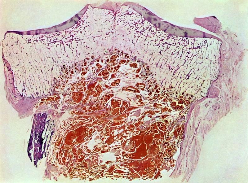
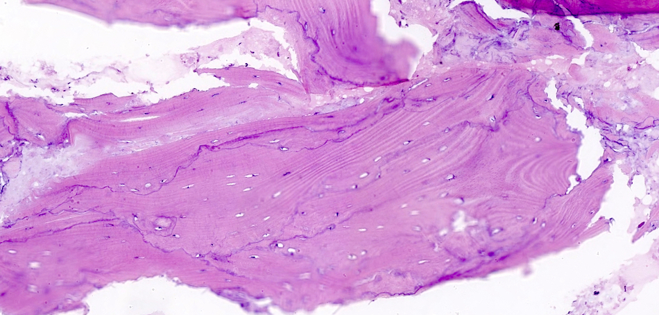
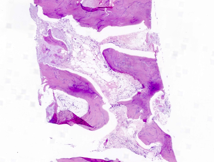
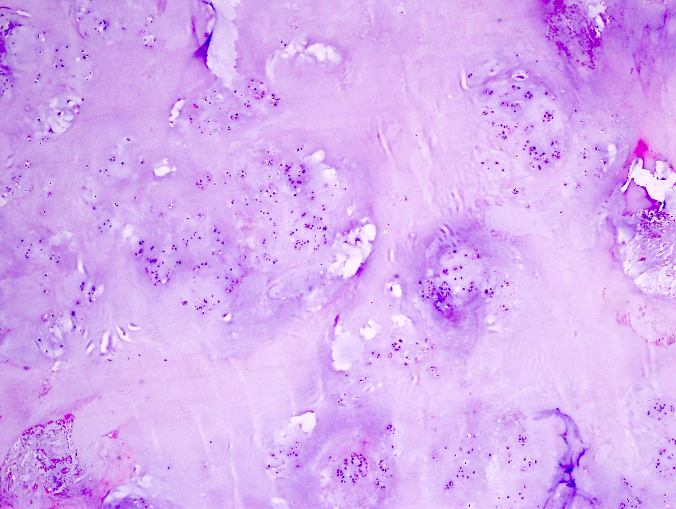
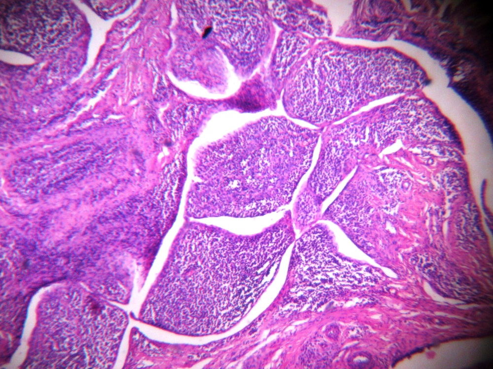
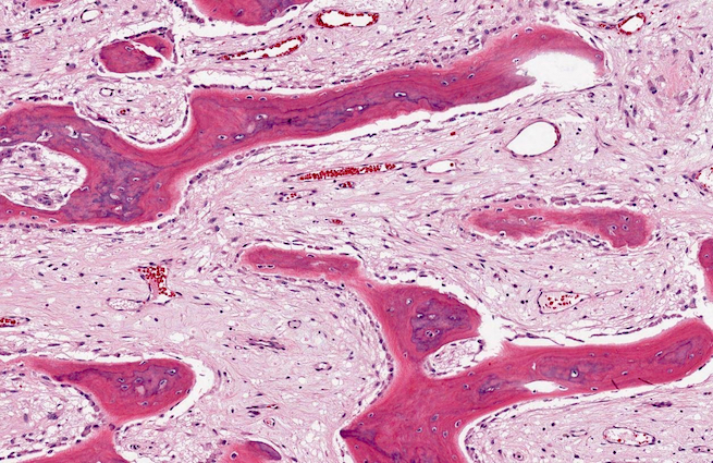
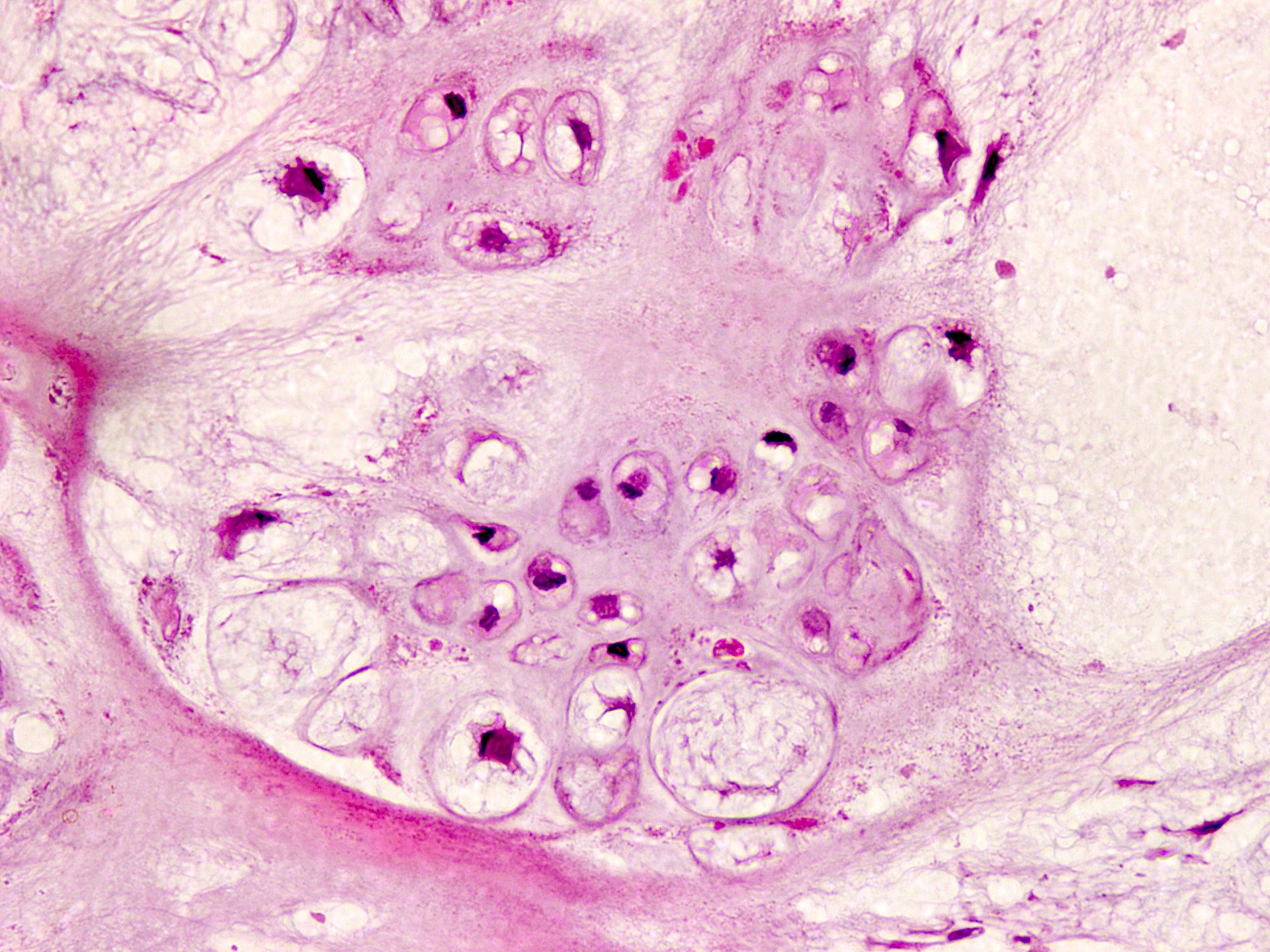
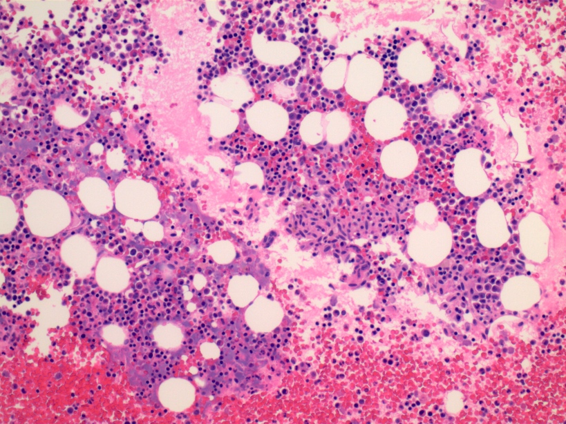
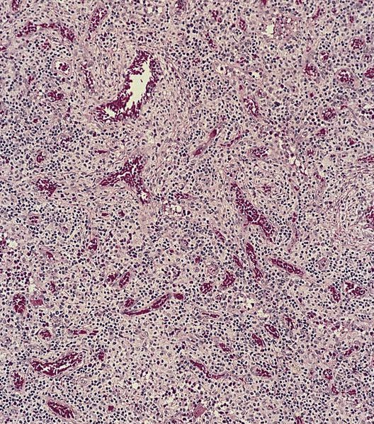
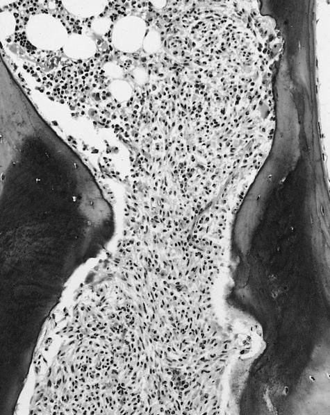
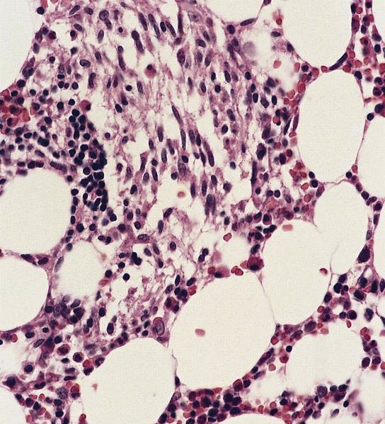
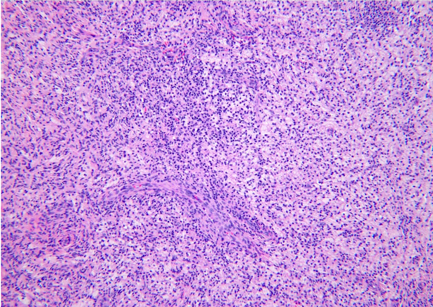
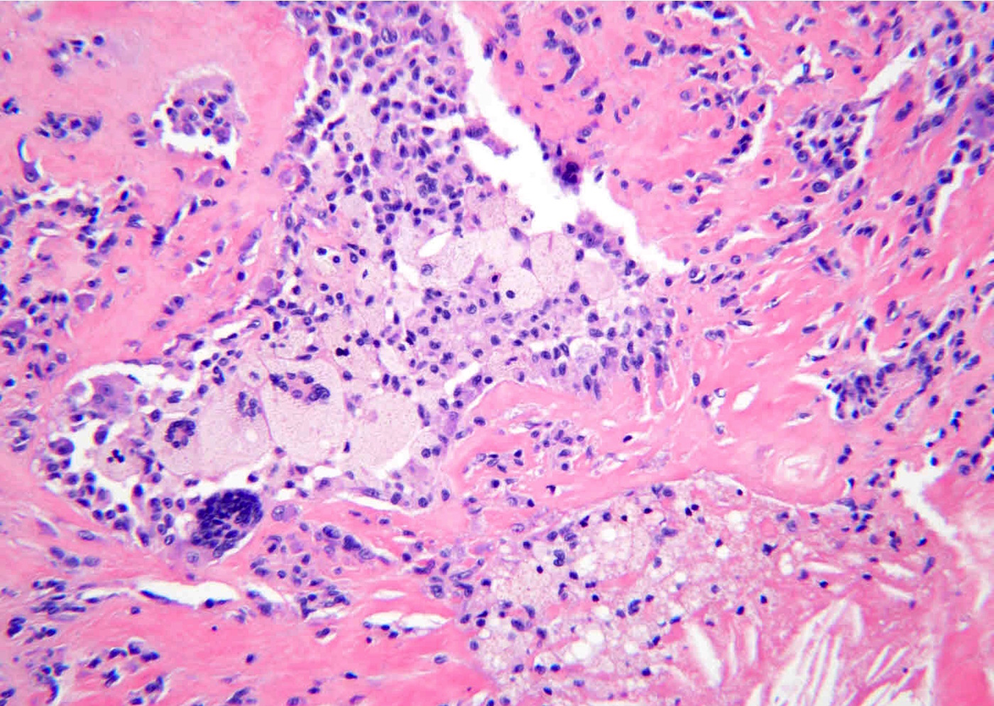
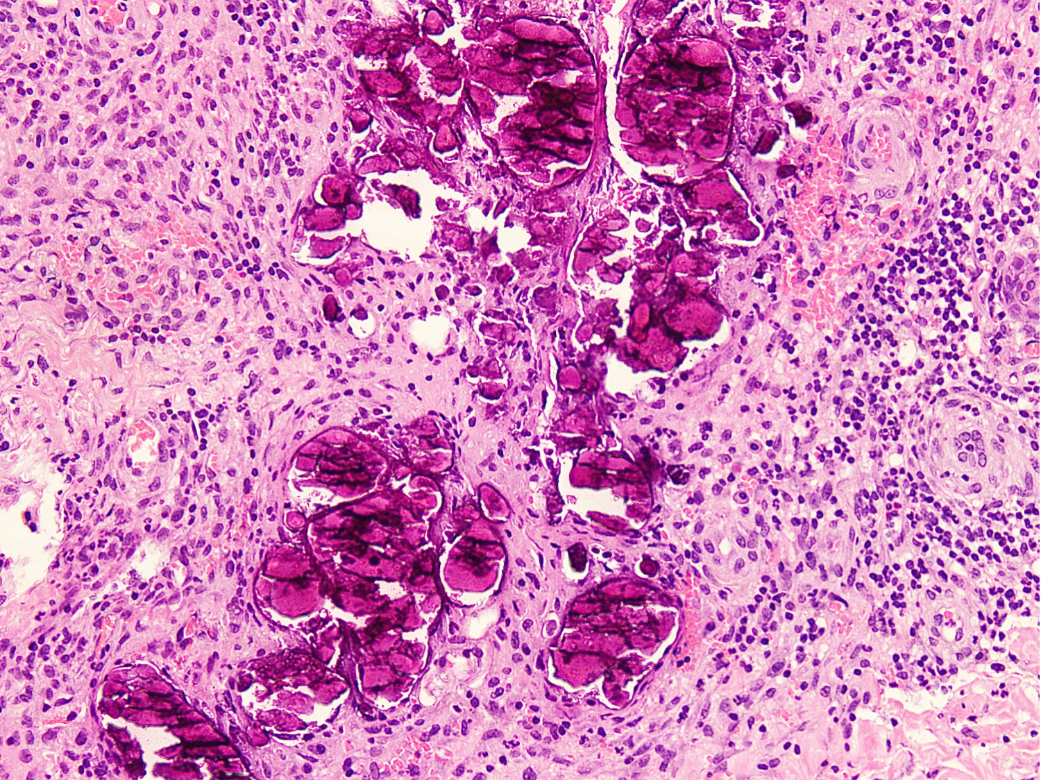
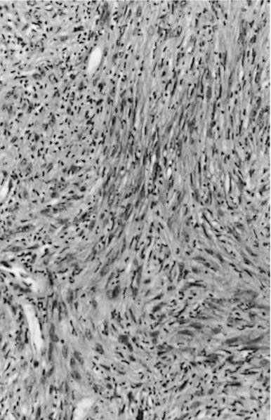
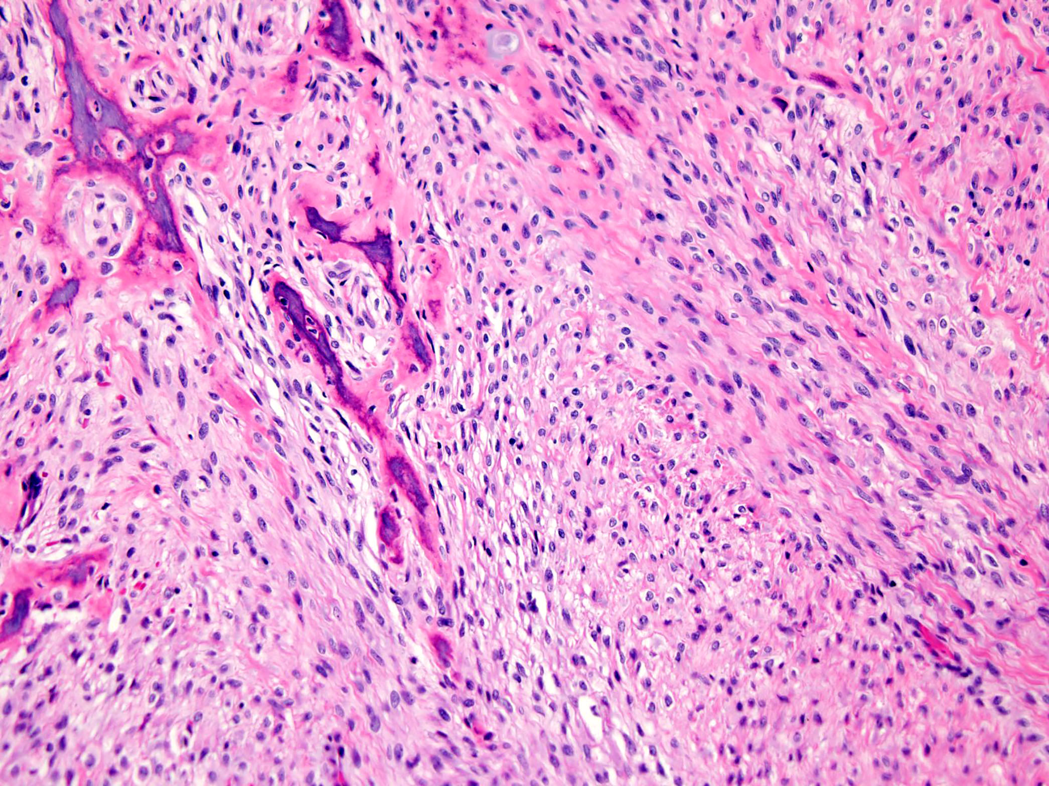
Fibro-osseous lesion
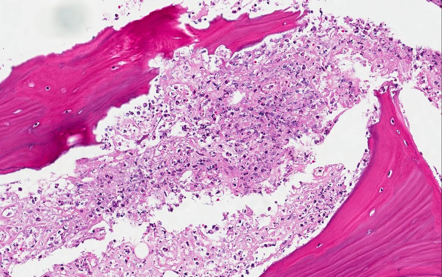
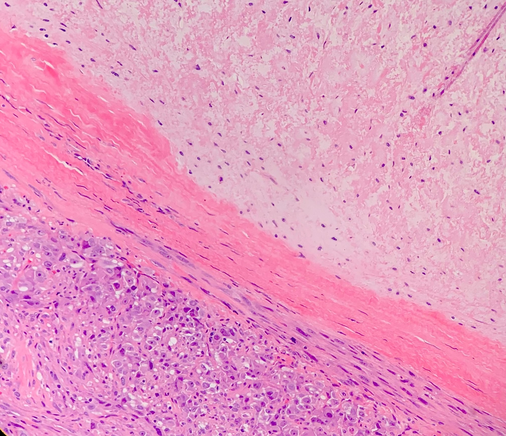
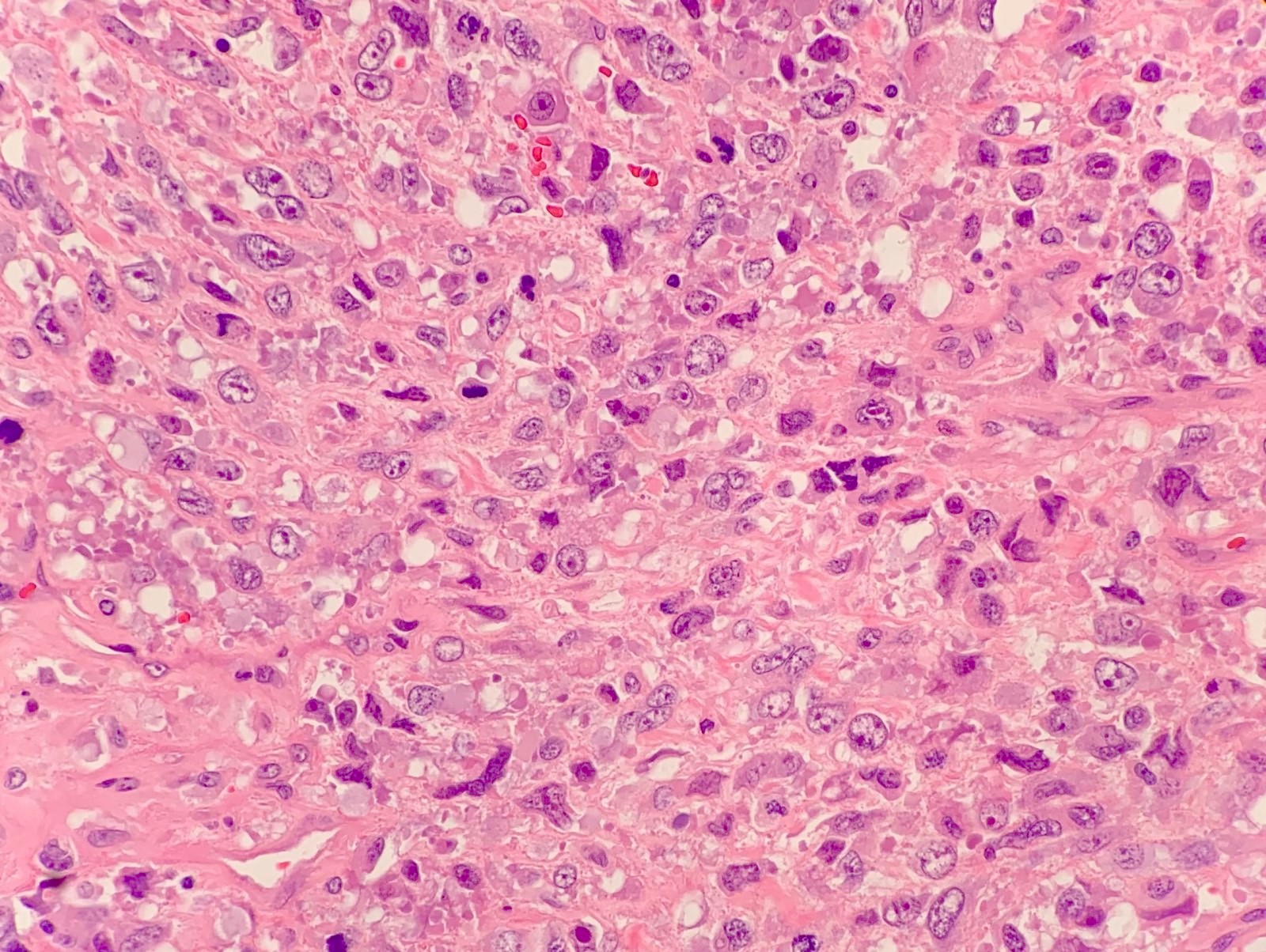
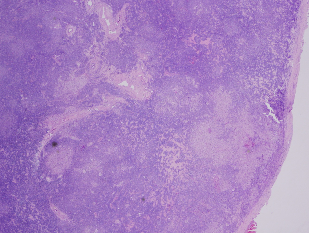
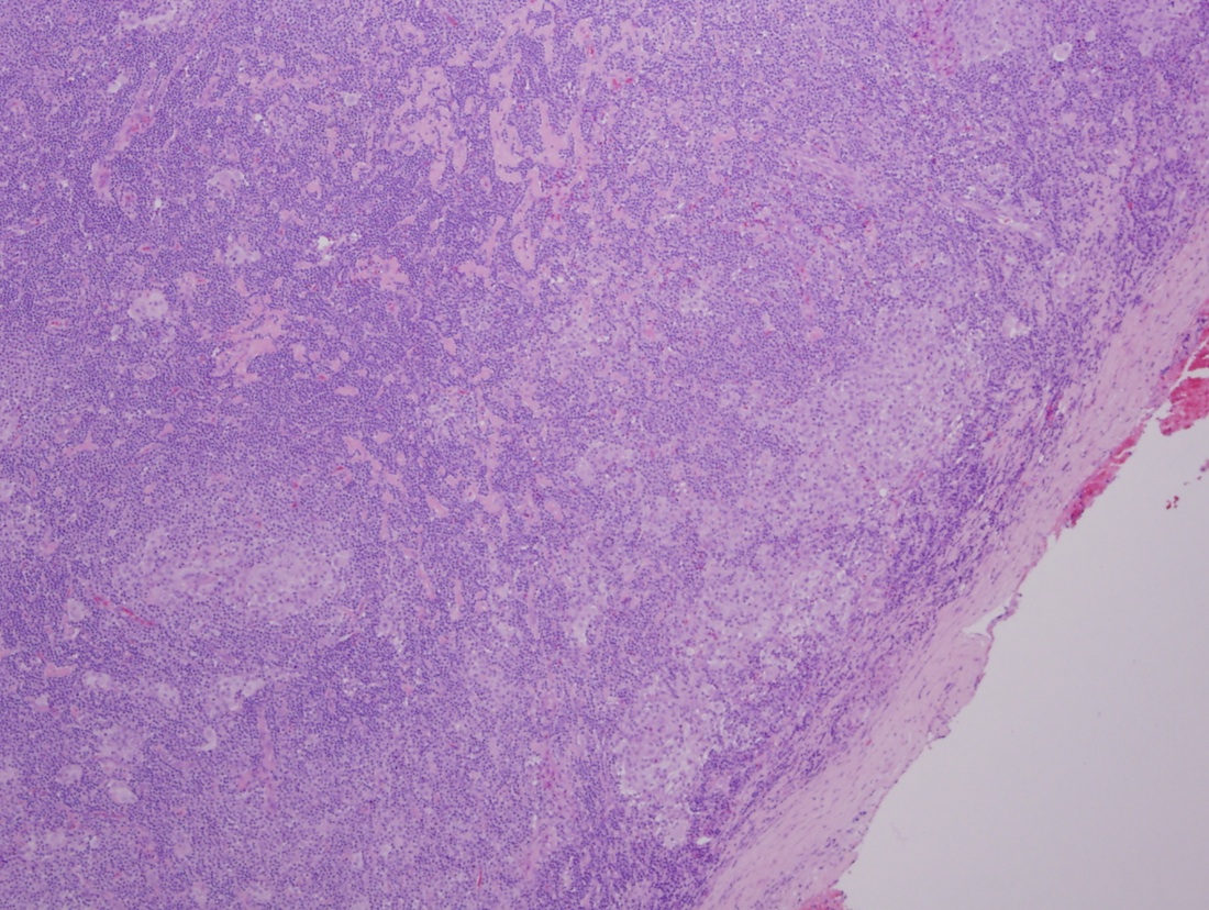
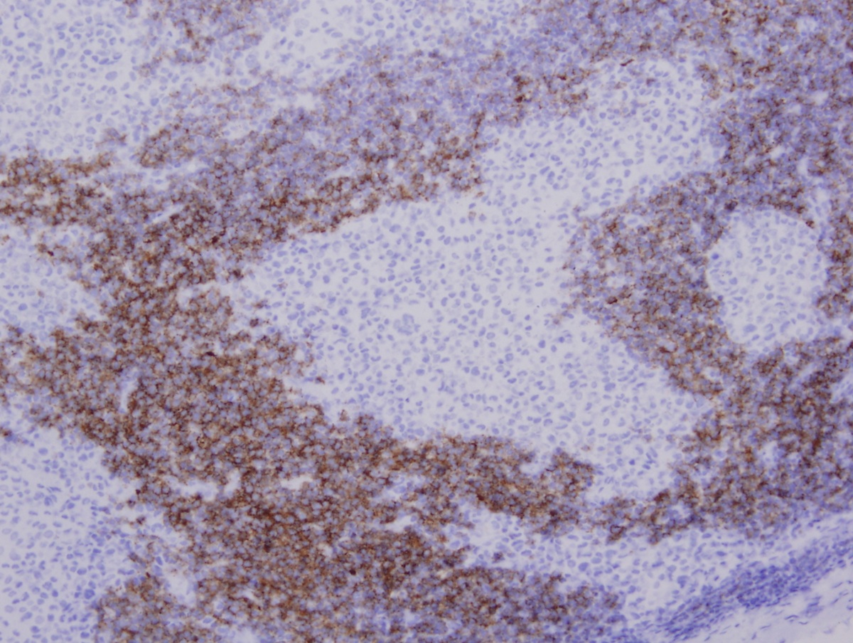
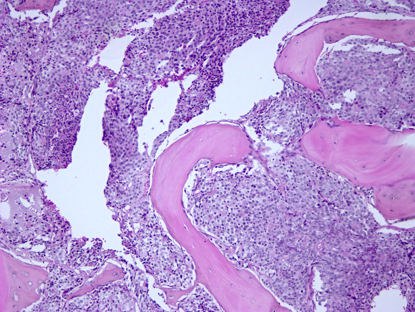
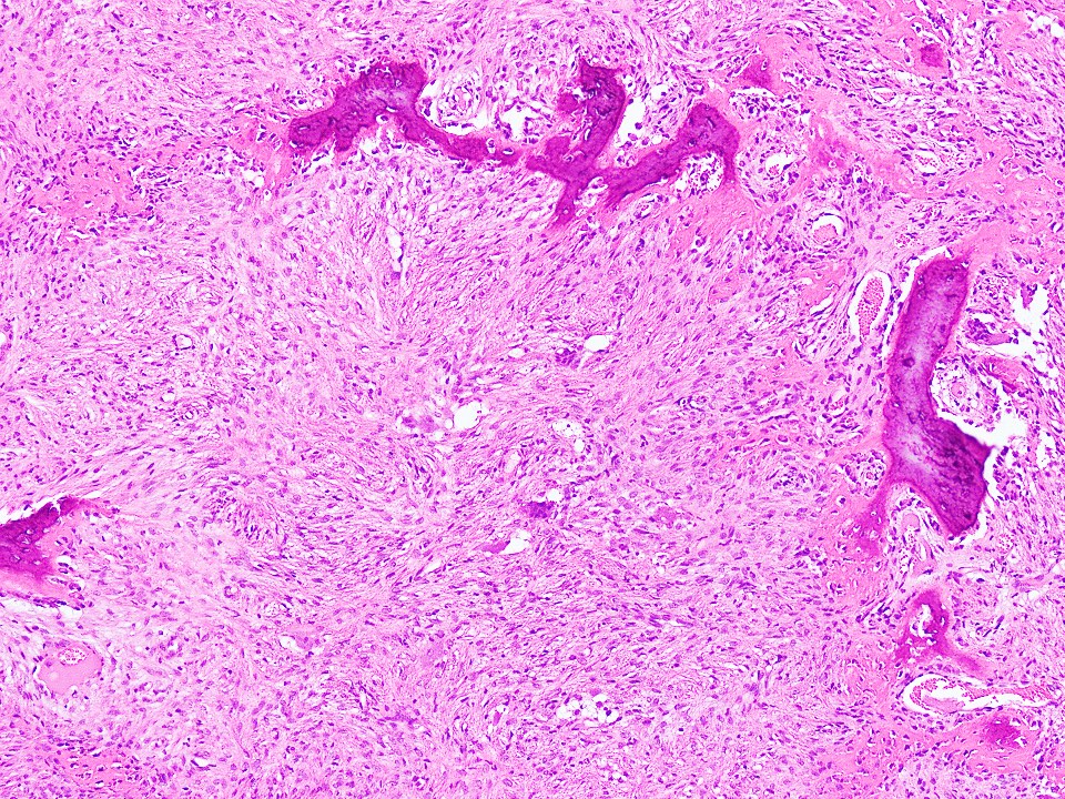
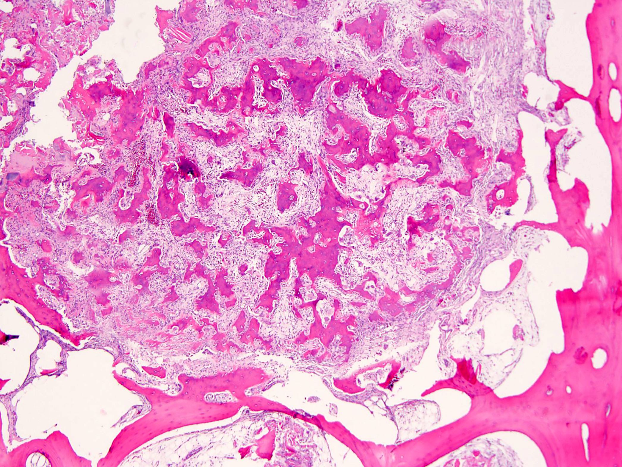
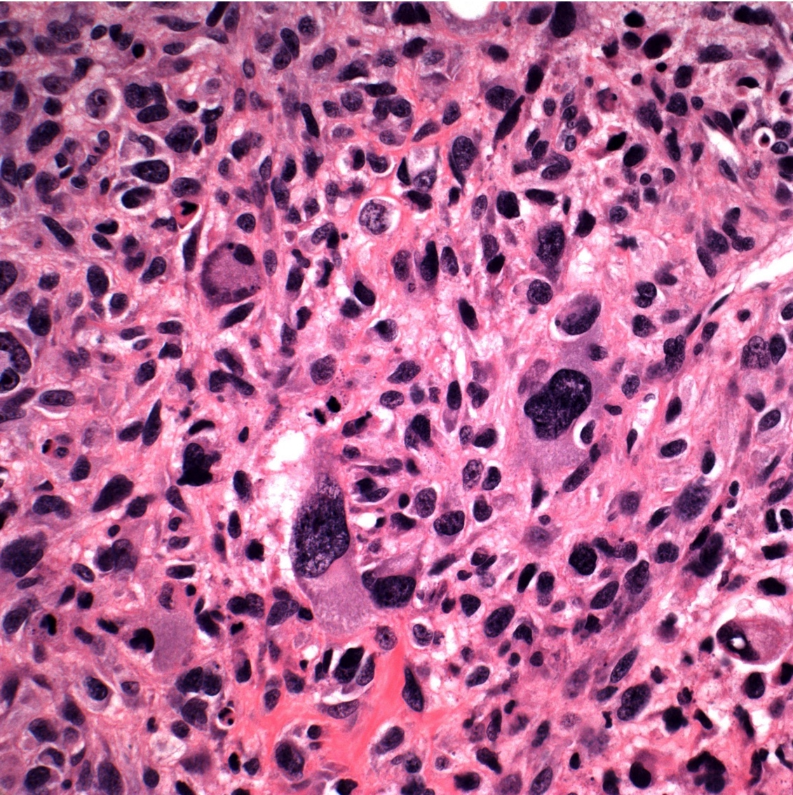
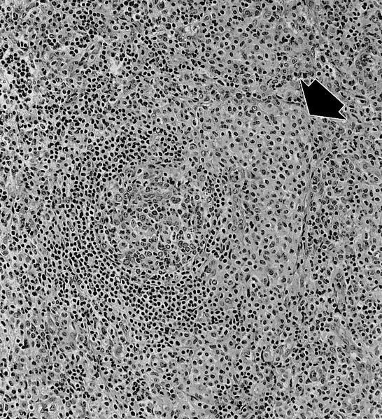
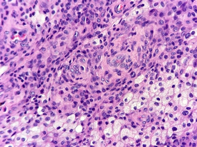
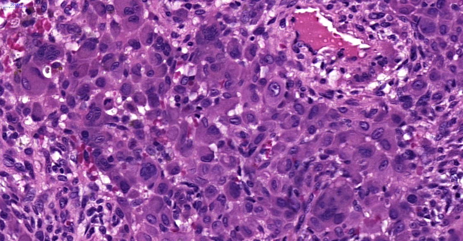
Trabecular growth pattern
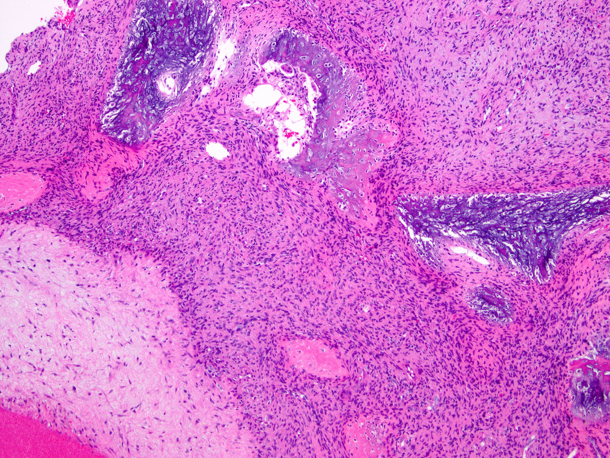
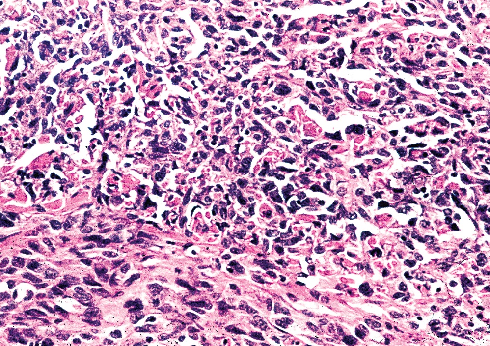
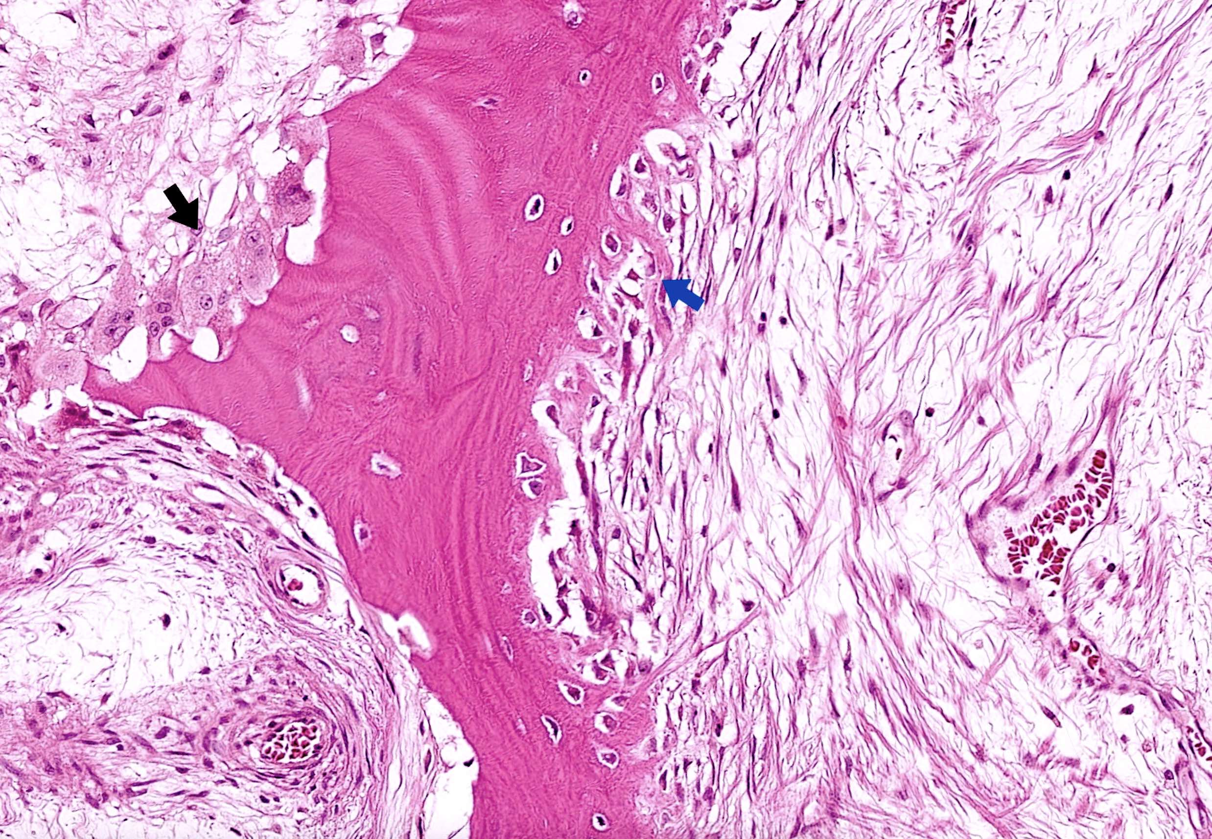
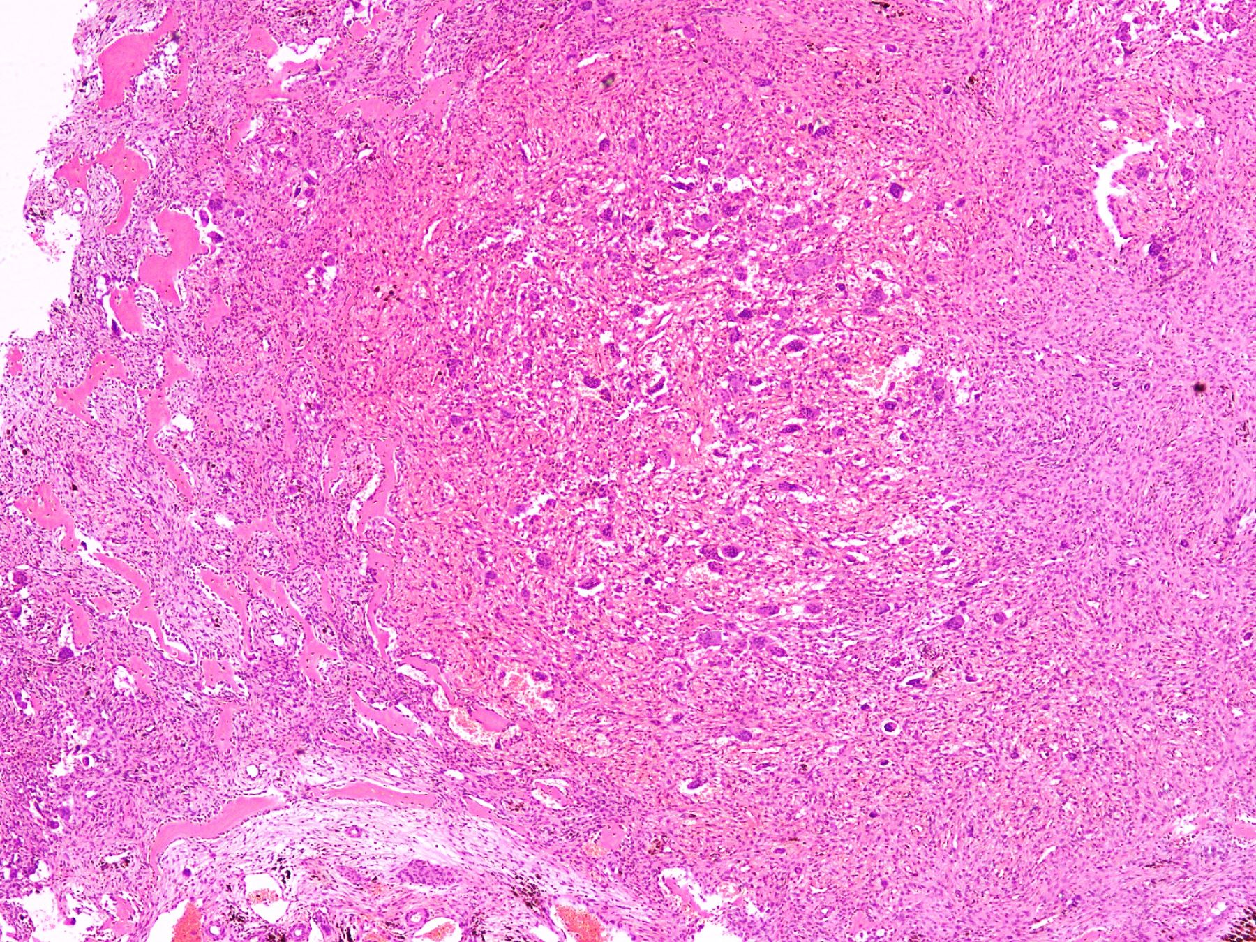
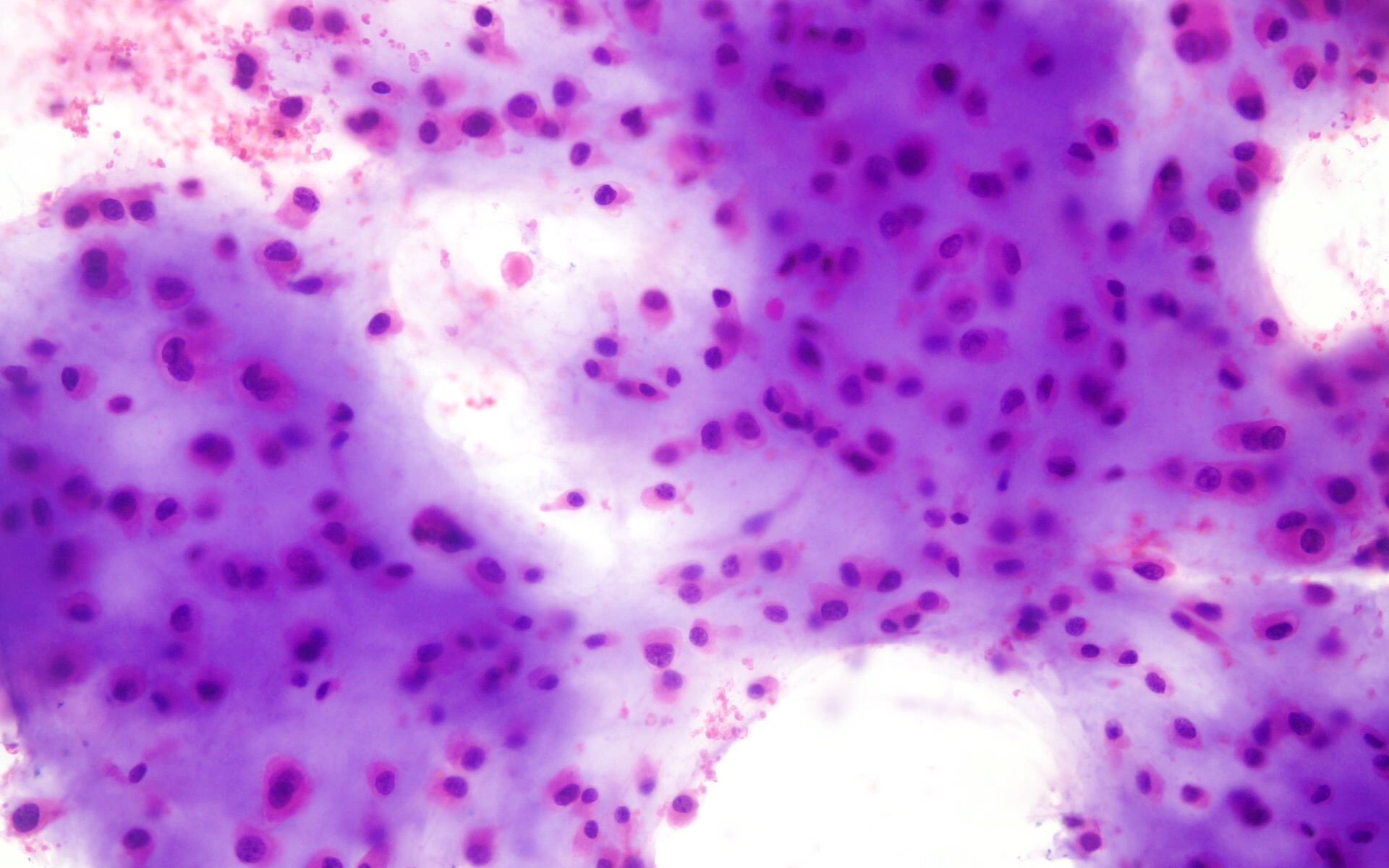
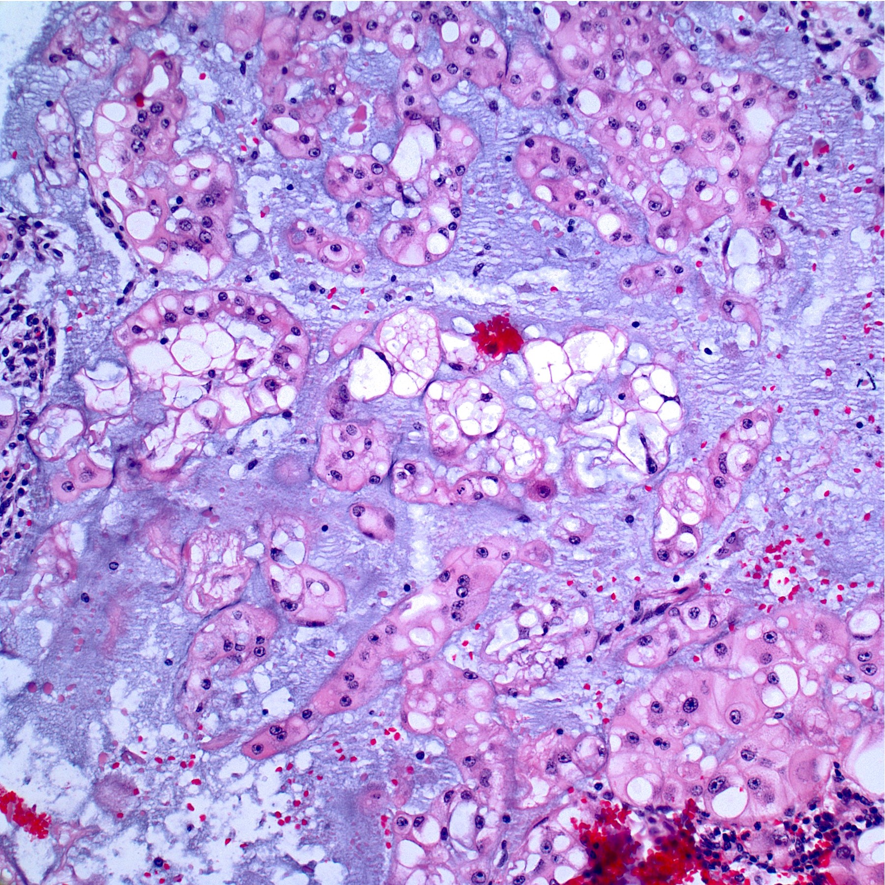
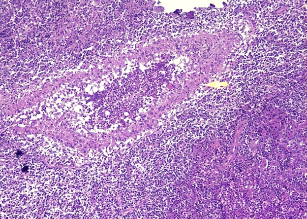
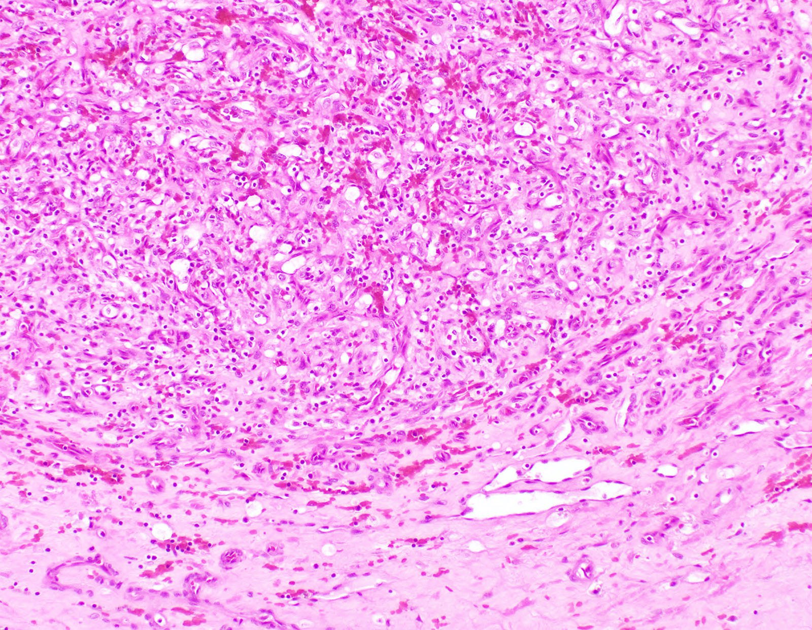
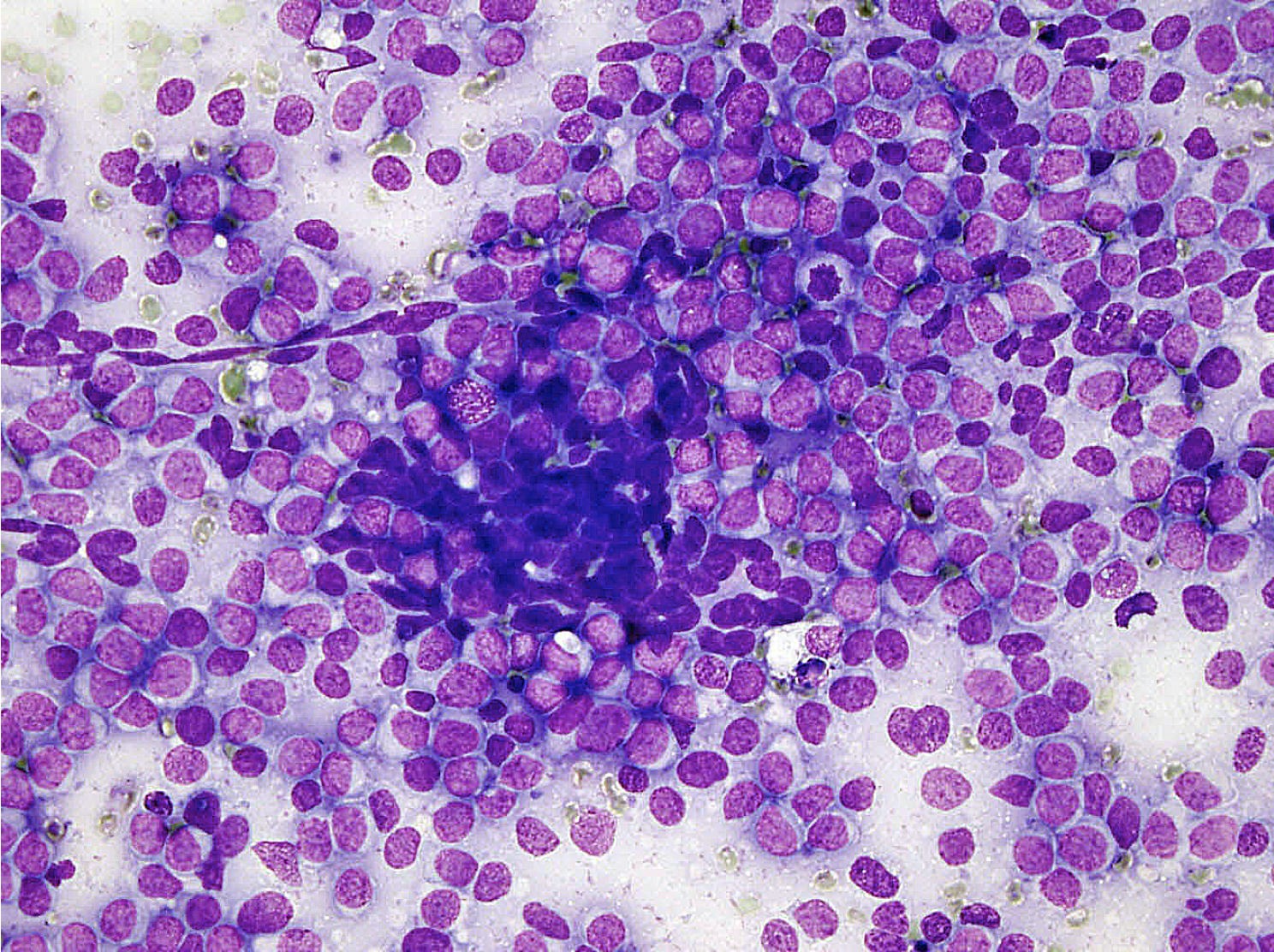
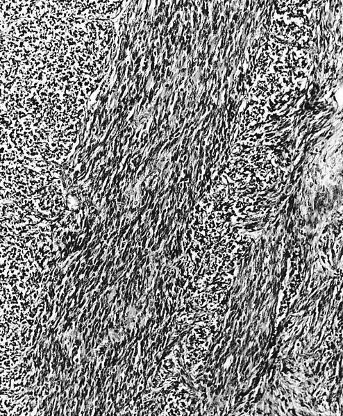
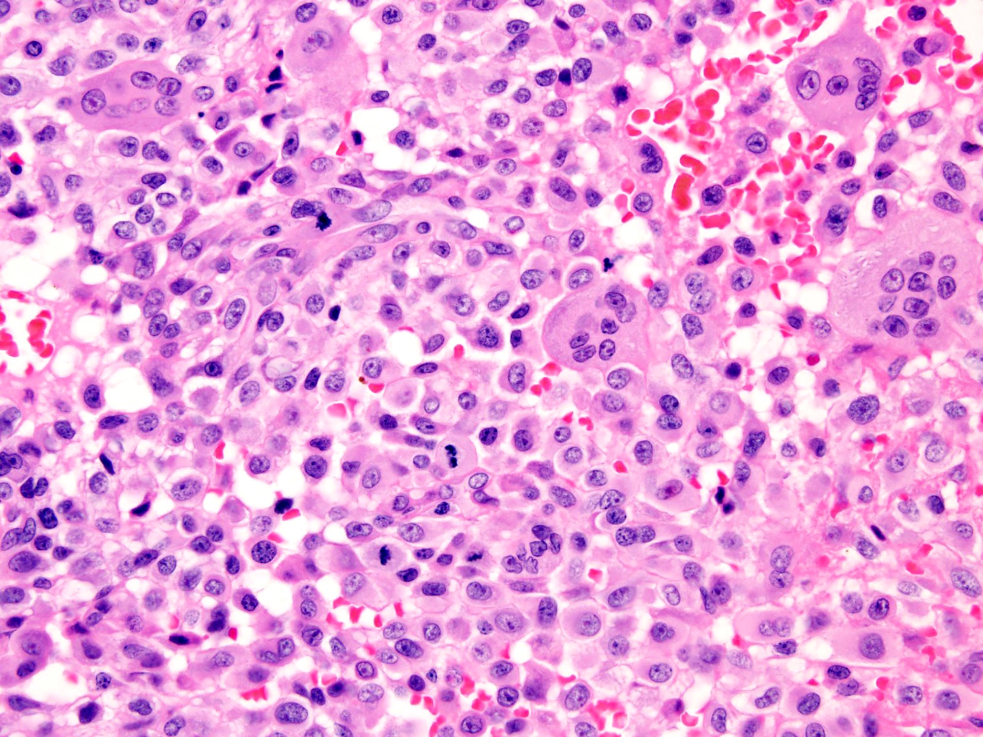
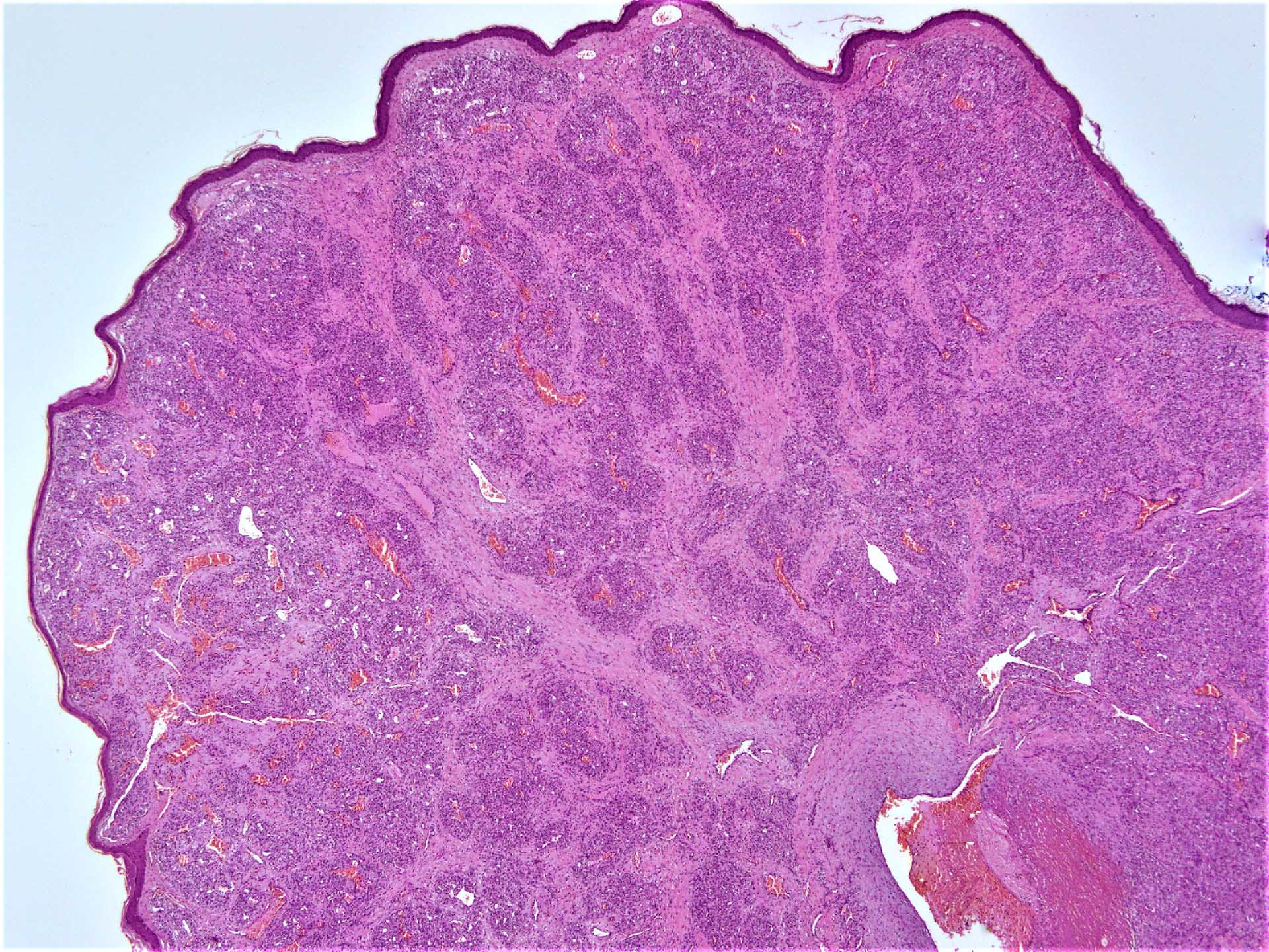
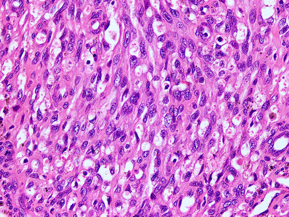
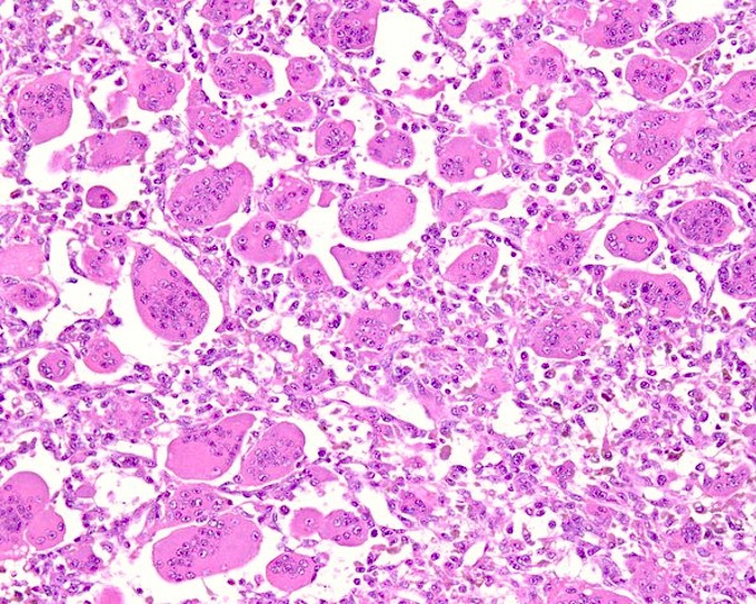
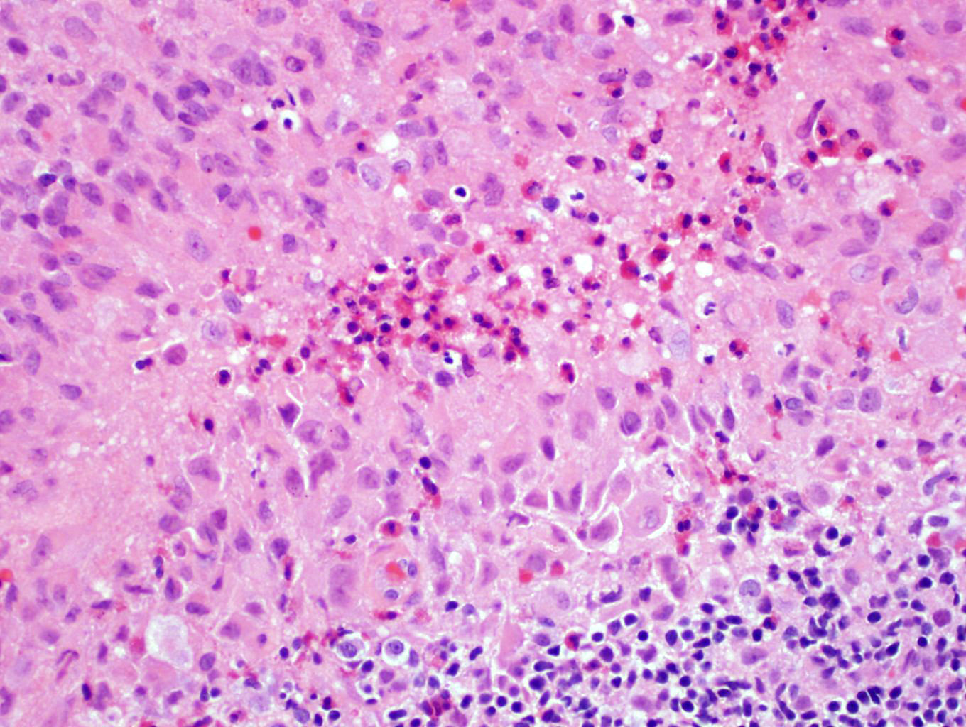
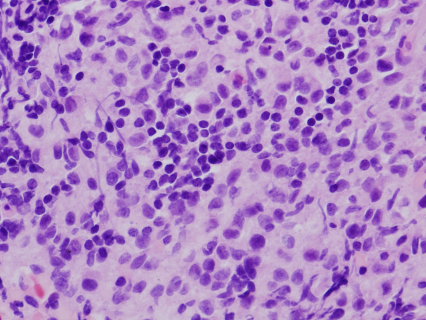
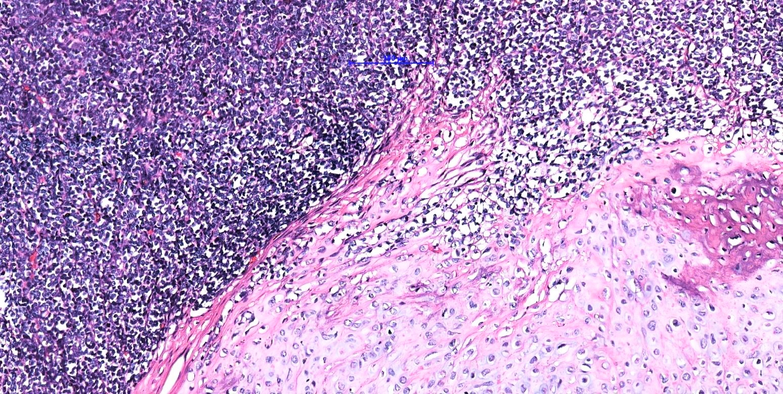
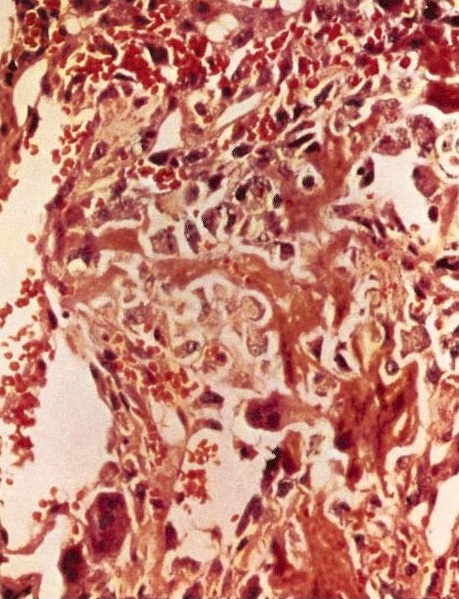
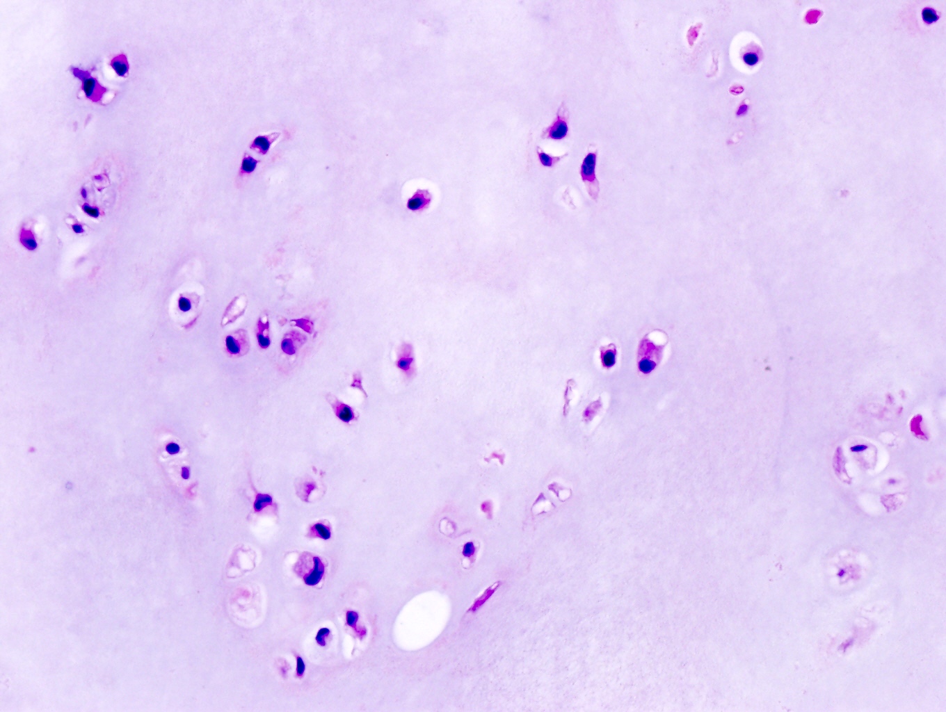
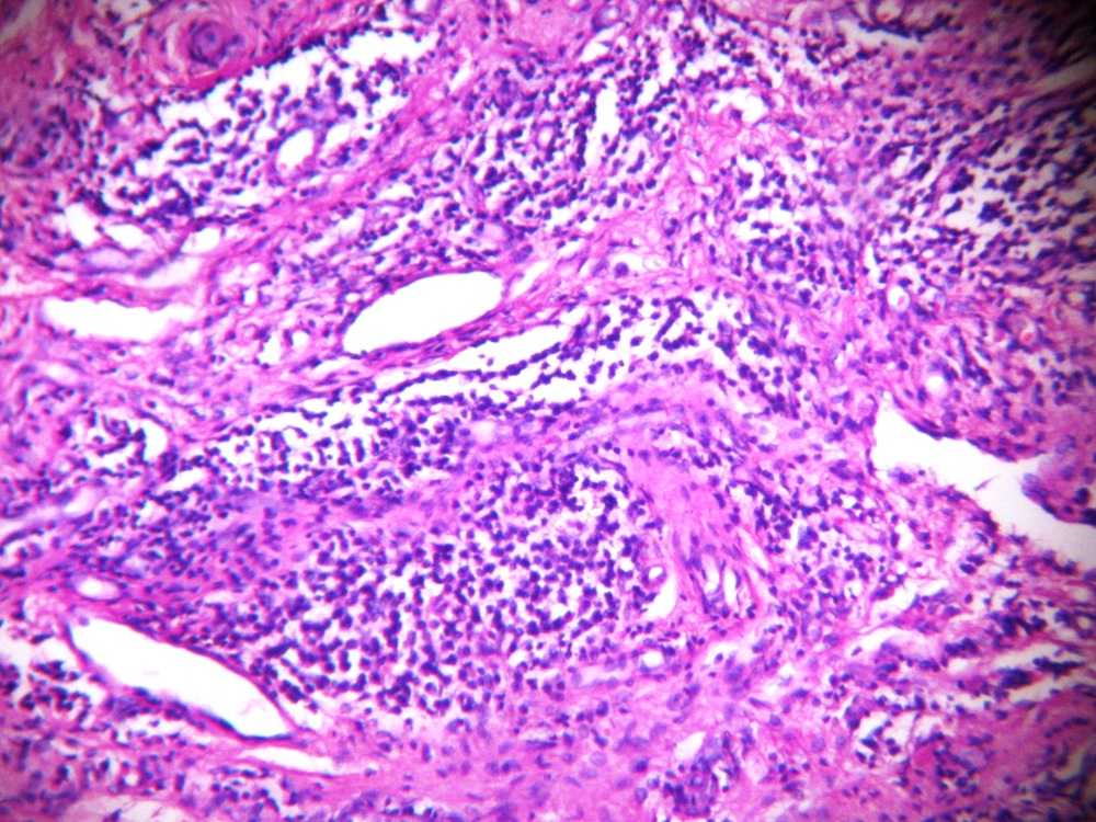
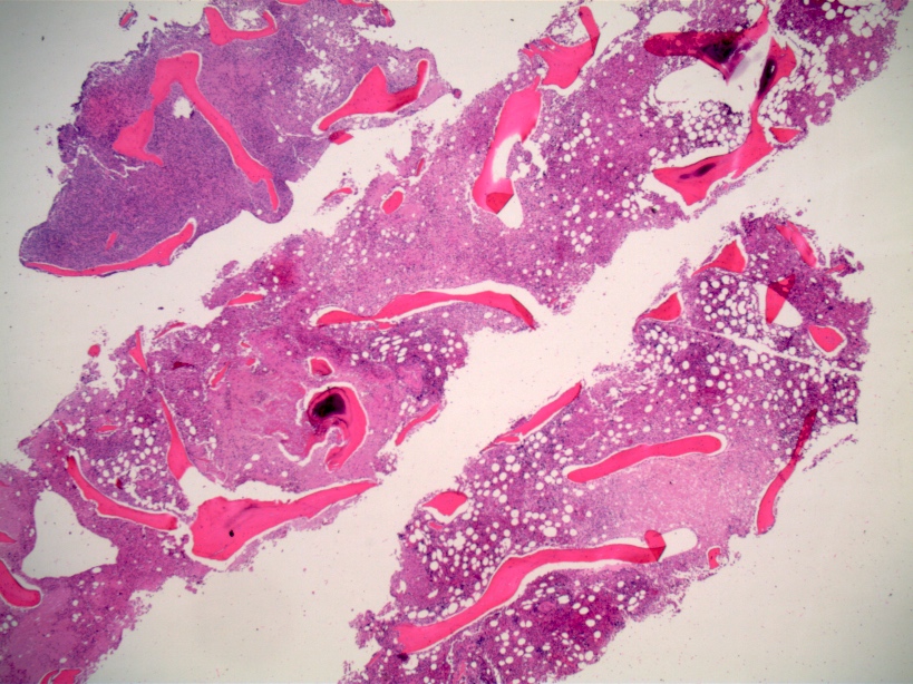
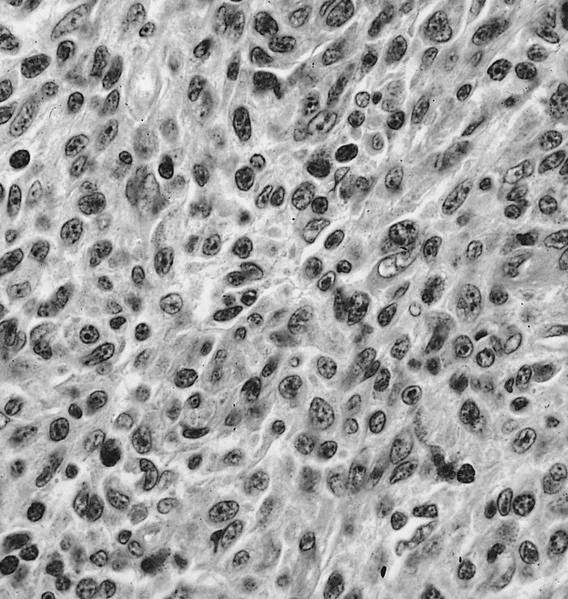
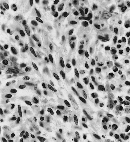
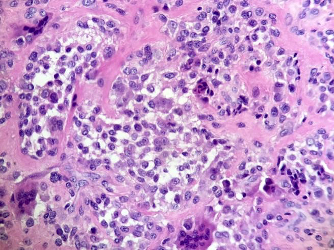
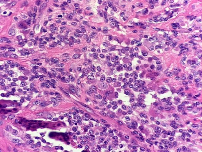
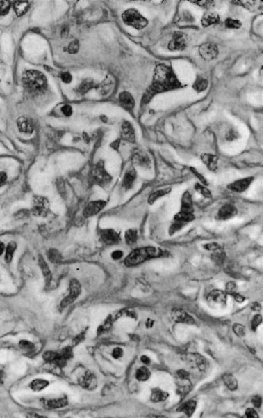
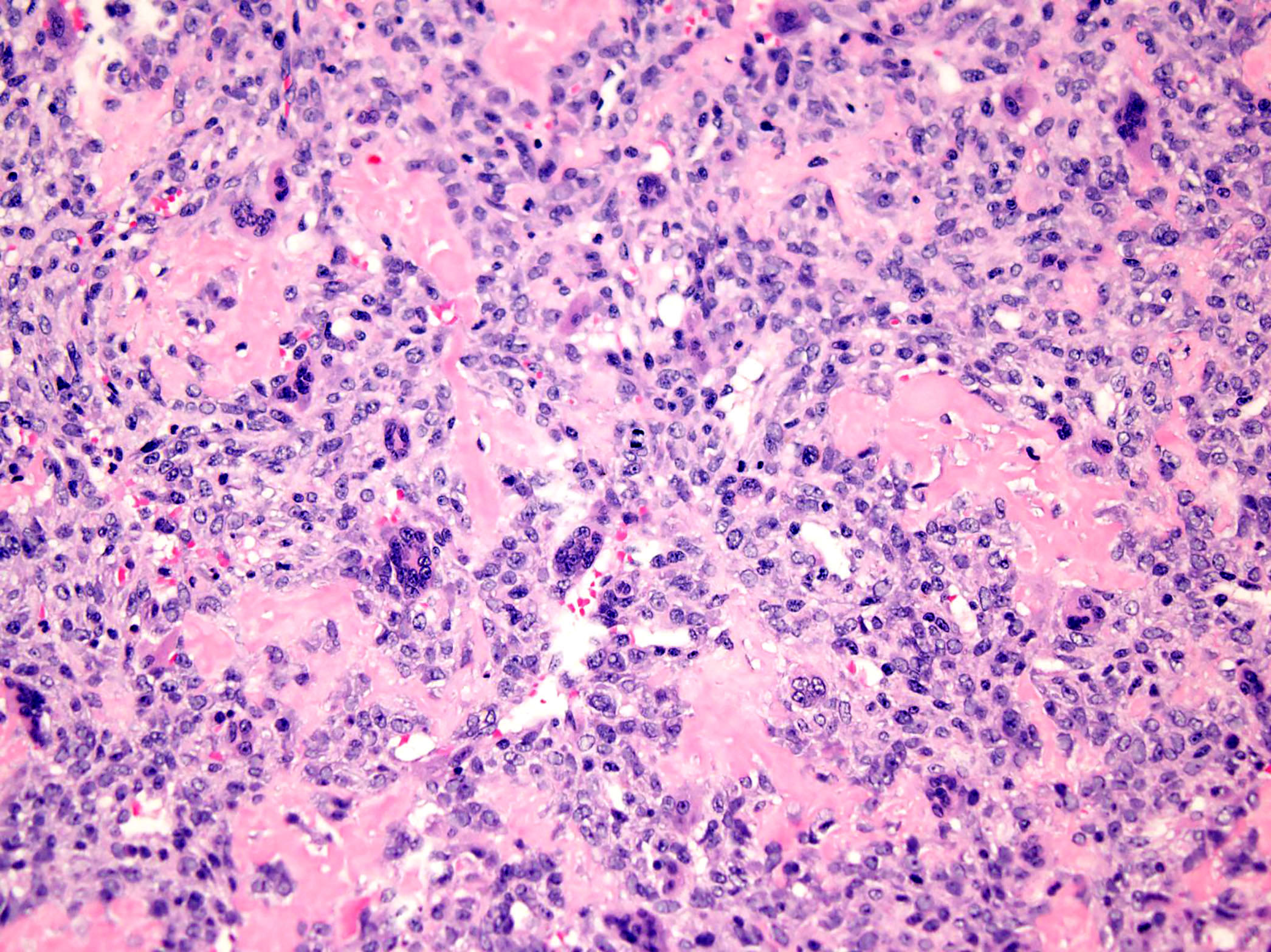
Anastomosing trabeculae of epithelial cells
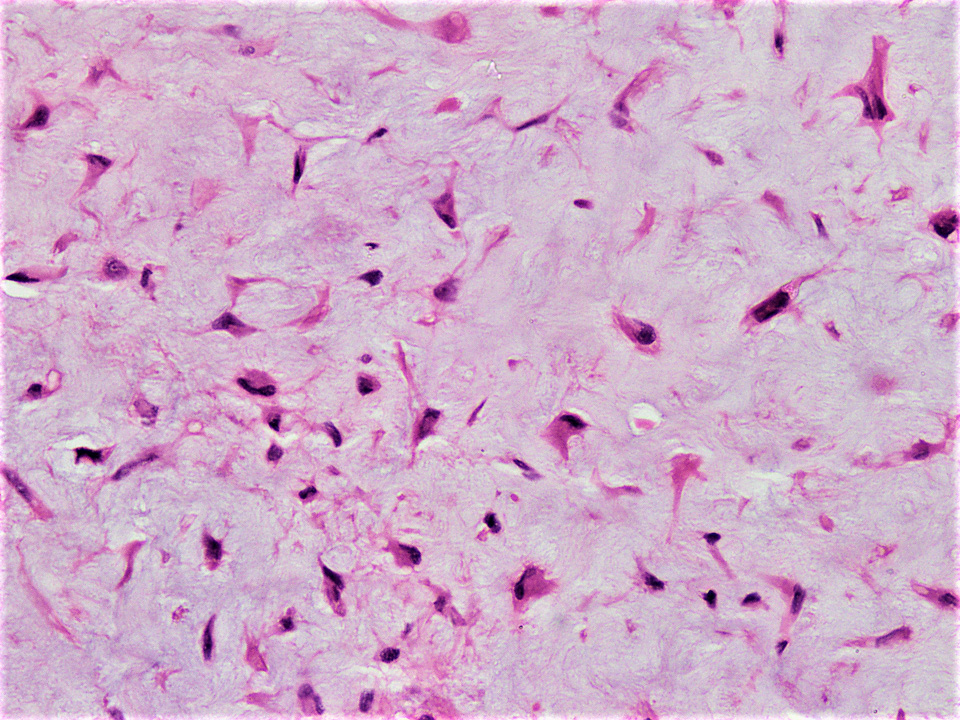
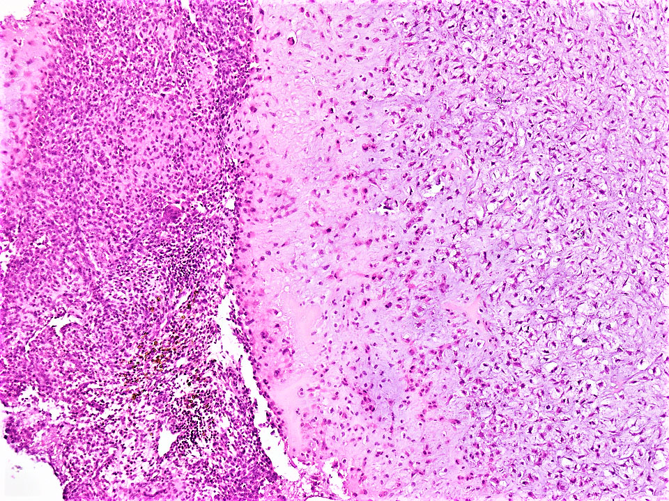
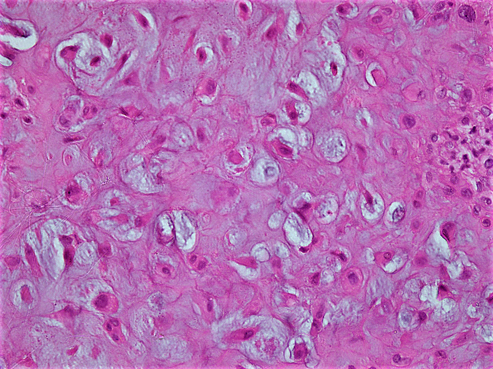
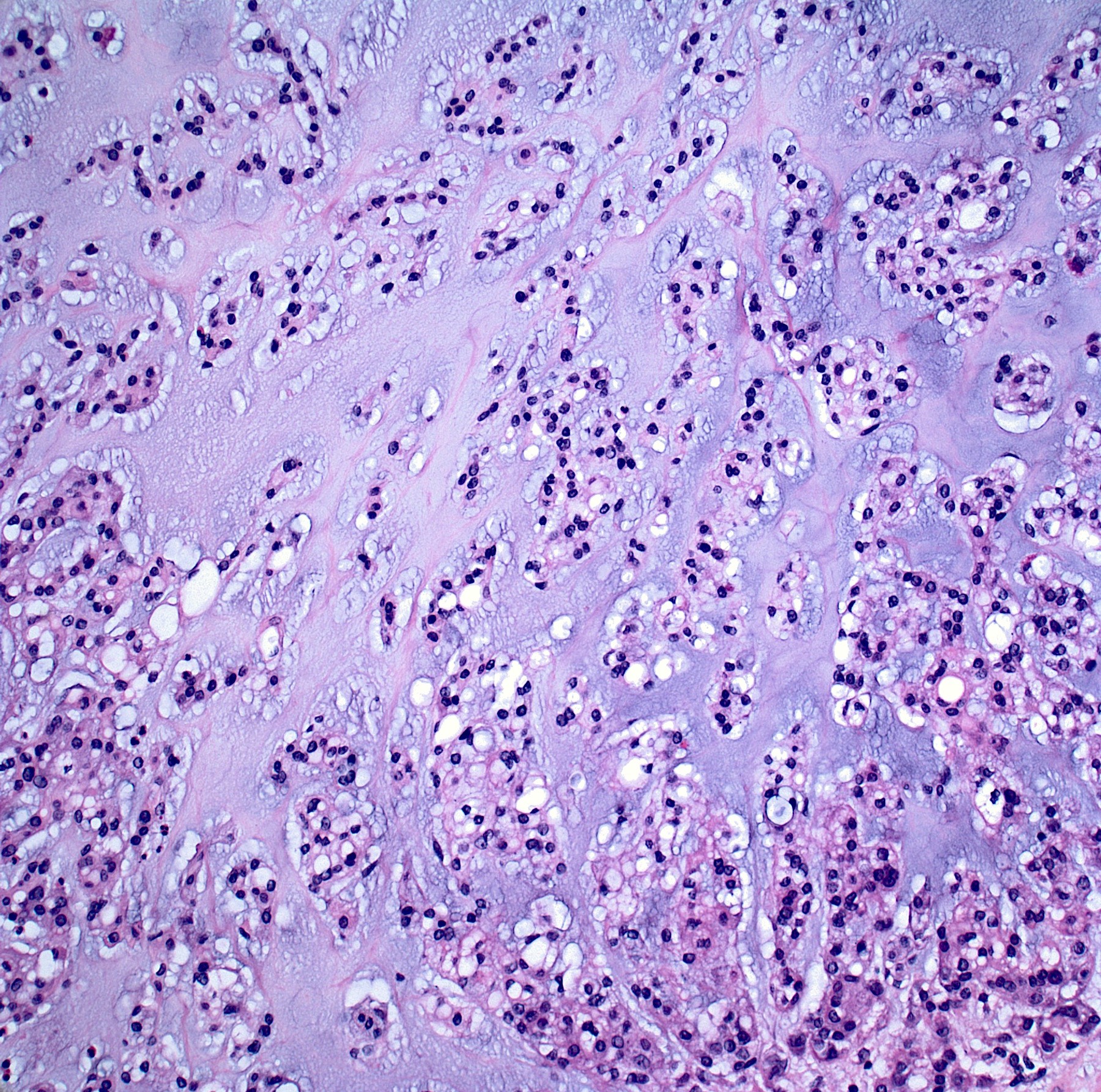
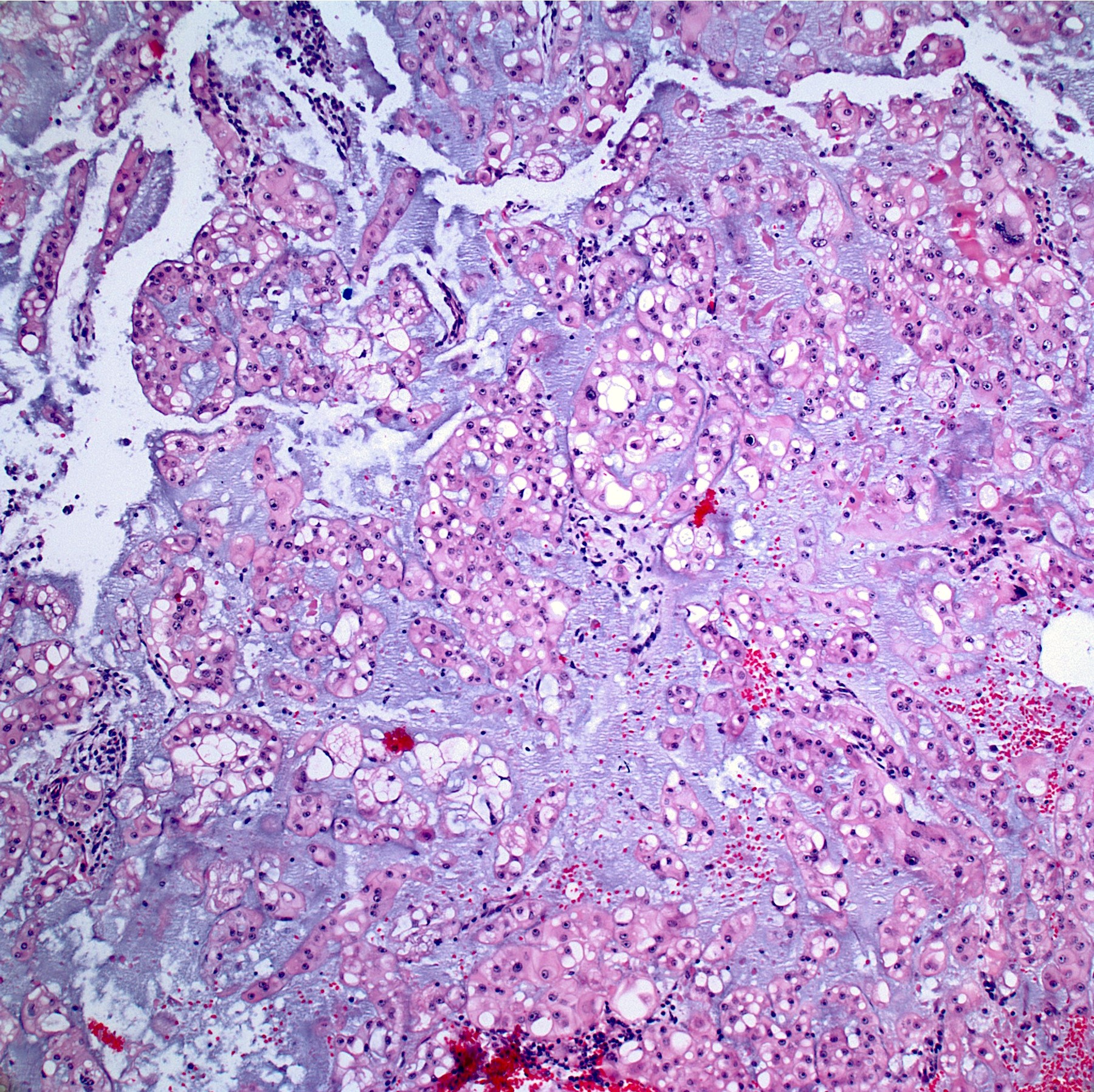
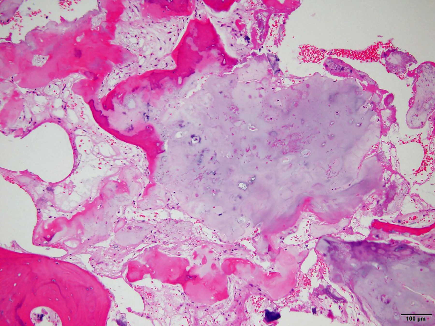
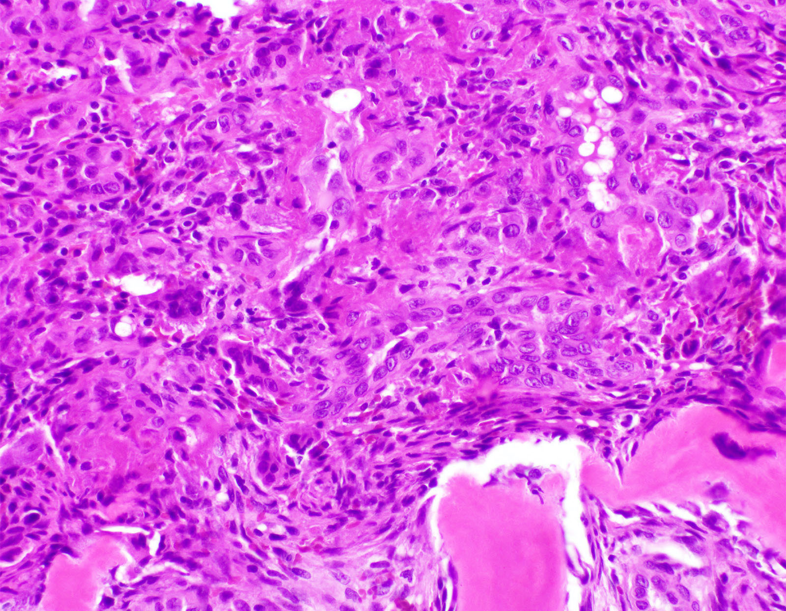
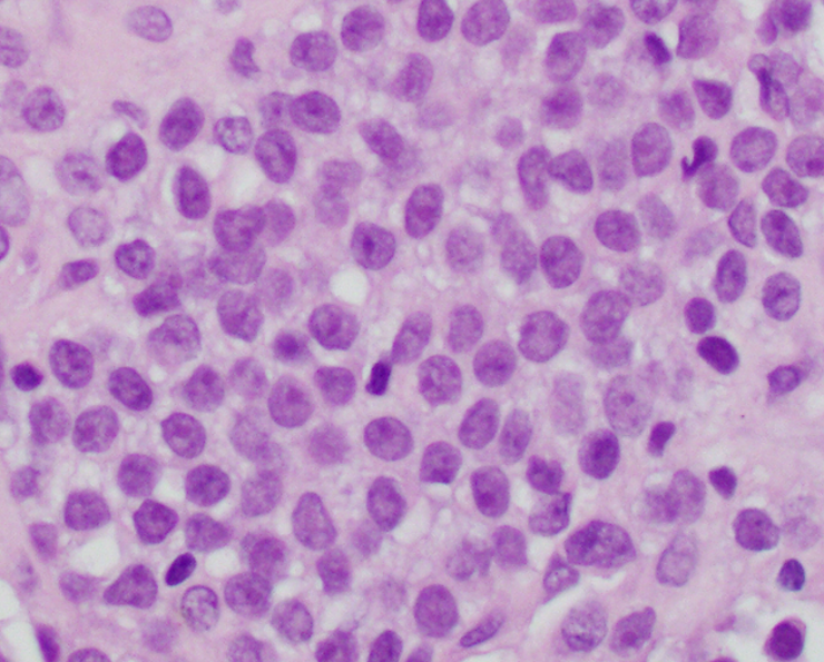
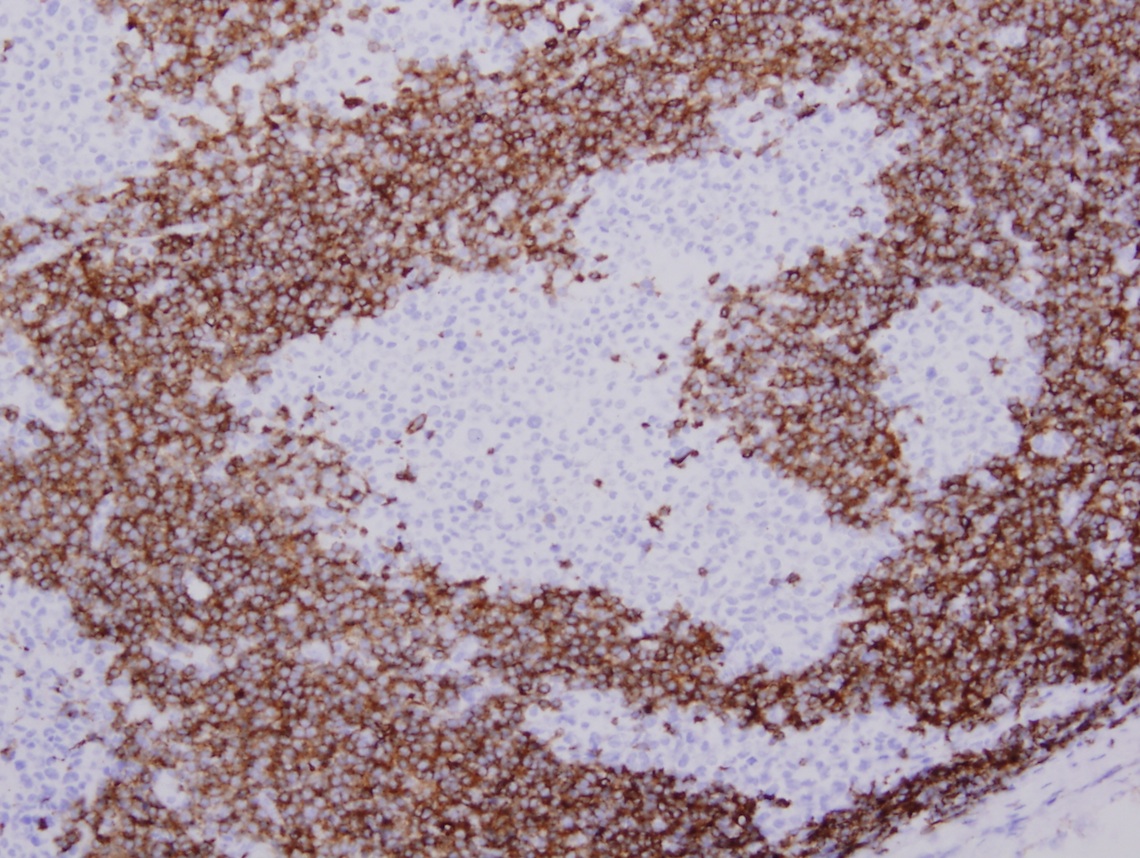
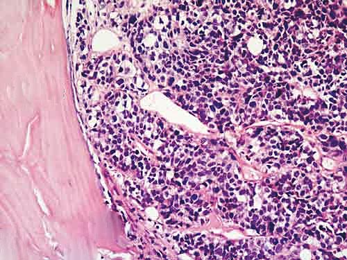
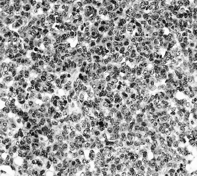
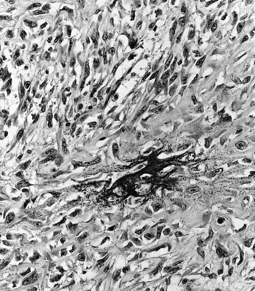
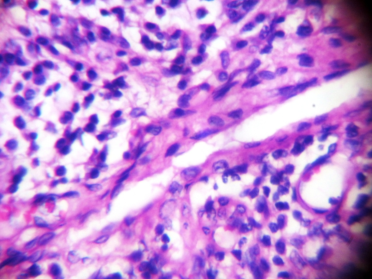
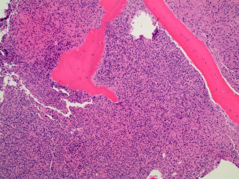
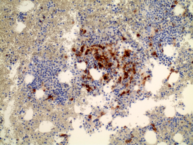
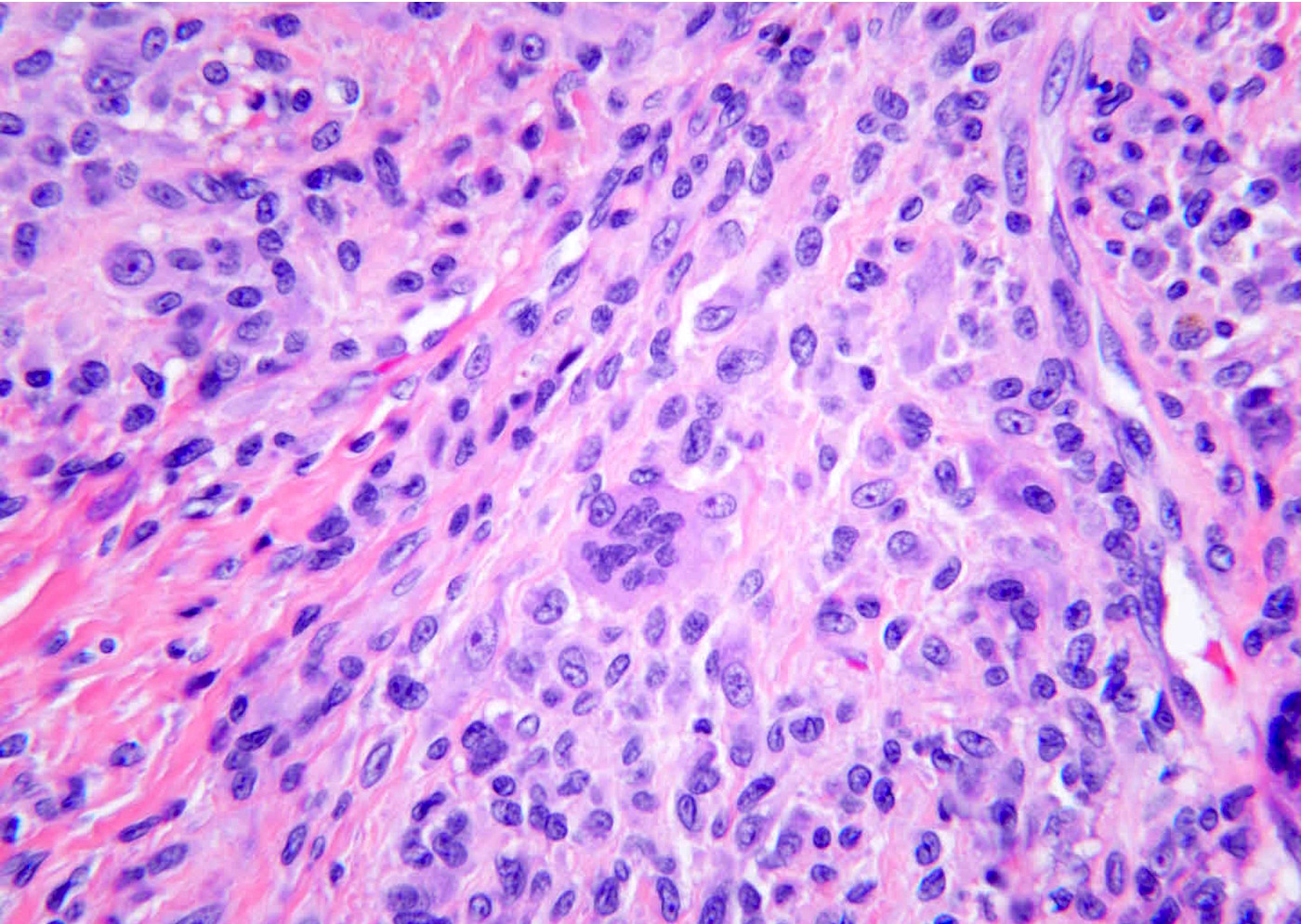
Epithelial cells

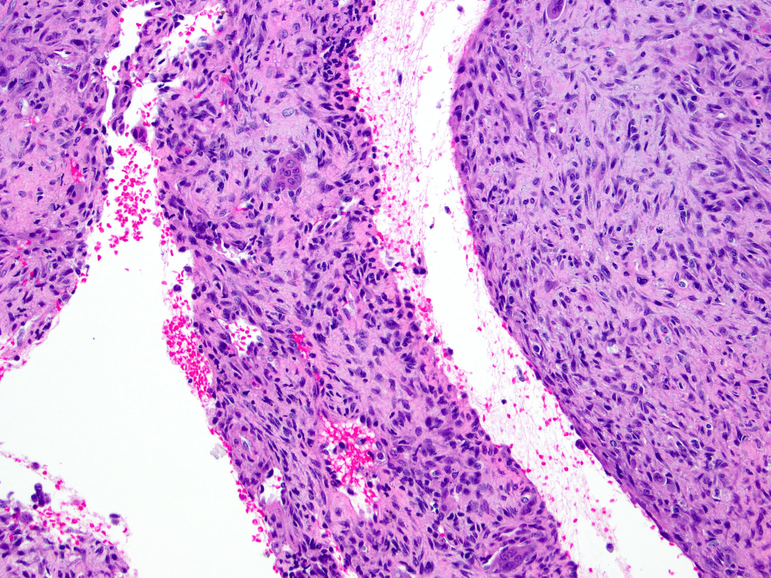
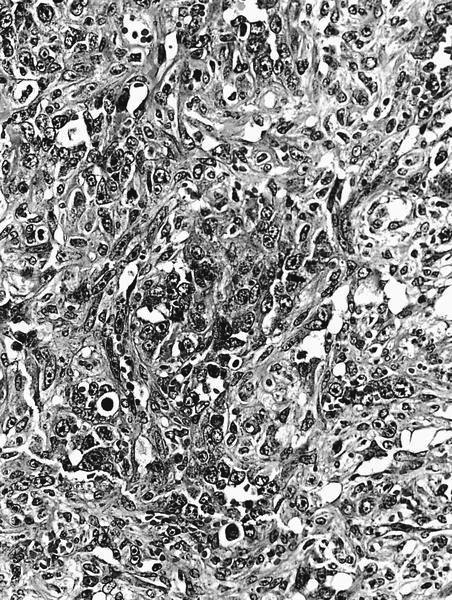
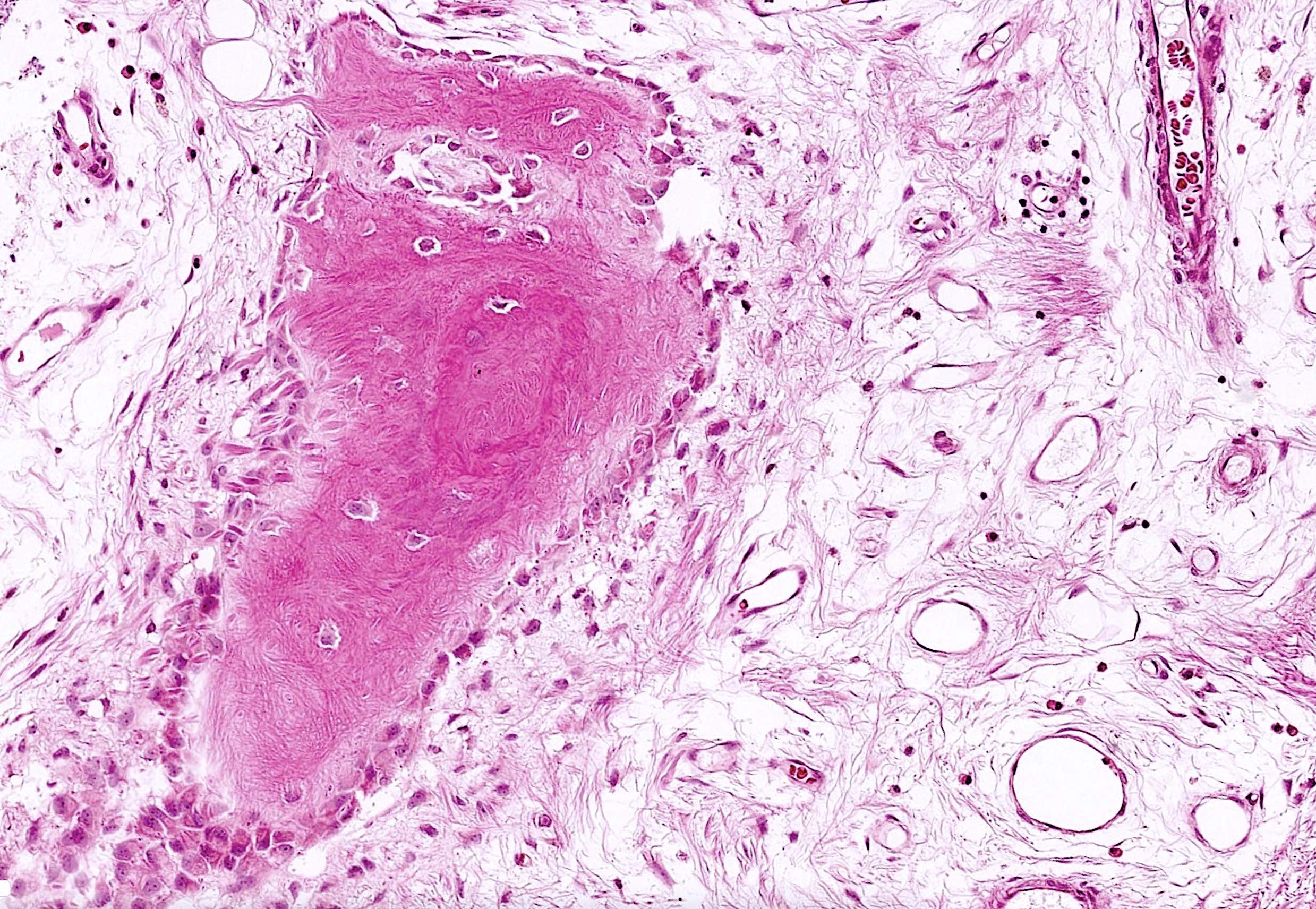
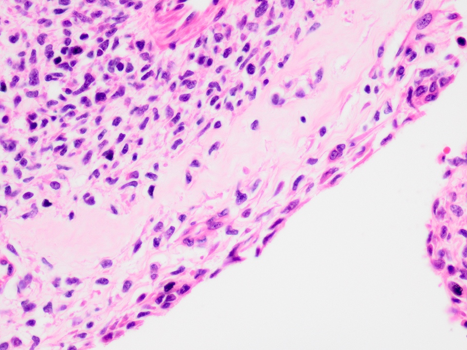
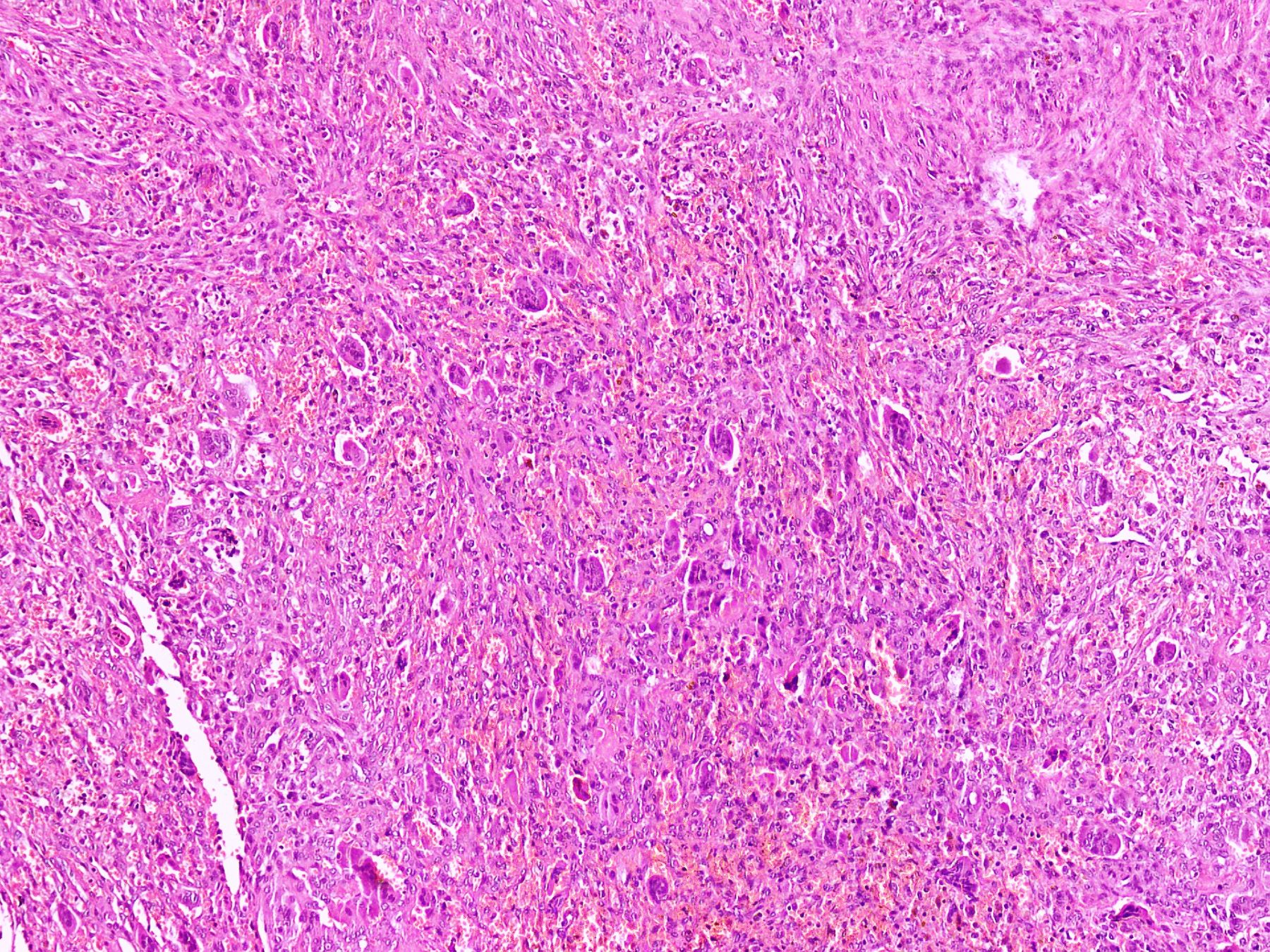
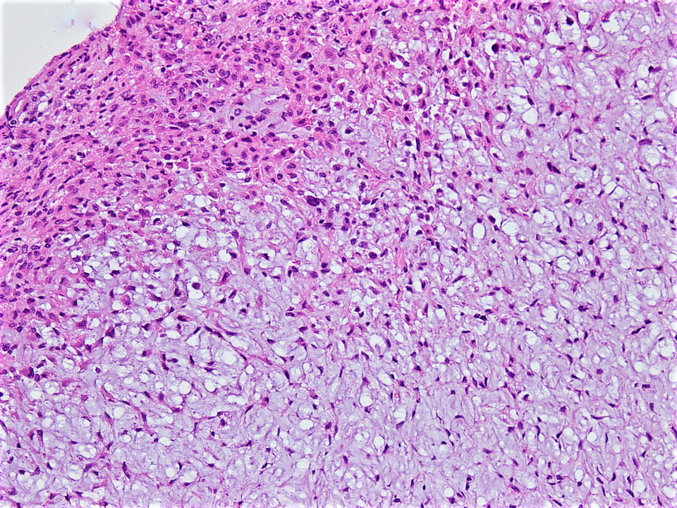
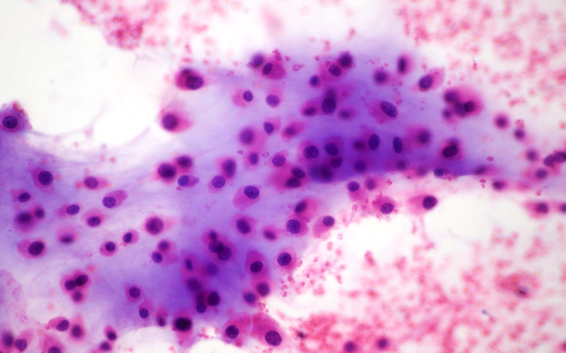
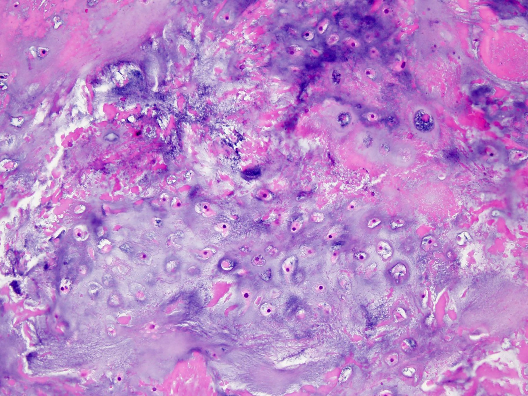
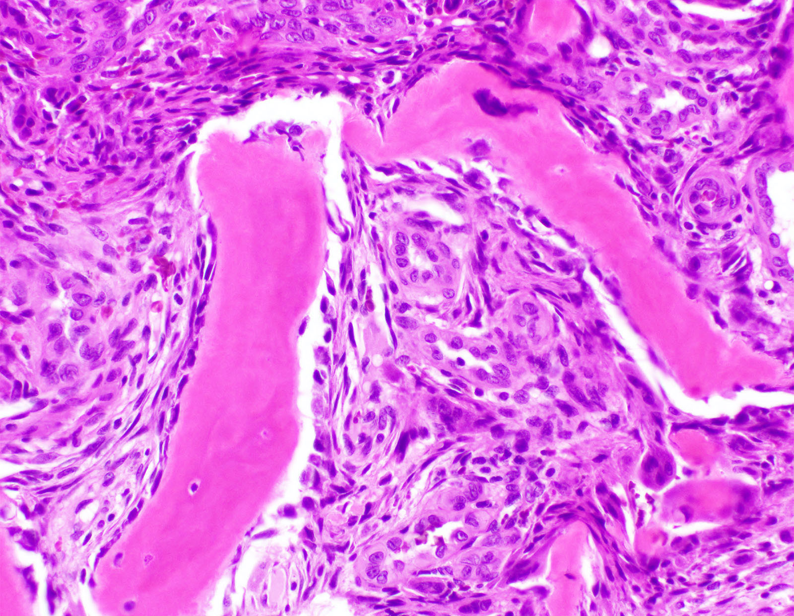
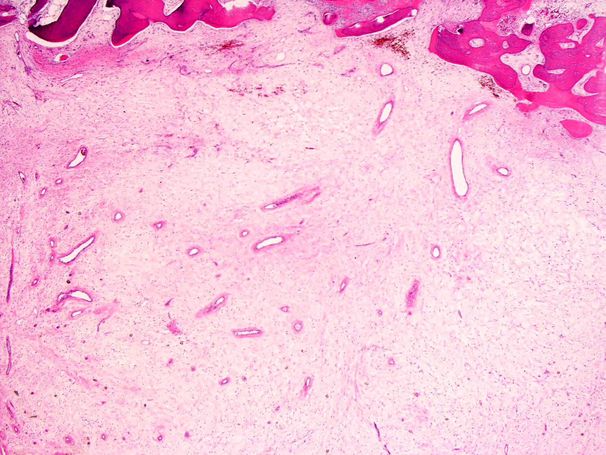
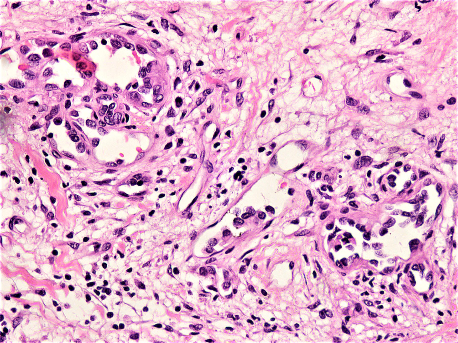
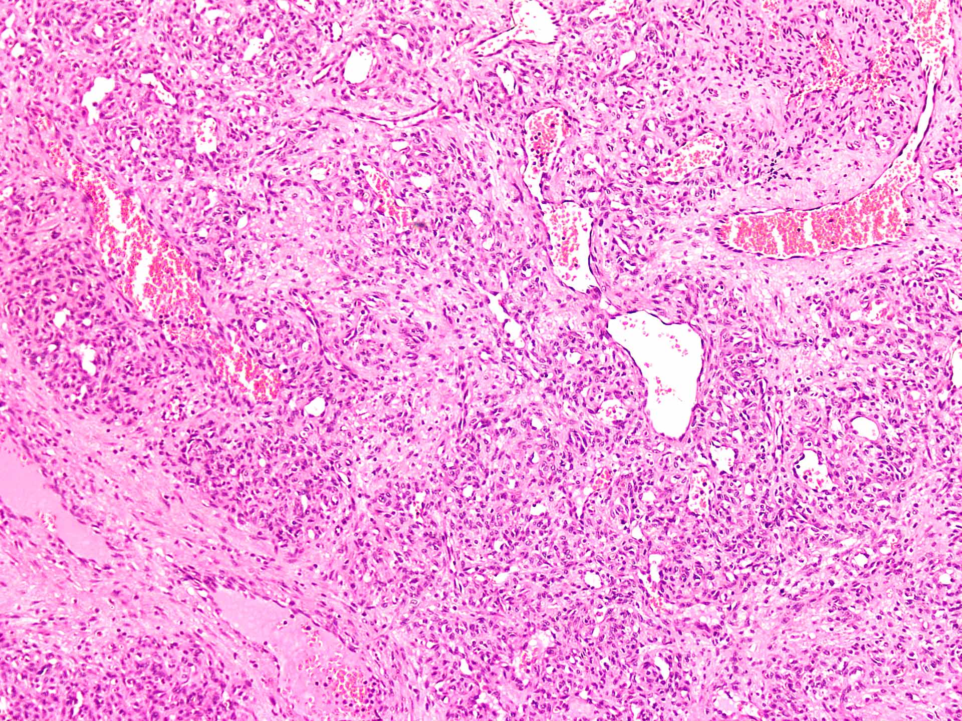
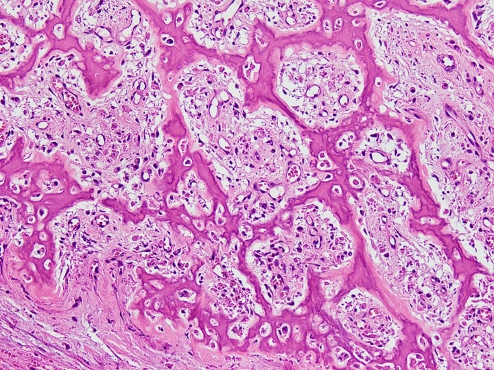
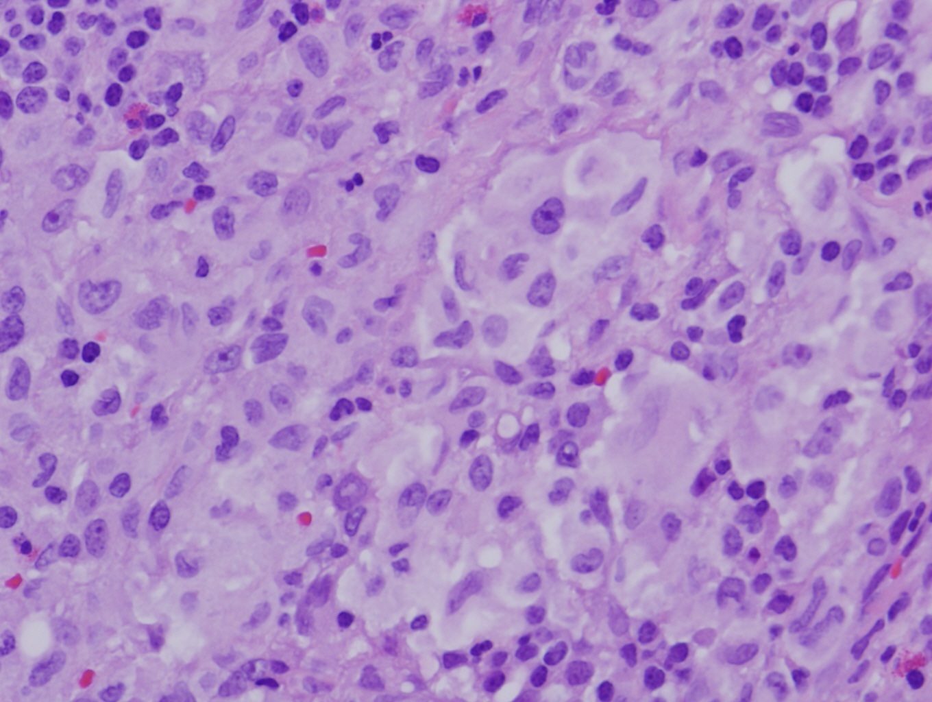
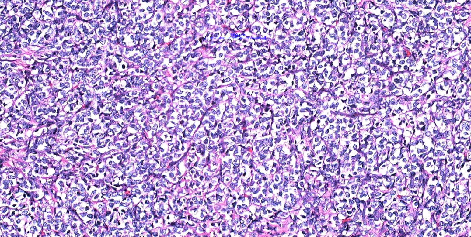
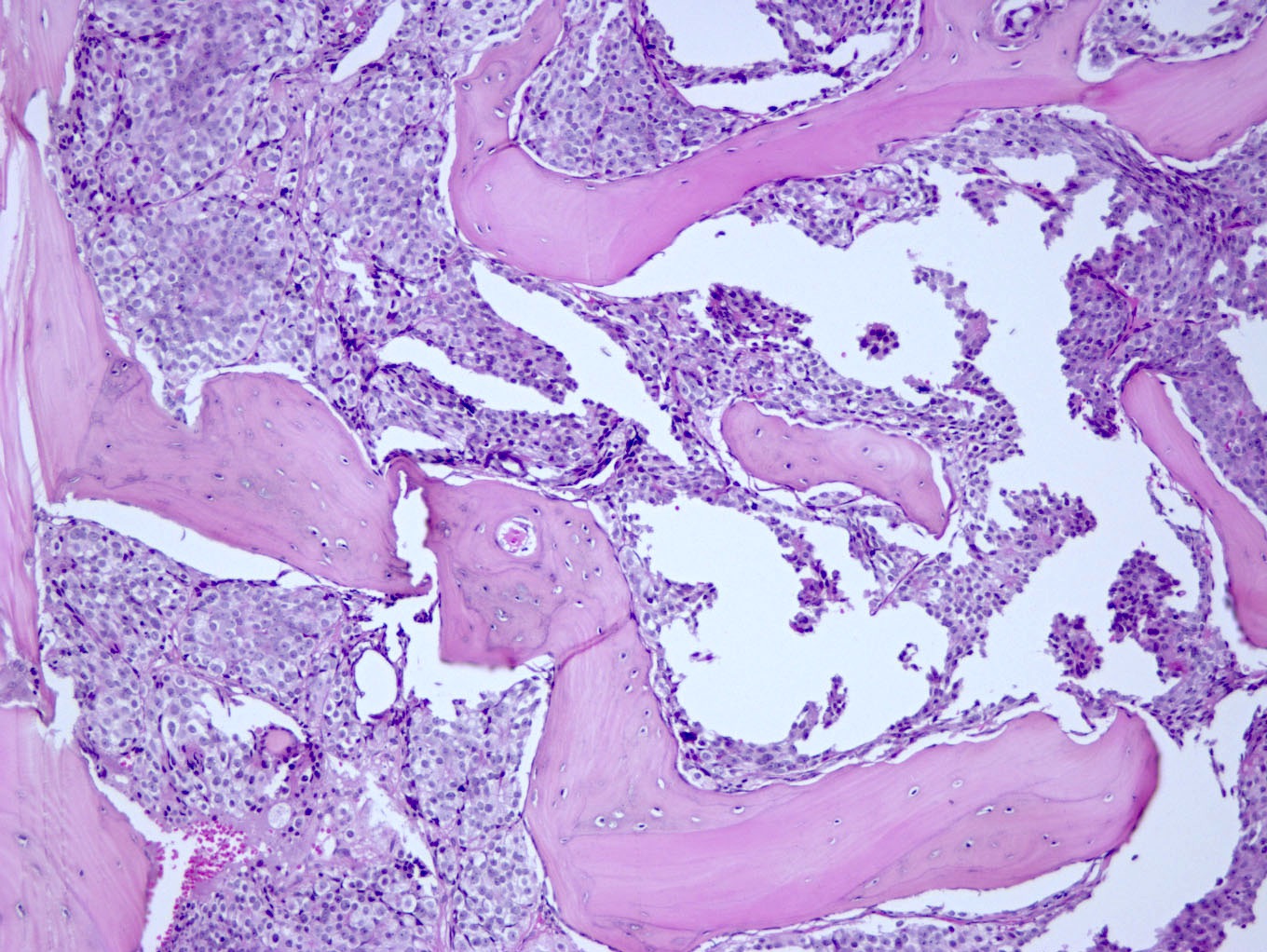
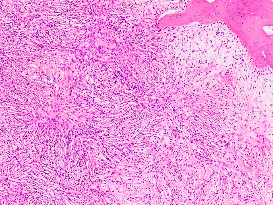
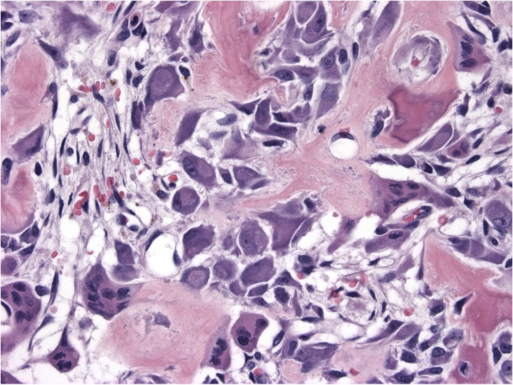
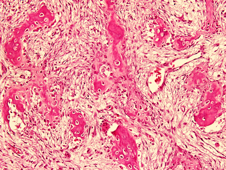
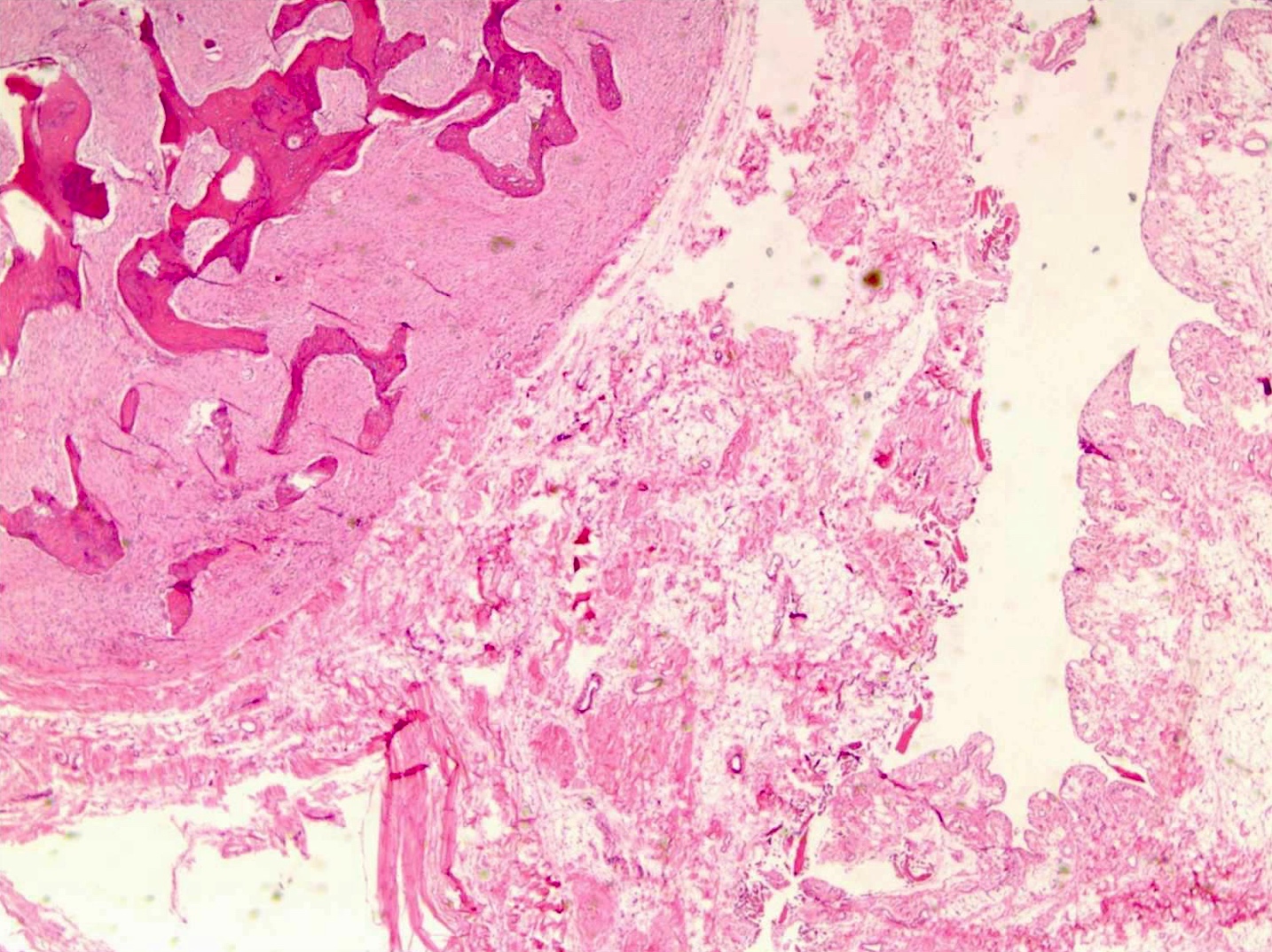
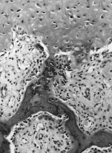
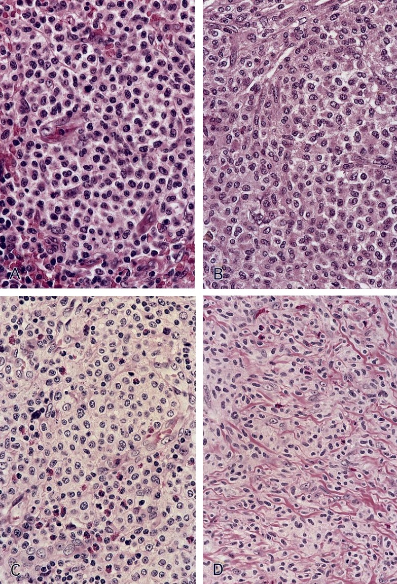
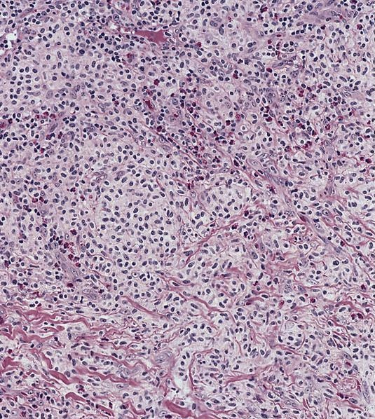
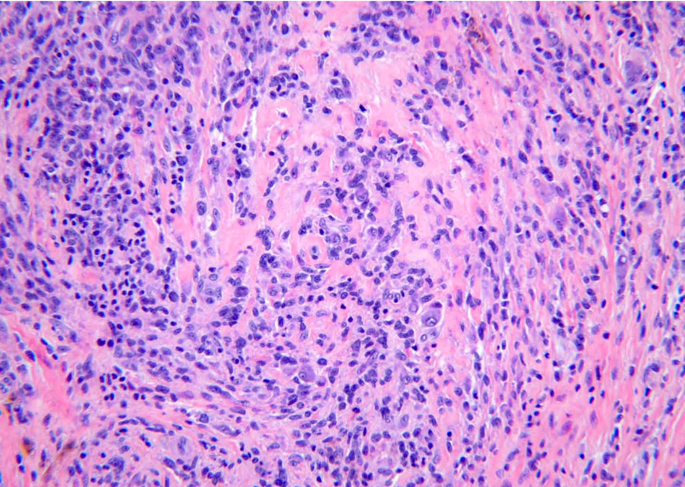
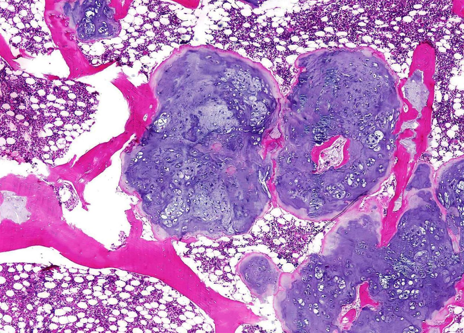
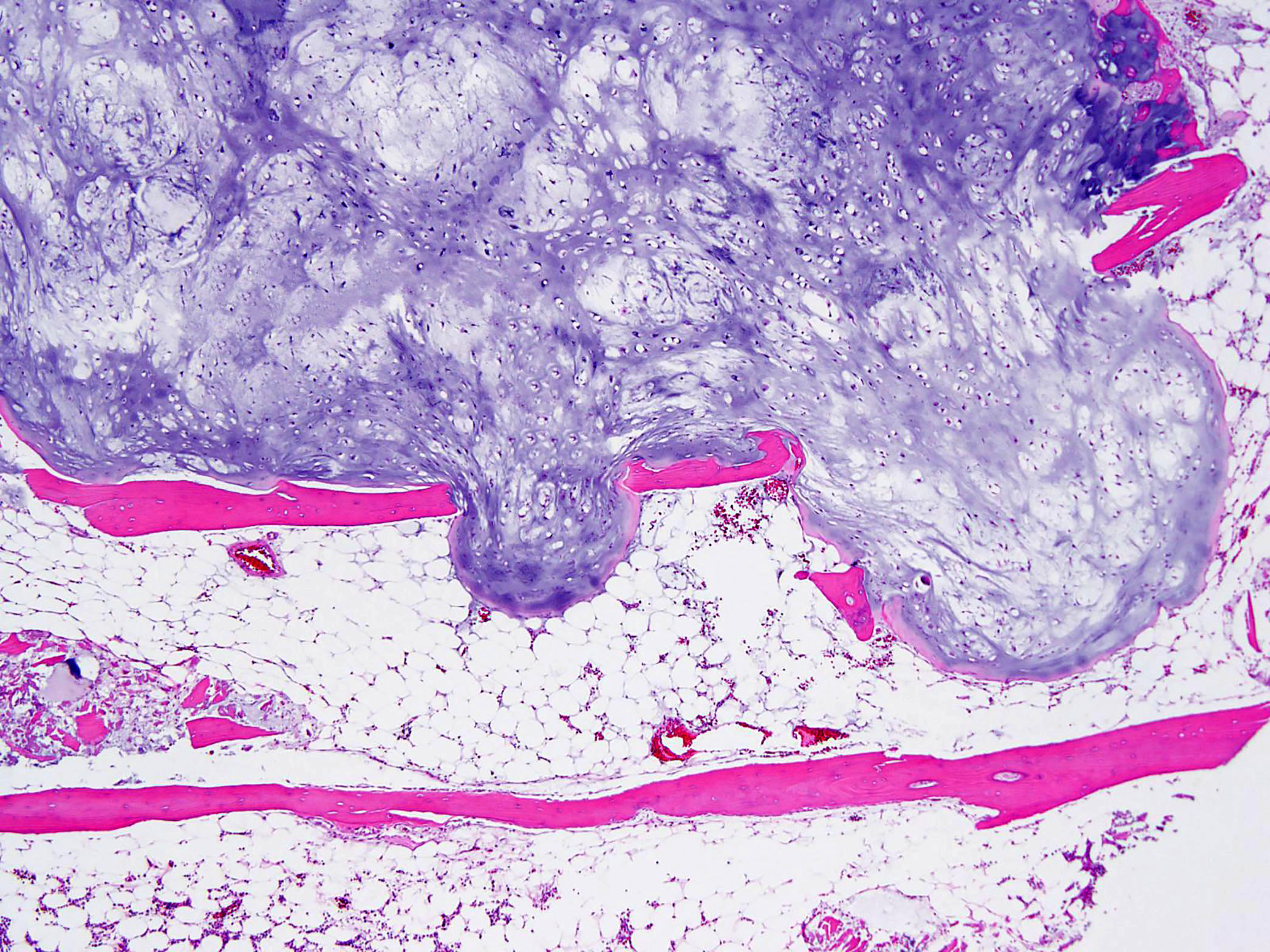
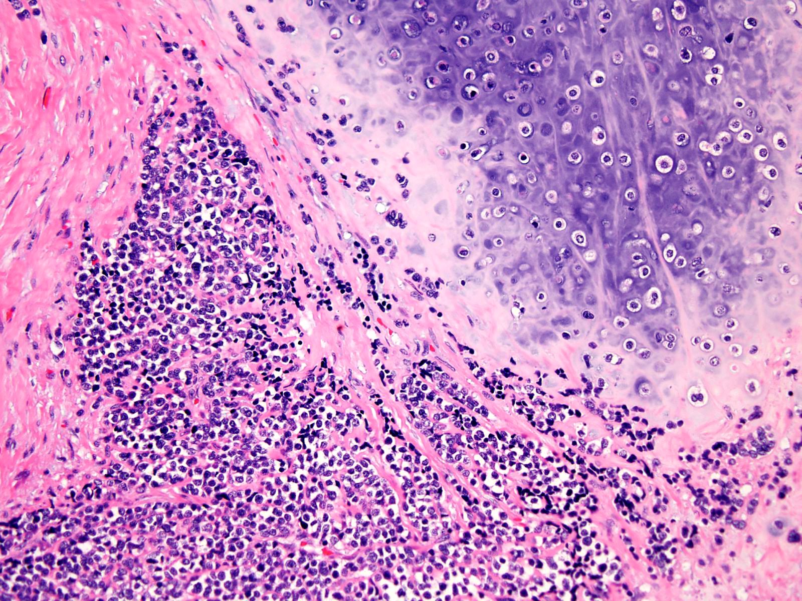
Classic adamantinoma
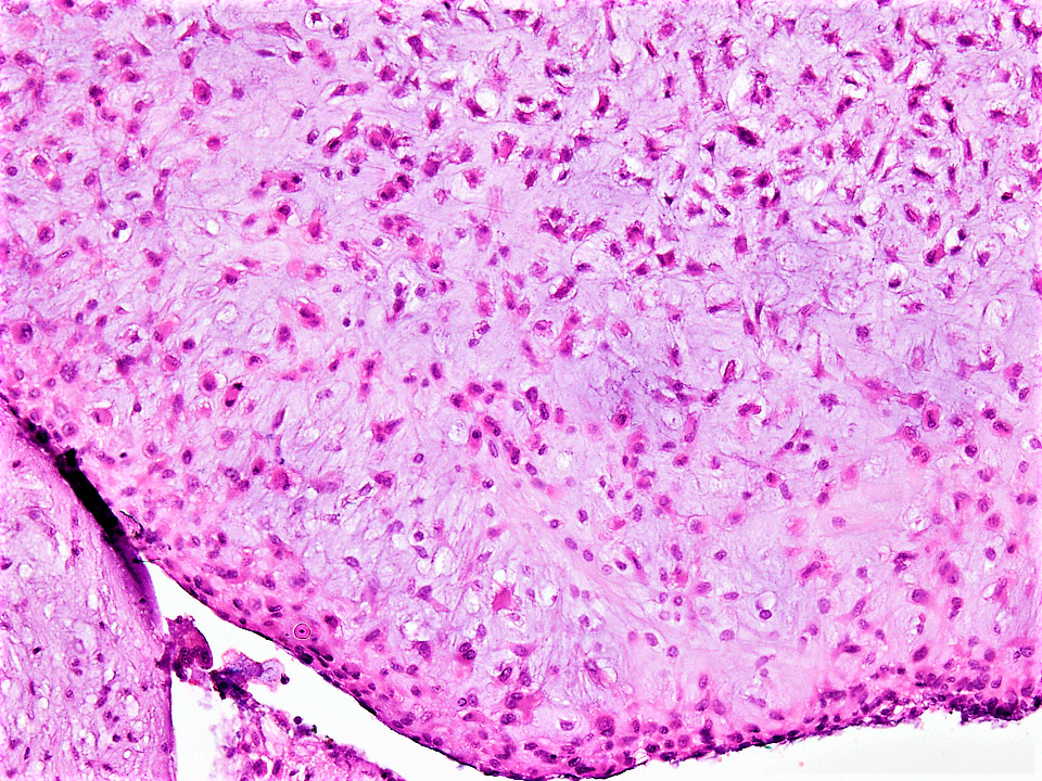
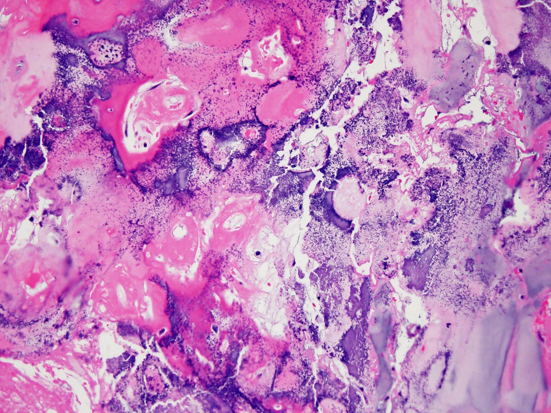
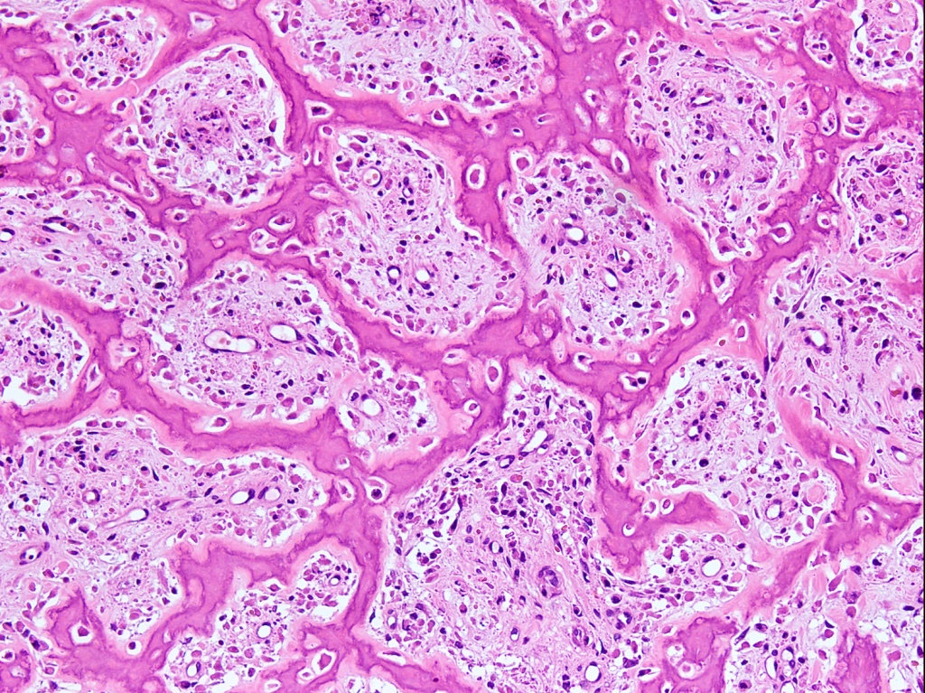
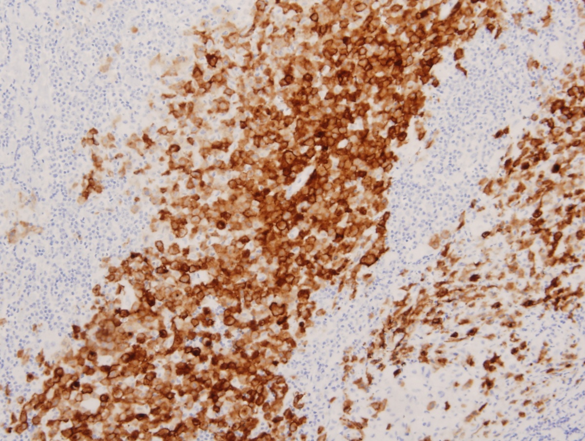
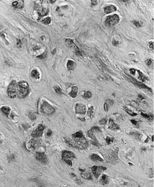
Osteofibrous dysplasia-like adamantinoma
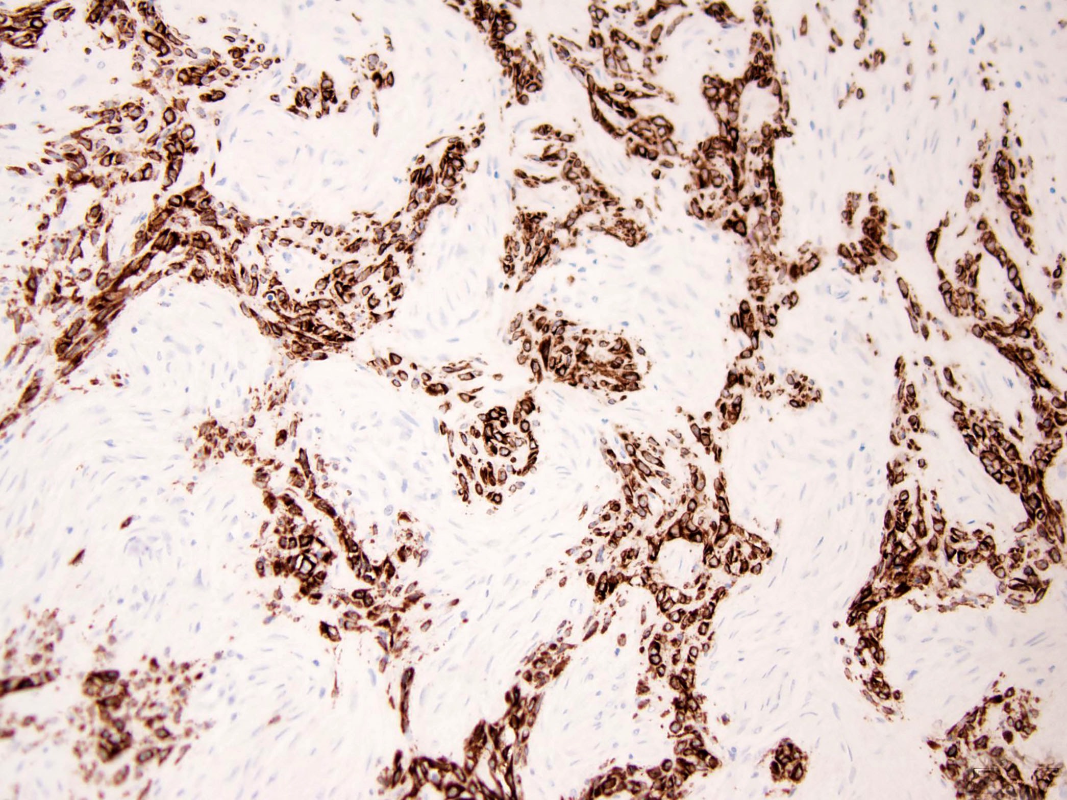
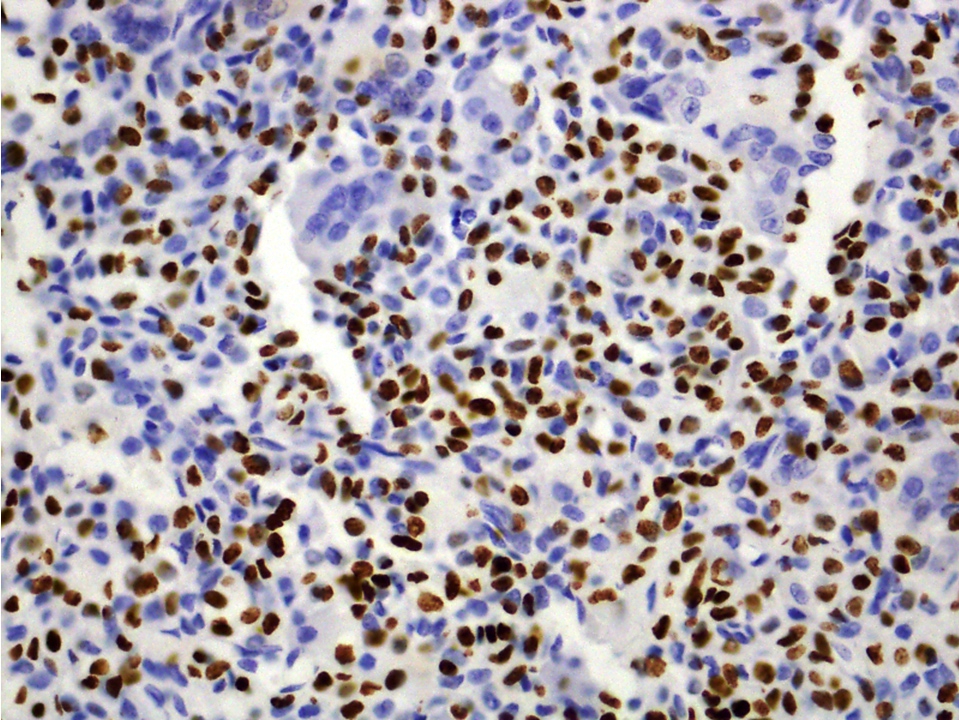
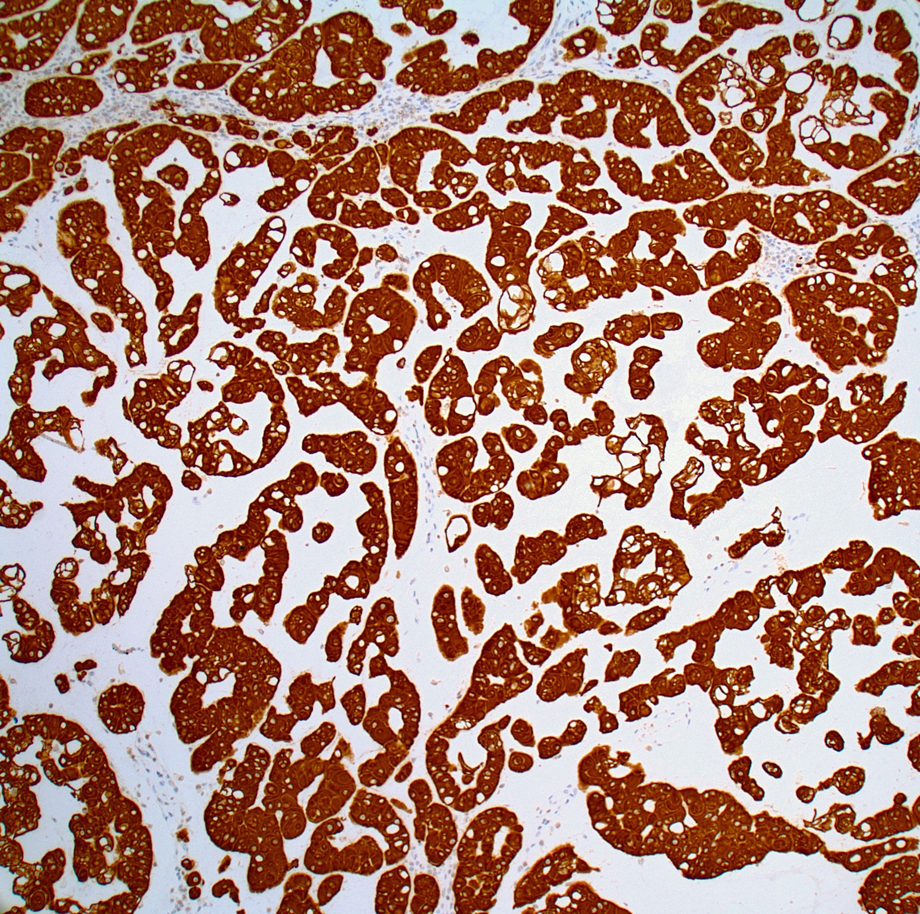
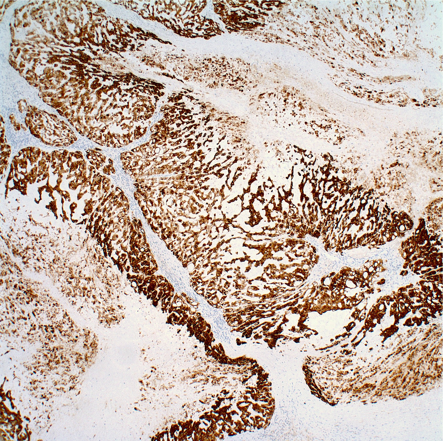
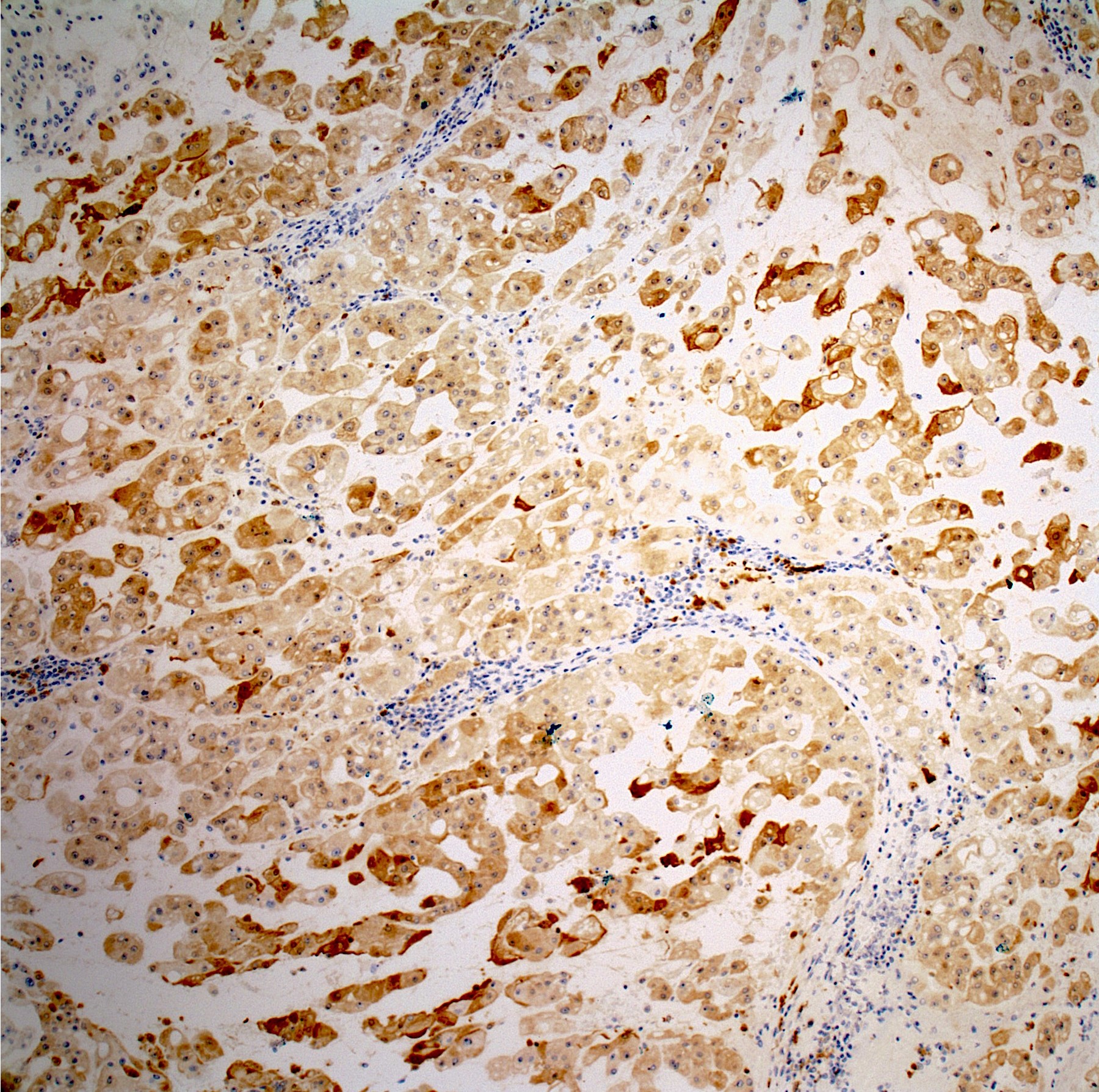
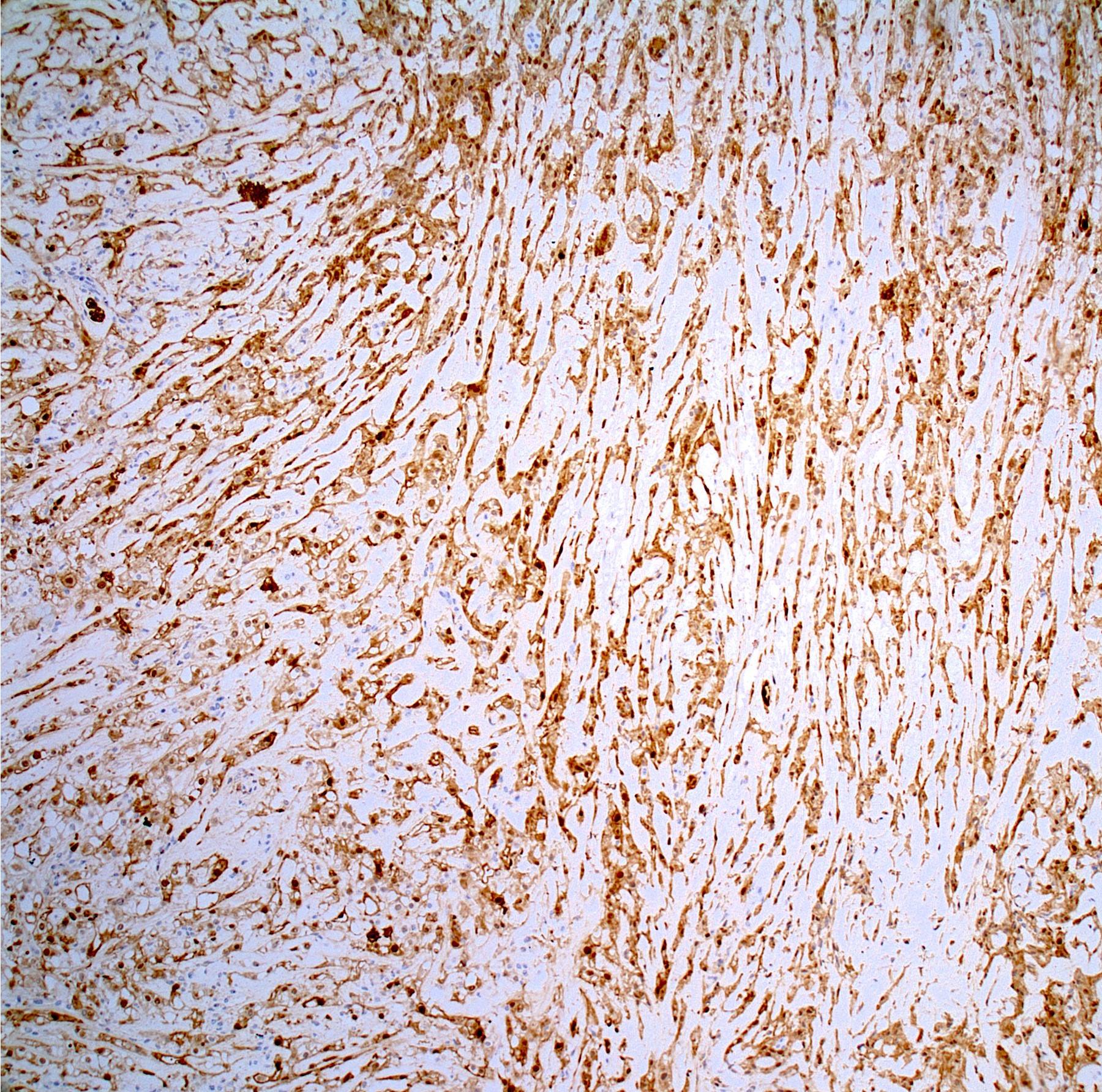
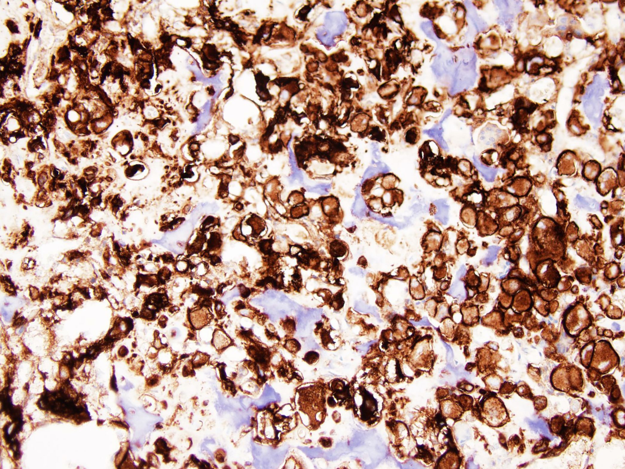
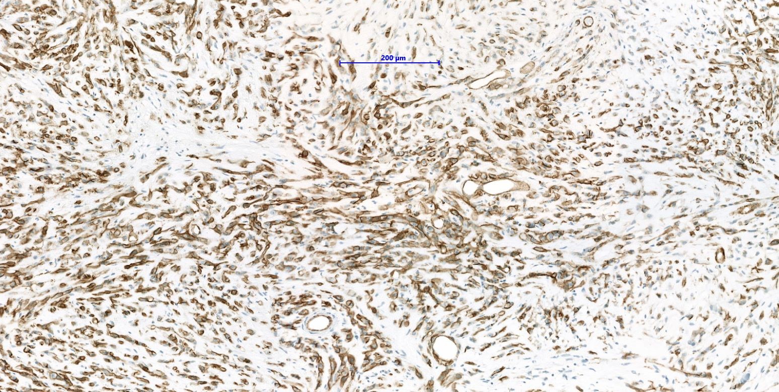
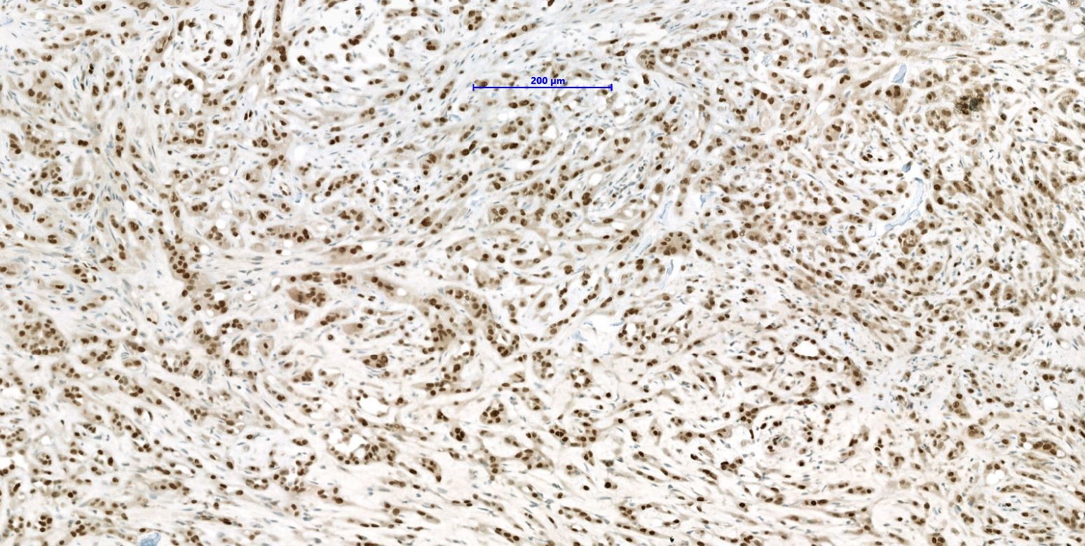
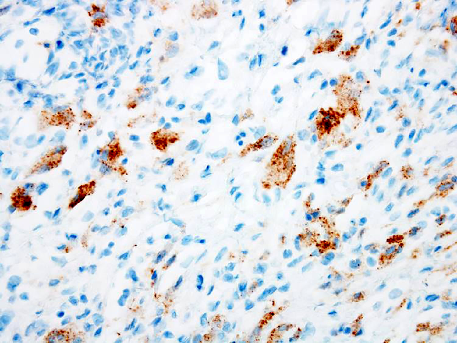
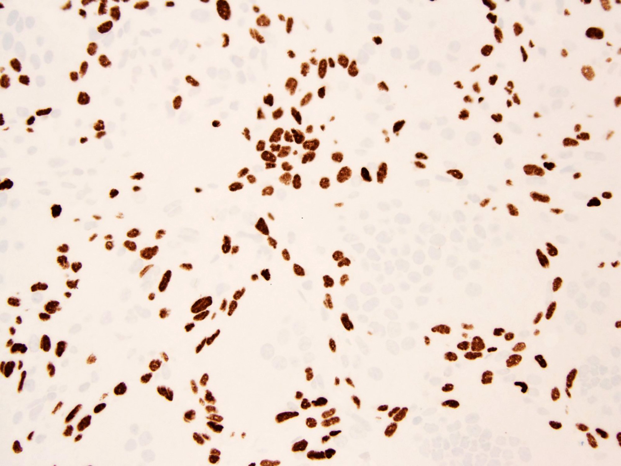
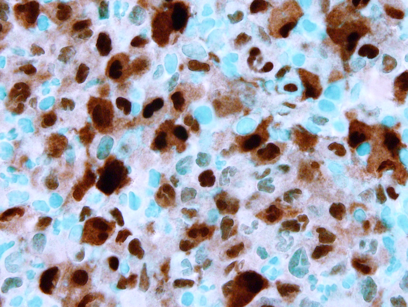
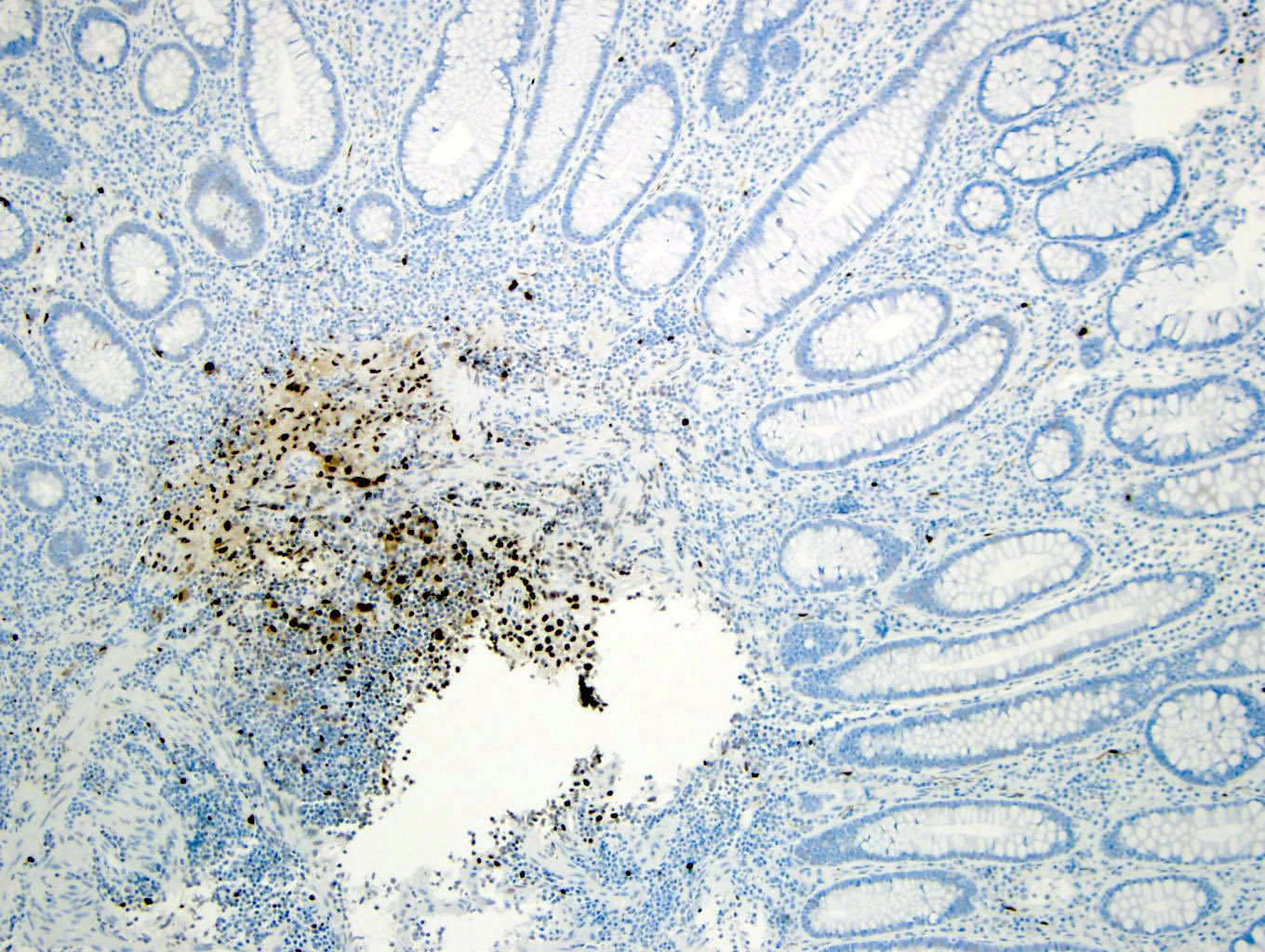
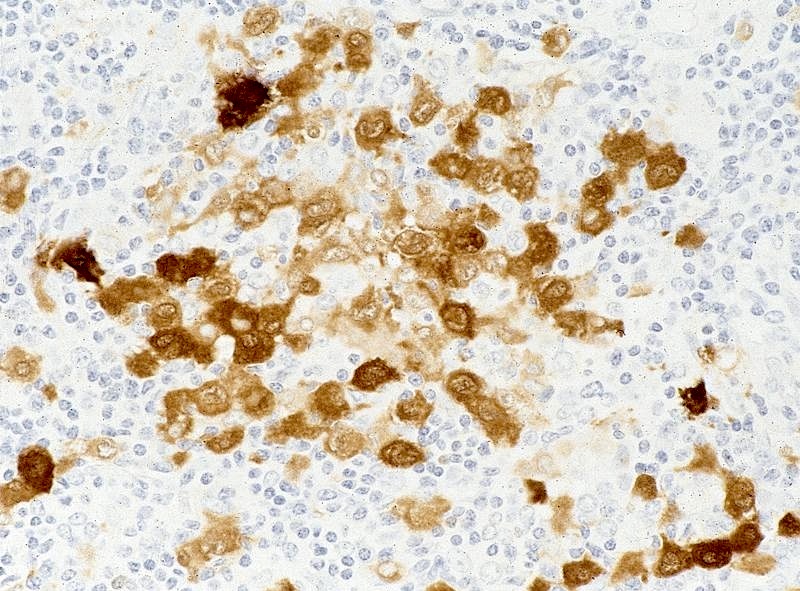
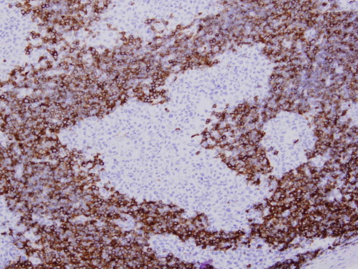
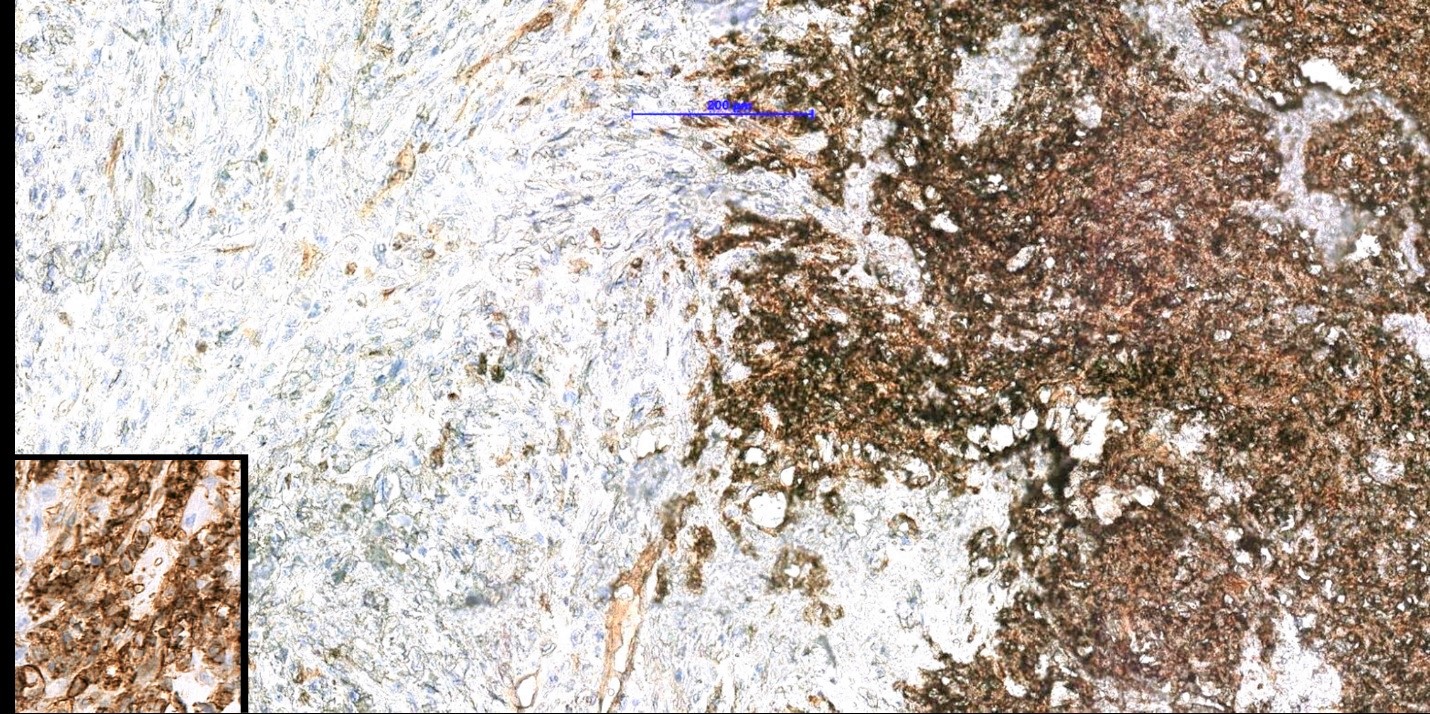
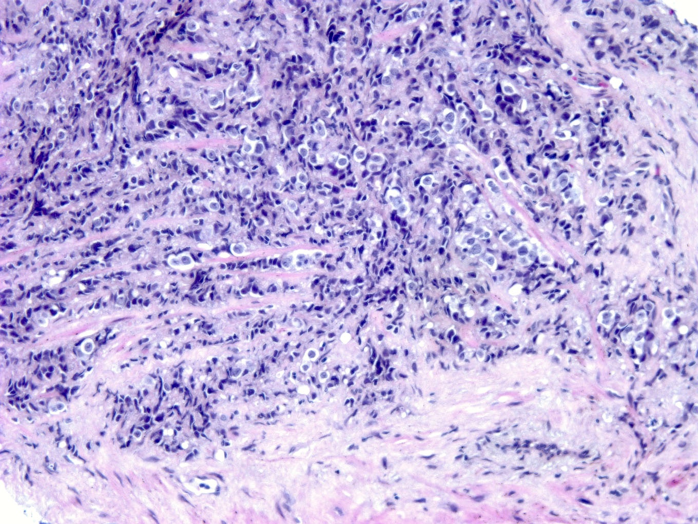
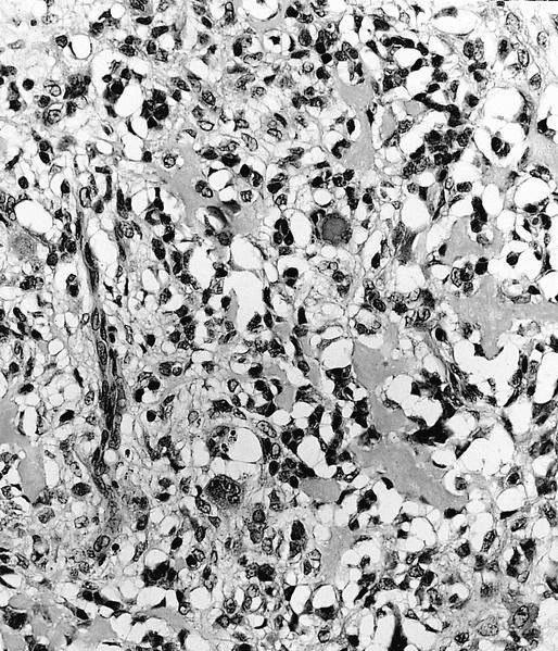
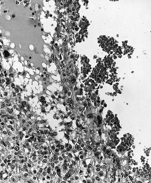
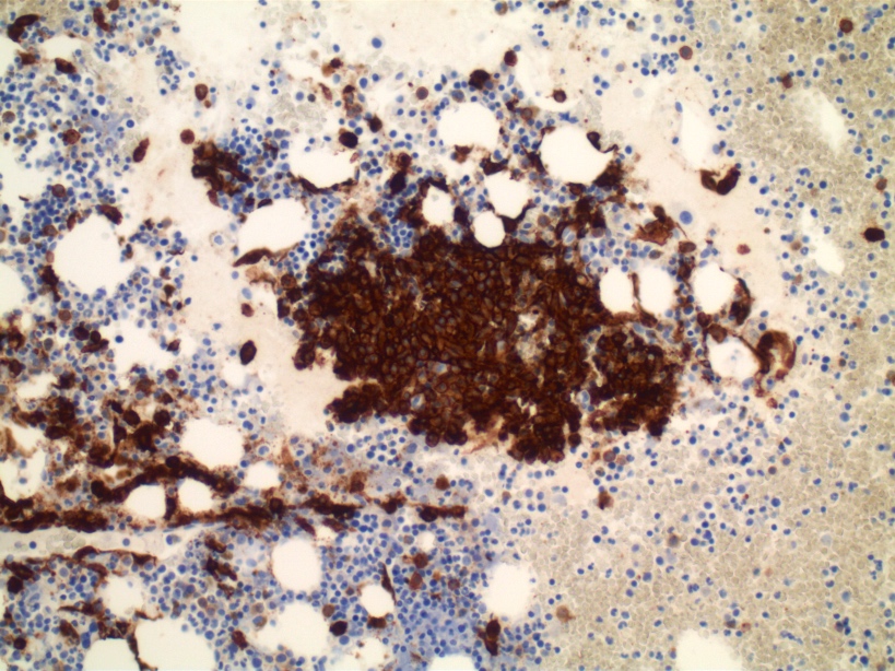
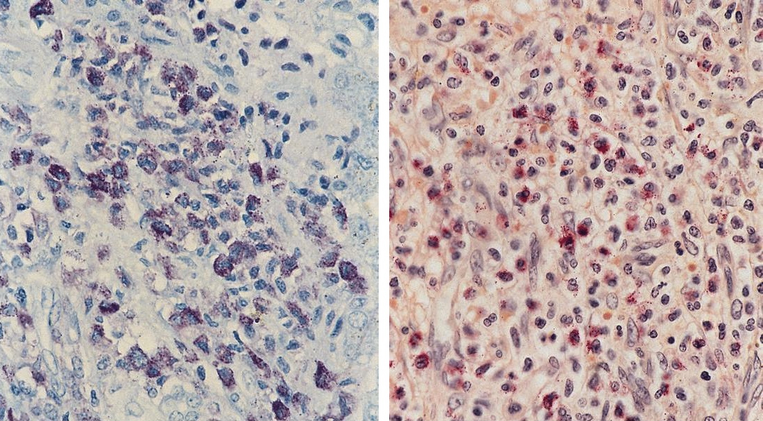
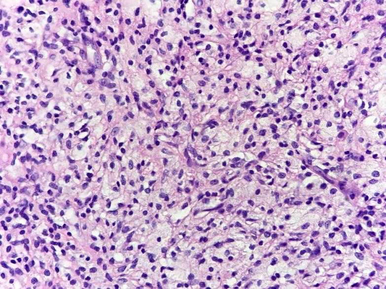
CK AE1 / AE3 expression
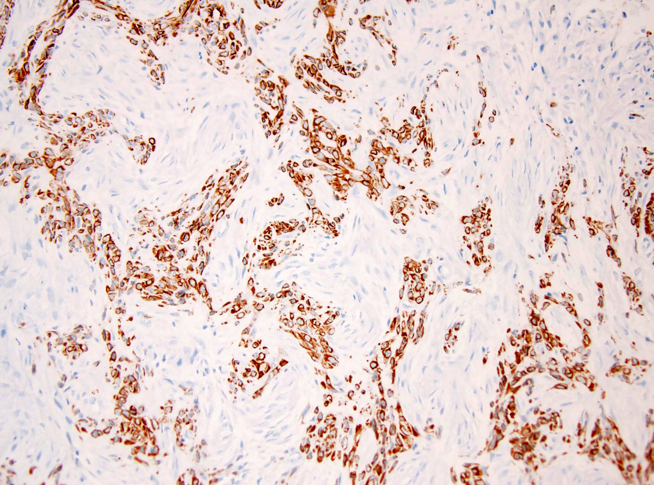
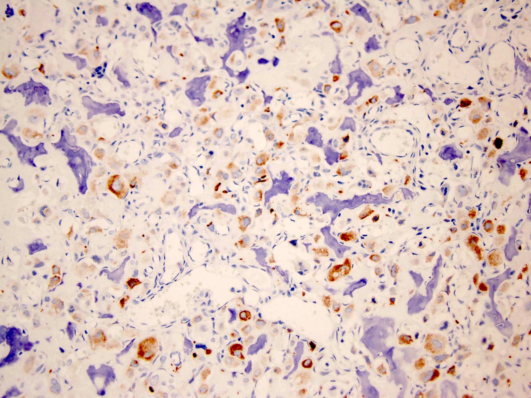
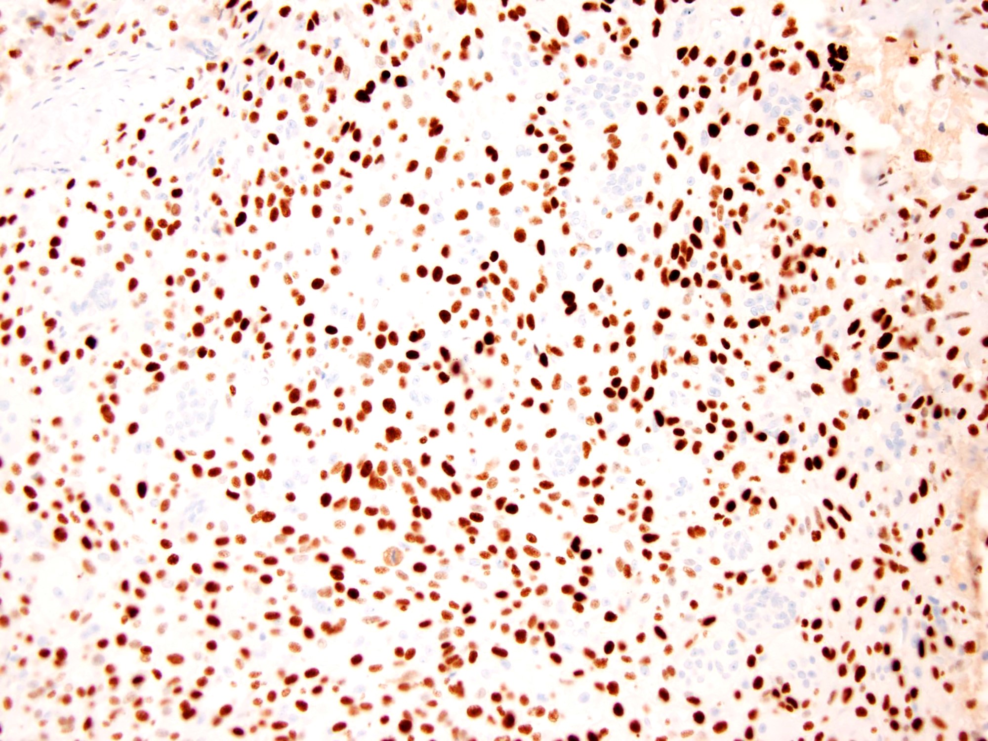
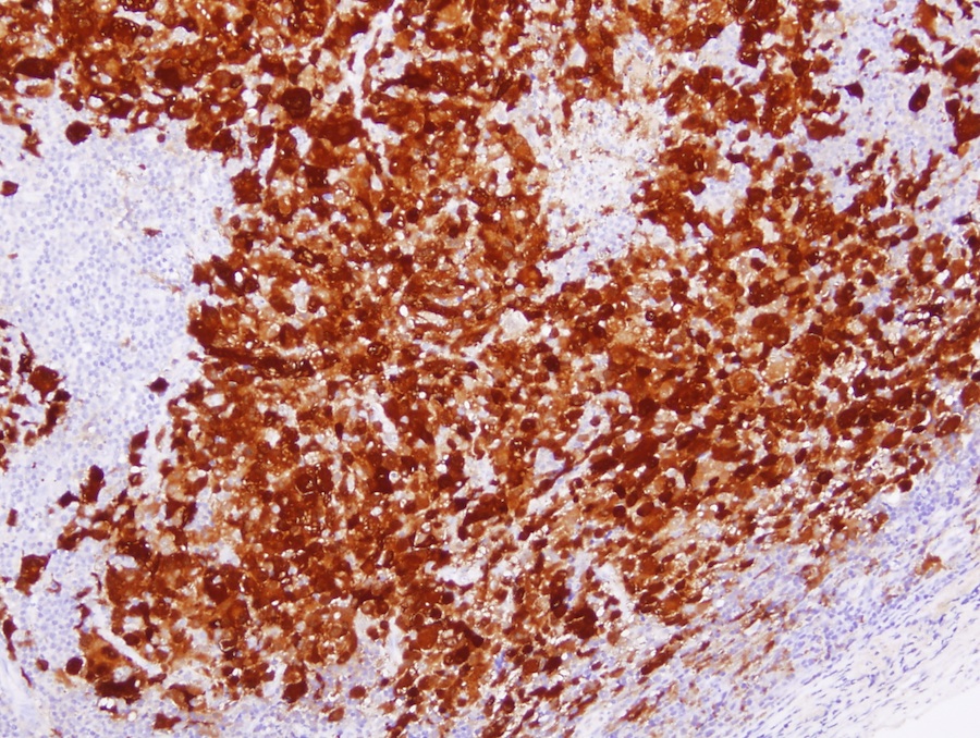
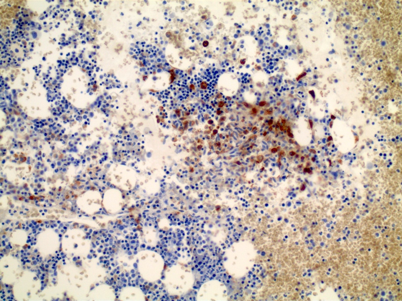
CK19 expression
Images hosted on other servers:


Femur


Humerus


Patella


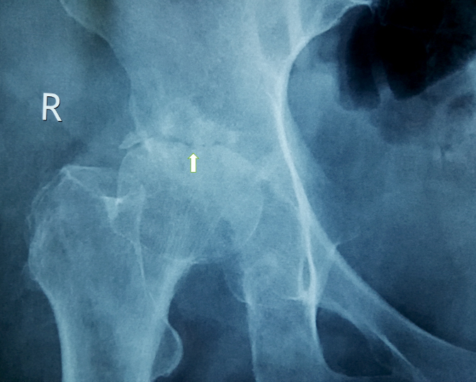
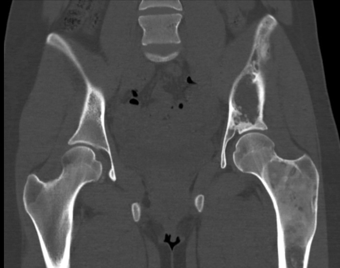
Pelvis: left-male, right-female


Tibia and fibula


Radius and ulna

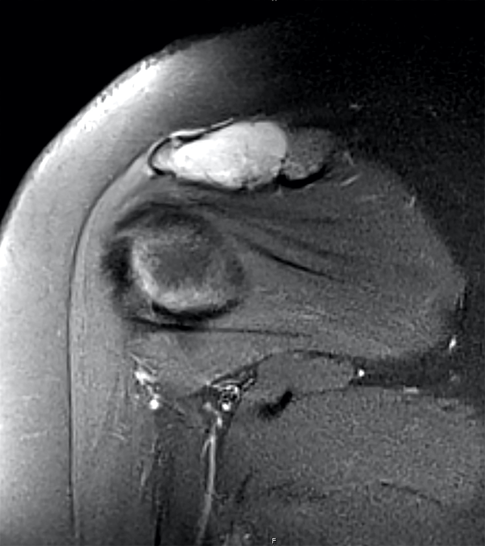
Shoulder joint


Different types of synovial joints
Images hosted on other servers:
.jpg)
Elbow joint; deep dissection; anterior view
Contributed by Elham Nasri, M.D.
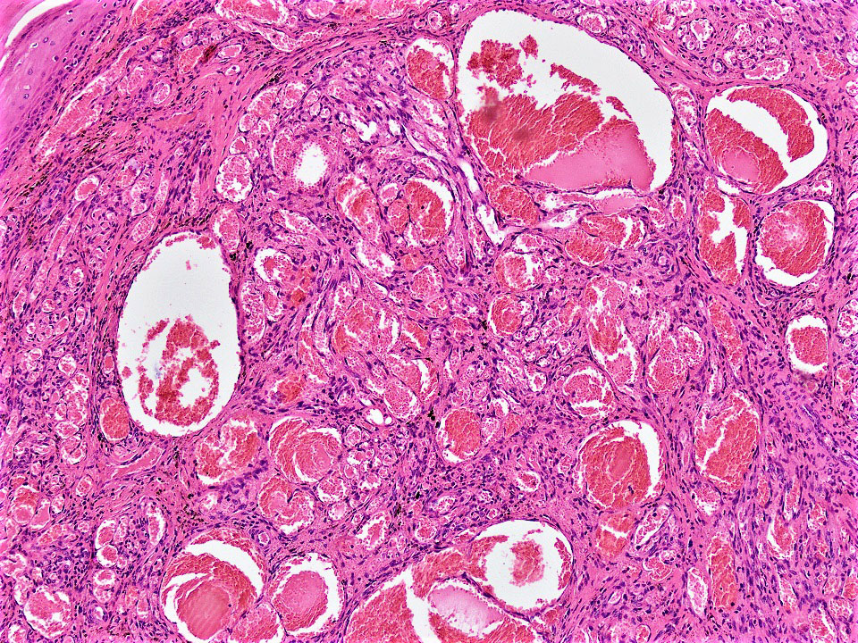
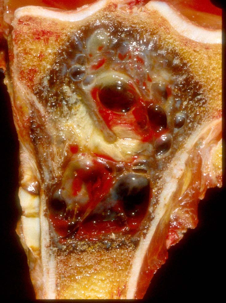
Aneurysmal bone cyst of proximal tibia
Aneurysmal bone cyst of humerus
Aneurysmal bone cyst of distal tibia
Aneurysmal bone cyst of calcaneus bone
Images hosted on other servers:

Intraoral tumor
Contributed by Elham Nasri, M.D.
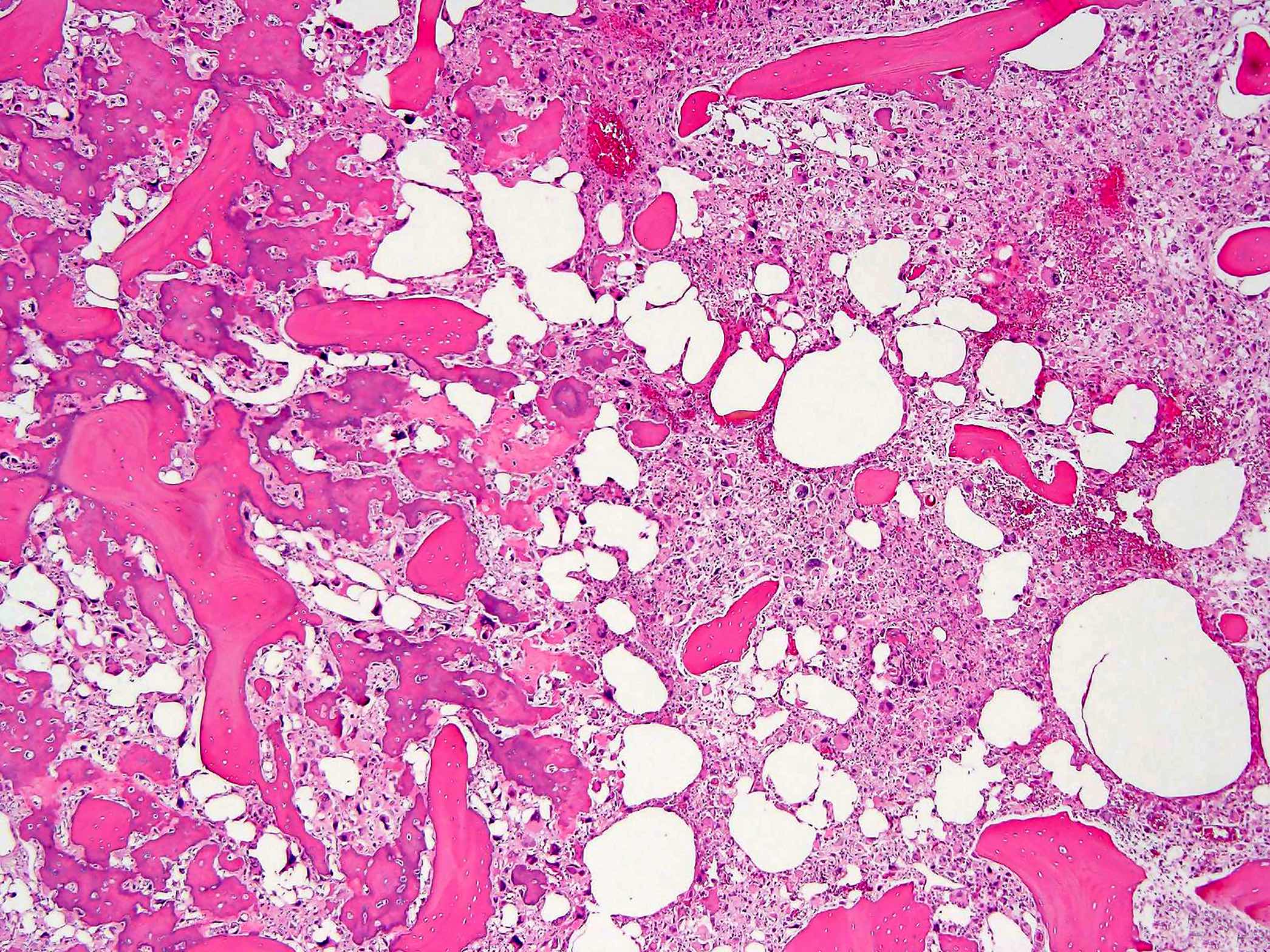
Blood filled cysts
Multiloculated cyst
Contributed by Elham Nasri, M.D. and Kelly Magliocca, D.D.S., M.P.H.
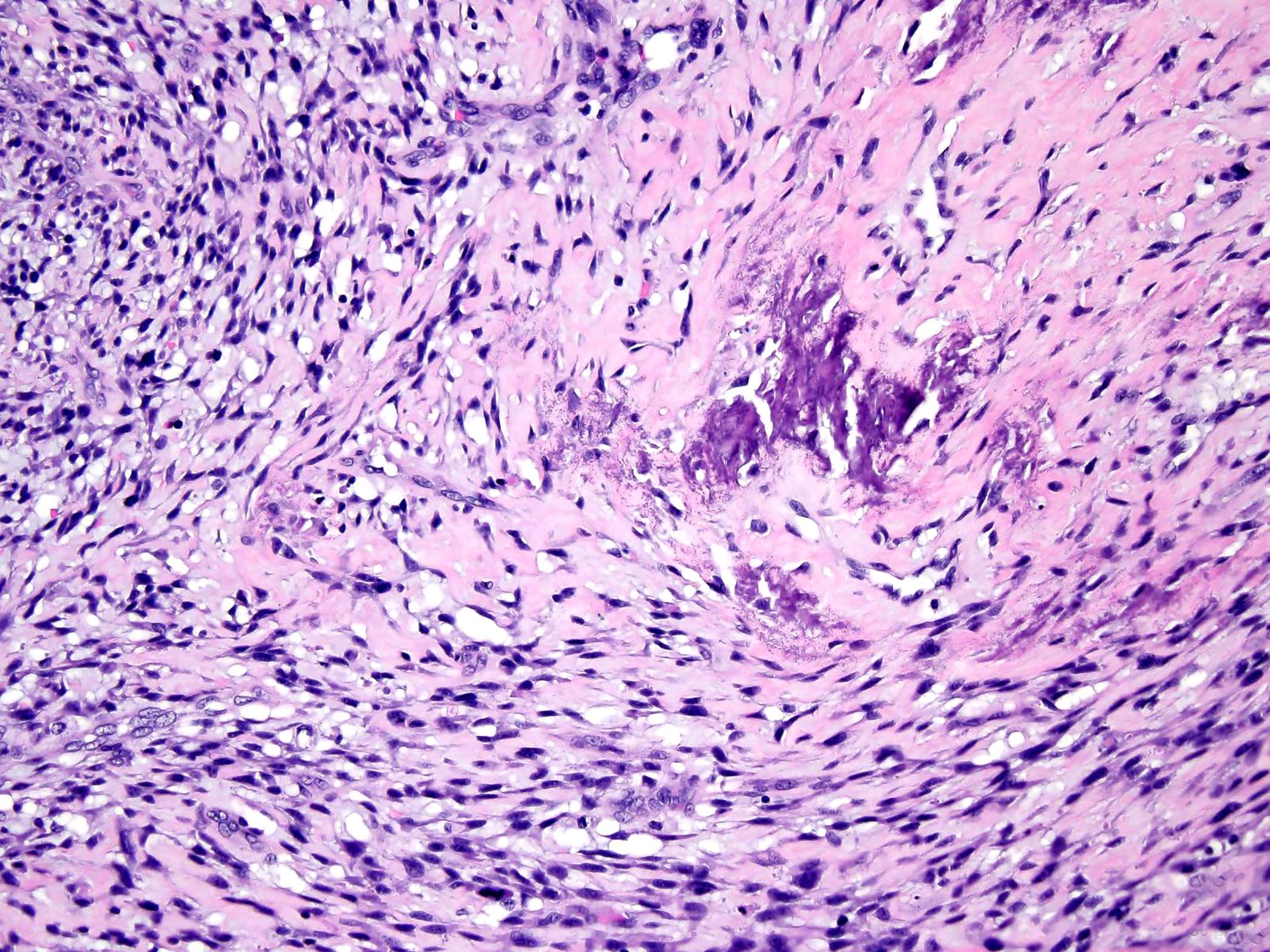
Cysts and giant cells
Blue chondroid like material
Blood filled cystic spaces
Solid area of cyst wall
Reticulated chondroid-like material
Multiple cystic spaces
Blood filled cysts
Reticulated chondroid-like material
Cyst wall and giant cells
Aneurysmal bone cyst
Contributed by Mark R. Wick, M.D.
Primary bone multifocal
Contributed by Mark R. Wick, M.D. and AFIP images
Primary bone
Grade 3
Contributed by Serenella Serinelli, M.D., Ph.D. and Gustavo de la Roza, M.D.
Xray and MRI appearance
Images hosted on other servers:

Xray and MRI appearance

Cartilaginous cap MRI appearance
Contributed by Serenella Serinelli, M.D., Ph.D. and Gustavo de la Roza, M.D.

Humeral
central atypical
cartilaginous
tumor
Images hosted on other servers:

Femoral secondary
peripheral atypical
cartilaginous tumor
Contributed by Serenella Serinelli, M.D., Ph.D. and Gustavo de la Roza, M.D.
Lobulated architecture
Bone permeation
Scalloping of cortical bone
Bone scalloping

Lamellar bone scalloping
Myxoid changes

Necrosis

Cytology features
Arising from osteochondroma
Chondrosarcoma versus enchondroma: bone pathology with Dr. Andrew Rosenberg
Images hosted on other servers:

Xray of femoral head AVN

MRI of bilateral AVN
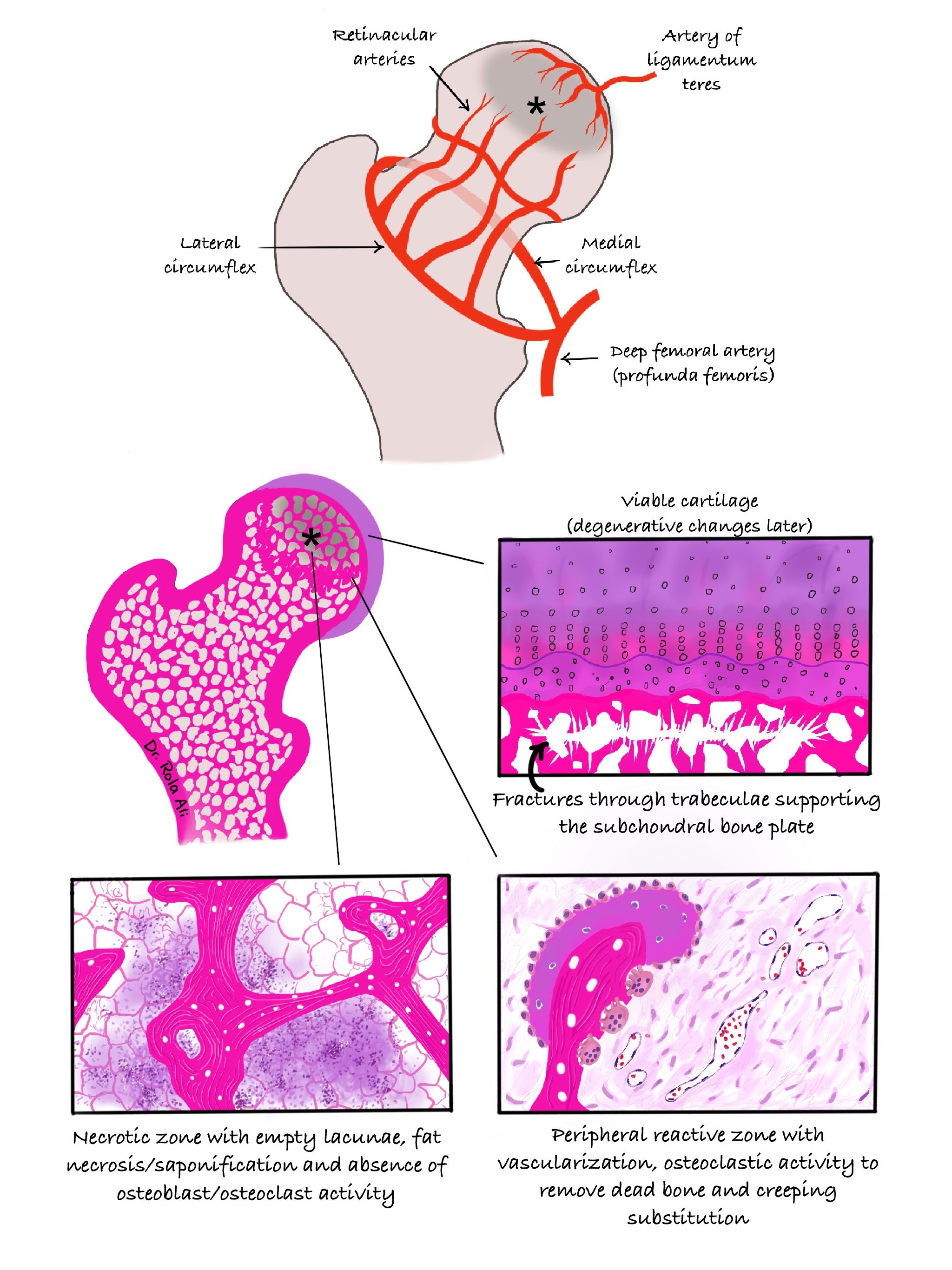
Contributed by Rola H. Ali, M.D.
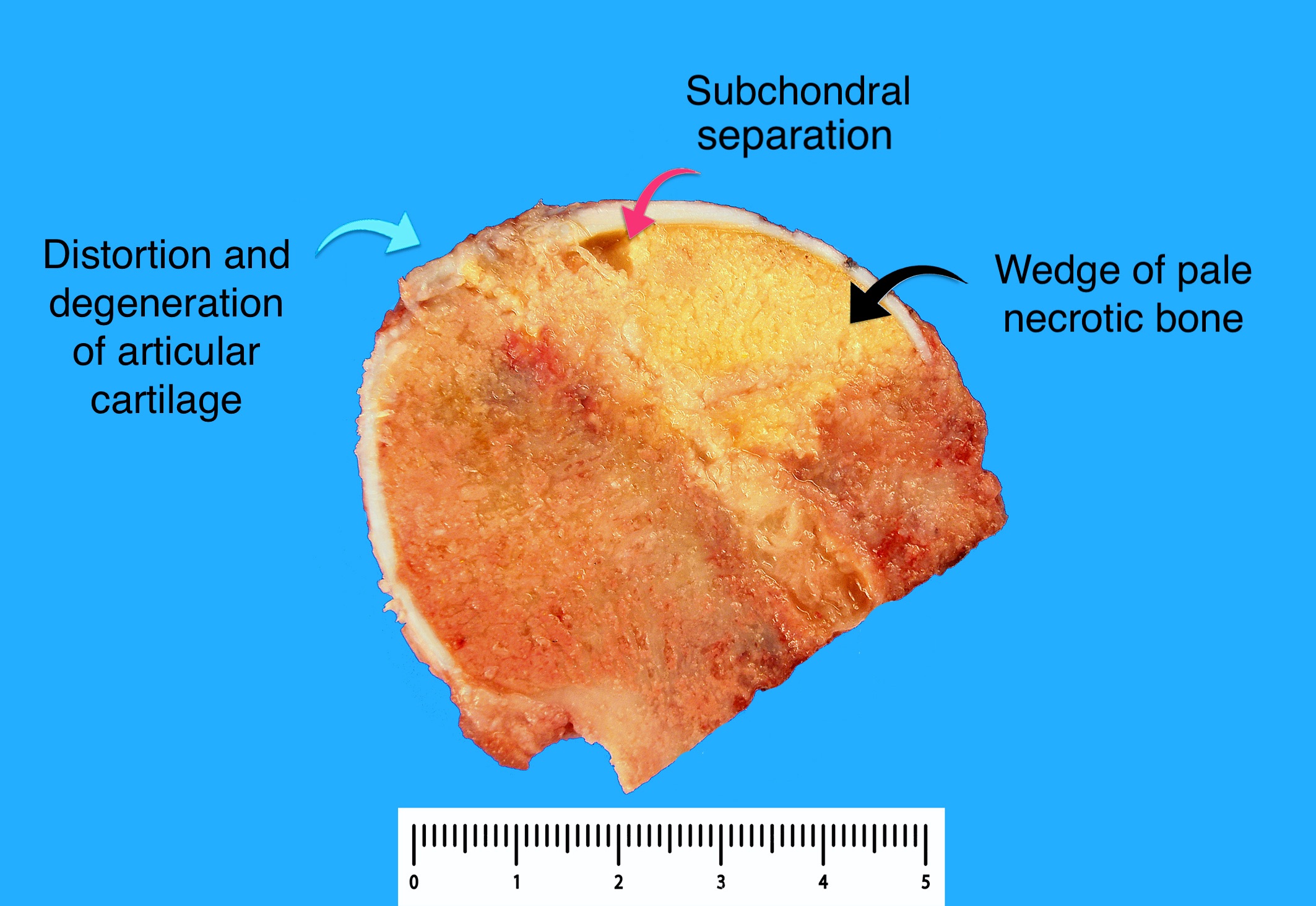
Wedge infarct
Subchondral collapse
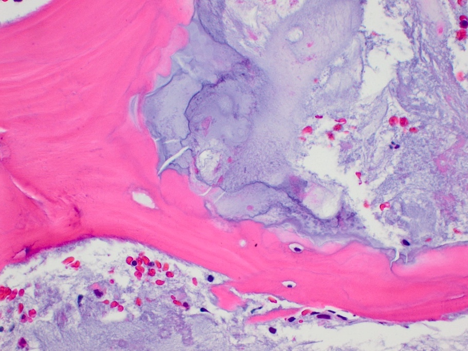
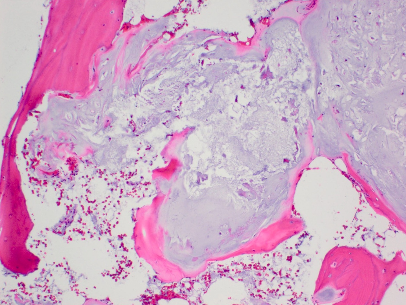
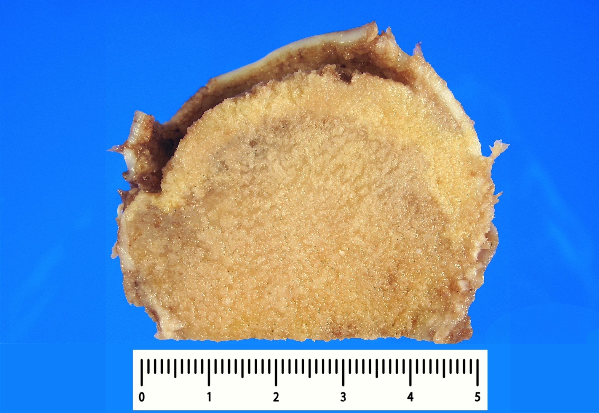
Contributed by Rola H. Ali, M.D.
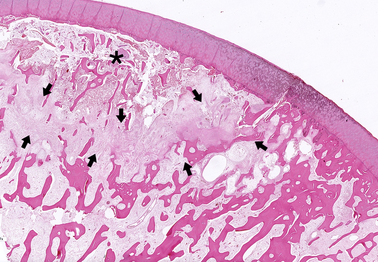
Subchondral infarct
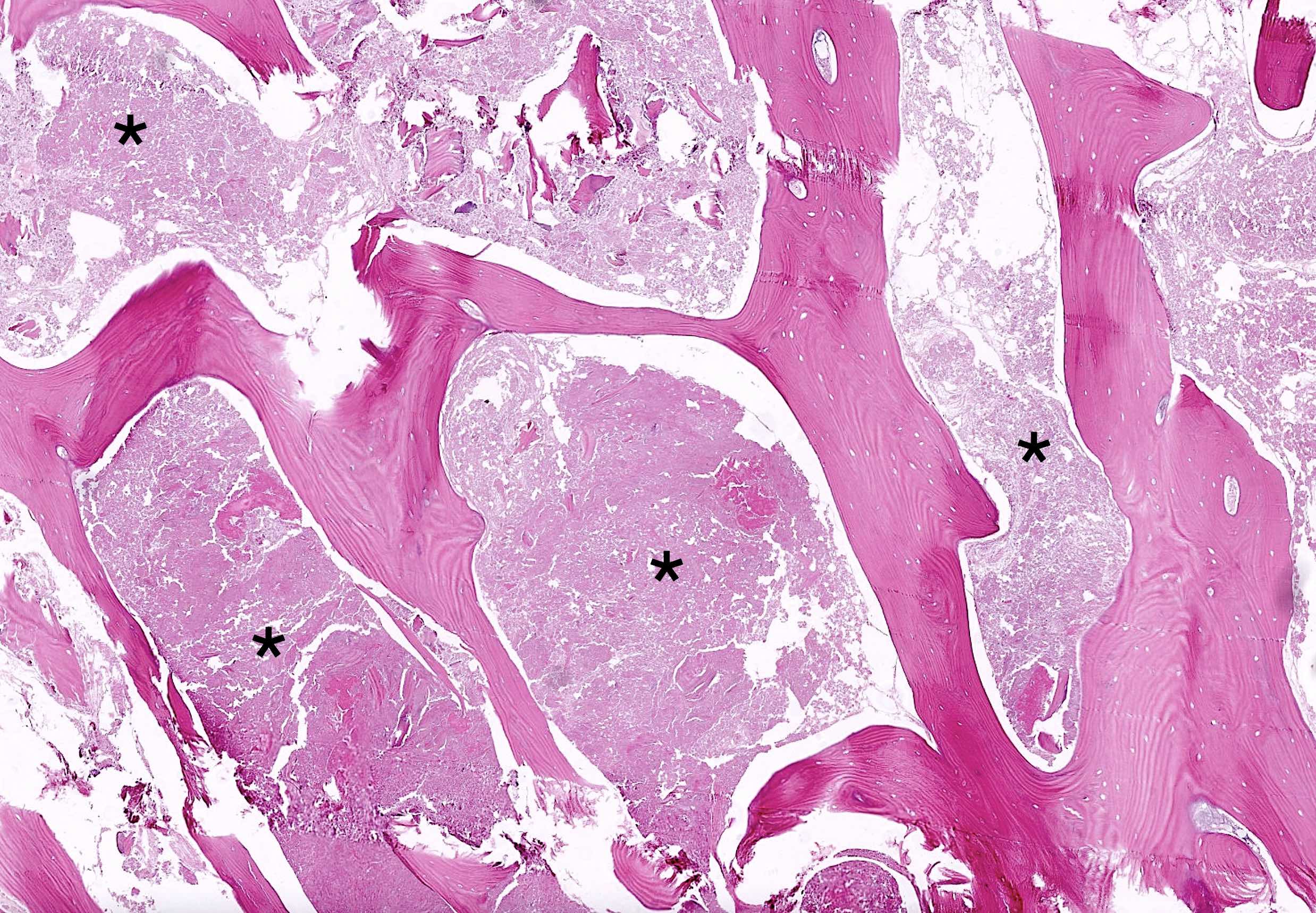
Necrotic zone
Empty lacunae
Reactive zone
New bone
Creeping substitution
Contributed by F. Bilge Ergen, M.D.
Plain Xray features
MRI of BCOR::CCNB3 sarcoma
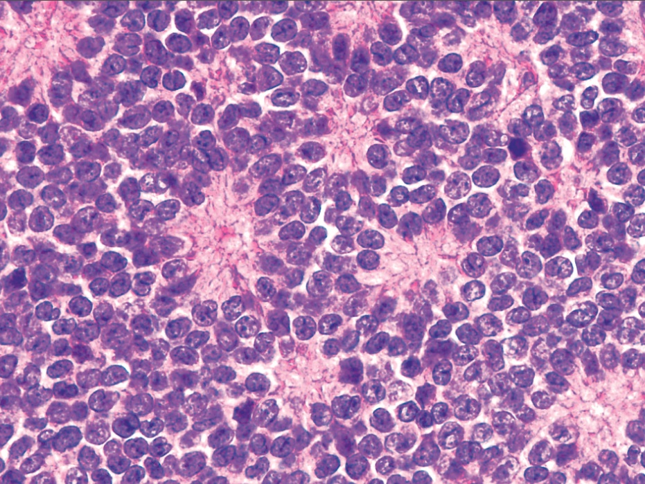
Contributed by Kemal Kösemehmetoğlu, M.D.

BCOR::CCNB3 sarcoma of radius
Contributed by Kemal Kösemehmetoğlu, M.D.

Typical morphology of BCOR::CCNB3 sarcoma
High power cytological morphology
Various cellularity
Hemangiopericytic pattern
Whorling pattern
Subtle osteoid formation
Epithelioid balls and nests
Telangiectatic areas

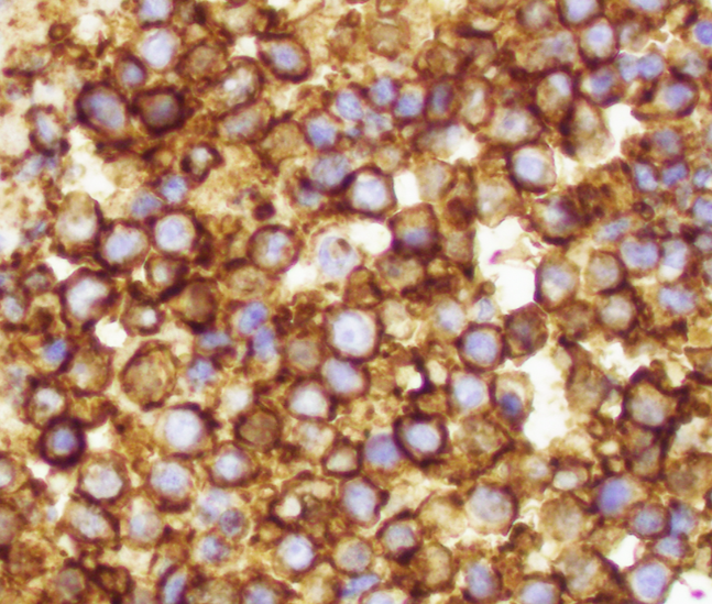
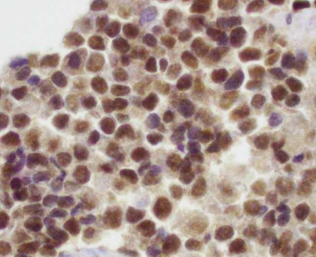
CCNB3 expression
CD99

BCOR

SATB2

TLE1
Focal EMA expression
Diffuse cyclin D1 expression
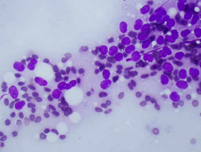
Contributed by Kemal Kösemehmetoğlu, M.D.
Cytological features of BCOR::CCNB3
Contributed by Kemal Kösemehmetoğlu, M.D.

Reverse transcription PCR for BCOR::CCNB3
BCOR::CCNB3 sarcoma of the proximal tibia
Images hosted on other servers:

Pathogenesis of osteomyelitis associated septic arthritis
Contributed by Mark R. Wick, M.D.
Bacterial osteomyelitis of tibia and vertebra in thoracic spine
Images hosted on other servers:

Osteomyelitis of the distal fourth metatarsal

Abnormal T1 weighted signal

Localized increased radioactive tracer uptake
Contributed by Dariusz Borys, M.D. and Mark R. Wick, M.D.
Acute osteomyelitis
Bacterial acute osteomyelitis
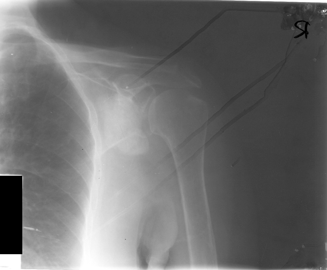
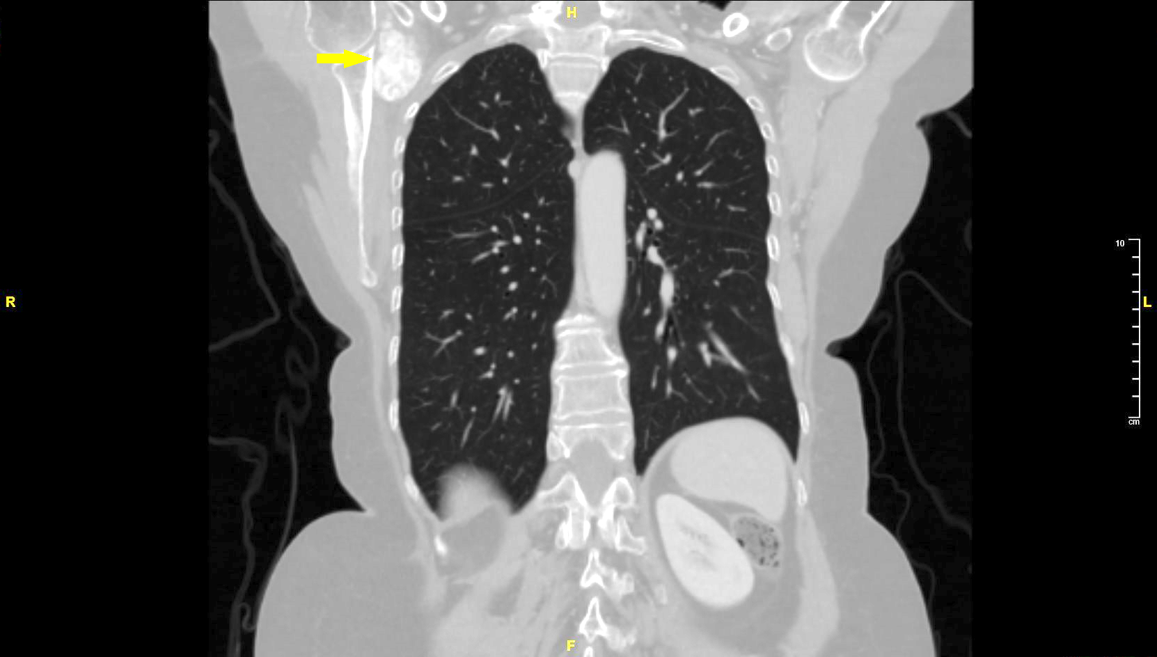
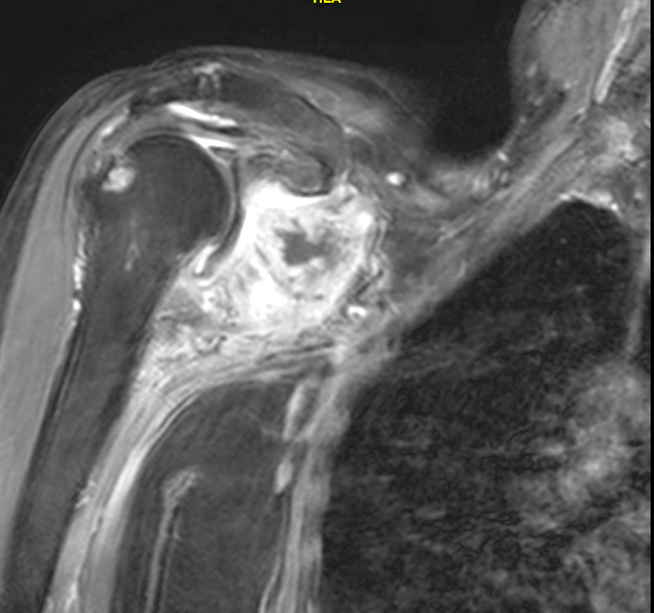
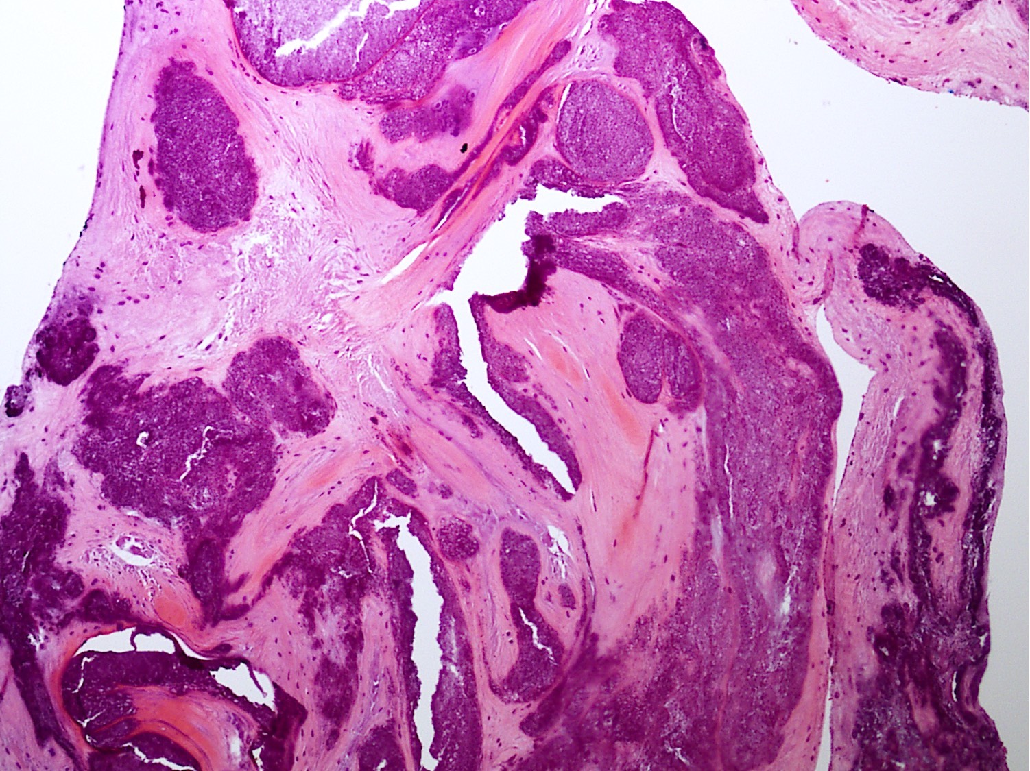
Contributed by Borislav A. Alexiev, M.D.
Left scapular neck mass
Contributed by Ajay R. Chapa, M.D.
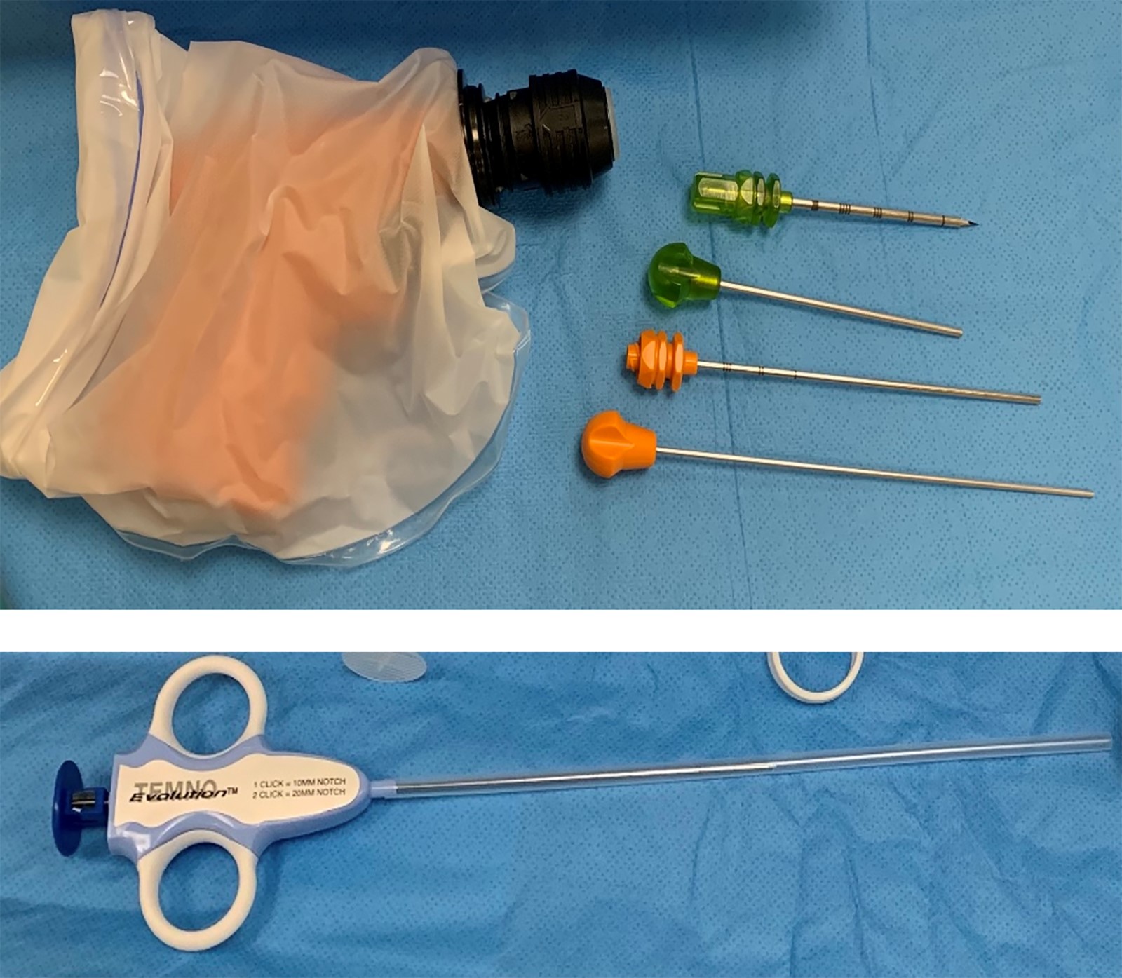
Bone biopsy system
Contributed by Mark R. Wick, M.D.
Various images
Contributed by Mark R. Wick, M.D.

Various images
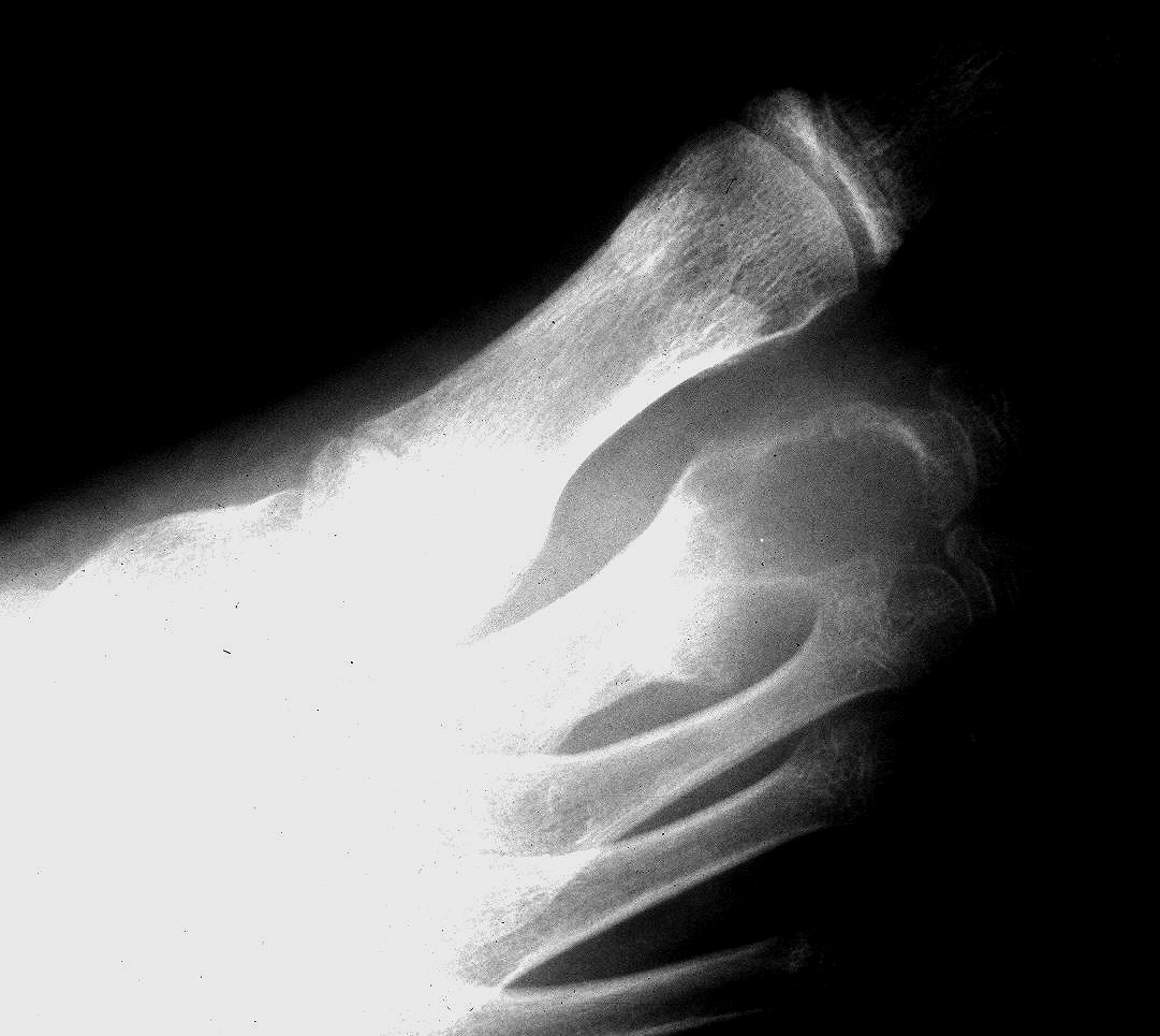
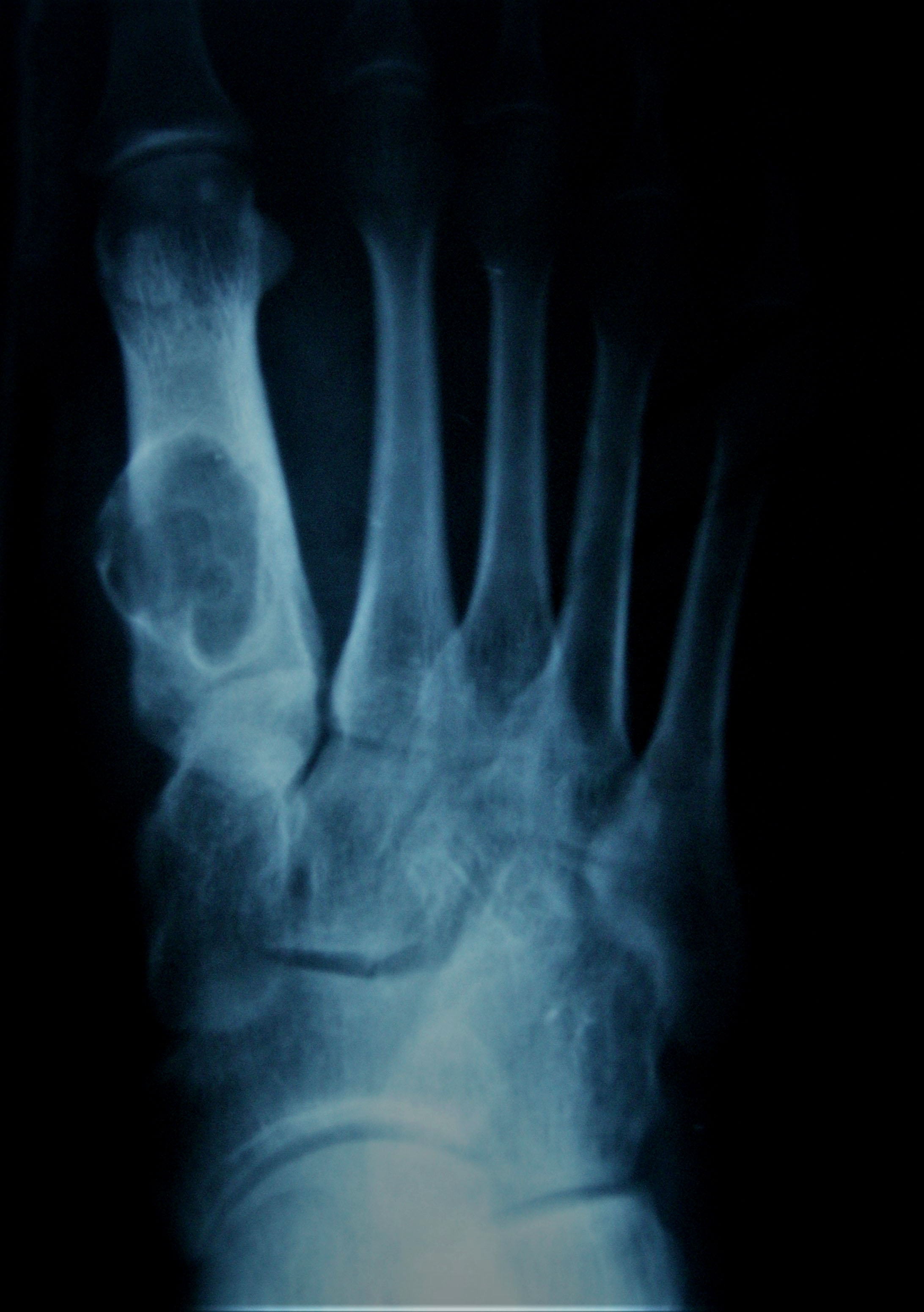
Nora lesion foot
Contributed by Mark R. Wick, M.D.
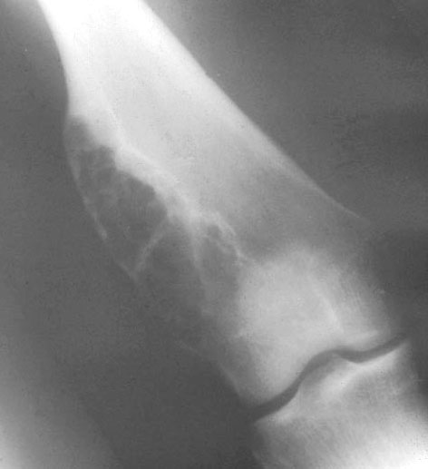
Femur Xray
Contributed by Mark R. Wick, M.D.
Femur
Contributed by Mark R. Wick, M.D.
Femur
Images hosted on other servers:

Pathogenesis
Contributed by Nasir Ud Din, M.B.B.S.
Multiple lytic lesions

Multiple lytic lesions
Images hosted on other servers:


Mandibular lesion
Images hosted on other servers:

Multilocular, expansile, hemorrhagic mass in rib
Contributed by Nasir Ud Din, M.B.B.S.
Lobulated pattern
Scattered nodular pattern
Vague circumscription of nodule

Vague nodularity
Irregularly distributed giant cells
Giant cells exhibiting tunneling resorption
Tunneling resorption
Abundant hemosiderin deposition in the lesion
Bone changes in hyperparathyroidism
Images hosted on other servers:

Elbow
Elbow: chronic fibrinous bursitis
Images hosted on other servers:

Extracellular pyrophosphate mechanism

Pathogenesis
Contributed by Nasir Ud Din, M.B.B.S. and Jose G. Mantilla, M.D.
Right knee joint, AP
Right knee joint, lateral

Talonavicular joint, foot
Shoulder CPPD Xray
Shoulder CPPD CT
Shoulder CPPD T1 MRI gadolinium
Images hosted on other servers:

Pyrophosphate arthropathy

Meniscal
chondrocalcinosis
Images hosted on other servers:

Right index finger swelling

Calcified brittle intradural mass
Images hosted on other servers:

Chalky white depositions
Contributed by Nasir Ud Din, M.B.B.S.
Basophilic deposits
Histiocytic reaction
Rhomboid crystals

Positive birefringence

Chondroid metaplasia
Images hosted on other servers:

Aggregates of birefringent crystals
Pseudogout
Contributed by Mark R. Wick, M.D., Borislav A. Alexiev, M.D. and AFIP
Femur Xray

Foot talus Xray
Humerus Xray
Left acromion lesion
Radiograph of tibia
Contributed by Ashley Patton, D.O., Ph.D., Nasir Ud Din, M.B.B.S. AFIP and @JMGardnerMD on Twitter
Chicken wire calcification and mature cartilage formation

Osteoclast-like giant cell

Sheets of chondroblasts
H3K36M antibody
Nuclei vary in size
Neoplastic cells with ovoid to spindled nuclei
Well formed chondroid matrix
Chicken wire appearance

Chondroblastoma
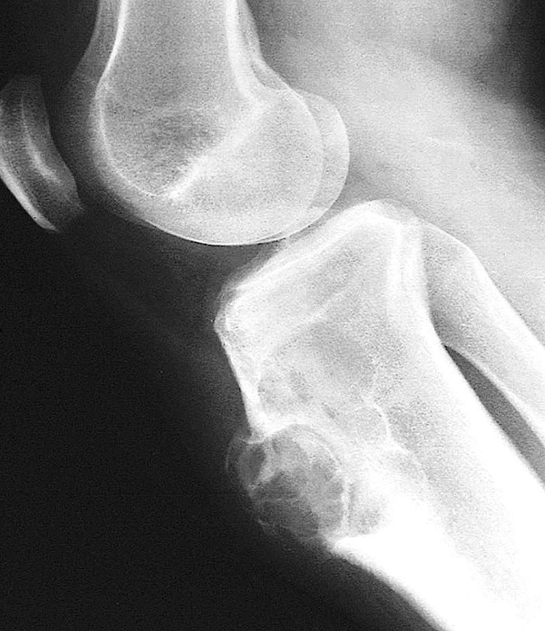
Contributed by Borislav A. Alexiev, M.D.
Giant cell rich lesion
Histological overview of chondrosarcoma
Contributed by Qurratulain Chundriger, M.B.B.S.
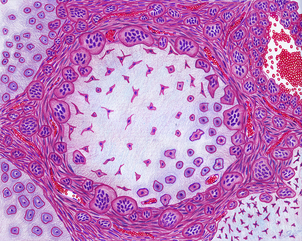
Drawing showing histology
Contributed by Mark R. Wick, M.D. and Nasir Ud Din, M.B.B.S.
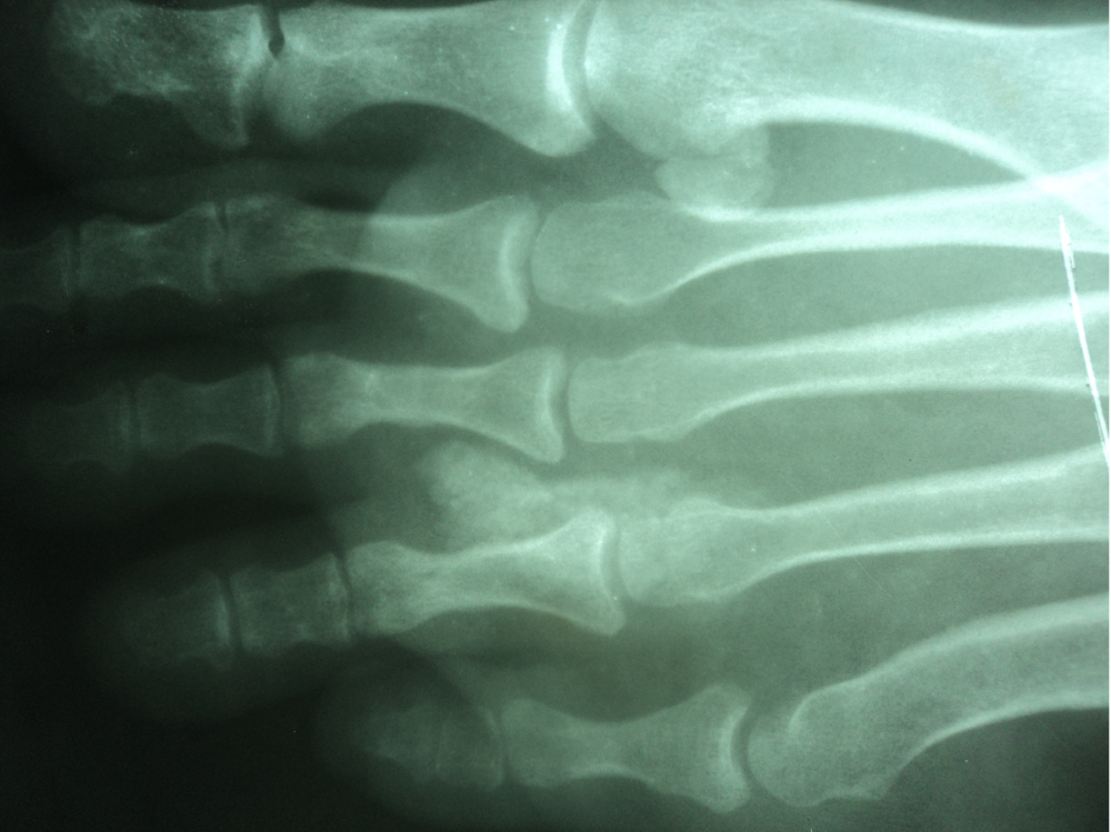
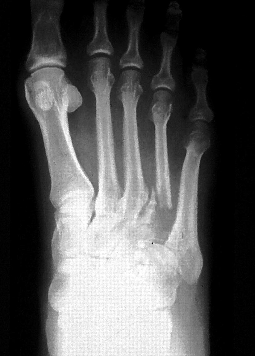
Metatarsal Xray
Femur Xray
Tibia Xray
Proximal femur
Distal tibia
Proximal tibia
Distal tibia
Metatarsal
Images hosted on other servers:

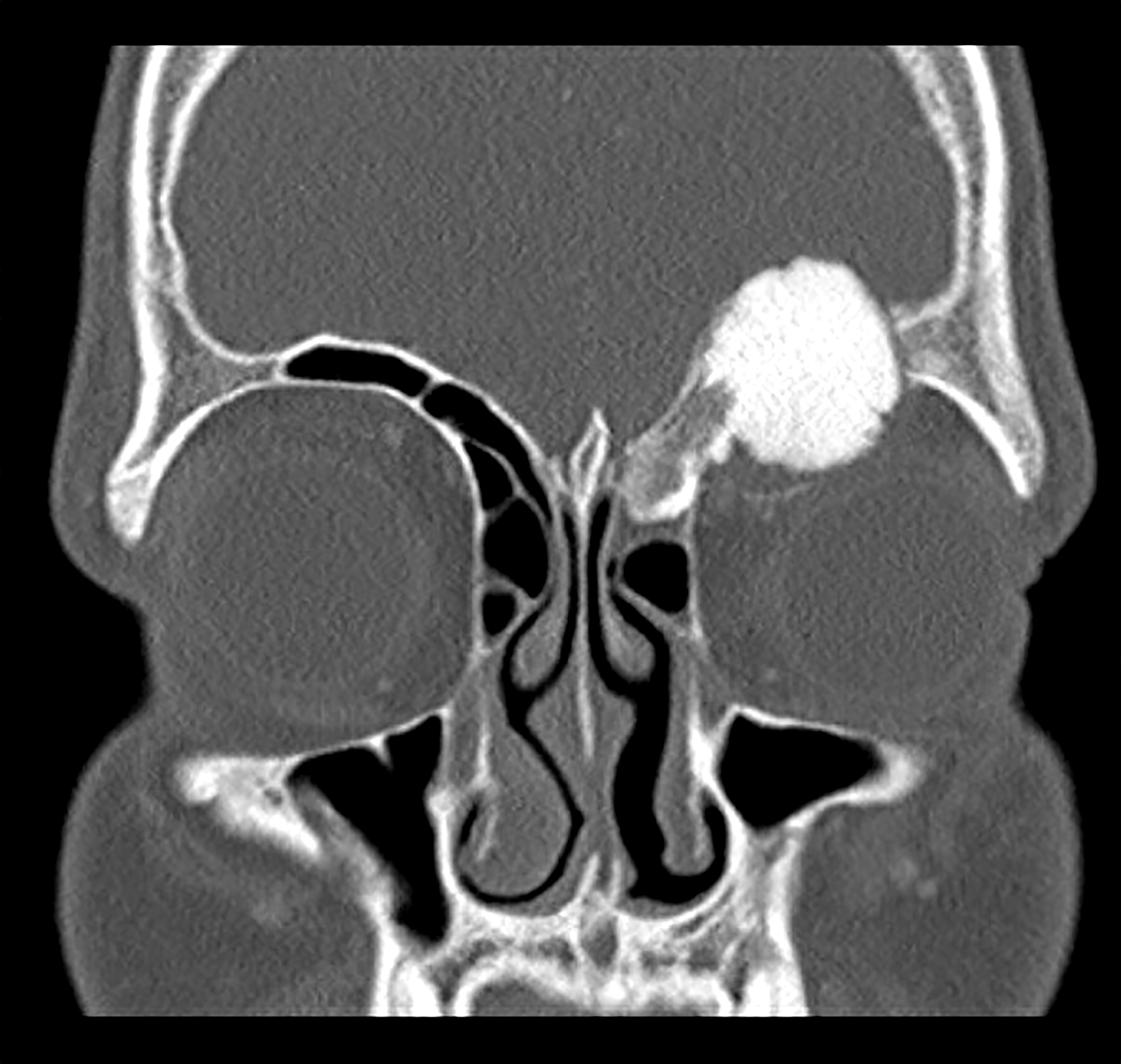
Nasal mass

Sternum

Sellar lesion


Radius


Rib

Sacrum

Metatarsal
Images hosted on other servers:


Lesion in mandible

Orbital tumor with globe displacement
Images hosted on other servers:

Orbital tumor resection specimen

Resection of rib tumor
Contributed by Nasir Ud Din, M.B.B.S.
Confluent nodules with giant cells
Well defined nodular growth
Fibroblastic population
Peripheral giant cell proliferation
Stellate mesenchymal cells in nodules
Adjacent ABC-like areas
Chondroid differentiation
Well developed chondroid areas
Calcifications
Images hosted on other servers:

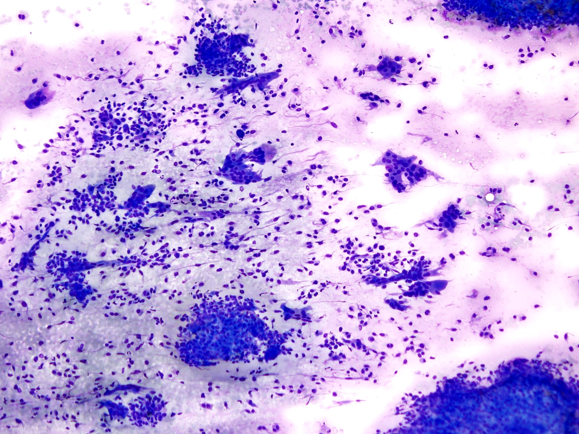
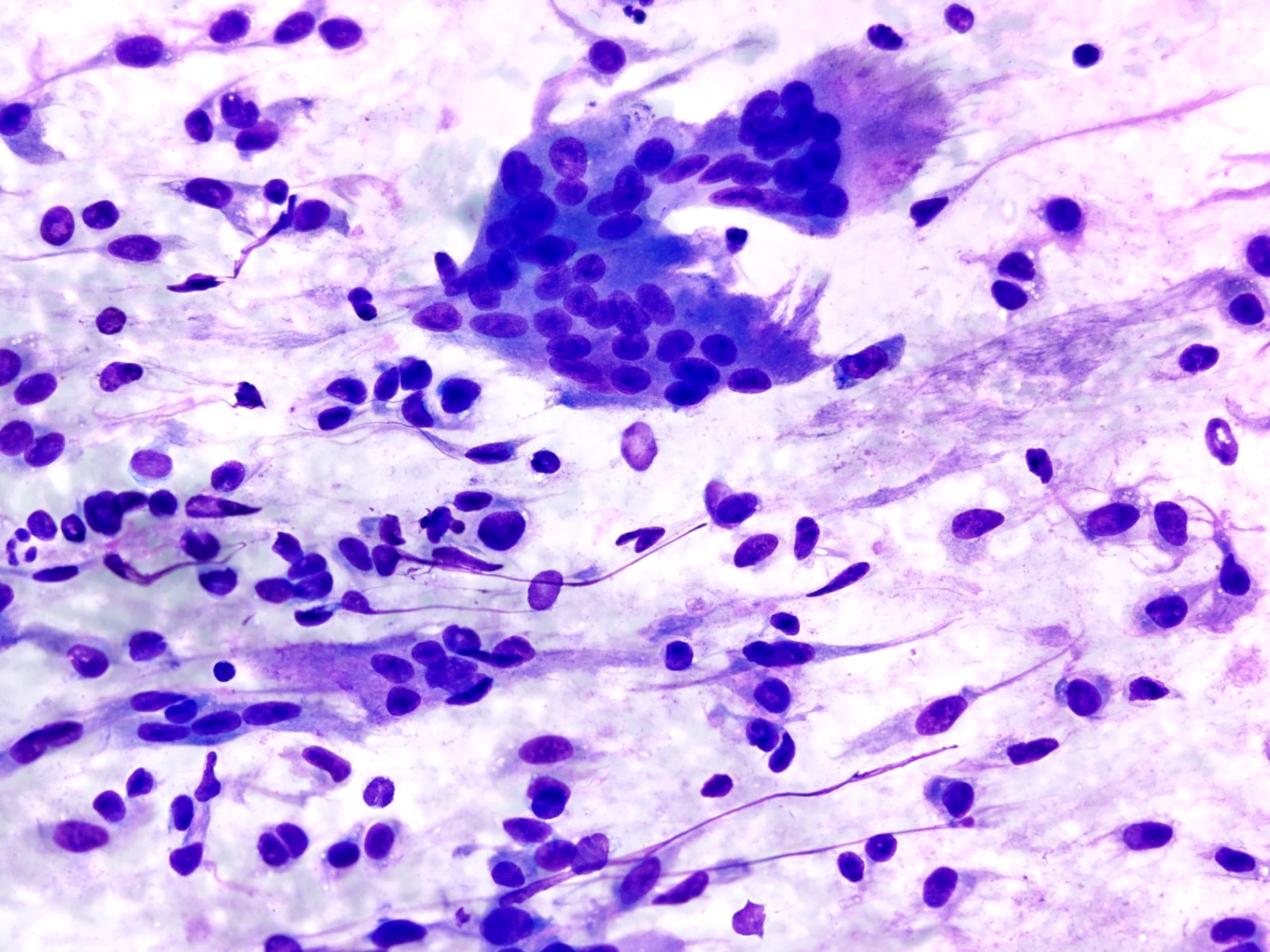
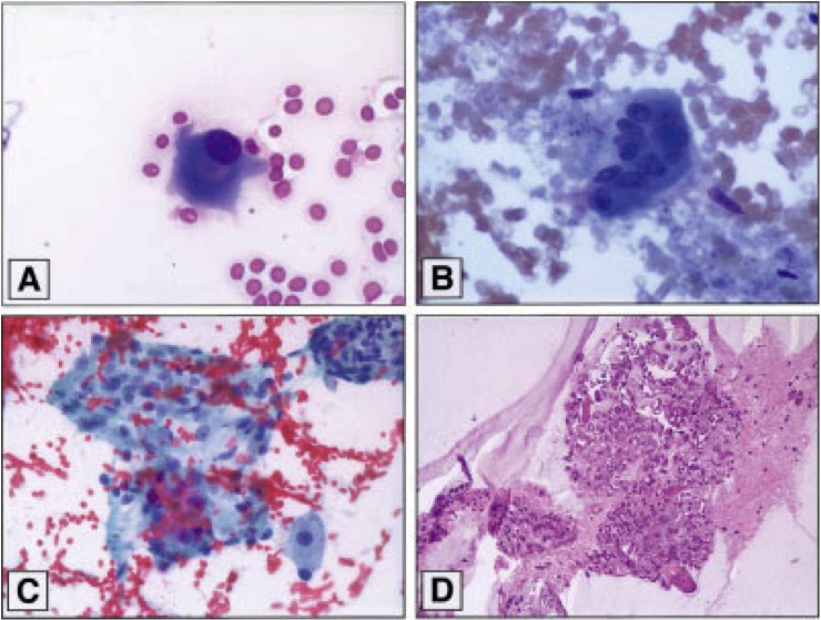
FNA of metatarsal lesion

FNA of lytic lesion in femur

FNA of metatarsal head lesion
Cartilage forming tumors
Contributed by Shadi Qasem, M.D.
Soft tissue extension
Corresponding MRI for previous image
Finger tumor
Pelvic tumor
Secondary tumor
Contributed by Shadi Qasem, M.D.
Femoral tumor
Pelvic tumor
Contributed by Shadi Qasem, M.D.
Grade I
Myxoid degeneration
Grade II
Grade III
Bone permeation
Contributed by Shadi Qasem, M.D.
Magenta color matrix
Multinucleation
Contributed by @Elena_PradosMD on Twitter
Chondrosarcoma
Chondrosarcoma versus enchondroma
Contributed by Jesse Hart, D.O. and Case #110
High signal intensity
Destruction of cervical vertebrae
MRI with sacral mass
Contributed by Jesse Hart, D.O.
Chordoma has fleshy cut surface
Chordoma has invaded the soft tissue
Contributed by Jesse Hart, D.O. and Nasir Ud Din, M.B.B.S.
Lobular architecture
Vesicular chromatin

Abundant myxoid stroma
Epithelioid cells with eosinophilic chords

Tumor necrosis
Poorly differentiated chordoma in 2 year old
Chondroid chordoma
Dedifferentiated chordoma
Brachyury
Cytokeratin cocktail
EMA
S100 protein
Contributed by Nasir Ud Din, M.B.B.S.
Epiphyseal osteomyelitis
Sequestrum
Periosteal reaction
Cystic
Images hosted on other servers:

Chronic osteomyelitis with wound infection
Contributed by Nasir Ud Din, M.B.B.S. and Mohammad Khurram Minhas, M.B.B.S.
Area of suppurative necrosis
Chronic granulomatous osteomyelitis
Images hosted on other servers:

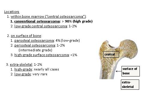
Gender and anatomical distribution
Contributed by Borislav A. Alexiev, M.D.
Conventional radiography
T1 weighted MRI
MRI STIR sequence
T2 weighted MRI
Contributed by Borislav A. Alexiev, M.D.
Femoral head mass
Contributed by Borislav A. Alexiev, M.D. and @JMGardnerMD on Twitter
Malignant bone neoplasm
Cells with pale cytoplasm
Woven bone formation
Large round nuclei
Cartilaginous component
S100
D2-40
AE1 / AE3
H3K36M
Clear cell chondrosarcoma
Images hosted on other servers:

Arthroscopic finding

External aspect of cyst

Cyst resection
Contributed by Mark R. Wick, M.D. and AFIP images
Pathological fracture
Cartilaginous matrix
Contributed by Mark R. Wick, M.D.
Gray-white cartilage next to tan sarcomatous component
Contributed by Erica Kao, M.D. and Mark R. Wick, M.D.
Abrupt demarcation
Low grade cartilage
High grade component
Low grade cartilage juxtaposed with high grade sarcoma
Images hosted on other servers:

Mechanism of osteoarthritis

Development of osteoarthritis
Contributed by Nasir Ud Din, M.B.B.S.
Left knee joint
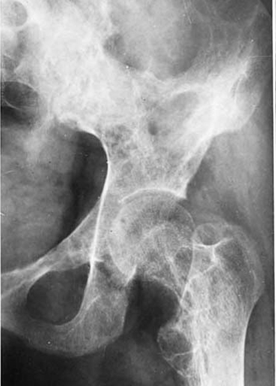
Right hip joint
Images hosted on other servers:

Hand osteoarthritis


Knee osteoarthritis

Hip joint osteoarthritis
Contributed by Jian-Hua Qiao, M.D.
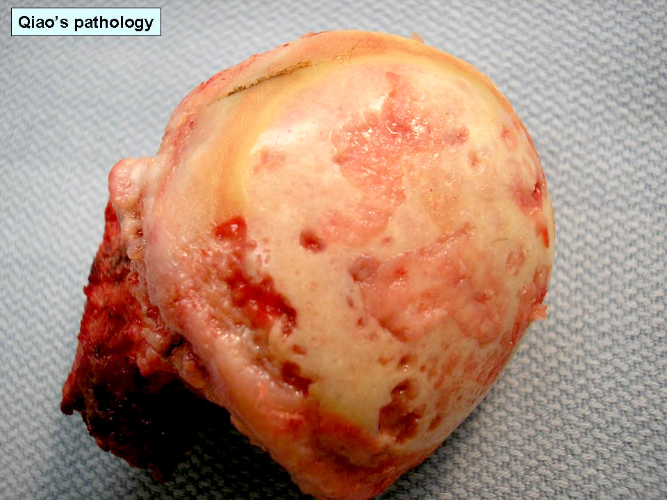
Advanced osteoarthritis of hip
Images hosted on other servers:

Cartilage fragment in synovial fluid; chondrocytes

Synoviocytes in synovial fluid
Images hosted on other servers:

Crystals in cartilage
Osteoarthritis - causes, symptoms, diagnosis, treatment & pathology
Contributed by Kelly Magliocca, D.D.S., M.P.H.
Desmoplastic fibroma
Images hosted on other servers:
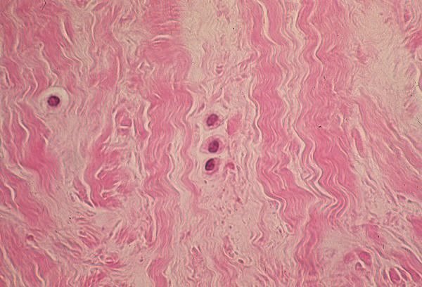
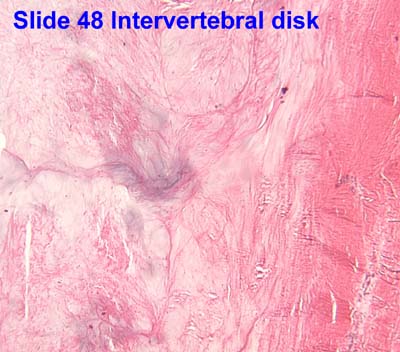
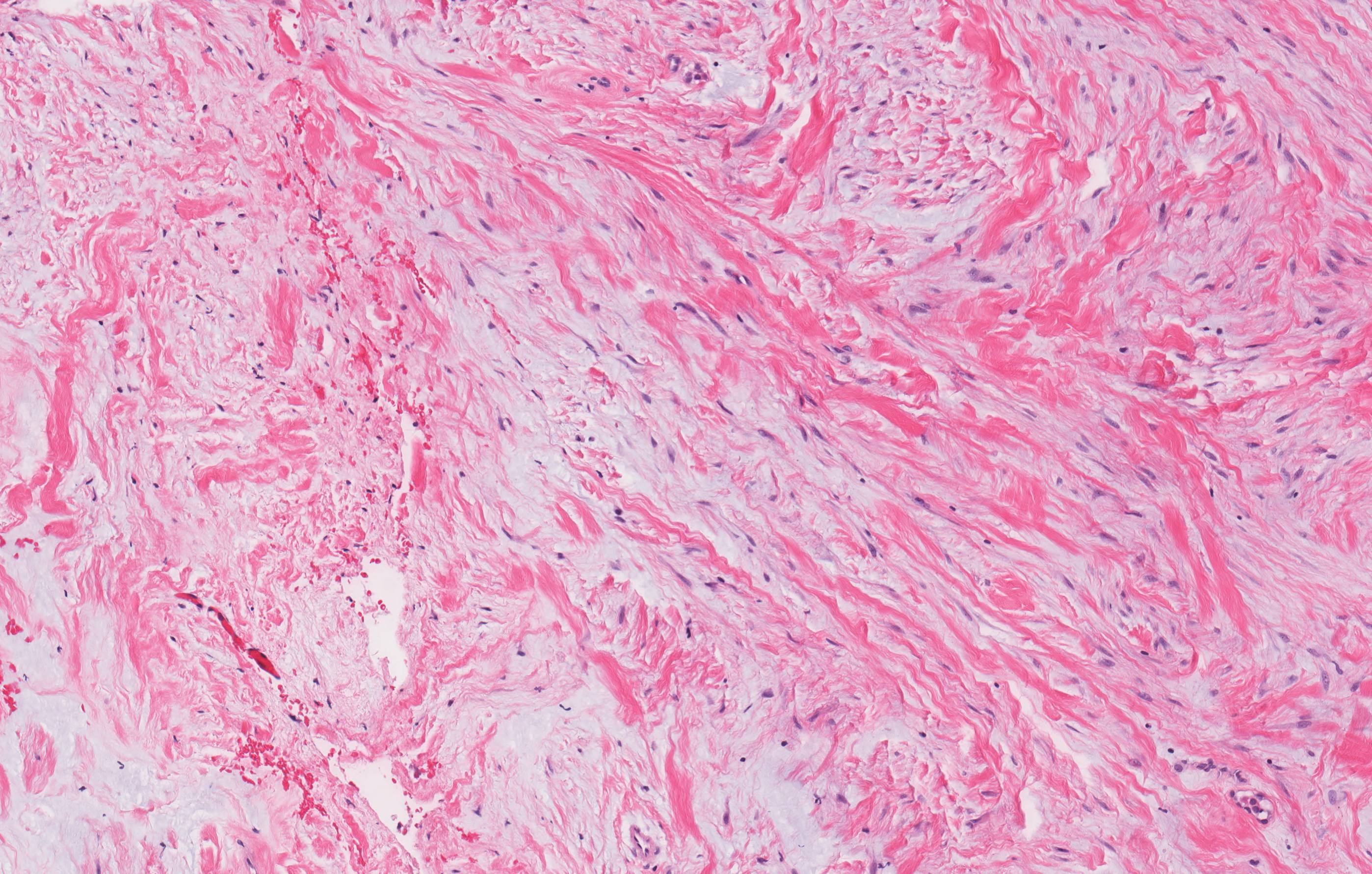
Intervertebral disc

Disc degeneration
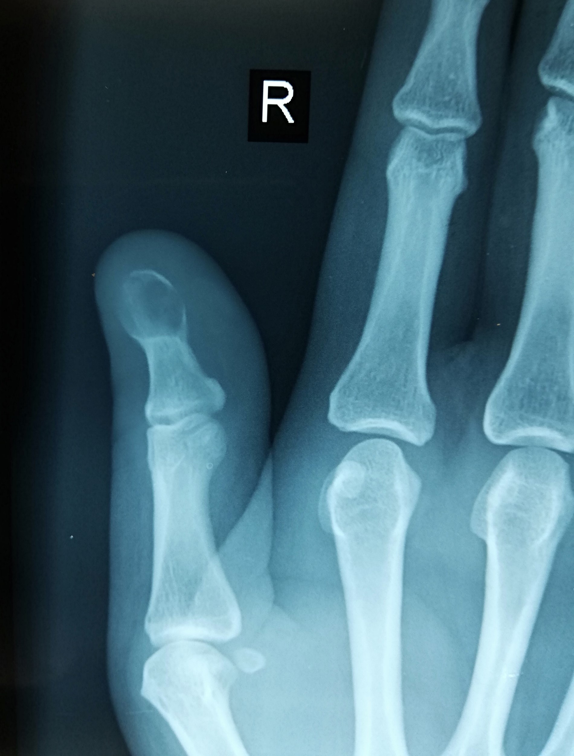
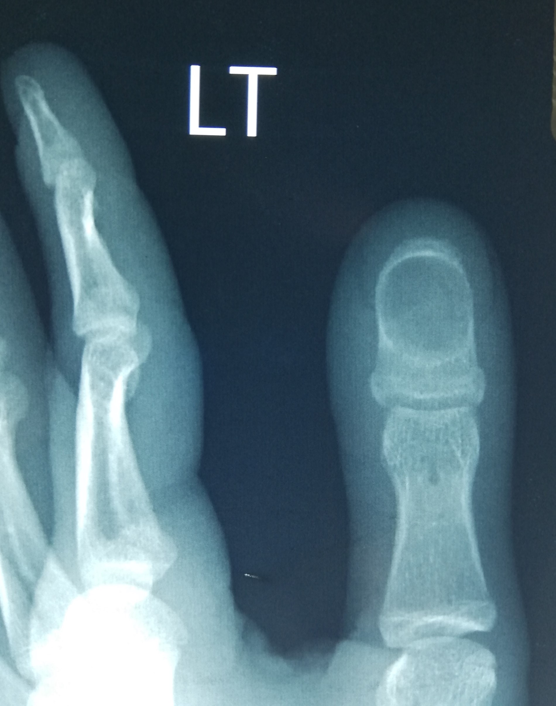
Contributed by Borislav A. Alexiev, M.D. and Terrance D. Peabody, M.D.
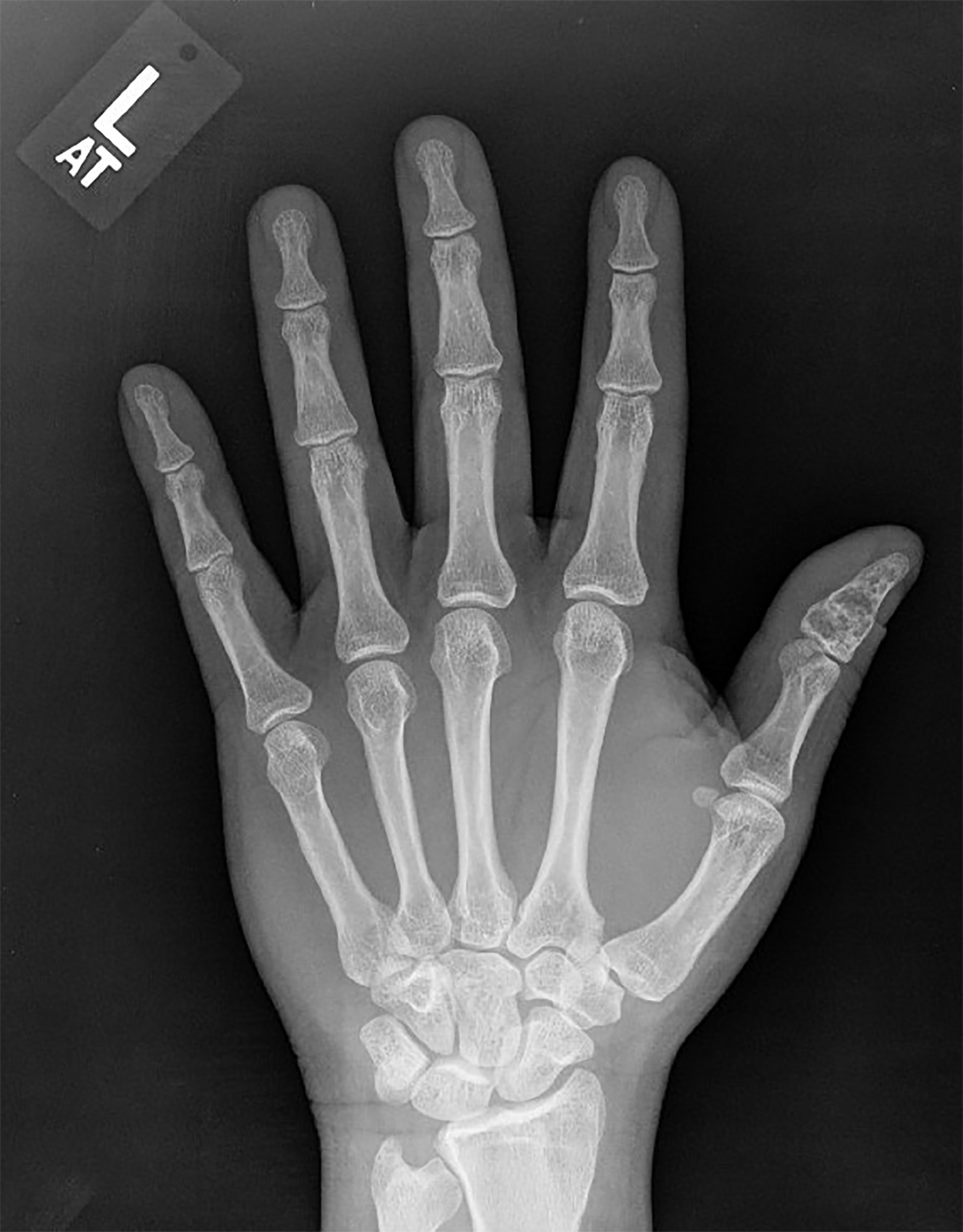
Left thumb Xray
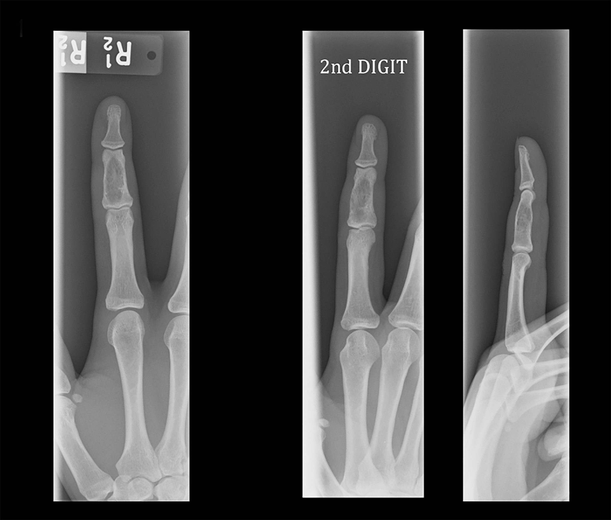
Right index finger Xray
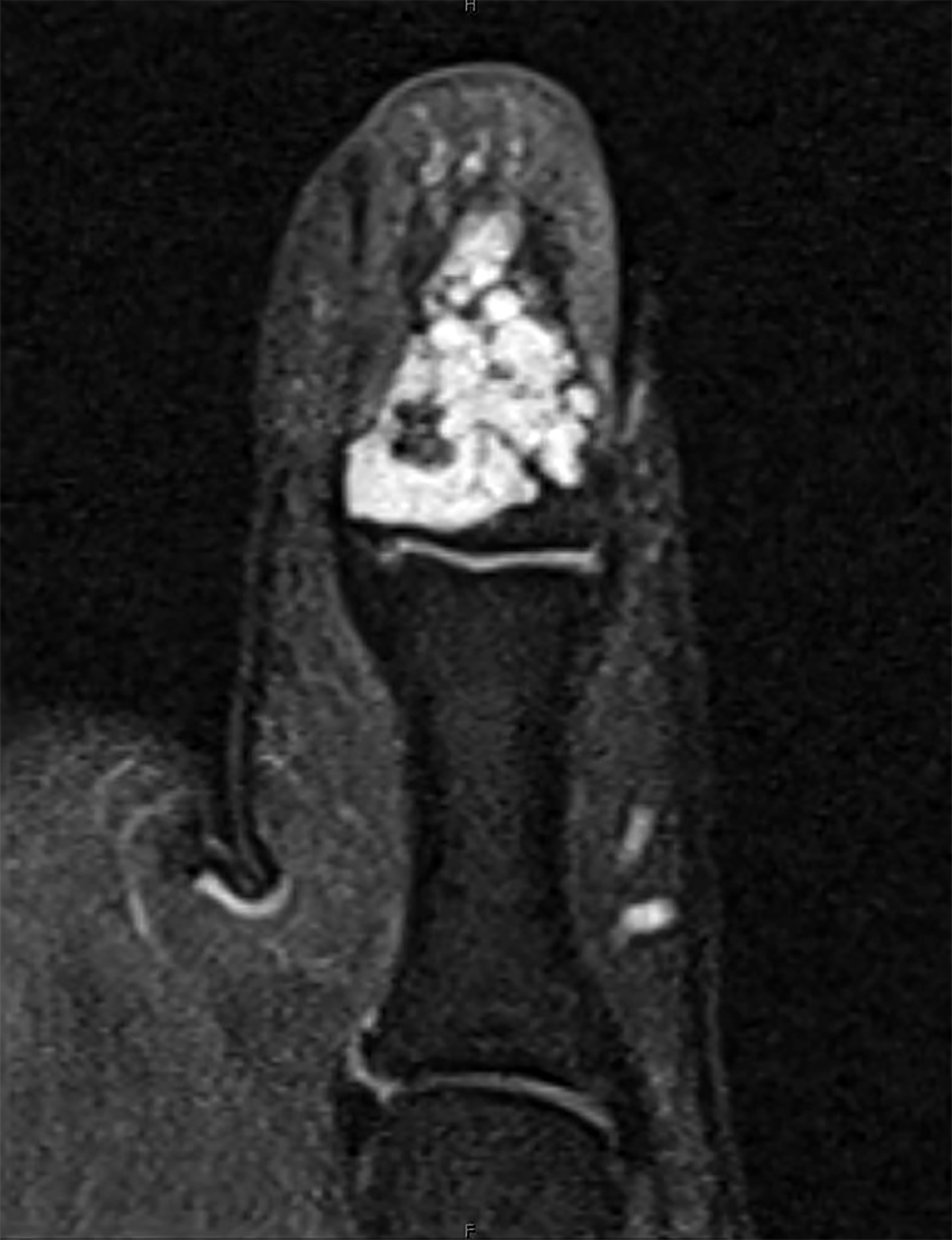
Left thumb MRI
Images hosted on other servers:

Ollier disease
Contributed by Borislav A. Alexiev, M.D. and Mark R. Wick, M.D.
First rib lesion
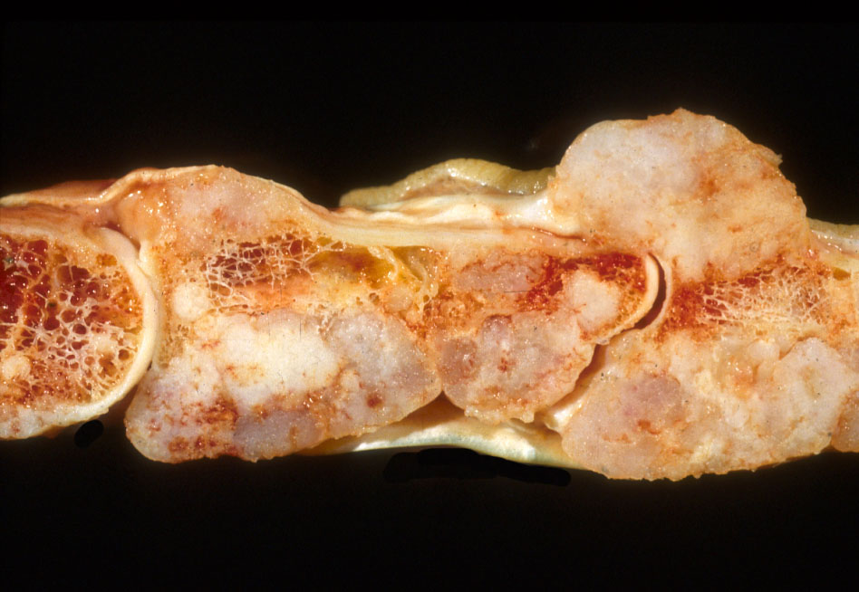
Multifocal in Ollier disease
Contributed by Borislav A. Alexiev, M.D.
Intramedullary lesion
Hypocellular lesion
Chondrocytes in lacunar spaces

Short tubular bone lesion
Degenerative changes
Contributed by Nasir Ud Din, M.B.B.S
Distal phalanx of thumb
Distal phalanx of fourth finger
Skull bone
Contributed by Nasir Ud Din, M.B.B.S.
Absence of adnexal structures
Keratin flakes in cystic cavity
Lamellated keratin
Squamous epithelium with granular layer
Reactive bone around the cyst
Chronic inflammation
Images hosted on other servers:


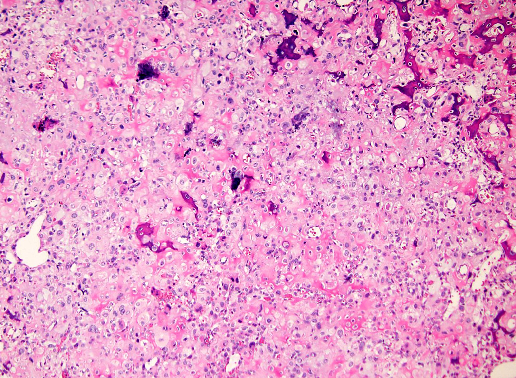
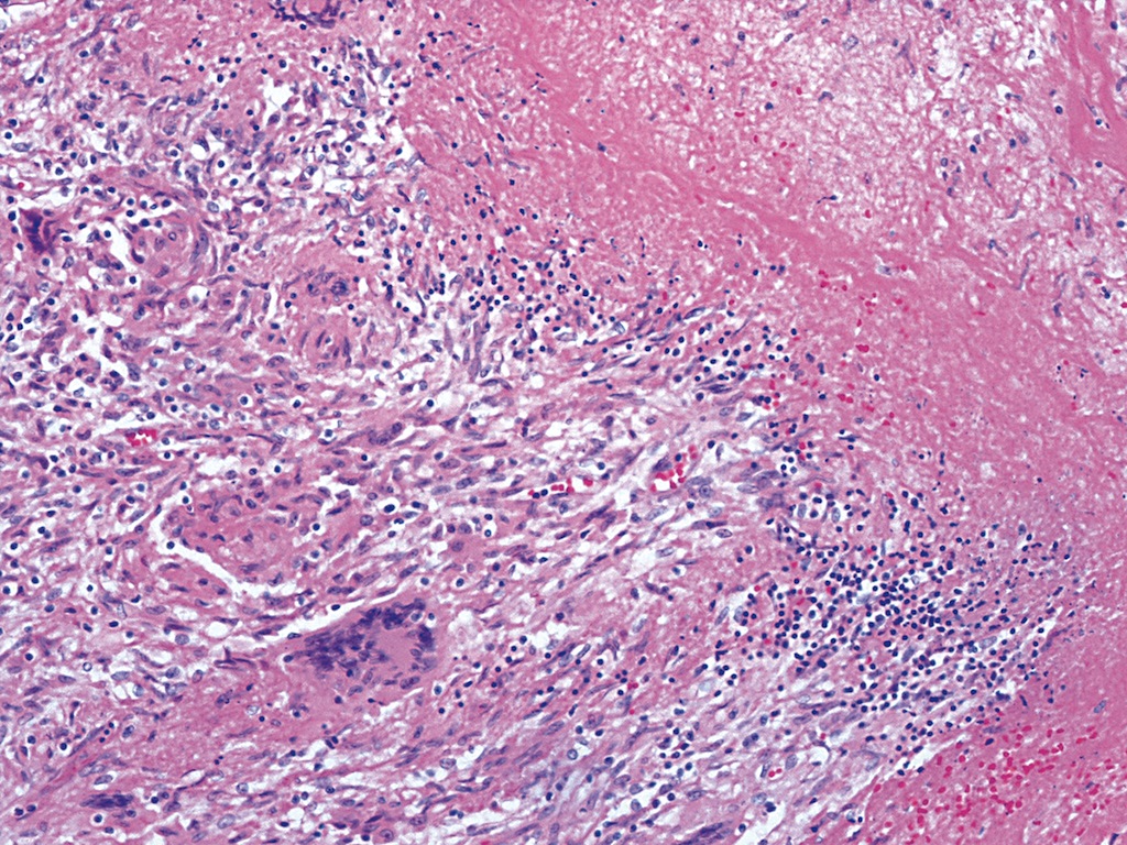
Multifocal liver lesions
Contributed by Iva Brčić, M.D., Ph.D. and Bernadette Liegl-Atzwanger, M.D.
Poorly circumscribed
Cords and nests
Tumor within bone
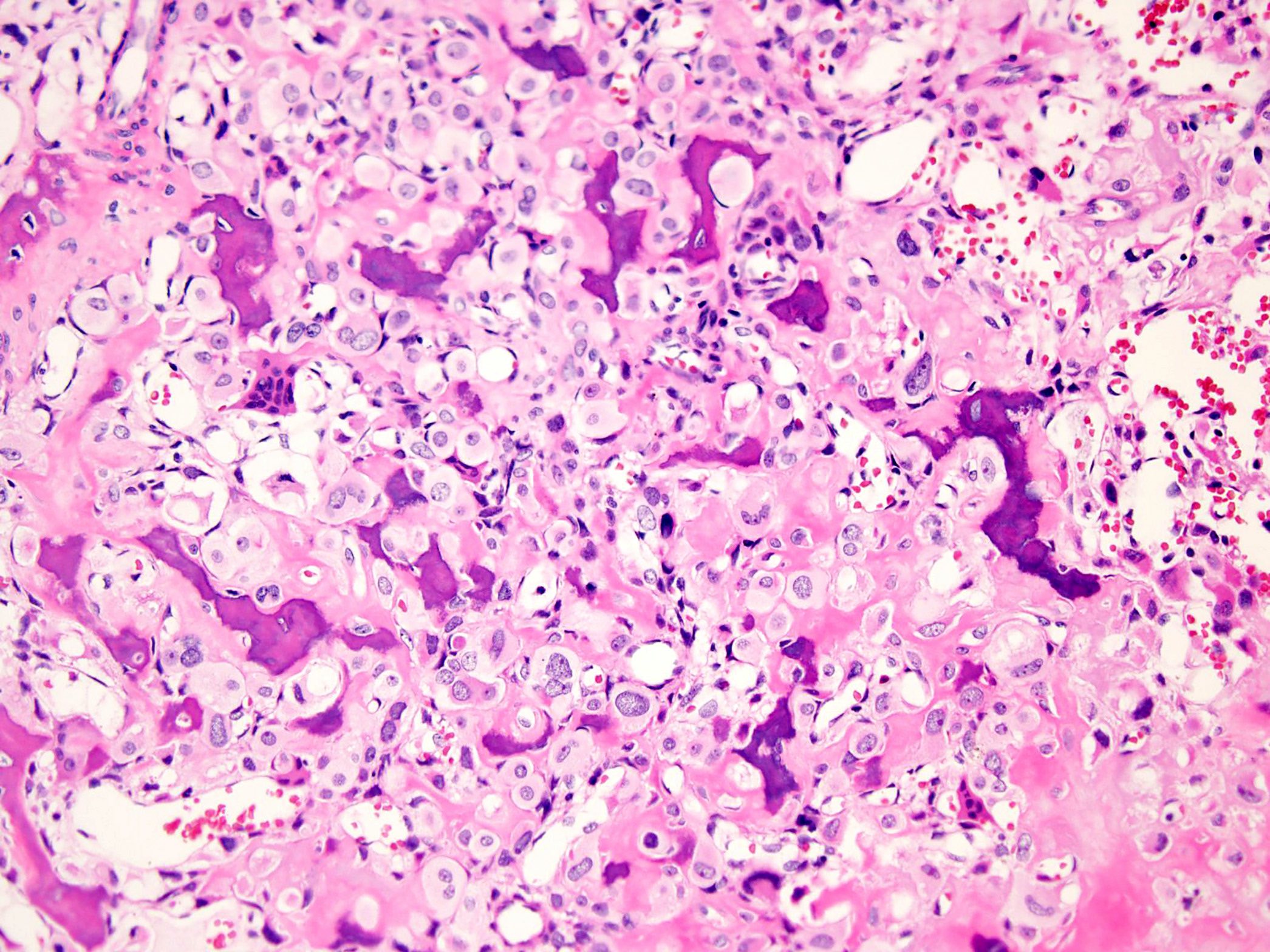
Erythrocytes in lumen
Cords in myxohyaline stroma
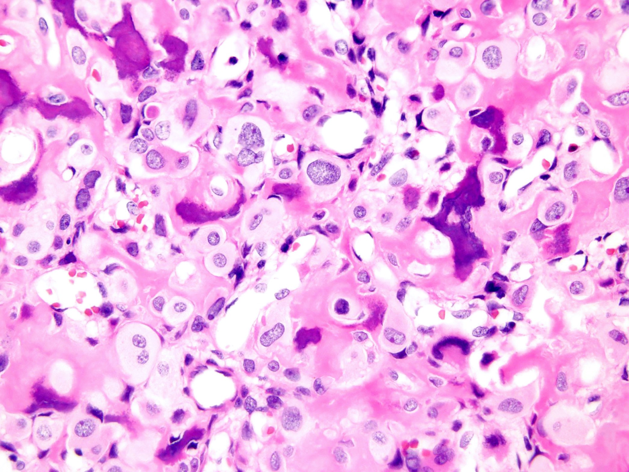
Cellular atypia
Mitosis and atypical cells
Nuclear CAMTA1 staining
Diffuse CD31
Diffuse ERG
CAM5.2 focally positive
Images hosted on other servers:

YAP1-TFE3 fusion
Contributed by Kshitij Arora, M.D.

CT of spine
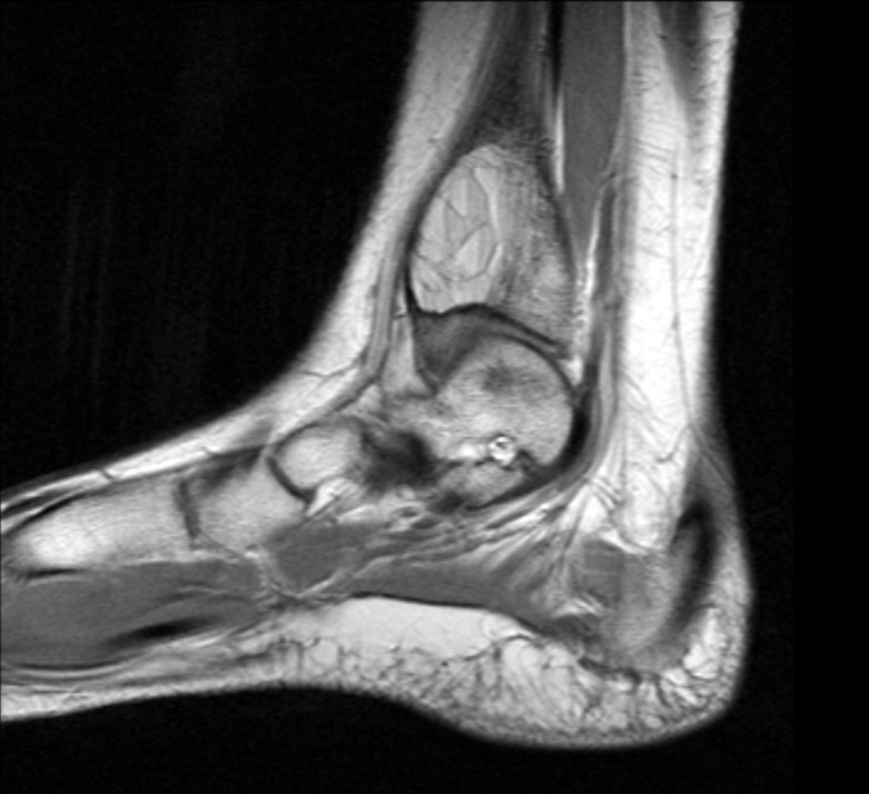
T1 weighted MRI of foot
Contributed by Andrew E. Rosenberg, M.D.
Foot amputation
Contributed by Andrew E. Rosenberg, M.D. and Kshitij Arora, M.D.
Well demarcated
Nodular architecture
Arteriole type vessels

Well formed vascular spaces
Epithelioid cells

Sheets of cells
Epithelioid endothelial cells lining the lumen
CD31 IHC

FOS IHC
Images hosted on other servers:

Skeletal and lung involvement

Peirenal soft tissue thickening
Contributed by Aishwarya Ravindran, M.B.B.S. and Karen L. Rech, M.D.
Excisional biopsy of perinephric soft tissue mass
CD163

Factor XIIIa

Core biopsy of soft tissue (buttock) mass
Images hosted on other servers:

AFIP images
Saucerization
Images hosted on other servers:

Radiograph of humeral lesion

Radiograph of humeral lesion (close up)

CT of humeral lesion

Radiograph of humeral head lesion

MRI of humeral head lesion
Images hosted on other servers:

Extraosseus Ewing sarcoma of the forearm
Contributed by Mark R. Wick, M.D.
Ewing sarcoma
Images hosted on other servers:

Ewing sarcoma of foot

Amputation for Ewing sarcoma
Contributed by Laura Warmke, M.D., Mark R. Wick, M.D. and Erdener Özer, M.D., Ph.D.
Small round cells
Clear cell change
Apoptotic cells
"Light" cell and "dark" cell appearance
Rosettes
CD99
FLI1
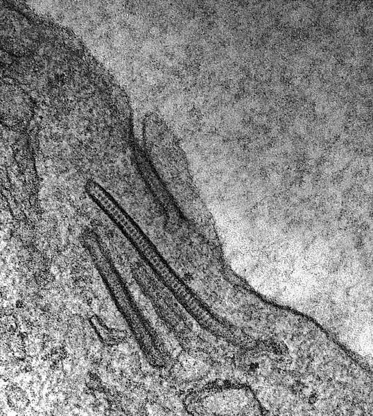
Contributed by Laura Warmke, M.D. and Erdener Özer, M.D., Ph.D.
Round cells
Fine chromatin
Small nucleoli
Cytological appearance
Images hosted on other servers:

Ultrastructure analysis of tumor
Images hosted on other servers:

Dual color break apart probe FISH test
Ewing family of tumors
Contributed by Mark R. Wick, M.D. and AFIP images
Humerus Xray
Radius
AFIP images

Well circumscribed tumor
Contributed by Mark R. Wick, M.D. and AFIP images

Various images

Well differentiated
Grade 2

Grade 3
Contributed by Jose G. Mantilla, M.D., Mark R. Wick, M.D. and AFIP images
Femur
Femur coronal
Mandible

Femur
Ulna
Expanded rib
Images hosted on other servers:

Craniofacial lesions

Buccal protrusion
Contributed by Jose G. Mantilla, M.D. and AFIP images
Bone lesion
Expanded rib
Expanded lesion of calvarium
Contributed by Jose G. Mantilla, M.D., Kelly Magliocca, D.D.S., M.P.H. and @JMGardnerMD on Twitter

Irregular trabeculae

Stromal cells
Aneurysmal bone cyst
Fibrous dysplasia
Fibrous dysplasia
Fibrous dysplasia
Fibrous dysplasia
Images hosted on other servers:

Types of bone fracture

Fracture healing

Salter-Harris classification of physeal fractures
Images hosted on other servers:

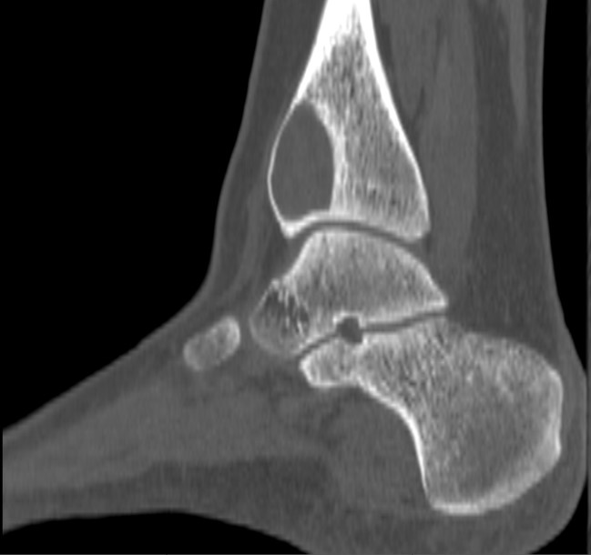
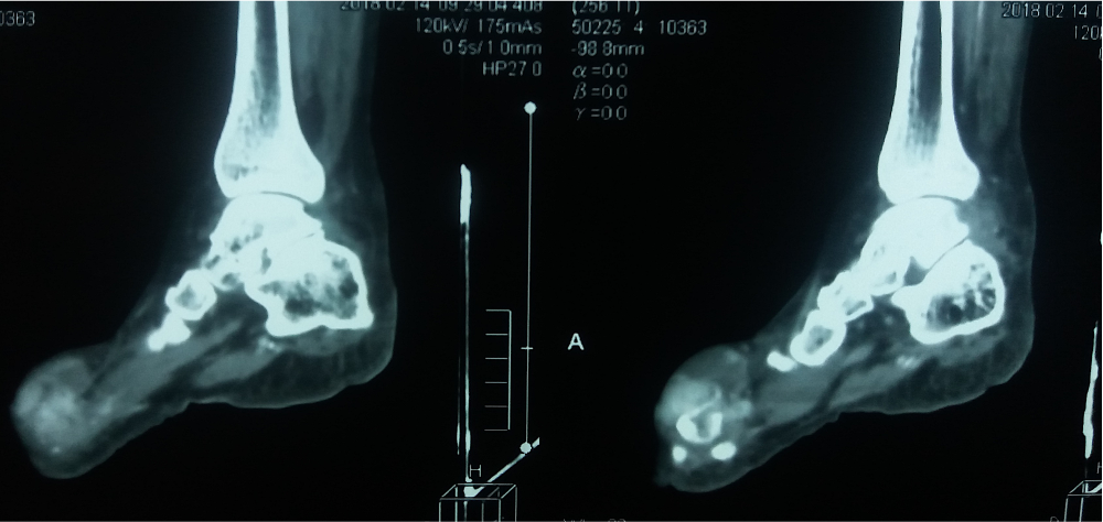
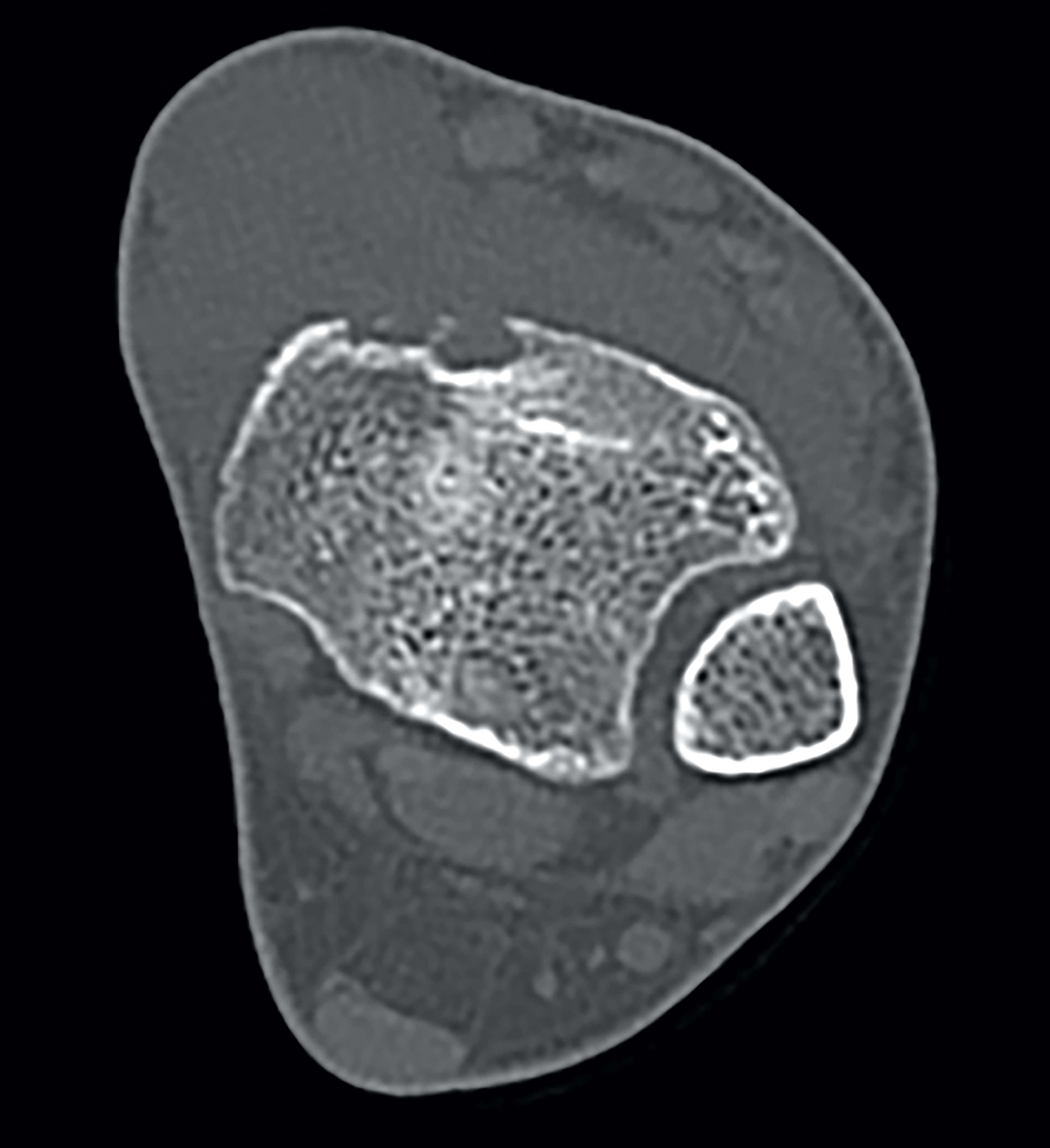
CT of left ankle, sagittal view

Xray of distal femur

CT of scaphoid, coronal view

Xray of mid tibia

Xray of mid leg fracture

Xray of femoral fracture

Xray of humerus fracture
Images hosted on other servers:

53 year old man with open fracture of tibia

External fixation of fracture

Segmental fracture of tibia

Open fracture

Bruising in broken arm
Contributed by Laura Warmke, M.D. and Mark R. Wick, M.D.

Hemorrhage

Fibrovascular stroma
Fracture callus

Endochondral ossification
Hemorrhagic background
Woven bone

Devitalized bone
Bone resorption
Inflammatory cells
Fracture callus
Atypical cartilage component
Healing
Common types of bone fracture
How do broken bones heal?
Images hosted on other servers:

Dorsal wrist ganglion

Ganglion: intraoperative view

Volar wrist ganglion
Images hosted on other servers:

Ultrasound: dorsal wrist ganglion

MRI: volar wrist ganglion
Images hosted on other servers:

Ganglion
Contributed by Serenella Serinelli, M.D., Ph.D.
Unilocular ganglion cyst
Cyst wall and content
Multilocular ganglion cyst
Myxoid changes
Absence of synovial lining
Images hosted on other servers:

Ganglion cyst mucoid material

Histiocytes in ganglion cyst
Ganglion cyst features
Images hosted on other servers:

Anatomical distribution
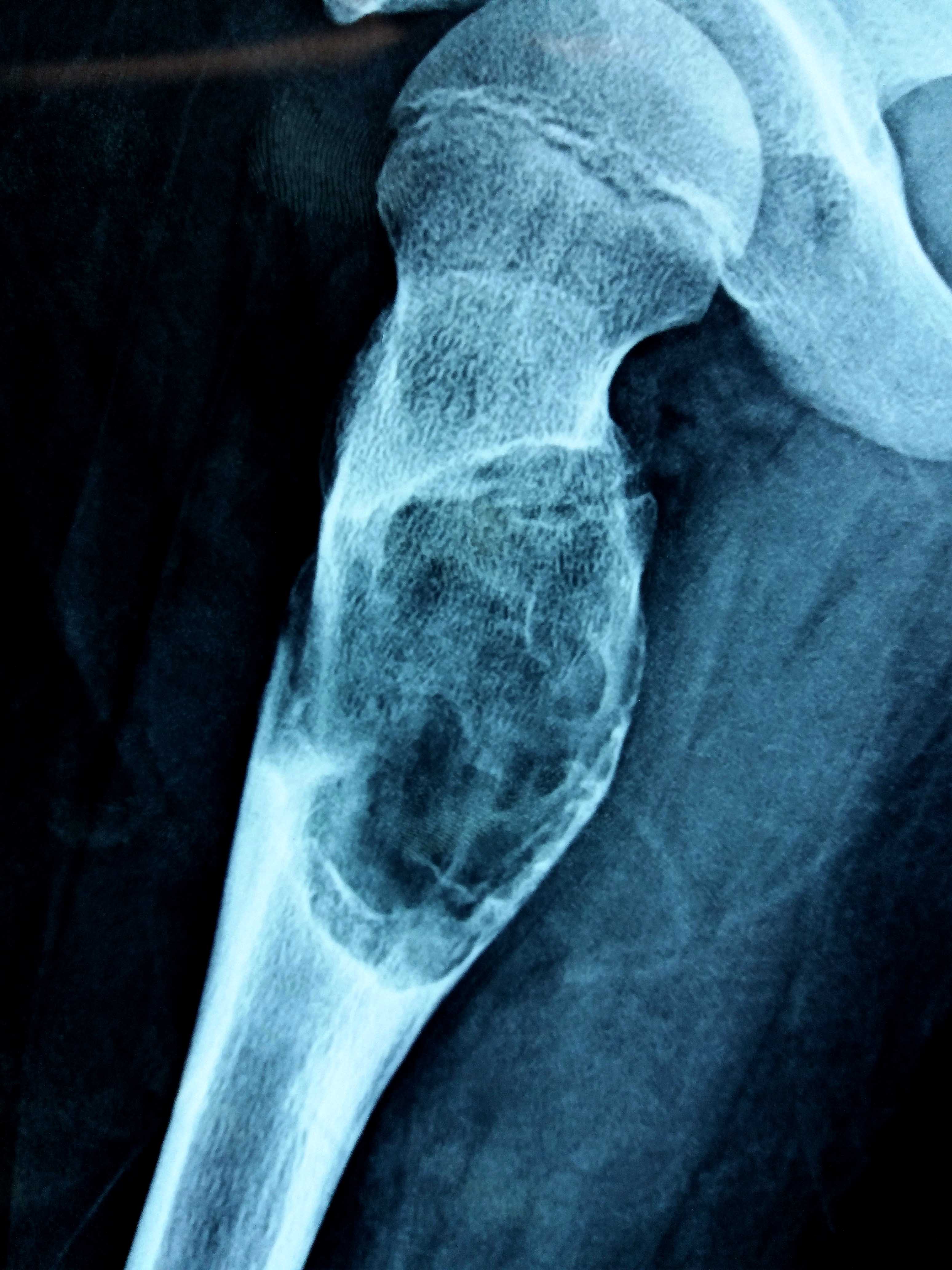
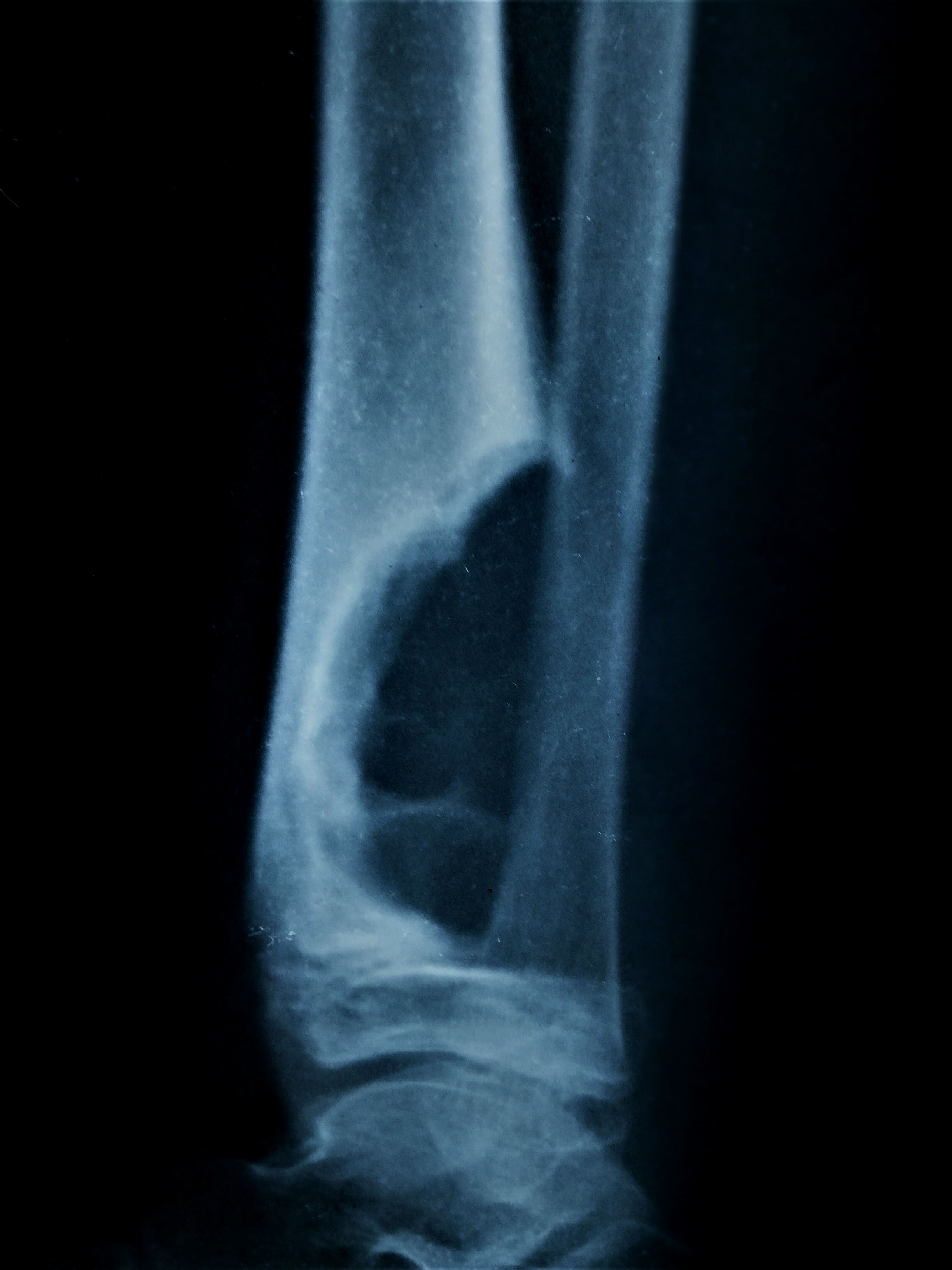
Contributed by Borislav A. Alexiev, M.D.
Lytic lesion
Expansile solid lesion
Mass with extraosseous component
Images hosted on other servers:

Right proximal humerus tumor
Contributed by Borislav A. Alexiev, M.D. and Mark R. Wick, M.D.
Left distal femur lesion
Distal femur
Contributed by Borislav A. Alexiev, M.D.
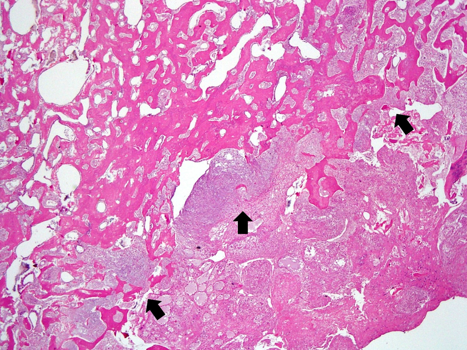
Osteoclasts-rich bone lesion
Typical features

Reactive bone formation
Aneurysmal change
Neoplastic mononuclear cells
Residual tumor
Depletion of giant cells
Reactive bone formation
Complete response with hemorrhage
Spindle cell proliferation
Complete response with fibrosis
G34W IHC
p63 IHC
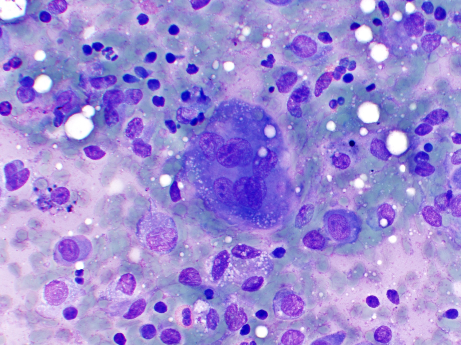
Contributed by Lucy Jager, M.D.
Cellular smears
Mononuclear and MGCs
Images hosted on other servers:

Acute gout

Chronic tophaceous gout

Pathogenesis of gout
Contributed by Nasir Ud Din, M.B.B.S.
Left foot
Foot
Images hosted on other servers:

Bilateral tophi, hands

Bilateral tophi, feet
Contributed by @Elena_PradosMD on Twitter
Gout and gouty arthritis
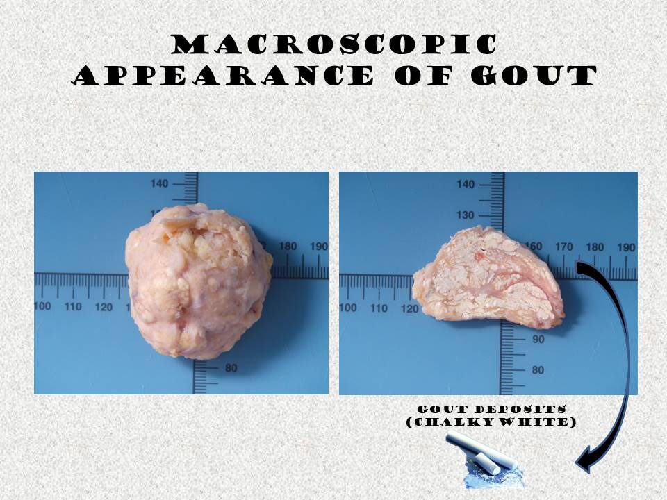
Images hosted on other servers:

Tan-white chalky, crumbly tophi
Contributed by Nasir Ud Din, M.B.B.S. and @Elena_PradosMD on Twitter
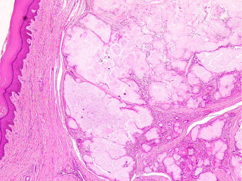
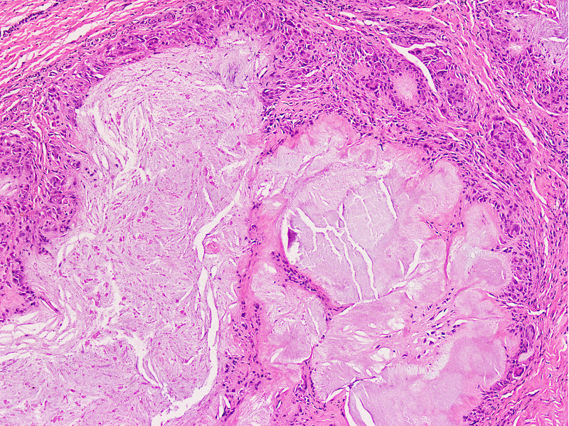
Nodular aggregates
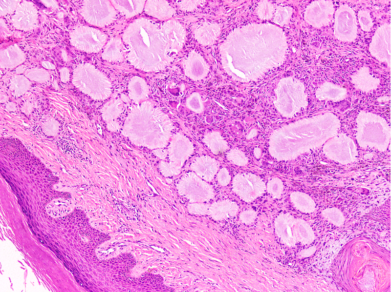
Granulomatous appearance
Feathery appearance
Needle shaped crystals
Chronic inflammatory cells
Intersecting pattern of arrangement
Brightly anisotropic crystals
Gout and gouty arthritis
Images hosted on other servers:


Gouty tophus with crystalline material and inflammatory response
Images hosted on other servers:

Needle-like crystals
Images hosted on other servers:

Urate transporters

Uric acid transportasome
Brilliant polarizable birefringent monosodium urate crystals
Images hosted on other servers:


Hepatic hemangioma

Intrathoracic hemangioma

Infantile thyroid hemangioma

Infantile hepatic hemangioma

Choroidal hemangioma (fundoscopy)


Hemangioma in long bones


Hemangioma in long bones
Contributed by Nasir Ud Din, M.B.B.S.
Noninvoluting congenital hemangioma
Images hosted on other servers:

Infantile hemangioma

Infantile hemangioma with ulceration

Infantile hemangioma in beard distribution

Rapidly involuting congenital hemangioma

Partially involuting congenital hemangioma

Laryngeal hemangioma (laryngoscopic view)
Images hosted on other servers:

Atrial hemangioma

Hepatic hemangioma

Pulmonary hemangioma
Contributed by Nasir Ud Din, M.B.B.S.
Anastomosing hemangioma
Skeletal hemangioma
Cavernous hemangioma
Hobnail hemangioma
Hepatic hemangioma
Infantile hemangioma
Infantile hemangioma
Lobular capillary hemangioma
Spindle cell hemangioma
Spindle cell hemangioma
Phleboliths in spindle cell hemangioma
Synovial hemangioma

Verrucous hemangioma
Images hosted on other servers:

Endothelial differentiation

Cutaneous hemangioma
Images hosted on other servers:

Sanger sequencing for RASA1
Spindle cell hemangioma
Cutaneous vascular tumors
Images hosted on other servers:

Bone structure

Bone architecture
Images hosted on other servers:

Cancellous and compact bone
Contributed by Nasir Ud Din, M.B.B.S.
Active osteoblasts
Inactive osteoblasts
Osteoclasts
Osteoclast type giant cells
Woven bone
Osteocytes in woven bone
Lamellar bone
Osteocytes in Lamellar bone
Cortical bone
Cortical bone
Cortical bone
Reversal cement lines
Periosteum
Images hosted on other servers:


Osteoclast structure
Normal bone histology & embryology 101 with Dr. Andrew Rosenberg
Bone cells and bone formation
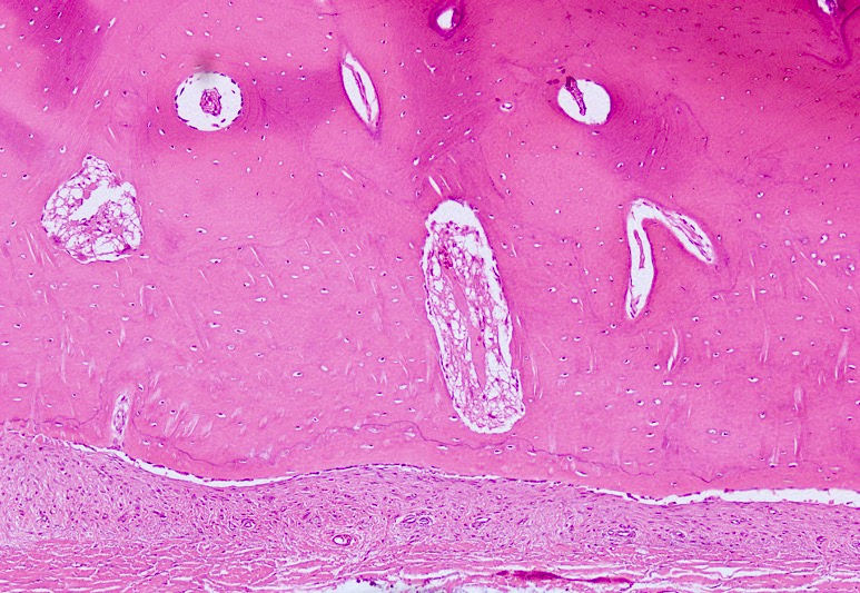
Contributed by Edward F. DiCarlo, M.D.

Tibial plateau
Contributed by Lingxin Zhang, M.D.

Articular cartilage
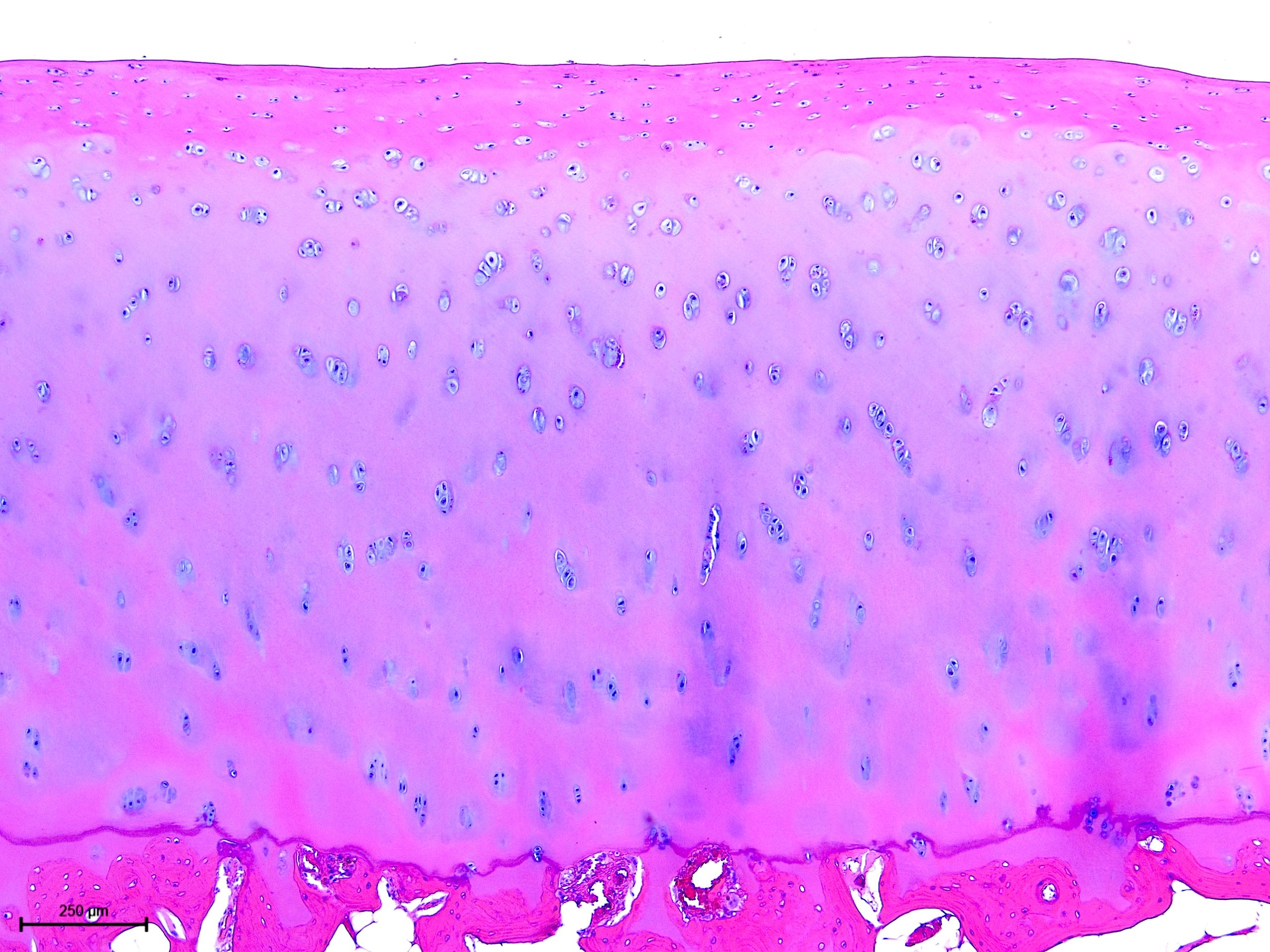
Normal articular cartilage
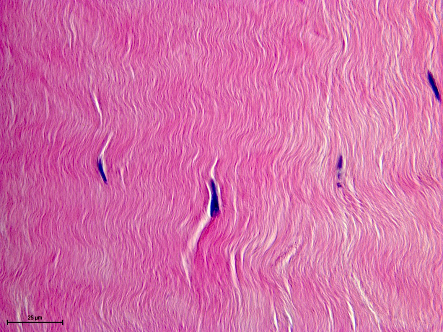
Ligament
Enthesis

Synovium
Histology of the joint
Embryology, history and pathology of articular cartilage
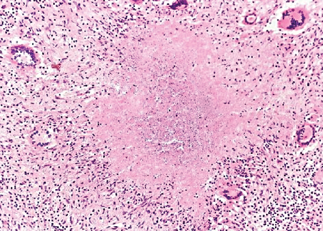
Case #300
Images hosted on other servers:

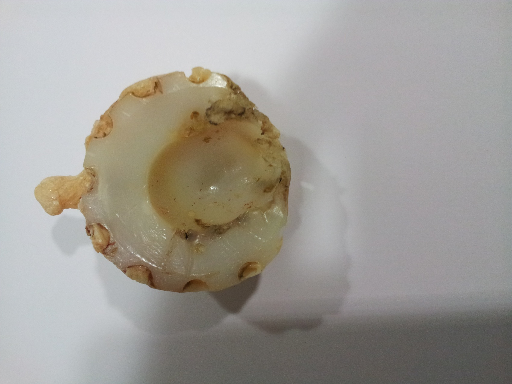
Polyethylene liner wear
Images hosted on other servers:

Metallosis surrounding implant and cervical neck
Case #300
Case #300

Polarizing light showed
crystal-like structures
consistent with polyethylene
particles
Images hosted on other servers:
Prosthetic synovitis:
hyperplastic synovial
membrane adjacent
to prosthesis
Methylmethacralate
debris (linear webs)
surrounded by
giant cells
Polyethylene flakes (shiny linear material) surrounded by foreign body giant cell reaction (polarized microscopy)

Calcium pyrophosphate crystals under polarized light
Contributed by Mark R. Wick, M.D.
Multifocal congenital Xray
Contributed by Mark R. Wick, M.D.
Multifocal congenital
Images hosted on other servers:

Cytokine signaling pathways
Images hosted on other servers:

Various images

Swelling and flexion contracture

Polyarticular disease

Systemic onset disease
Images hosted on other servers:

Various images
Images hosted on other servers:

Arthroscopic image
of lesion within
suprapatellar pouch
Images hosted on other servers:

Glassy appearance
Images hosted on other servers:

Myxoid stroma and spindle cells

Bland myxoid
neoplasm without
mesenchymal atypia
or hypercellularity
Contributed by Vignesh Shanmugam, M.D.

Misguided myeloid / dendritic cell model
Contributed by Vignesh Shanmugam, M.D.

Sagittal view

Coronal view
Images hosted on other servers:

LCH
Contributed by Vignesh Shanmugam, M.D.
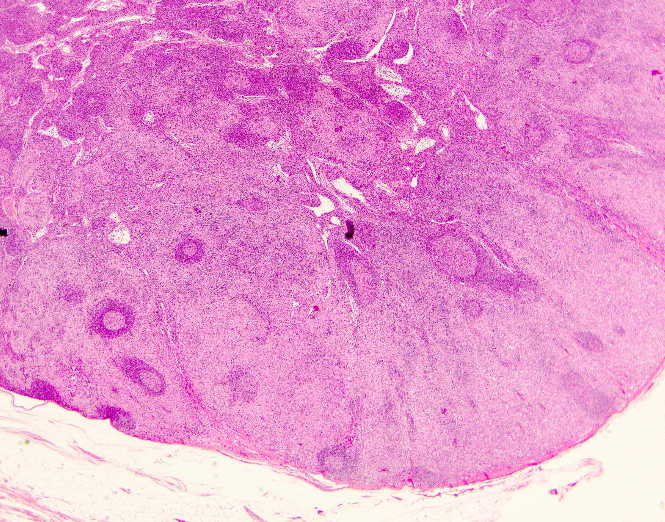
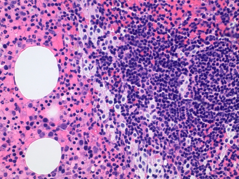
Preservation of follicle centers

Sinusoidal infiltration
Infiltrate of subcapsular sinus
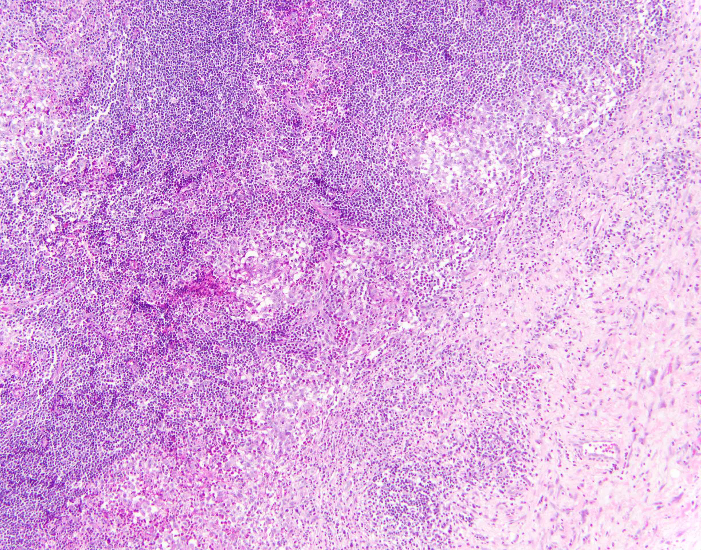
Frequent admixed eosinophils
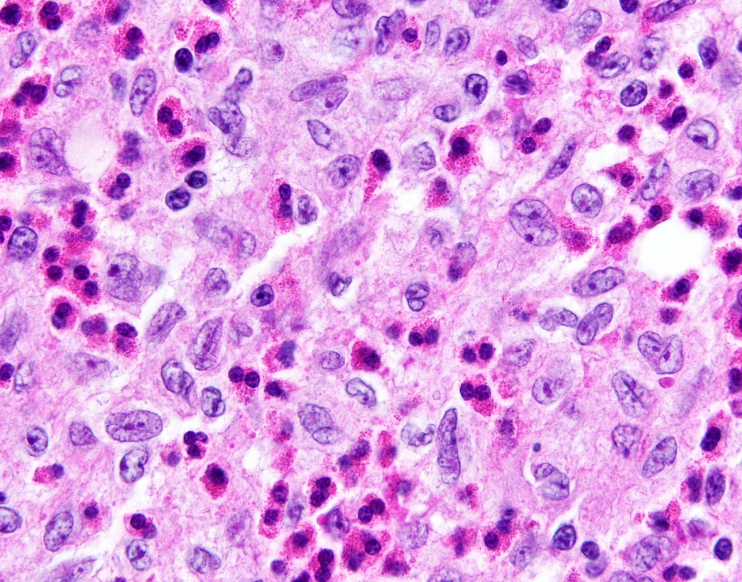
Prominent nuclear grooves
Admixed multinucleated giant cells
Langerin
CD1a
S100
Cyclin D1
Contributed by Catherine Hagen, M.D.
Langerhans cell histiocytosis

CD1a positive LCH
S100 positive LCH
AFIP images


With Hodgkin lymphoma
Involving spleen
Malignant
S100
Nuclear and cytoplasmic staining S100
Case #314
Diffuse involvement
Sinusoidal and pericapsular involvement
Pericapsular involvement
Admixed eosinophils
Nuclear grooves

Mitotic activity

CD1a
CD5
CD20
CD23
Langerin
S100
Contributed by Vignesh Shanmugam, M.D.

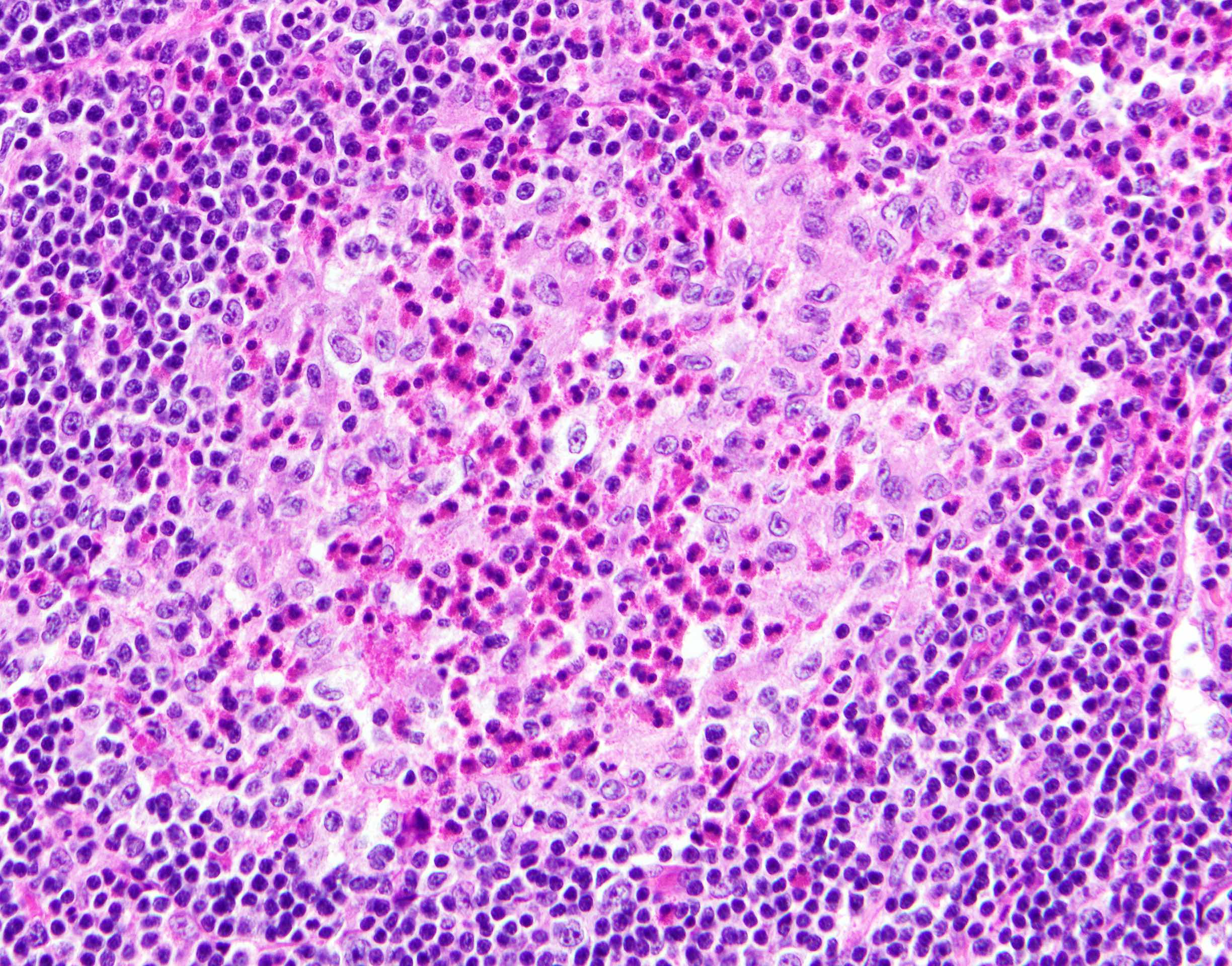
Prominent nuclear grooves and admixed eosinophils
Scattered multinucleated giant cells
AFIP images
Birbeck granules
Images hosted on other servers:

Birbeck granules
Contributed by Borislav A. Alexiev, M.D.
MRI of tibia mass
Contributed by Borislav A. Alexiev, M.D.
Bone mass
Contributed by Borislav A. Alexiev, M.D.
Cortical destruction
Well formed neoplastic bone trabeculae
Fascicles of spindle cells

Lack of bone matrix
Mild nuclear atypia
Contributed by Madina Sukhanova, Ph.D.

MDM2 FISH
Images hosted on other servers:

Erythema migrans skin lesion

Bullseye rash

Lyme arthritis of right knee
Images hosted on other servers:

Borrelia burgdorferi
AFIP images
Femur
Images hosted on other servers:

Imaging of sacral lymphoma
AFIP images

Dense nuclei and histiocyte-like cells
Images hosted on other servers:

Sacral diffuse large B cell lymphoma
Images hosted on other servers:

FISH: diffuse
large B cell
lymphoma with
MYC translocation
Contributed by Jessica L. Davis, M.D.
Fibrous dysplasia
Intramuscular myxoma
Images hosted on other servers:

Ulnar fracture

Lucency in long bones
Images hosted on other servers:

5 year old girl: jagged "coast of Maine" borders

Café au lait spots
Images hosted on other servers:

Ovarian abnormalities
Contributed by Jessica L. Davis, M.D.

Fibrous dysplasia
Contributed by Borislav A. Alexiev, M.D.

Anterior mediastinal mass
Contributed by Iva Brčić, M.D., Ph.D. and Bernadette Liegl-Atzwanger, M.D.
Lobulated mass
Calcifications
Contributed by Iva Brčić, M.D., Ph.D. and Bernadette Liegl-Atzwanger, M.D.
Biphasic tumor
Abrupt transition
2 components
Spindle cell morphology
Transition higher power
Cellular atypia
CD99 staining
Contributed by Mark R. Wick, M.D. and AFIP images
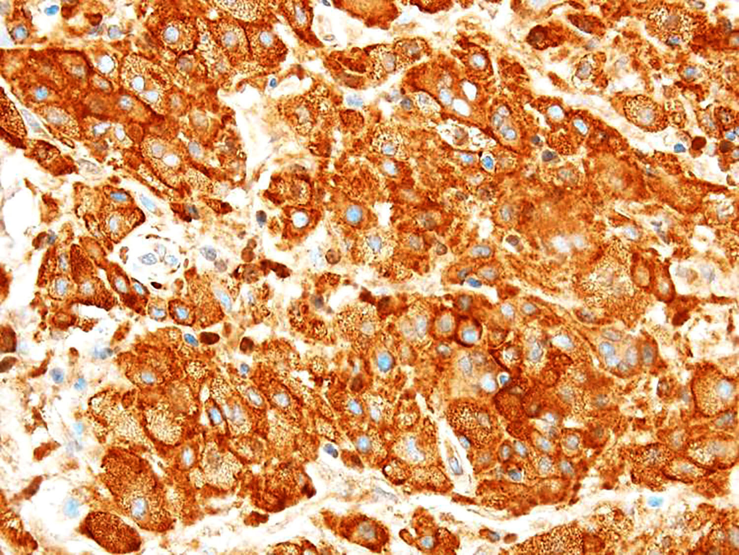
Breast carcinoma
Lung carcinoma to metatarsal
Prostate adenocarcinoma
Thyroid carcinoma
Lung carcinoma metastasis
Prostate carcinoma - osteoblastic features

Thyroid carcinoma
Contributed by Mark R. Wick, M.D.

Lung carcinoma in humeral head
Lung squamous cell carcinoma
Contributed by Semir Vranić, M.D., Ph.D., Mark R. Wick, M.D. and AFIP images
Bone metastasis
Primary prostate adenocarcinoma
Lung carcinoma to metatarsal
Lung carcinoma to metatarsal
Lung carcinoma to metatarsal TTF1
Prostate adenocarcinoma
Renal cell carcinoma
Lung: metastatic carcinoma
Lung: metastatic carcinoma BerEp4
Prostate: metastatic carcinoma
Thyroid: metastatic carcinoma
Images hosted on other servers:

Different stages

Pathogenesis
Contributed by Nasir Ud Din, M.B.B.S.
Xray of early stage
Xray of mid to late stage
MRI buttock for soft tissue mass
Images hosted on other servers:

Different stages
Images hosted on other servers:

Large right pectoral mass and chest radiograph
Images hosted on other servers:

Excised and bisected mass
Contributed by Ghazi Zafar, M.B.B.S., Nasir Ud Din, M.B.B.S. and @JMGardnerMD on Twitter
Periphery
Circumscription
Zonation
Central zone
Foci of cartilage
Osteoid production
Multinucleated giant cells
Myositis ossificans and fibro-osseous pseudotumor of digits
Images hosted on other servers:

Cytological features
Myositis ossificans (benign mimic of sarcoma) bone / soft tissue pathology & radiology correlation
Contributed by Qurratulain Chundriger, M.B.B.S.
Schematic presentation
Contributed by Nasir Ud Din, M.B.B.S. and AFIP images

Distal tibia

Proximal tibia
Proximal fibula
Femur with fracture
Multifocal
Nonossifying fibroma
Images hosted on other servers:


Radius (plain radiograph)

Radius (MRI)

Femur (plain radiograph)

Femur (CT scan)

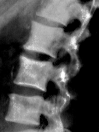
Vertebral body


Mandible


Growth stages
Images hosted on other servers:

Lesion in clavicle
Images hosted on other servers:

Lesion in clavicle

Lesion in mandible
Contributed by Nasir Ud Din, M.B.B.S.
Circumscribed border on resection
Storiform arrangement

Interspersed giant cells

Few giant cells
Resembling ABC
Reactive woven bone
Aggregates of foamy macrophages
Hemorrhage and inflammatory cells
Necrosis after fracture
Images hosted on other servers:

Lesion in femur
Bone and cartilage tumors
Images hosted on other servers:

Epidemiology and localization
Contributed by Borislav A. Alexiev, M.D. and David R. Lucas, M.D.
Left frontal sinus mass
Calcified mass
T7 osteoblastoma with central nidus
Contributed by Borislav A. Alexiev, M.D. and David R. Lucas, M.D.
Bony mass with nidus
Osteoblastoma of clavicle
Spinal T7 osteoblastoma
Contributed by Borislav A. Alexiev, M.D. and David R. Lucas, M.D.
Well marginated lesion
Loose edematous fibrovascular stroma
Osseous trabeculae
Demarcated tumor
Activated osteoblasts
Anastomosing trabeculae

Central nidus of sclerotic woven bone

Woven bone trabeculae with osteoblastic rimming
Degenerative atypia
Contributed by David R. Lucas, M.D.
Cytology smears
Images hosted on other servers:

Osteoblasts with eccentric nuclei

Osteoblast with
indented nucleus
Images hosted on other servers:

Head of femur

Osteochondritis of left humeral head
with secondary deformation of bone,
eighth to ninth century from Iona
Images hosted on other servers:

Pathological specimen of Danish sow
Contributed by Jose G. Mantilla, M.D., Mark R. Wick, M.D. and AFIP images
Osteochondroma arising in the proximal femur
Osteochondroma arising in the distal femur
Distal portion of femur
Metatarsal Xray
Xray
Contributed by Jose G. Mantilla, M.D. and AFIP images
Hyaline cartilage cap
Outer and cut surfaces
Contributed by Jose G. Mantilla, M.D. and AFIP images
Whole slide
Cartilage cap
Whole mount section
Cellular cartilaginous foci
Junction of cartilage cap and underlying bone
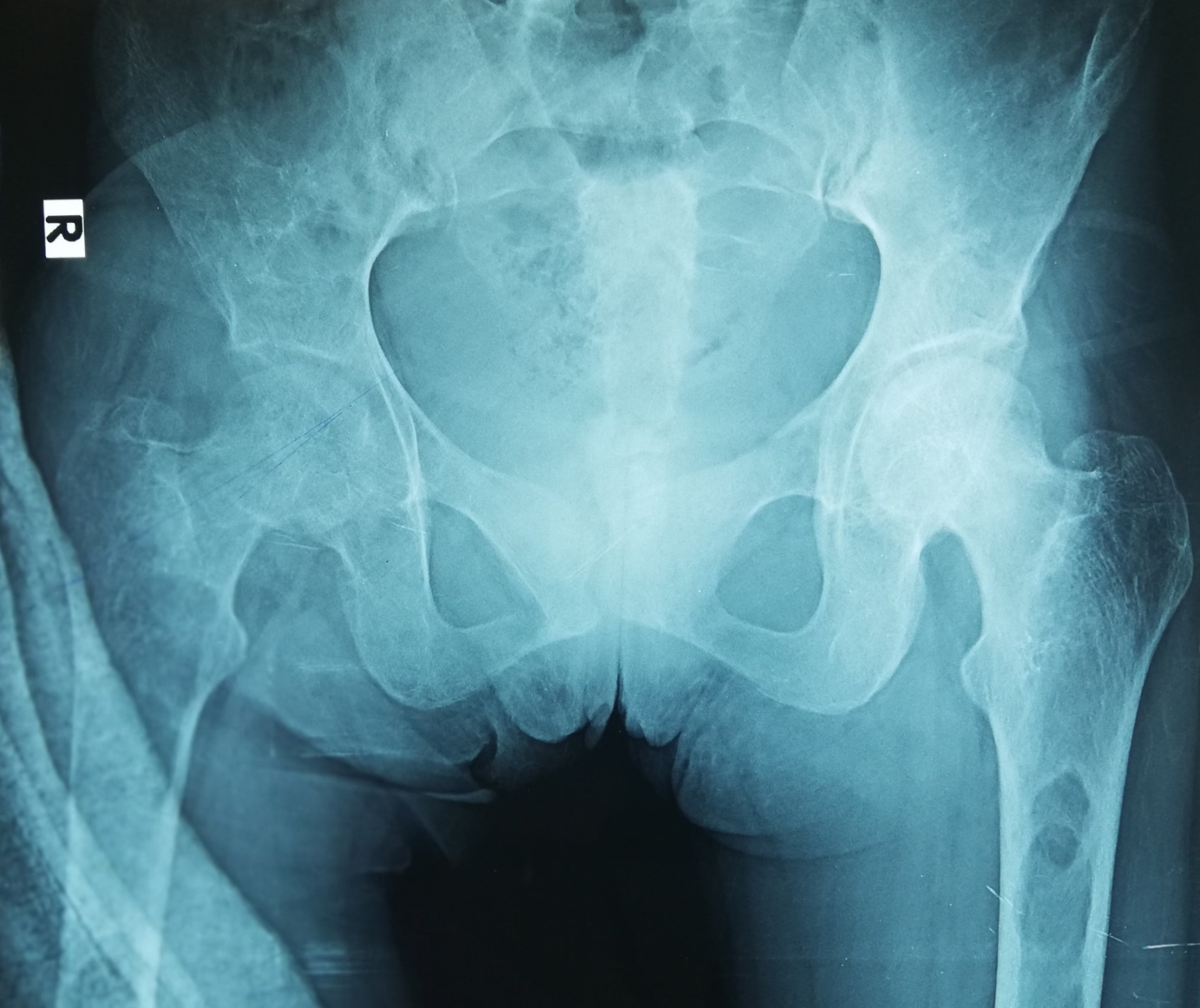
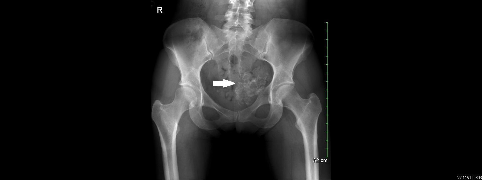
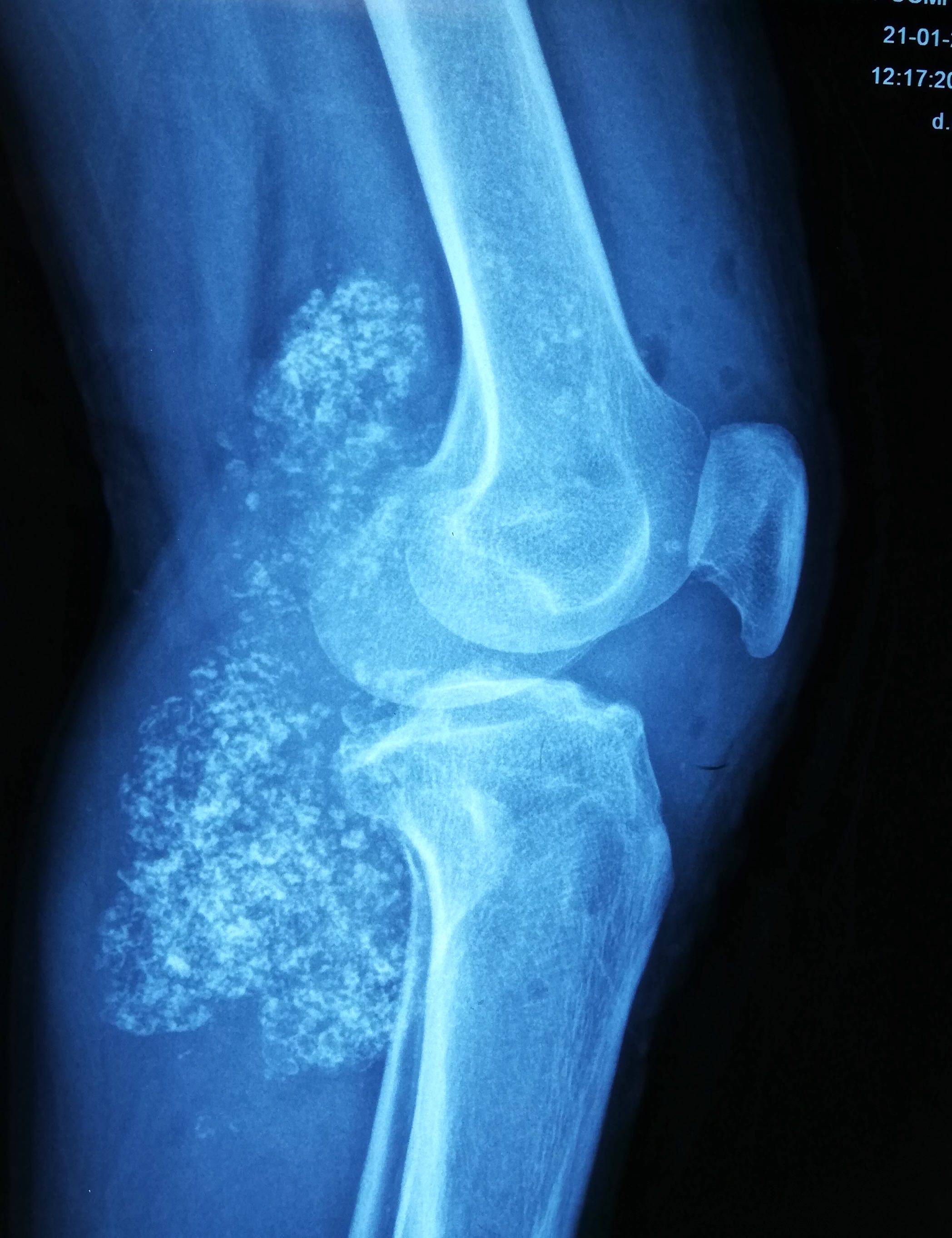
Contributed by Nasir Ud Din, M.B.B.S., Mark R. Wick, M.D. and AFIP images
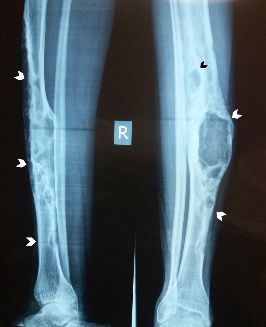
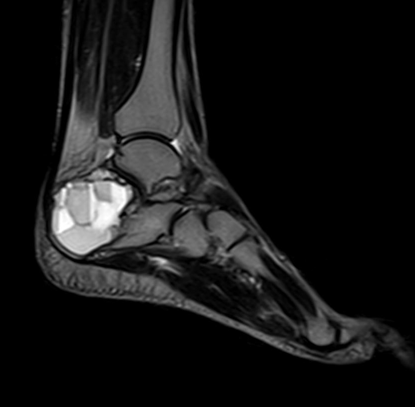
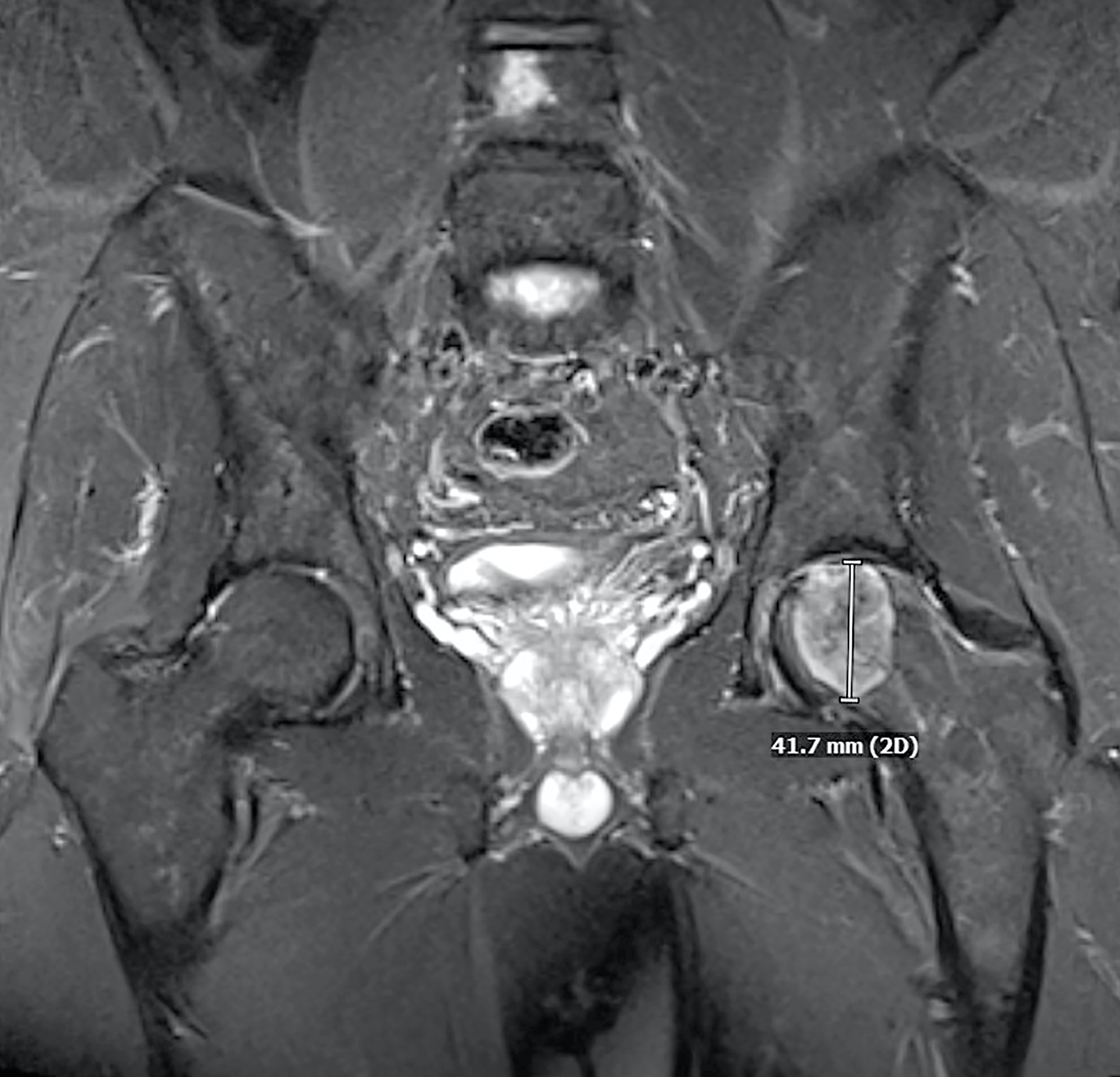
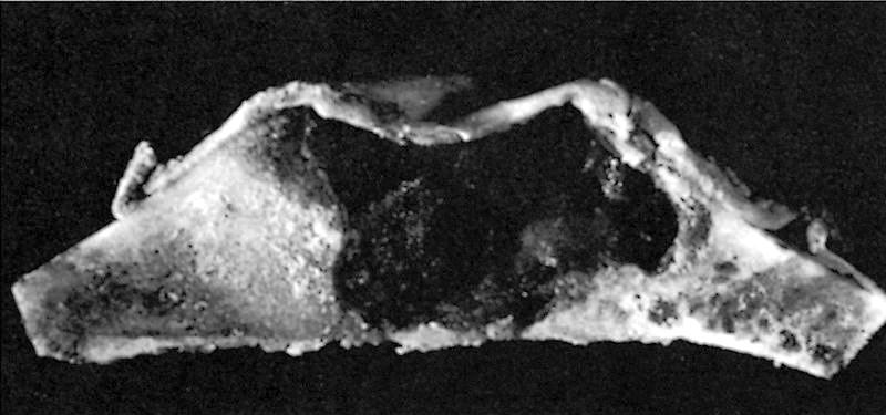
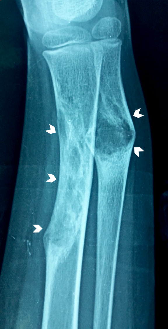
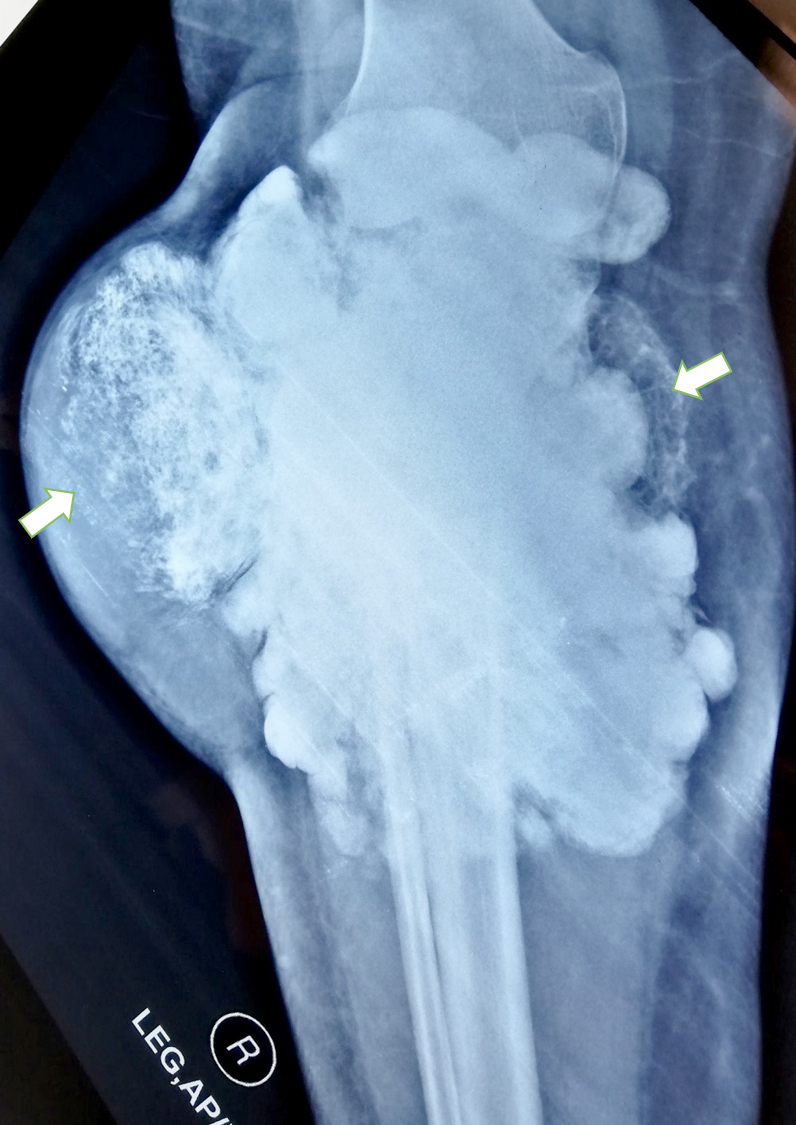
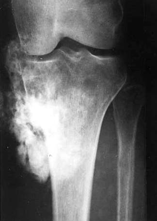
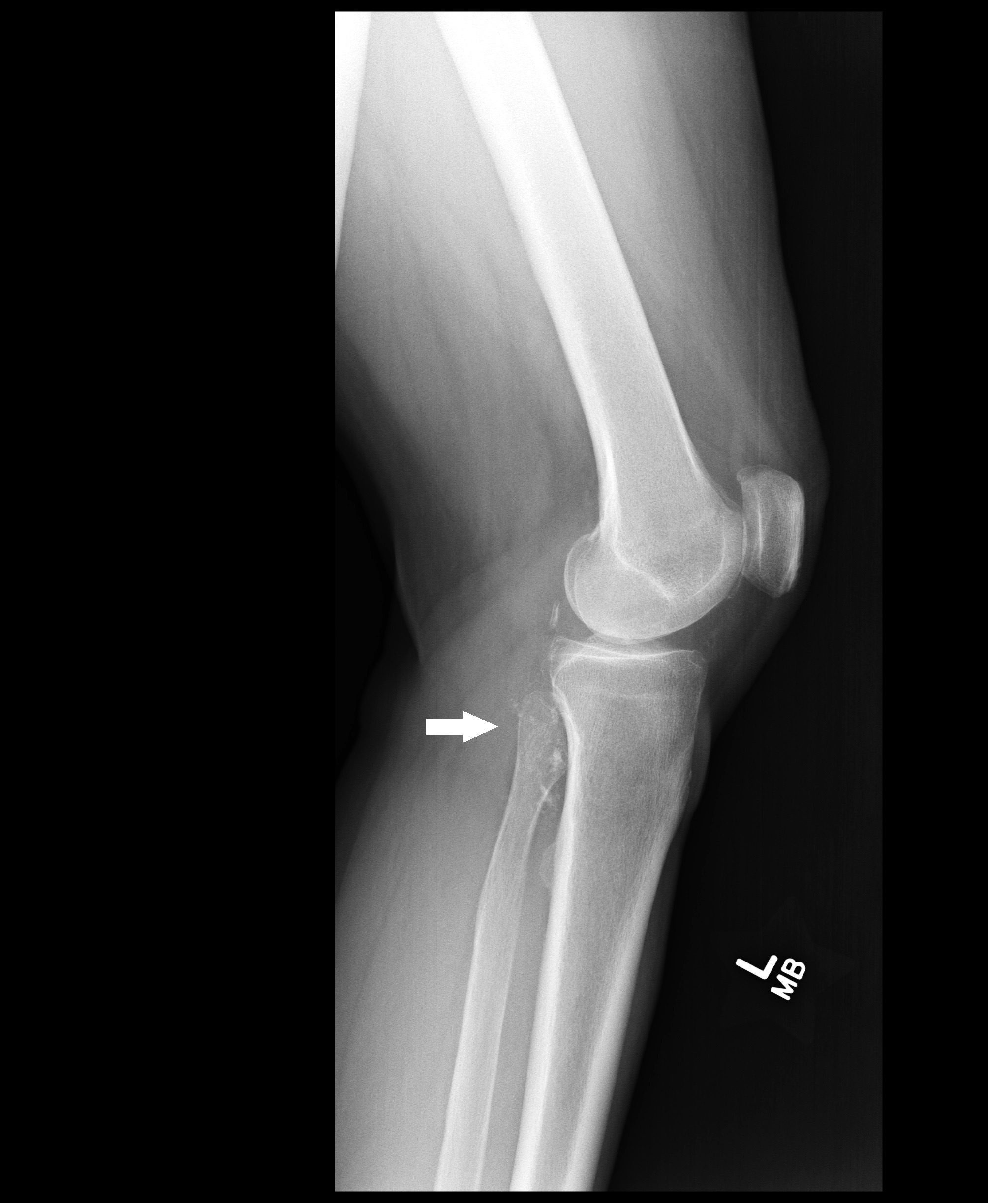
Multiloculated radiolucent lesion in tibia
Multiloculation and sclerotic borders

Thickened sclerotic areas
%20xray.jpg)
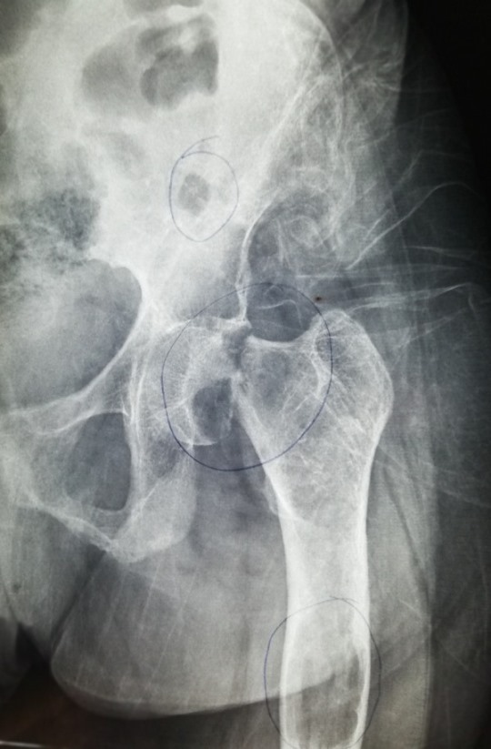
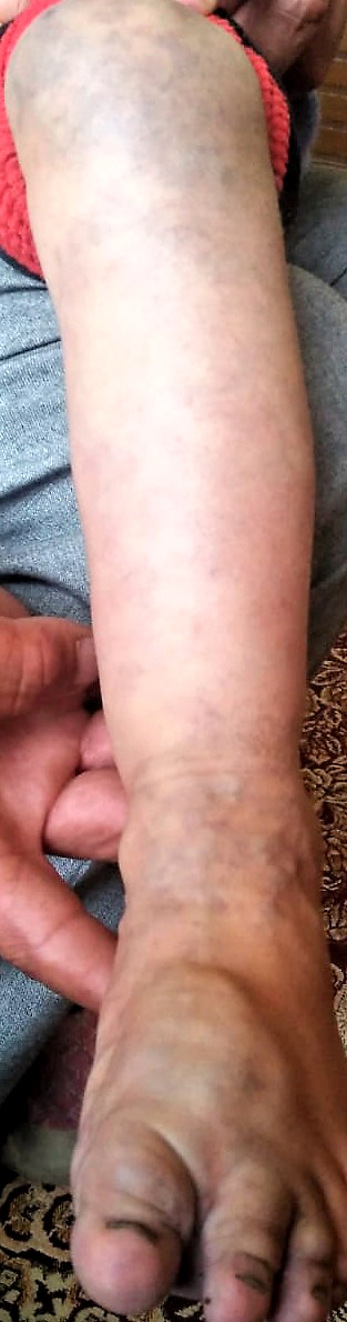
Marked bowing deformity
Synchronous tumors in tibia and fibula
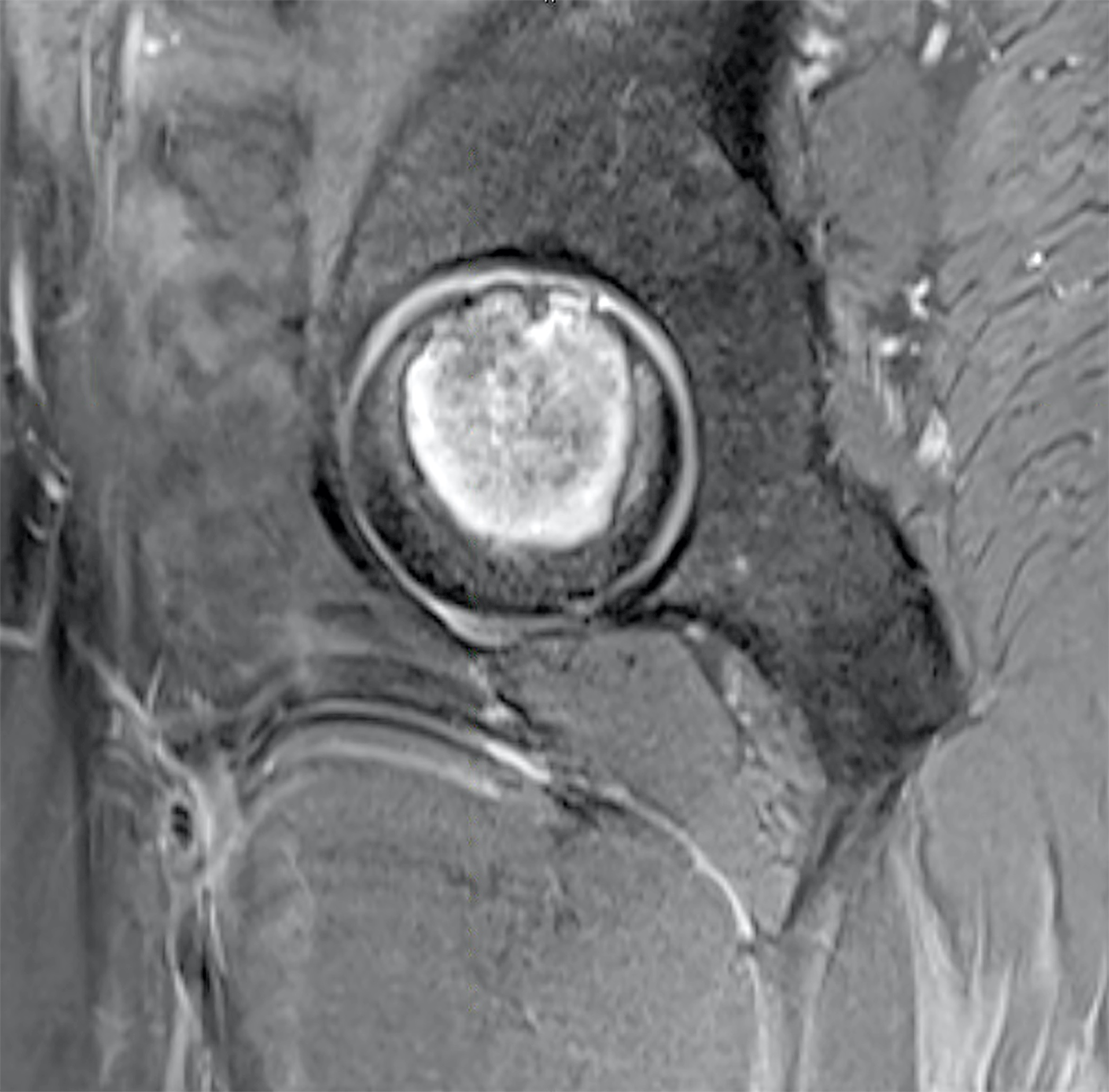
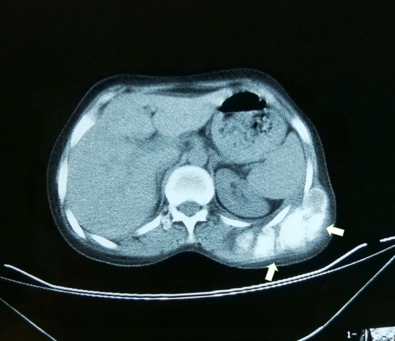
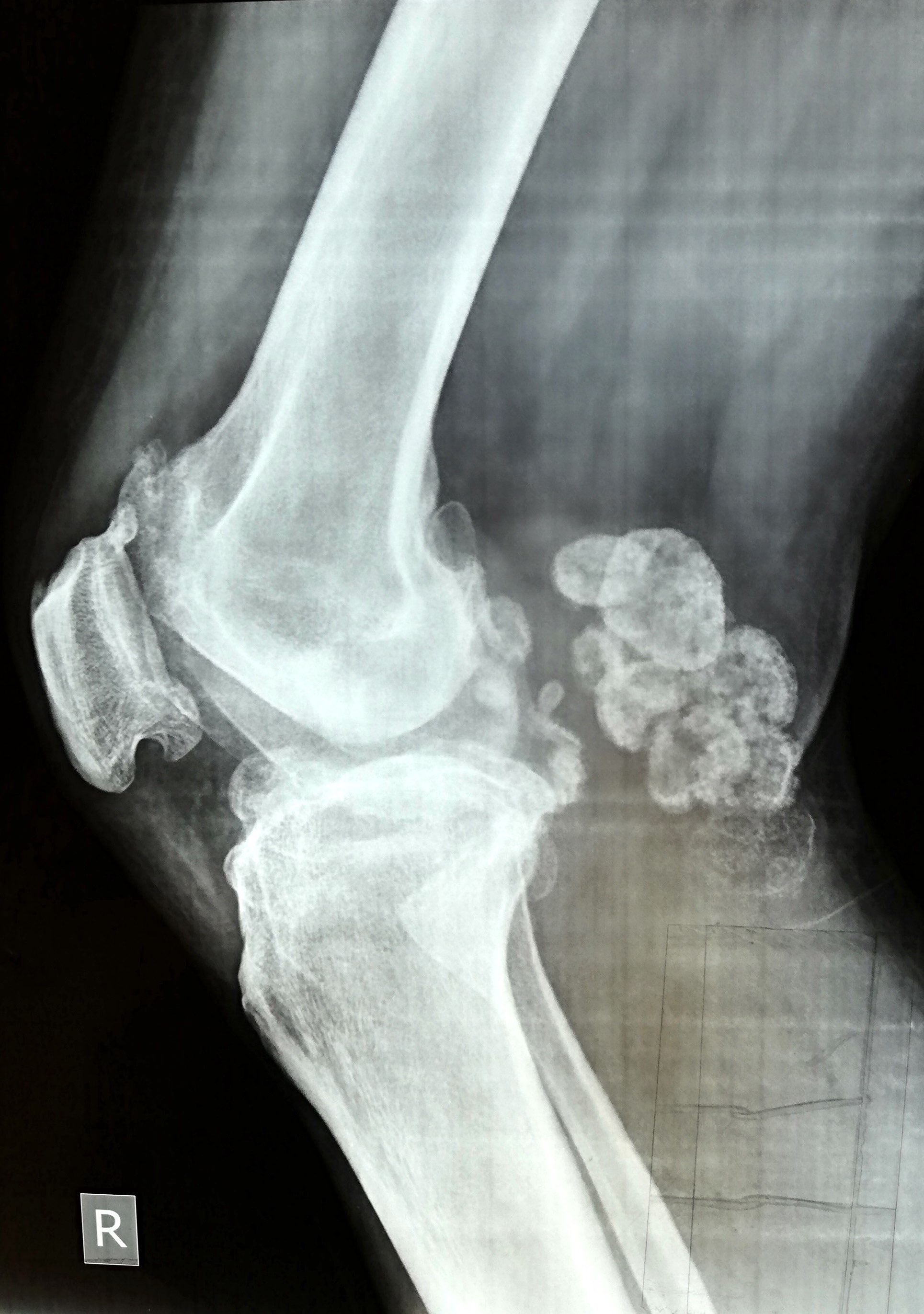
Contrast enhancing lesion on CT scan
Contrast enhancing lesion on MRI scan
%20xray4.jpg)
Radiolucent and sclerotic areas
Ill defined lucencies and bowing deformity
Images hosted on other servers:

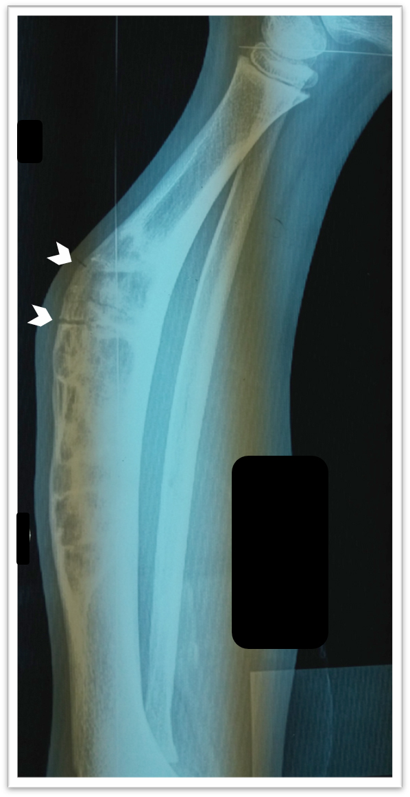
Soft tissue and medullary cavity involvement

Soap bubble appearance on CT scan

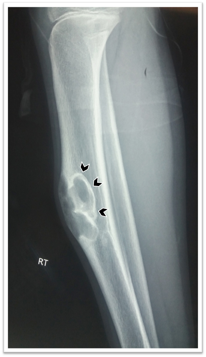
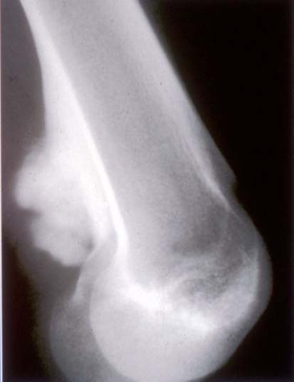
Cortical lesion with sclerotic rim
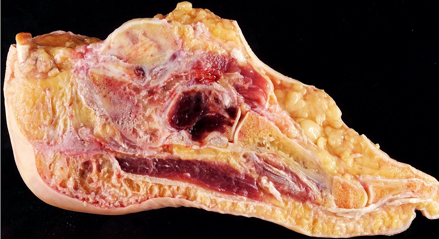
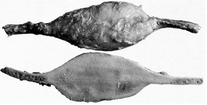
Contributed by Mark R. Wick, M.D. and AFIP images
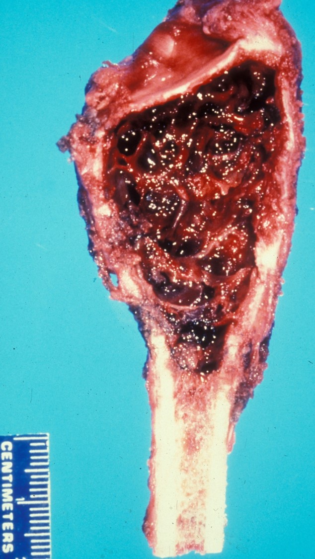
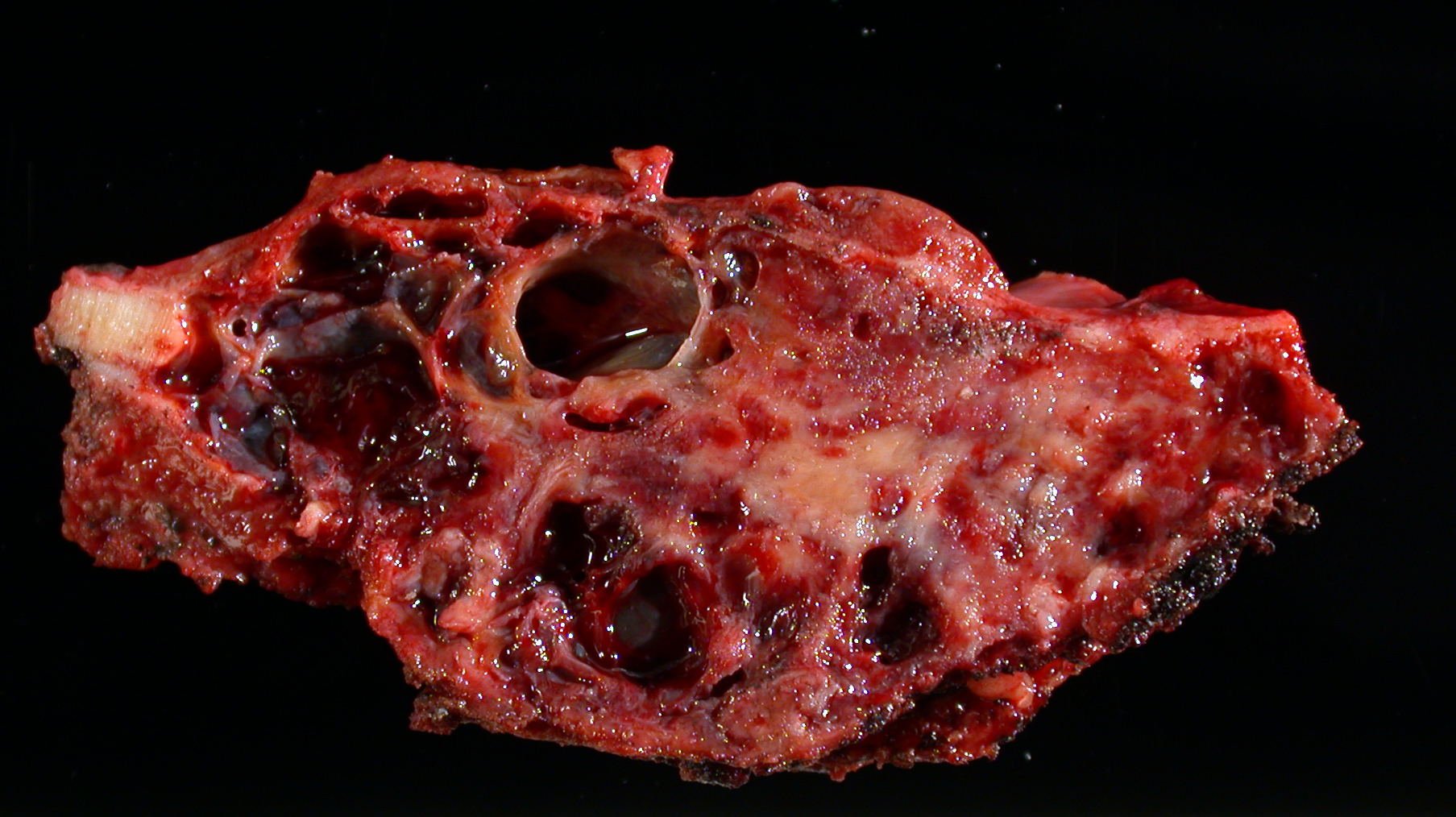
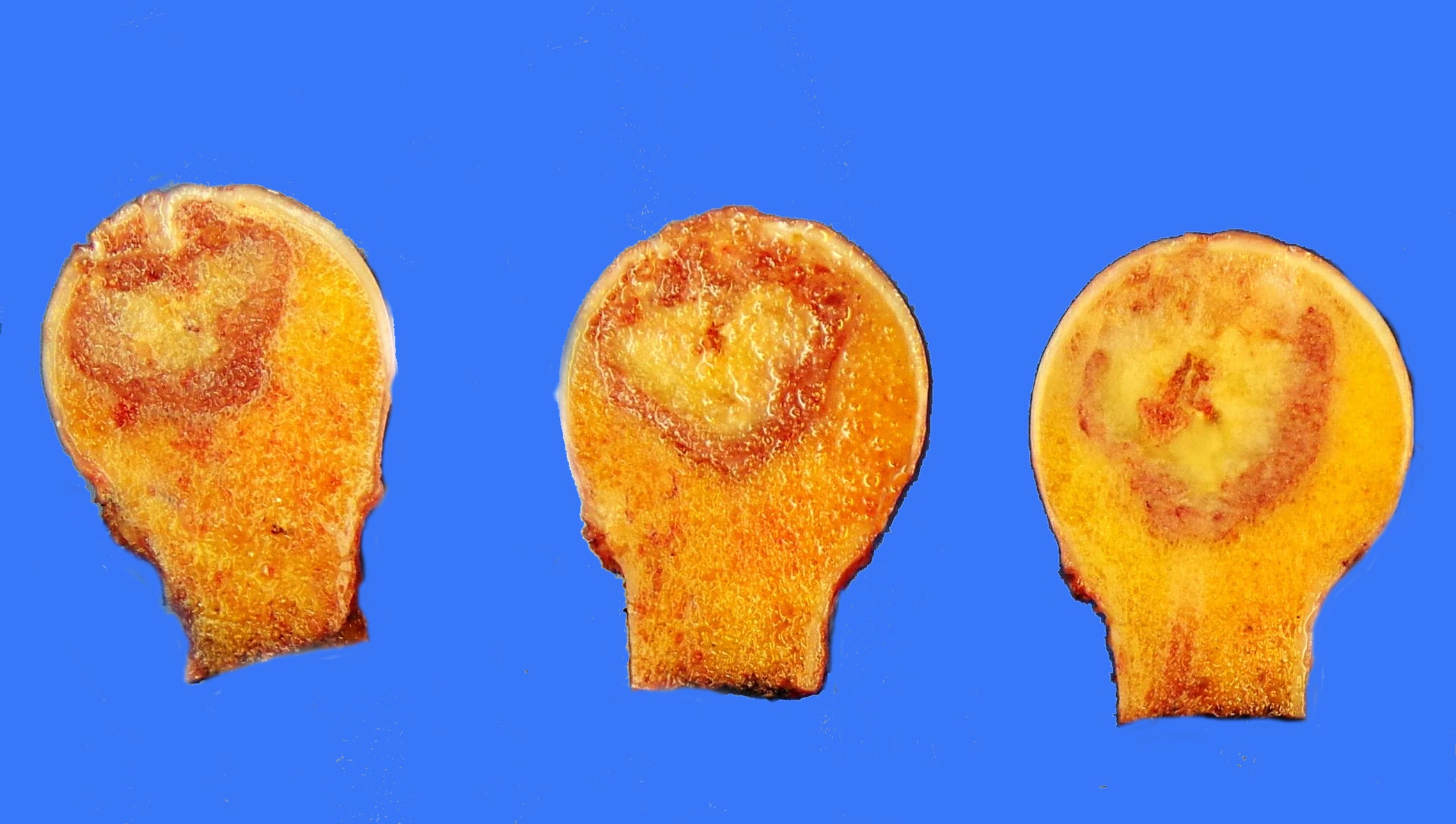
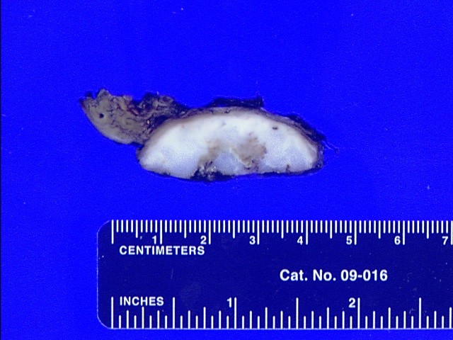
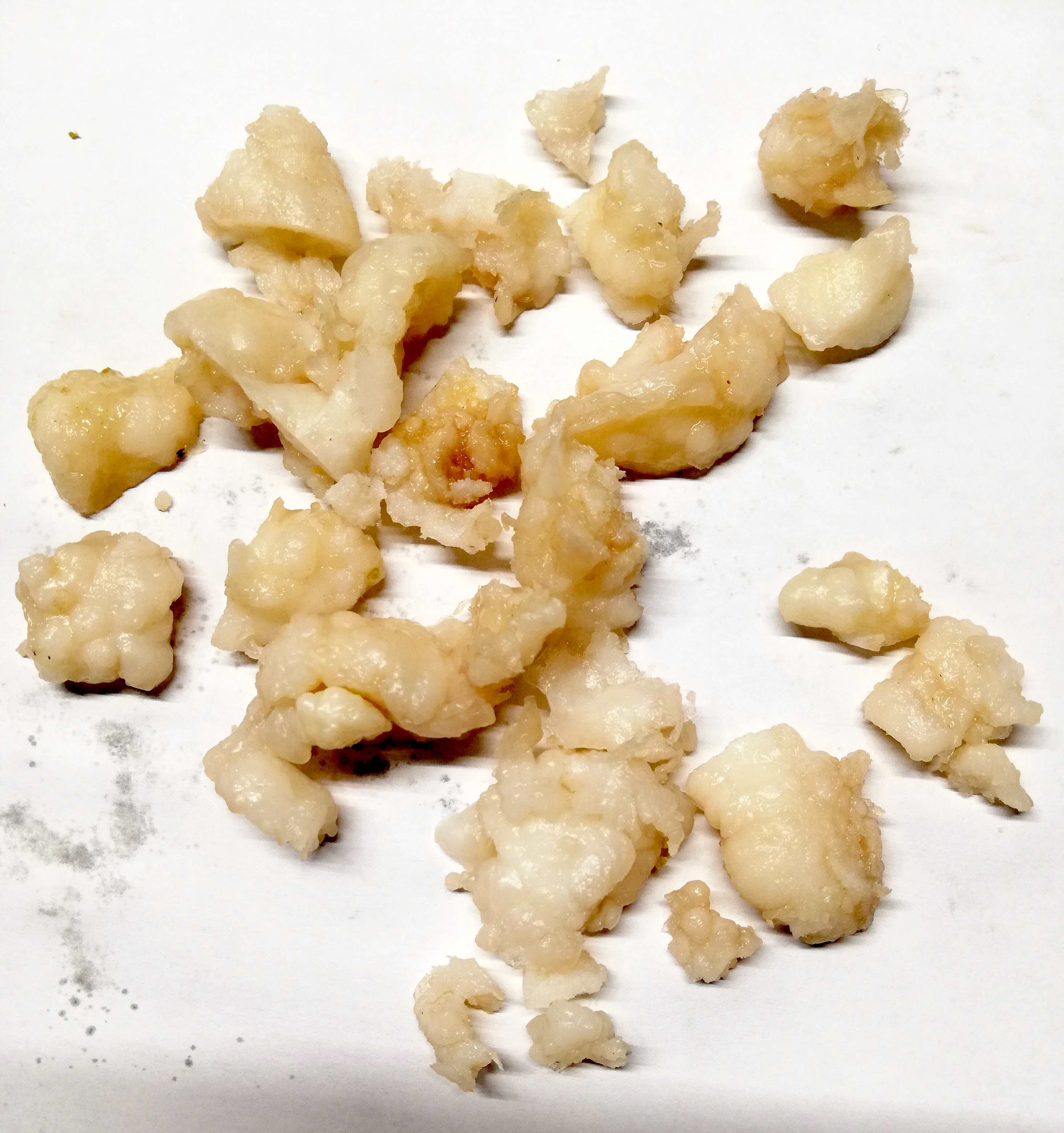
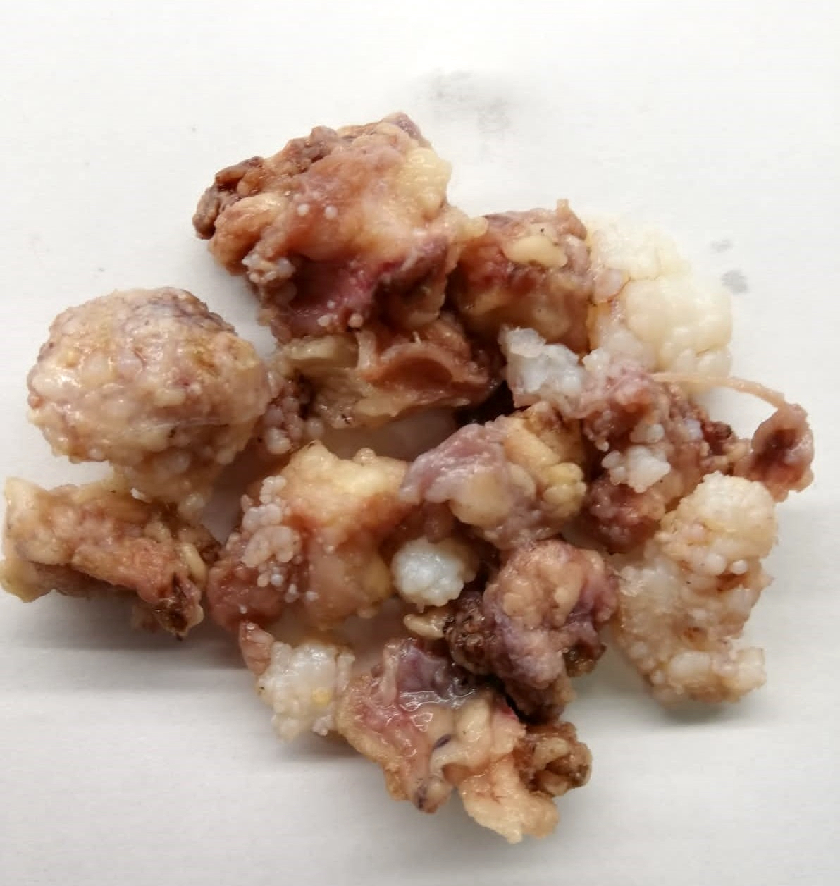
%20gross.jpg)
Solid tumor involving cortex and medulla
Focally eroded cortex
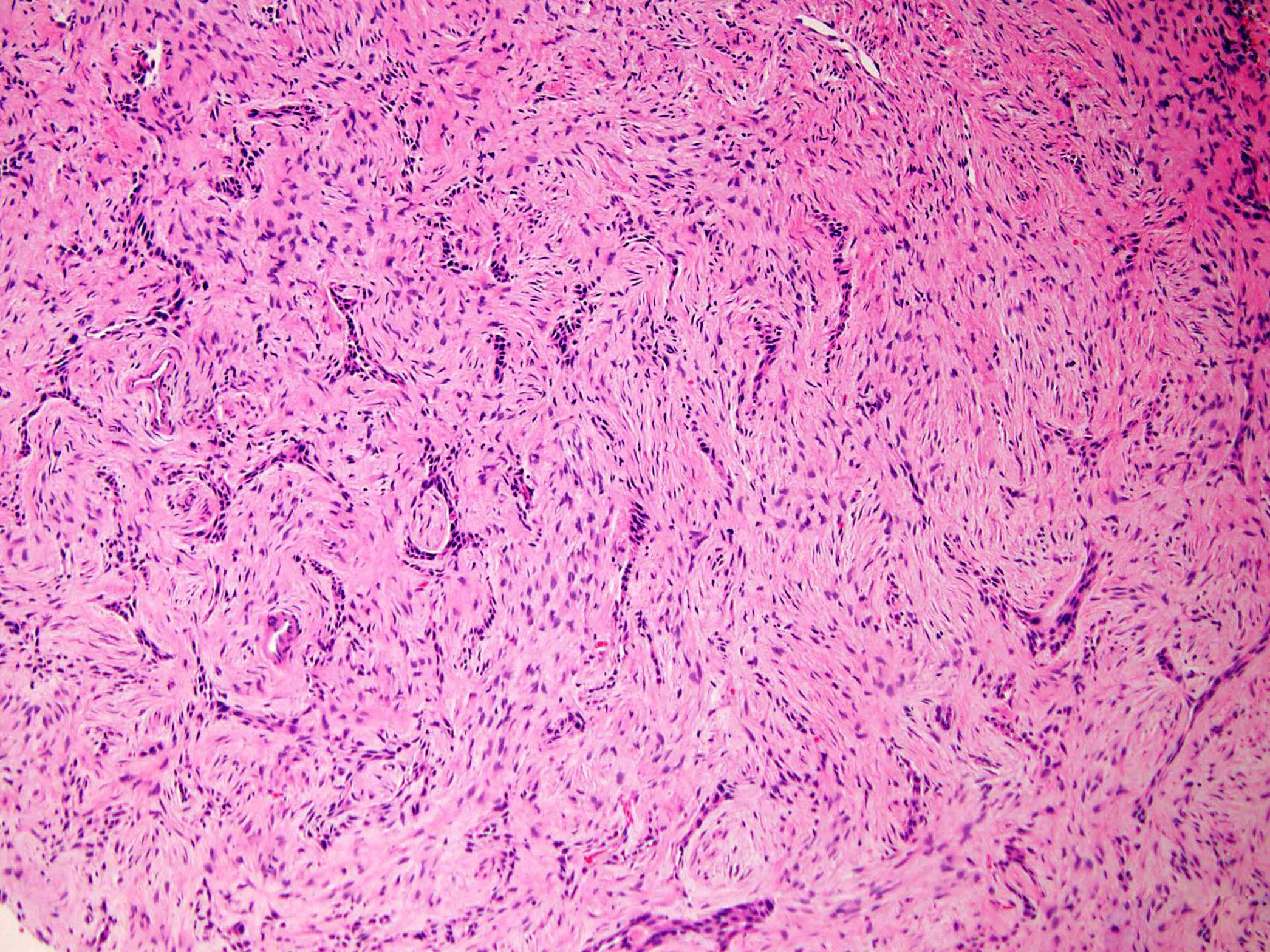
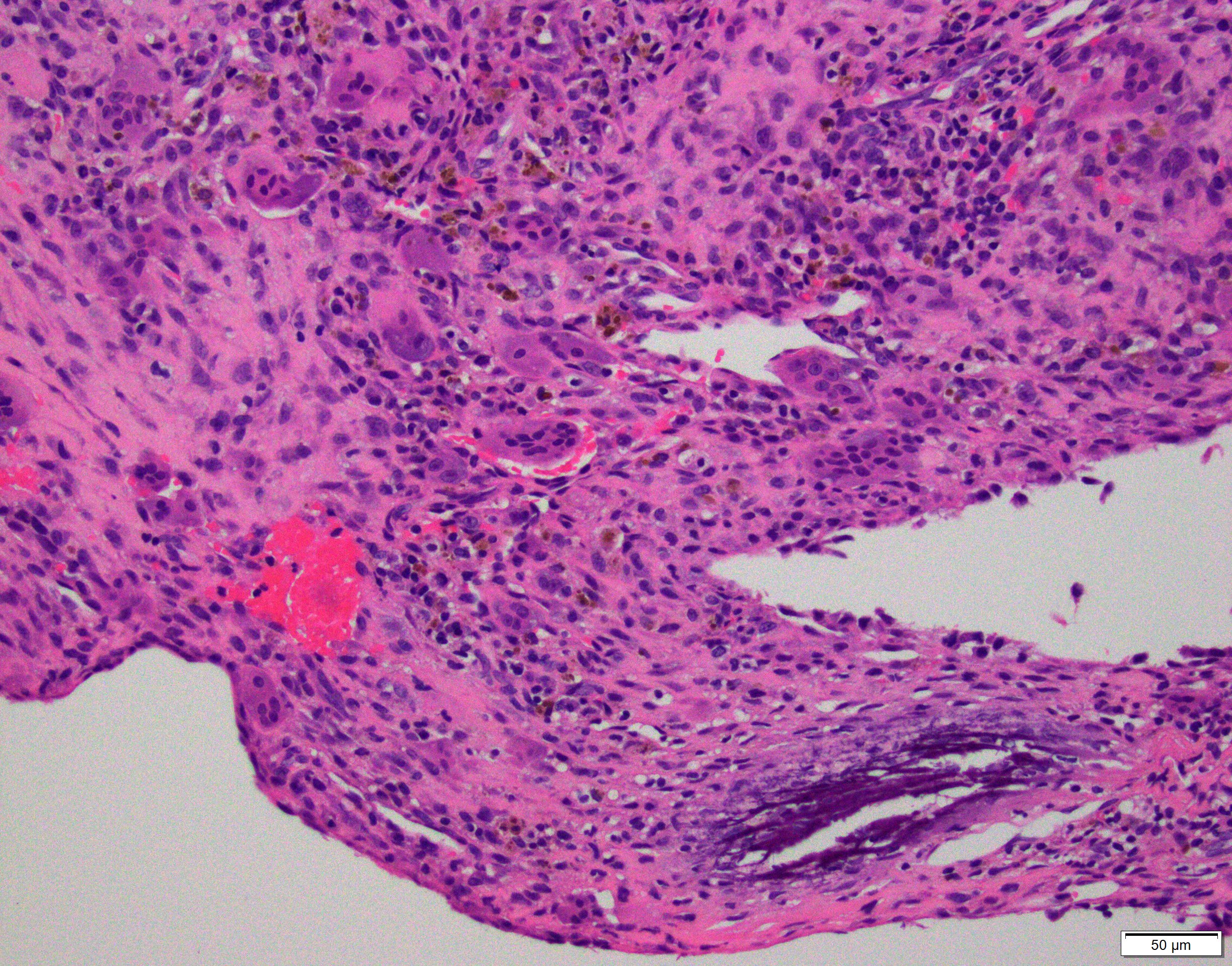
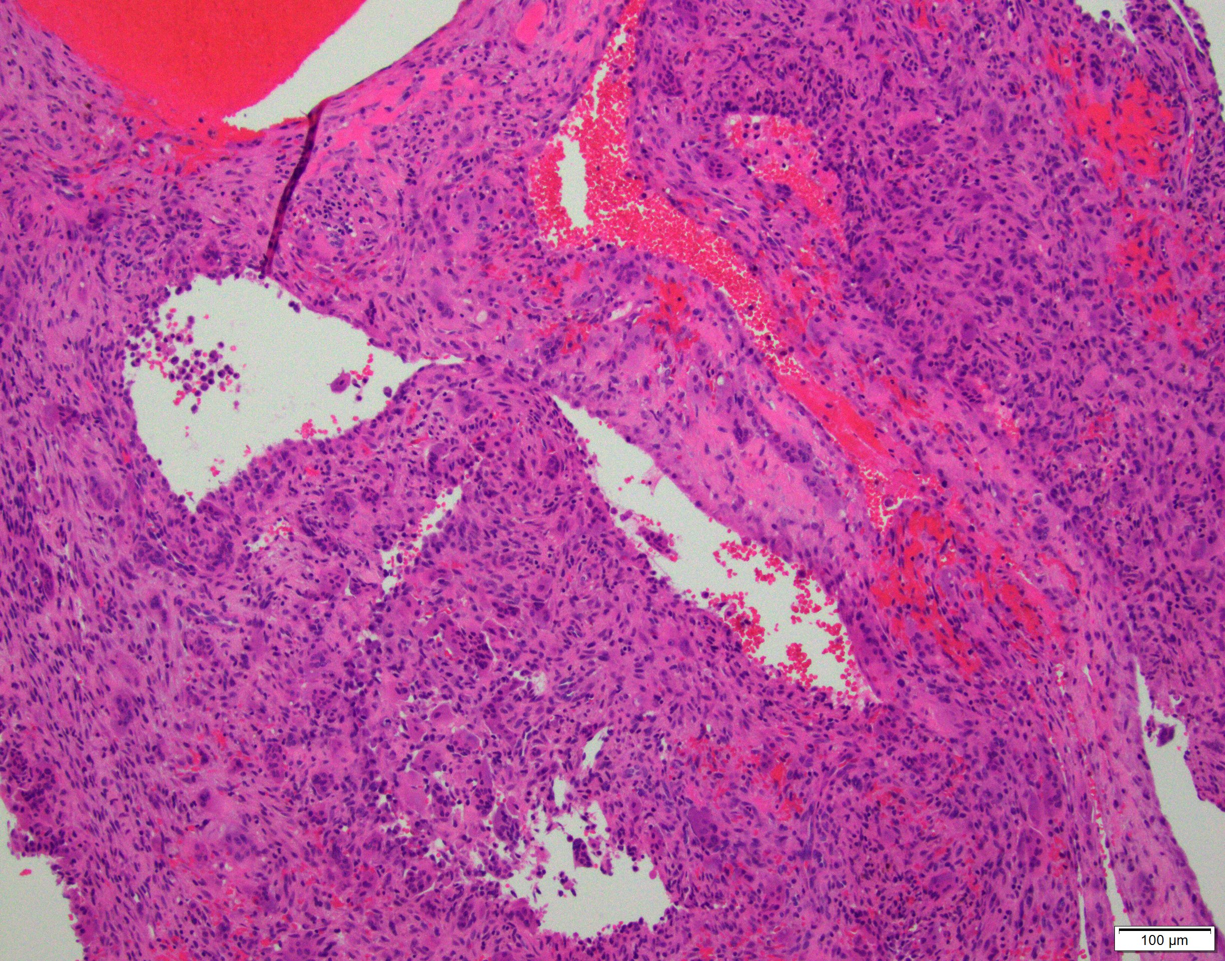
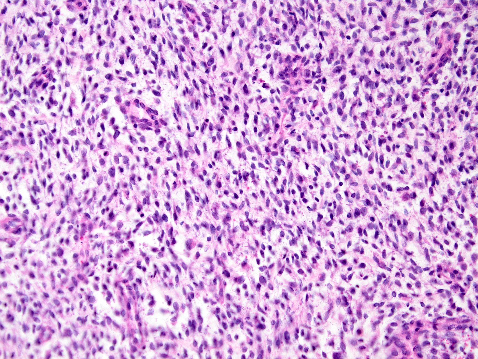
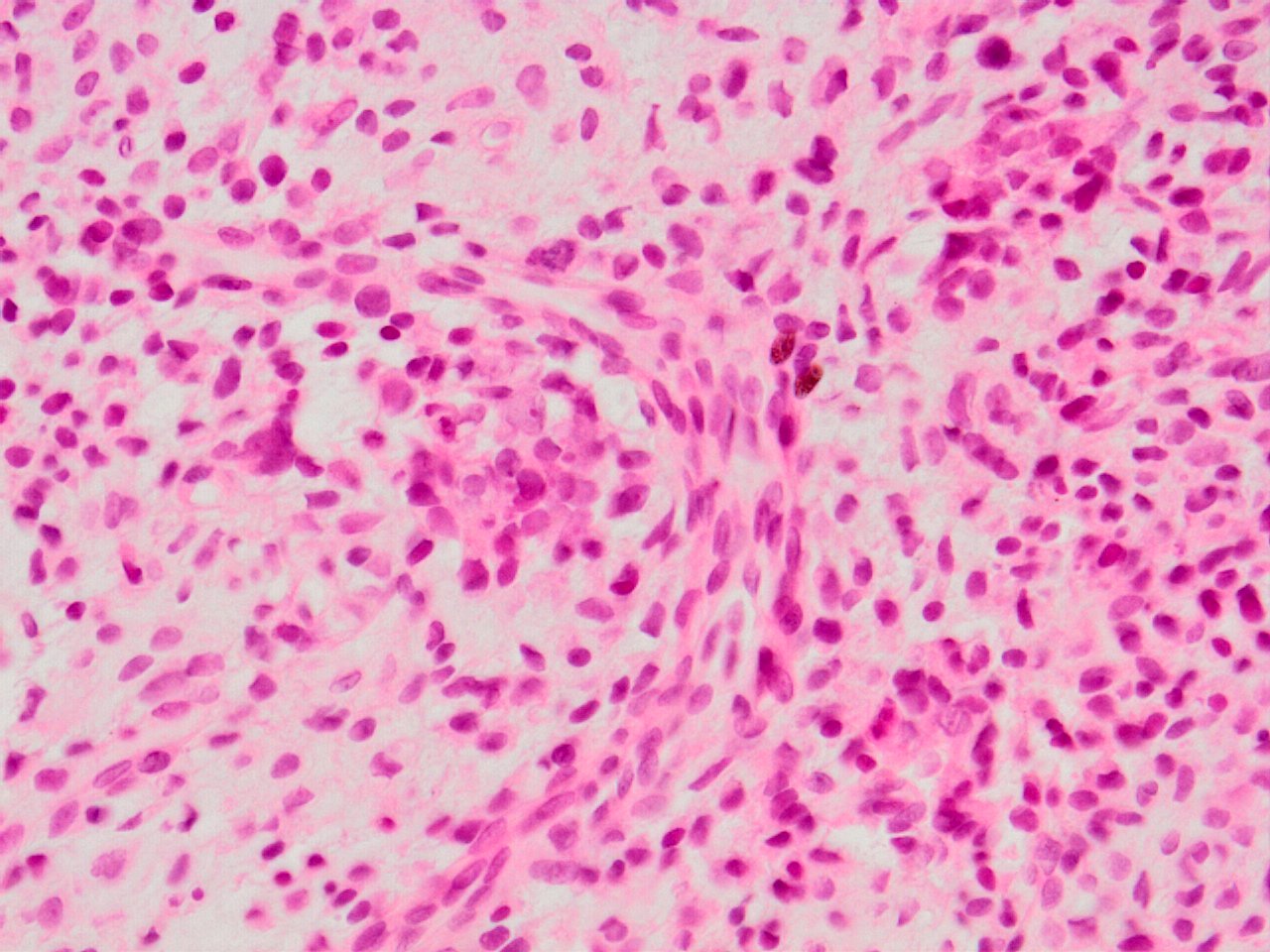
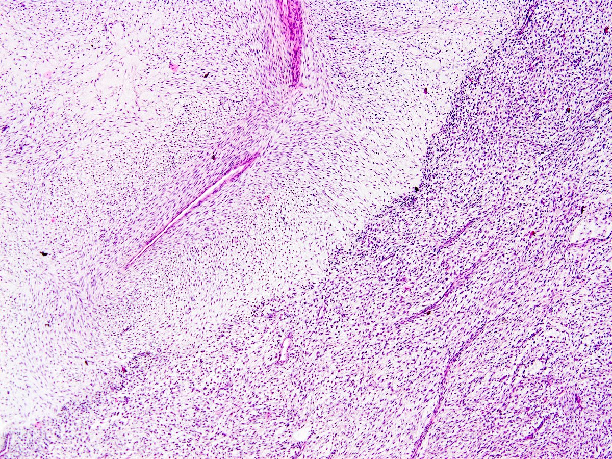
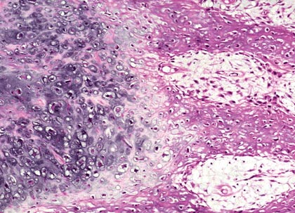
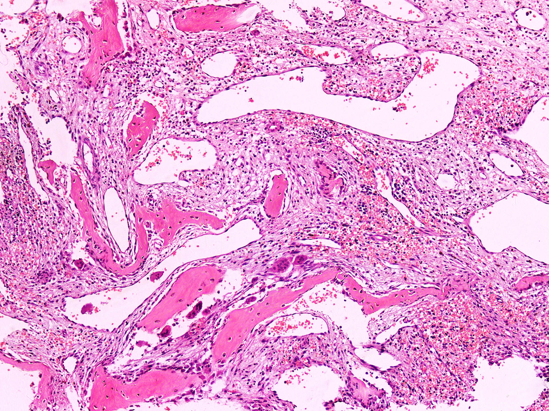
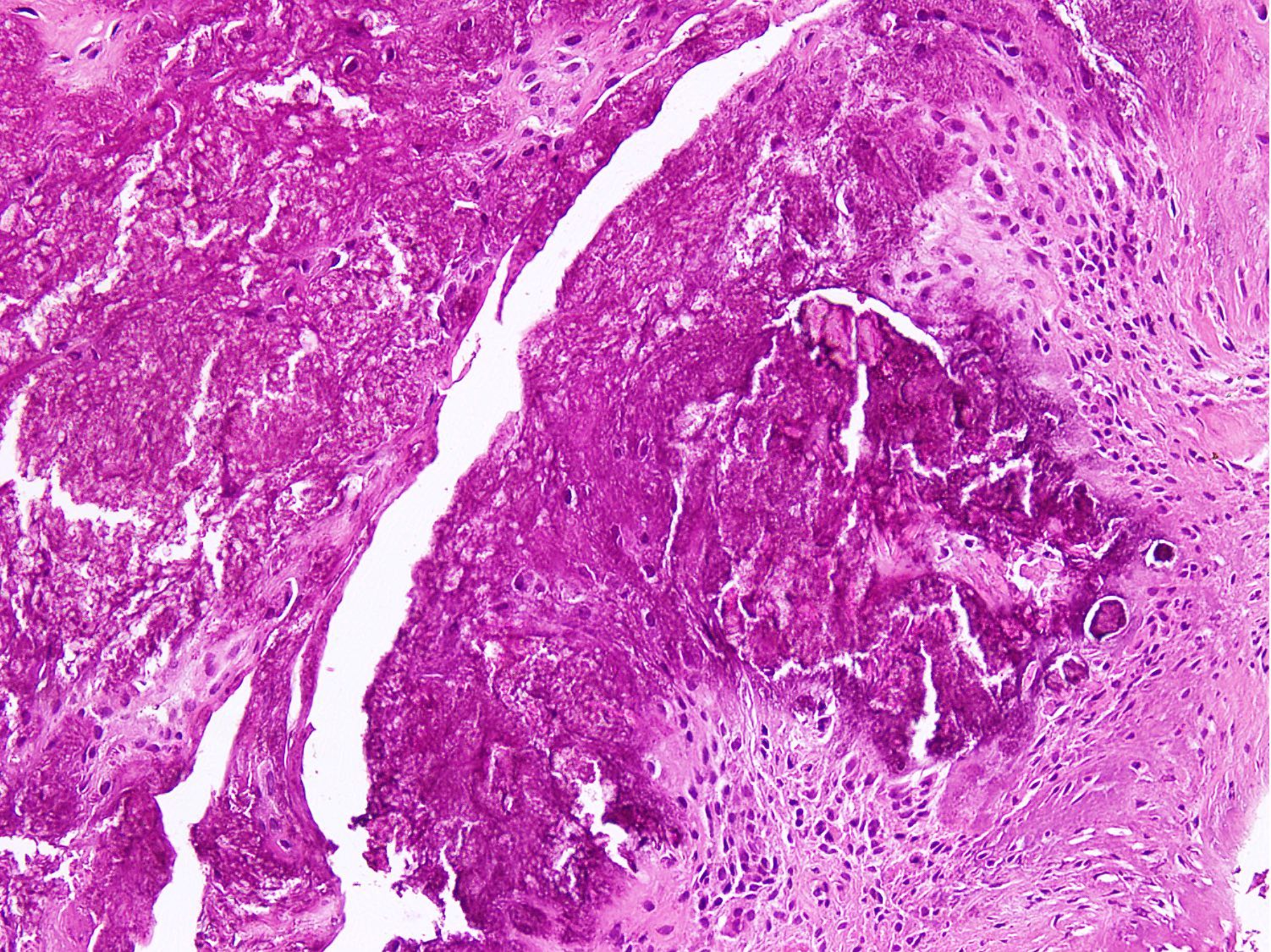
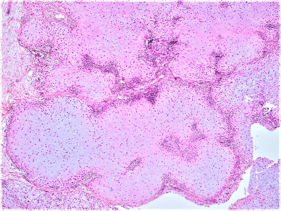
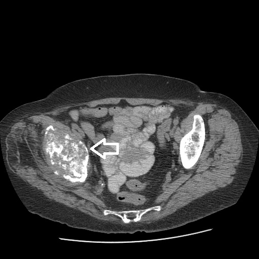
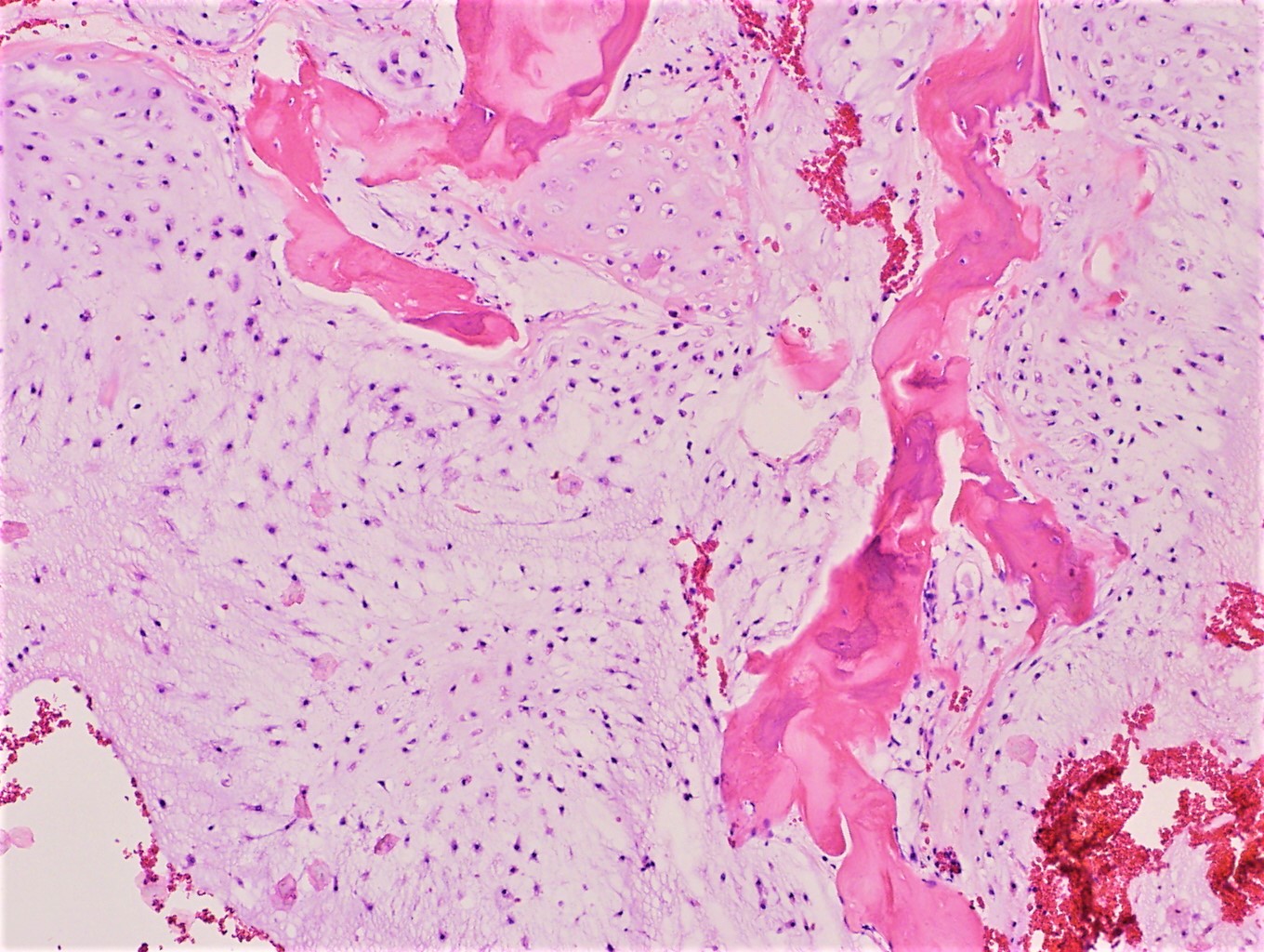
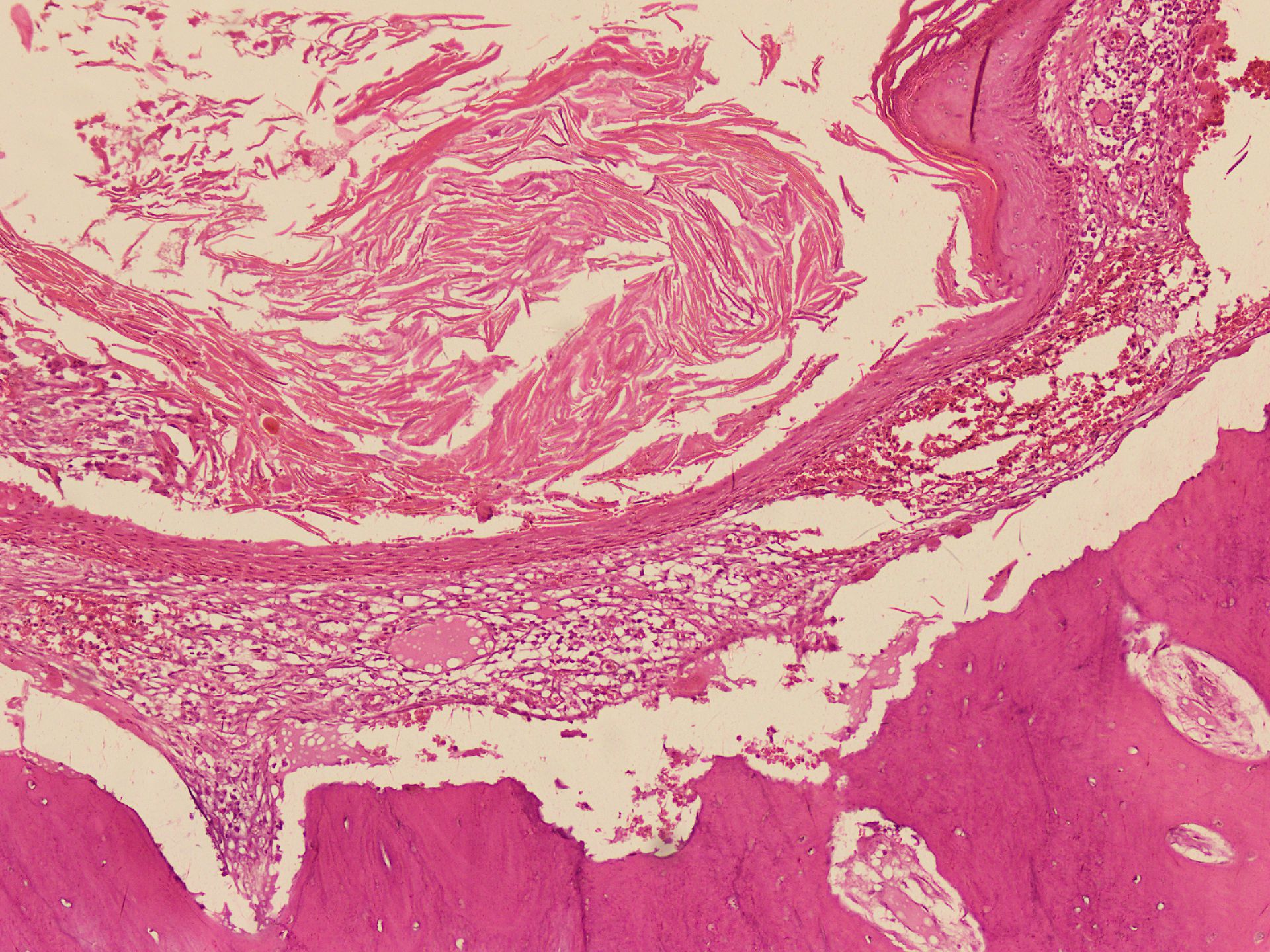
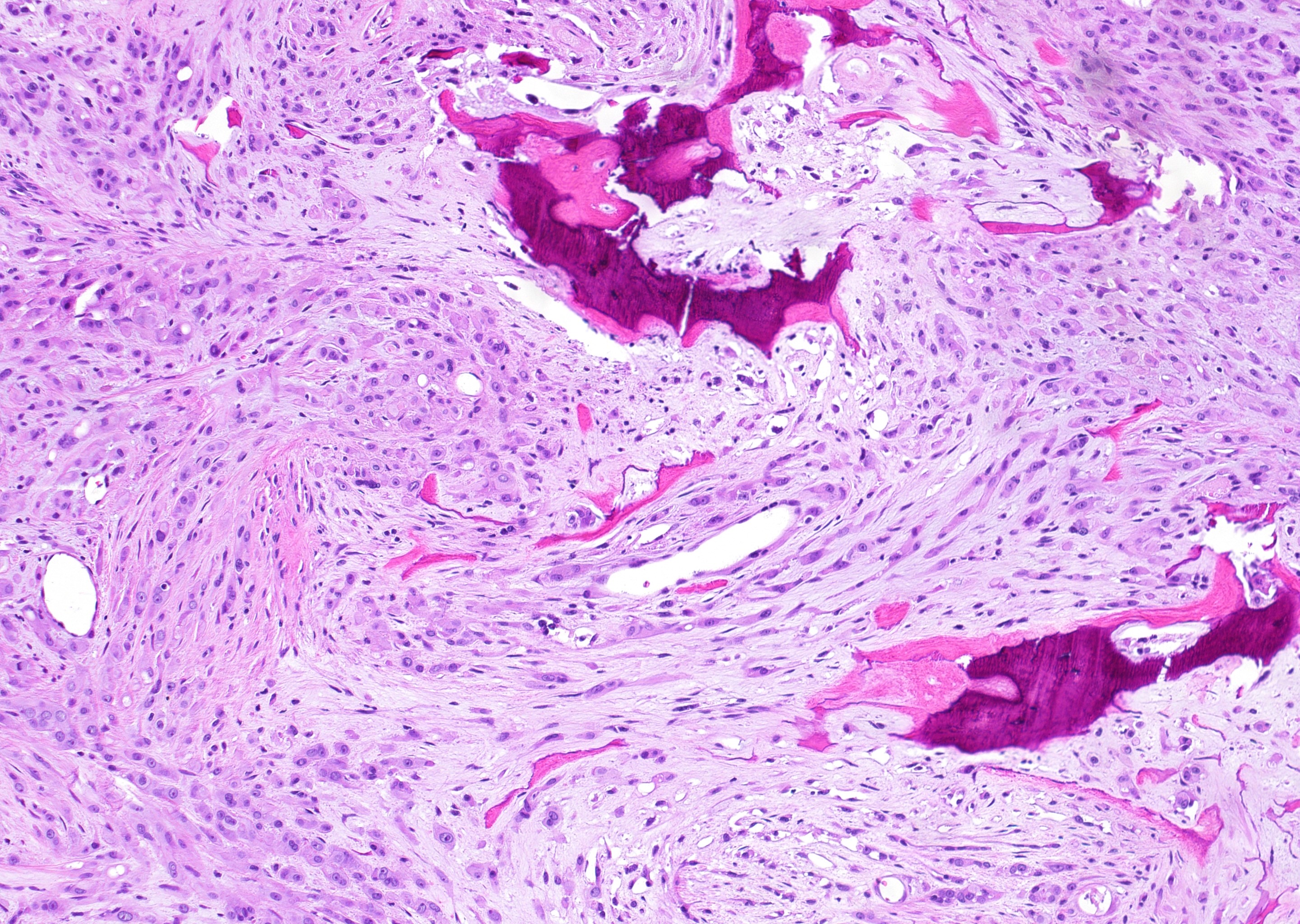
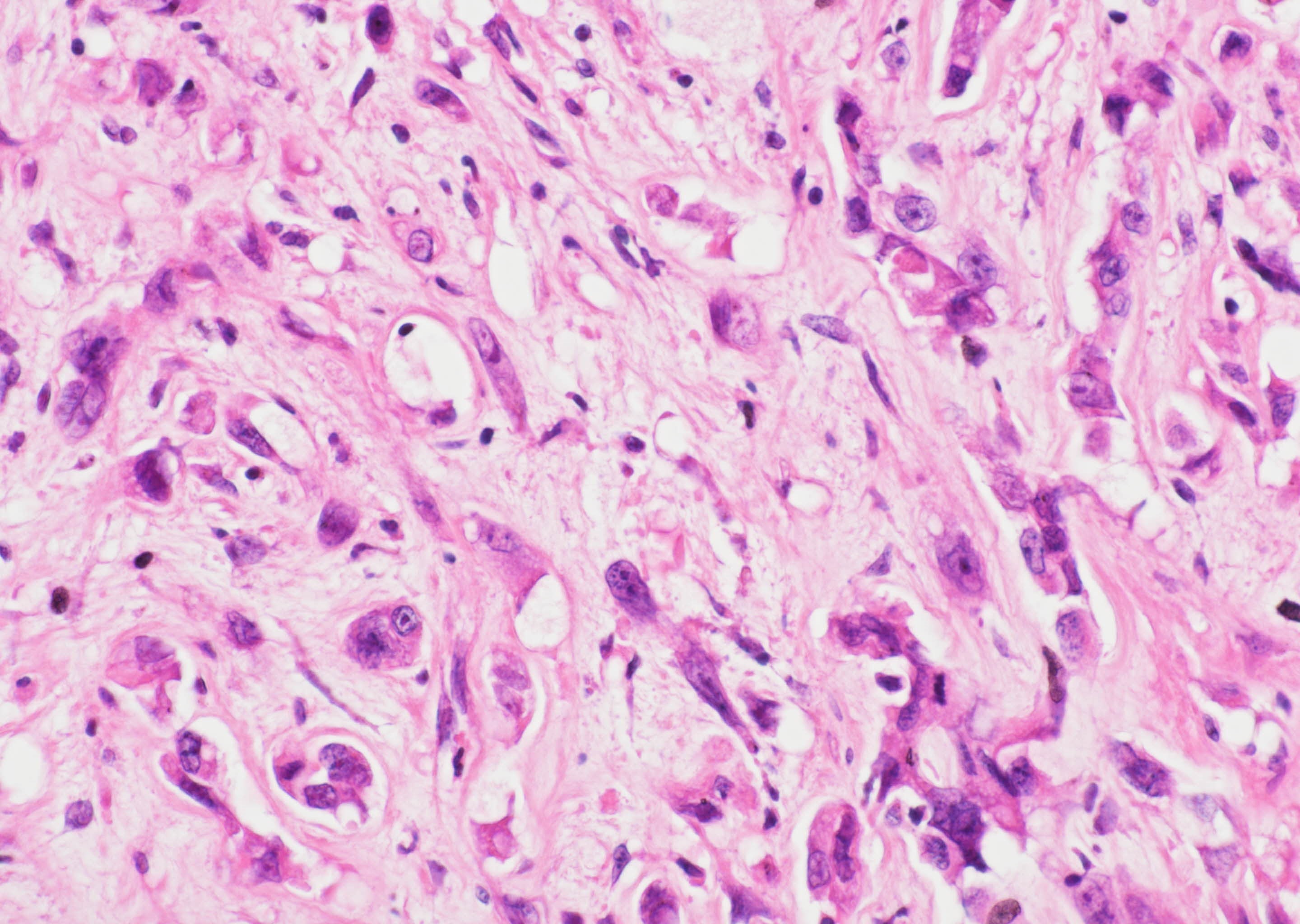
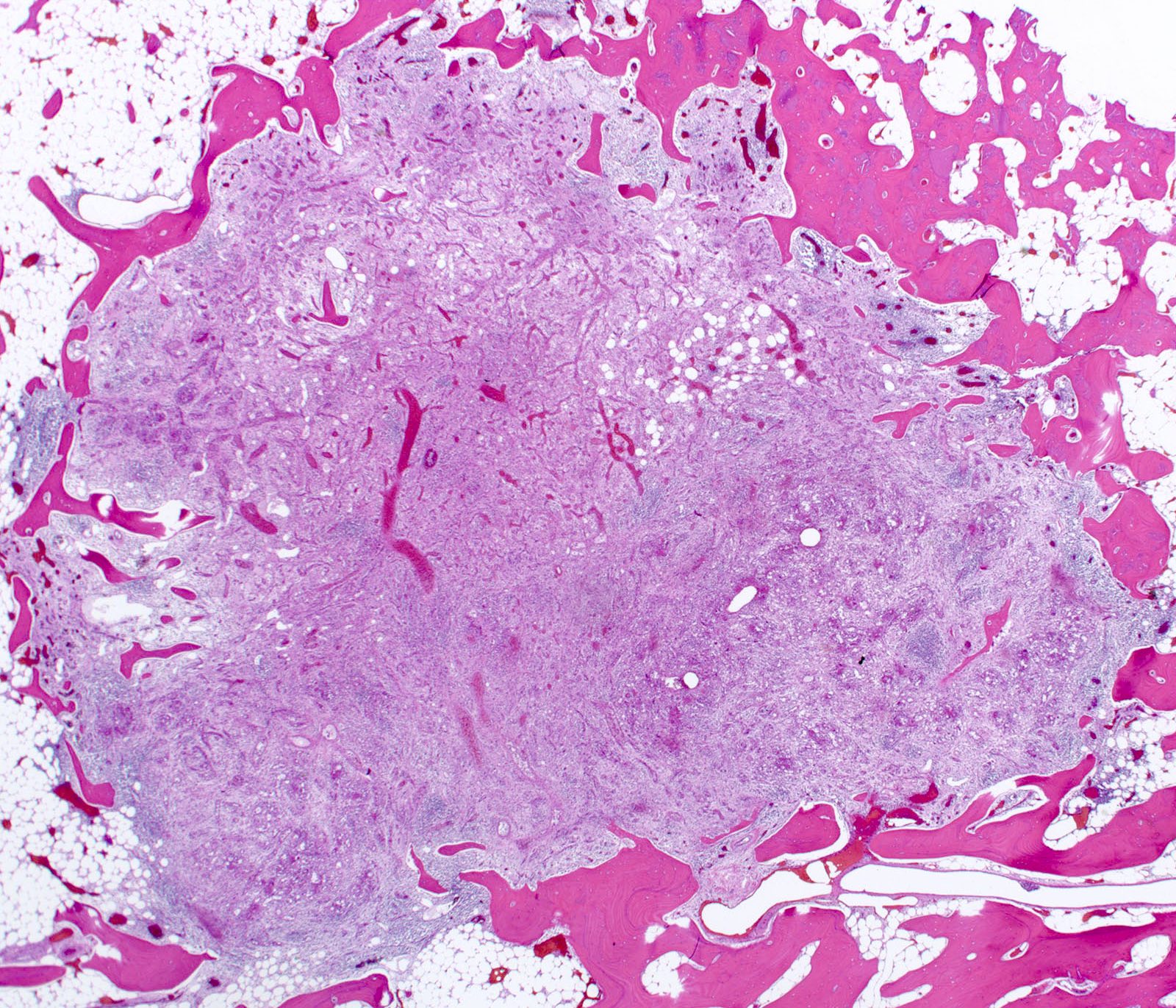
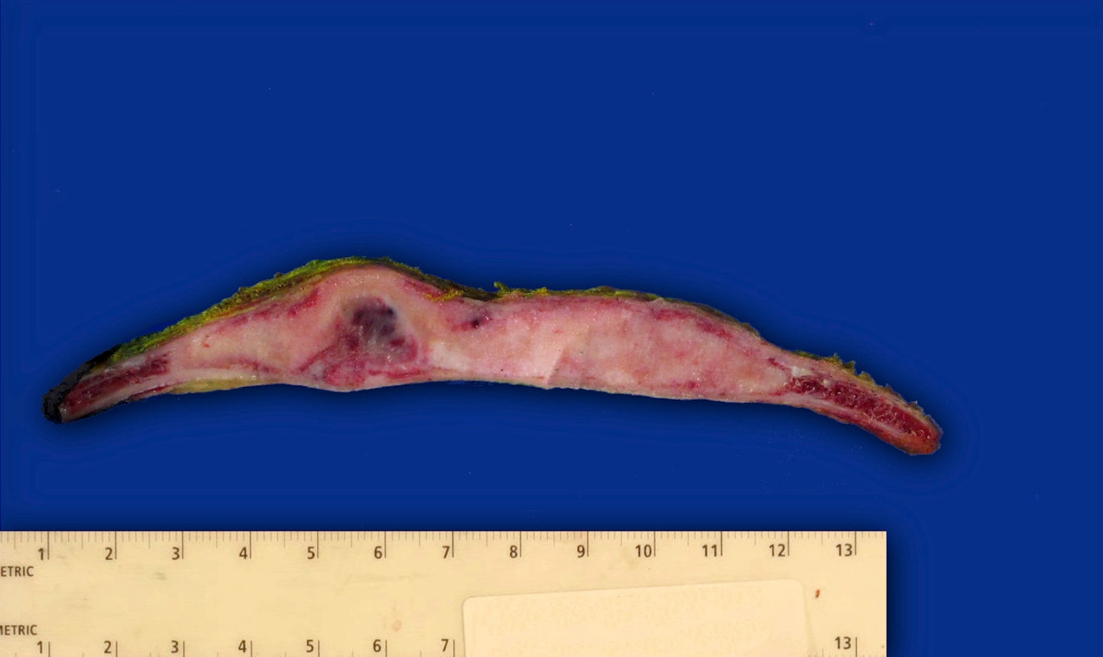
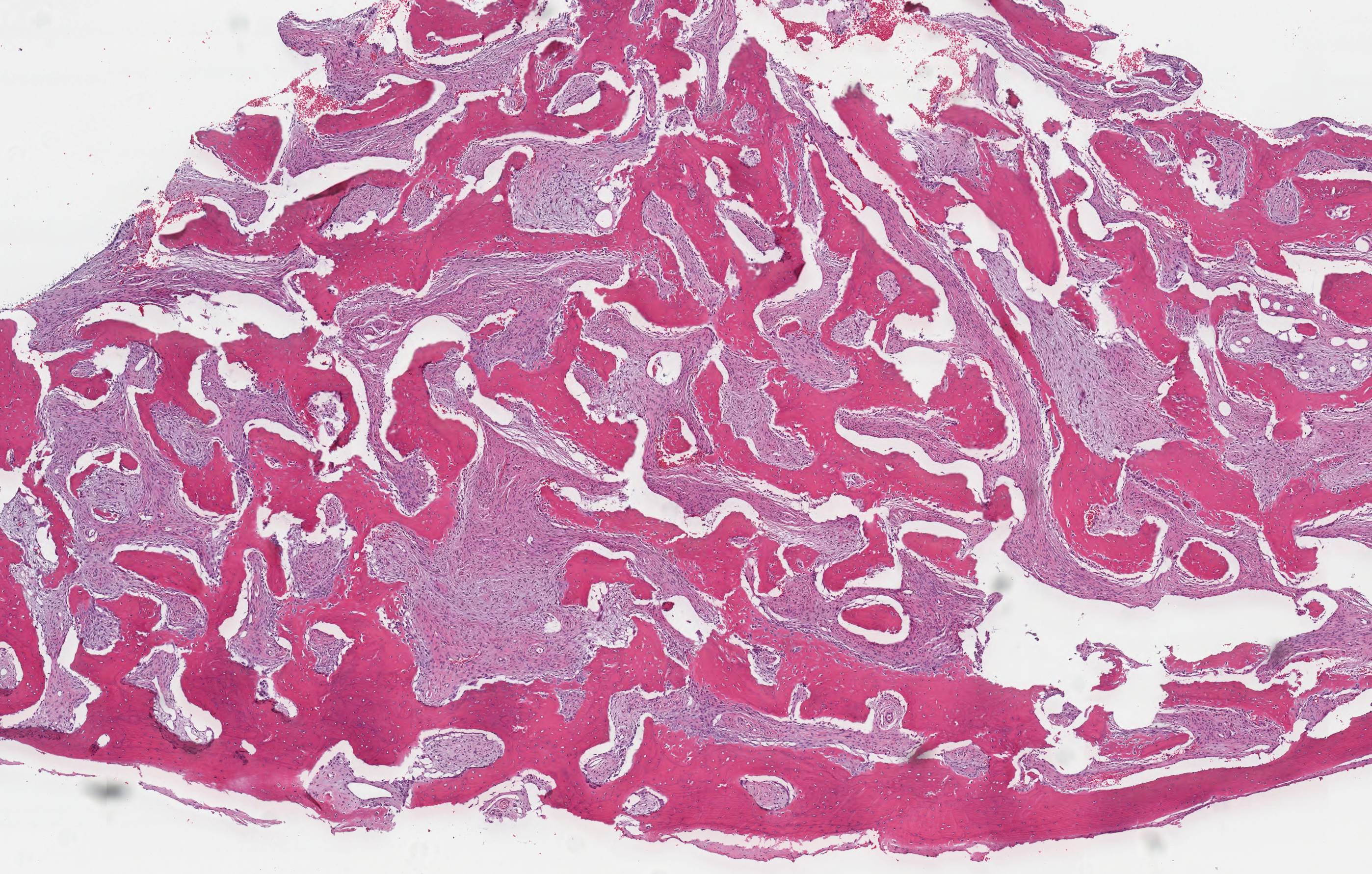
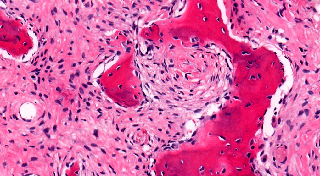
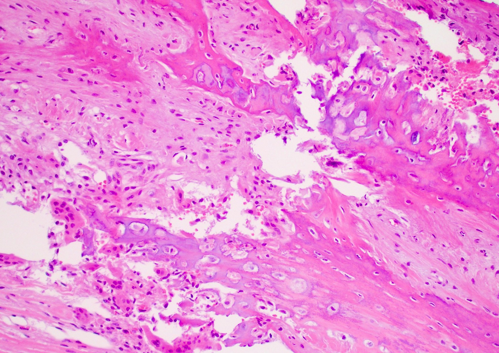
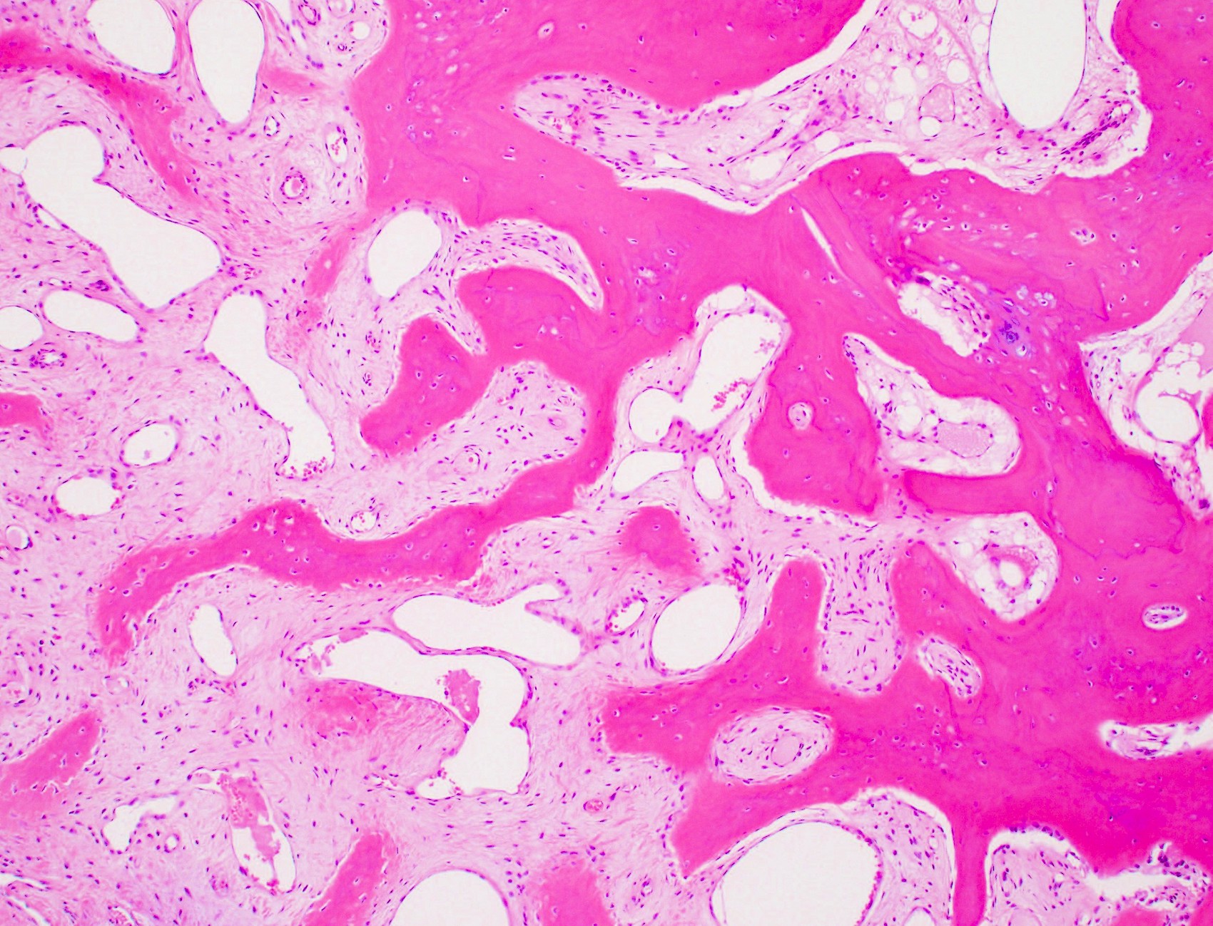
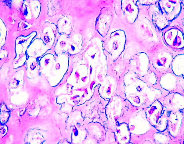
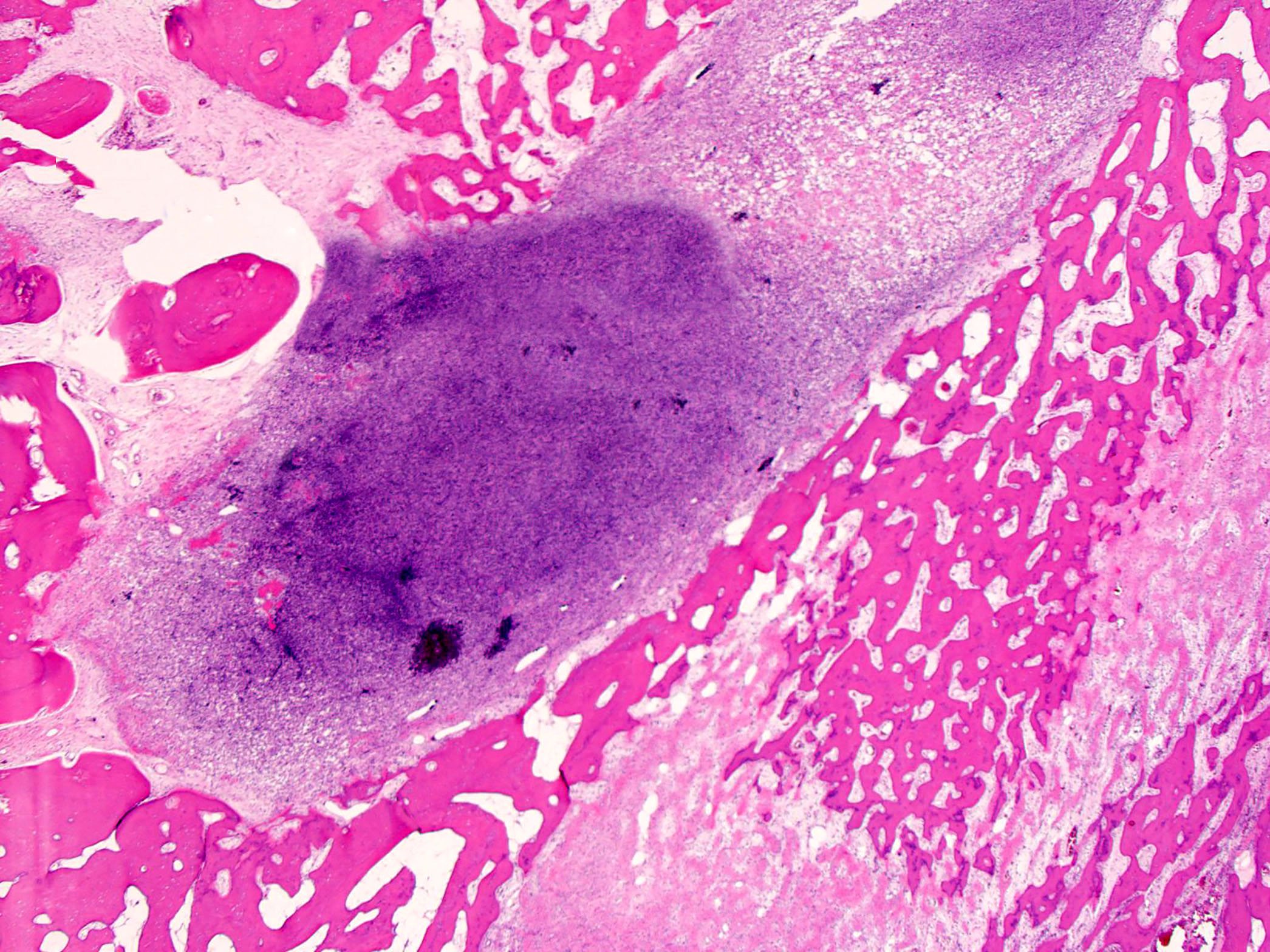
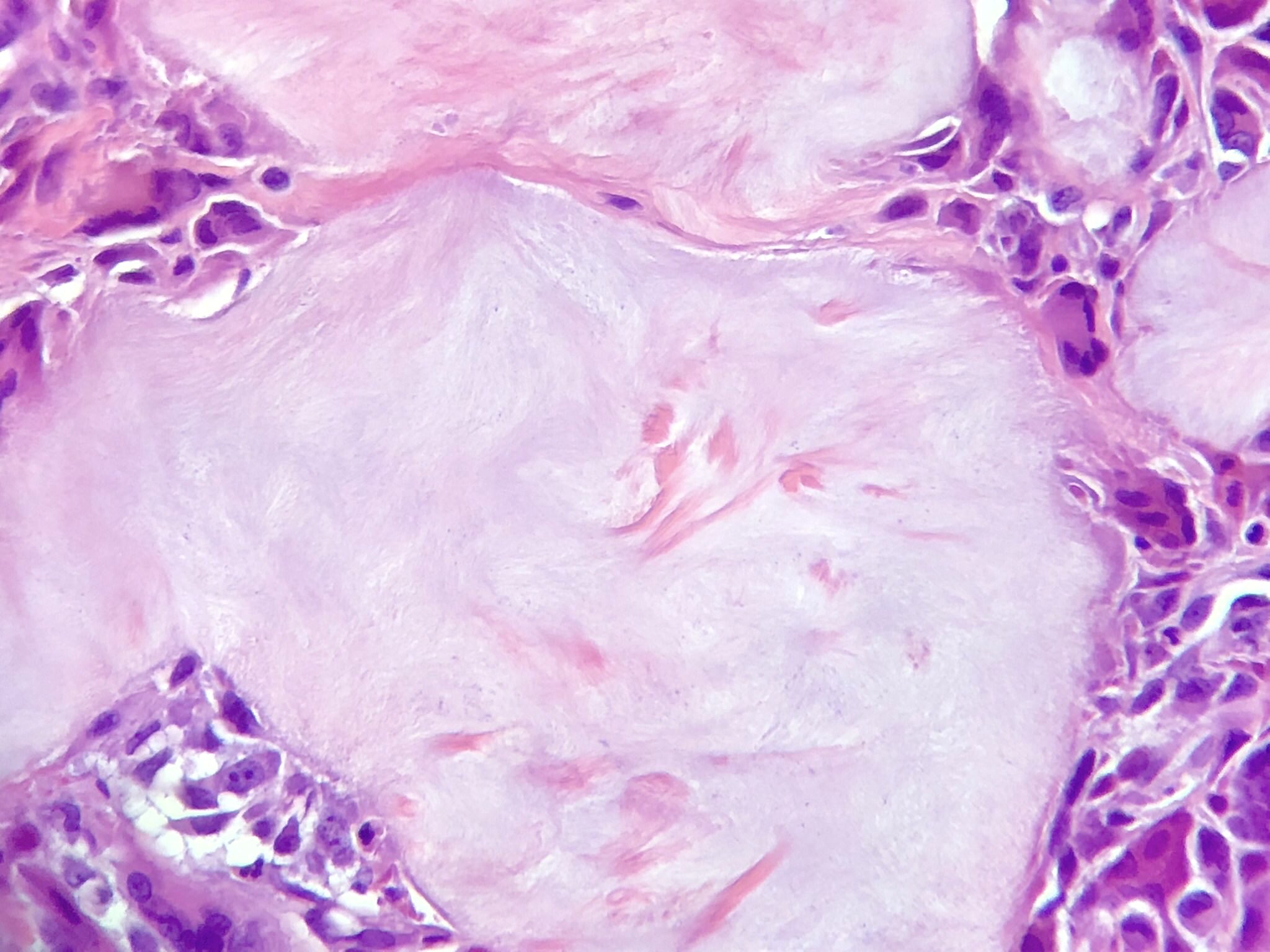
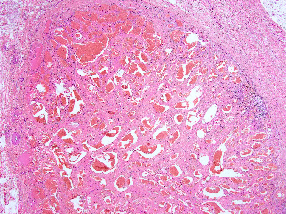
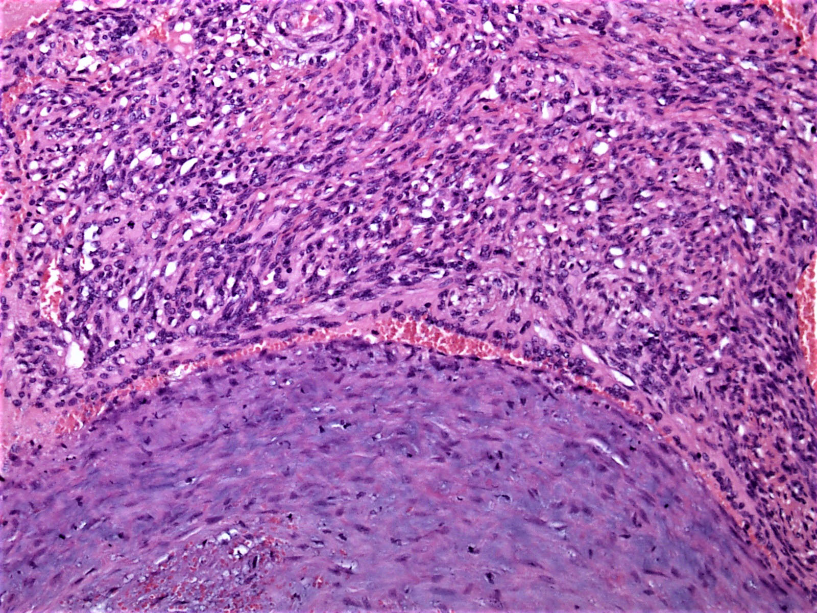
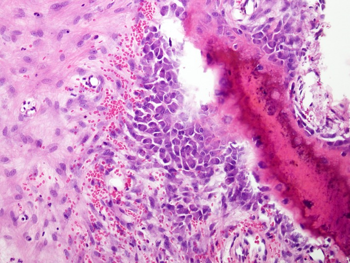
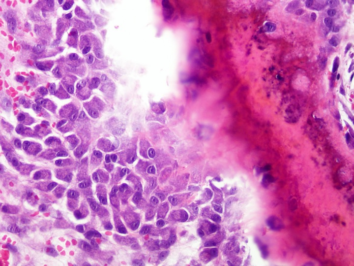
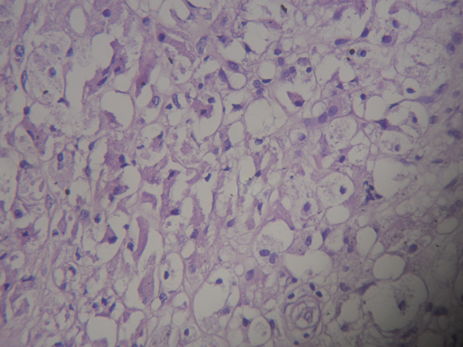
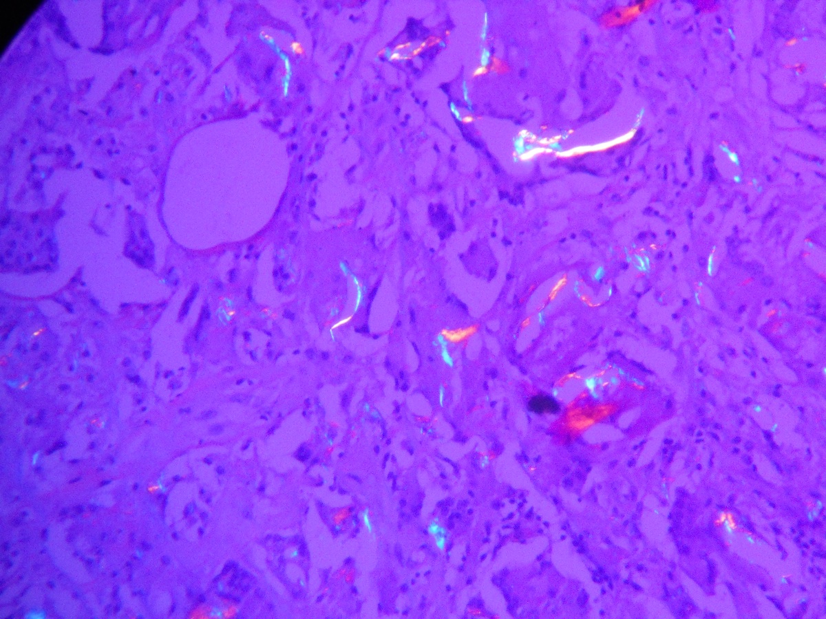
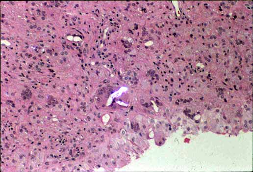
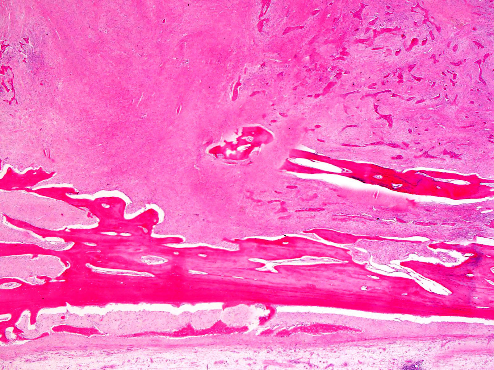
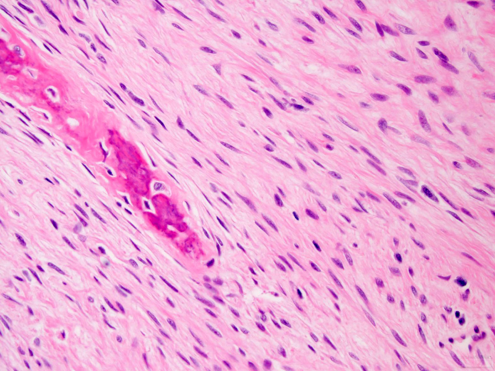
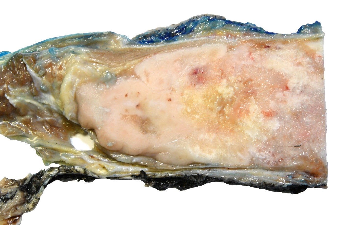
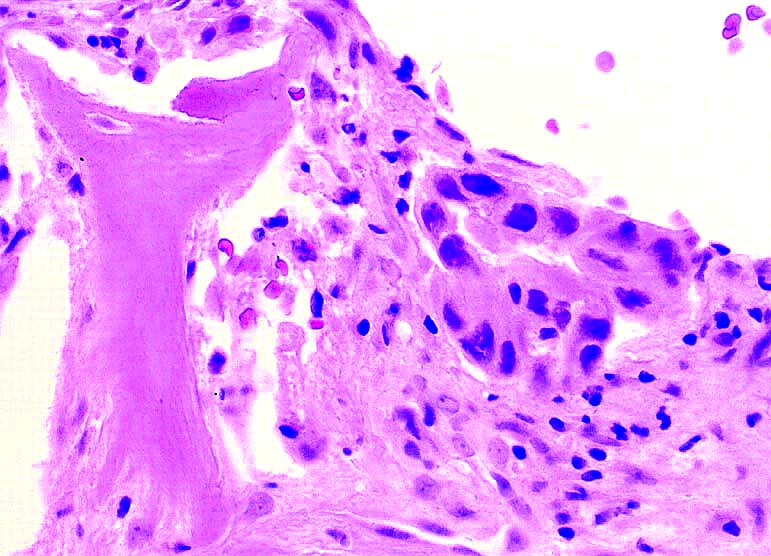
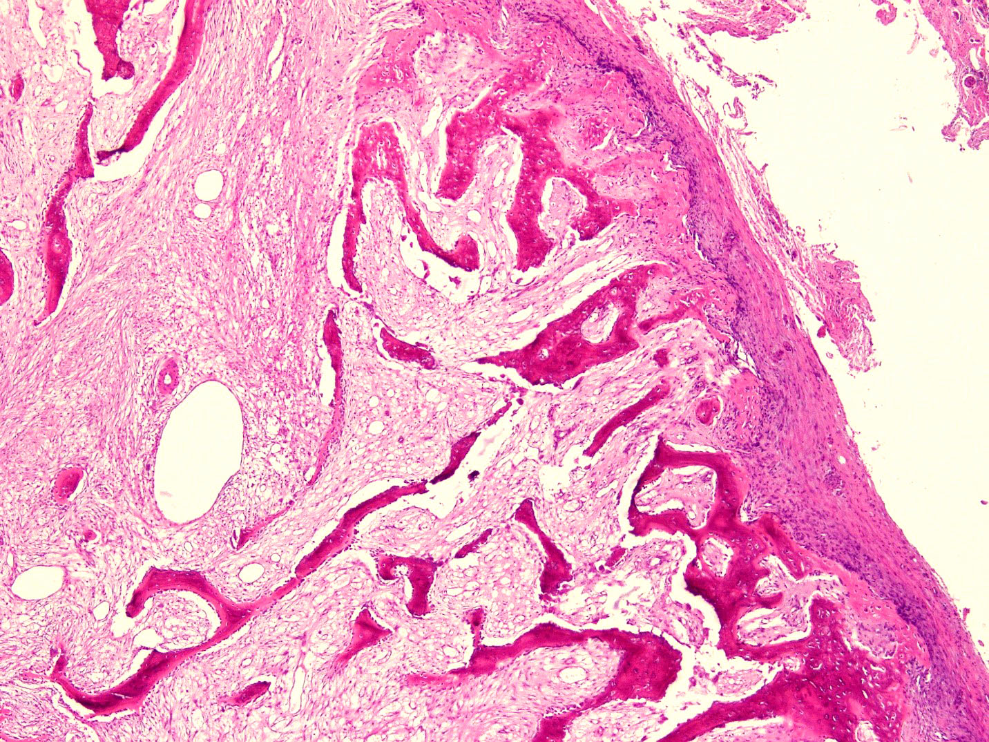
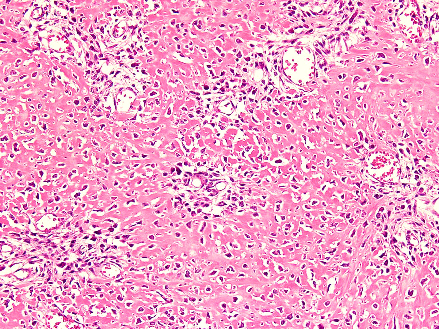
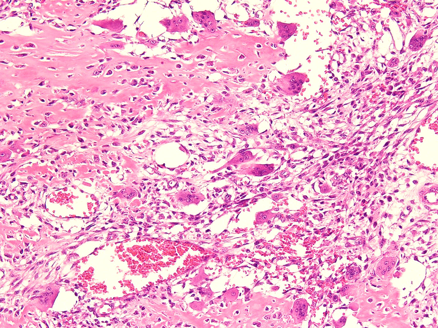
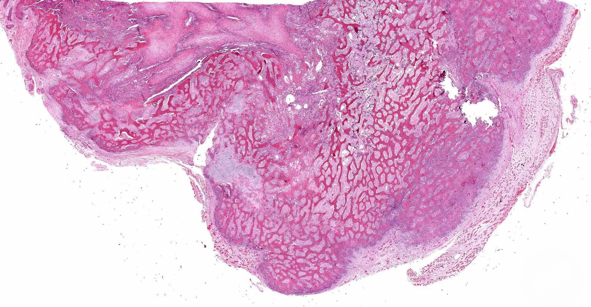
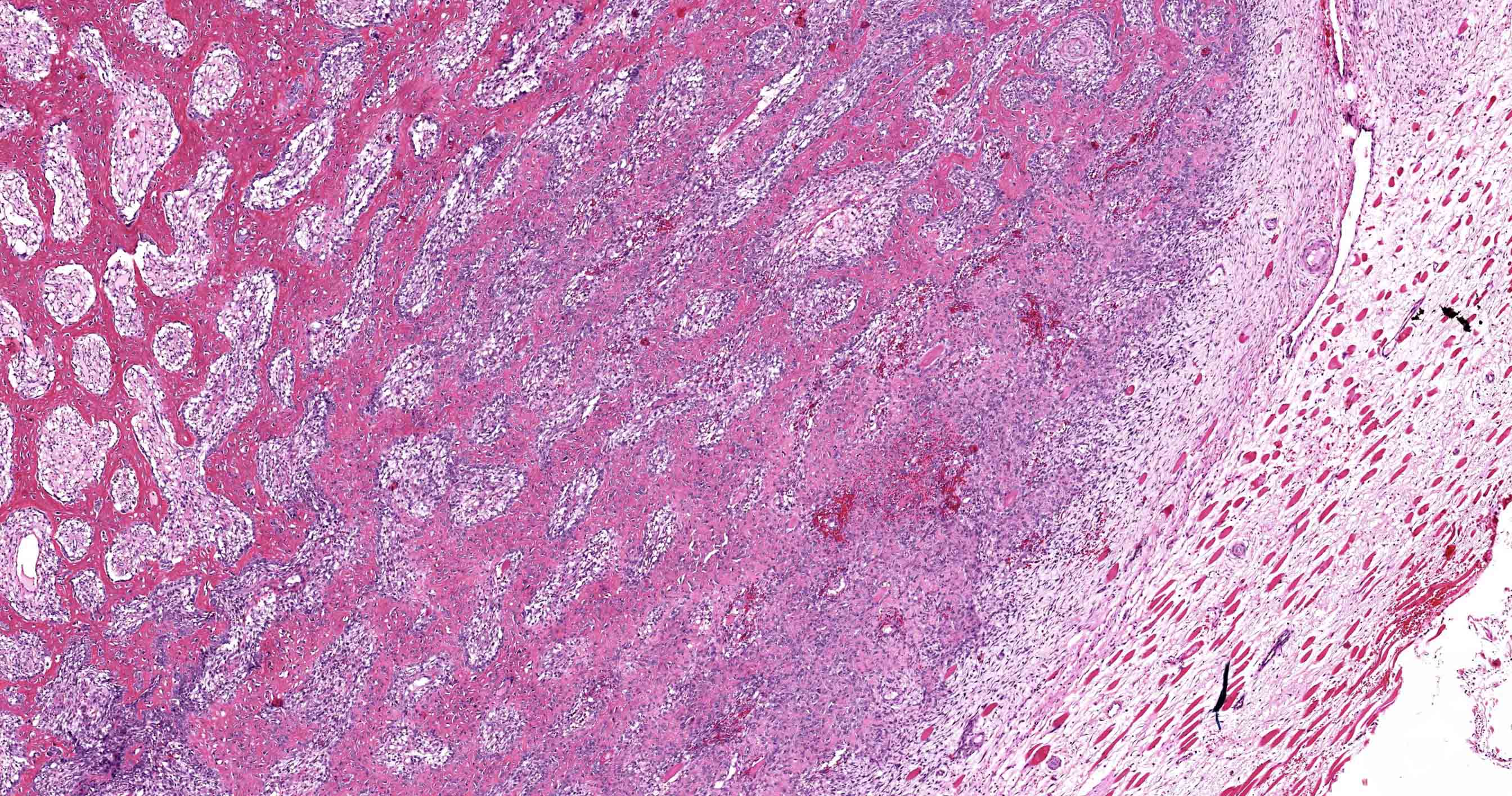
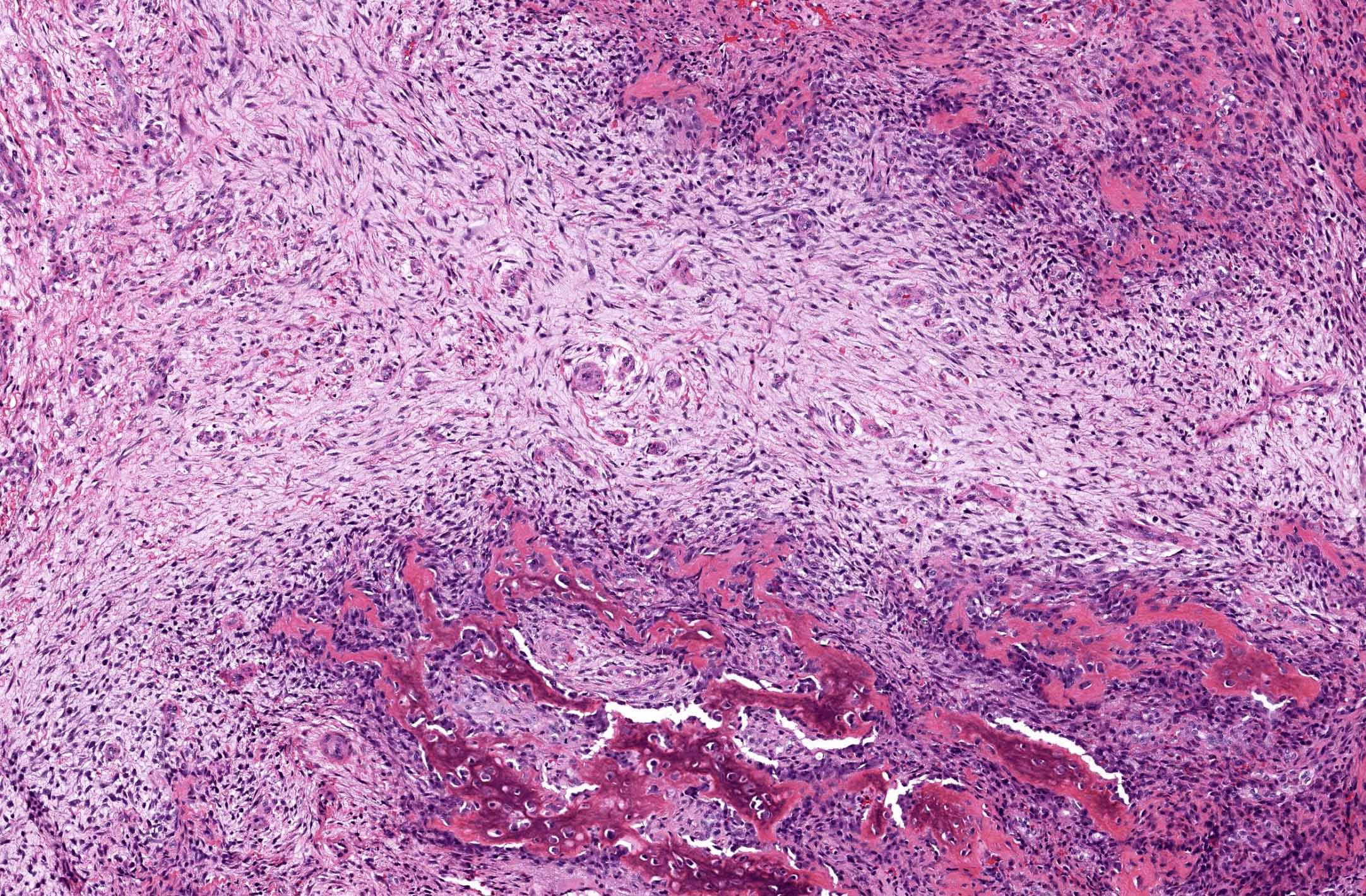
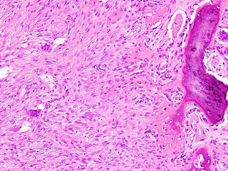
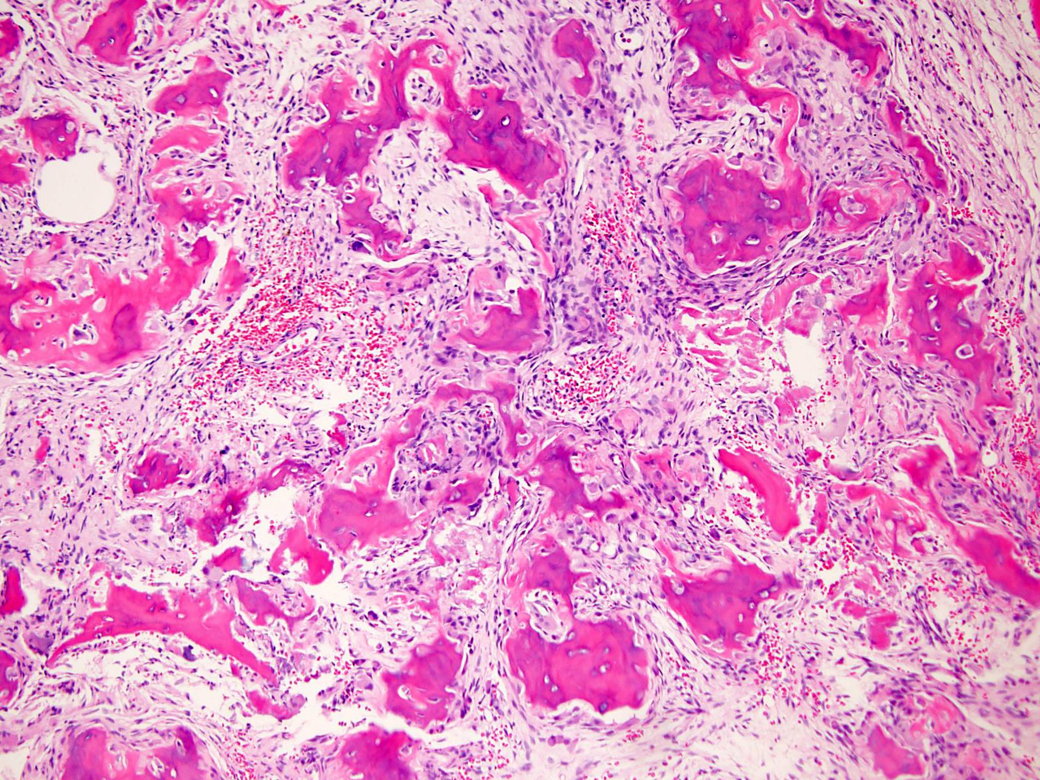
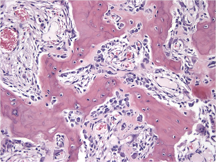
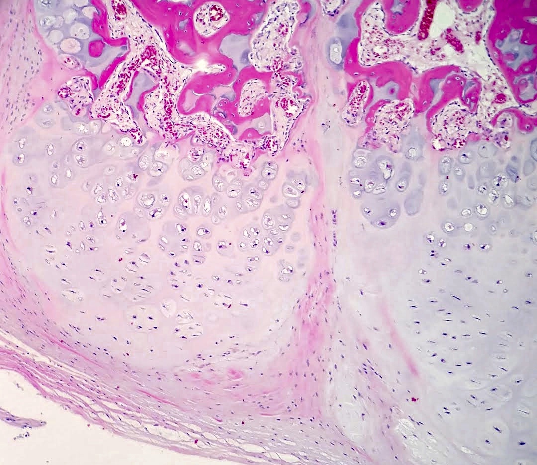
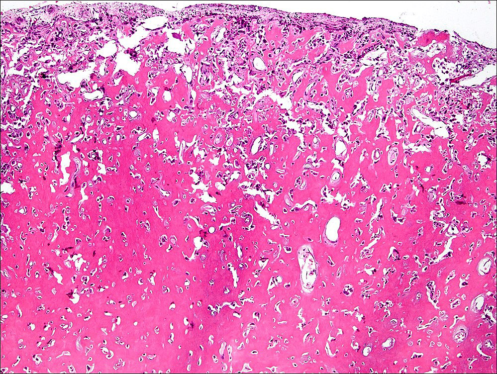
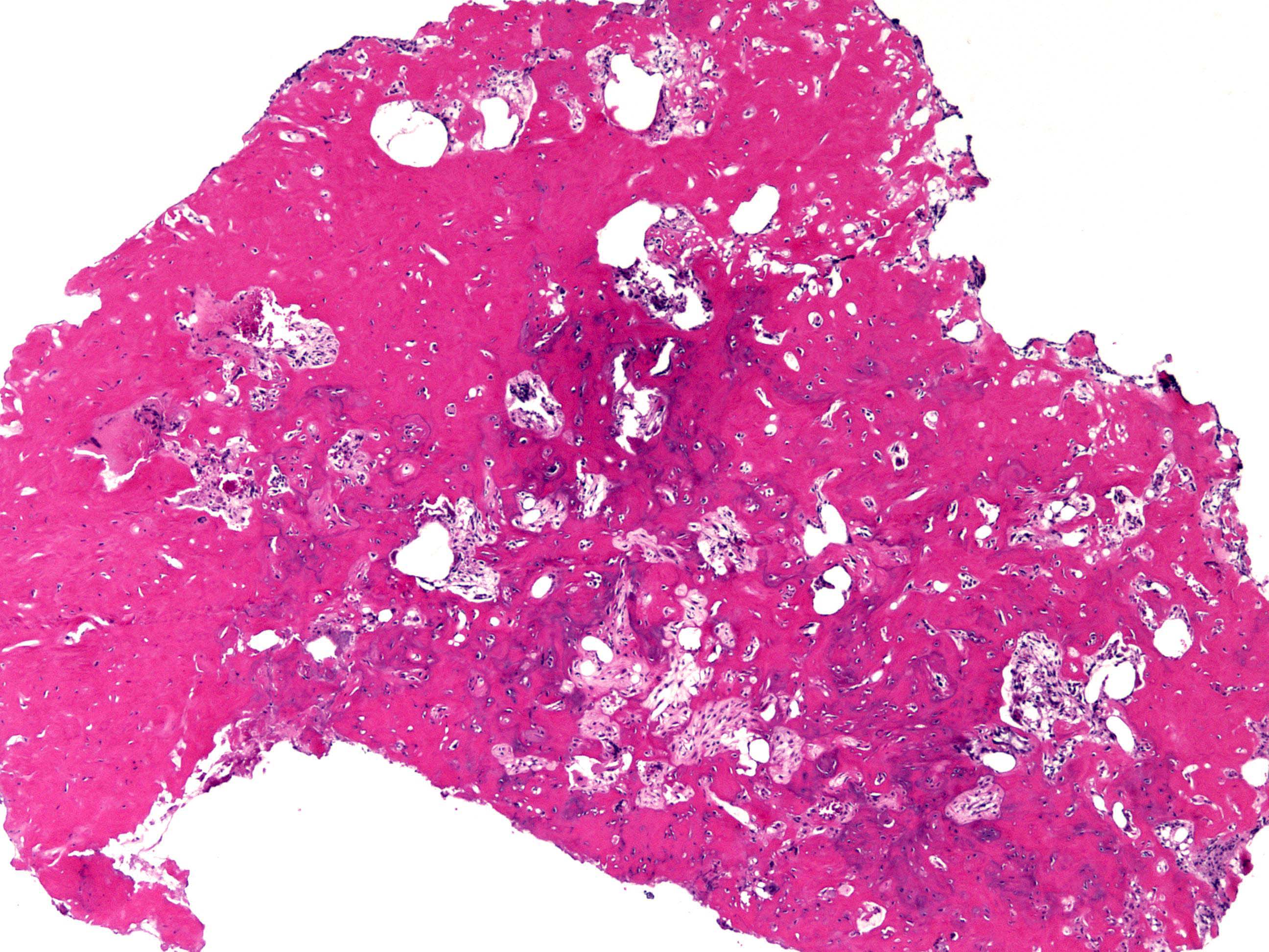
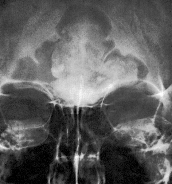
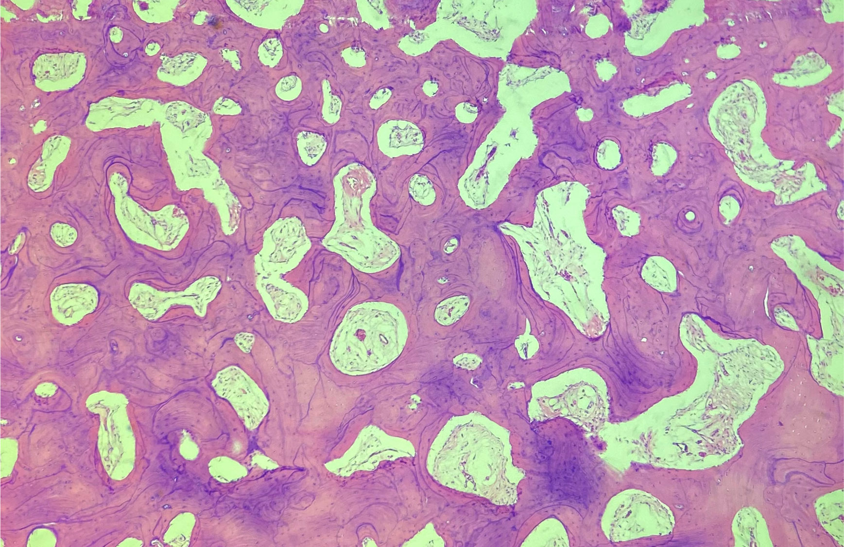
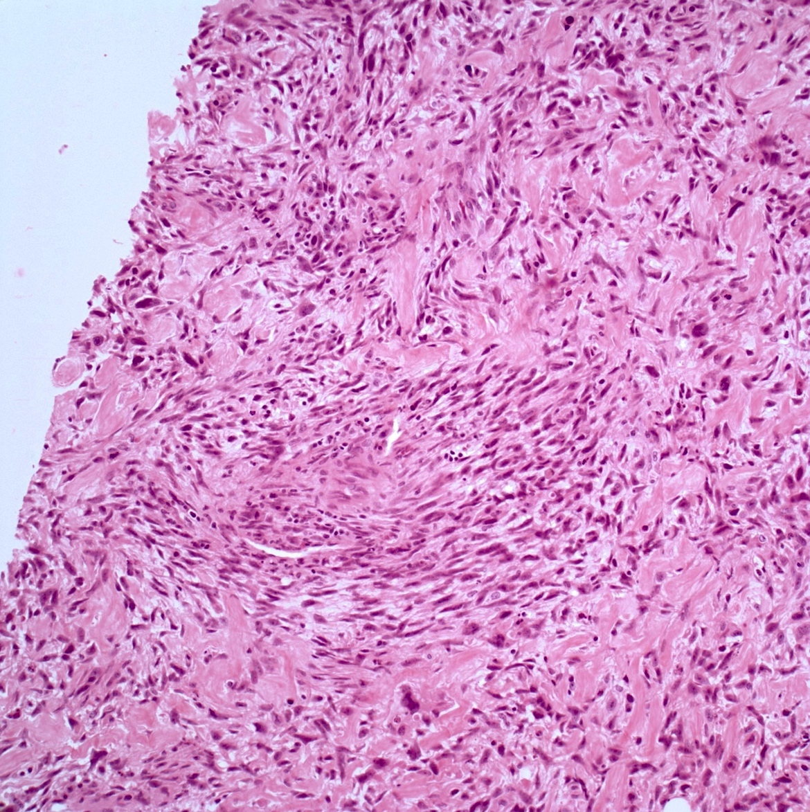
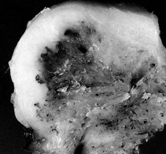
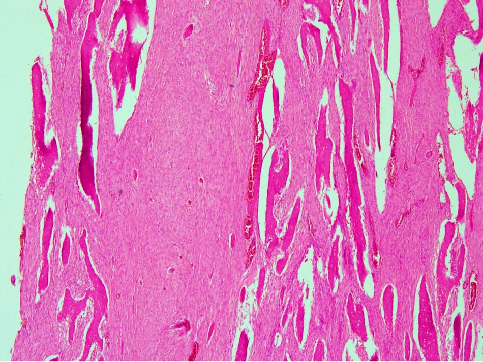
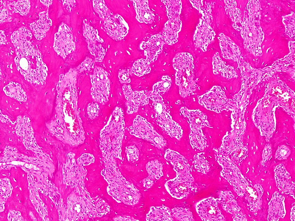
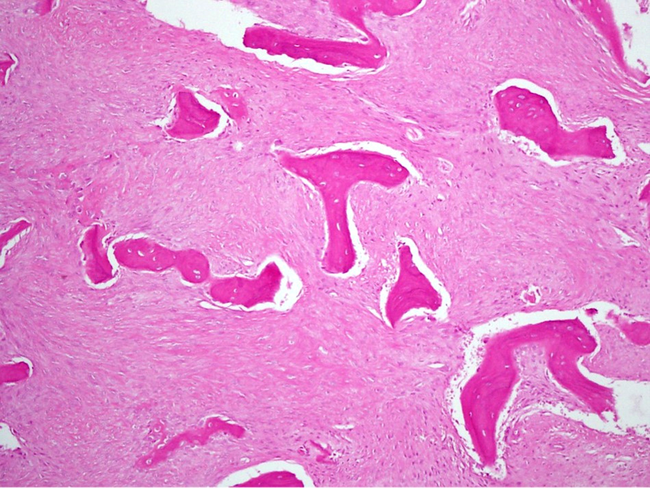
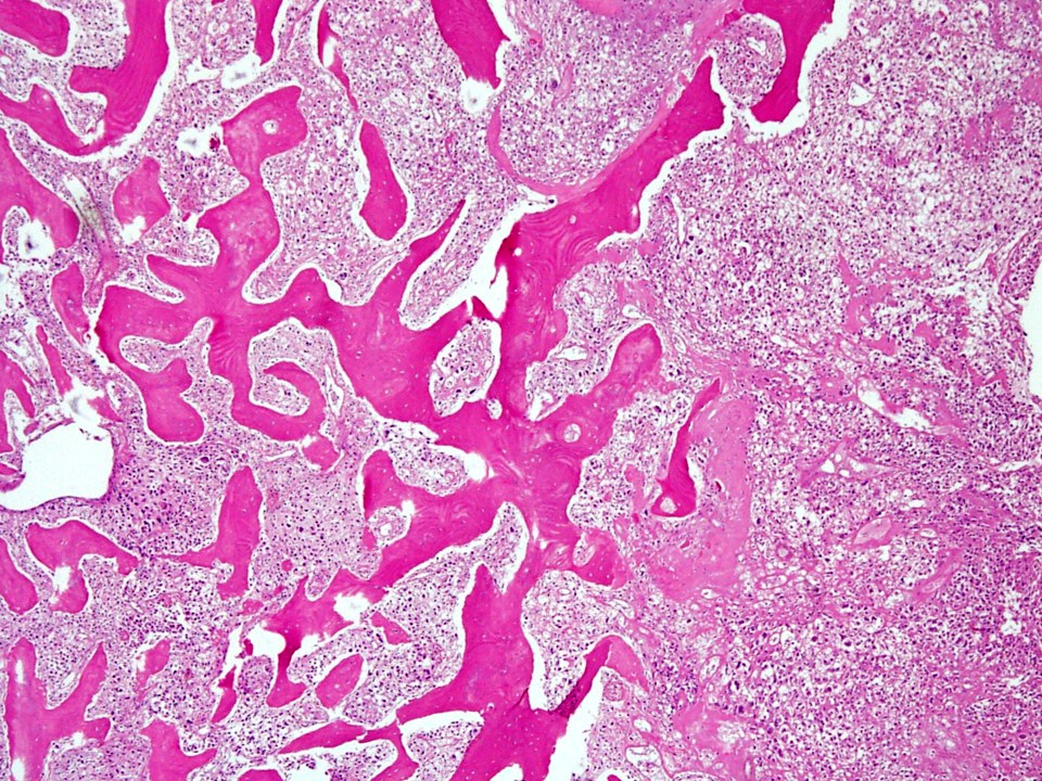
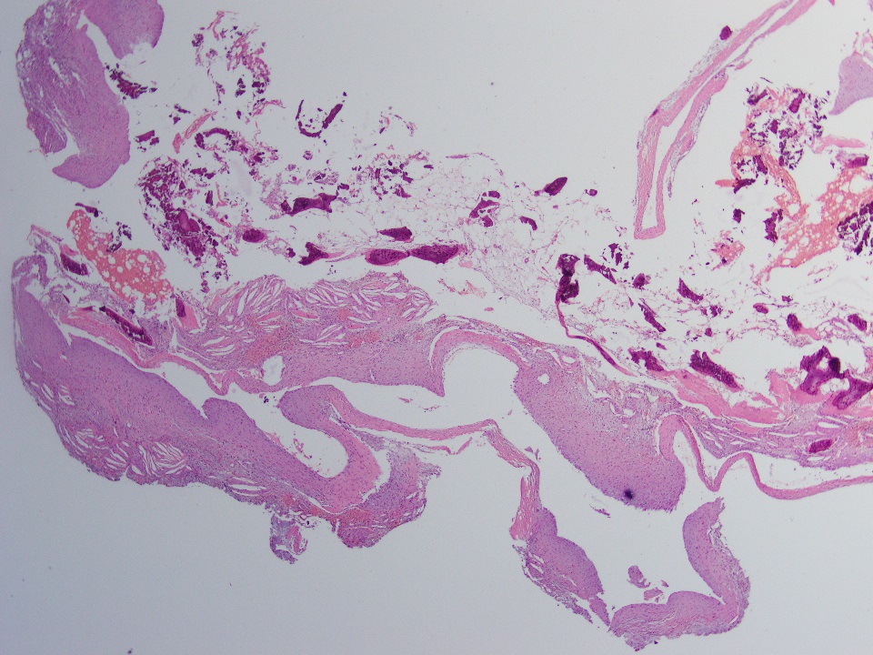
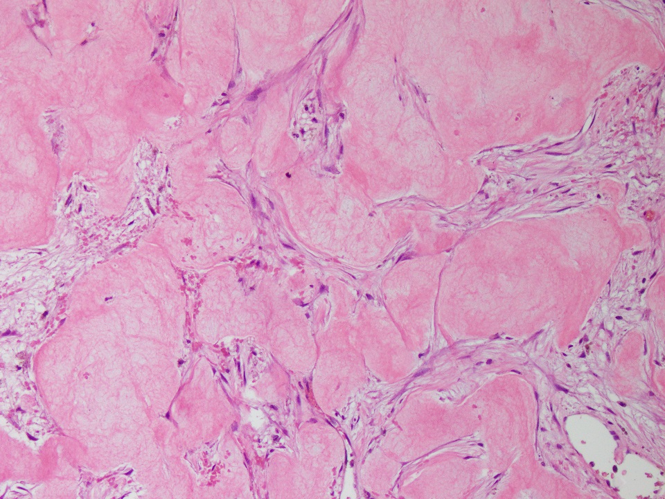
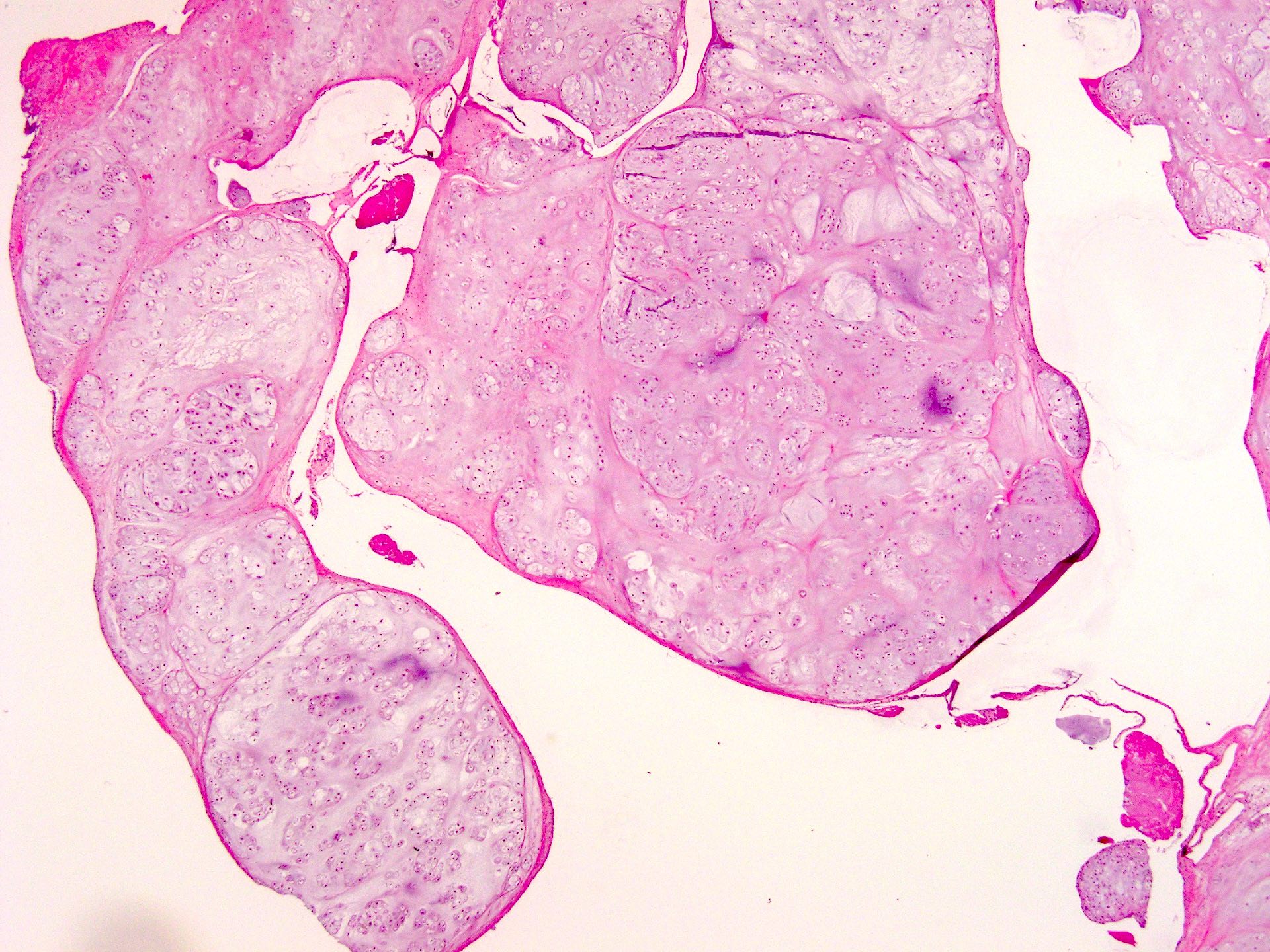
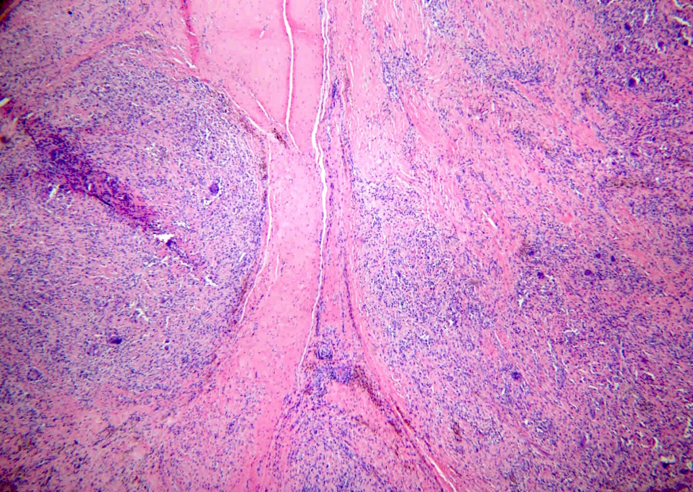
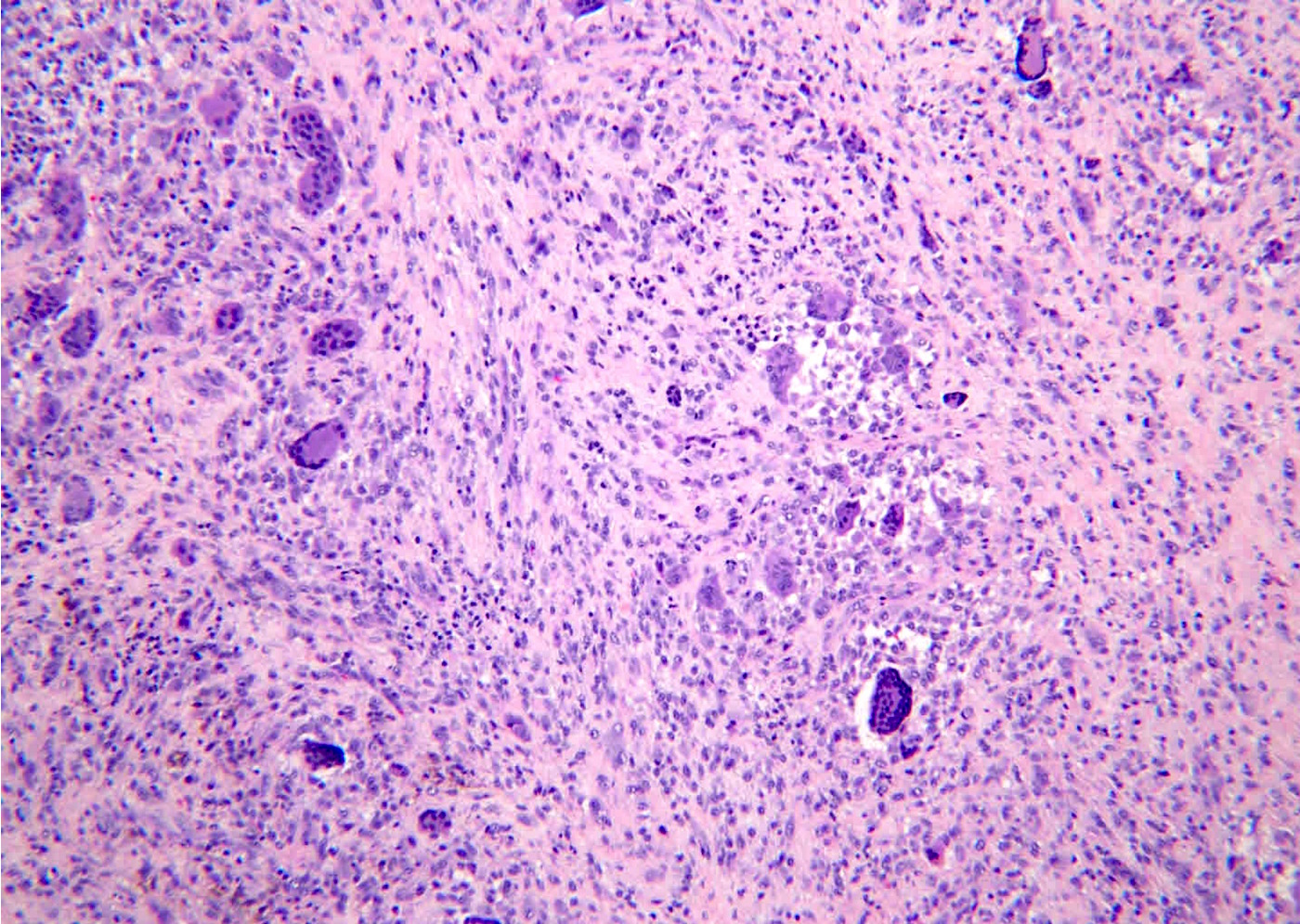
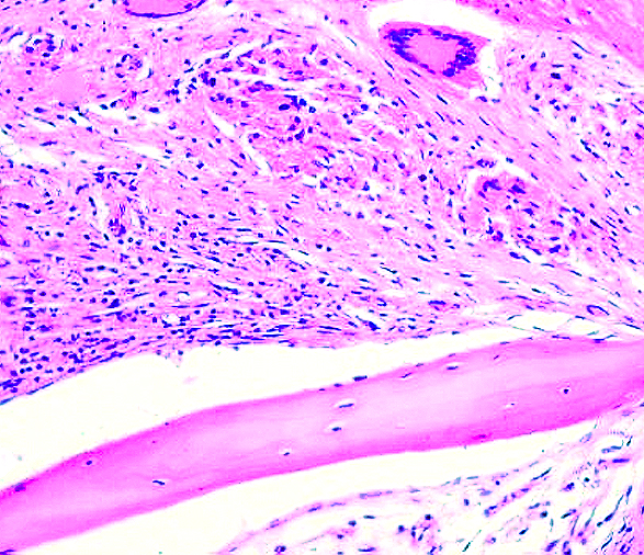
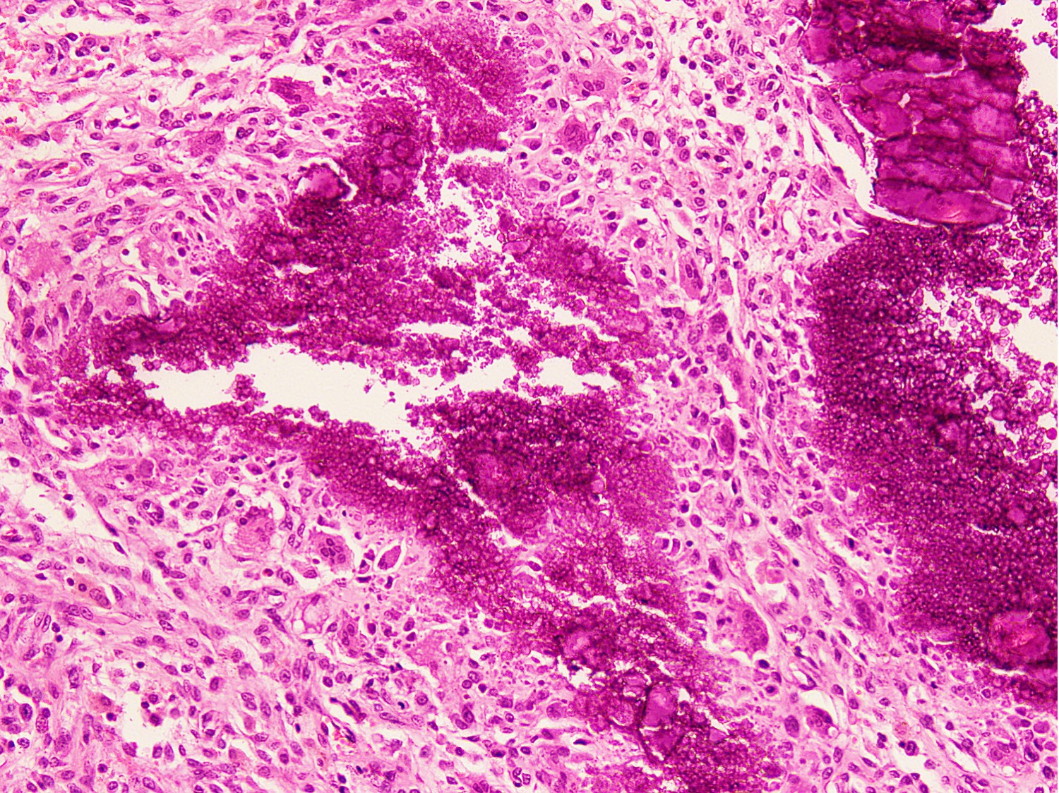
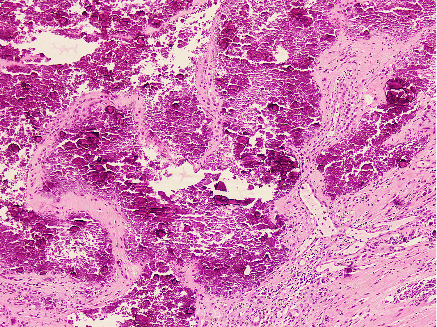
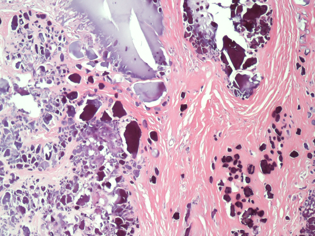
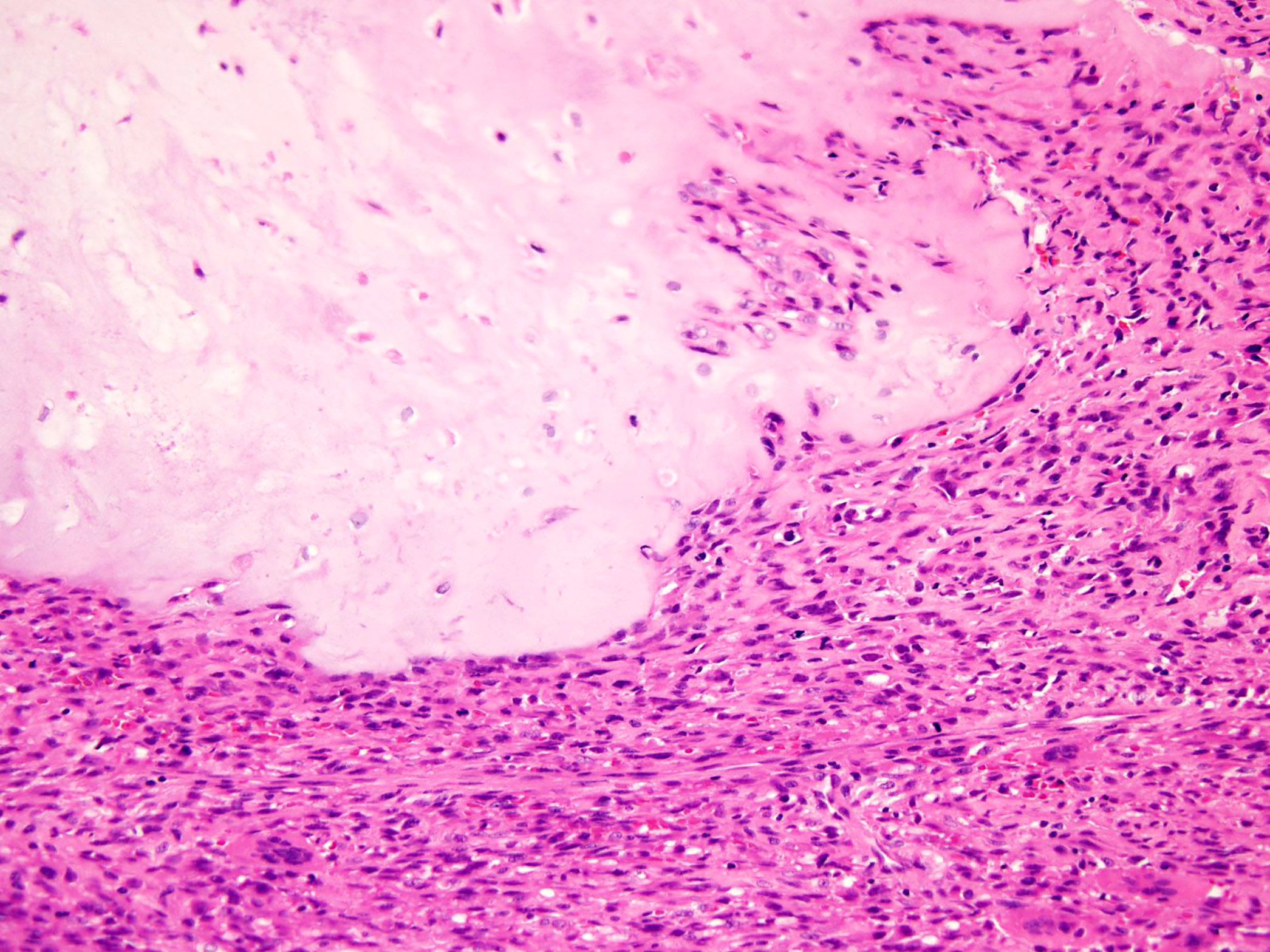
Contributed by Nasir Ud Din, M.B.B.S. and Mark R. Wick, M.D.
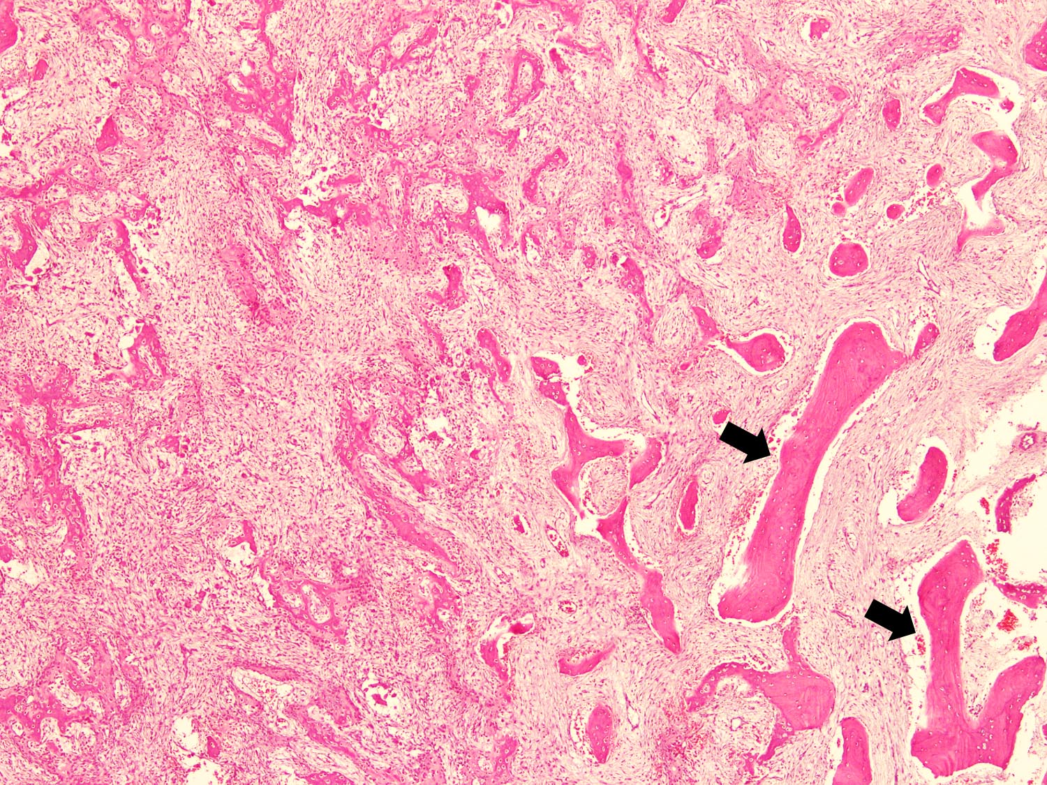
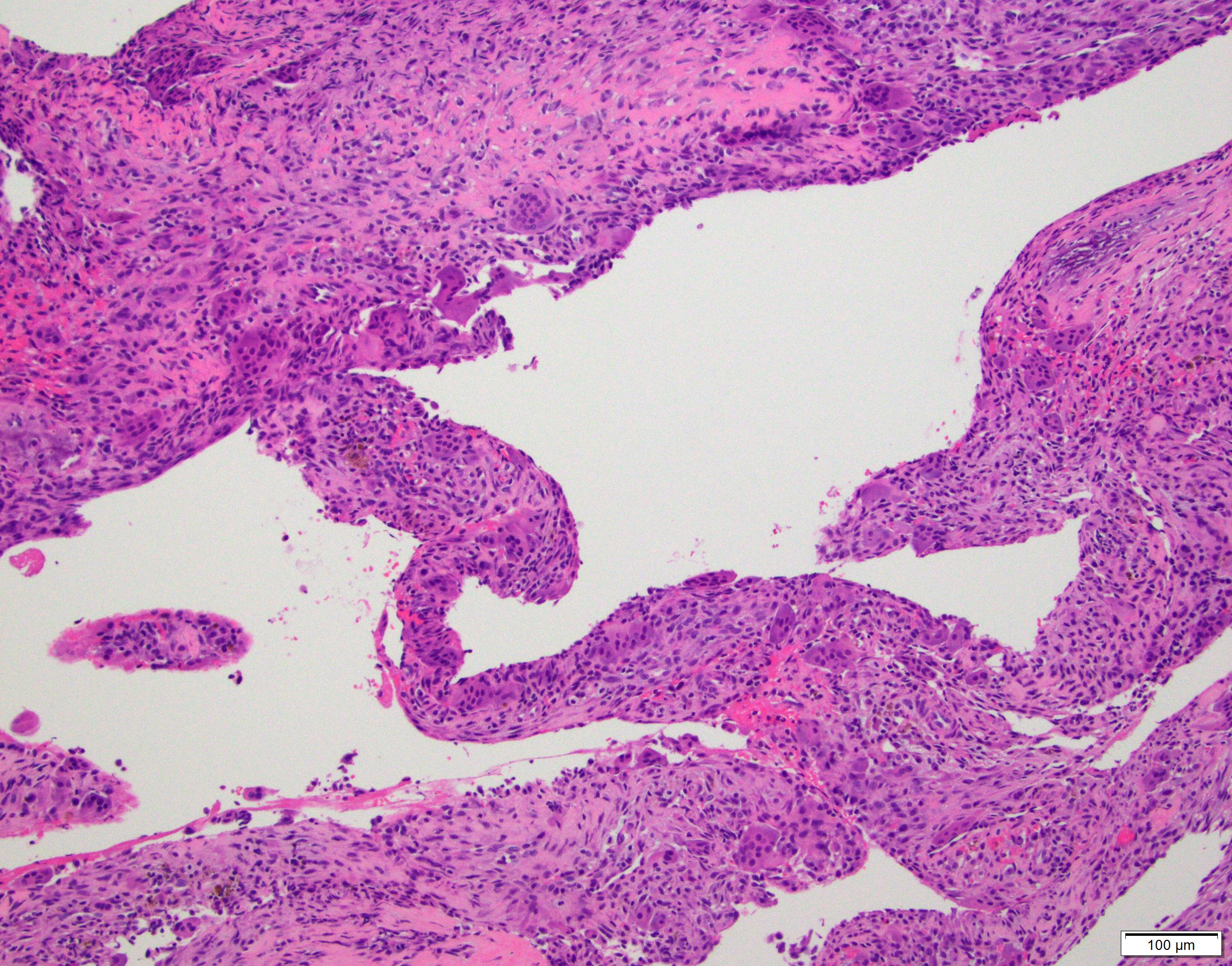
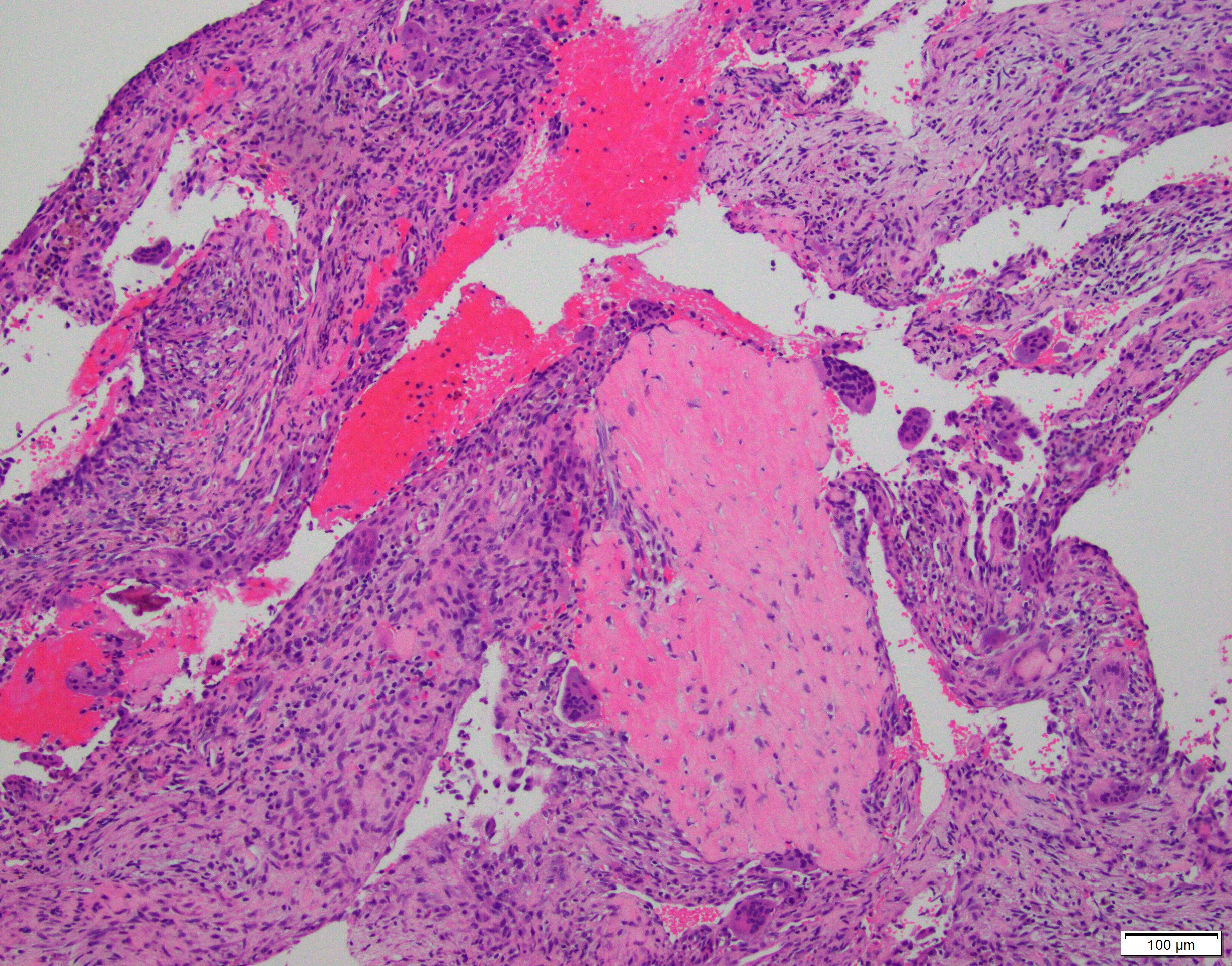
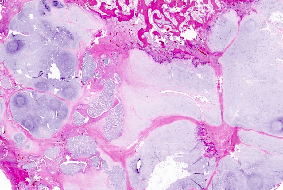
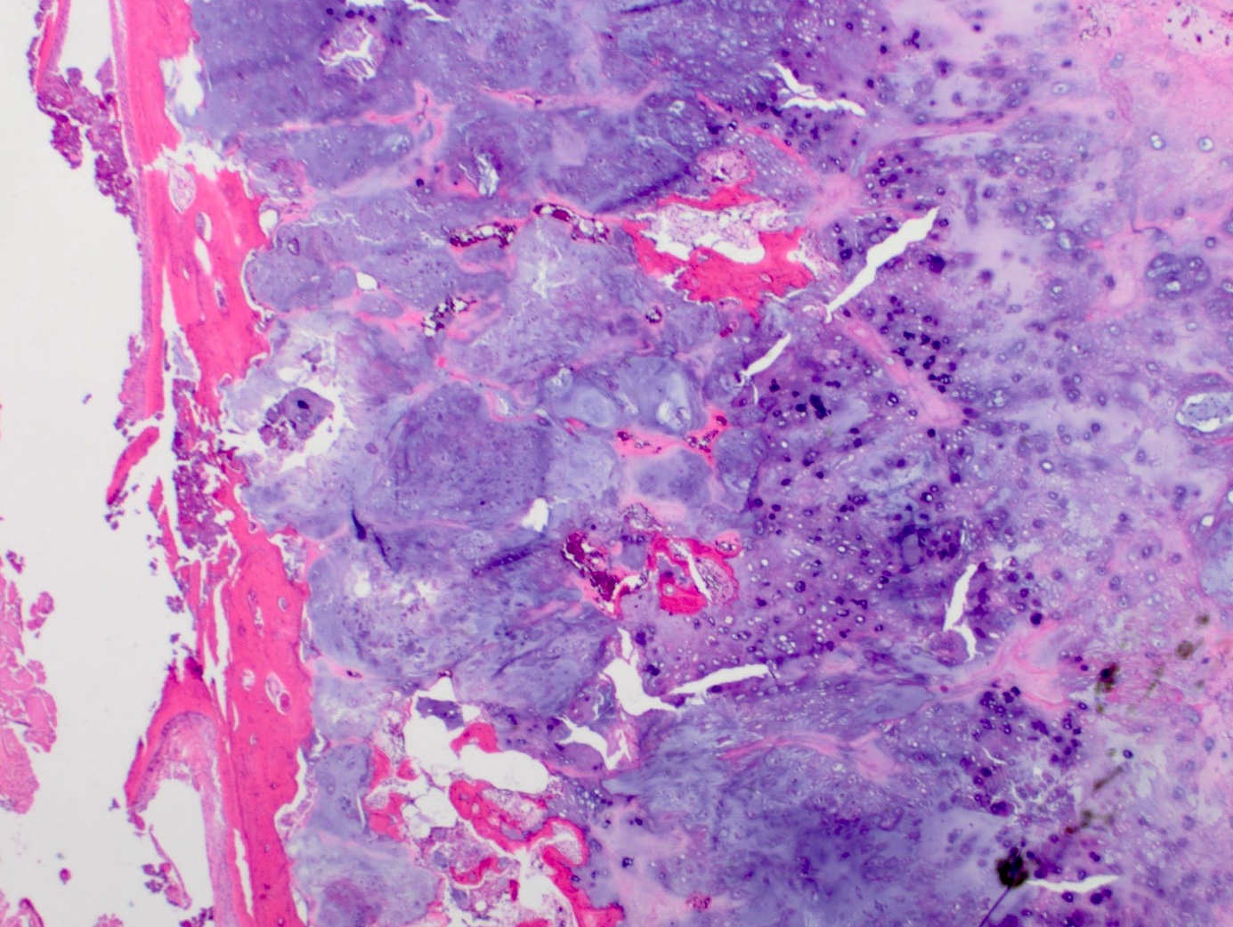
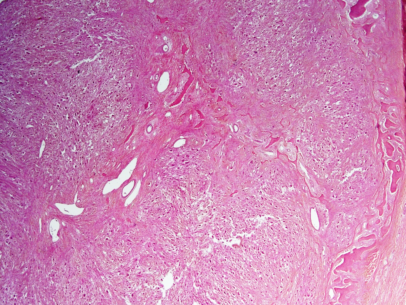
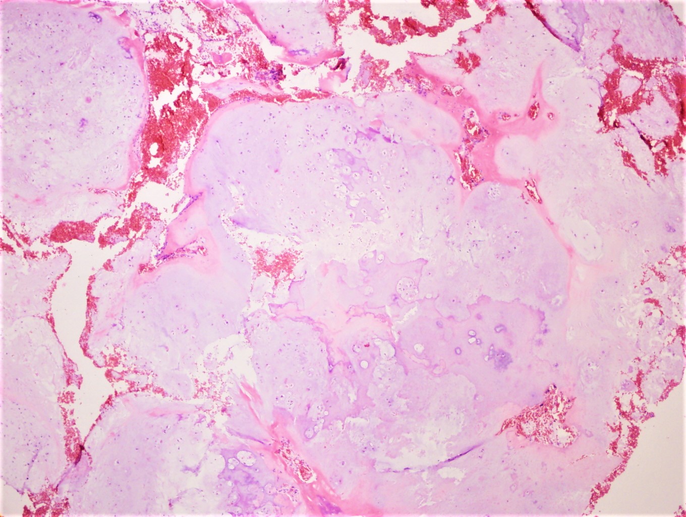
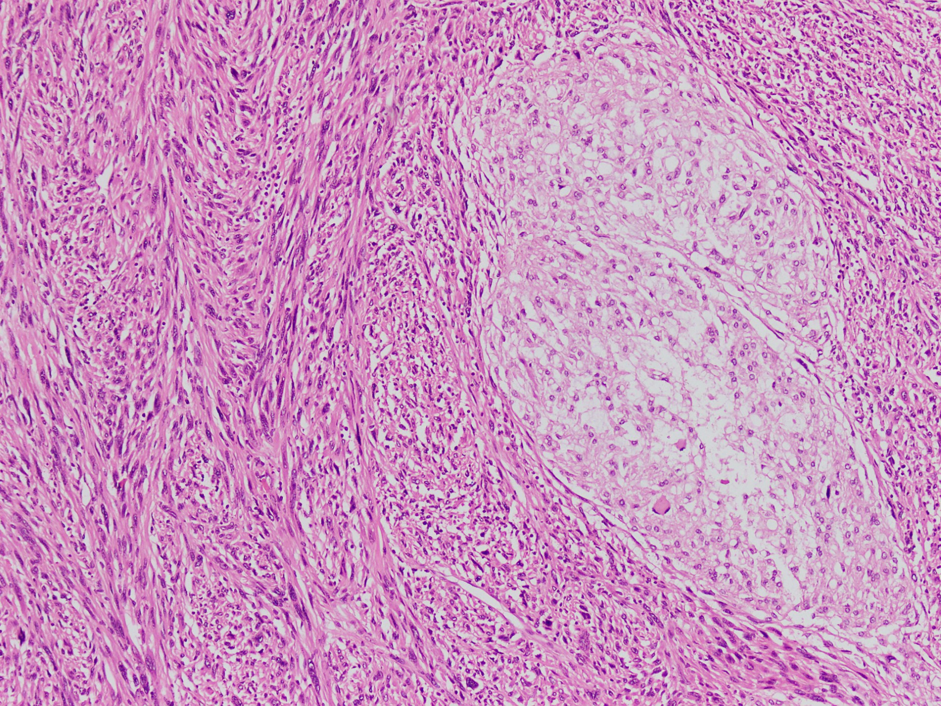
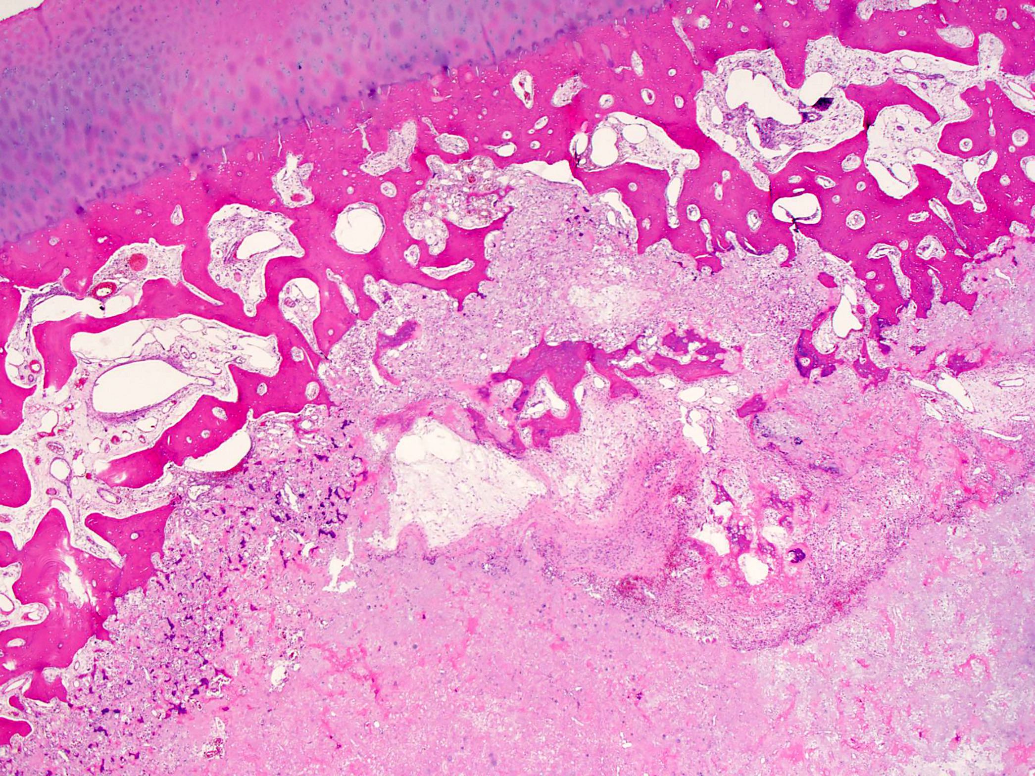
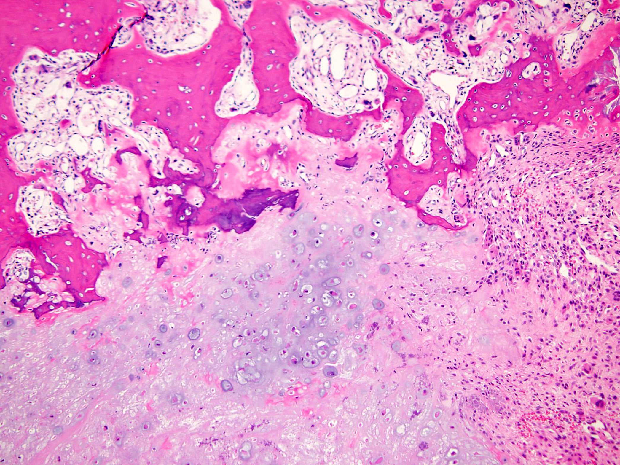
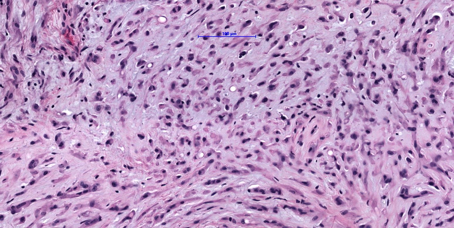
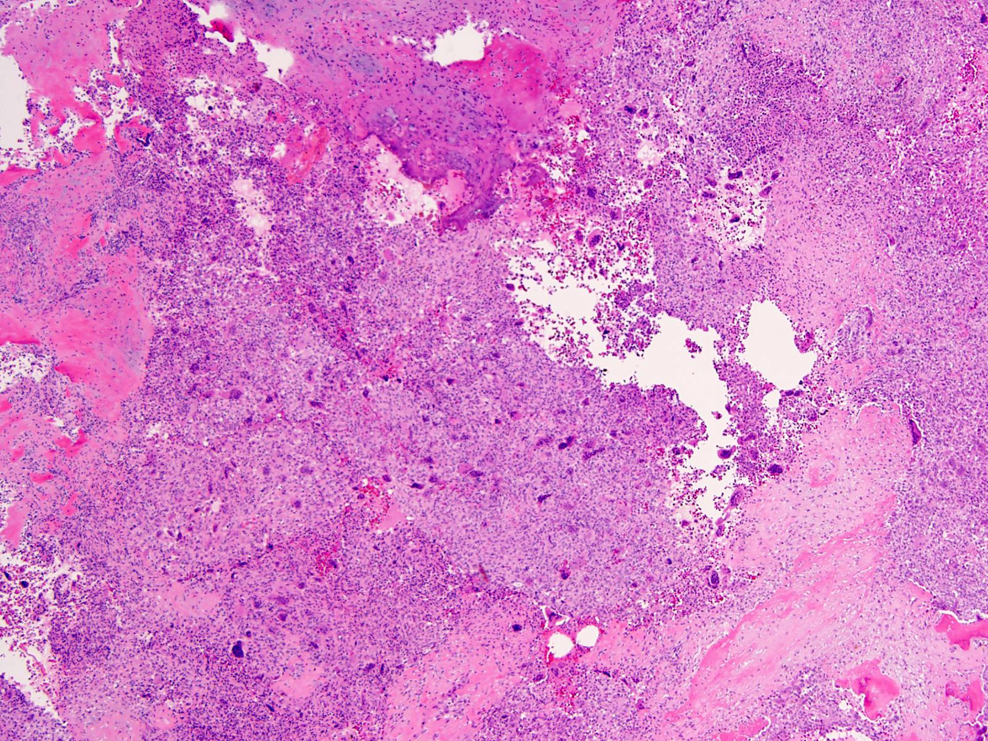
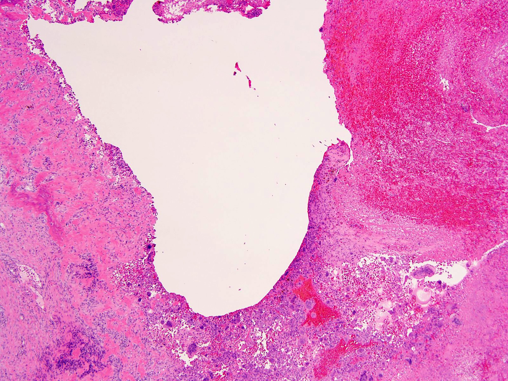
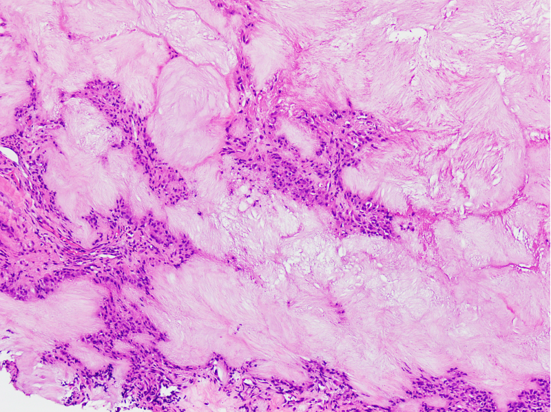
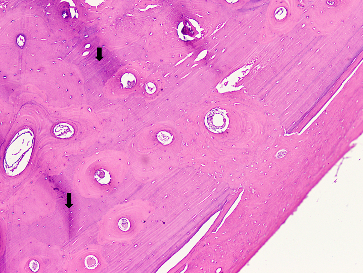
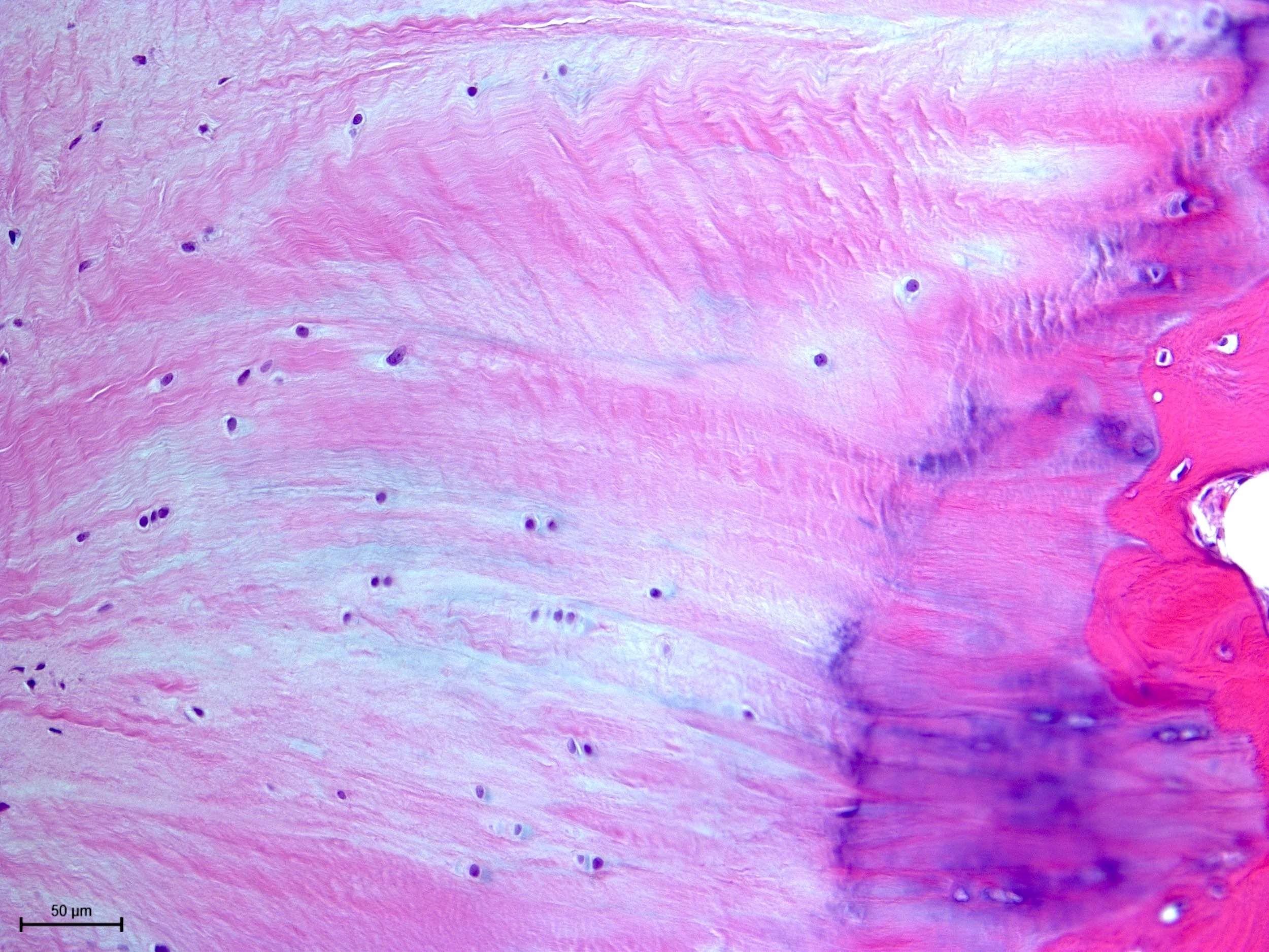
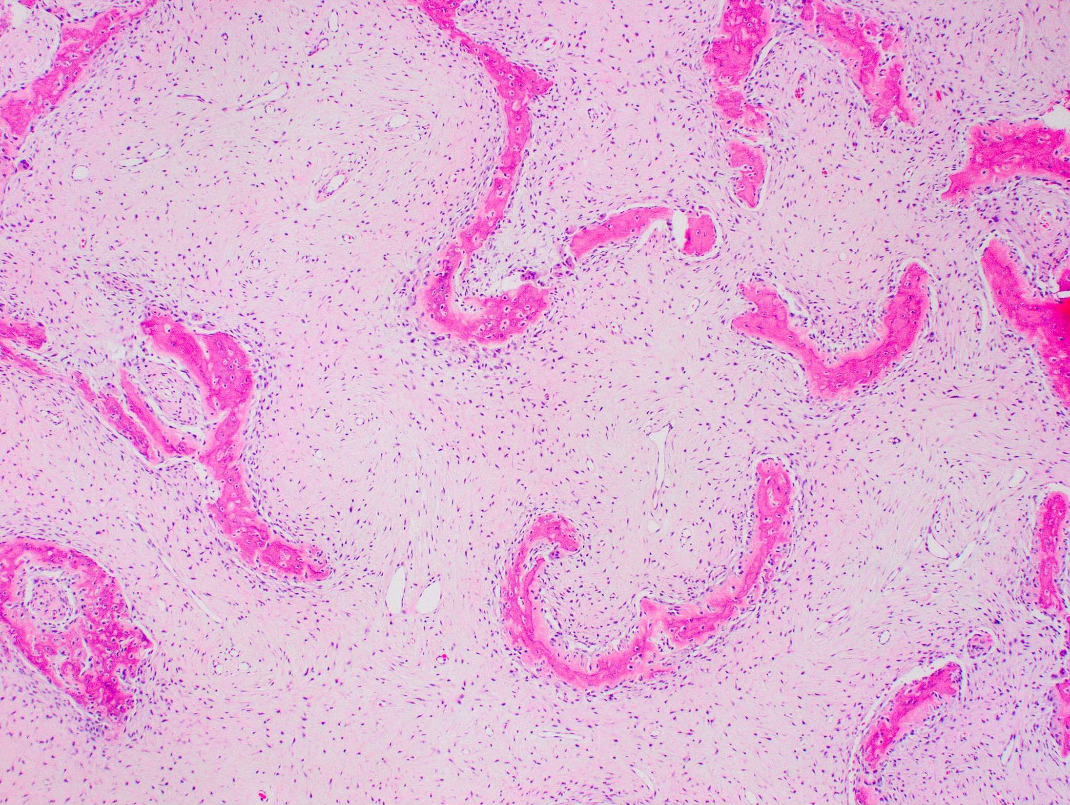
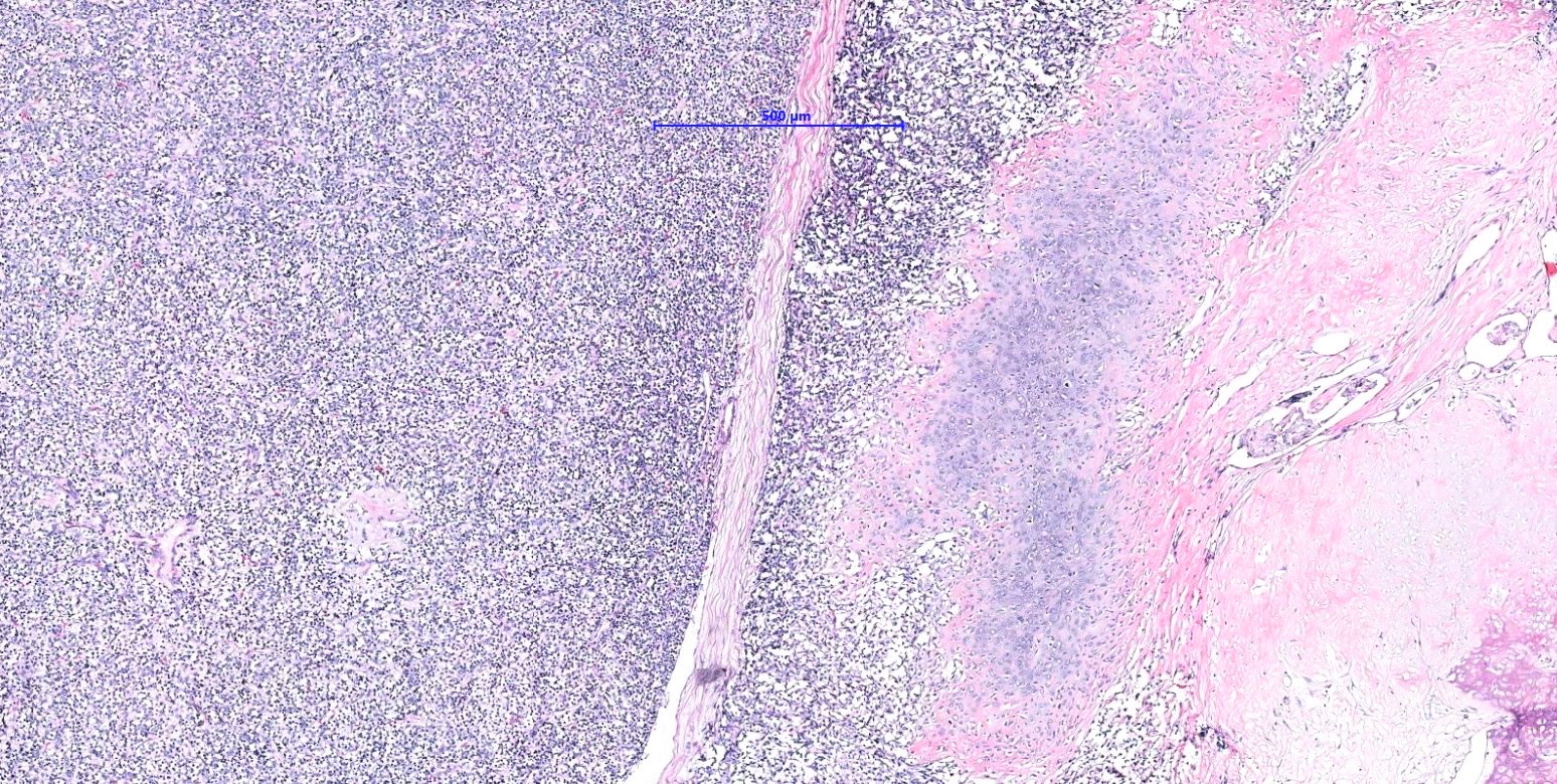
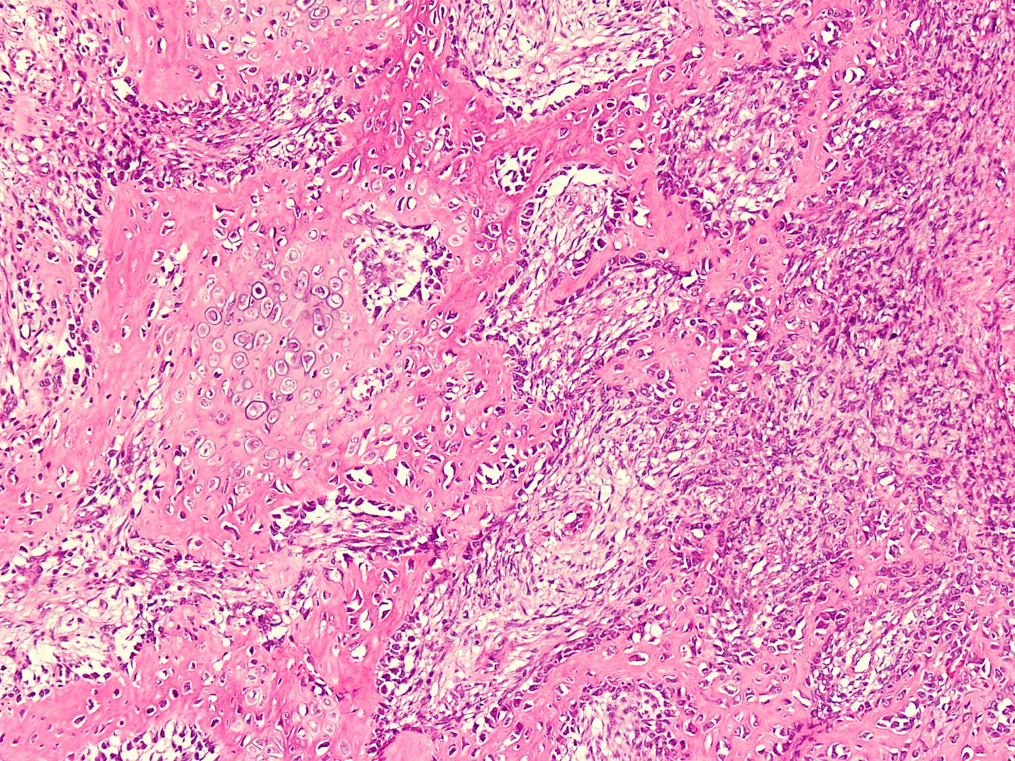
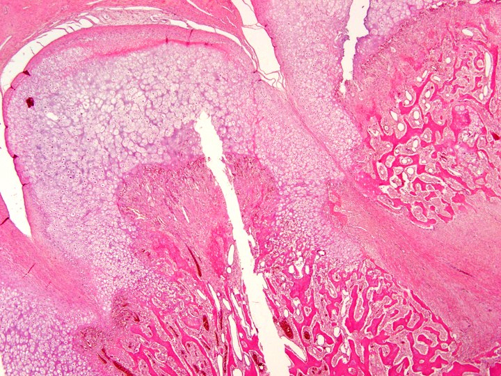
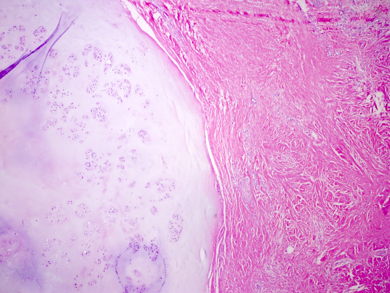
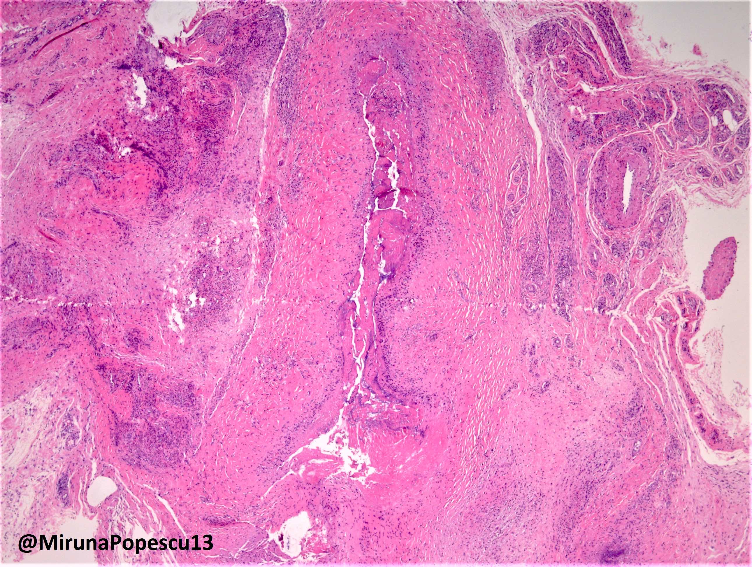
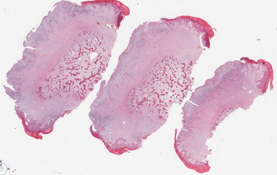
Fibro-osseous lesion with zonation pattern
Thickened trabeculae at the periphery
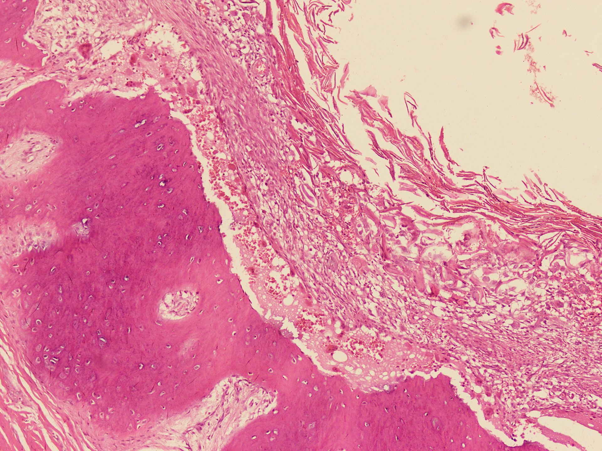
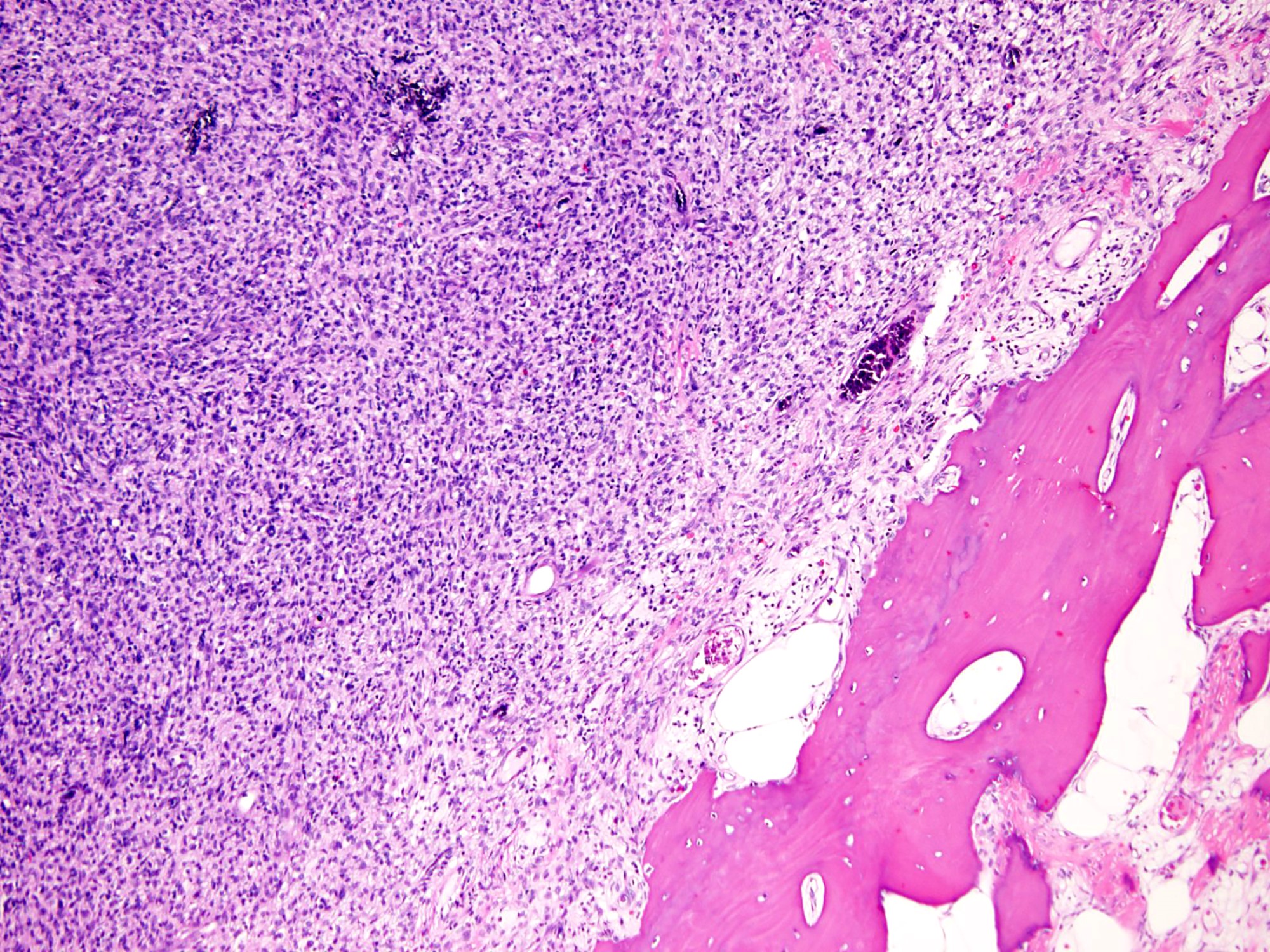
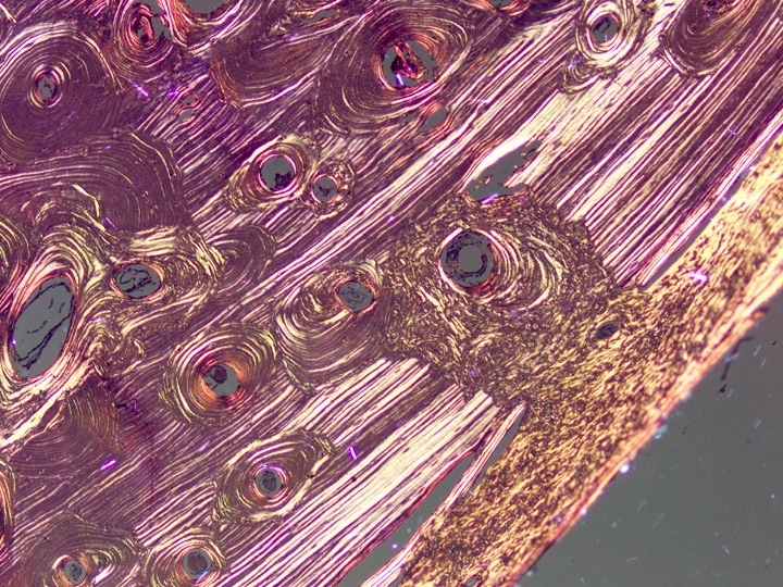
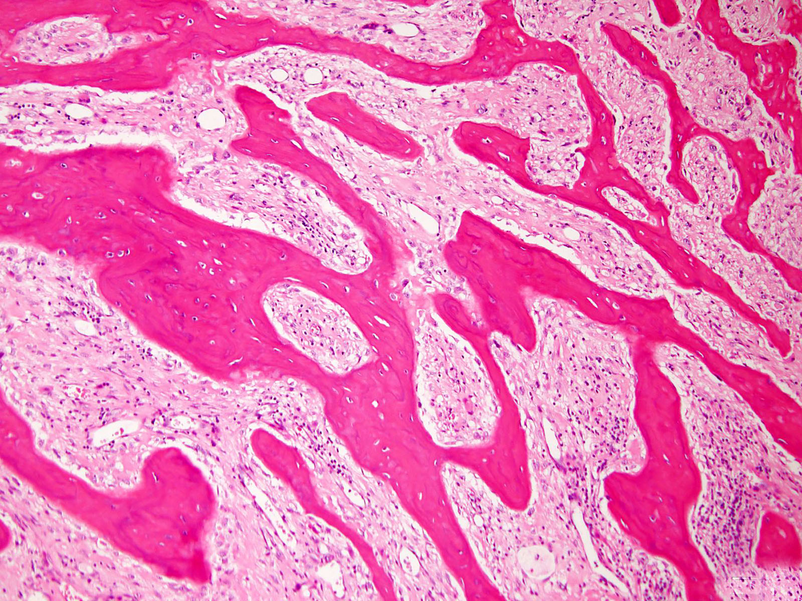
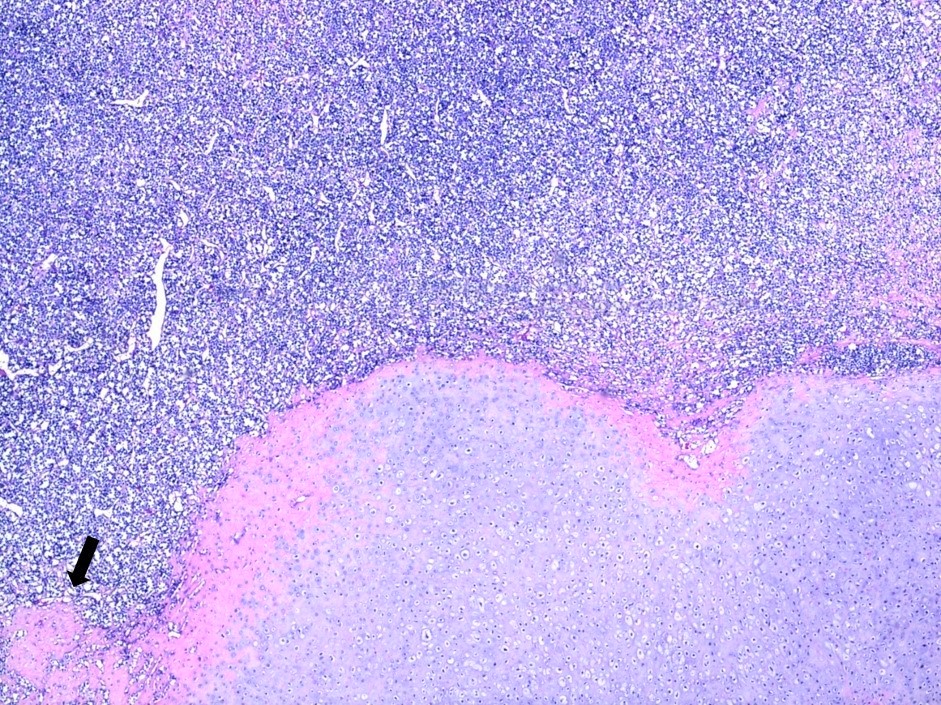
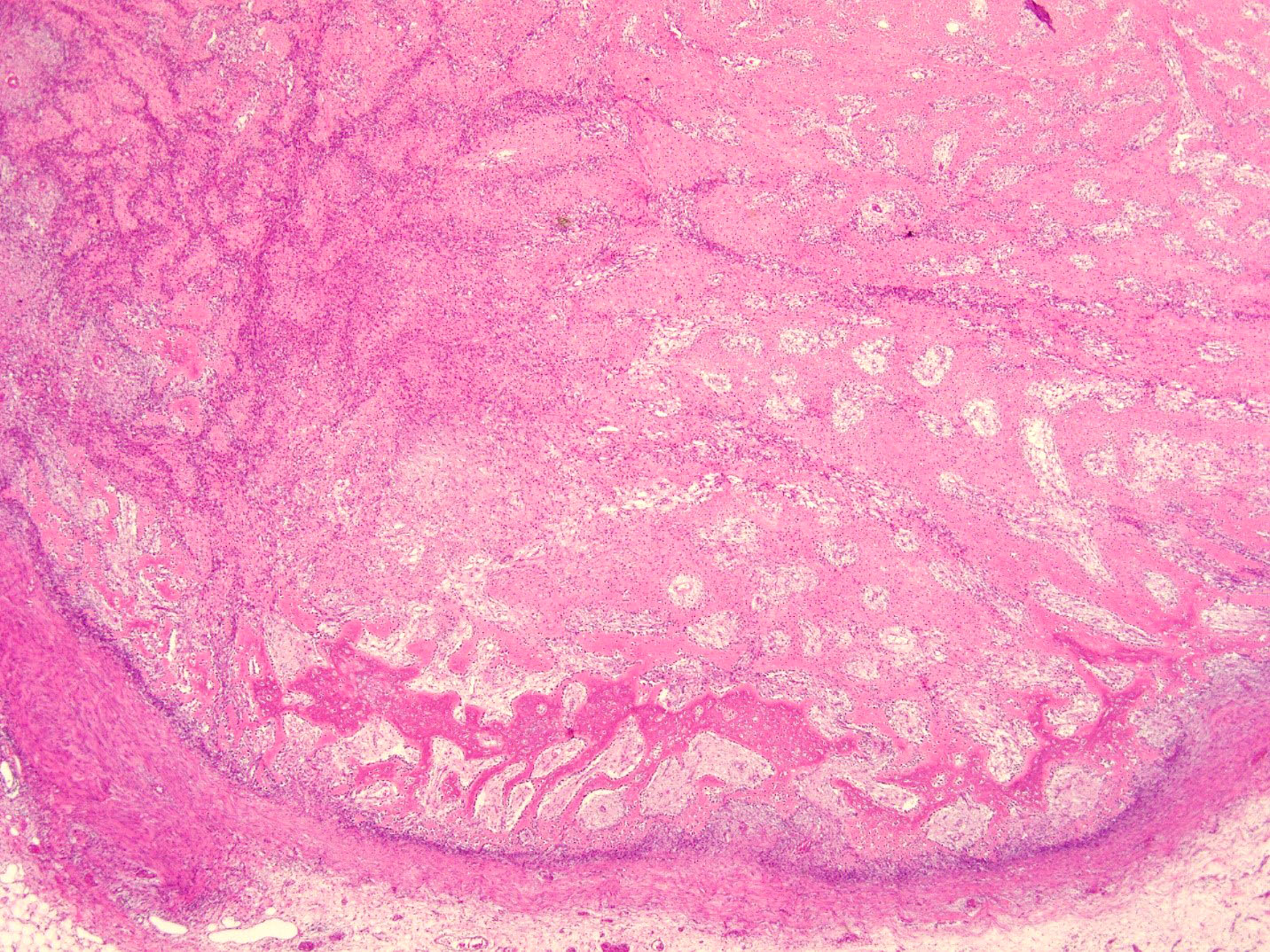
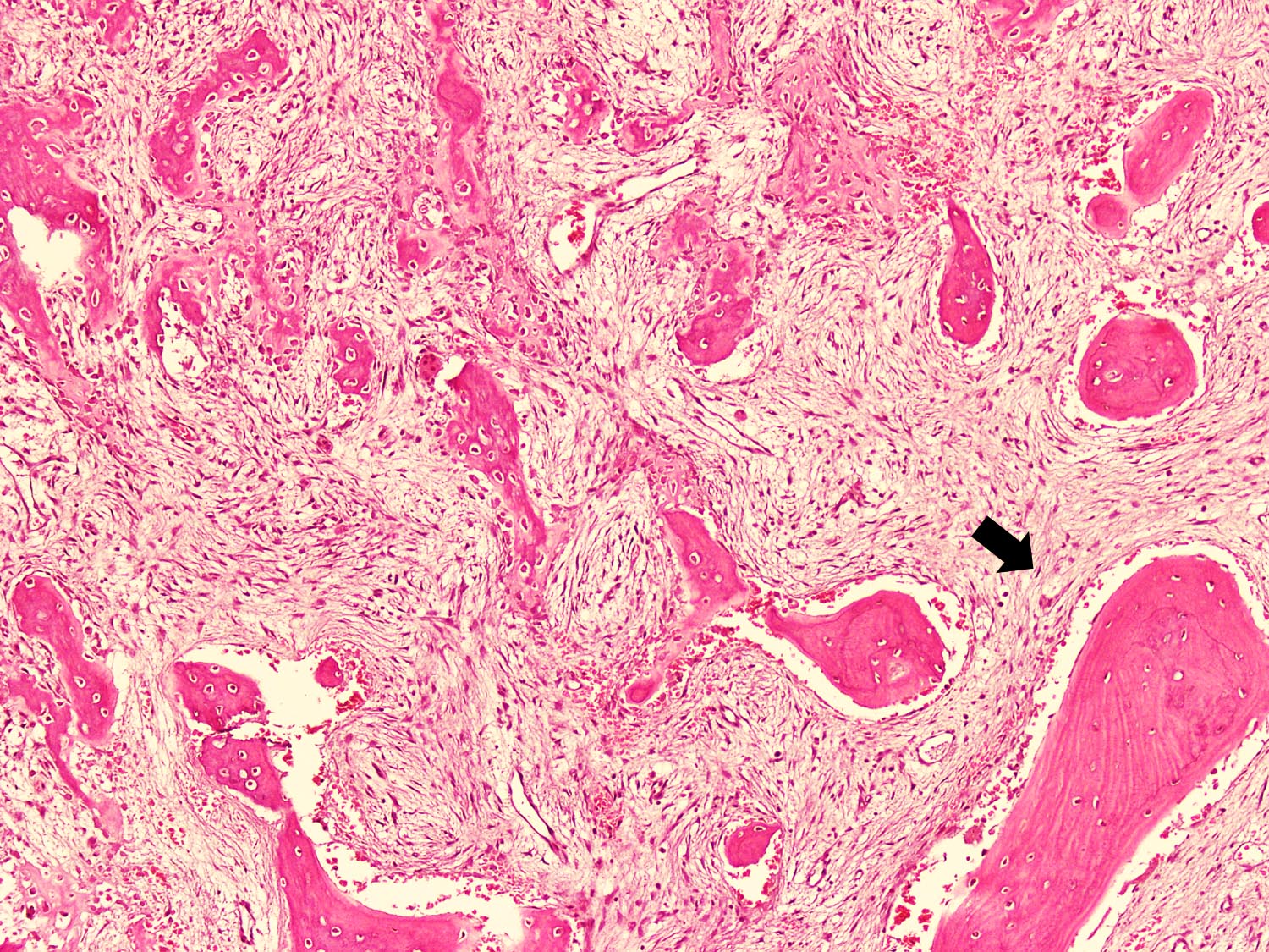
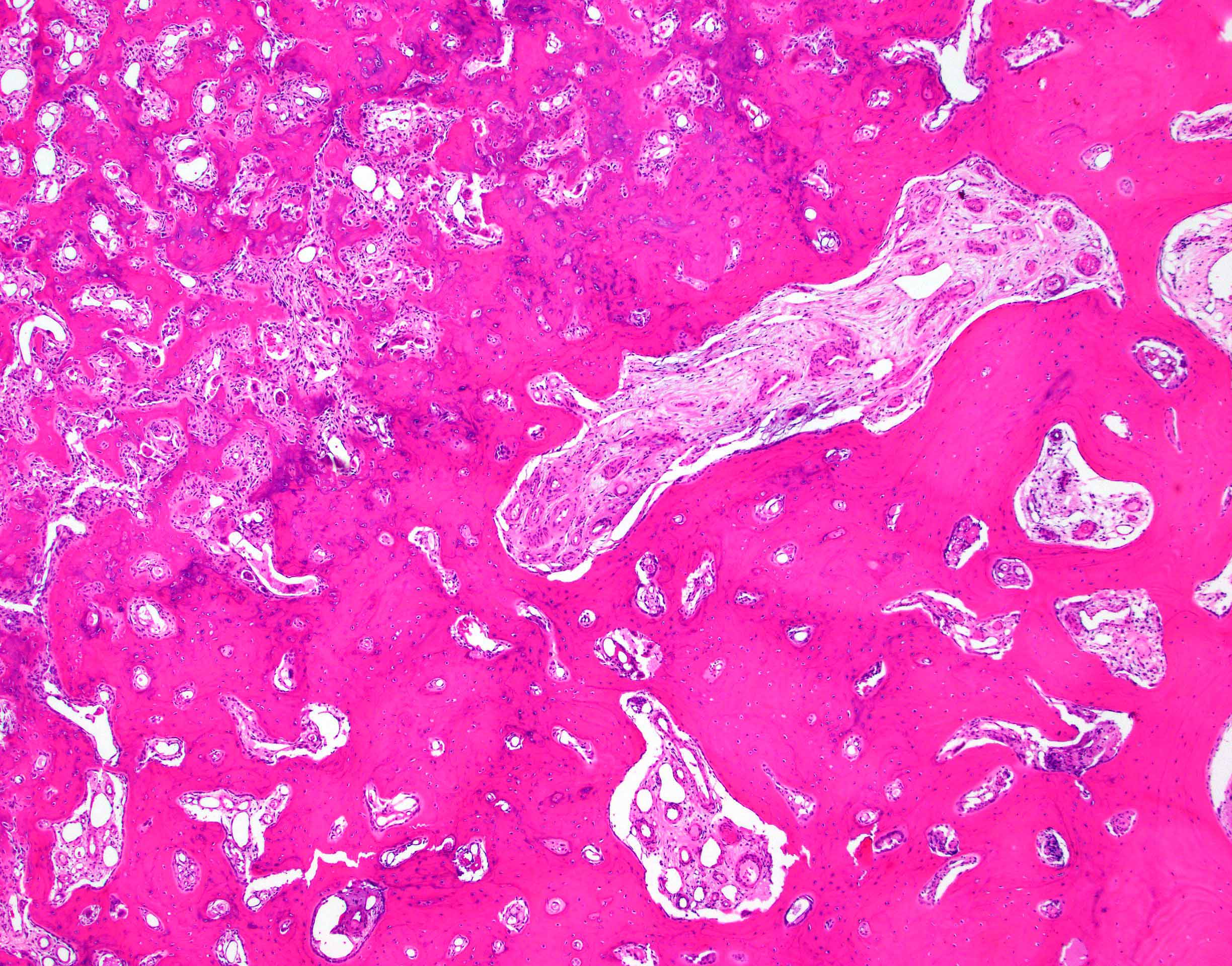
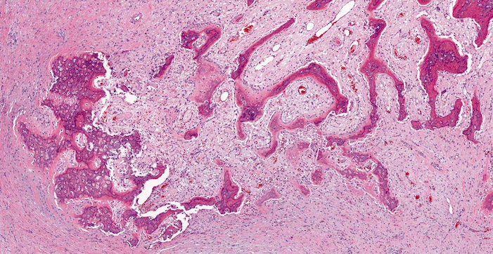
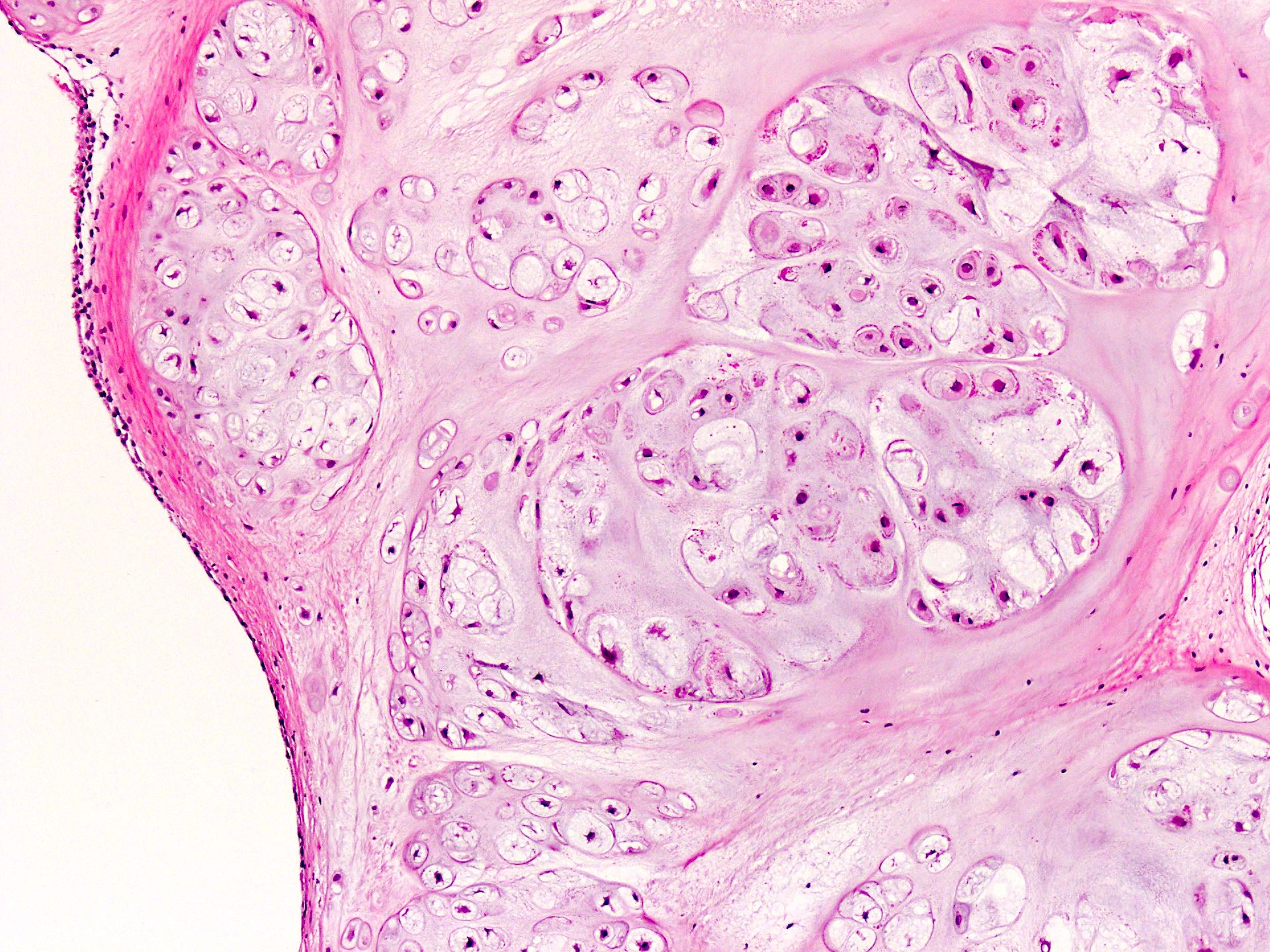
Bone trabeculae and storiform pattern
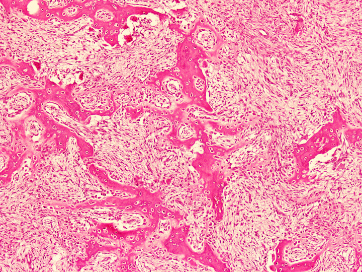
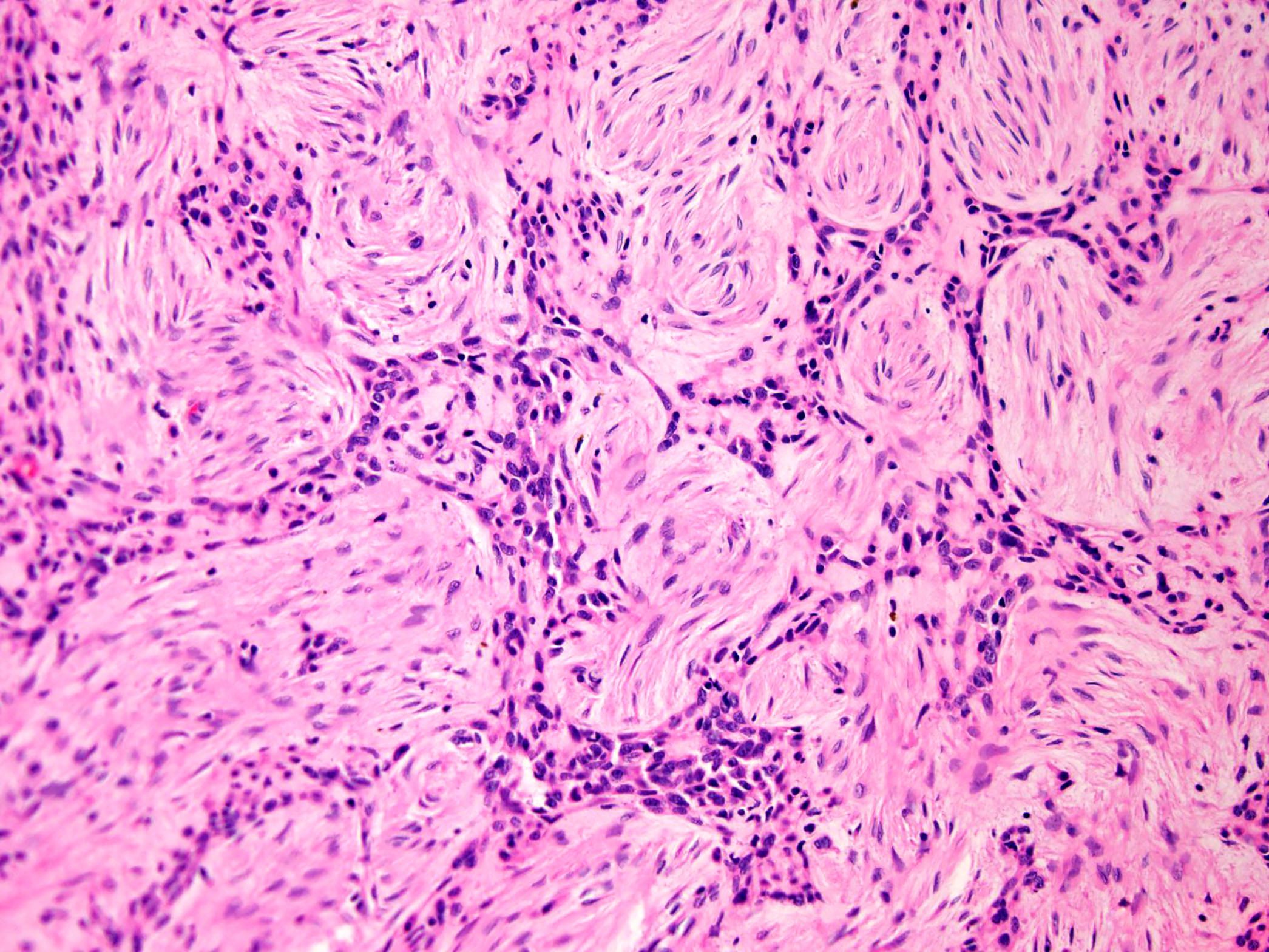
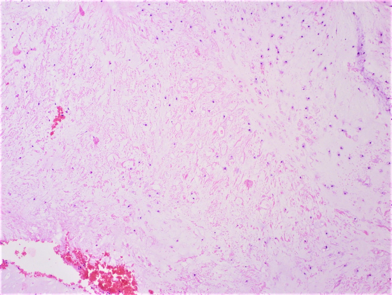
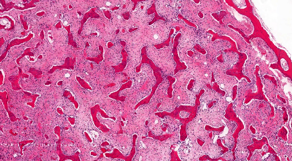
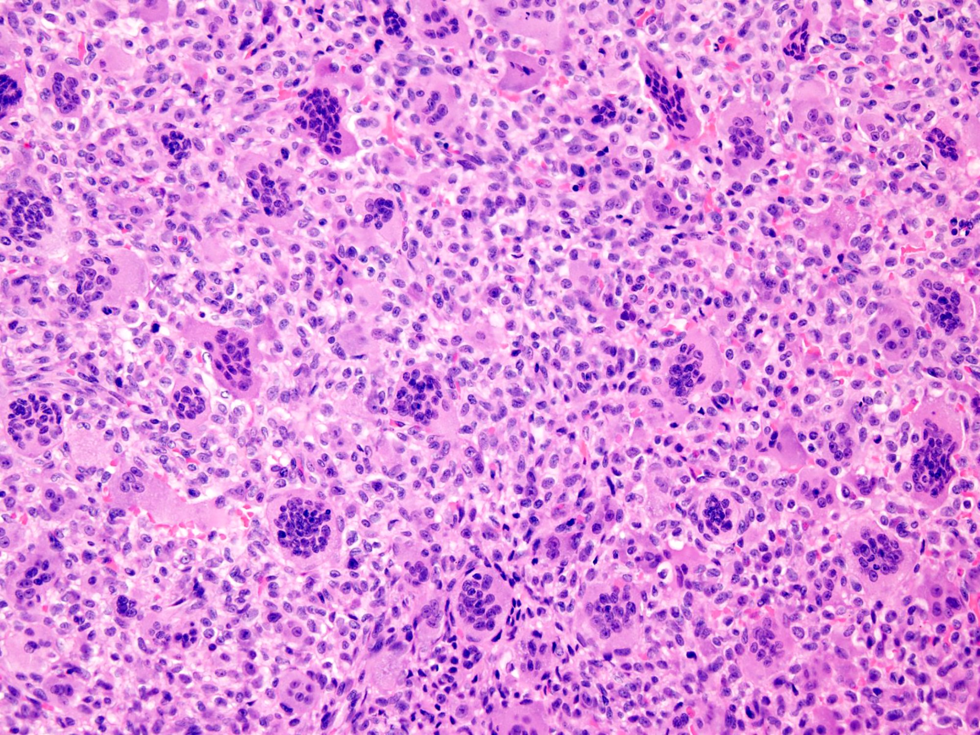
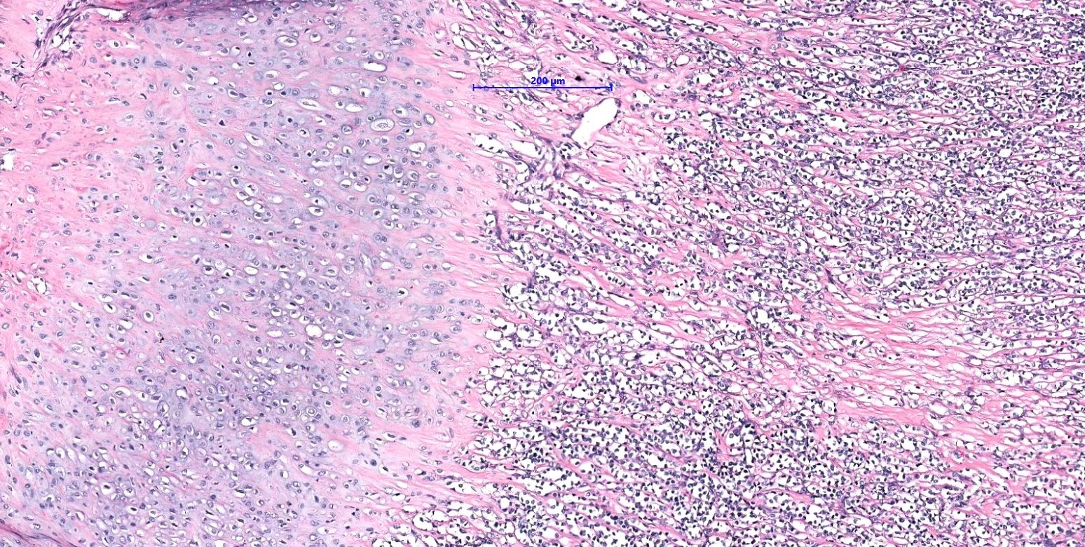
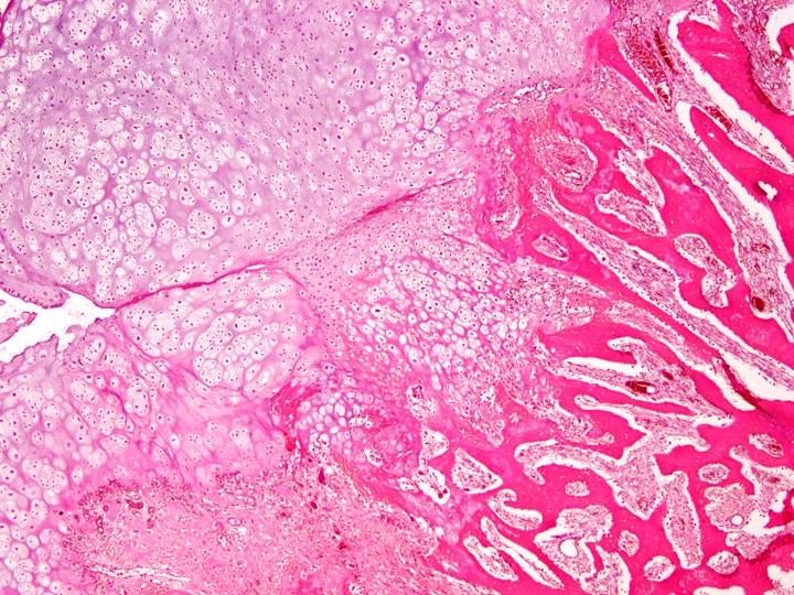
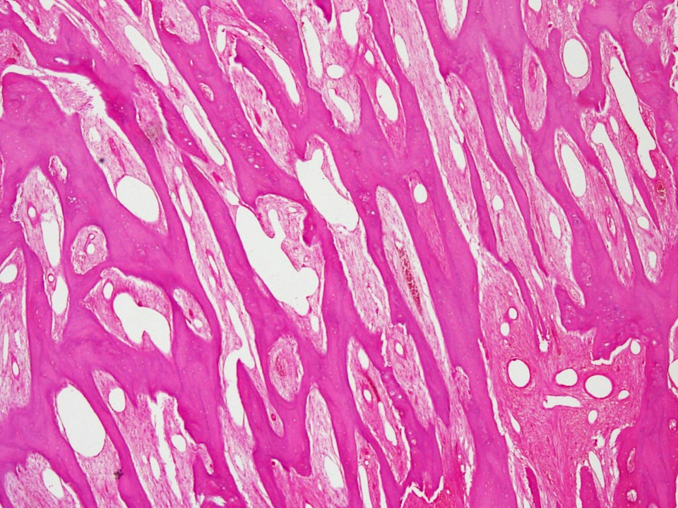
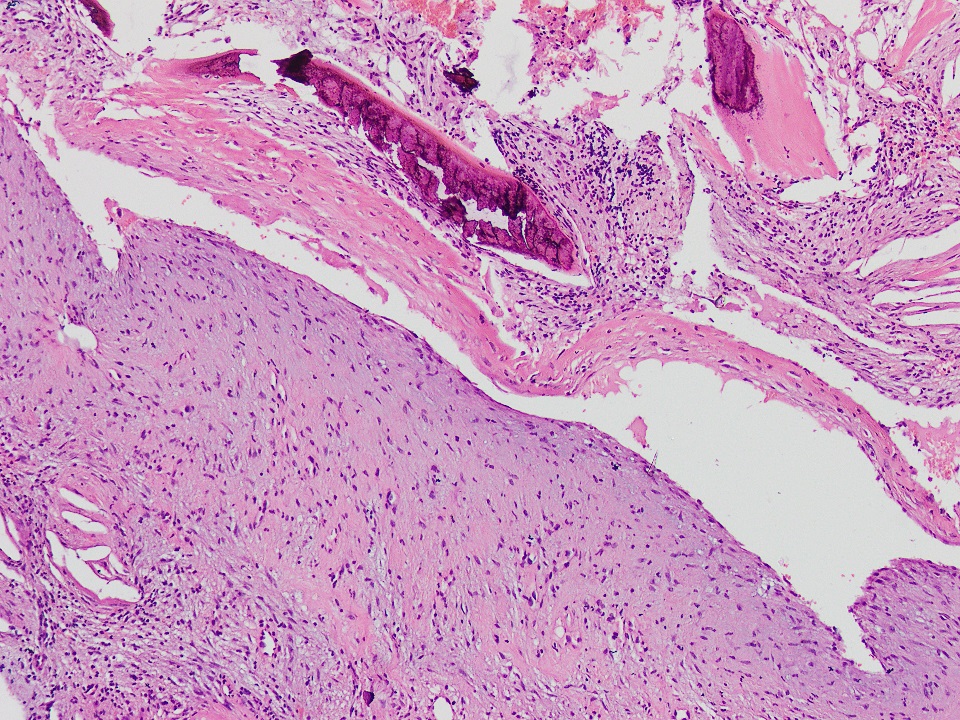
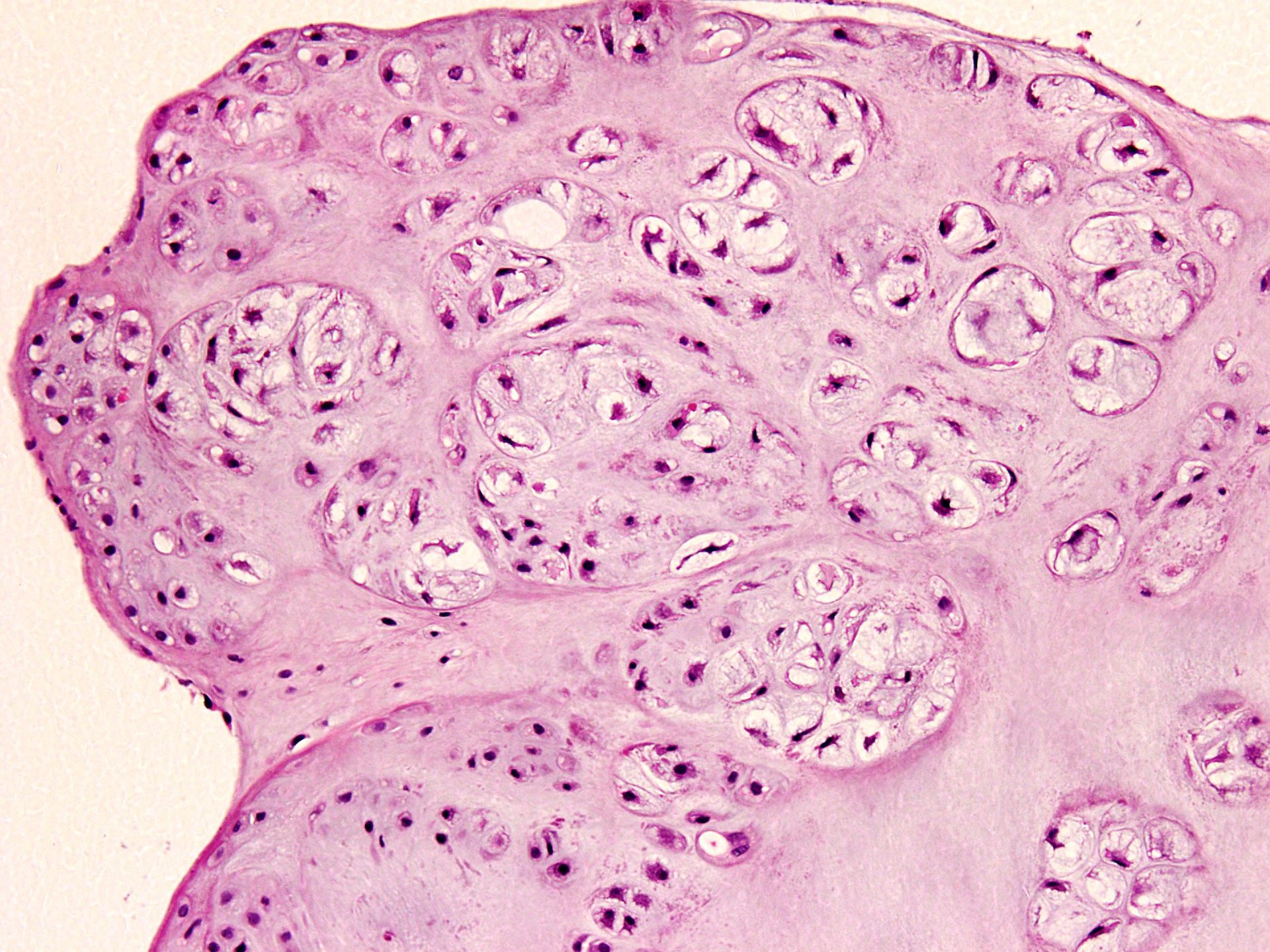
Osteoblastic rimming of bone trabeculae
Osteoblastic rimming and storiform pattern
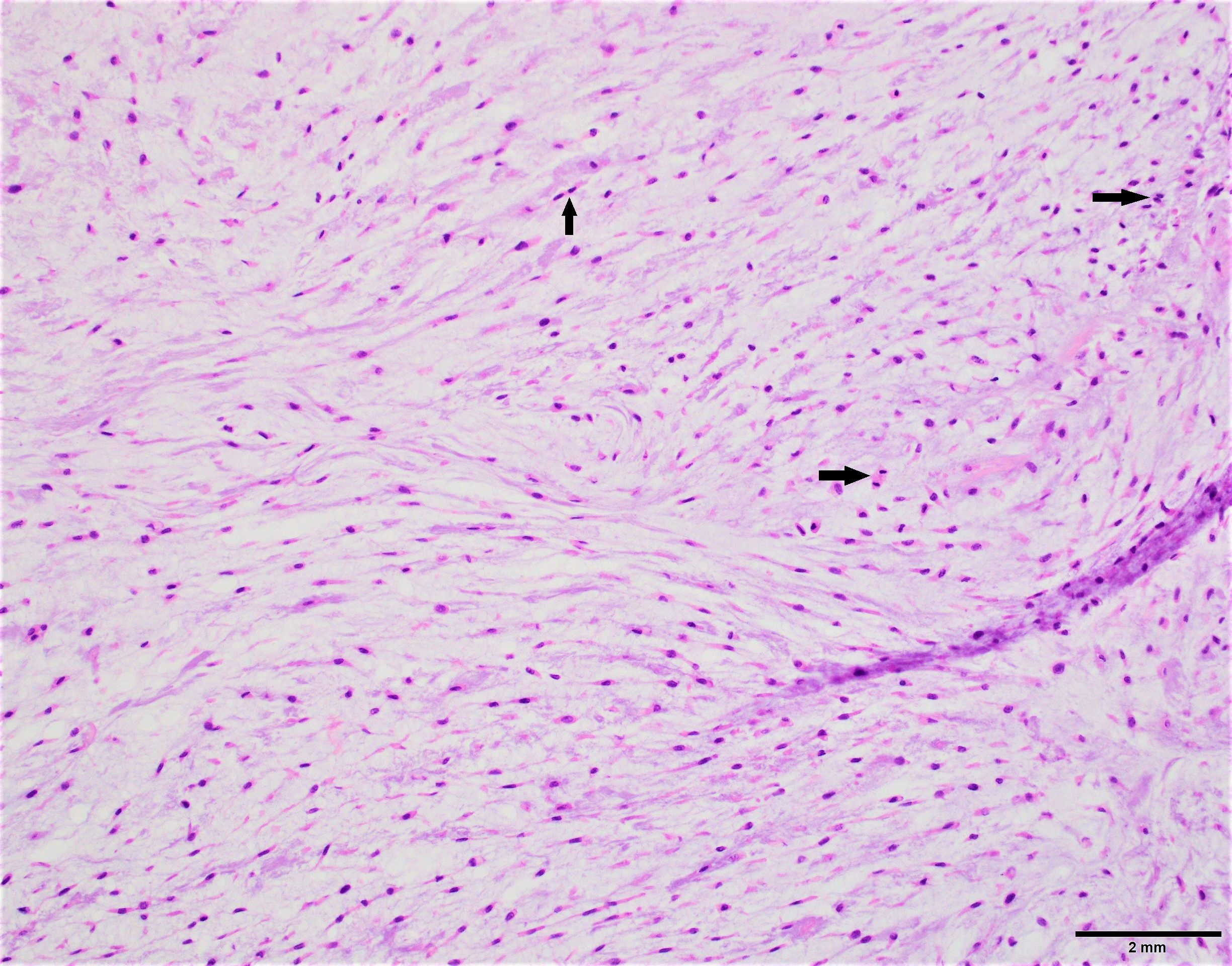
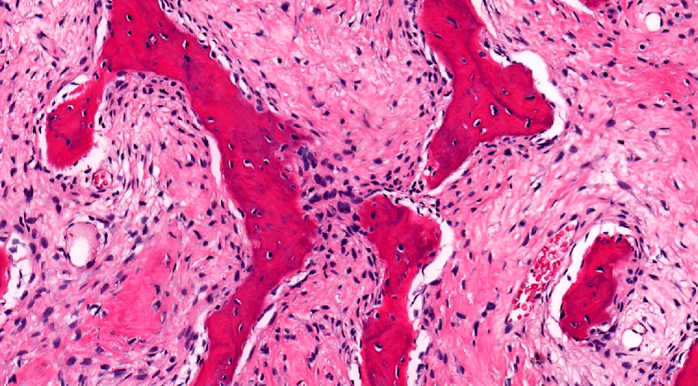
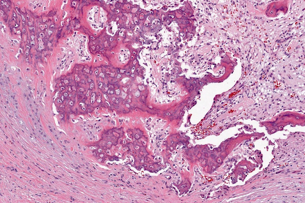
Osteoblastic rimming
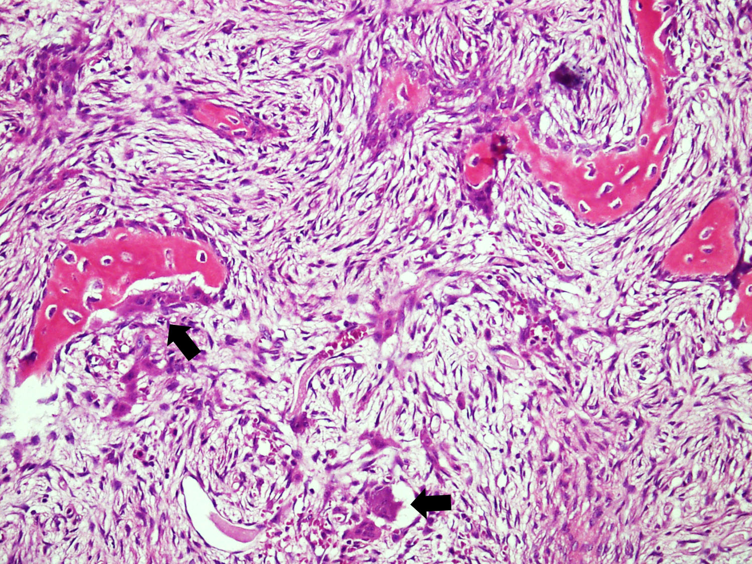
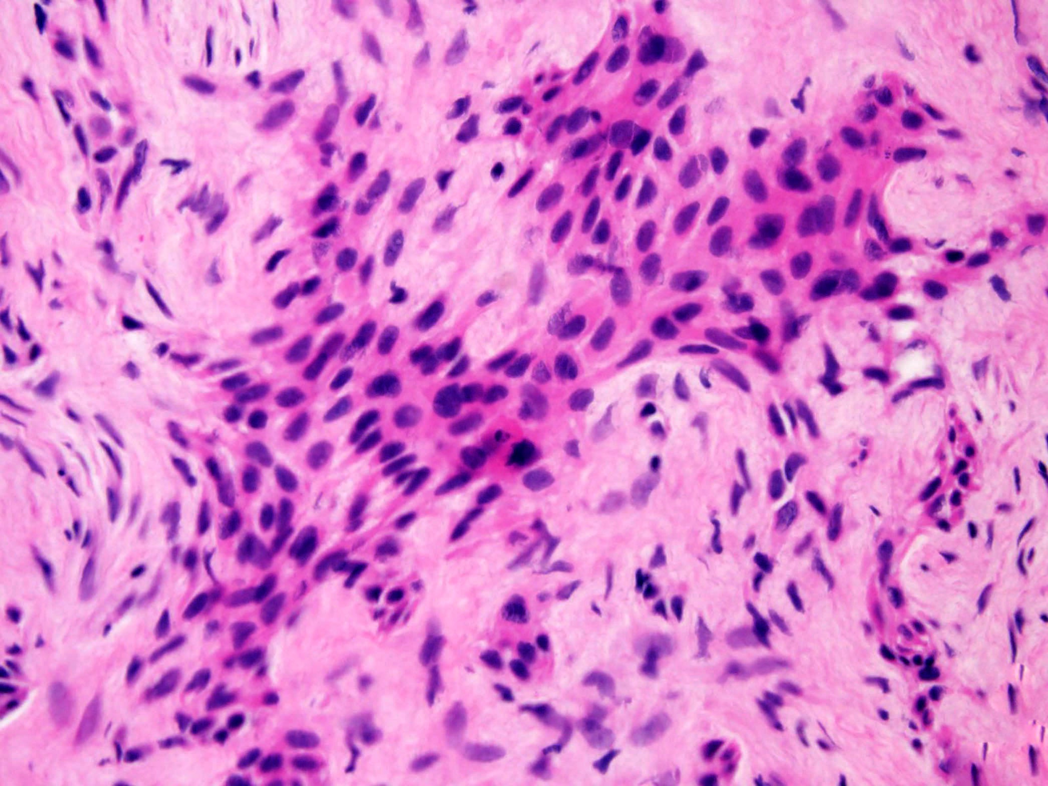
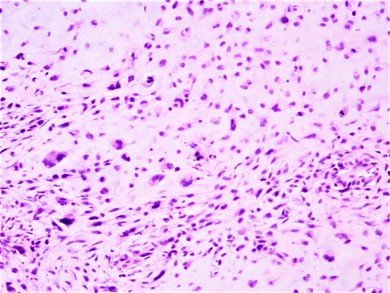
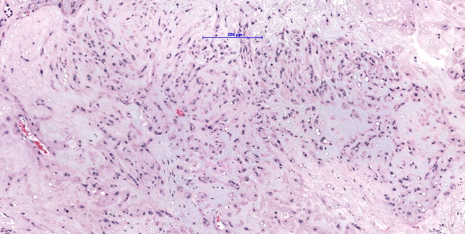
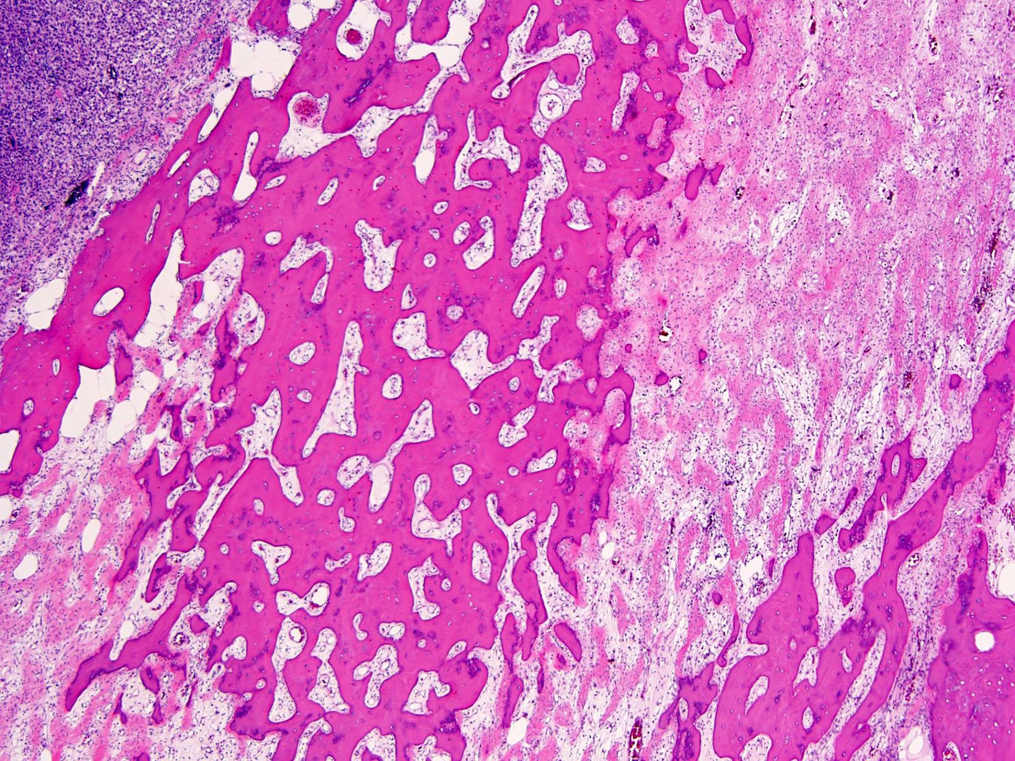
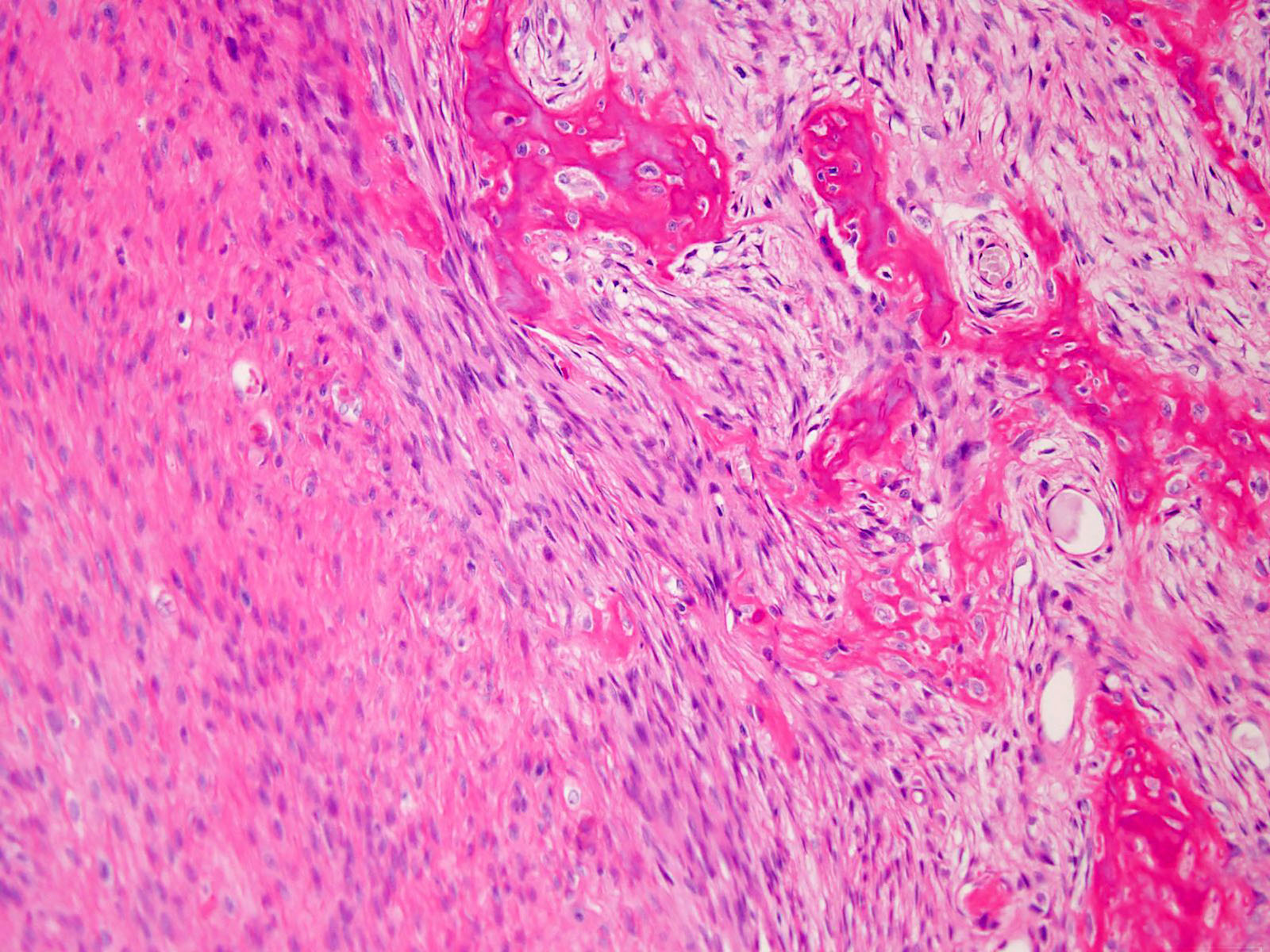
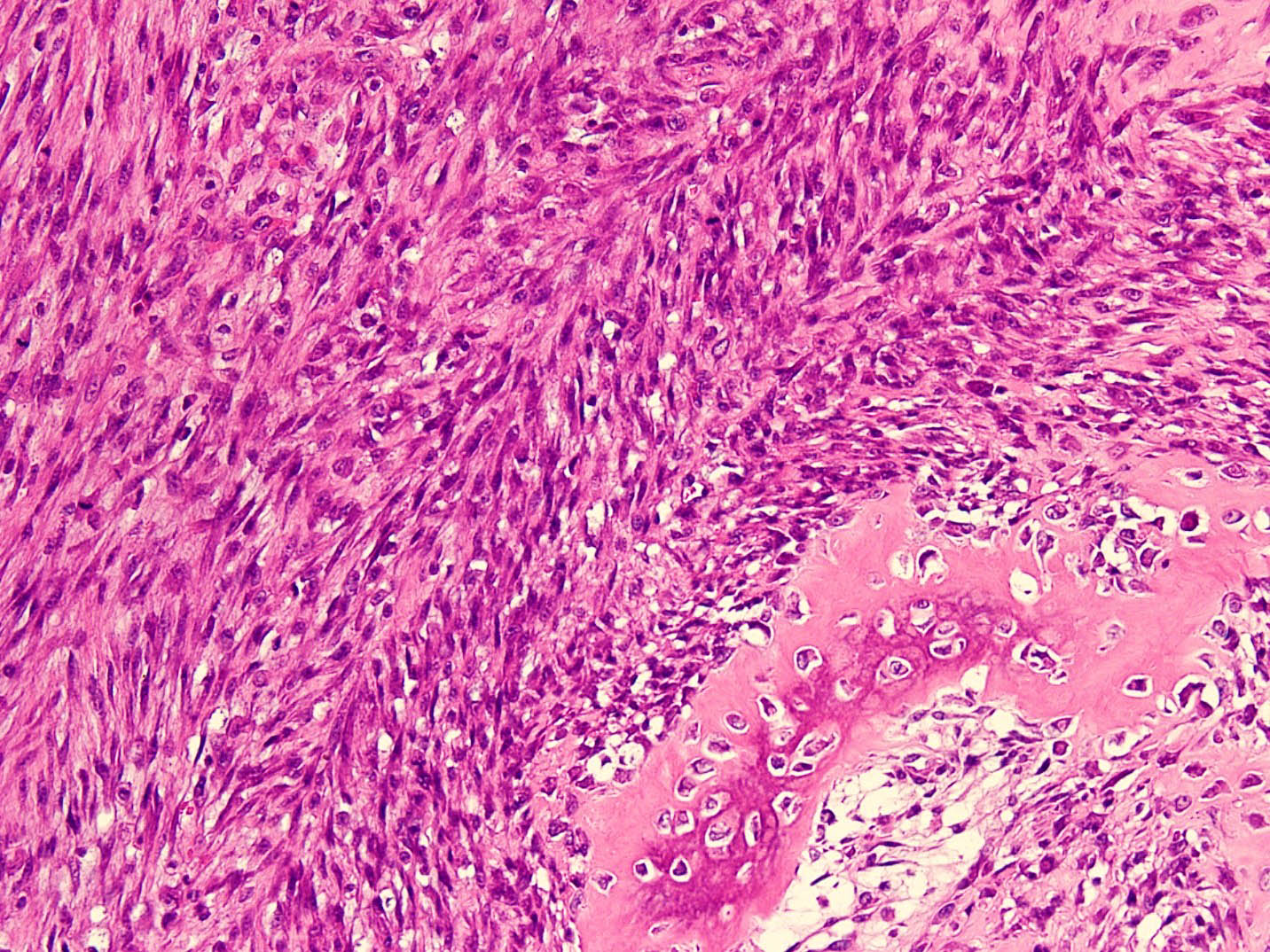
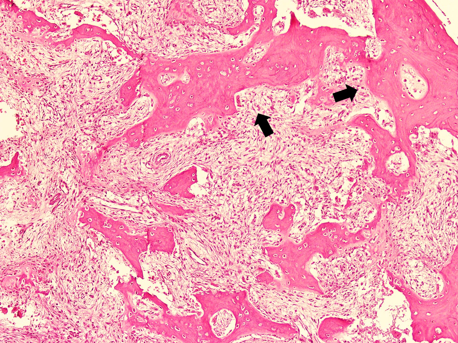
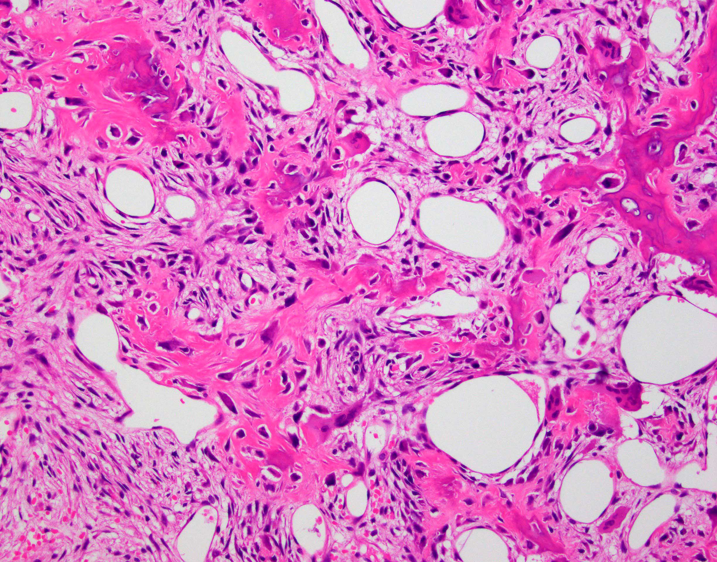
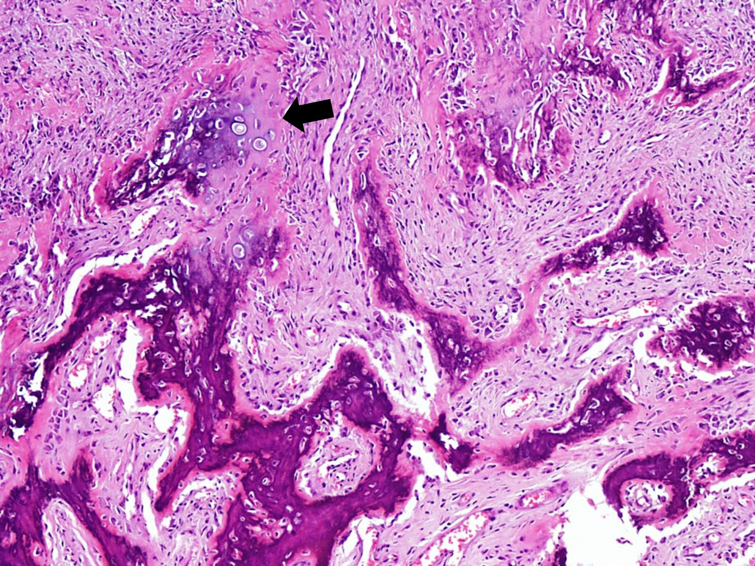
Multinucleated osteoclast-like
giant cells
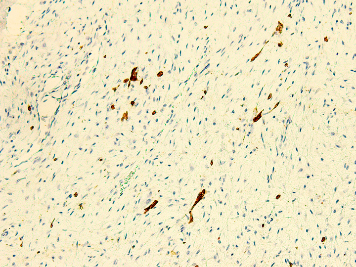
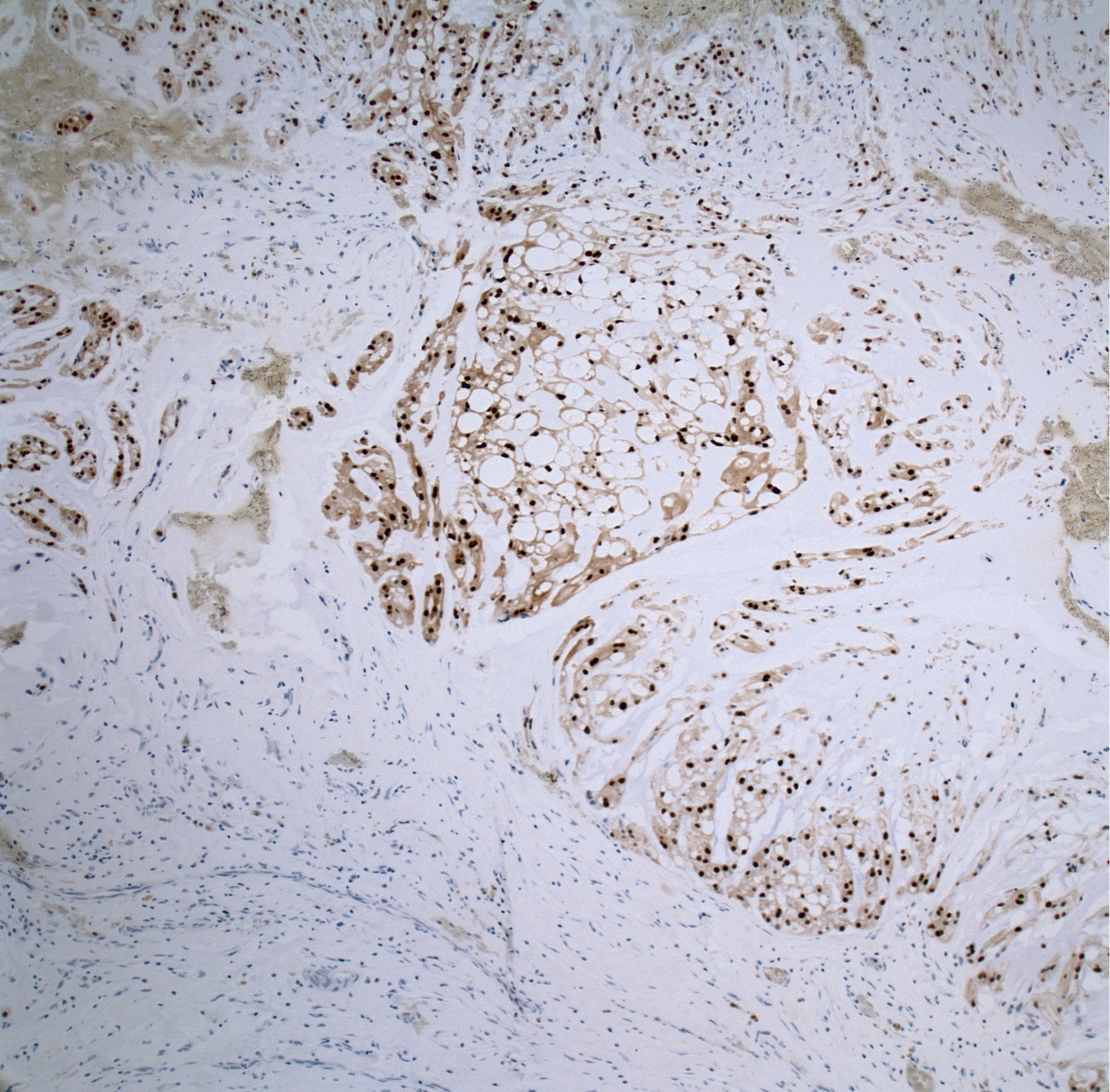
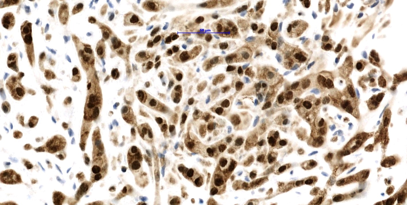
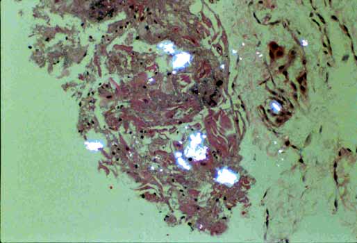
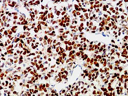
%20keratin%20immunost.jpg)
Keratin immunostain
Osteofibrous dysplasia
Fibrous dysplasia and osteofibrous dysplasia: bone forming tumors,
part 4
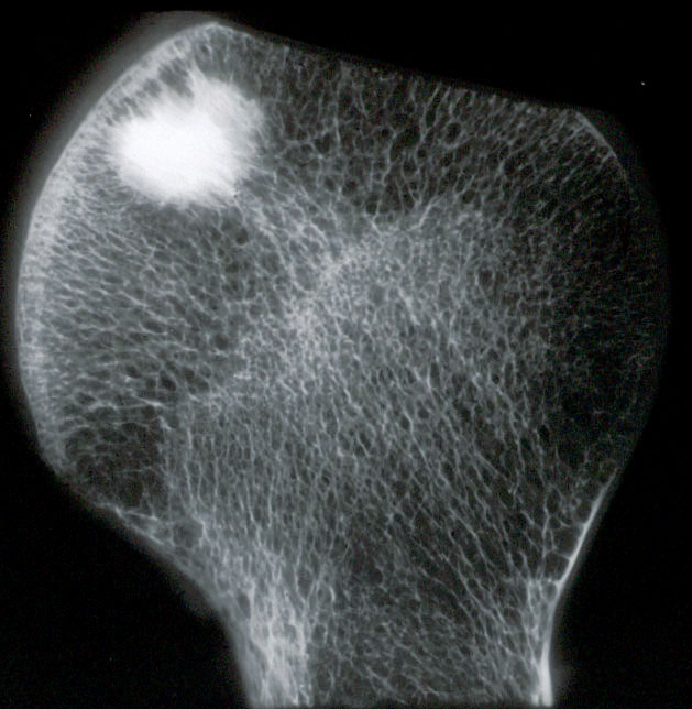
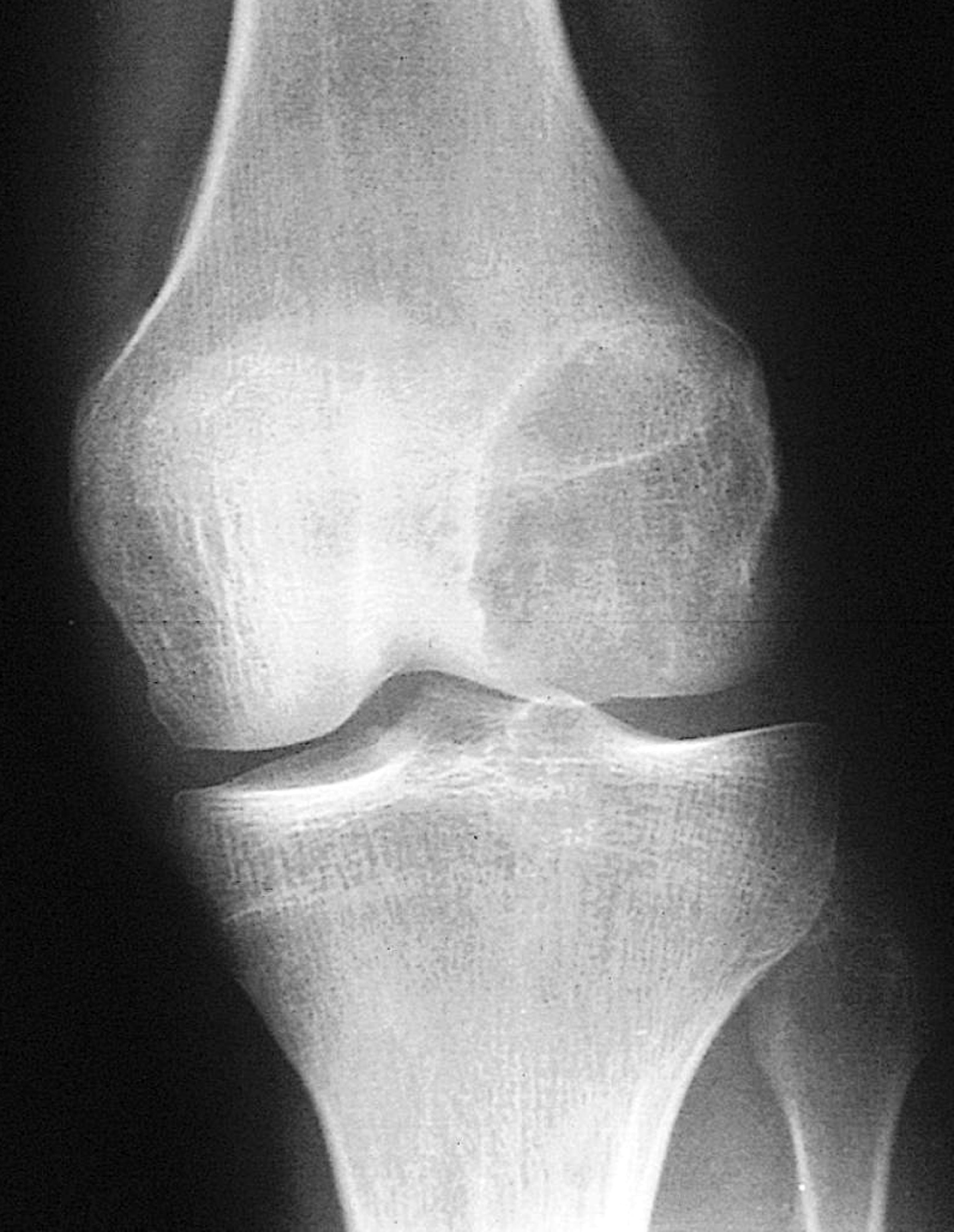
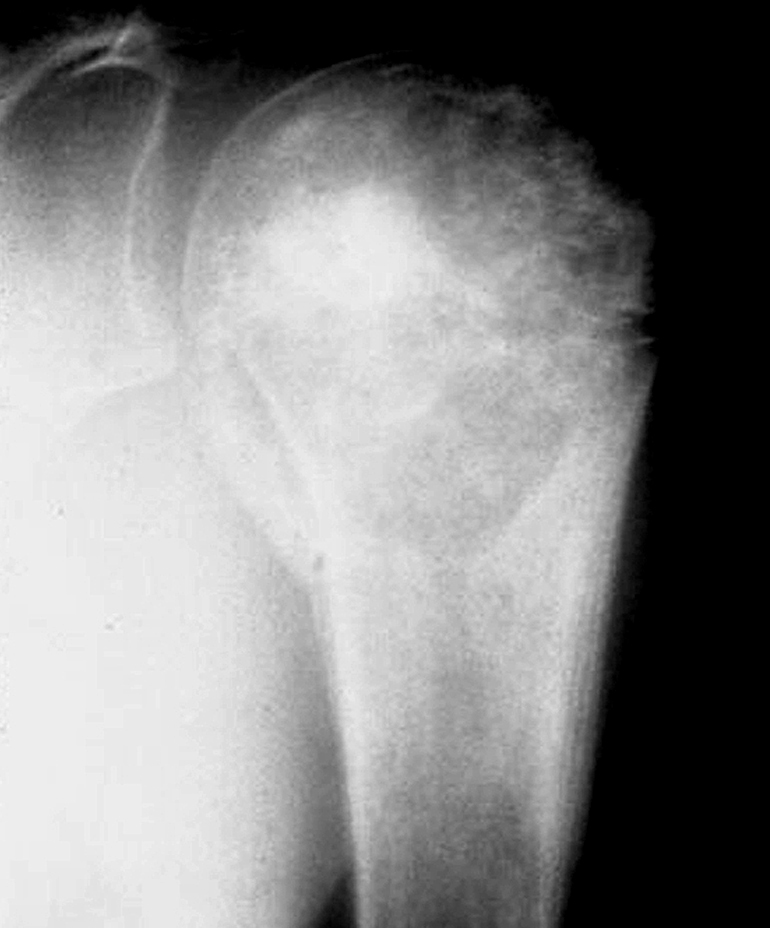
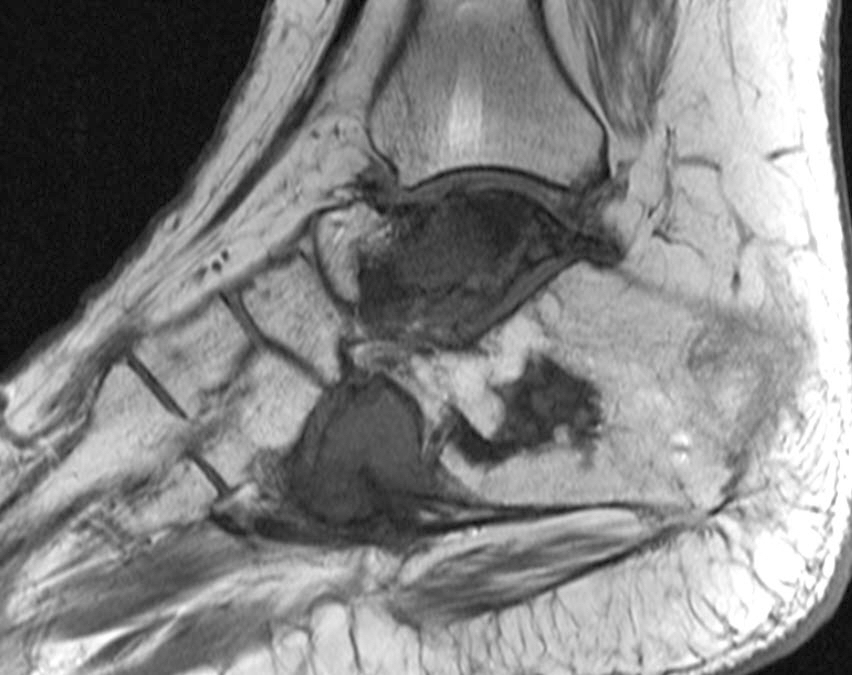
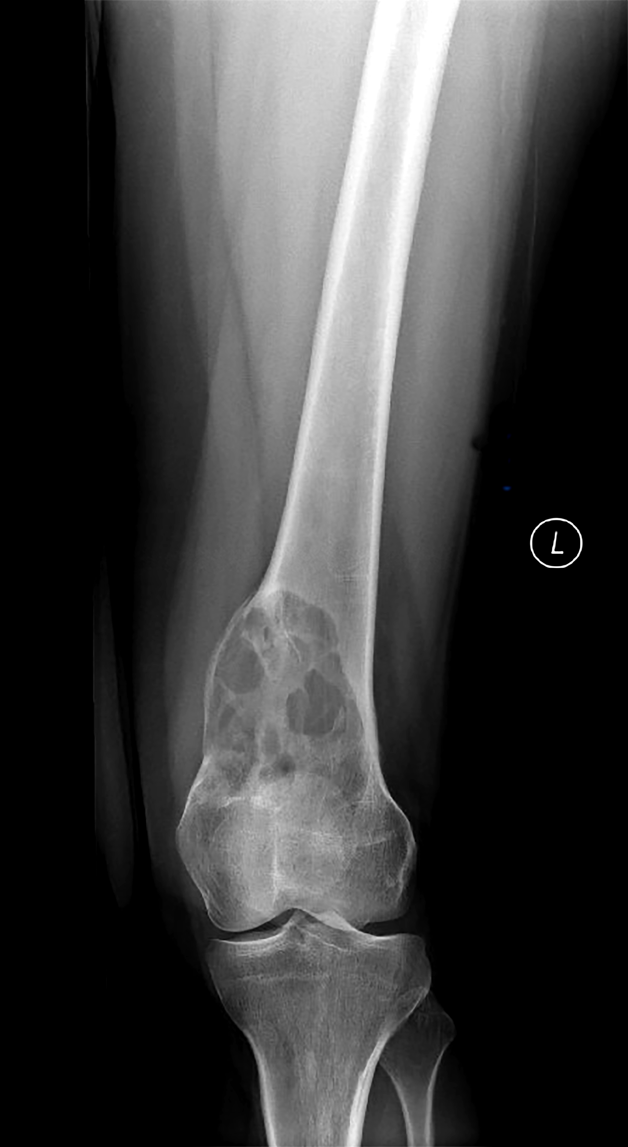
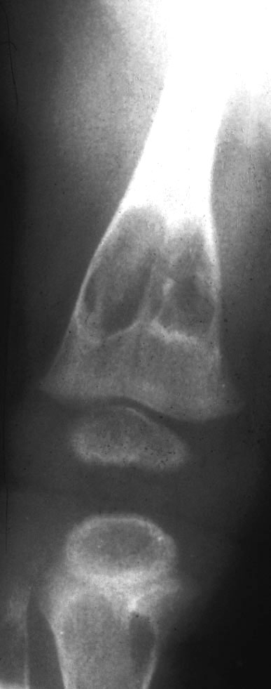
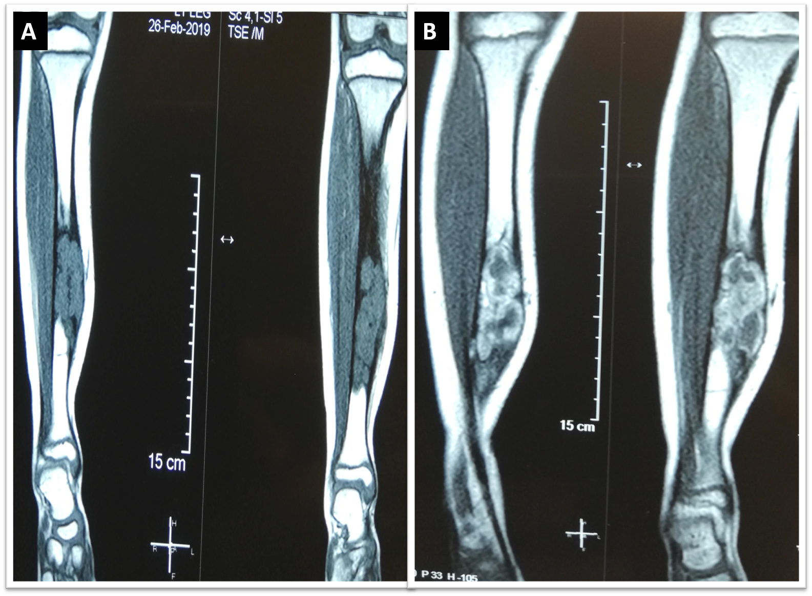
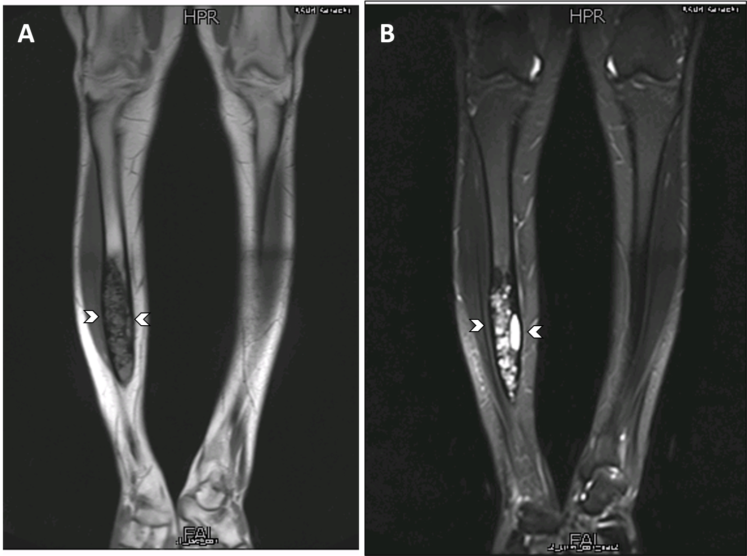
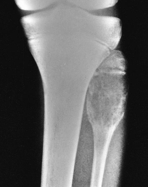
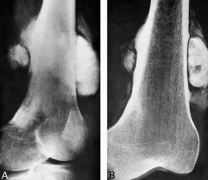
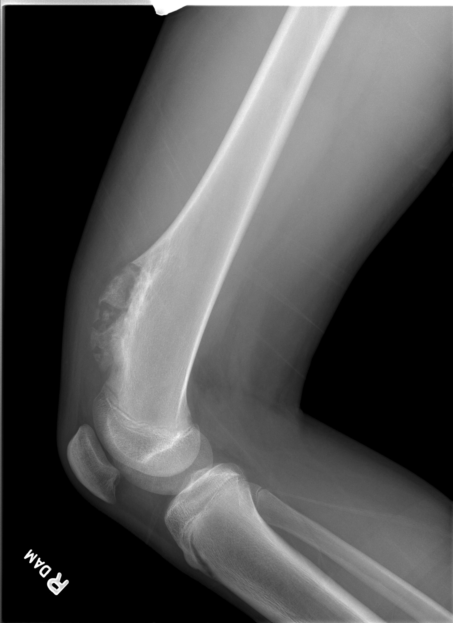
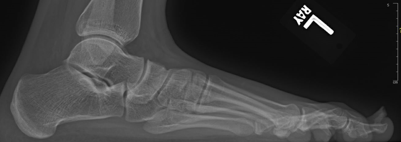
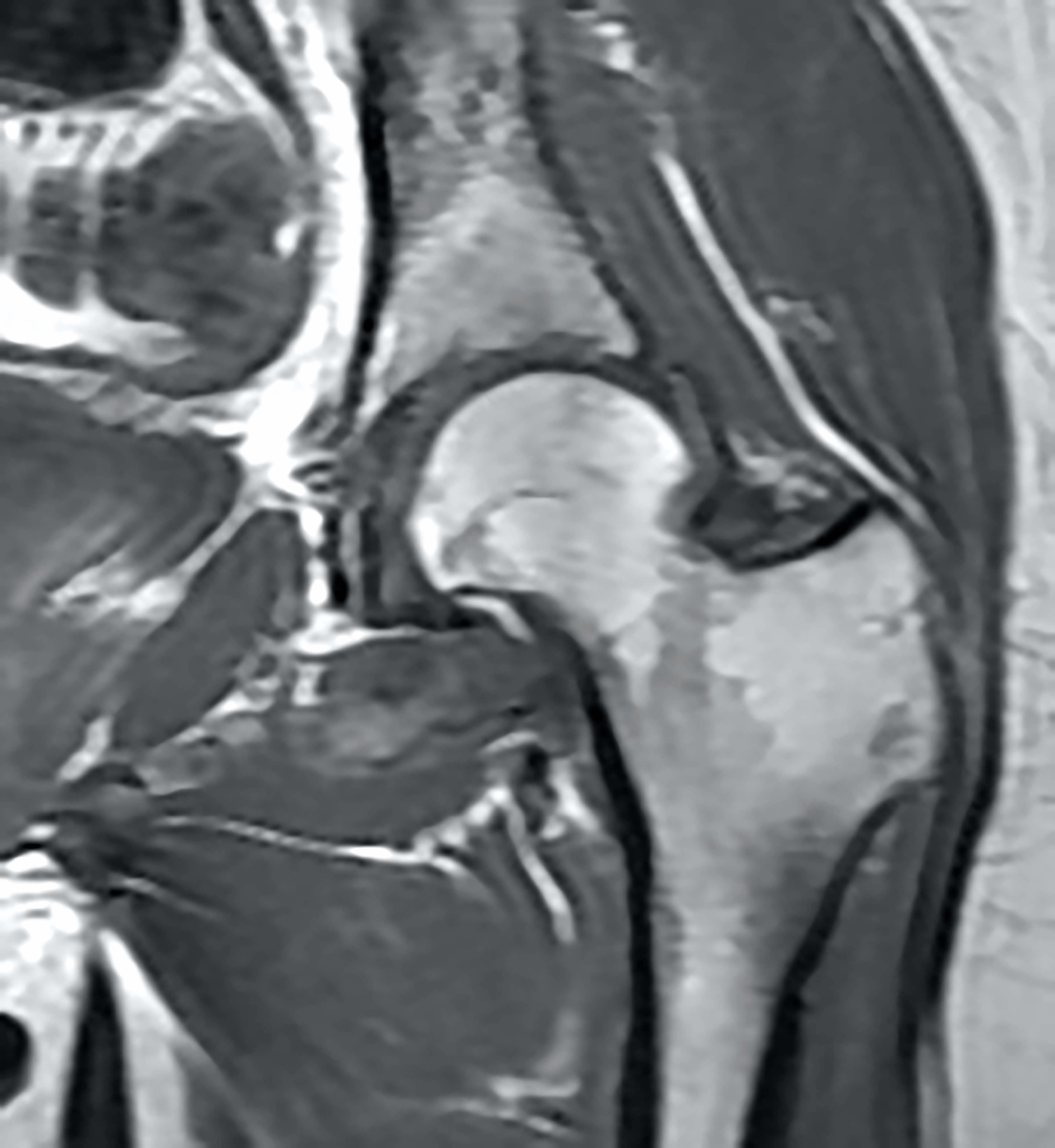
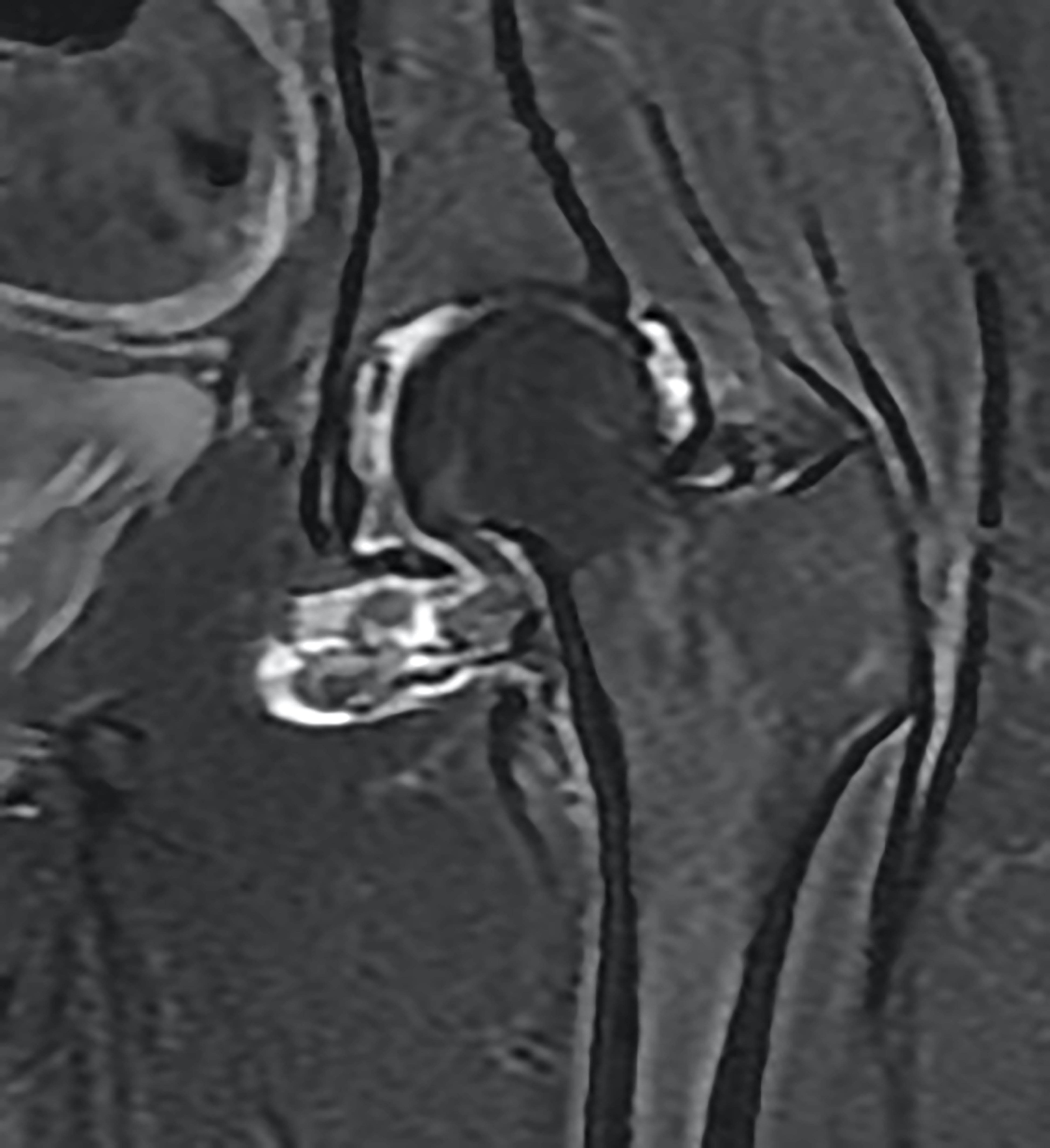
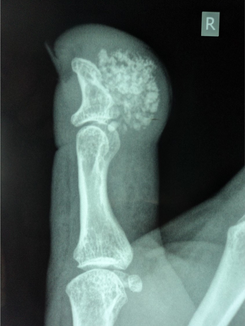
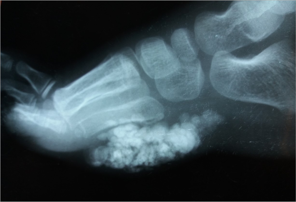
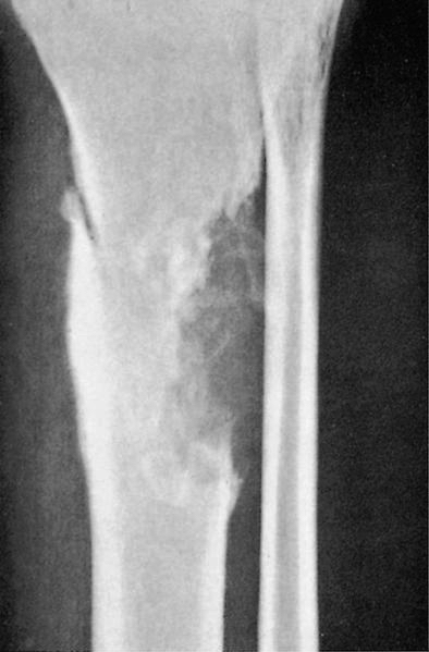
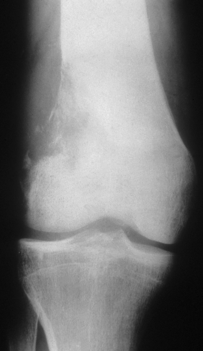
Imaging osteofibrous dysplasia
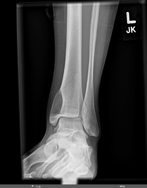
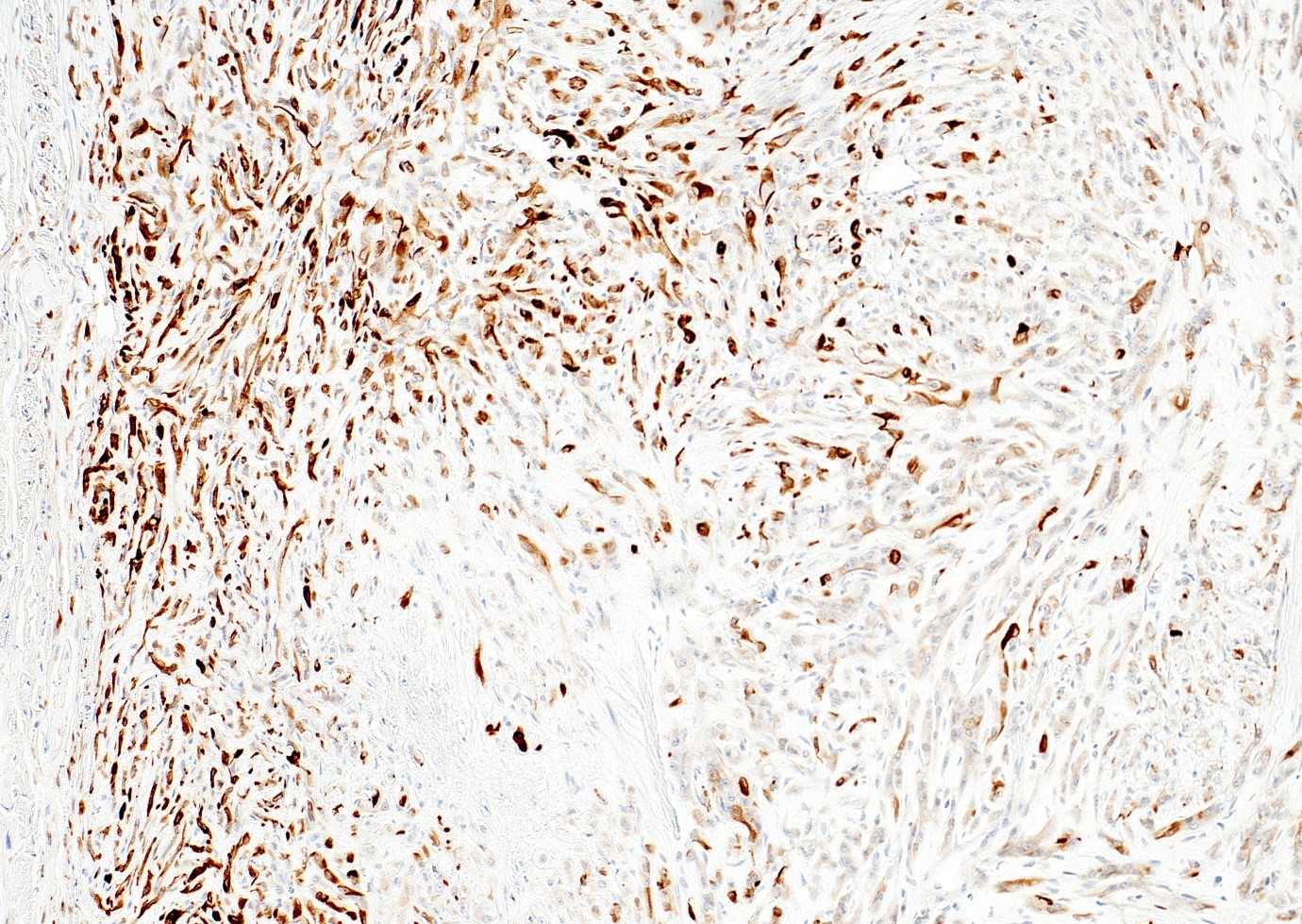
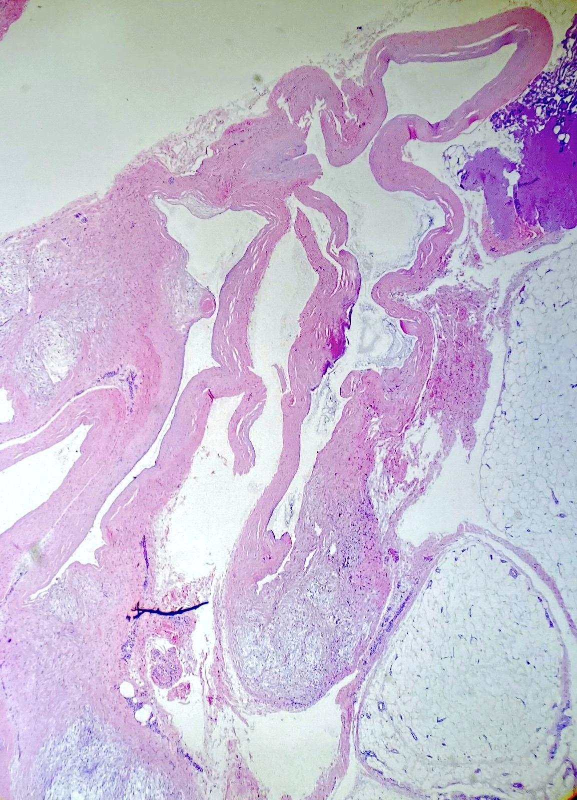
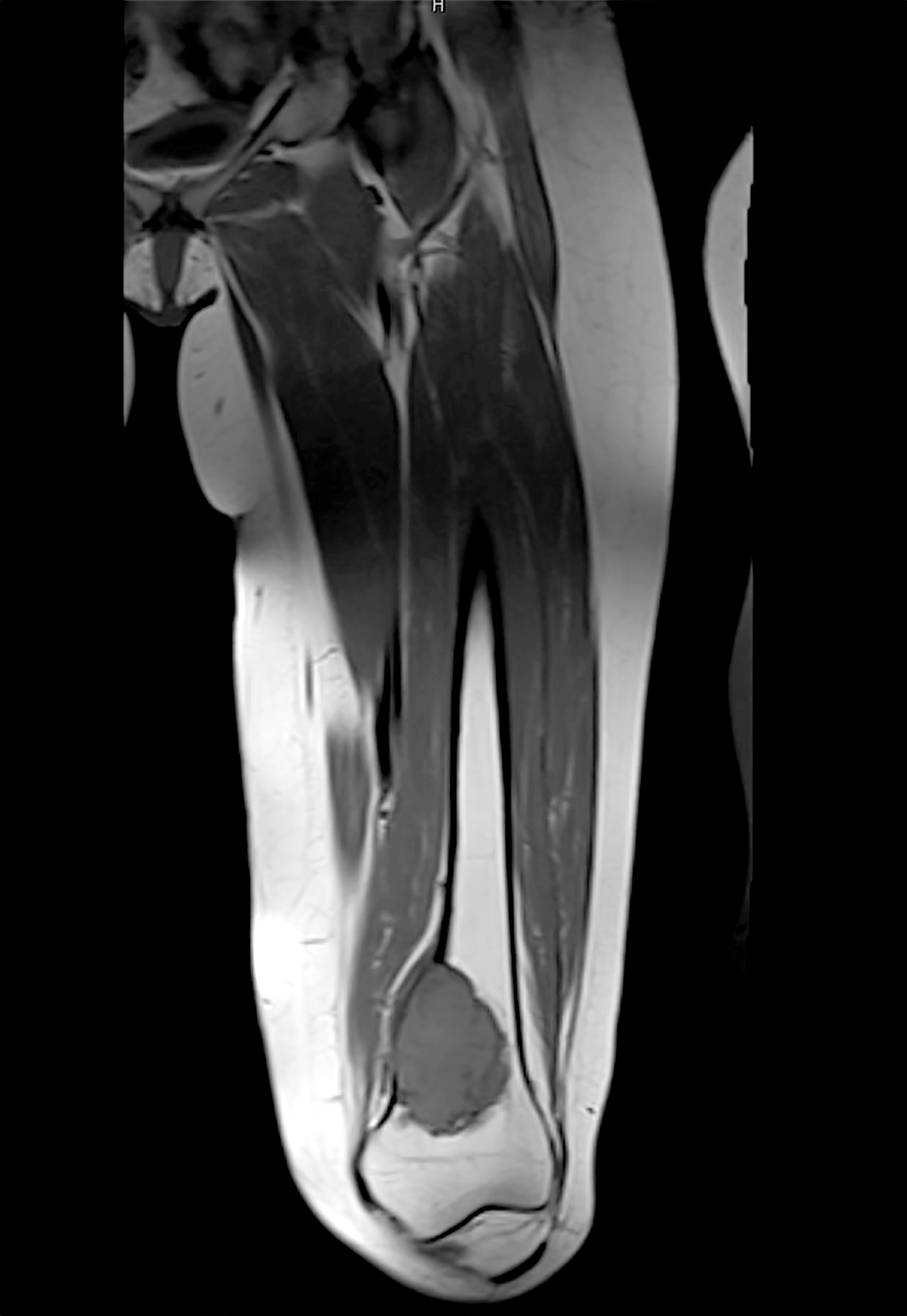
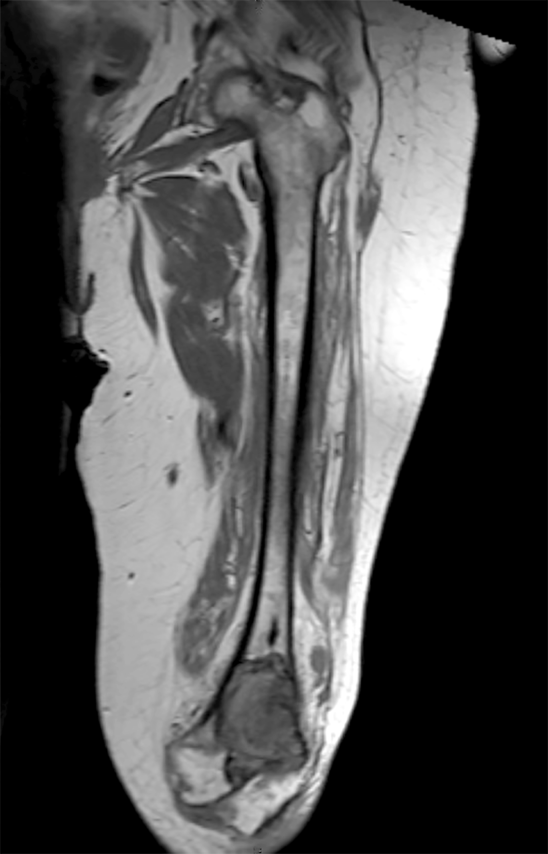
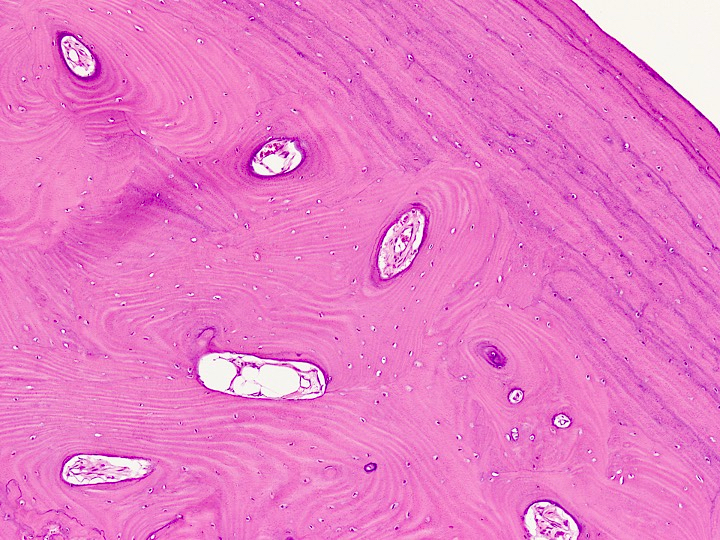
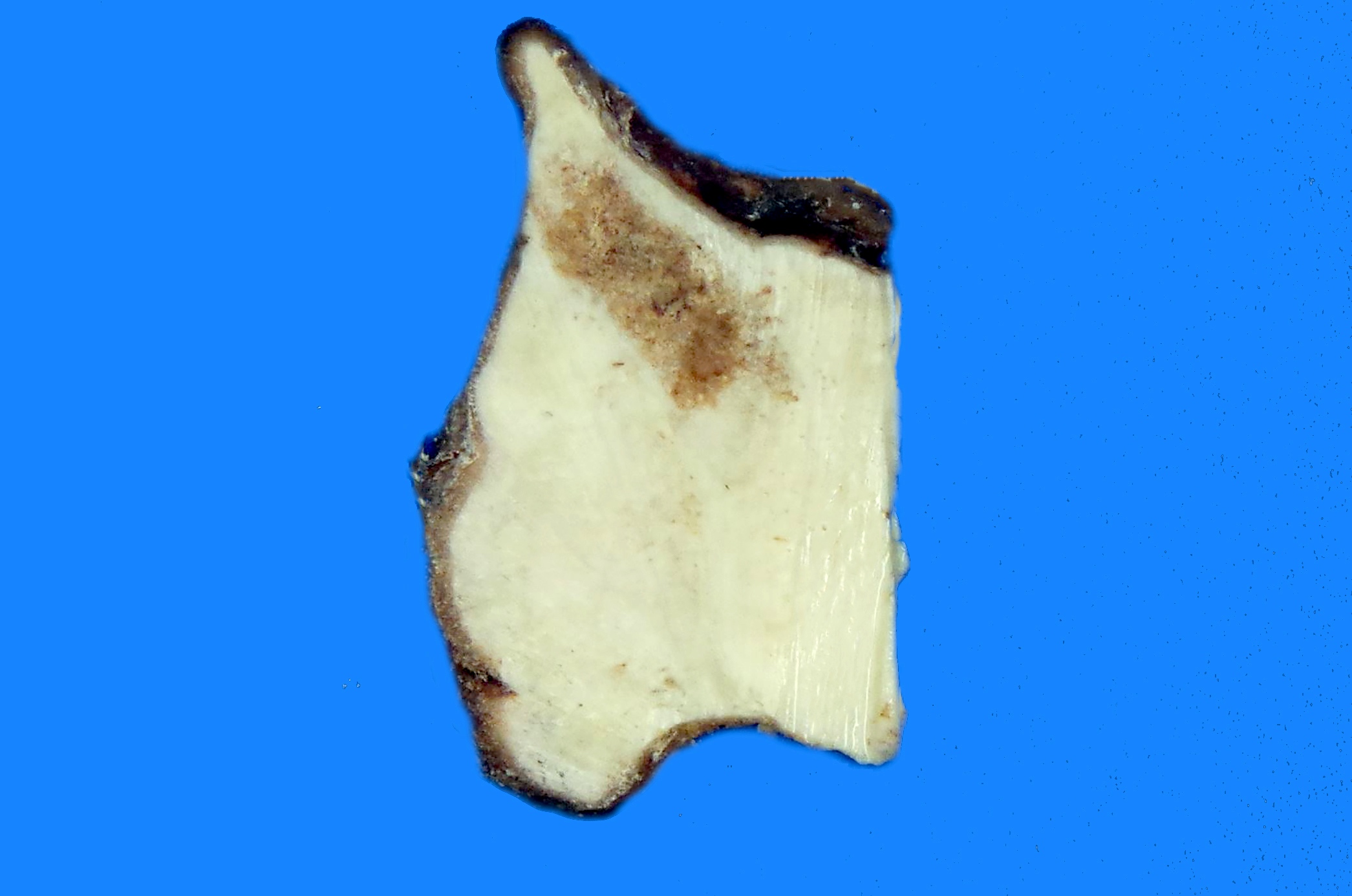
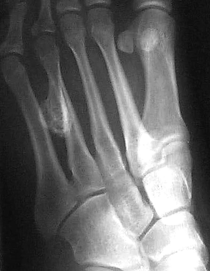
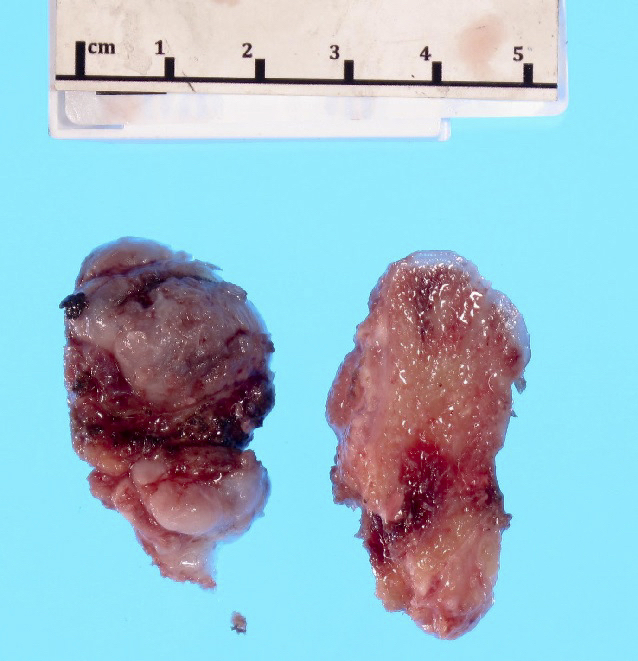
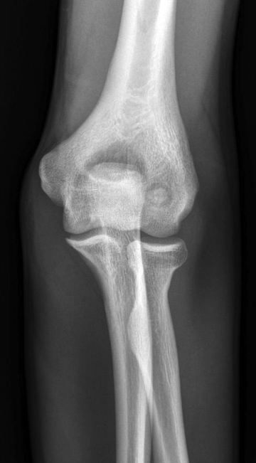
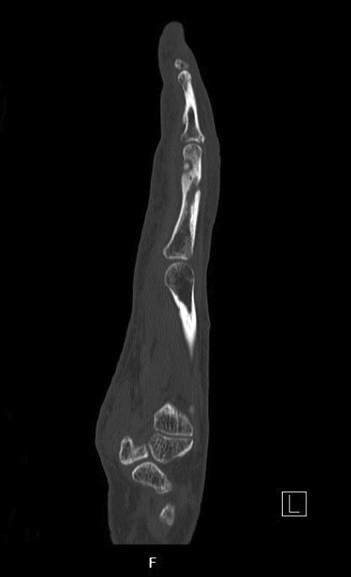
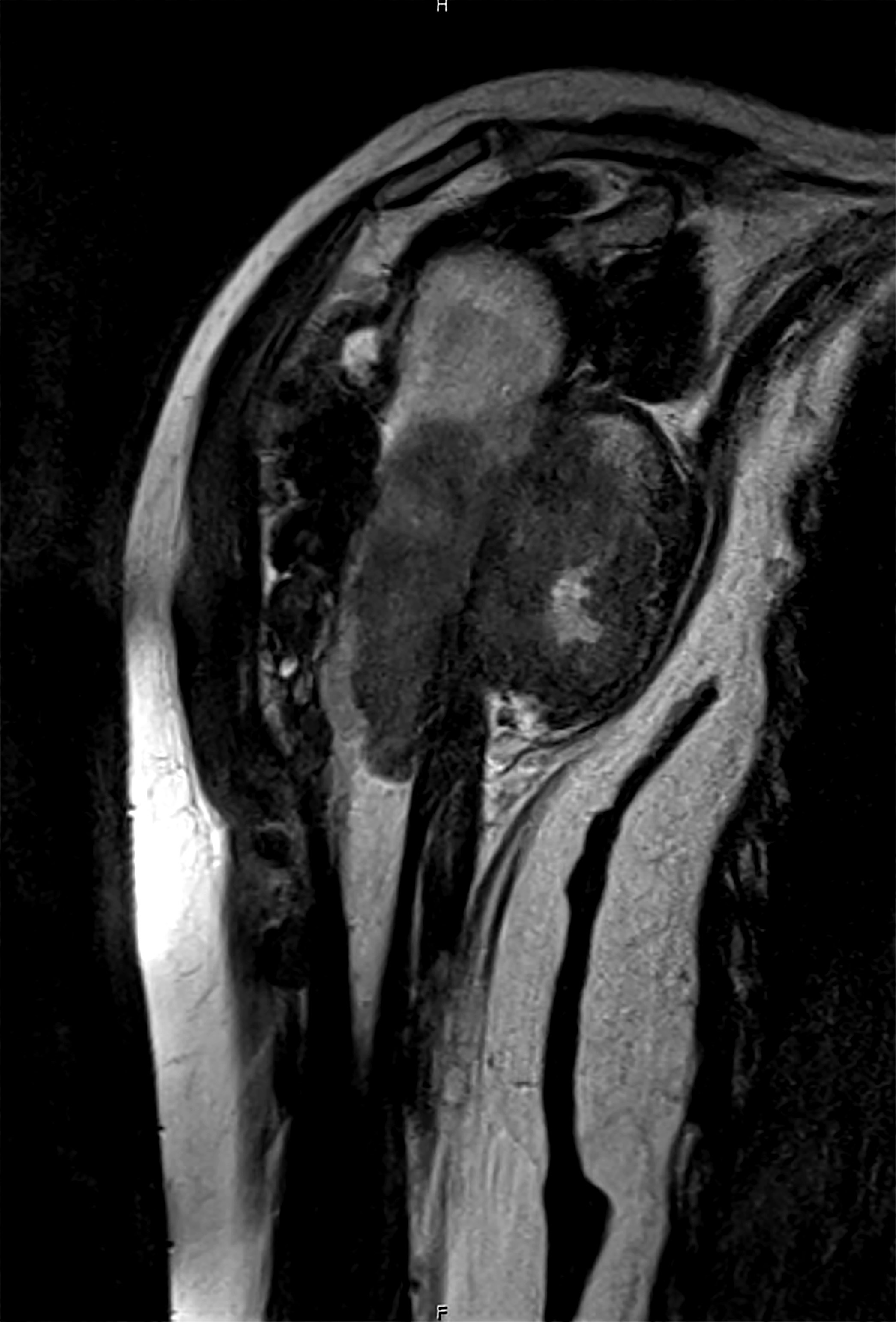
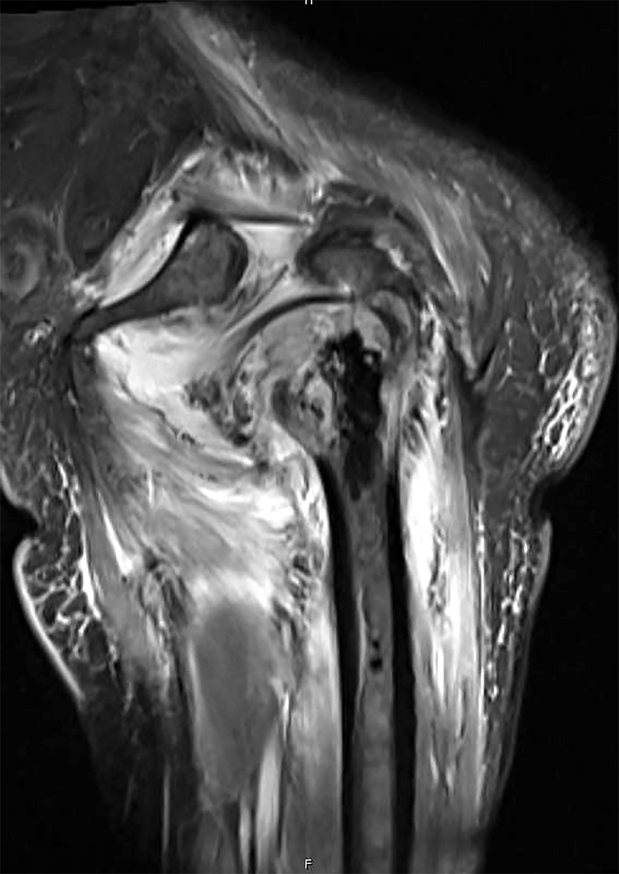
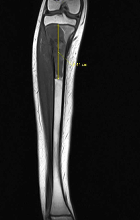
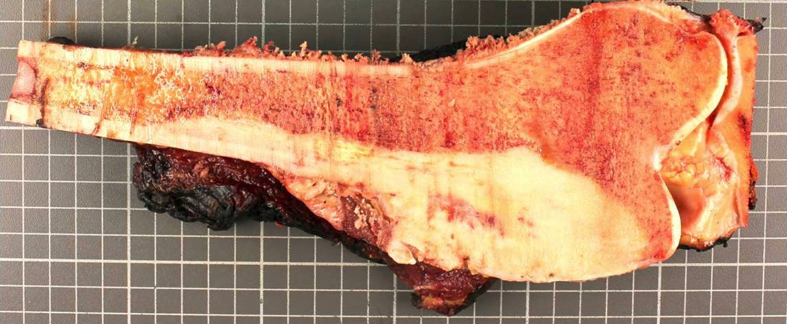
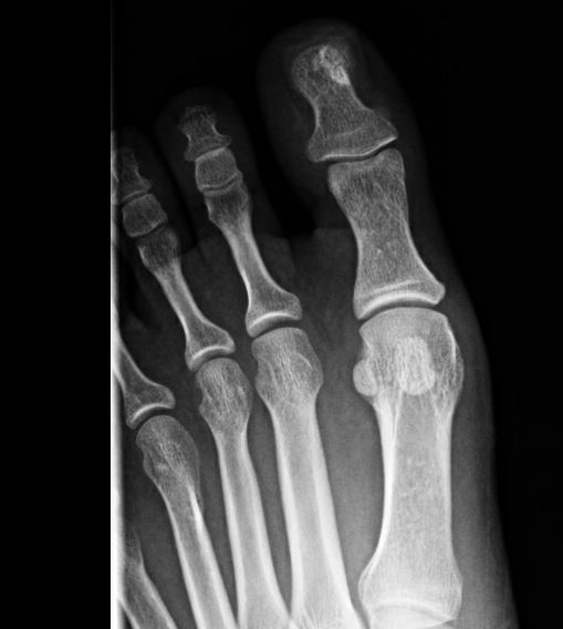
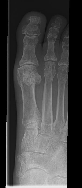
Contributed by Elham Nasri, M.D. and John D. Reith, M.D.
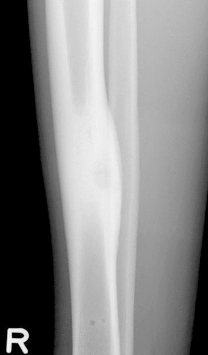
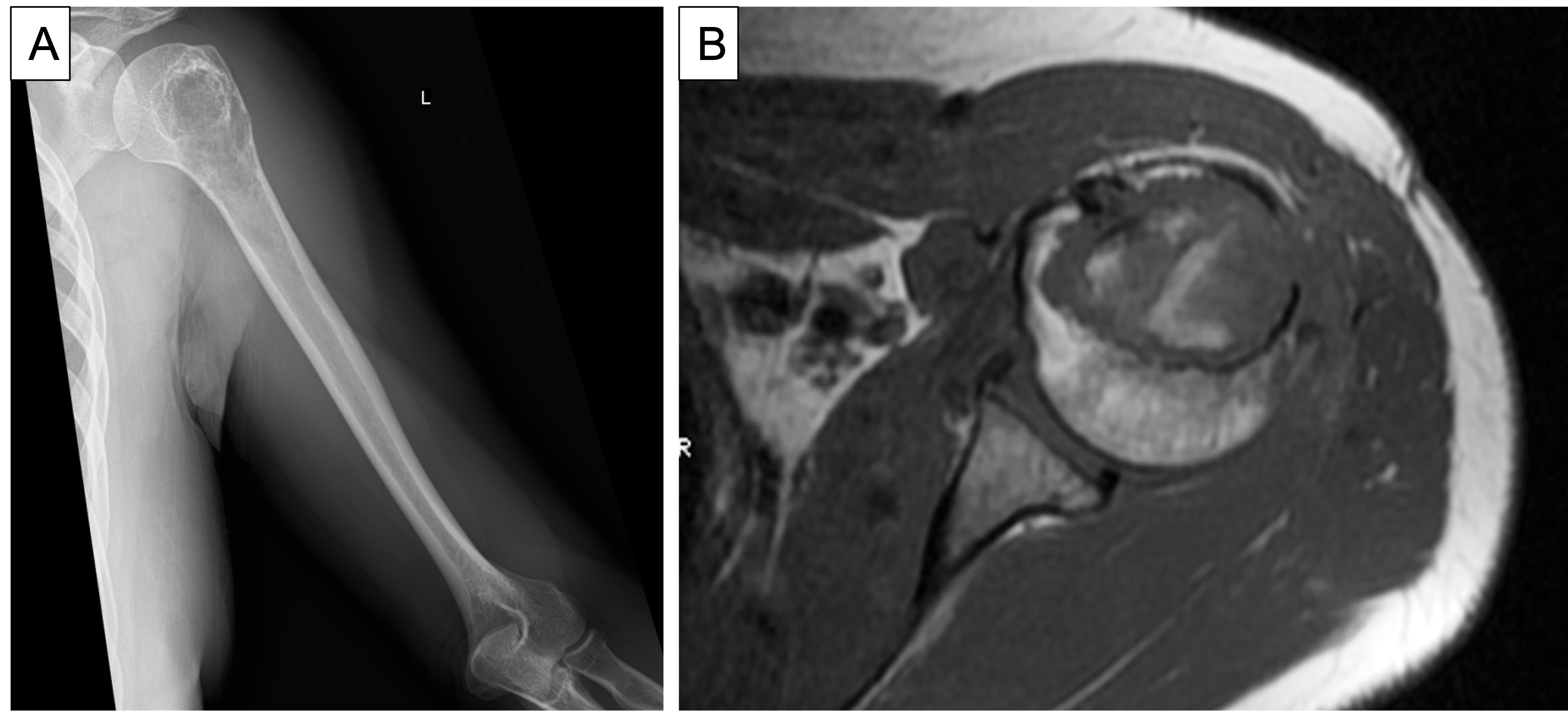
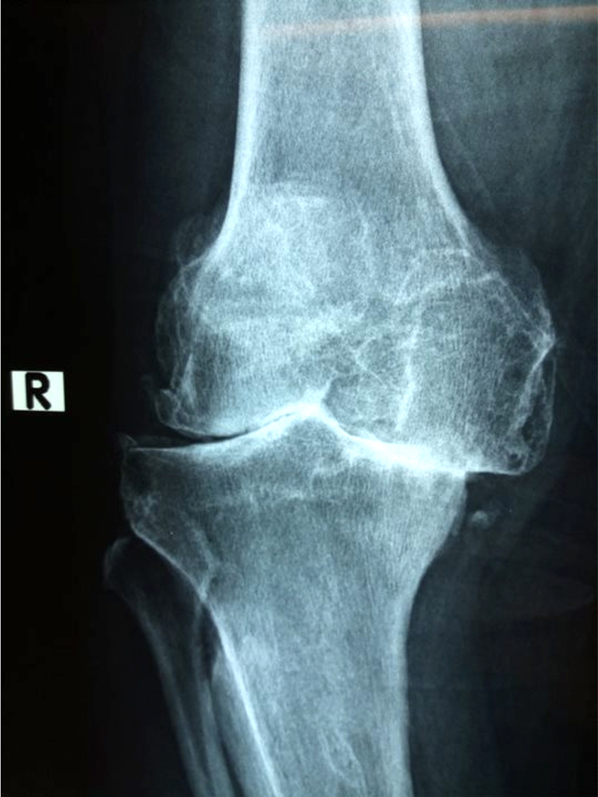
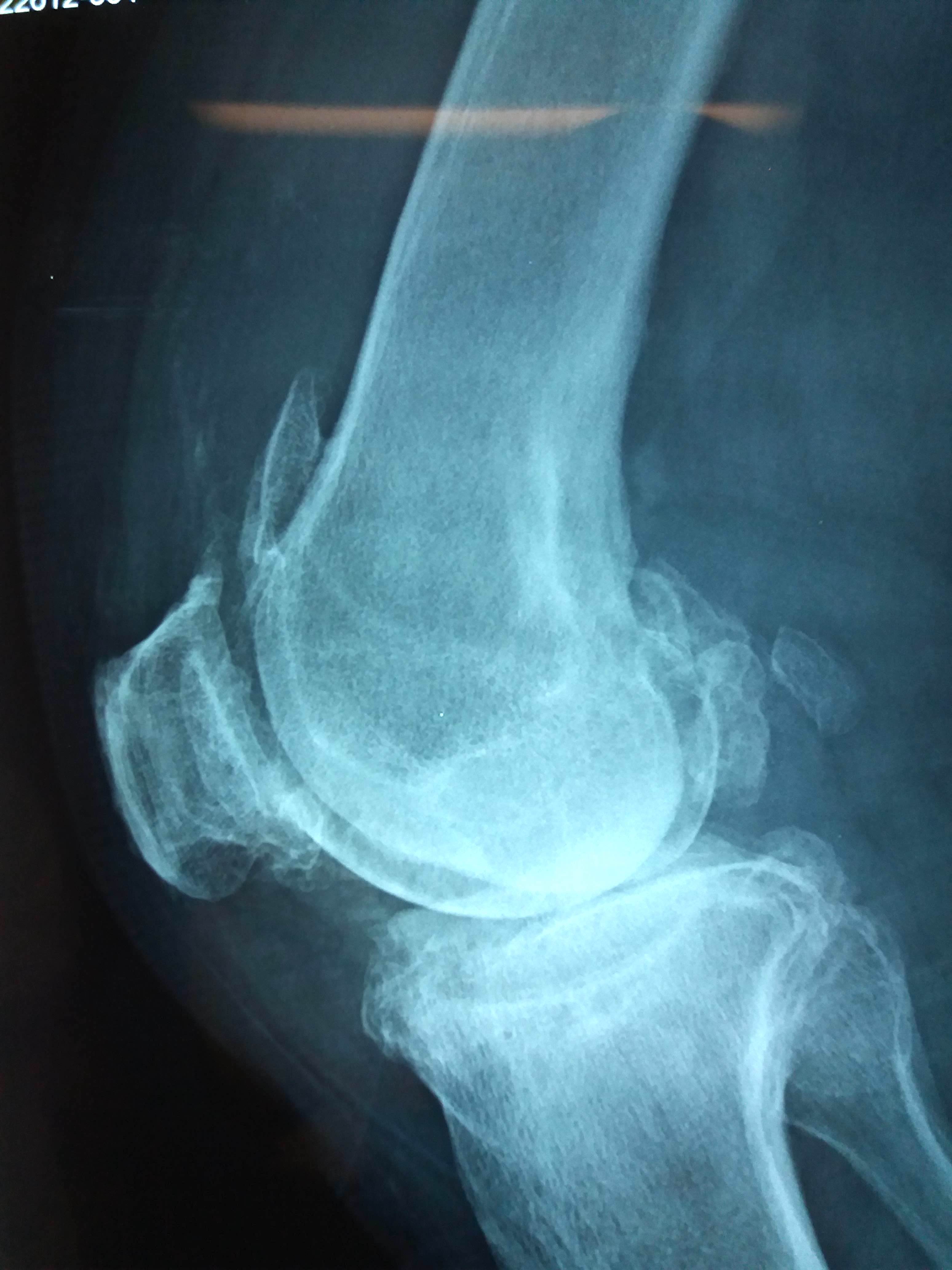
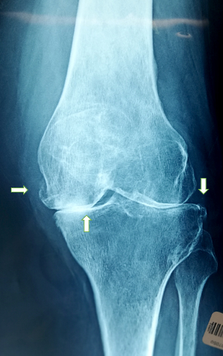
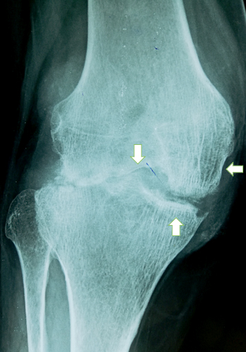
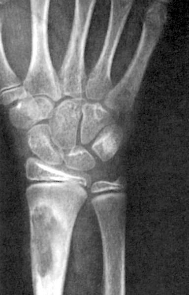
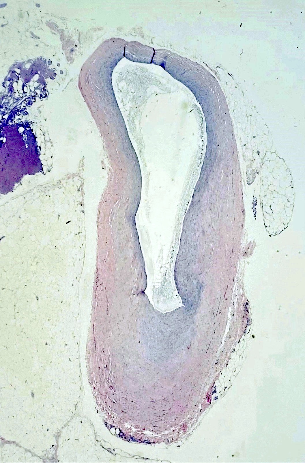
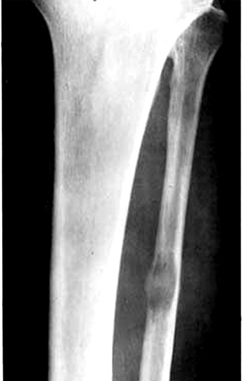
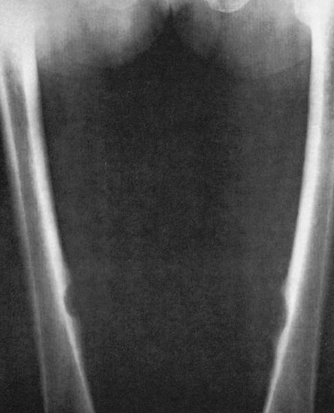
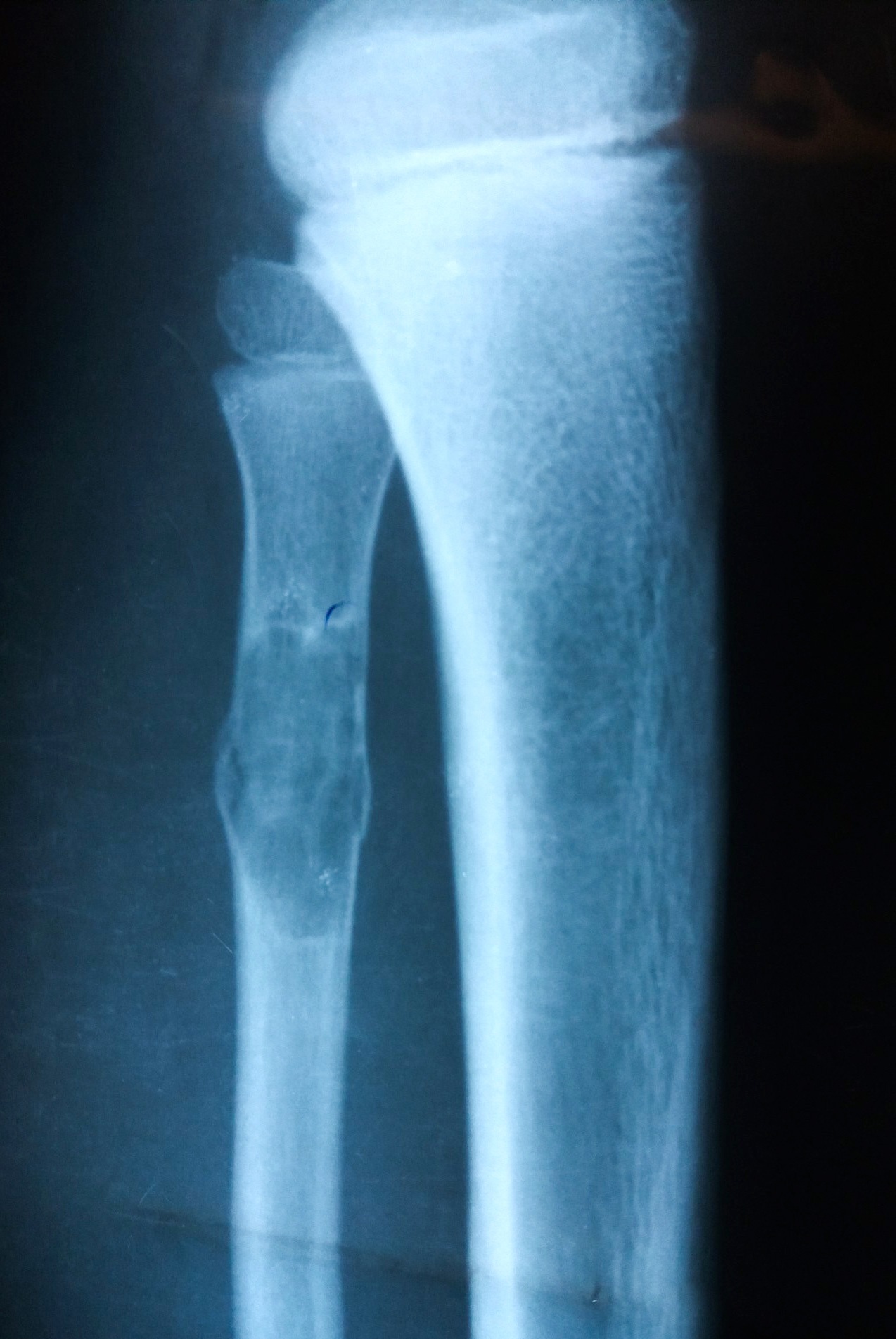
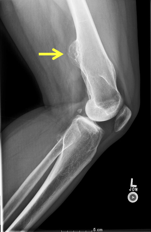
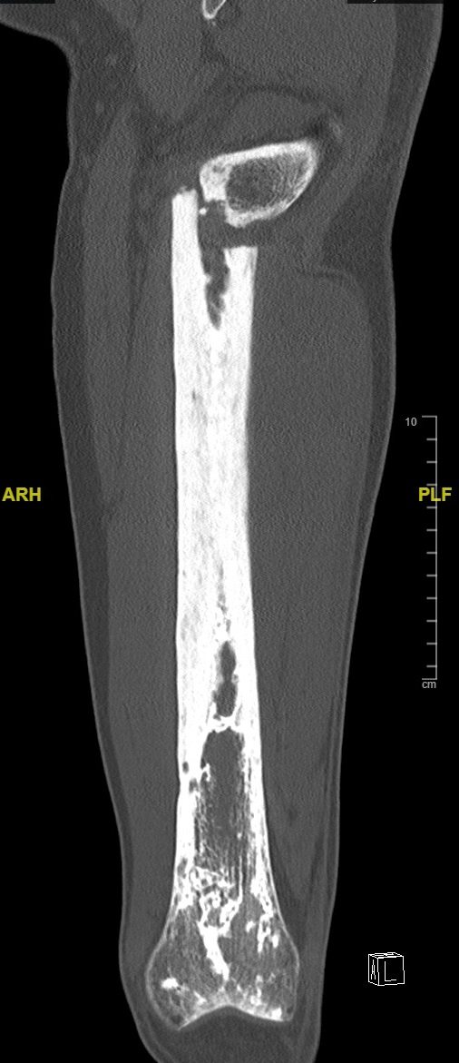
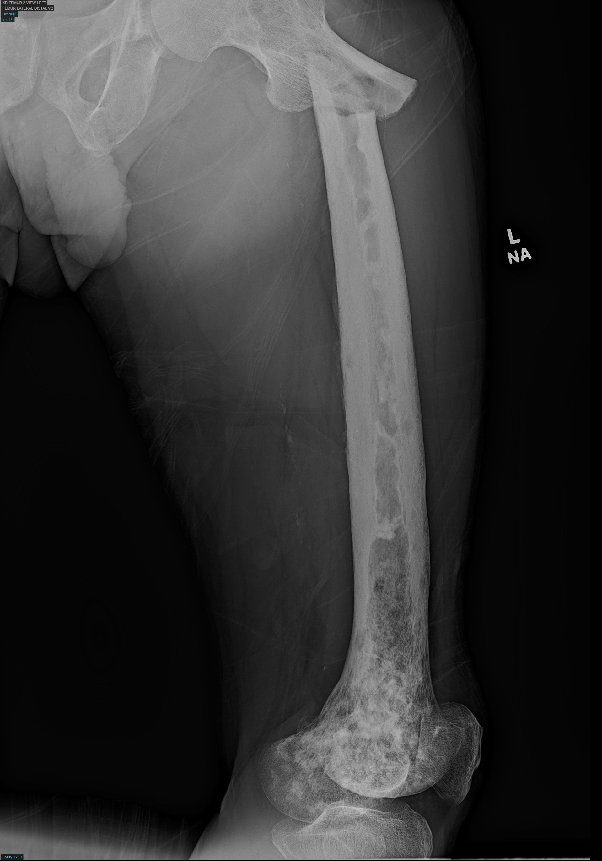
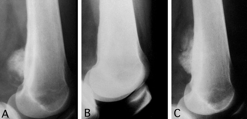
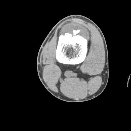
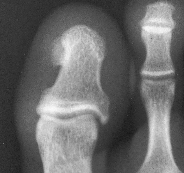
Tibia
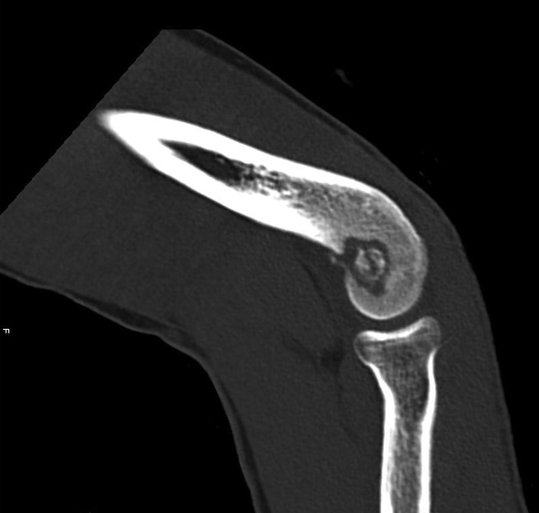
Elbow
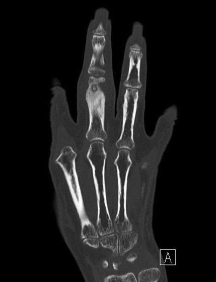
Finger
Distal femur
Distal femur
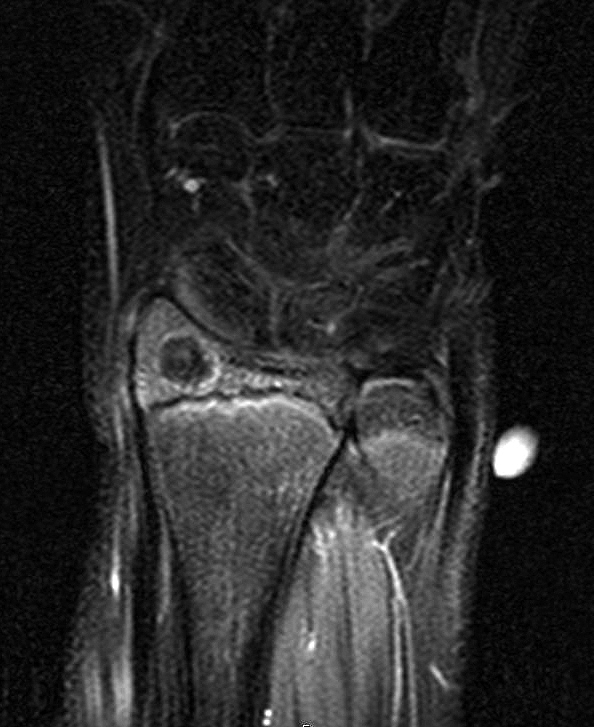
Wrist
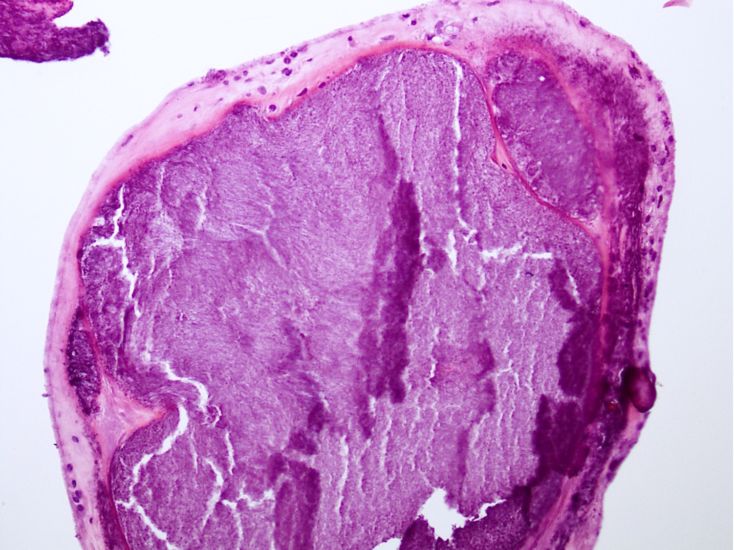
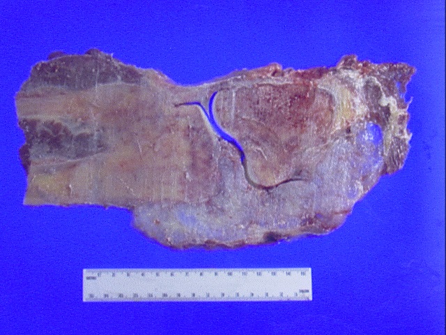
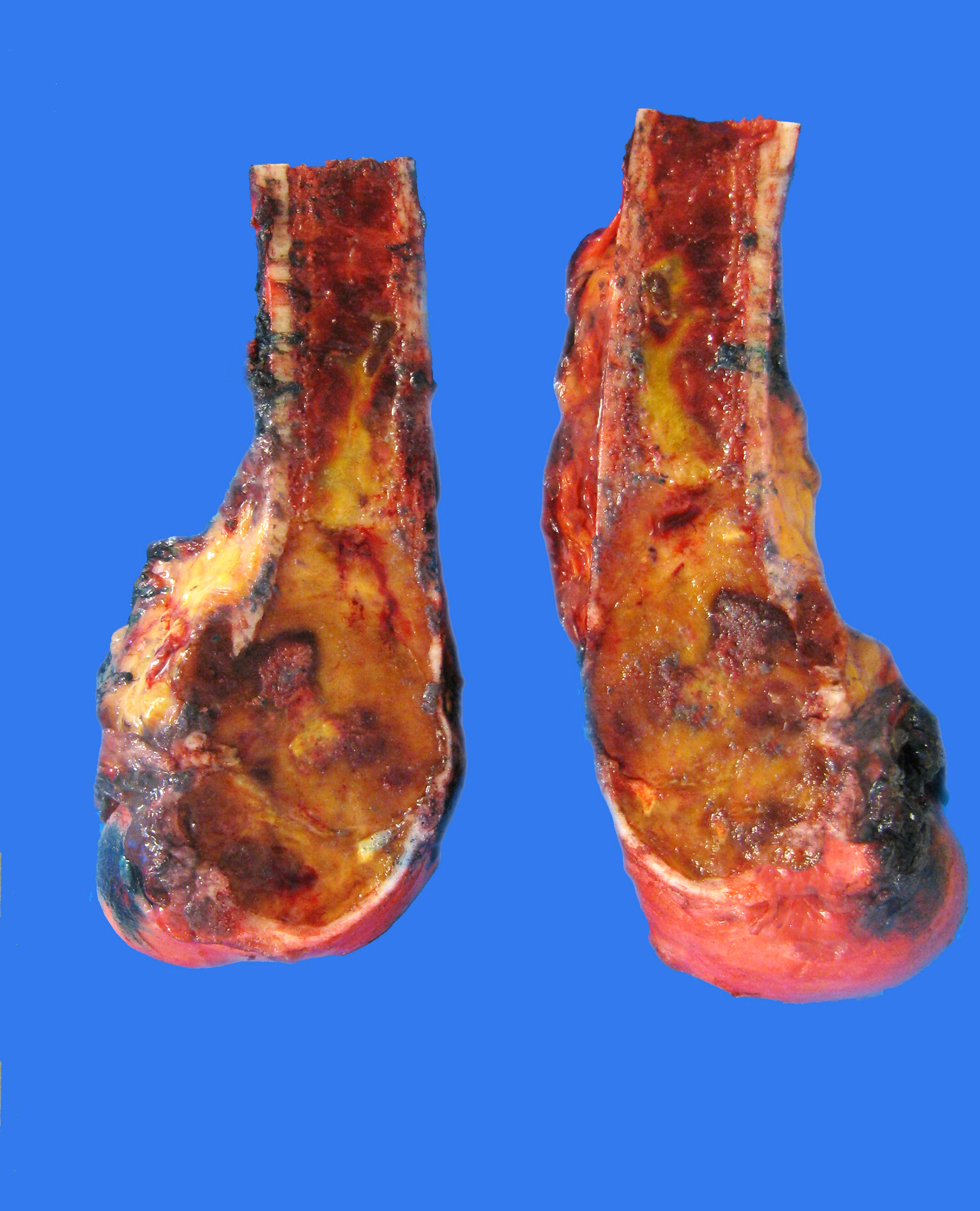
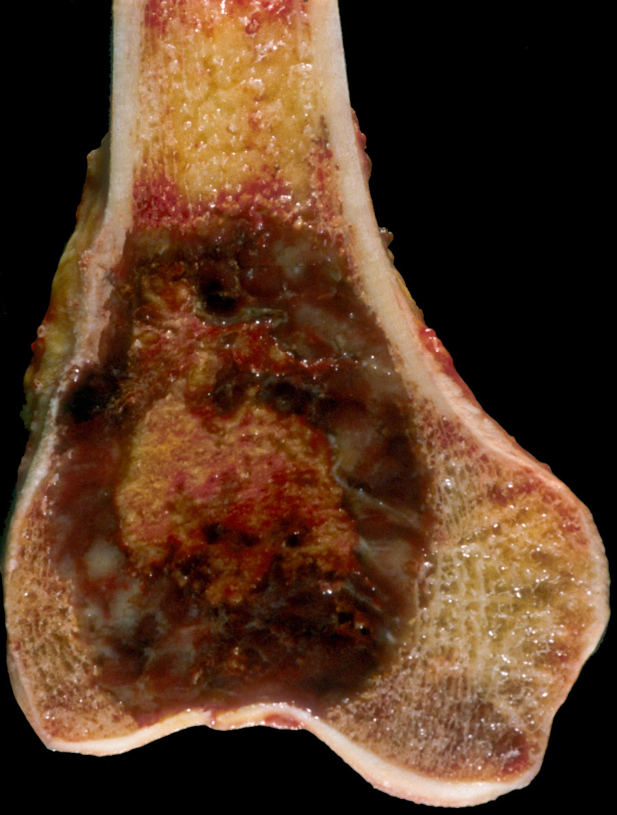
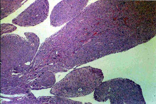
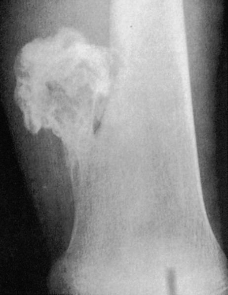
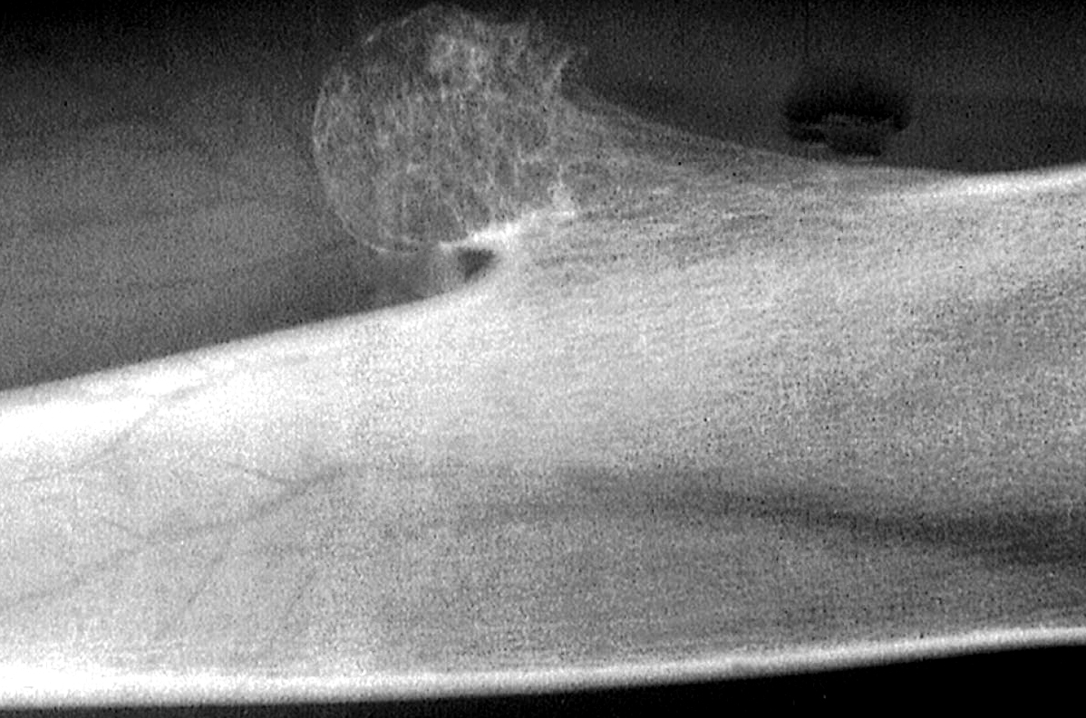
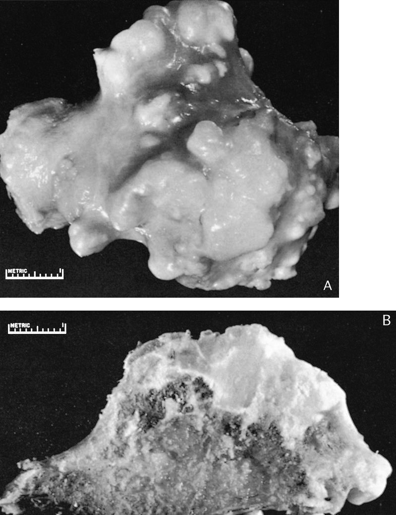
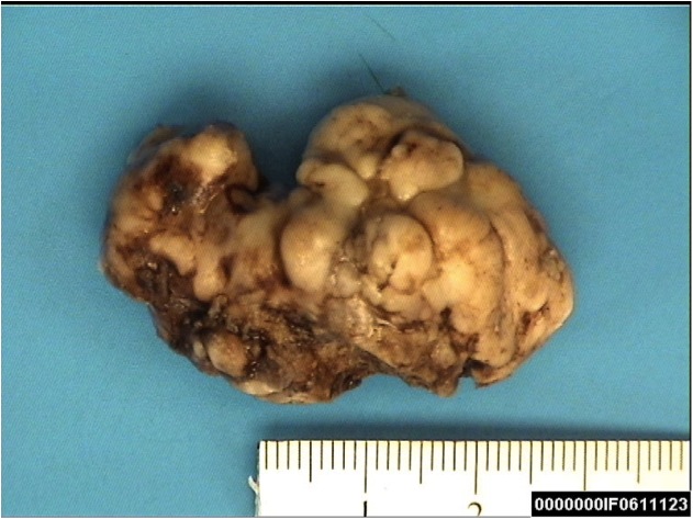
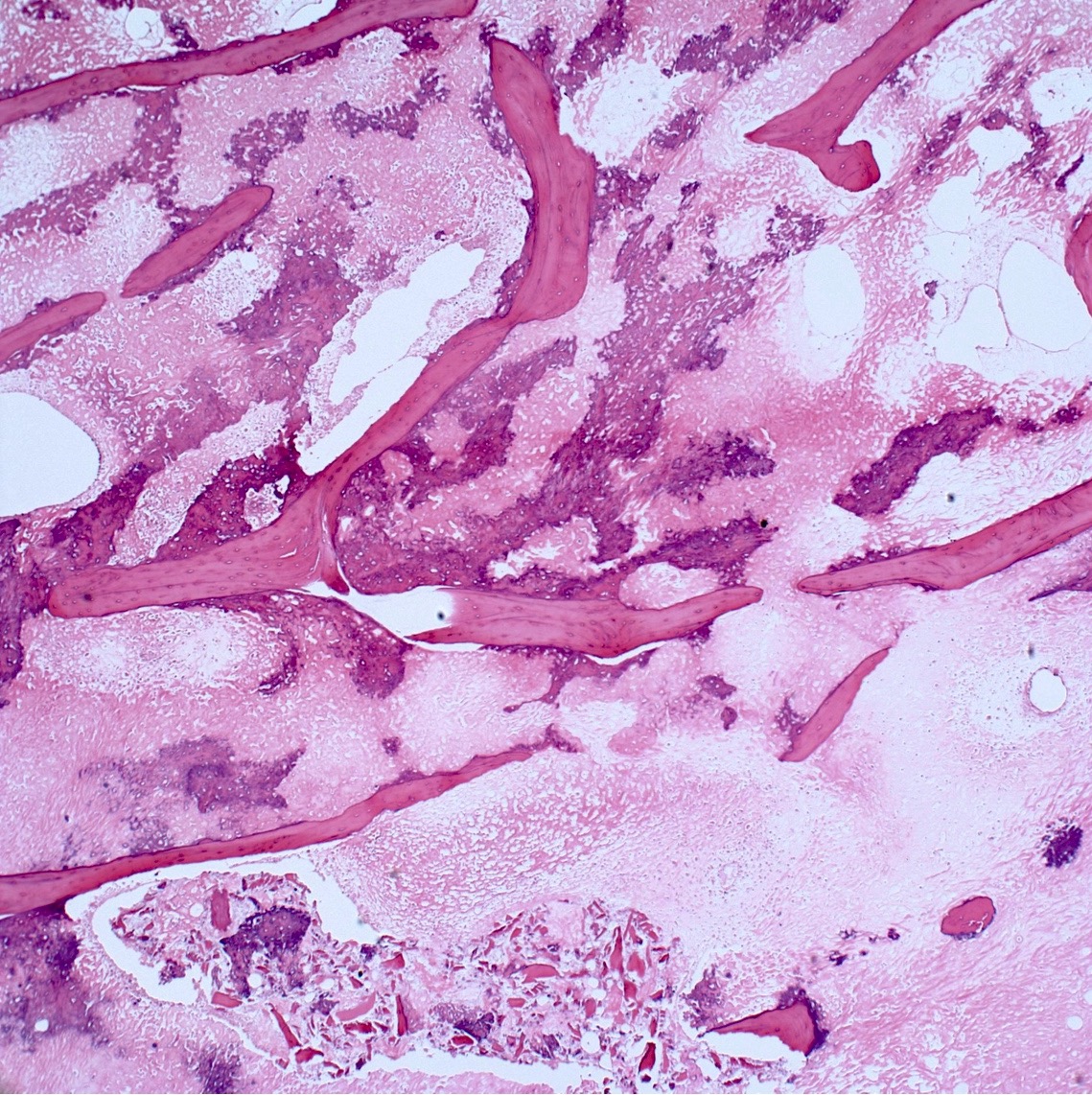
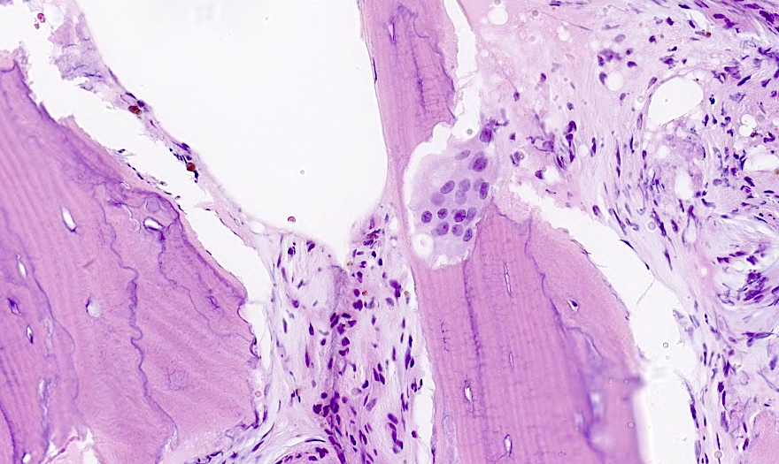
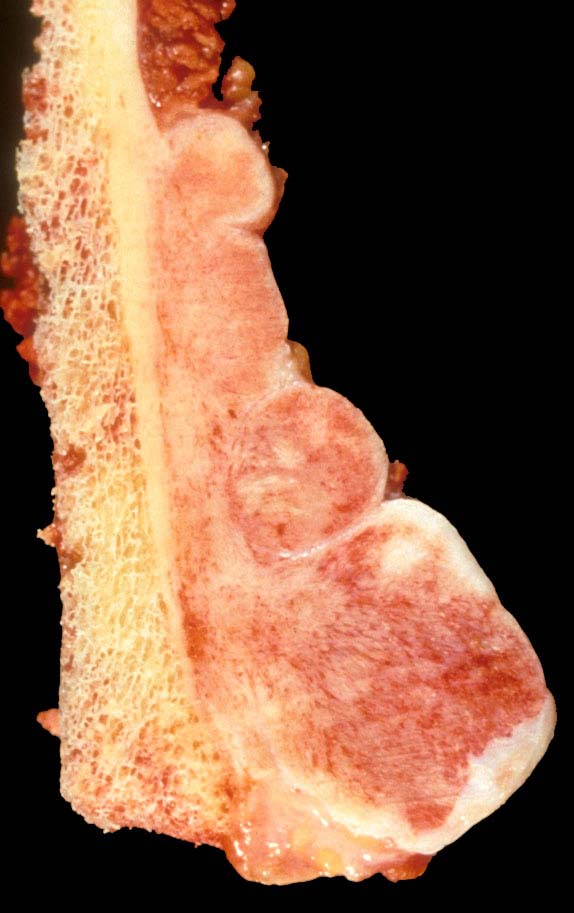
Contributed by John D. Reith, M.D.
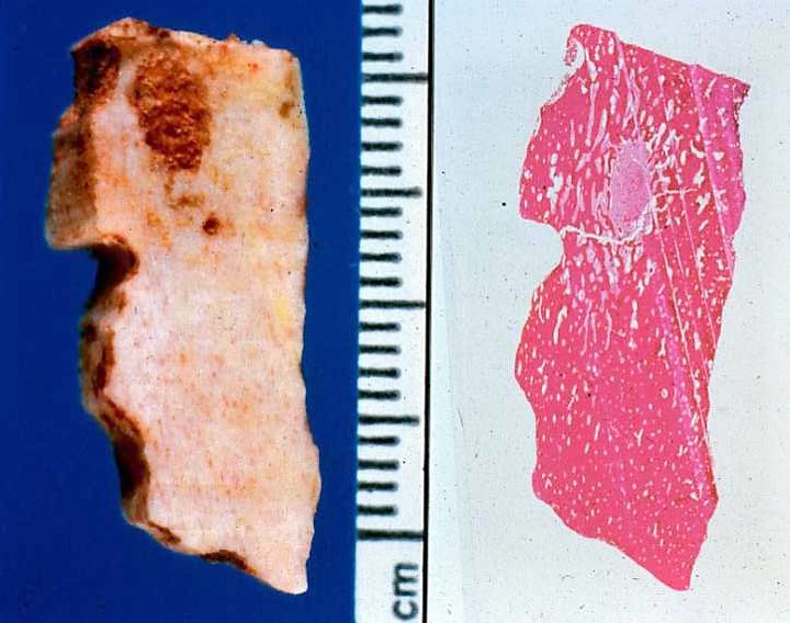
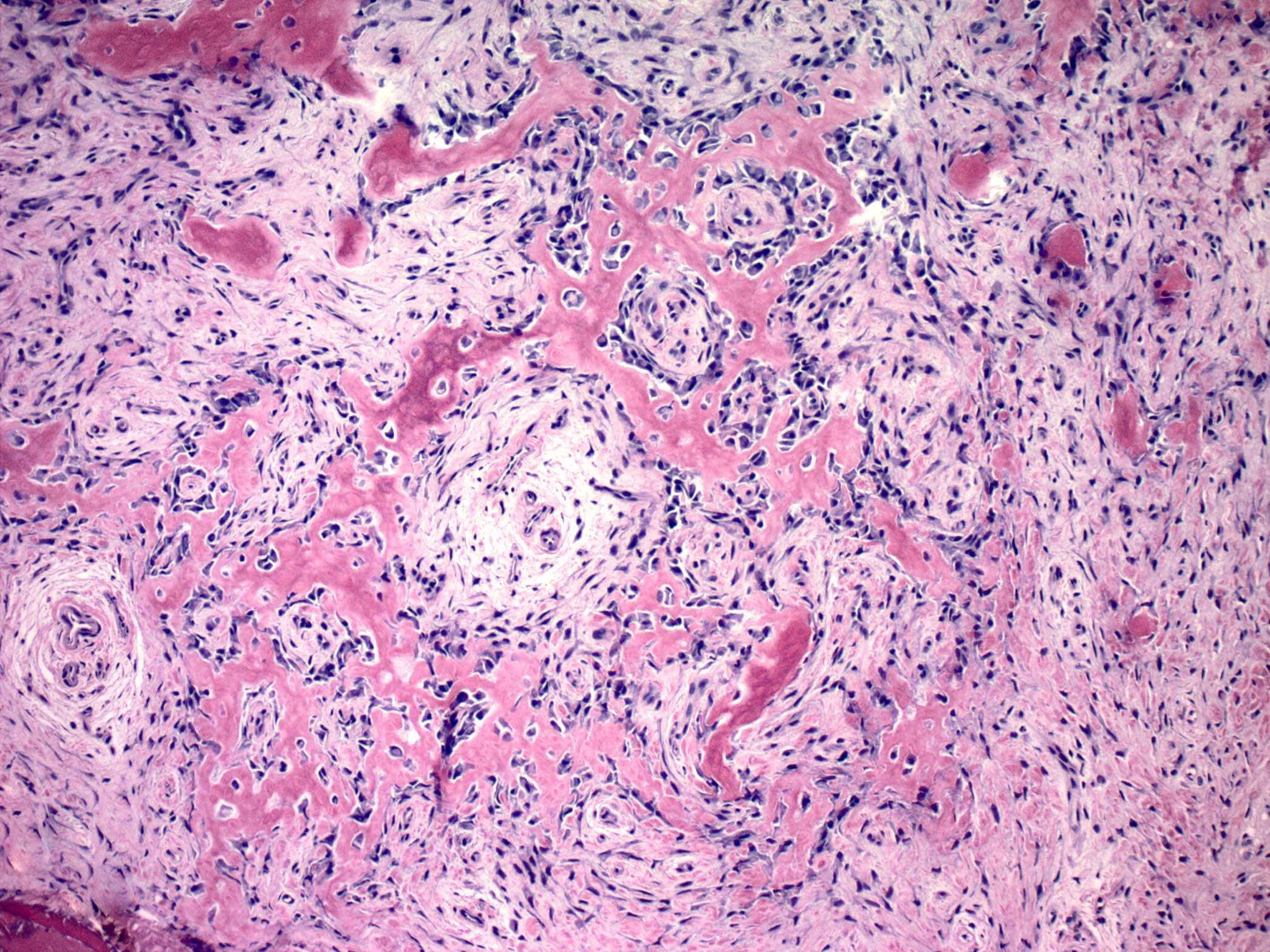
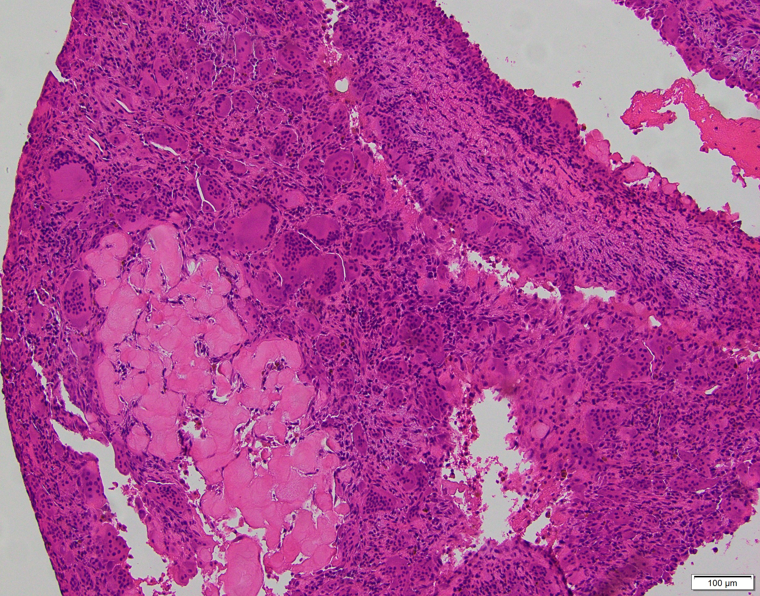
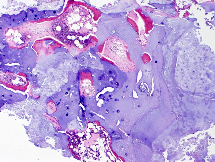
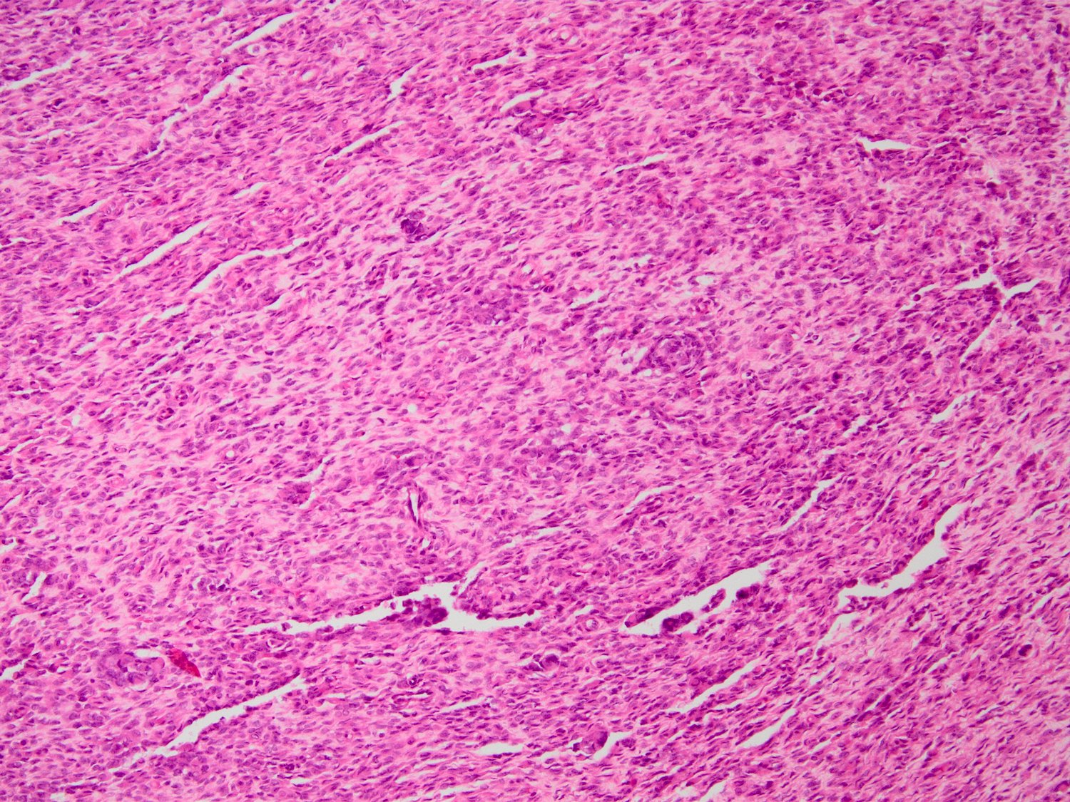
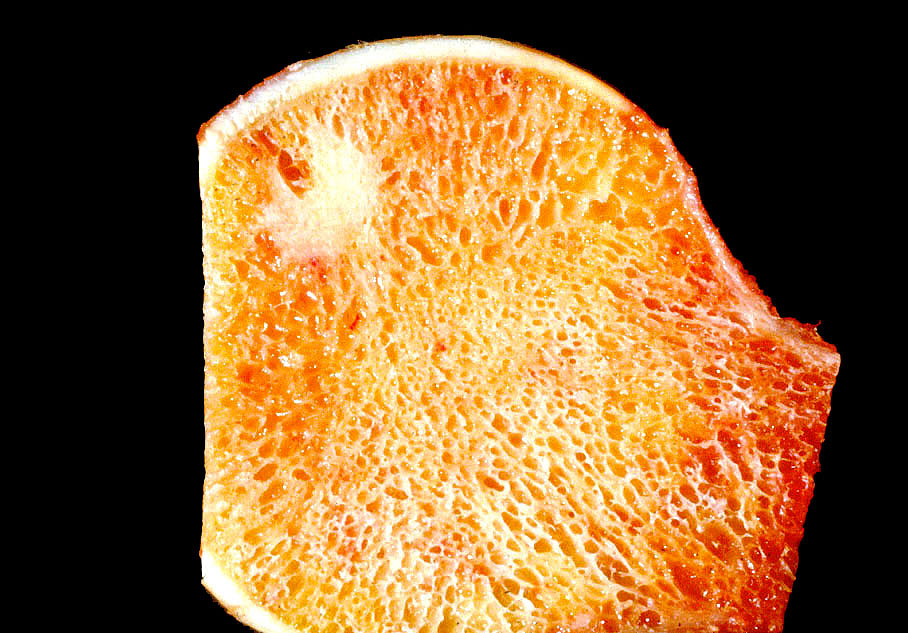
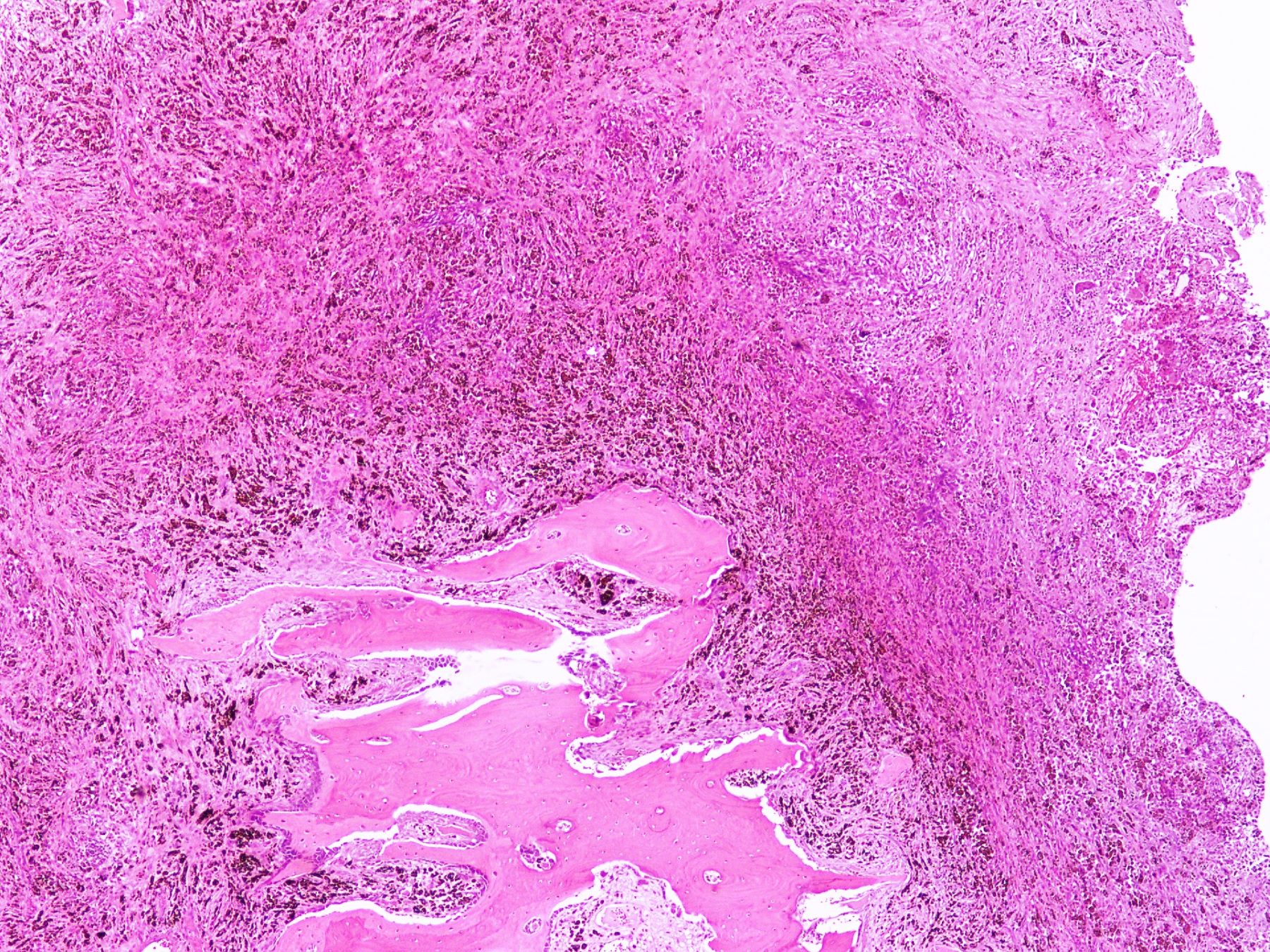
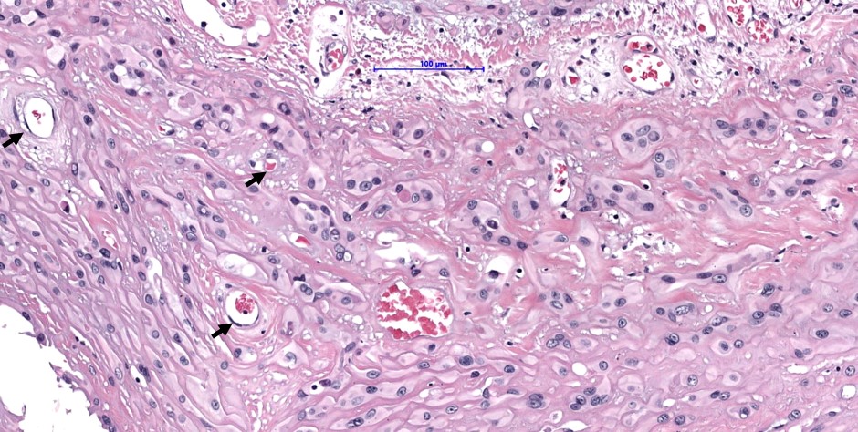
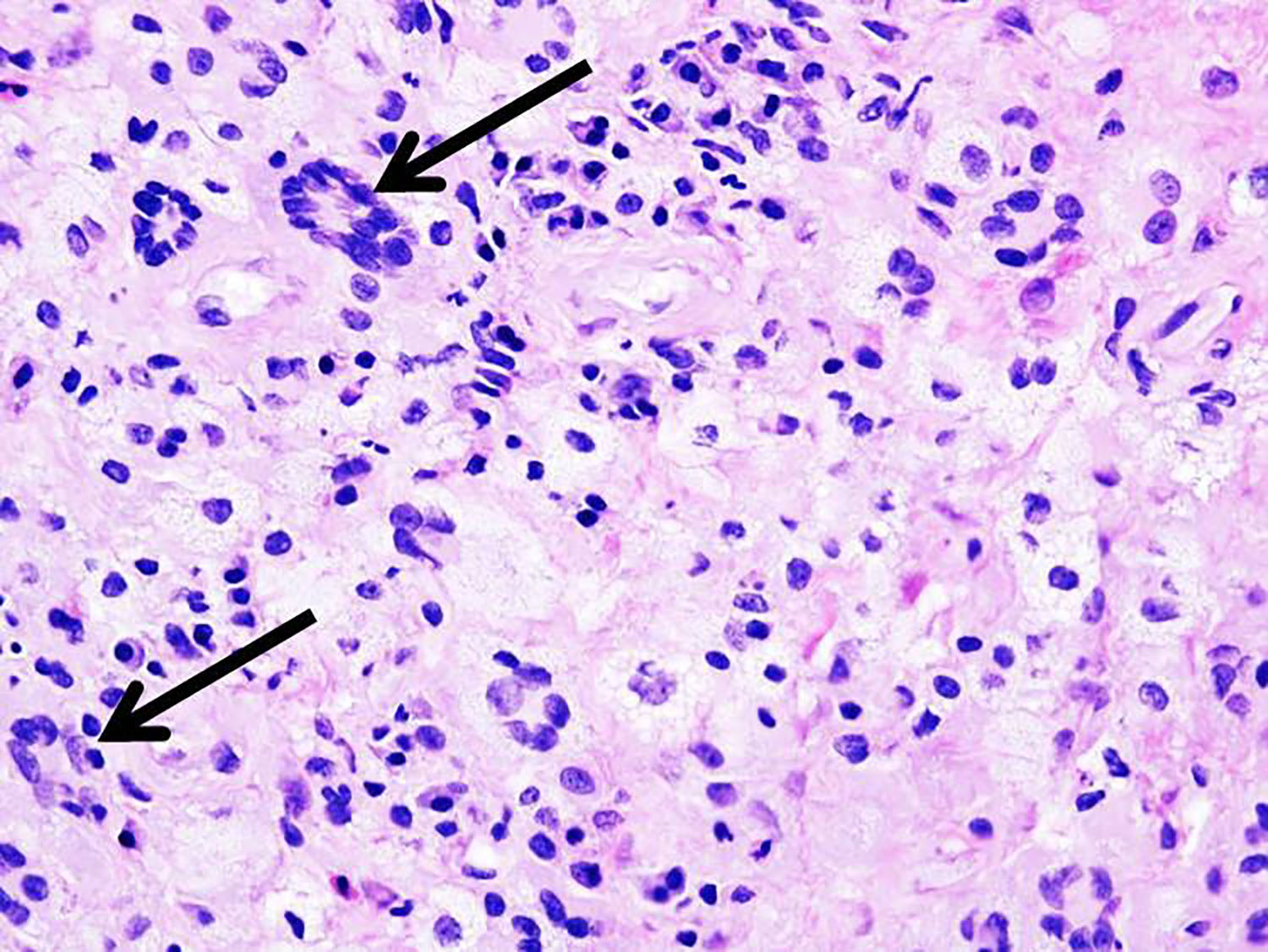
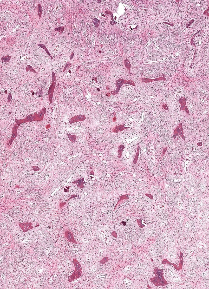
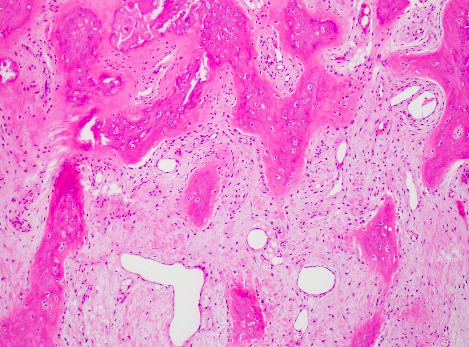
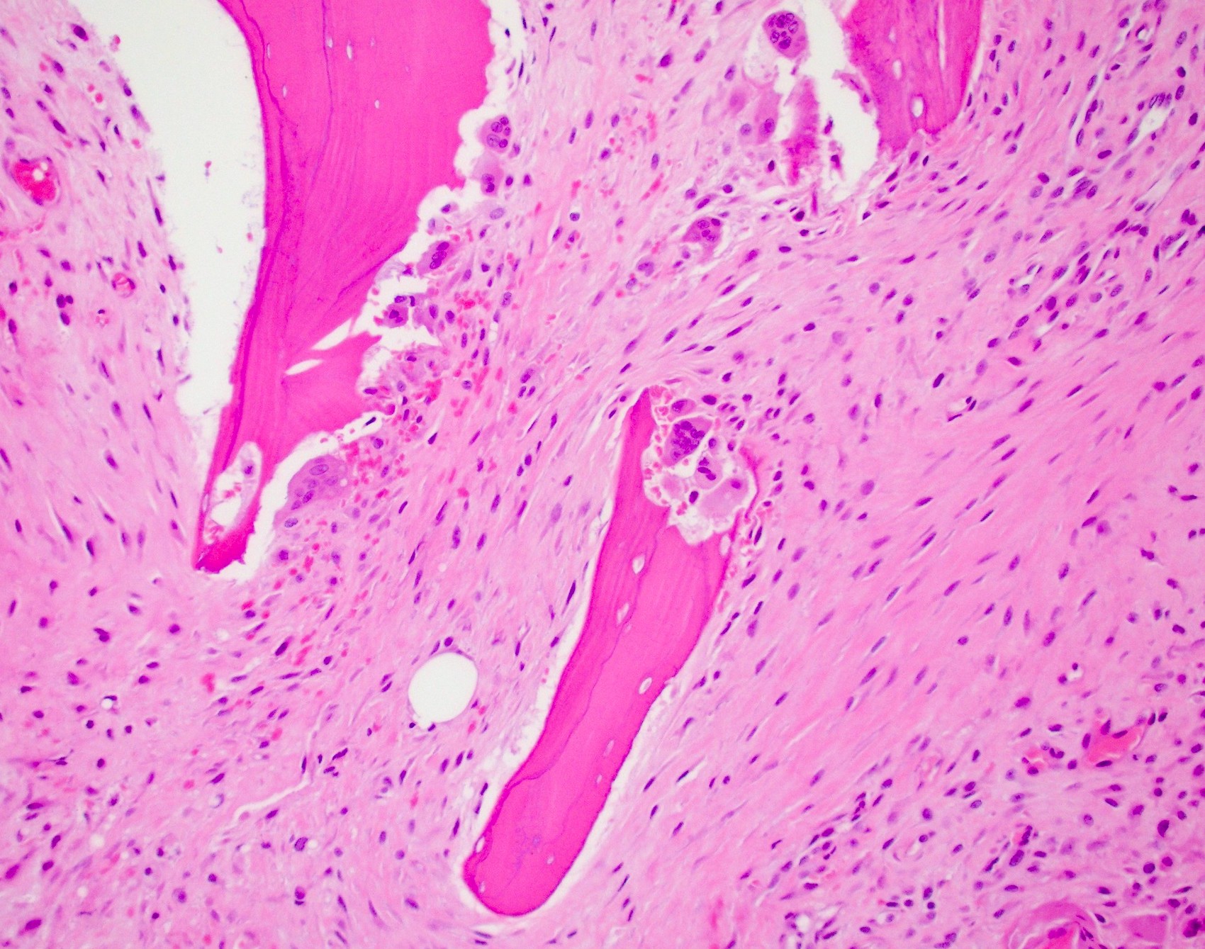
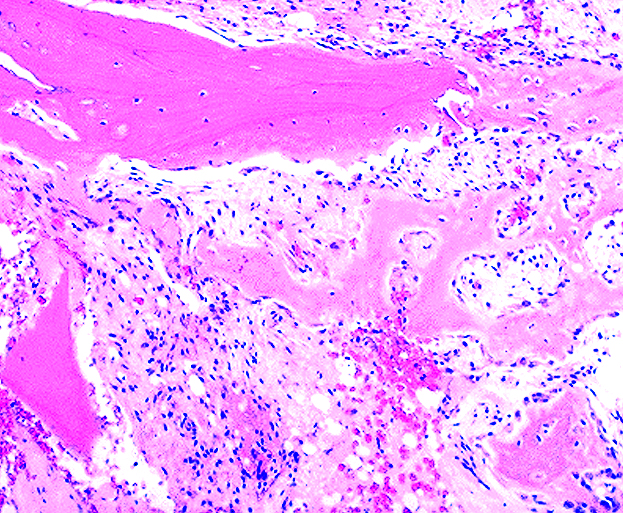
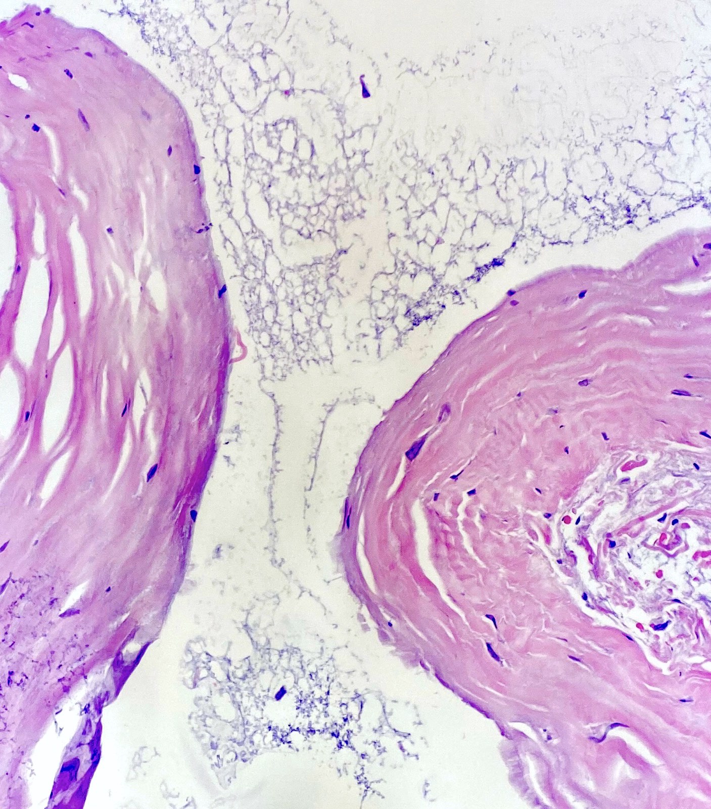
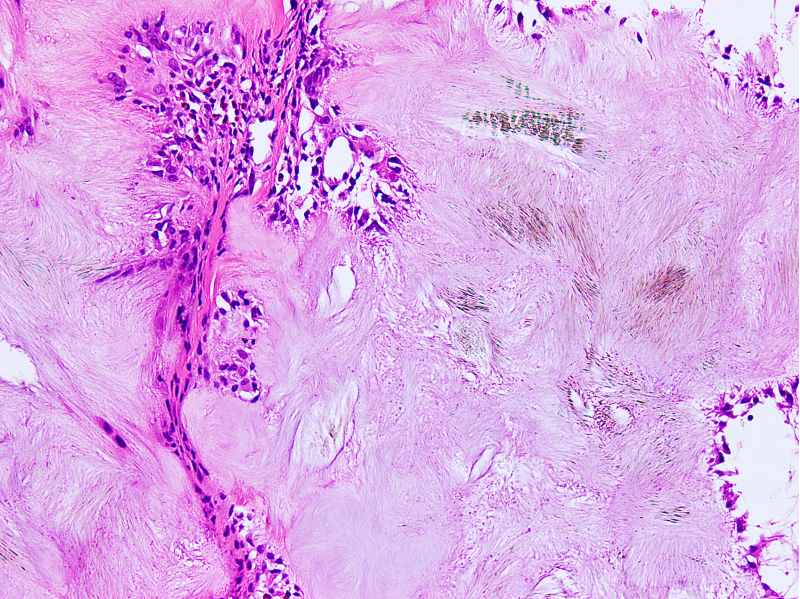
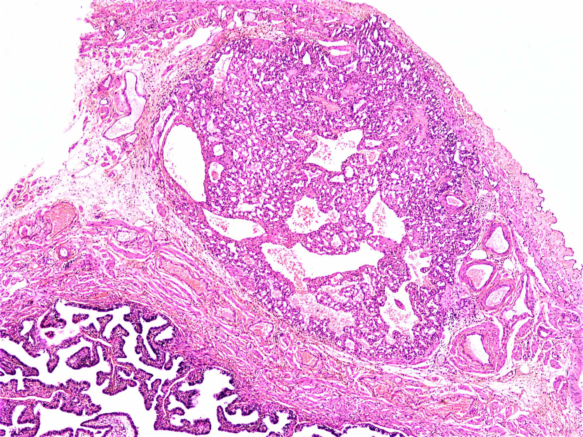
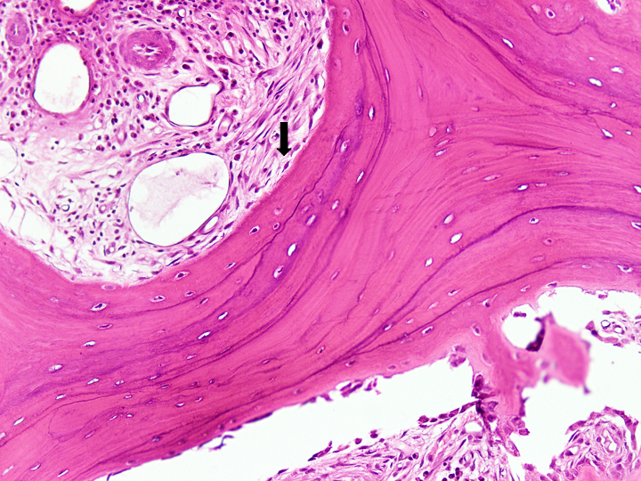
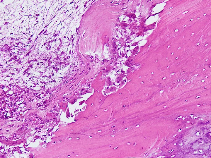
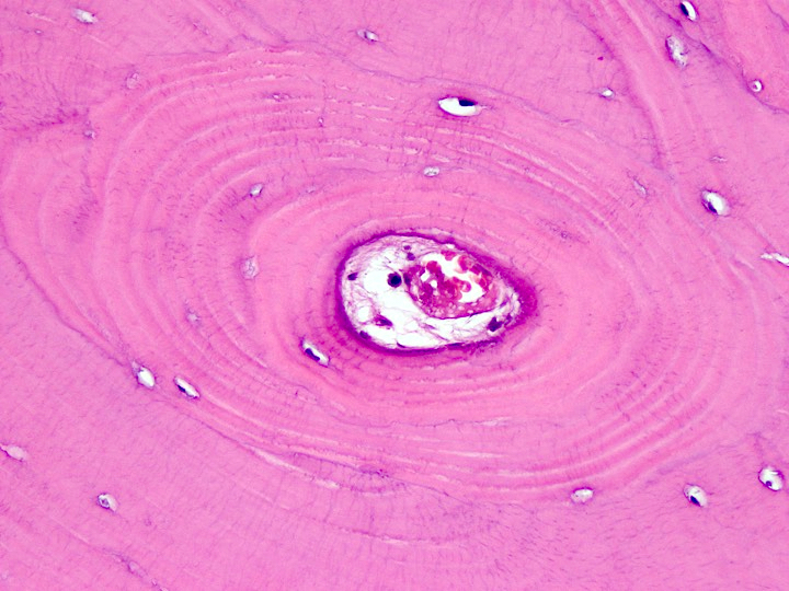
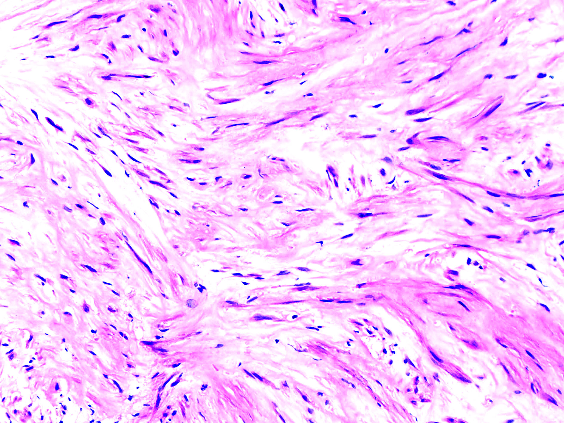
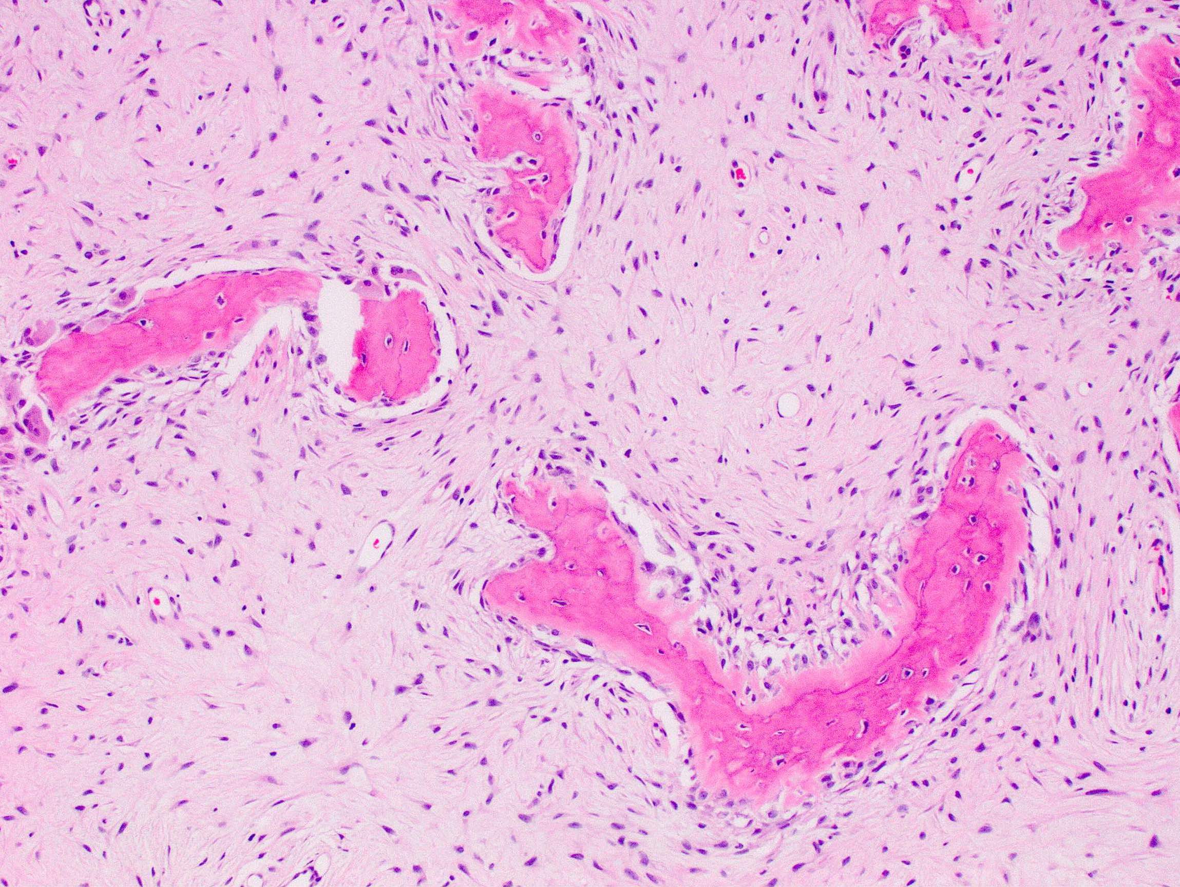
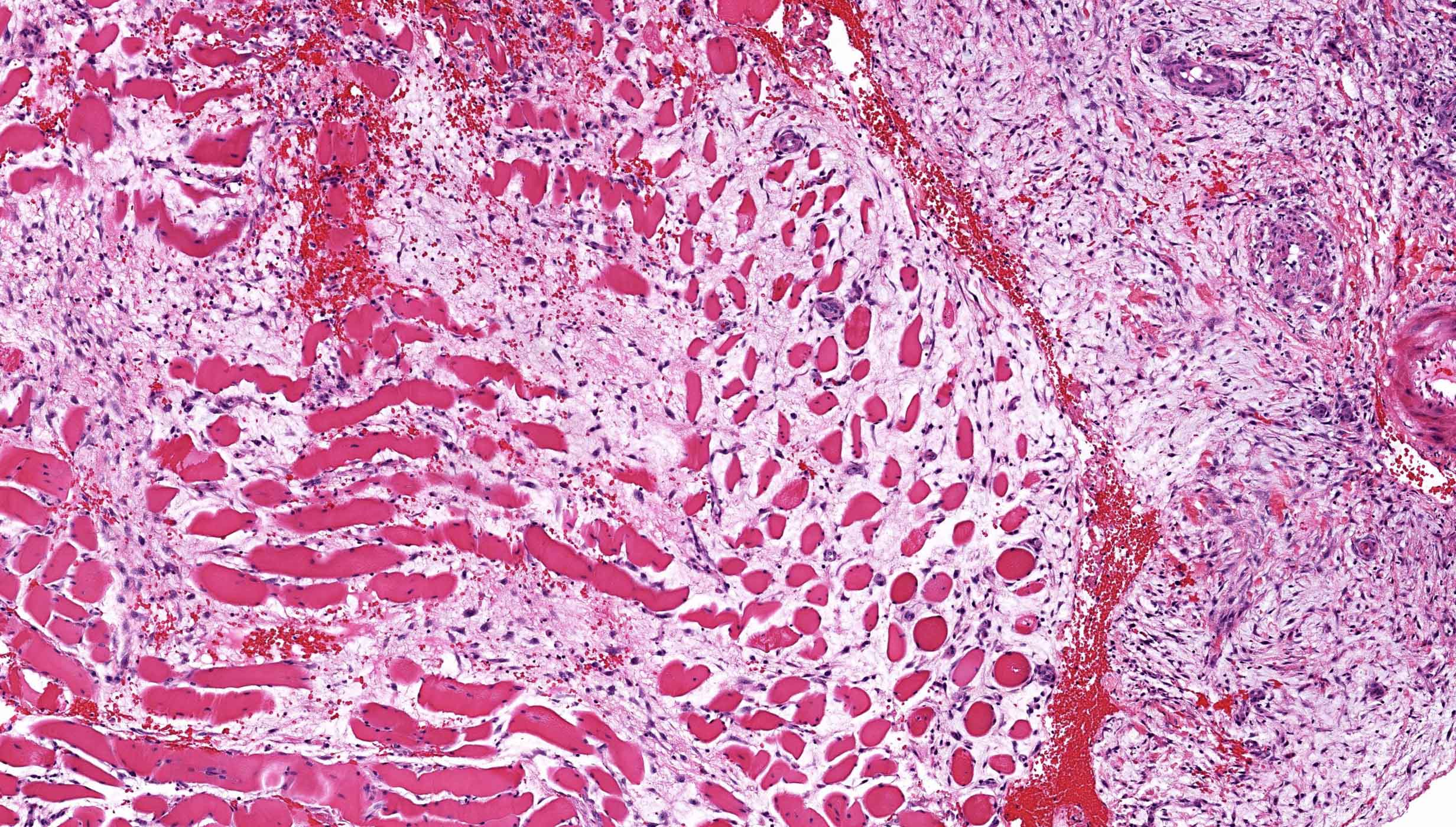
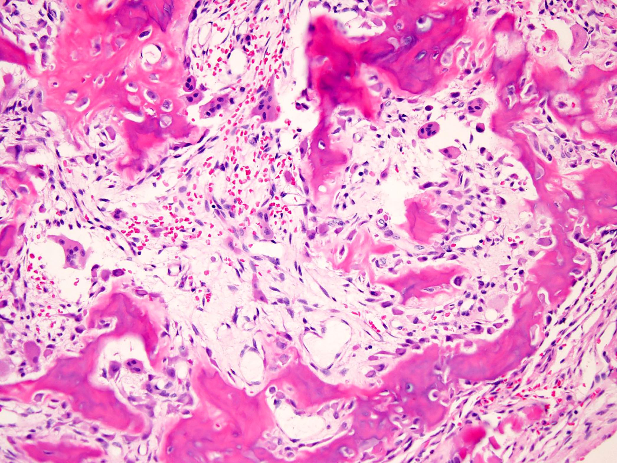
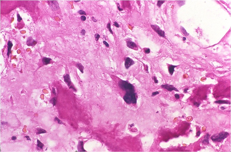
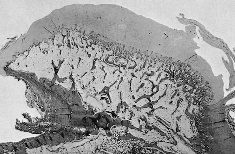
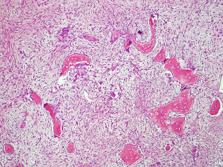
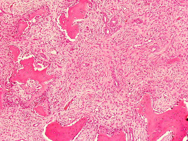
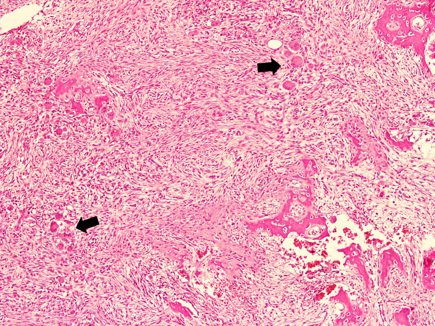
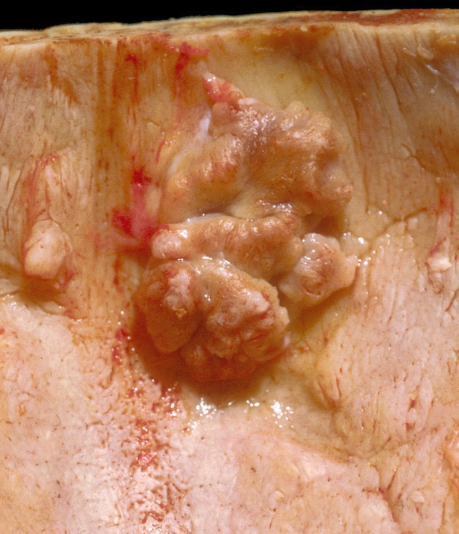
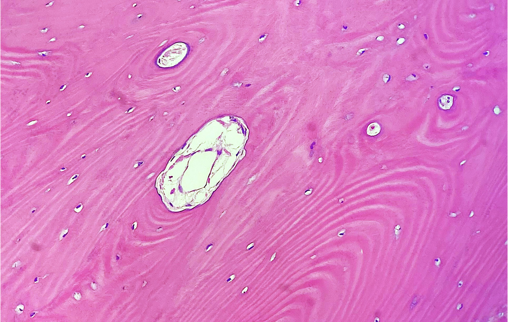
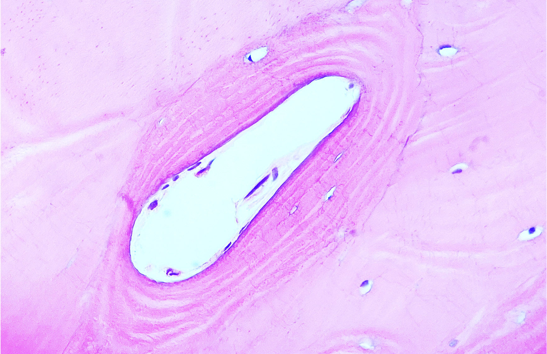
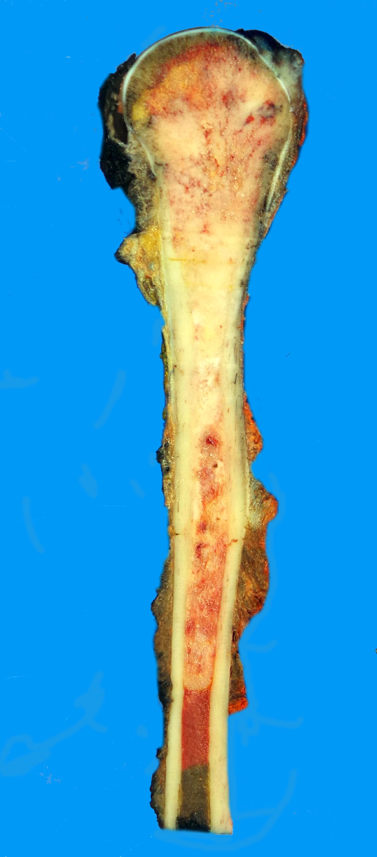
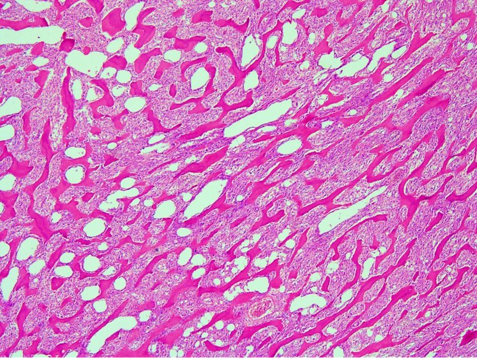
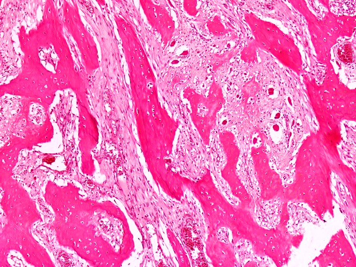
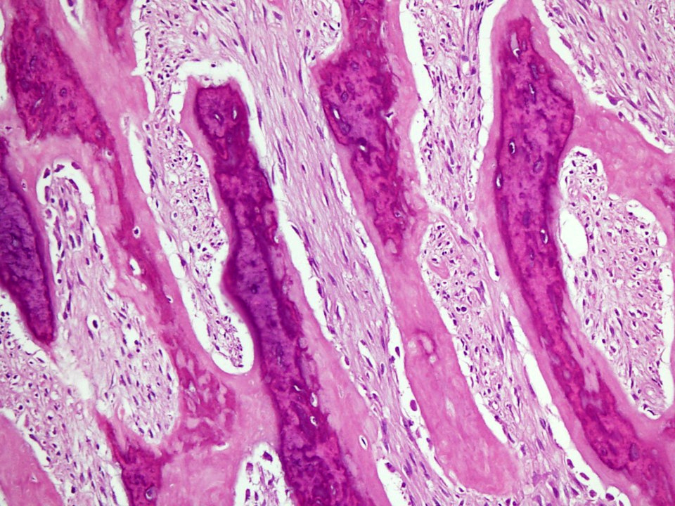
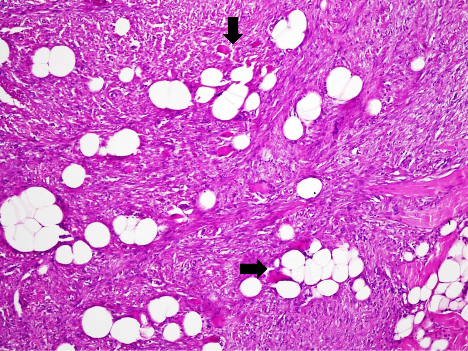
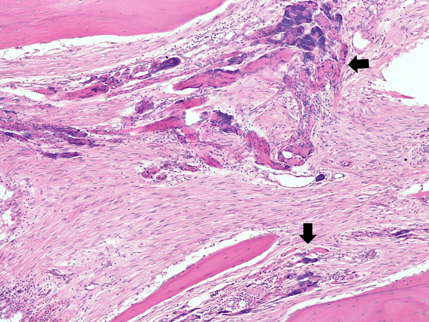
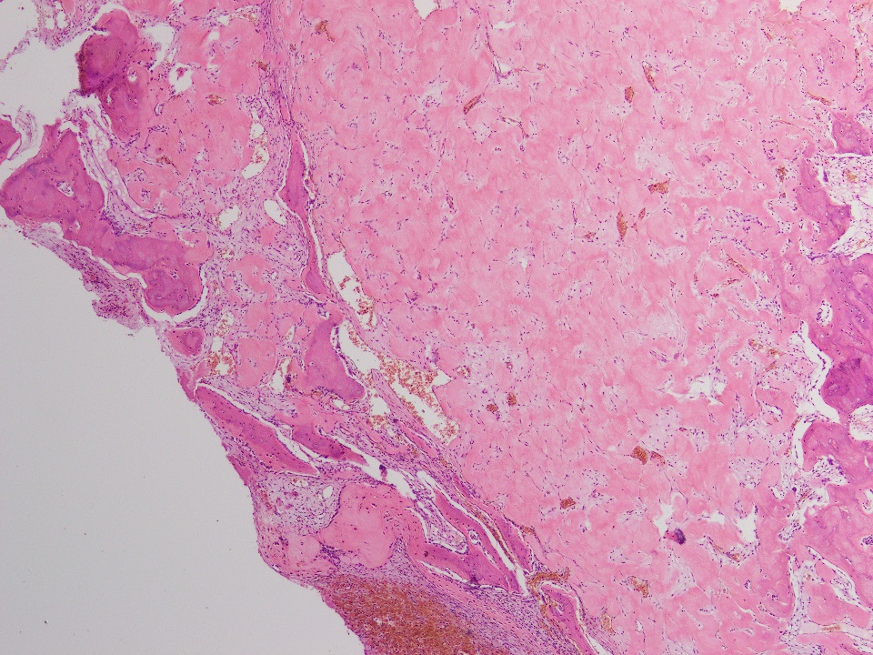
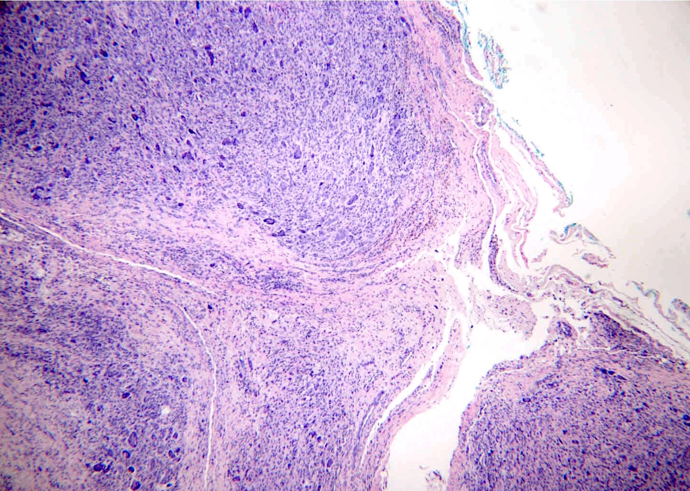
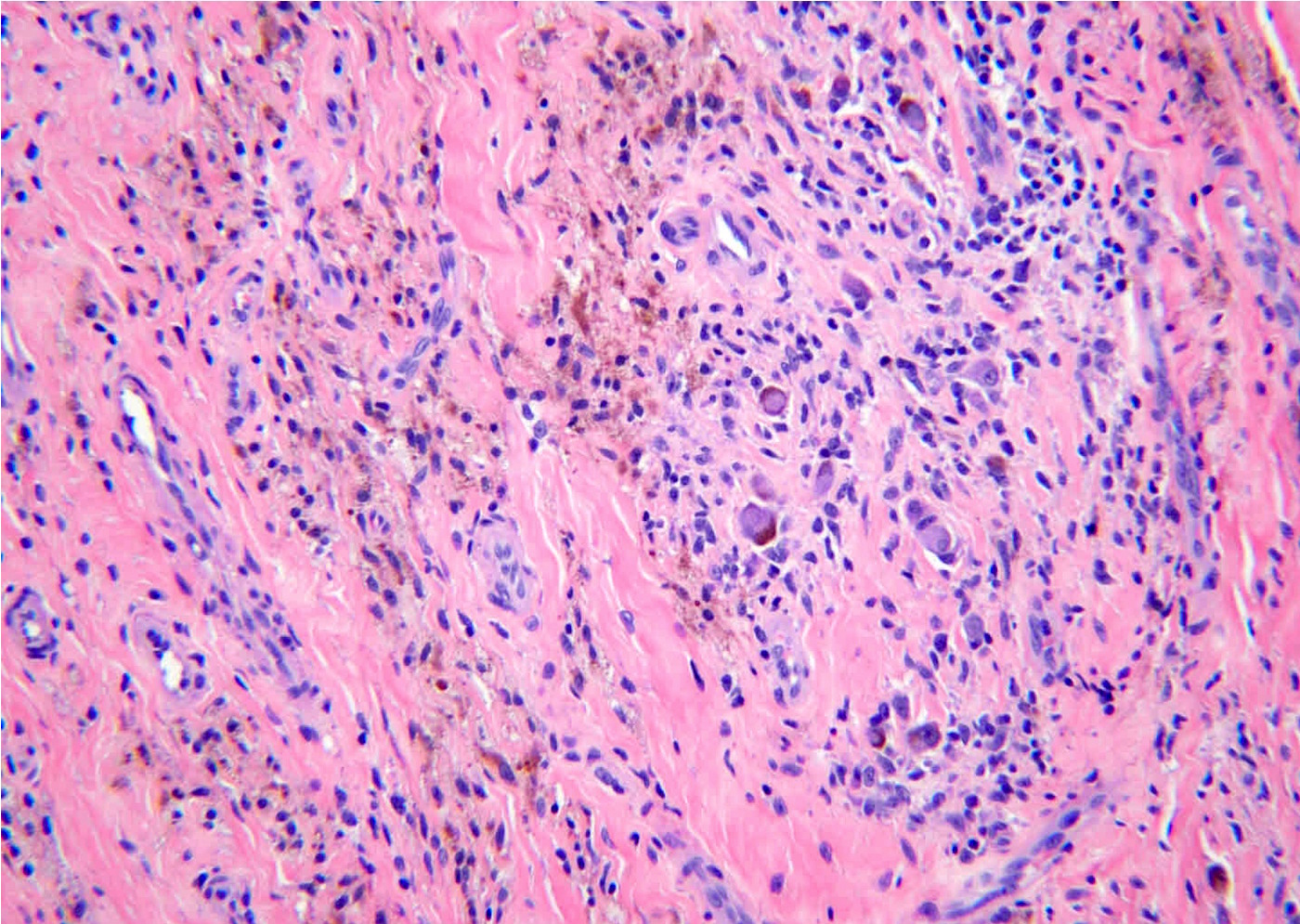
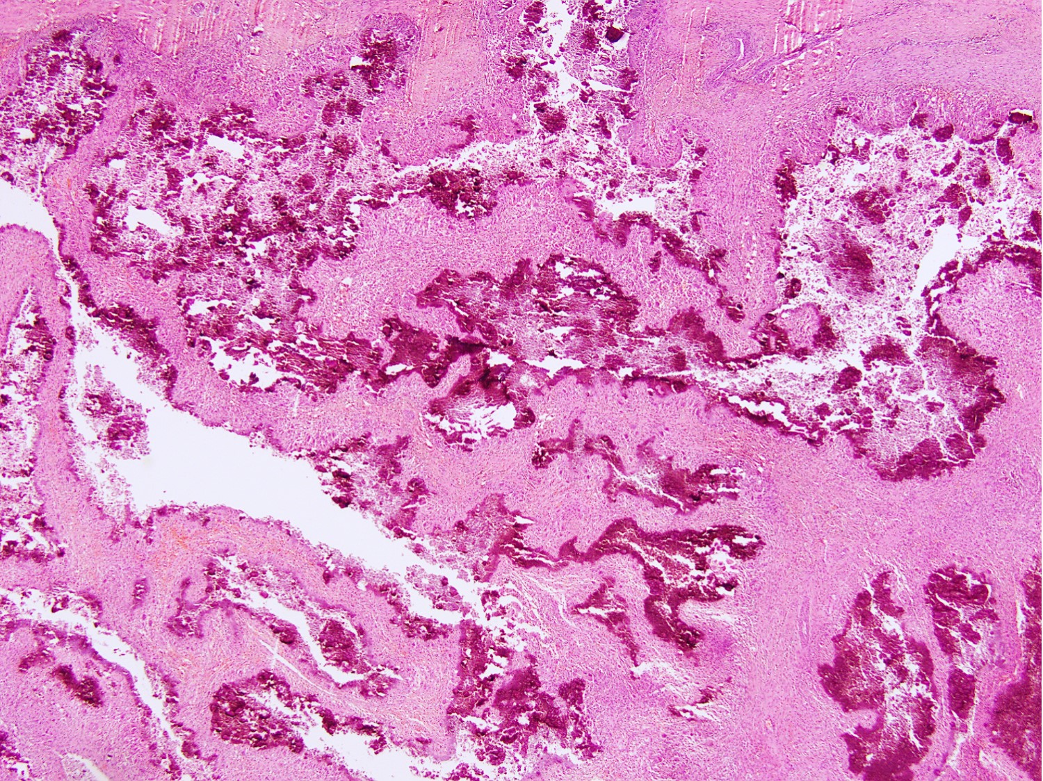
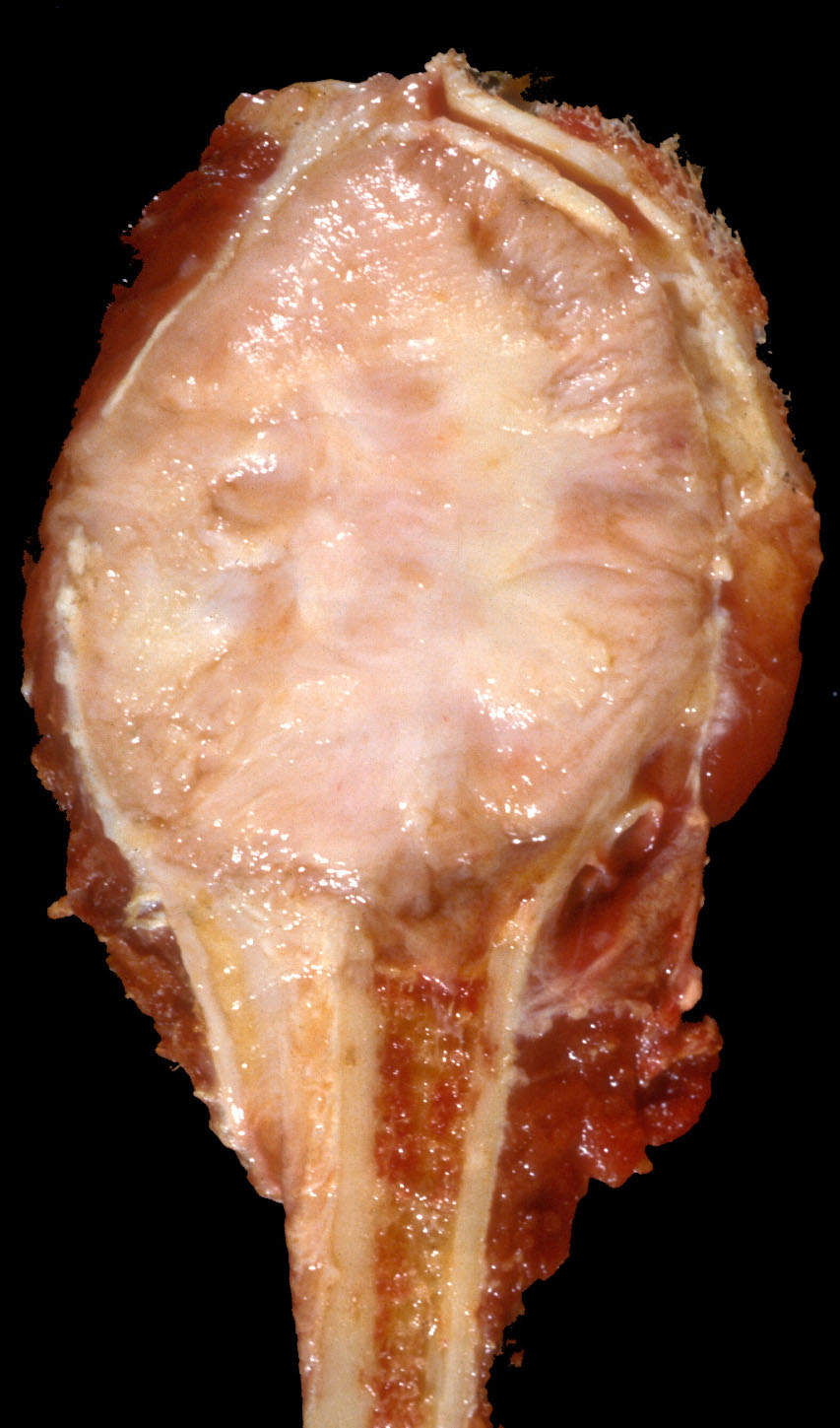
Nidus tissue with surrounding bone
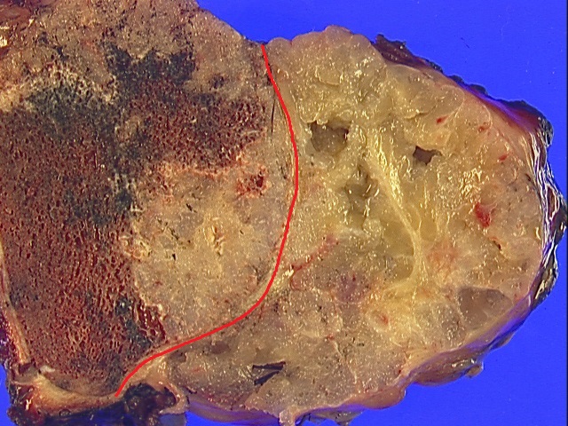
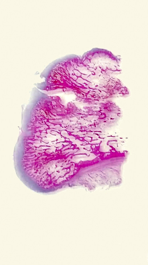
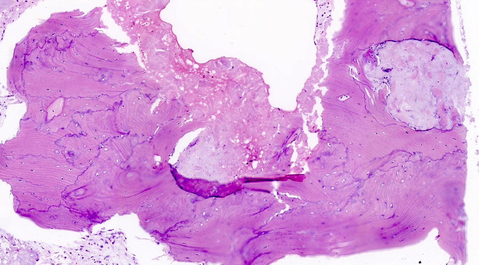
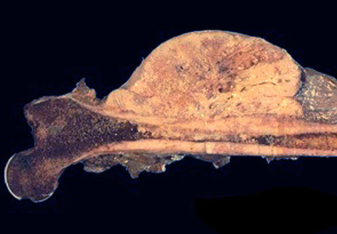
Contributed by Elham Nasri, M.D. and John D. Reith, M.D.
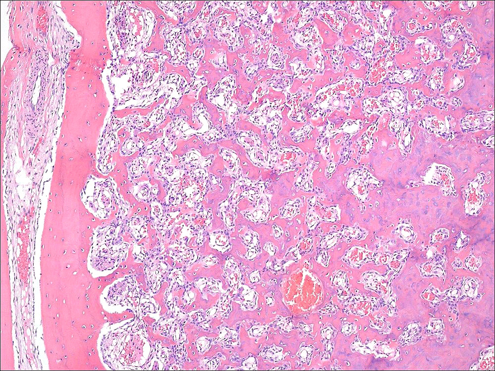
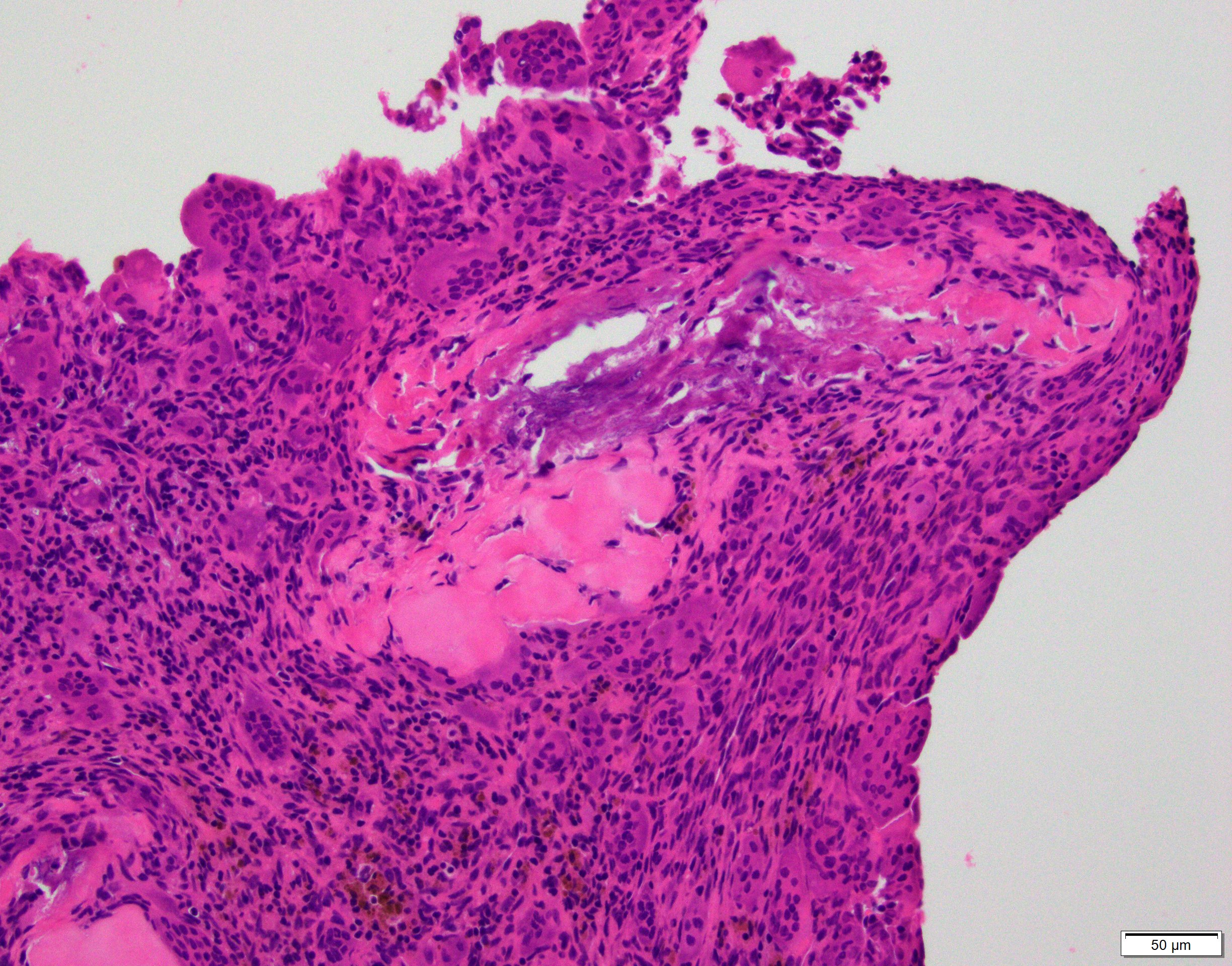
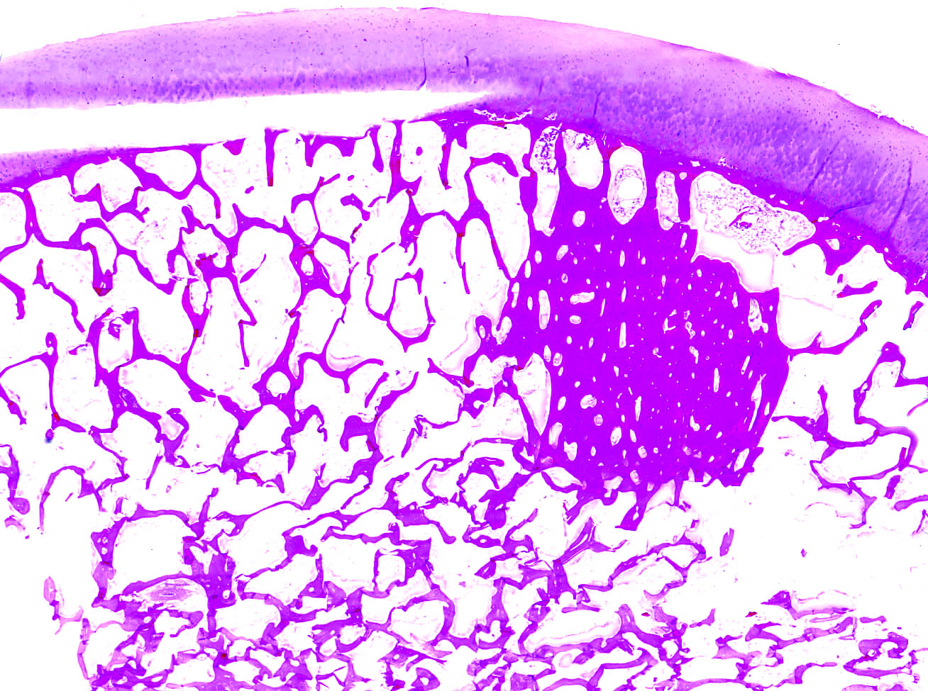
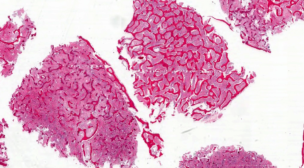
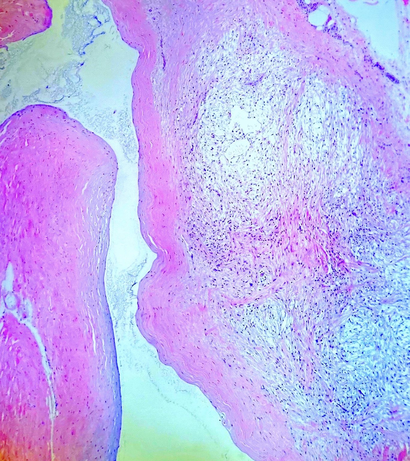
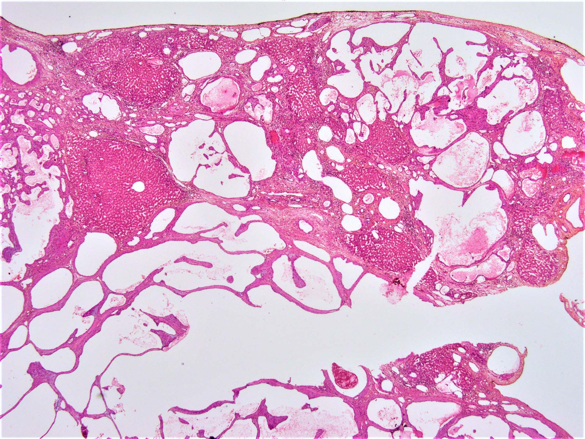
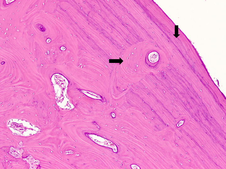
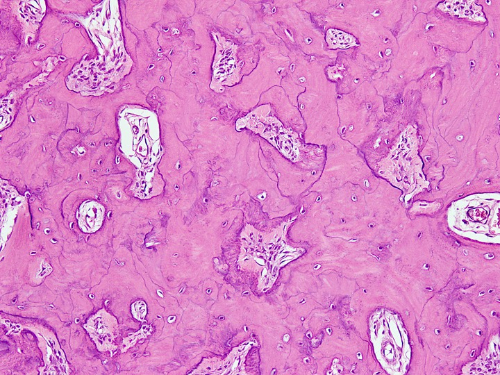
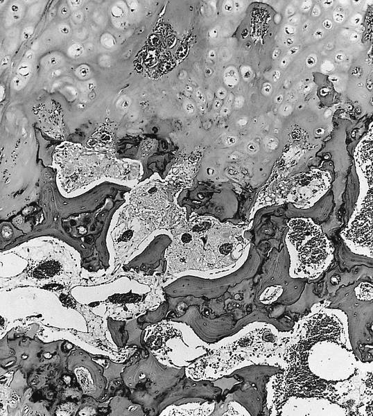
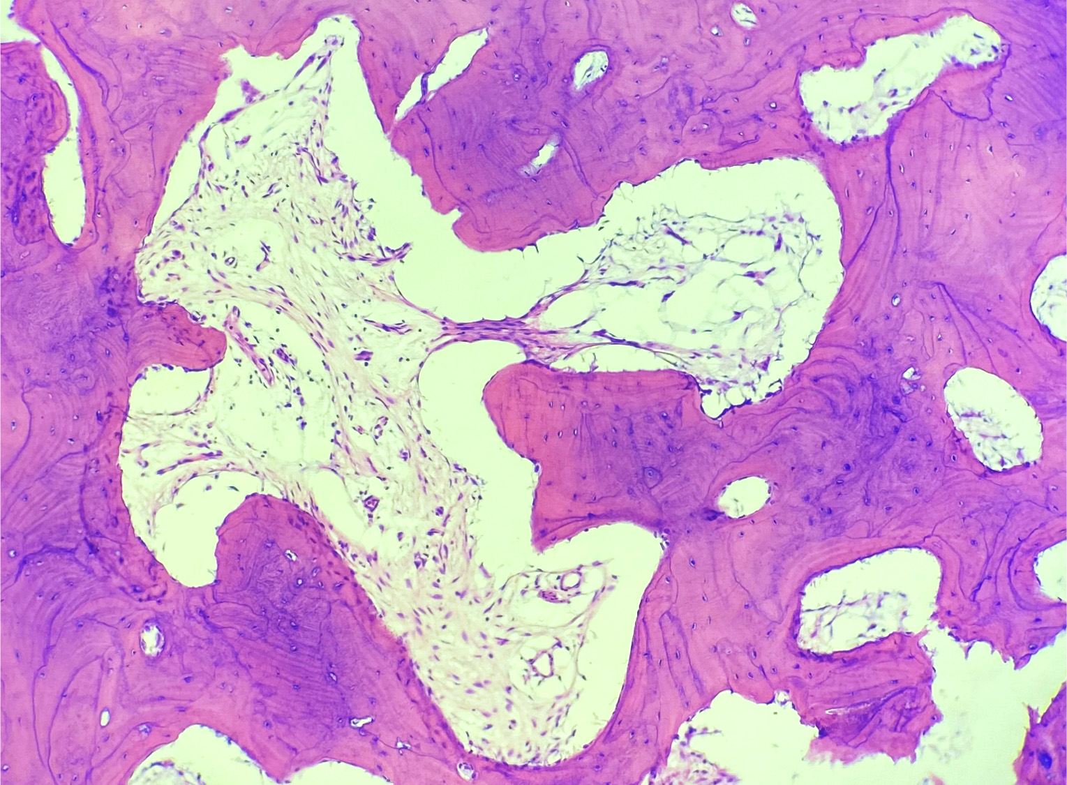
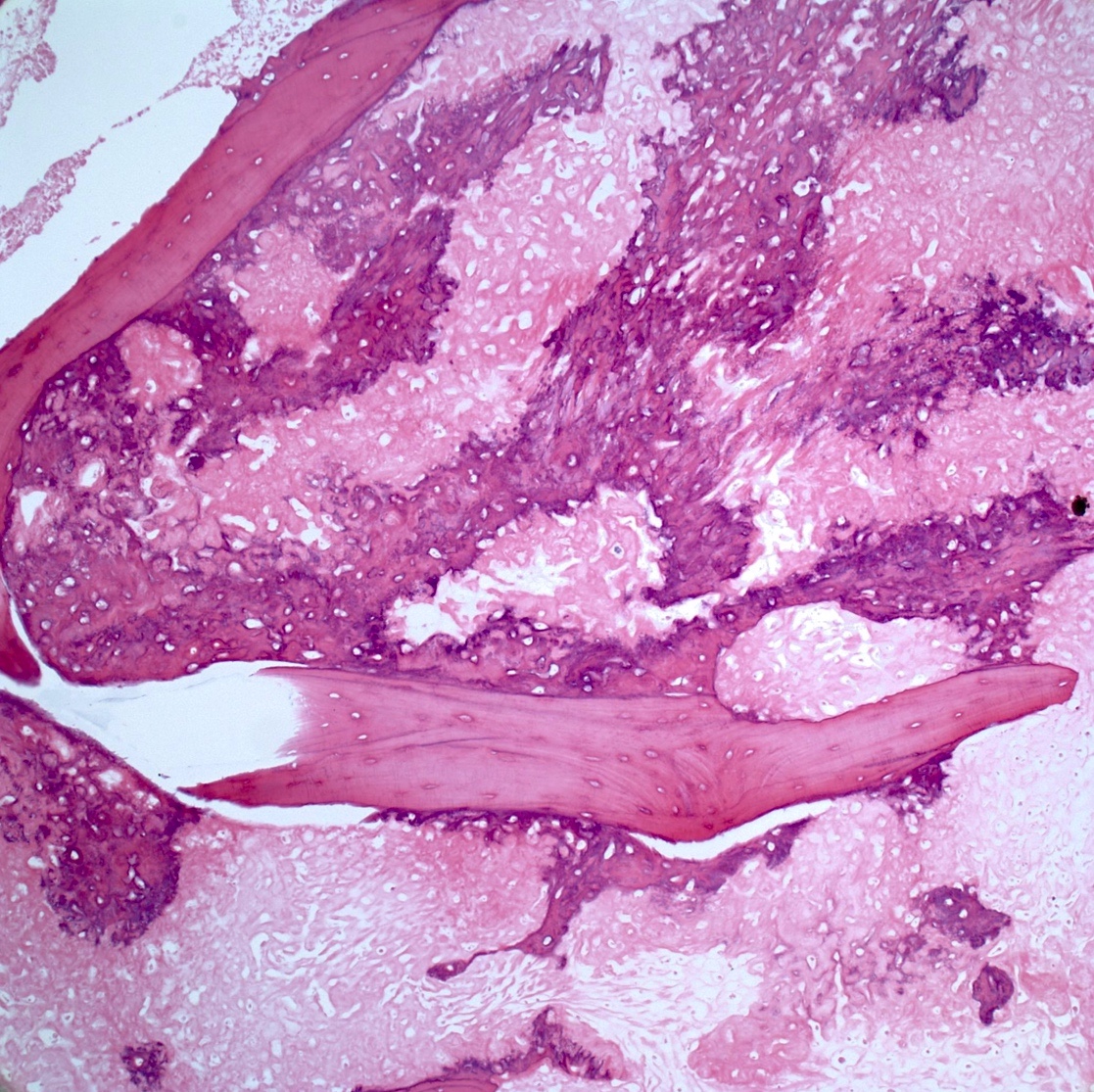
Well defined borders
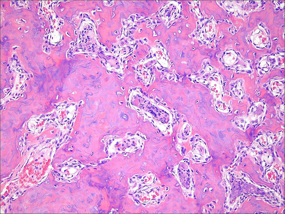
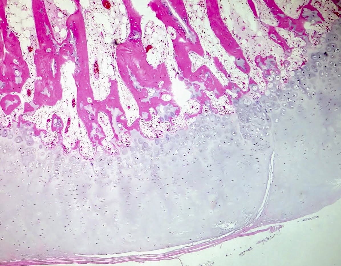
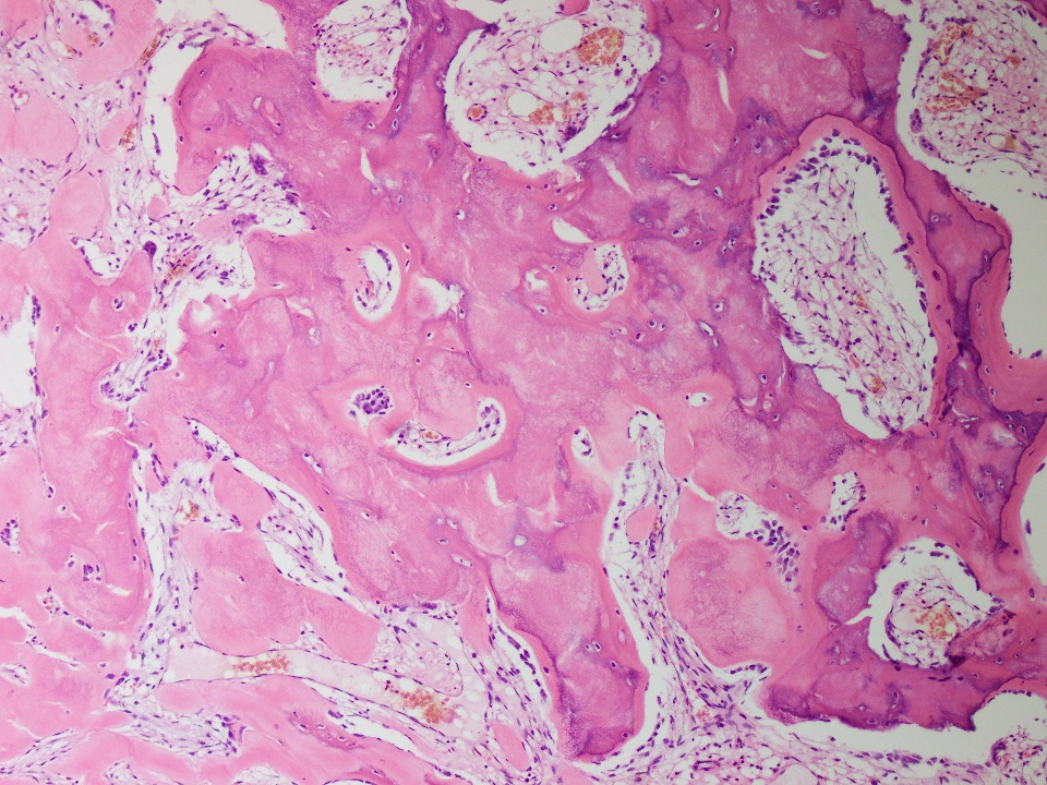
Osteoblastic rimming
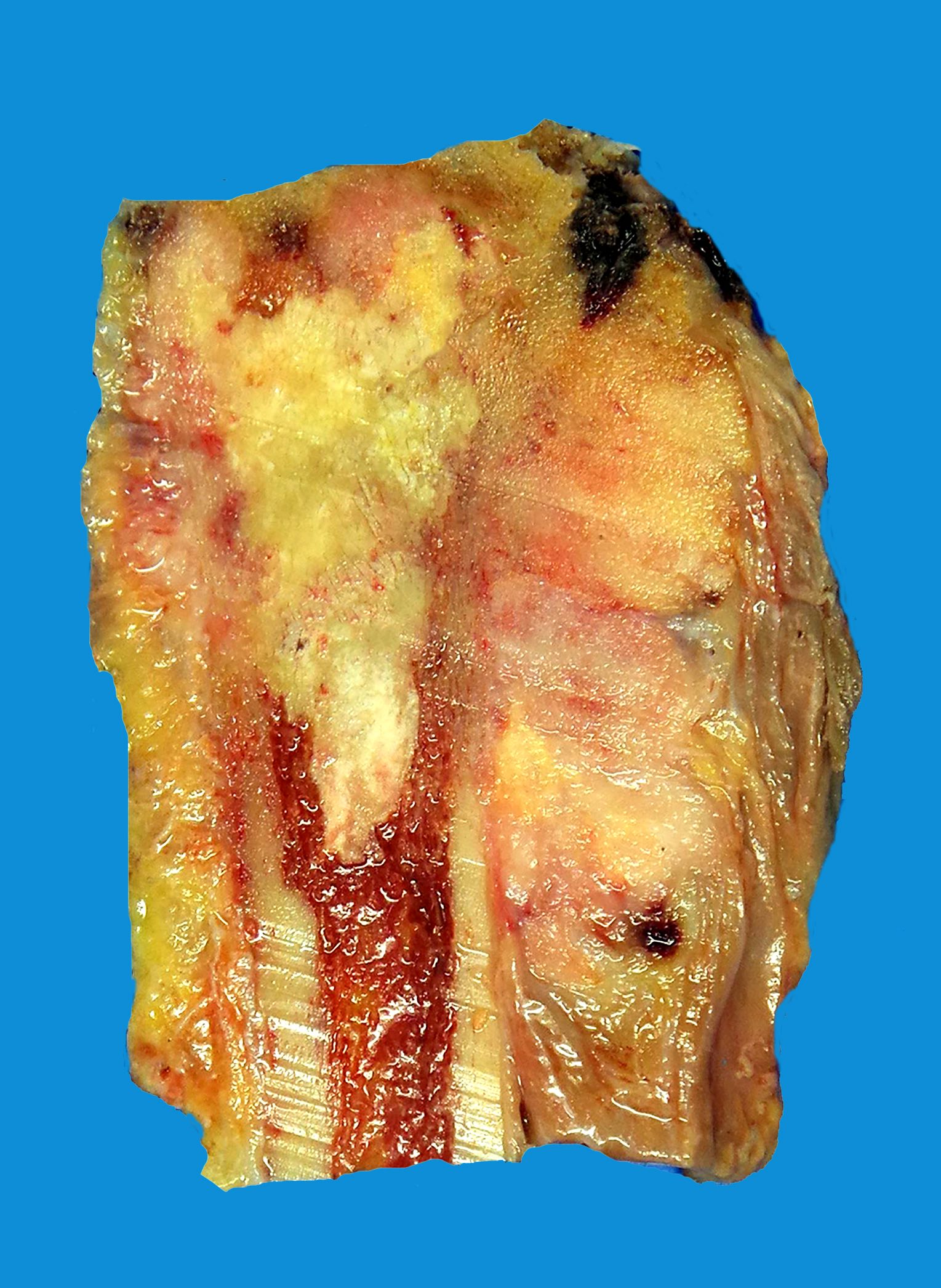
Sheet-like osteoid deposition
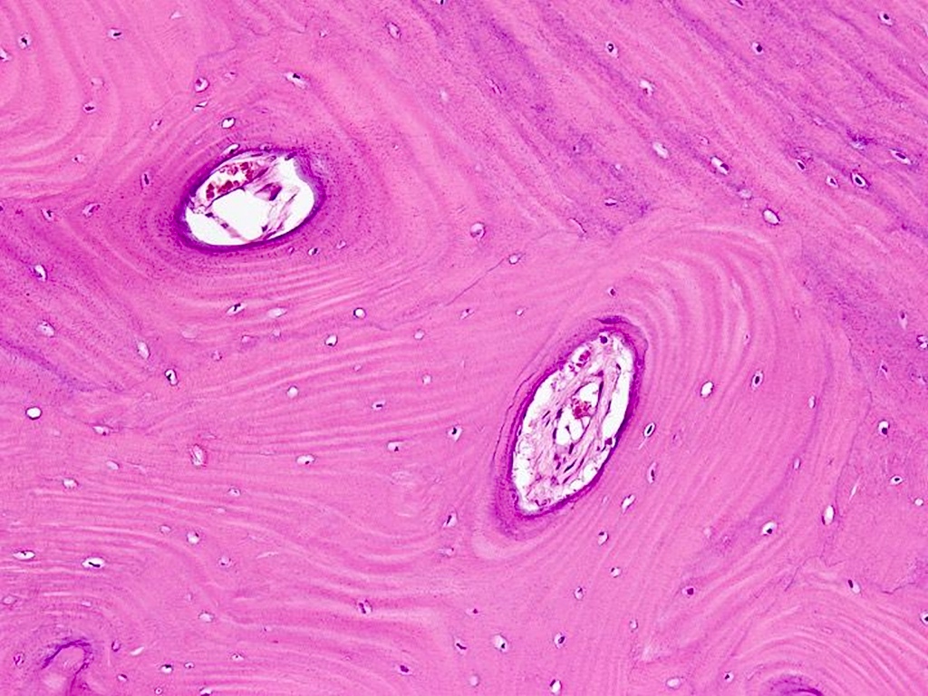
Sclerotic nidus
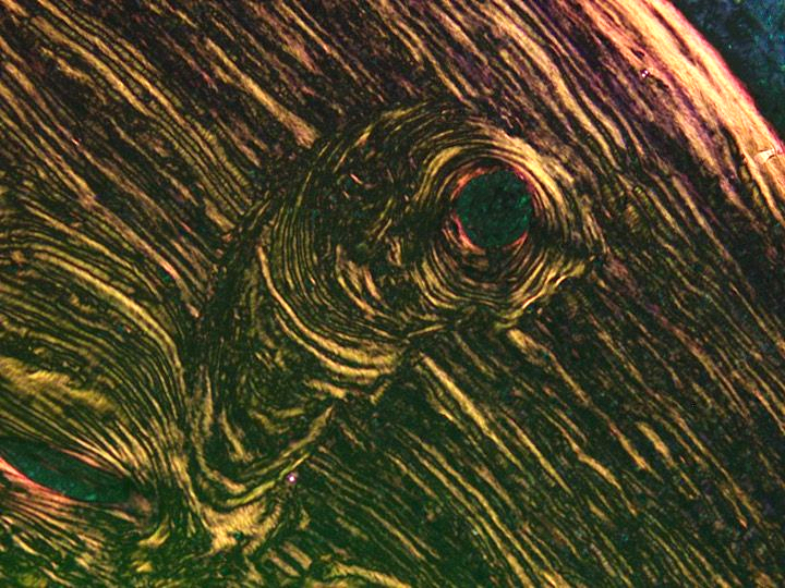
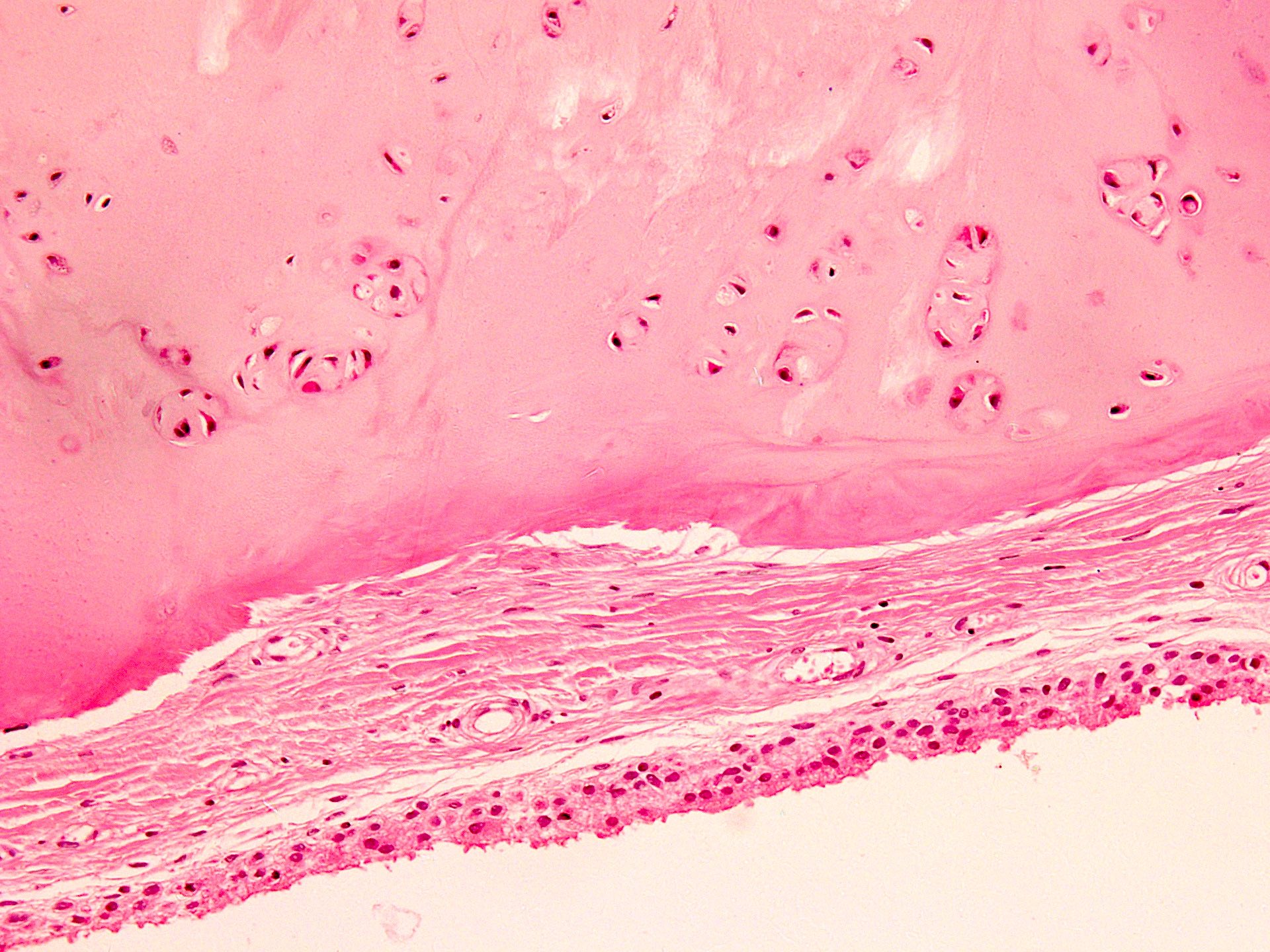
Peripheral transition to sclerotic bone
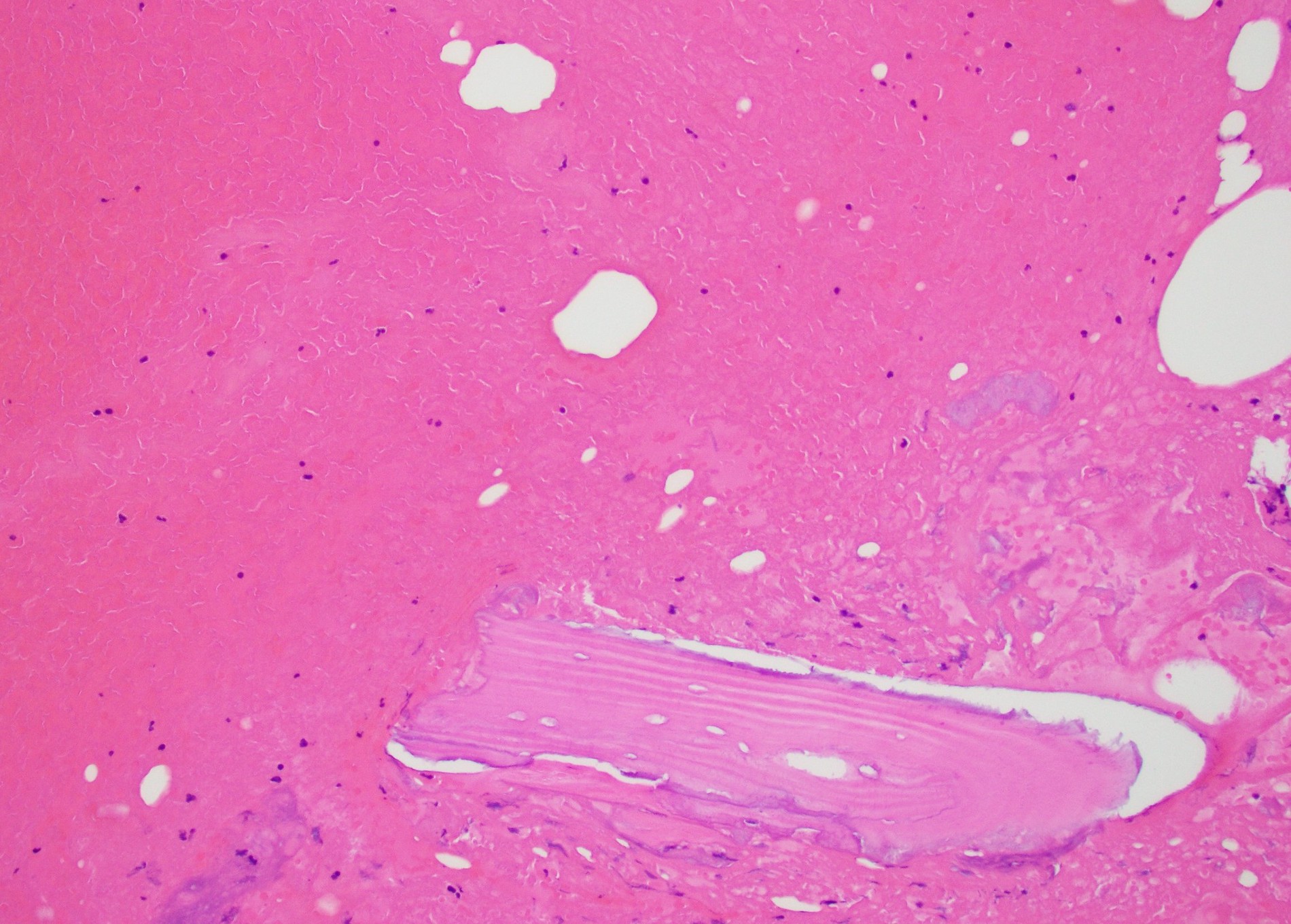
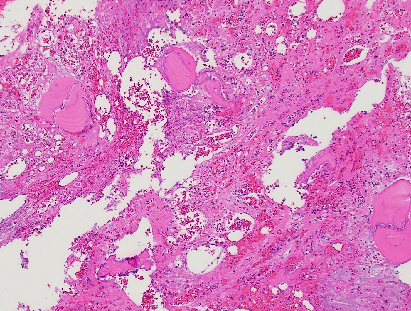
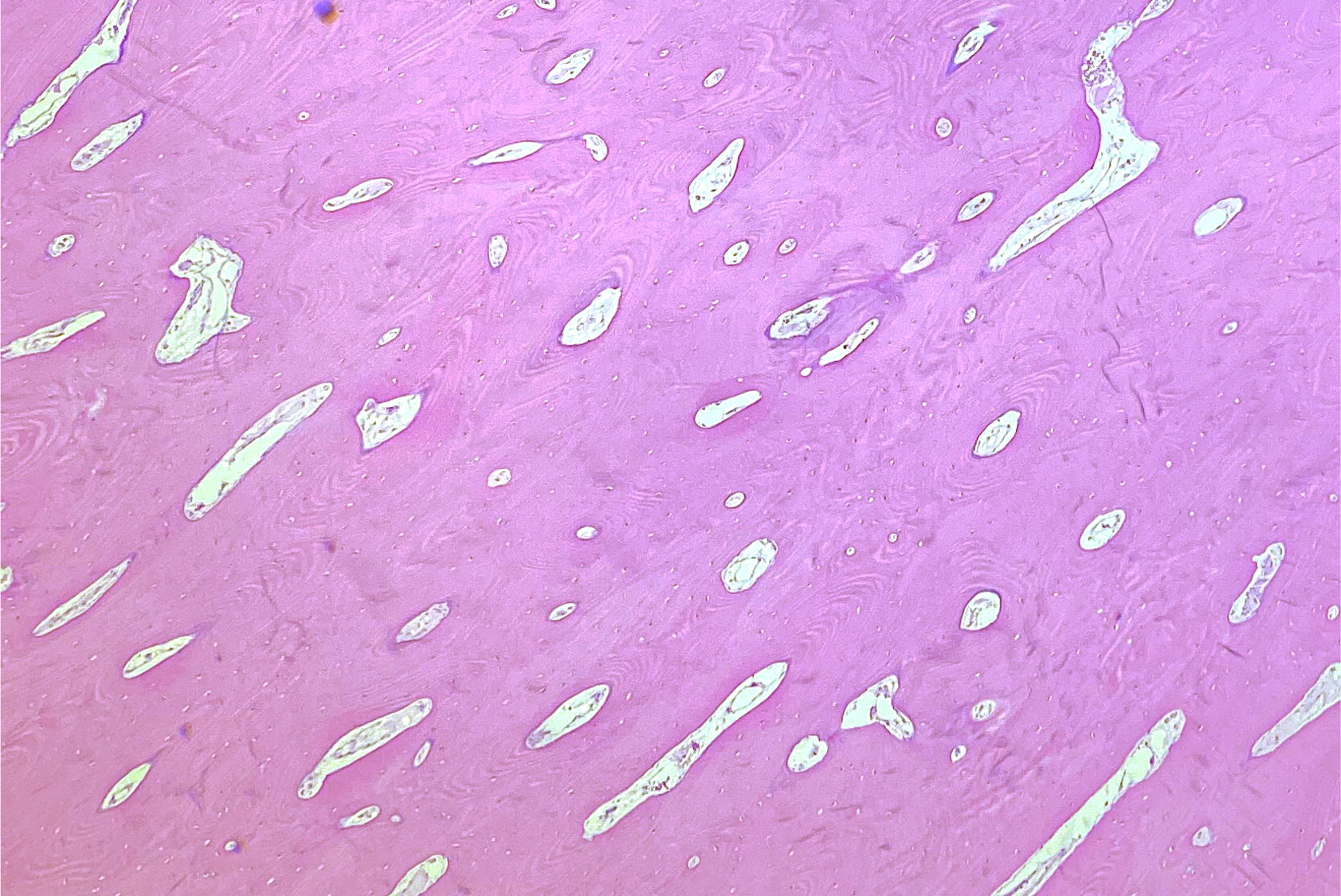
Bone trabeculae with variable mineralization
AFIP images
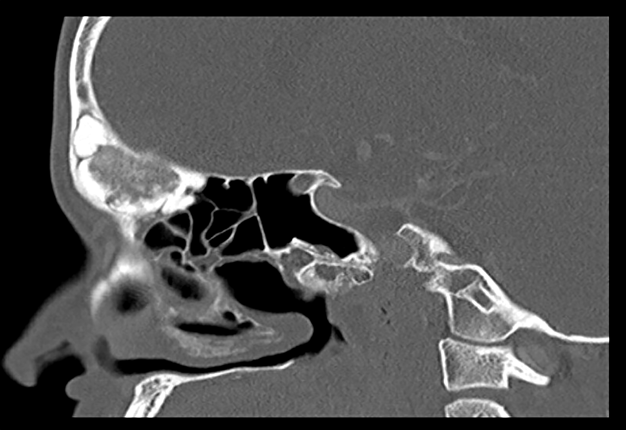
Dense lobular mass in frontal sinus
Images hosted on other servers:

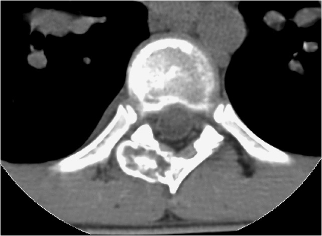
CT bone window
Images hosted on other servers:

Osteoma of anterior maxilla

Forehead osteomas

Nasal cavity osteoma with actinomycosis
Contributed by David R. Lucas, M.D. and Mark R. Wick, M.D.

Osteoblastoma-like osteoma
Skull osteoma
Images hosted on other servers:

Osteoma with smooth bosselated surface

Giant orbit osteoma

Fronto-ethmoidal osteoma
Contributed by Serenella Serinelli, M.D., Ph.D., Gustavo de la Roza, M.D. and Kelly Magliocca, D.D.S., M.P.H.
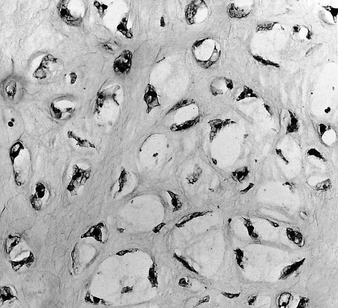
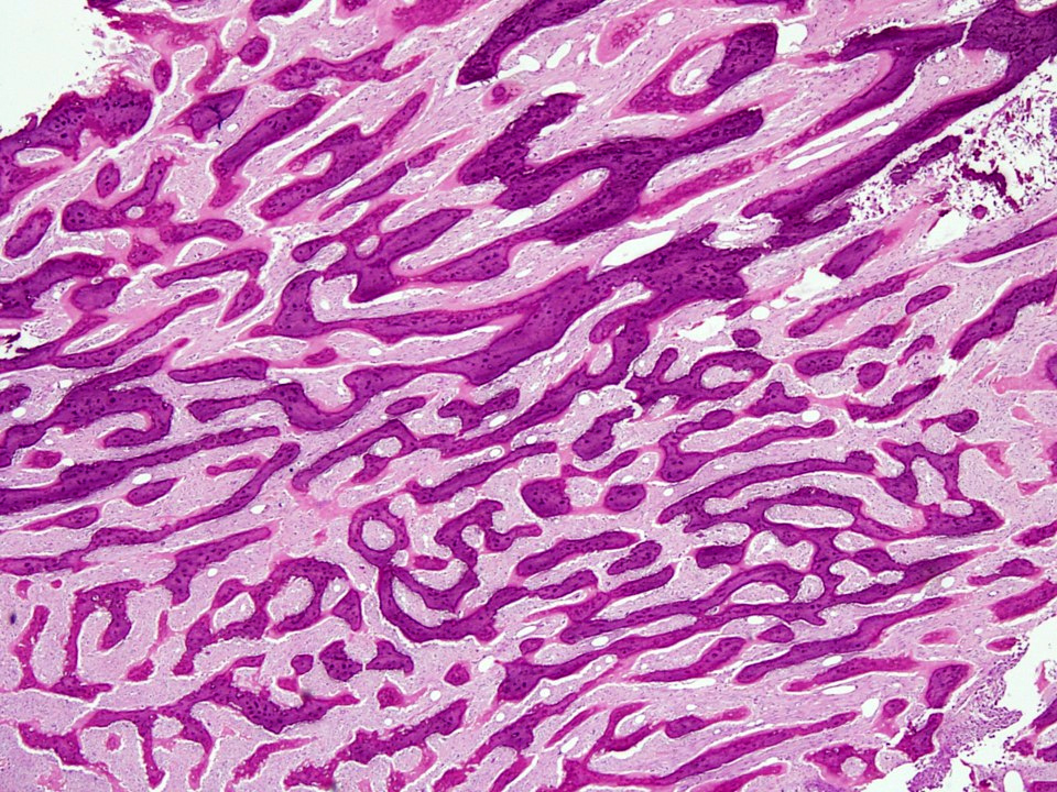
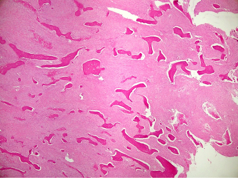
Cortical type bone architecture
Trabecular bone architecture
Lamellar bone
Osteoblasts and osteocytes
Fibrous stroma in marrow spaces
Osteoma
Osteoma of bone
Images hosted on other servers:
Osteoporosis
Contributed by Jesse Hart, D.O., Borislav A. Alexiev, M.D. and AFIP
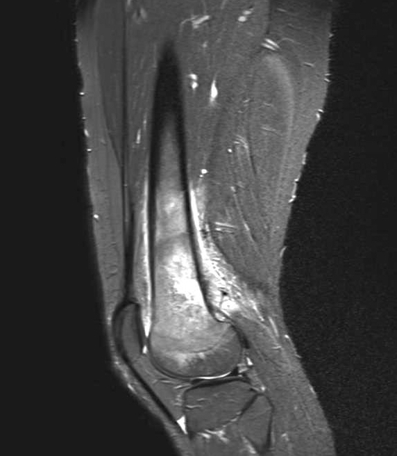
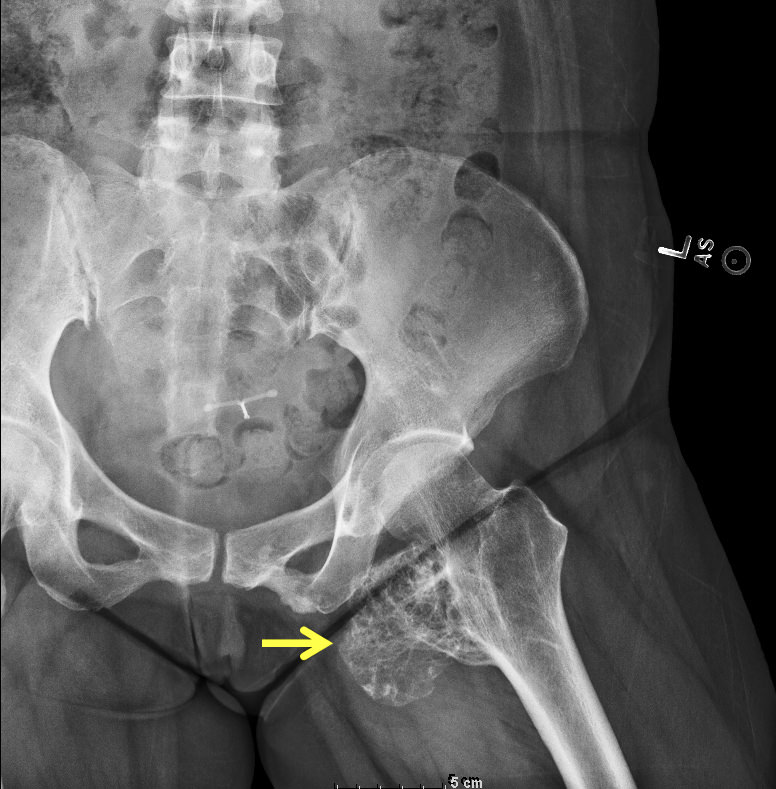
Soft tissue extension
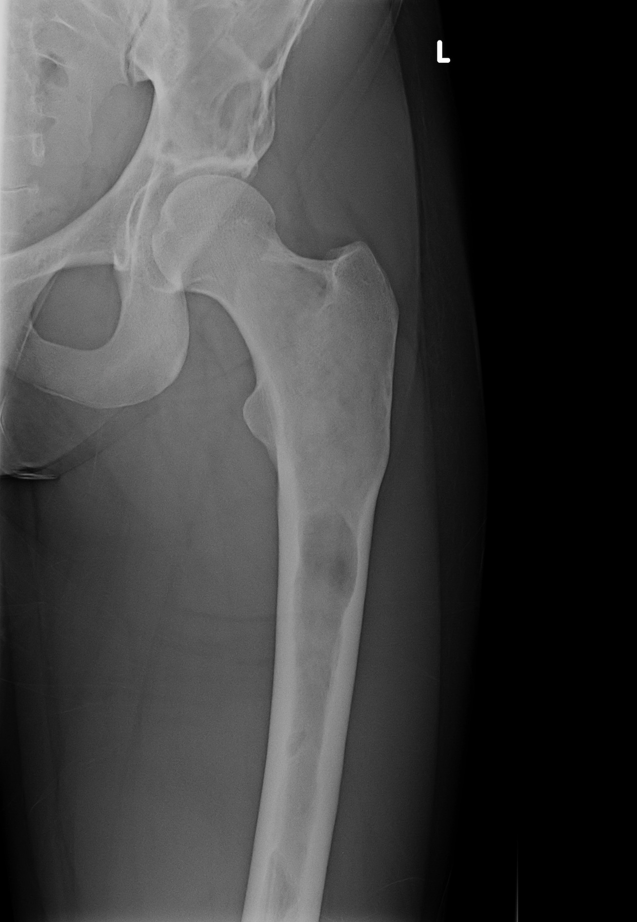
Proximal humerus mass
Proximal femur mass
Metadiaphyseal aspect of proximal tibia
Proximal fibula
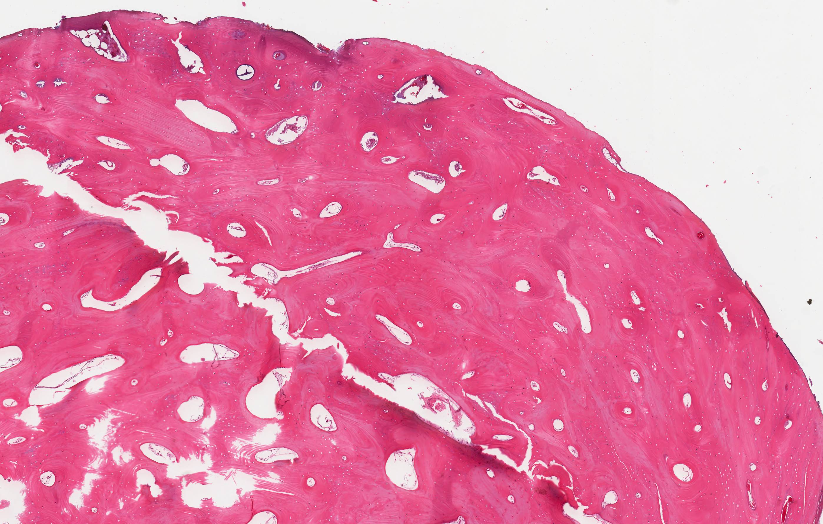
Contributed by Jesse Hart, D.O., Borislav A. Alexiev, M.D. and Mark R. Wick, M.D.
Low grade central osteosarcoma of femur
Osteosarcoma of the humerus
Osteosarcoma of the femur
Telangiectatic osteosarcoma
Grid pattern diagram
Contributed by Jesse Hart, D.O., Borislav A. Alexiev, M.D. and AFIP
Permeative growth pattern
Lace-like bone
Marked pleomorphism
Soft tissue extension
Chondroblastic osteosarcoma with focal bone
Fibroblastic osteosarcoma
Fibroblastic osteosarcoma spindle cells
Postneoadjuvant resection
Postneoadjuvant tumor cell drop out
Postneoadjuvant loose, fibrous scar
Low grade central osteosarcoma
Low grade central osteosarcoma invasion

Small osteoblasts with indistinct cytoplasm
Lymphoma-like pattern
Small foci of osteoid
Telangiectatic osteosarcoma
Telangiectatic osteosarcoma
Contributed by Jose G. Mantilla, M.D.
Coronal CT of femur
Xray of femur
Contributed by Dana J. Hariri, M.D.
Osteoblastic rimming
Jigsaw pattern
Haphazard lamellar bone
Peritrabecular fibrosis
Overview of diagnosis and management of Paget disease
Contributed by Nasir Ud Din, M.B.B.S., Mark R. Wick, M.D. and AFIP
Surface tumor with intact cortex

Intact cortex
Radiolucent cleavage of intact periosteum
Lobulated tumor with cartilage component
Parosteal osteosarcoma of rib
Distal femur Xray
Proximal tibia Xray
Mass behind knee
Femur
Contributed by Mark R. Wick, M.D. and AFIP
Distal femur
Proximal femur tumor
Cross section
Images hosted on other servers:

Tumor encircling bone

Dedifferentiated parosteal osteosarcoma
Contributed by Nasir Ud Din, M.B.B.S.
Cartilage cap, bone and spindle cells
Cartilage nodule, bone and spindle cells
Parallel bone trabeculae
Parallel and interconnected bone trabeculae
Parallel and interconnected bone trabeculae
Interconnected bone trabeculae
Small islands of bone
Chinese letter-like bone trabeculae
Low grade spindle cell component
Intermediate grade spindle cell component
Dedifferentiated areas
Fat and muscle entrapment
Skeletal muscle atrophy
Images hosted on other servers:

MDM2 gene amplification by FISH
Parosteal osteosarcoma
by Lewis Hassell
Parosteal osteosarcoma made easy by Vikram Deshpande
Contributed by Shadi Qasem, M.D., M.B.A.
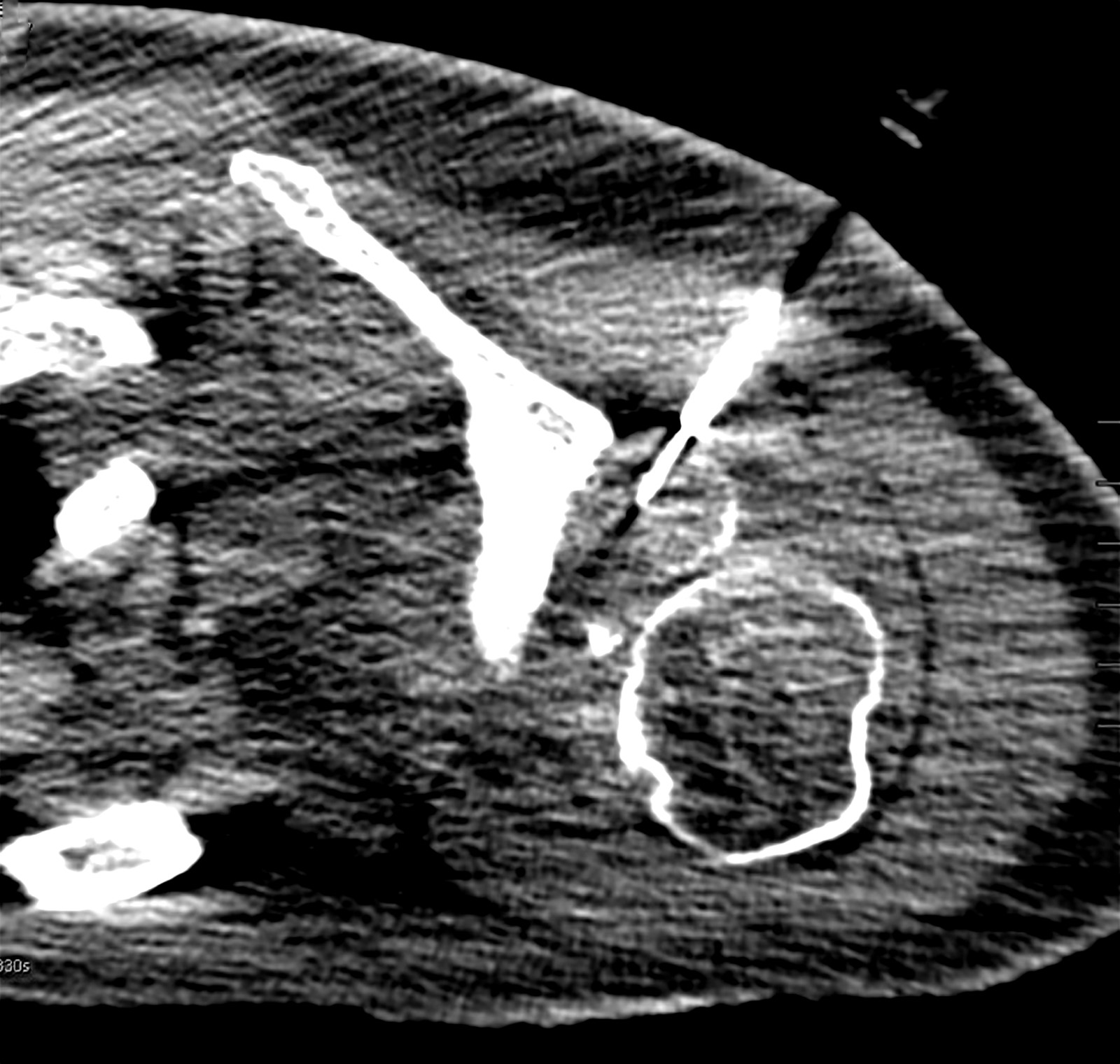
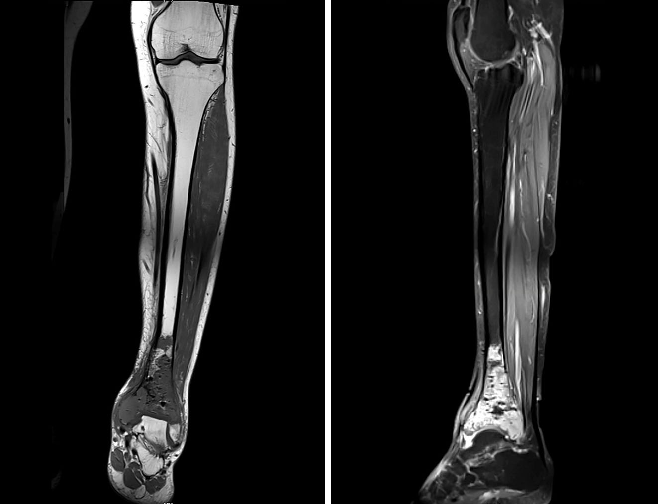
Xray of distal femur lesion
CT scan of distal femur lesion
Proximal tibia lesion
Contributed by Shadi Qasem, M.D., M.B.A. and Mark R. Wick, M.D.
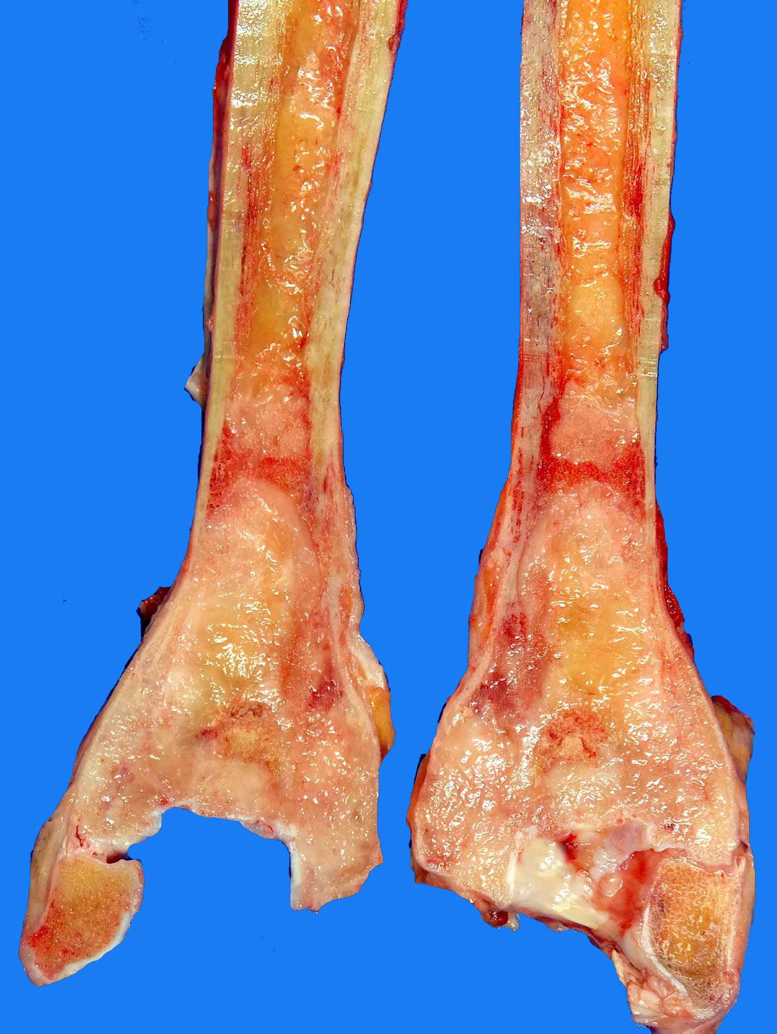
Marginal resection specimen
%20type%20gross.jpg)
Wide resection specimen
Contributed by Shadi Qasem, M.D., M.B.A.
Well defined margin
Low cellularity
No significant atypia
Images hosted on other servers:

Yellowish discharge of altered synovial fluid
Contributed by @MirunaPopescu13 on Twitter and Case #308
Rheumatoid arthritis

Various images
Images hosted on other servers:


Amorphous, pink, necrotic material
Images hosted on other servers:

HLA-DR4 molecule
Images hosted on other servers:

Ankle joint
Contributed by Nasir Ud Din, M.B.B.S.

Proximal humerus
Humerus
Proximal femur
Proximal femur MRI
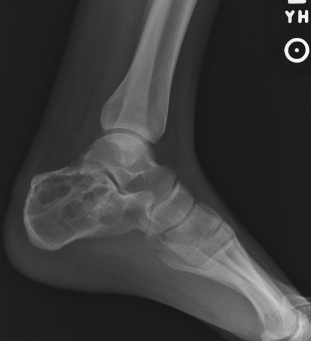
Calcaneus
Images hosted on other servers:

Mandible
Images hosted on other servers:

Humerus
Contributed by Nasir Ud Din, M.B.B.S.
Cyst fragments
Cyst wall
Fibrin-like deposits

Reactive bone
Images hosted on other servers:

Osteogenesis imperfecta:
Postmortem photograph shows
deformed extremities, findings
that are consistent with fractures
Images hosted on other servers:

Intervertebral disc
(top center) destroyed
by vertebral abscess

Spinal cord compressed by abscess
Images hosted on other servers:


C. tropicalis
Contributed by Anshu Bandhlish, M.D. and AFIP images
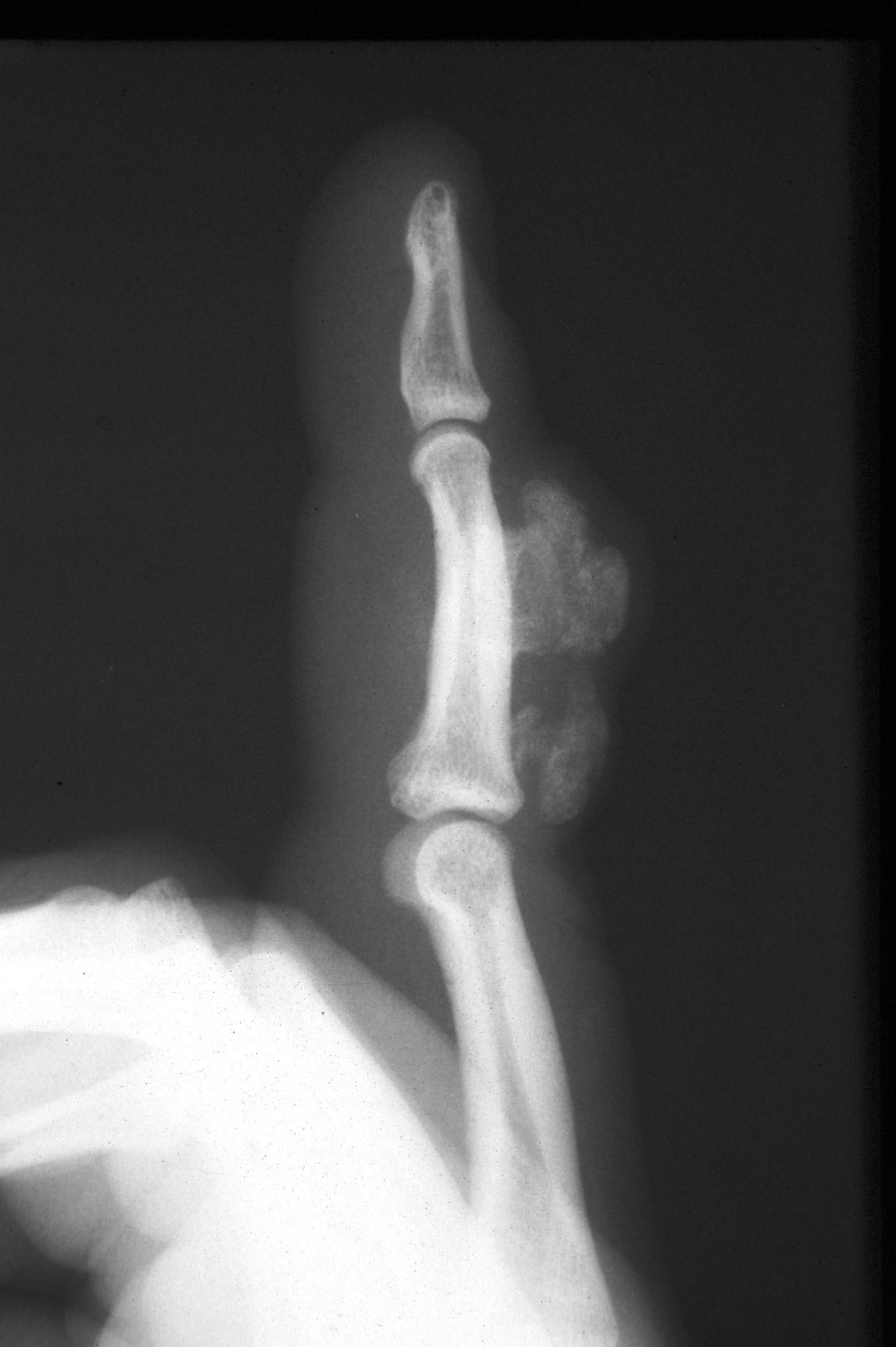
Distal phalanx Xray
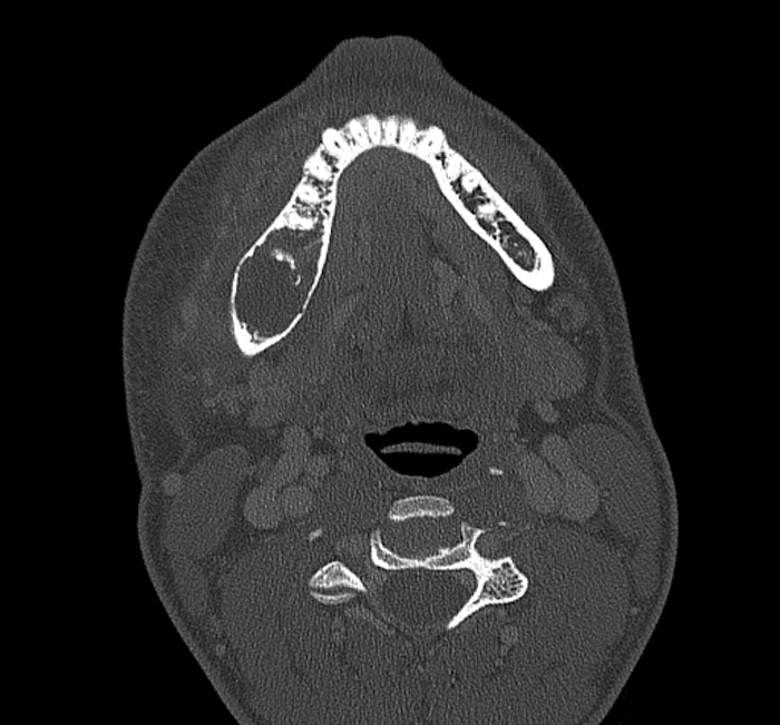
Distal phalanx of great toe
Images hosted on other servers:

Subungual exostosis of index finger

Subungual exostosis in an 8 year old child

Subungual exostosis of right fifth toe
Contributed by Robert Ricciotti, M.D. and AFIP images
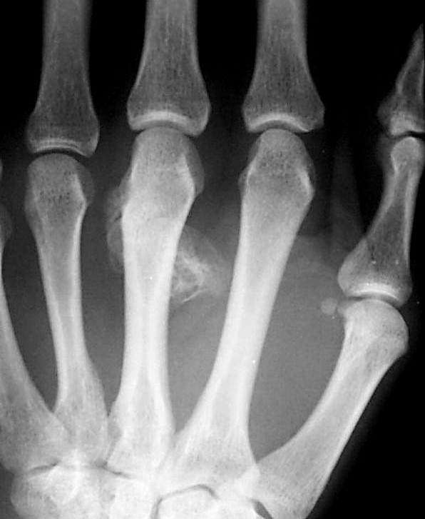
Exophytic lesion of distal phalanx
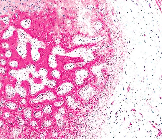
Enchondral ossification
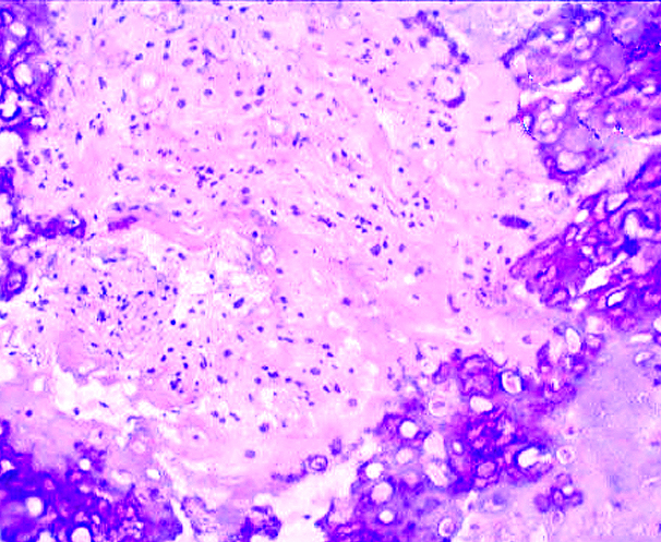
Thin cartilaginous cap
Reactive bone
Moderately cellular cartilage
What is a subungual exostosis?
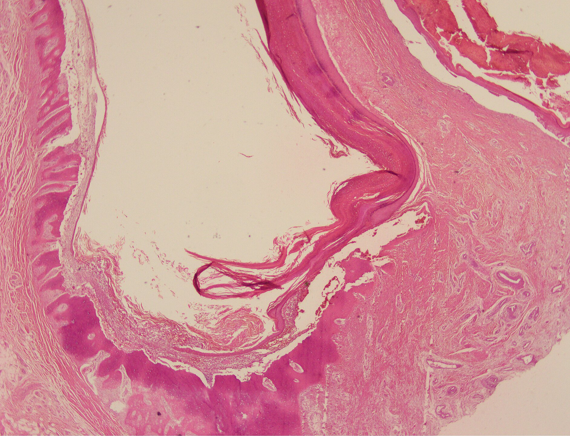
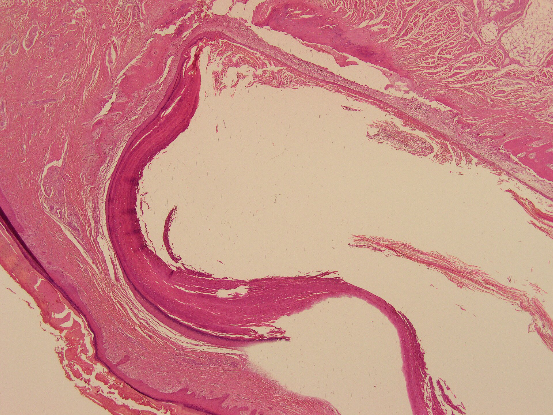
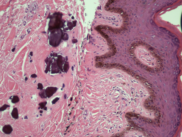
Contributed by Nasir Ud Din, M.B.B.S.
Multiple osteochondral bodies
Small
osteocartilaginous
bodies
Clumps of cartilaginous bodies

Multiple rounded to oval lesions
Images hosted on other servers:

Right thumb: dorsal aspect, lateral aspect, palmar aspect

Intraoperative view and excised loose bodies
Contributed by Nasir Ud Din, M.B.B.S.

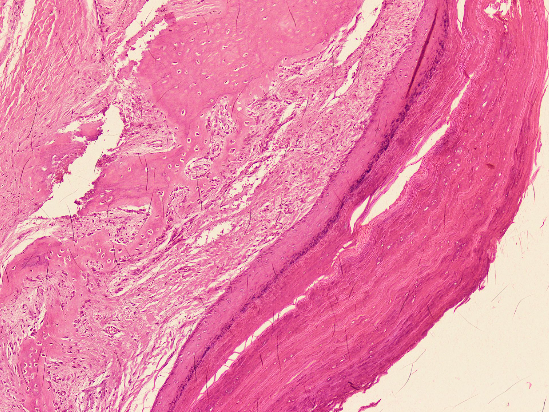
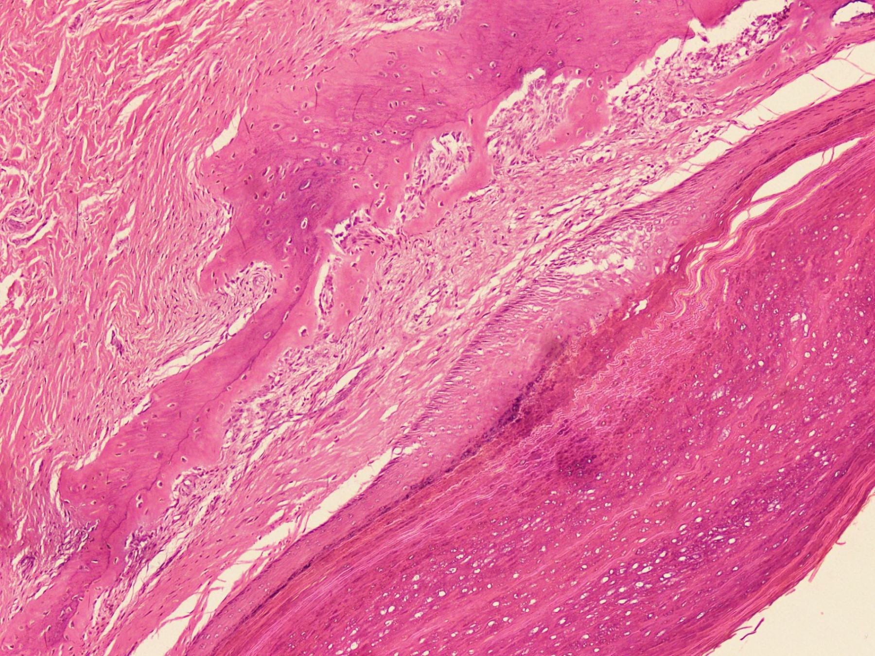
Multiple osteochondral loose bodies
Chondral loose bodies
Contributed by Nasir Ud Din, M.B.B.S.
Loose chondral bodies
Subsynovial cartilaginous bodies
Clustering of chondrocytes

Mild cellularity
Subsynovial cartilaginous body
Mild atypia
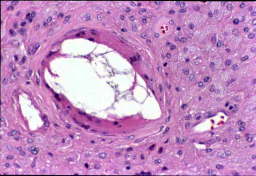
Calcification
Synovial chondromatosis:
5 minute pathology pearls
by Dr. Jerad Gardner
Images hosted on other servers:


Baker cysts
Images hosted on other servers:

Spinal synovial cyst
Images hosted on other servers:

Baker cysts


Cutaneous metaplastic synovial cyst
Images hosted on other servers:


MRI of Baker cyst
Images hosted on other servers:


Cutaneous metaplastic synovial cyst with numerous villous-like structures; vimentin+ CD34- CD68-


Hemorrhagic spinal synovial cyst with thin fibrous wall
Contributed by Mark R. Wick, M.D.
Images hosted on other servers:

Villi-like structures

Papillary architecture

Mature adipocytes

Focal hyperplasia

Few mature lipocytes
Images hosted on other servers:

Correctable swan neck deformities and ulnar deviation


Readily correctable swan neck deformities and subluxation of right fifth MCP

Metacarpal heads - no cartilage damage seen

Noncorrectable deformities of PIP joints

Mild ulnar deviation, especially on right
Images hosted on other servers:

Synovial membrane
Contributed by Mark Girton, M.D. and AFIP
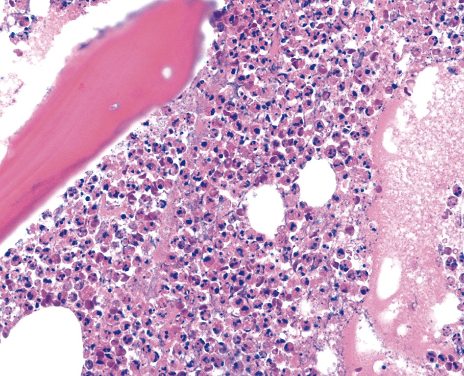
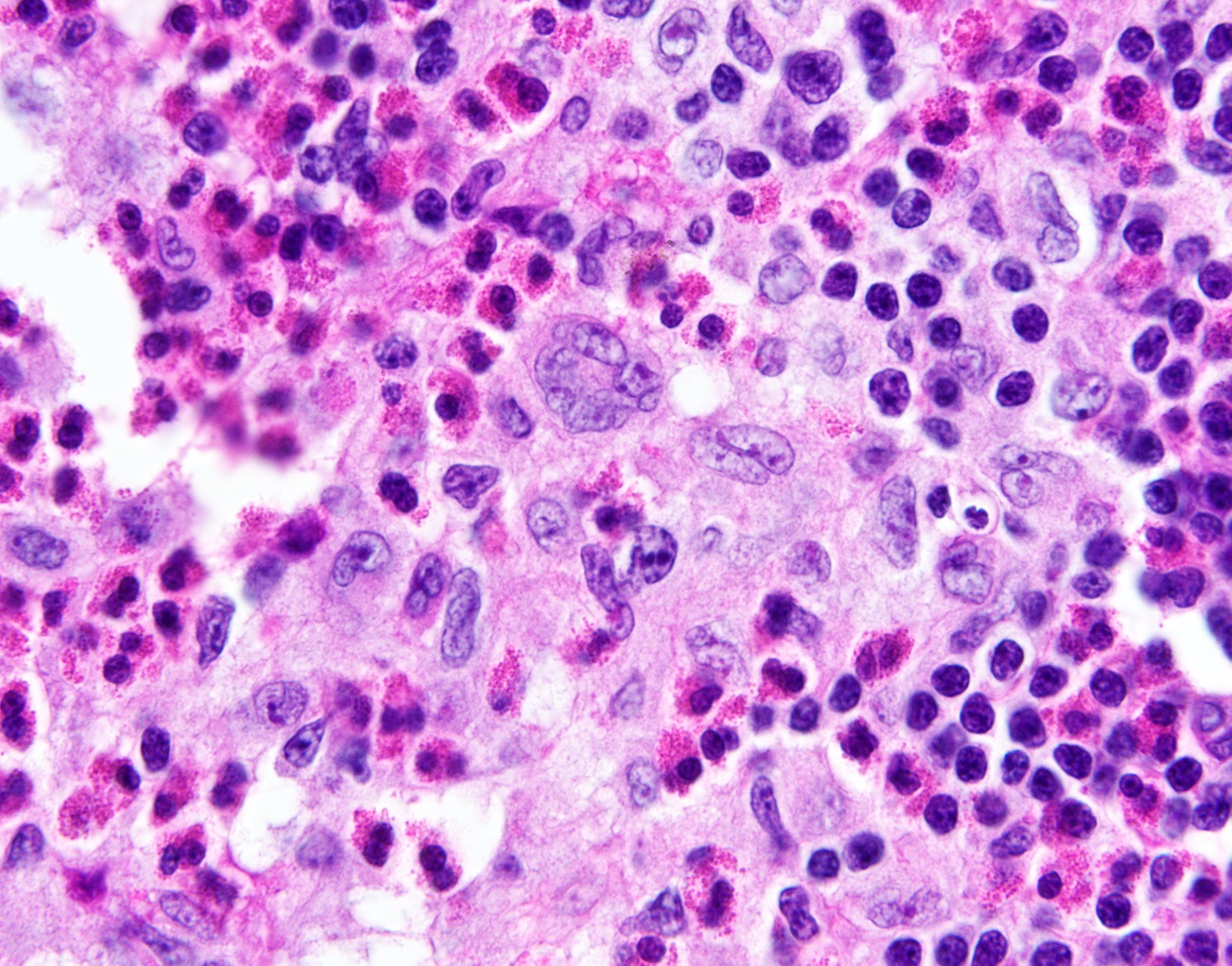
Marrow mast cell cluster
Intermixed with lymphoid aggregate
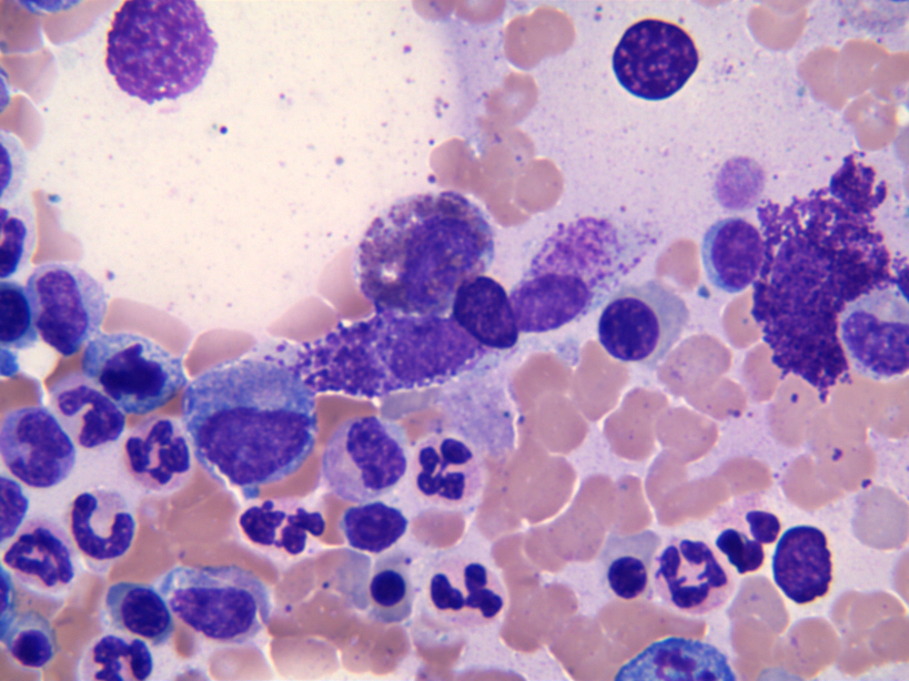
Aspirate with mast cells
Mast cell cluster
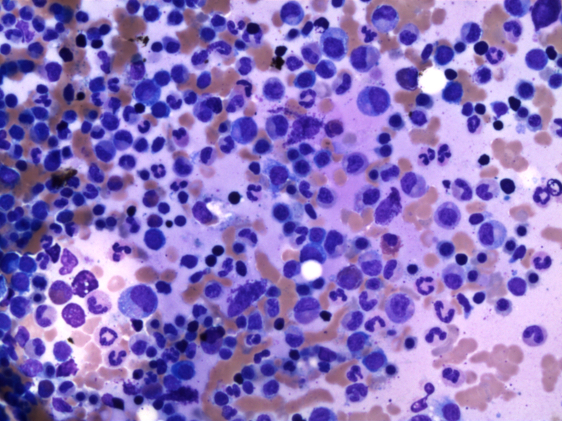
Hypercellular bone marrow biopsy
Bone marrow biopsy with marrow replacement
Cytologic spectrum
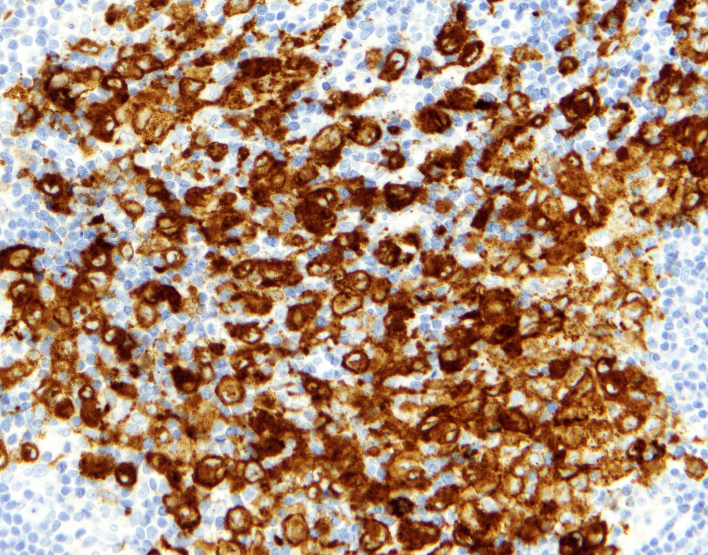
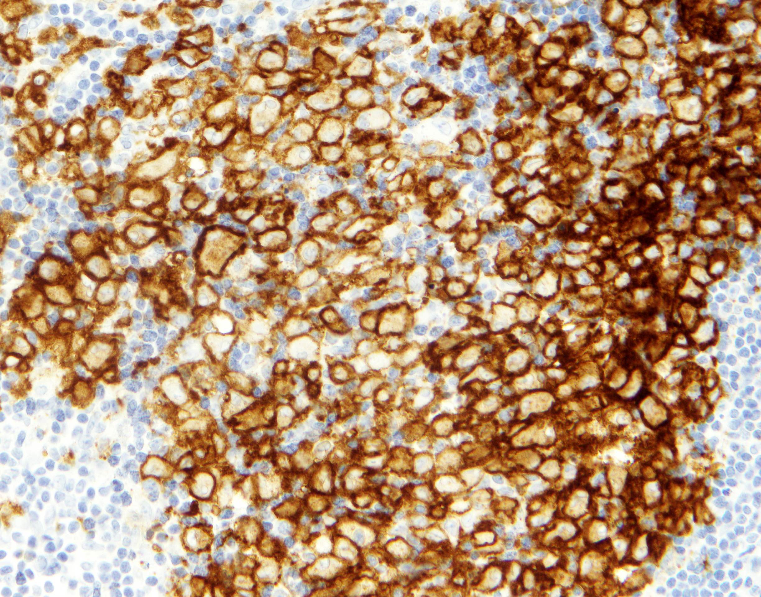
CD117 positive mast cells
CD25 positive mast cells
CD2 positive mast cells
CD30 positive mast cells
CD117 positive mast cells
Mast cell tryptase positive
CD25 positive mast cells
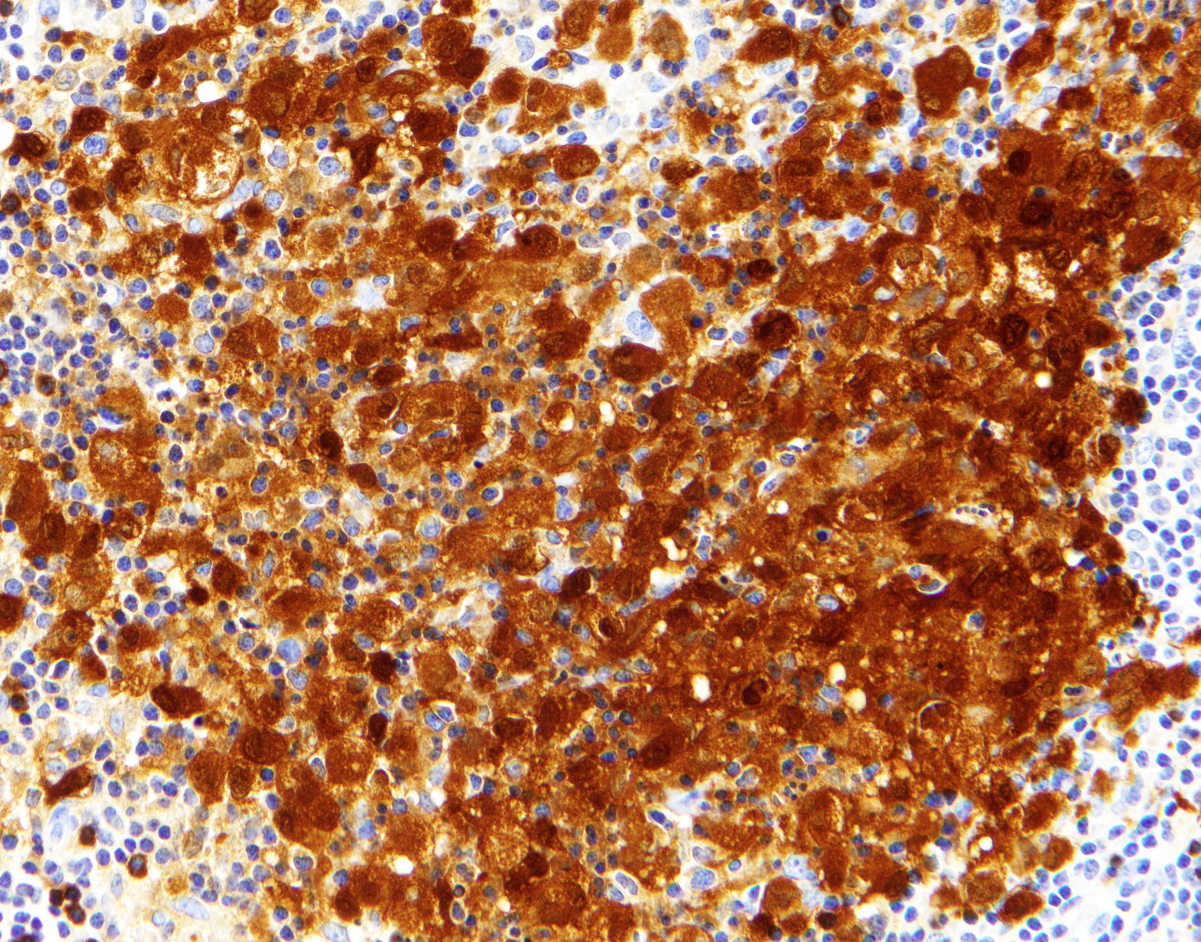
Perifollicular distribution

Characteristic sclerosis
Mast cell infiltrate
Delicate collagen fibrils and eosinophils
Mast cell staining
Interfollicular pattern
Diffuse involvement
Ovoid nuclei with eosinophilic cytoplasm
Spindle shaped mast cells
Spindly configuration
Contributed by Mark Girton, M.D.
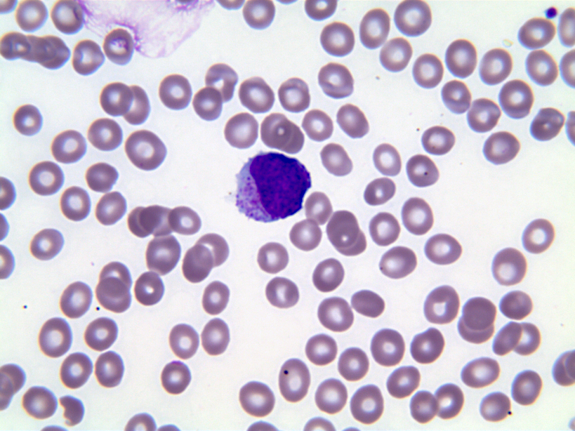
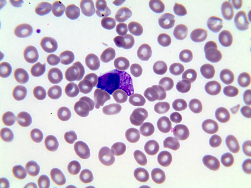
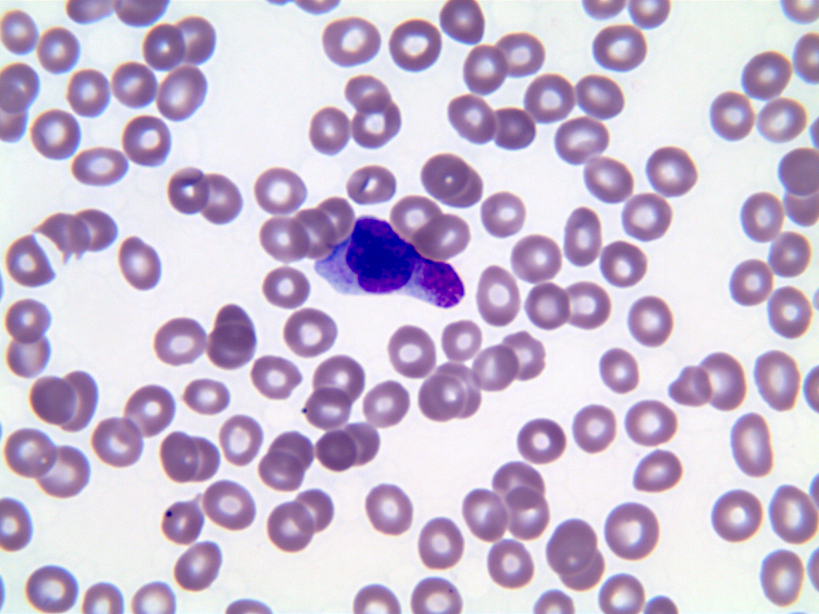
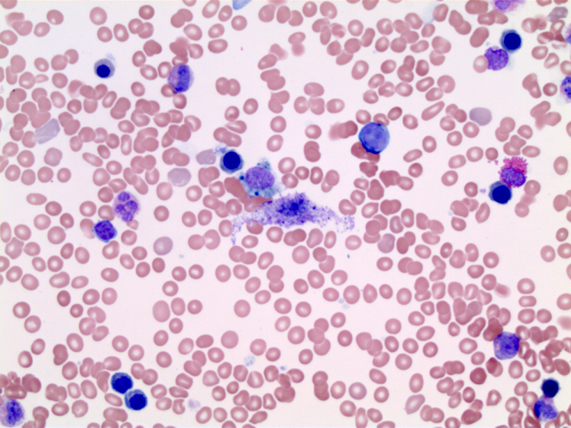
Peripheral blood mast cell
Contributed by Jigang Wang, M.D., Ph.D. and Jiufa Cui, M.D., Ph.D.
Diffuse type
Bone erosion on CT
Low signal on MR
Low signal on MR
Low signal on MR
Images hosted on other servers:
Localized type

Mass on the tendon

Ultrasound of finger lesion

Hand MRI
Diffuse type


MRI of the ankle
Malignant

Progressive increasing mass
Contributed by Mark R. Wick, M.D.
Localized type
Digit lesion
Images hosted on other servers:
Localized type

Mass over lateral right foot

Tumor at surgery

Neurovascular bundle involvement

Swelling appearance

Lateral route
Diffuse type

Brownish mass
Contributed by Mark R. Wick, M.D.
Localized type
Cut surface
Images hosted on other servers:
Diffuse type

Tumor mass
Contributed by Jigang Wang, M.D., Ph.D., Jiufa Cui, M.D., Ph.D. and Michella Whisman, M.D.
Localized type
Fibrous bands
Lobular appearance
Large epithelioid cells
Multinucleated giant cells
Pigment laden histiocytes
Foamy histiocytes
Foamy histiocytes
Mononuclear component
Diffuse type
Foamy histiocytes (xanthoma cells)
Mononuclear cells
Large mononuclear cells
Chondroid metaplasia
Papillary growth pattern
Numerous mononuclear cells
Images hosted on other servers:
Localized type

Mononuclear stromal cells, osteoclast giant cells
Malignant

Mitosis
Images hosted on other servers:
Diffuse type

Structural chromosomal aberration
Images hosted on other servers:

Arthroscopic view of elbow joint
Images hosted on other servers:

Synovial tissue
Contributed by Mark R. Wick, M.D.
Paraspinal tuberculous abscess MRI
Contributed by Mark R. Wick, M.D.
%20with.jpg)
Tubeculous vertebral osteomyelitis
Contributed by Mark R. Wick, M.D.
Various images
Images hosted on other servers:

Schematics of tumoral calcinosis

Approach for tumoral calcinosis
Contributed by Nasir Ud Din, M.B.B.S.
Right finger, distal phalanx
Calcified lesion, foot
Images hosted on other servers:

Nodules on patient’s hands

Metastatic
calcifications
presenting as
firm solid masses

NFTC and dermatomyositis
Images hosted on other servers:

Cystic spaces filled with yellowish material
Contributed by Nasir Ud Din, M.B.B.S. and John Irlam, D.O.
Irregular basophilic deposits
Foreign body giant cell reaction

Amorphous granular pink deposits
Chunky basophilic deposits
Psammoma body-like calcospherites
Calcinosis
Images hosted on other servers:

Amorphous pink deposits
Basophilic, amorphous calcium deposits
Scrotal calcinosis - pathology mini tutorial
Contributed by AFIP and Mark R. Wick, M.D.
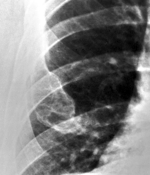
Radiograph of tibial tumor
Primary, distal femur
Contributed by Mark R. Wick, M.D.
Primary
Contributed by Hatem Kaseb, M.D., Ph.D., M.P.H., Mark R. Wick, M.D. and AFIP

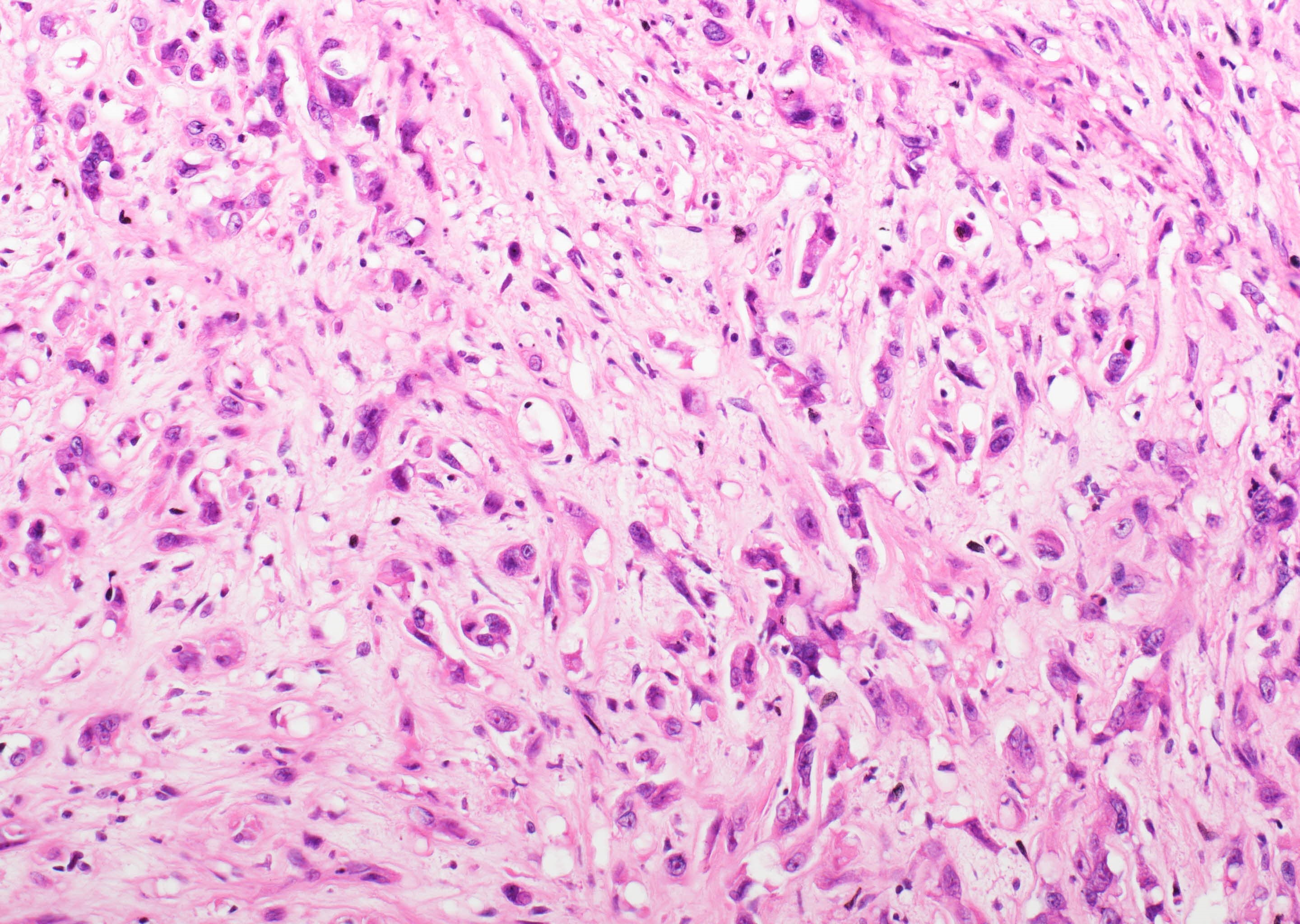
Significant pleomorphism
Significant pleomorphism
Significant pleomorphism
Fascicles of fibroblasts
Noncalcified collagen
Contributed by Borislav A. Alexiev, M.D.
Enchondroma
Chondrosarcoma
Dedifferentiated chondrosarcoma
Mesenchymal chondrosarcoma
Low grade osteosarcoma
Osteosarcoma
Aneurysmal bone cyst
Chondroblastoma

Giant cell tumor of bone
Nonossifying fibroma
Conventional chordoma
Bocklage: 2014
Czerniak: 2015
Deyrup: 2015
Dodd: 2014
Folpe: 2022
Forest: 1998
Greenspan: 2015
Horvai: 2012
IARC: 2020
Jo: 2015
Klein: 2011
Montgomery: 2020
Nielsen: 2021
Nielsen: 2021
Picci: 2019
Santini-Araujo: 2020
Unni: 2009
Wei: 2013
Find related Pathology books: soft tissue & bone
