


Prof. Dr. Fernando Schmitt:
The WHO System for Reporting Lung Cytopathology
Contributed by Reid Wilkins, M.D.
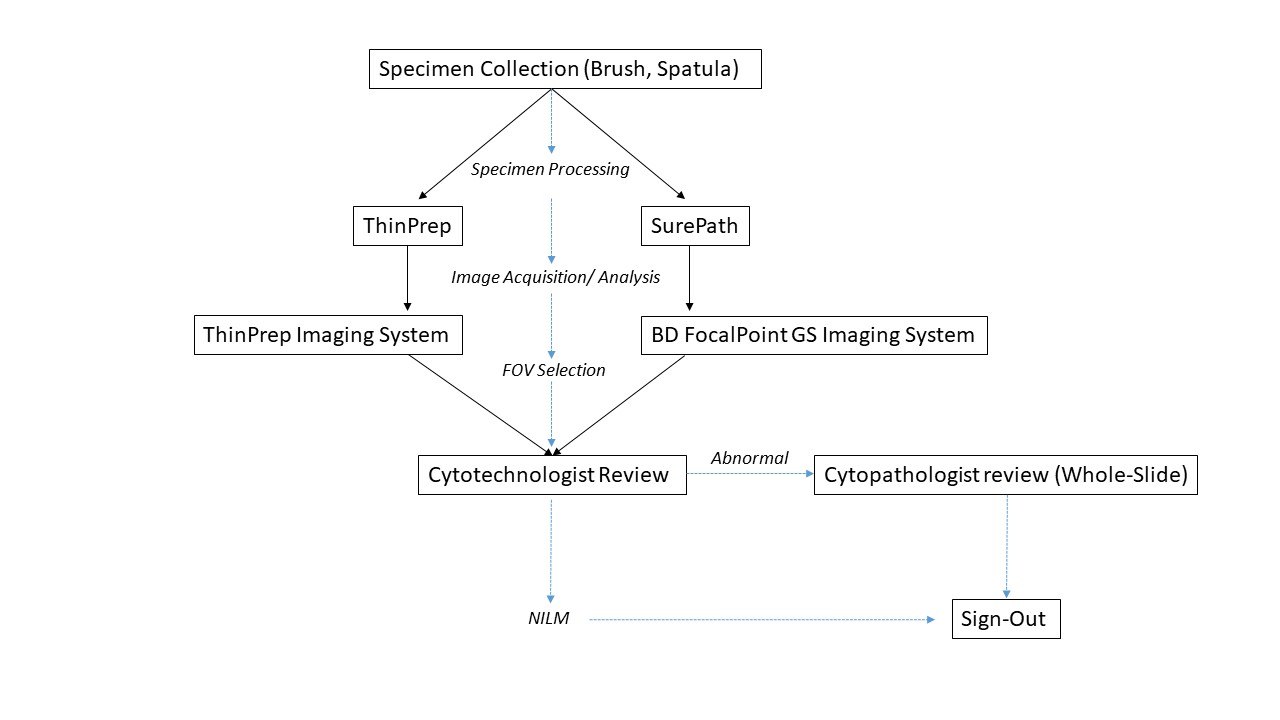
Overview of automation workflow
Images hosted on other servers:

ThinPrep imaging system

BD FocalPoint GS imaging system
Contributed by Xiaobing Jin, M.D., Ph.D. and Heather I-Hsuan Chen-Yost, M.D.

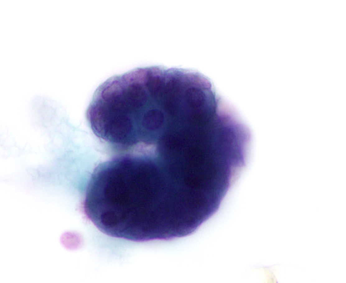
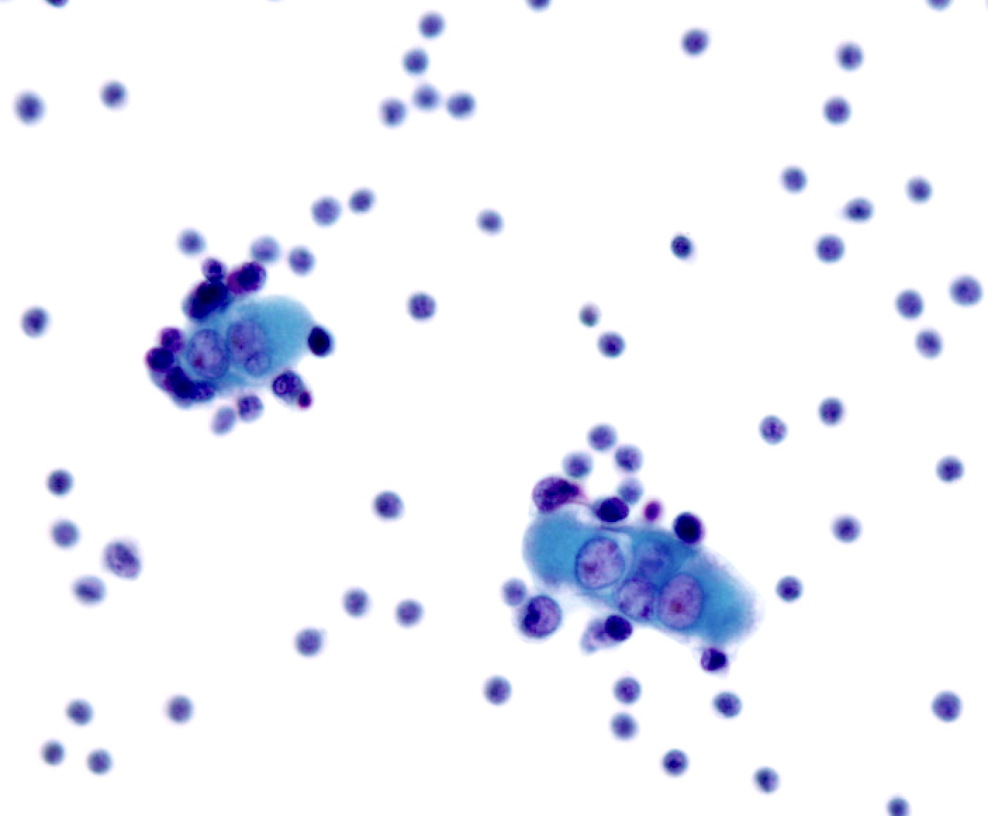
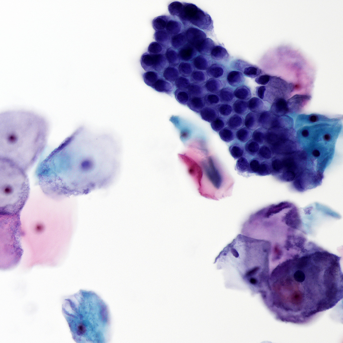
Bronchial cells

Bronchial cells cross section
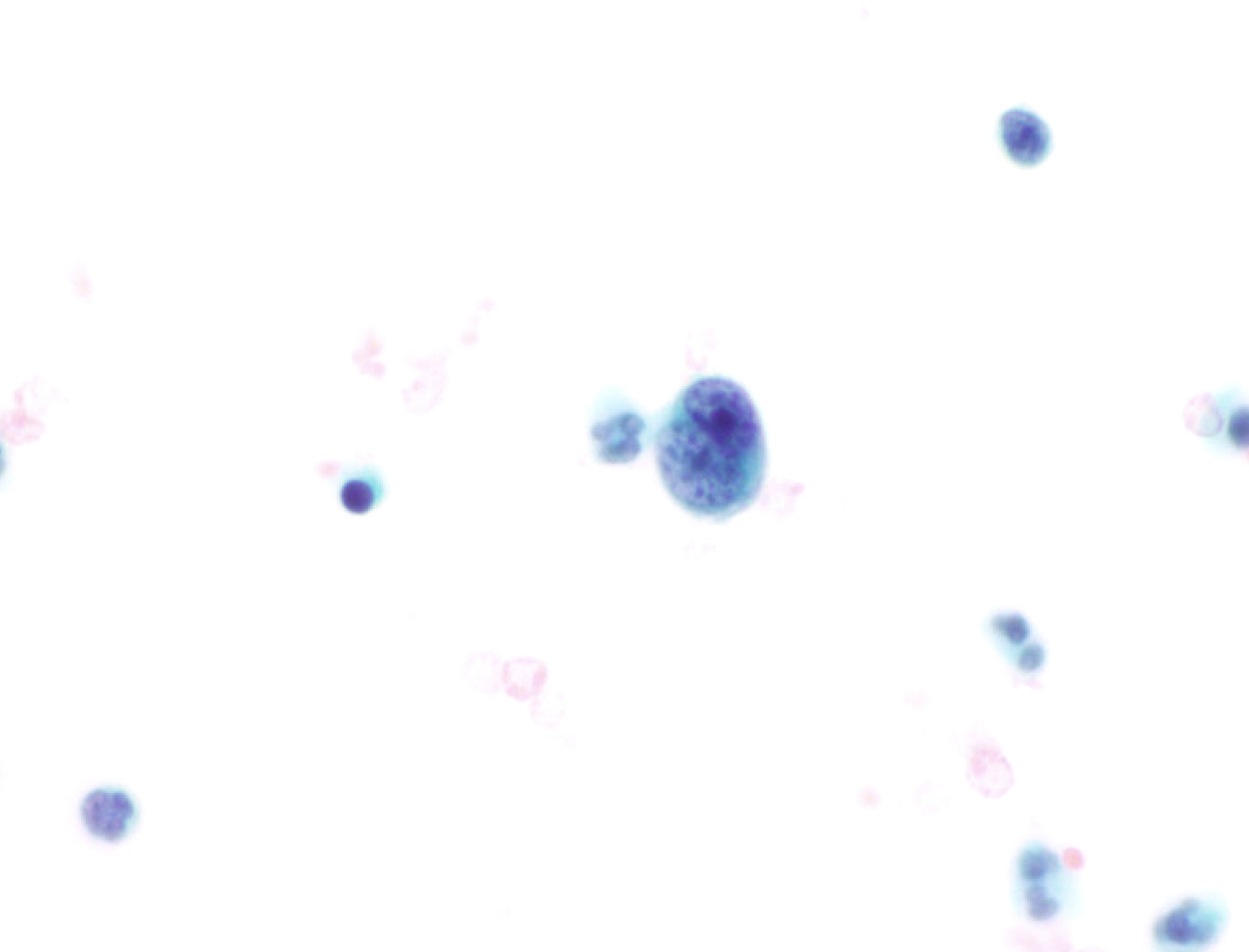
Macrophages (ThinPrep)
Anthracosis

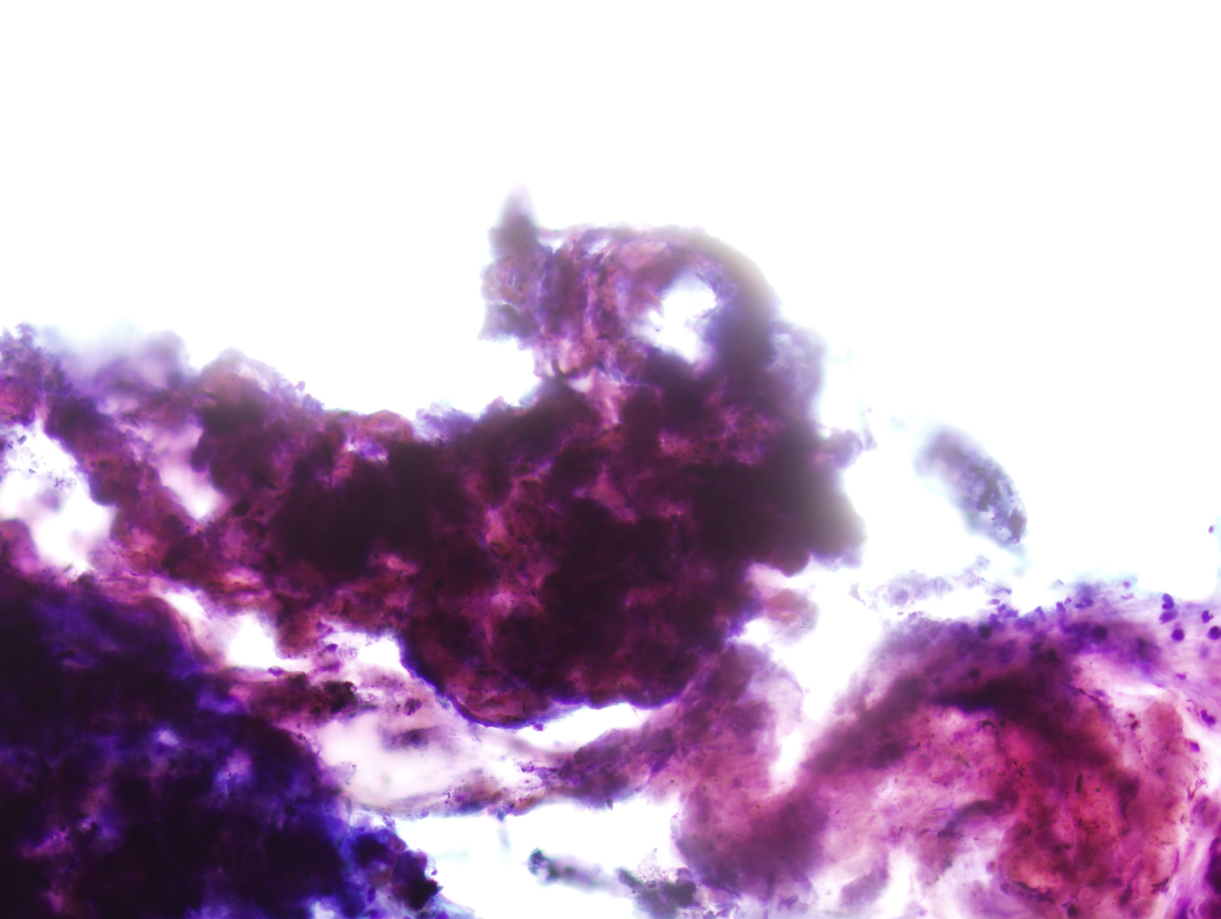
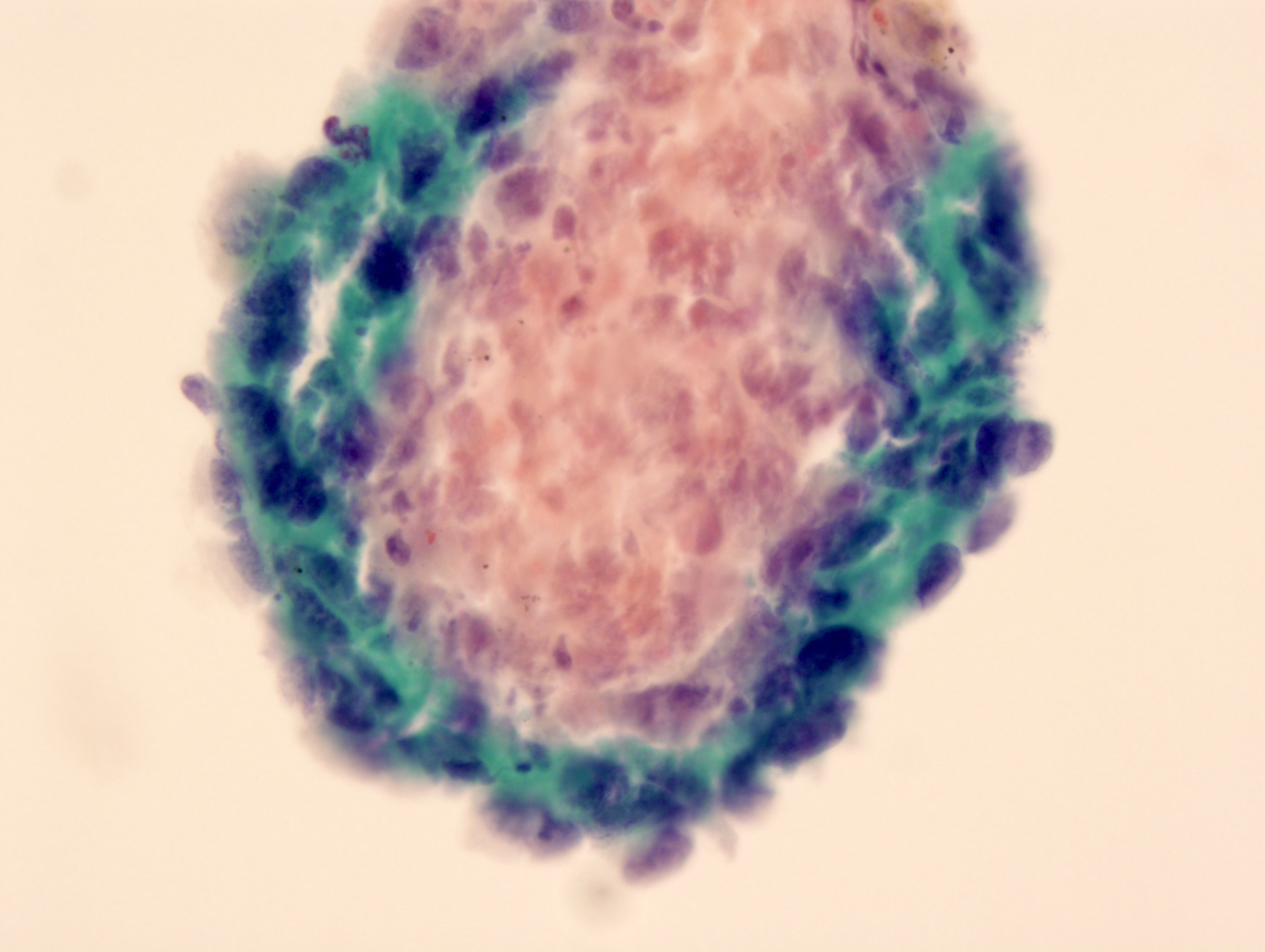
Hamartoma
(Diff-Quik)

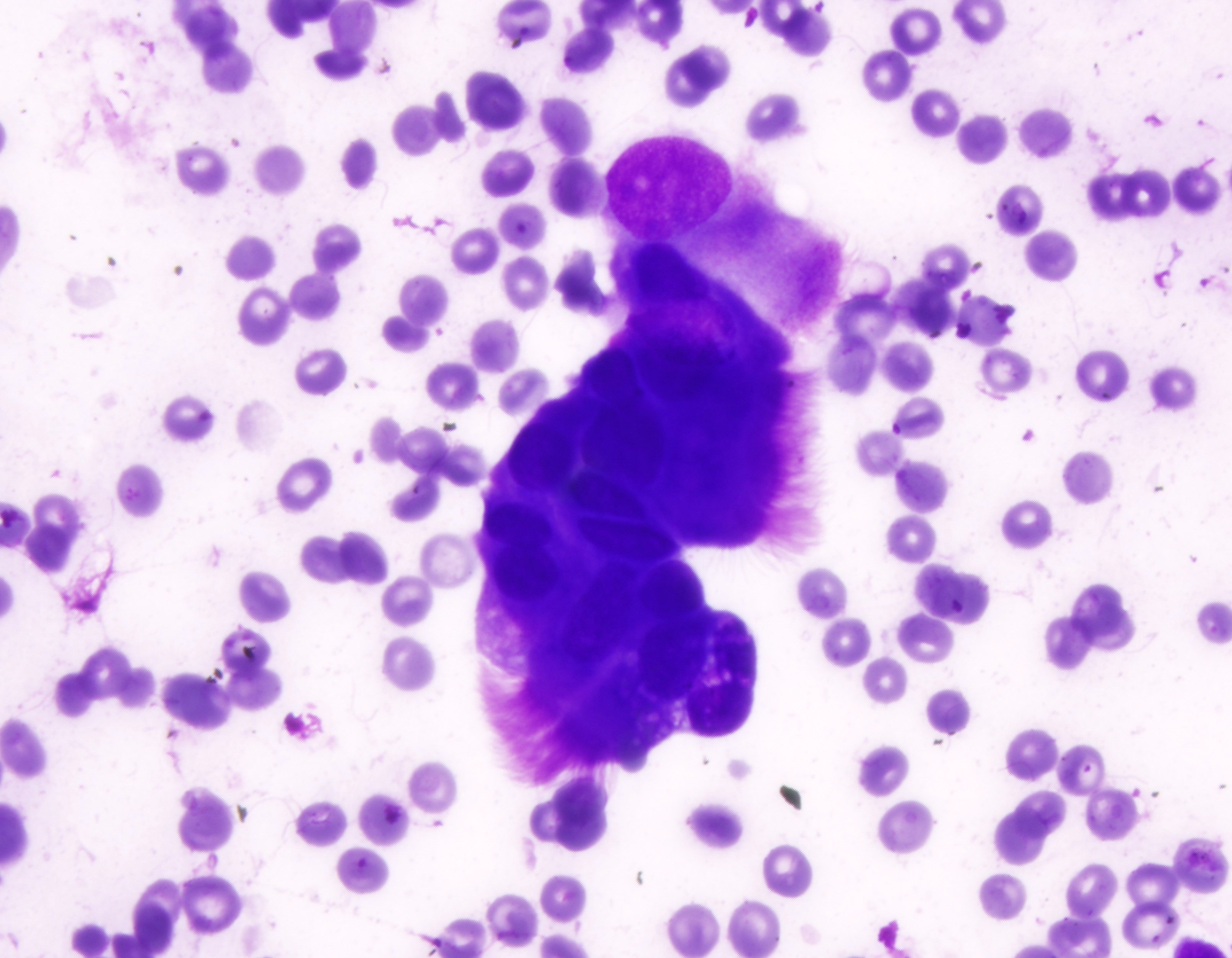
Meningioma (Diff-Quik)
Images hosted on other servers:

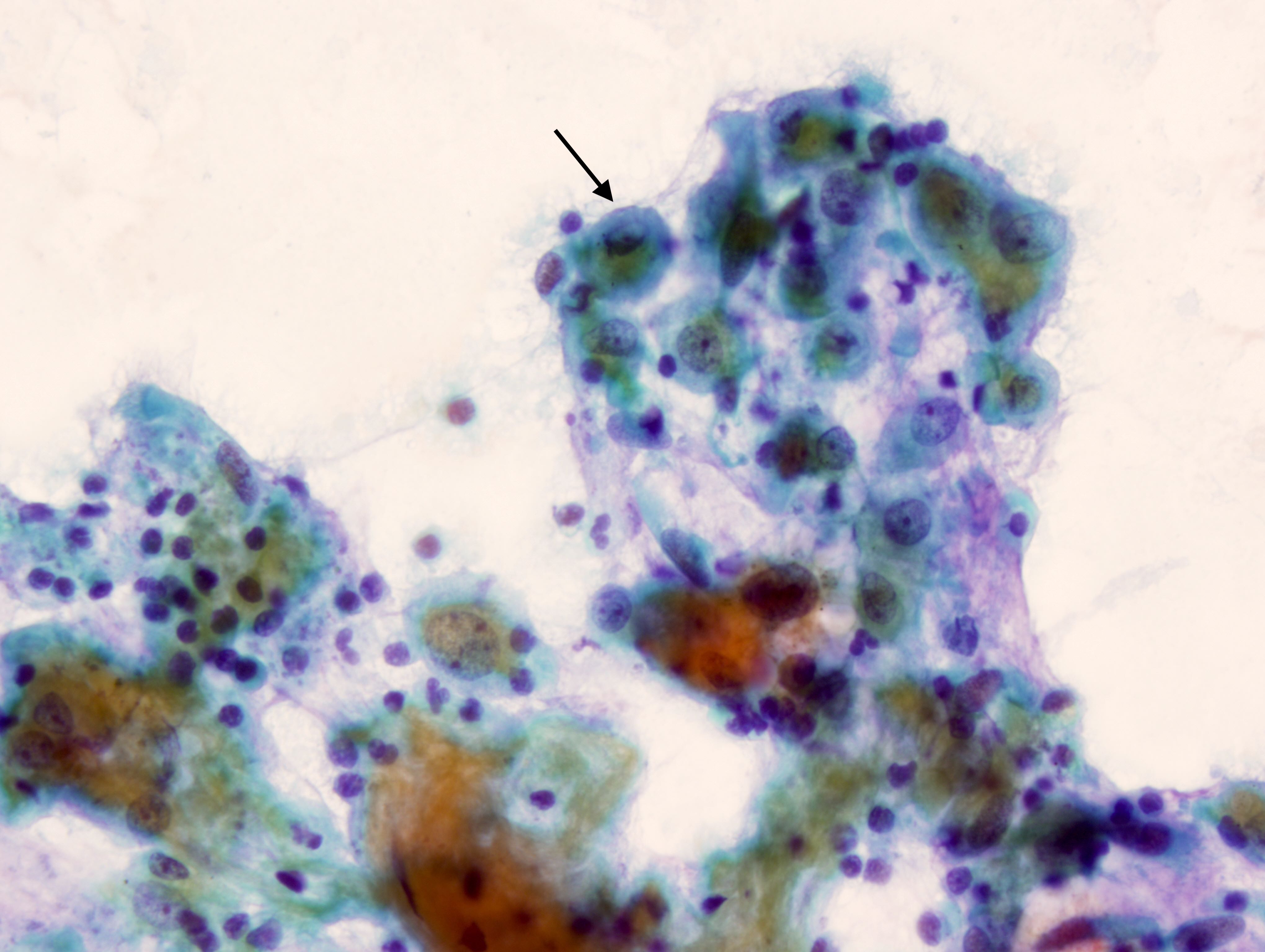
Herpes inclusions

Cytomegalovirus (CMV)

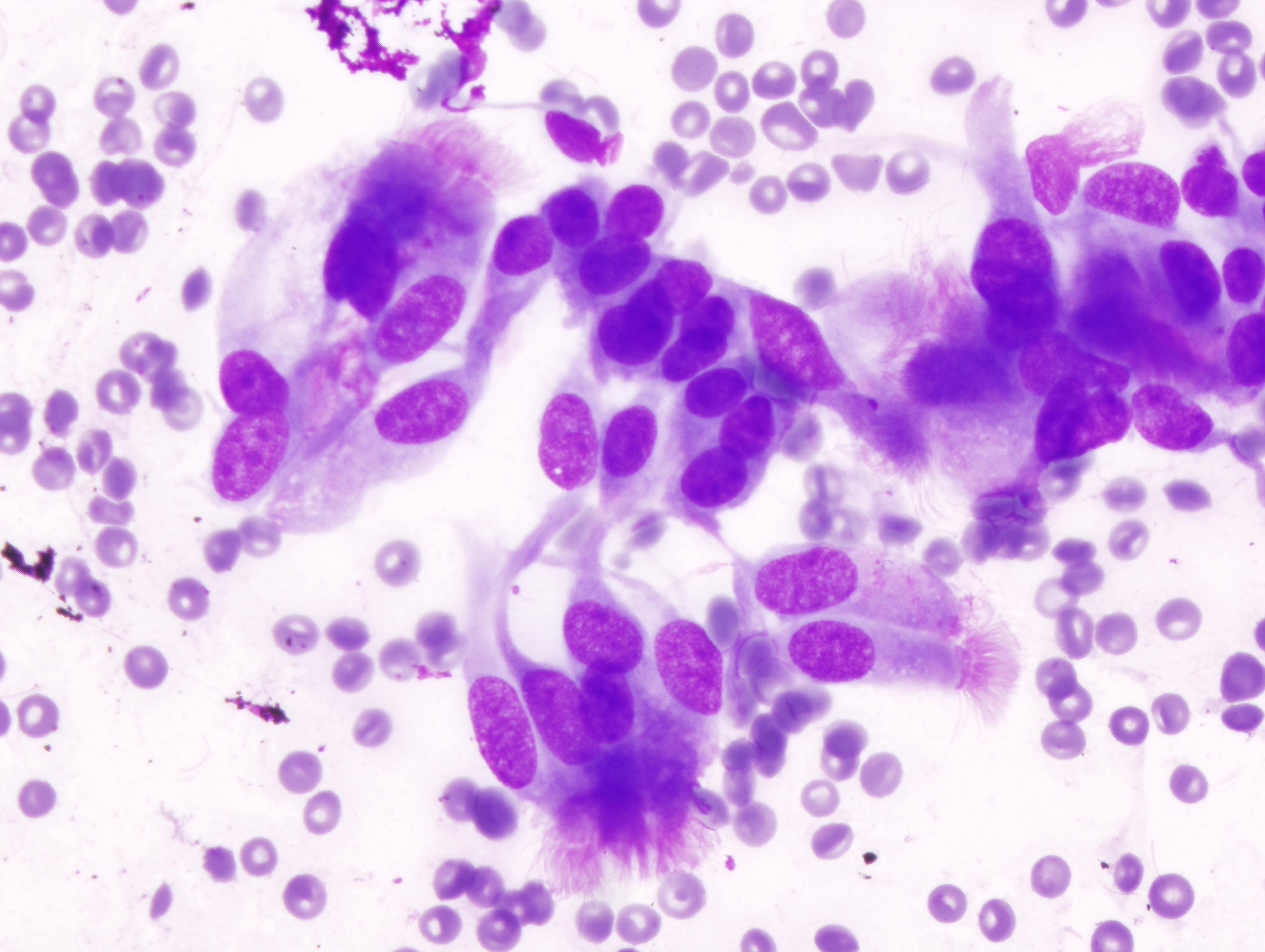
Creola body
Contributed by Joseph D. Jakowski, M.D. and Susan Meanor, R.T., R.D.M.S.
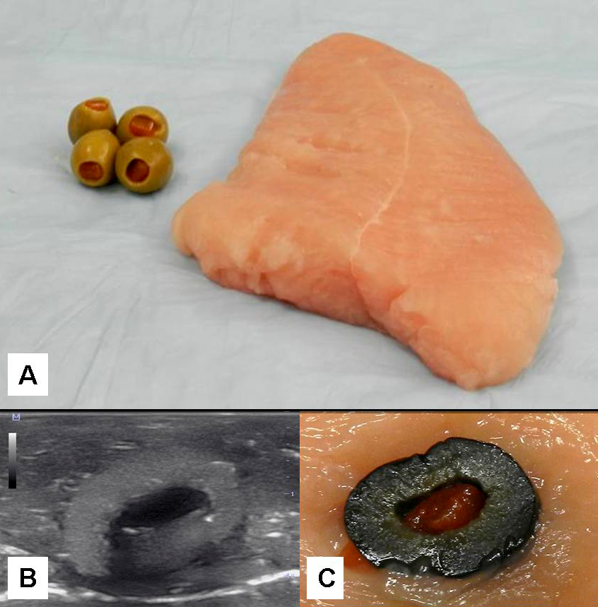
Turkey phantom
Contributed by Lawrence Hsu Lin, M.D., Ph.D. and Tamar C. Brandler, M.D., M.S.
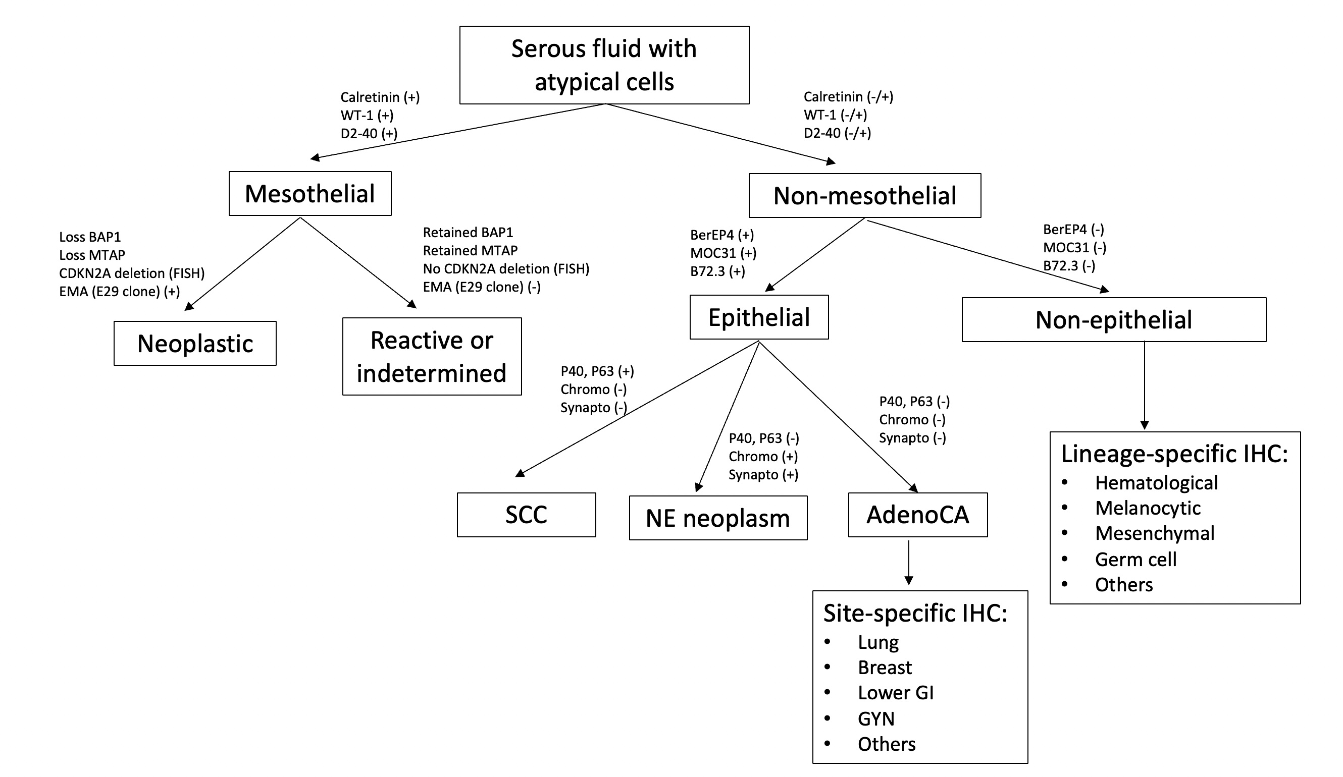
Serous fluid immunostain work up
Contributed by Lawrence Hsu Lin, M.D., Ph.D. and Tamar C. Brandler, M.D., M.S.

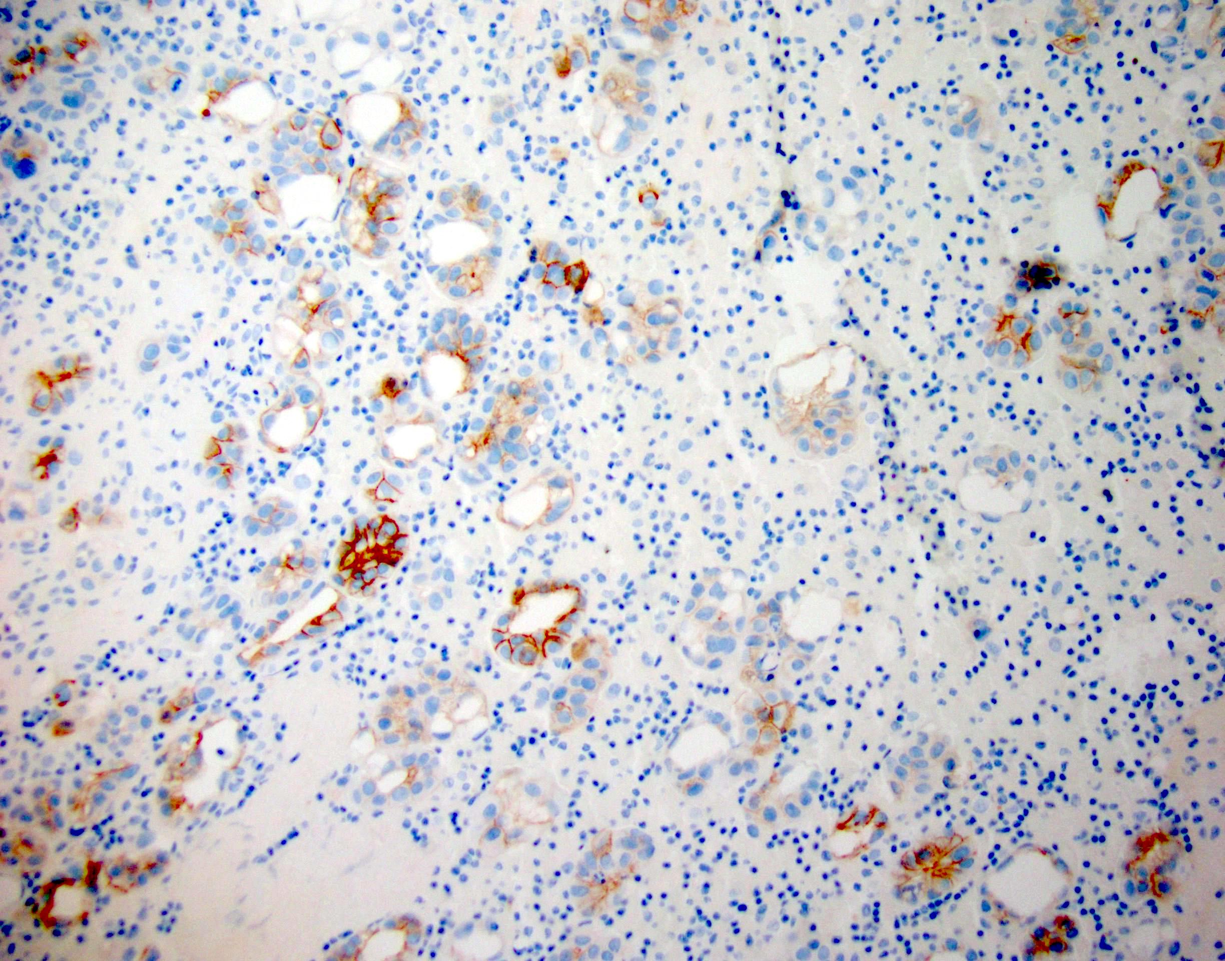
BerEP4 in pleural fluid
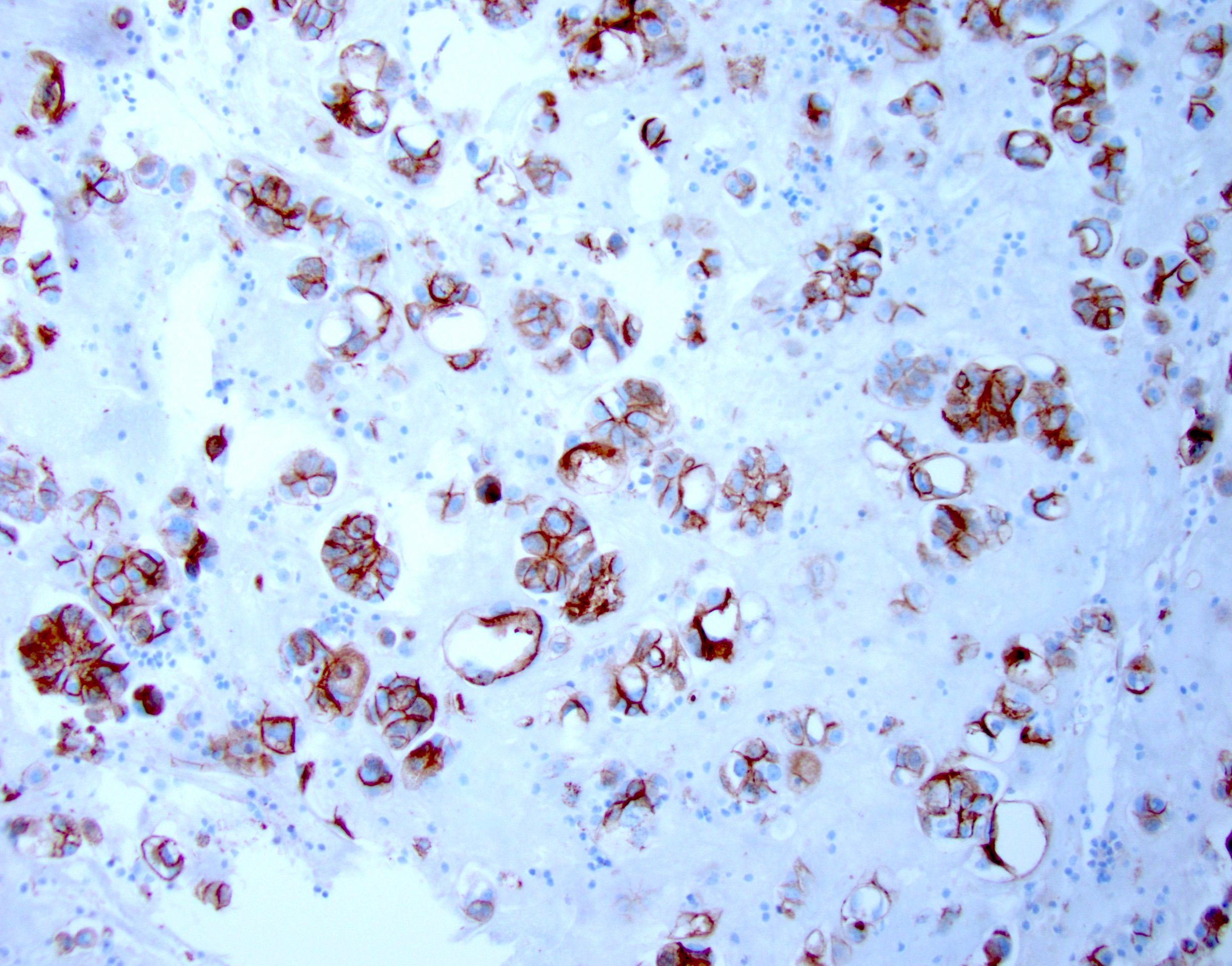
MOC31 in pleural fluid
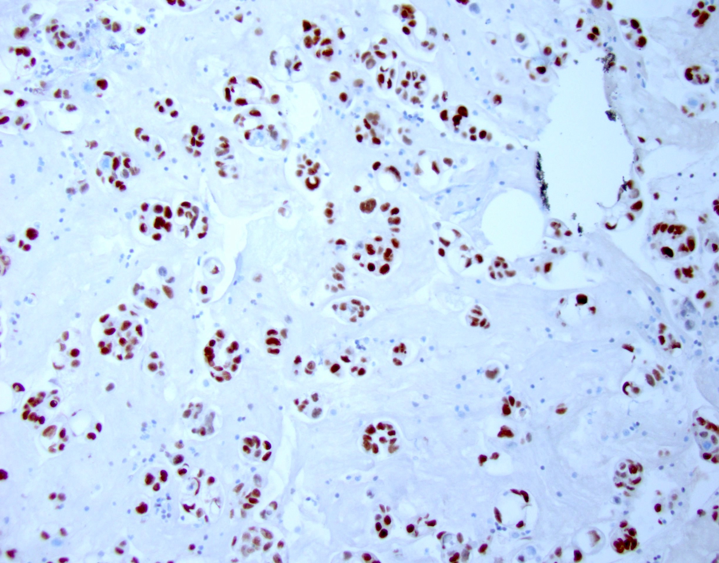
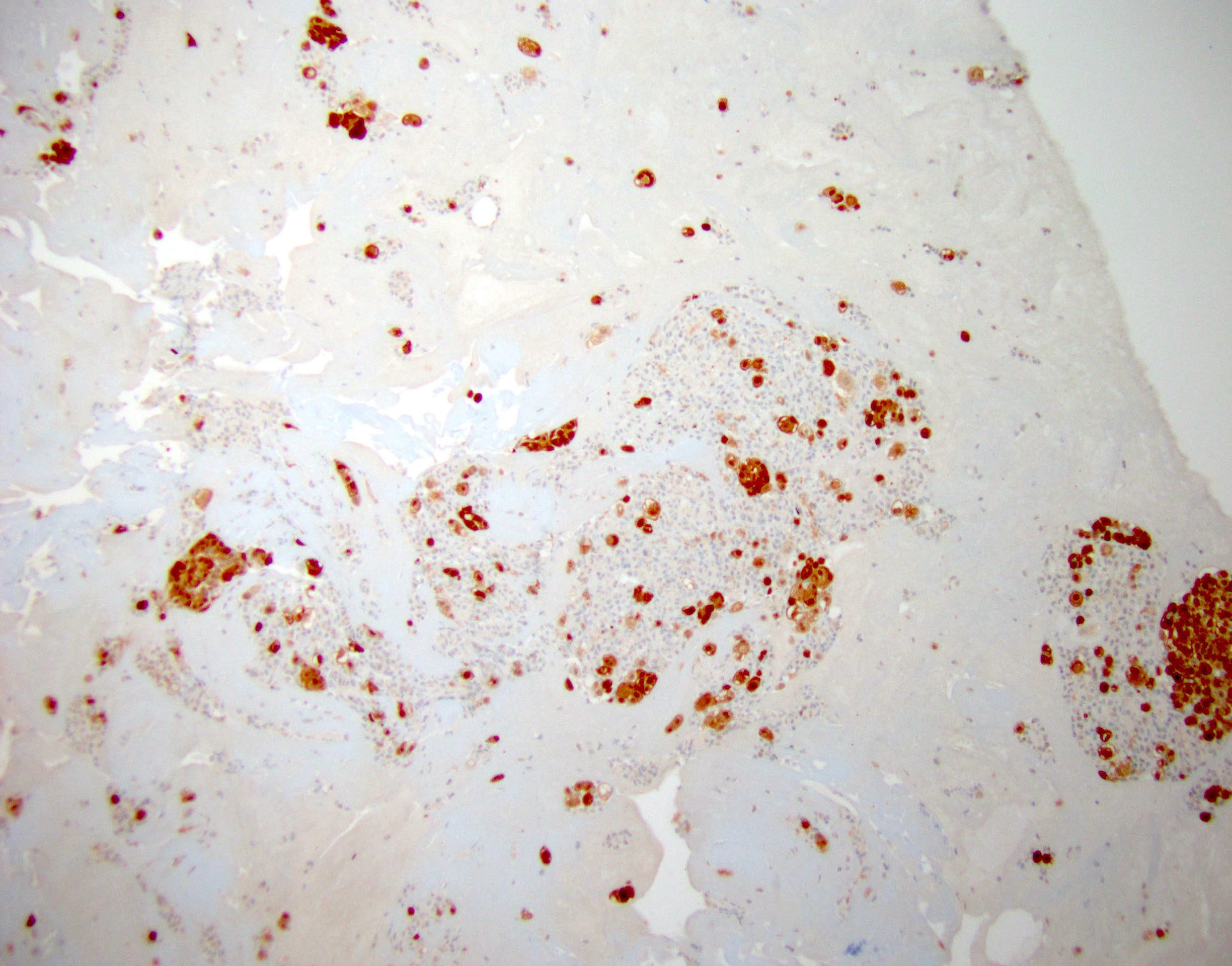
GATA3 in pleural fluid
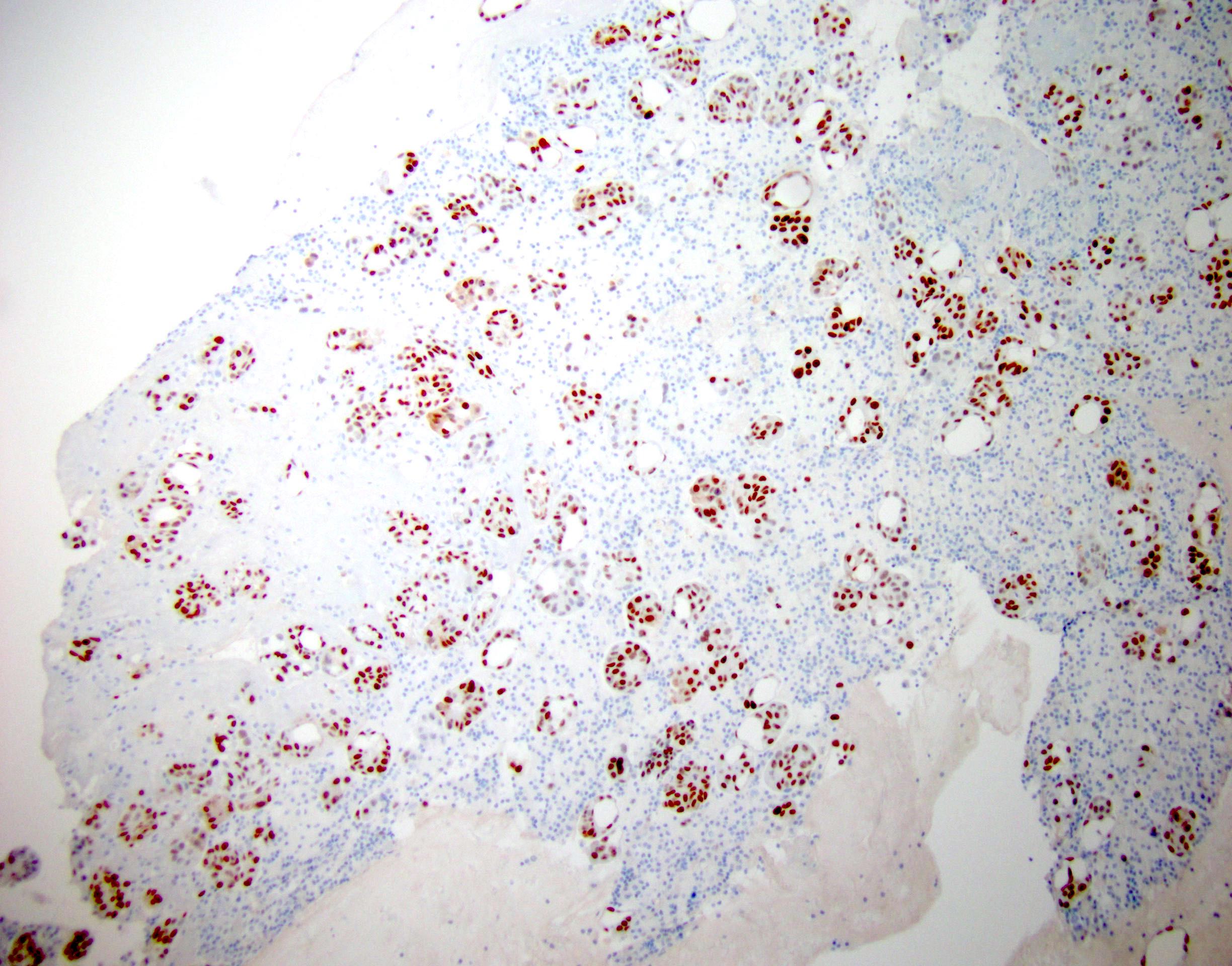
PAX8 in pleural fluid
NKX3.1 in ascites
TTF1 in pleural fluid
Napsin A in pleural fluid

p40 in ascites

CDX2 in ascites
Calretinin in ascites
D2-40 in ascites

WT1 in ascites
ER in pleural fluid
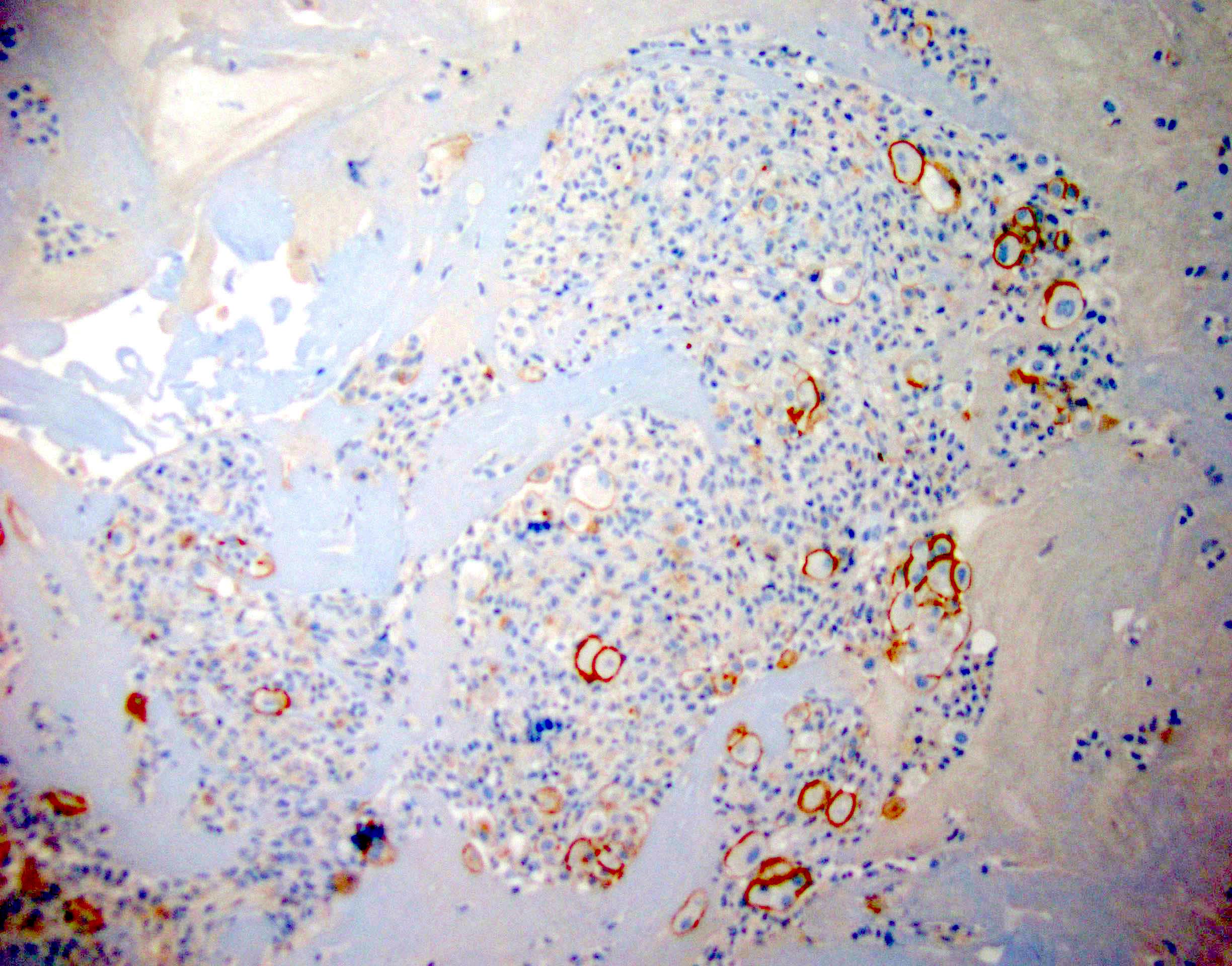
HER2 in pleural fluid
Images hosted on other servers:

BAP1 in mesothelioma
Images hosted on other servers:

p16 / CDKN2A deletion (FISH) in malignant mesothelioma

Absence of p16 / CDKN2A deletion by FISH
Contributed by Lawrence Hsu Lin, M.D., Ph.D. and Tamar C. Brandler, M.D., M.S.

Serous fluid cytology flow diagram
Contributed by Lawrence Hsu Lin, M.D., Ph.D. and Tamar C. Brandler, M.D., M.S.
Bloody specimen (ND)
Scant cellularity (ND)
Poor preservation (ND)

Reactive mesothelial cells (NFM)
Reactive mesothelial cells and histiocytes (NFM)
Collagen ball and mesothelial cells (NFM)
Endosalpingiosis (NFM)
Scant cell cluster with mild / moderate atypia (AUS)
Lymphocytic effusion (AUS)

Serous borderline tumor (AUS)
Few suspicious cells (SFM)
Highly atypical cell clusters with inflammation (SFM)
Highly atypical single cells (SFM)
Cell clusters with scalloped borders (MAL-P)
Highly atypical cluster of cells (MAL-P)
Cell cluster with smooth borders (MAL-S)
Highly atypical cell cluster (MAL-S)
Intracellular mucin (MAL-S)
Large atypical cells (MAL-S)
Contributed by Karen Villar Zarra, M.D.

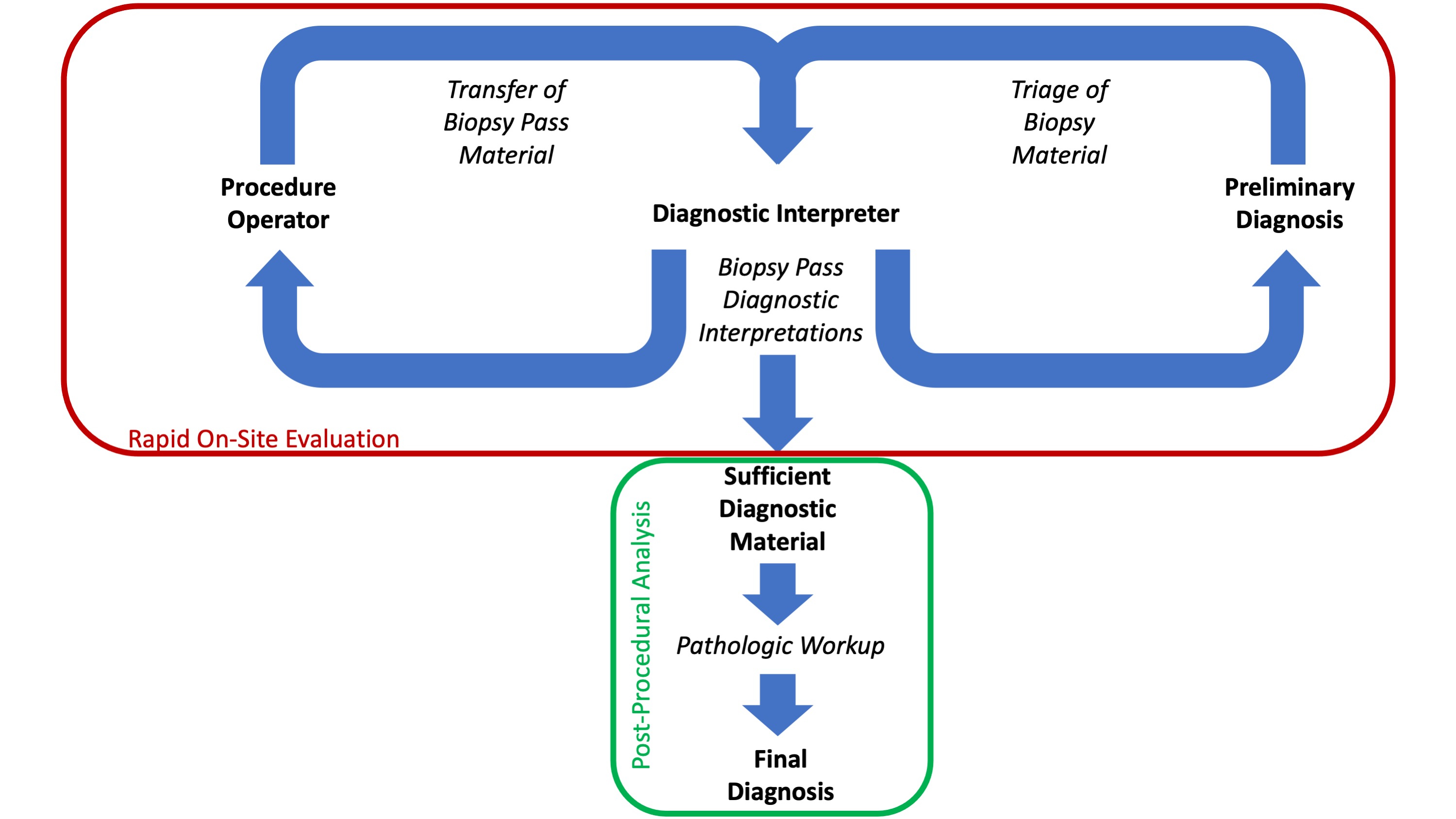
Decision making in interventional pathology one stop clinic
Contributed by Karen Villar Zarra, M.D.
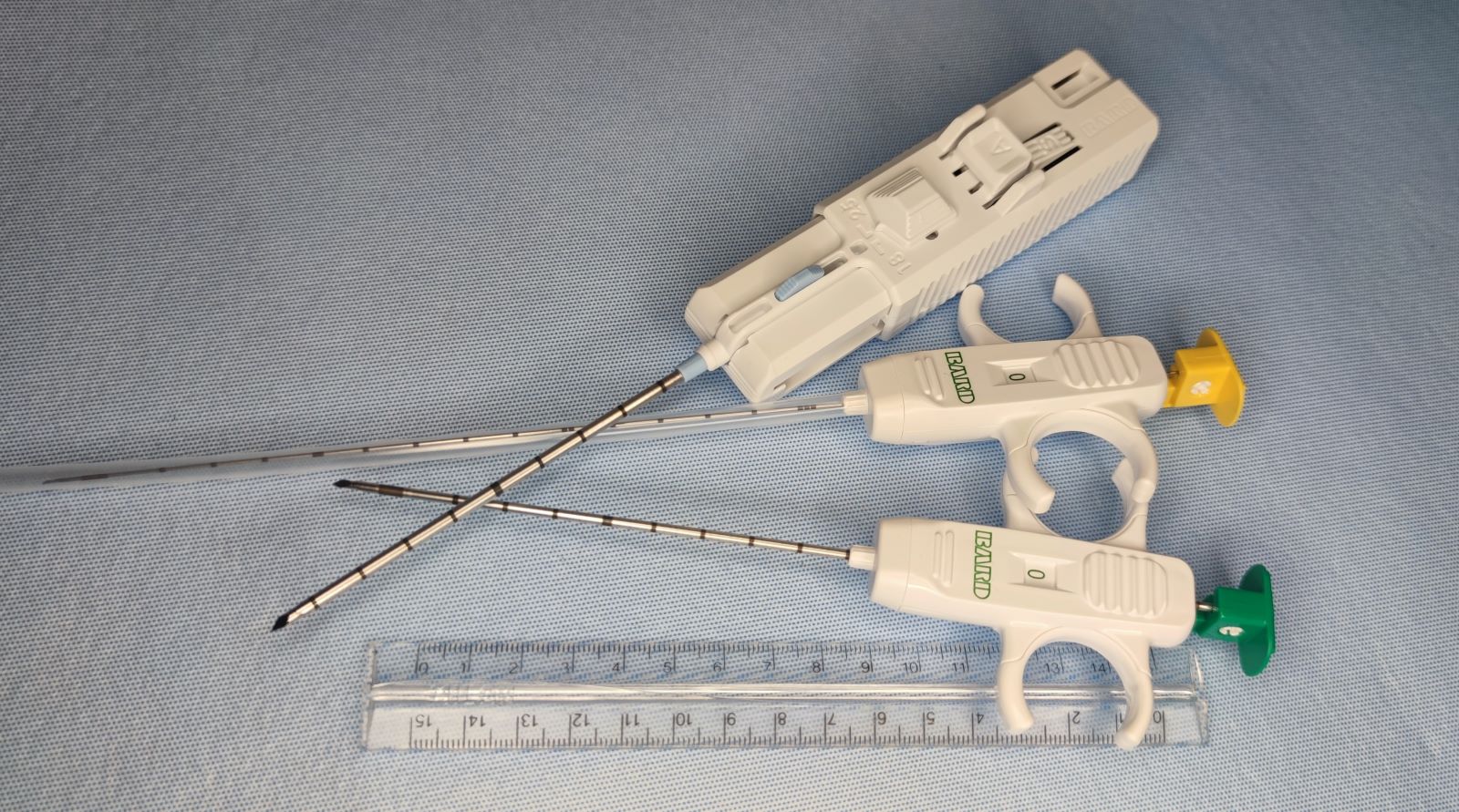
Automatic and manual trigger biopsy devices

Minimal percutaneous point of entry for core biopsies

Sample obtained with core biopsy devices
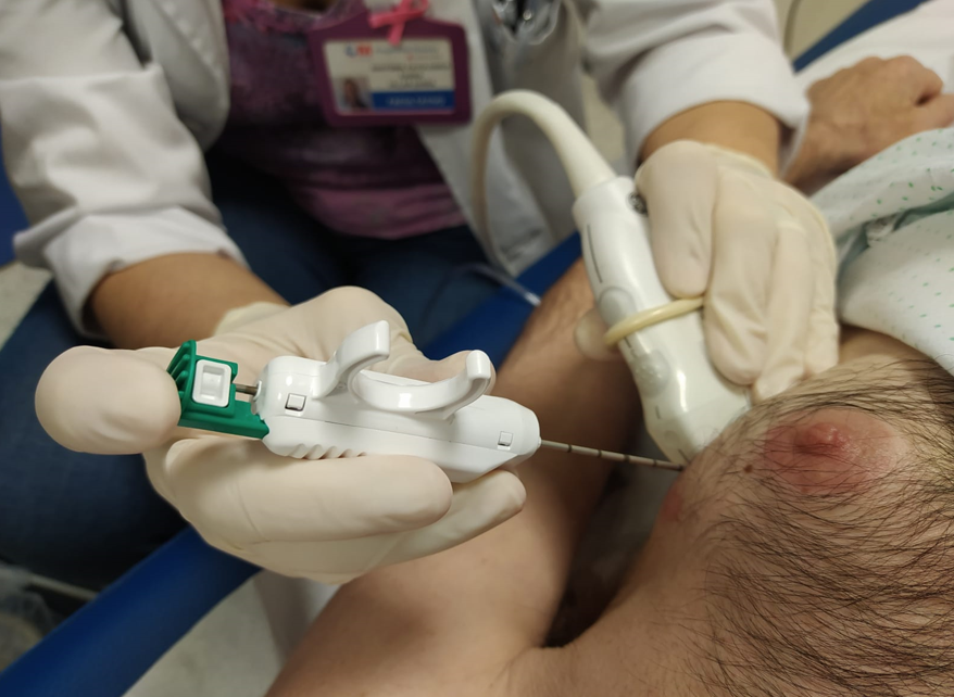
Ultrasound guided core biopsy - parallel approach

Different gauges of core biopsies from home made phantom
Contributed by Sadia Sayeed, M.D.
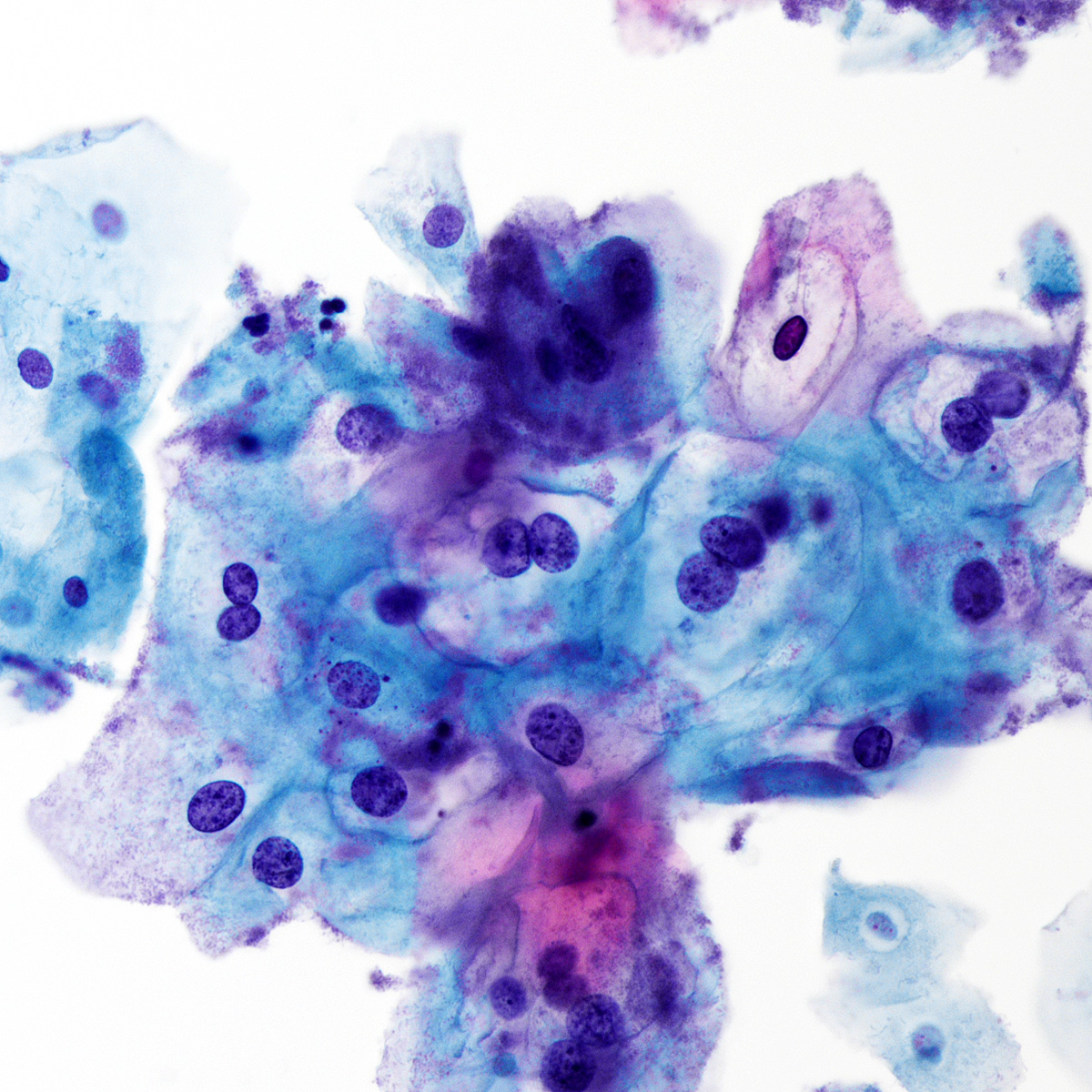
LSIL ThinPrep
HGSIL ThinPrep
HGSIL SurePath
Benign endocervical cells ThinPrep
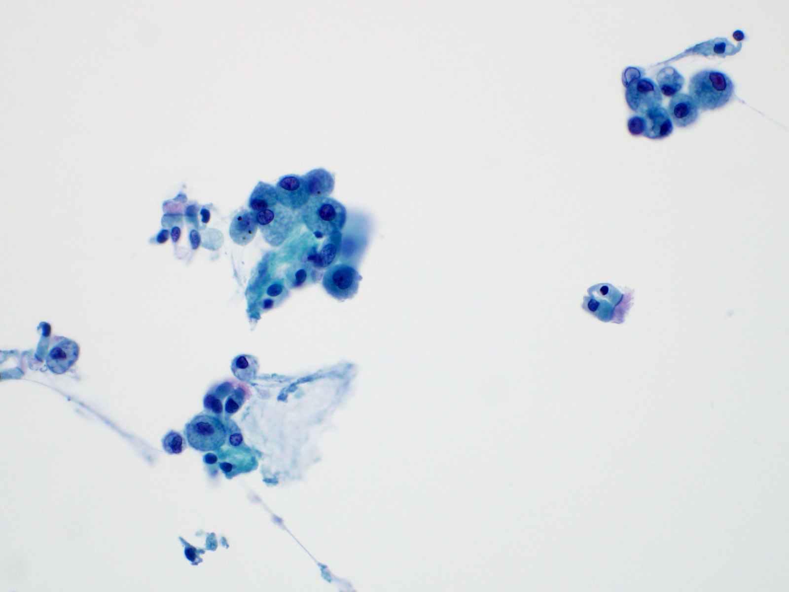
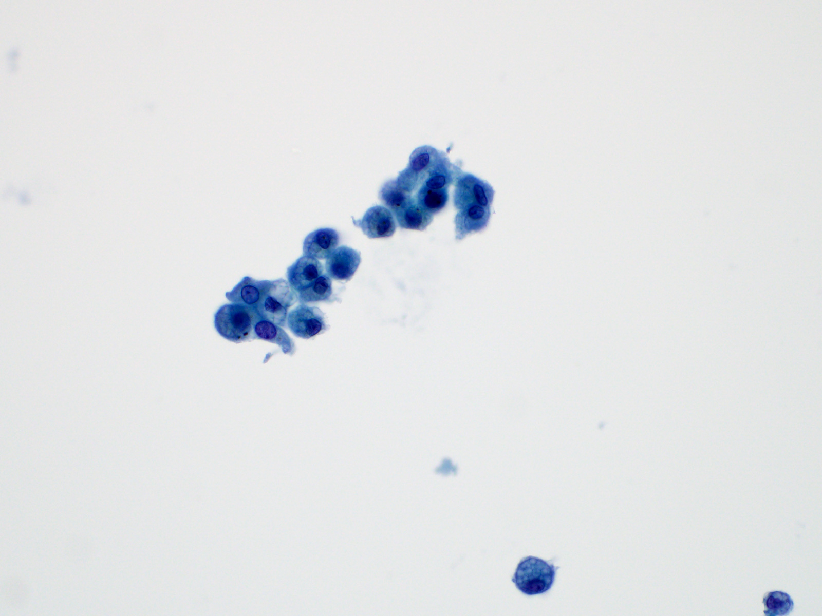
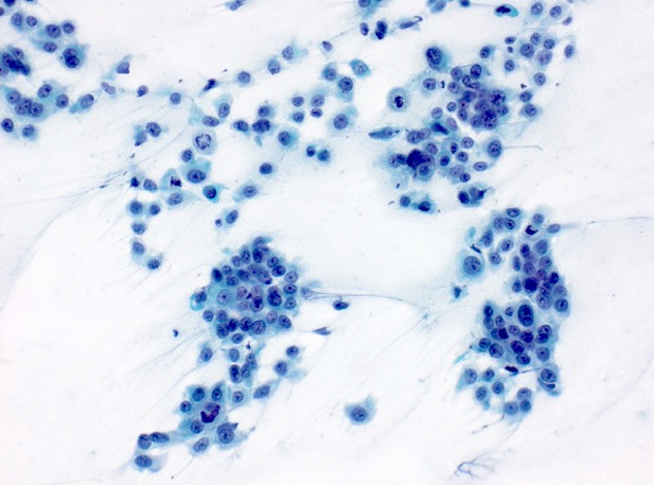
Adenocarcinoma ThinPrep
Reactive mesothelial cells ThinPrep
Benign, BAL
HGUC, urine
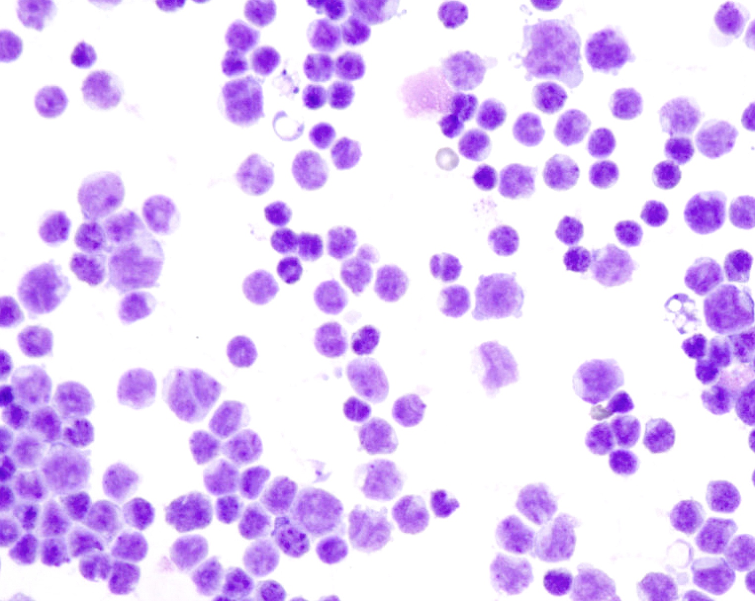
Suspicious thyroid aspirate
Benign thyroid aspirate
Images hosted on other servers:

Left lower lobe mass
Contributed by Reima El Naili, M.D.

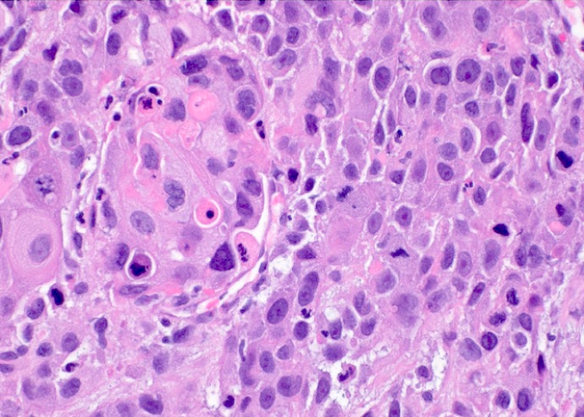
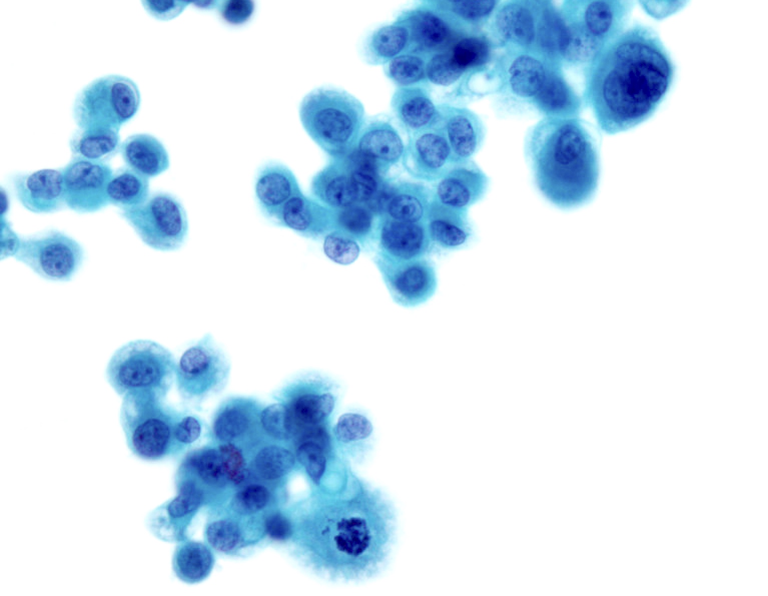
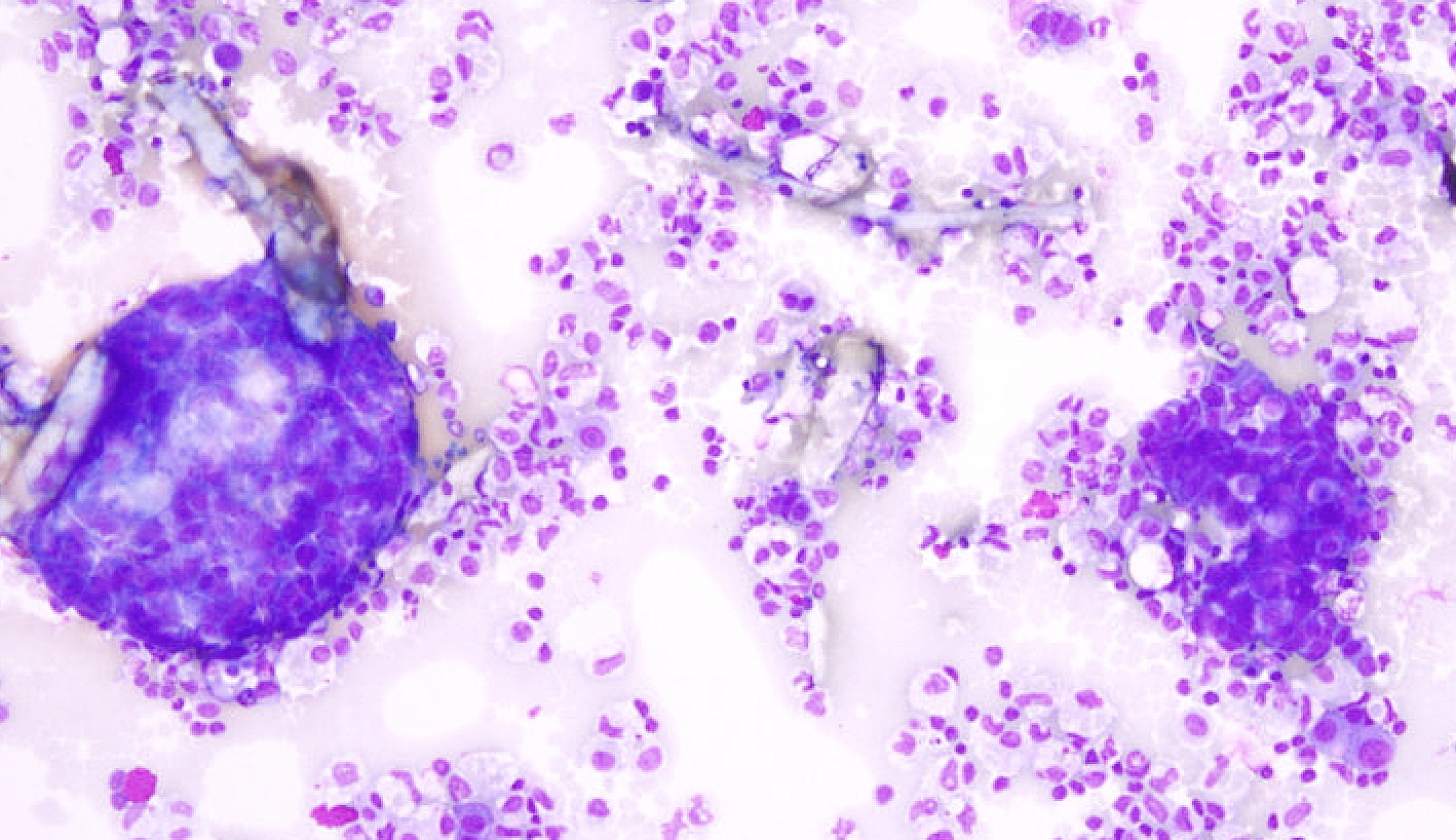
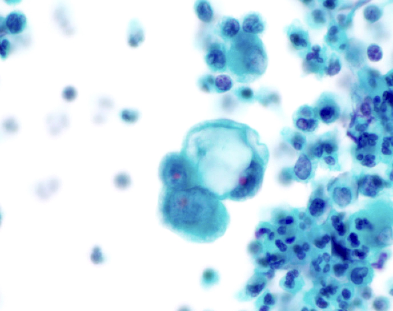
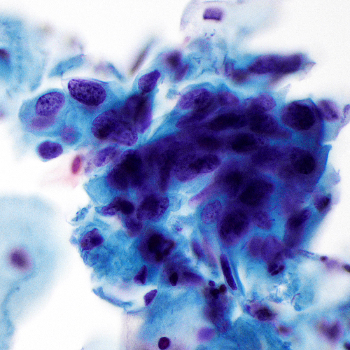
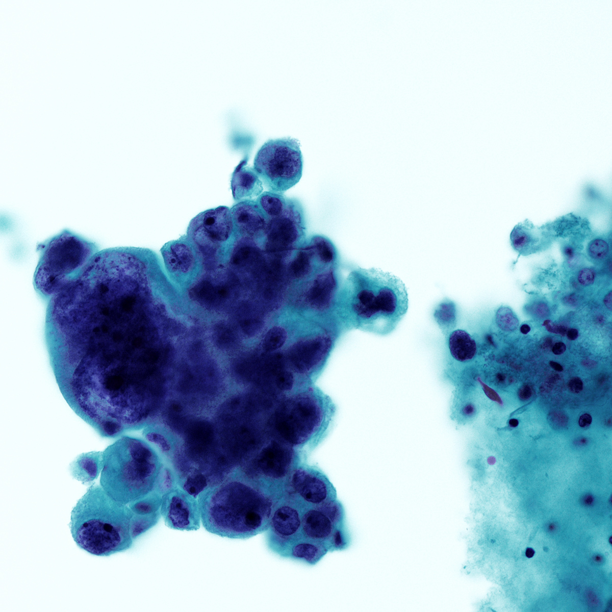
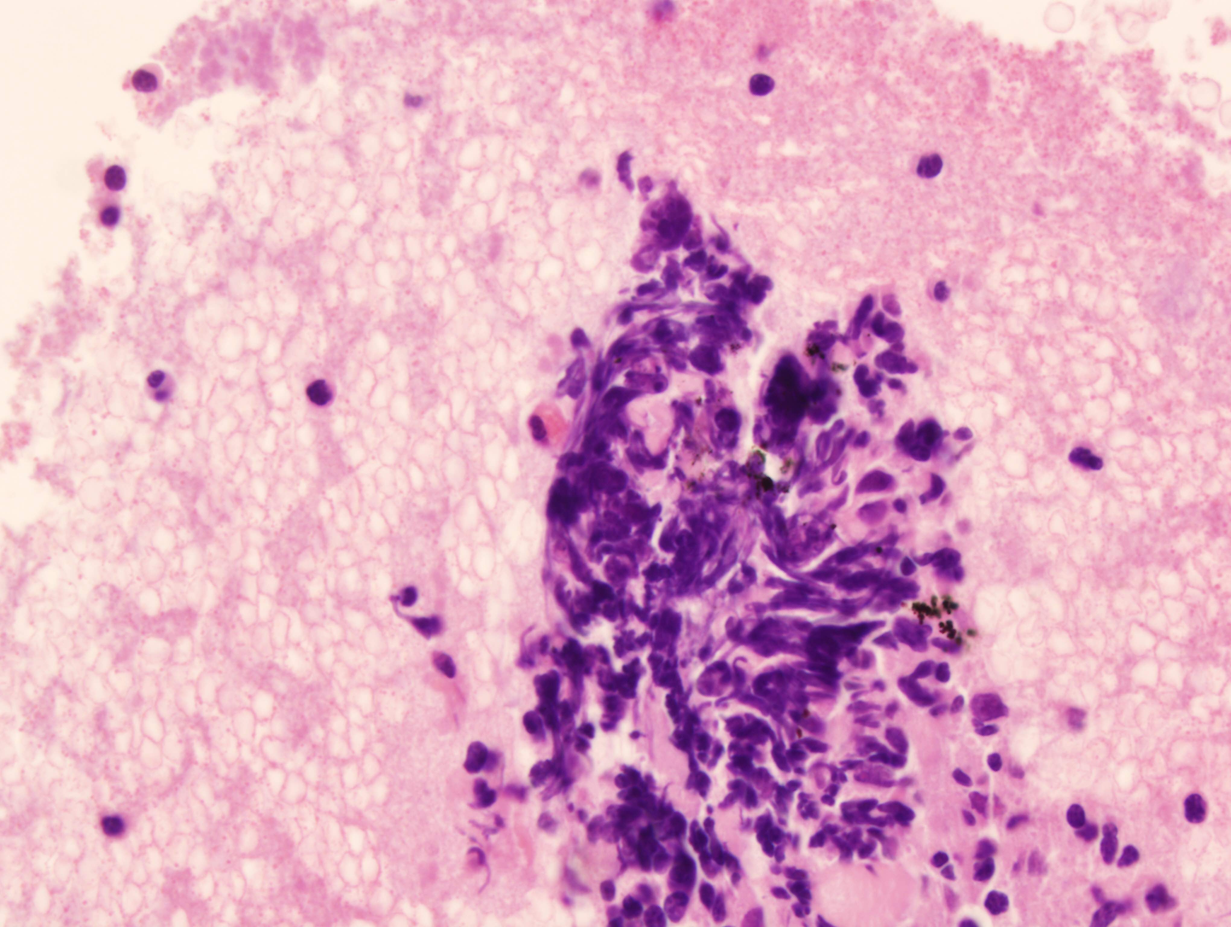
Squamous cell carcinoma

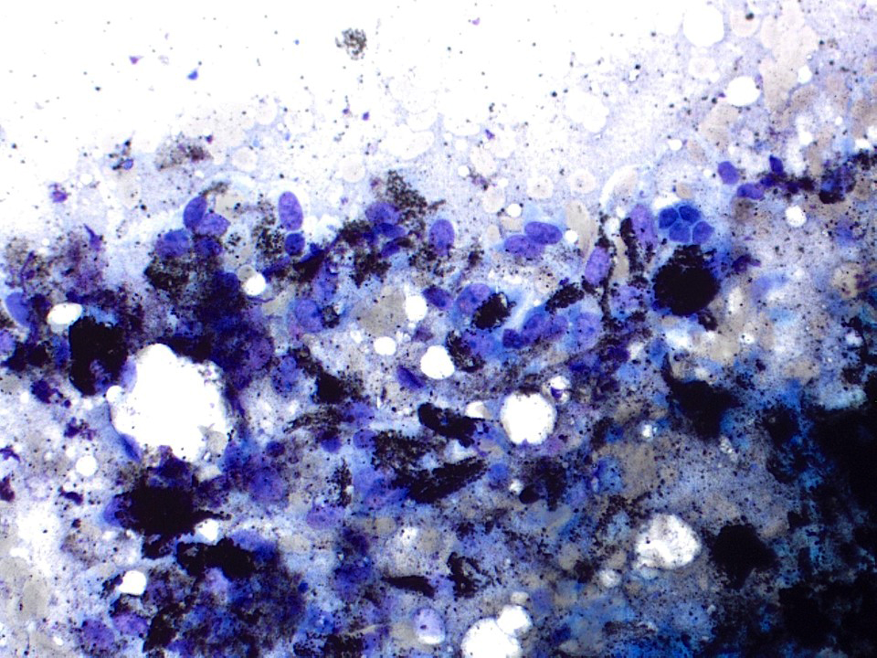
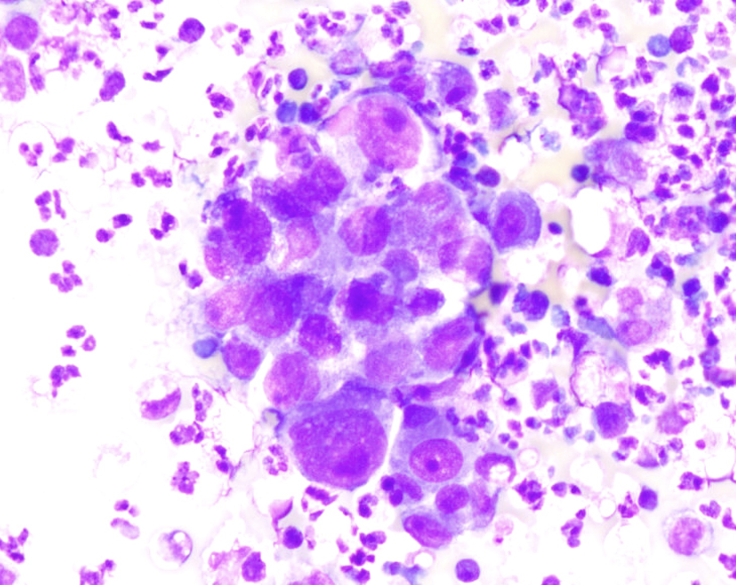
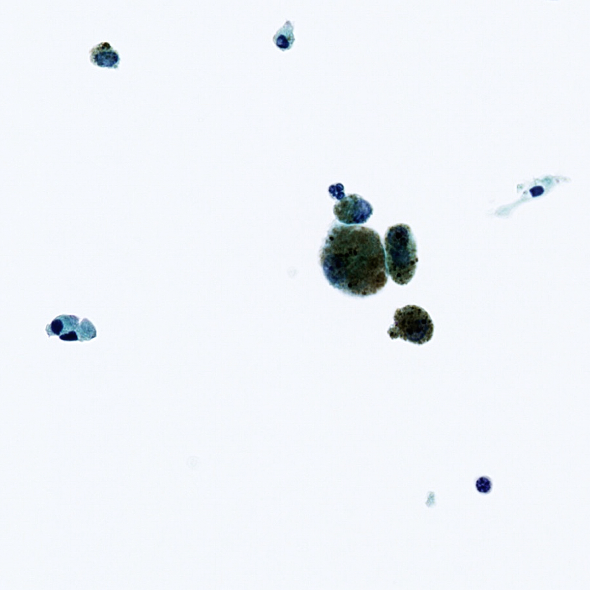
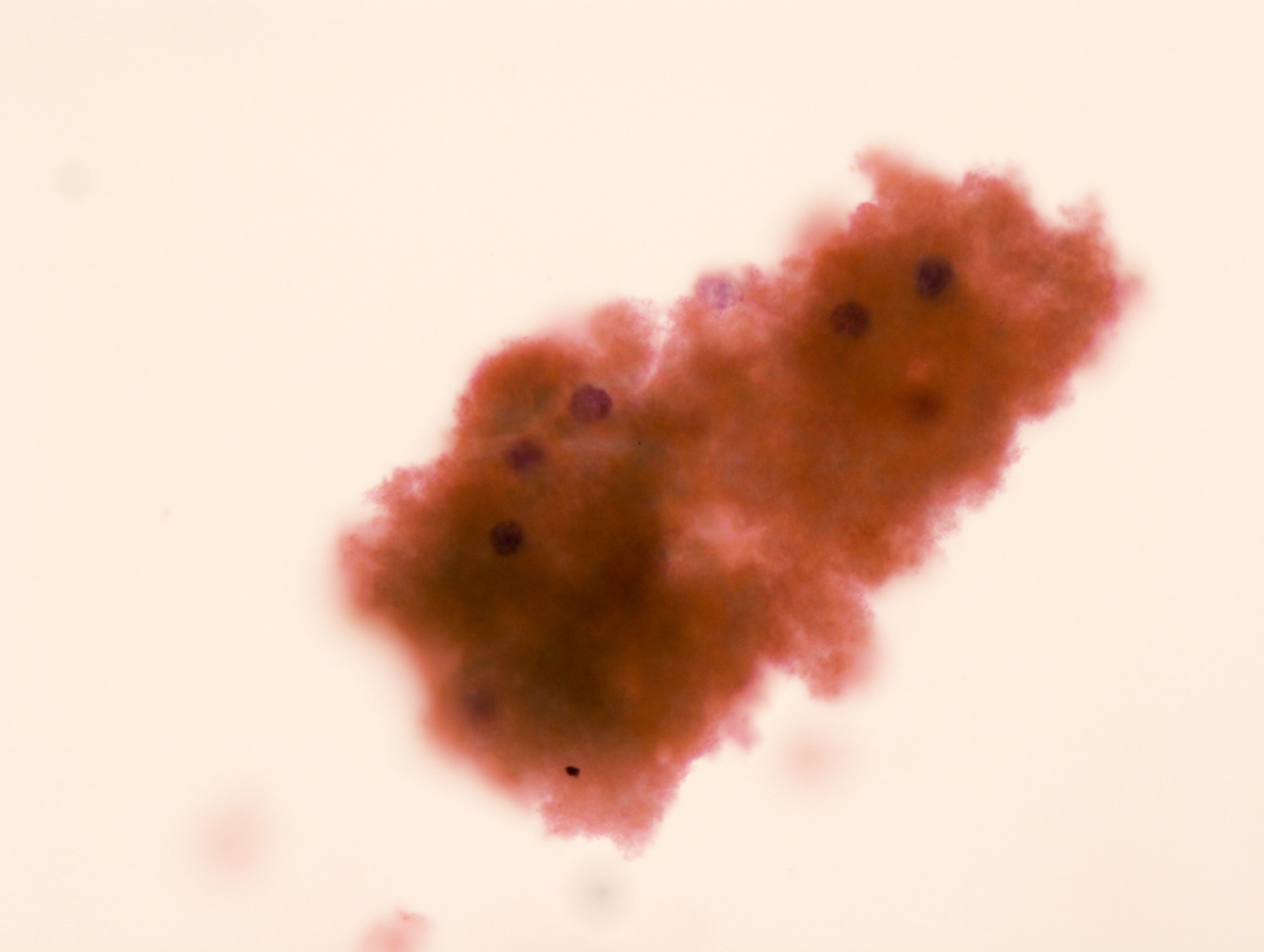
Prominent necrosis in small cell carcinoma

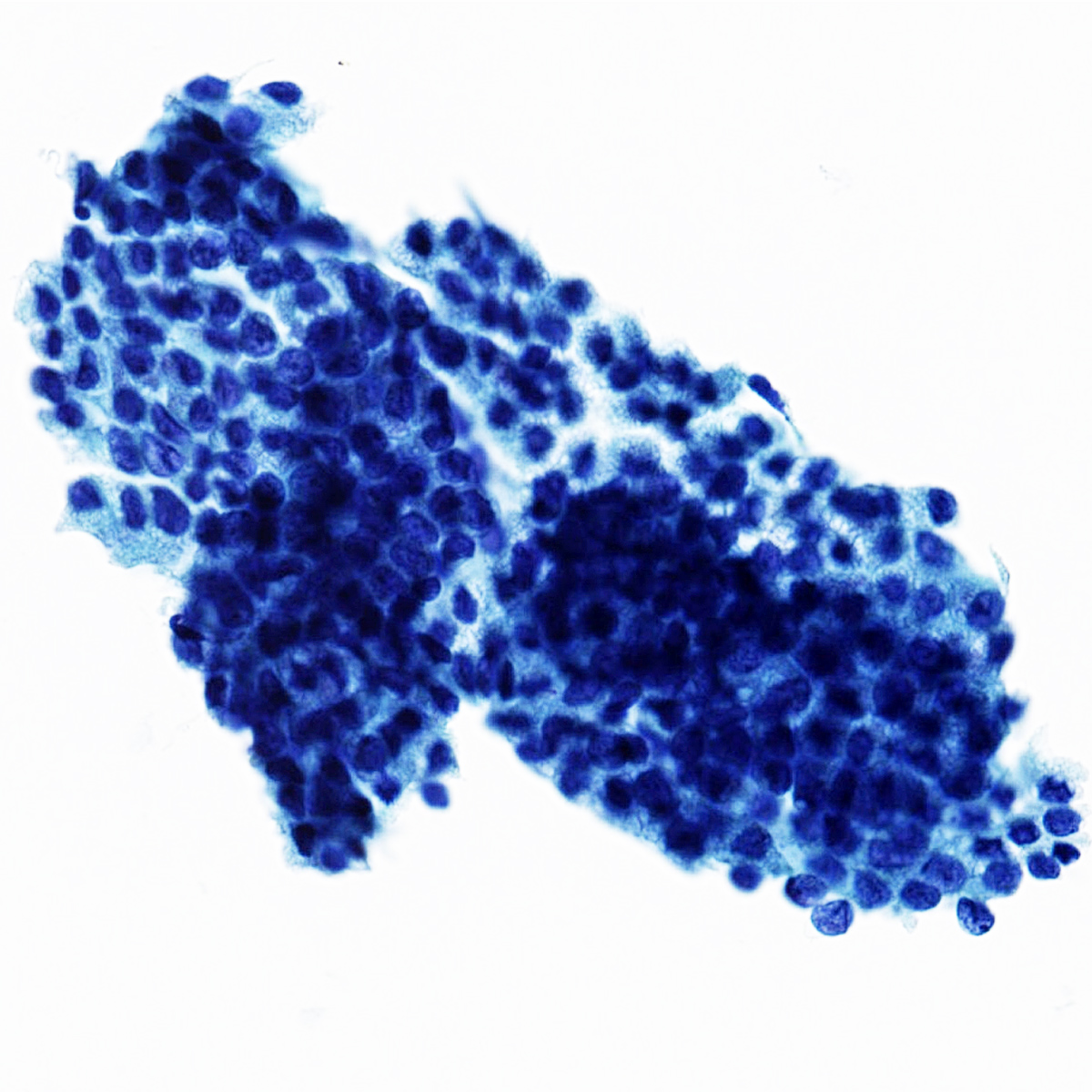
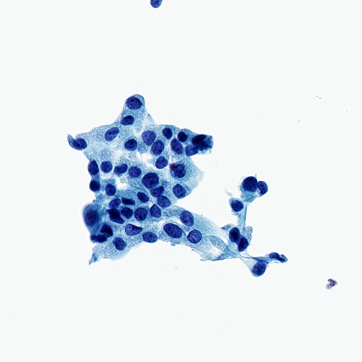
Cell block, adenocarcinoma

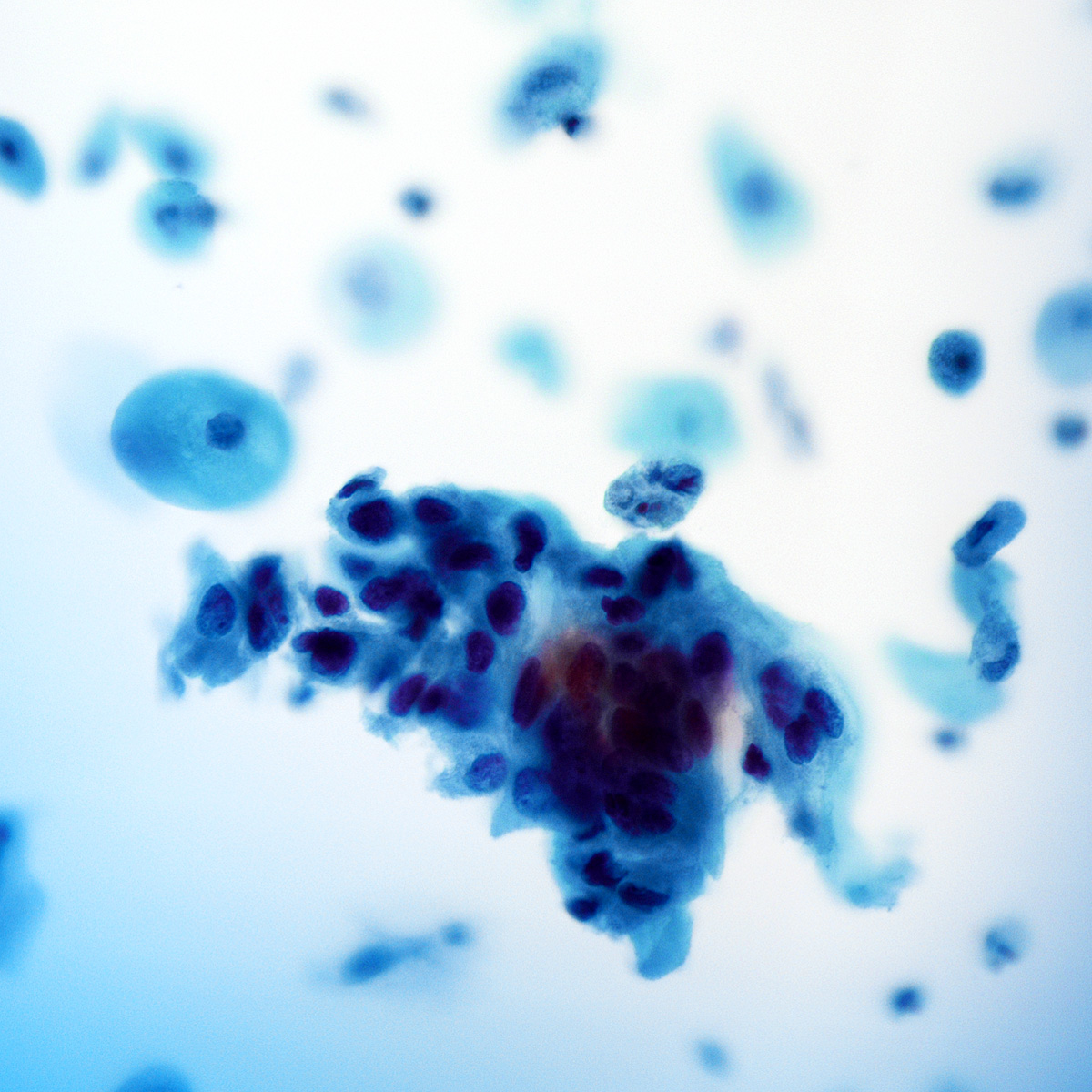
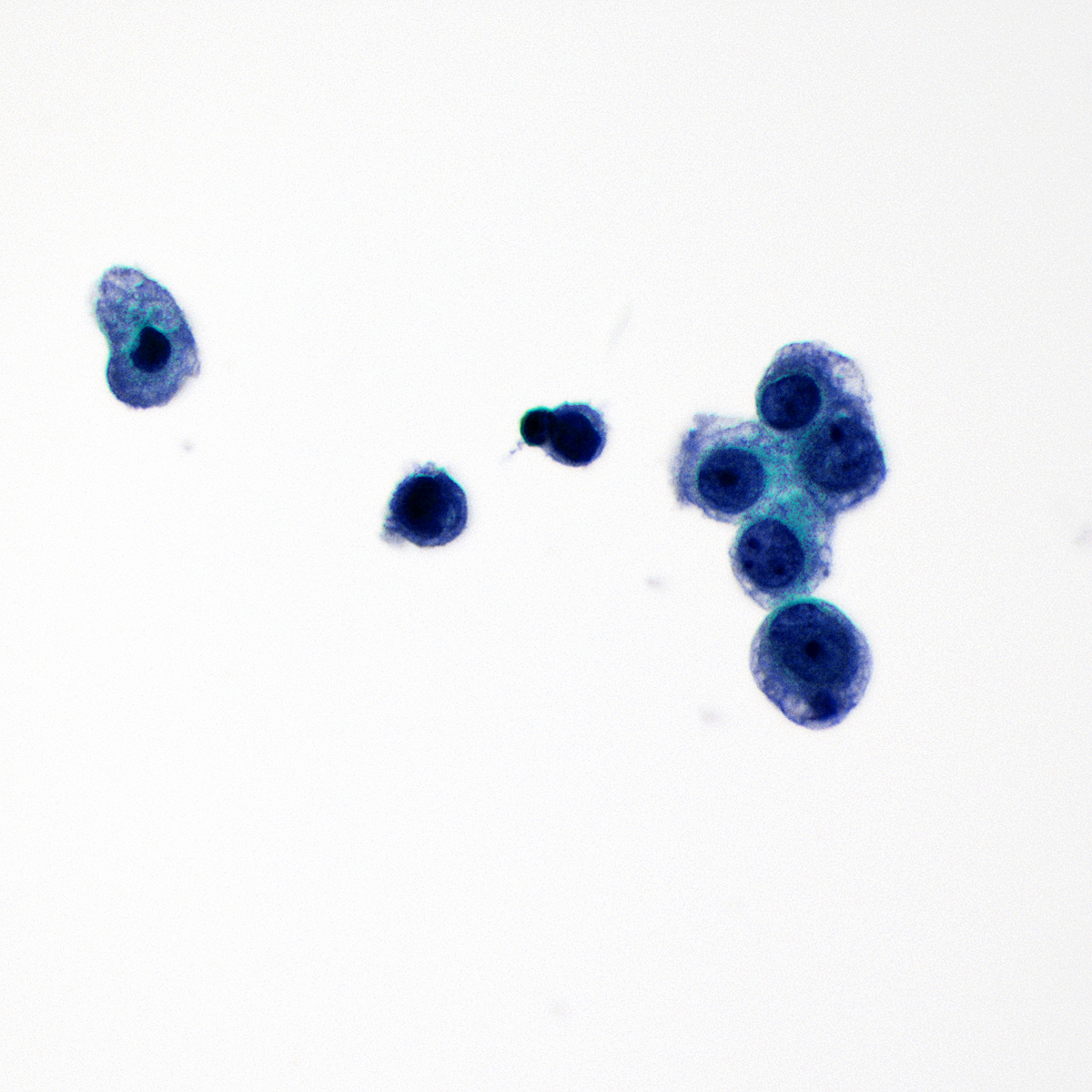
Adenocarcinoma
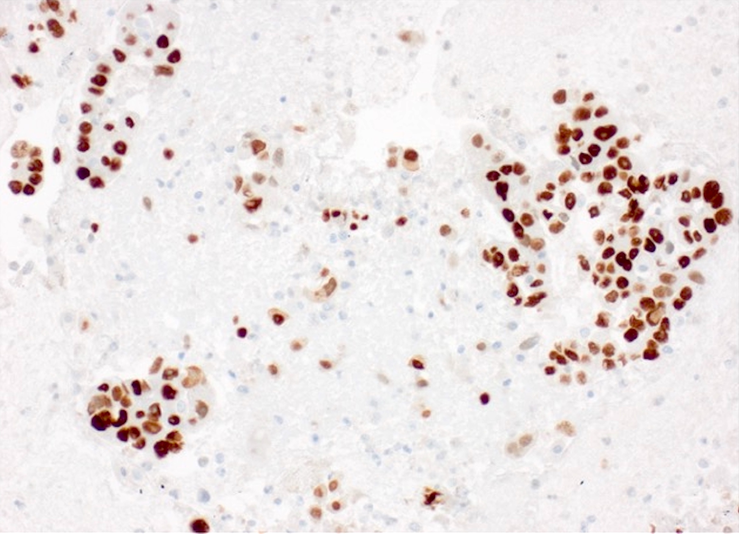
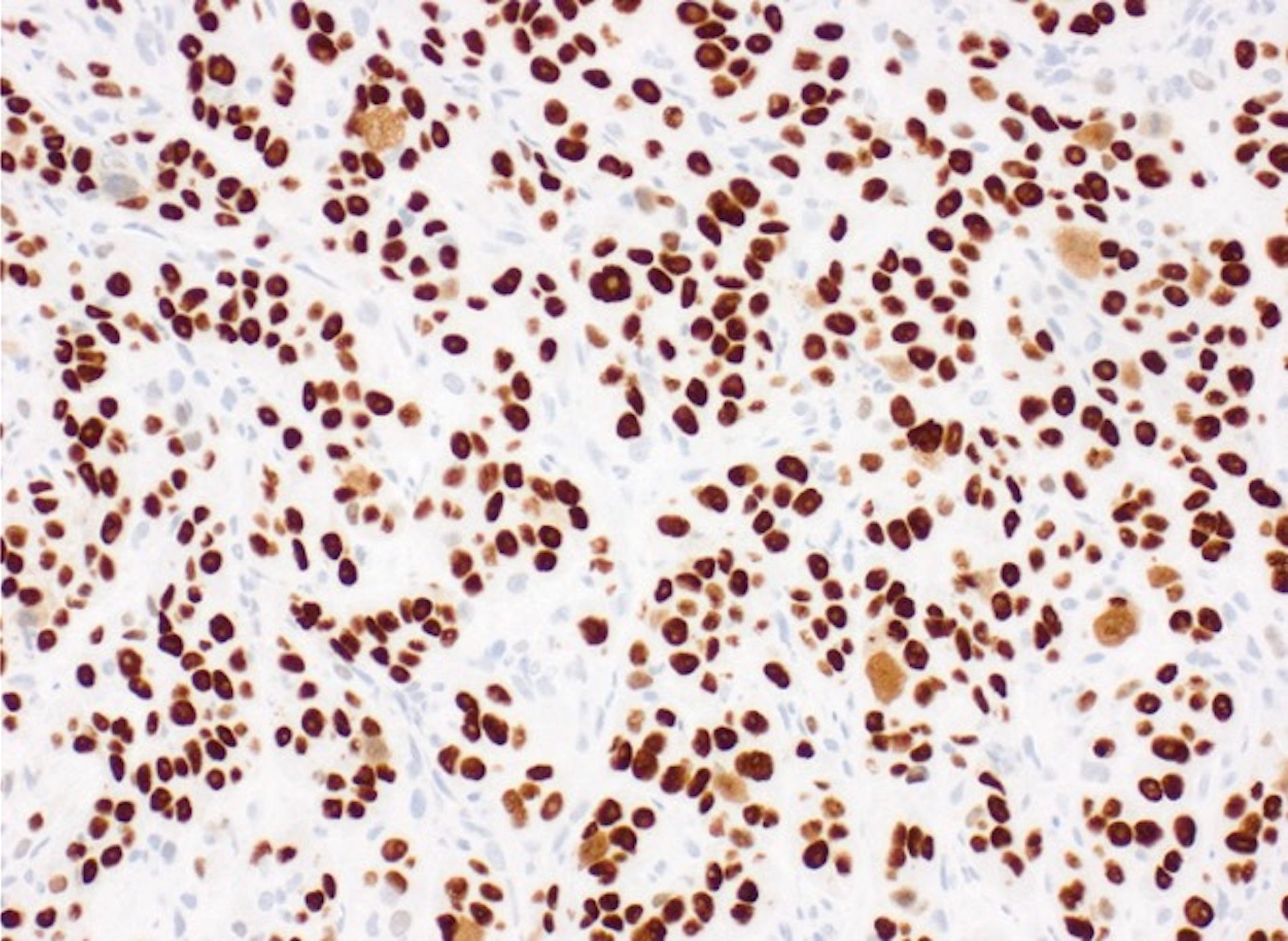
TTF1 in adenocarcinoma

p40 in adenocarcinoma

Squamous cell carcinoma
p40 in SCC

CK5/6 in SCC

TTF1 in SCC
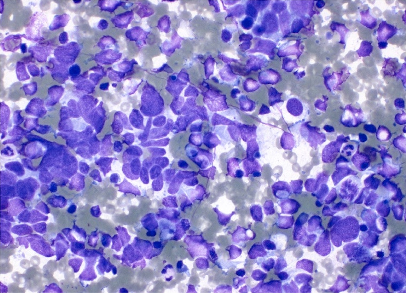
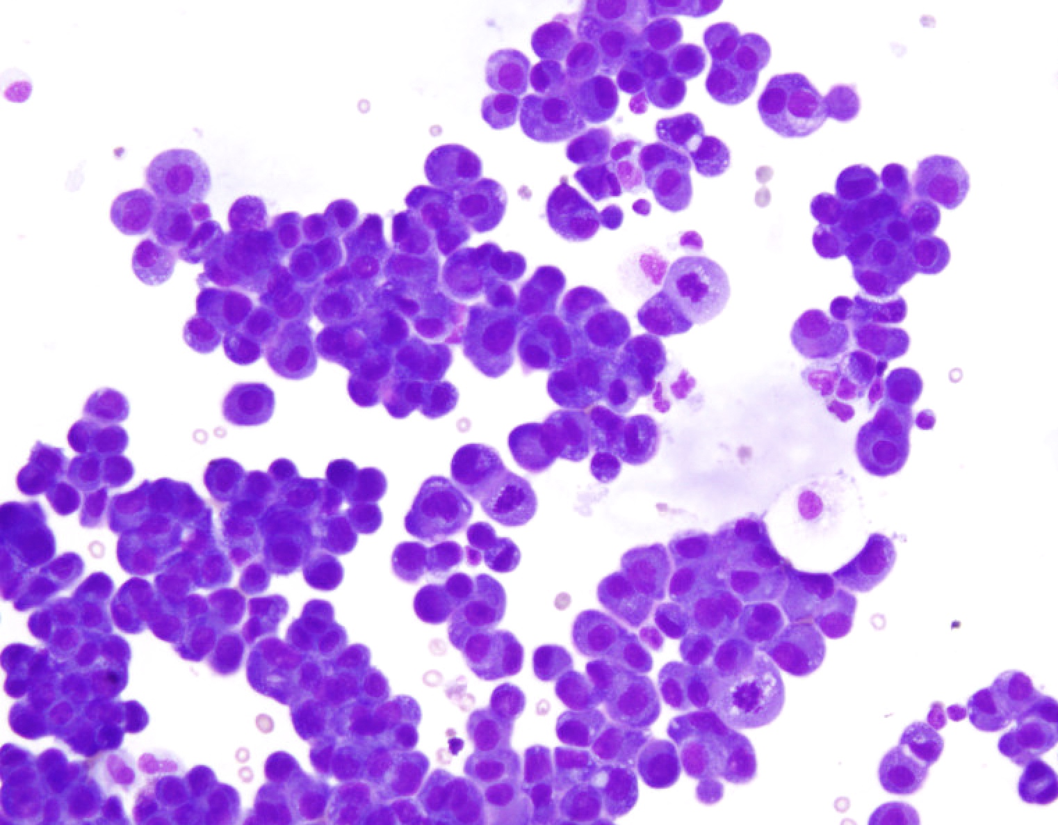
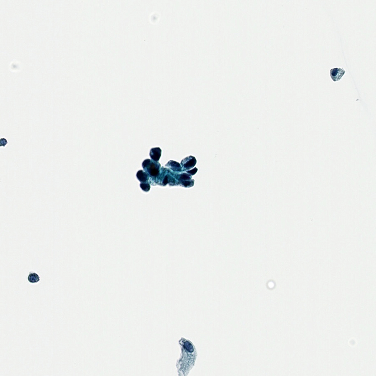
Small cell carcinoma

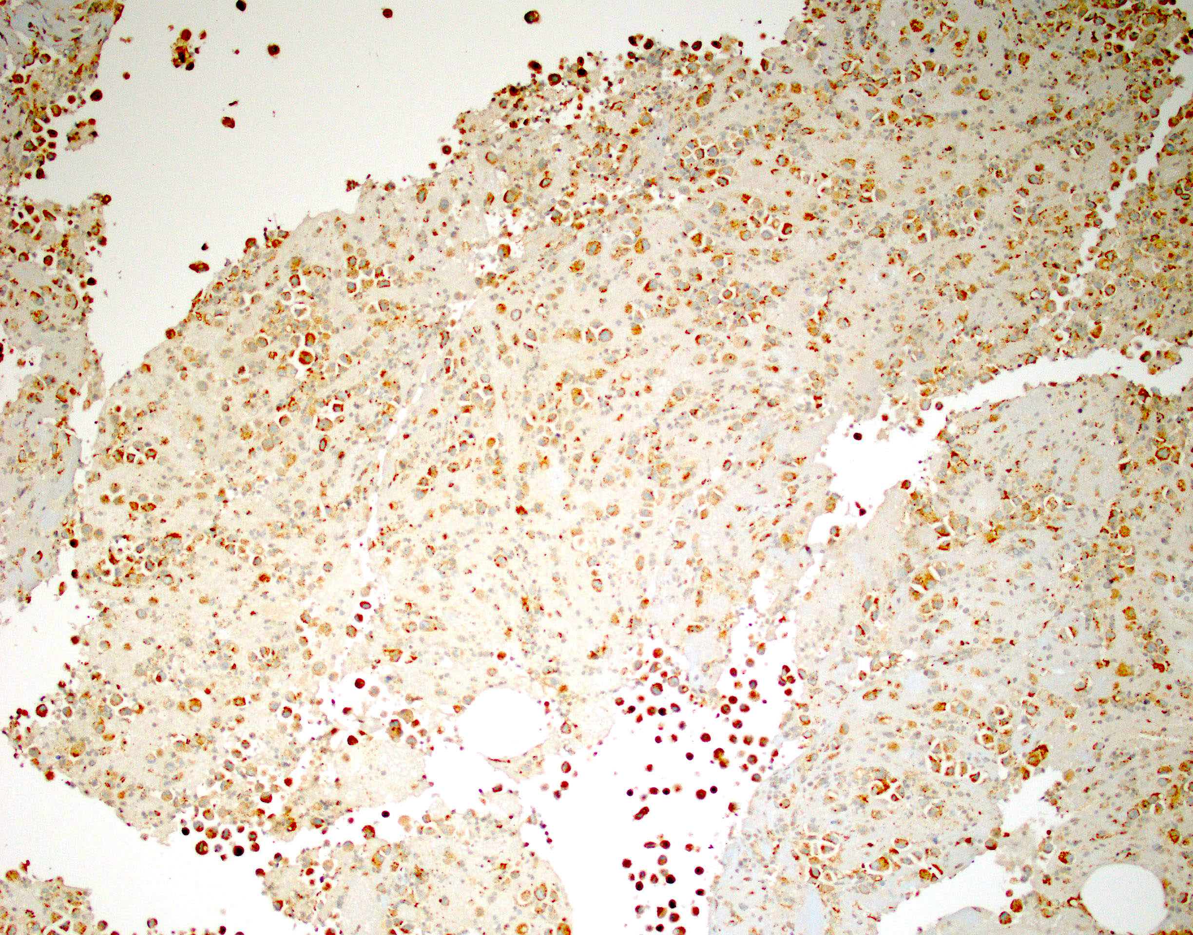
Synaptophysin in small cell carcinoma

CD56 in small cell carcinoma

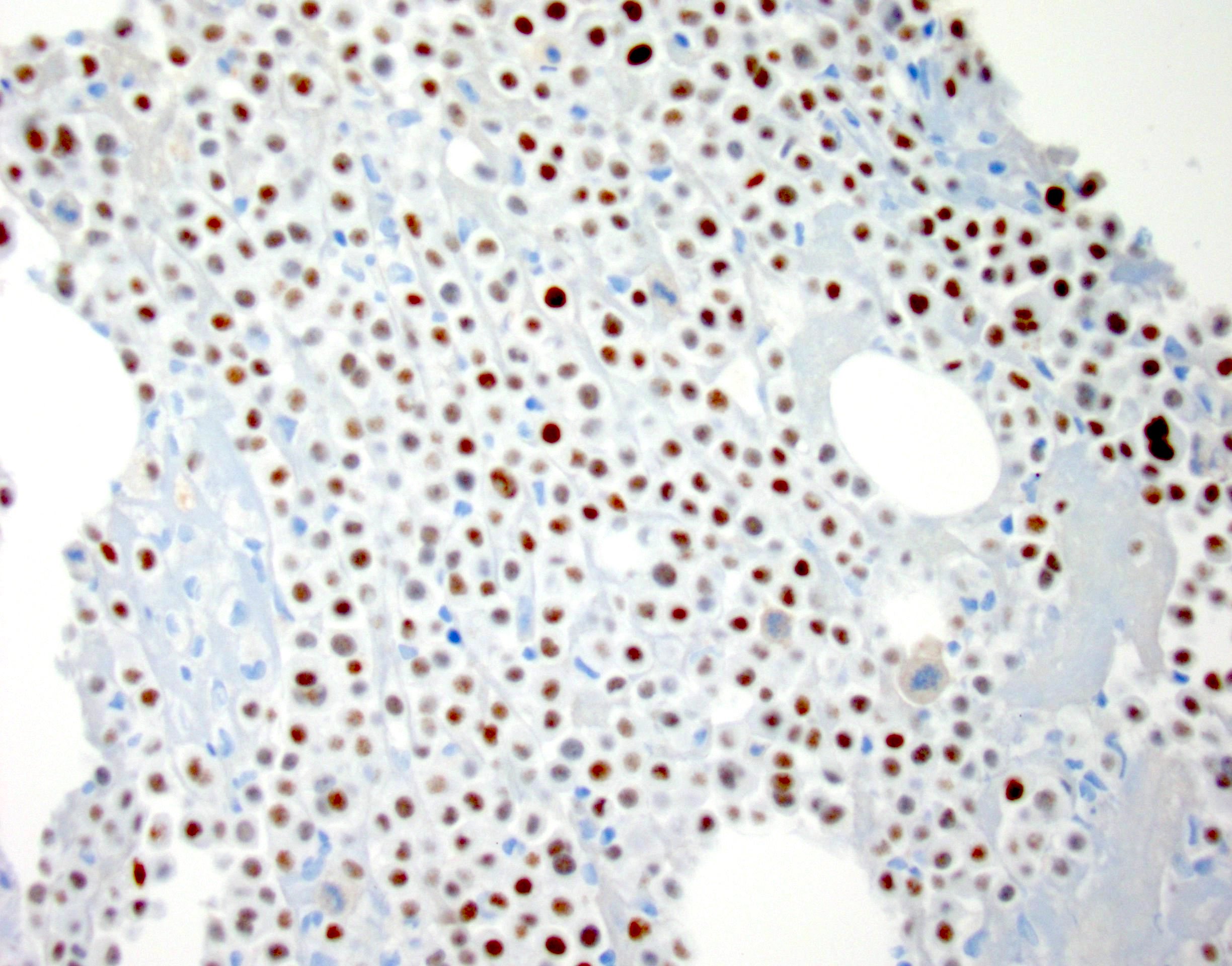
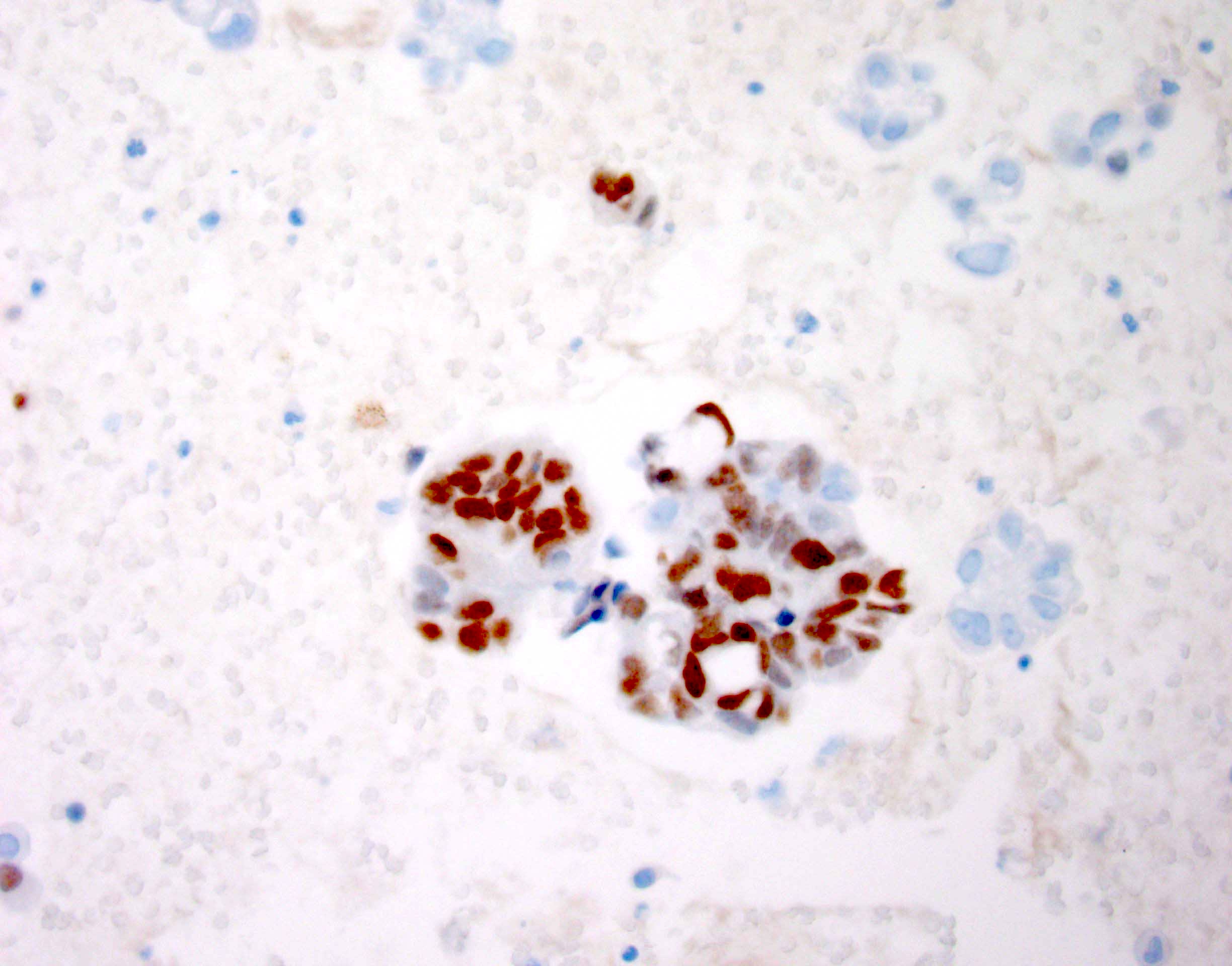
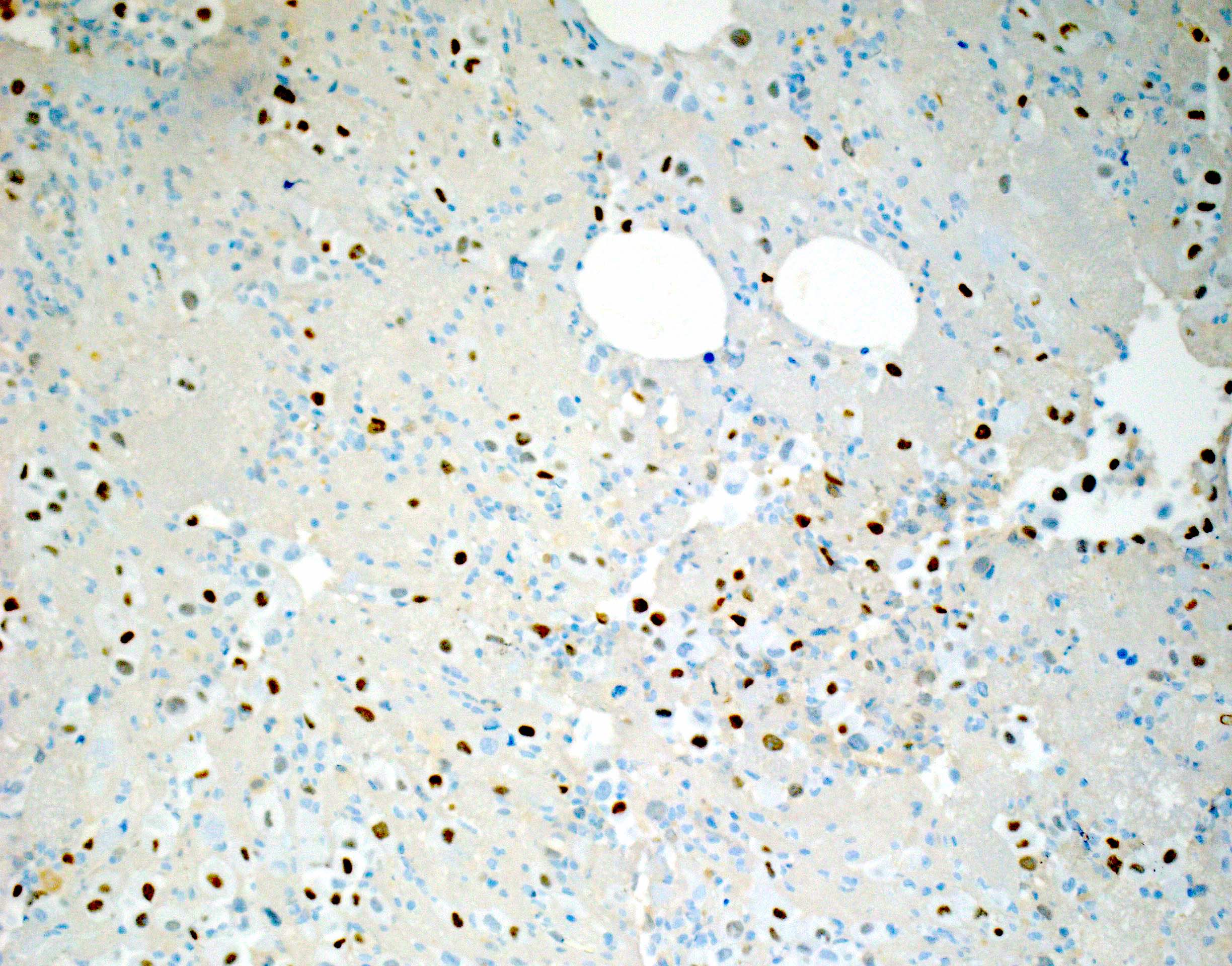
TTF1 in small cell carcinoma

CD45 in small cell carcinoma

Ki67 in small cell carcinoma
Images hosted on other servers:

FISH showing ALK rearrangement
Contributed by Mir Yousufuddin Ali Khan, M.D.
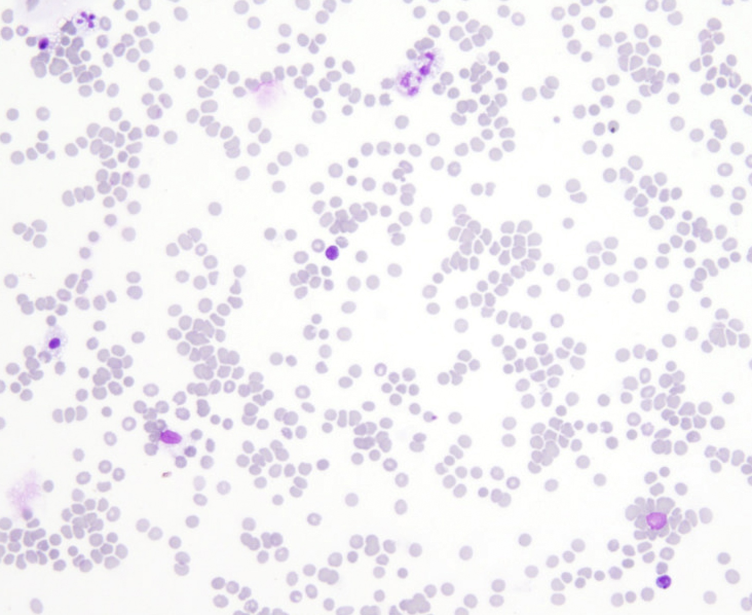
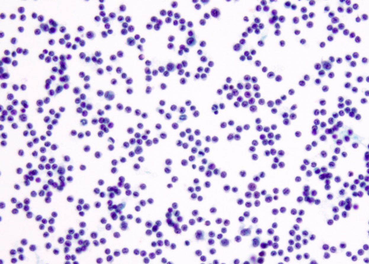
Obscuring blood
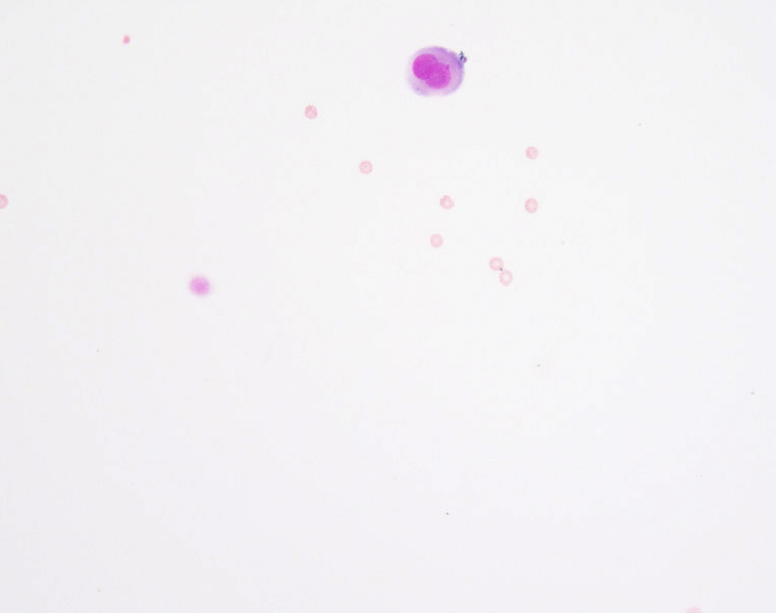
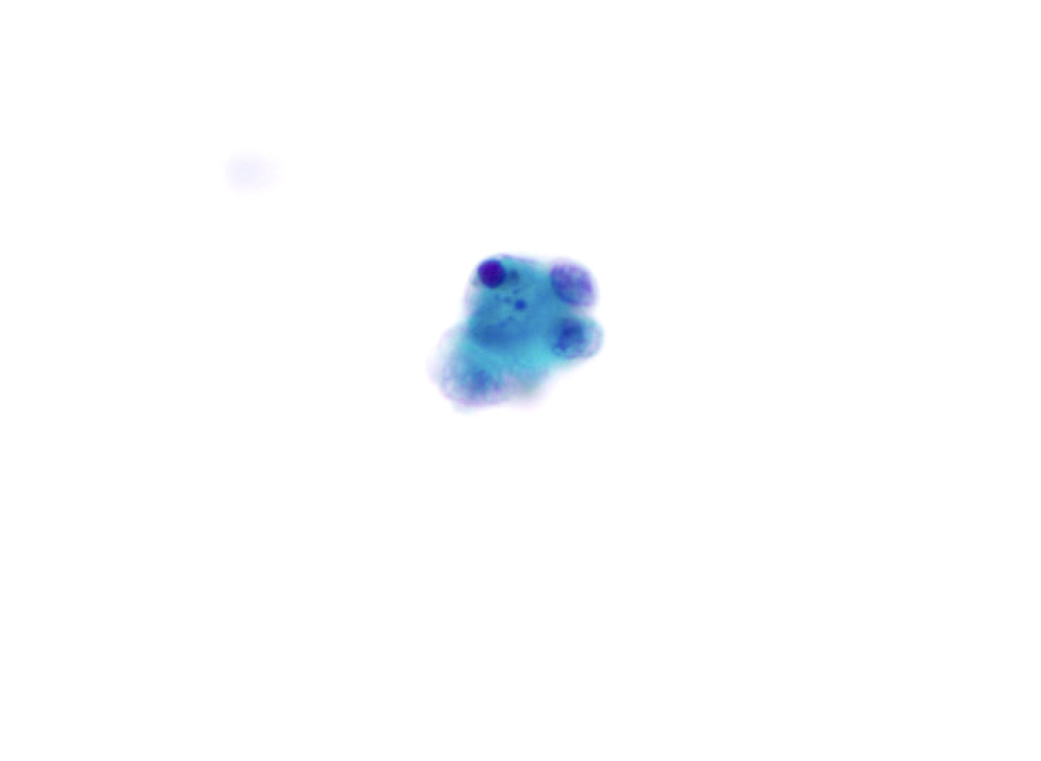
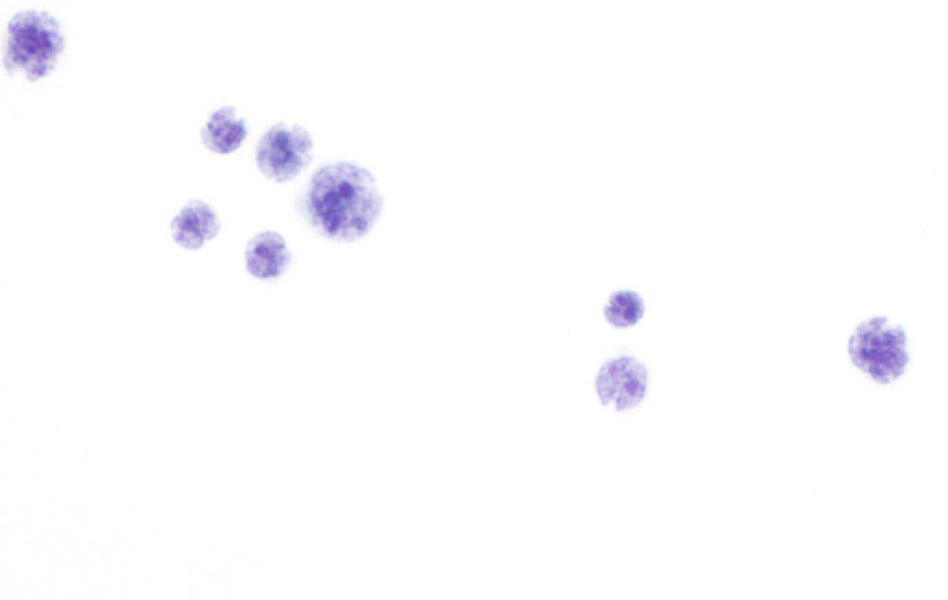
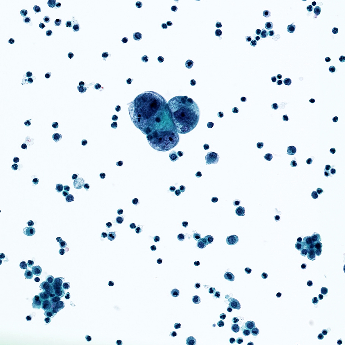
Alveolar macrophages
Fibrin thrombi
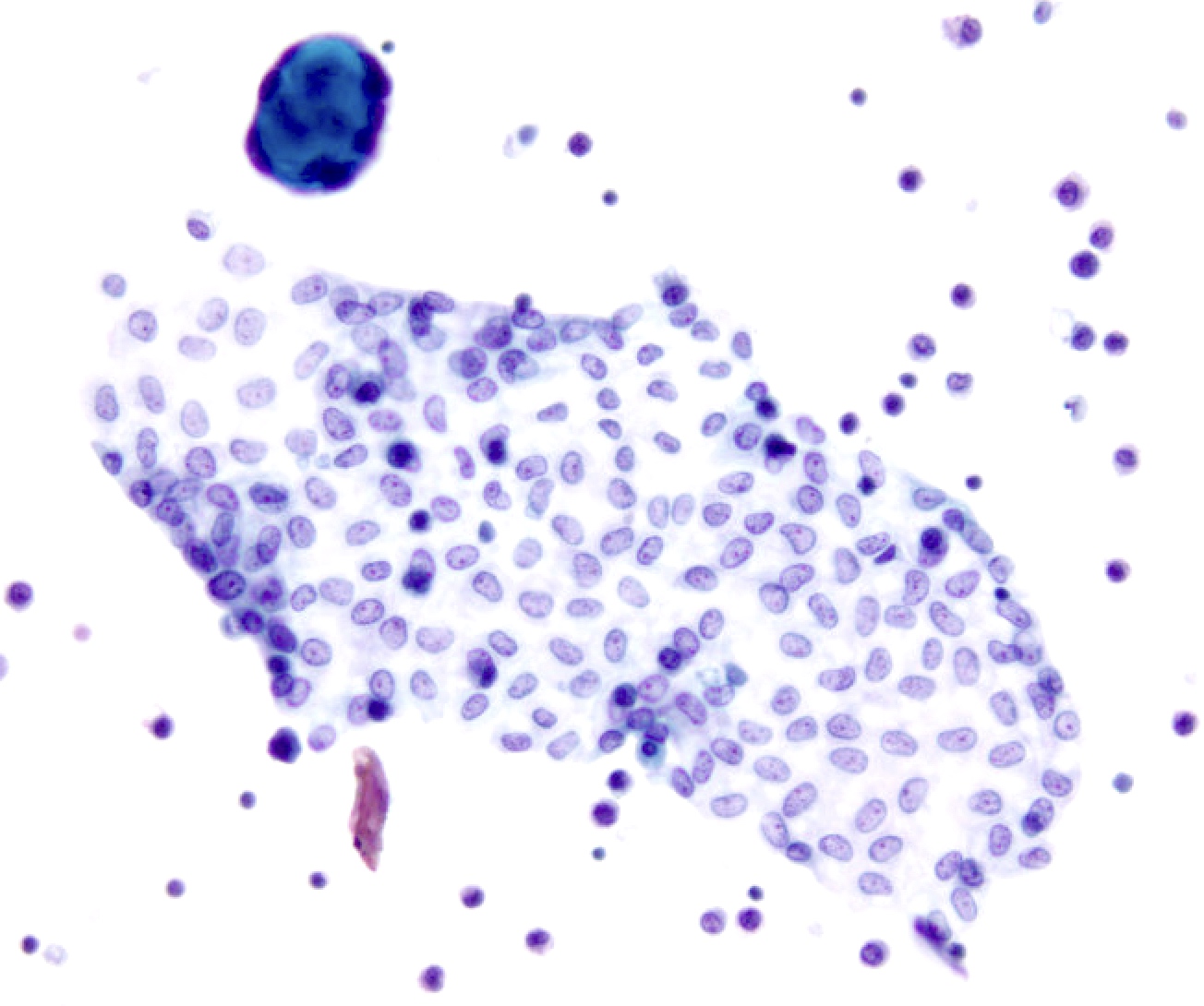
Bronchial cells
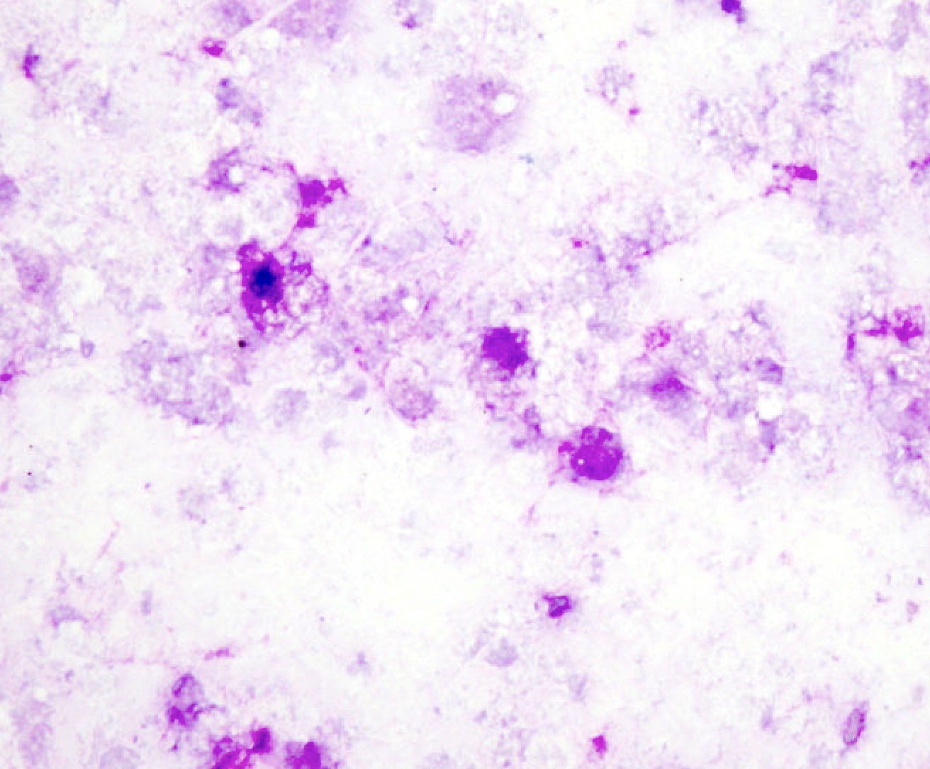
Crush artifact
Poor preservation and poor staining
Bronchial cells

Bronchial contamination
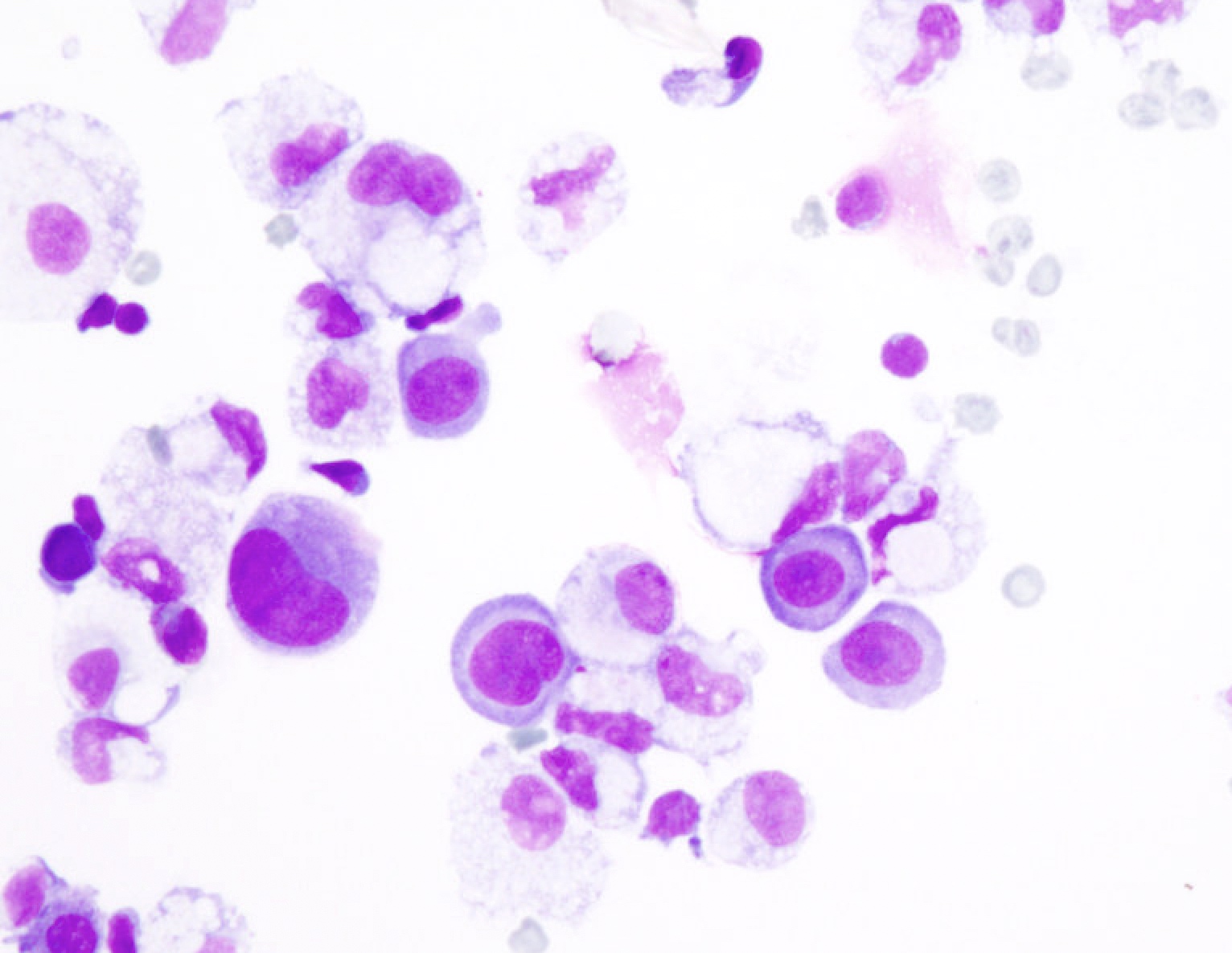
Pneumocytes
WHO reporting system for lung cytopathology
Adequacy criteria in EBUS FNA
Overview of lung cytopathology

Images hosted on other servers:

NGS analysis on smears
Ali: 2023
Bibbo: 2014
Bishop Pitman: 2015
Chandra: 2020
Cibas: 2020
DeMay: 2012
Faquin: 2023
Field: 2016
Field: 2020
IAC-IARC-WHO: 2023
IAC-IARC-WHO: 2023
Jiang: 2023
Layfield: 2018
Mody: 2022
Nayar: 2015
Ramzy: 2017
Shidham: 2021

VandenBussche: 2019
VandenBussche: 2022
Wojcik: 2022
Find related Pathology books: breast, cytopathology, GI, GU/adrenal, gynecologic, head & neck/endocrine, lung

