
Images hosted on other servers:

Papule on ear
Images hosted on other servers:

Acquired cholesteatoma
Contributed by Ashley Aiken, M.D.
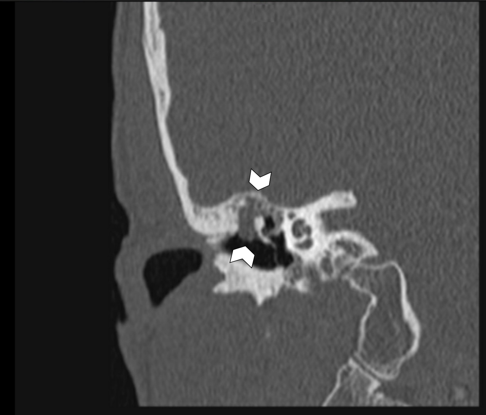
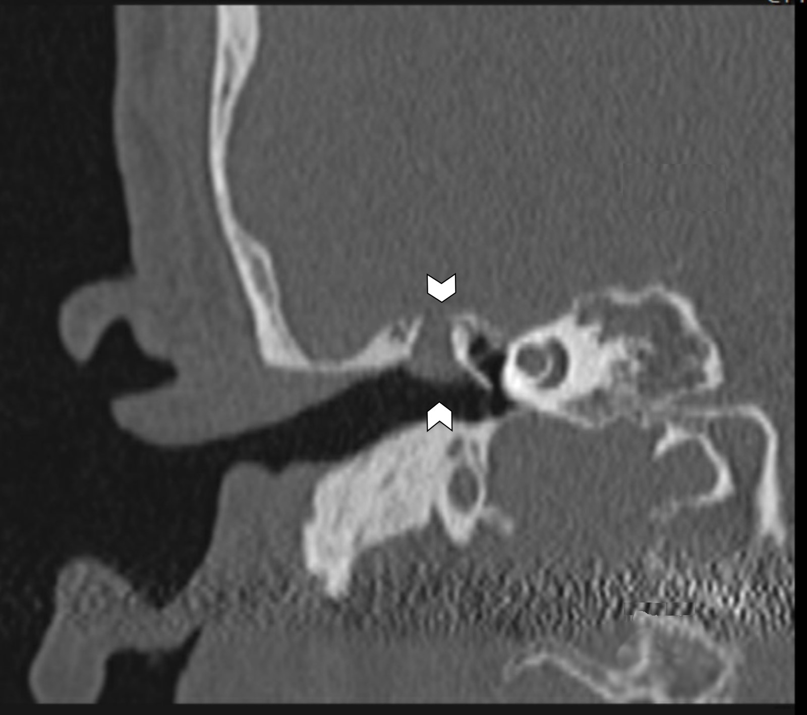
CT, coronal
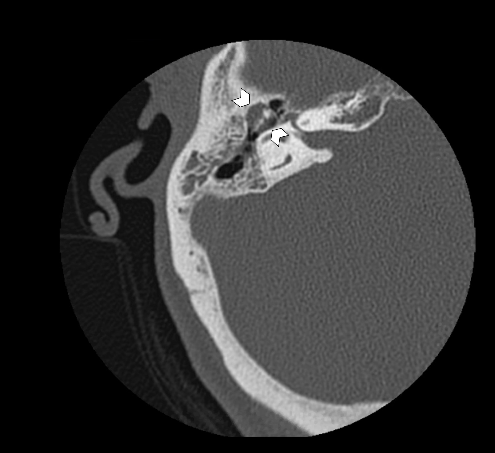
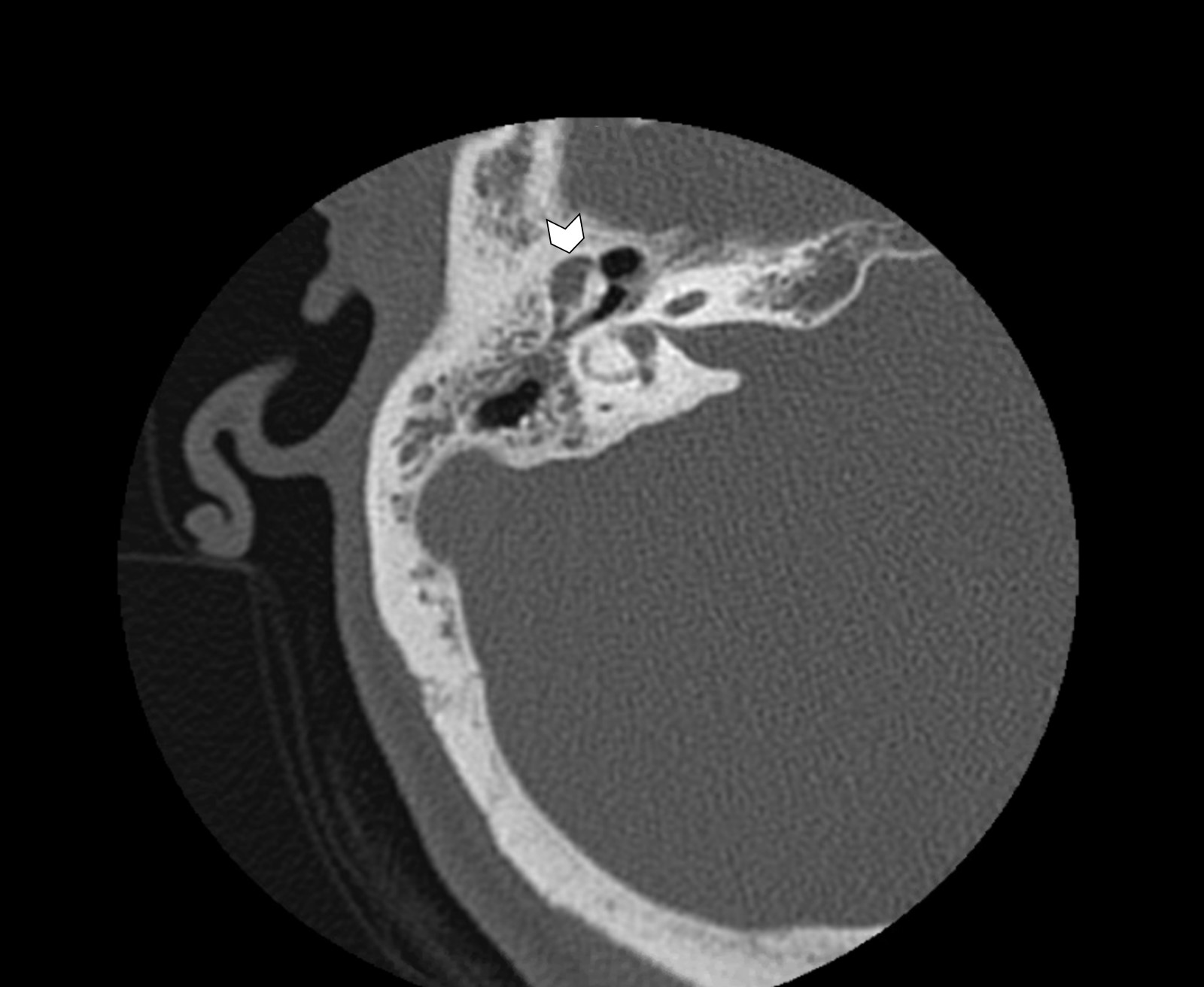
CT, axial
Contributed by Esther Vivas, M.D.
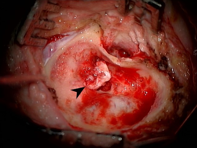
Mastoid cholesteatoma, right ear
Images hosted on other servers:

Bilateral congenital cholesteatoma

Congenital cholesteatoma

Acquired cholesteatoma
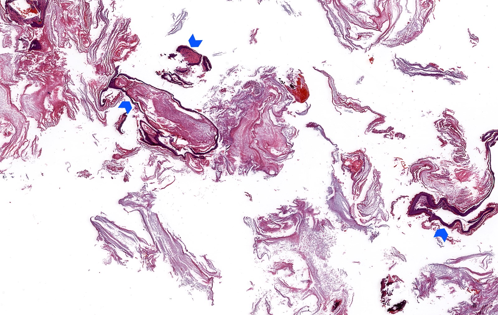
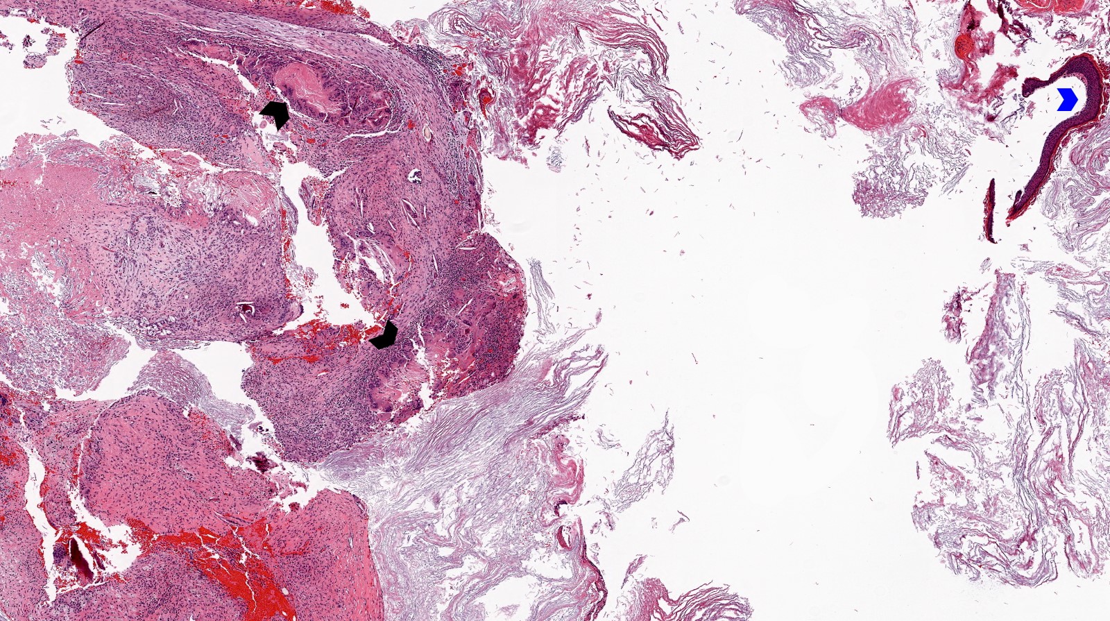
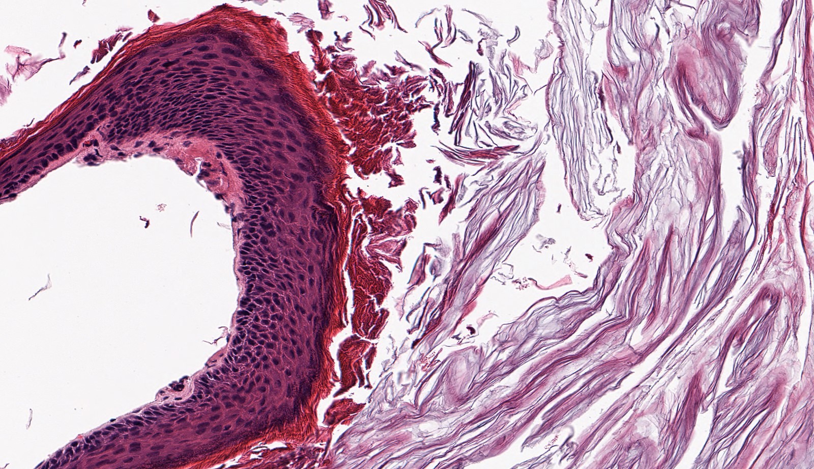
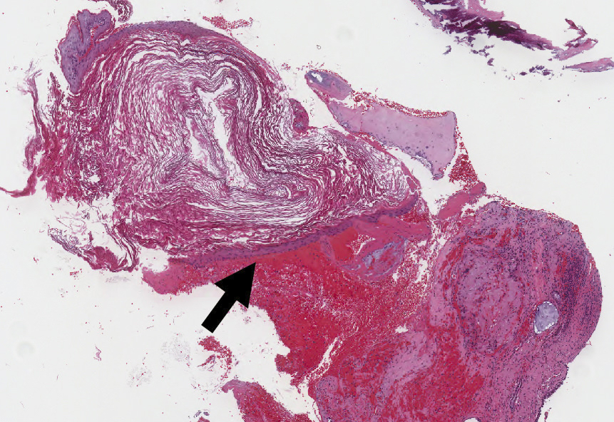
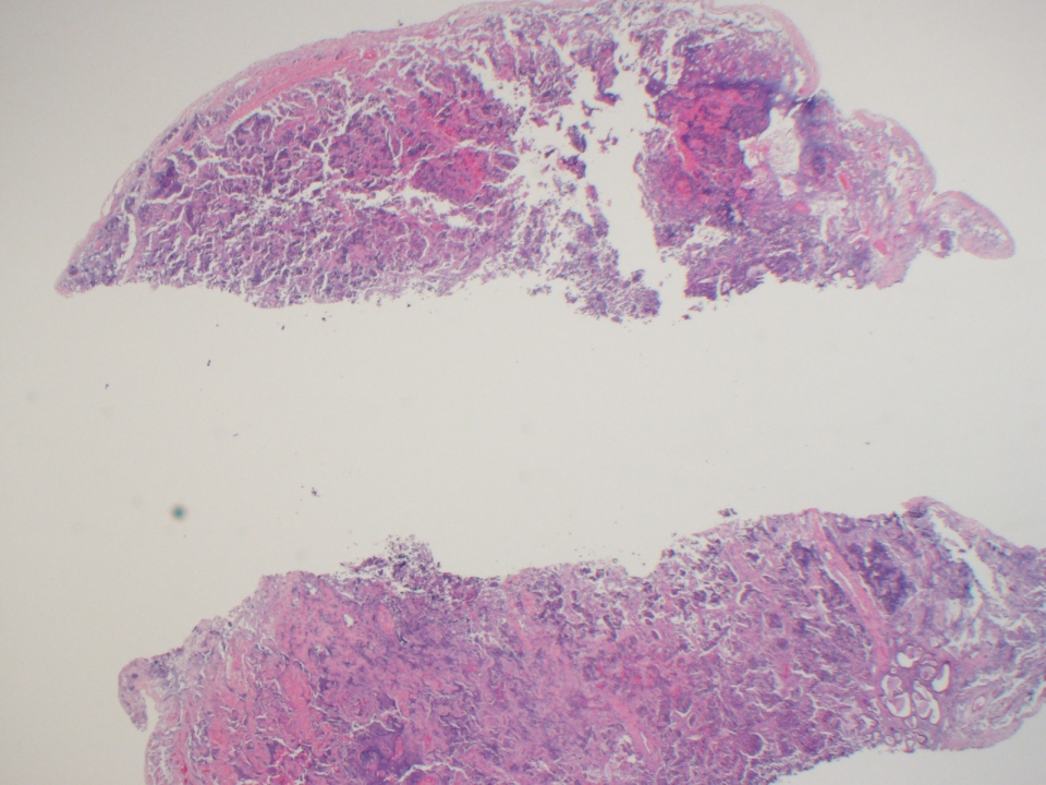
Contributed by Kelly Magliocca, D.D.S., M.P.H.

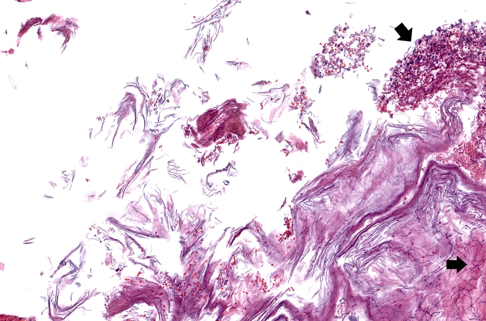
Fragments of hemorrhagic soft tissue
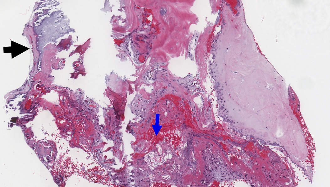
Contributed by Kelly Magliocca D.D.S., M.P.H.
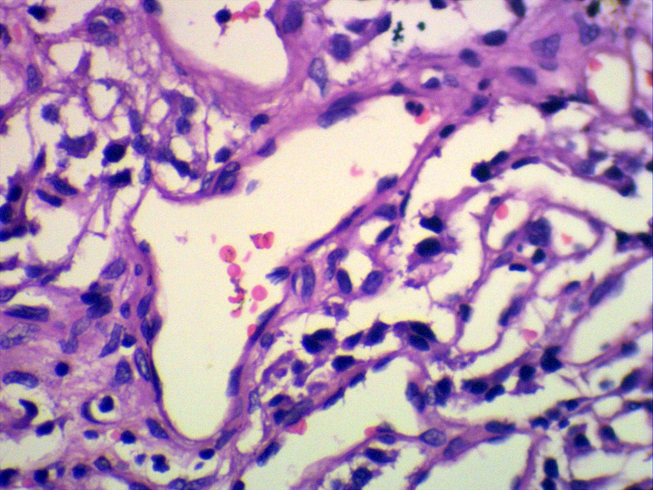
Cholesteatoma epithelium and keratin
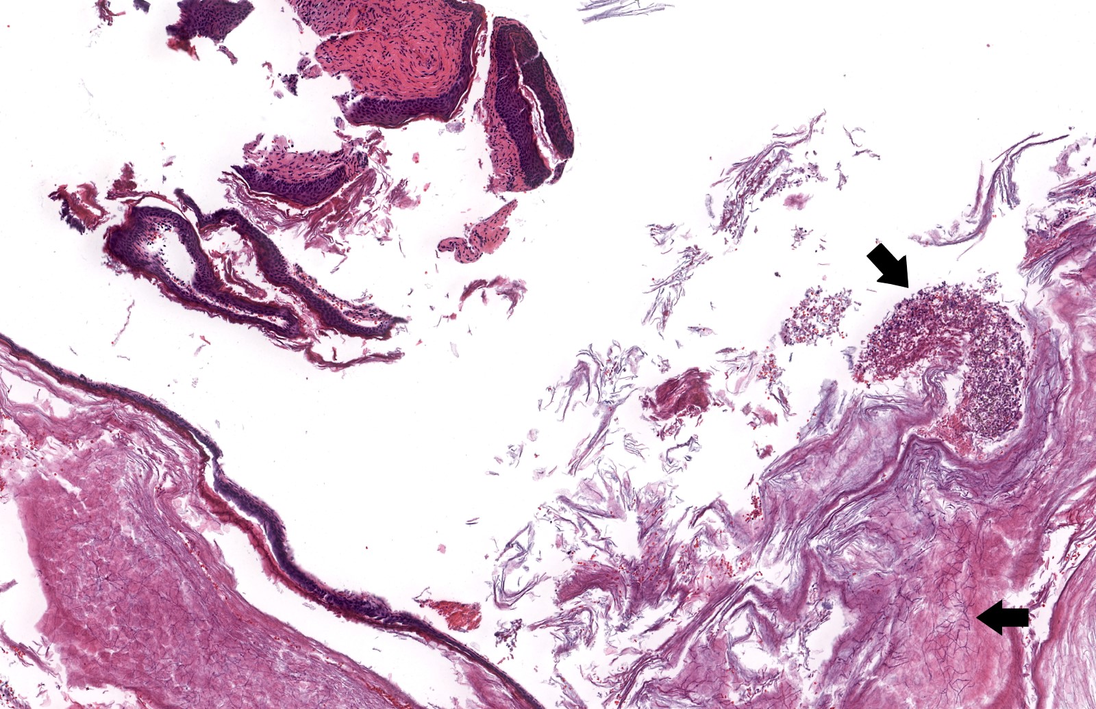
Fungal forms within keratin debris
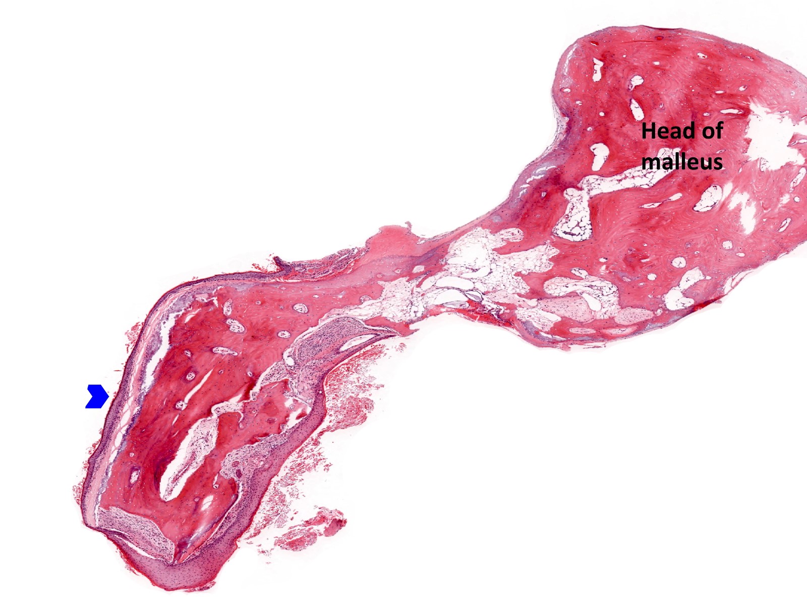
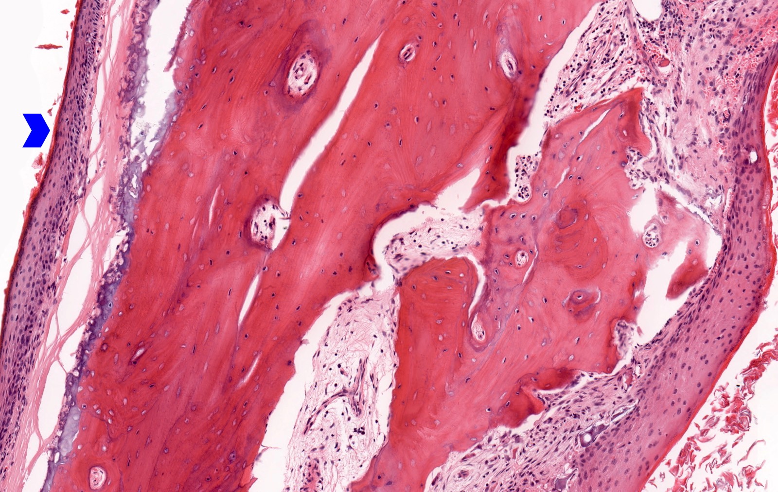
Cholesteatoma eroding malleus
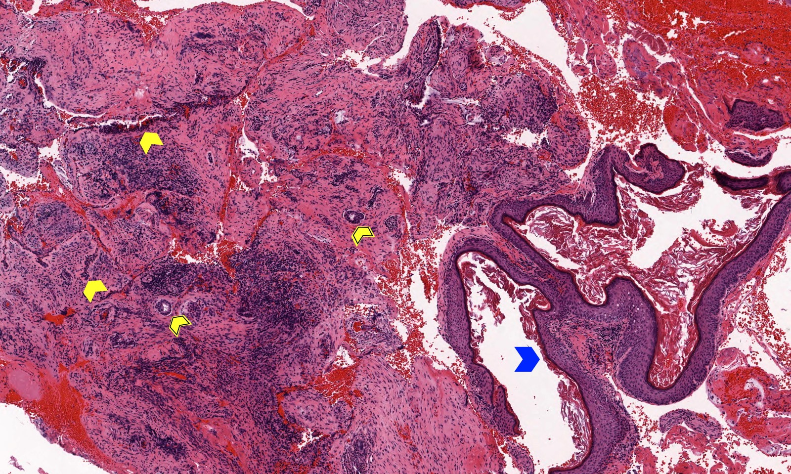
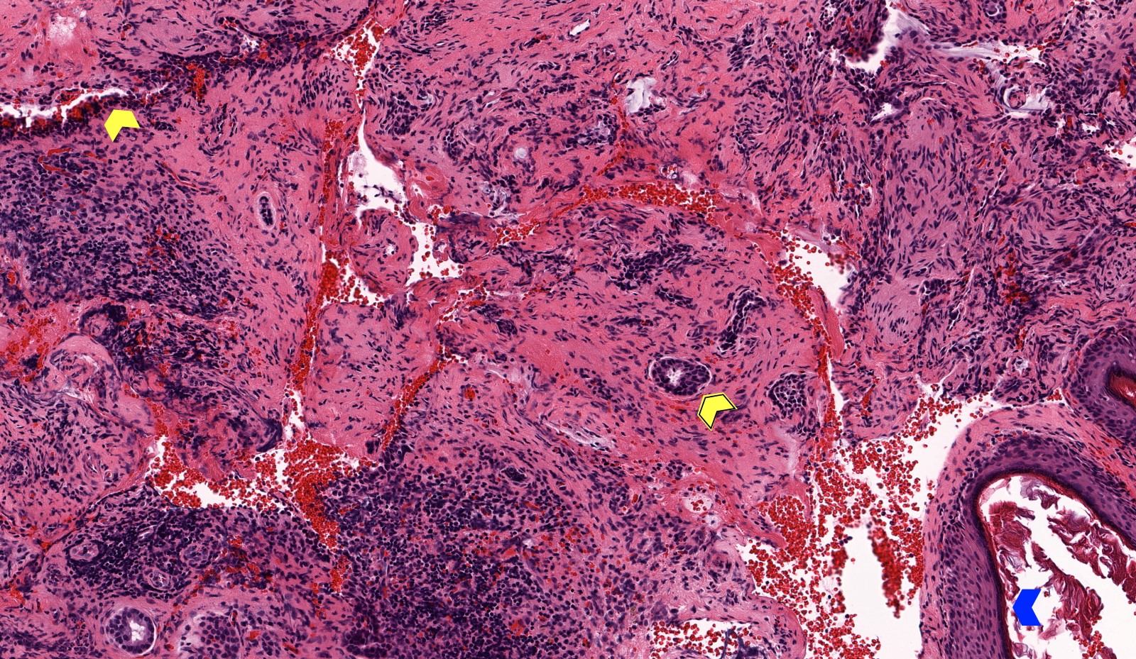
Cholesteatoma and middle ear mucosa
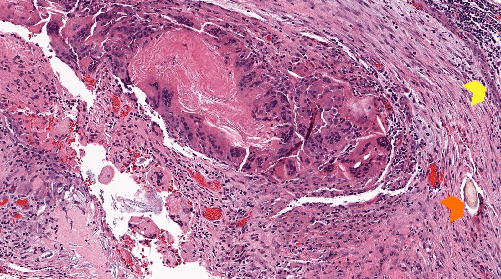
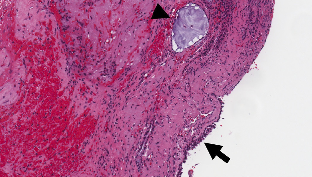
Cholesteatoma, inflamed
Cholesteatoma epithelium and keratin
Inflamed middle ear mucosa
Residual fibrous tissue and underlying cartilage
Middle ear mucosa with hyperplasia
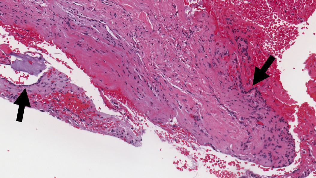
Cuboidal middle ear epithelium
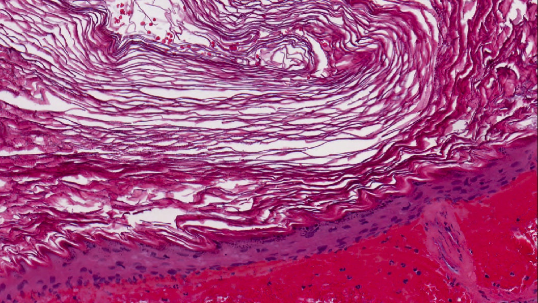
Keratinizing stratified squamous epithelium

Keratin granuloma within ossicular tissue
Primary and secondary acquired cholesteatoma
Images hosted on other servers:

Infiltration of papillary dermis by epithelioid (E) cells


External and middle ear
Contributed by Kelly Magliocca D.D.S., M.P.H.
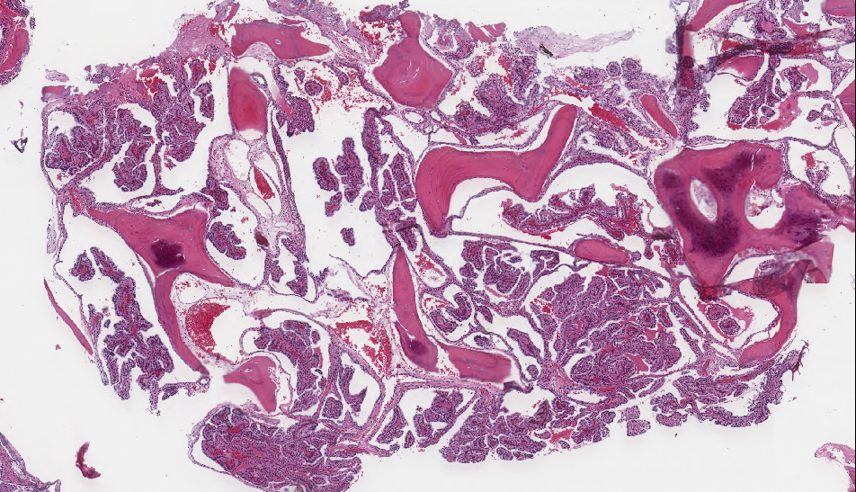
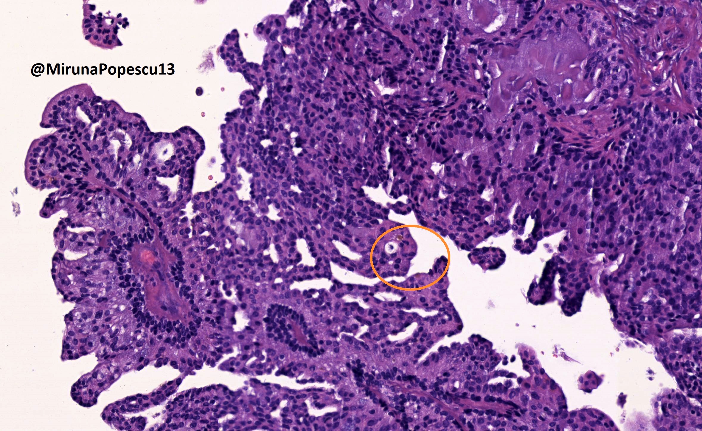
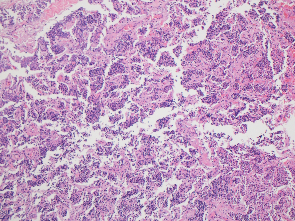
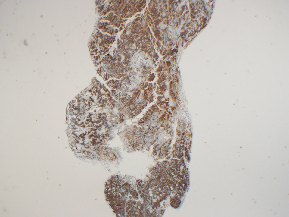
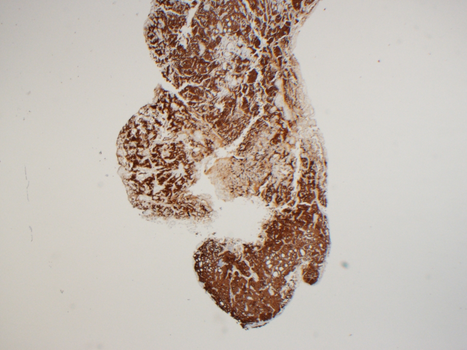
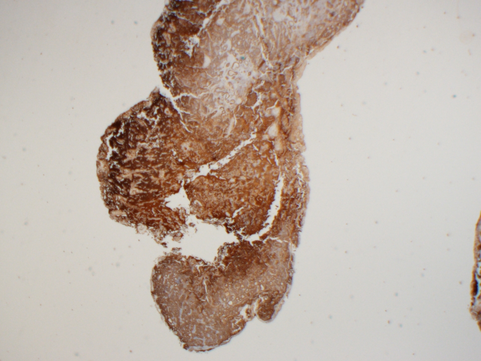
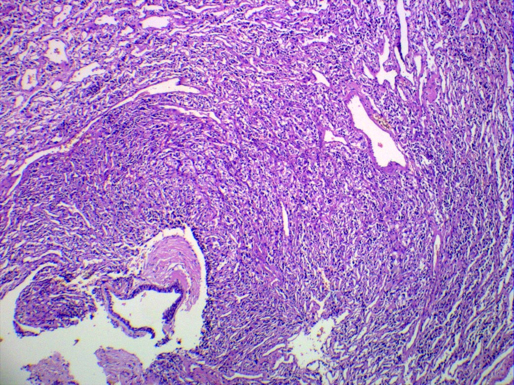
Endolymphatic sac tumor, 2x
Endolymphatic sac tumor, 5x
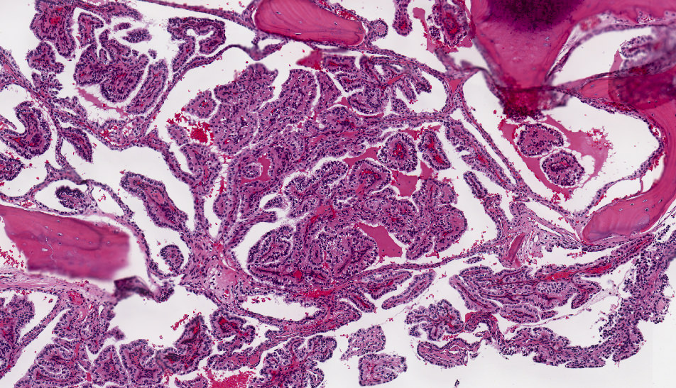
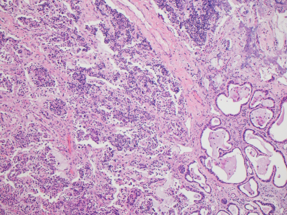
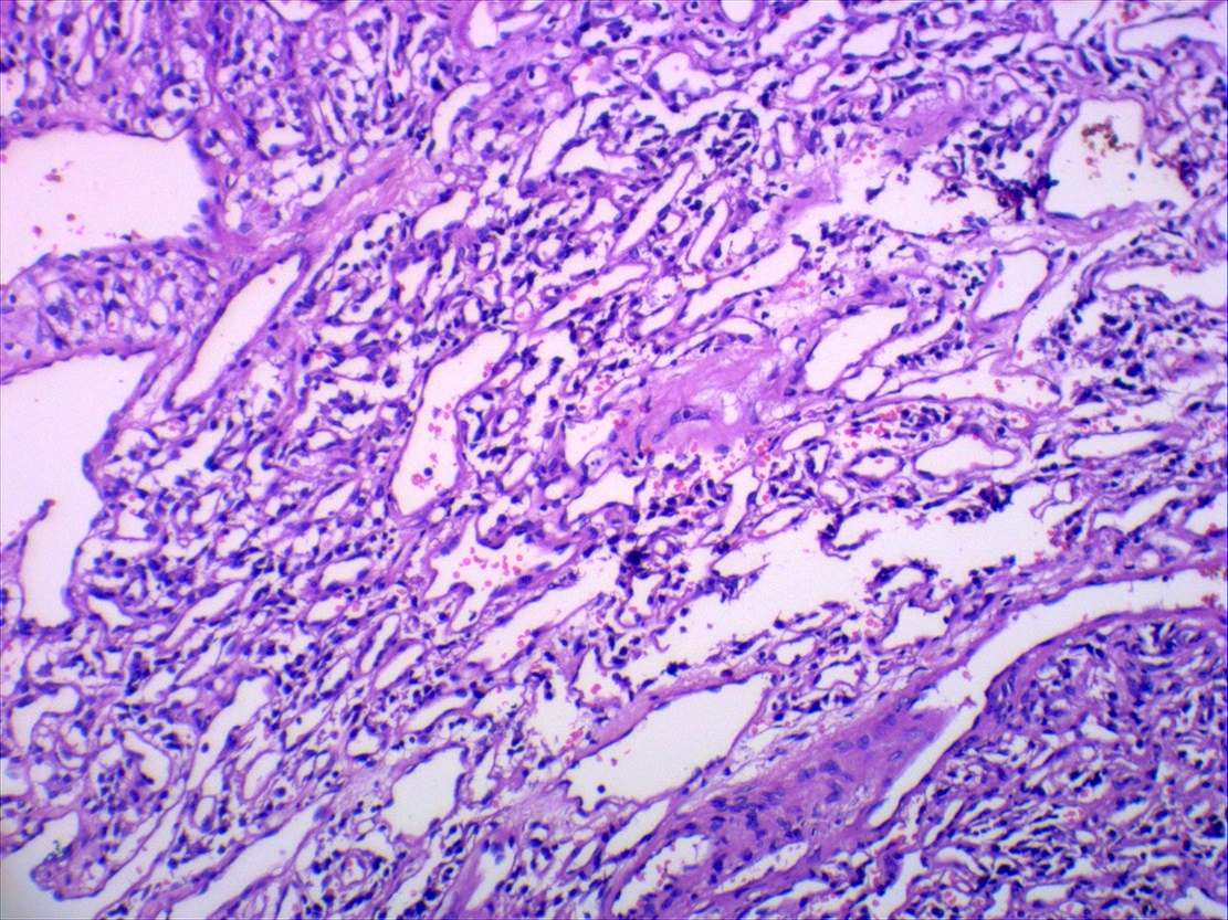
Endolymphatic sac tumor, 15x
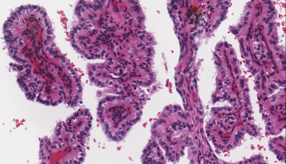
Endolymphatic sac tumor, 20x

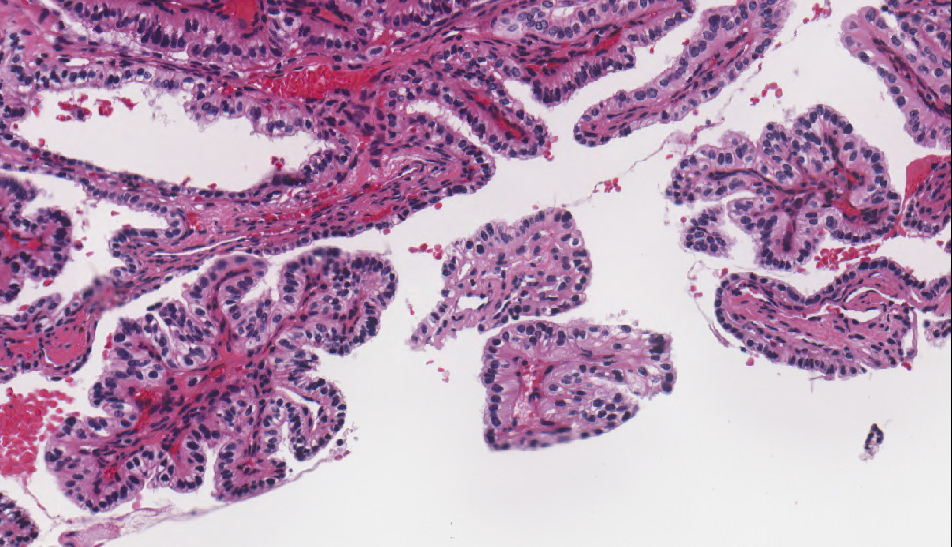
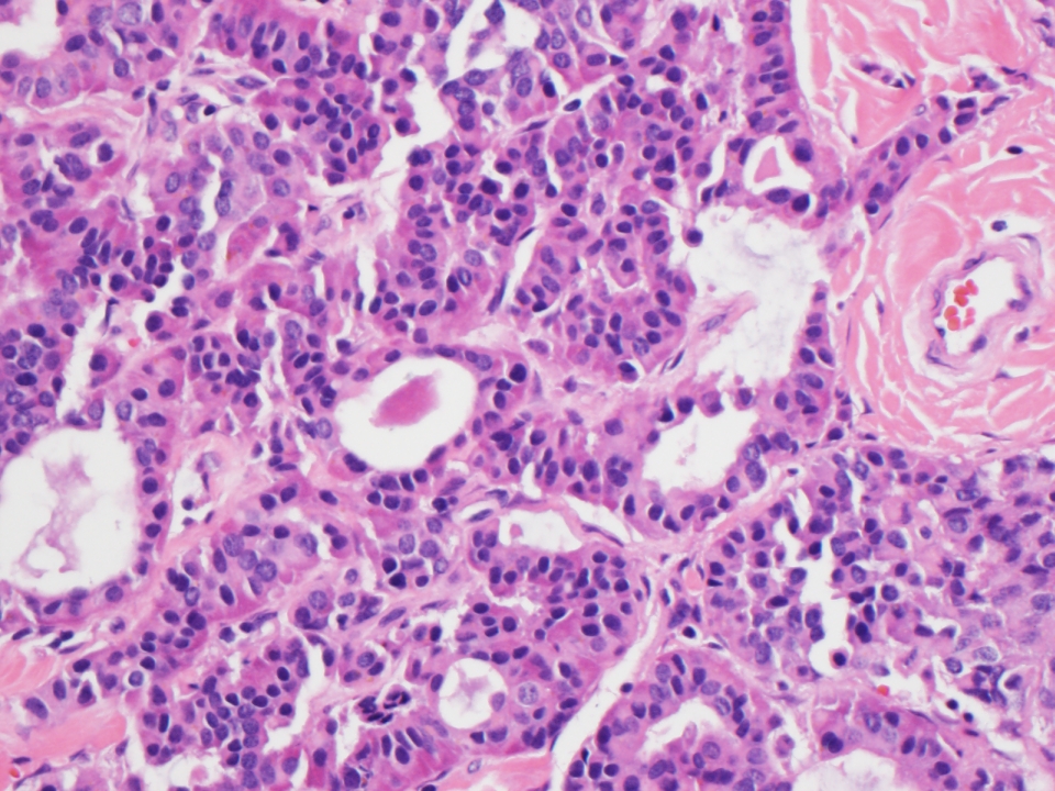
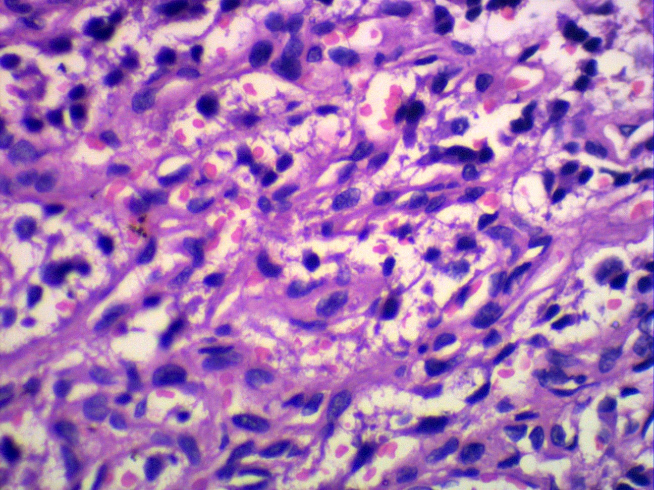
Endolymphatic sac tumor, 40x

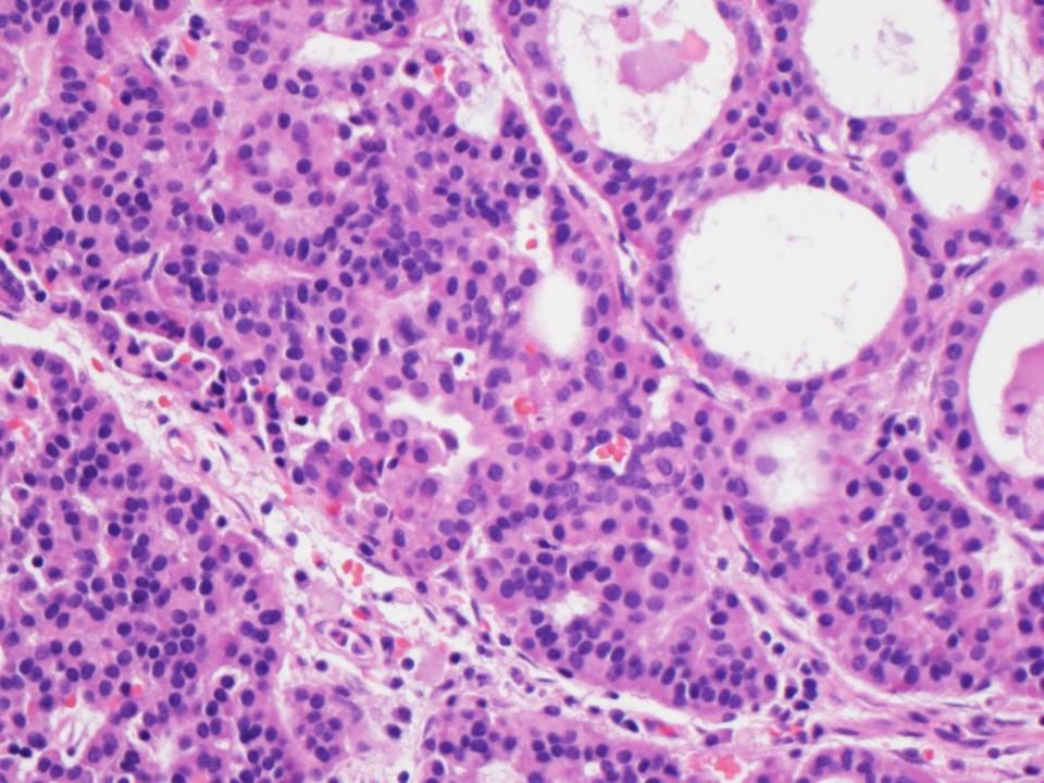
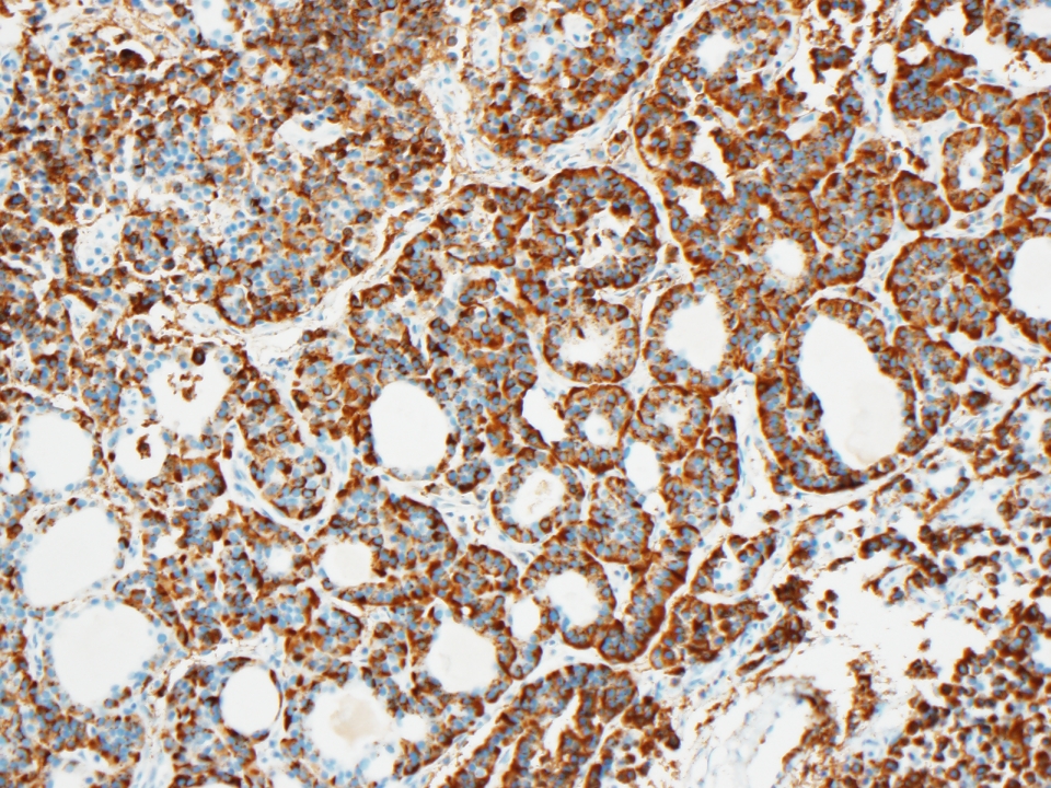
CAIX, 20x
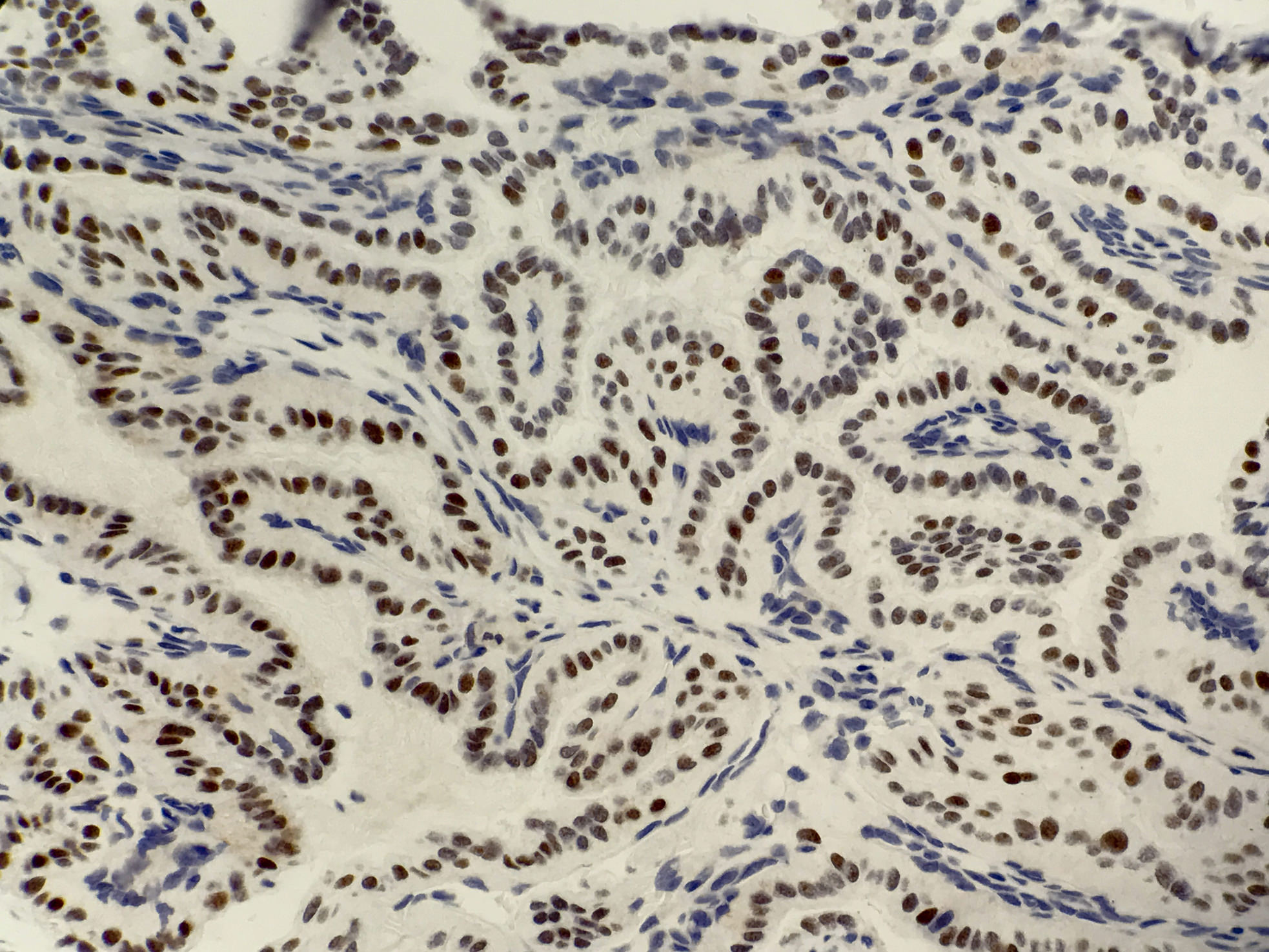
PAX8, 40x

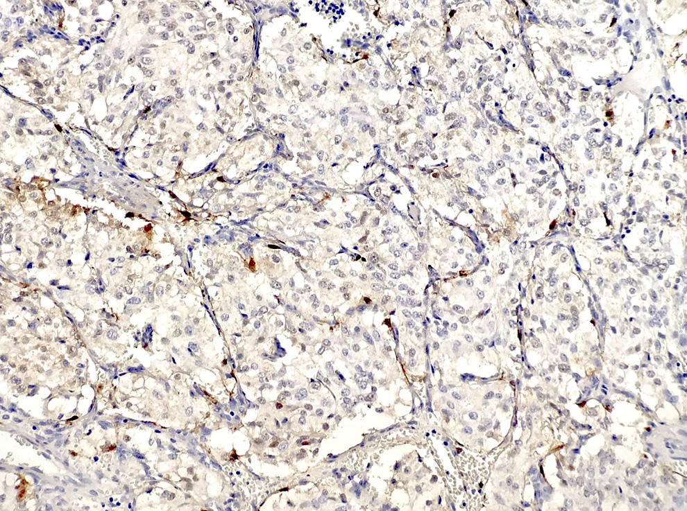
Negative RCC, 20x
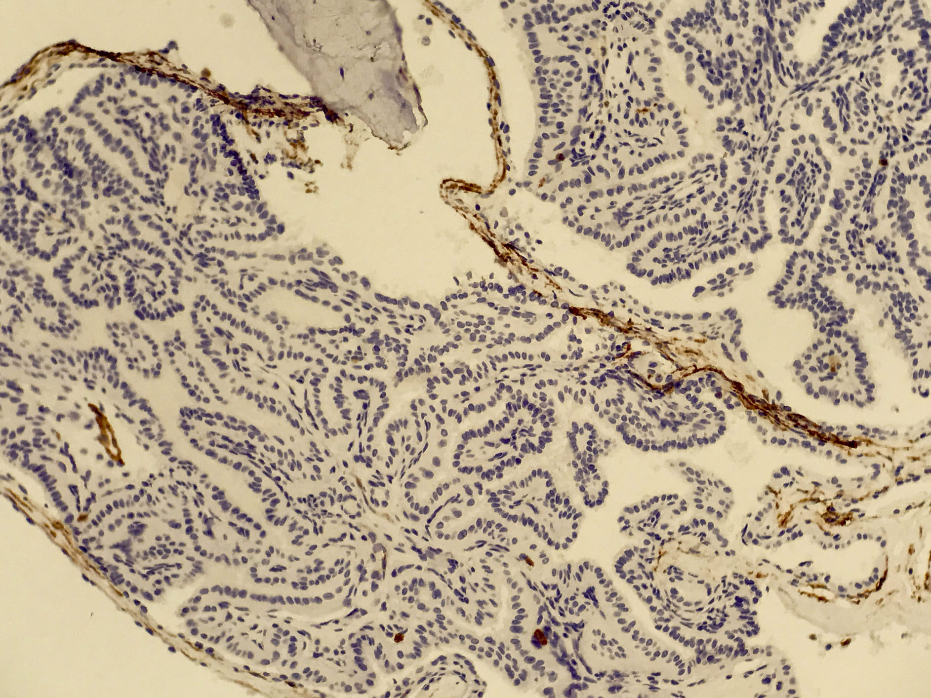
Negative CD10, 20x
Images hosted on other servers:

Various images

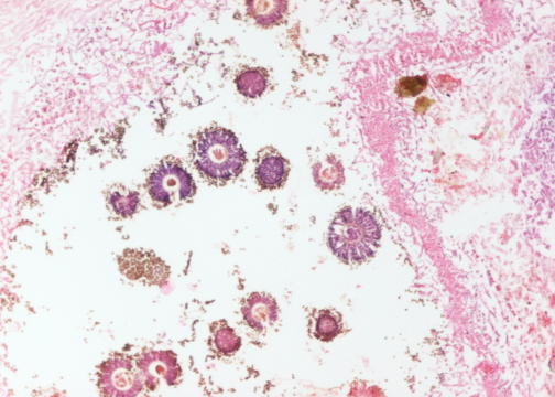
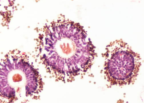
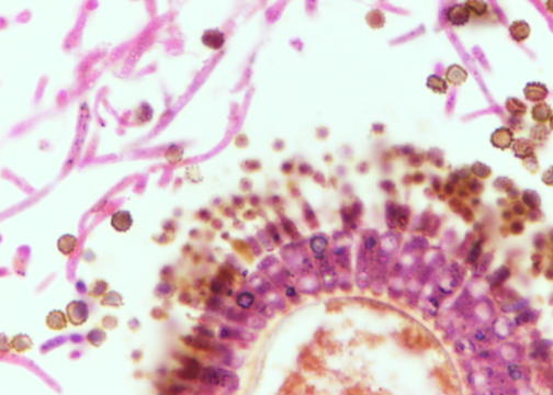
Papilla with hemorrhagic material

Papilla with vacuolated cells and colloid-like material
Images hosted on other servers:

with soft tissue
density occupying
the middle ear
Images hosted on other servers:

glial tissue and
fibrovascular elements

GFAP+, S100+
Images hosted on other servers:

Central cystic degeneration of auricular cartilage

Factor VIII+ lining of pseudocyst

Contributed by Agnes Ikpoto Udoh, M.D., M.B.A.
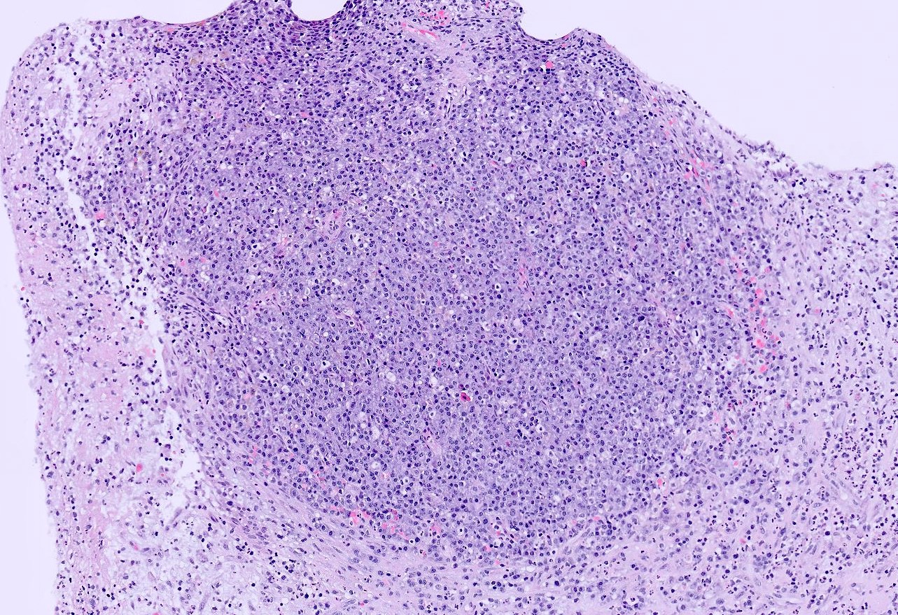
Well circumscribed mass
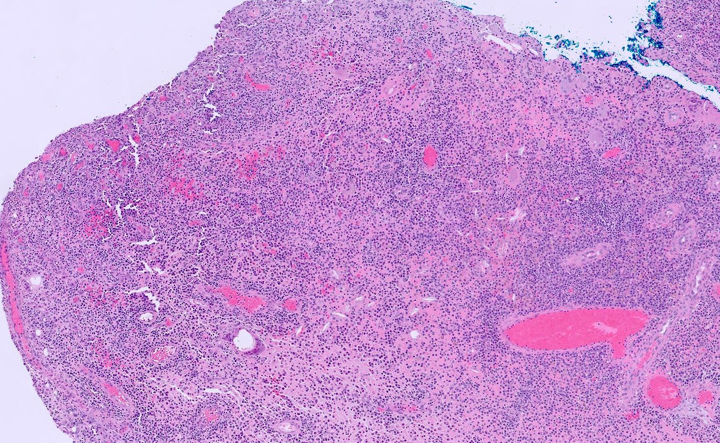
Abundant mixed inflammation

Mixed inflammatory infiltrates within otic polyp
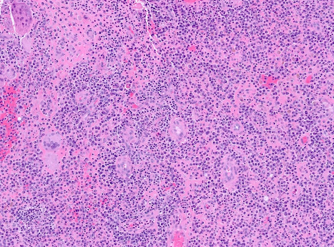
Otic polyp with giant cells
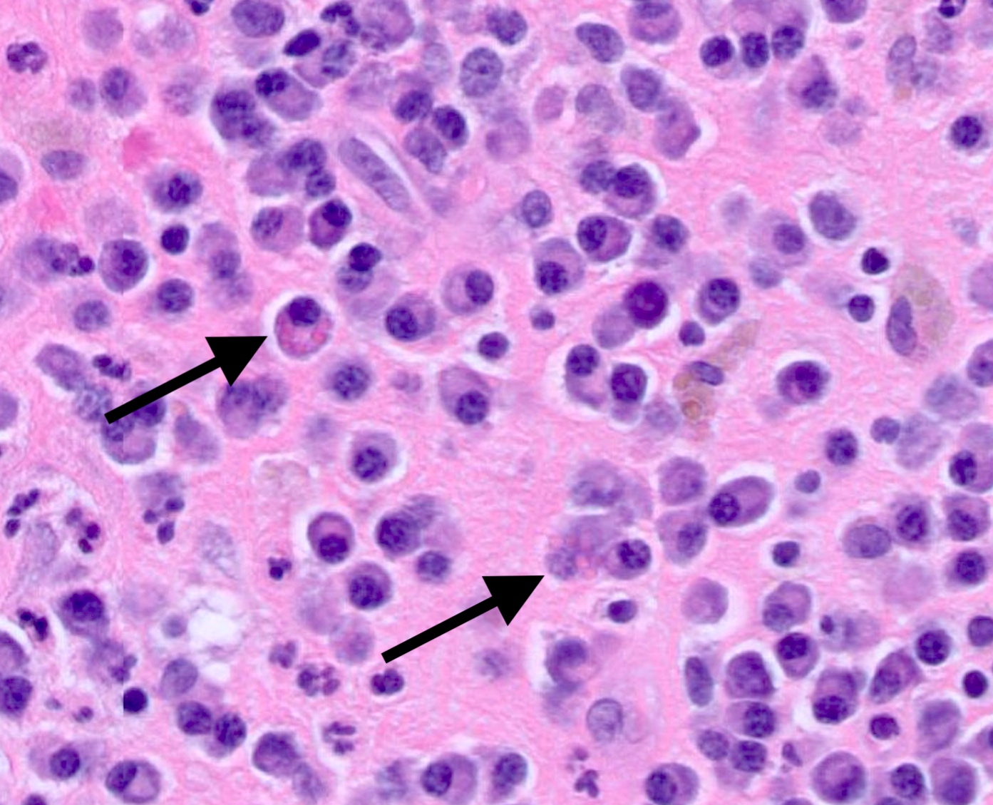
Mott cells

Abundant plasma cells
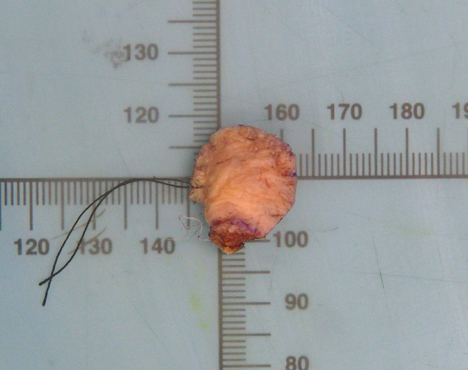
Infected aural polyp ear wax removal
Images hosted on other servers:

Keloid ultrasound appearance
Images hosted on other servers:

Keloids and hypertrophic scars
Contributed by Andrew Dettrick, M.B.B.S. and Ruta Gupta, M.D.
Raised dome with hairless surface
Cut surface
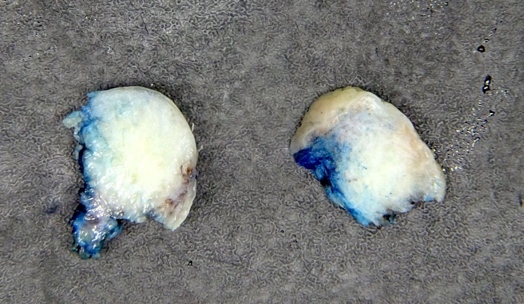
Contributed by Andrew Dettrick, M.B.B.S.
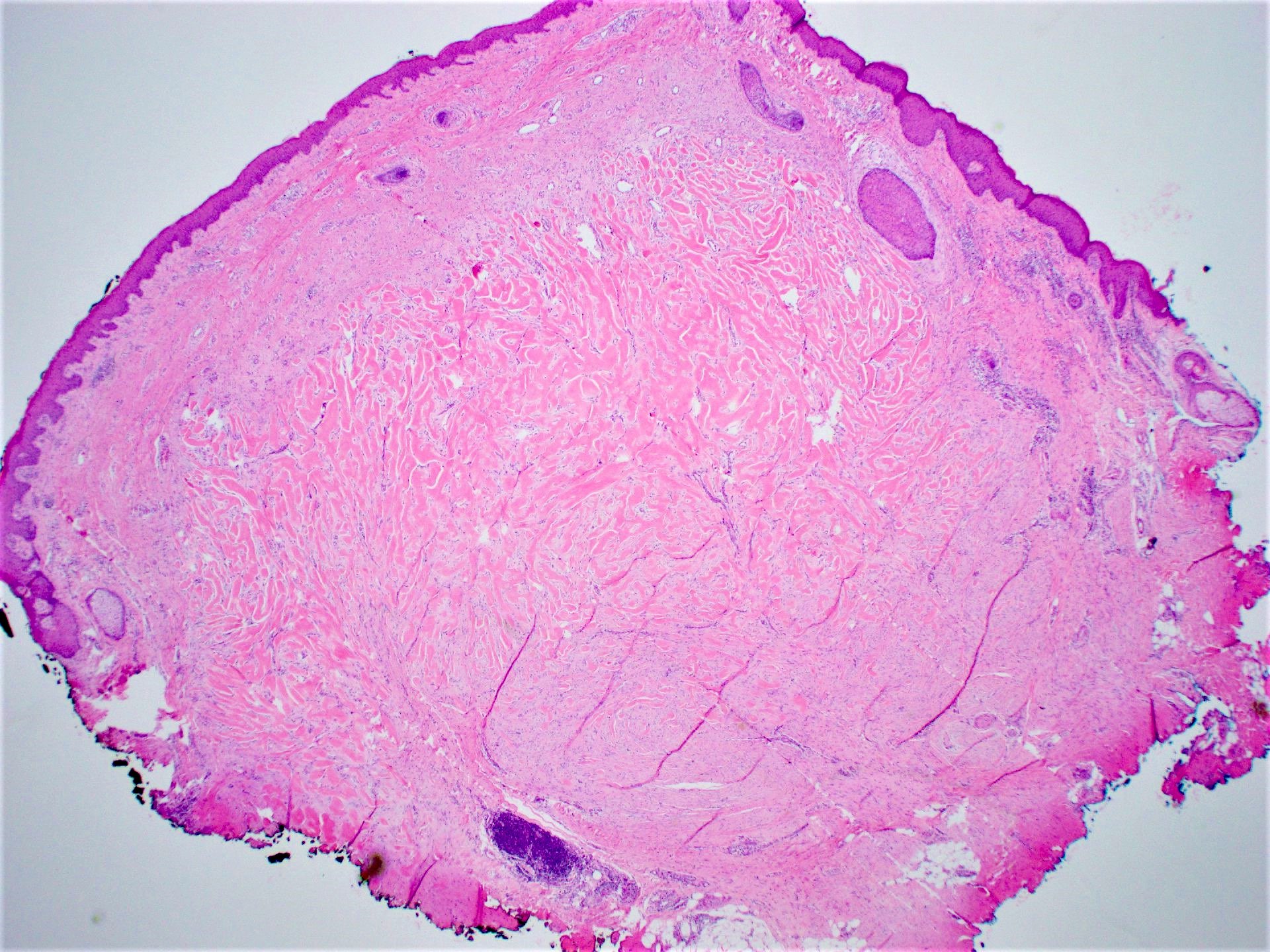
Dermal lesion
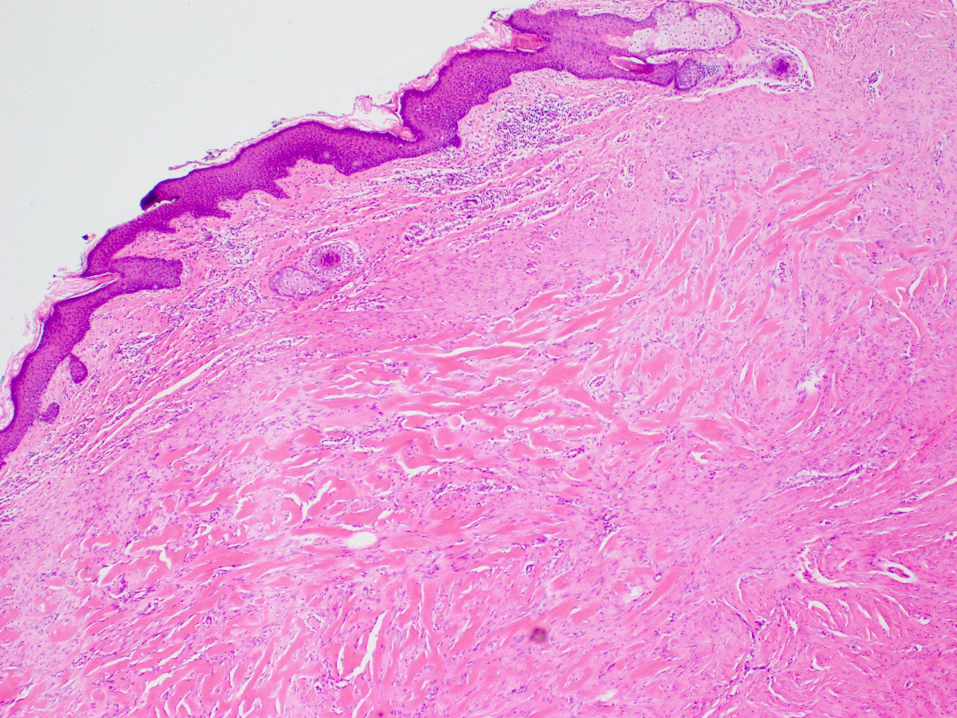
Edge of lesion
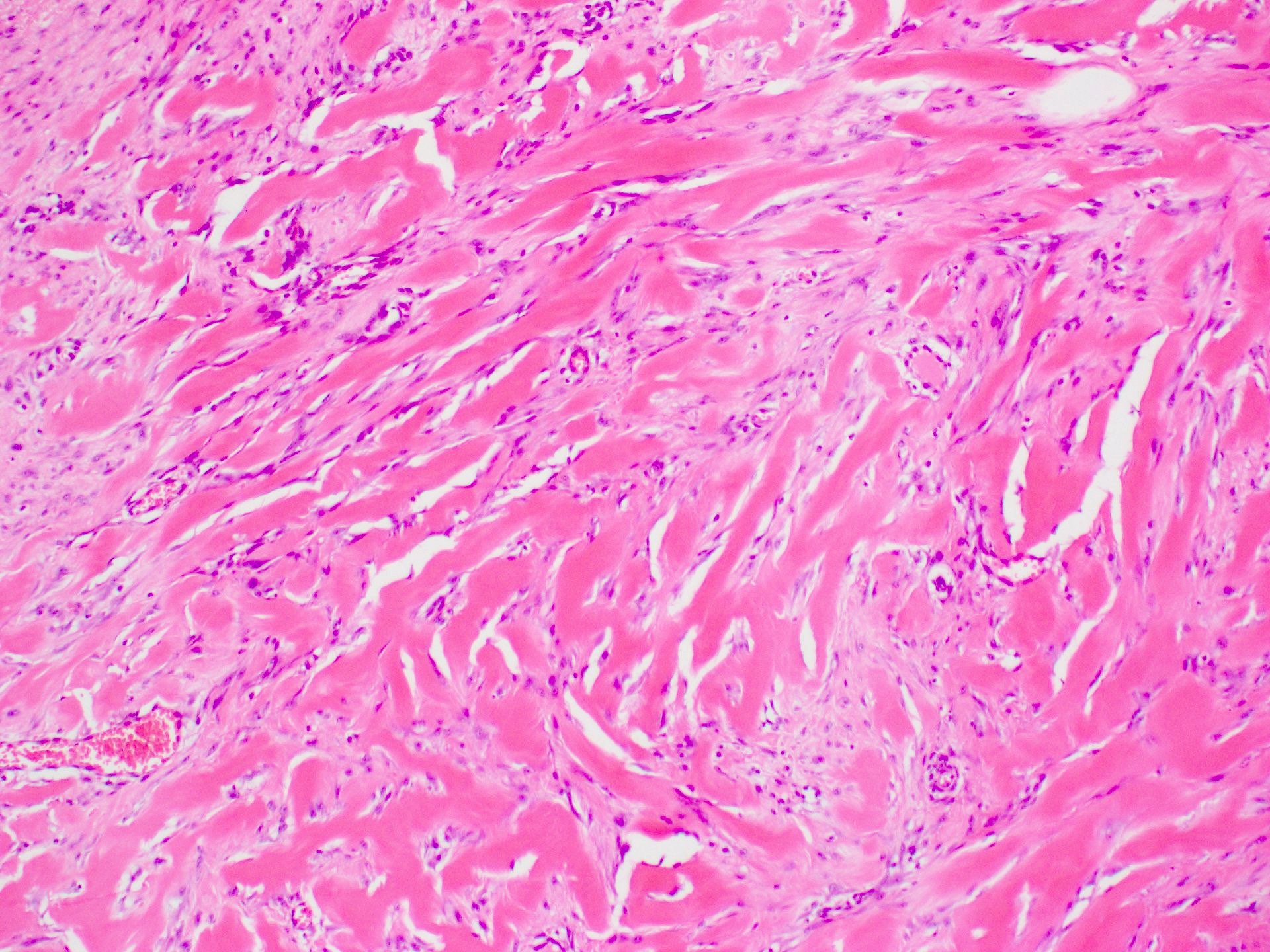
Keloidal collagen
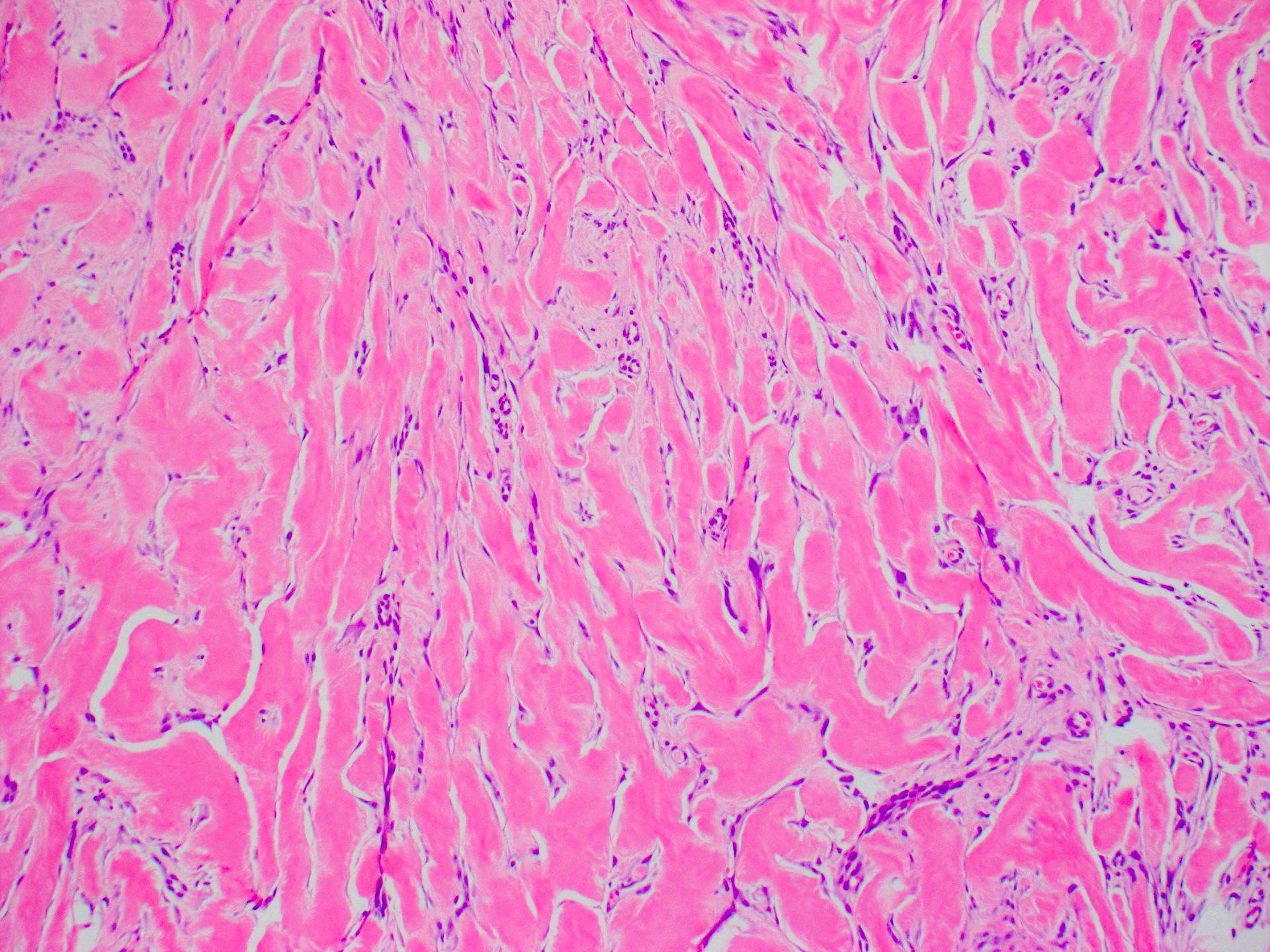
Increased fibroblasts
Keloid scar:
5 minute pathology pearls
by Dr. Jerad Gardner
Images hosted on other servers:

Lipochoristoma of internal auditory canal
Images hosted on other servers:

Section through cochlea
Case #376


Various images
Chromogranin

Synaptophysin

Cytokeratin AE1 / 3
Images hosted on other servers:

Trabeculae and ribbons

Prominent glandular pattern
Images hosted on other servers:

Neurosecretory granules
Contributed by Kelly Magliocca D.D.S., M.P.H.
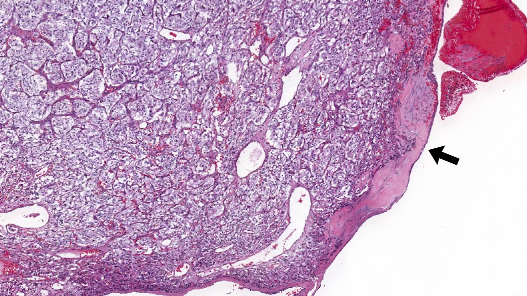
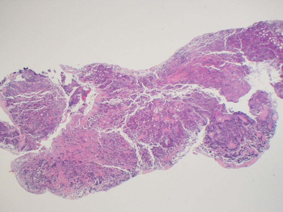
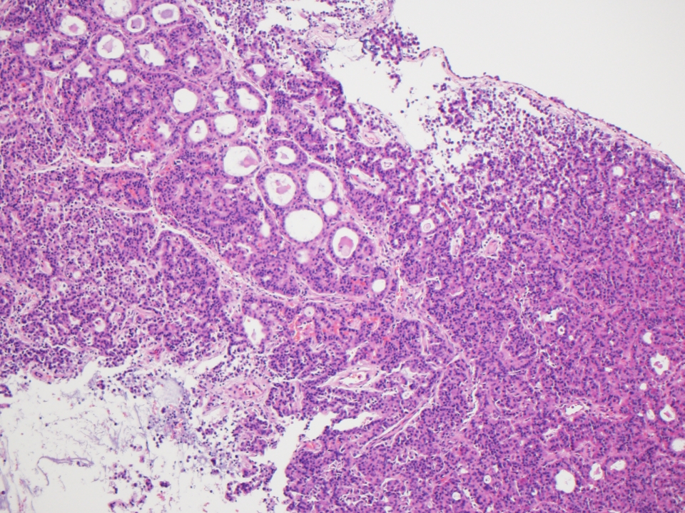
Middle ear paraganglioma, 4x
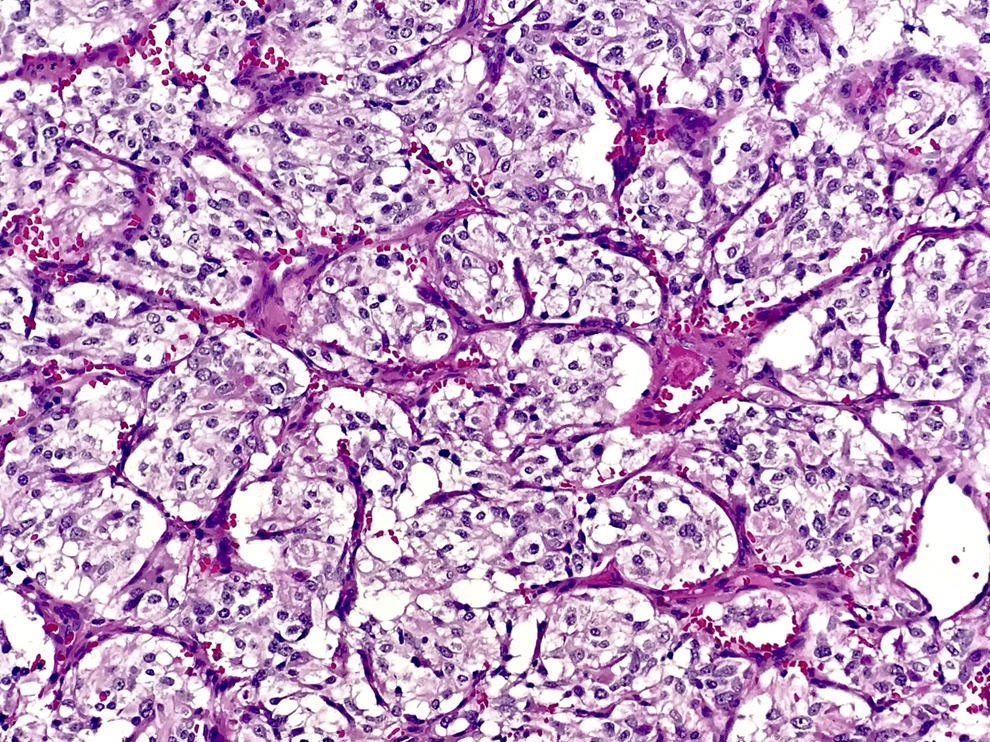
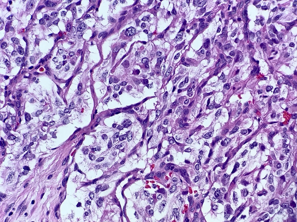
Middle ear paraganglioma, Zellballen
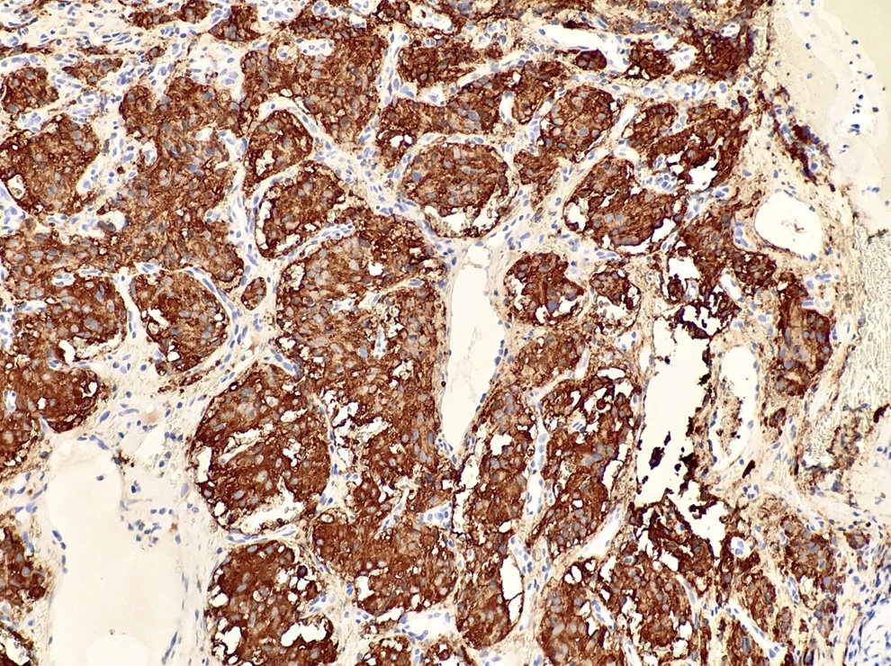
Synaptophysin positive in chief cells, 20x
S100 in sustentacular pattern, 40x
Case #349
H&E images
Contributed by Veena Maheshwar, M.D., Kiran Alam, M.D., Anshu Jain, M.D.
Various images
Contributed by Semir Vranic, M.D., Ph.D.
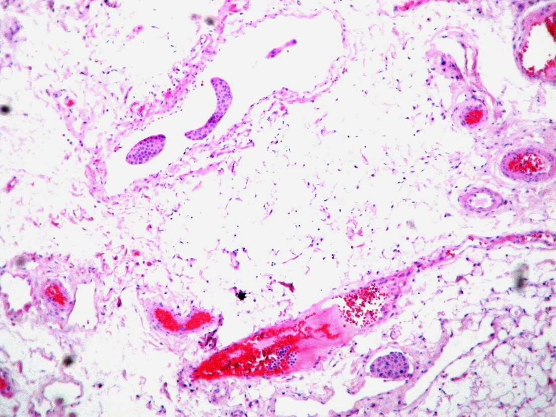
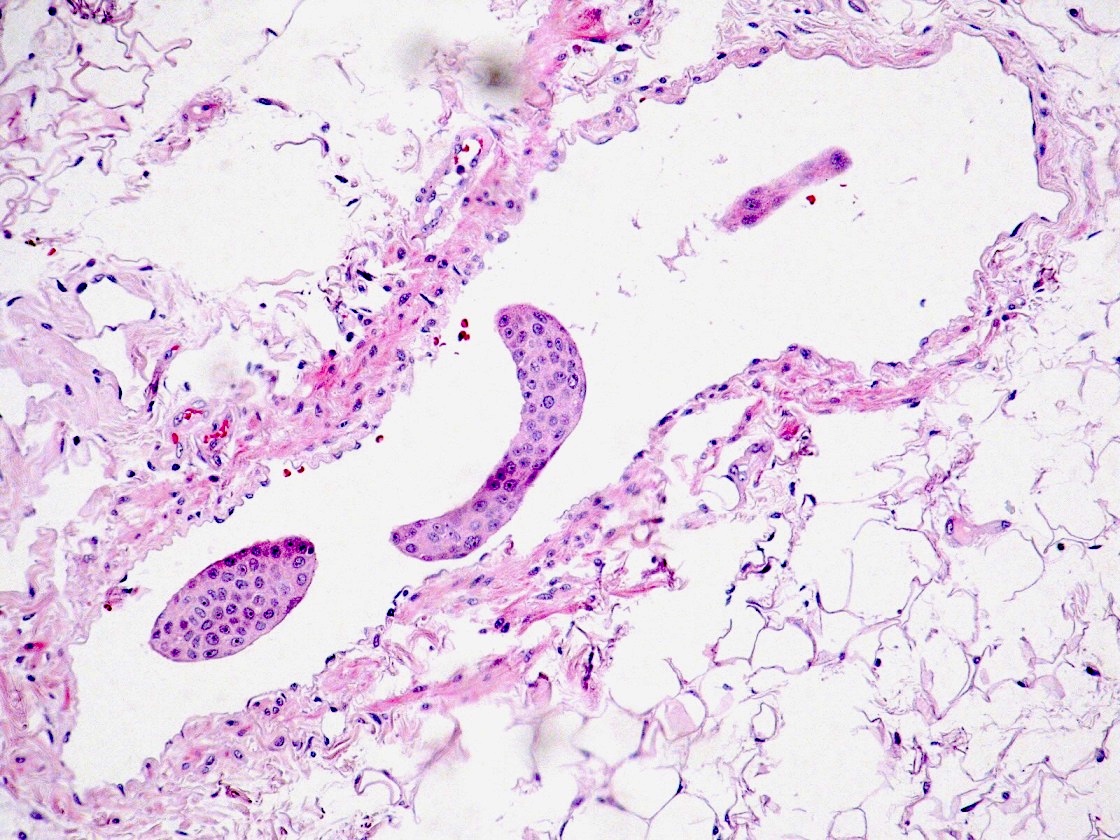
Angiolymphatic invasion
Cardesa: 2016
Franchi: 2020
Gnepp: 2021
IARC: 2024
Stelow: 2020
Thompson: 2022
Wenig: 2017
Wenig: 2024
Find related Pathology books: head & neck/endocrine
