

Images hosted on other servers:

Forms of cardiac rejection

Cardiac allograft dysfunction workup
Images hosted on other servers:

T2 cardiac MRI

Hyperenhancement on cardiac MRI

Doppler echocardiography

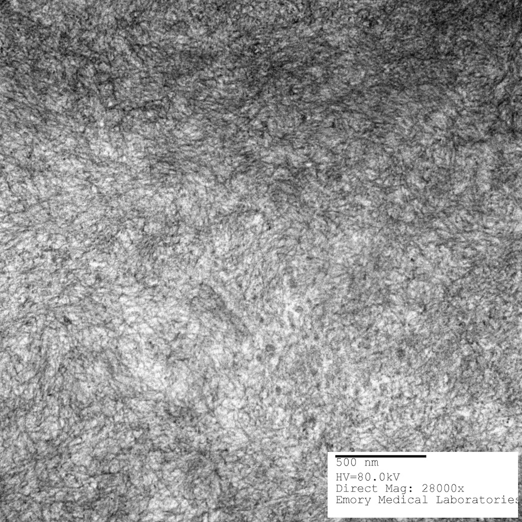
Doppler strain
Contributed by Carolyn Glass, M.D., Ph.D.
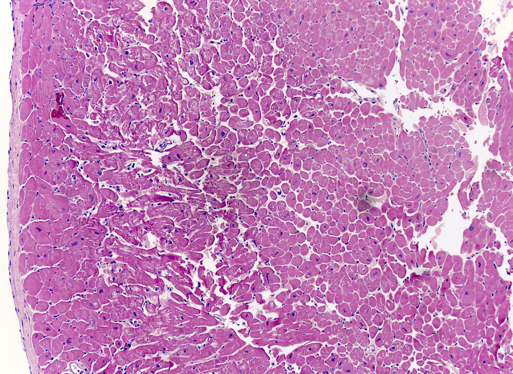
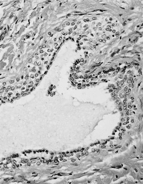
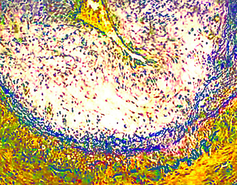
ISHLT 0R
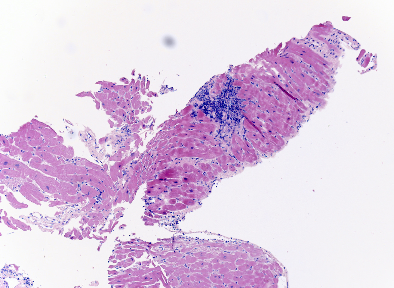
ISHLT 1R
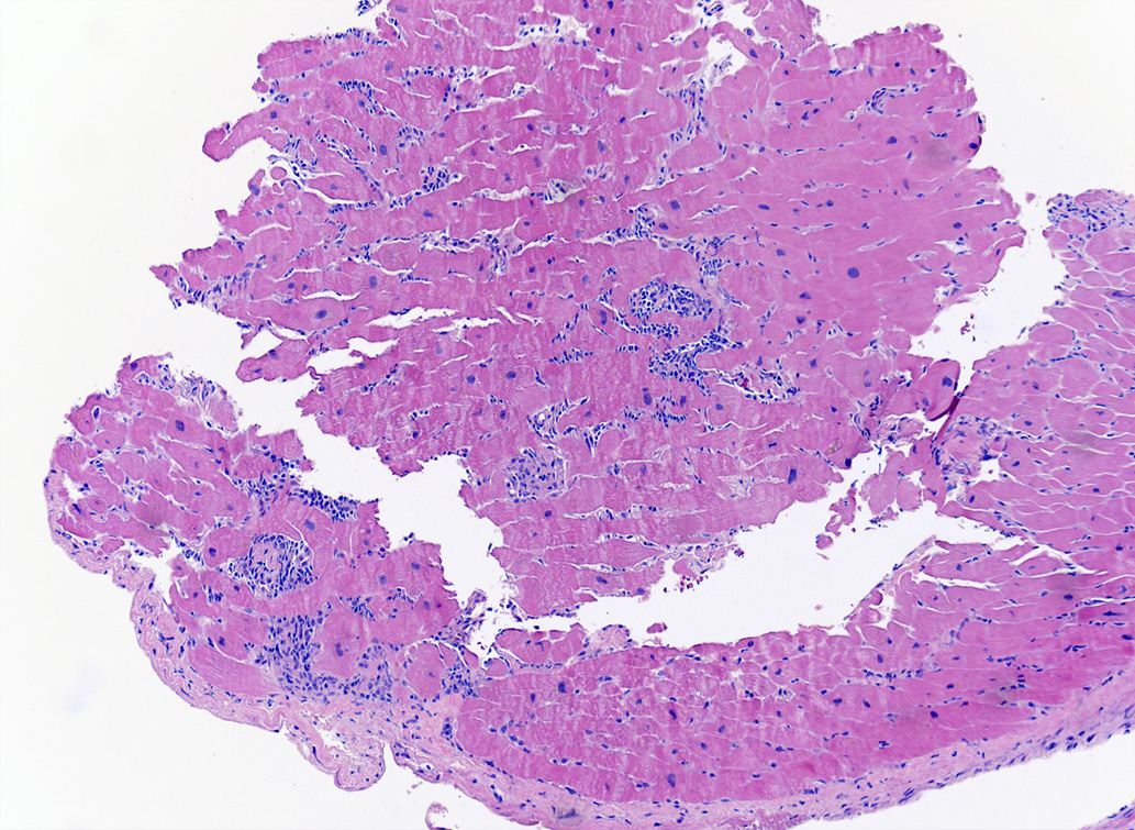
ISHLT 2R
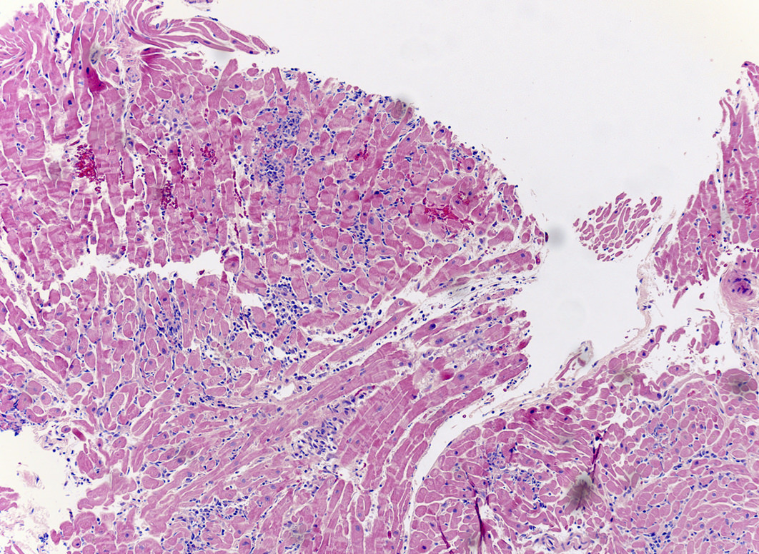
ISHLT 3R
Images hosted on other servers:

Biopsy proved ATTR cardiac amyloidosis with positive uptake
Contributed by Jaishree Jagirdar, M.D.
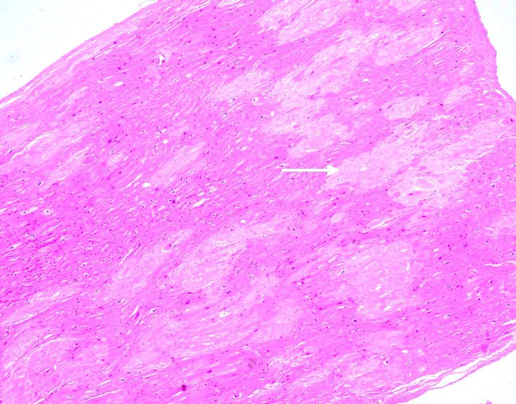
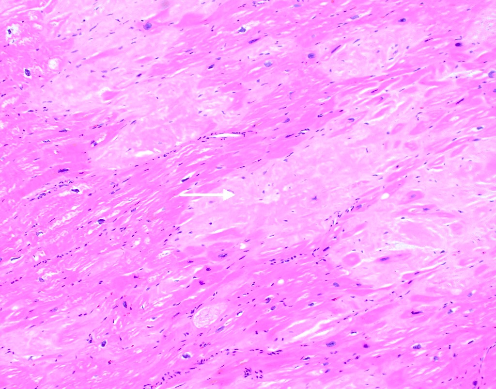
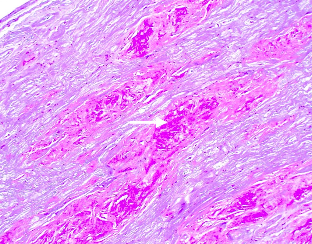
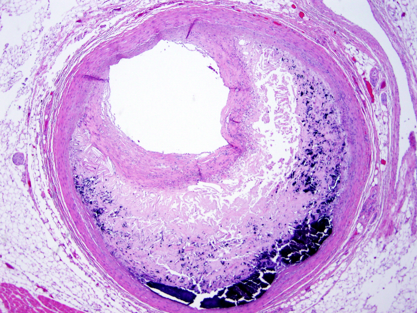
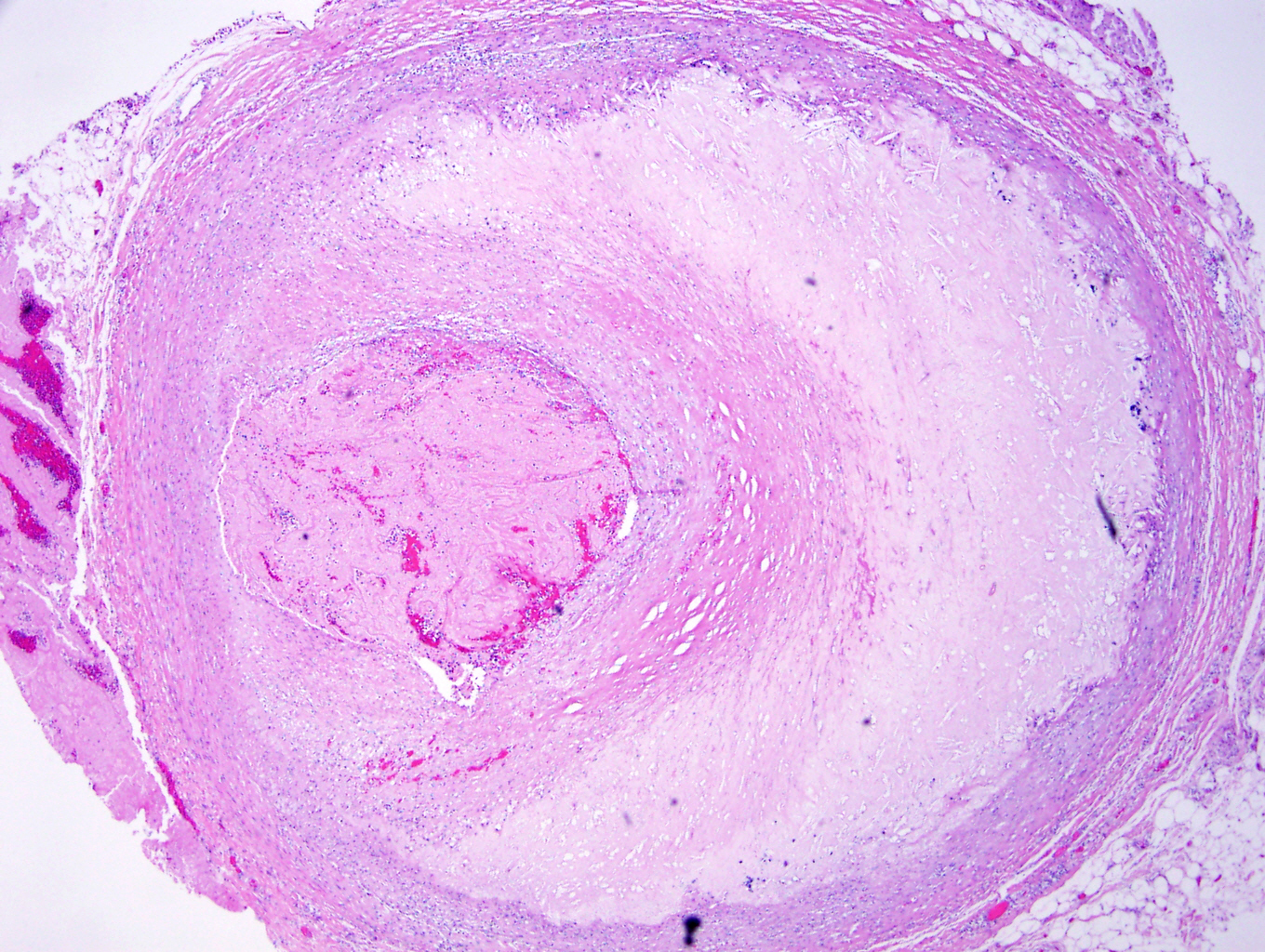
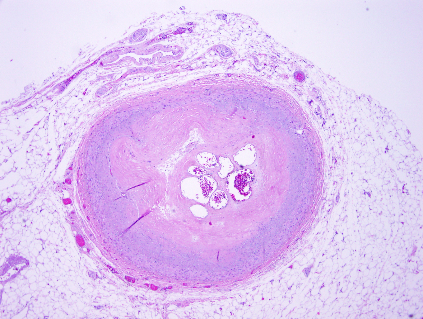
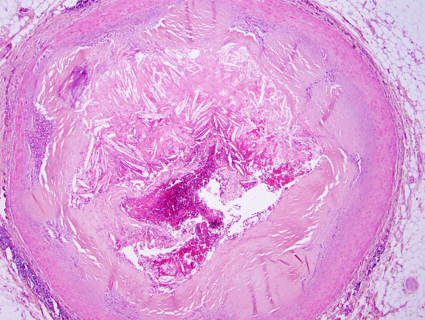
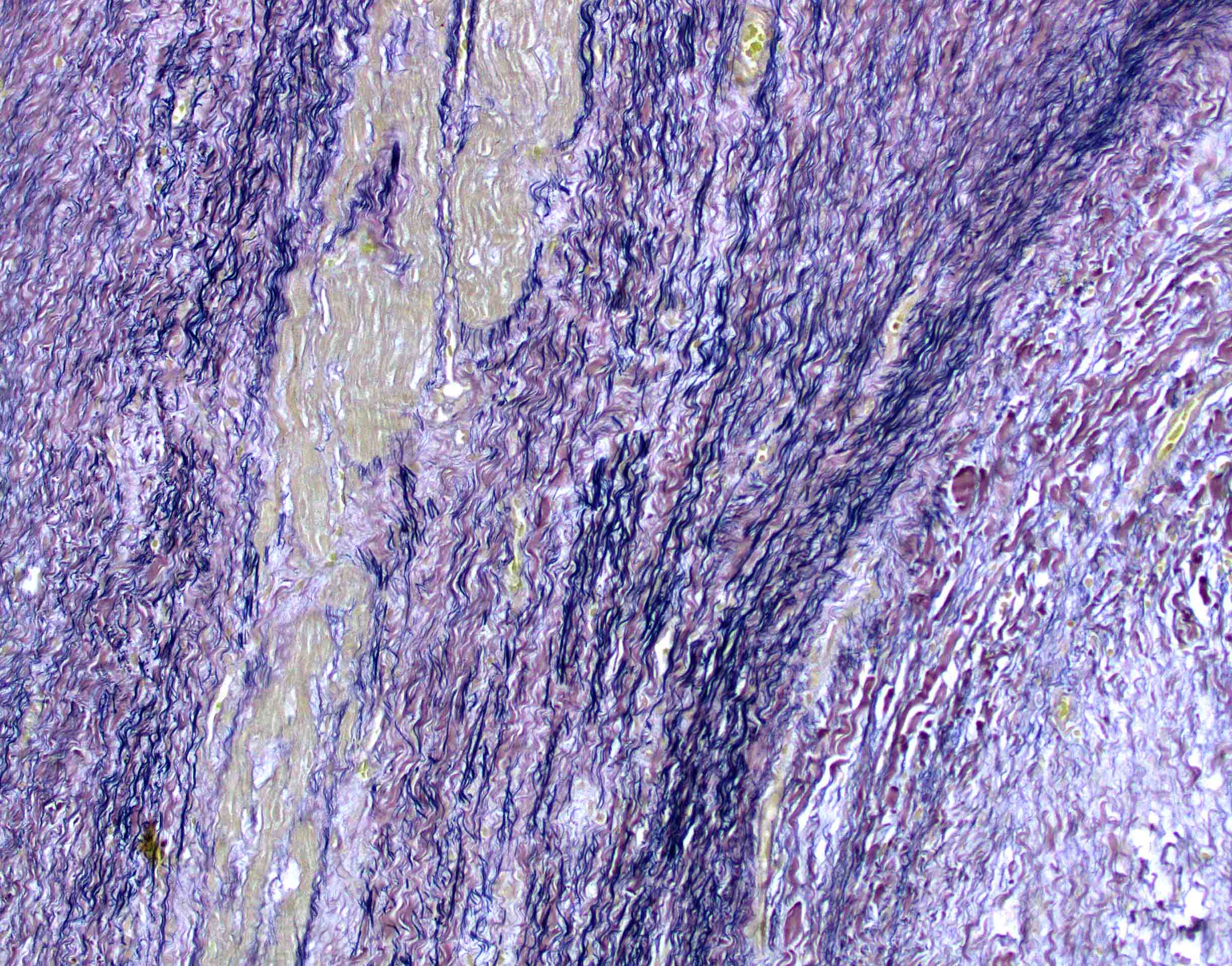
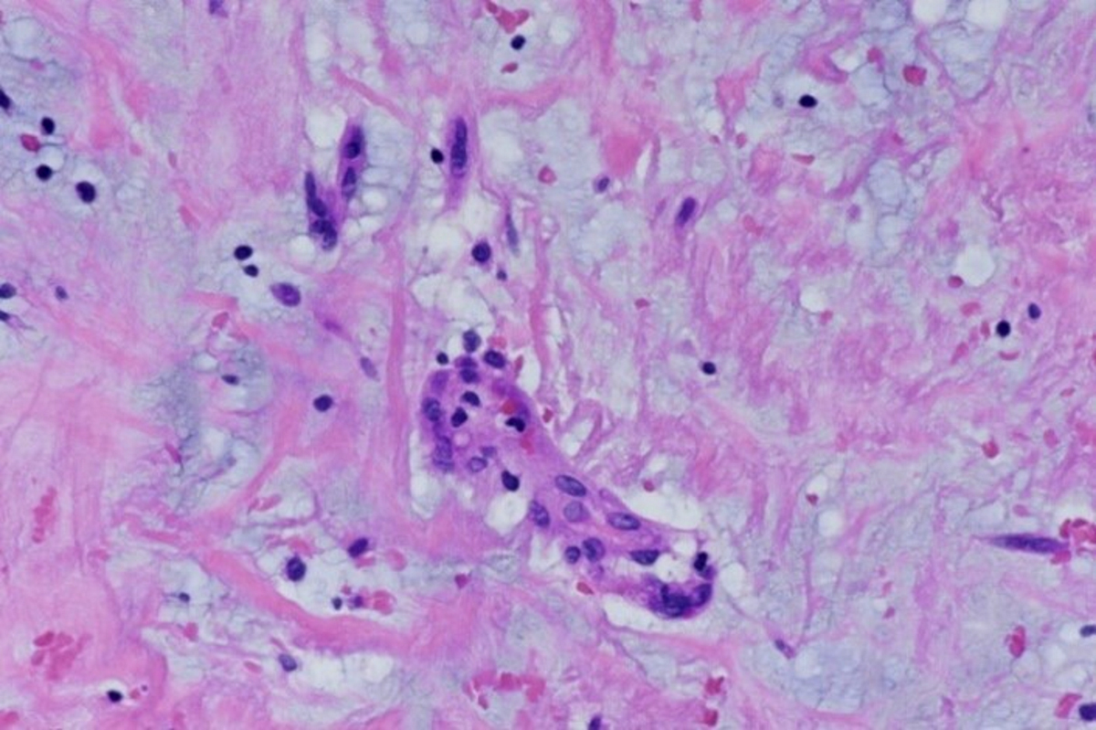
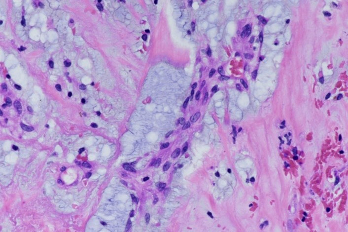
Amyloid deposition in myocardium
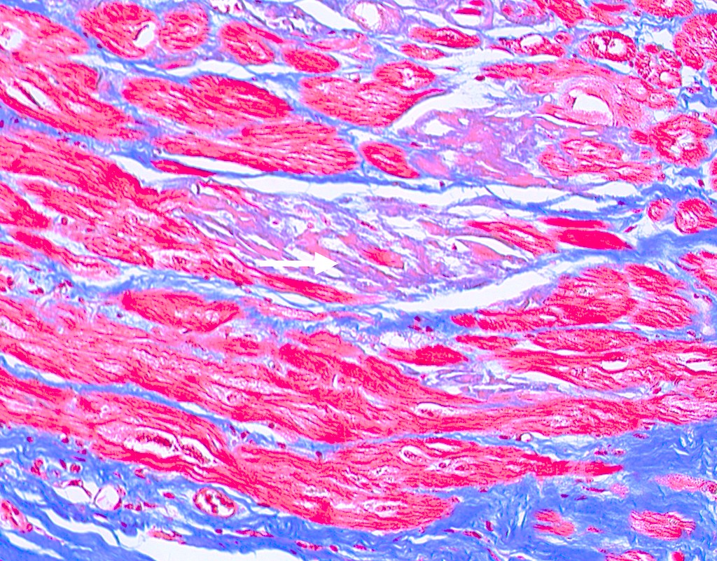
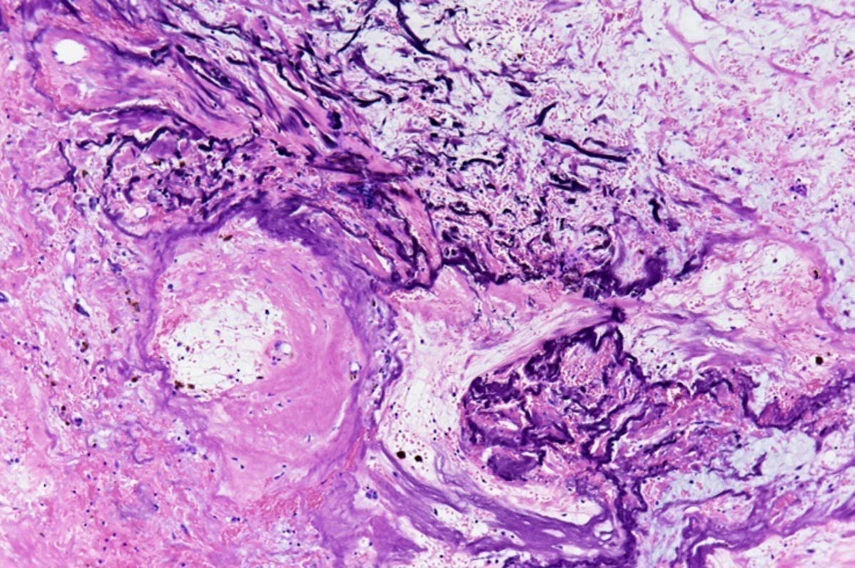
Trichrome stain
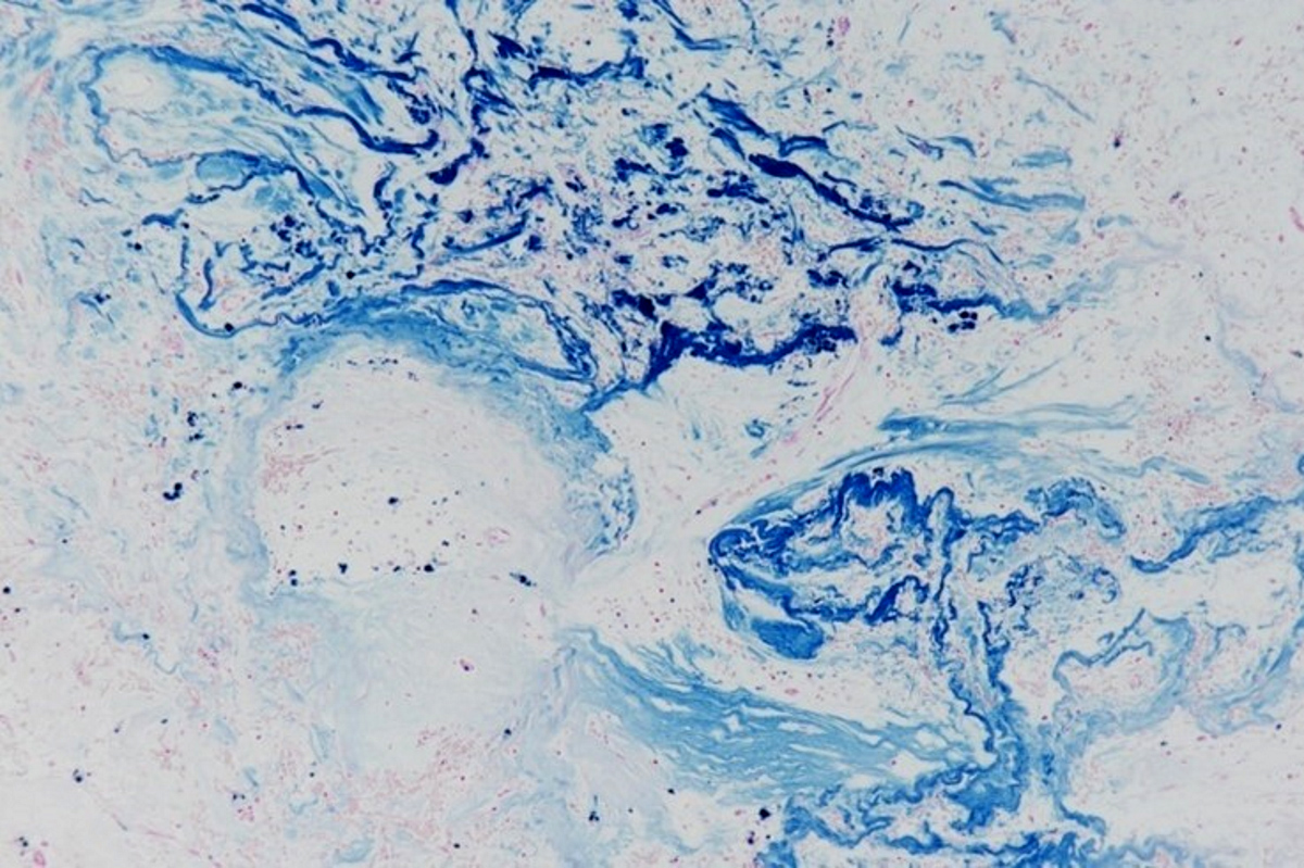
Crystal violet stain
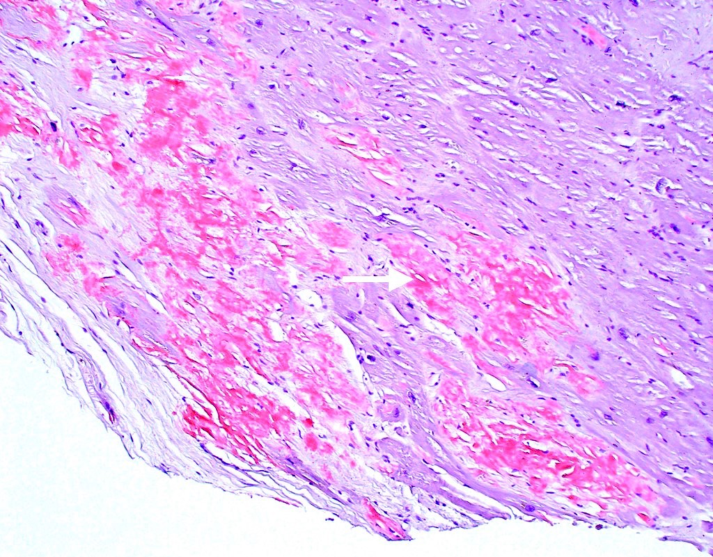
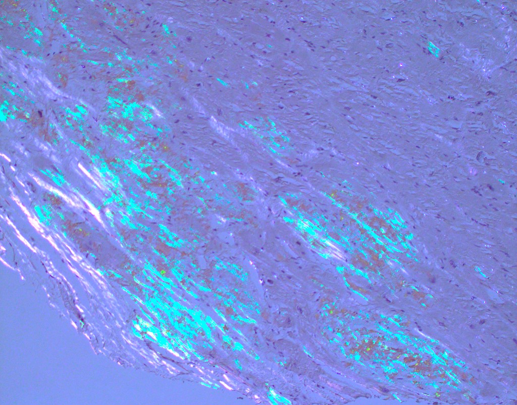
Congo red stain
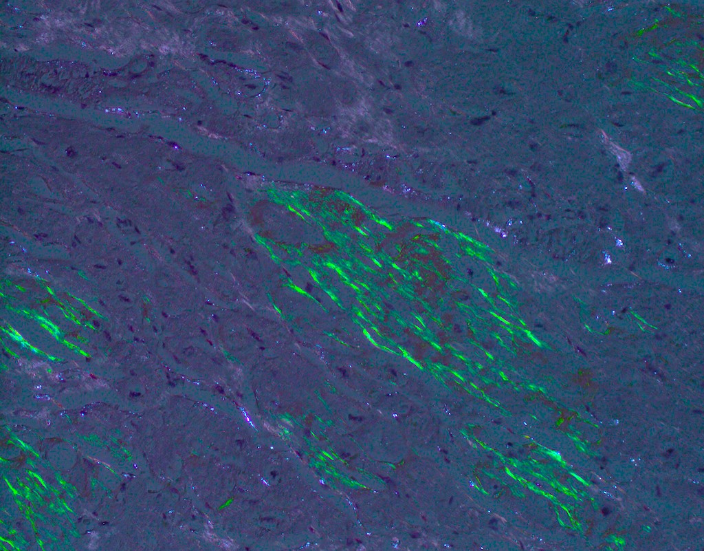
Congo red stain
Contributed by Bradley Farris, M.D.
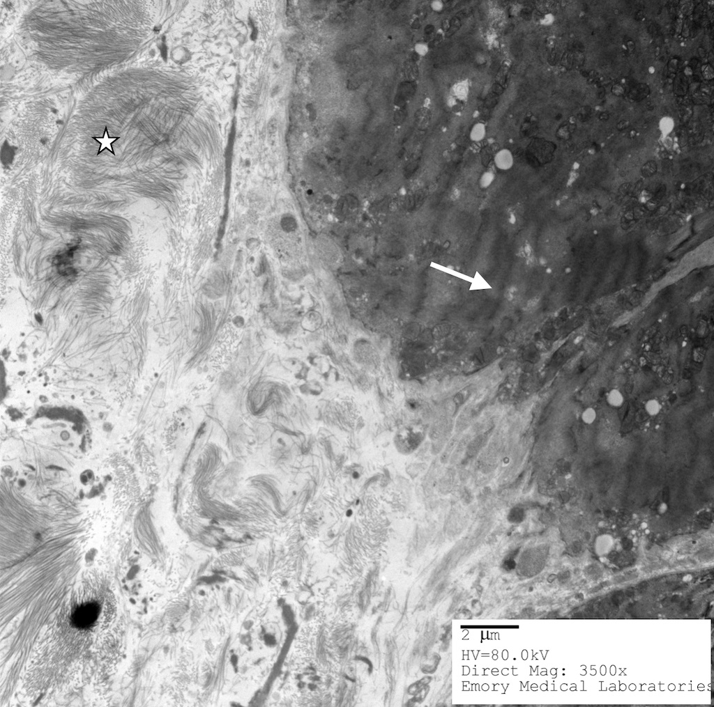
Amyloid deposition with beta pleated sheets
Images hosted on other servers:

Classification of coronary anomalies
Images hosted on other servers:

MRI during cardiac systole and end diastole

Coronary arteries with 3D reconstructions
Images hosted on other servers:

Chest radiograph


Ultrasound

Computed tomography

Angiography

50 mm round mass with calcification

Coronary angiogram

After CABG

Distal left main aneurysm
Images hosted on other servers:

Descending artery aneurysm
Contributed by Carla Dominguez Gonzalez, B.S.
Autopsy findings
Images hosted on other servers:

Right coronary artery
Contributed by Carla Dominguez Gonzalez, B.S.
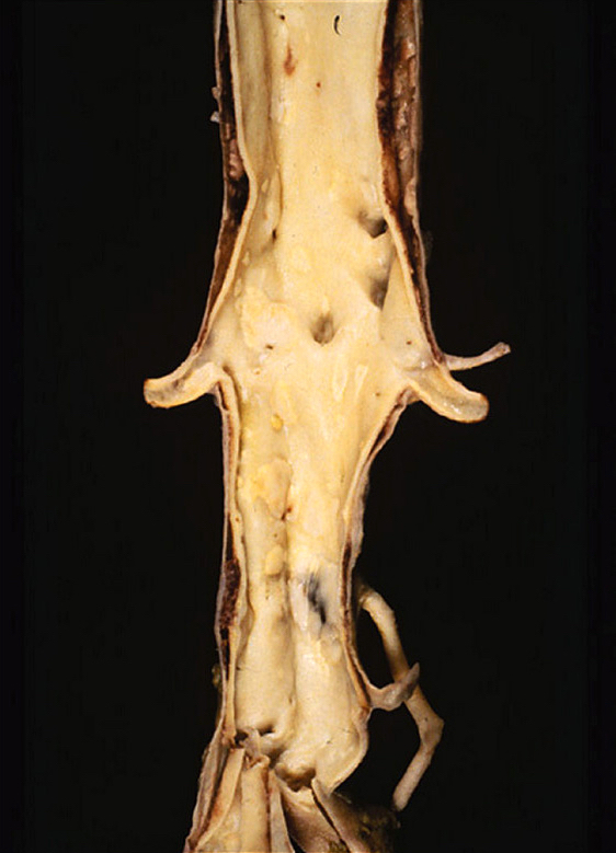
Abdominal aortic aneurysm with rupture
Thoracic aortic aneurysm
Severe medial atrophy
Elastic lamellar disruption
Adventitia inflammation and lipid deposition
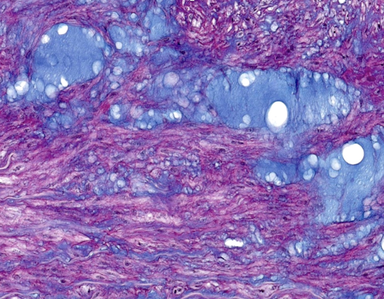
Alcian blue staining - MEMA
Images hosted on other servers:

Increase of transverse cardiac diameter

Enlargement of right ventricle and atrium

Cardiac echocardiogram
Images hosted on other servers:

Severe dilation of RV

RV dilatation
Images hosted on other servers:

Early fibrosis and adipocytes infiltration
Images hosted on other servers:

Coronary angiogram

Intravascular ultrasound

Multidetector computed tomography
Images hosted on other servers:

Coronary artery atherosclerosis

Aorta involved by
atherosclerosis
Contributed by Robert F. Corliss, M.D. and Jefree J. Schulte, M.D.
Coronary artery plaque
Coronary artery thrombus
Coronary artery recanalized thrombus
Coronary plaque with early thrombus
Lipid rich plaque
Foam cells
Cholesterol clefts
Contributed by Stephen P. Sanders, M.D.
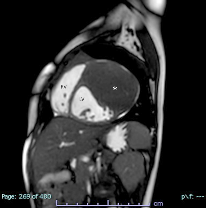
MRI (cine SSFP)
MRI (LGE technique)
Contributed by Chrystalle Katte Carreon, M.D. and AFIP
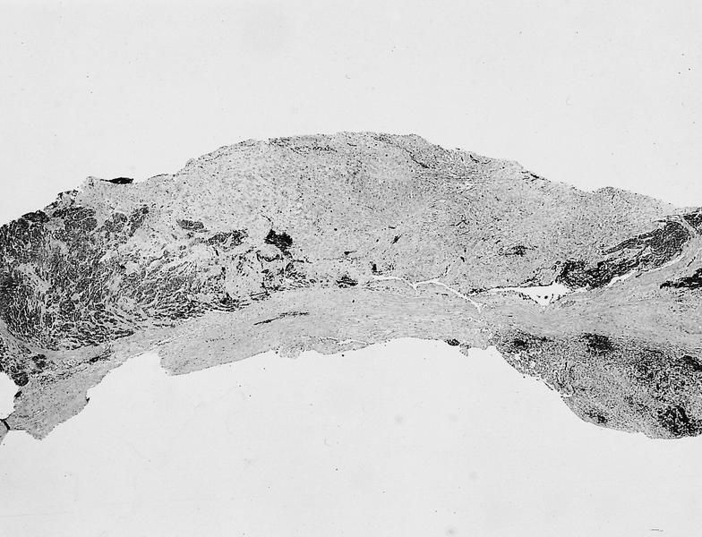
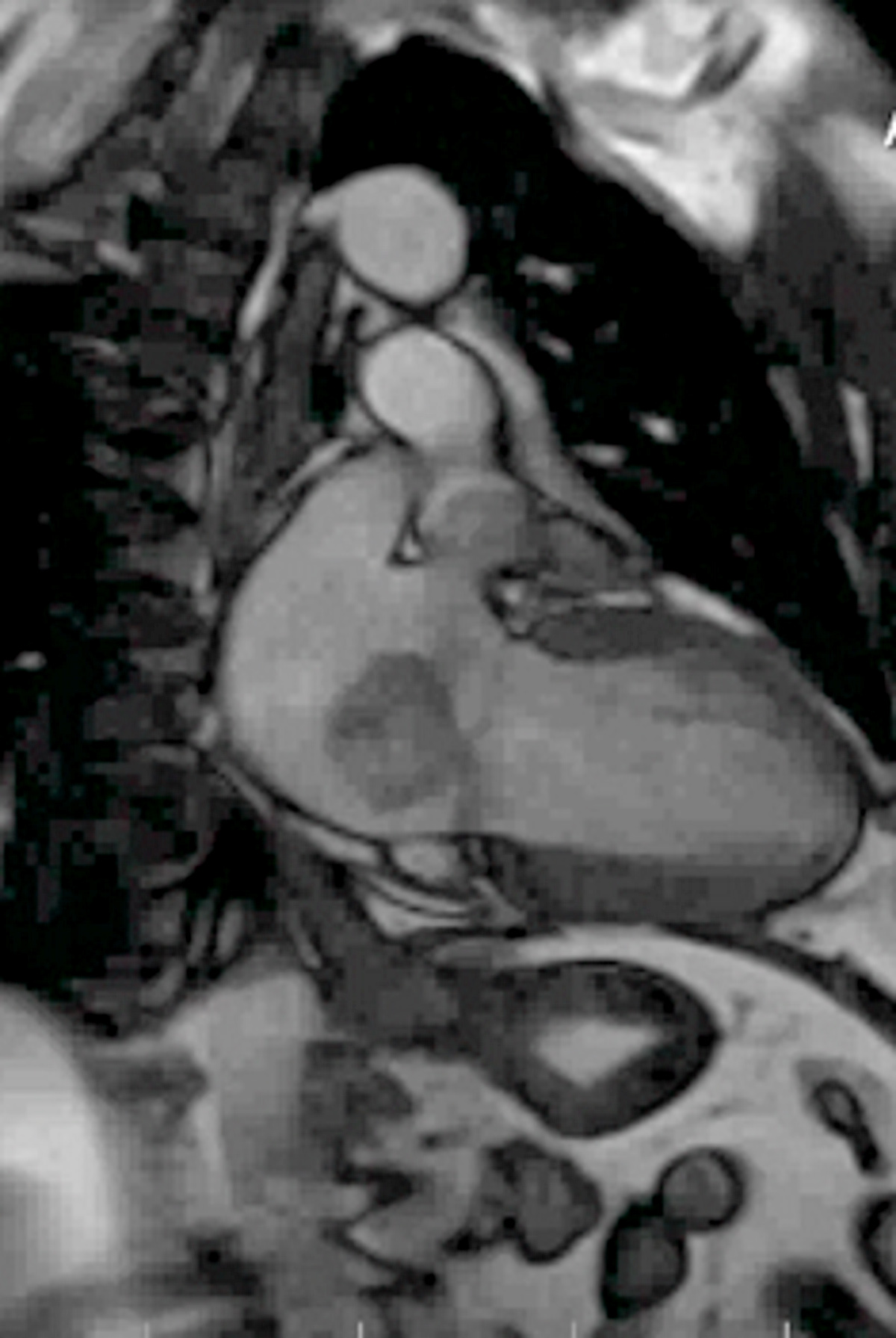
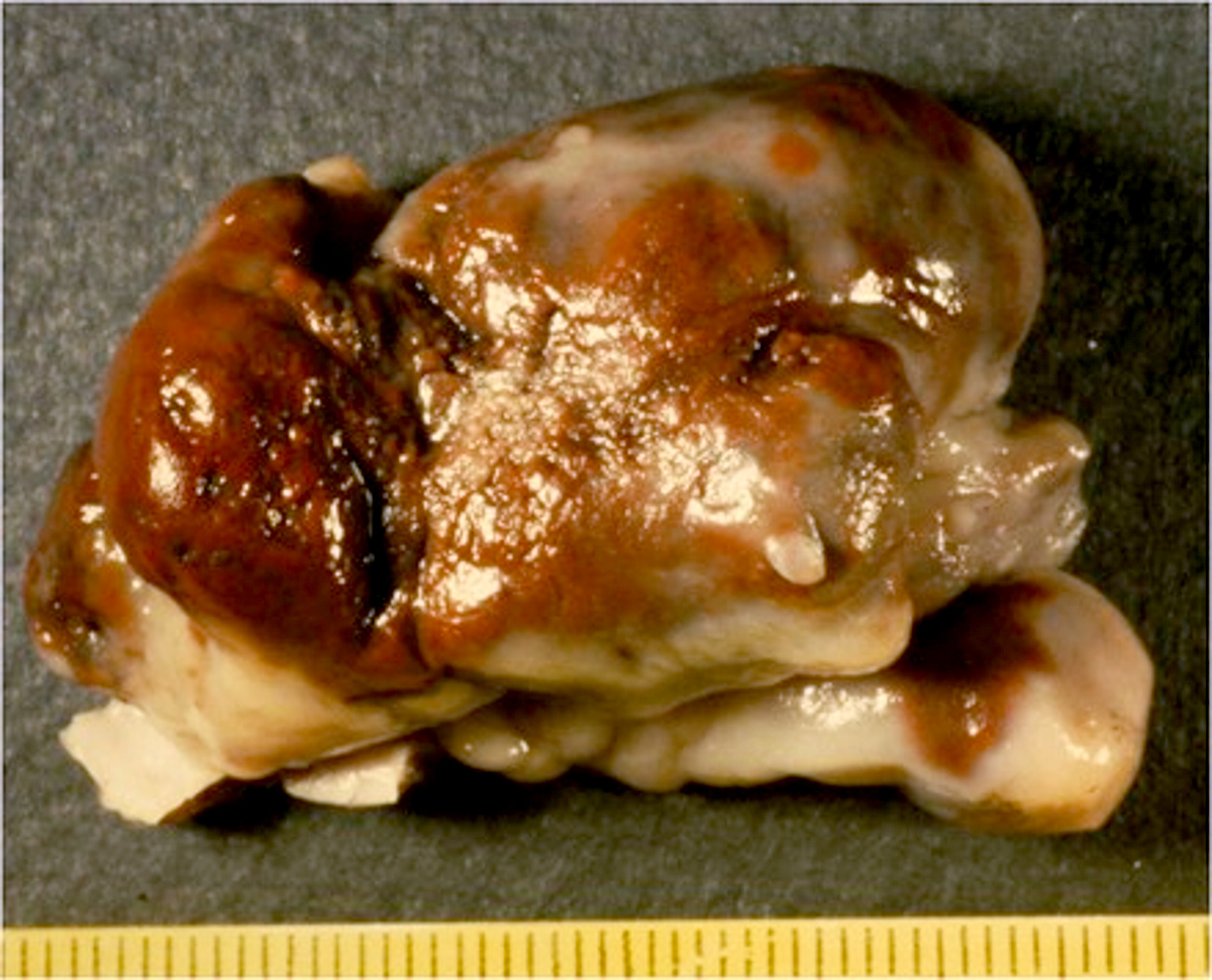
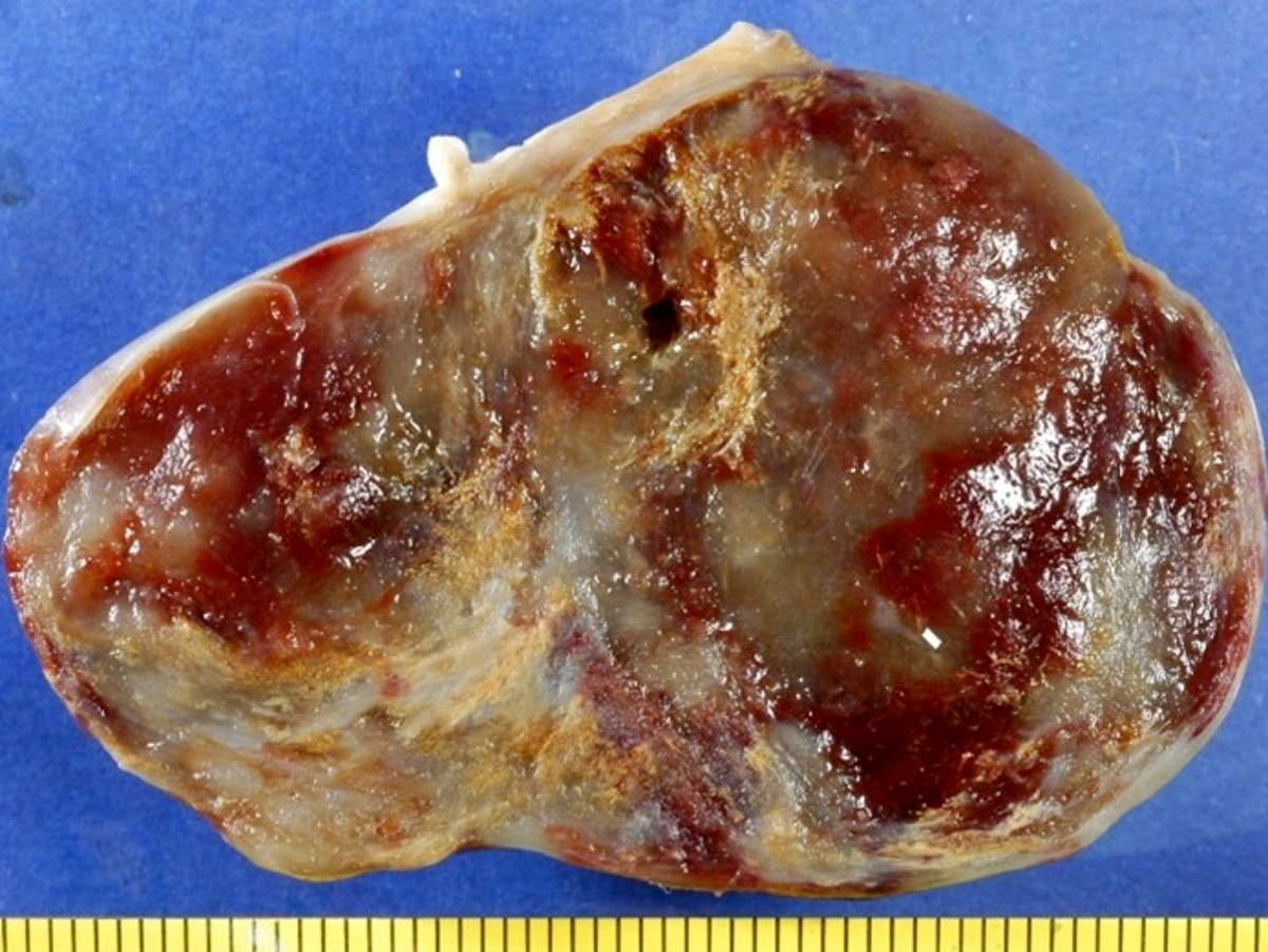
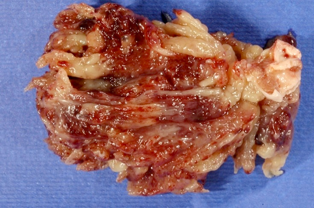
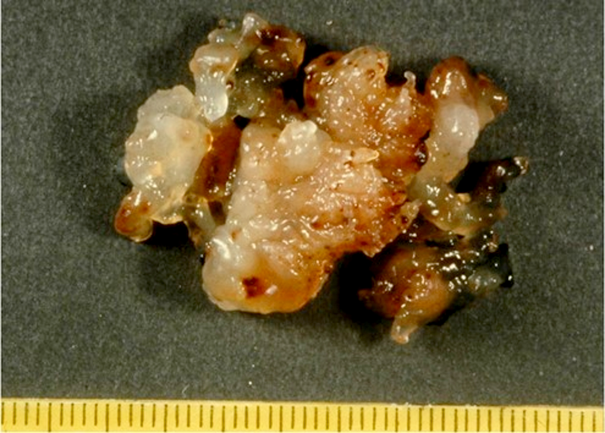
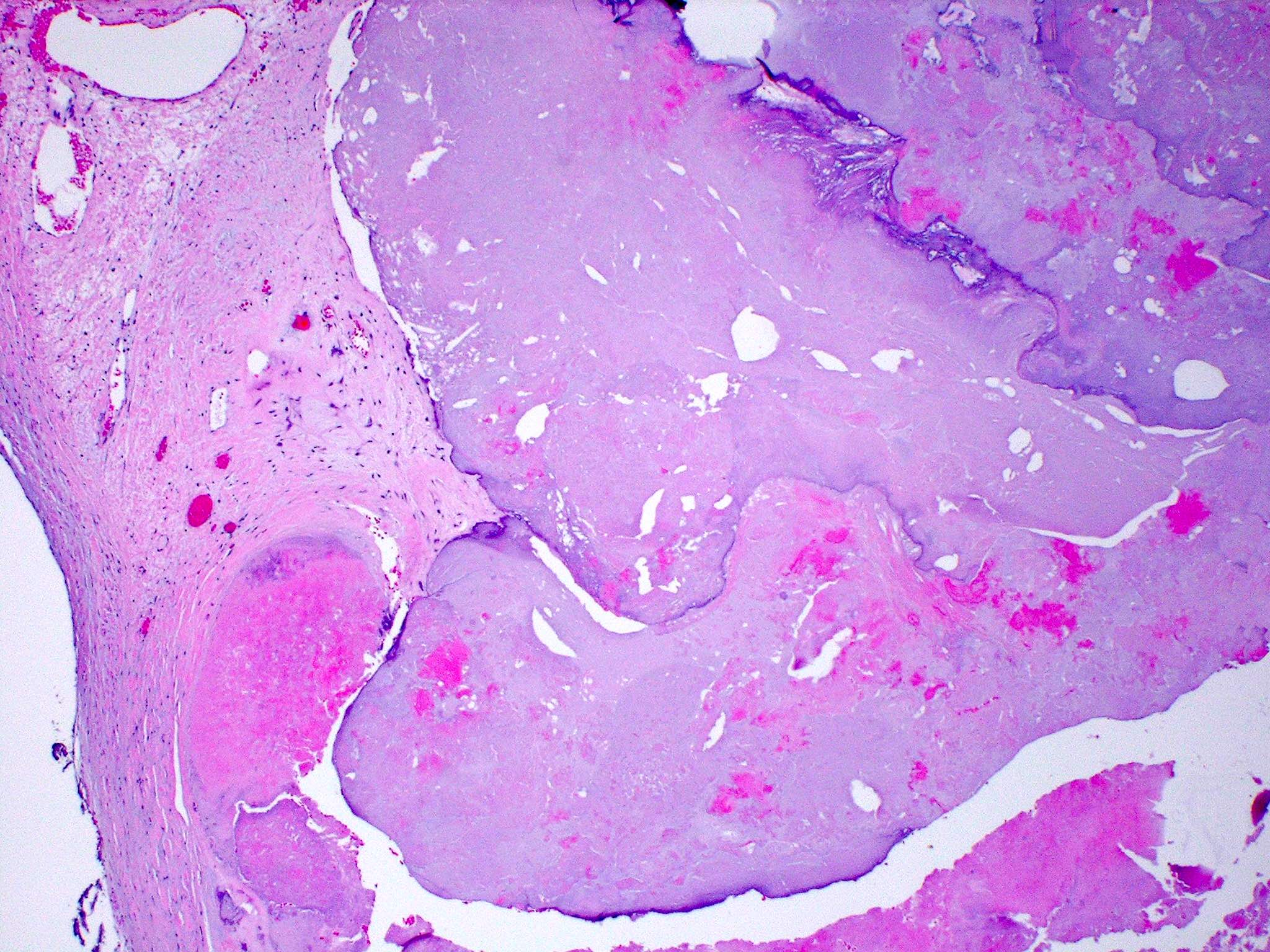
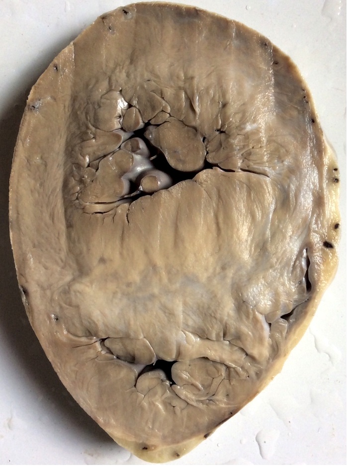
Macroscopic features
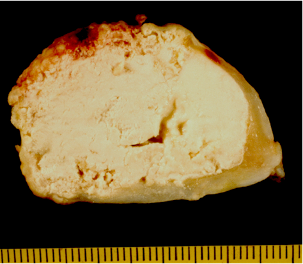
Cut surface features
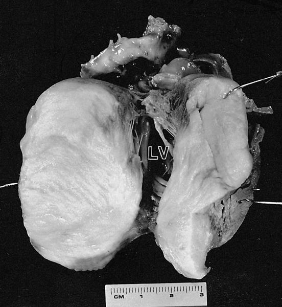
Large septal mass
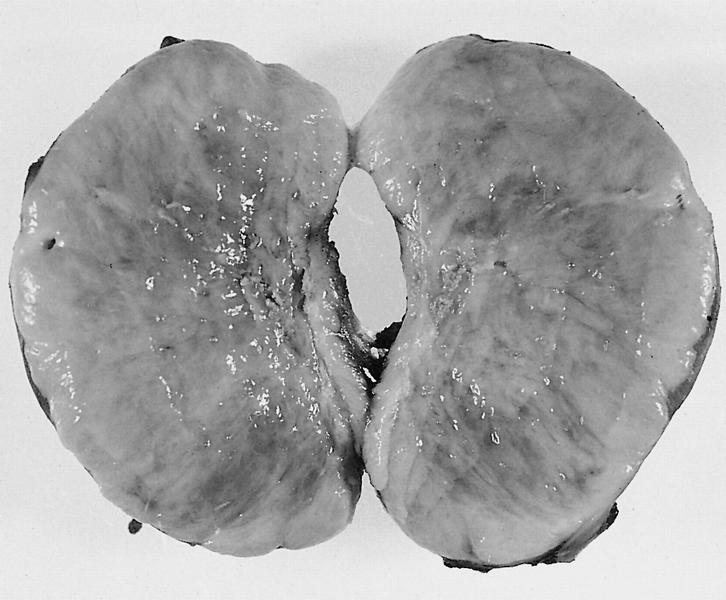
Homogeneous mass
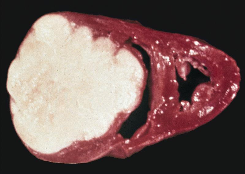
Large circumscribed mass
Contributed by Chrystalle Katte Carreon, M.D.
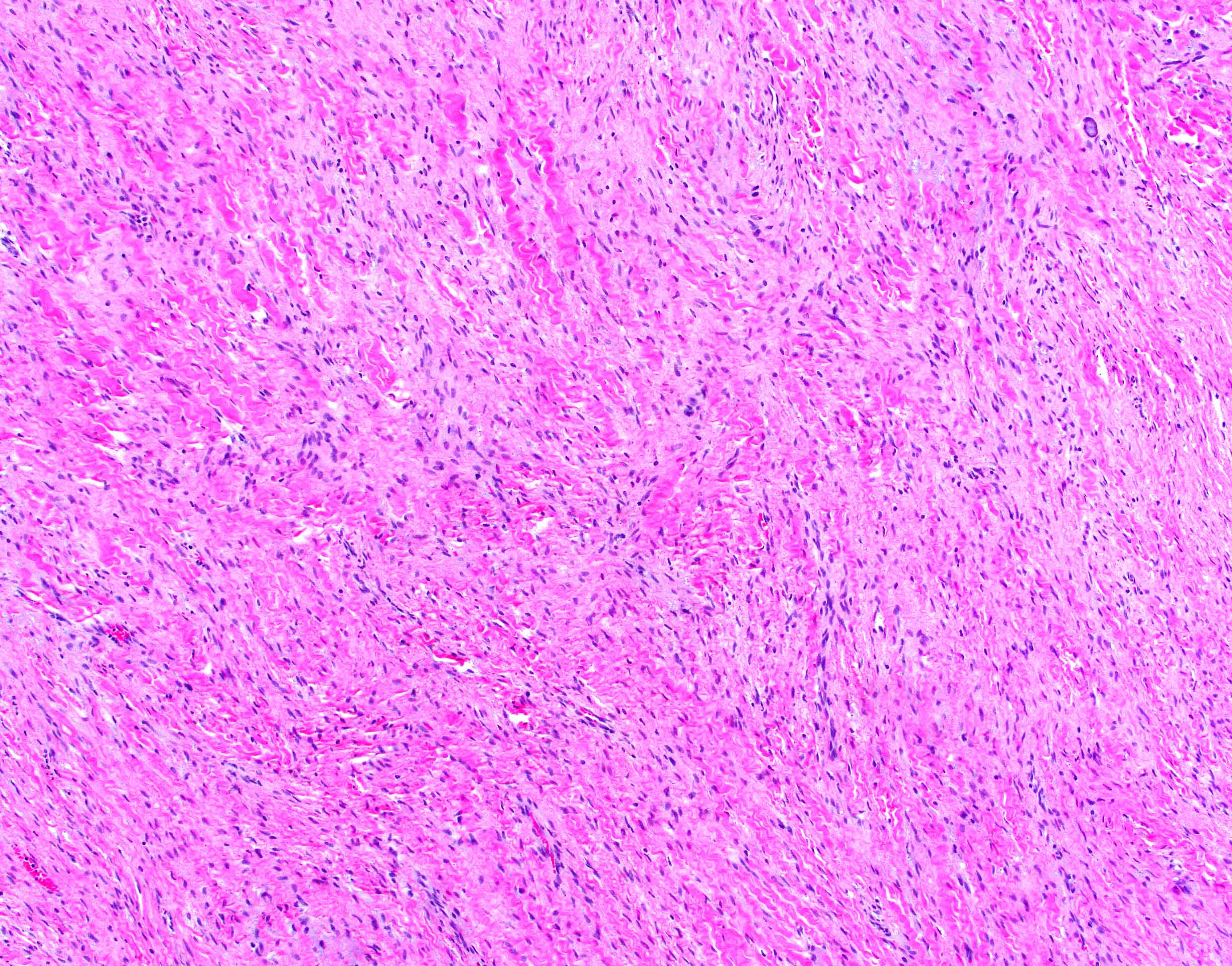
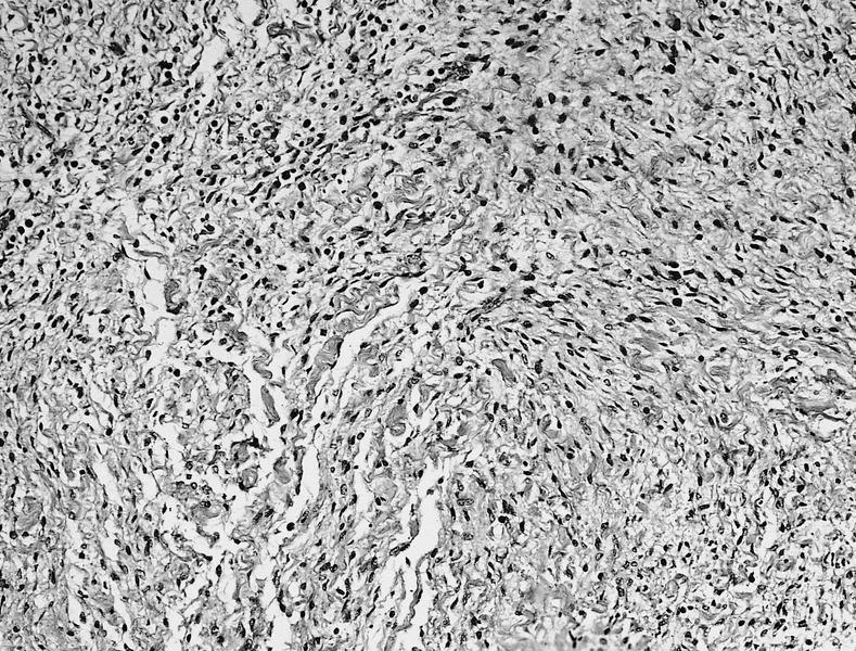
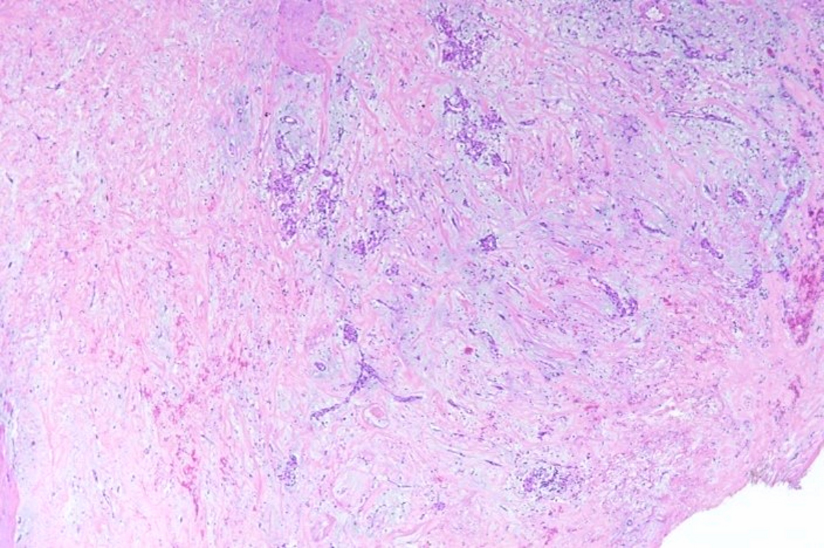
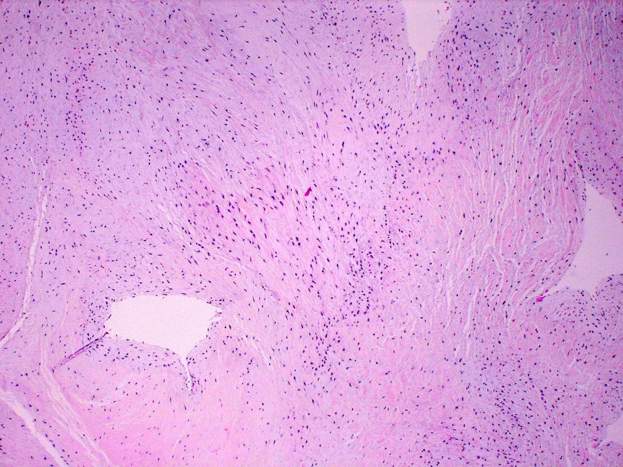
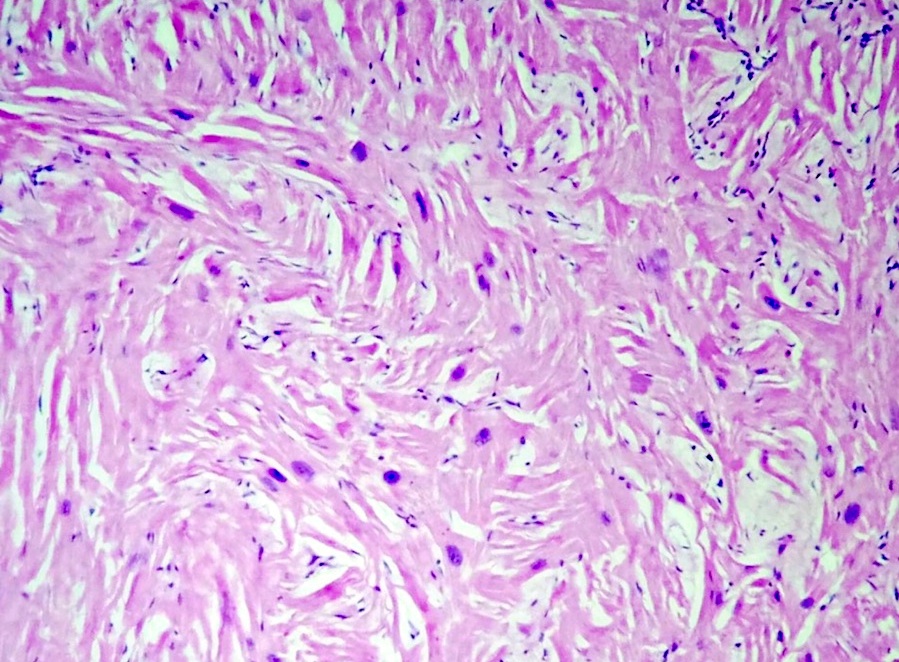
Abundant collagen
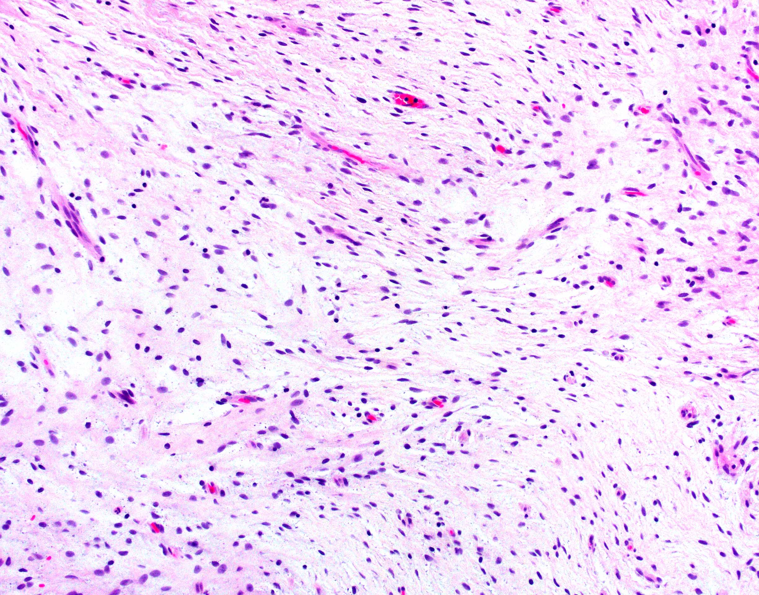
Immature appearance
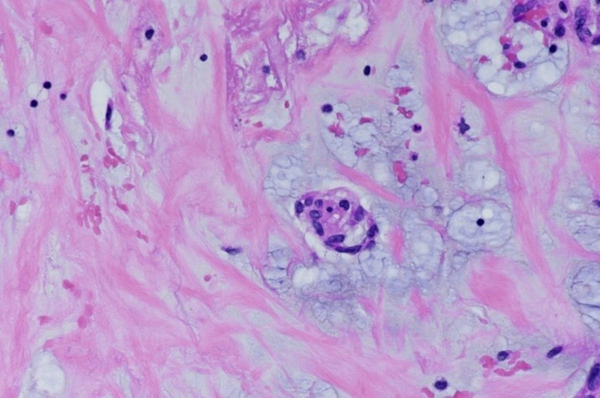
Entrapped myocardium

Dystrophic calcifications
Lack of tumor capsule
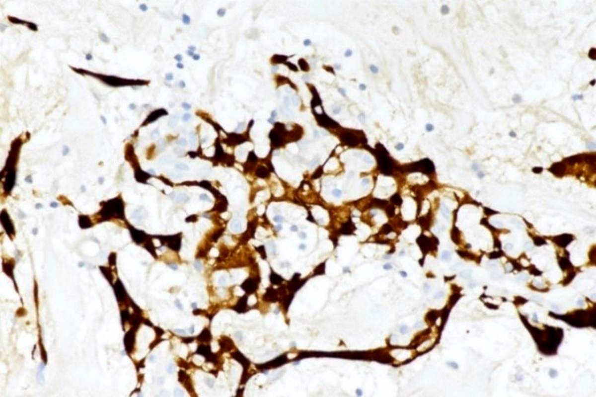
Entrapped myocardium (desmin)
Abundant elastic fibers
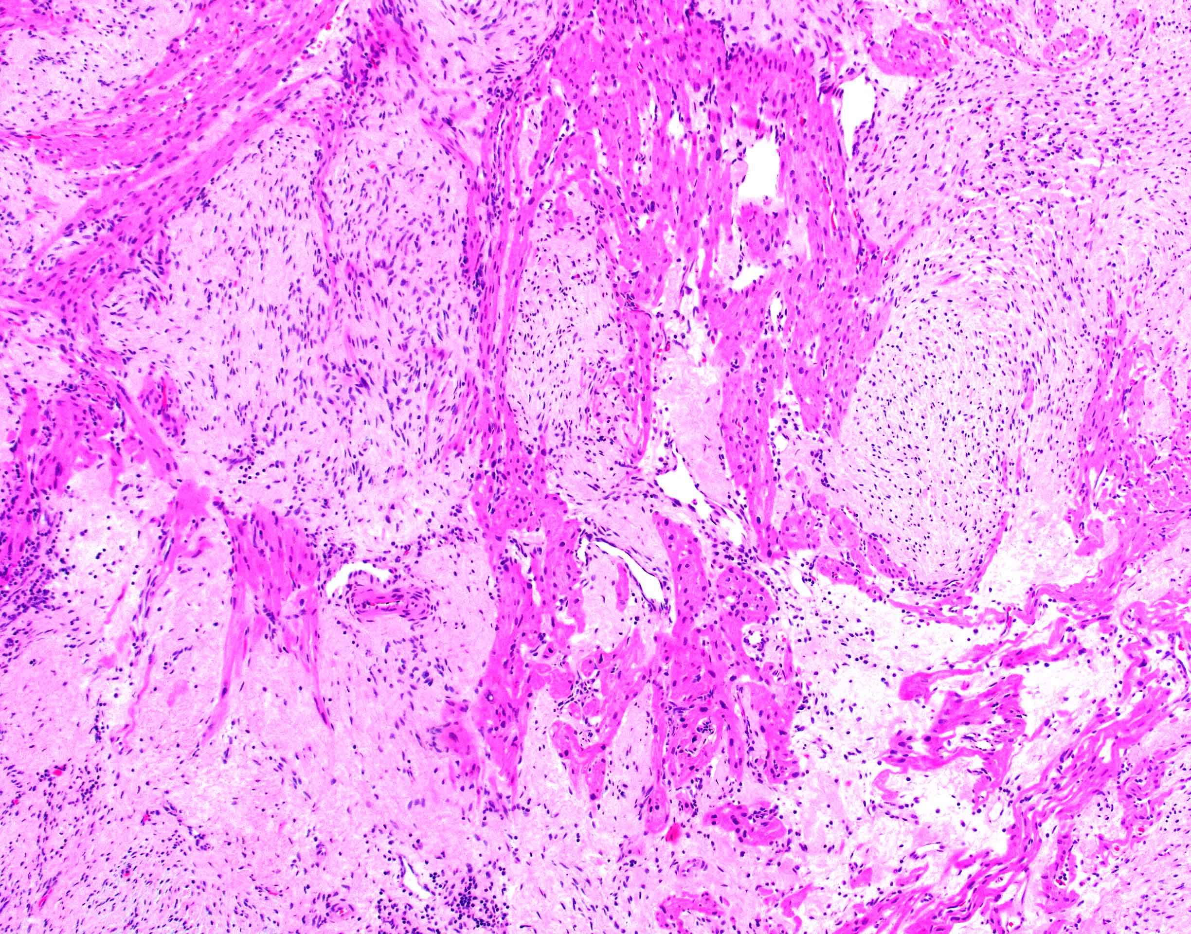
AFIP images
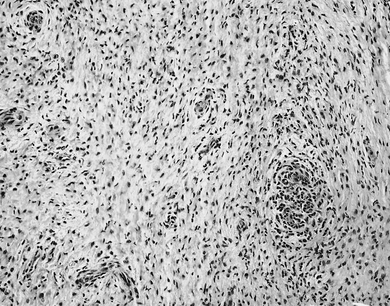
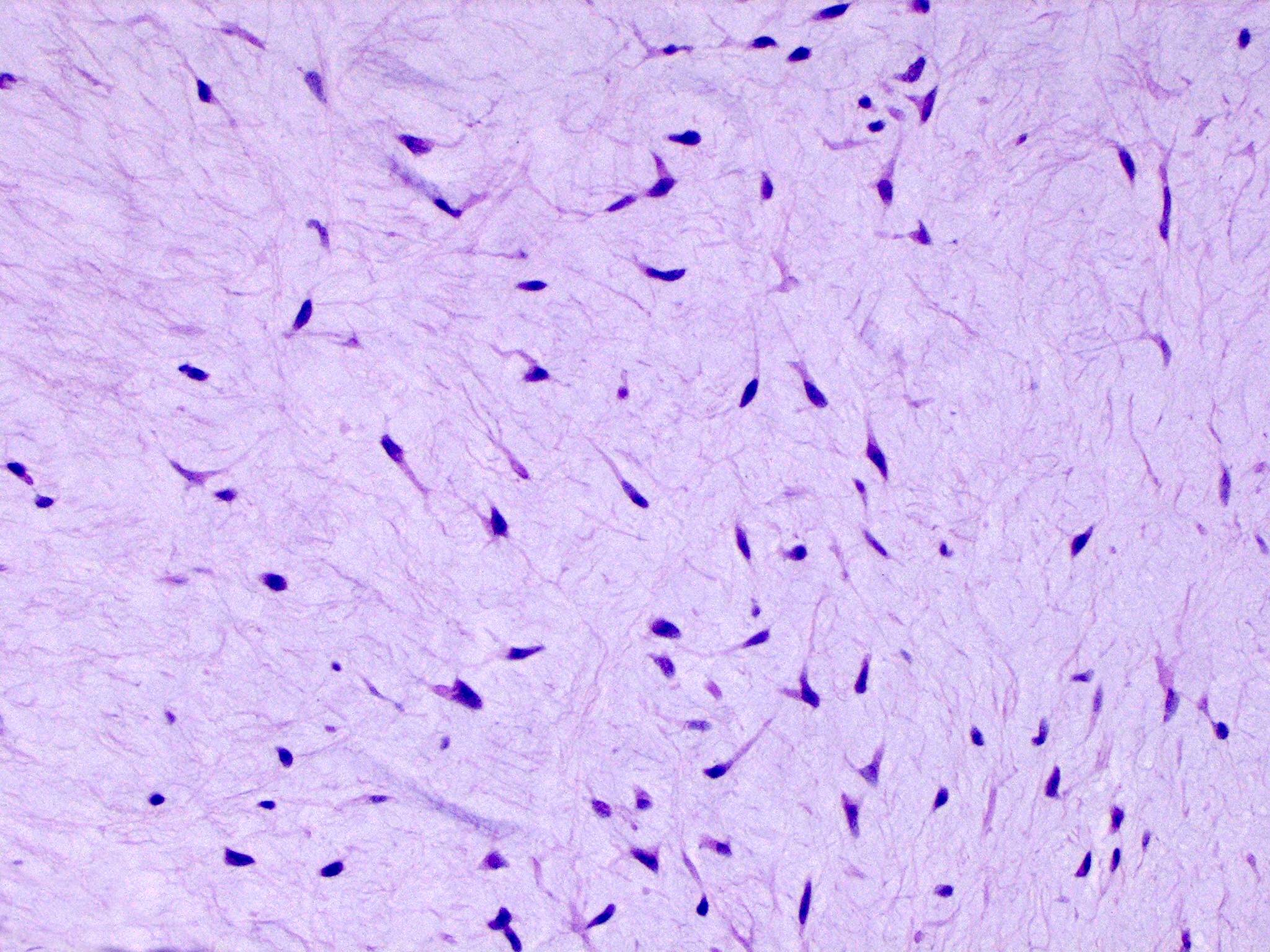
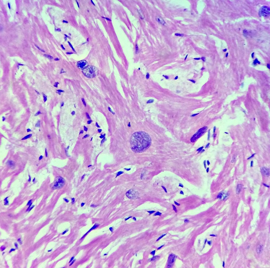
Cellular lesion in infant
Collagen deposition
Central fibroma cells

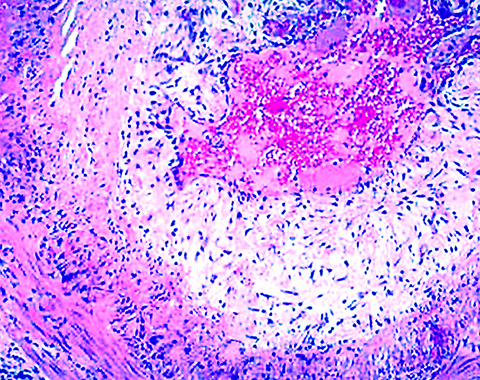
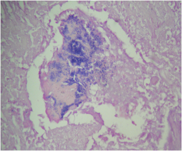
Calcification
Infiltrative margin
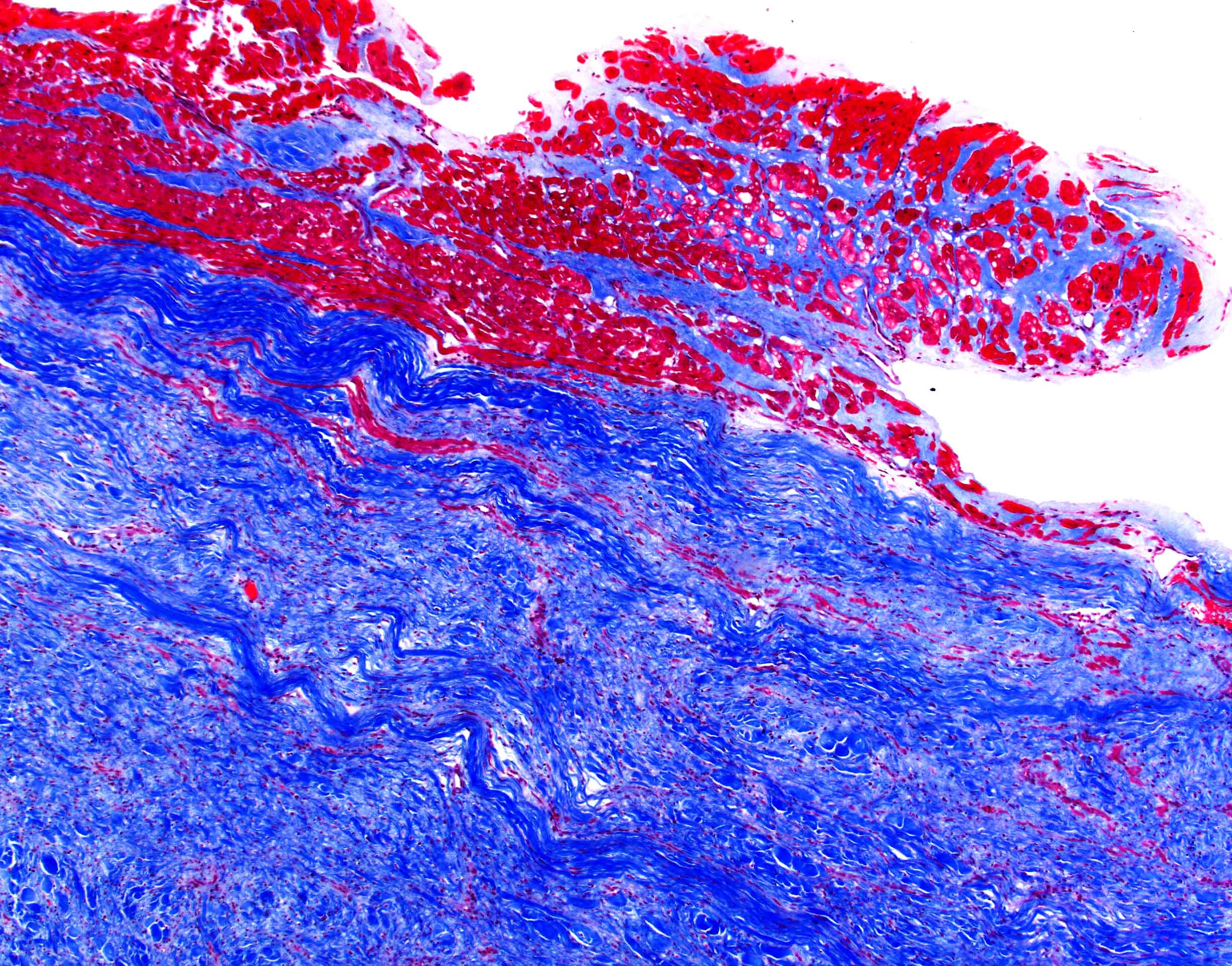
Trichrome
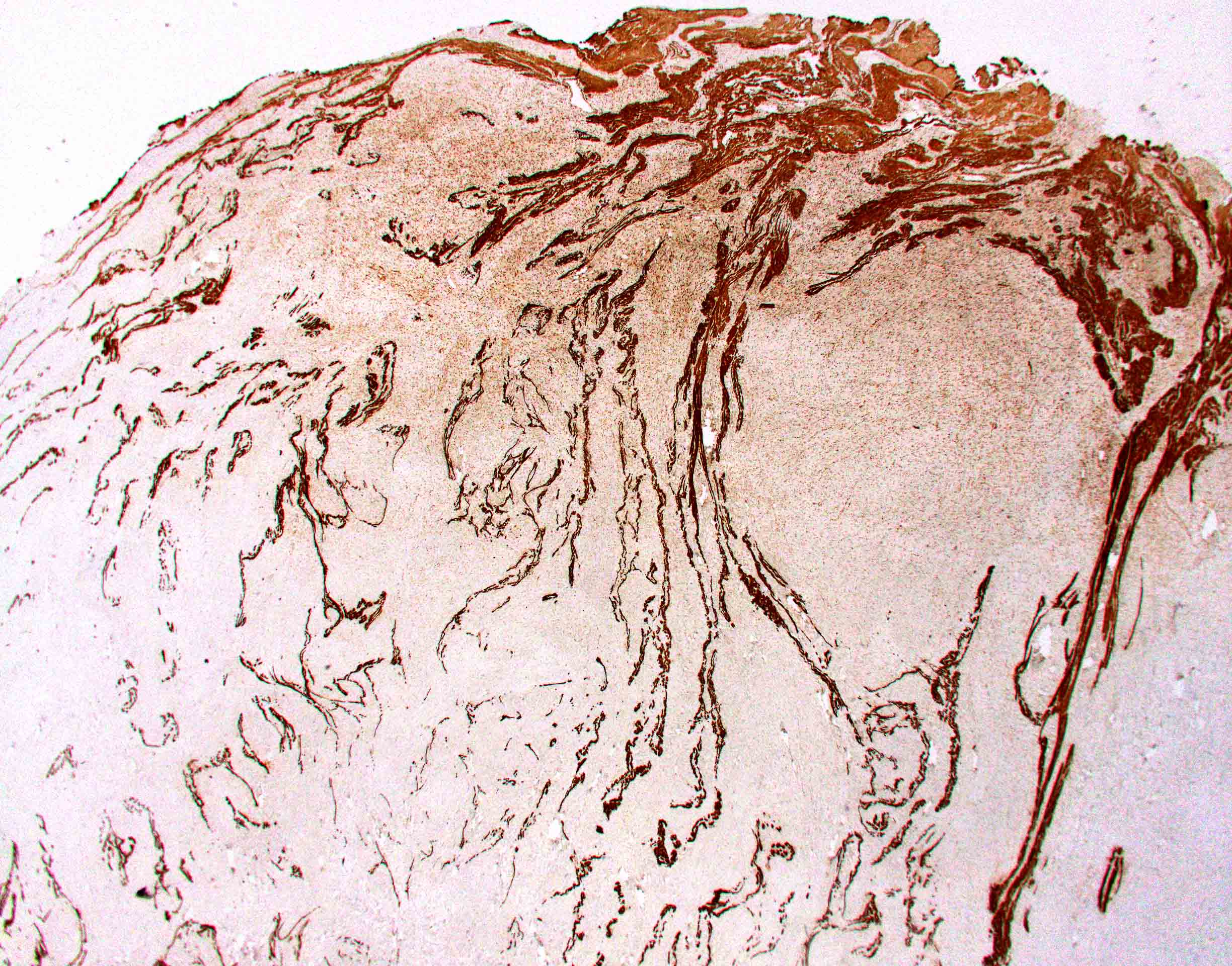
Von Gieson elastin
Contributed by Giuseppe Vergaro, M.D., Ph.D. and Vladyslav Chubuchnyi, M.D.
2D echocardiography
Cardiac magnetic resonance
Contributed by Angela Pucci, M.D., Ph.D. and Giovanni Bartoloni, M.D.
Polypoid
Hemorrhage
Villous morphology
Soft and friable projections
Chalky aspect
Contributed by Angela Pucci, M.D., Ph.D. and @DrTravisBrown on Twitter
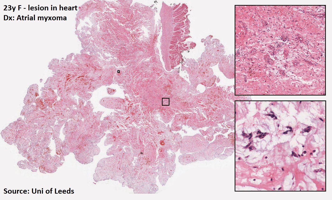
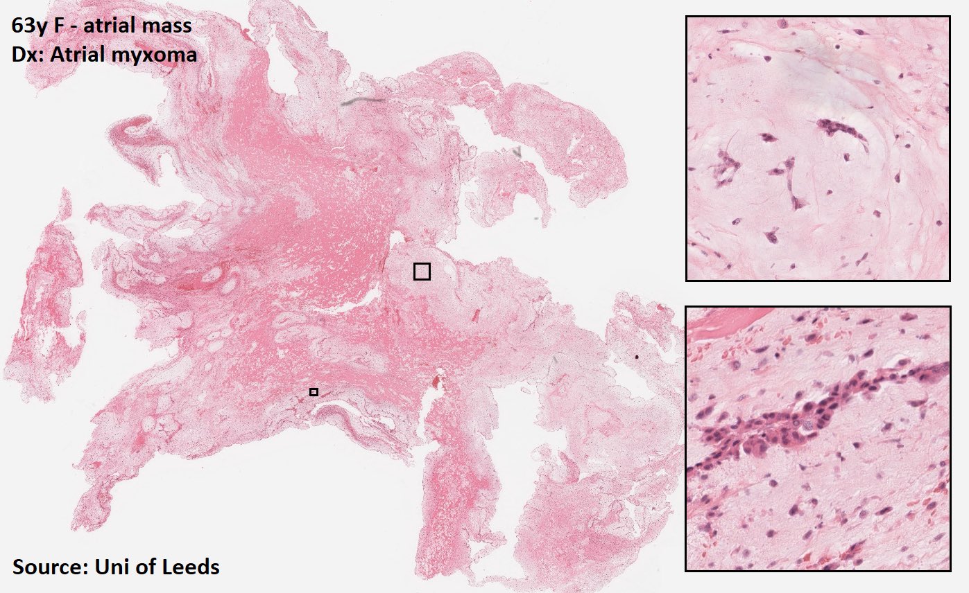
Left atrial myxoma
Myxoma cell nest
Myxoma cells in cord structure or isolated
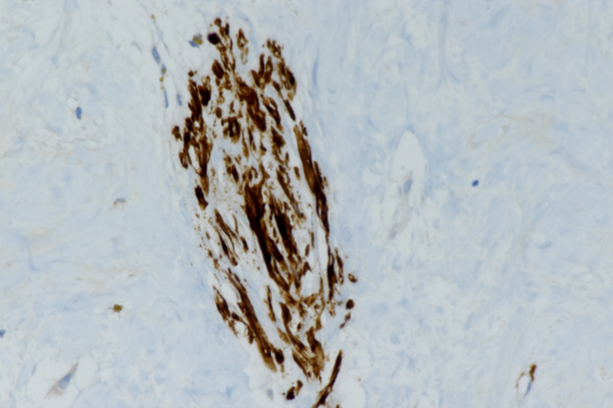
Myxoma cells around blood vessel
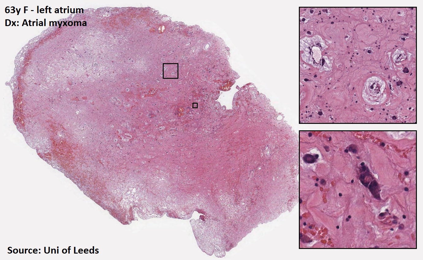
Gandy-Gamna bodies
Gandy-Gamna bodies (Perls staining)
Inflammatory infiltrates
Hemorrhage
Glands in cardiac myxoma
Calretinin
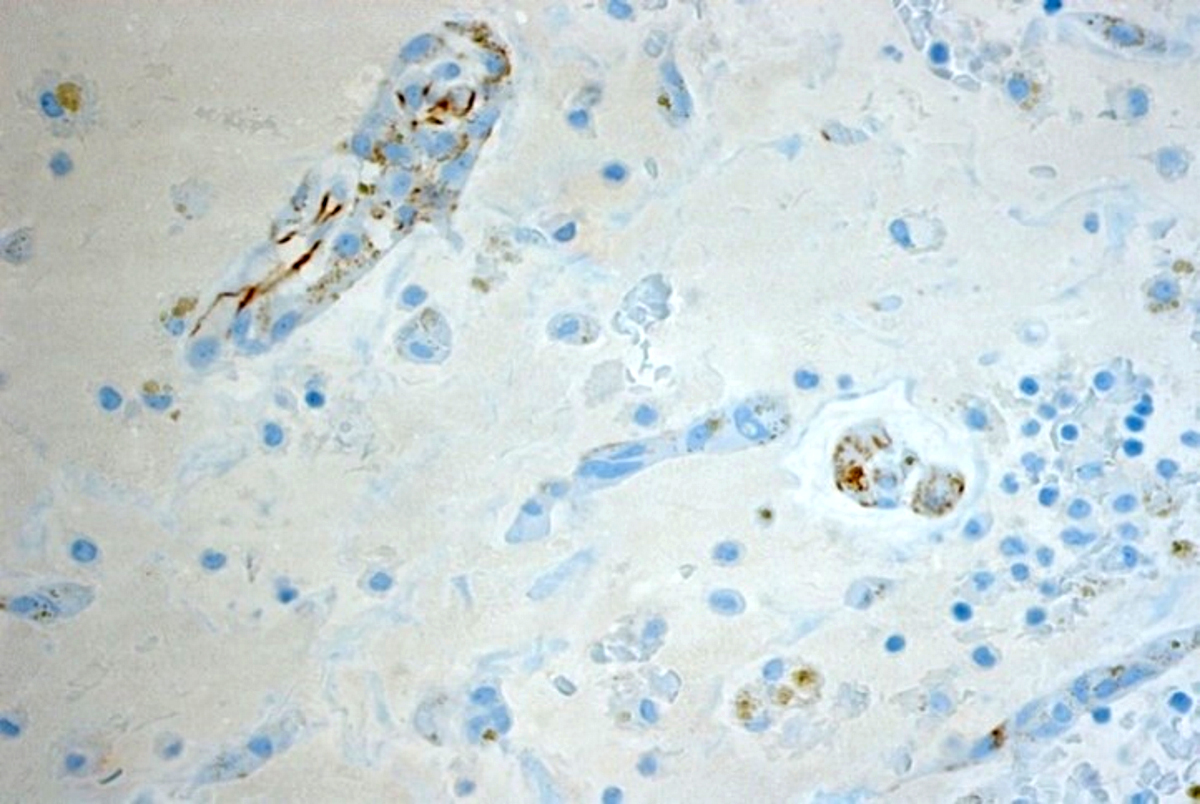
CD31+ vessels

S100 immunoreactivity
αSMA+
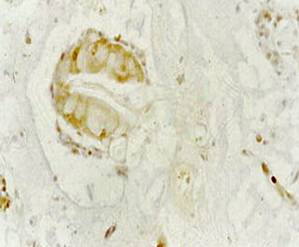
CK+ gland in cardiac myxoma

Cardiac myxoma
Contributed by Angela Pucci, M.D., Ph.D. and Giovanni Bartoloni, M.D.
Myxoma cell
Images hosted on other servers:

Trisomy 13

ADS and VSD
Images hosted on other servers:

Arteriographic classification
Images hosted on other servers:

Coronary angiography

CT angiography

Intravascular ultrasound

Coronary angiography

Optical coherence tomography
Images hosted on other servers:

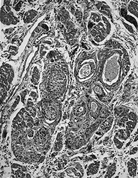
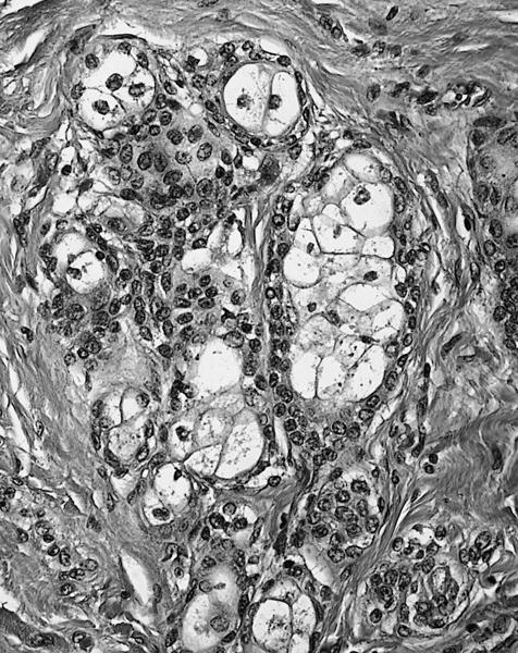
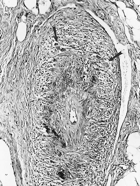
Coronary artery hyperplasia
Contributed by Carolyn Glass, M.D., Ph.D.
Explant, concentric intimal hyperplasia

Explant, diffuse intimal hyperplasia
Explant, coronary artery stenosis
Explant, coronary artery occlusion
Images hosted on other servers:

Purkinje network
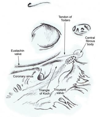
Compact atrioventricular node

Conduction system
Images hosted on other servers:


Sinoatrial (SA) node


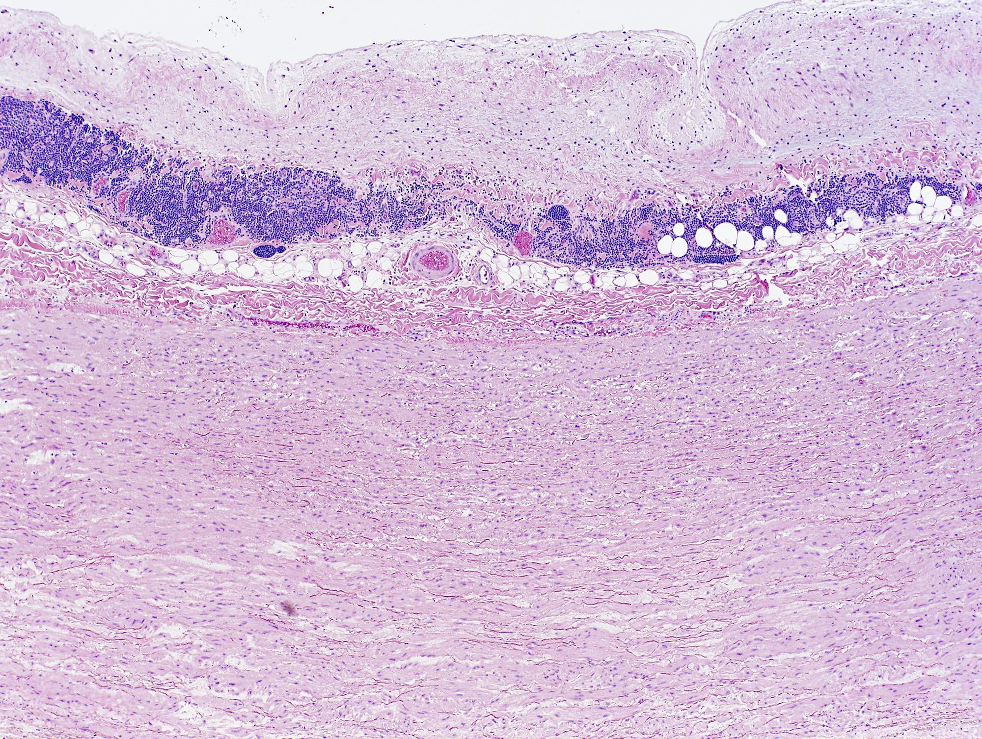
Sinus node fibrosis
Images hosted on other servers:

Rheumatic diseases and sites of involvement
Images hosted on other servers:


Various images
Images hosted on other servers:

Anterior circulation

Posterior circulation

Coronary arteries
Images hosted on other servers:

Venule


Various images
Images hosted on other servers:

Small arteriole
Images hosted on other servers:

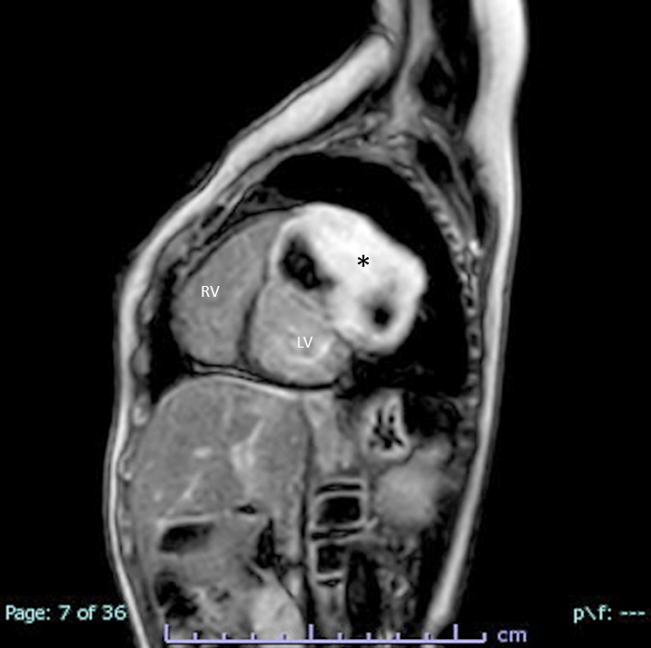
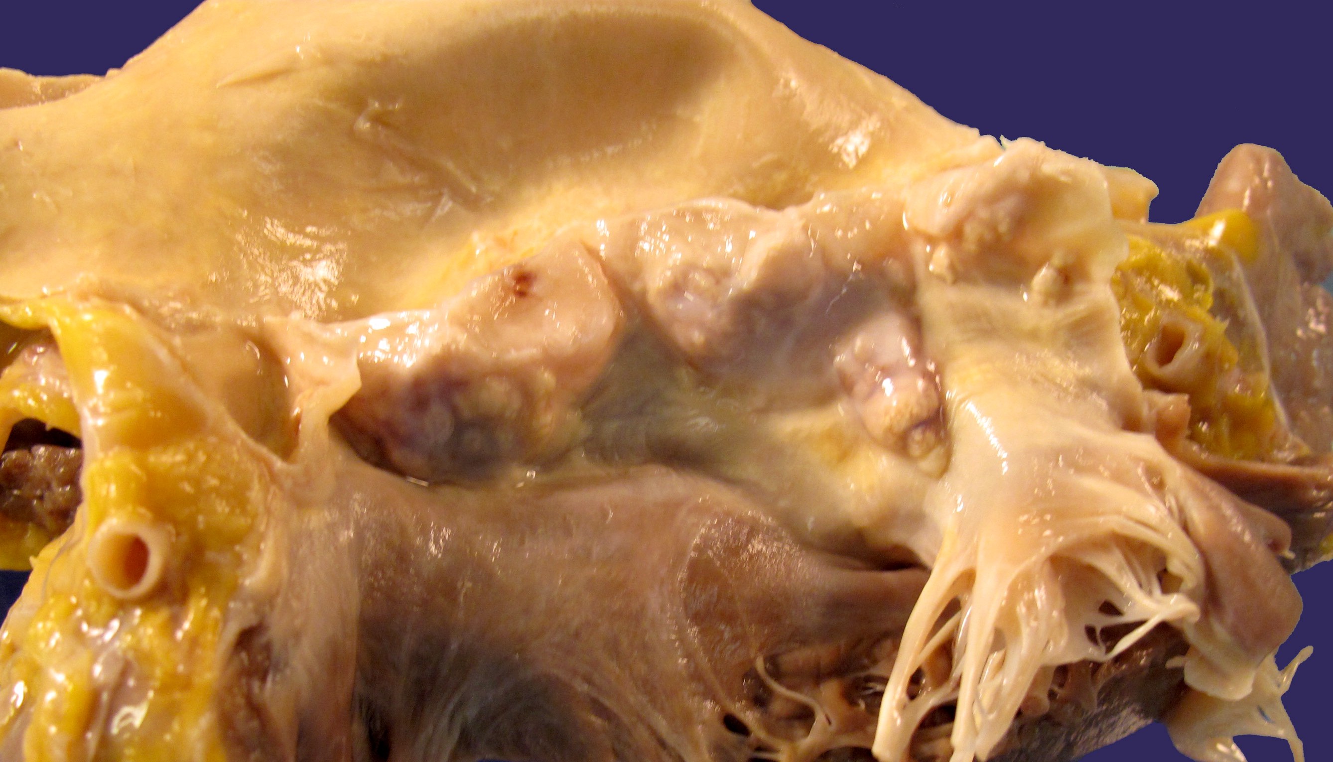
MRI of the thorax showing tumor
AFIP images
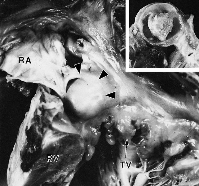
arrowheads (RA-right
atrium, RV-right ventricle,
TV-tricuspid valve)
AFIP images
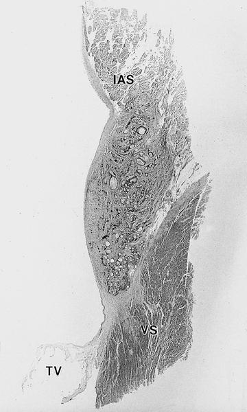
IAS-interatrial septum (tumors are
located inferiorly), TV-tricuspid
valve and VS-ventricular septum
filled with
proteinaceous
debris
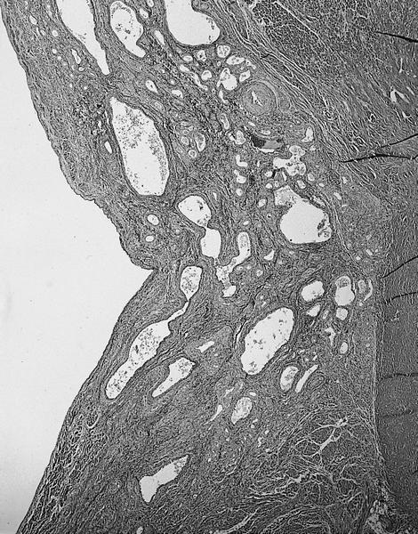
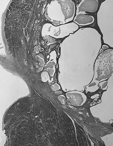
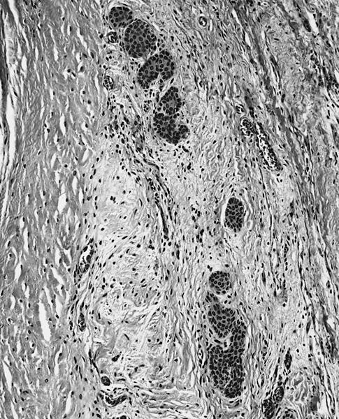
Multiple small cysts not visible to naked eye
Cysts with irregular shape surrounded by fibrous stroma
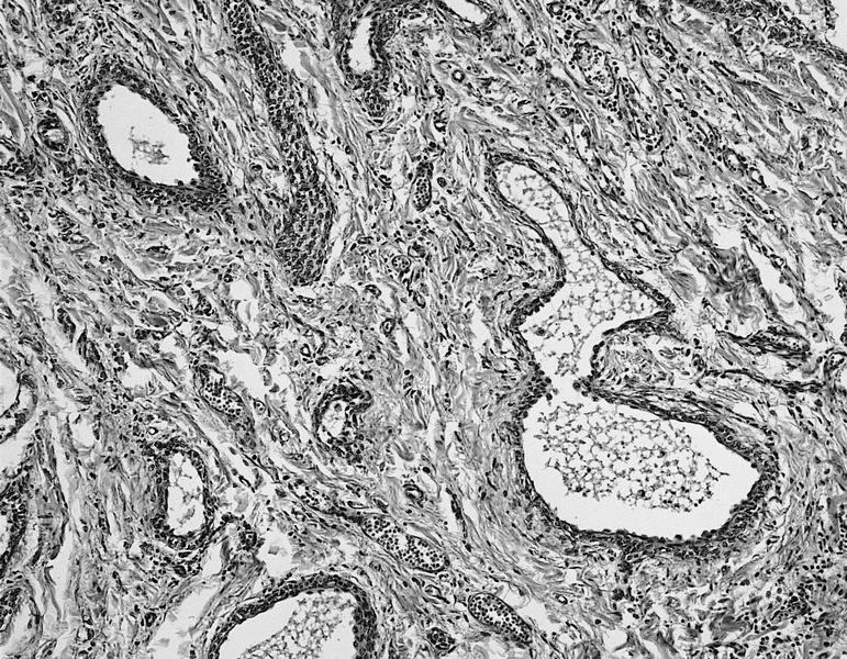
cell lining, with inner
lining composed of
small cuboidal cells
cysts replace muscle
bundles in inferior
interatrial septum
Cuboidal cells and clear, sebaceous-type cells
Squamous differentiation and calcification of luminal debris
Nests of cells resembling urothelium
Images hosted on other servers:
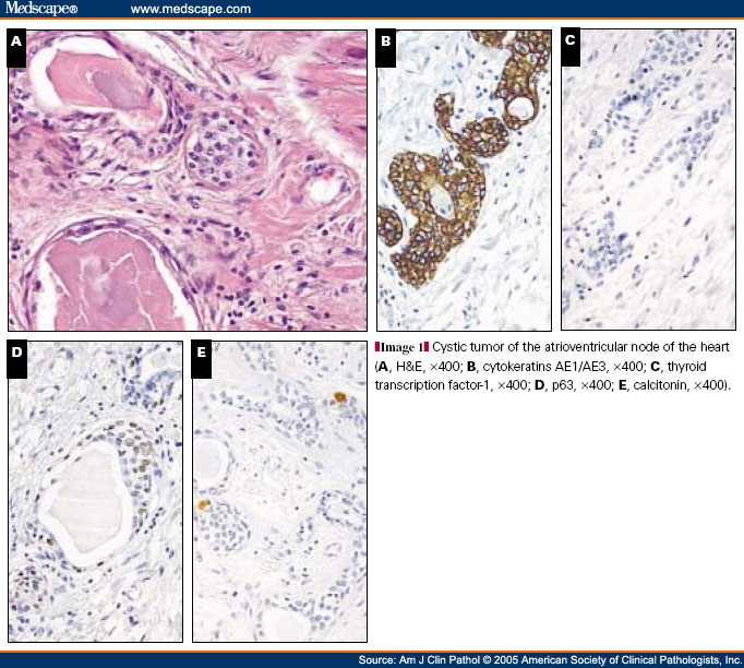
H&E and AE1/AE3+

cyst lining of simple
cuboidal epithelium
Images hosted on other servers:


Pathophysiology of degeneration of valves

Potential pathways

MVP subtypes
Contributed by Aliya N. Husain, M.D.
Mitral valve
Images hosted on other servers:

Minimally diseased
aortic valve (left) and
severely stenotic
aortic valve (right)
Contributed by Aliya N. Husain, M.D.
Mitral valve
Aortic valve calcification
Mitral valve
Images hosted on other servers:

5HT signaling pathways

Heart chambers, valves, and valvular histology
Images hosted on other servers:

Mitral valve leaflets, Movat pentachrome stain
Images hosted on other servers:

Development of heart
Images hosted on other servers:

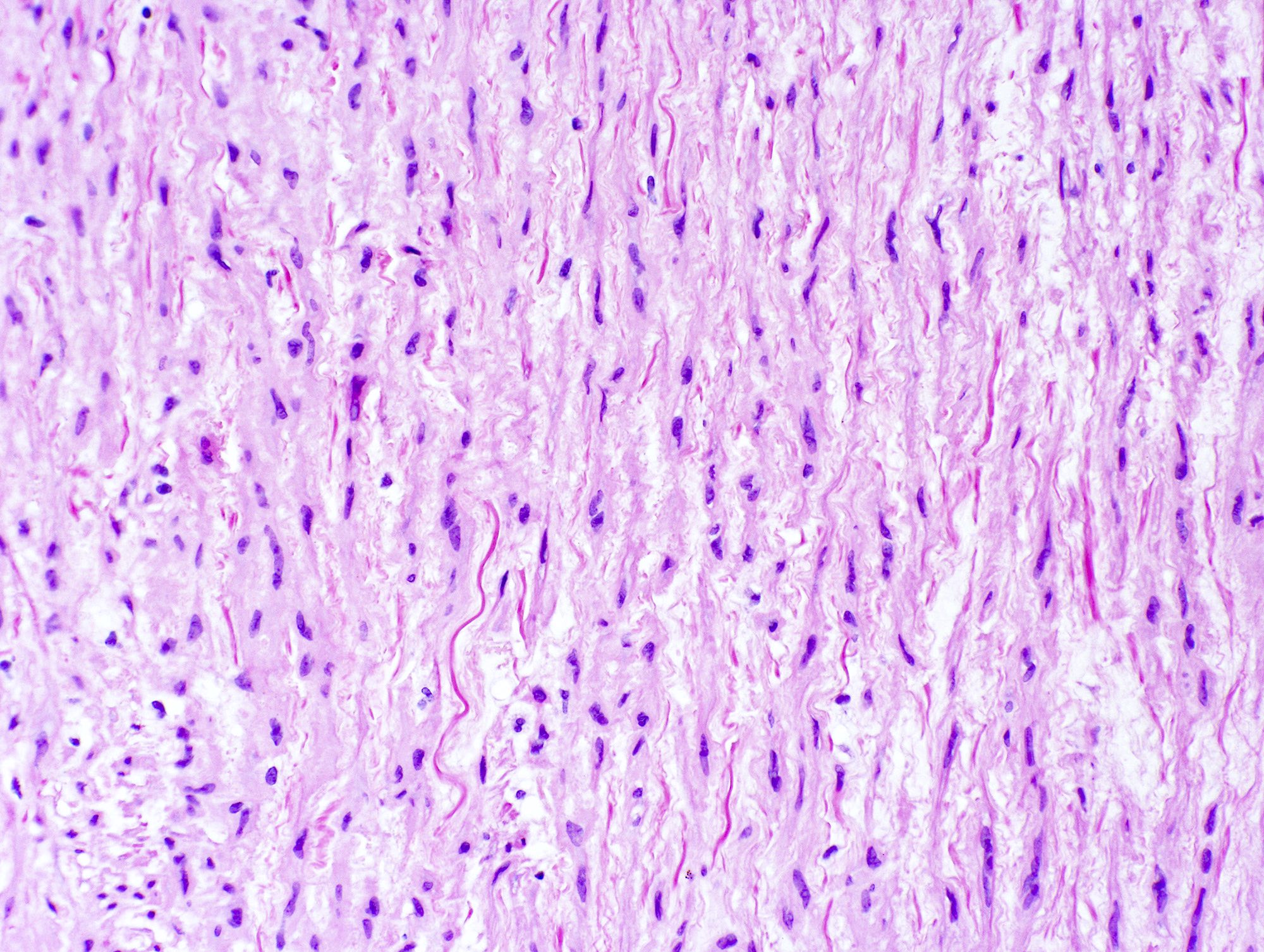
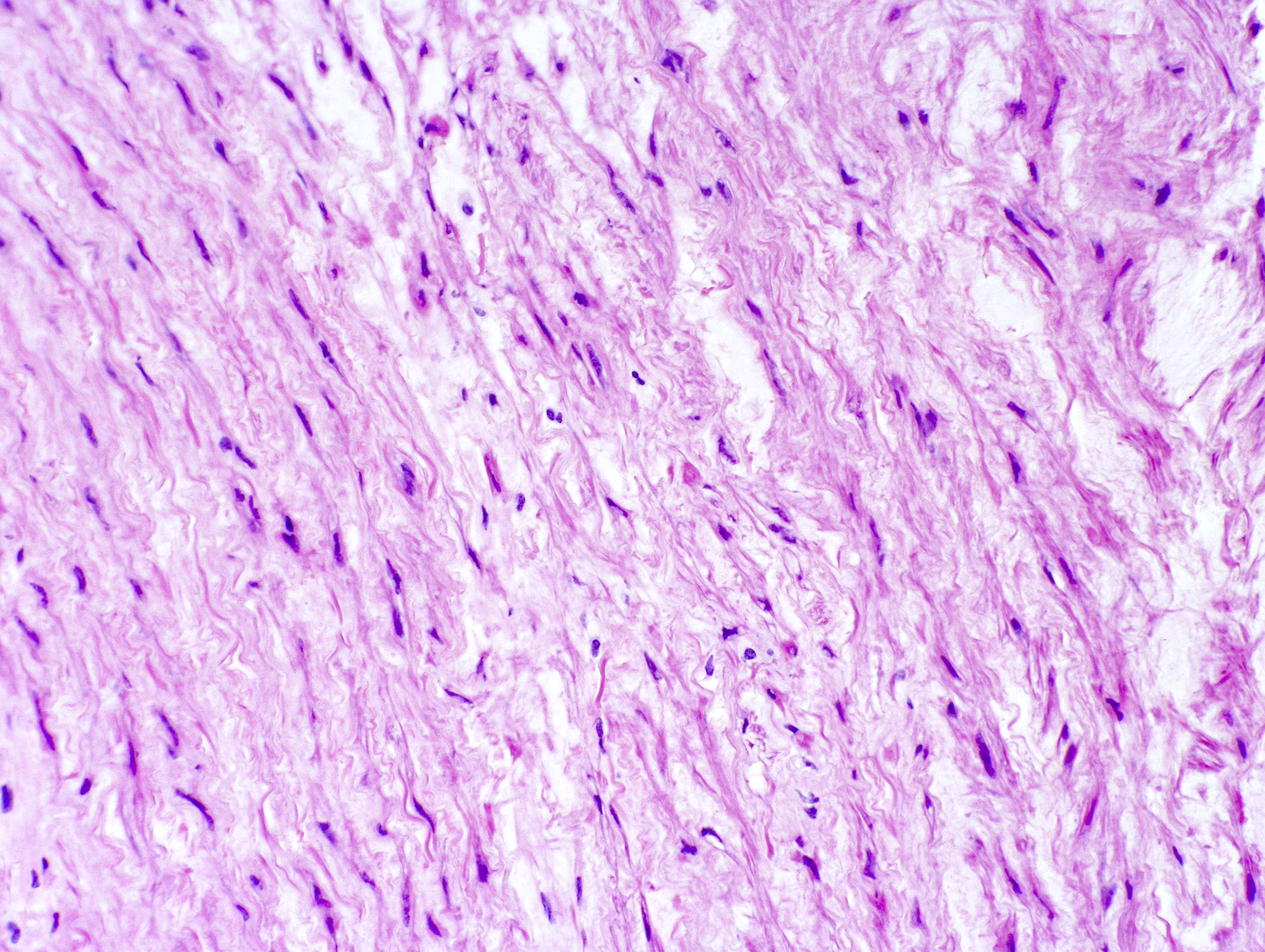
Medial fibroplasia

Intimal fibroplasia
Images hosted on other servers:

Medial fibroplasia
Contributed by Mark R. Wick, M.D.

Various images
Elastic stain
AFIP images
Intramammary arterial lumen
is mostly obliterated by
subintimal and mural inflammation,
including giant cells (arrows)

Transmural necrotizing inflammation
of medium sized mammary vessels,
with elastic stain showing partial
destruction of elastic layer
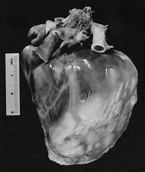
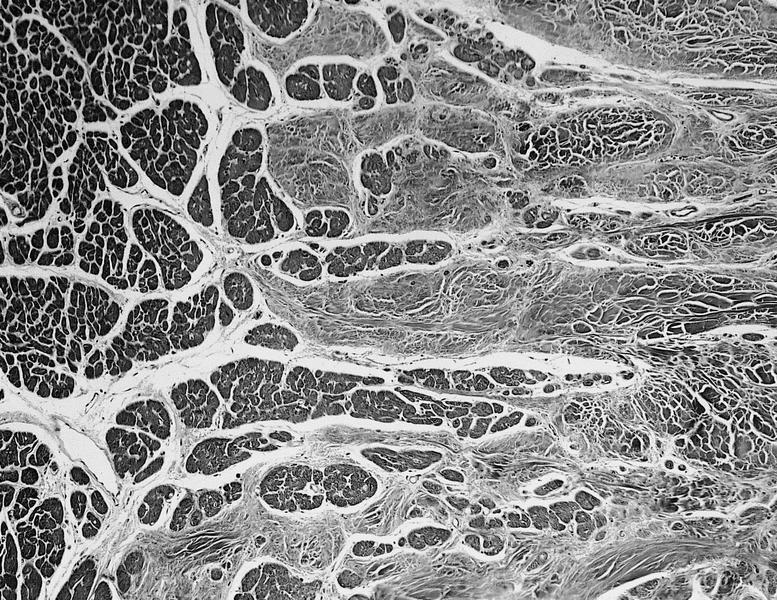
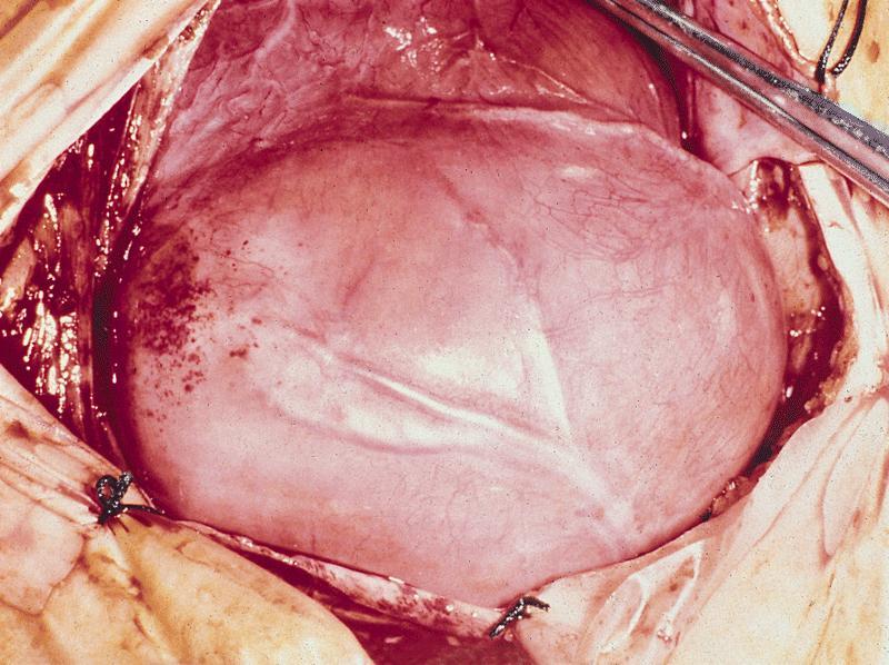
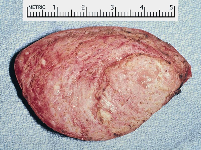
Normal cardiac specimen
Contributed by Melanie C. Bois, M.D.
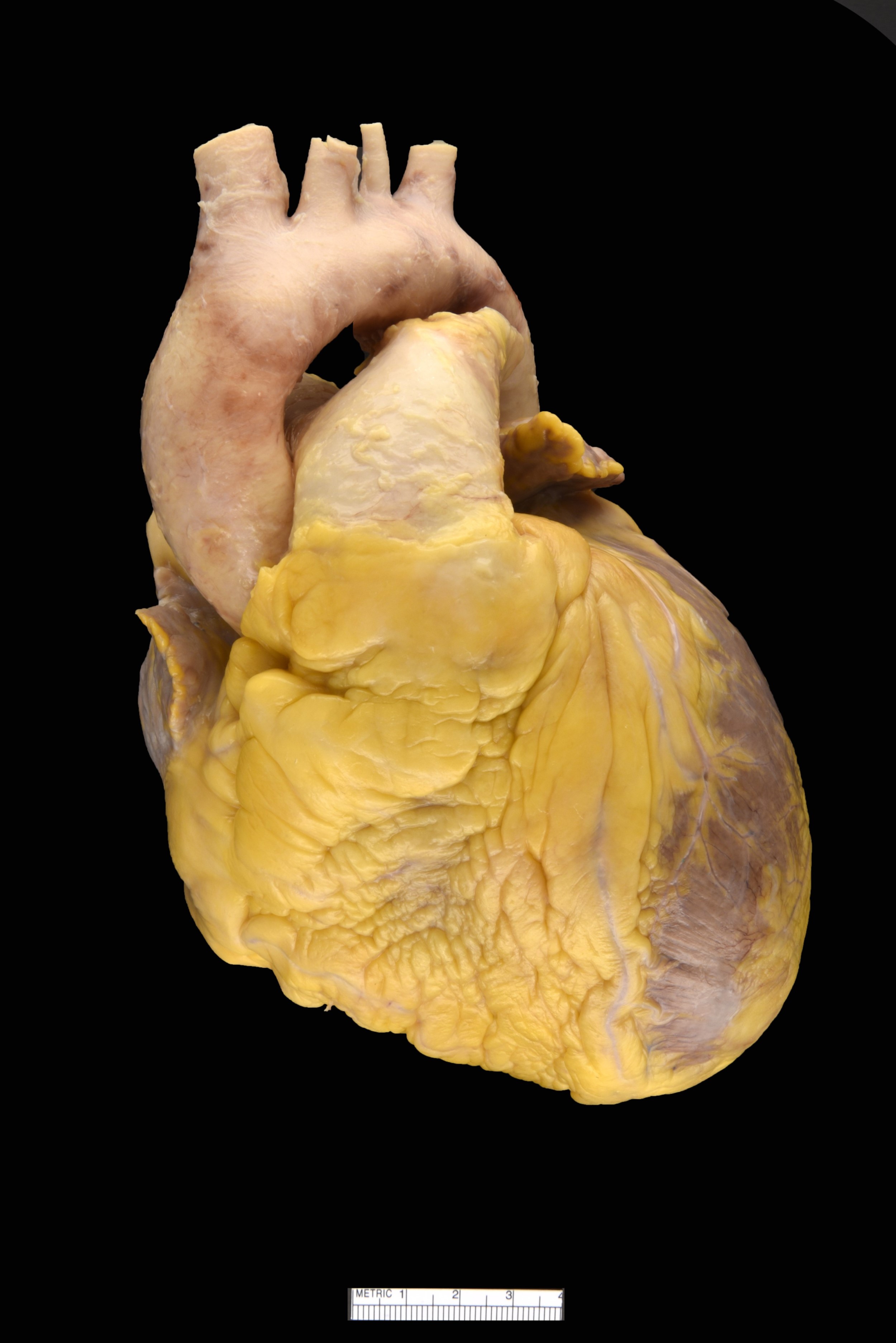
Heart, anterior view

Heart, short axis section
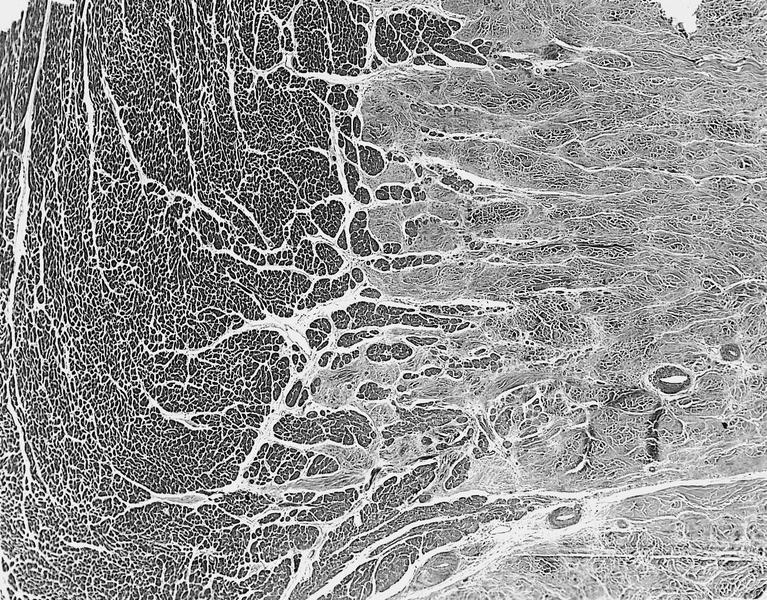
Contributed by Melanie C. Bois, M.D.

Heart, microscopy
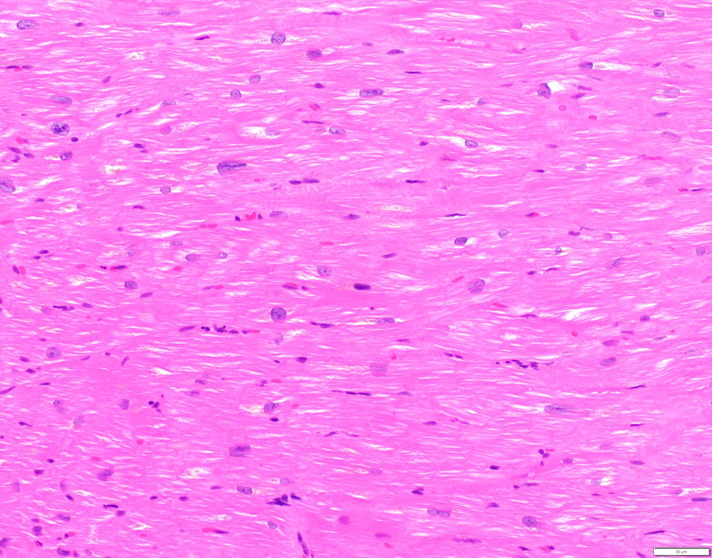
Cardiomyocytes
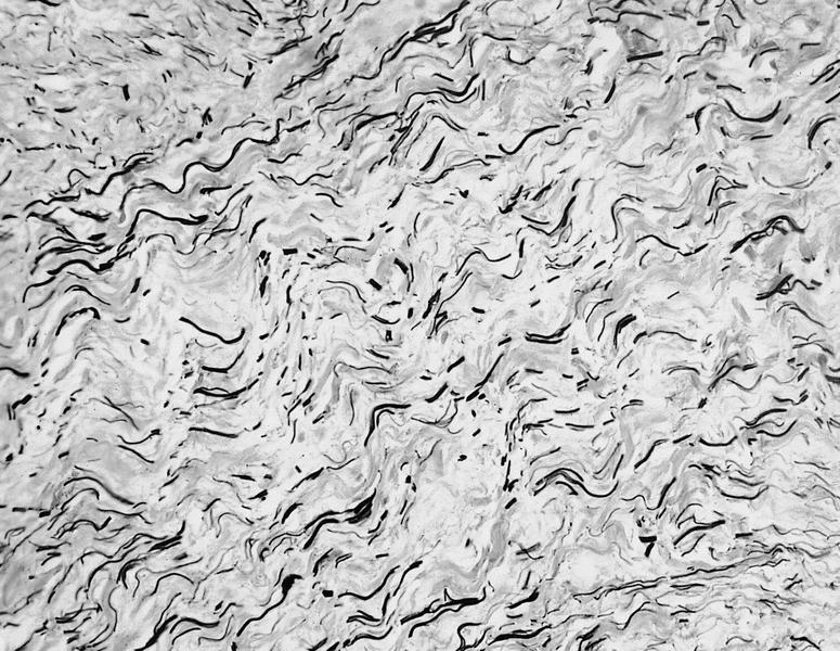
Contributed by Saranya Singaravel, M.B.B.S.
Transverse section
Contributed by Saranya Singaravel, M.B.B.S.
Myocardial hypertrophy and disarray
Hypertrophied cardiomyocyte with a whorled apperance
Images hosted on other servers:

Echocardiographic morphological
features typical of idiopathic RCM
Images hosted on other servers:

men demonstrating
prominent biatrial
enlargement
Images hosted on other servers:
microscopy
showing marked
interstitial fibrosis
Images hosted on other servers:

Duke criteria for diagnosis
Images hosted on other servers:

Osler nodes on a finger and foot
Images hosted on other servers:

Large vegetation on atrial aspect of valve
Contributed by R. Amita, M.D.
Mycotic aneurysm
Dense inflammation and fibrinoid necrosis
Valve destruction with necrosis, bacterial clump
Images hosted on other servers:

H&E, gram stain
Images hosted on other servers:

MRI: acute myocarditis

Increased T2-weighted signal intensity

Echocardiograms

Gadlinium-enhanced cardiac MRI
Images hosted on other servers:

Pathophysiological process of viral myocarditis

Algorithm for diagnosis in suspected myocarditis

Treatment algorithm based on EMB findings
Images hosted on other servers:

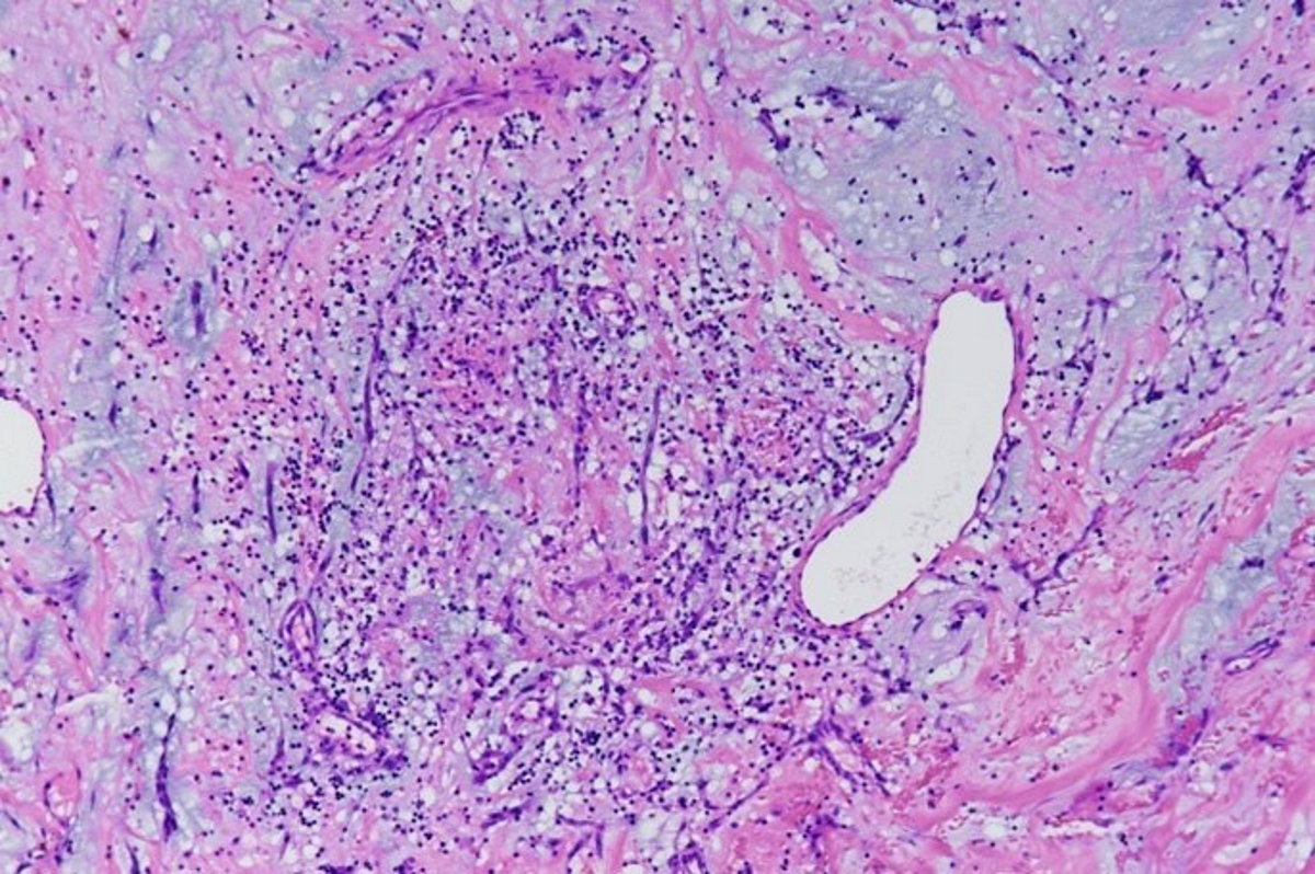
Endomyocardial biopsy

Acute and chronic myocarditis
Contributed by Pallavi Khattar, M.D., Puneet Bedi, M.D. and John T. Fallon, M.D., Ph.D.
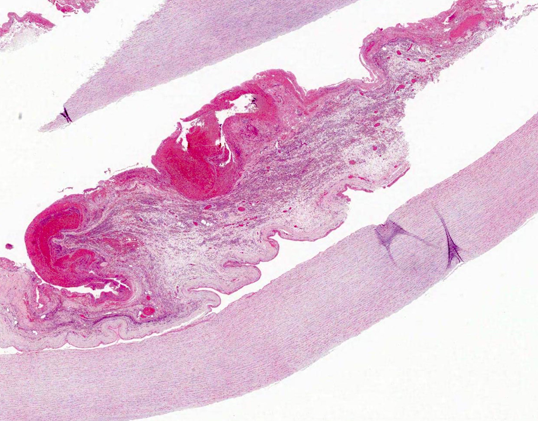
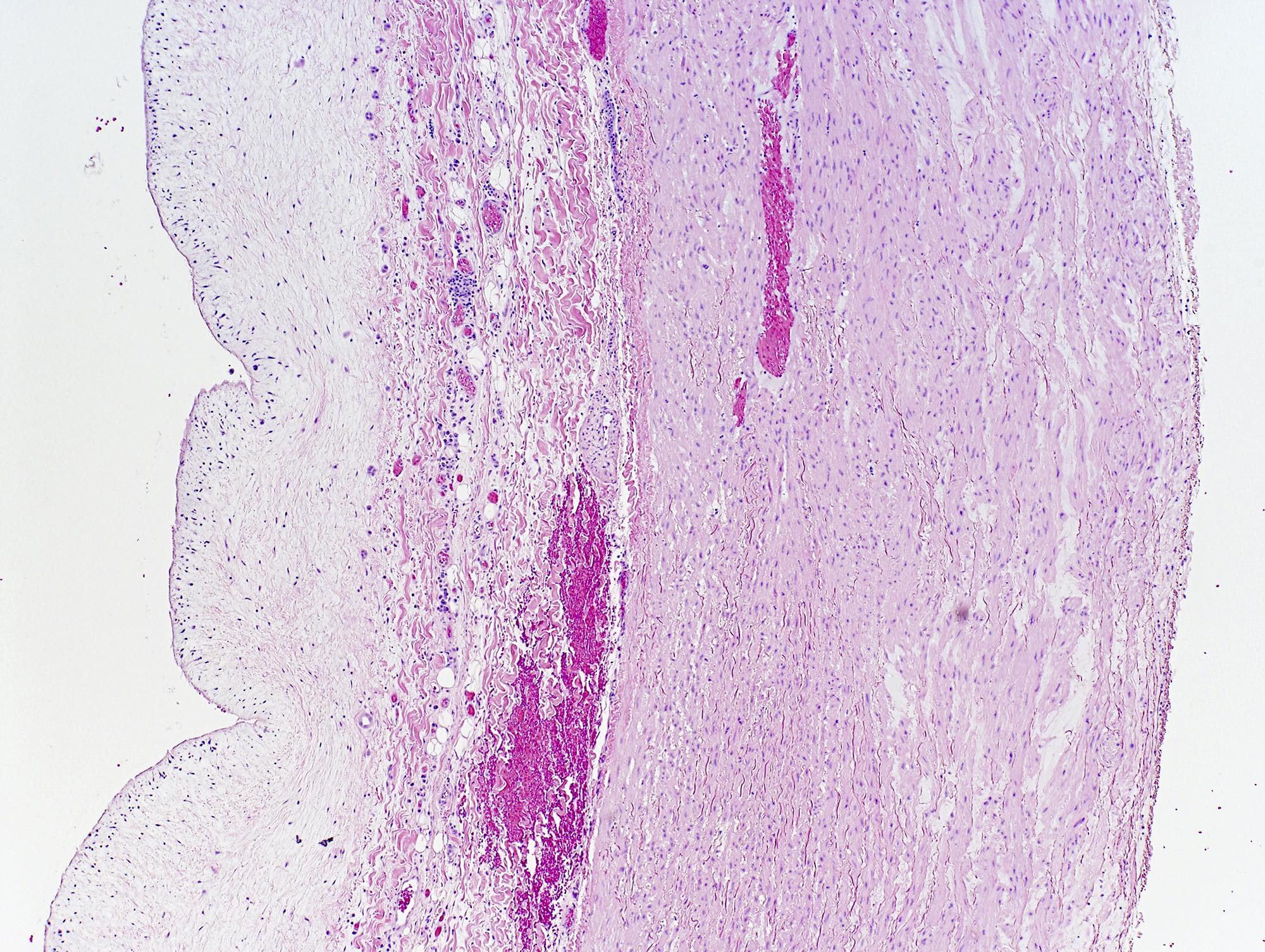
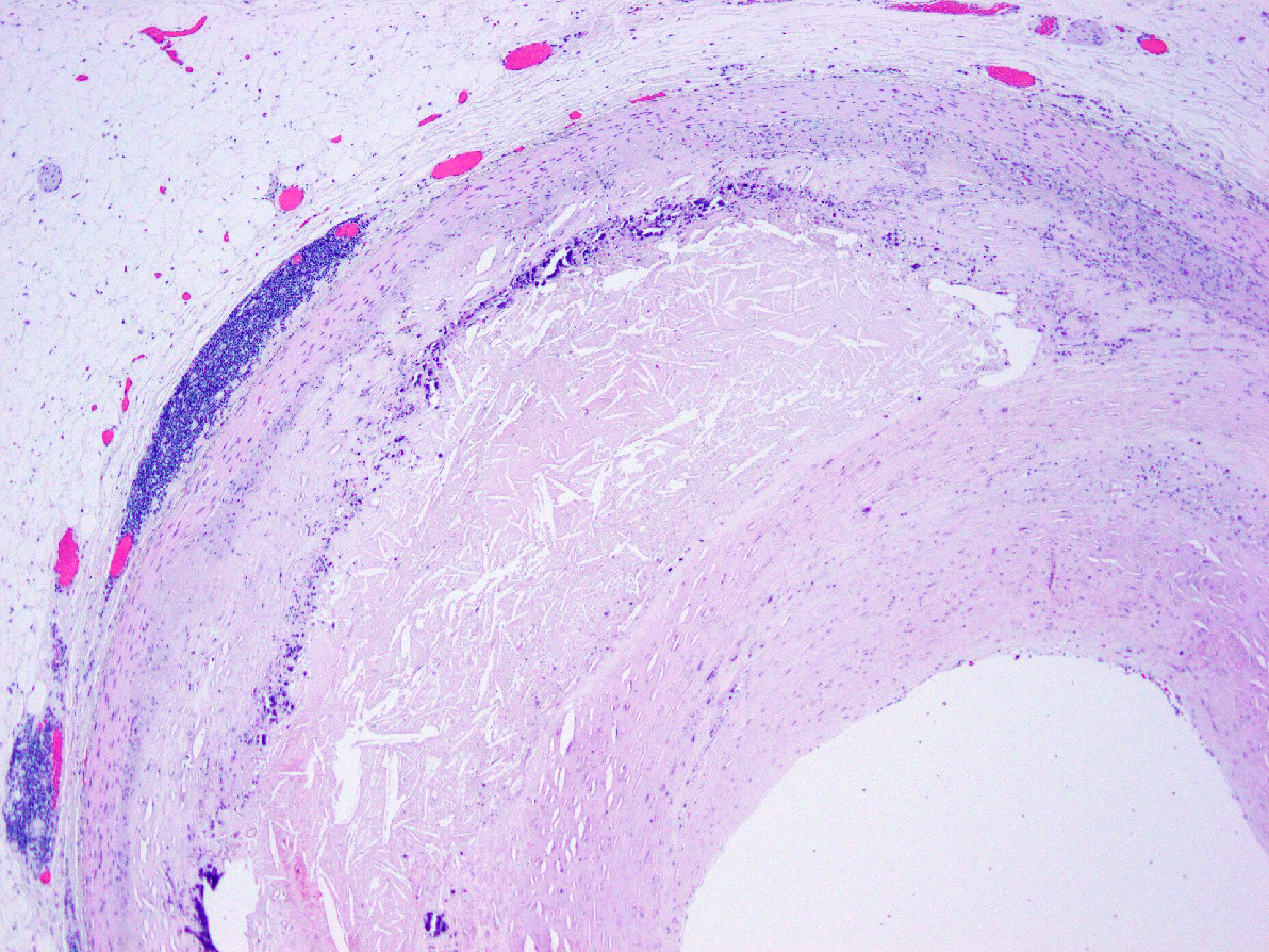
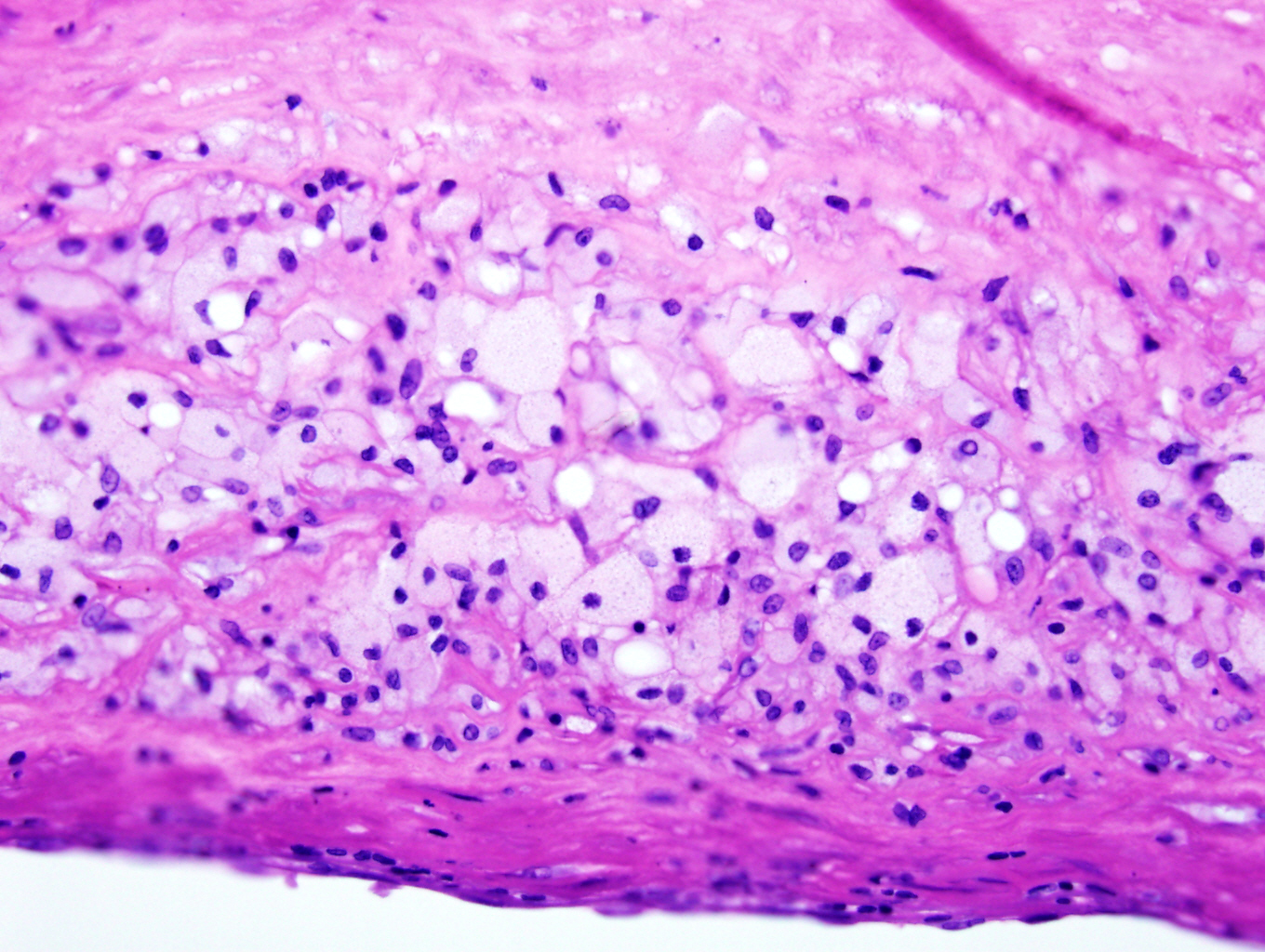
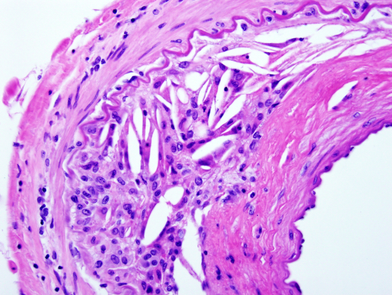
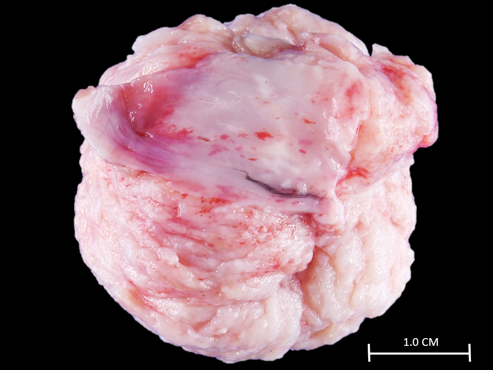
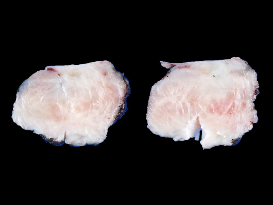
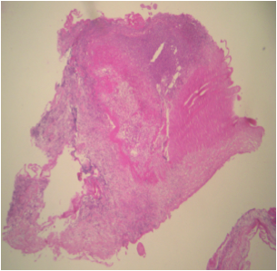
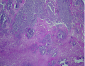
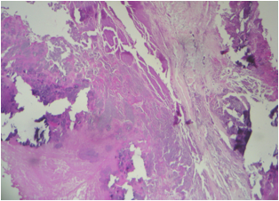
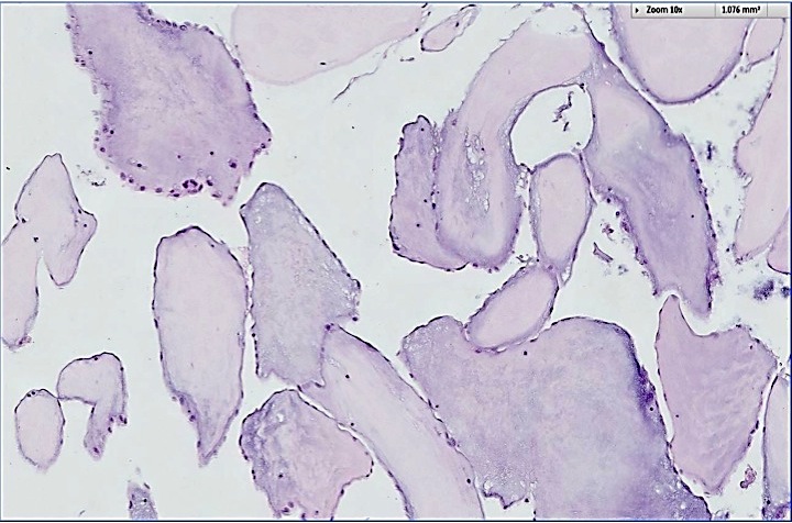
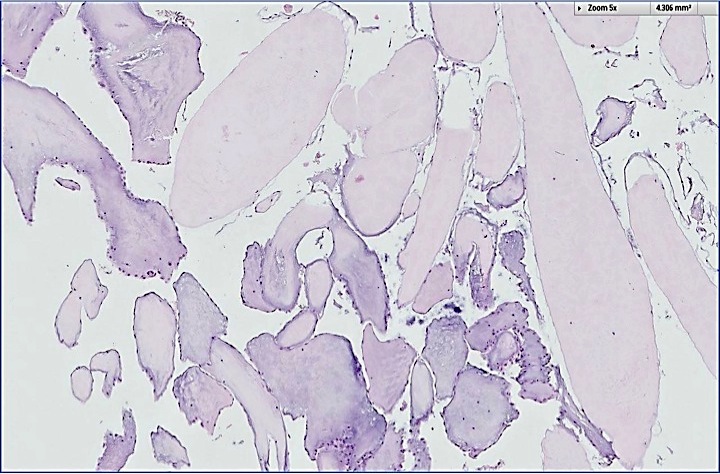
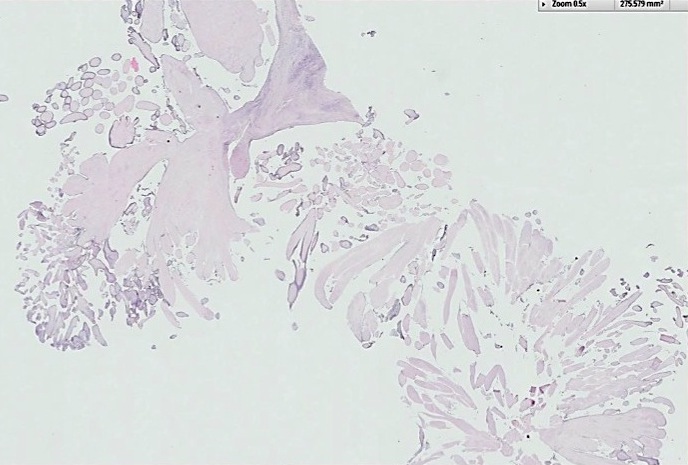
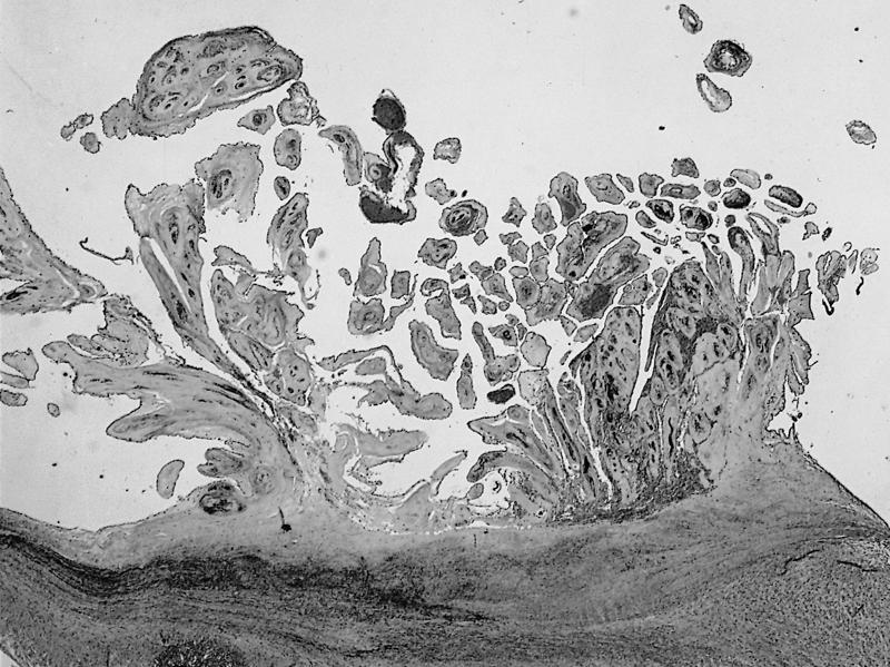
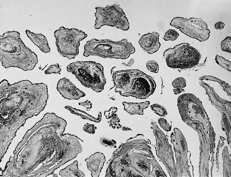
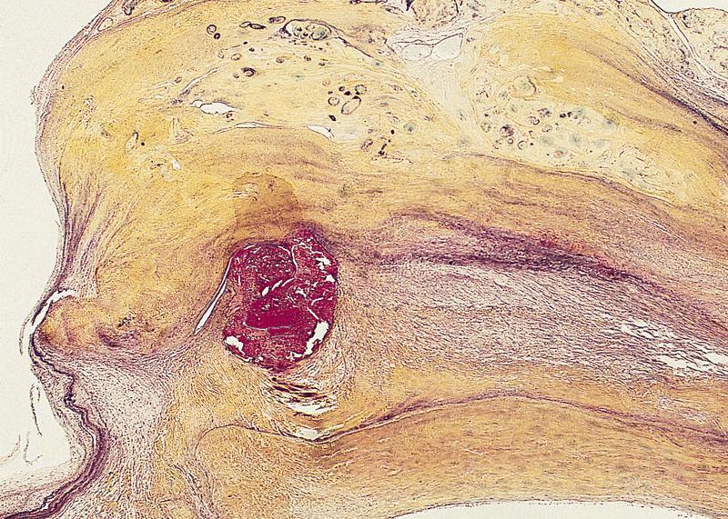
Papillary giant fibroelastoma of aortic valve
Contributed by Pallavi Khattar, M.D., Puneet Bedi, M.D. and John T. Fallon, M.D., Ph.D.
H&E
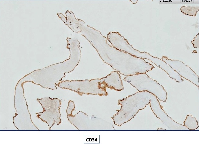
CD34
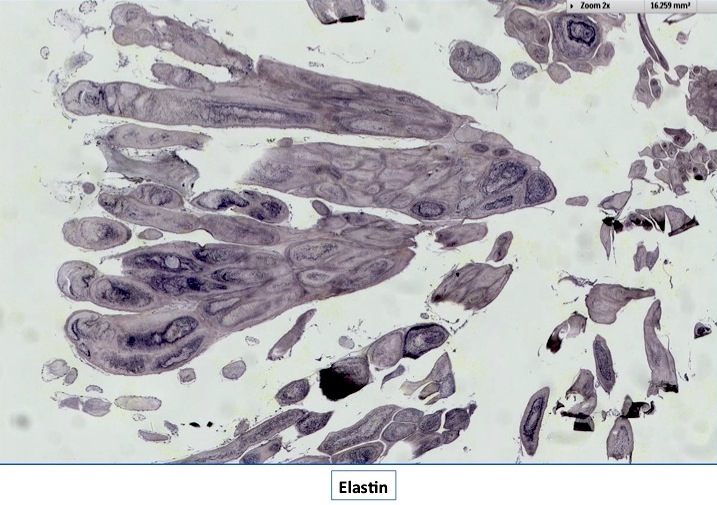
Elastin stain
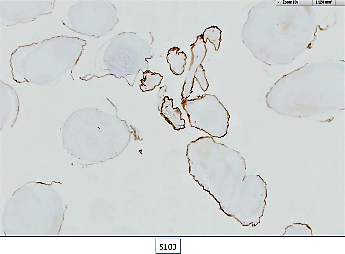
S100
AFIP images
Finger-like projection
extending from valve
surface without branching
Images hosted on other servers:


Coronary artery aneurysm

Small aneurysm

Small aneurysm in thyrocervical trunk

Multiple aneurysmal changes
Images hosted on other servers:

Differences between NBTE and infective endocarditis
Images hosted on other servers:
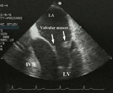
Mitral valve with masses

Mitral valve with mass

Enlarging mitral valve mass
Images hosted on other servers:

Typical finding

Typical appearances

Large NBTE vegetations
Images hosted on other servers:

Uniform eosinophilic appearance
Images hosted on other servers:

Etiology of Myocarditis
Images hosted on other servers:


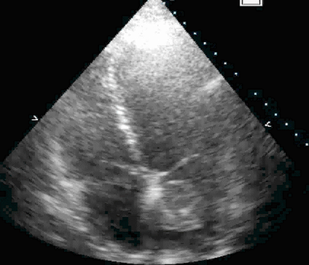
Transthoracic cardiac echocardiograms

MRI, TIRM

Pericardial effusion of 1.2 cm

MRI, IR, FLASH


Subendocardial delayed enhancement

Increased septal wall thickness
Images hosted on other servers:

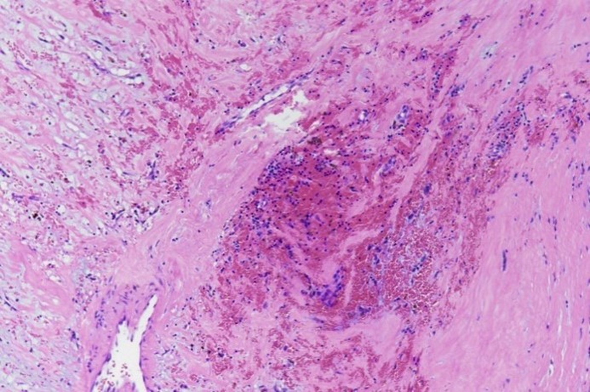
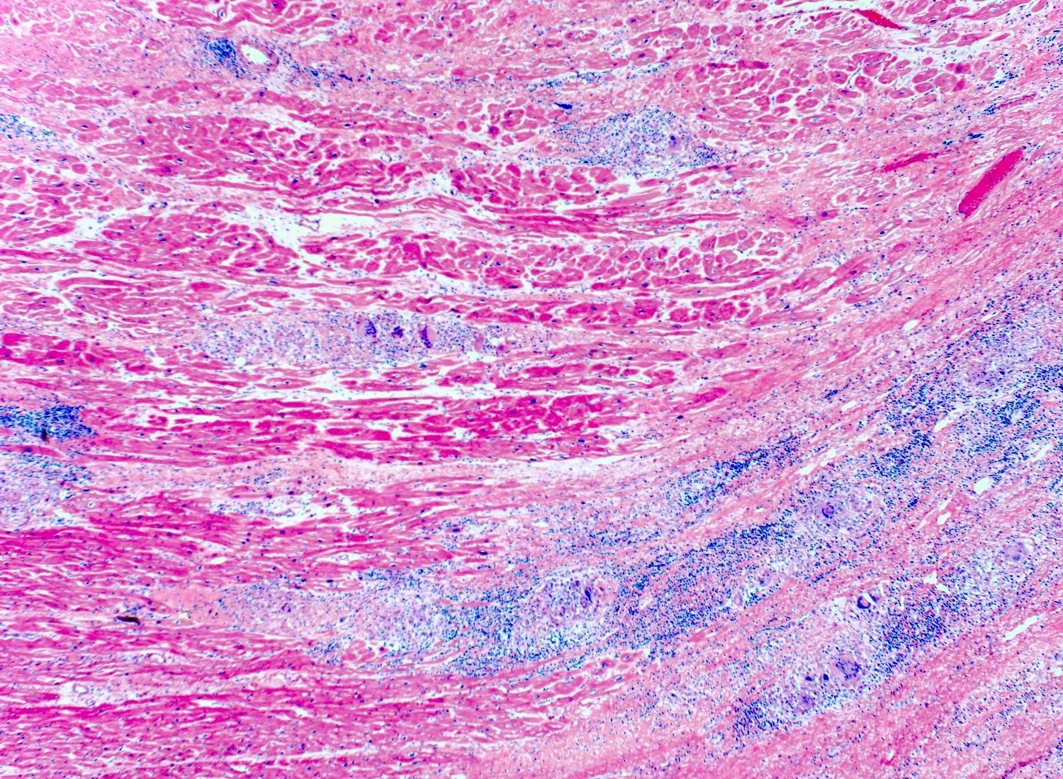
Acute myocarditis with lymphocytic infiltration

Endomyocardial biopsy

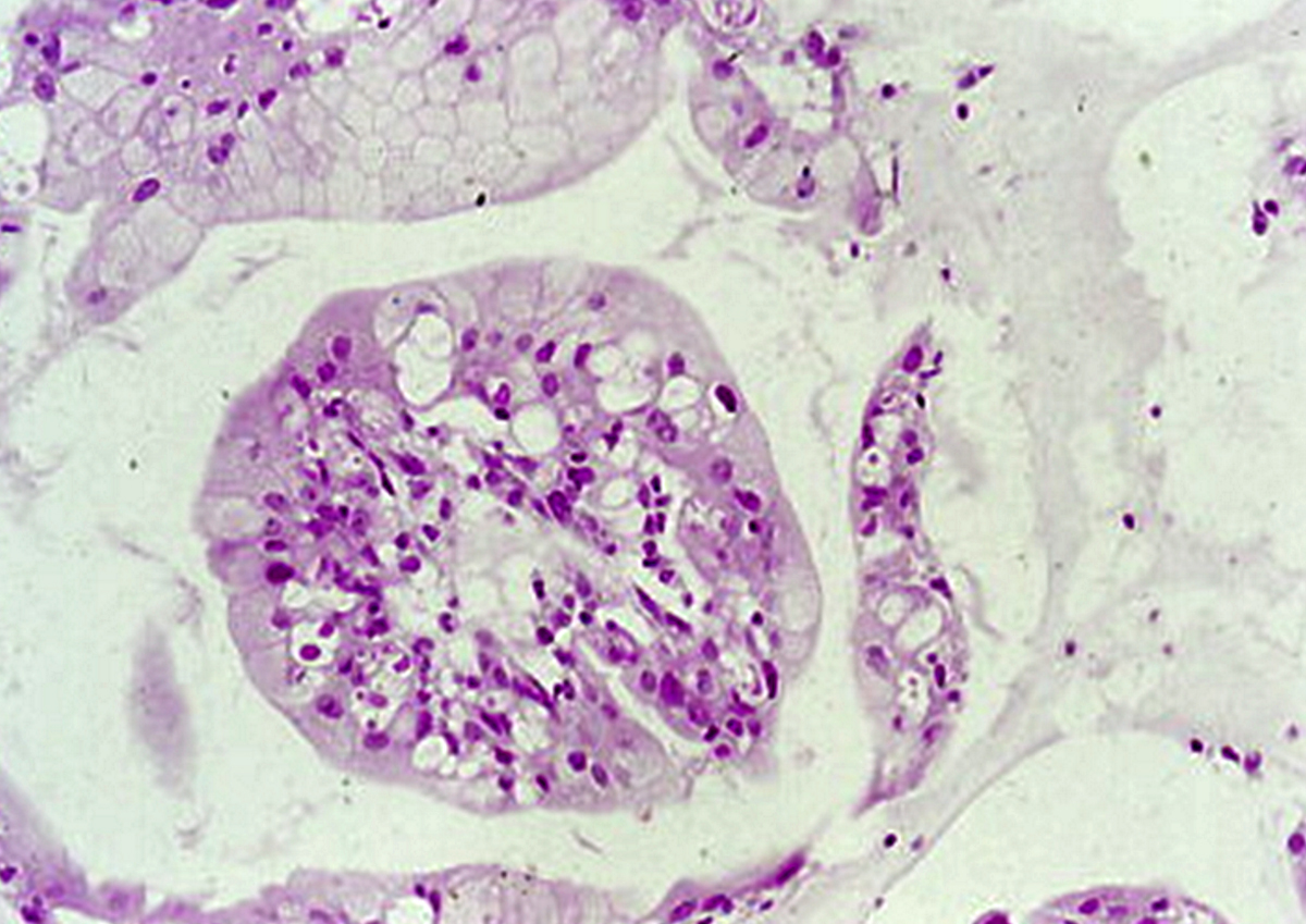
Eosinophilic infiltration

Necrotizing eosinophilic myocarditis

Eosinophilic myocarditis
Images hosted on other servers:

No granulomatous inflammation
Images hosted on other servers:

Echocardiography

Echocardiography and MRI
Images hosted on other servers:

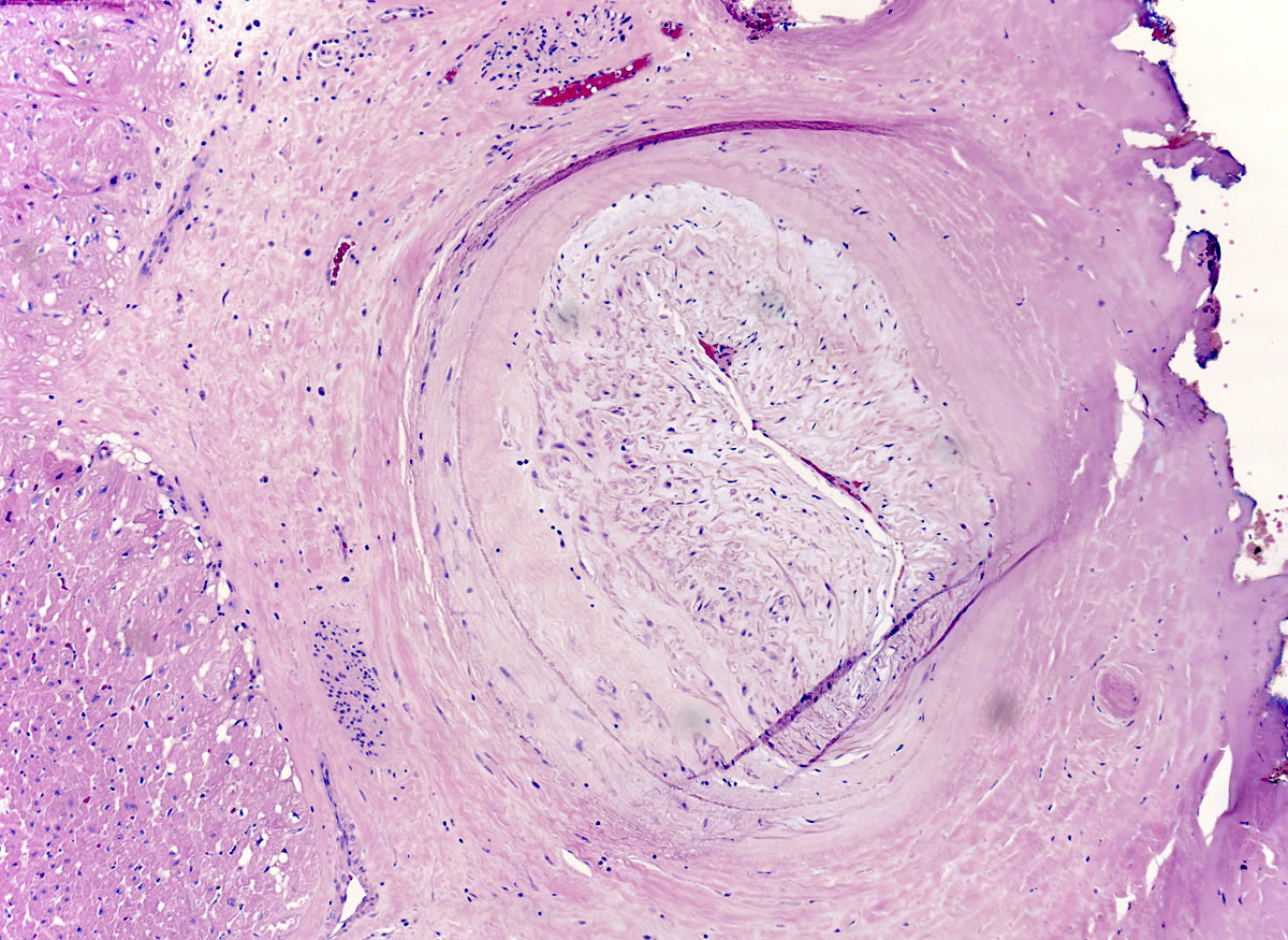
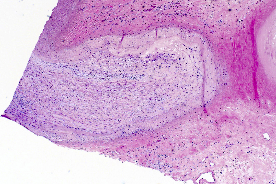
Tricuspid valve
Contributed by Melanie C. Bois, M.D.
Surgical excision
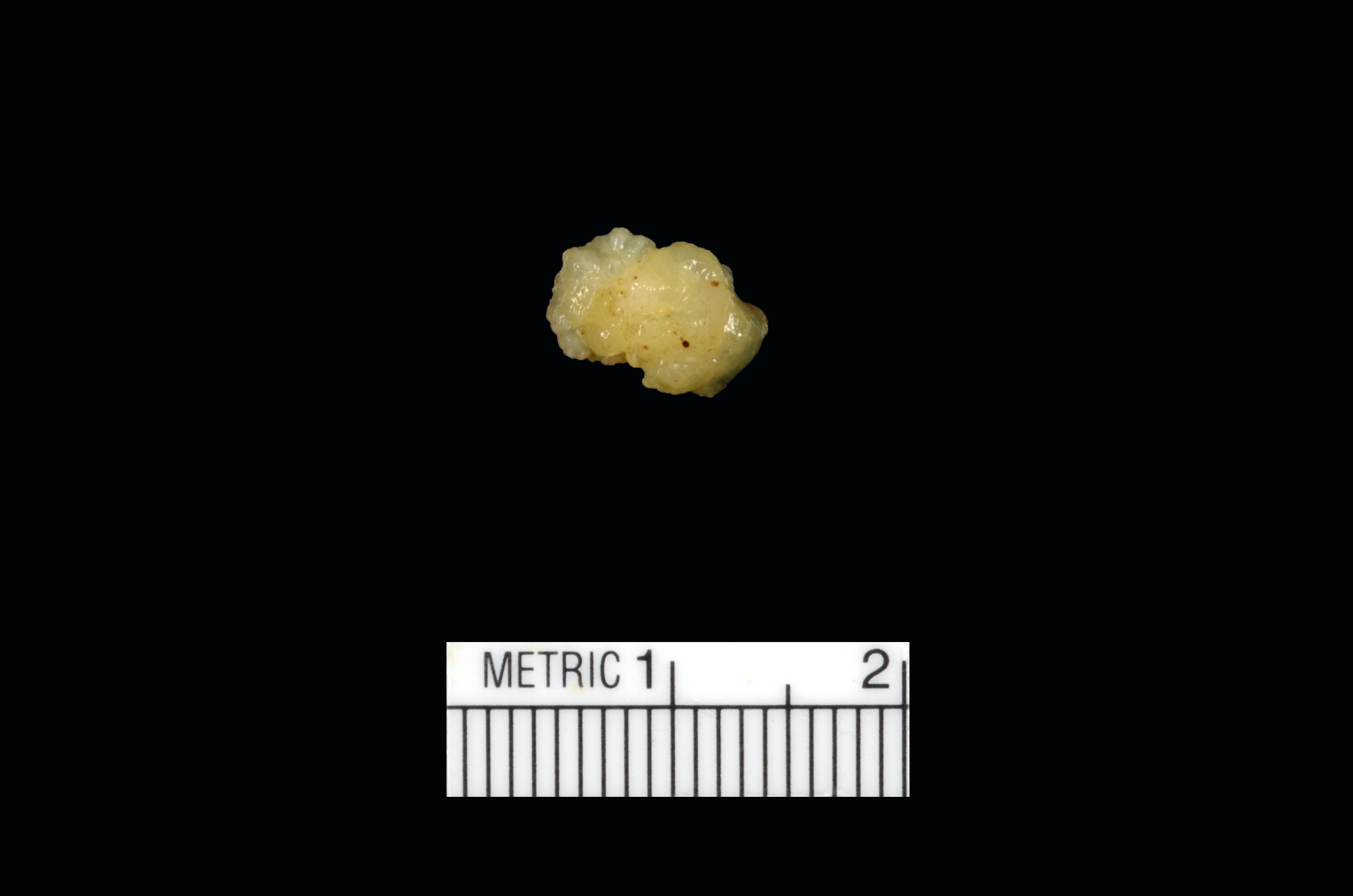
In nonaqueous medium
AFIP images
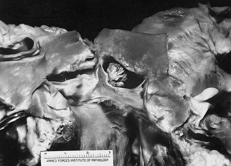
Tumor in noncoronary sinus
Images hosted on other servers:

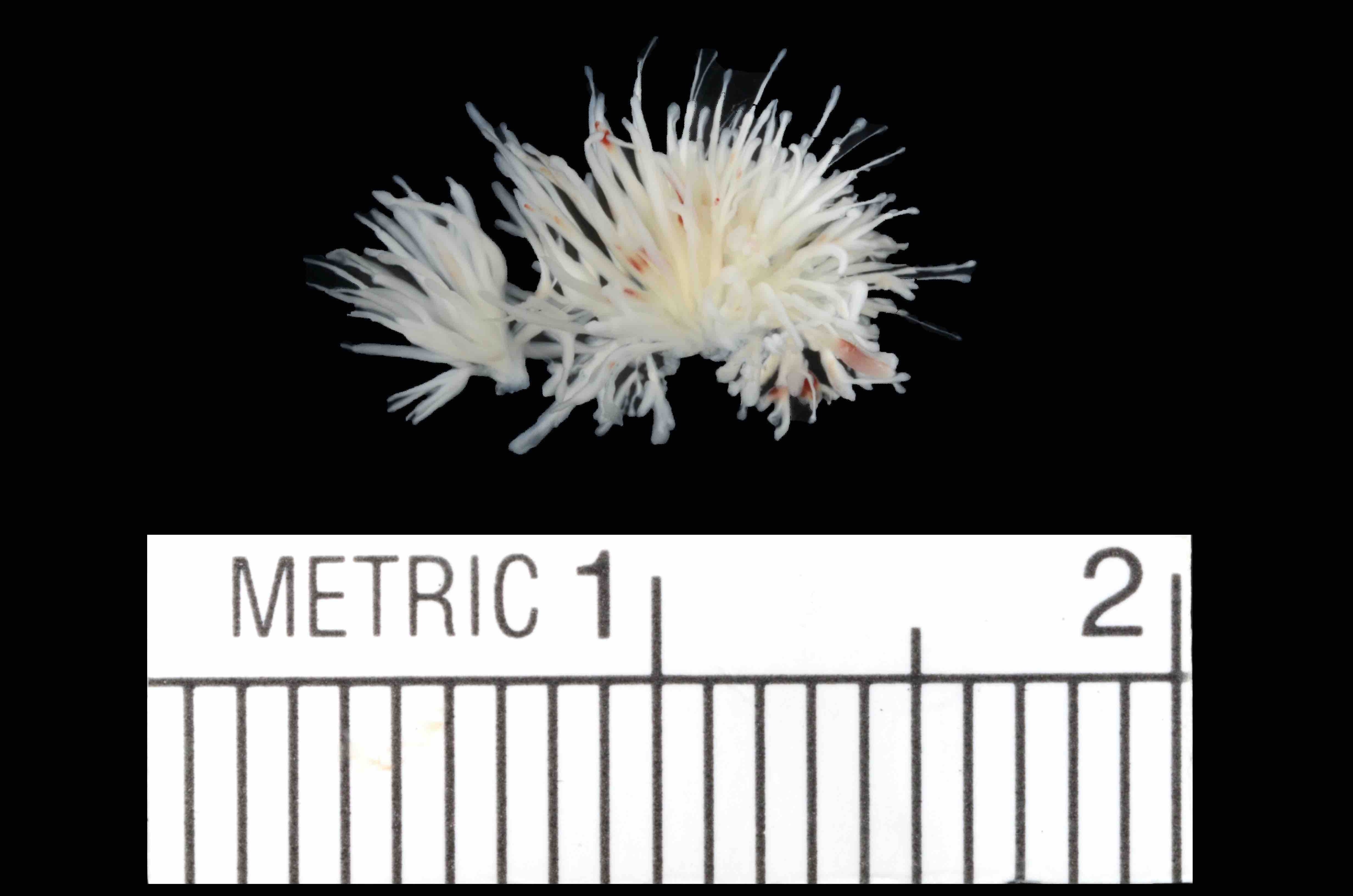
Multiple small fronds resembling a sea anemone

death due to
occlusion of right
coronary ostium
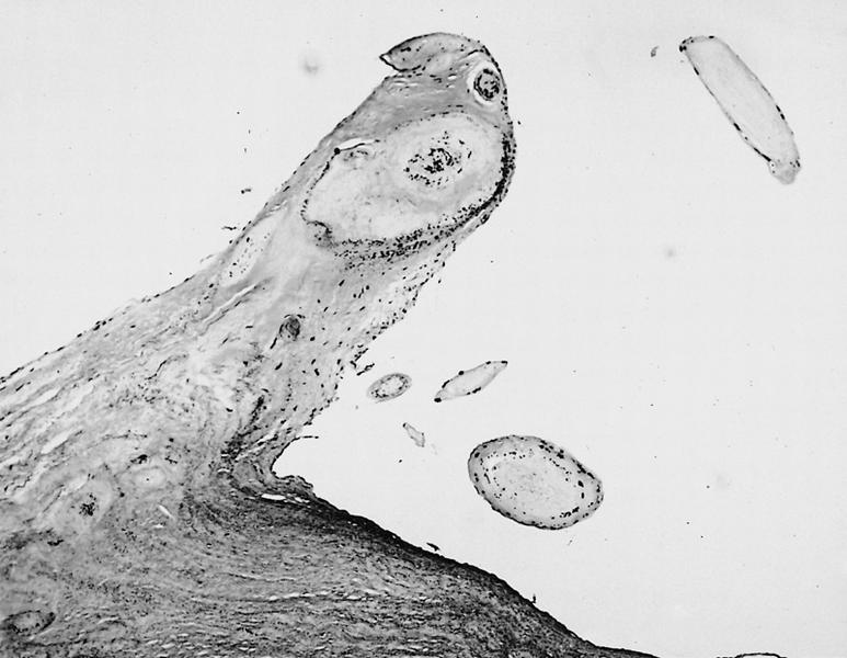
Contributed by Melanie C. Bois, M.D.
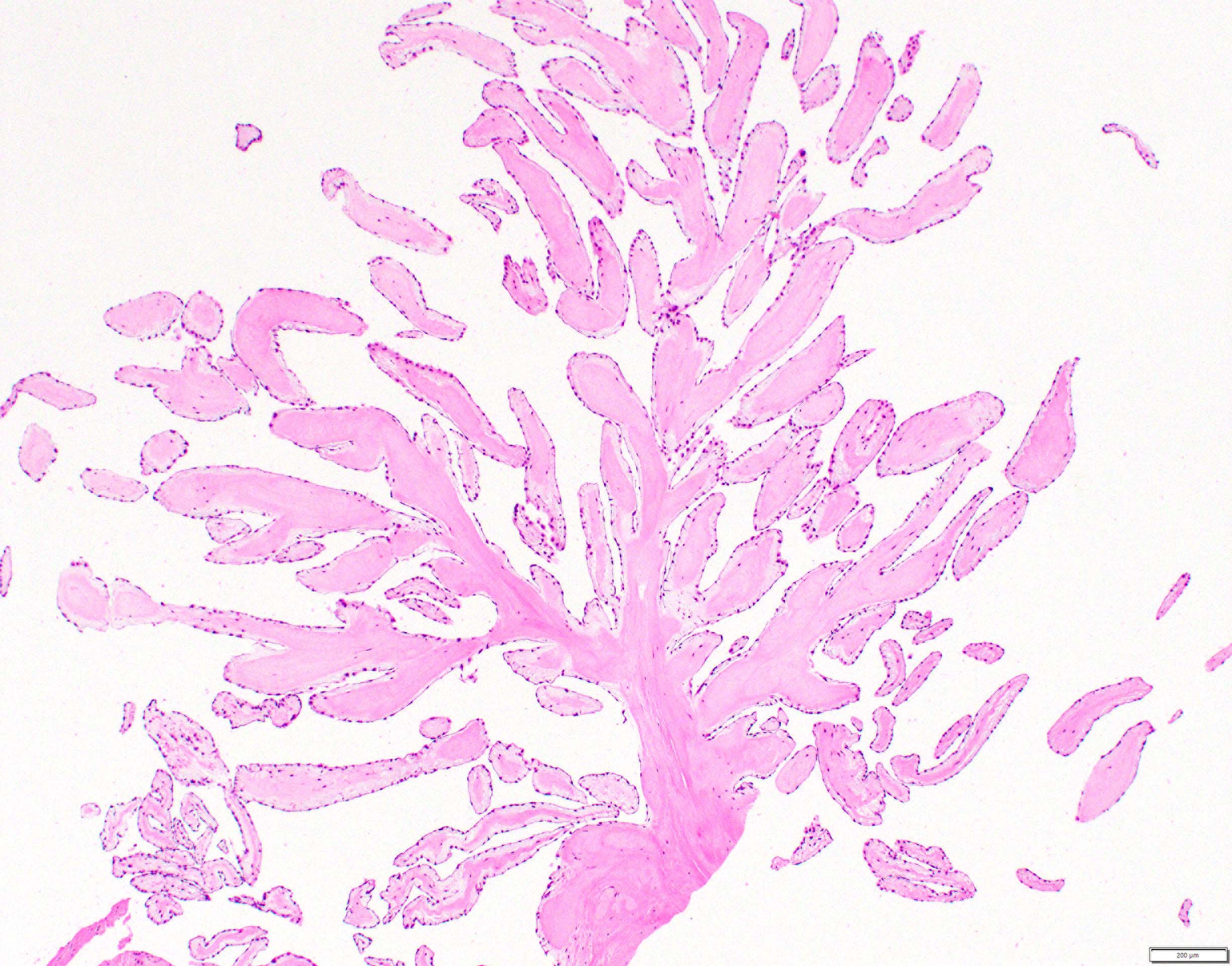
Arborizing fronds
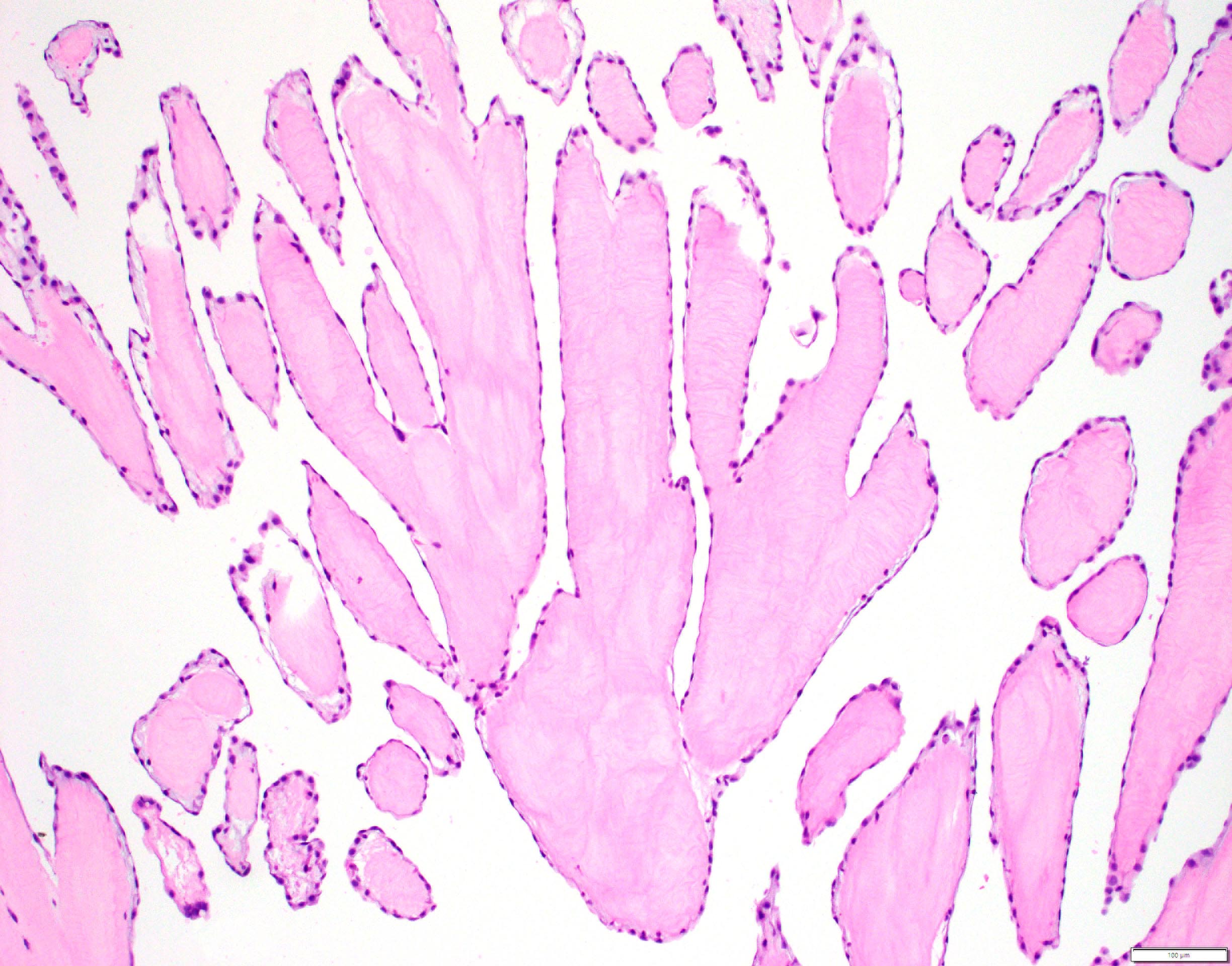
Avascular cores
AFIP images

Arising from aortic valve cusp
Movat pentachrome stain
Avascular tumor fronds growing into a cardiac chamber
Architecture obscured by scarring
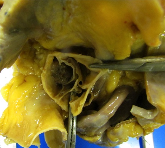
Resection of large aortic valve papillary fibroelastoma
Images hosted on other servers:

Ventricles dilated more than atria
Images hosted on other servers:

Cardiomyocyte multinucleation

Adenomatous hyperplasia
Images hosted on other servers:

Starr-Edwards mechanical valve

Bjork-Shiley concavo-convex valve

St Jude Medical bileaflet prosthesis

Prosthetic aortic valve


Aortic prosthetic valve before & after IV heparin
Images hosted on other servers:

Prosthetic cardiovascular devices

AHA/ACC recommendations

Gross examination of prosthetic heart devices
Images hosted on other servers:

shows significant pannus

Mechanical mitral valve prosthesis

Synthetic chordae tendinae
Images hosted on other servers:

endocardium, native
mitral leaflet tissue

Synthetic chorda surrounded by tissue fibrosis

Segment of coronary artery
AFIP images
Right ventricular mass
with tuberous sclerosis
and multiple minute tumors
studding epicardium
Images hosted on other servers:


Left ventricular tumor causing sudden death in children ages 1-2
AFIP images
and intracellular
myofilaments
(oil emersion)
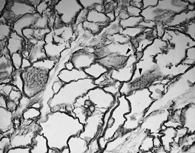
highlights spider
cells and cross
striations (arrowheads)
Images hosted on other servers:

Drawings from 1938 report
AFIP images
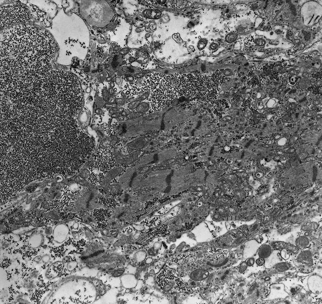
sparse mitochondria, fragmented
irregular myofilaments with Z bands
Images hosted on other servers:

Late gadolinium enhancement (LGE)
Images hosted on other servers:

Myocardial infiltrates
Contributed by Carolyn Glass, M.D., Ph.D.

Cardiac biopsy
Images hosted on other servers:

Dilated aortic root and ascending aorta

Enlarged aortic root
Images hosted on other servers:


Various images


Images hosted on other servers:

Interstitial widening in acute phase
Illustrated walkthrough of the essential features of Takotsubo cardiomyopathy
Ventricular angiography video showing the 4 main subtypes of Takotsubo cardiomyopathy
Images hosted on other servers:

Fibrous skeleton of heart

Aortic root structures

Mitral valve annulus
Images hosted on other servers:


The four heart valves

Tricuspid valve

Isolated tricuspid valve

Pulmonary valve

Mitral valve and aortic root

Mitral leaflets


Mirtal valve apparatus

Mitral valve
commissural
chordae tendinae

Aortic value, right coronary leaflet
Images hosted on other servers:

Tricuspid valve

Annulus of the pulmonary root


Section through the pulmonary root

Posterior mitral leaflet


Section through aortic root

IHC of aortic valves
Images hosted on other servers:

Annulus tissue

Commissure tissue


Arrangement of collagenous fibrils
Ashworth: 2019
Buja: 2015
Burke: 2015
Husain: 2021
IARC: 2015
Leone: 2017
Lucena: 2015
Miller: 2023
Thiene: 2016
Find related Pathology books: cardiovascular, forensic, lung, mediastinum/serosa, pediatric
