
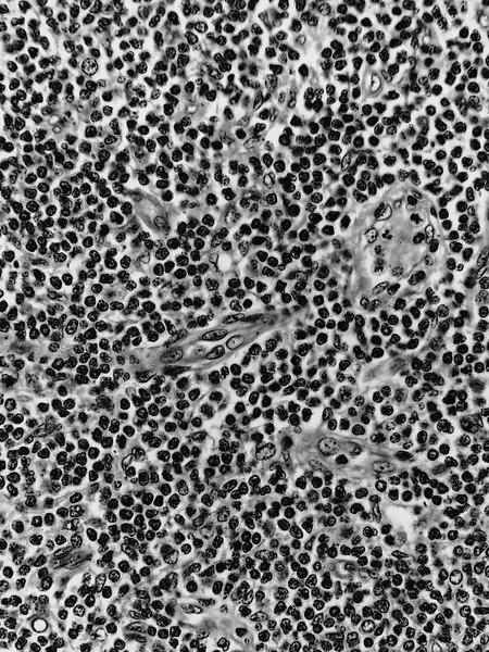
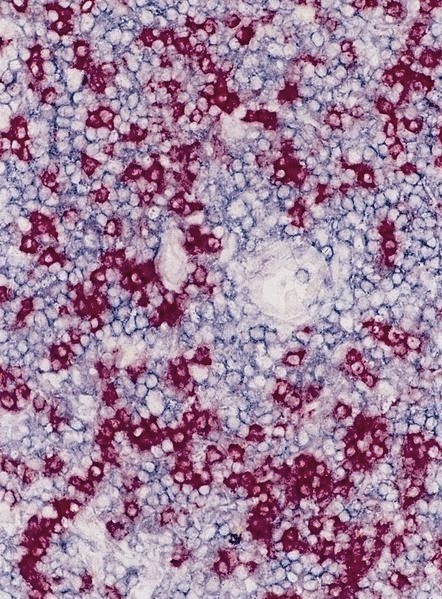
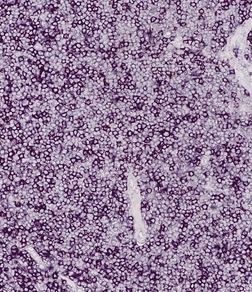
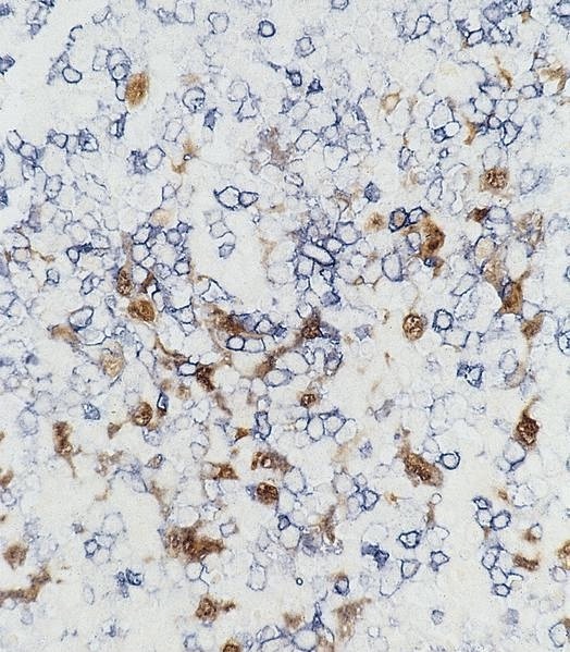
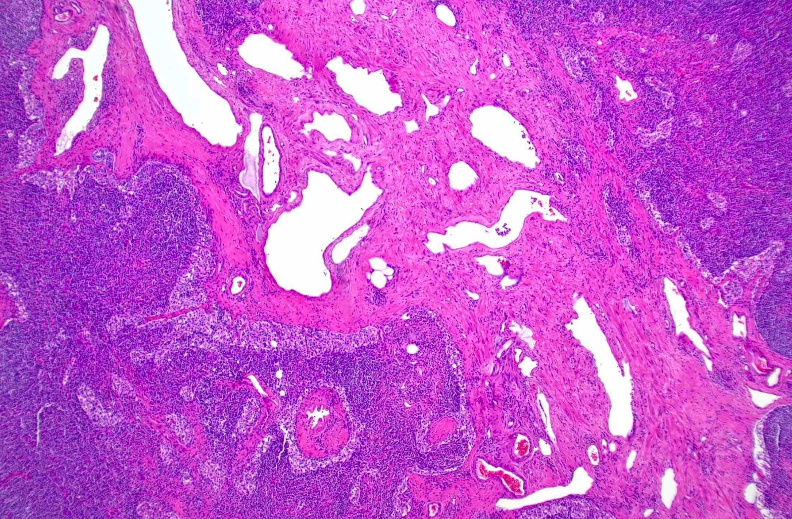
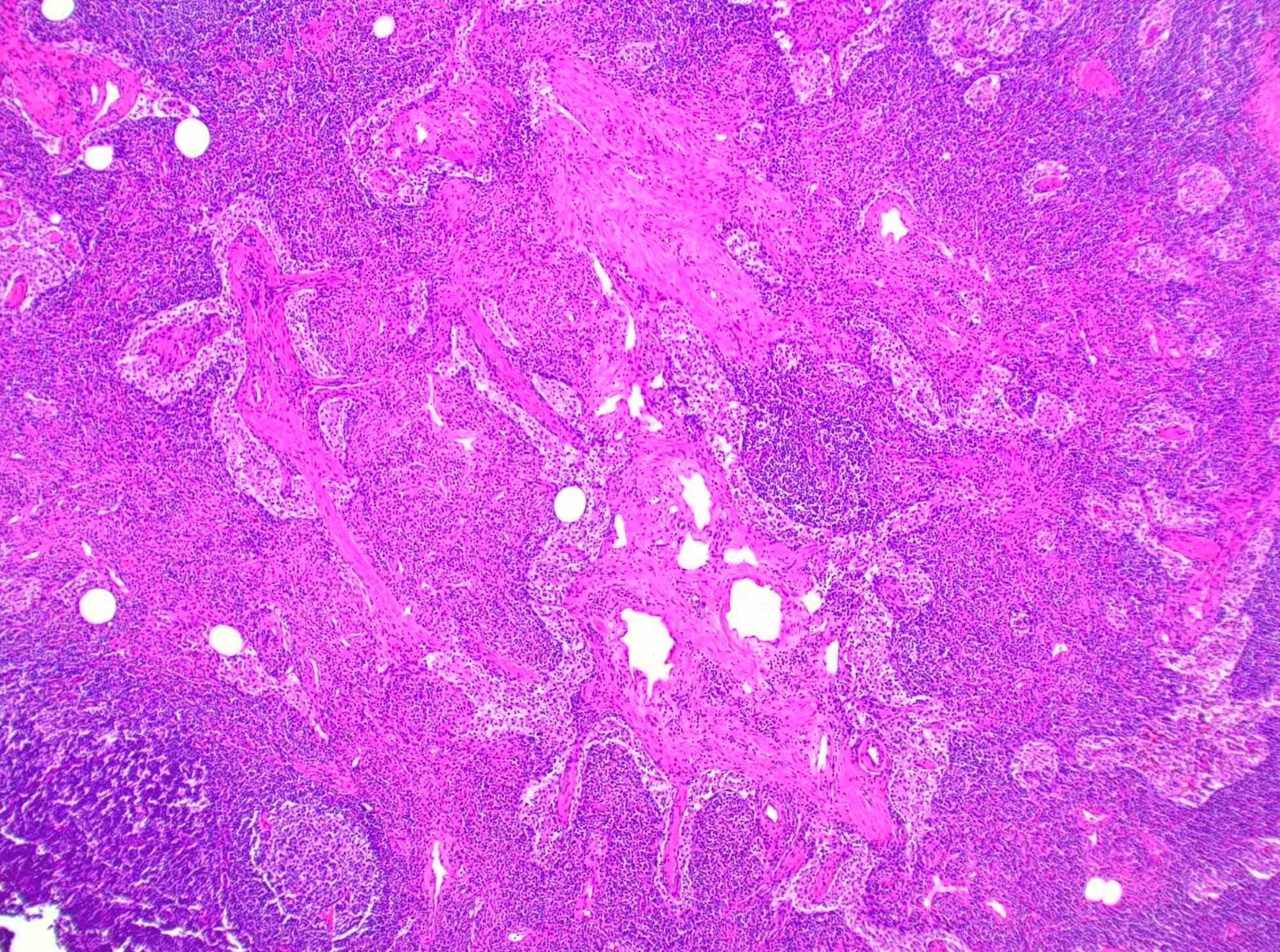
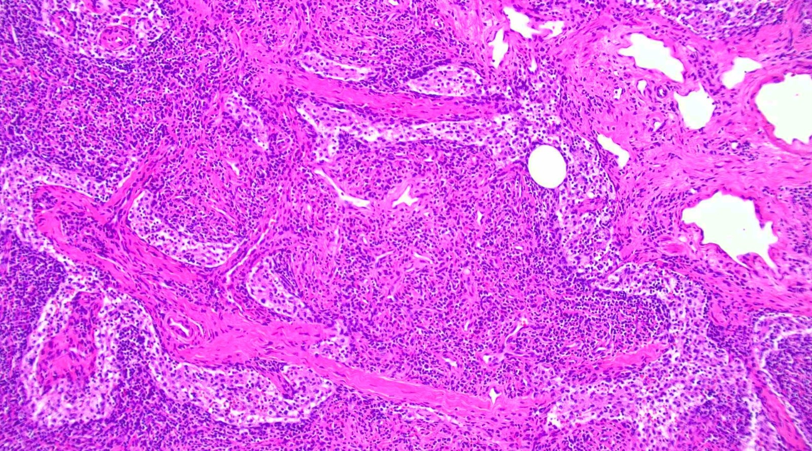
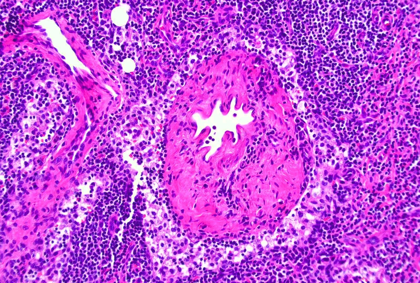
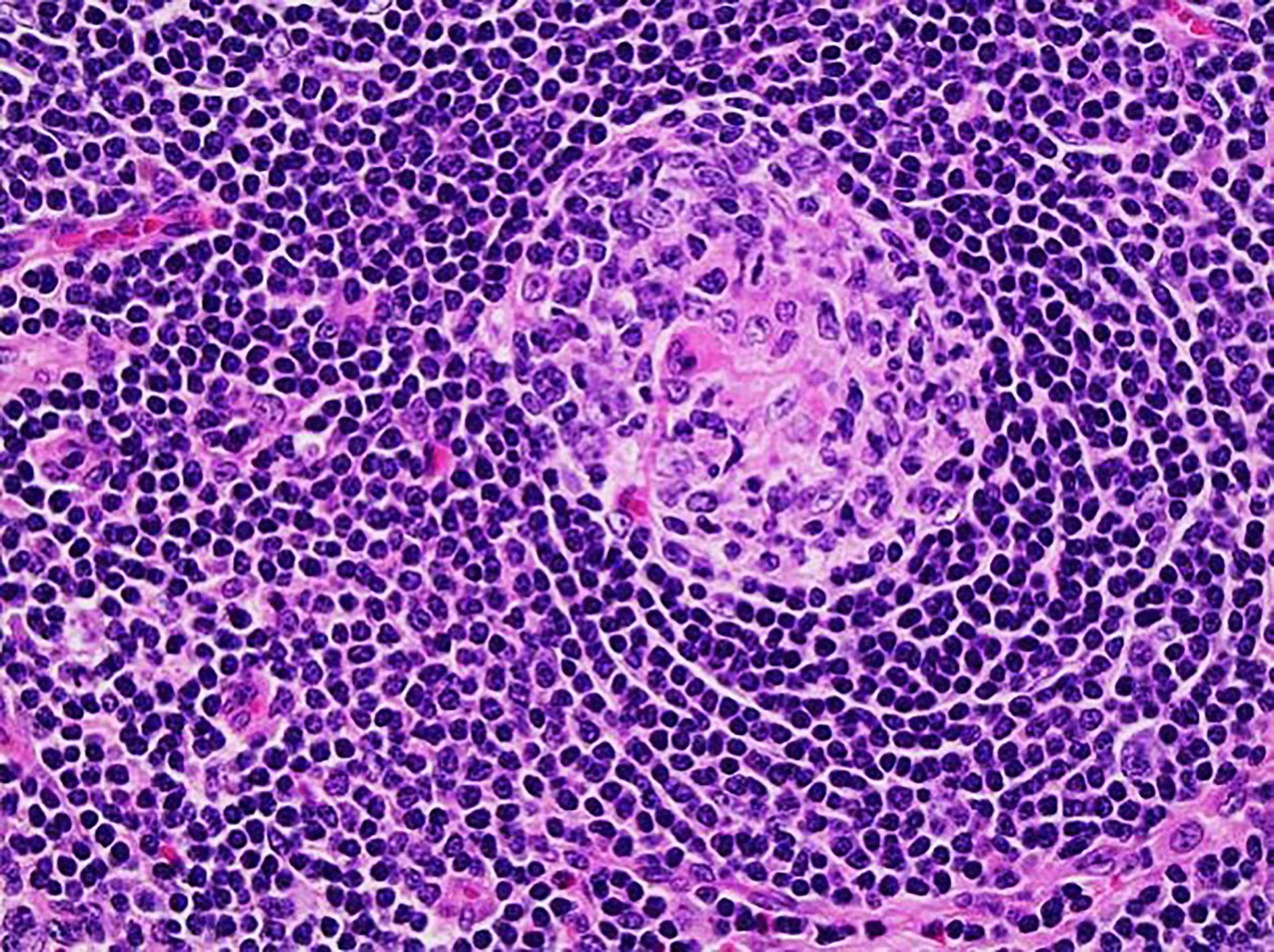
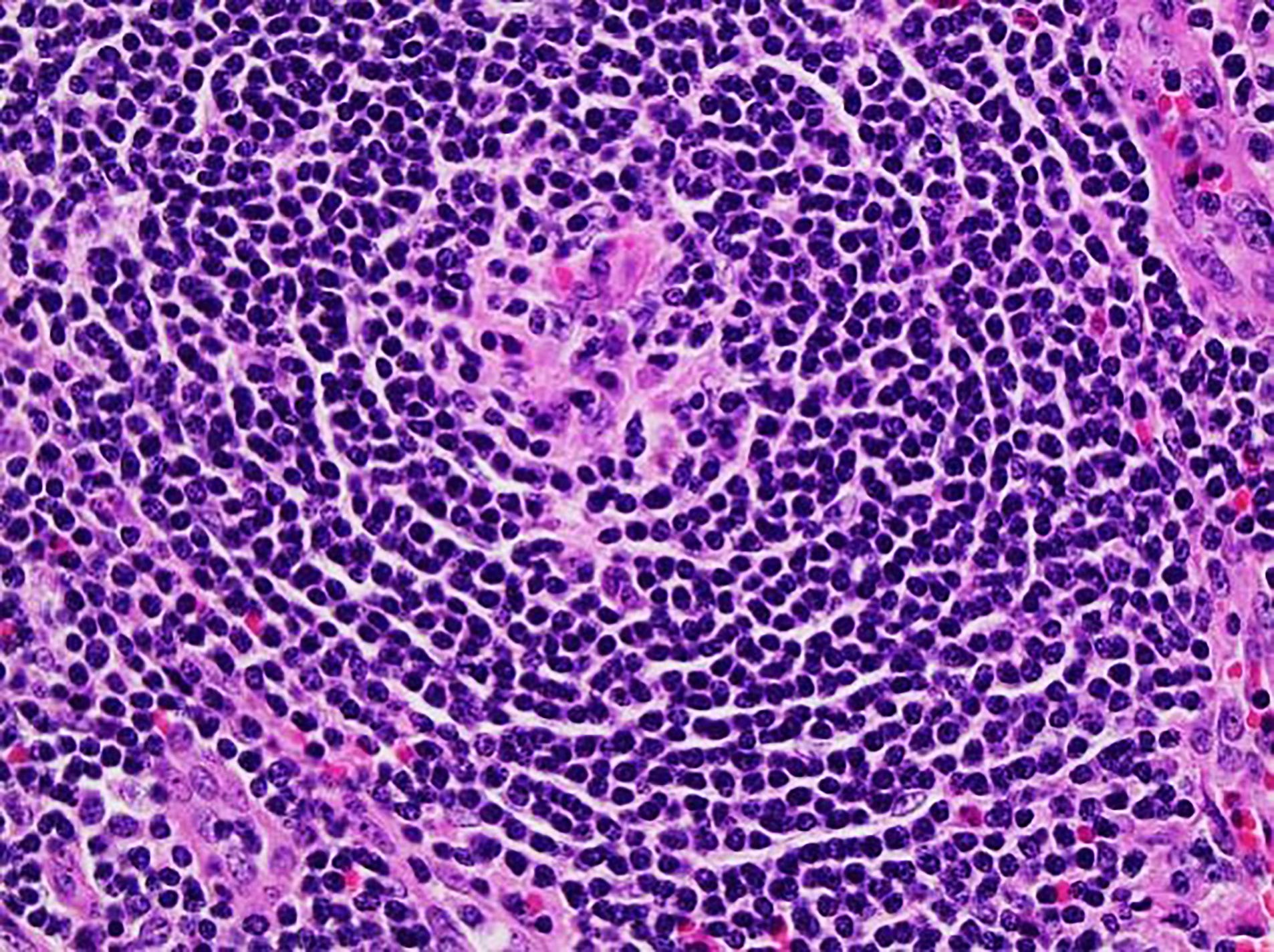
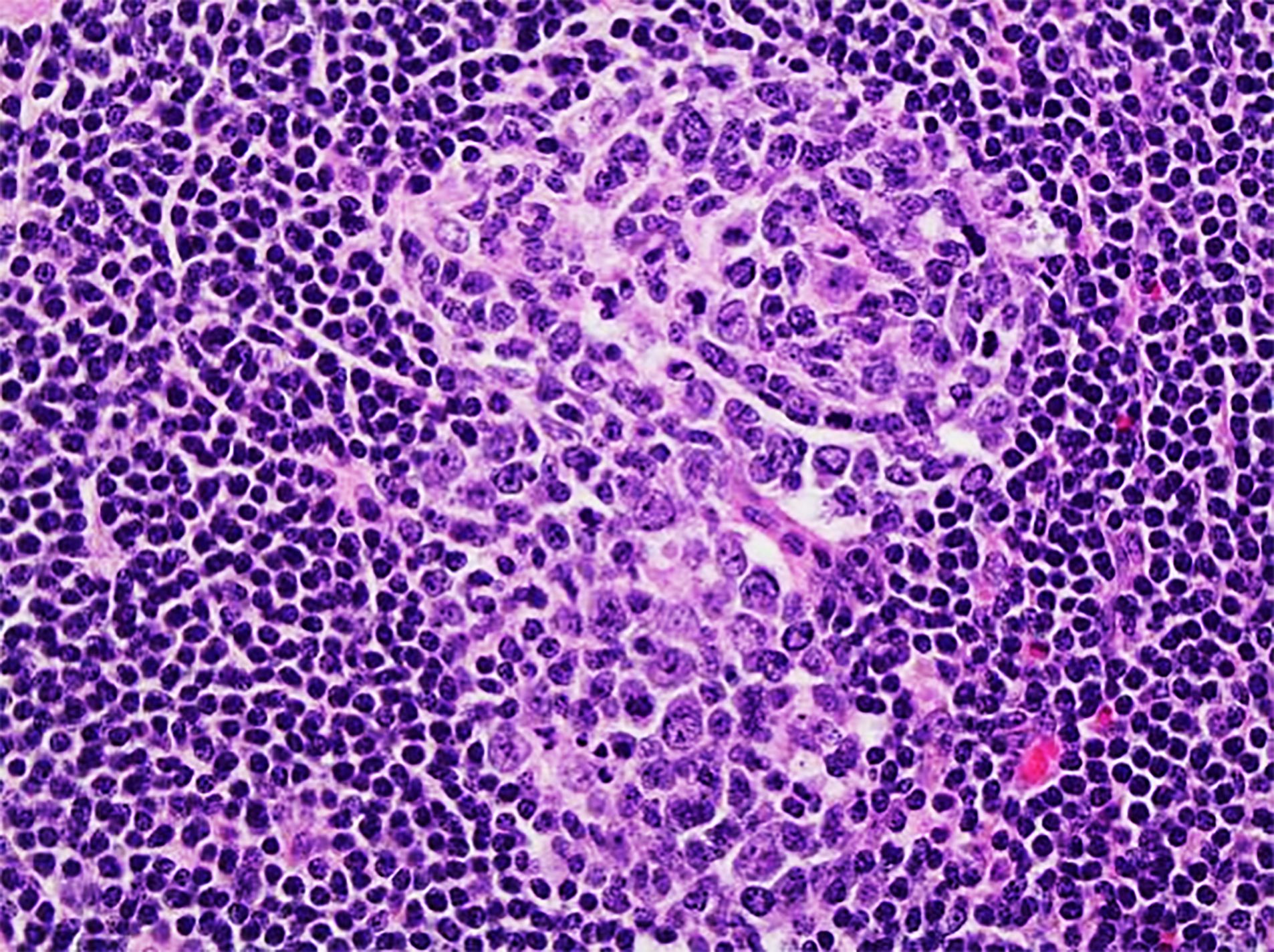
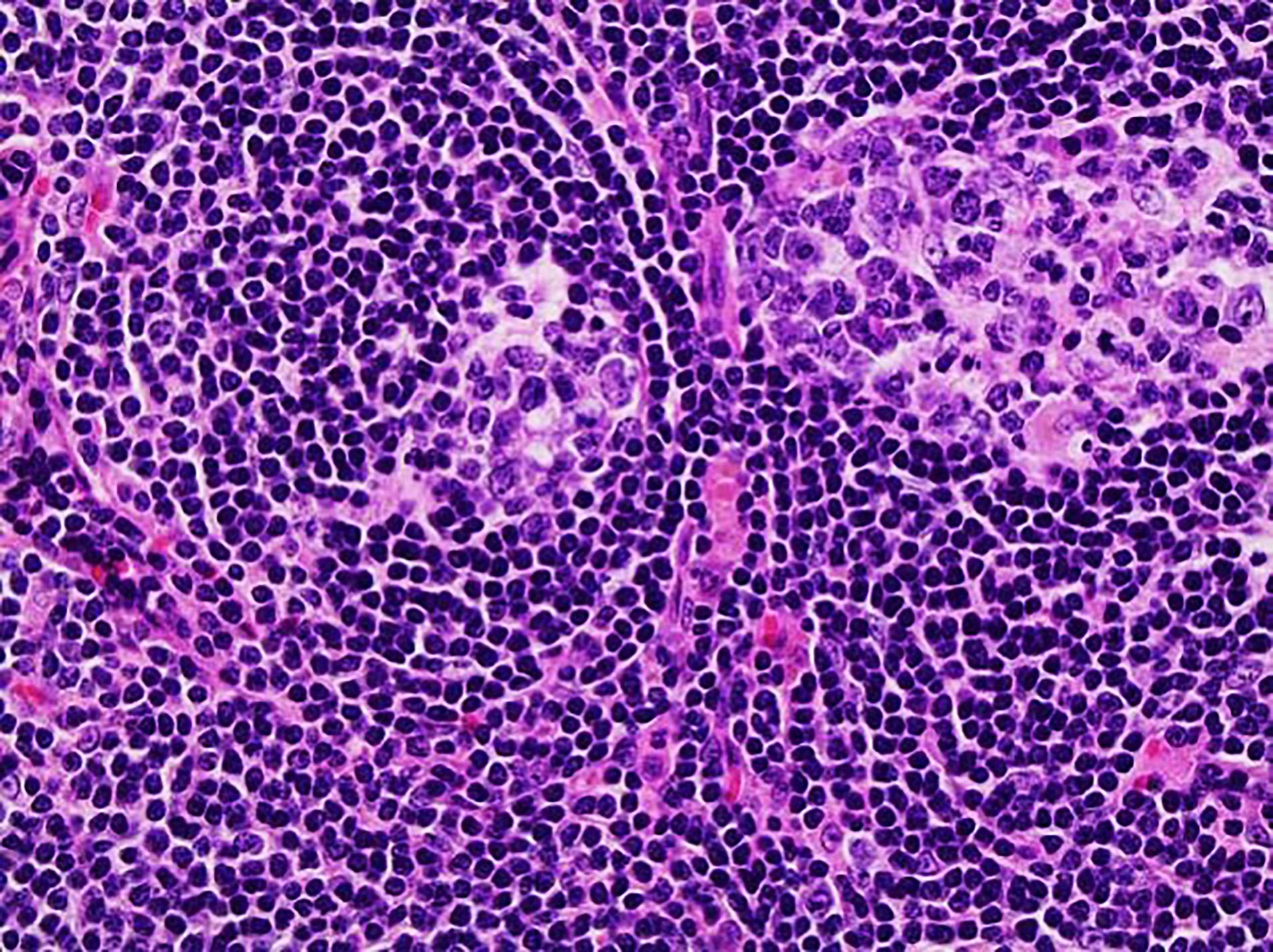
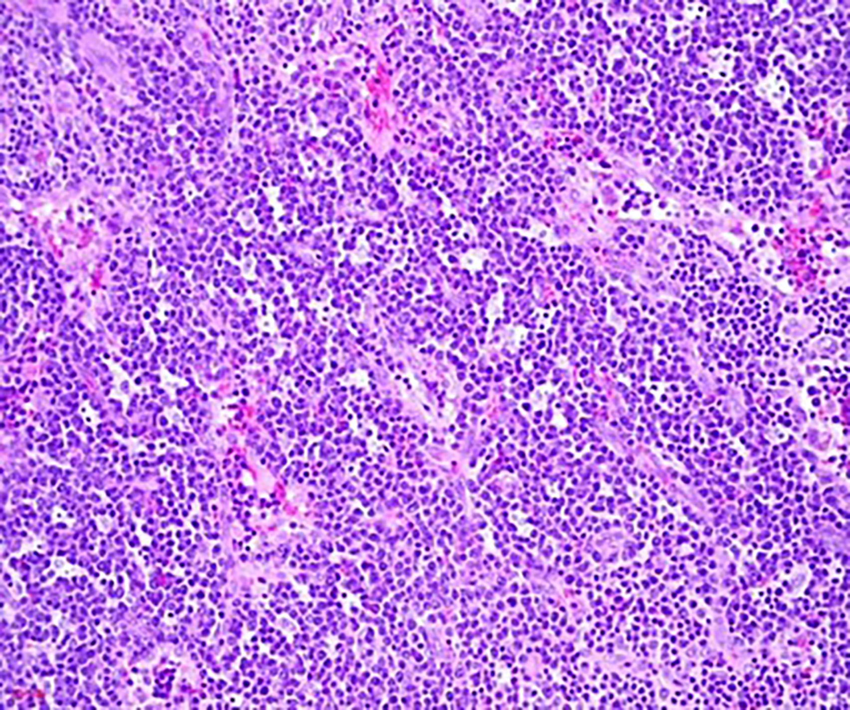
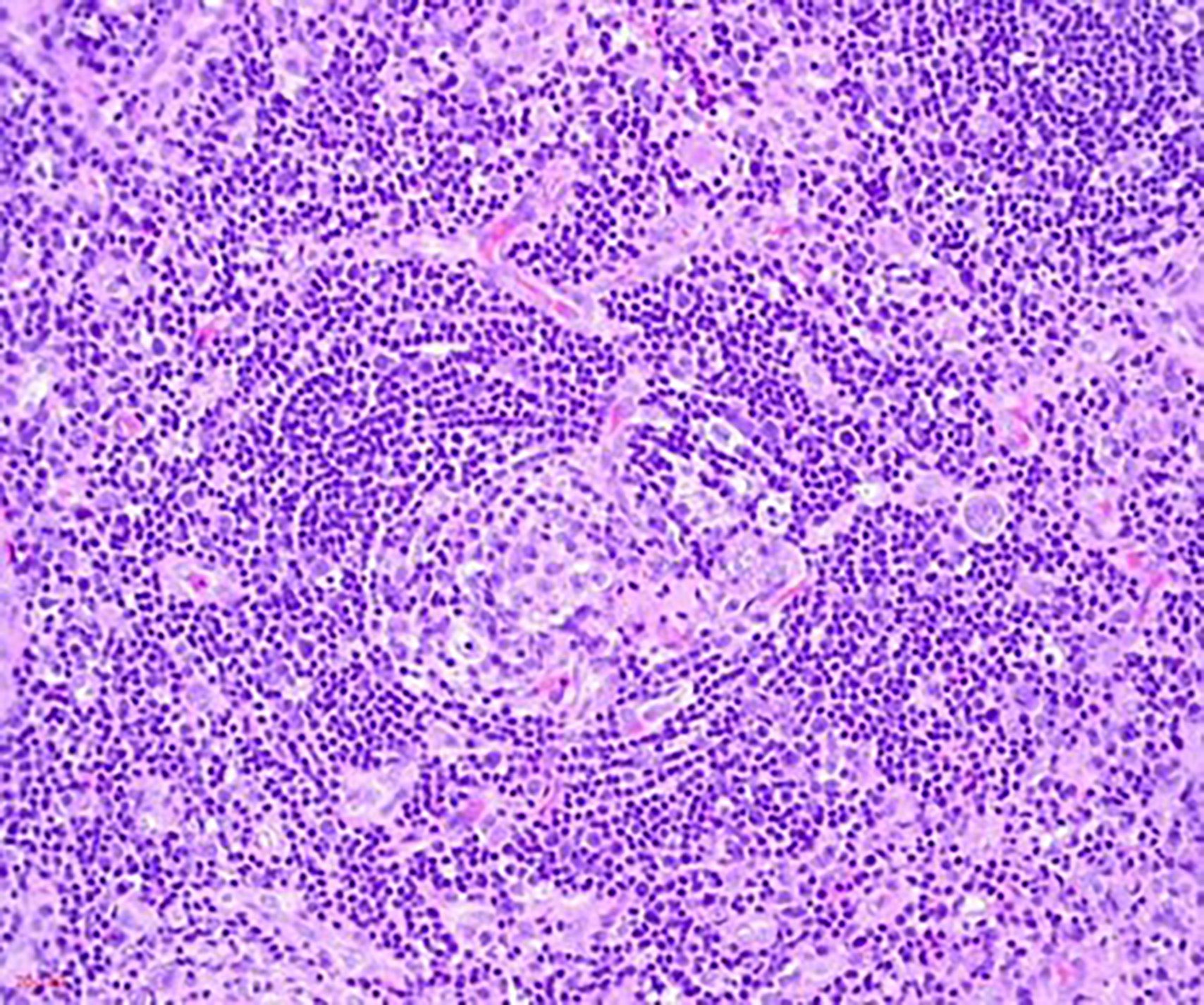
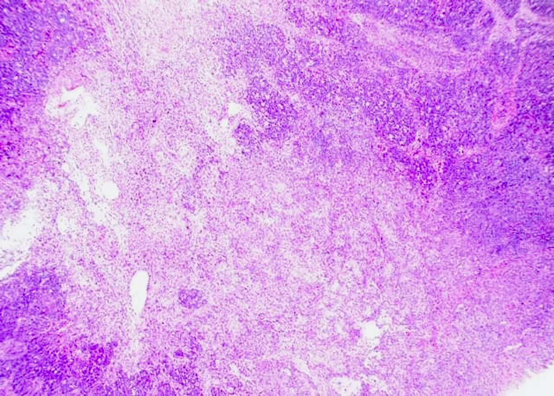
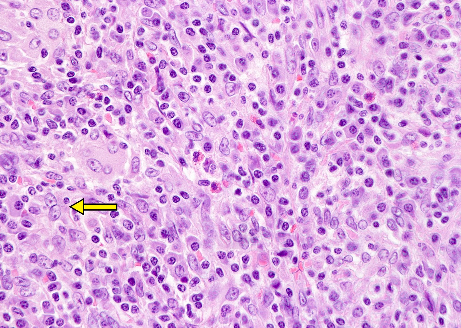
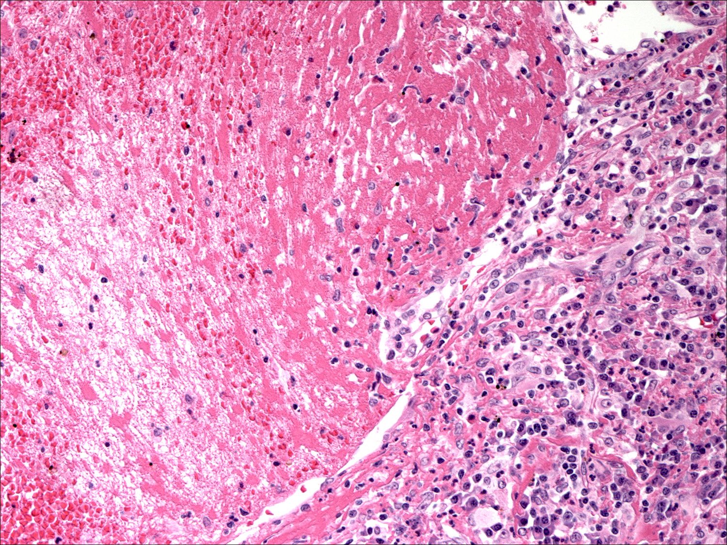
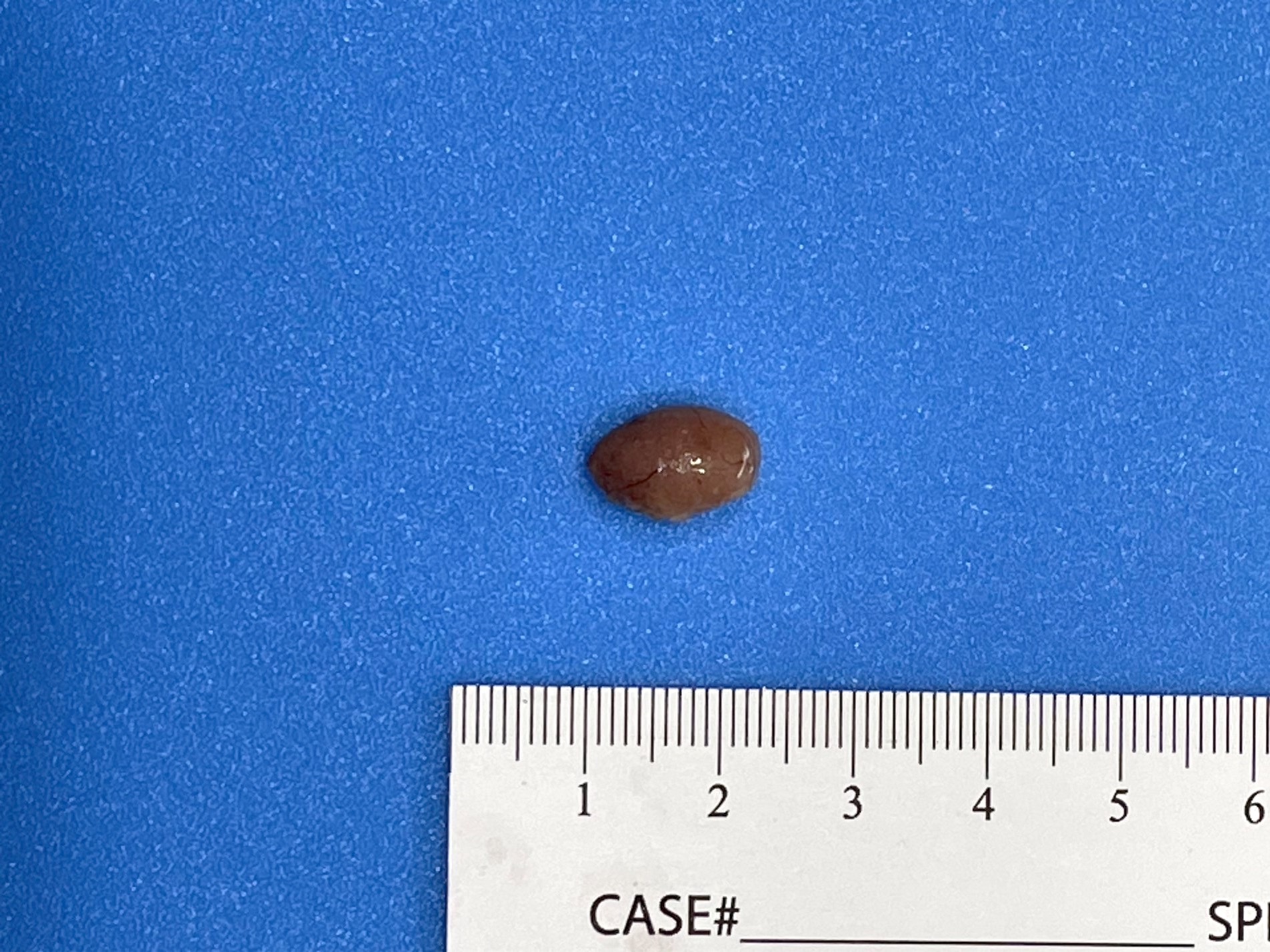
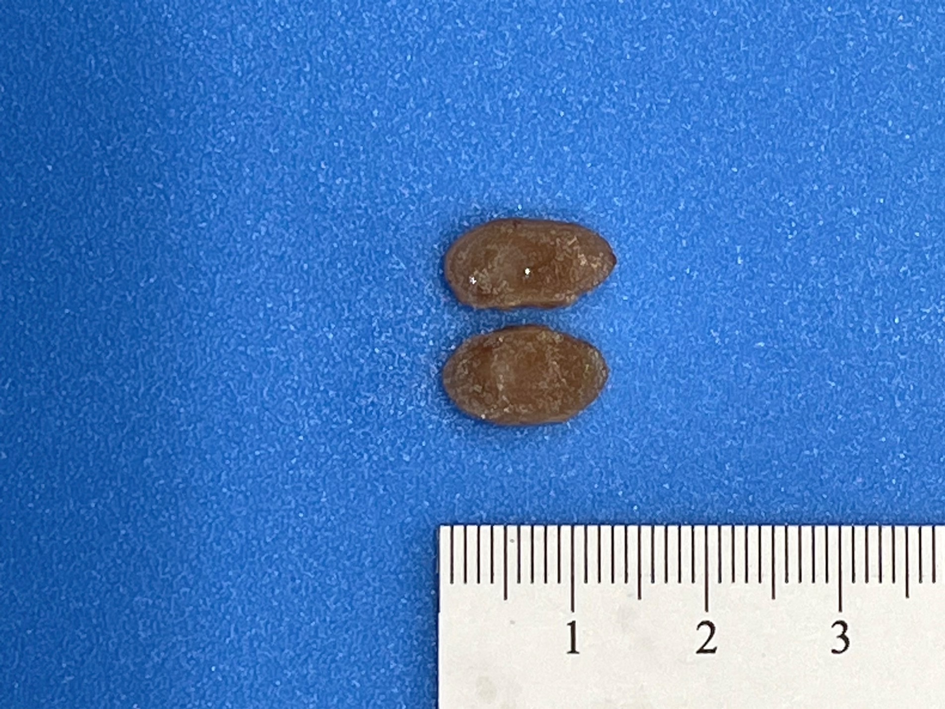
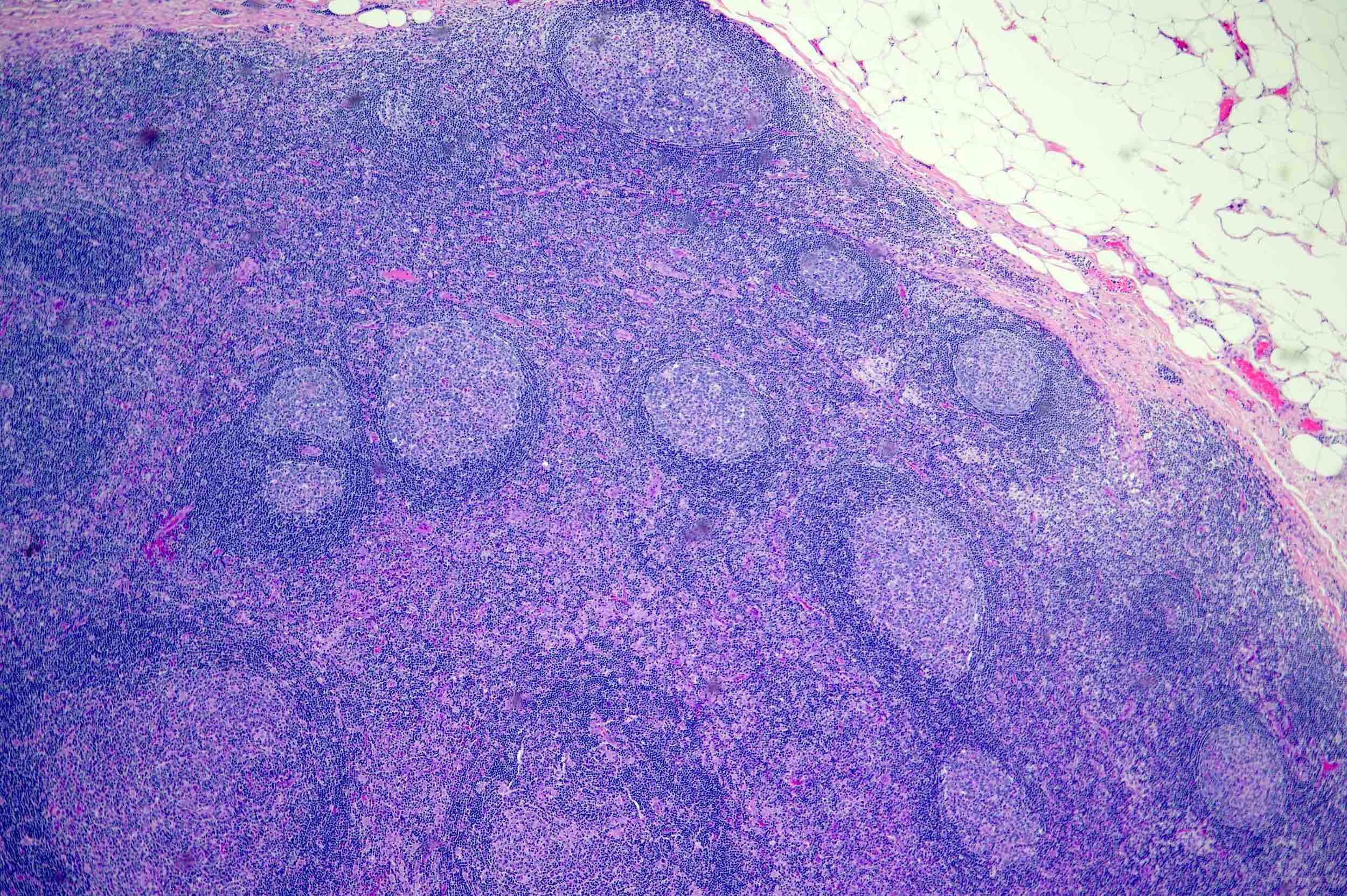
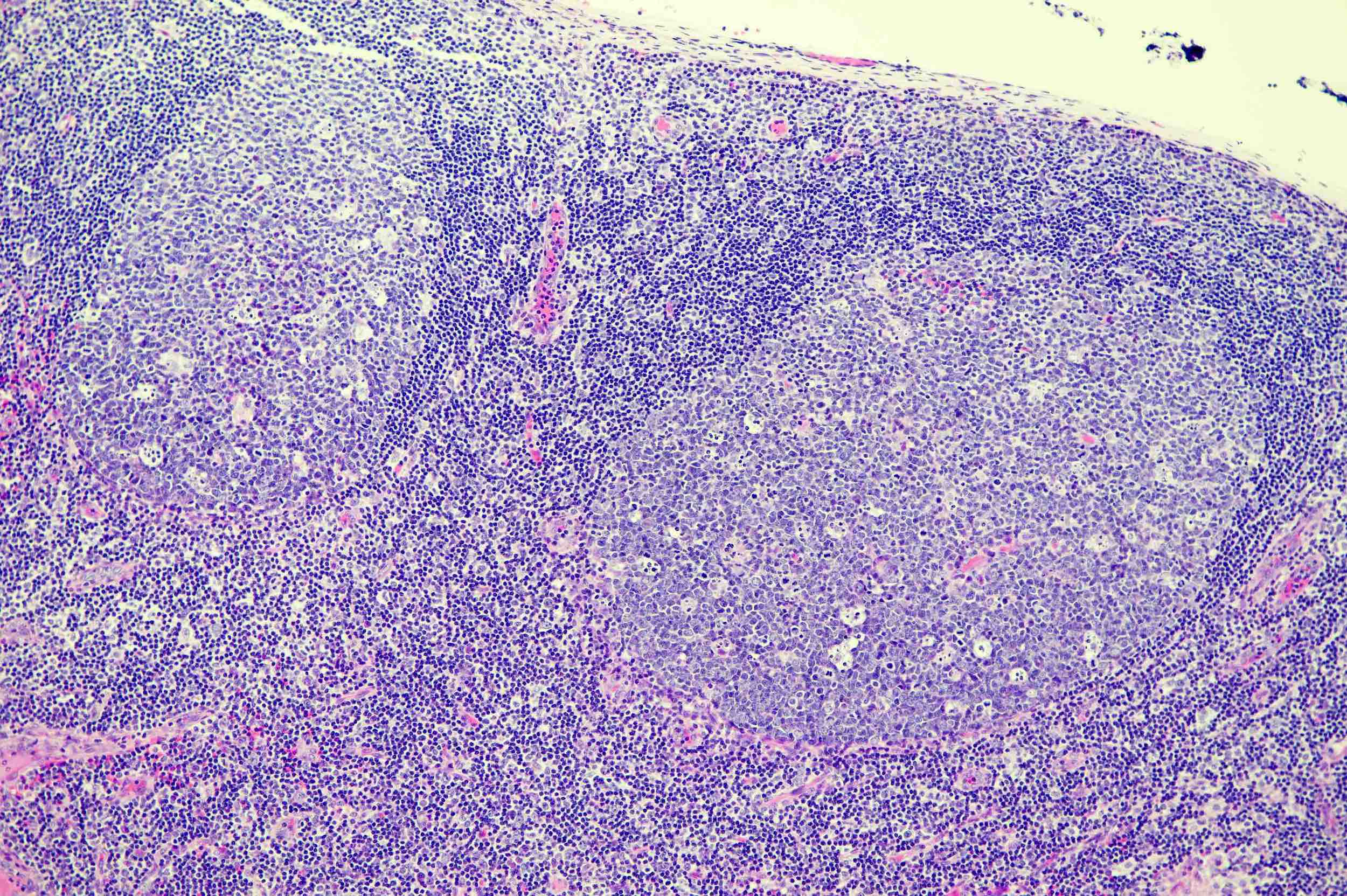
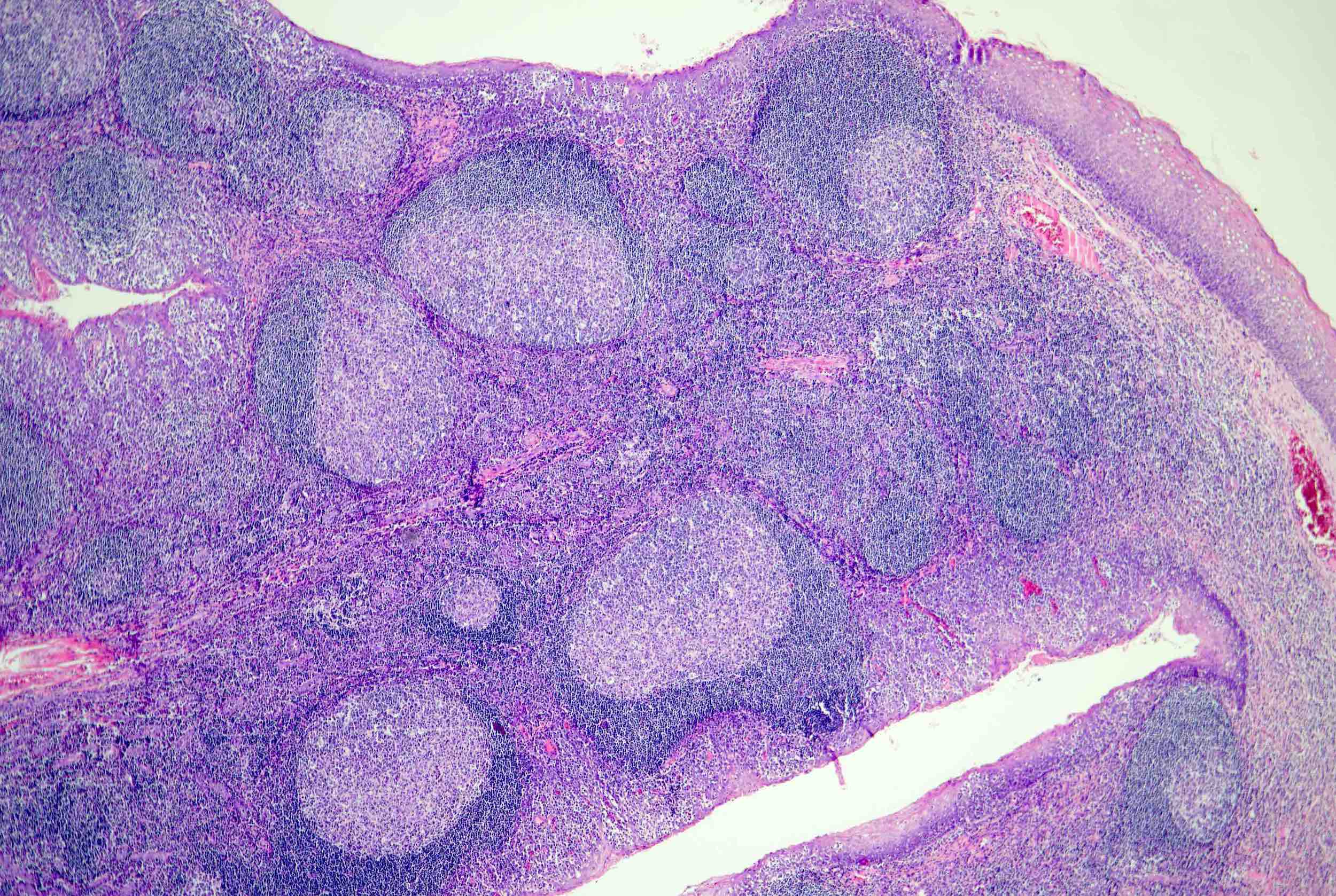
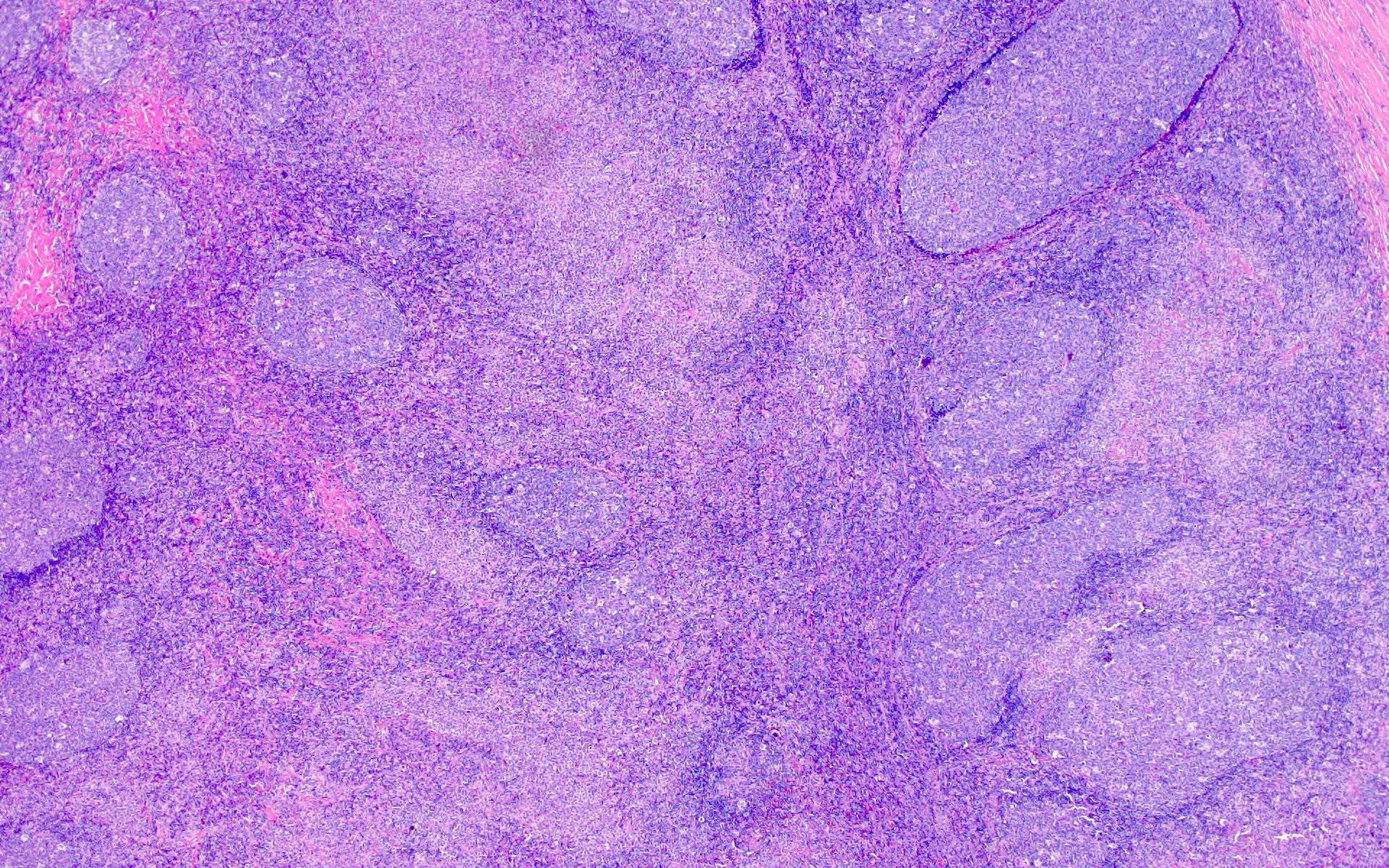
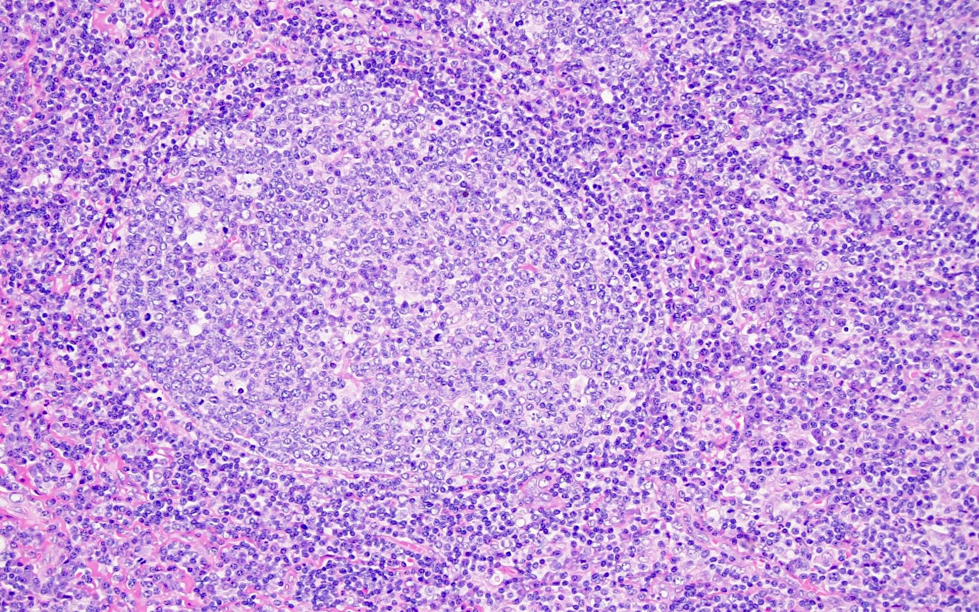
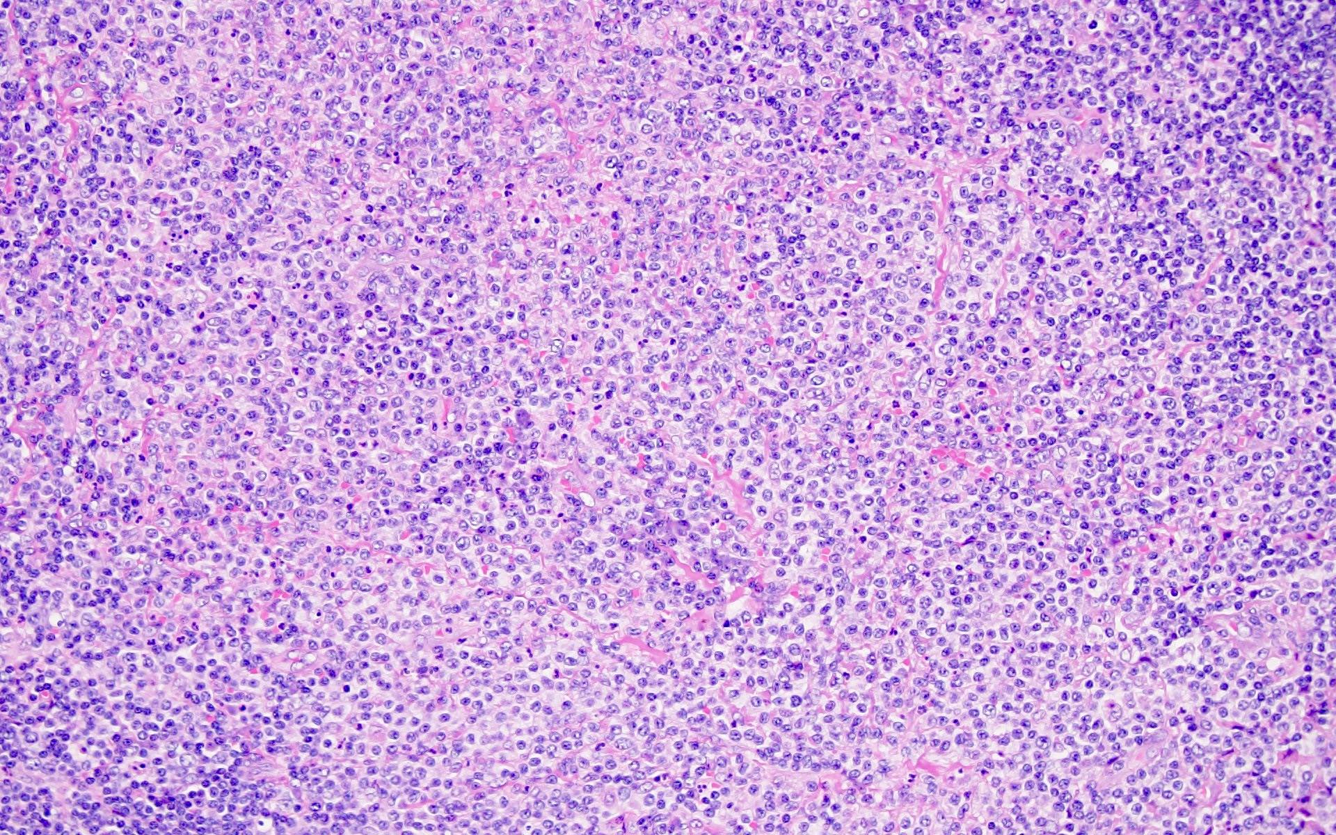
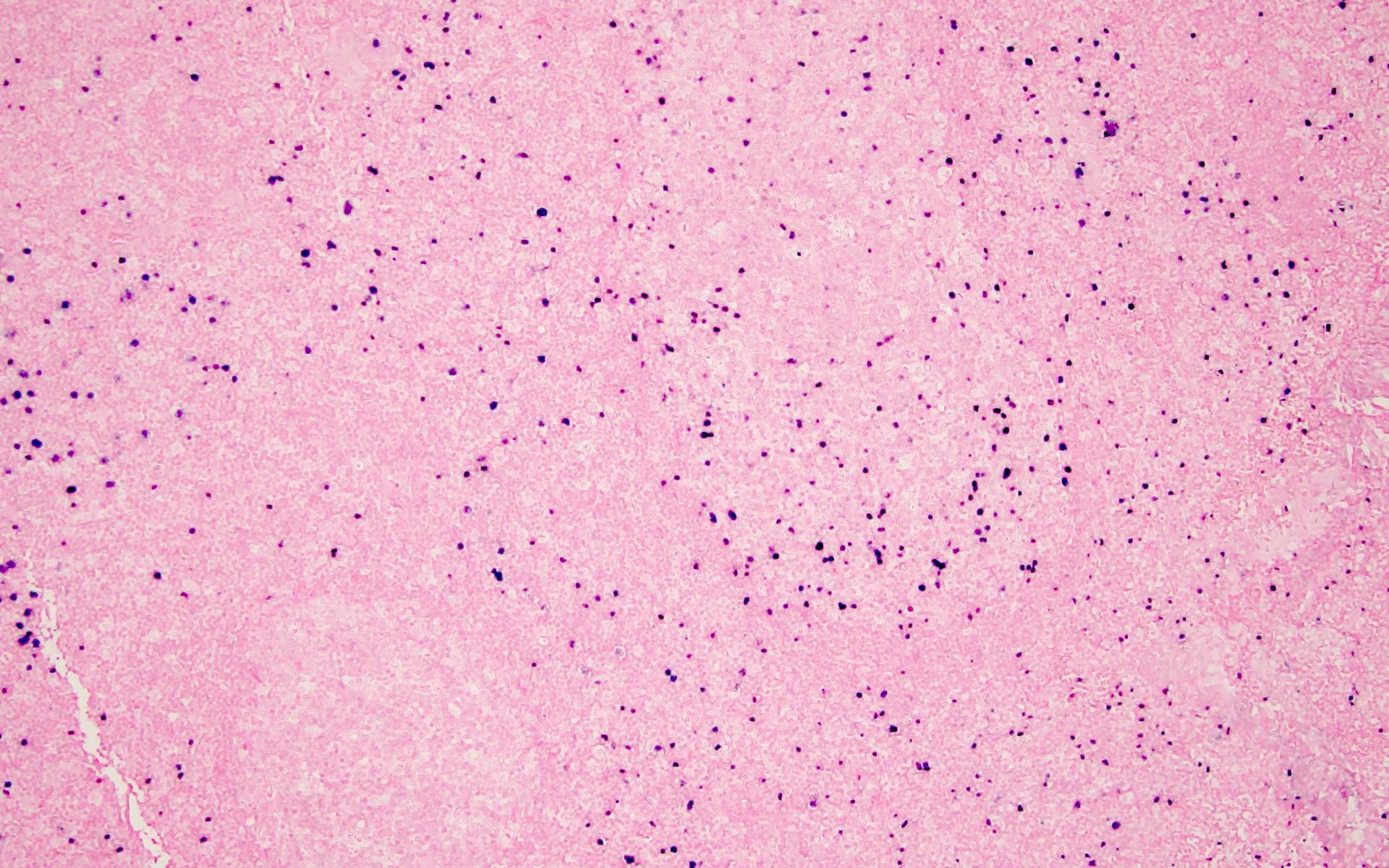
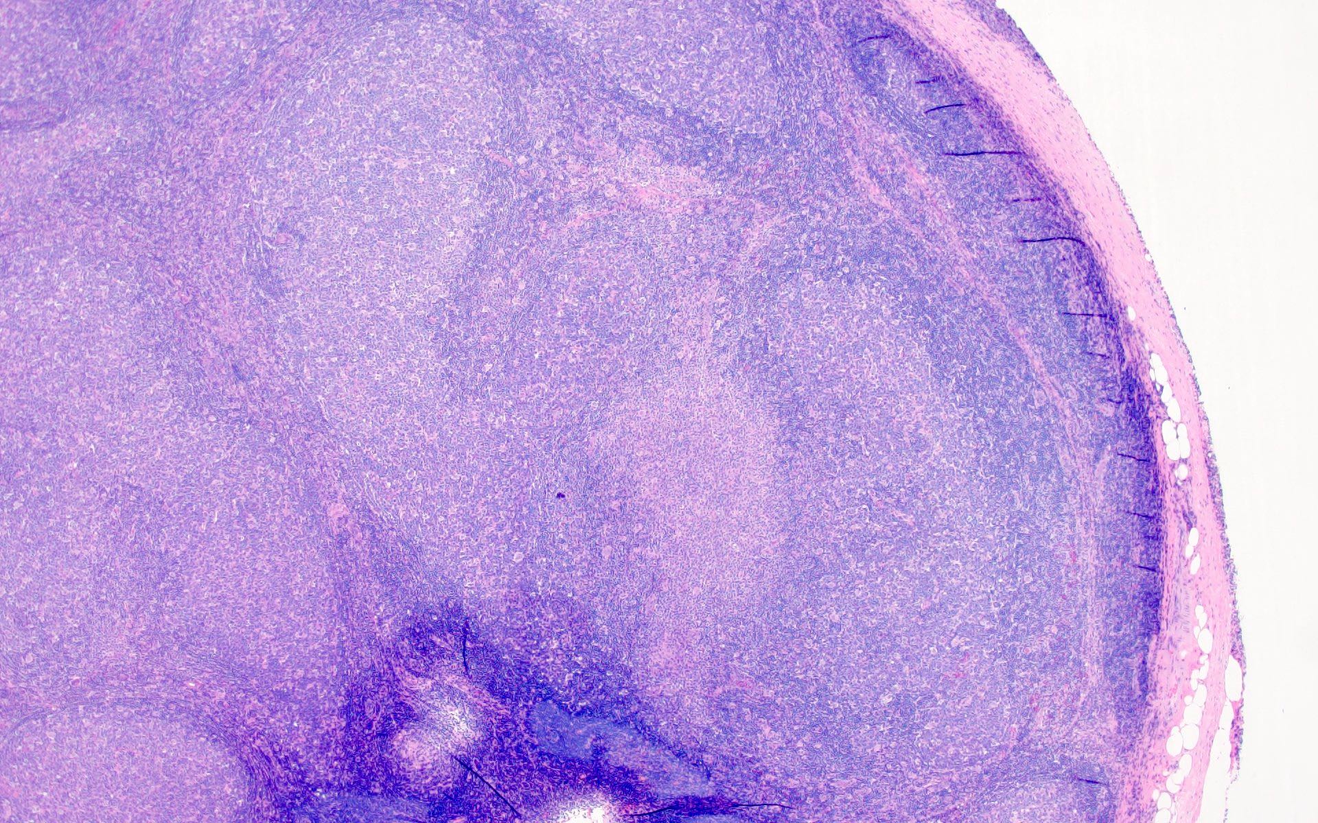
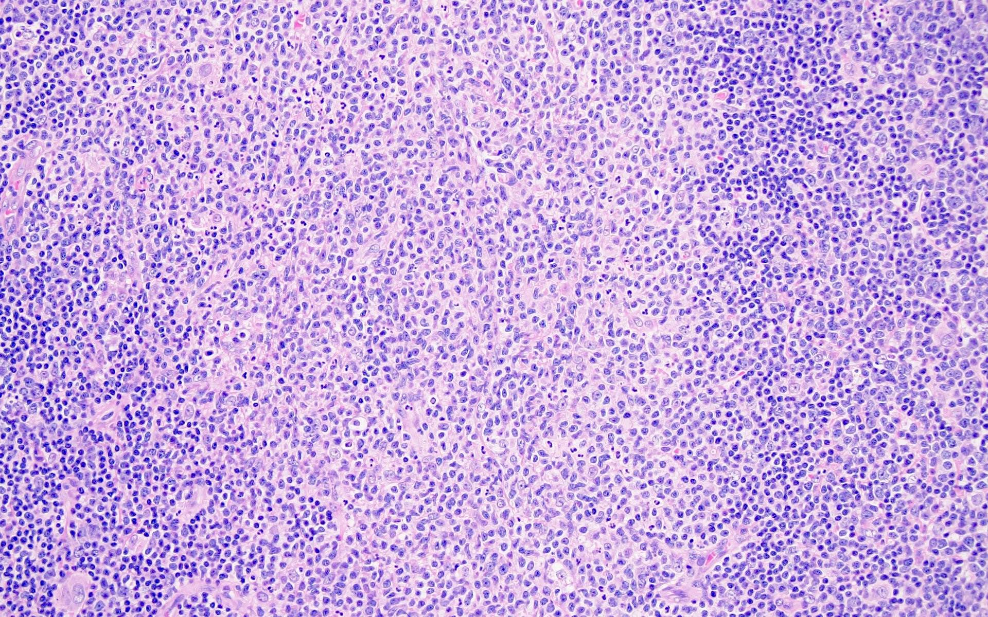
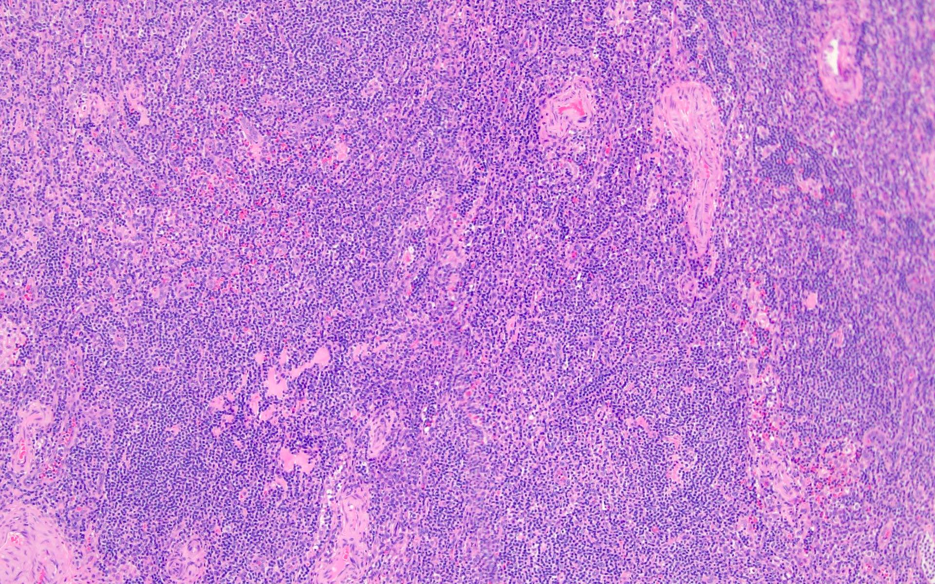
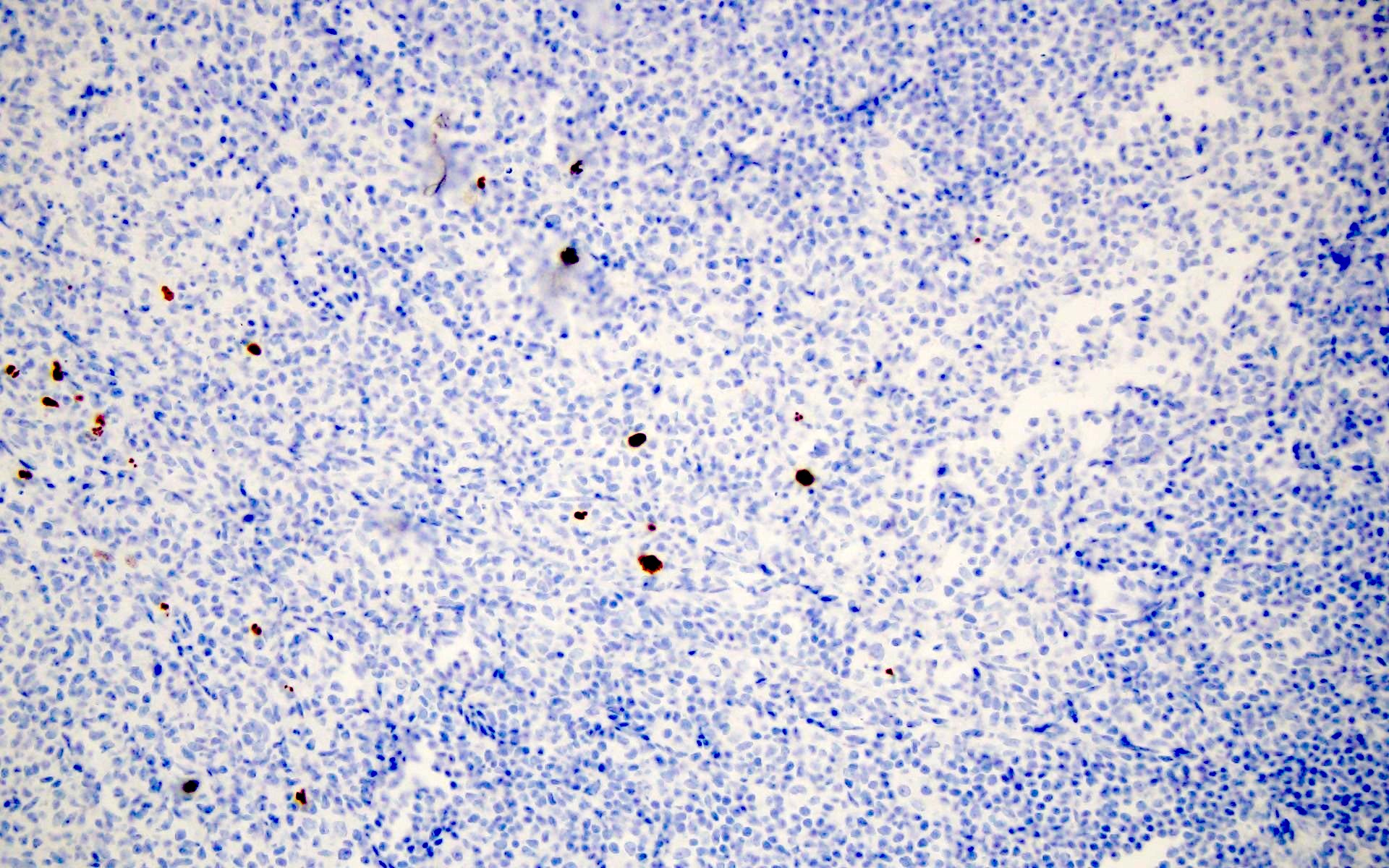
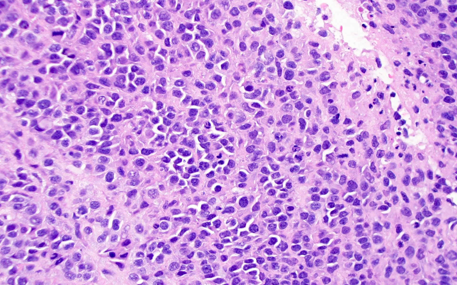
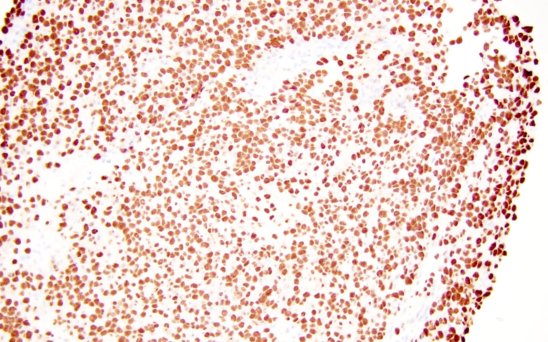
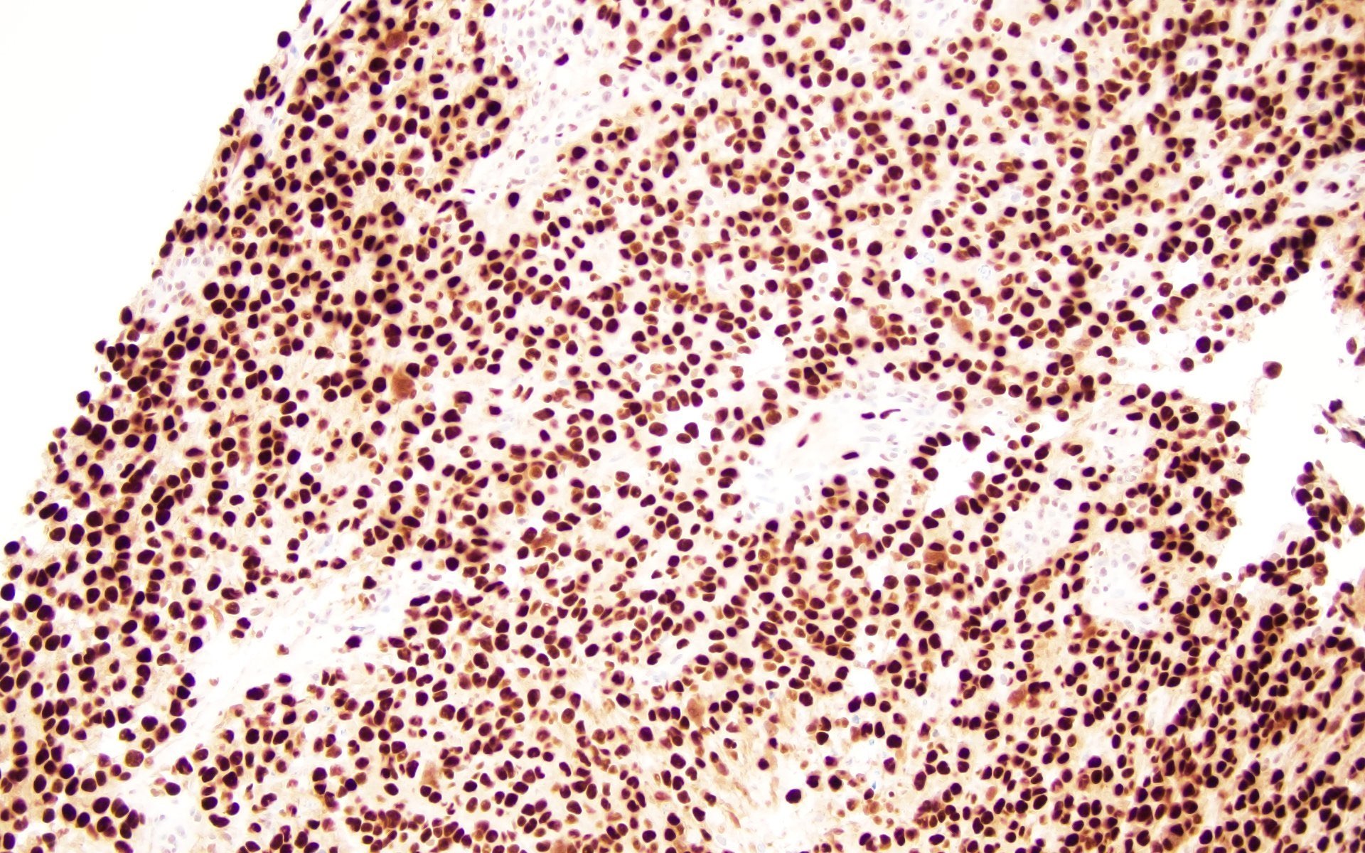
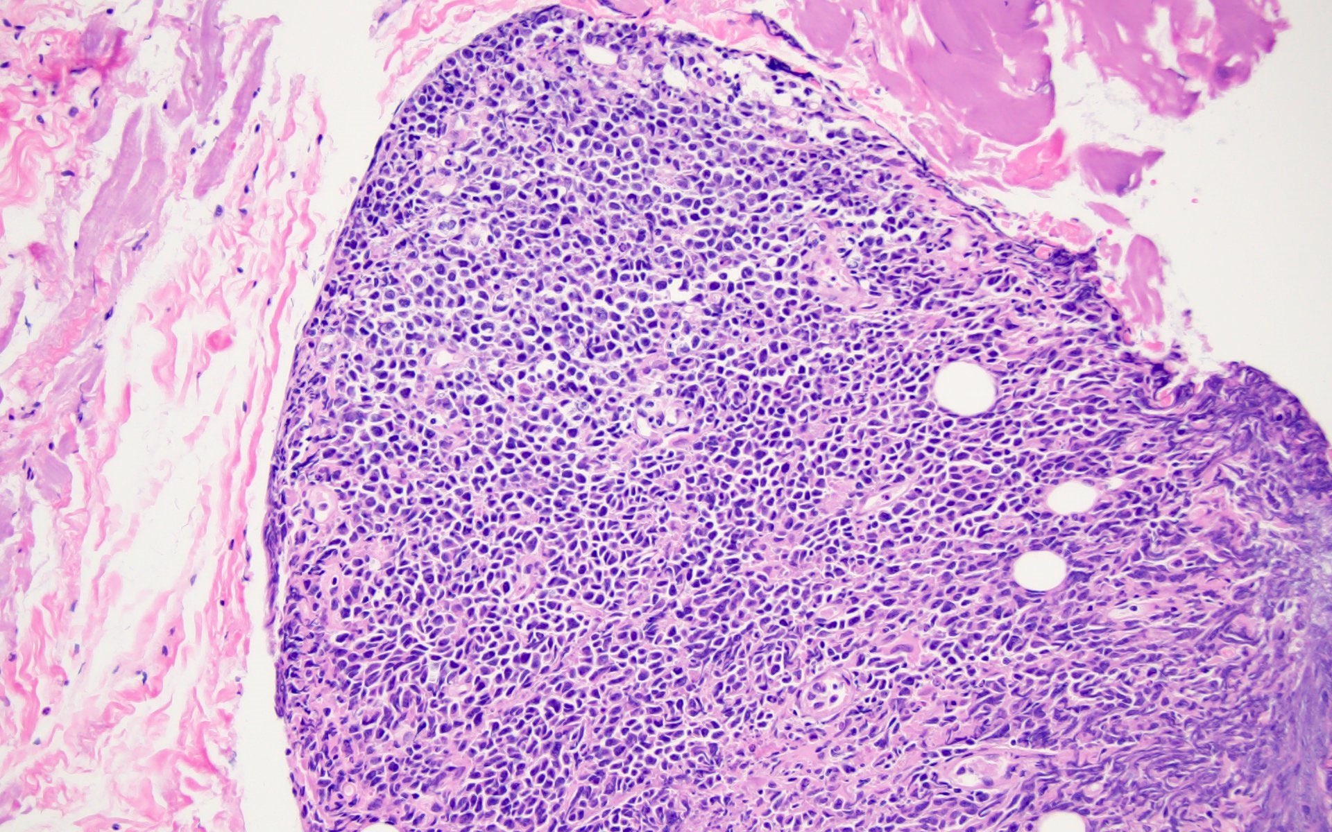
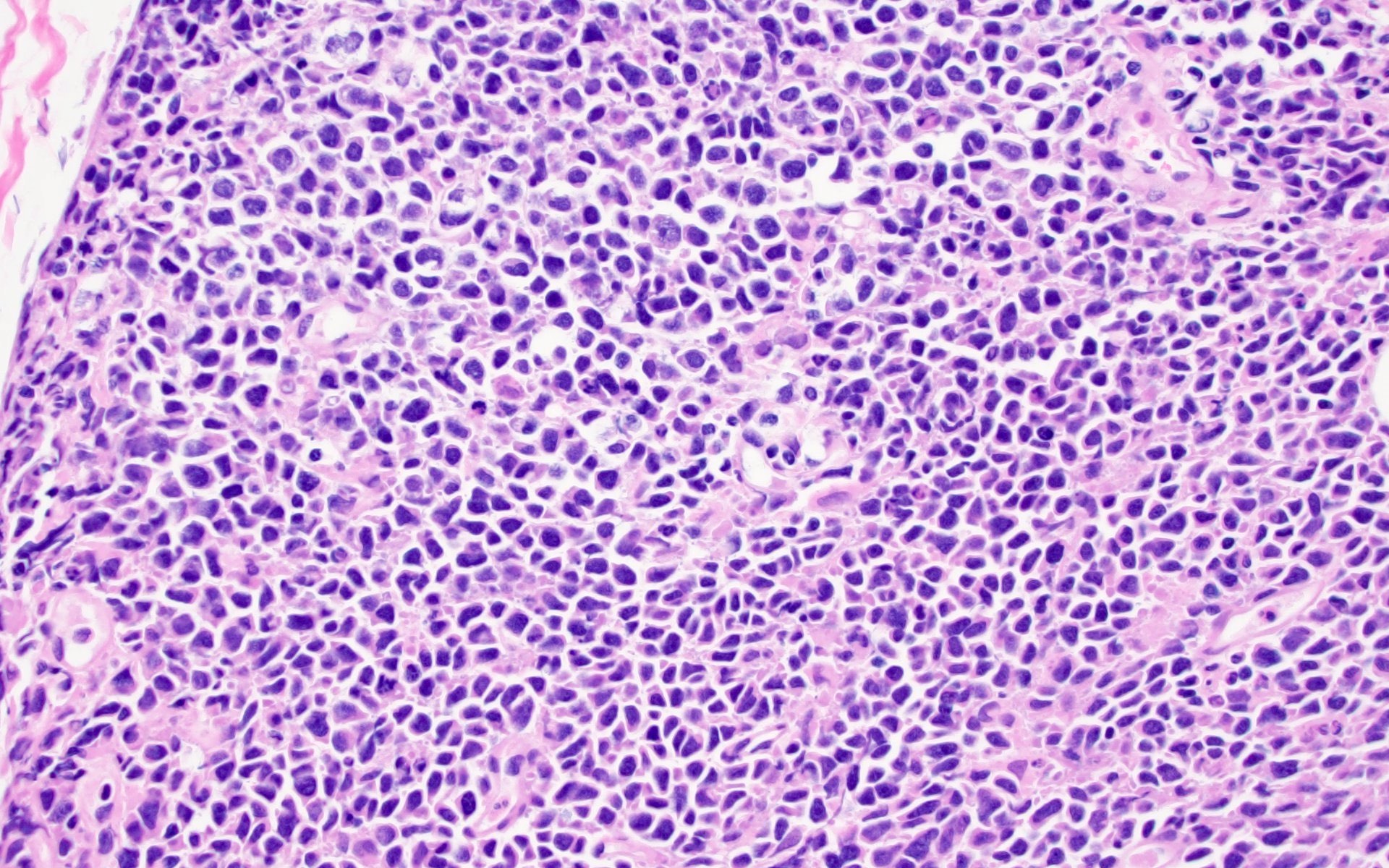
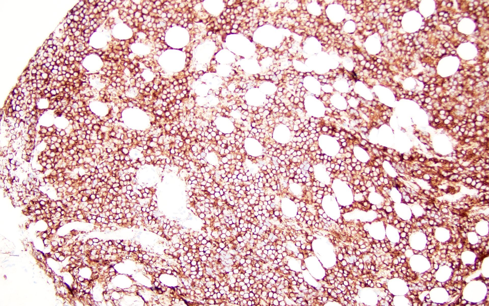
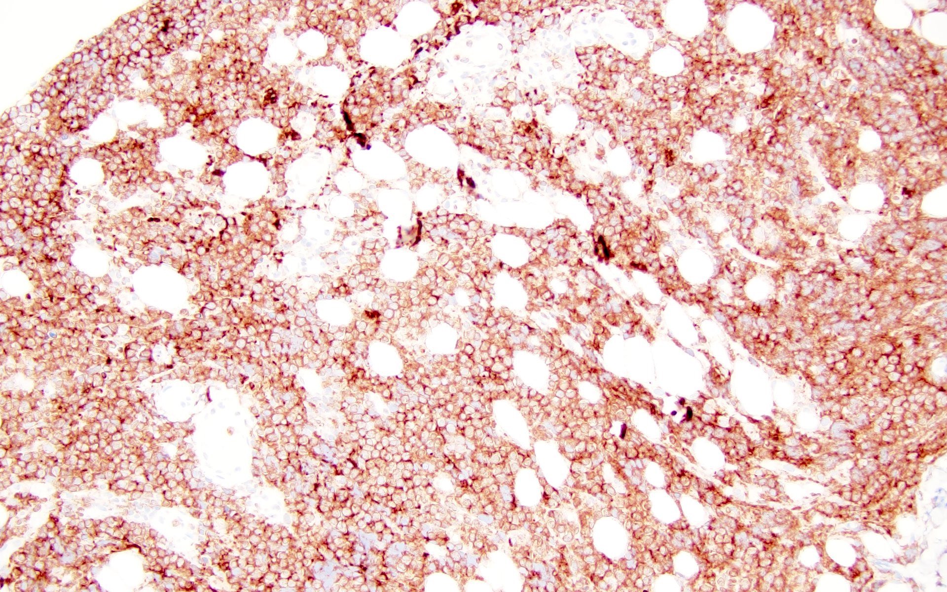
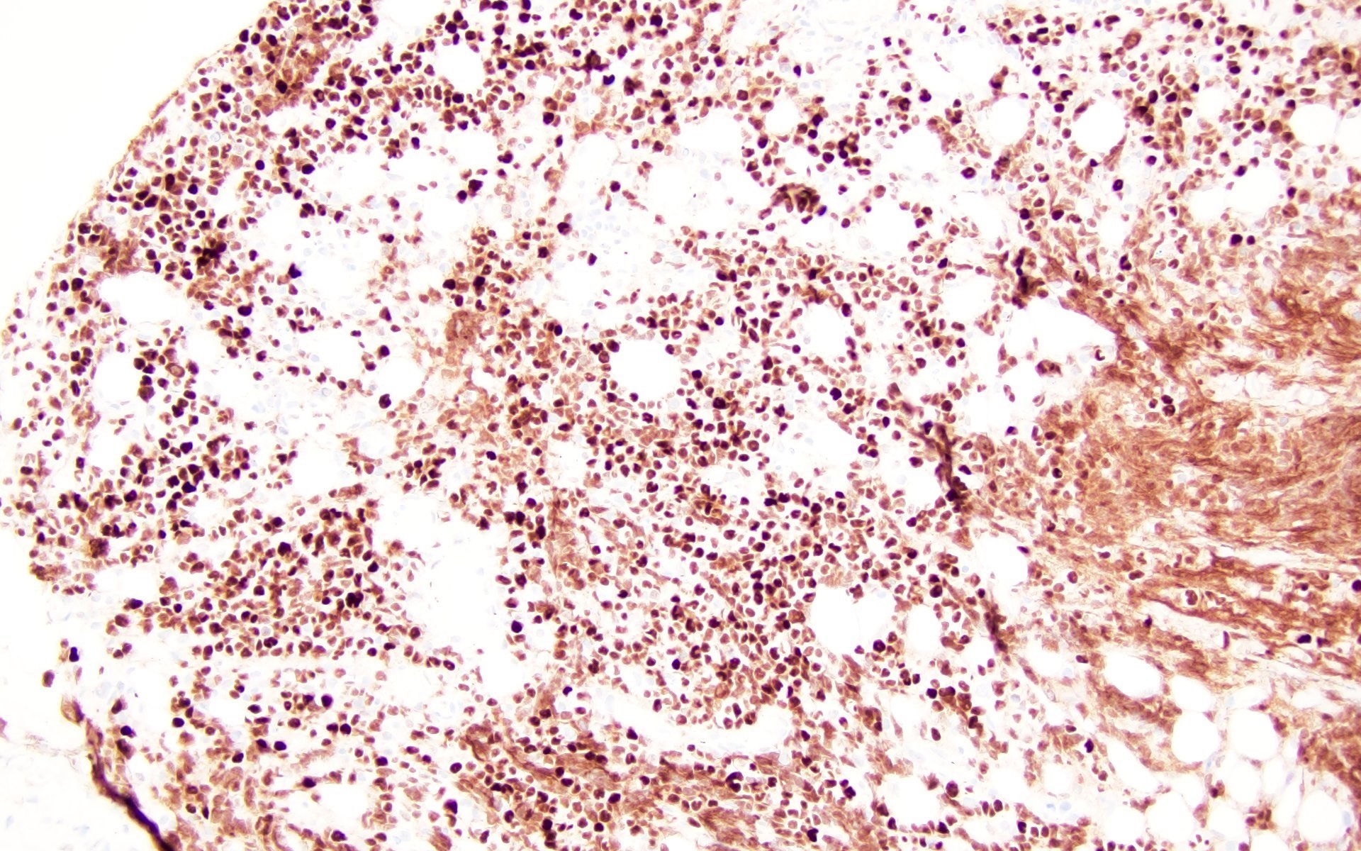
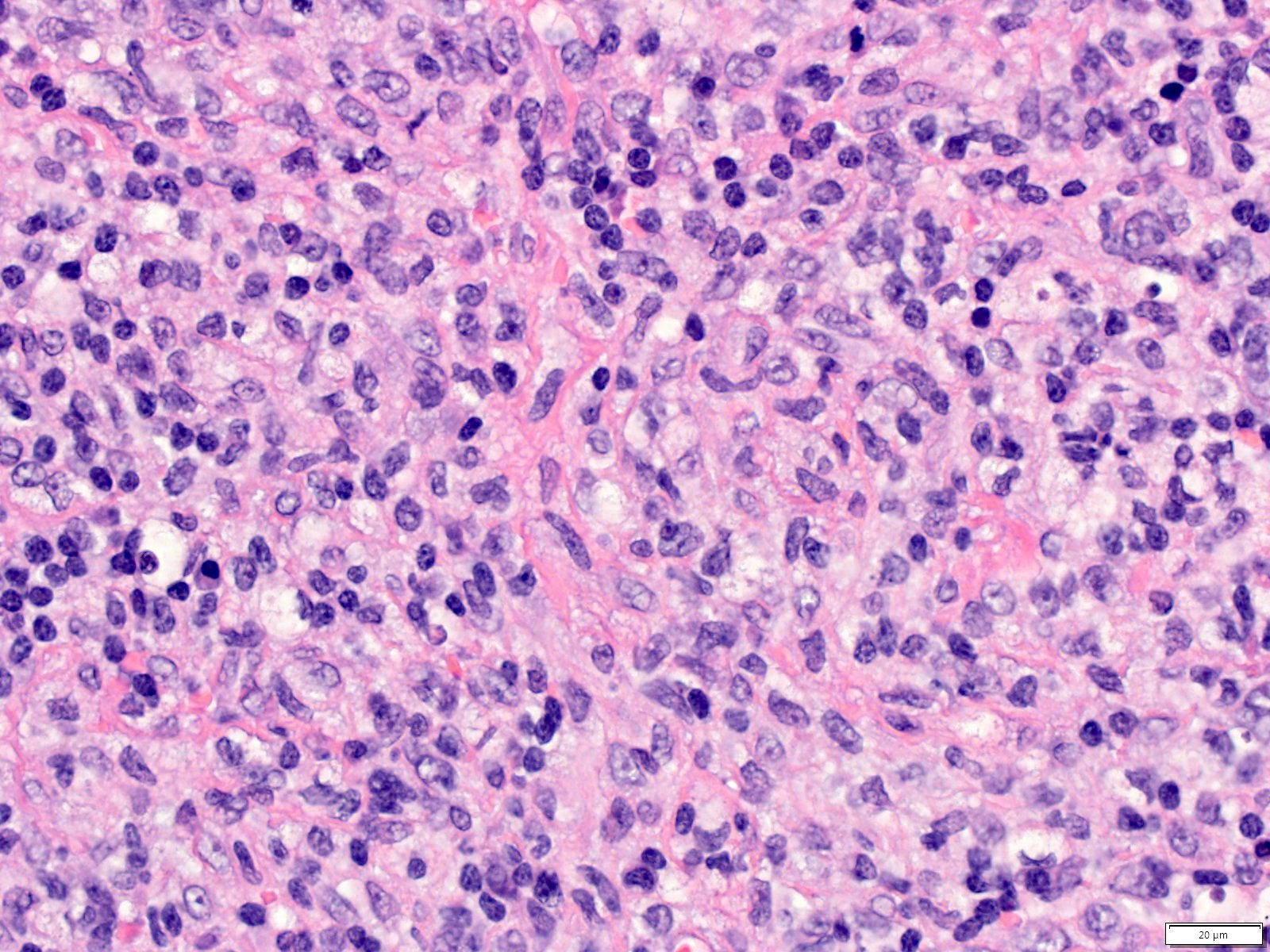
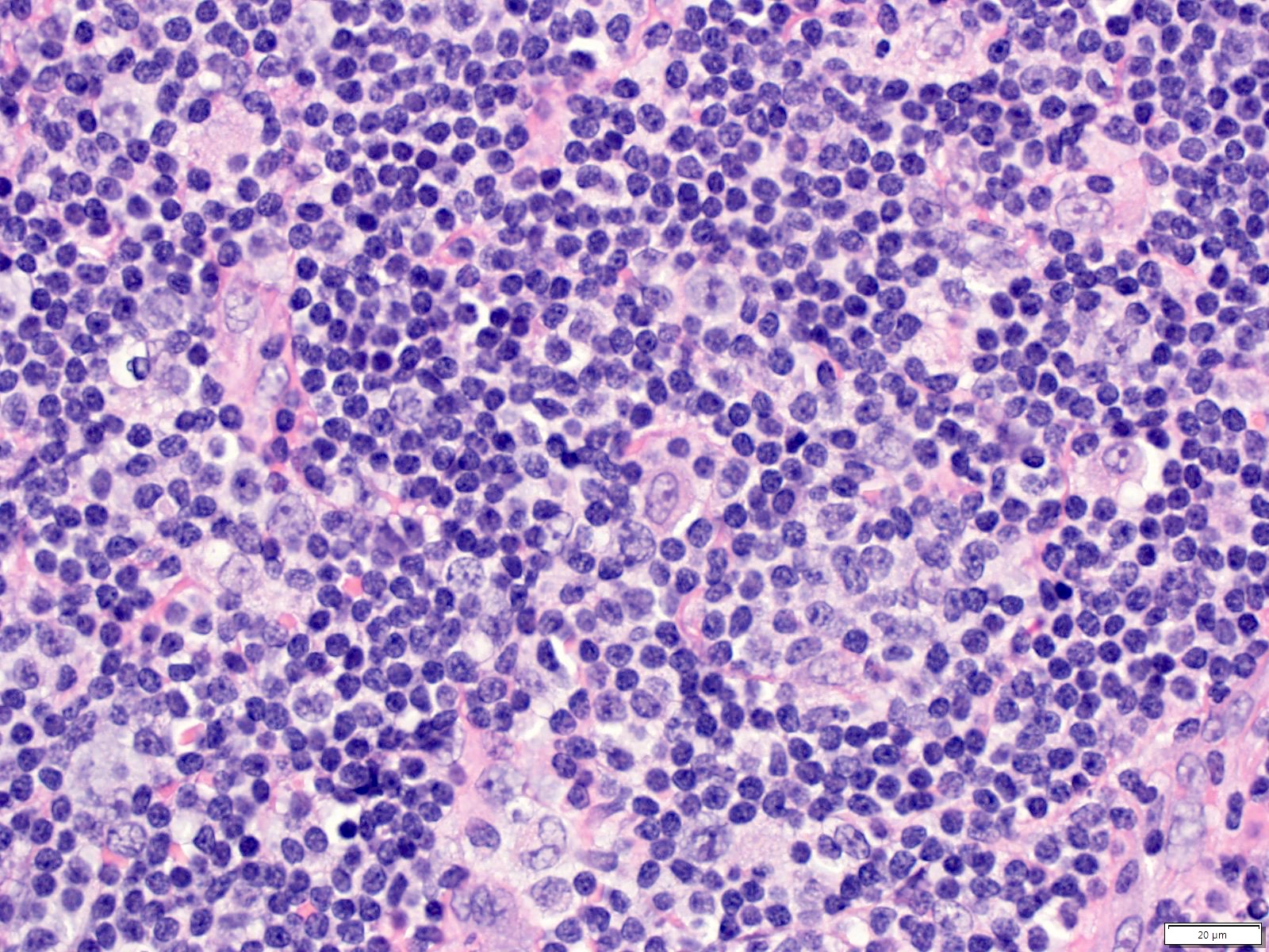
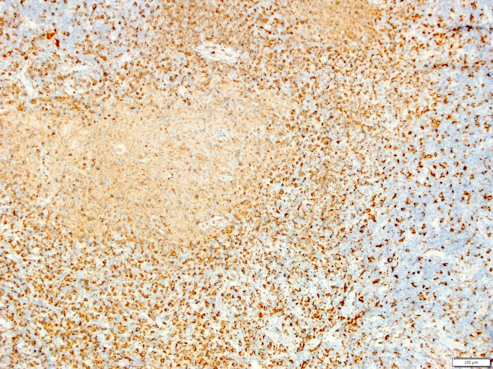
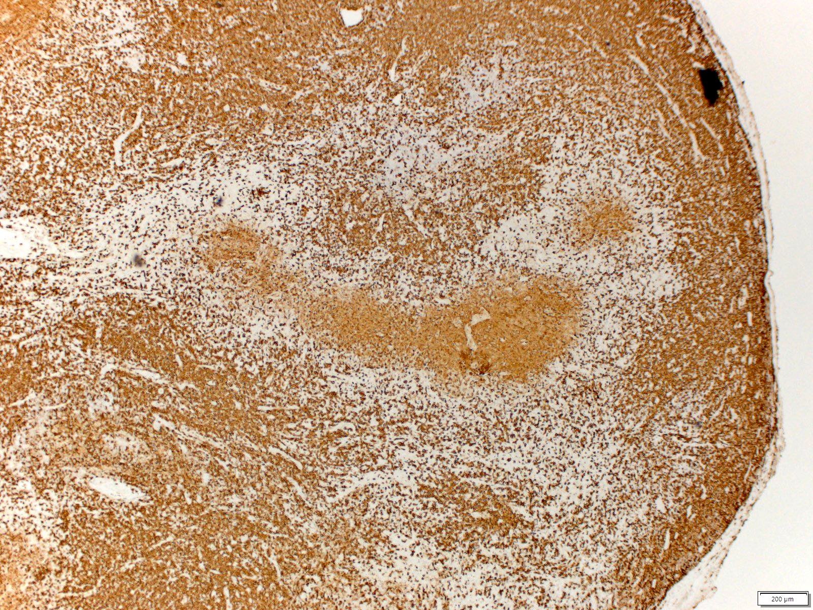
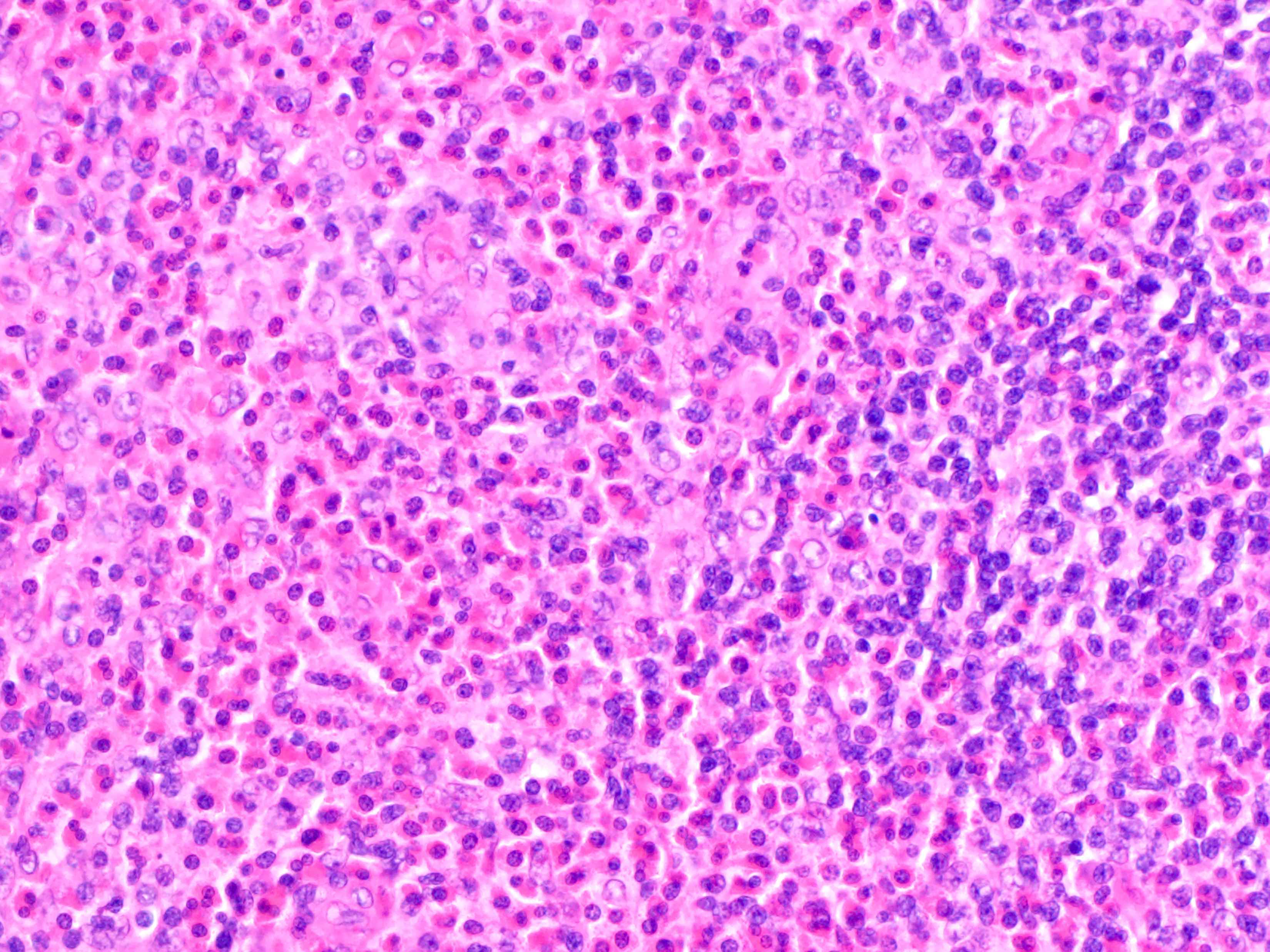
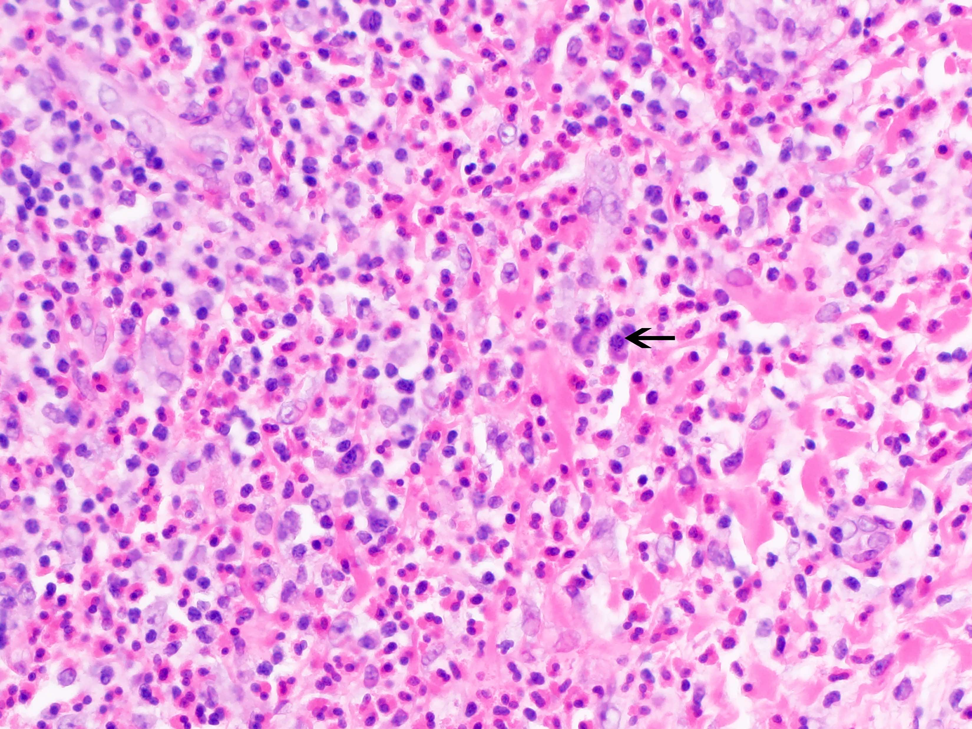
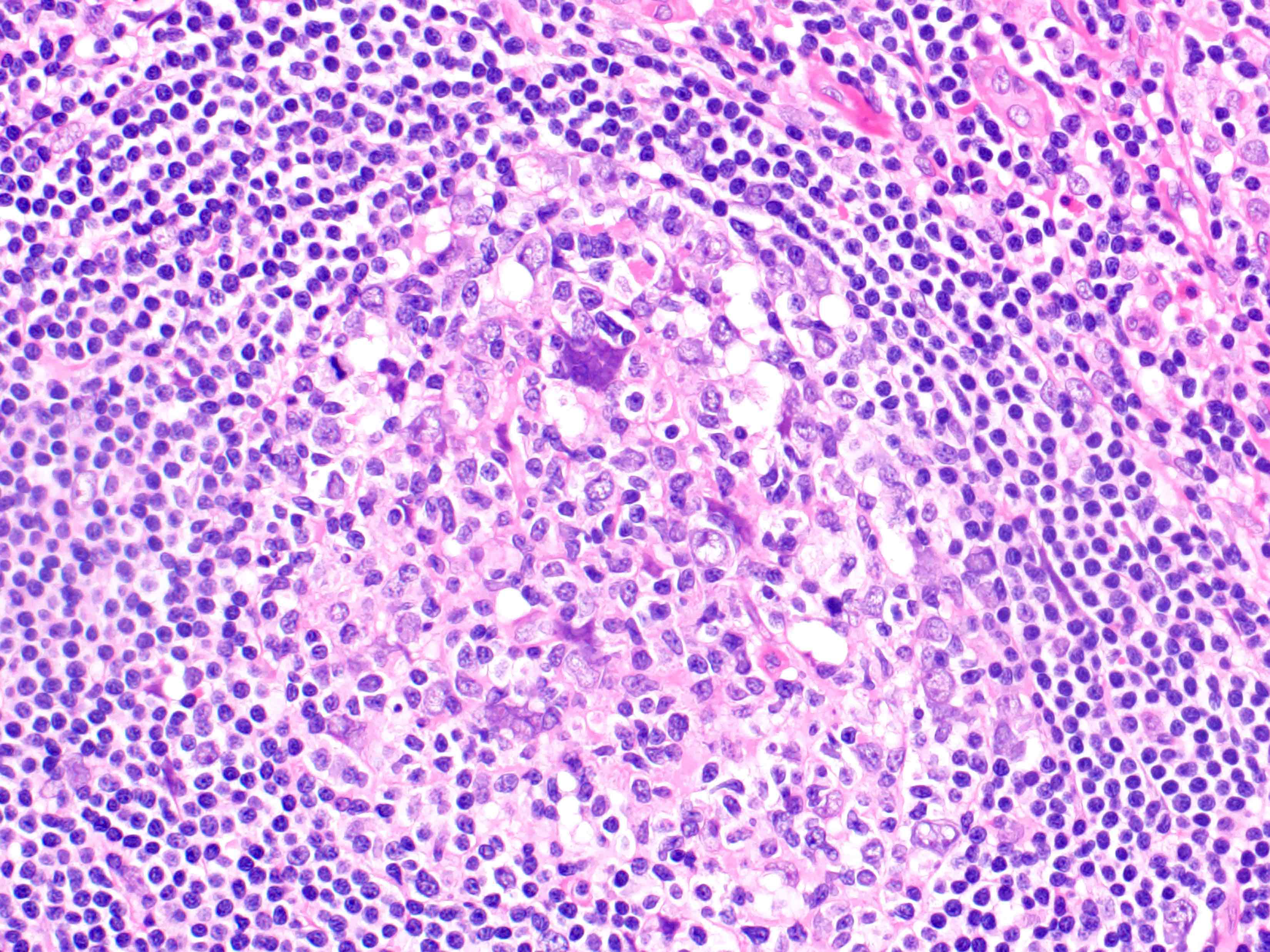
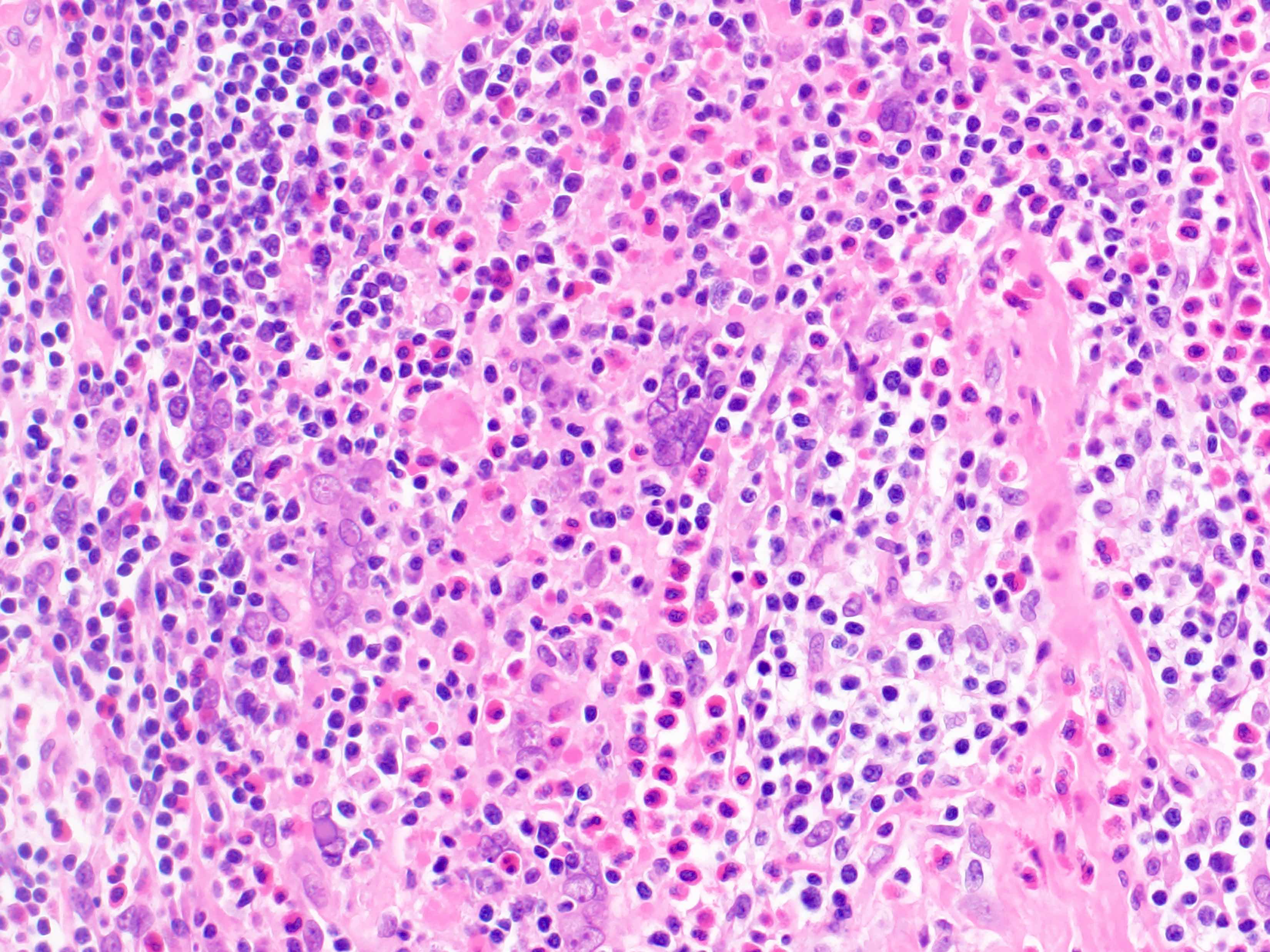
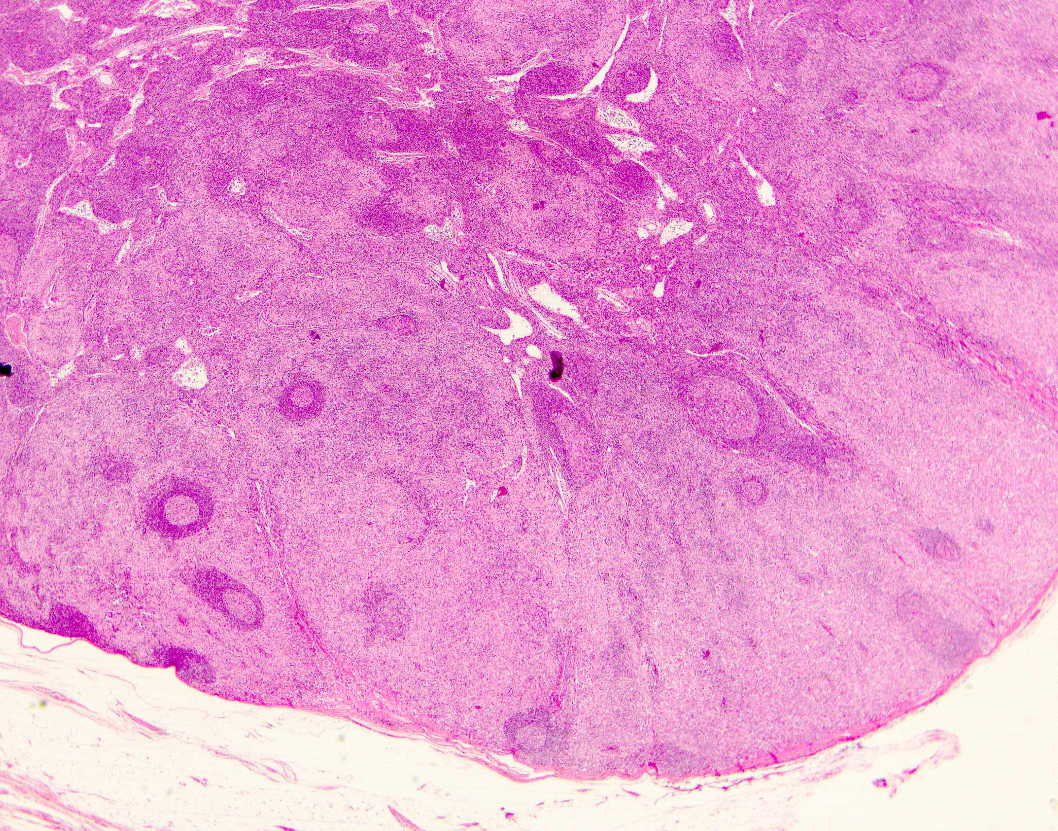
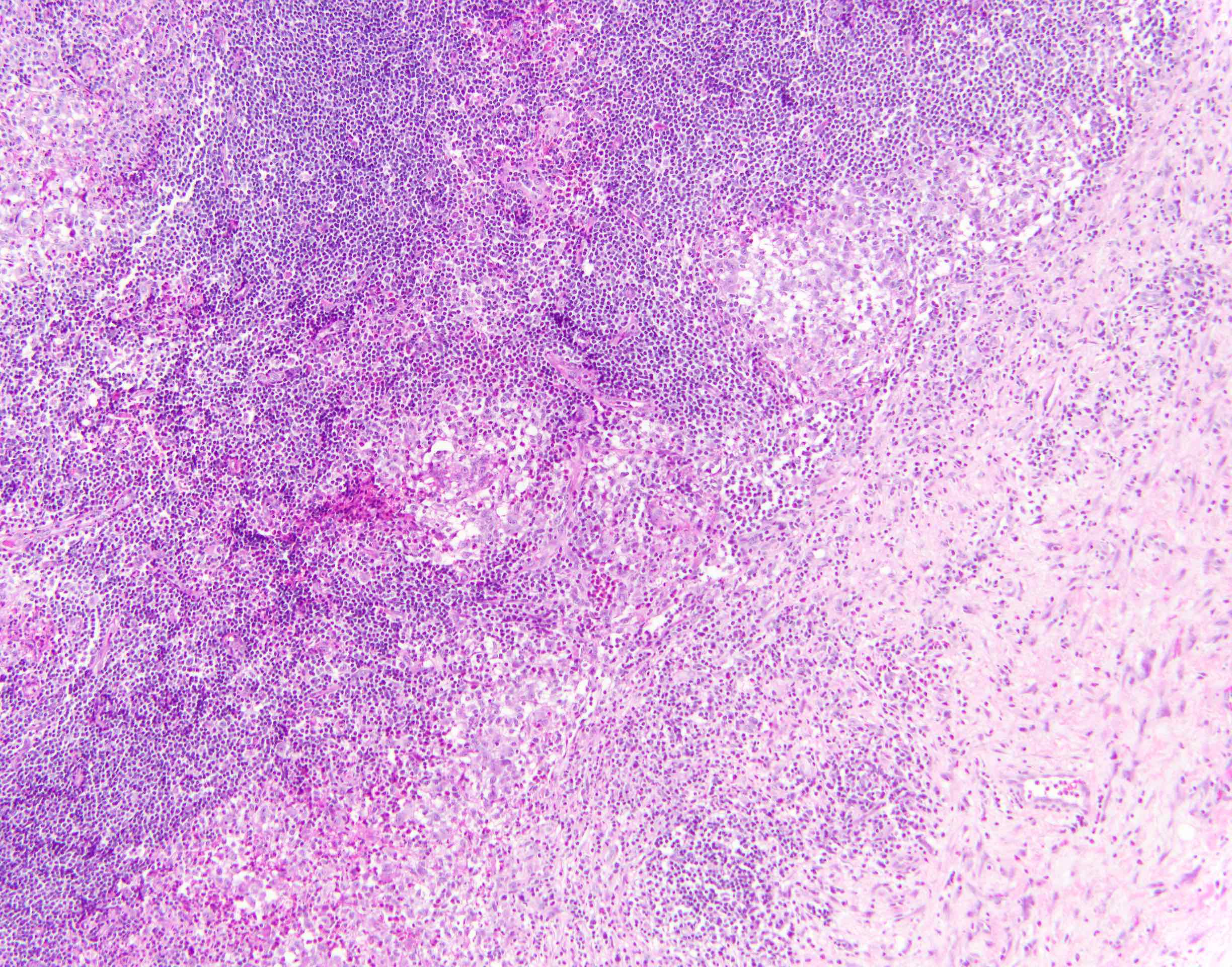
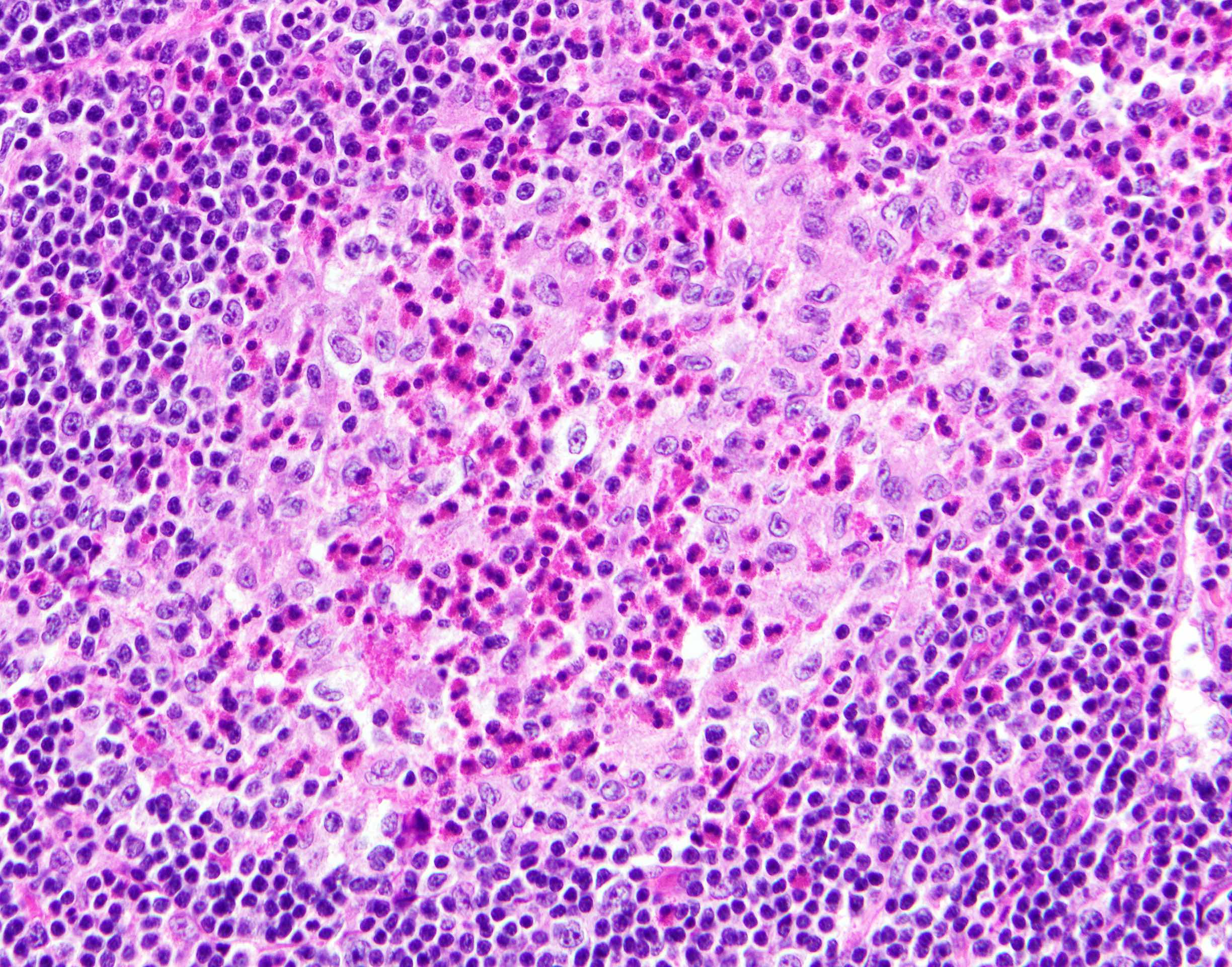
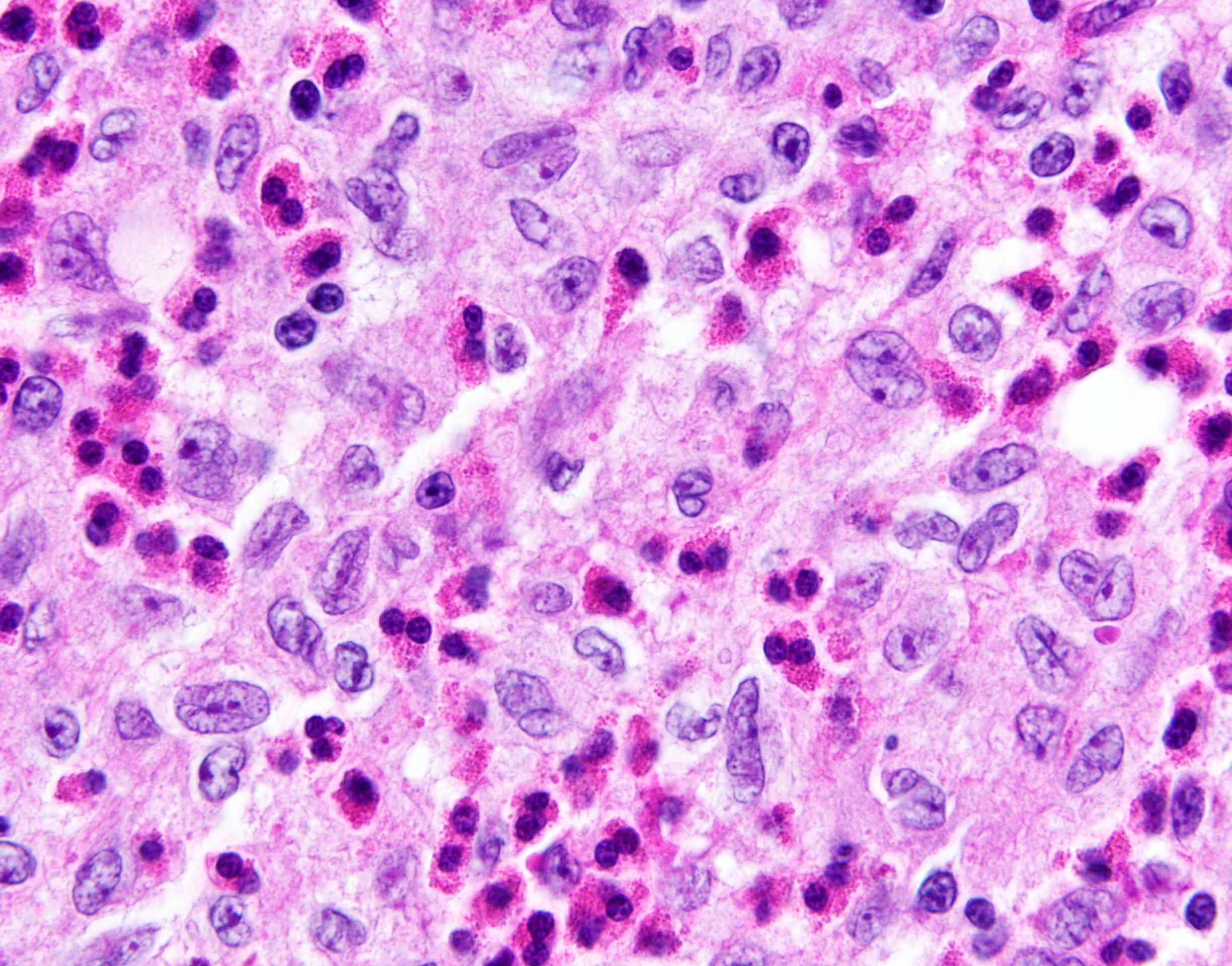
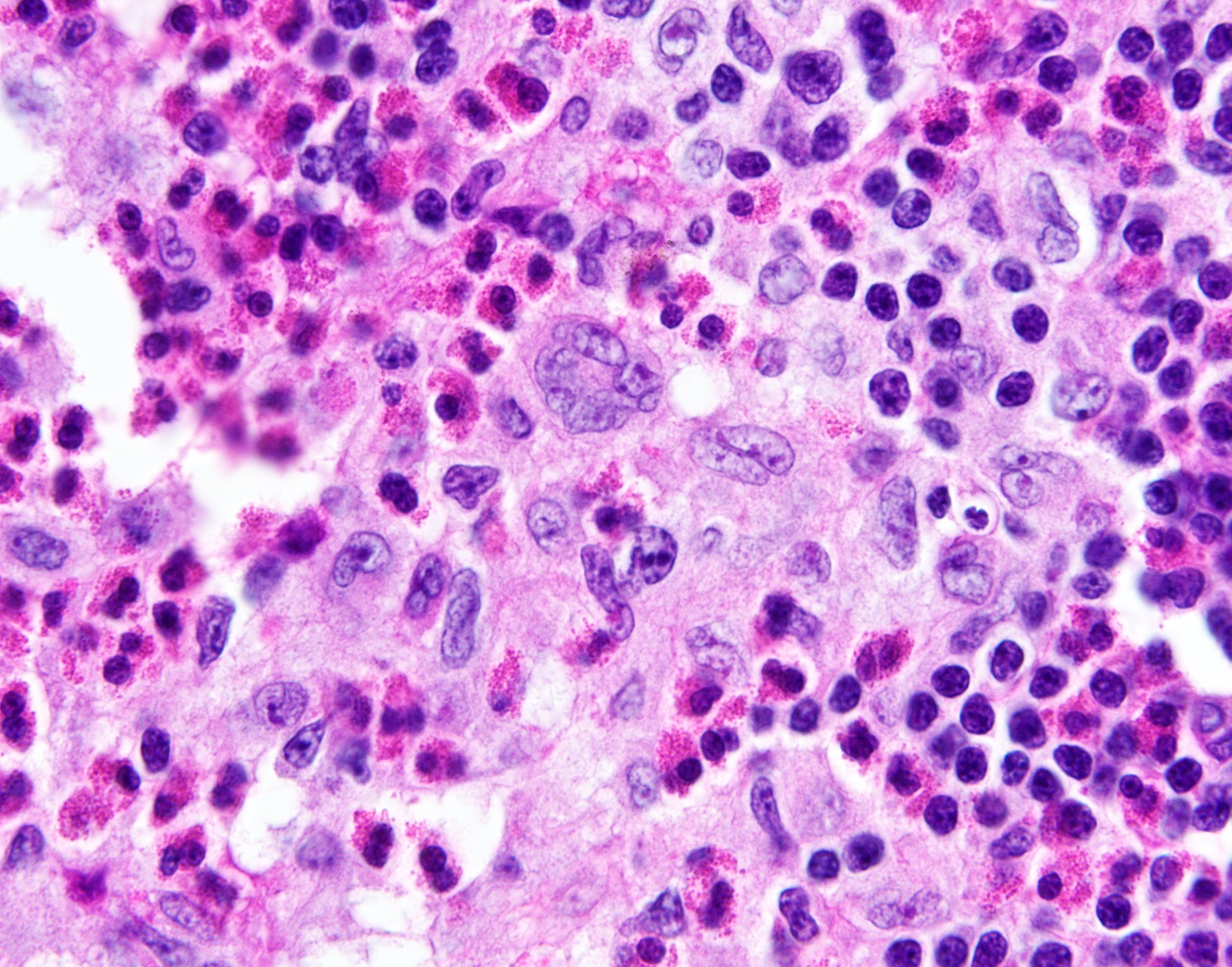
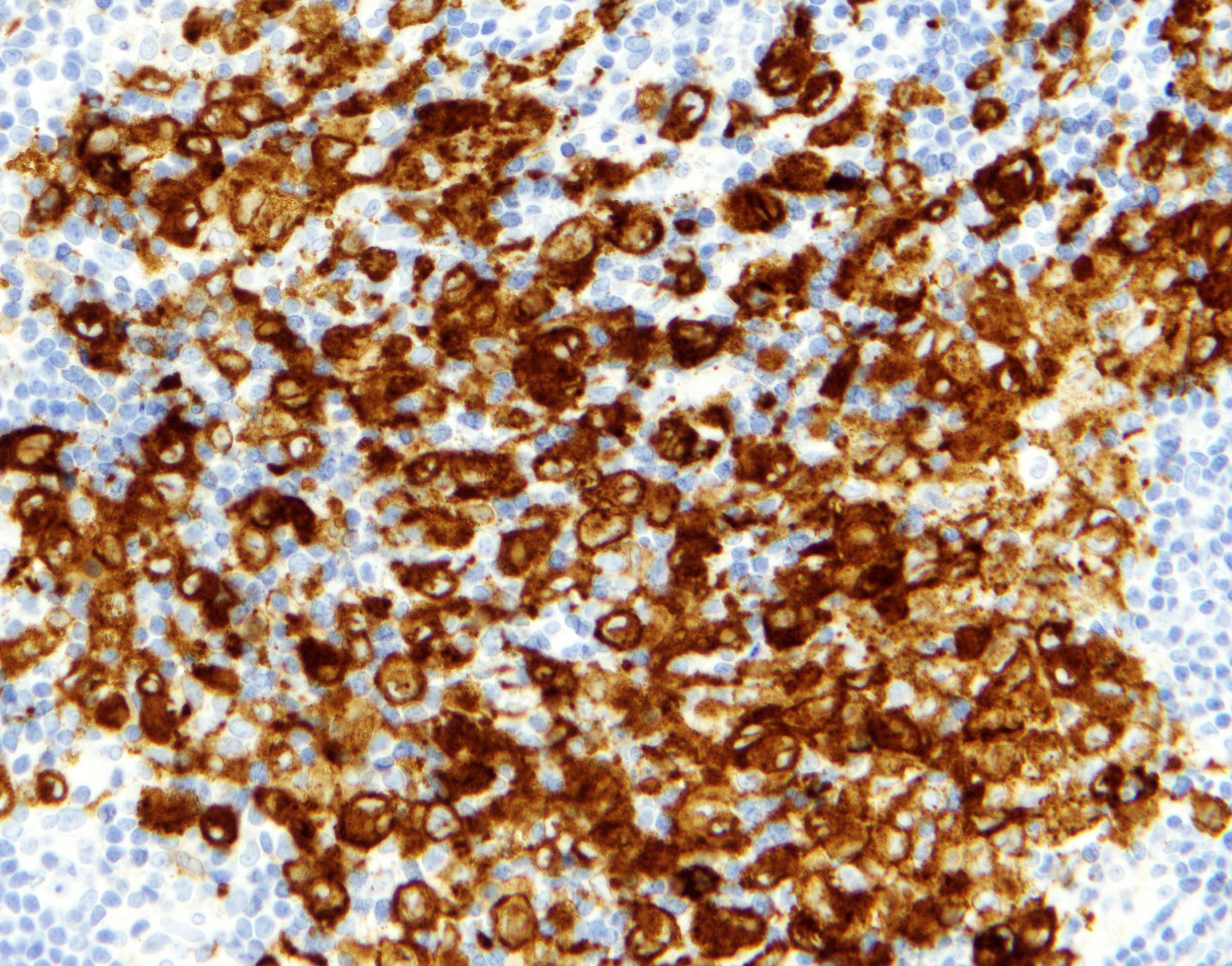
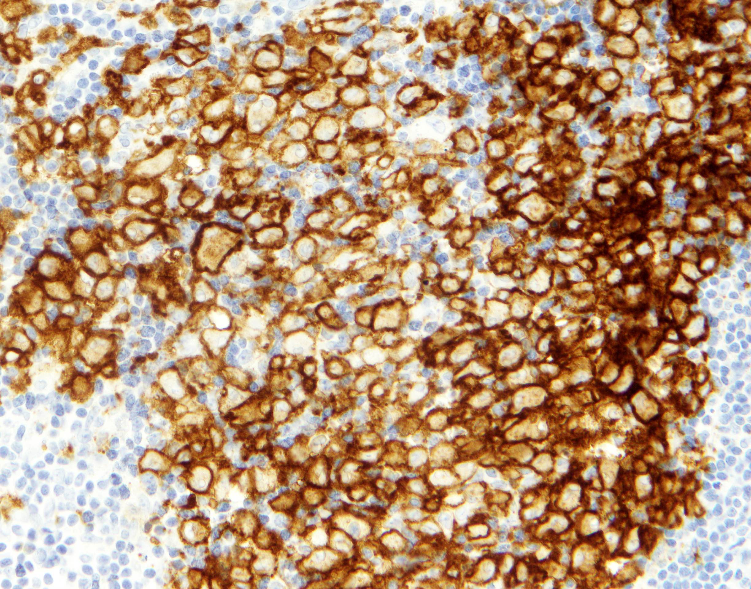
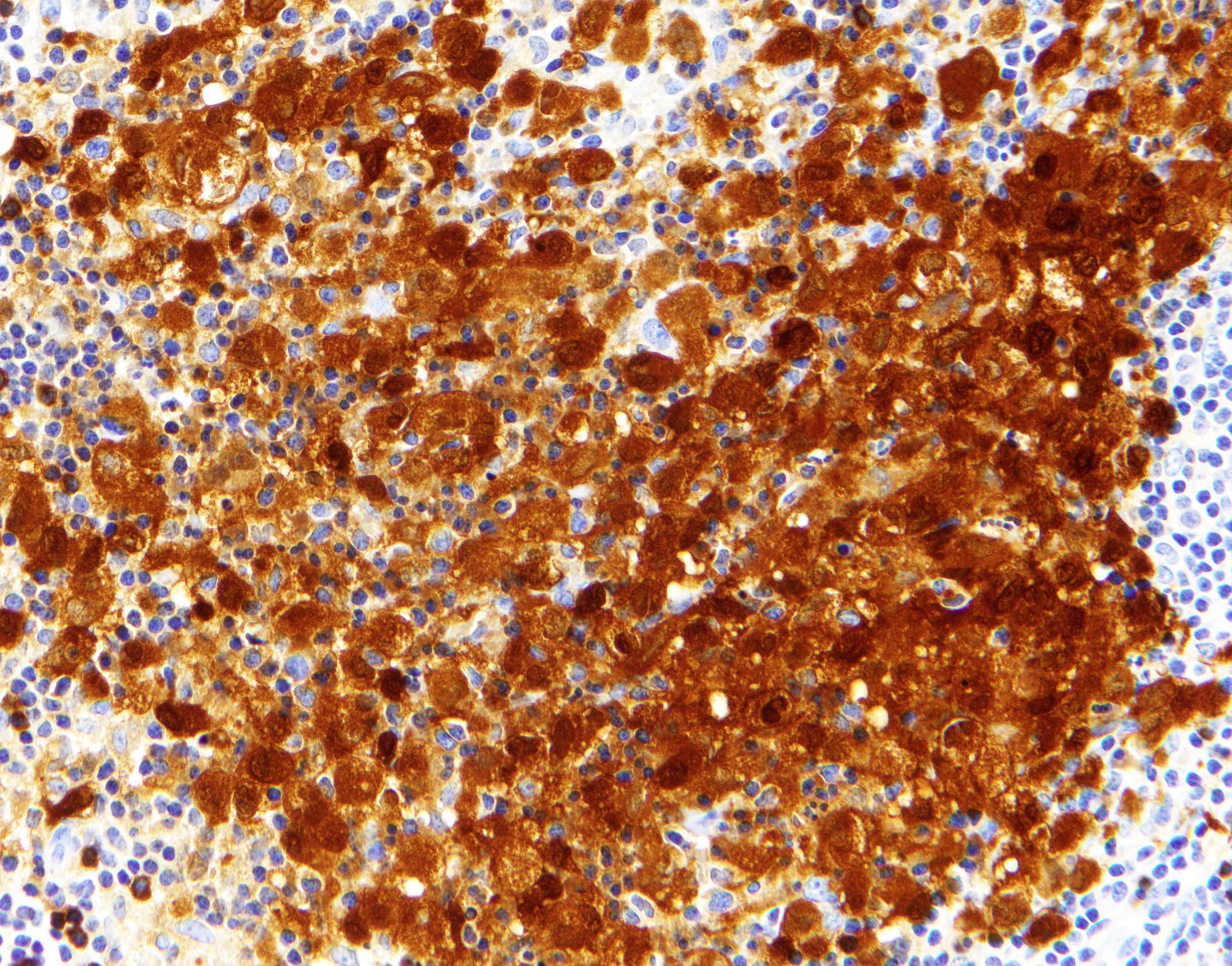
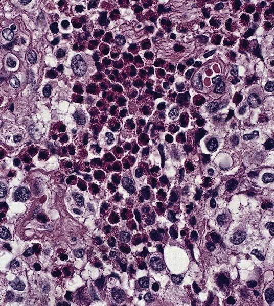
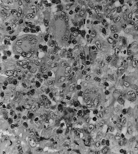
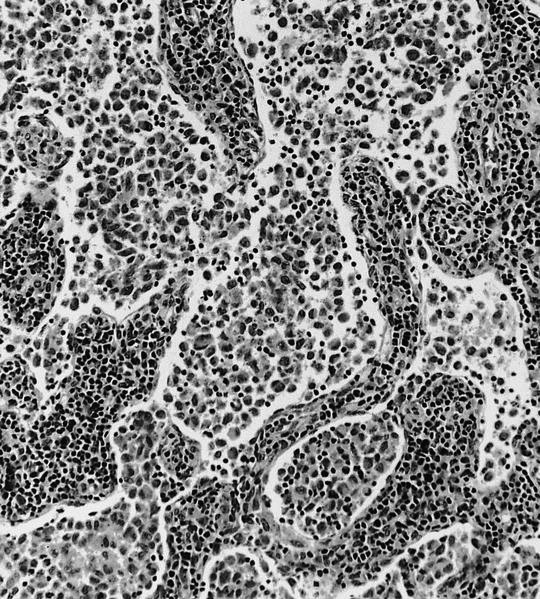
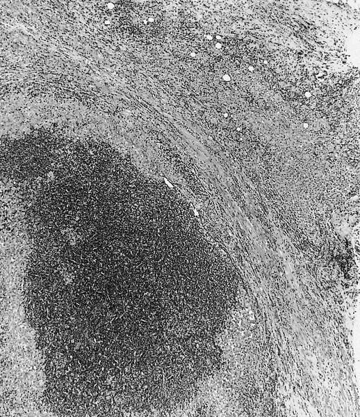
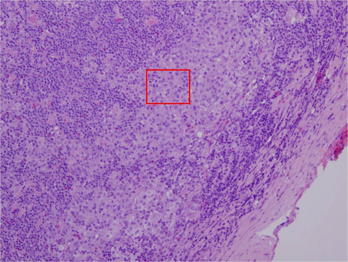
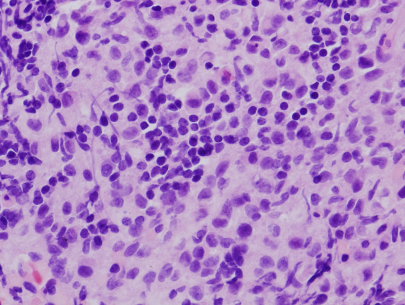
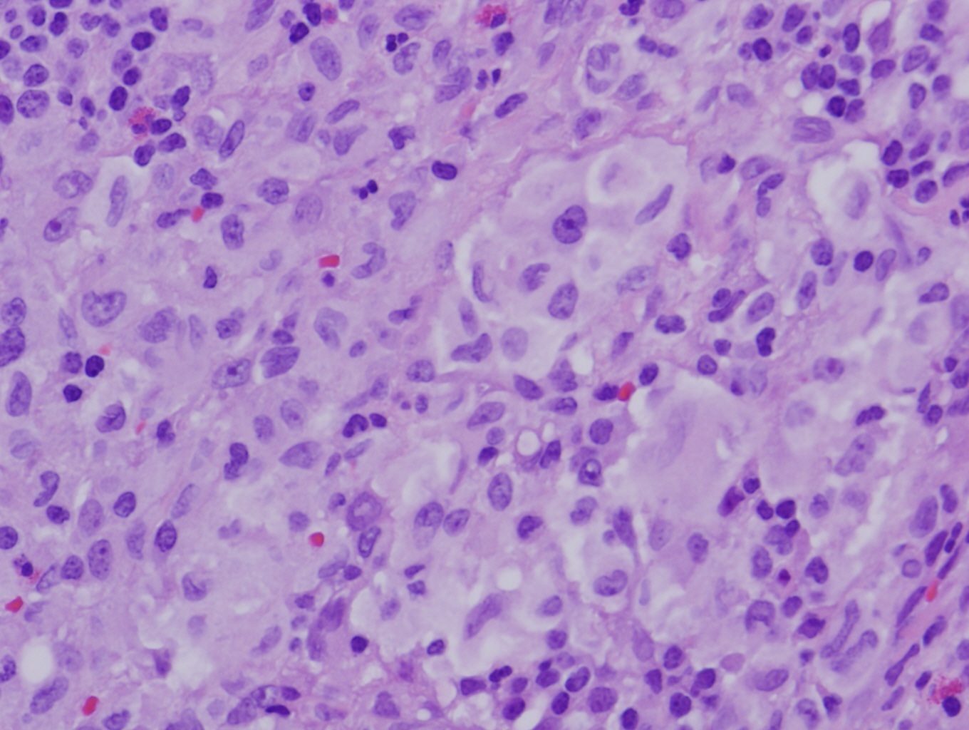
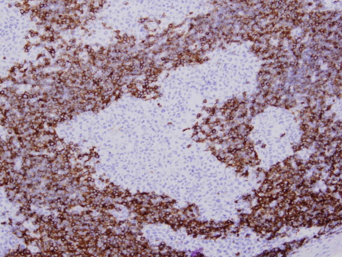
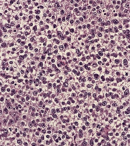
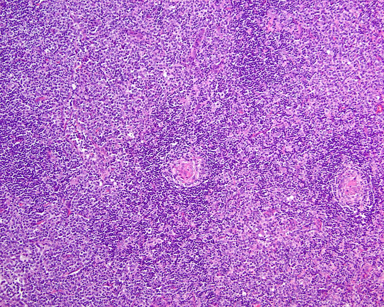
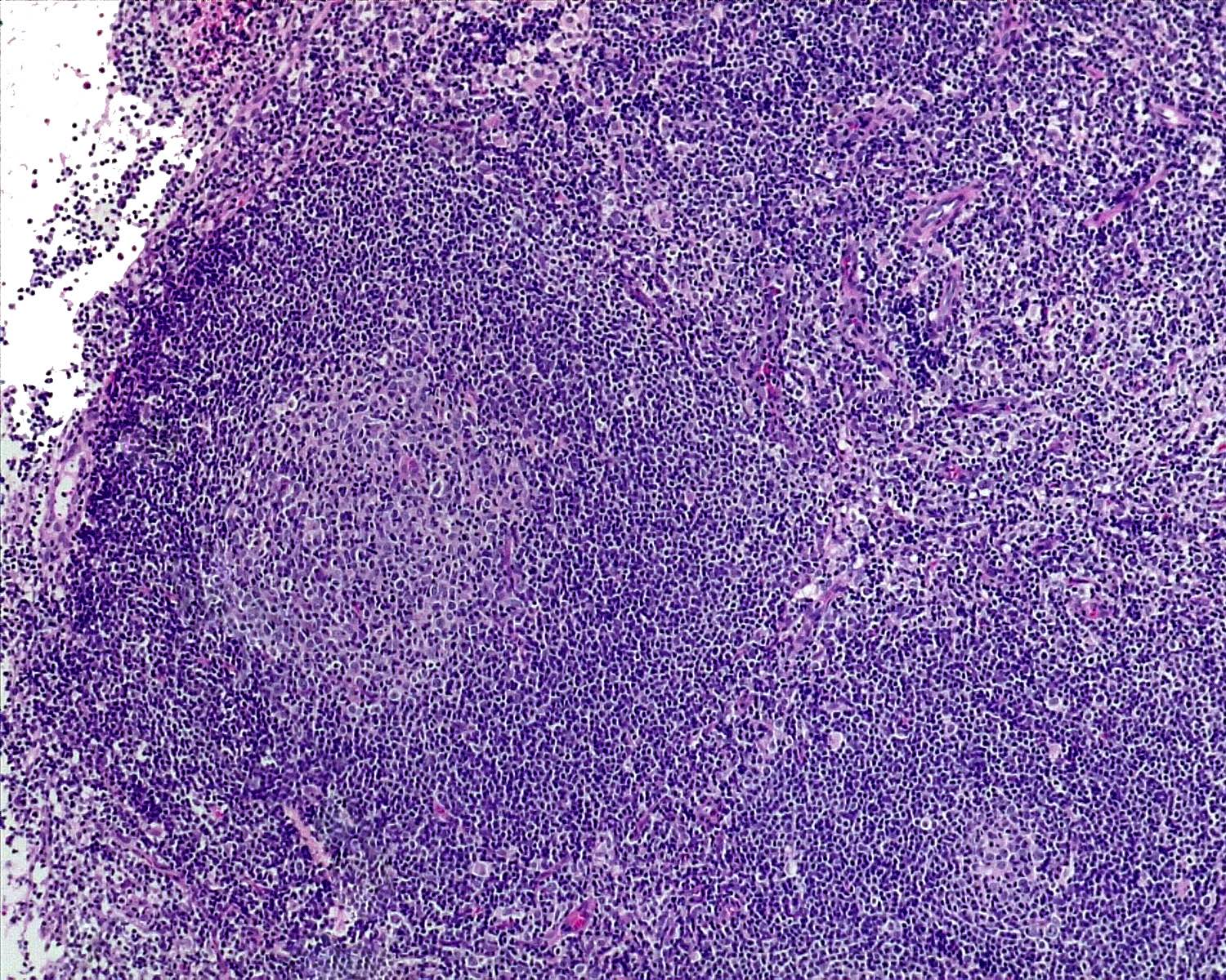
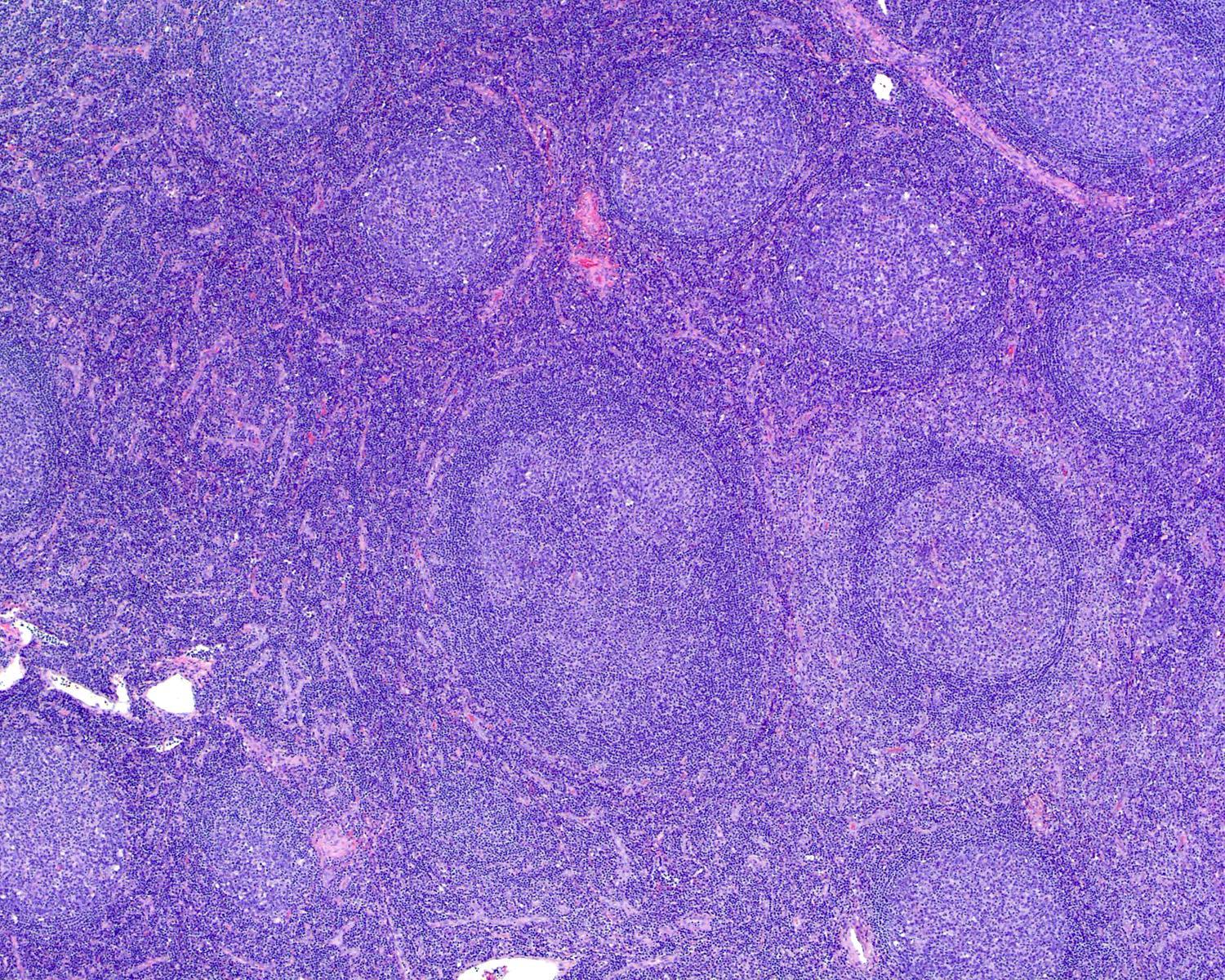
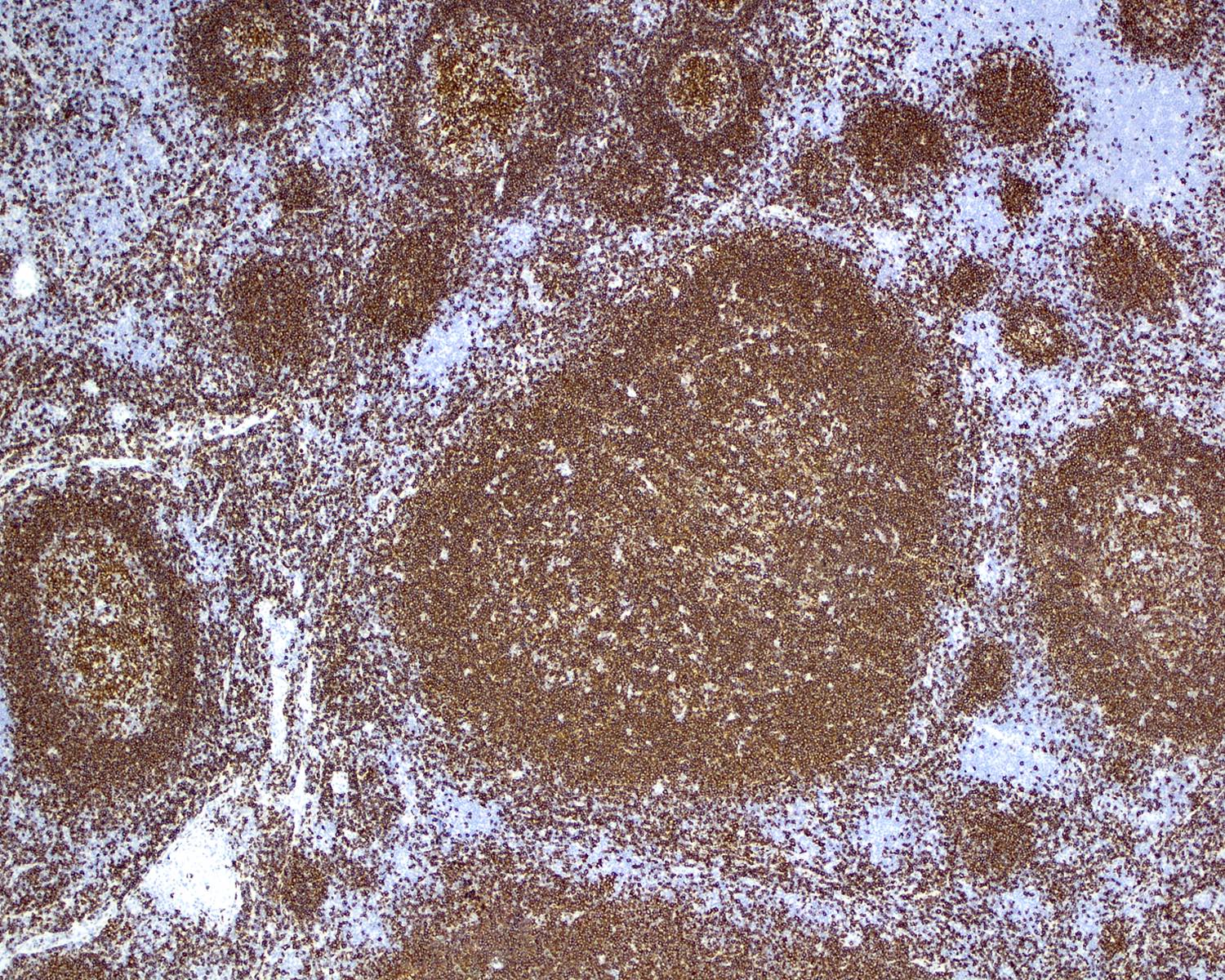
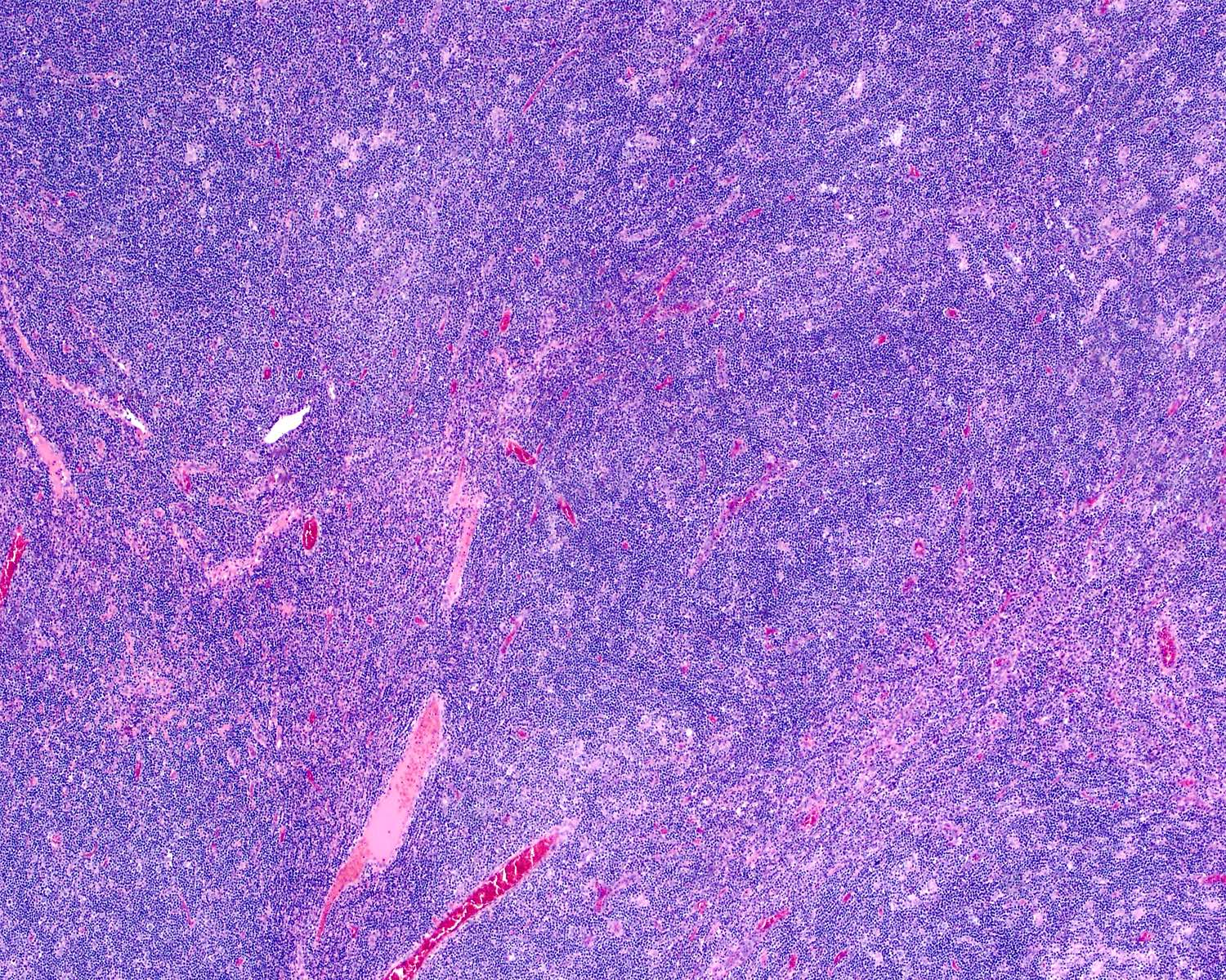
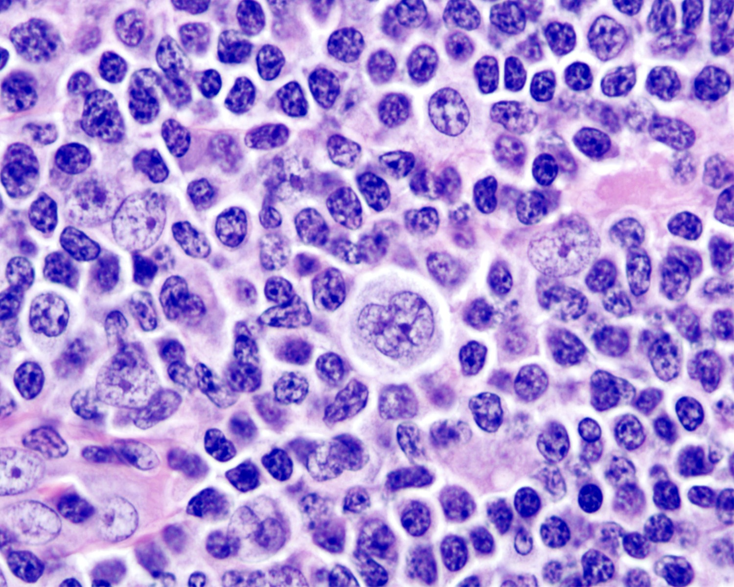
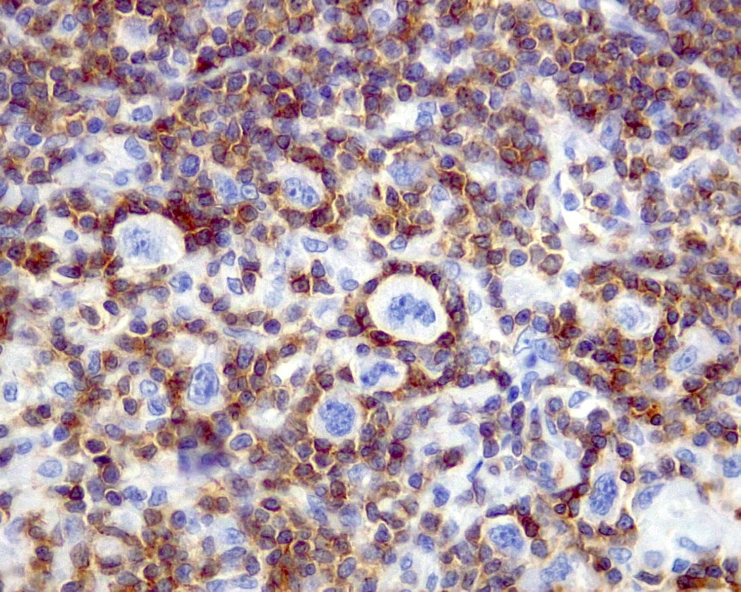
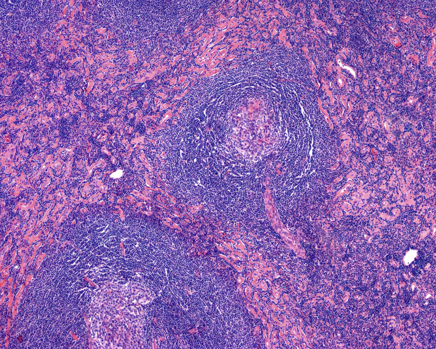
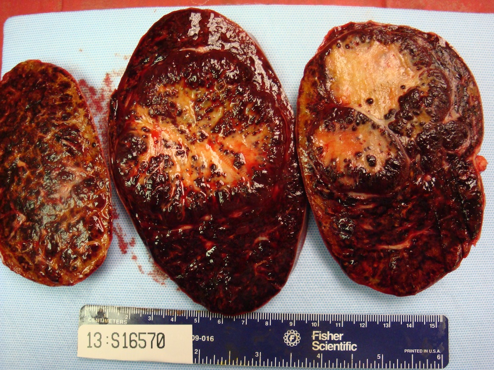
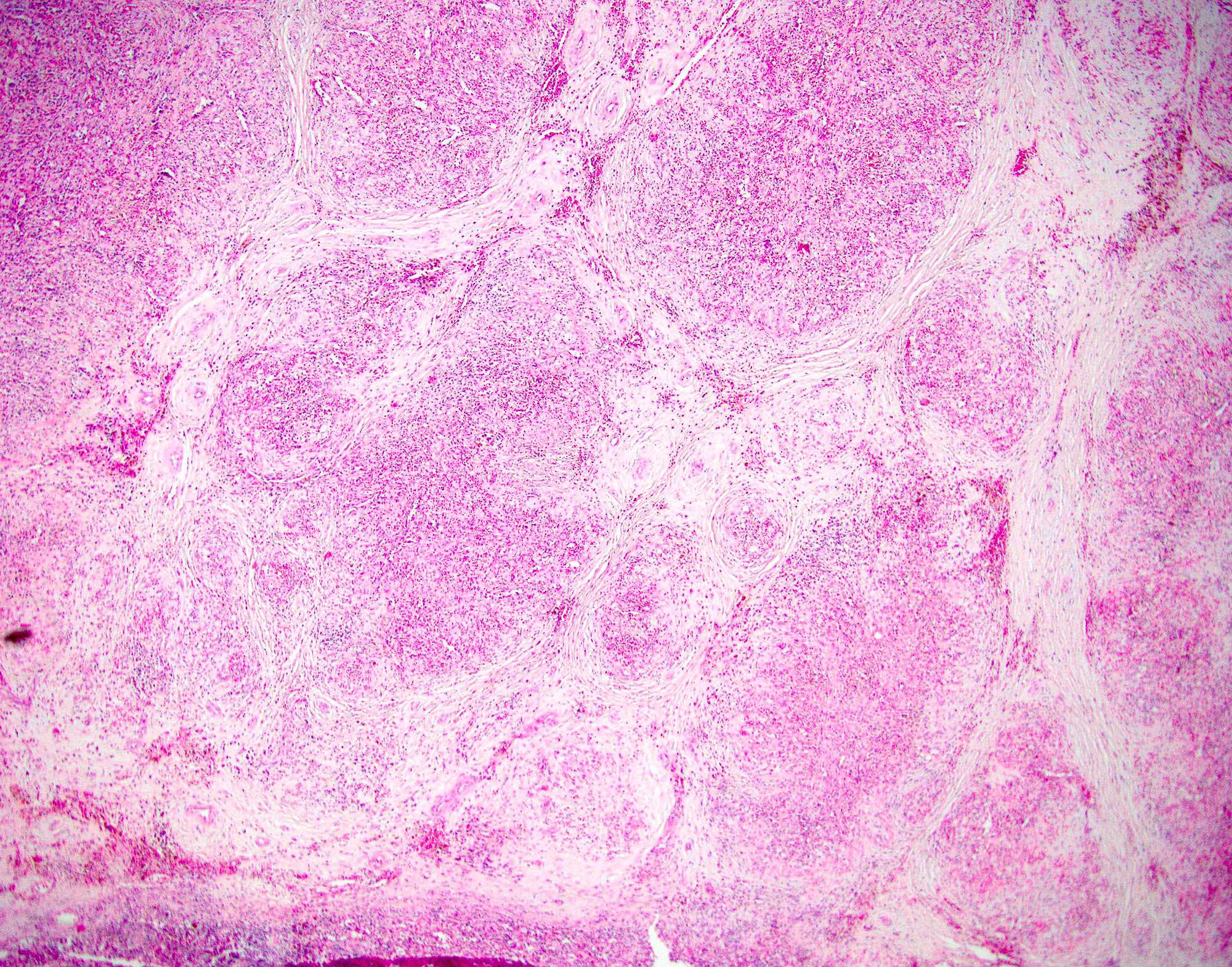
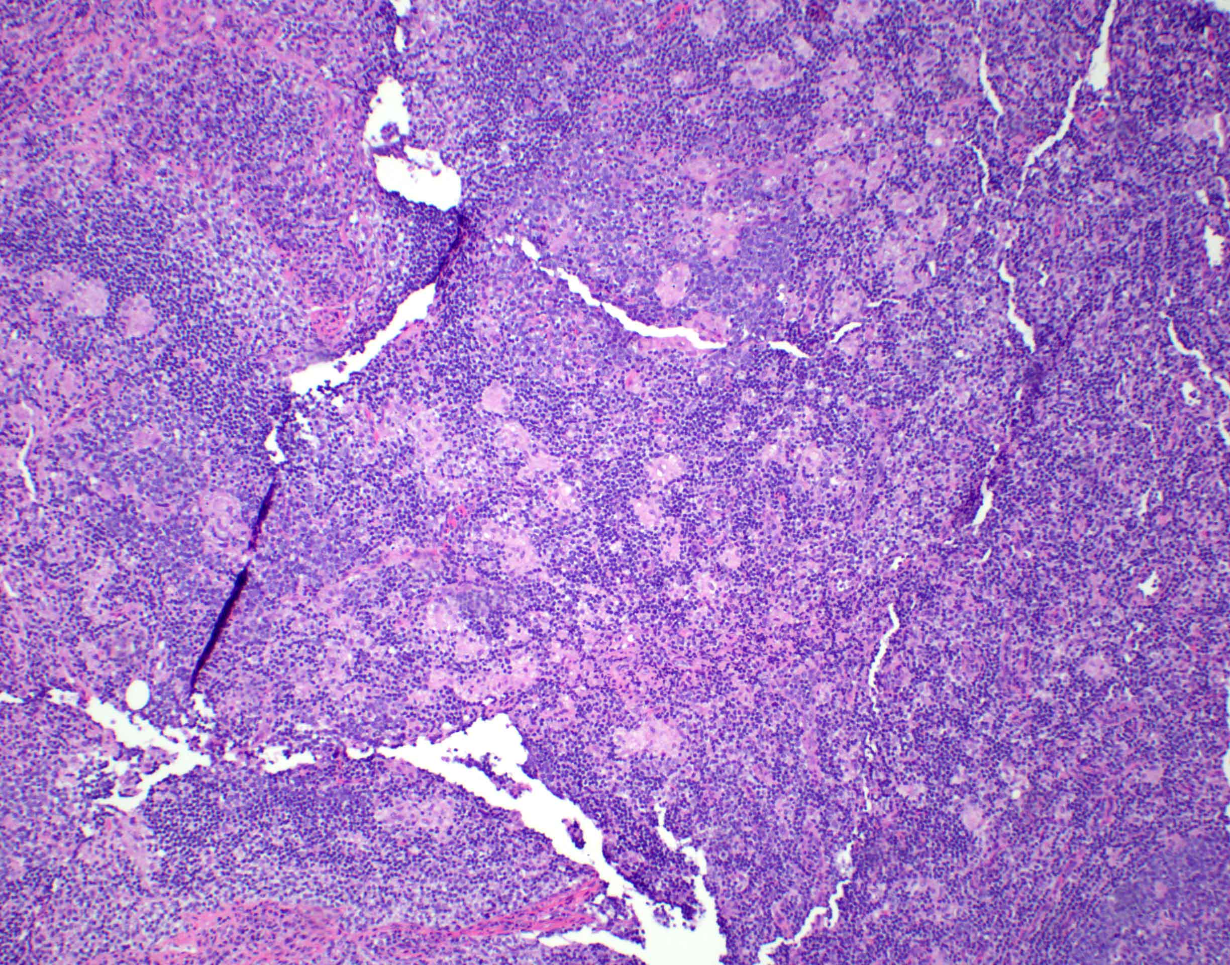
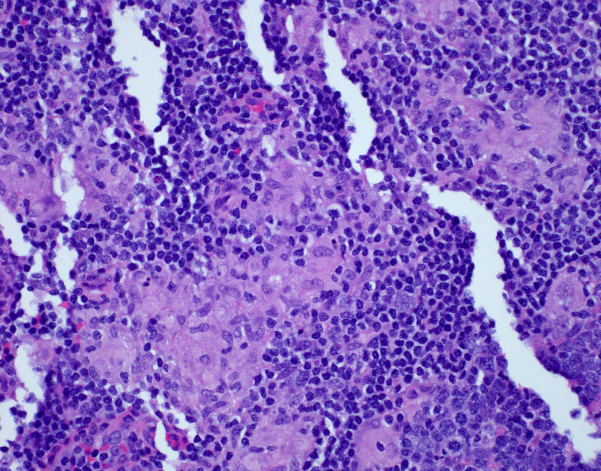
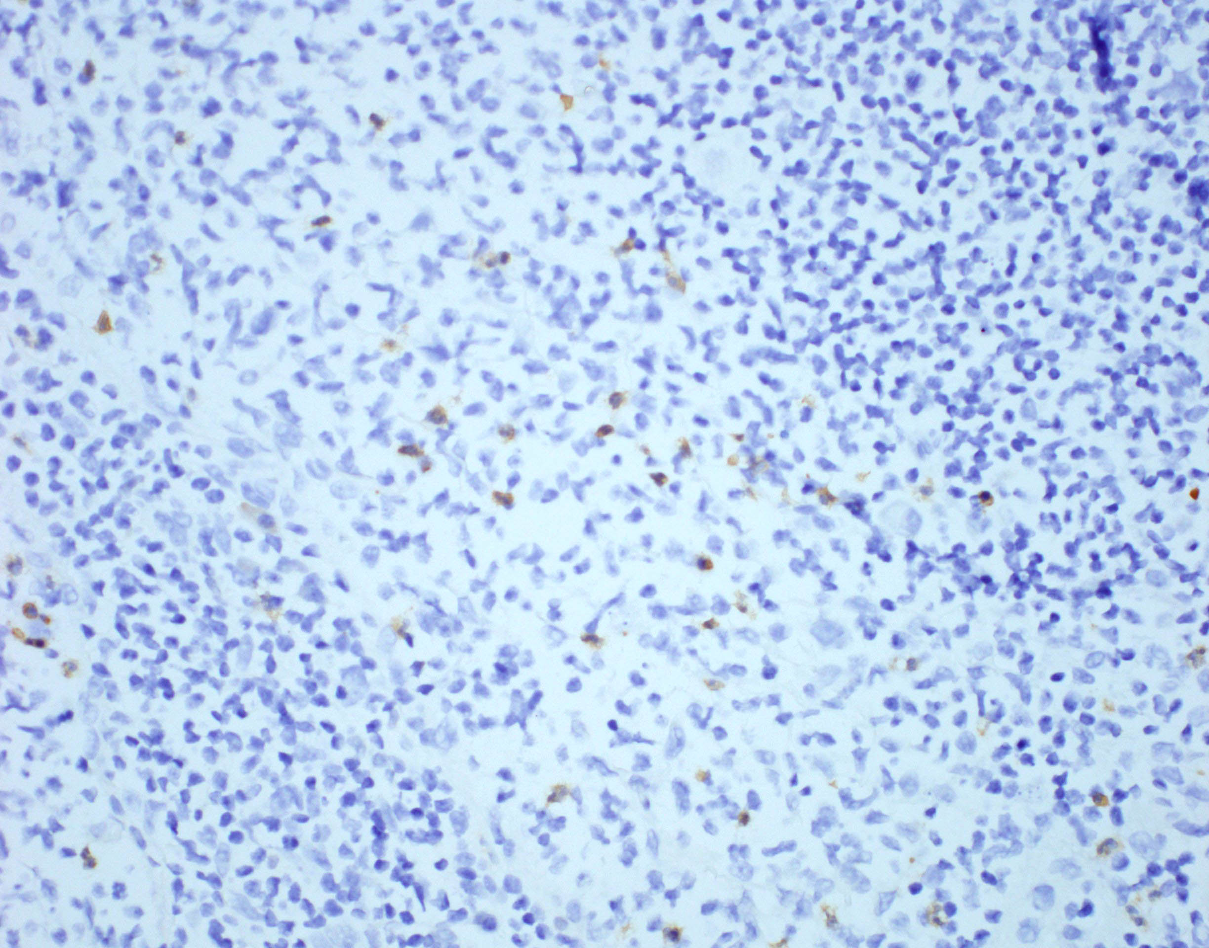
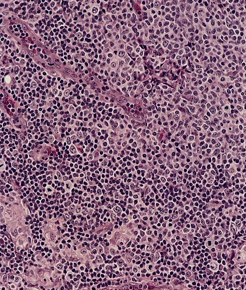
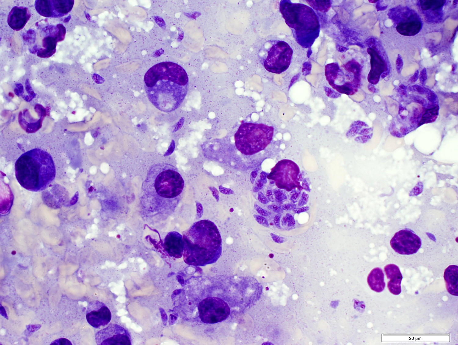
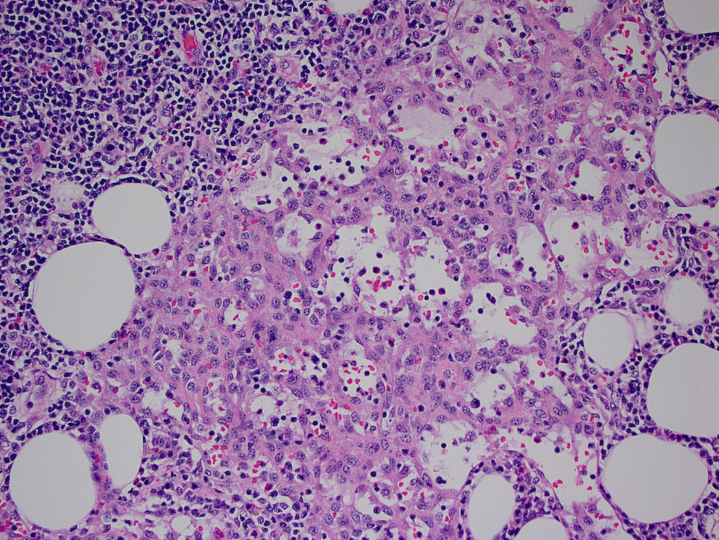
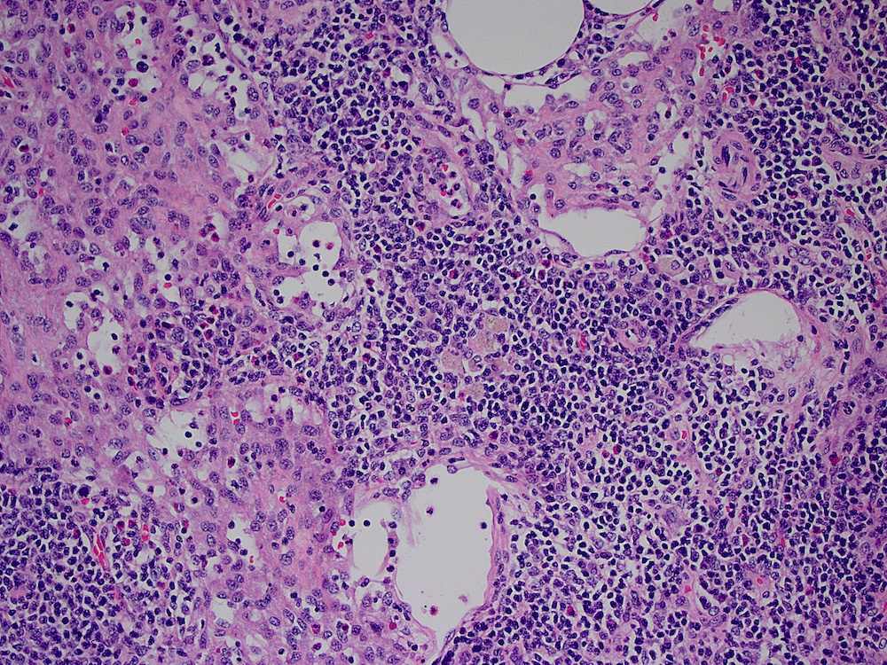
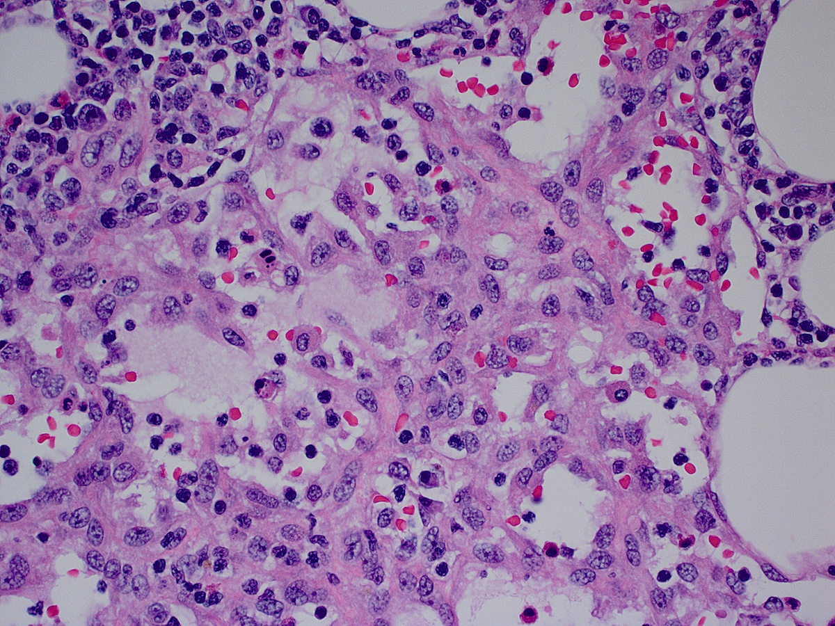
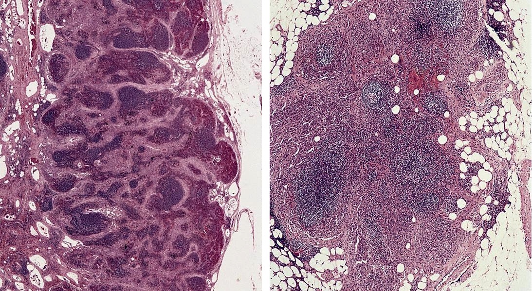
Case of the Week #406
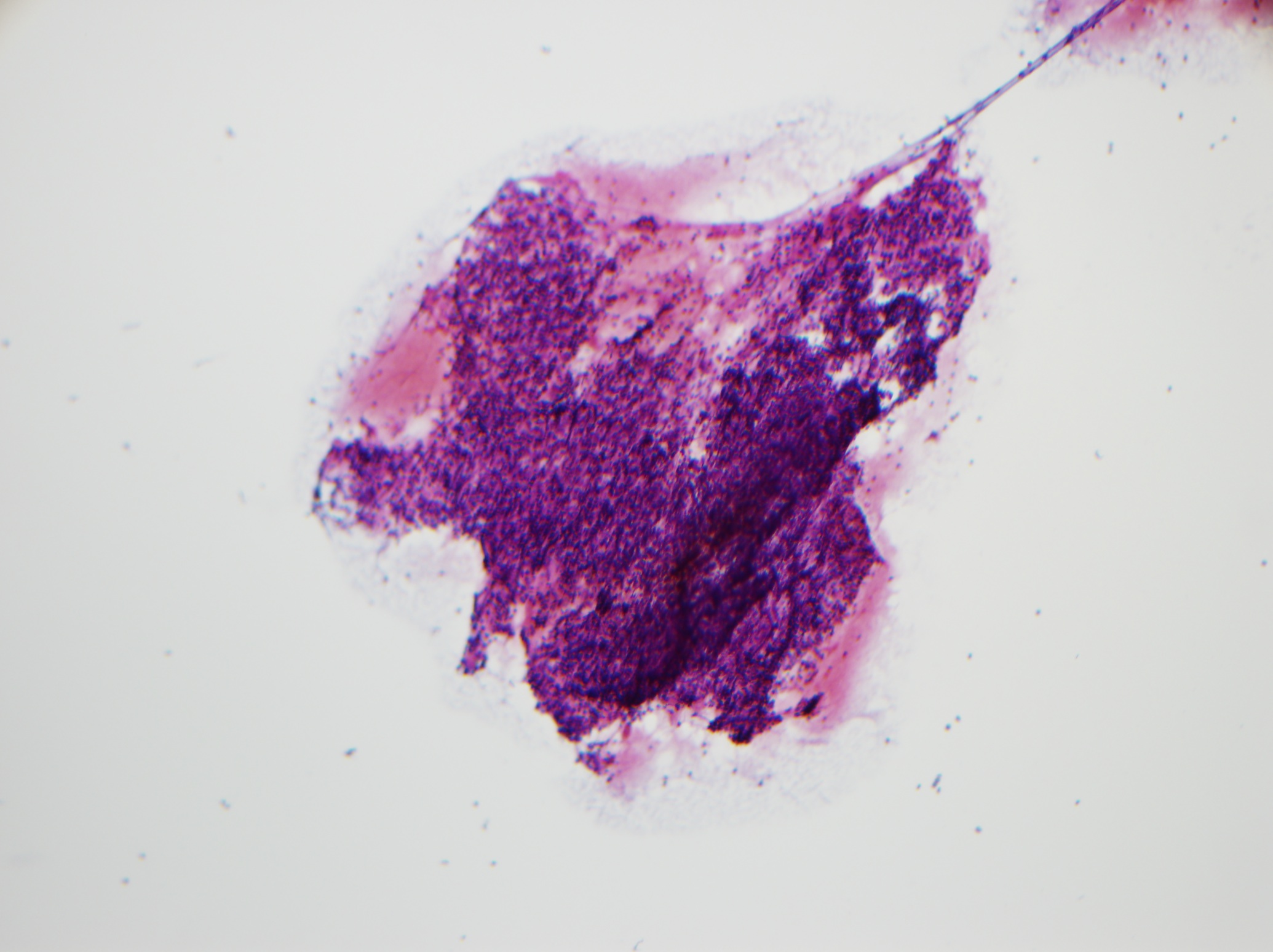
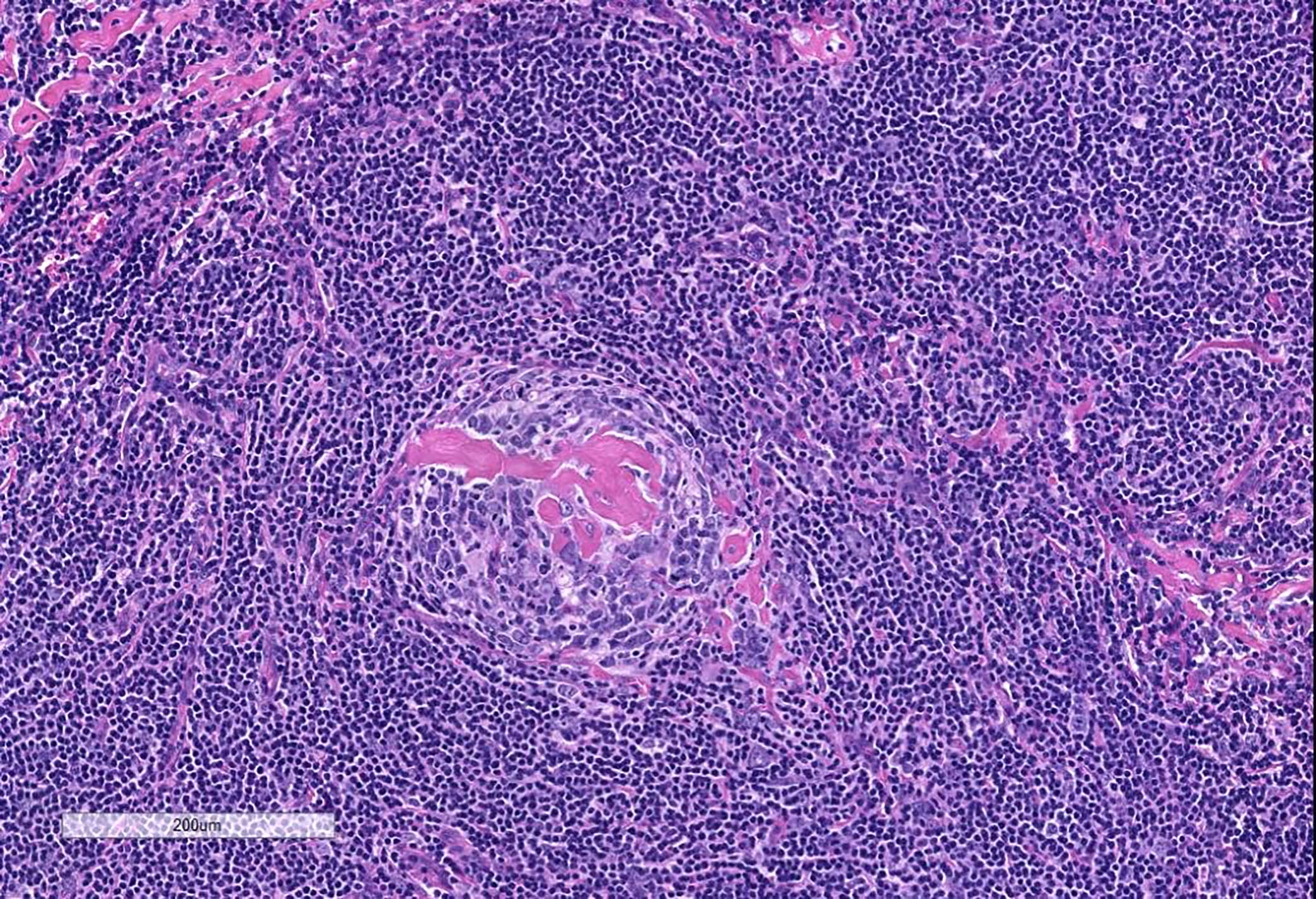
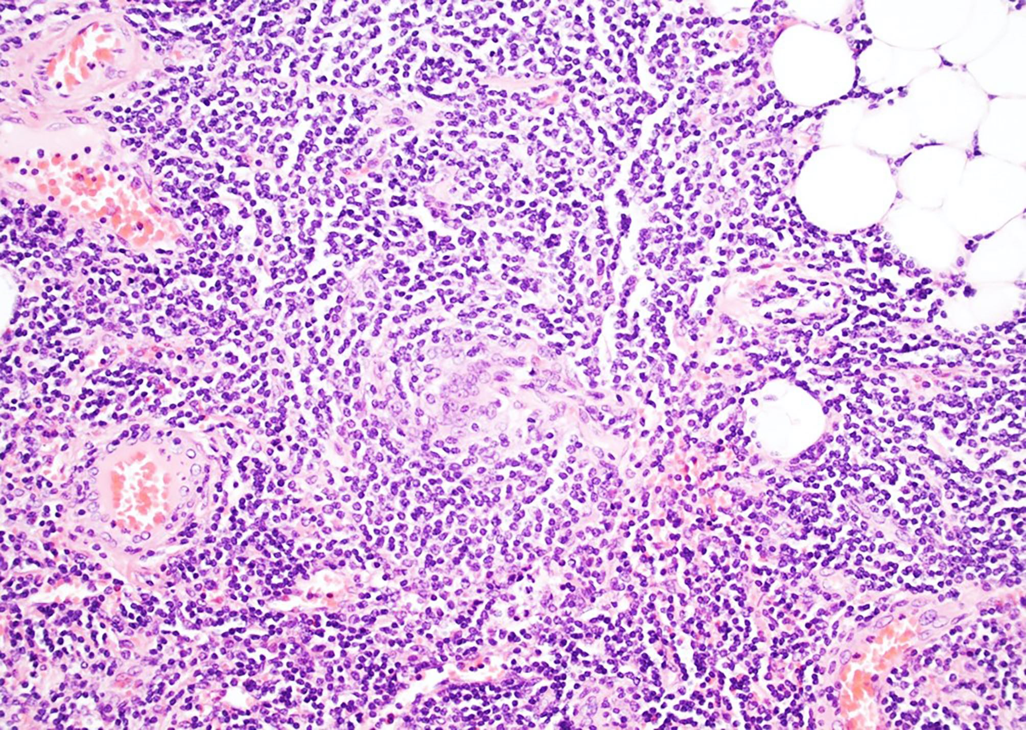
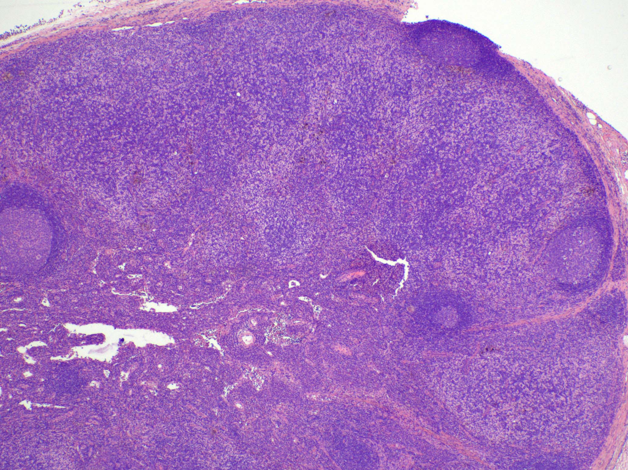
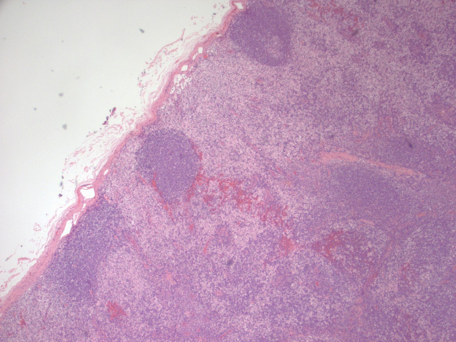
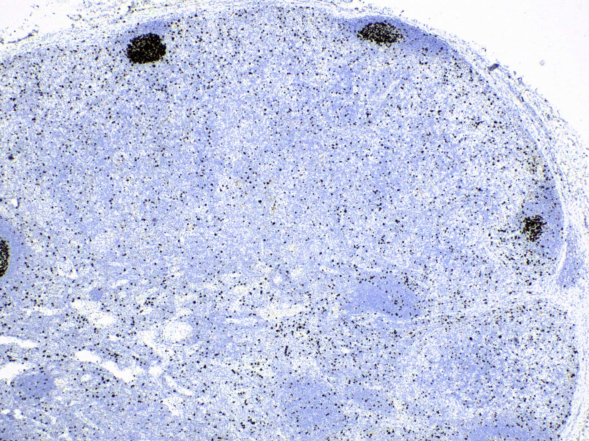
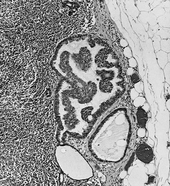
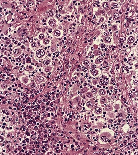
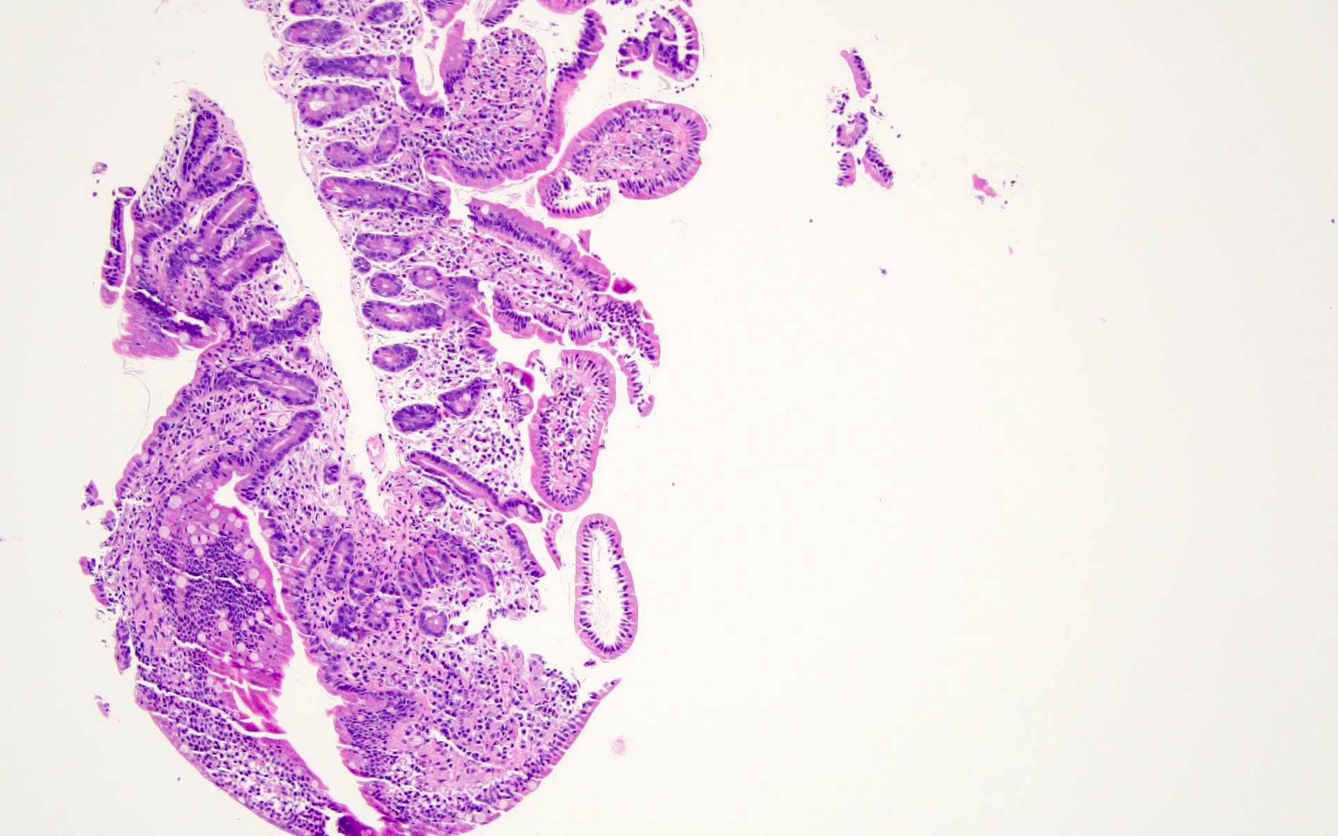
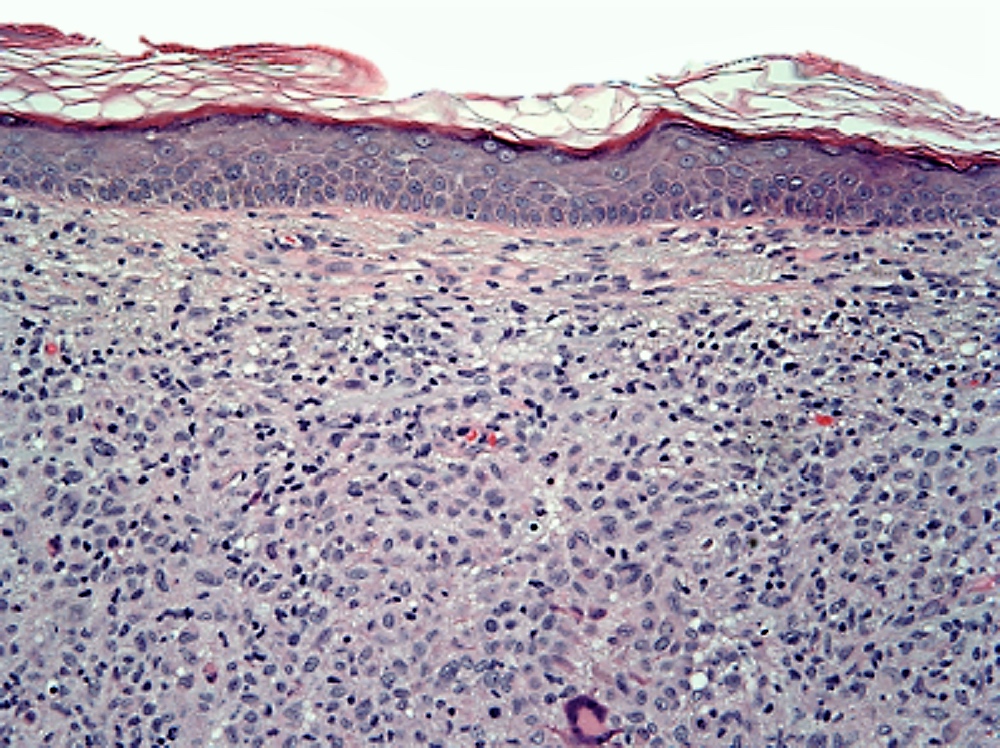
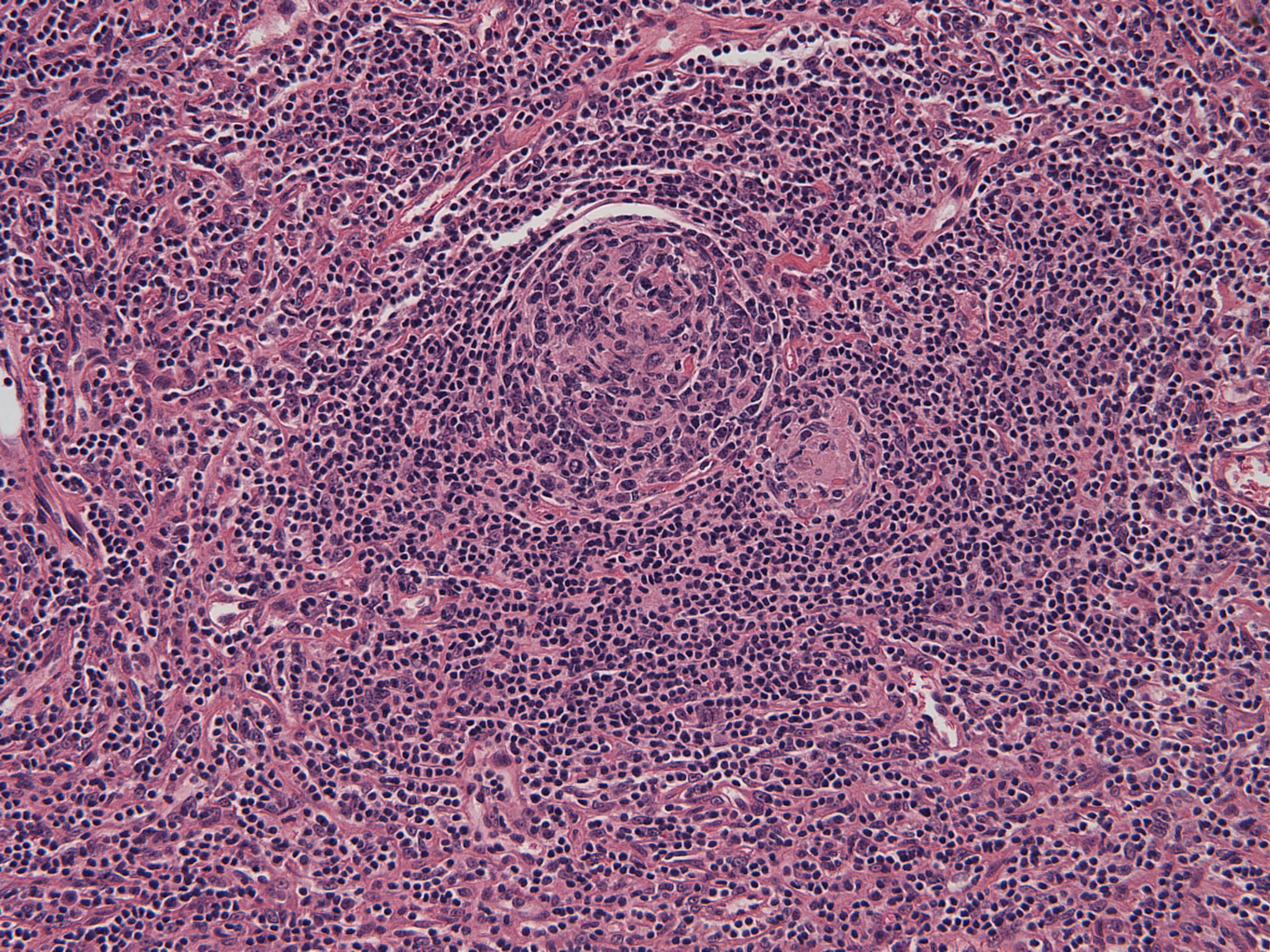
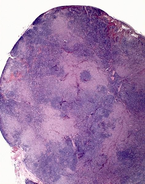
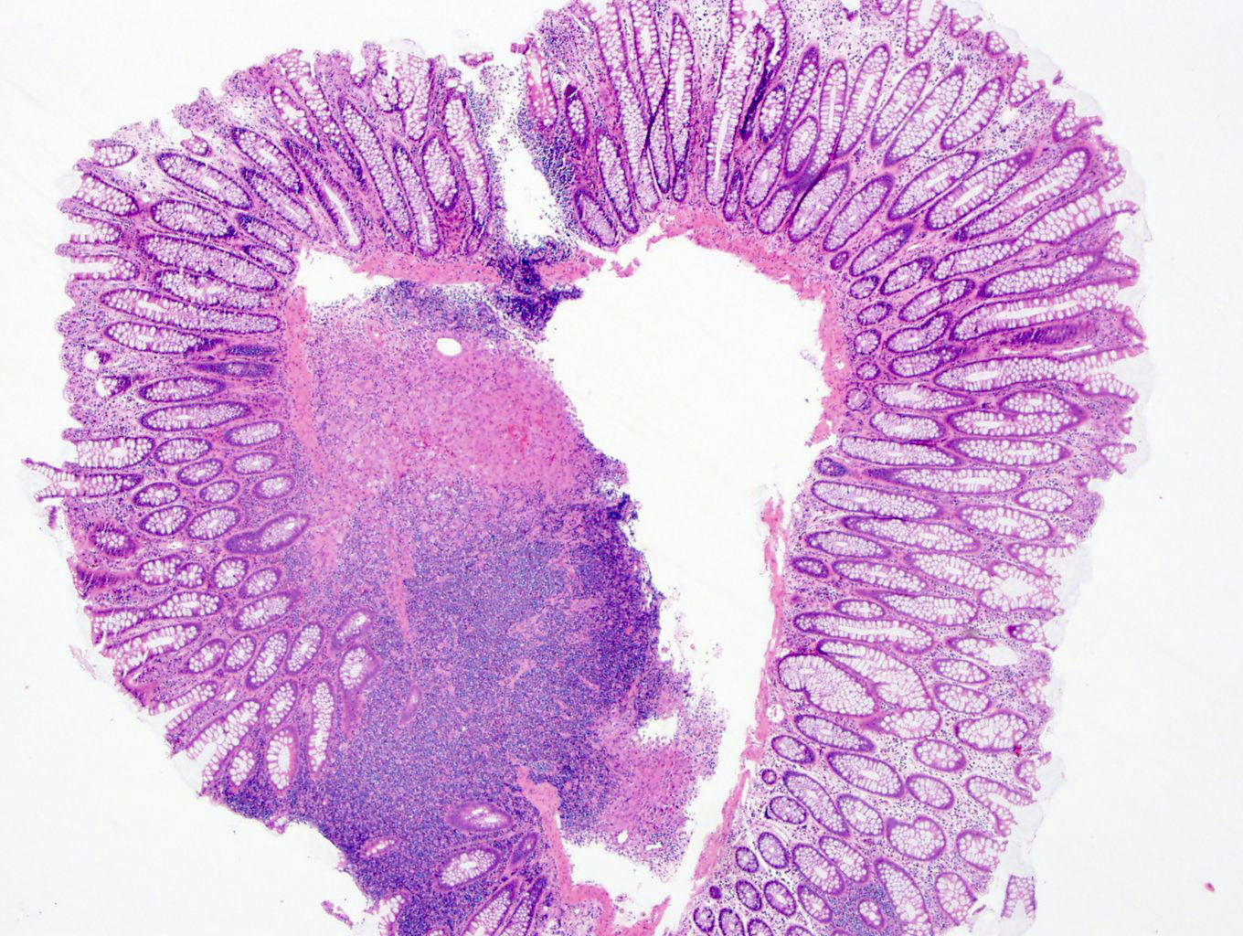
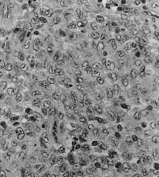
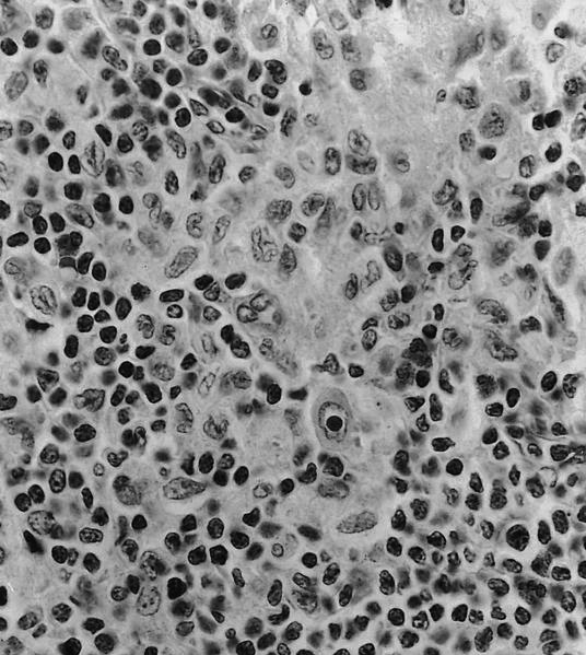
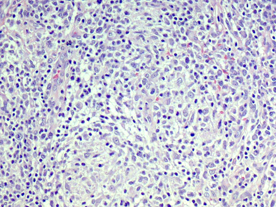
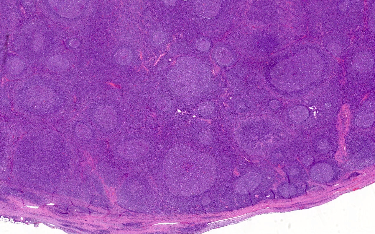
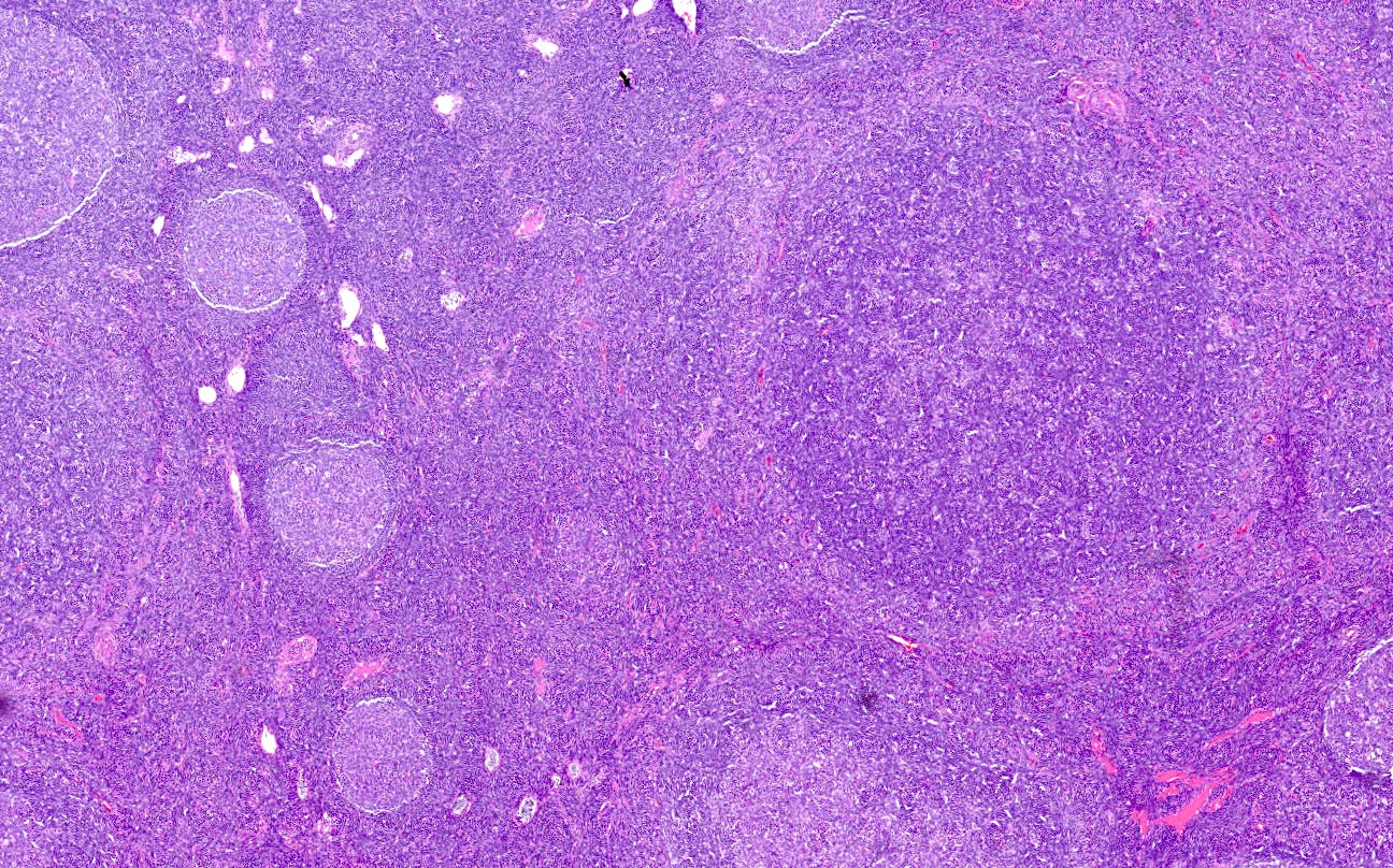
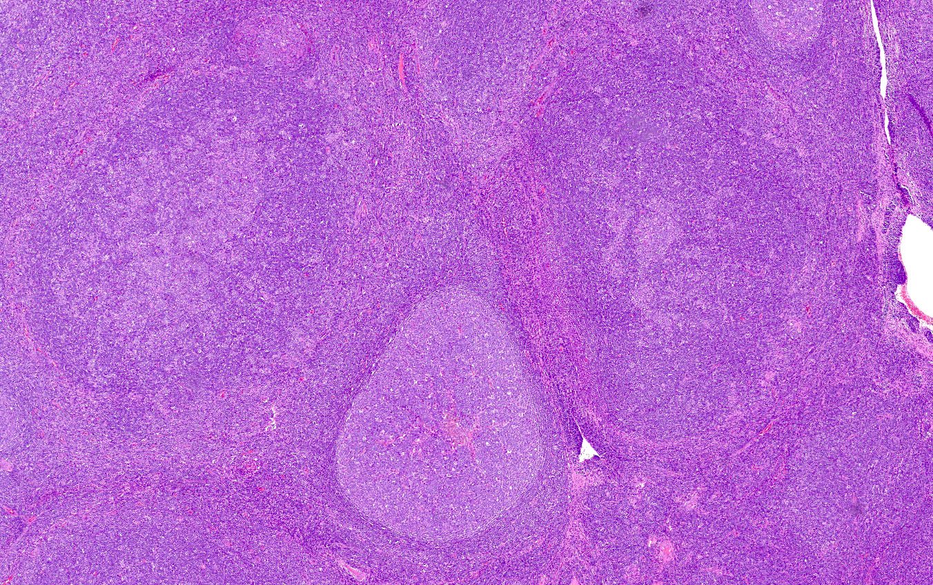
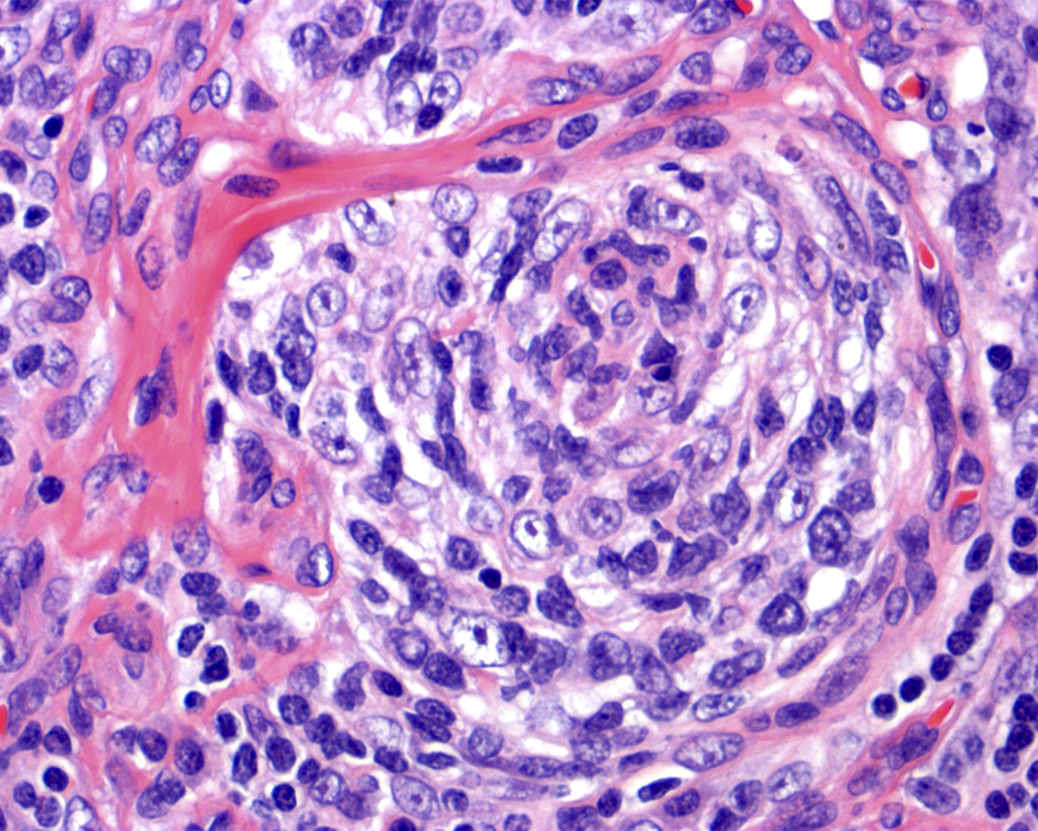
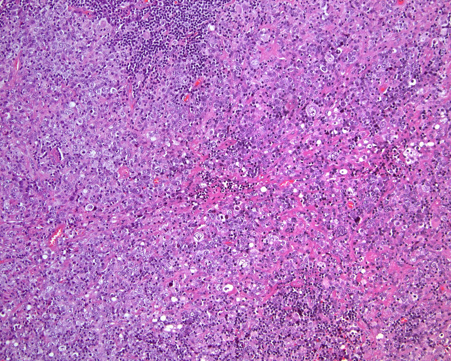
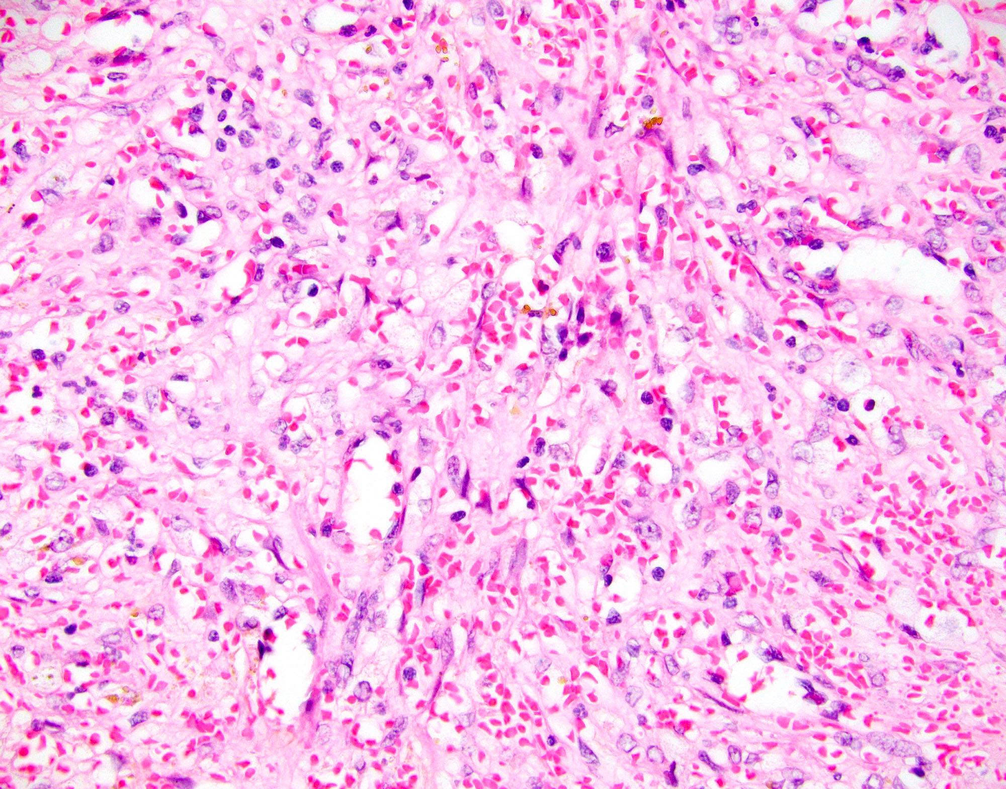
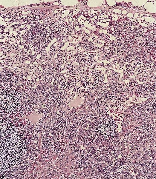
10x
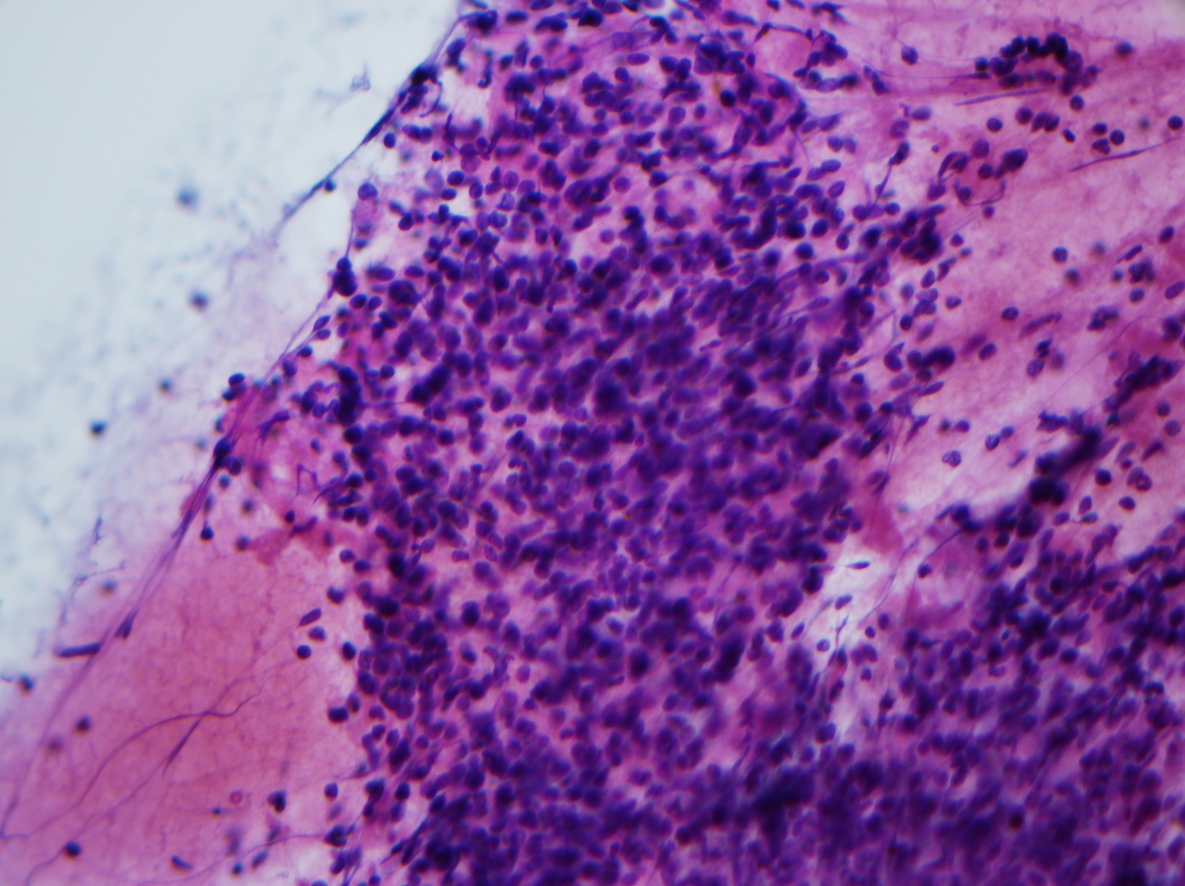
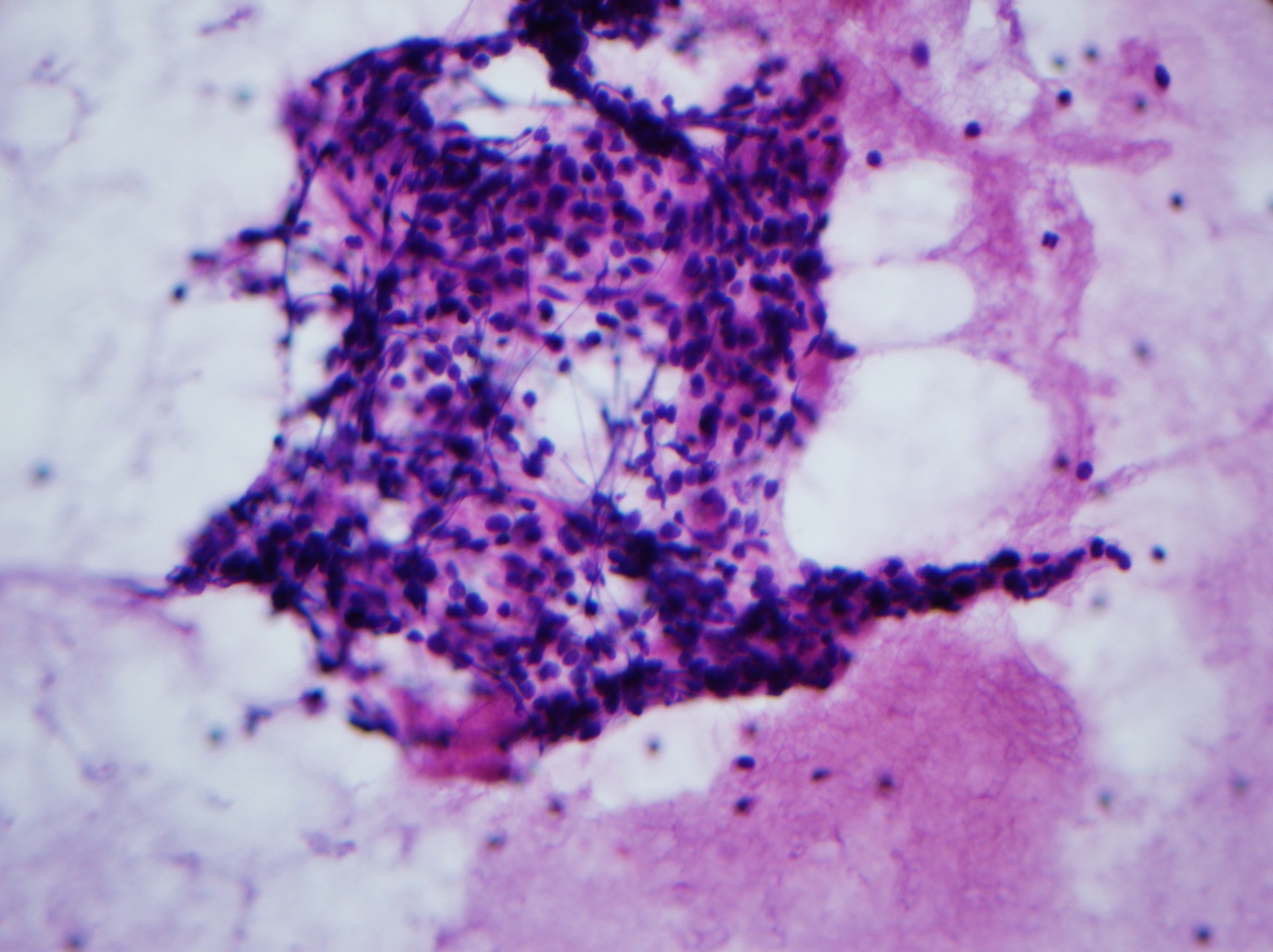
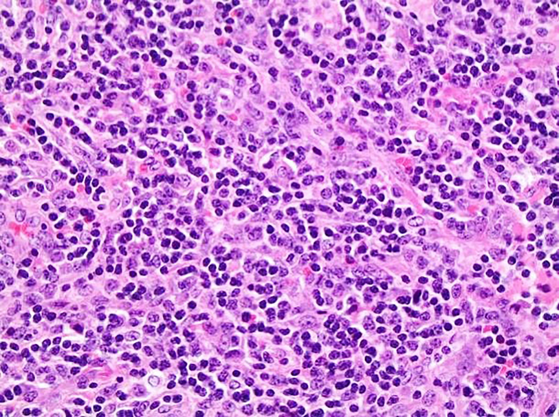
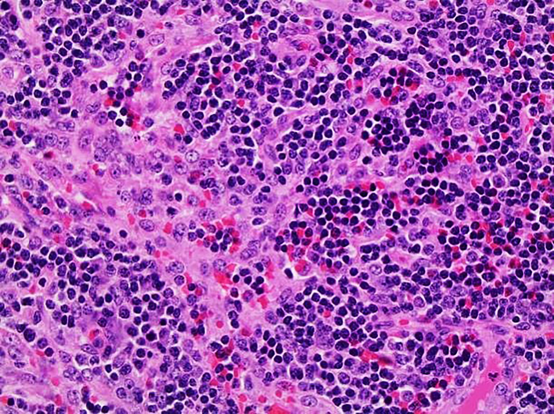
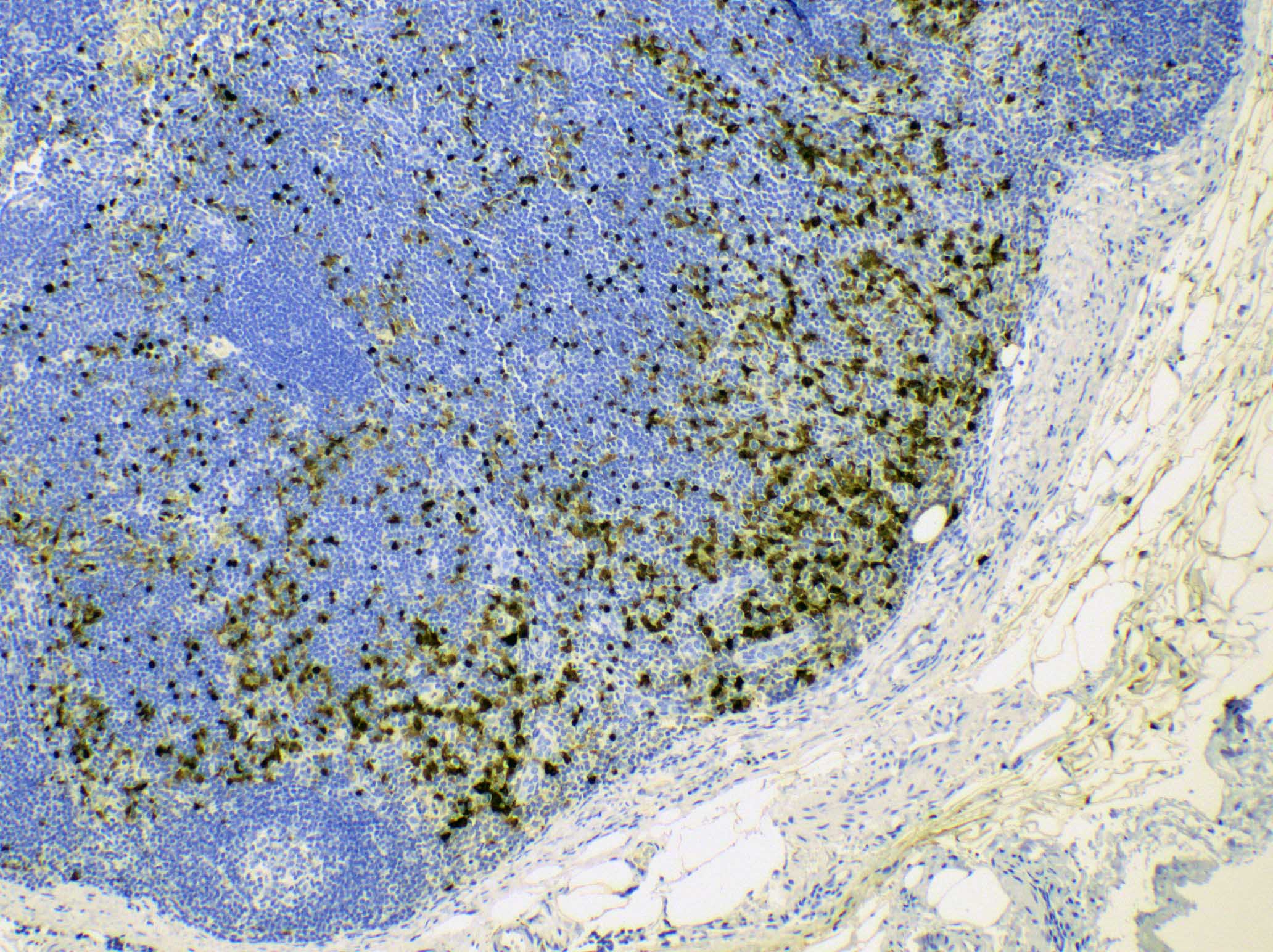
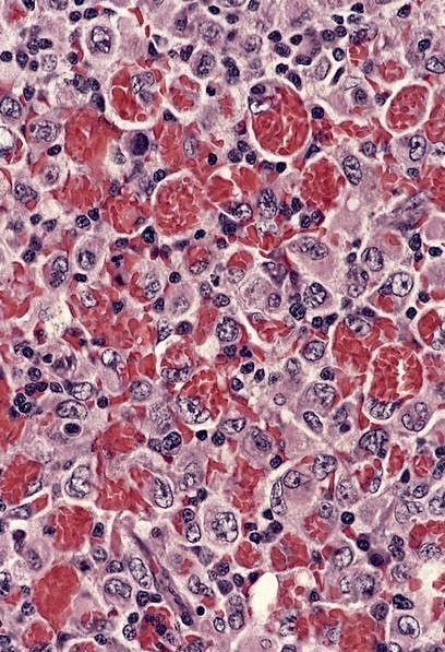
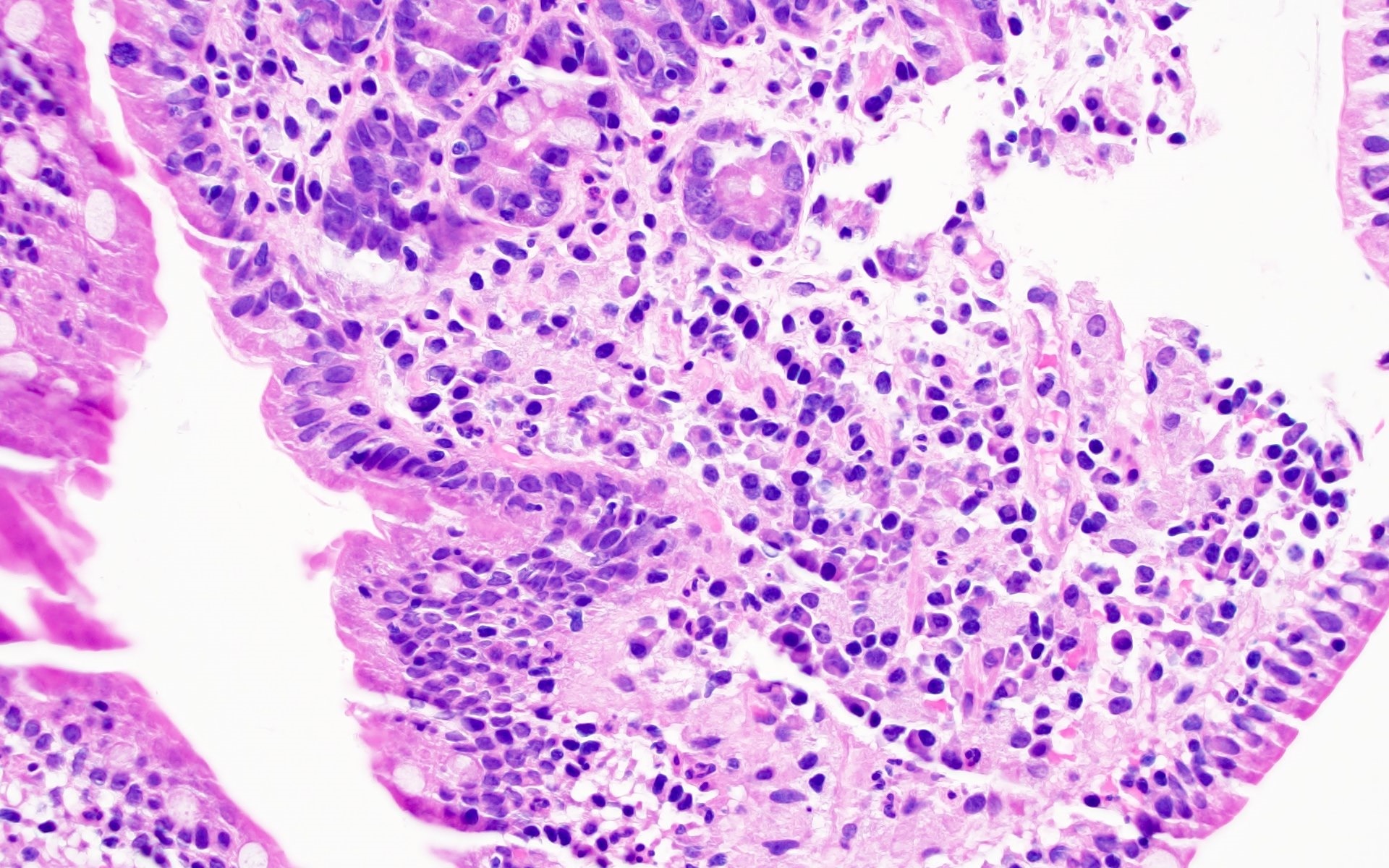
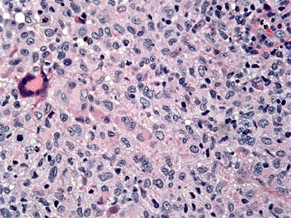
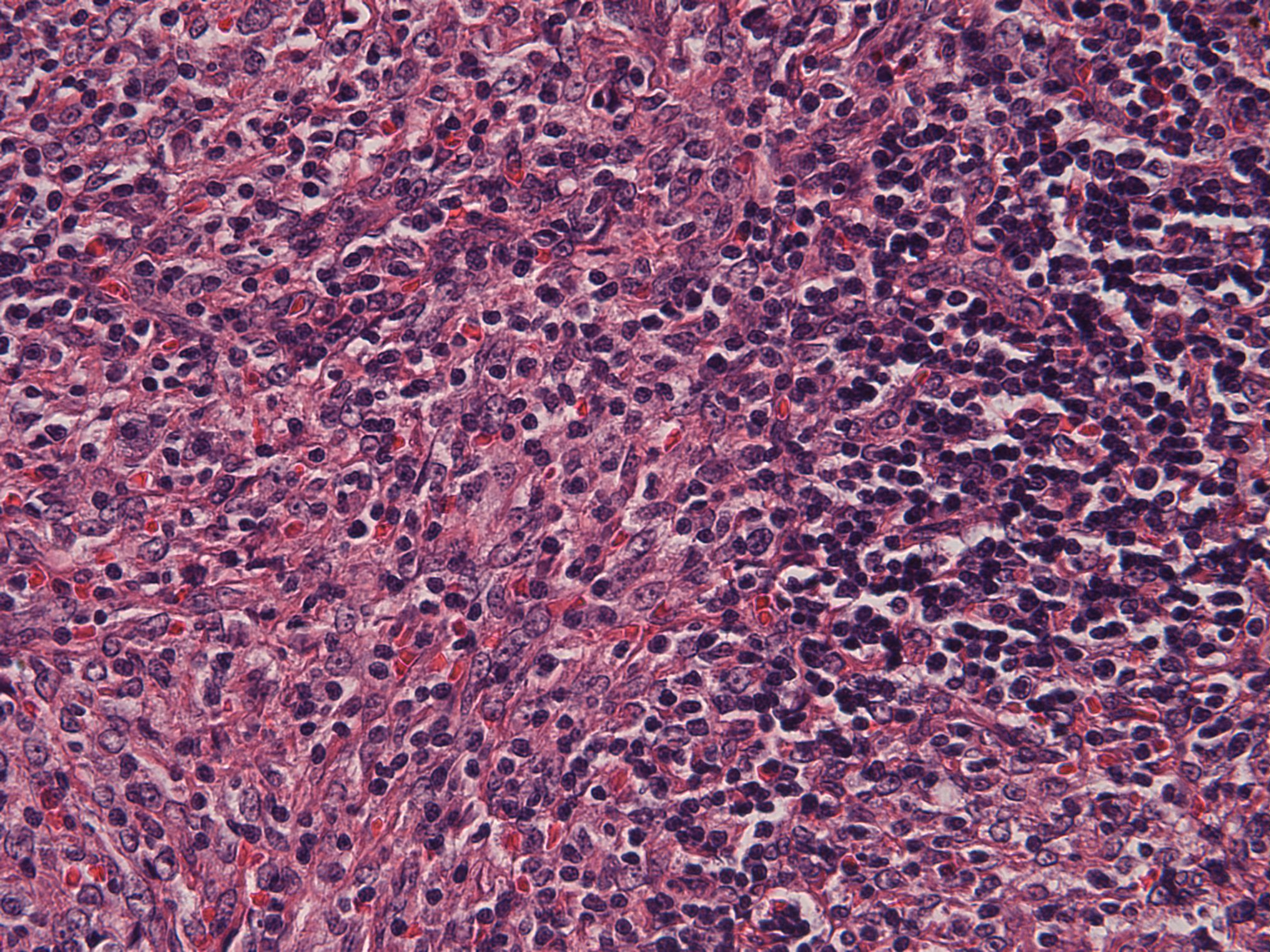
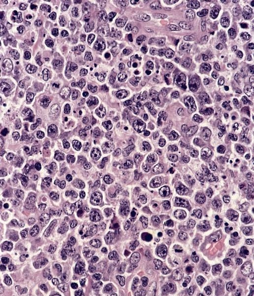
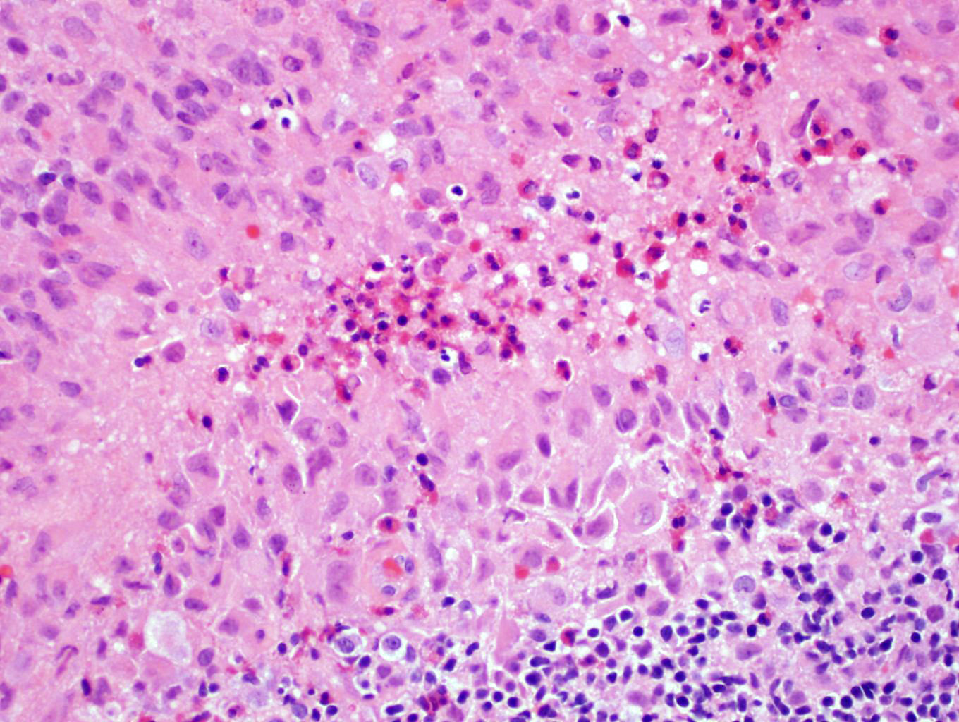
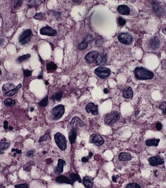
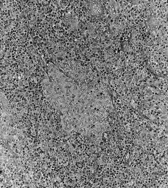
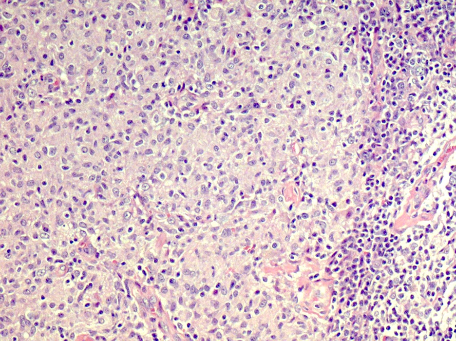
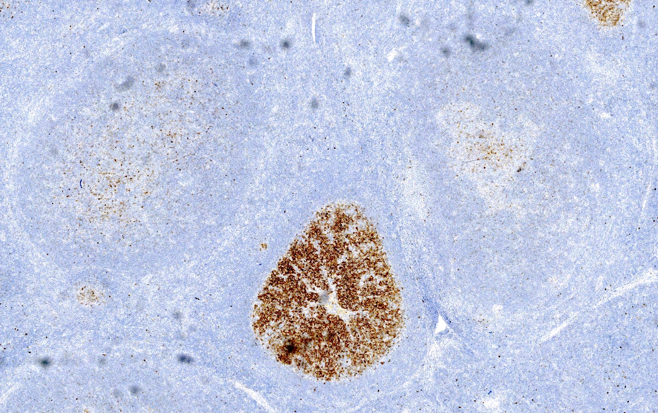
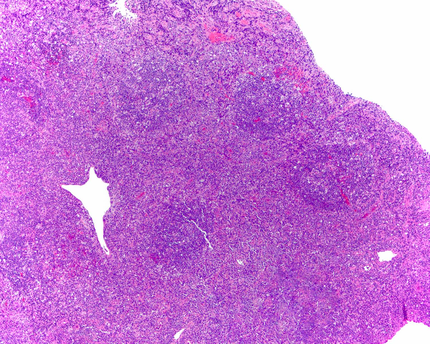
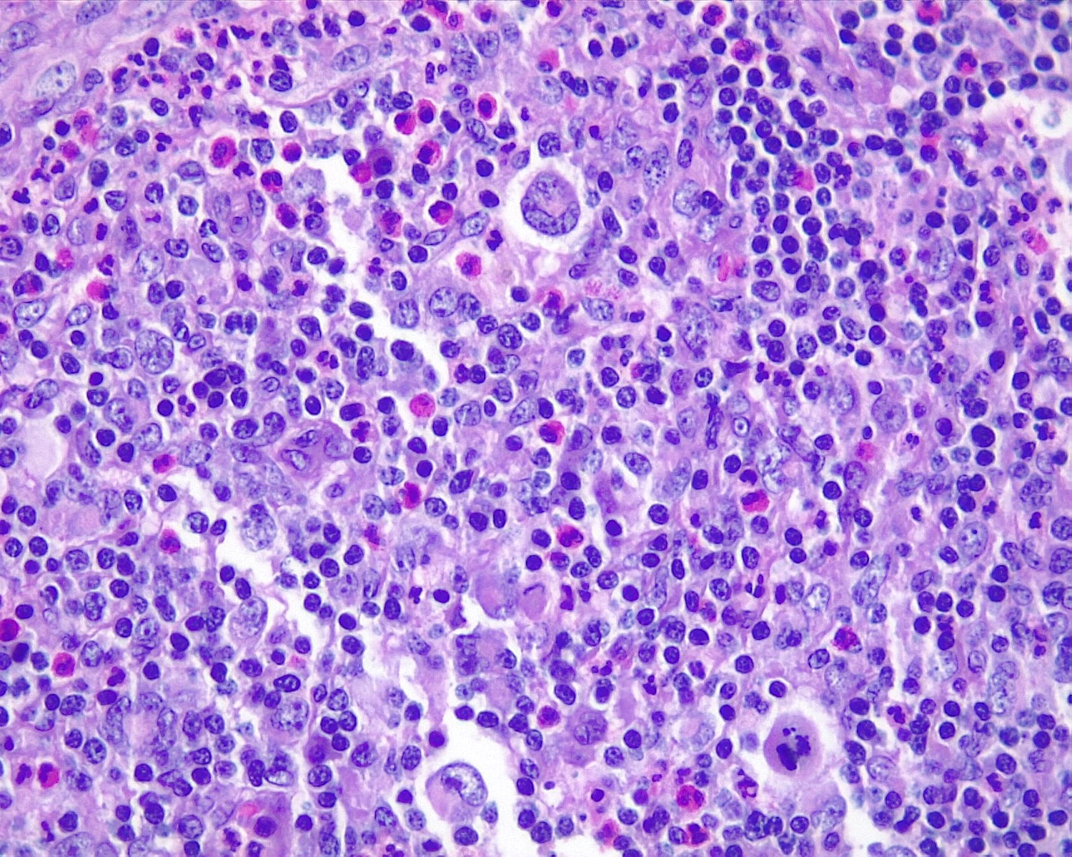
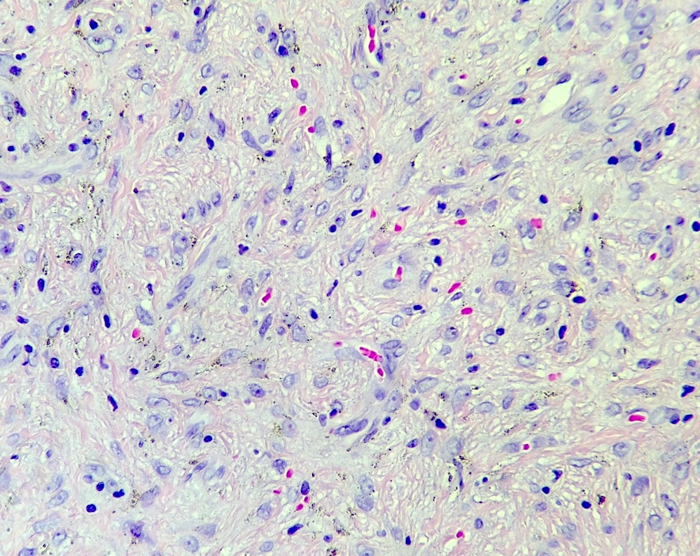
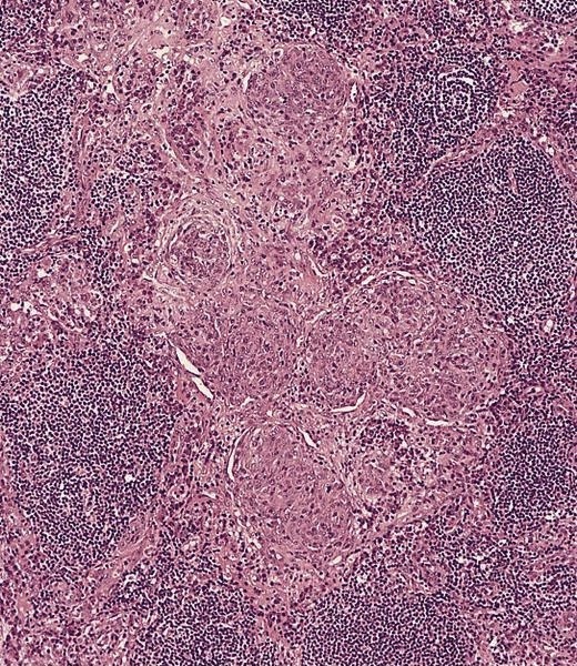
40x
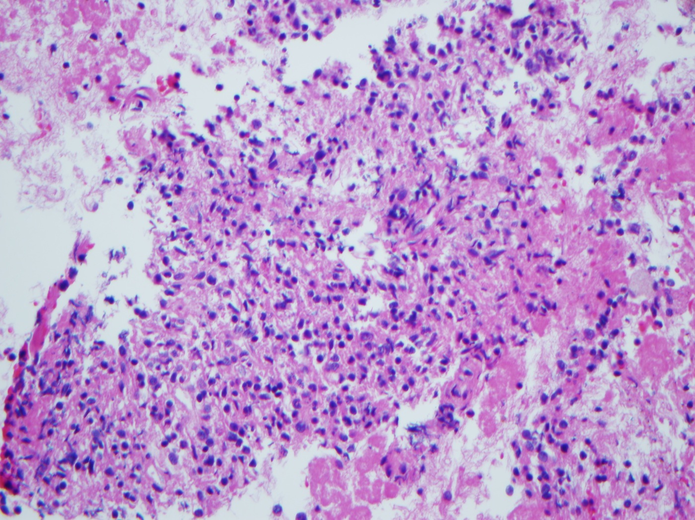
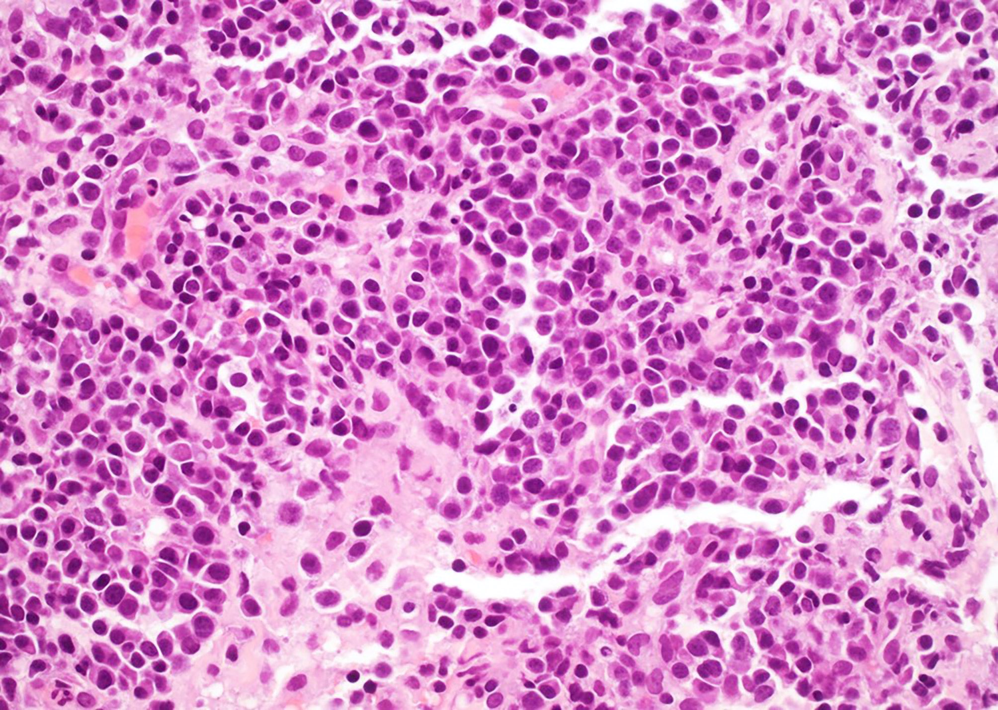
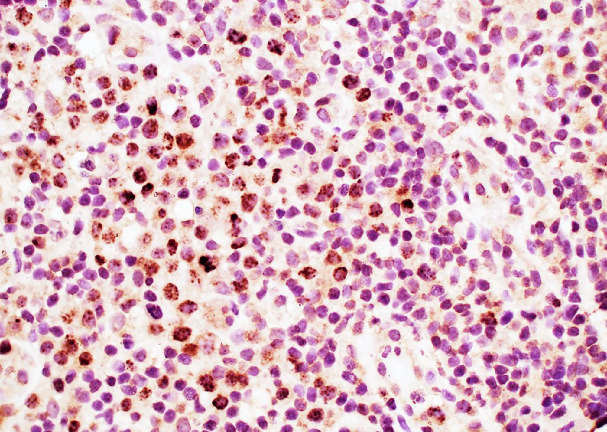
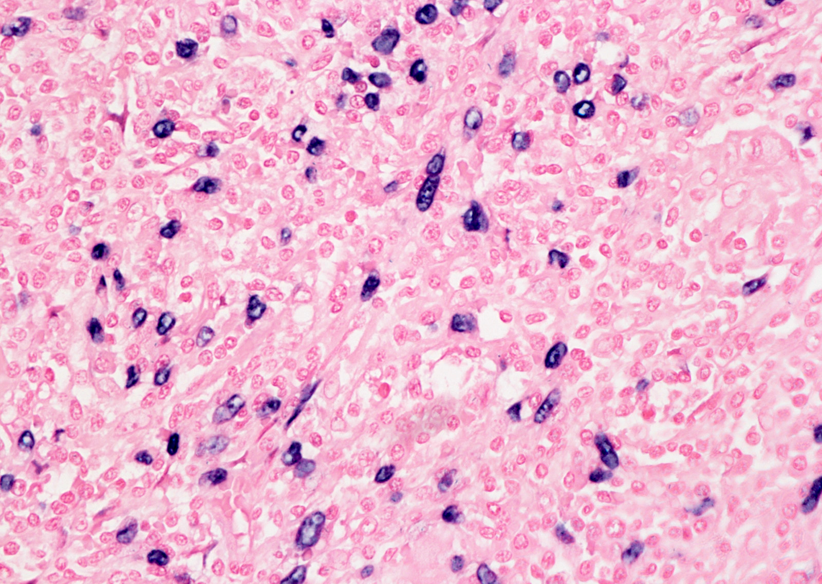
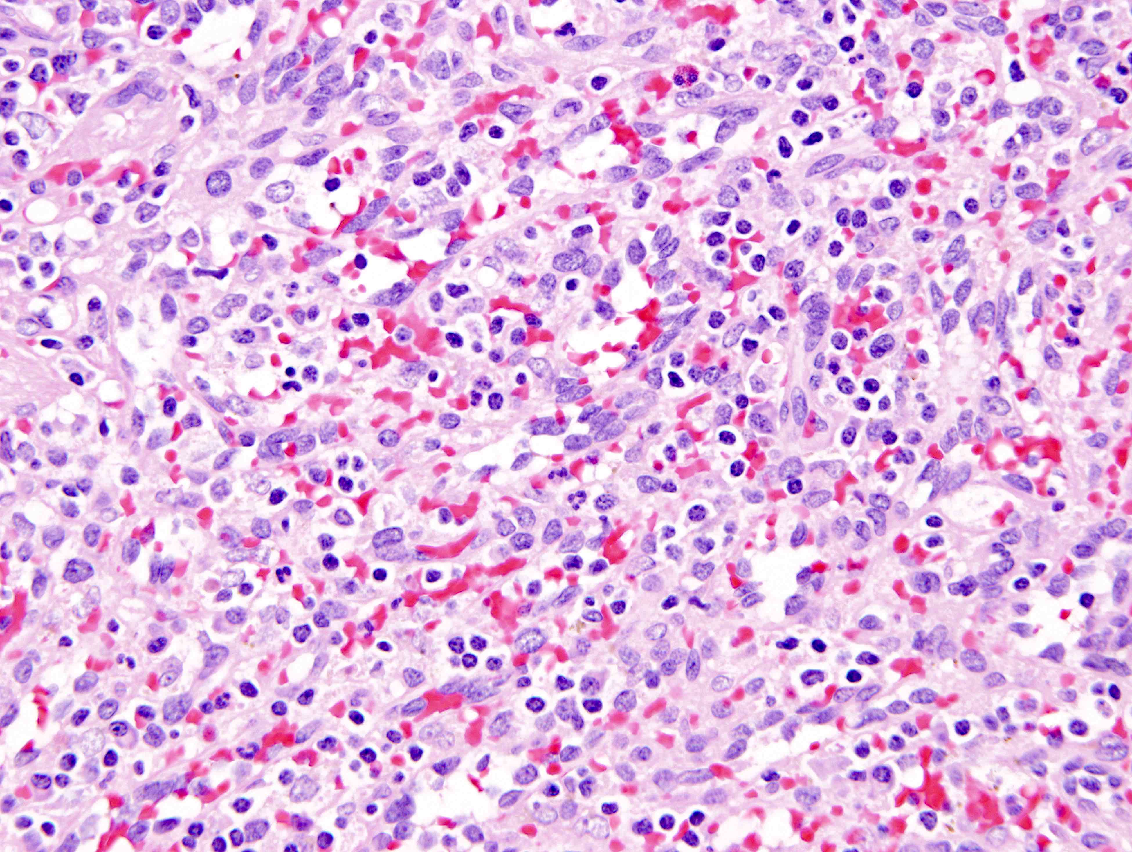
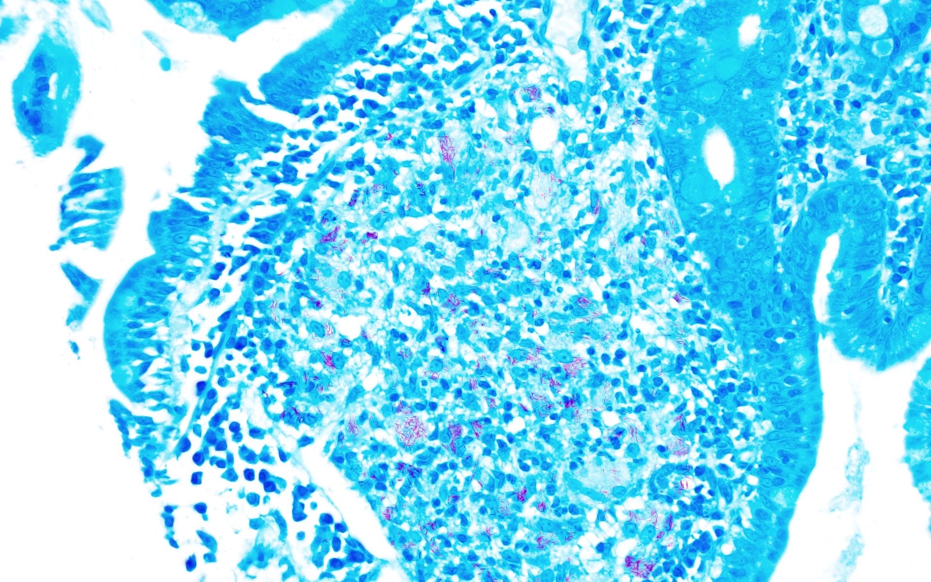
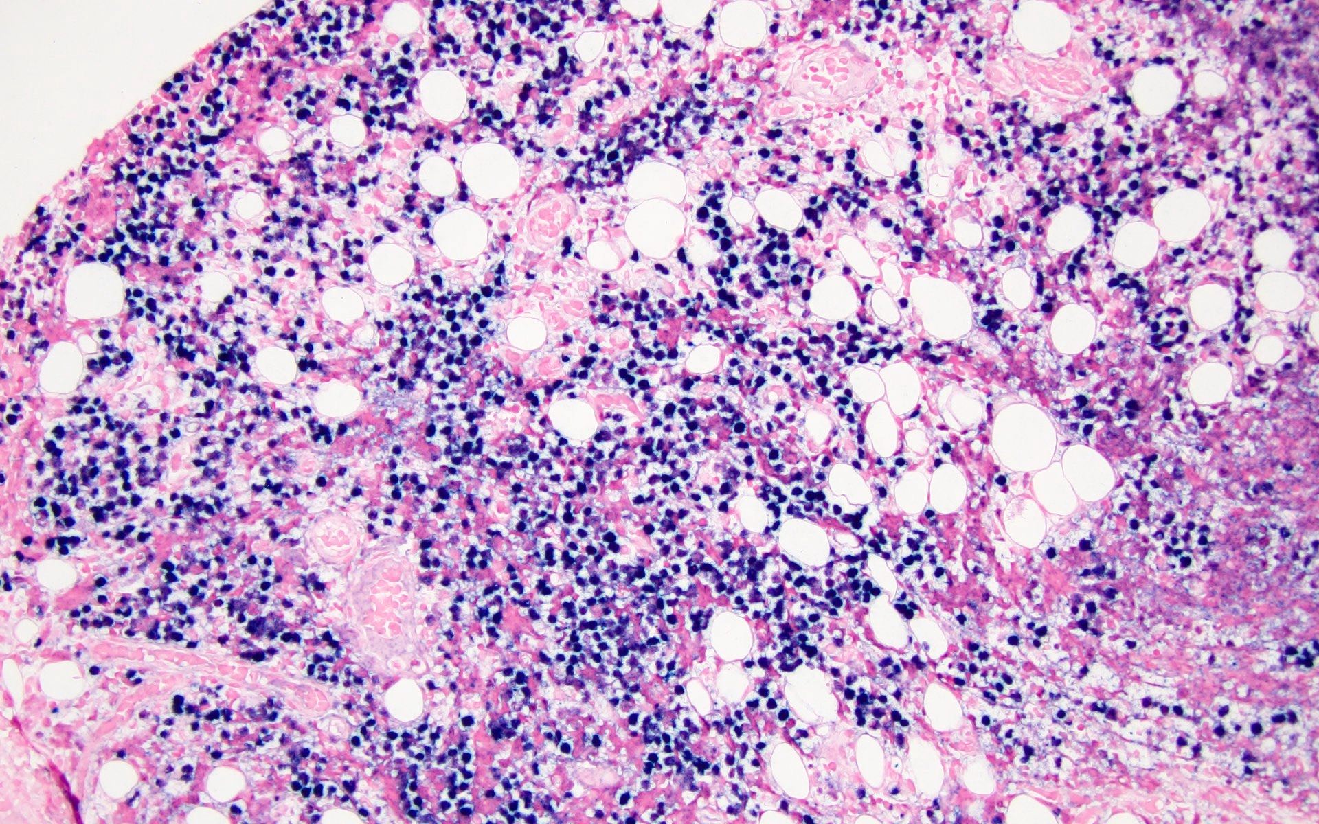
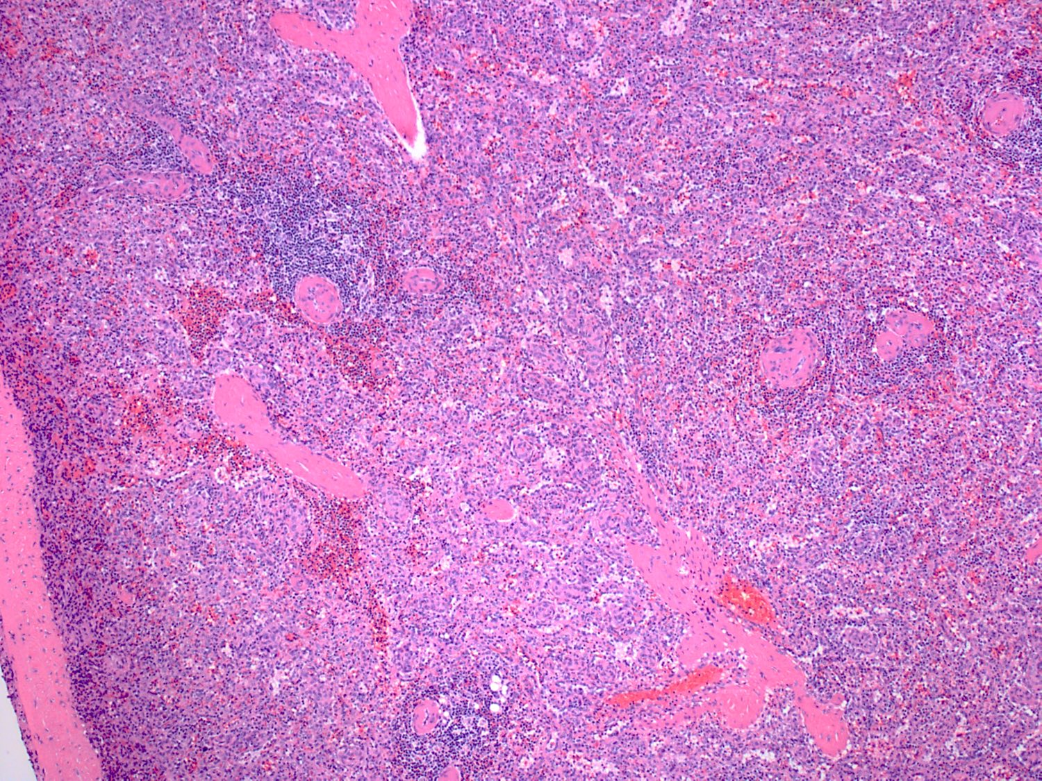
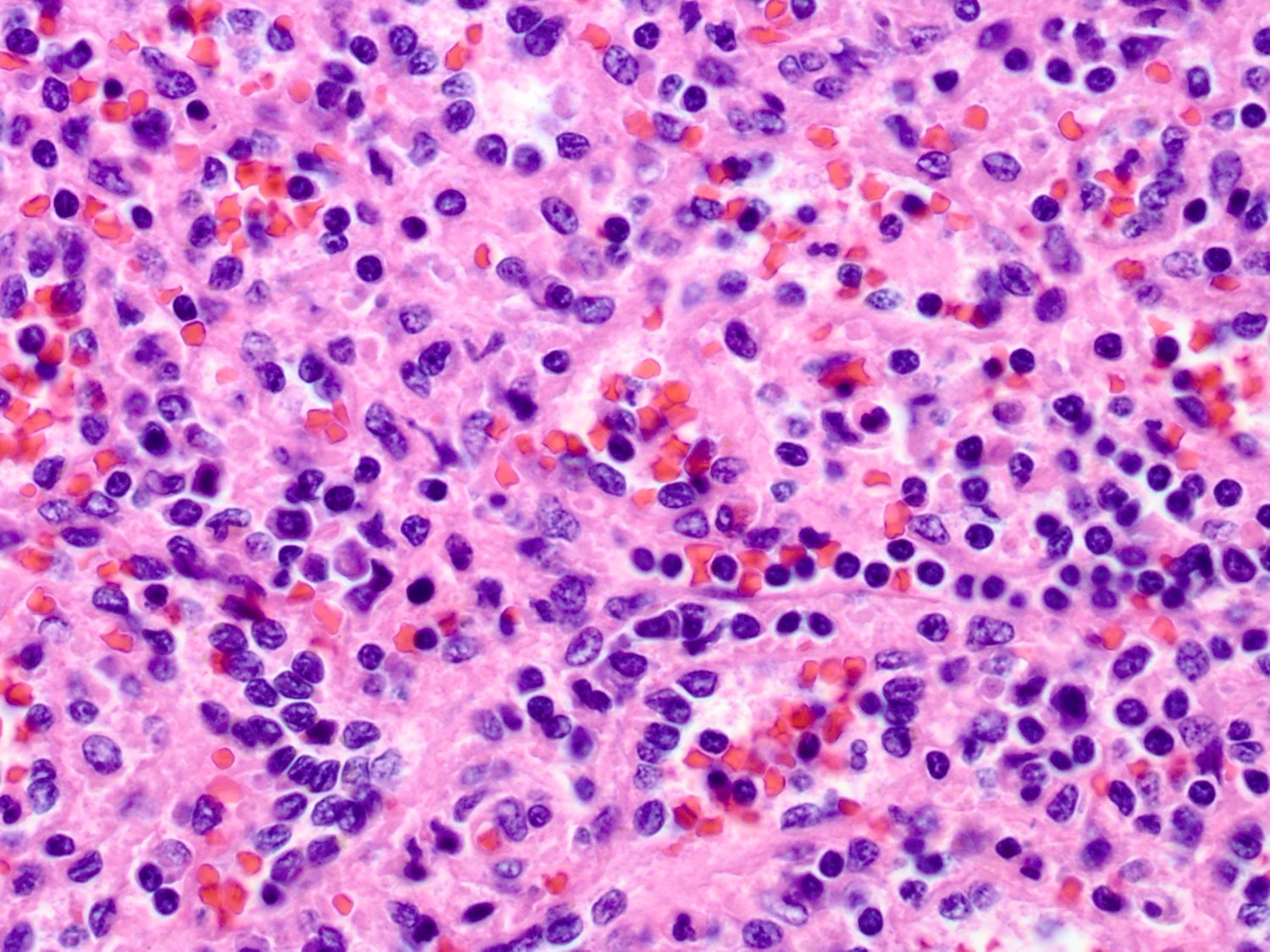
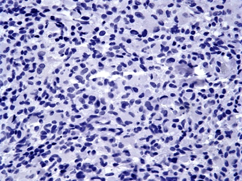
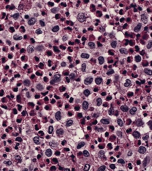
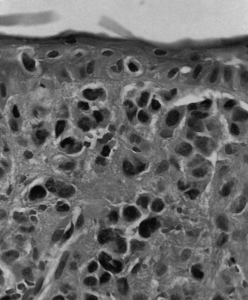
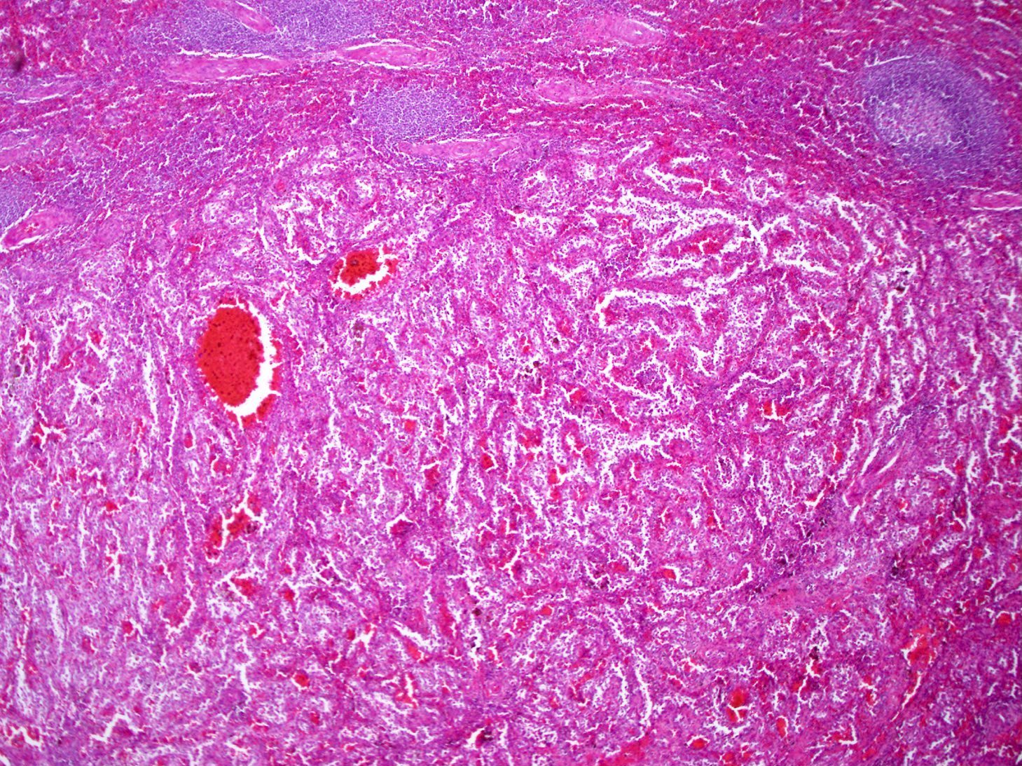
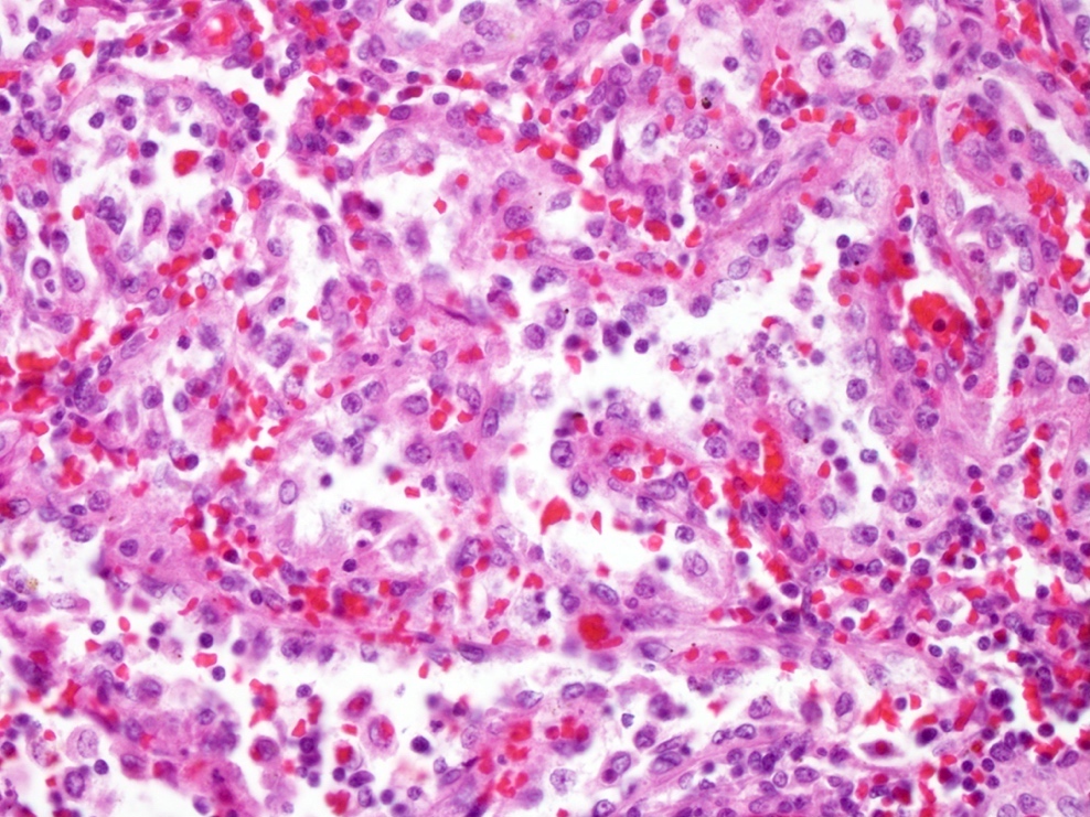
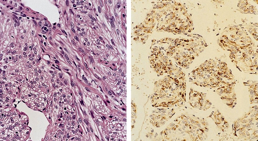
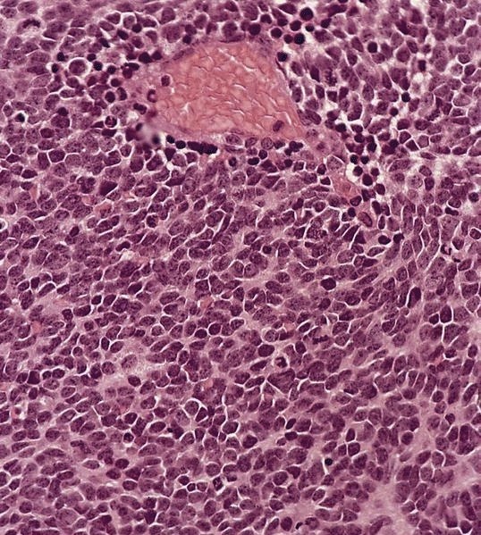
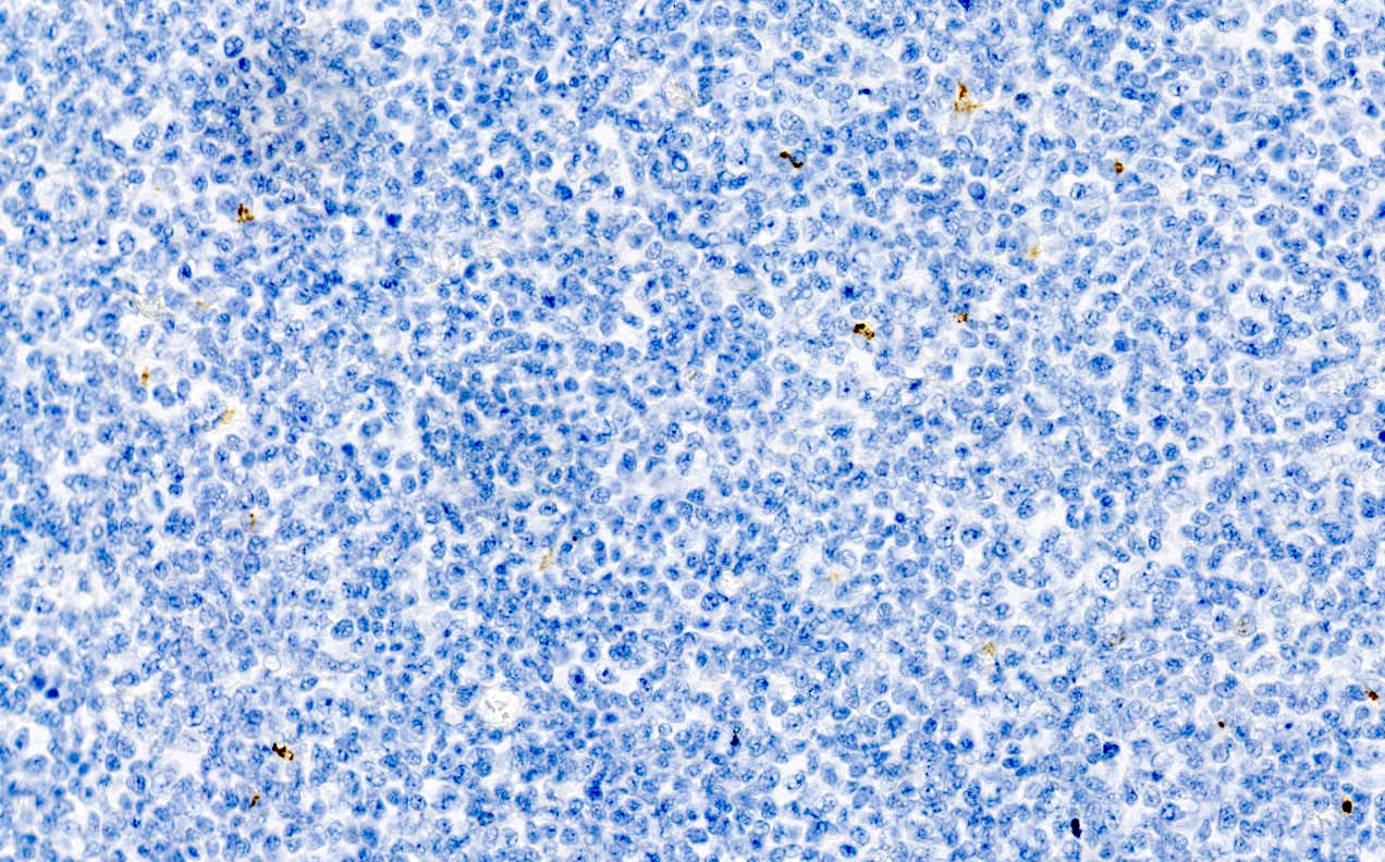
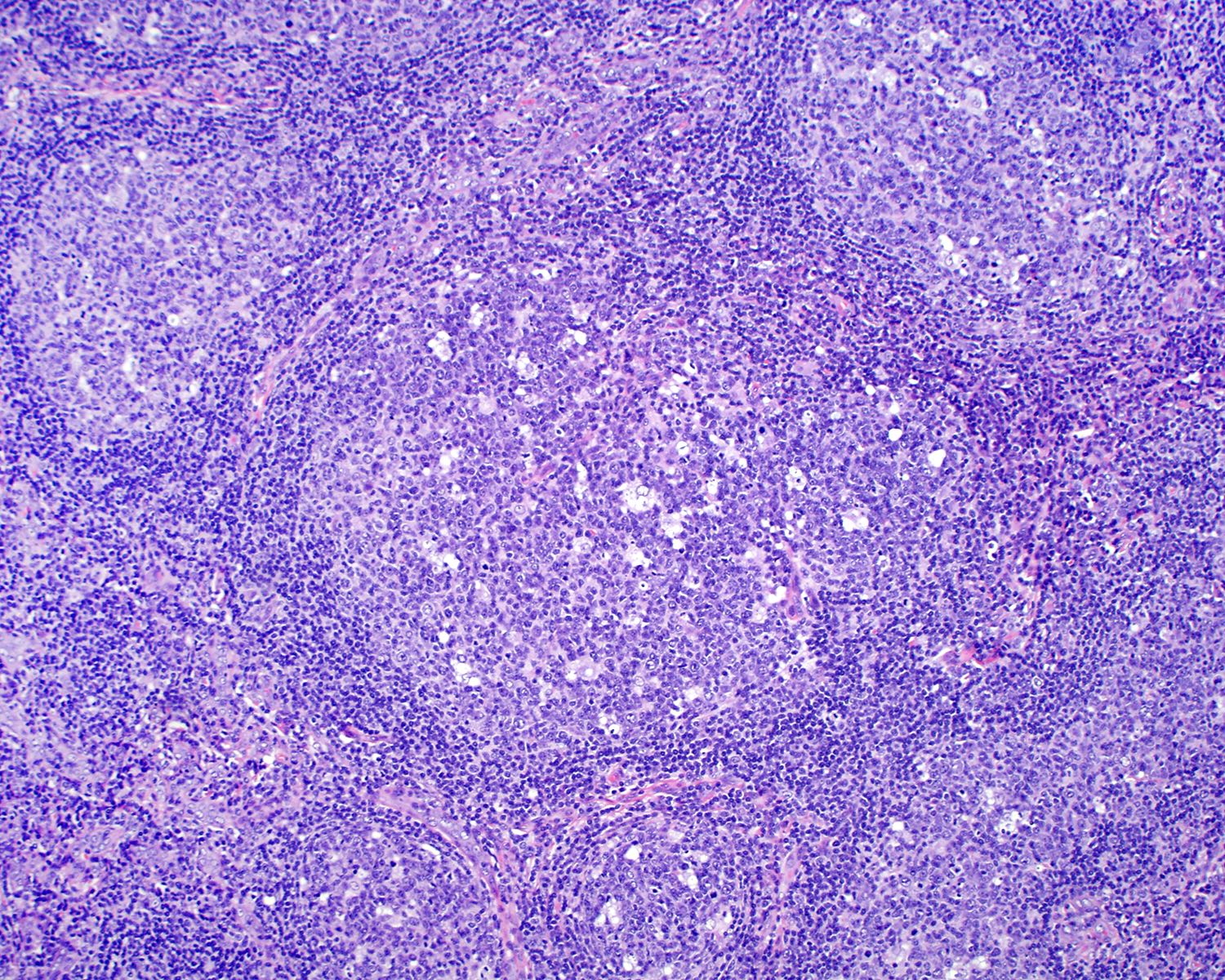
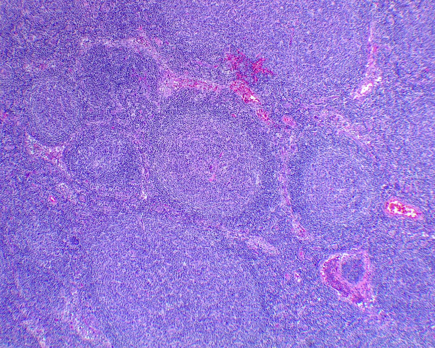
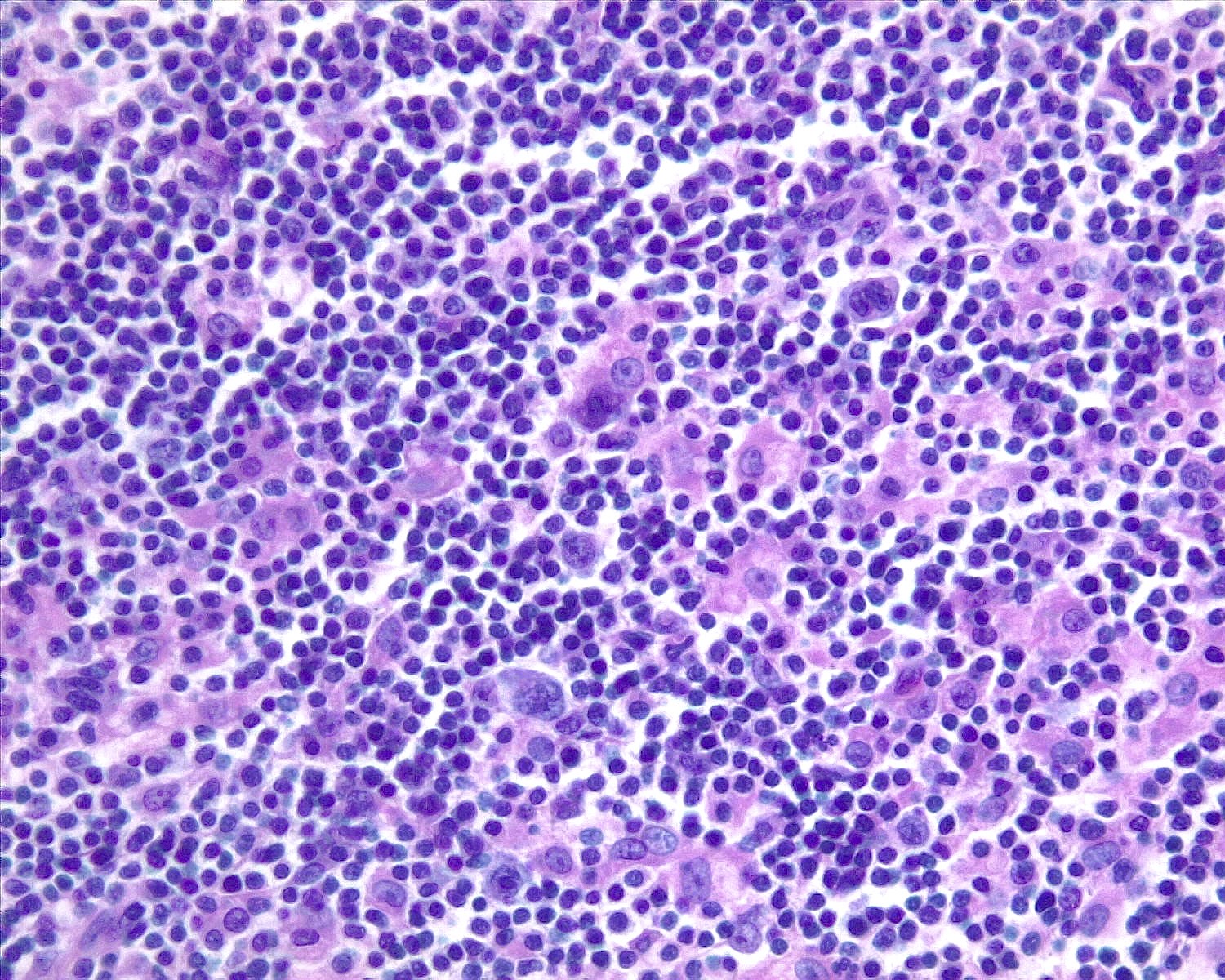
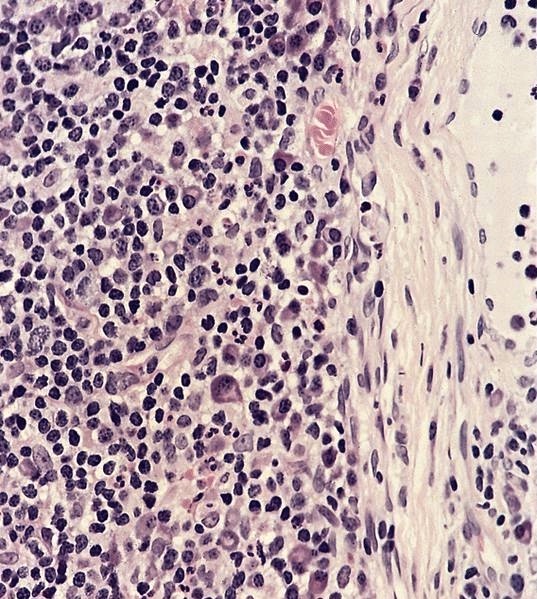
H&E

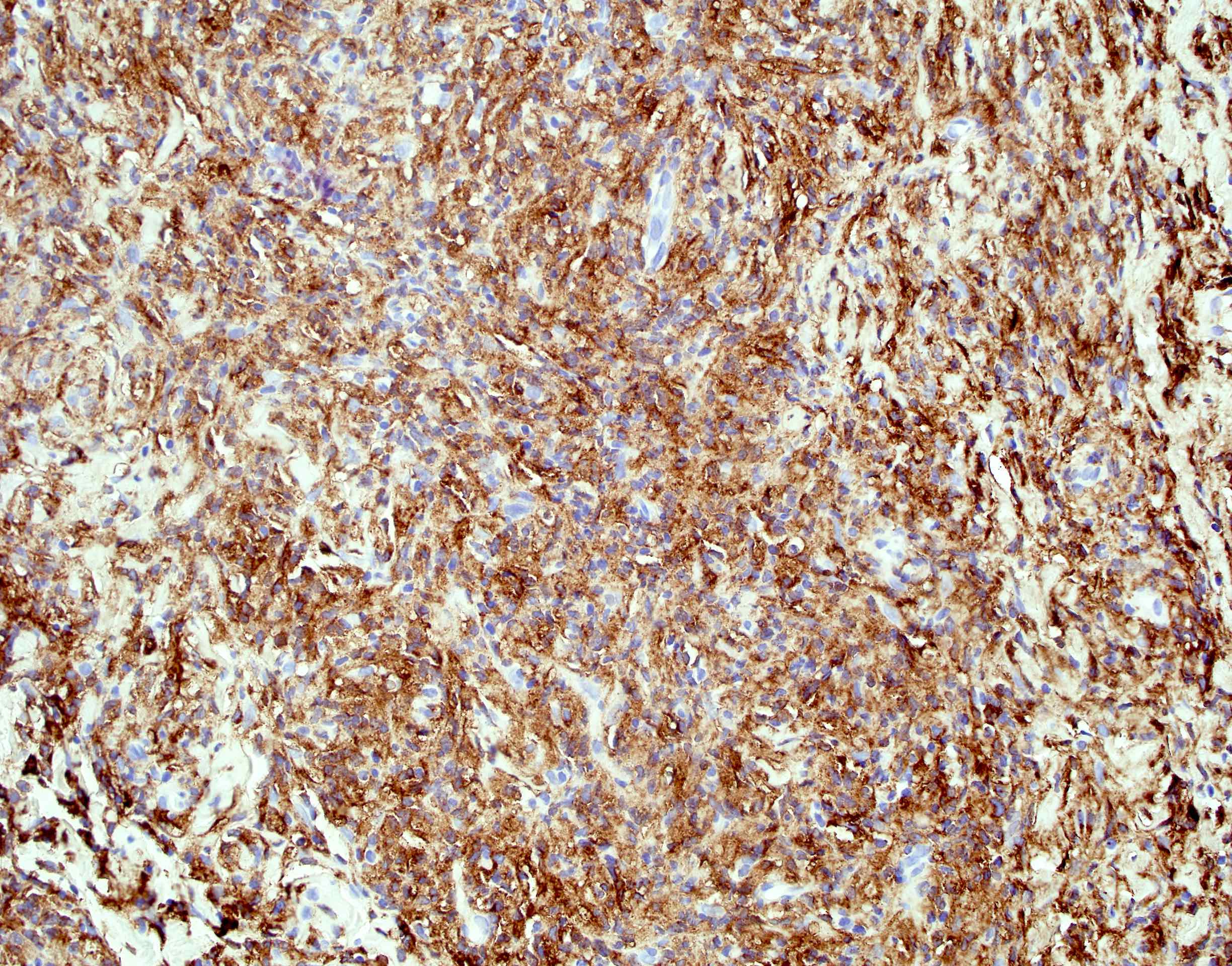
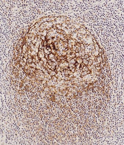
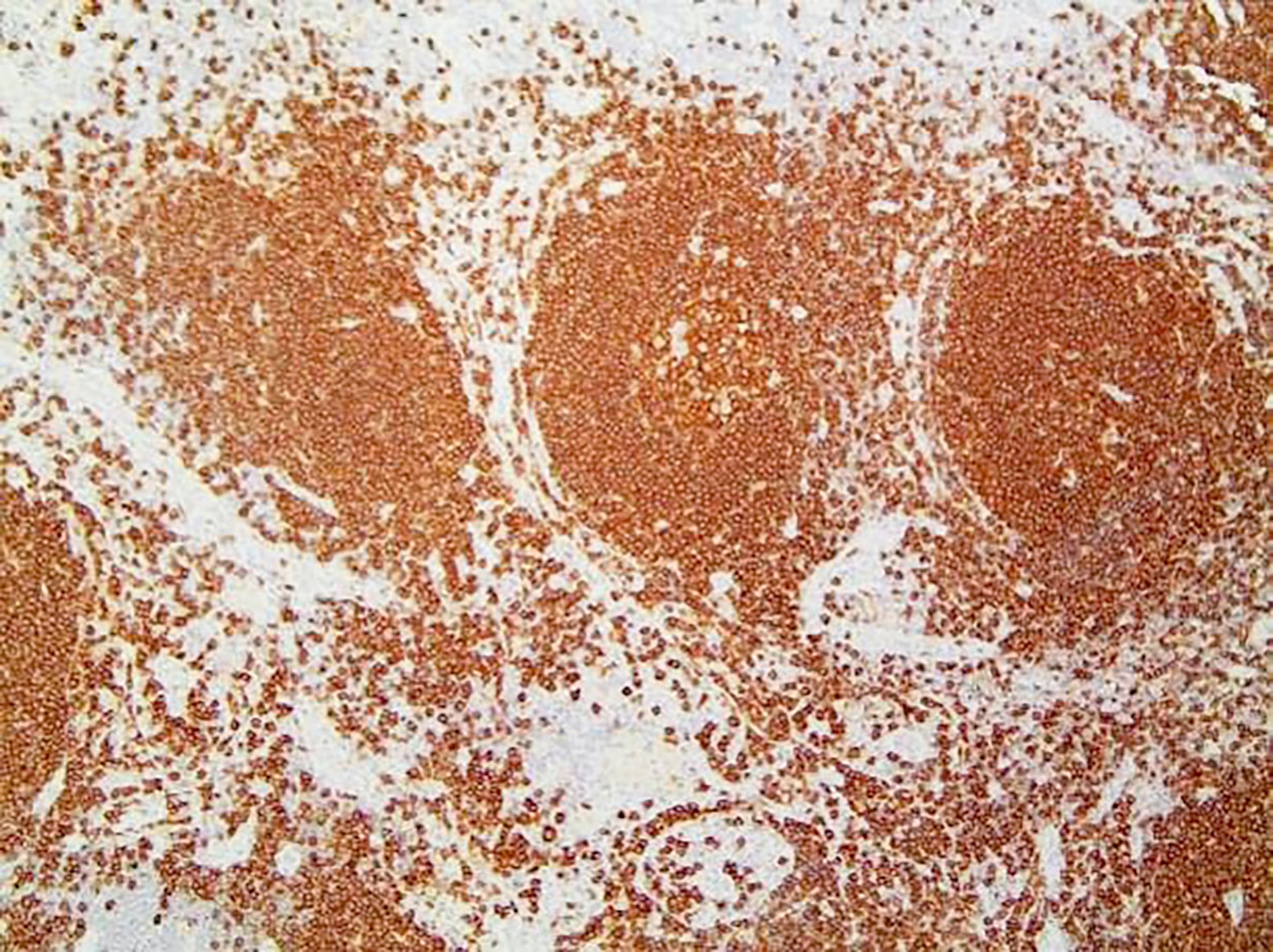
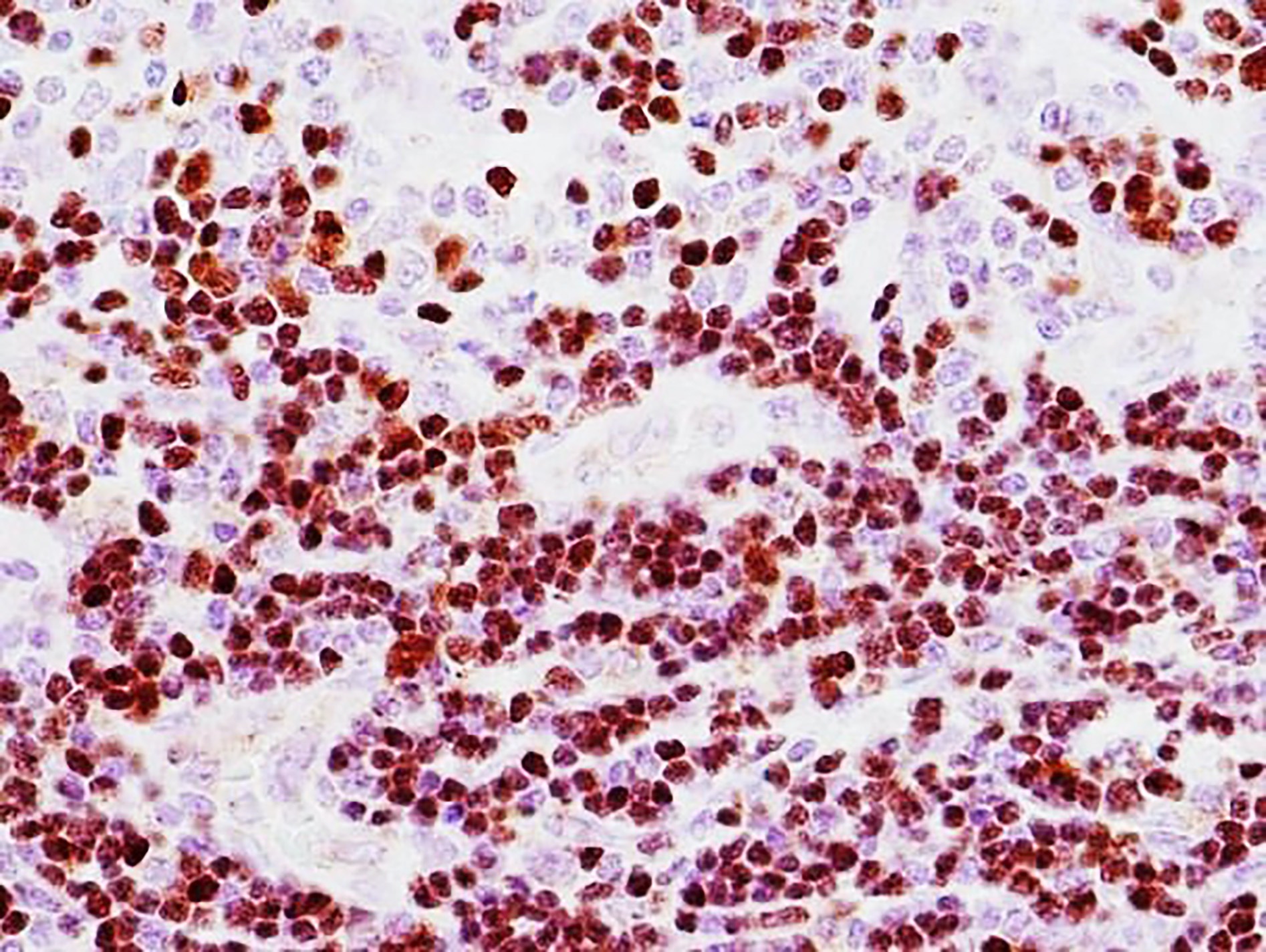
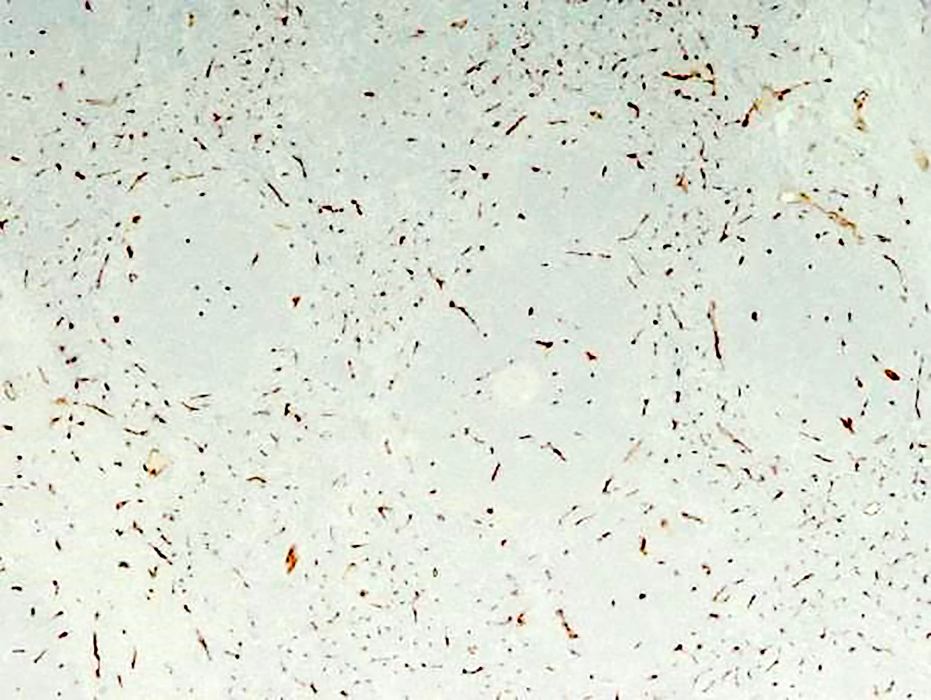
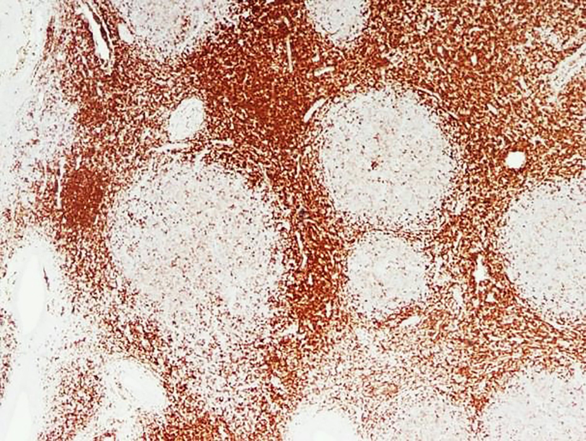
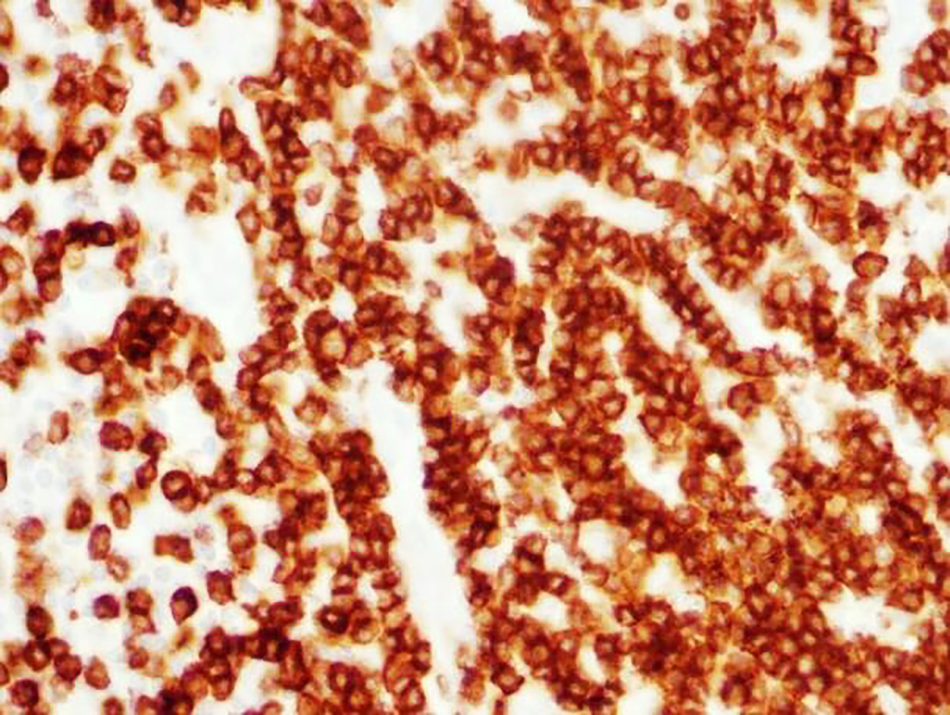
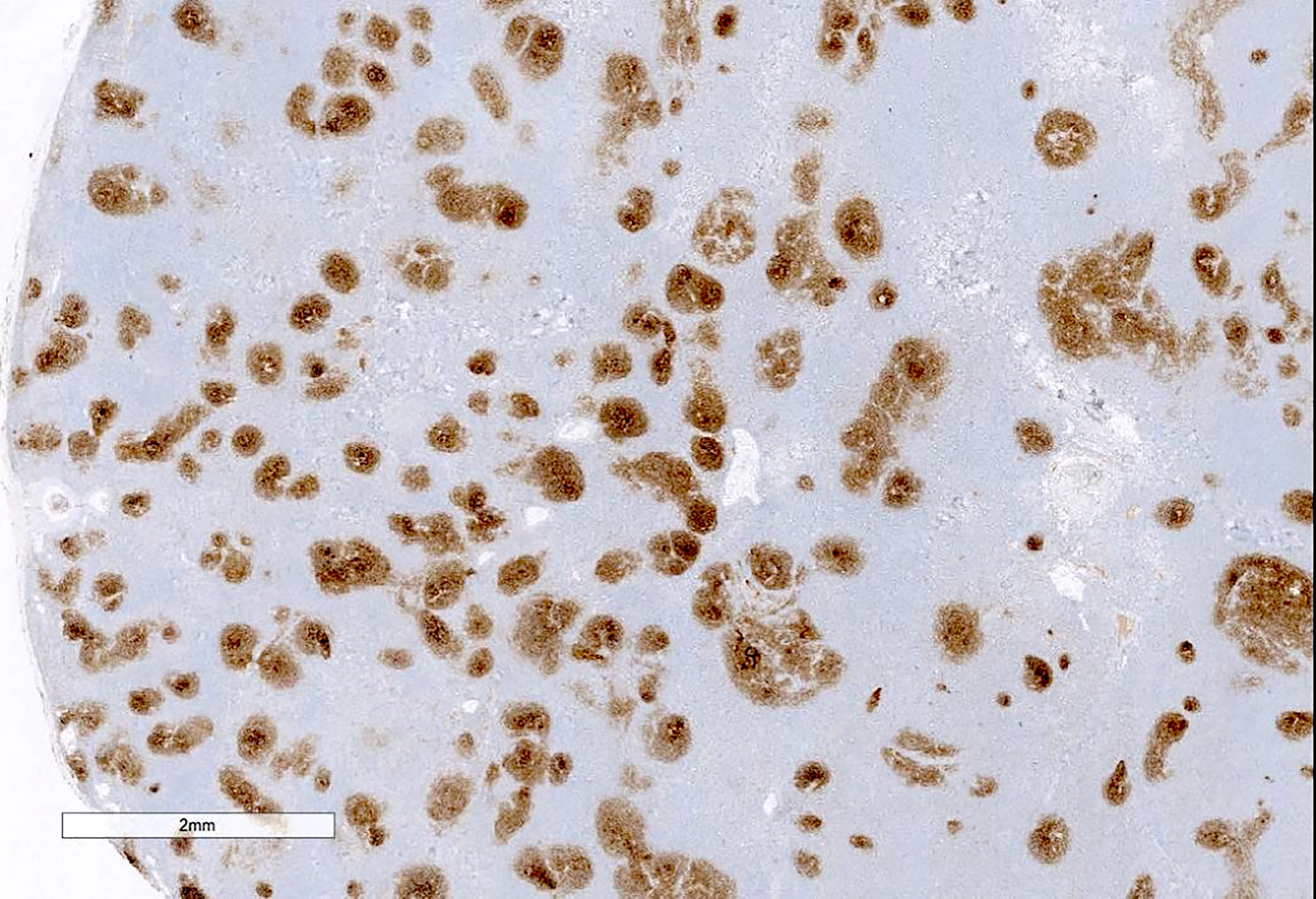
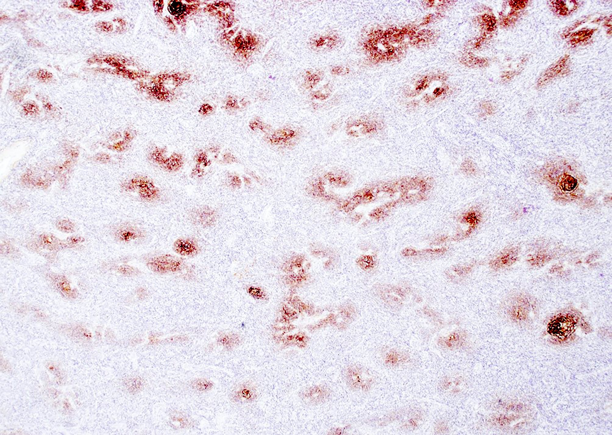
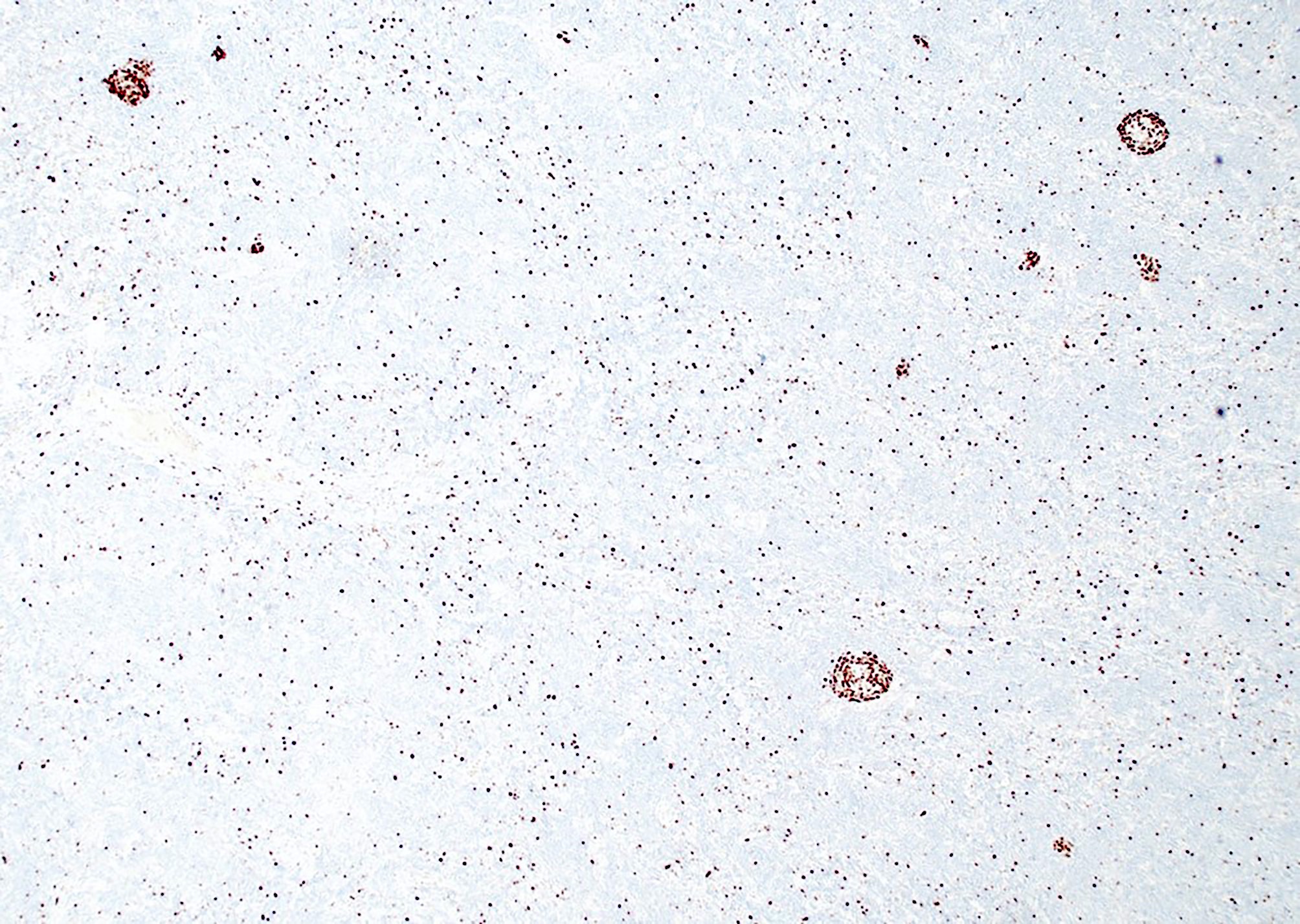
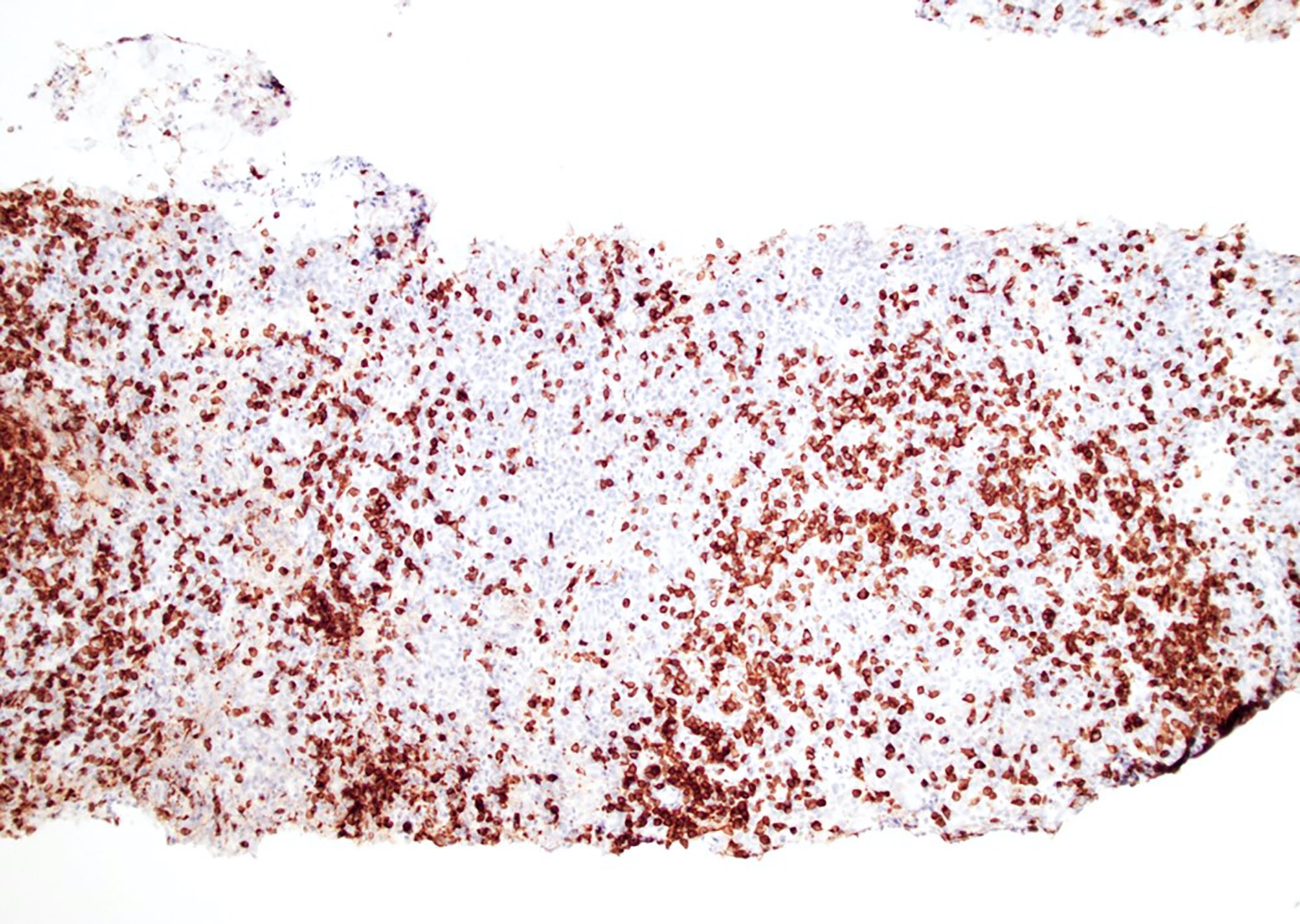
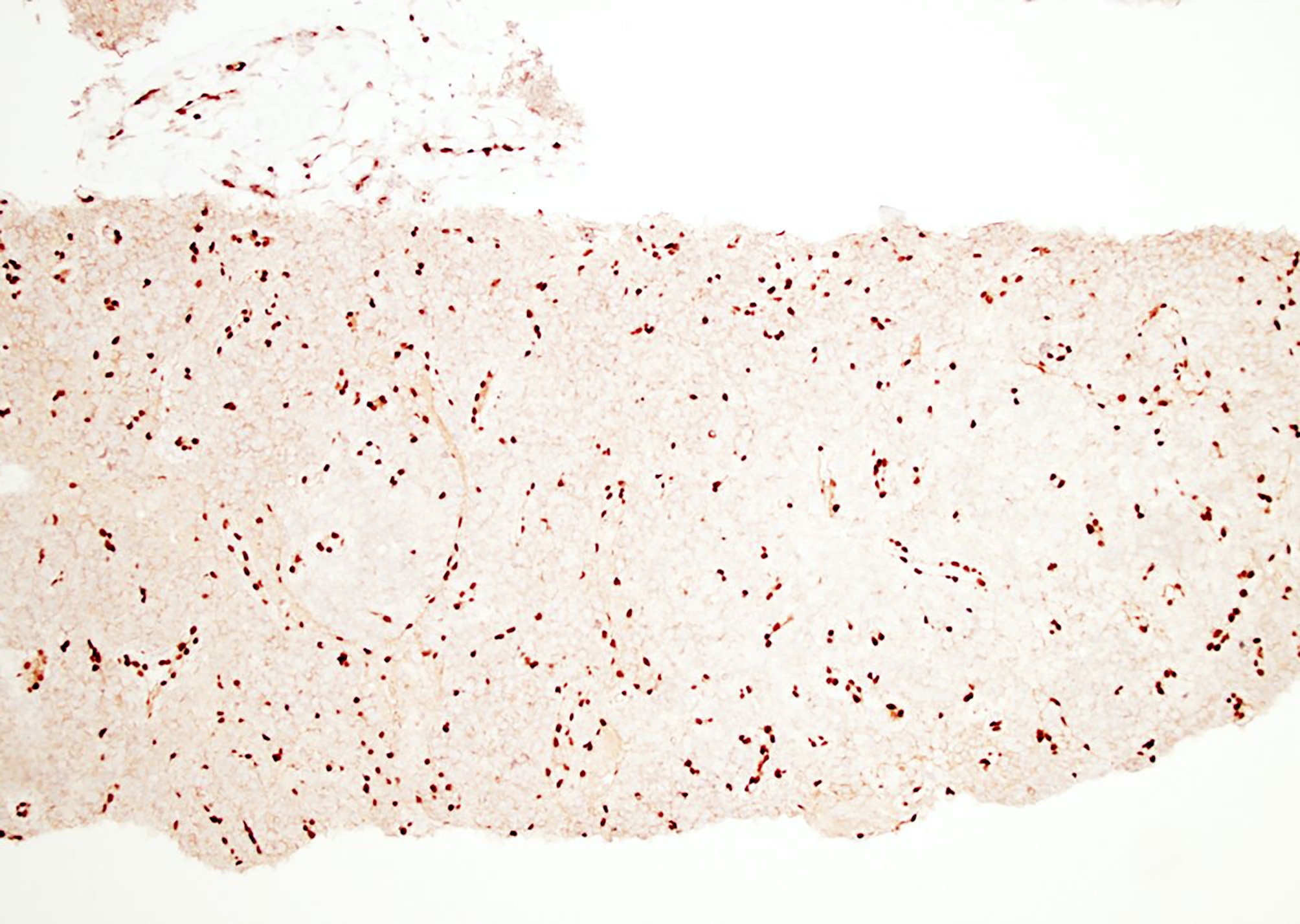
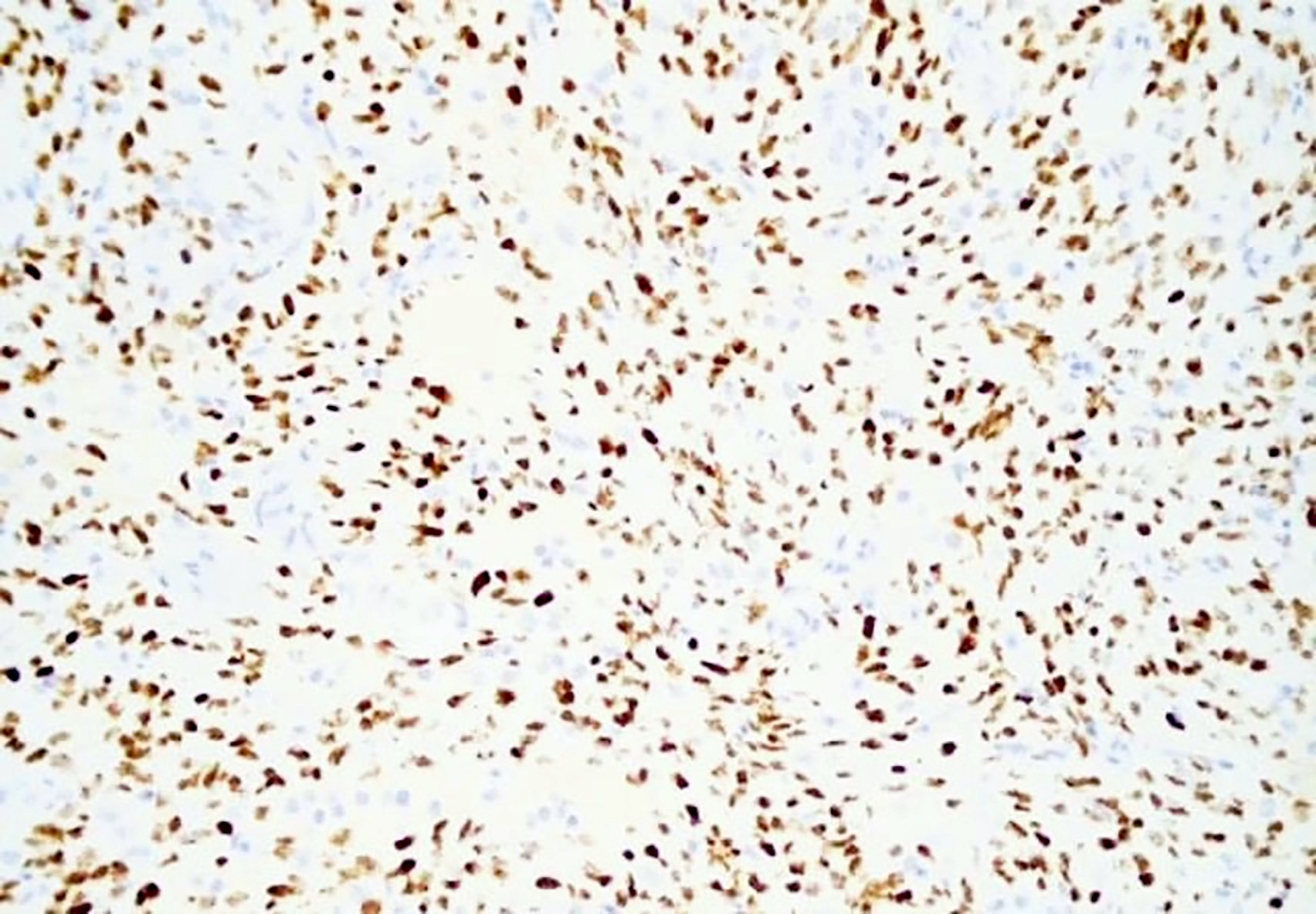
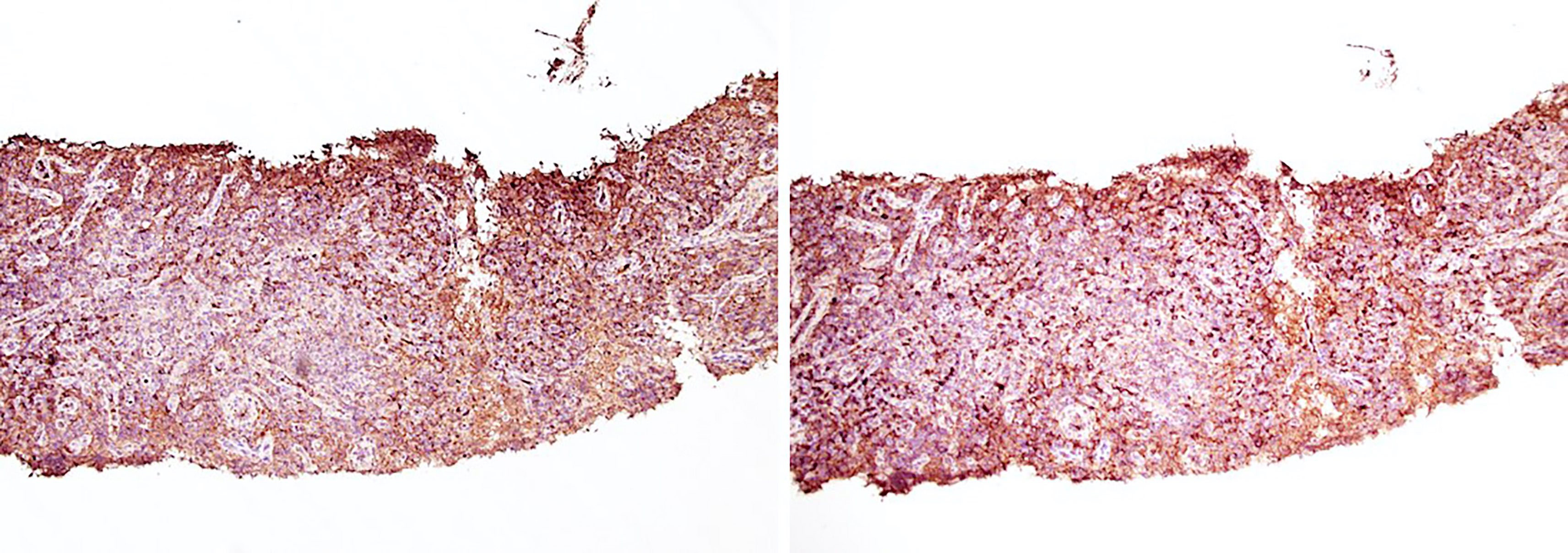
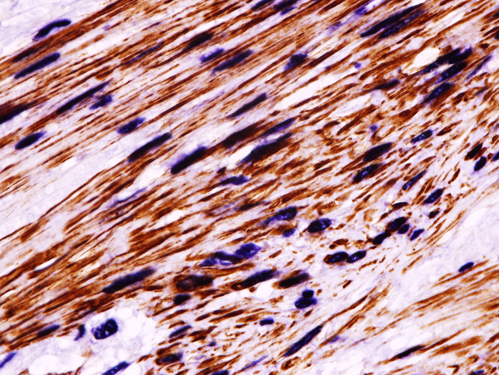
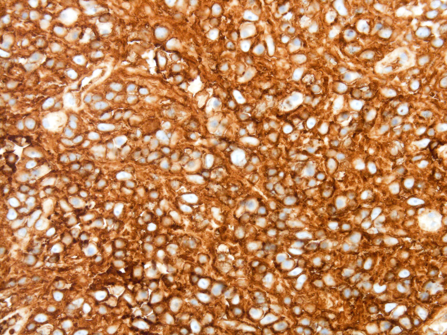
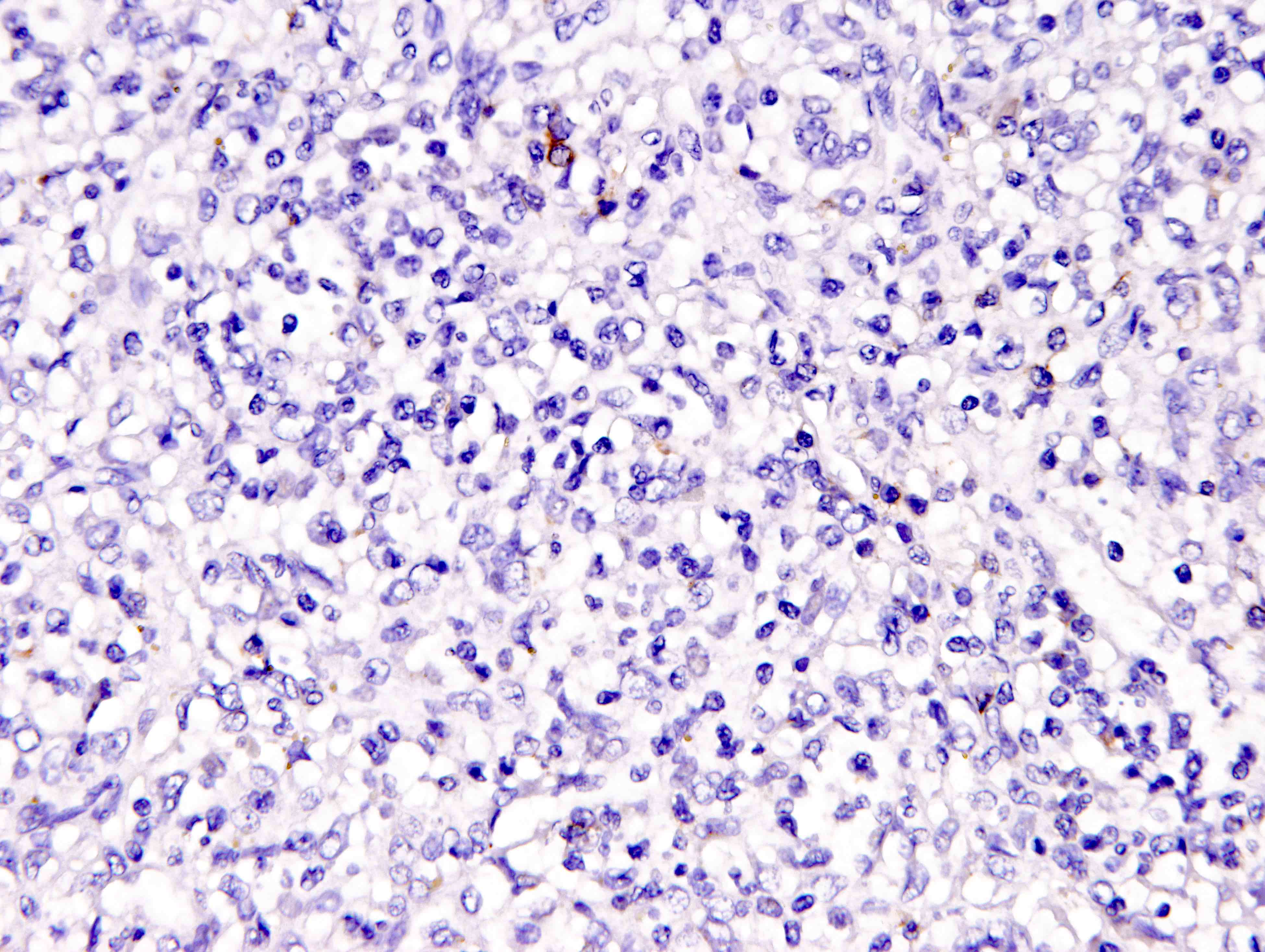
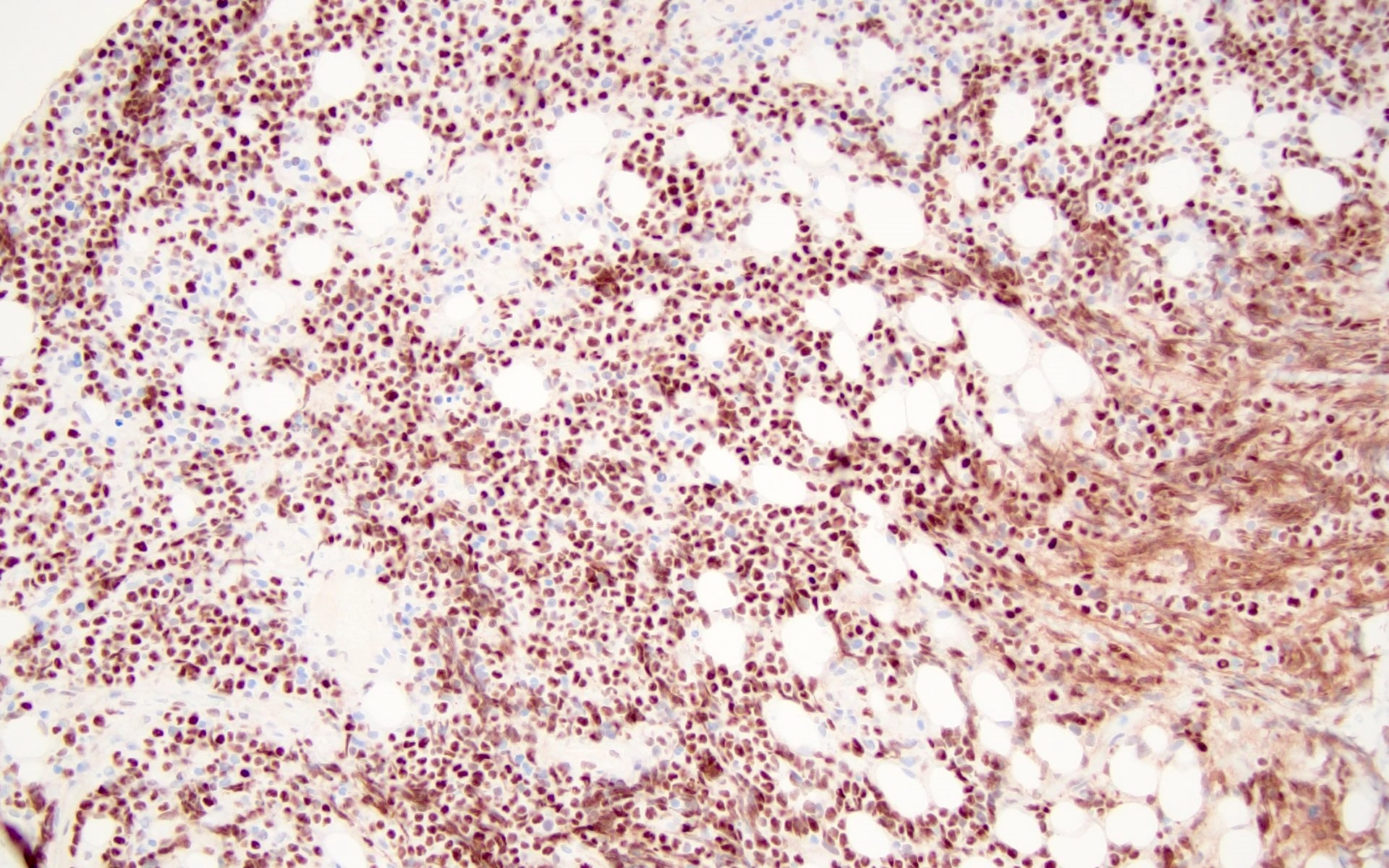
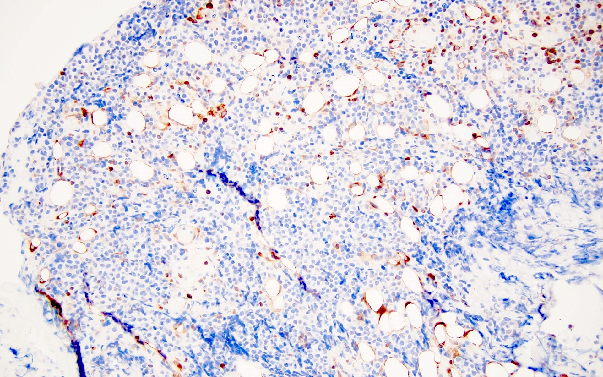
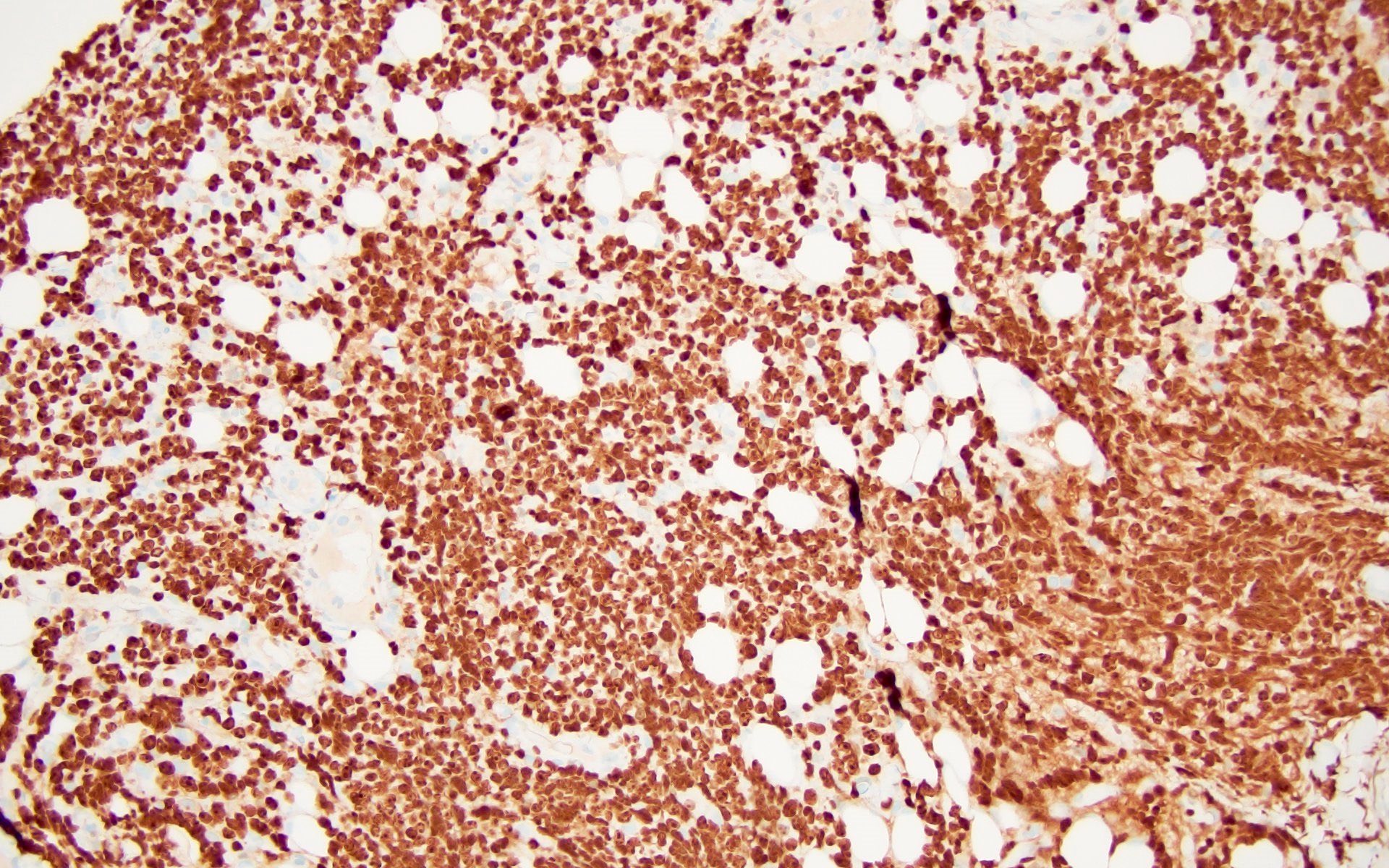
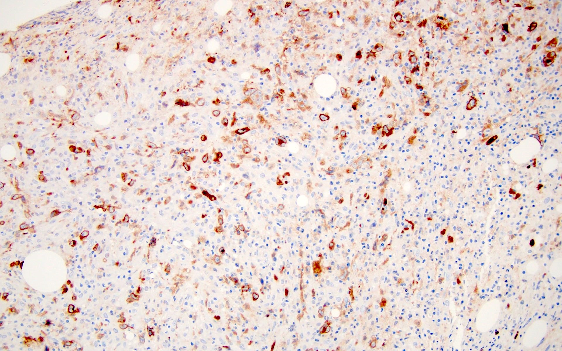
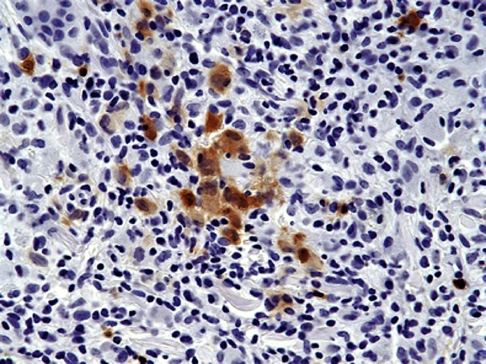
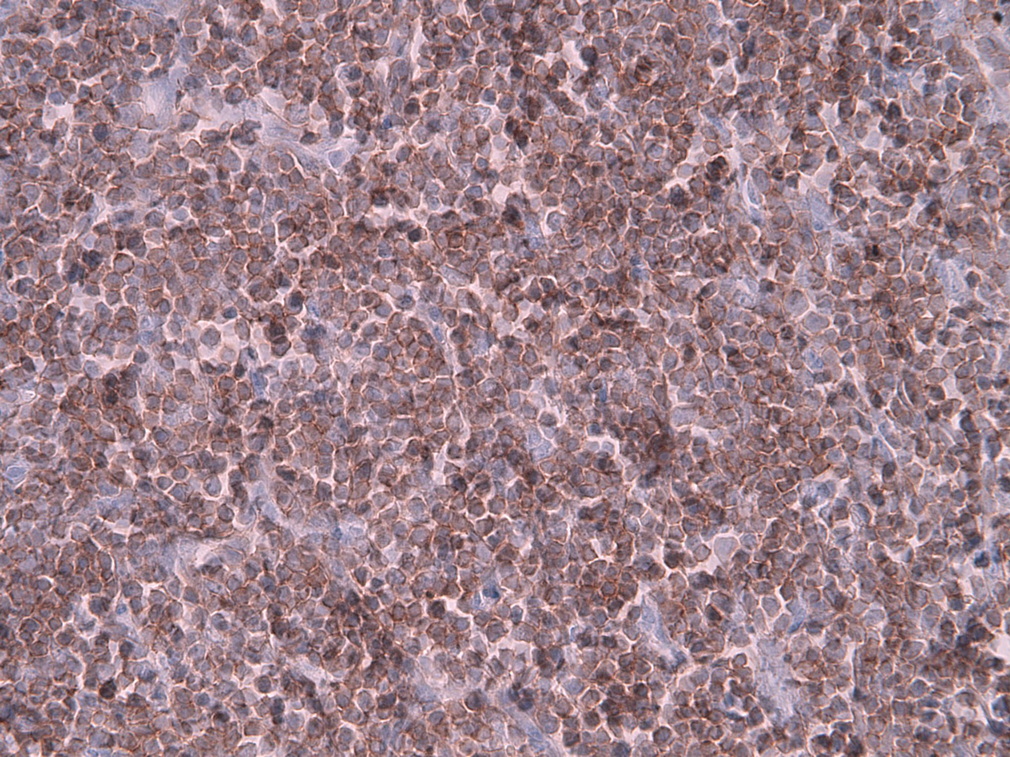
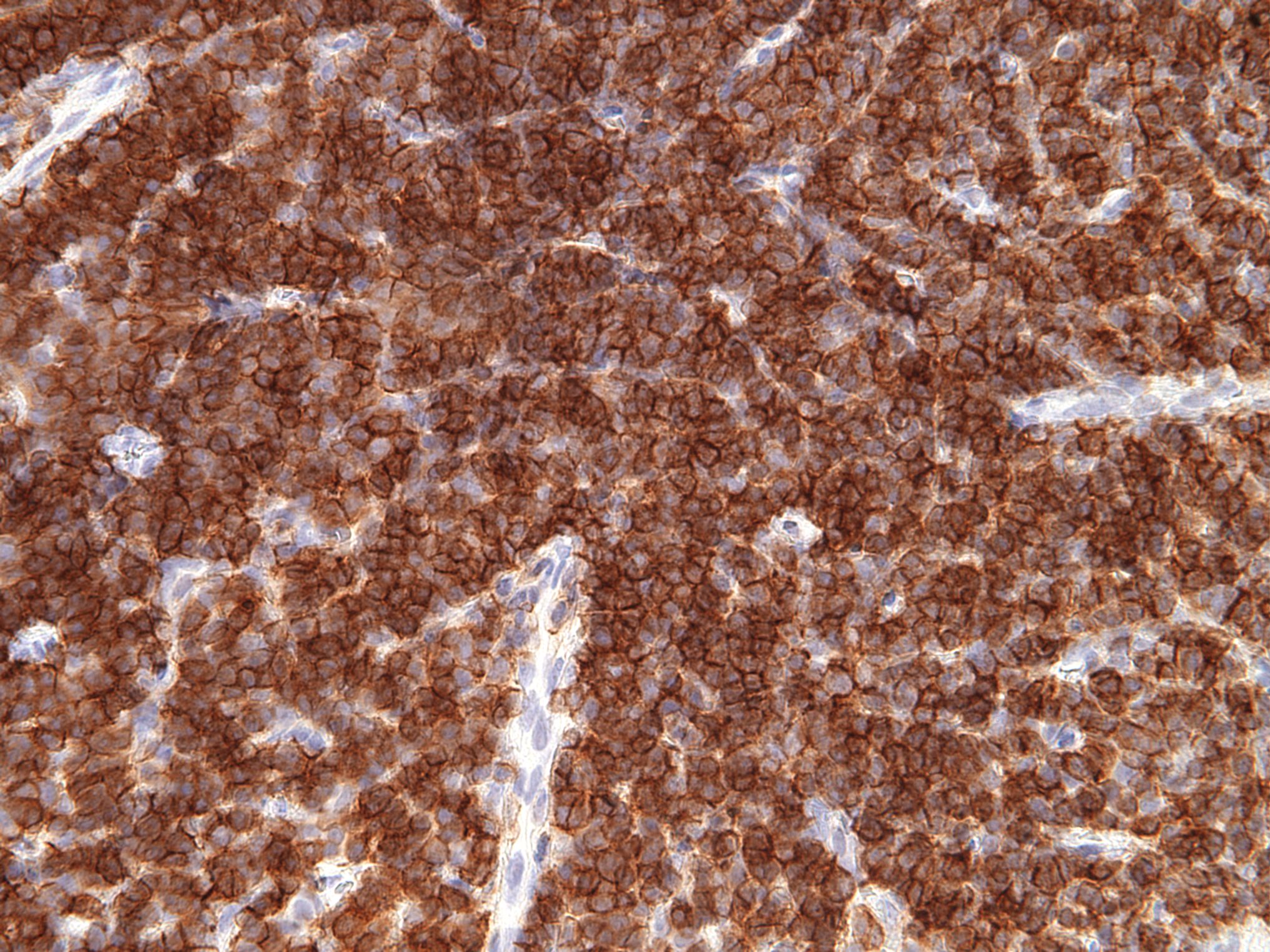
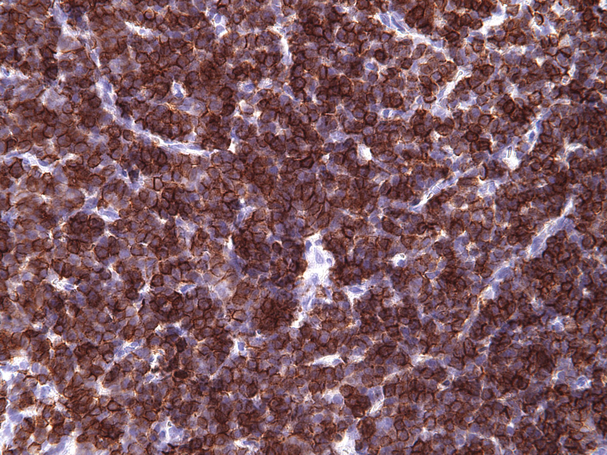
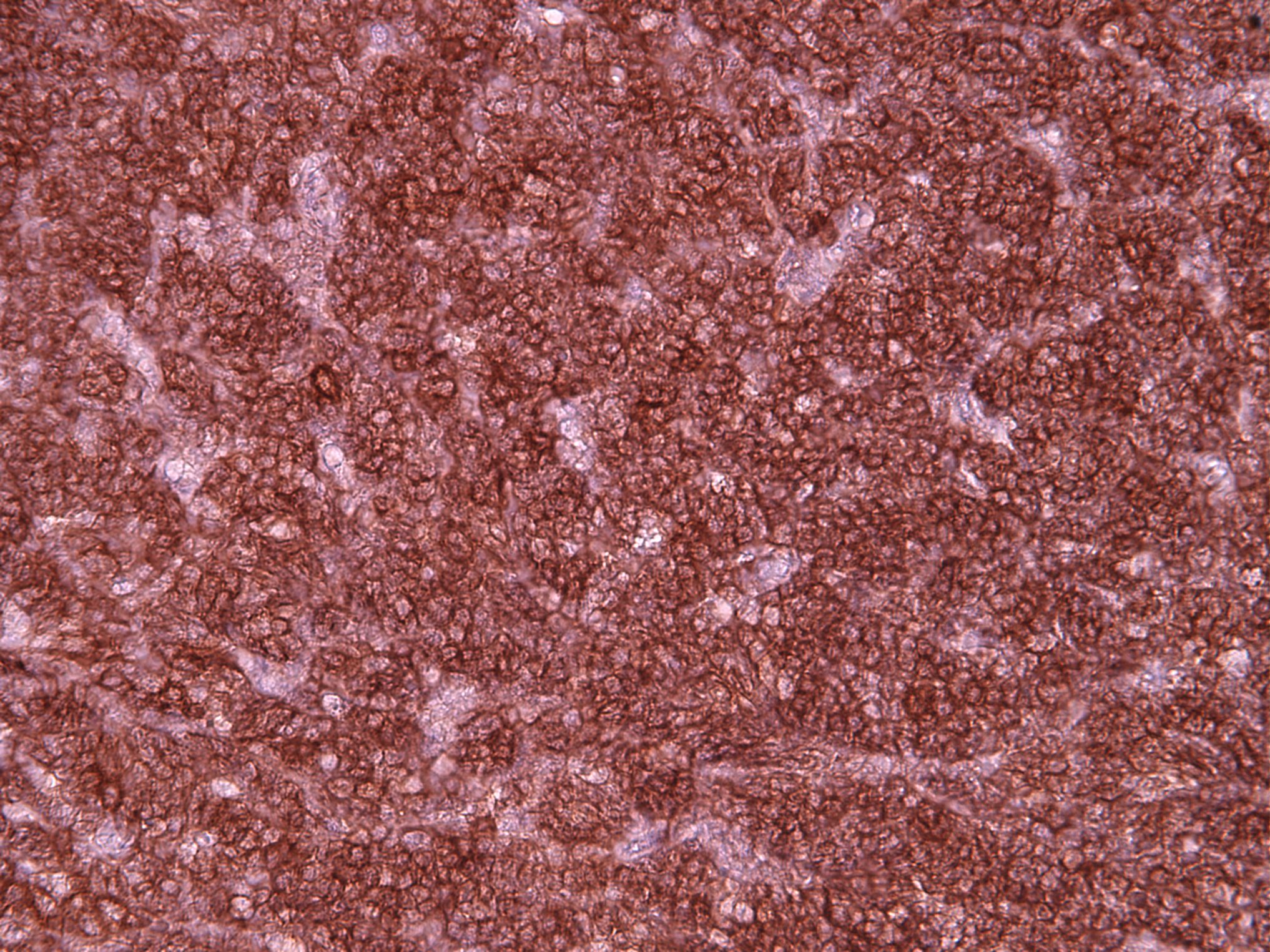
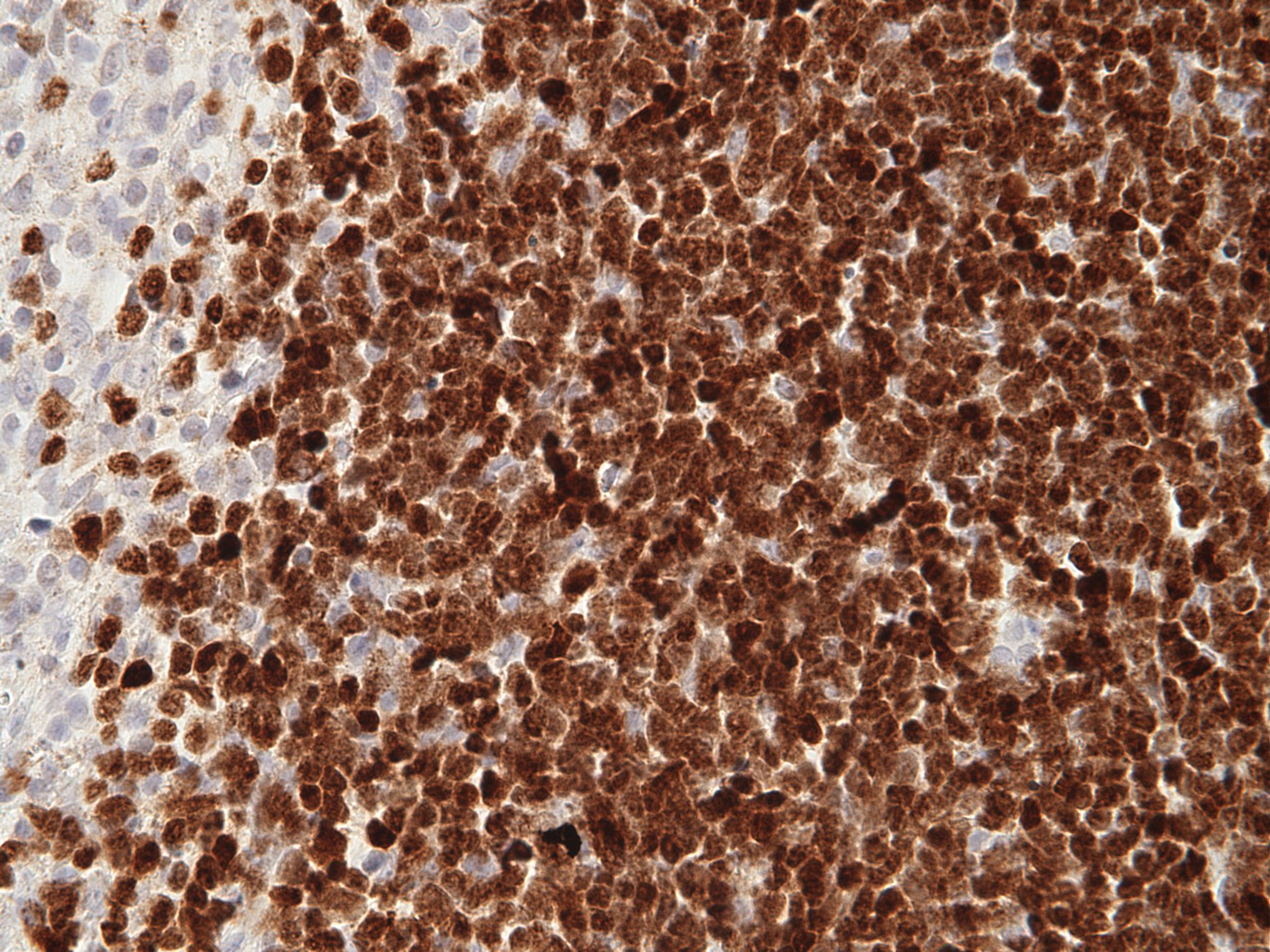
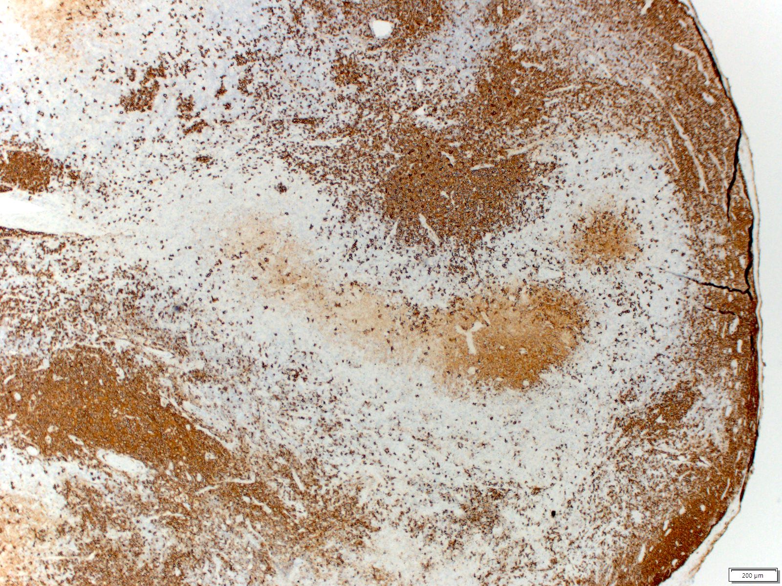
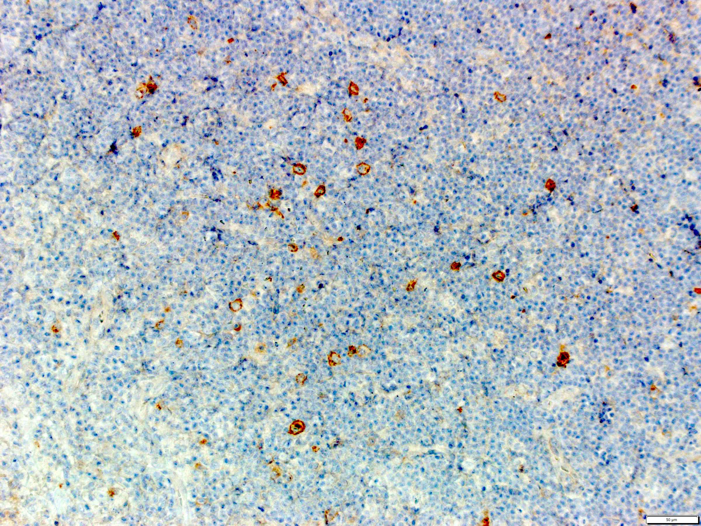
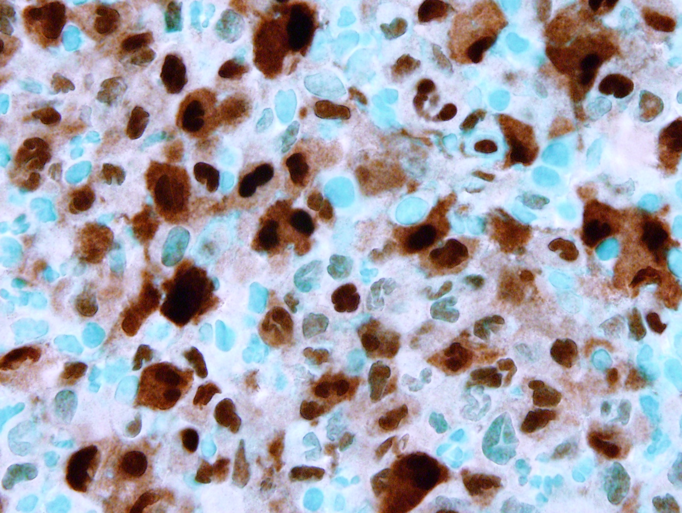
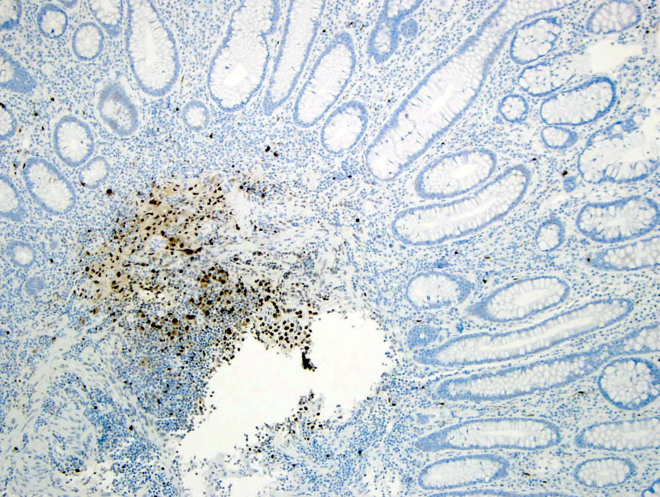
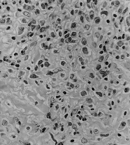
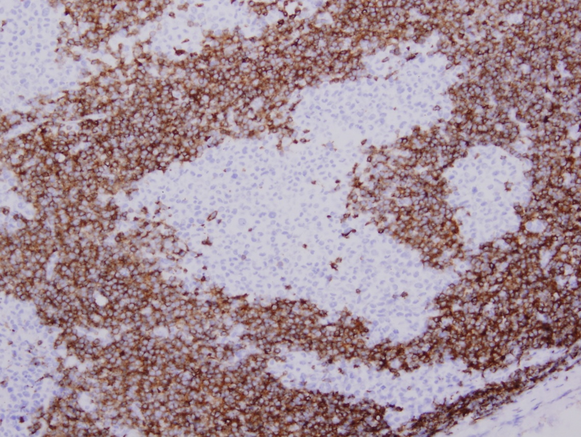
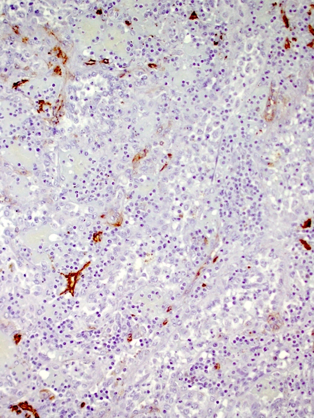
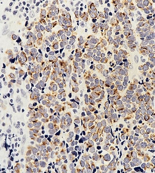
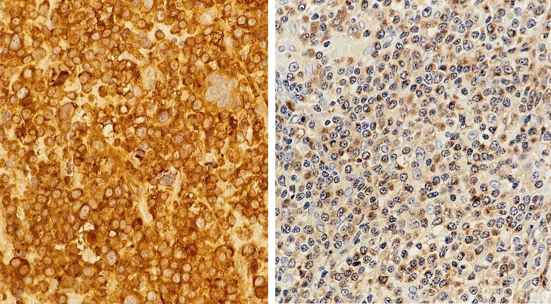
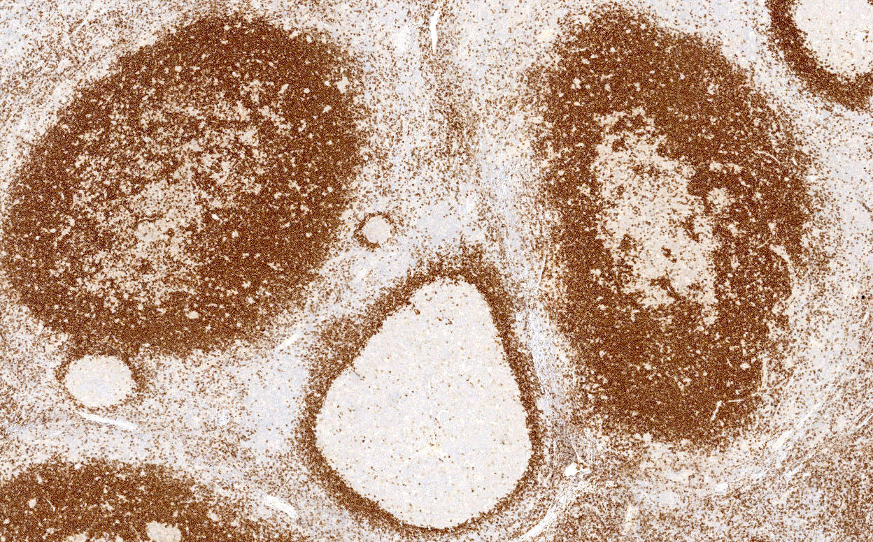
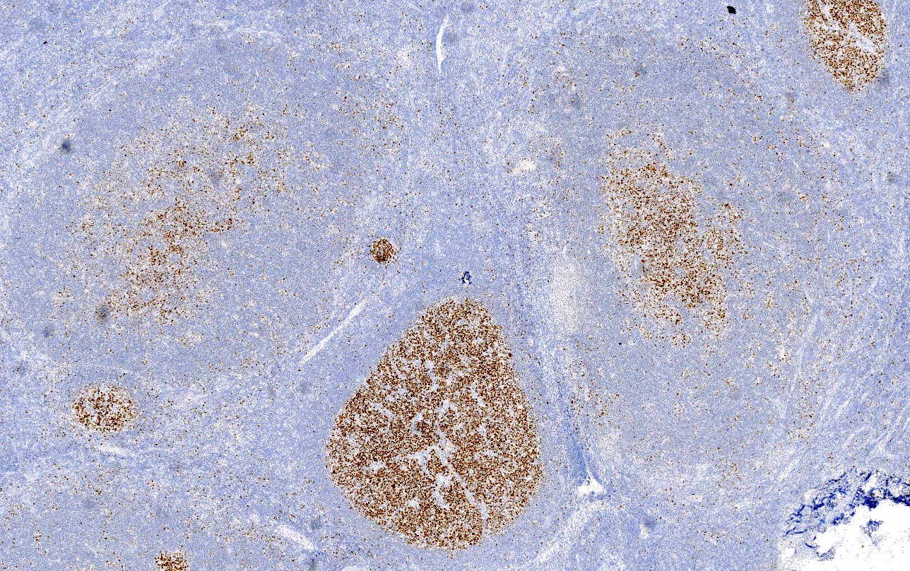
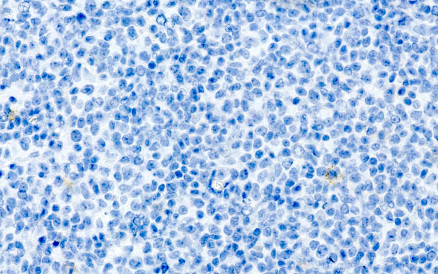
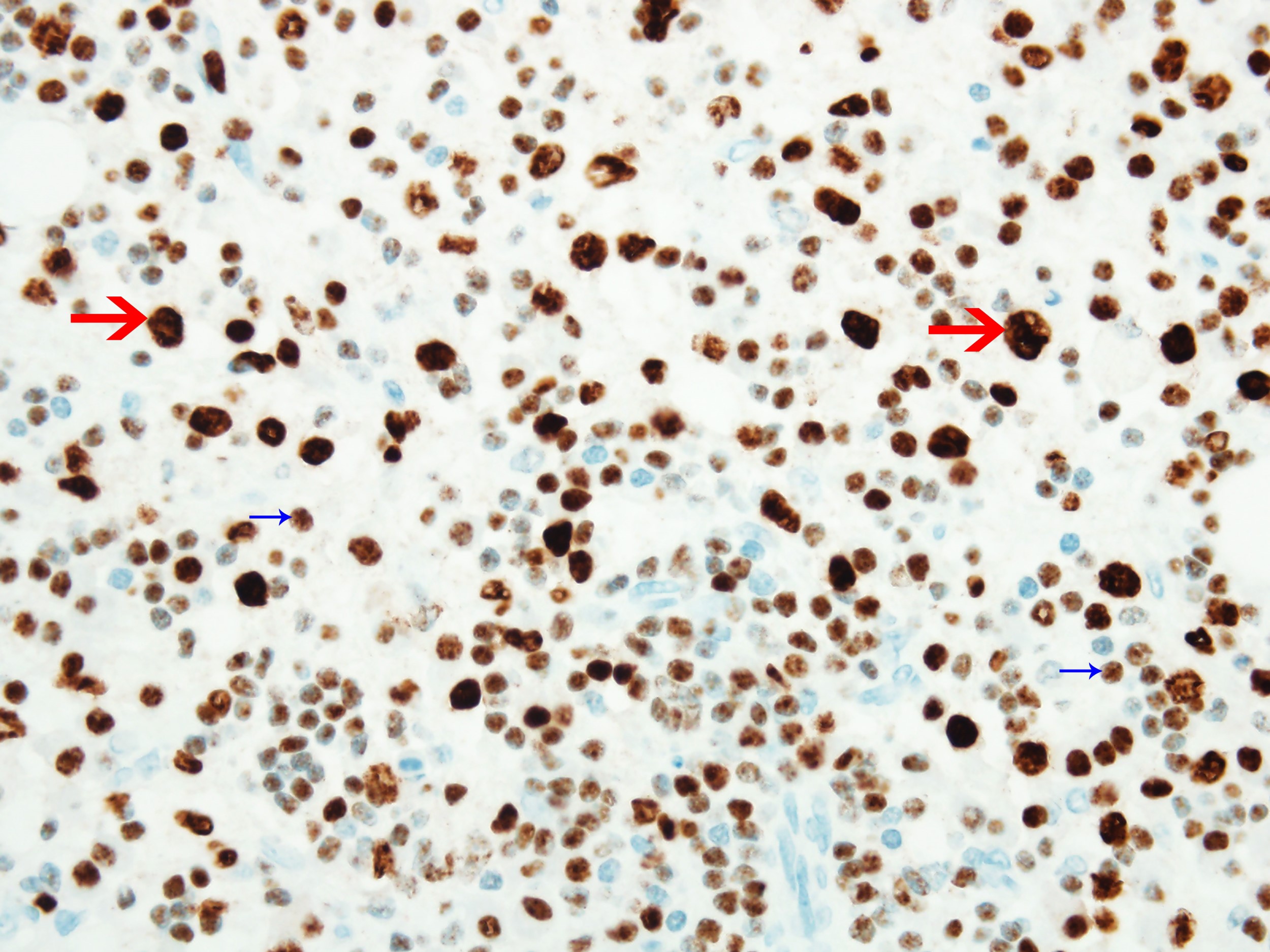
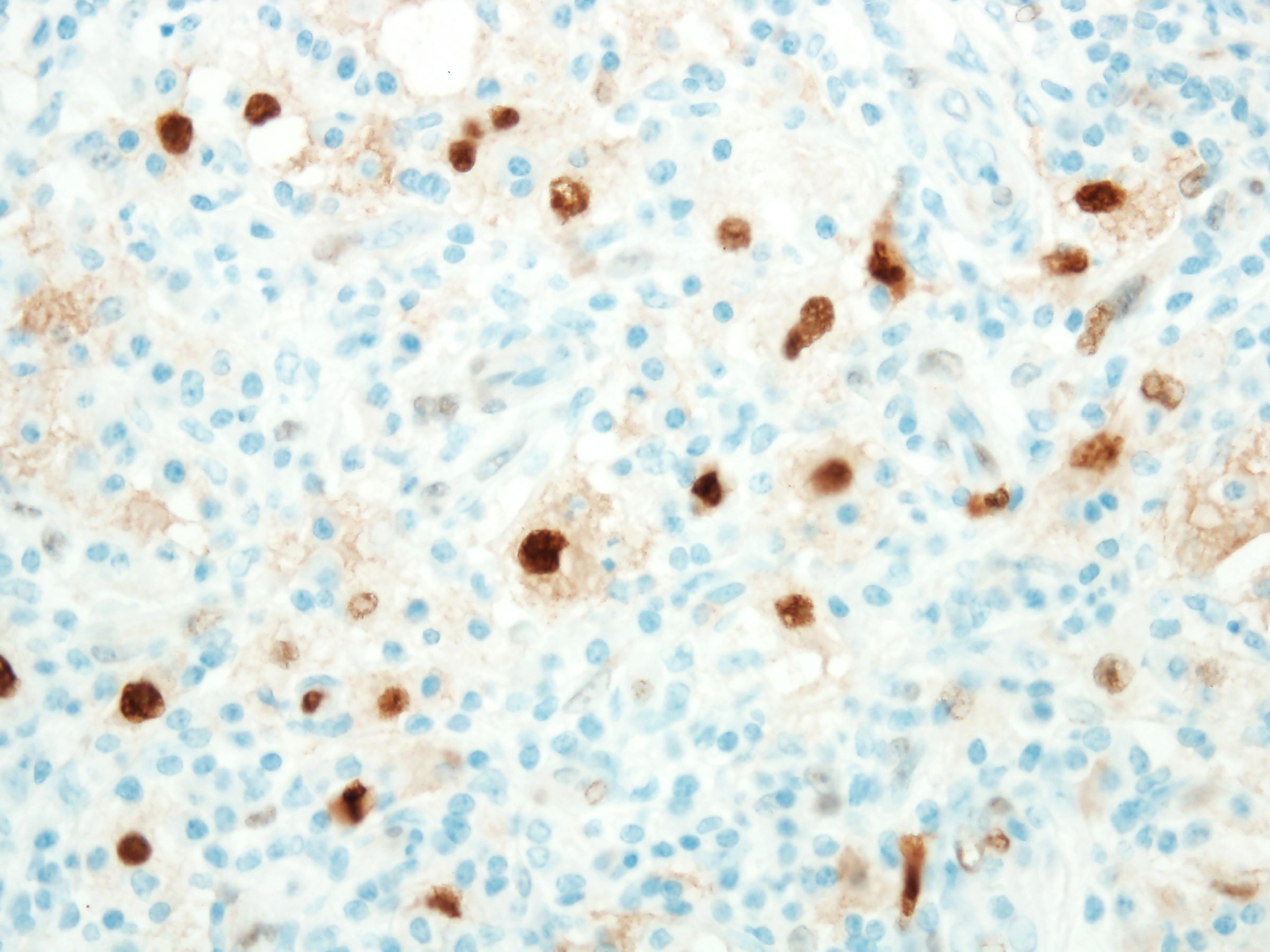
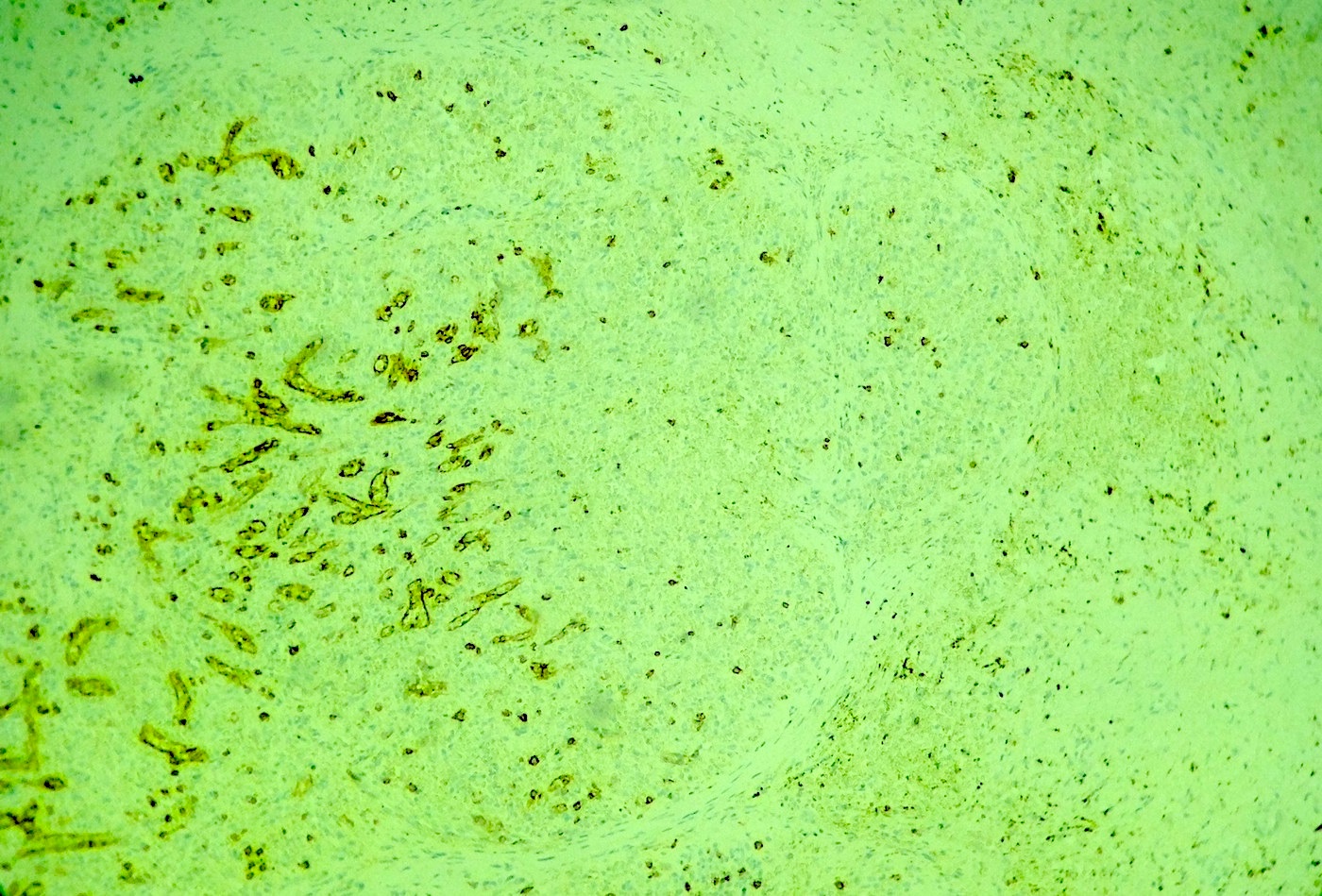
CD8
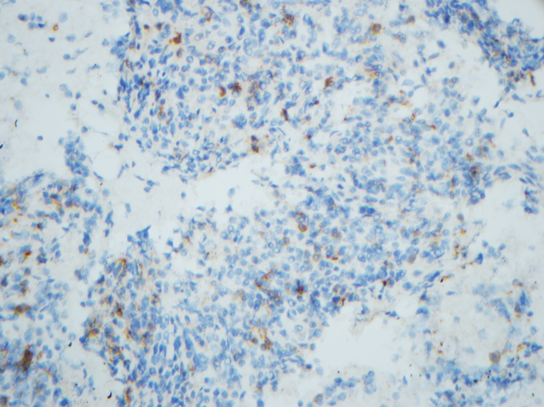
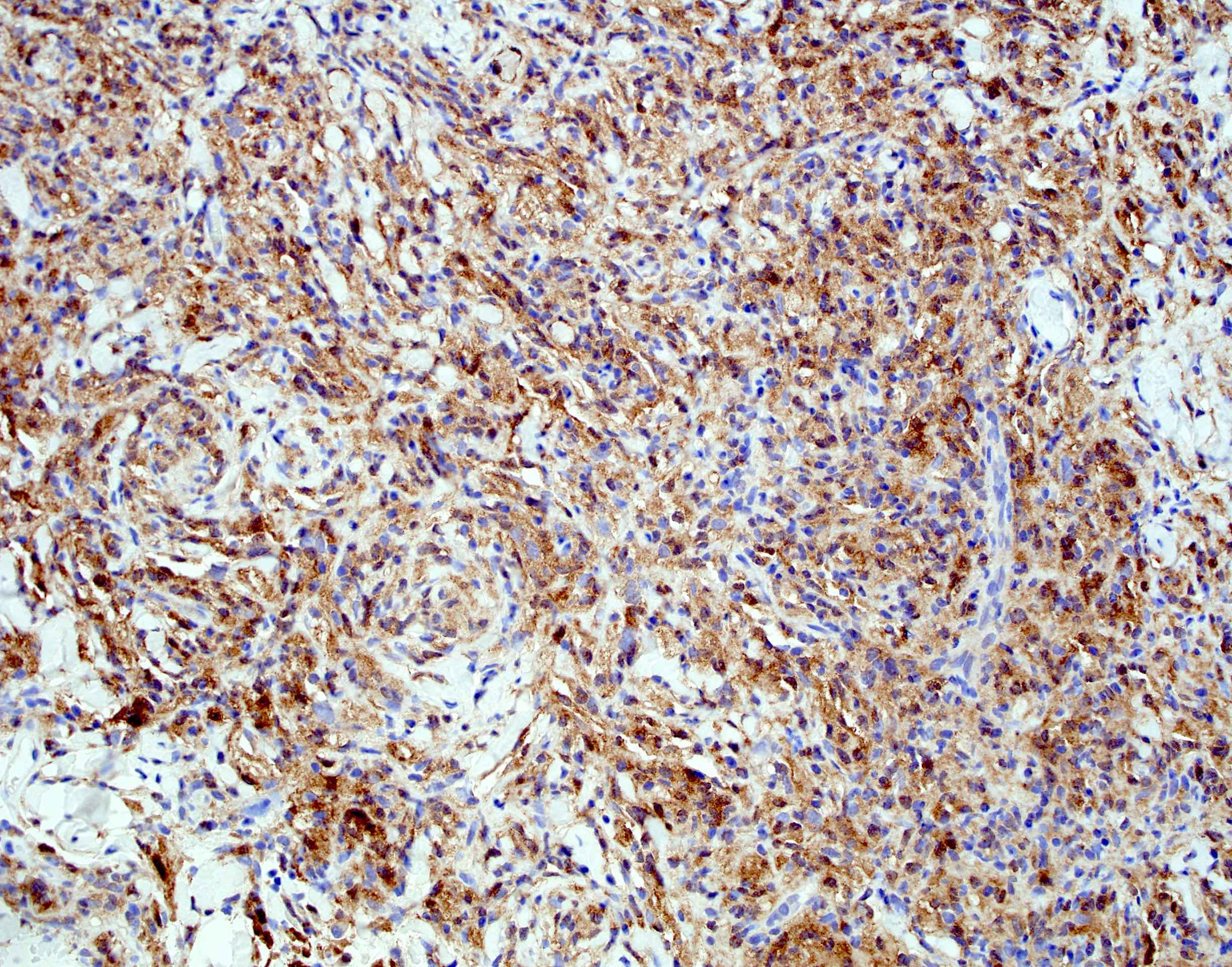
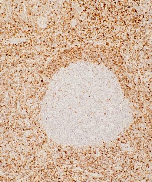
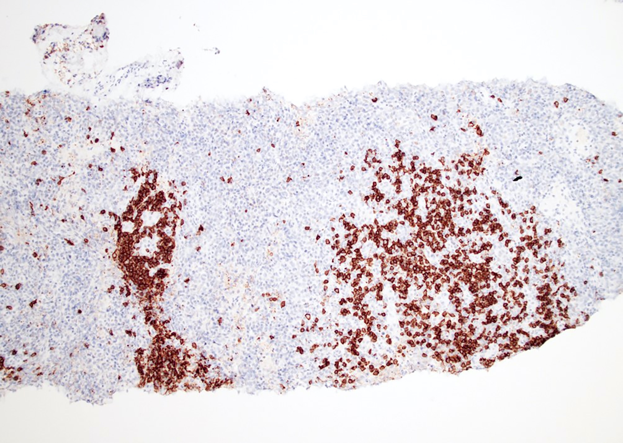
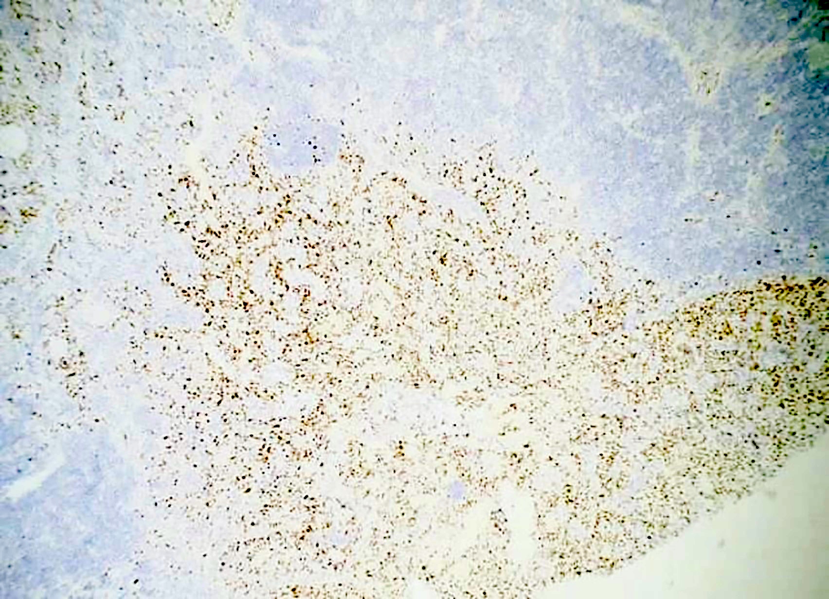
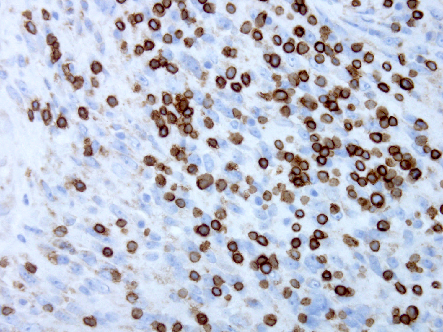
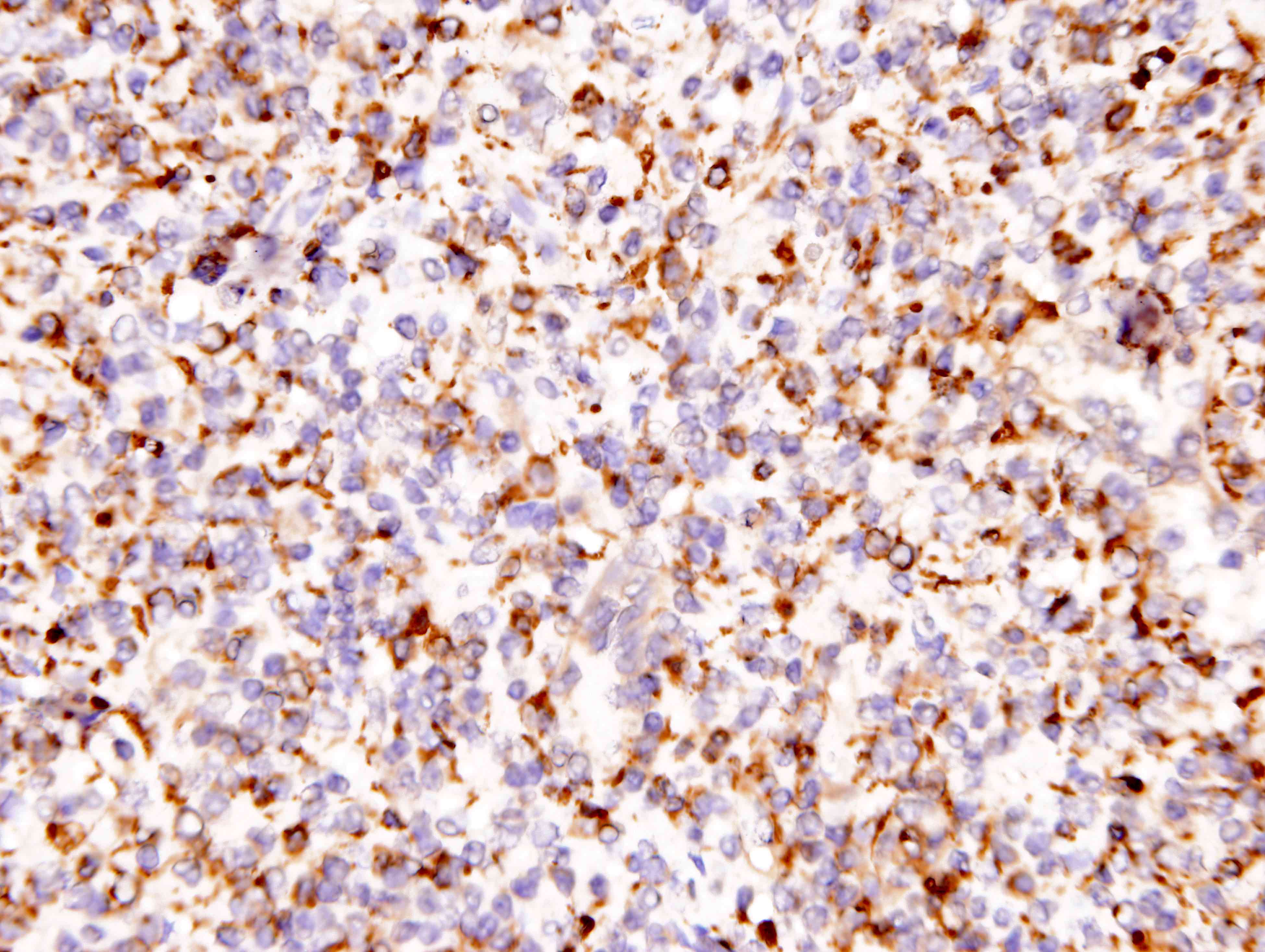
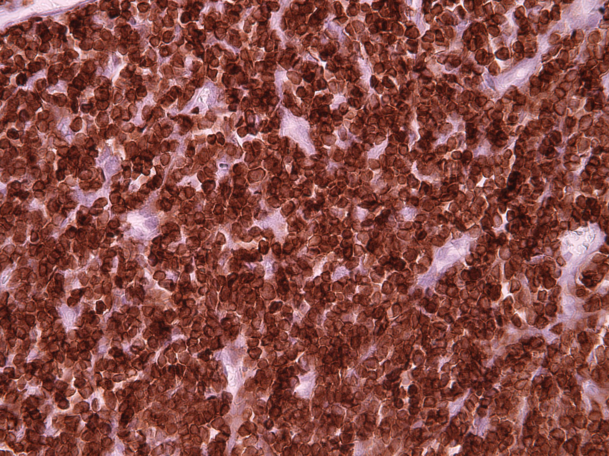
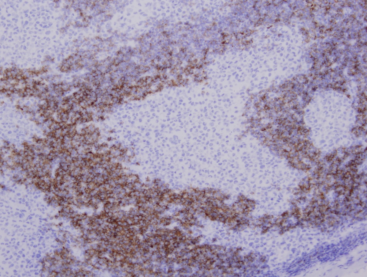
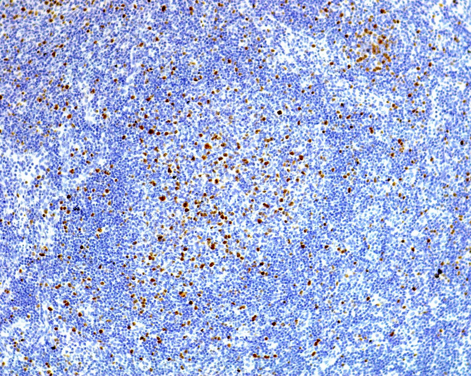
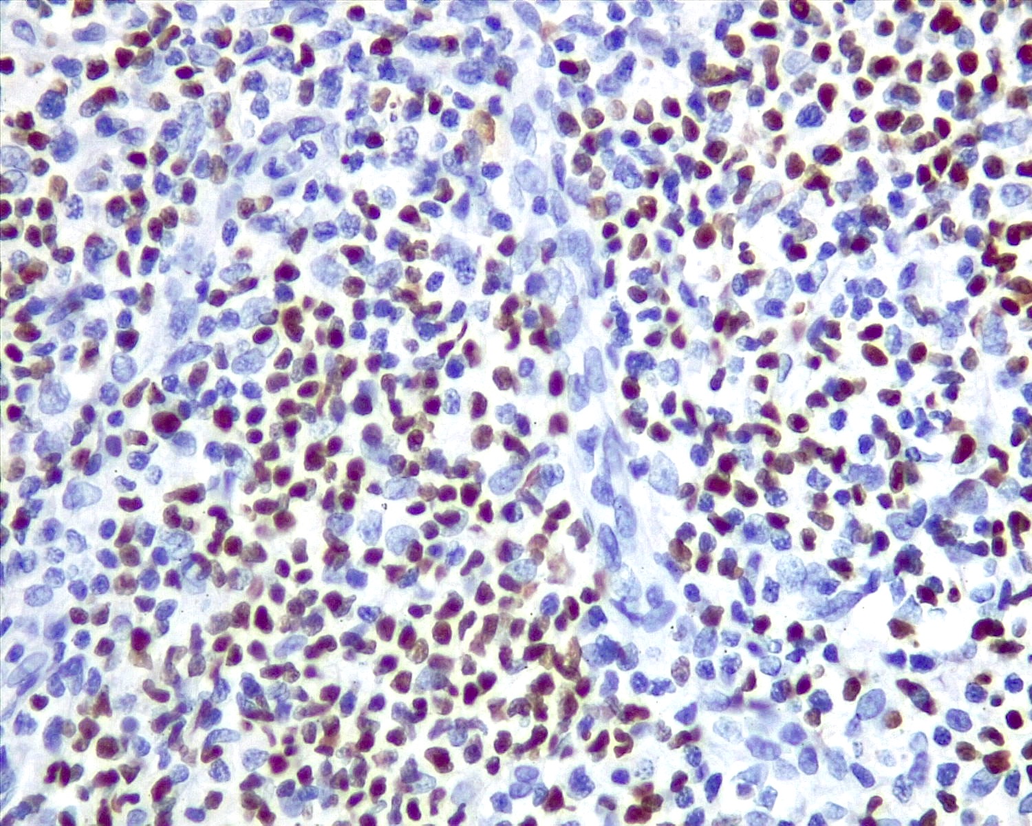
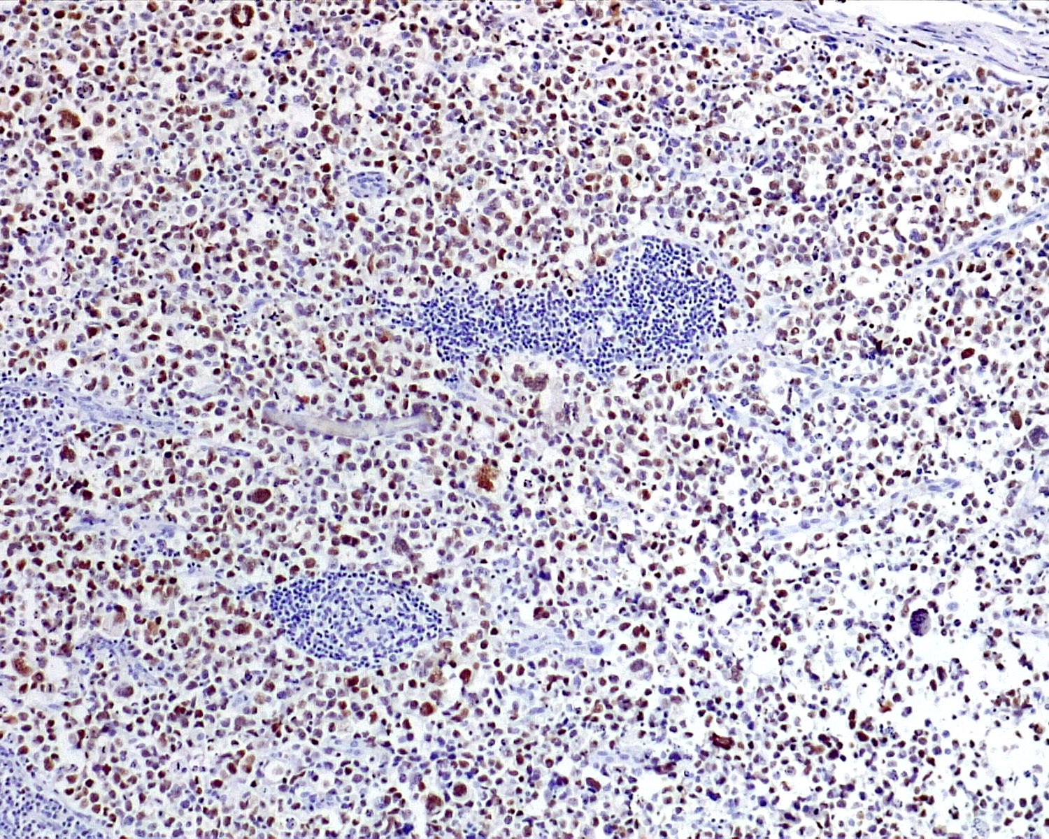
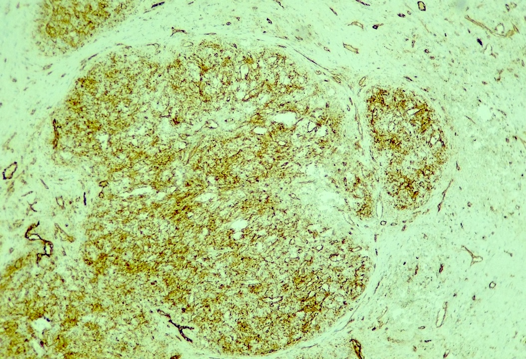
CD56
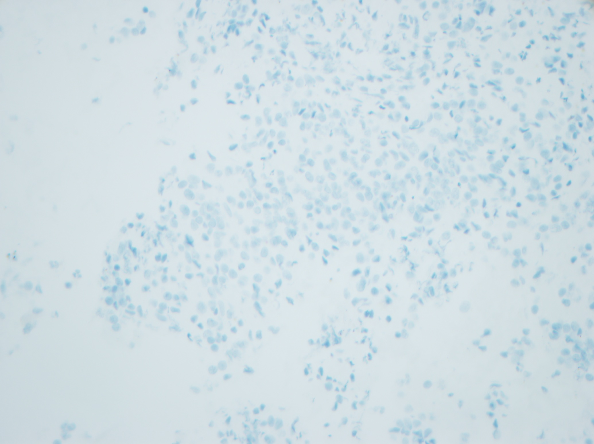
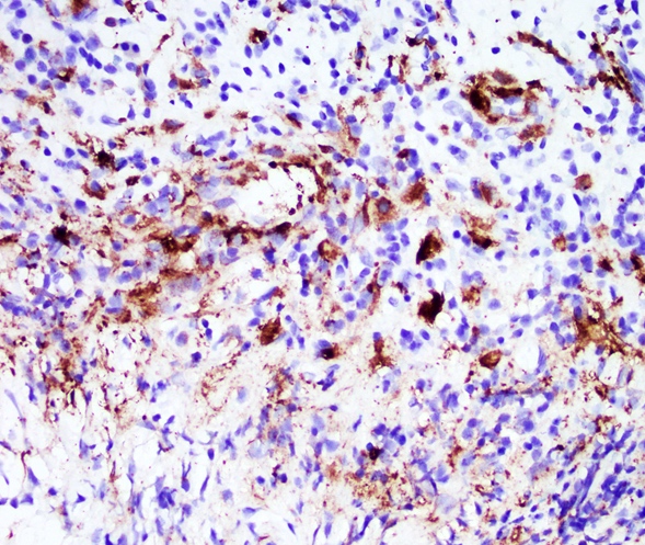
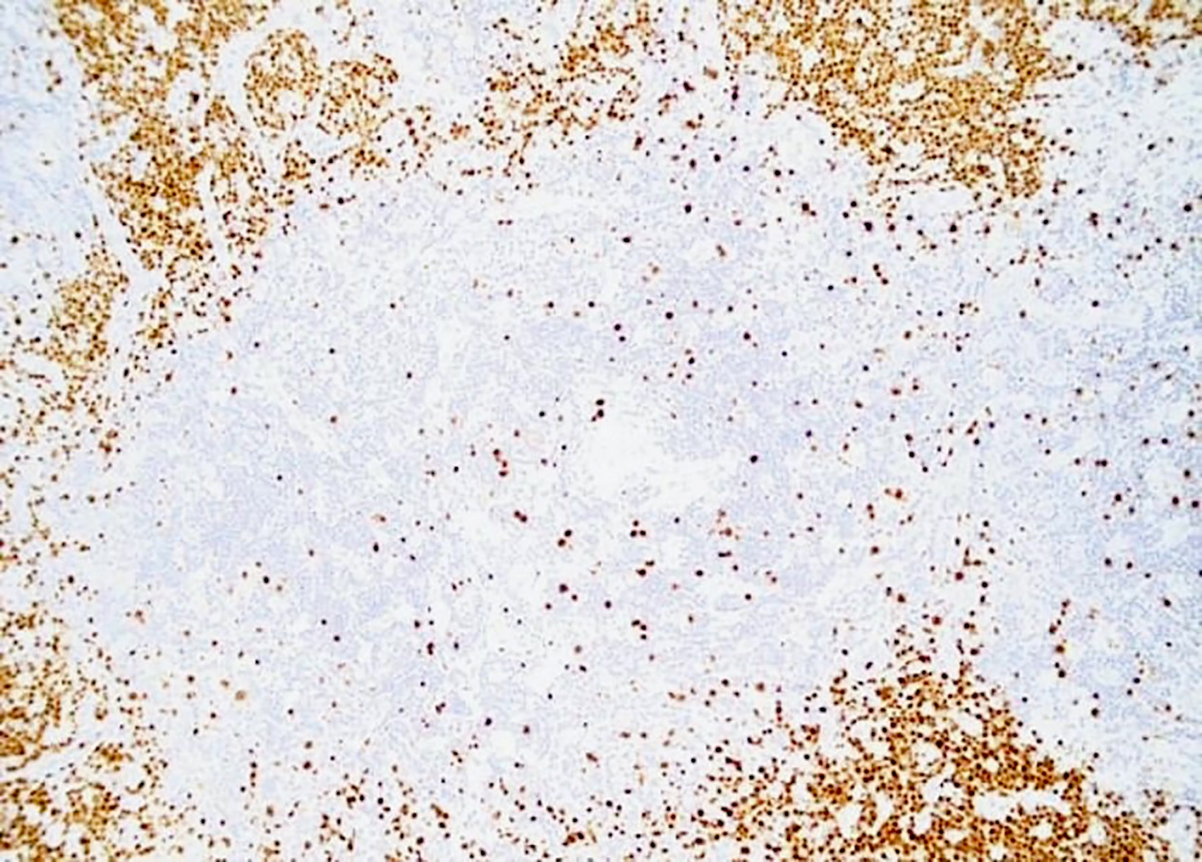
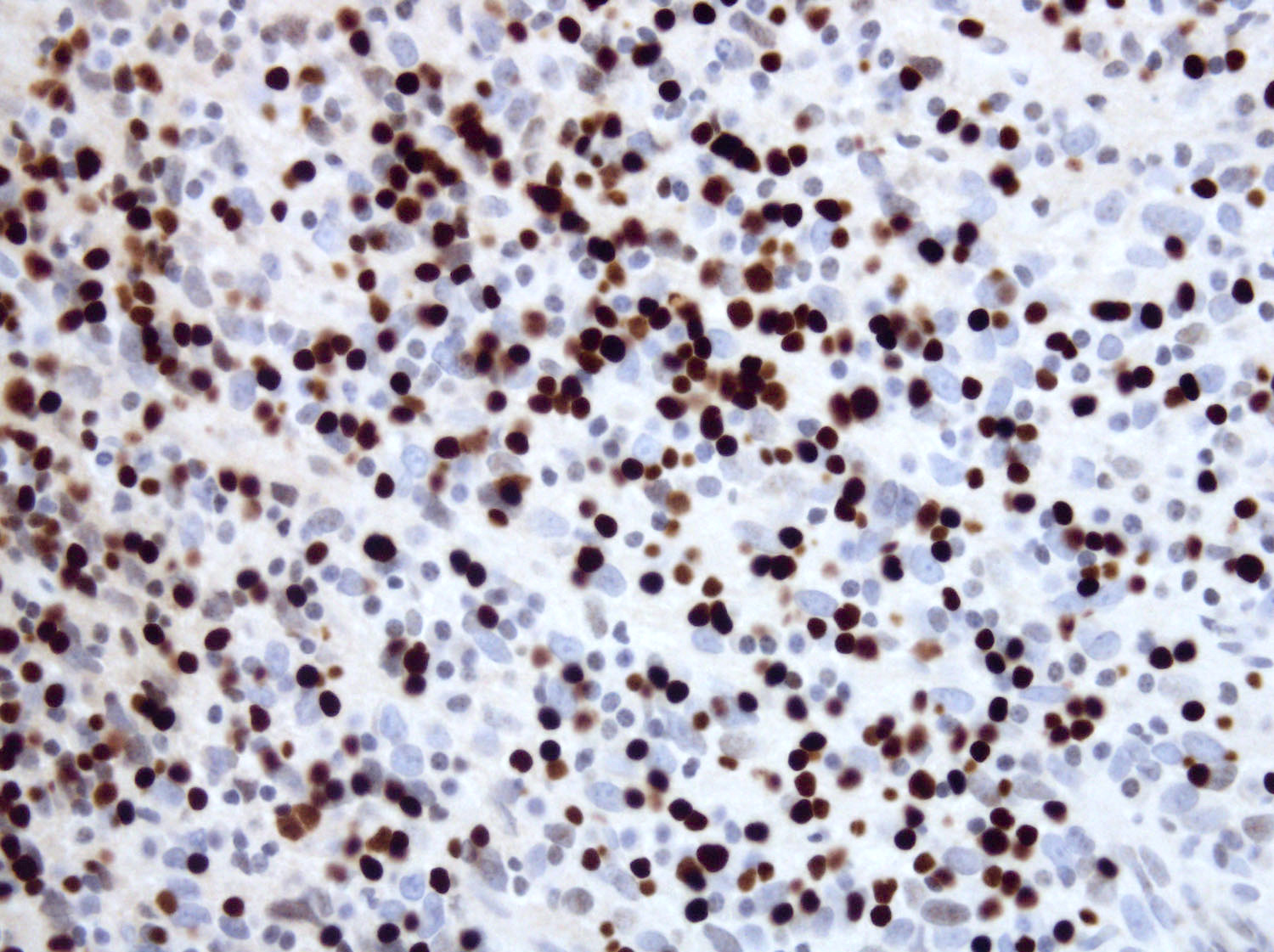
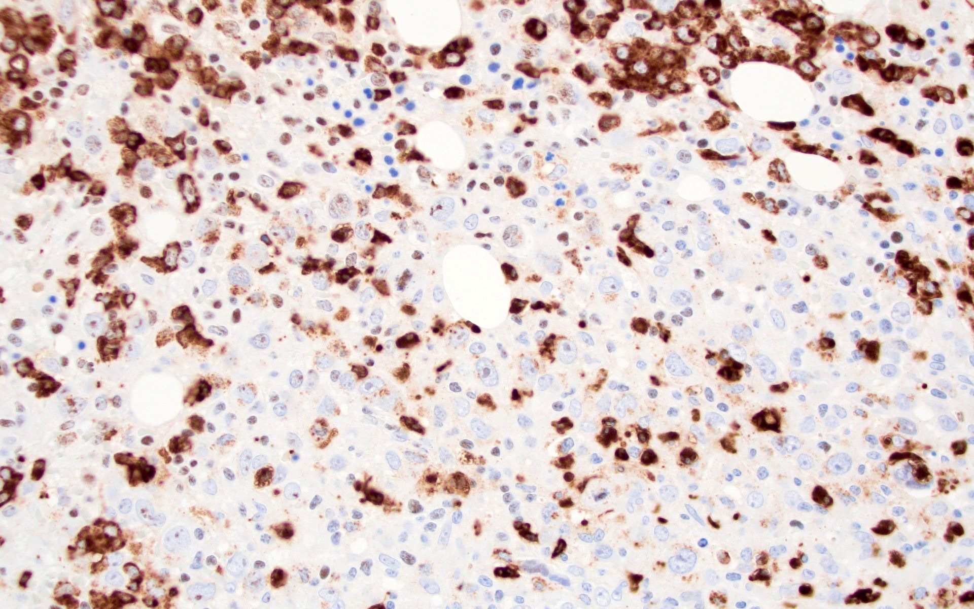
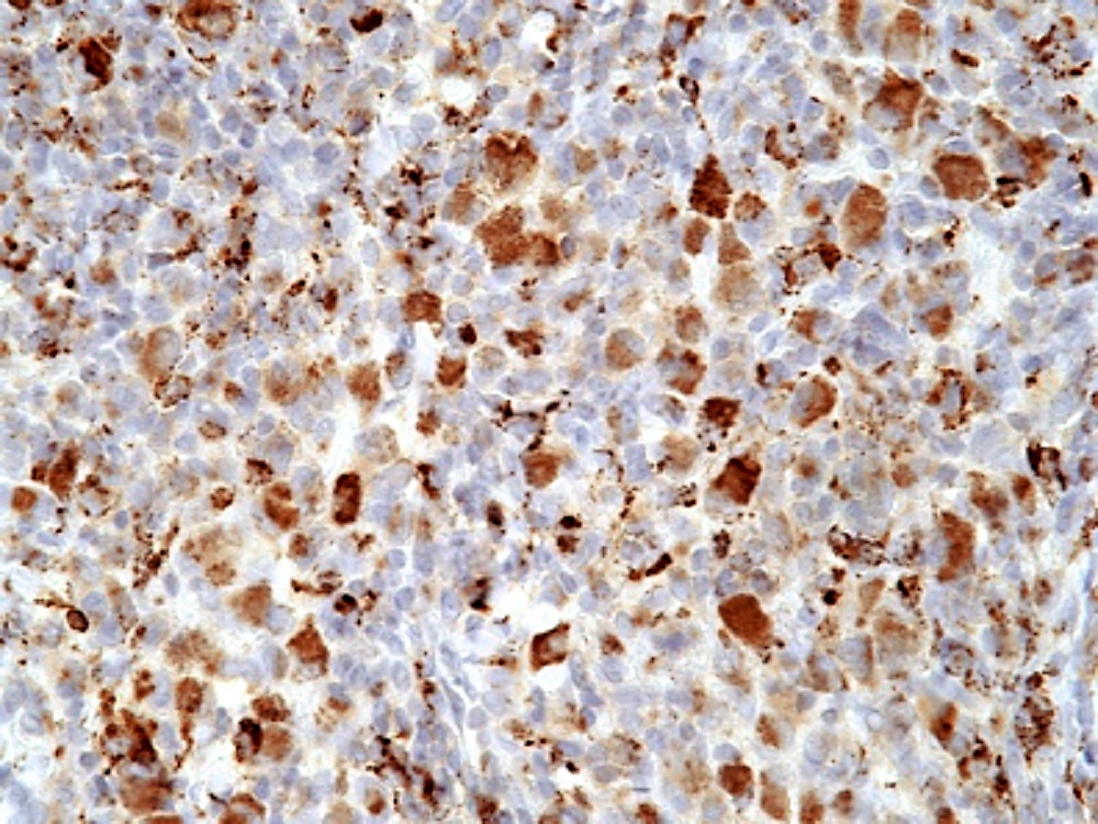
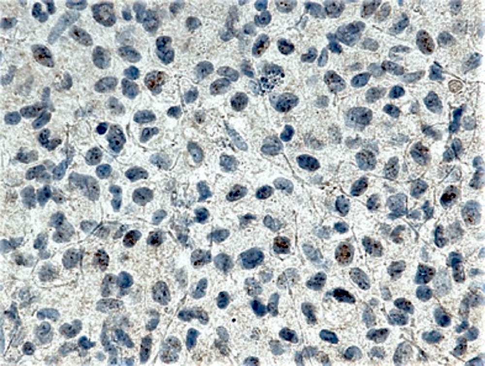
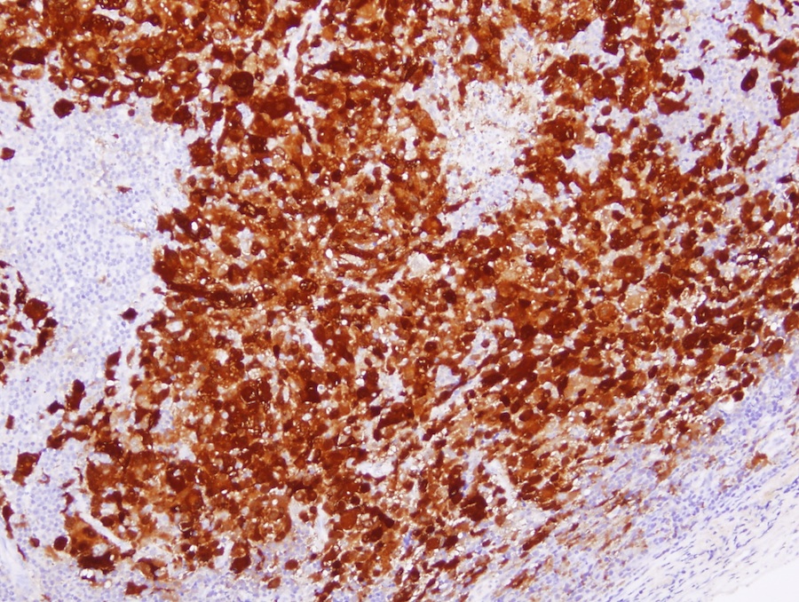
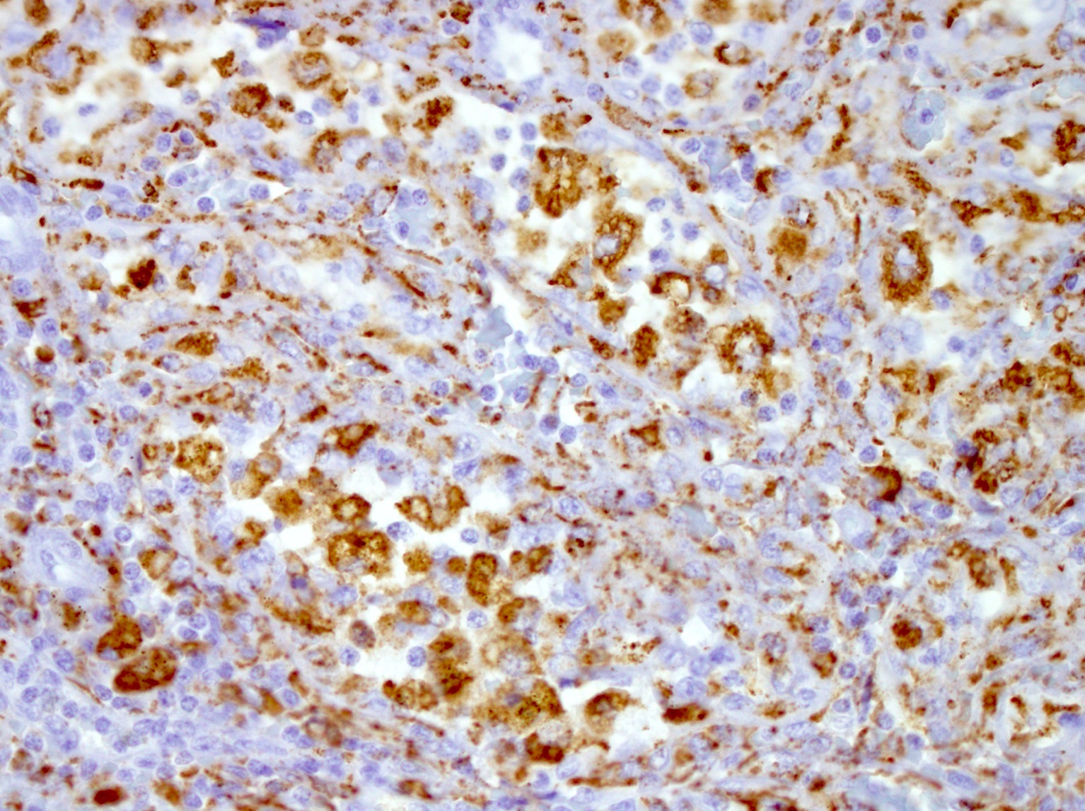
Chromogranin

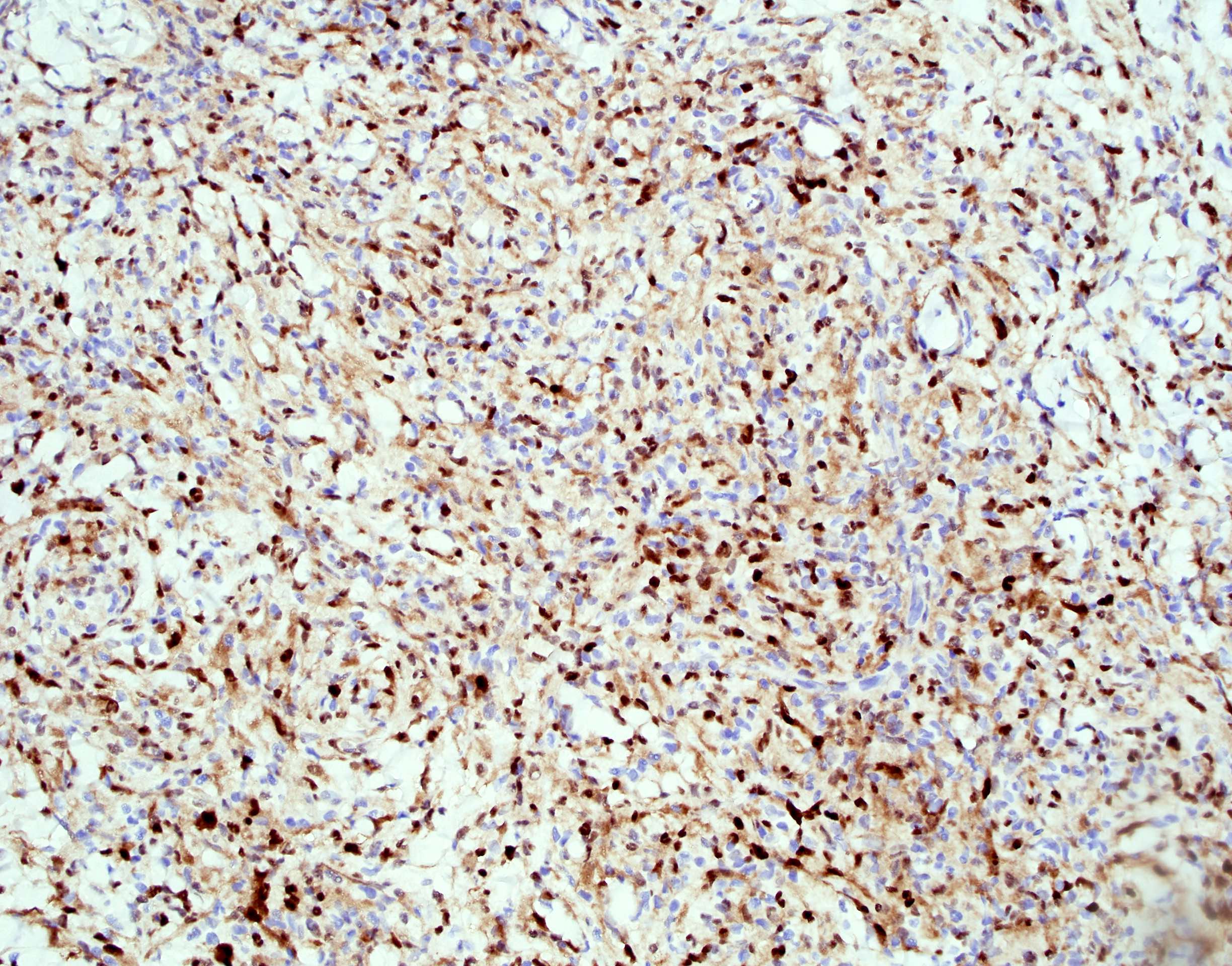
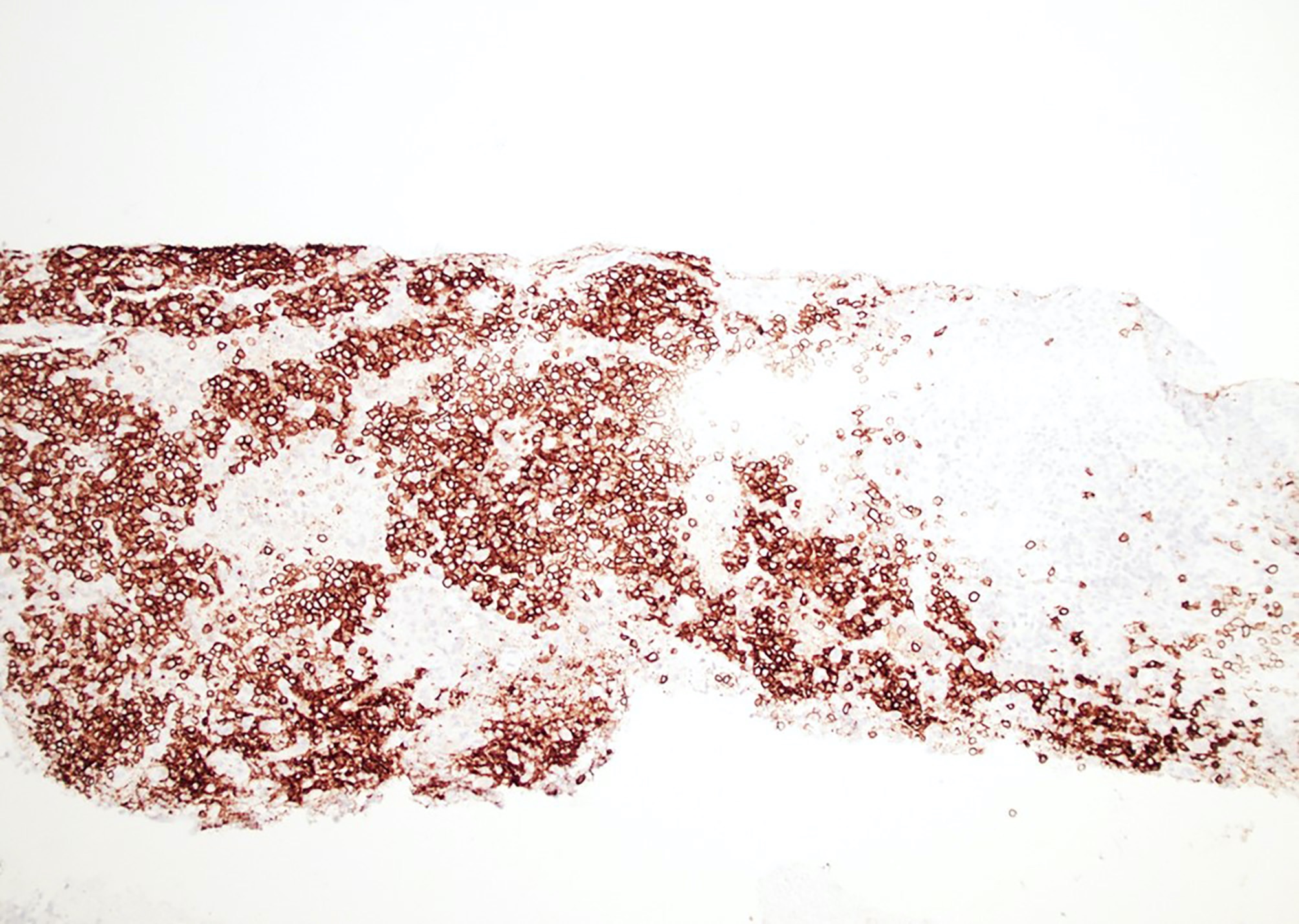
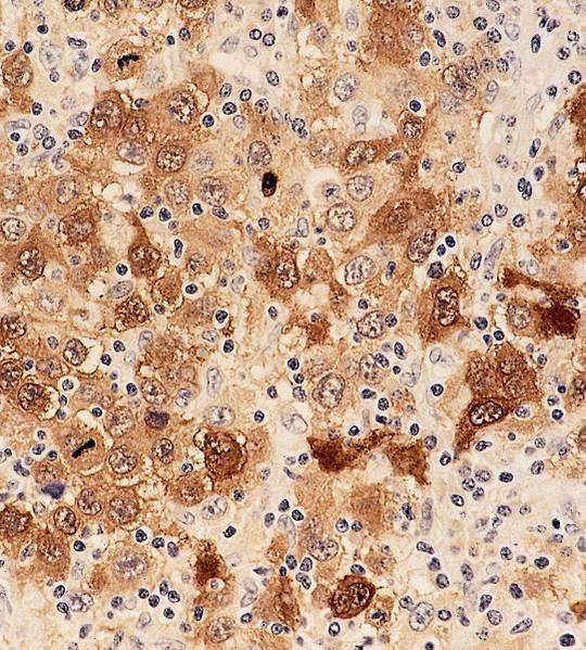
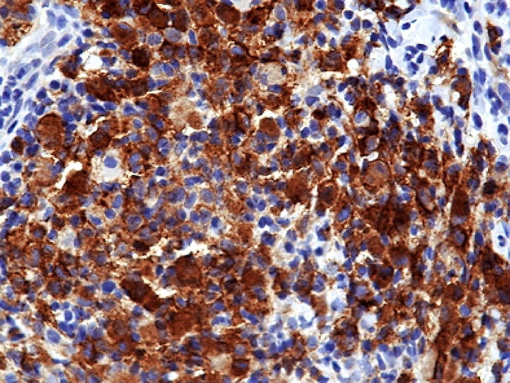
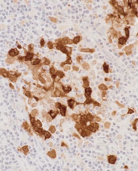
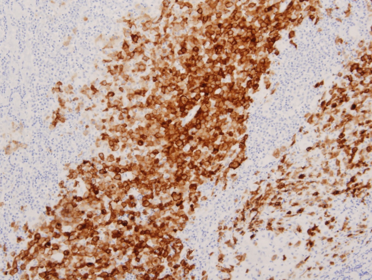
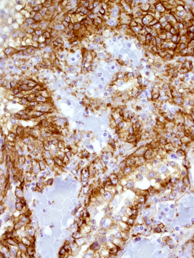
Synaptophysin
Images hosted on other servers:

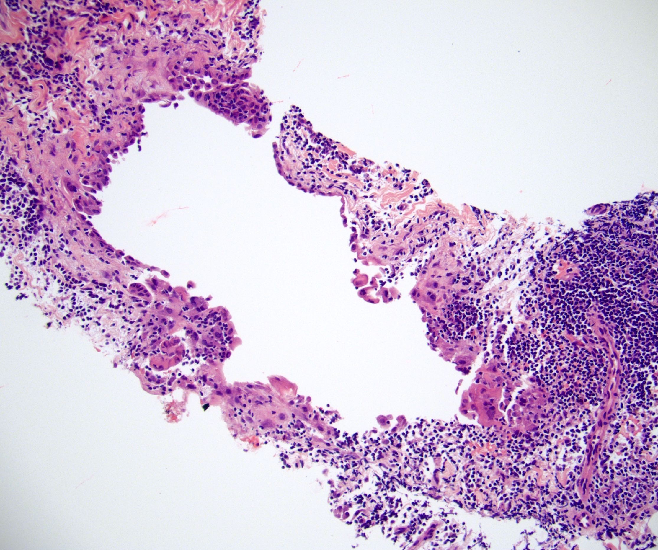
Acute lymphadenitis
Images hosted on other servers:

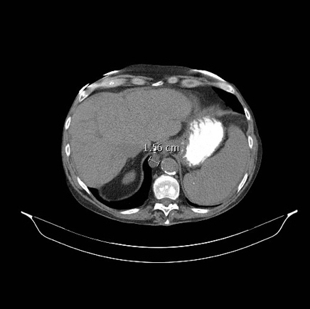
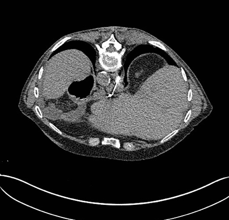
Progressively enlarging lymph node

Largest intra-abdominal adenopathy
Contributed by Jayalakshmi Balakrishna, M.D.
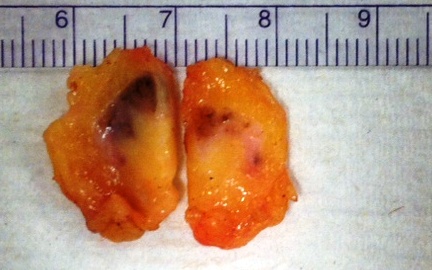
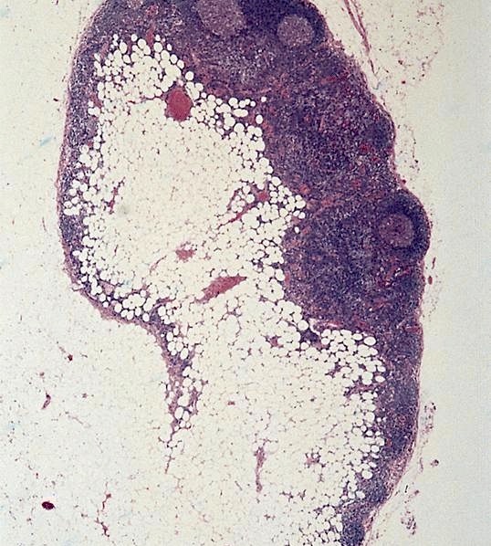
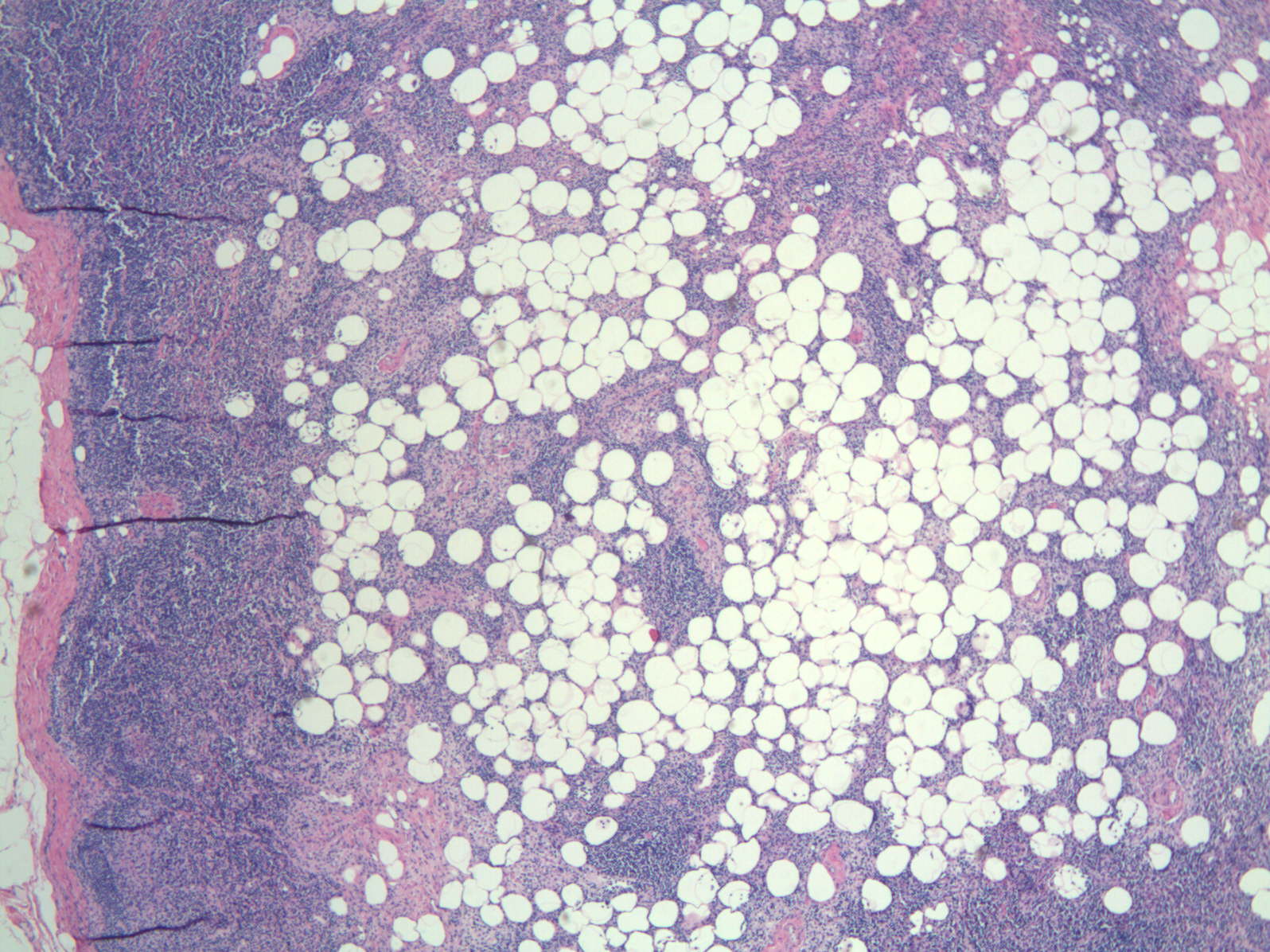
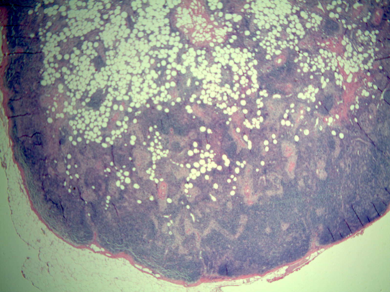
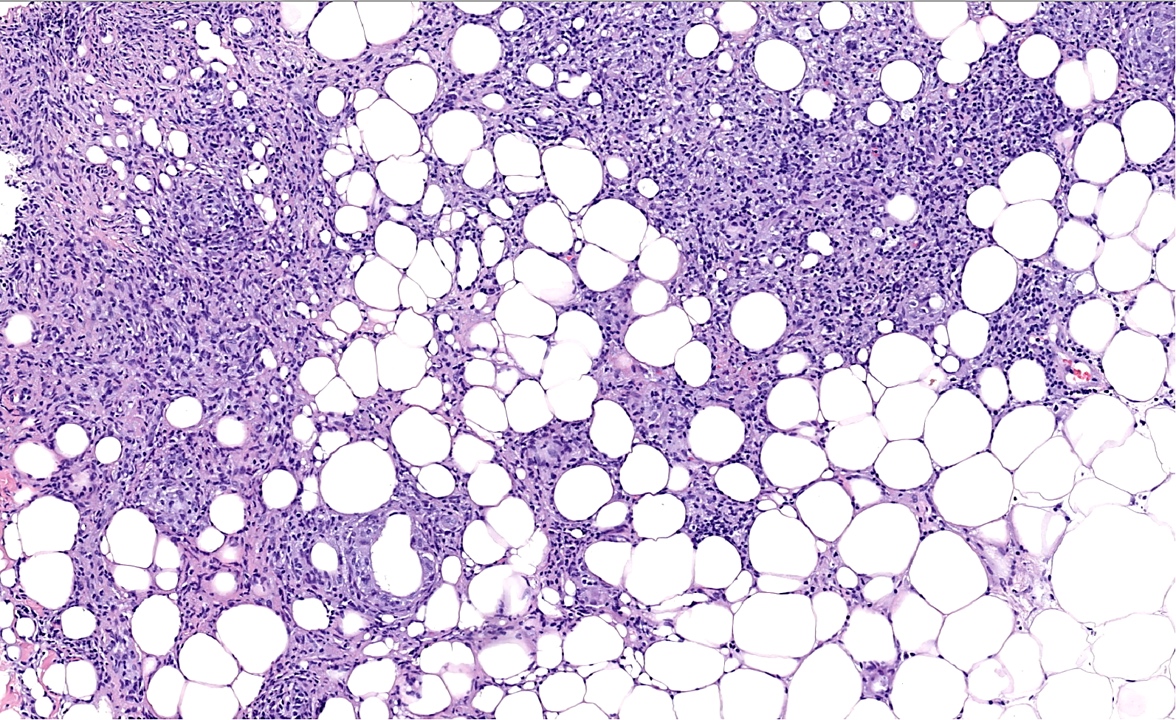
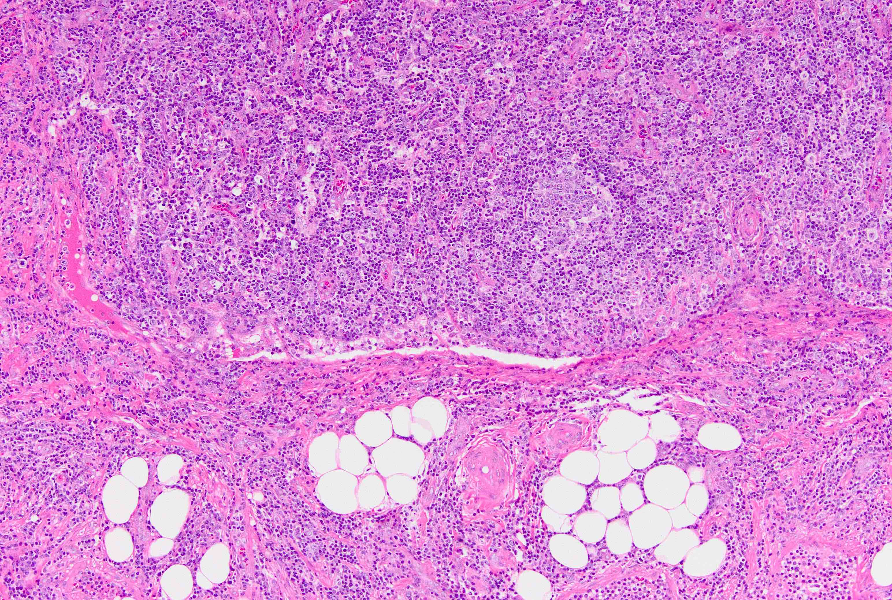
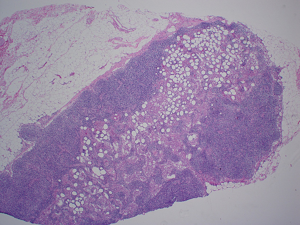
Lymph node replaced by adipose tissue
AFIP images
Lipomatosis
Contributed by Jayalakshmi Balakrishna, M.D.
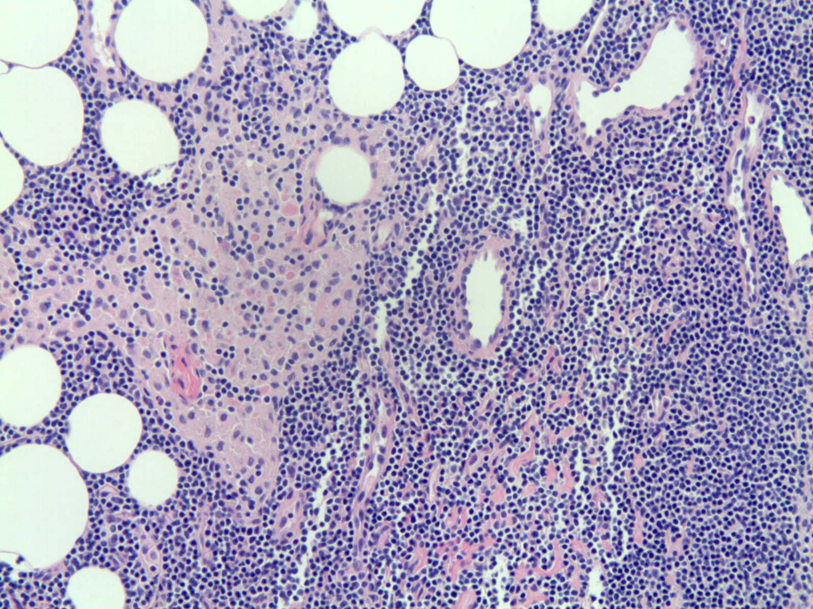
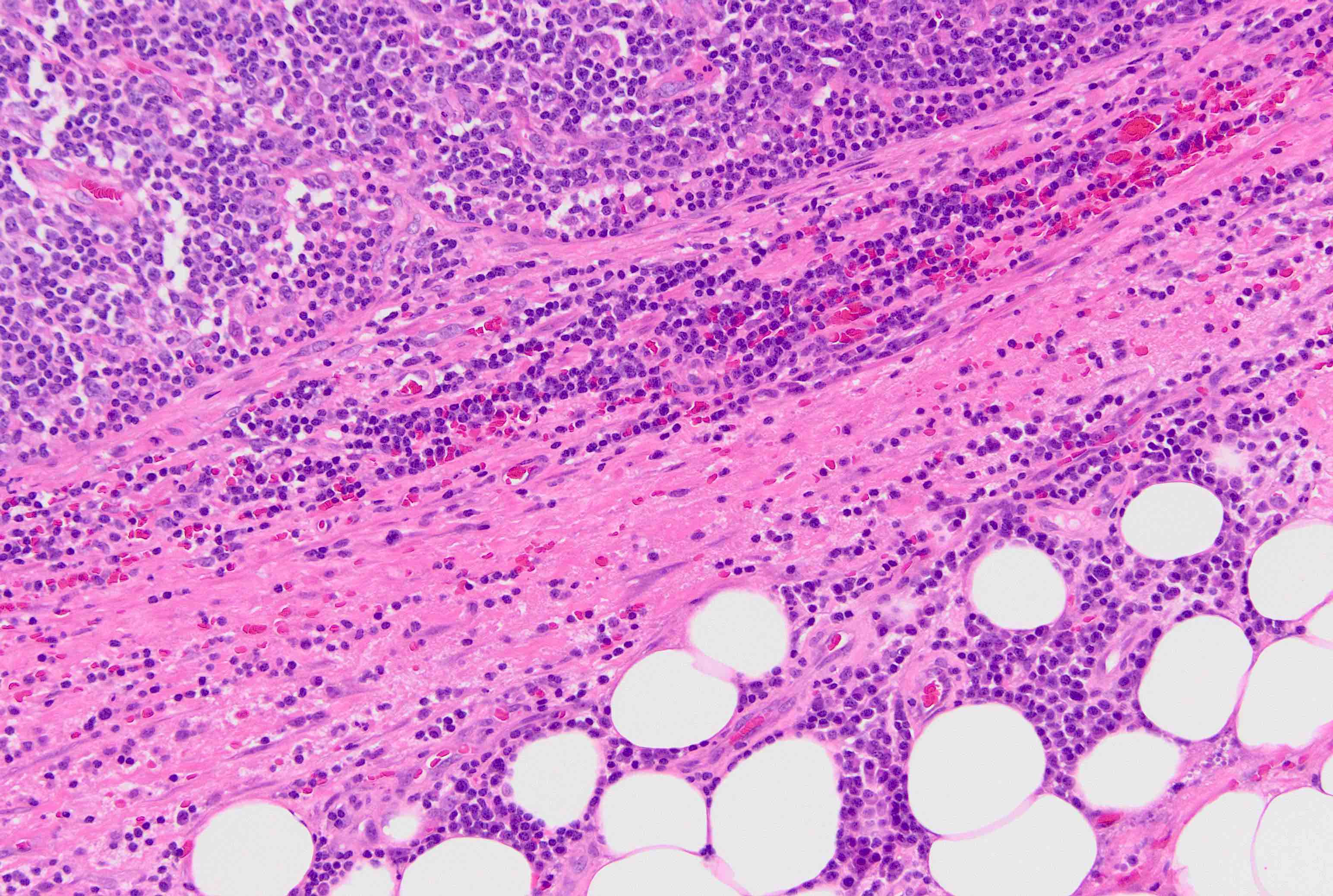
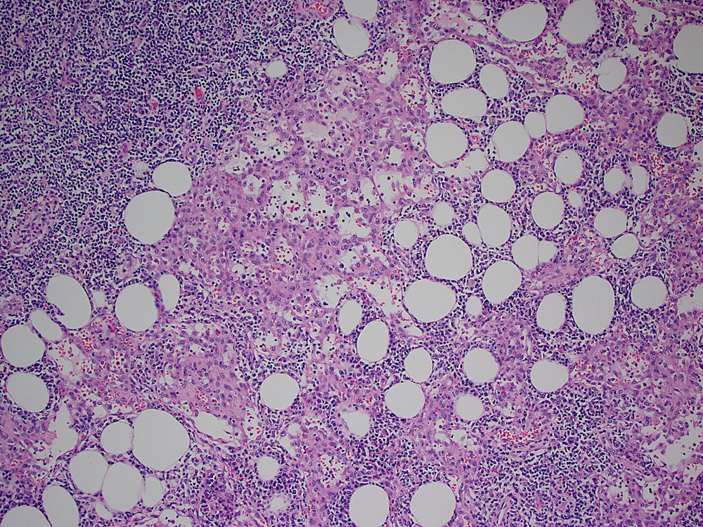
Lymphoid tissue closely admixed with adipocytes
Lymph node with adipocytes within capsule
Images hosted on other servers:

Reported sites of involvement
Contributed by Keri Janowiak, M.D., M.Ed.
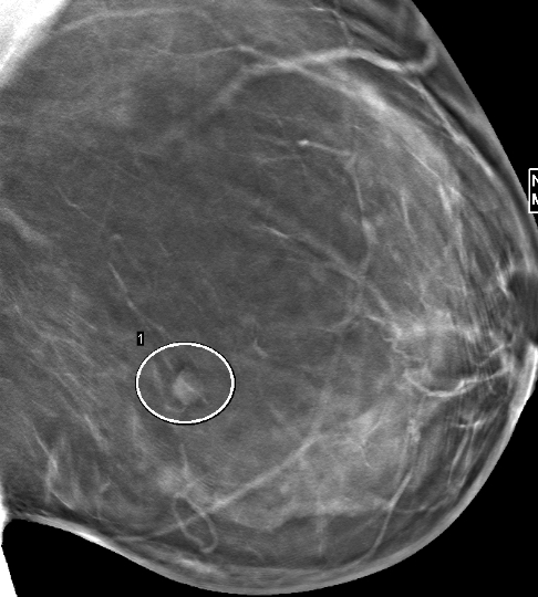
Screening mammography
Images hosted on other servers:

Nonneurologic disease manifestations

Left frontal lobe tumor
Images hosted on other servers:

Scalp lesion
Images hosted on other servers:

Breast APH lesion

Filum terminale
Contributed by Keri Janowiak, M.D., M.Ed., Ekene Okoye, M.D. and Aishwarya Ravindran, M.D.
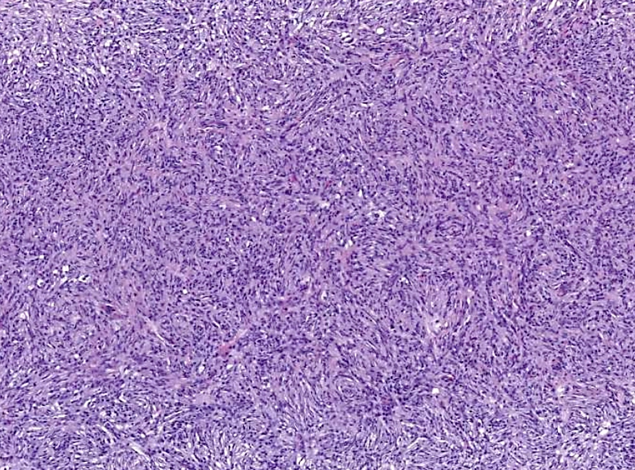
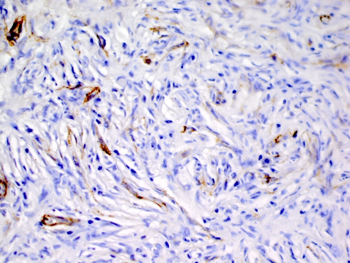
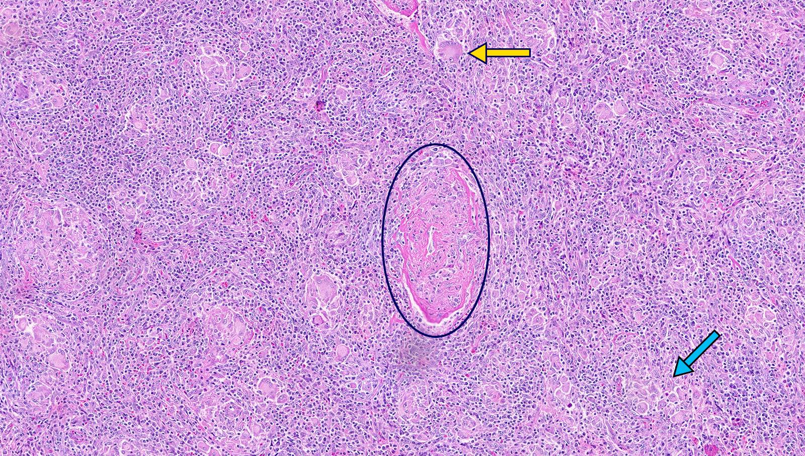
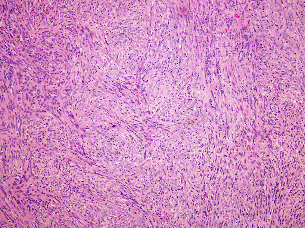
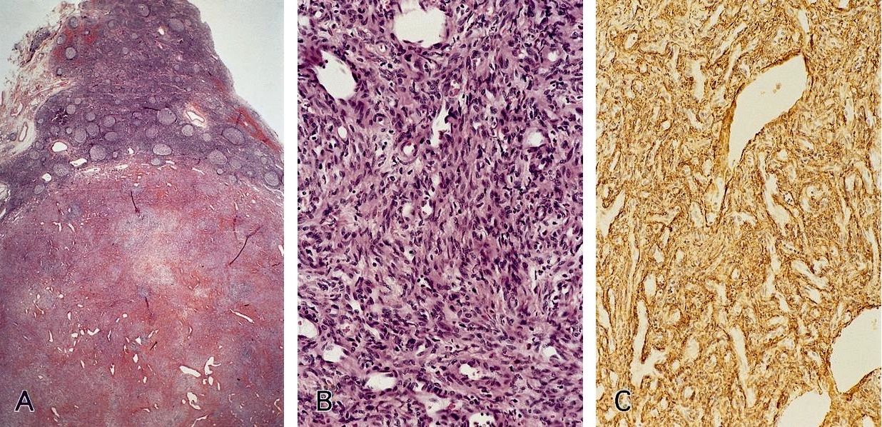
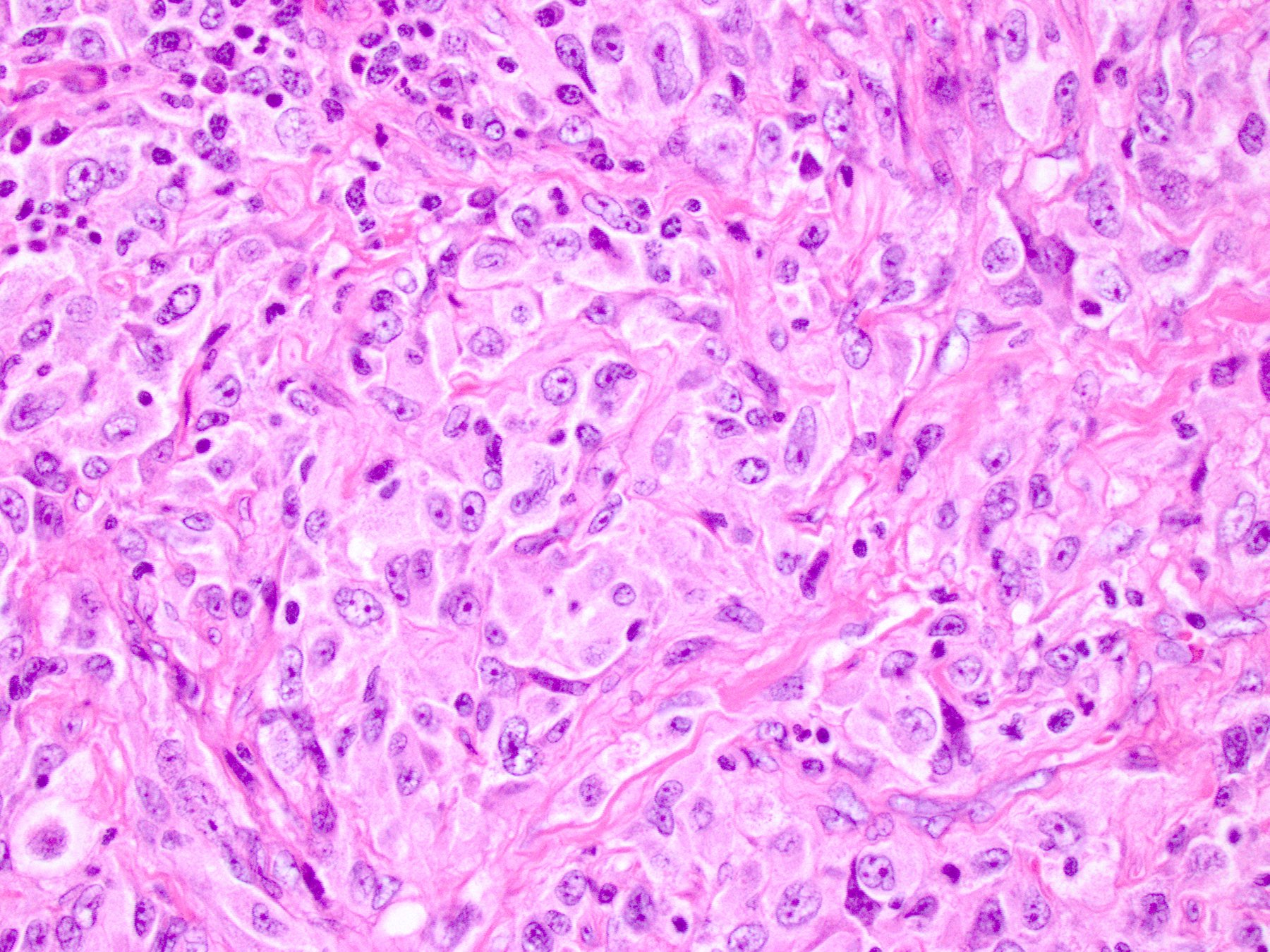
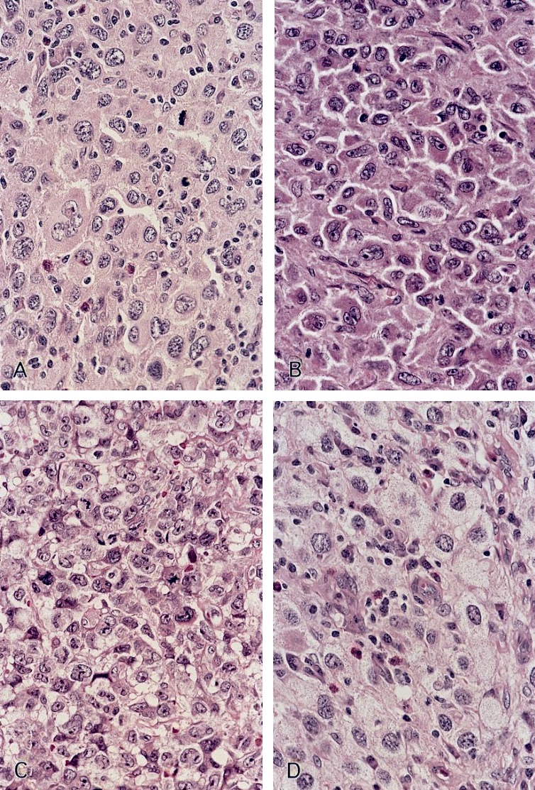
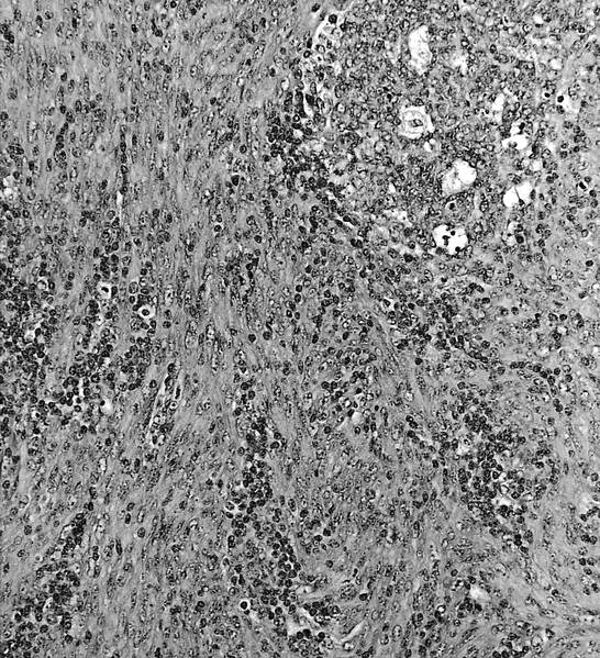
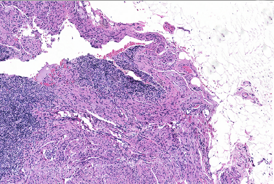
Storiform and fascicular growth
Infiltrative growth
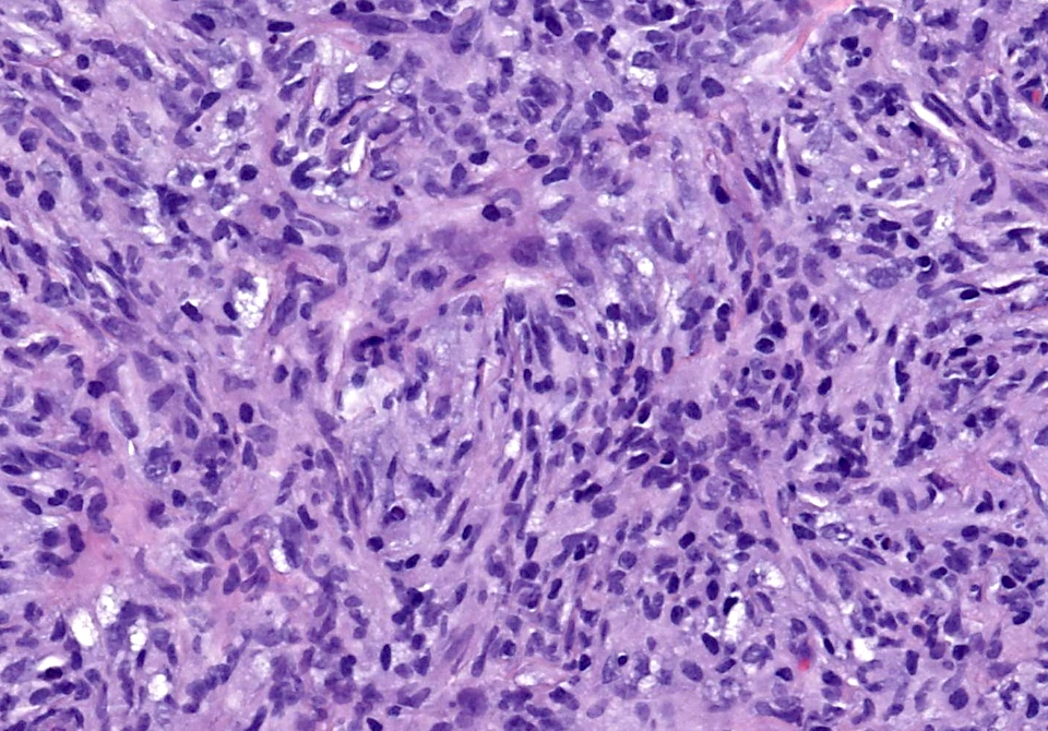
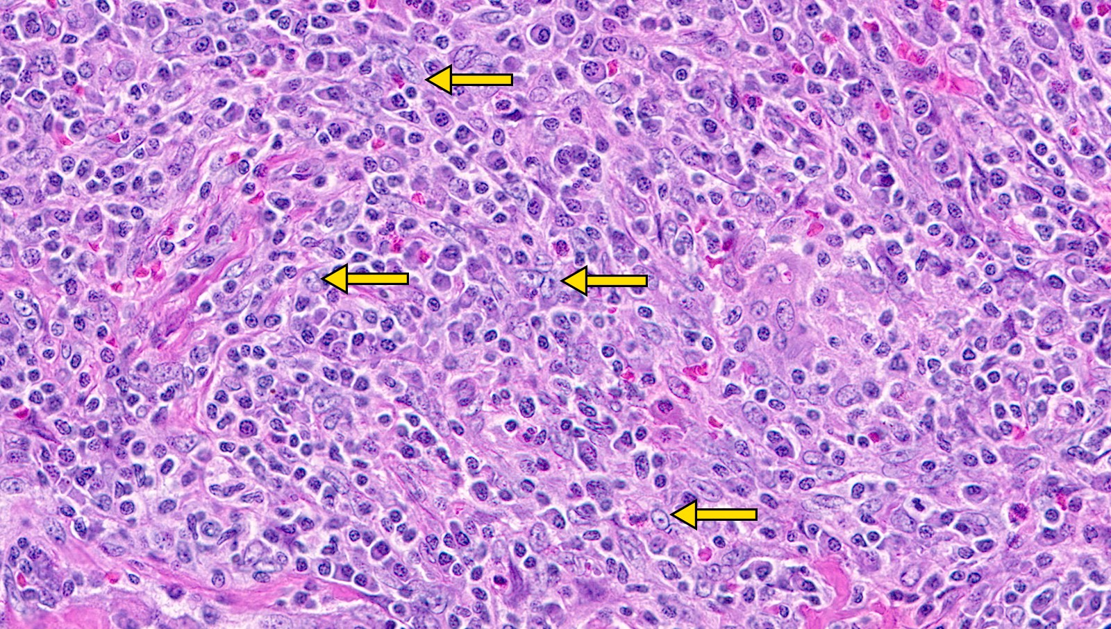
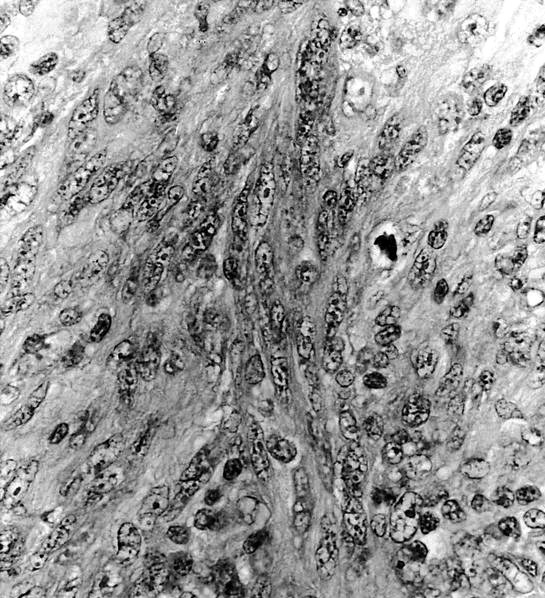
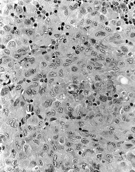
Histiocyte morphology

Touton giant cells
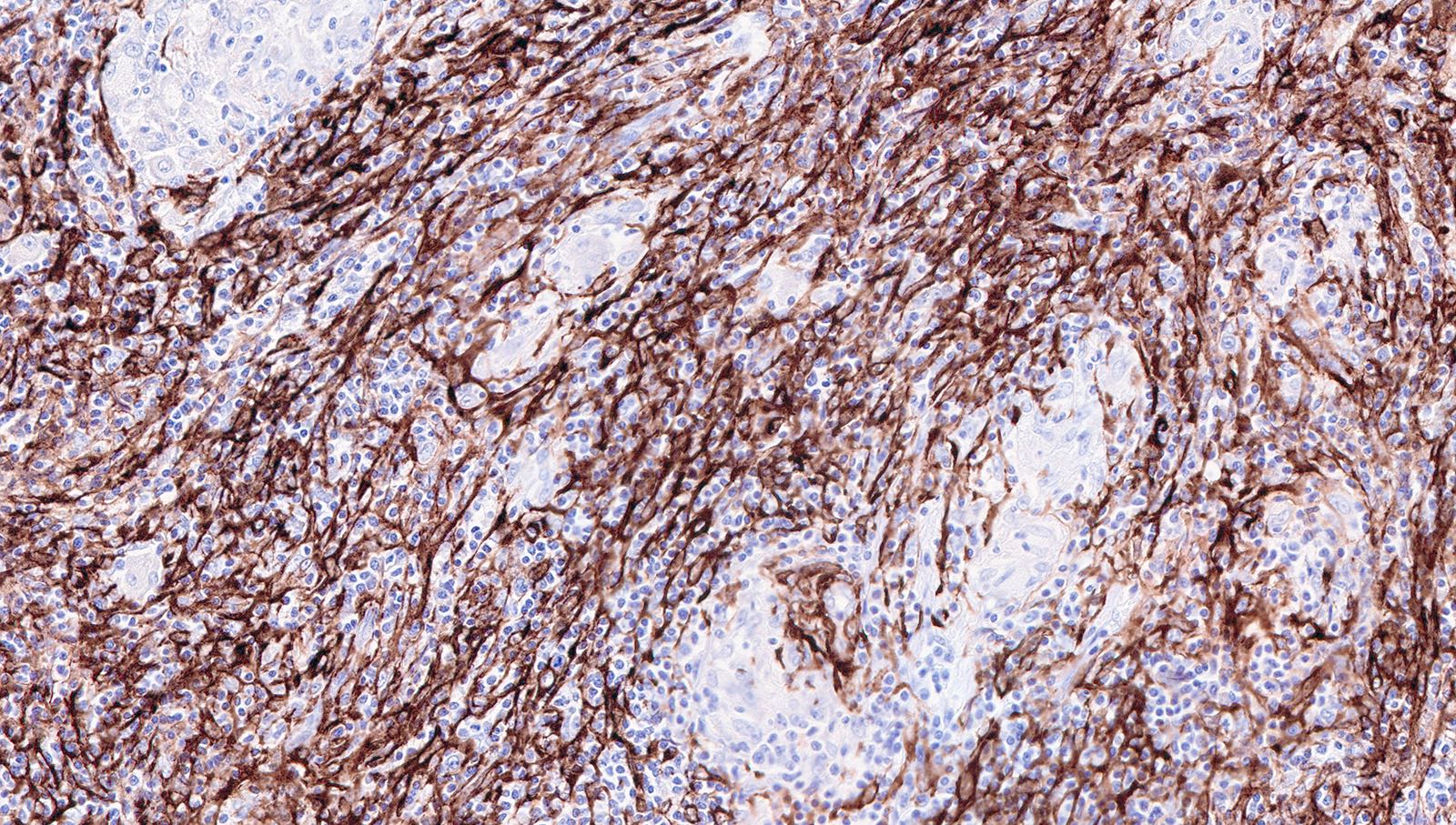
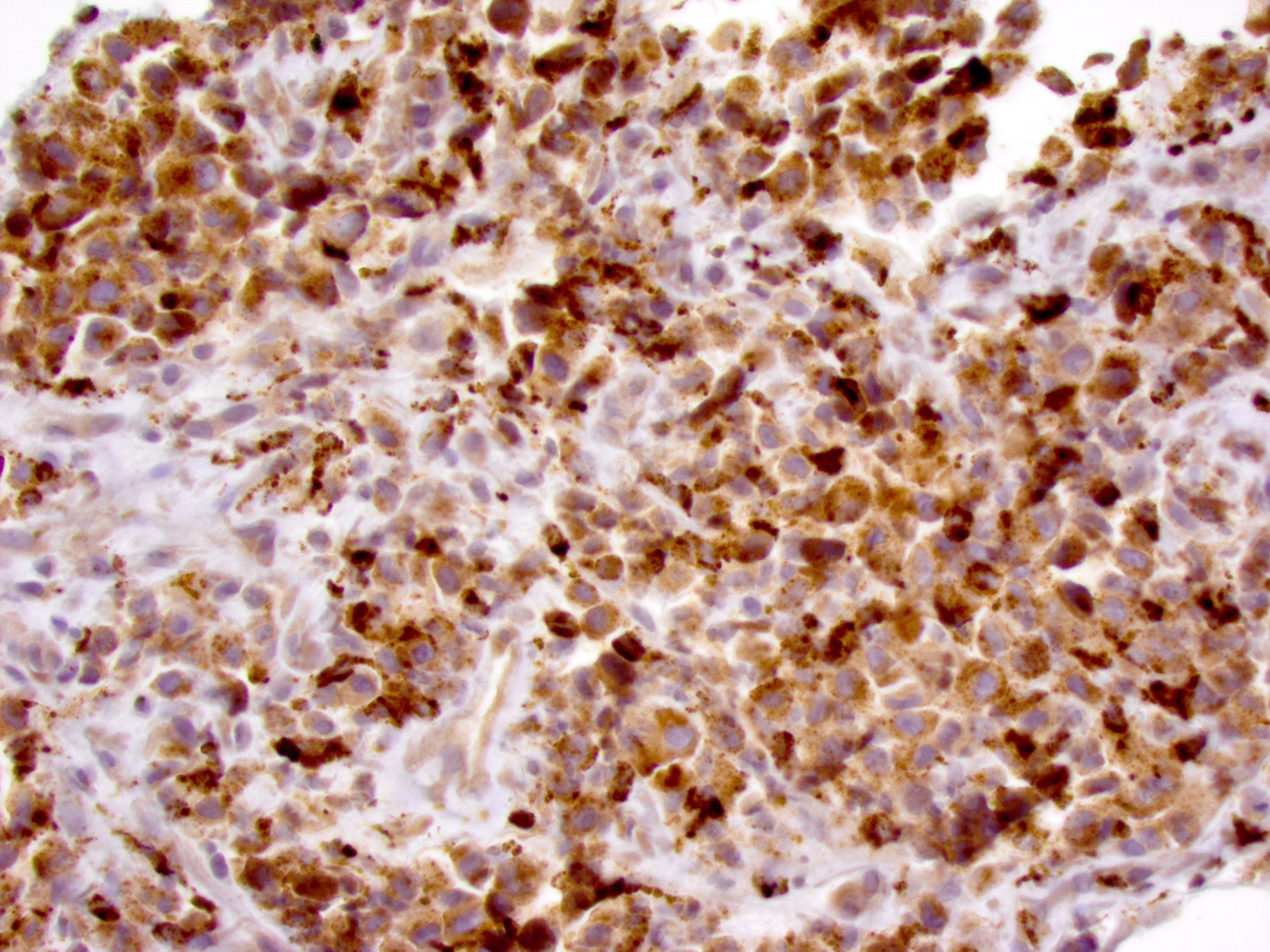
CD68
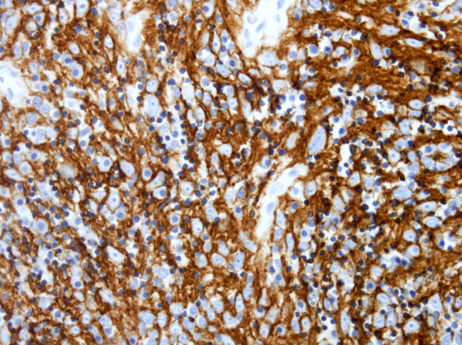
CD163
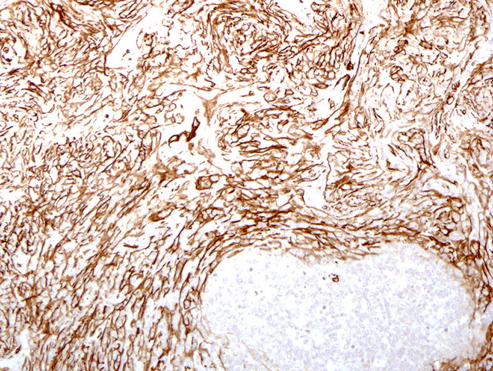
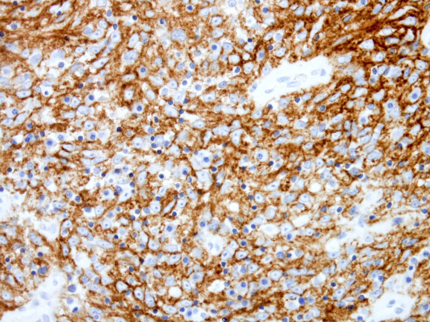
Factor XIIIa
ALK
Cyclin D1
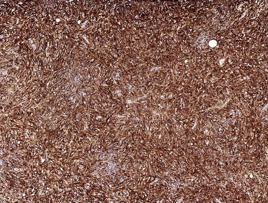
SMA
Images hosted on other servers:

ALK FISH, breakapart probe
Contributed by Mark R. Wick, M.D.

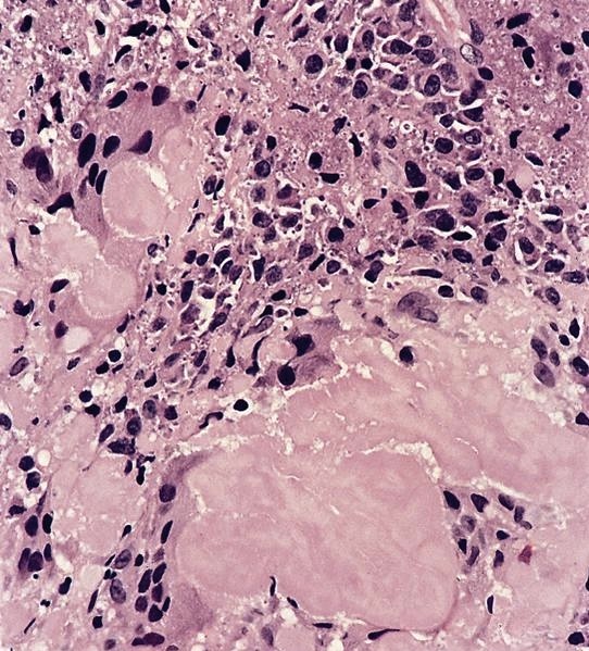
Amyloidosis of lymph node
AFIP images
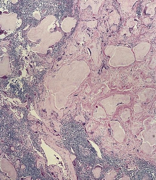

Amyloidosis of lymph node
Contributed by Mark R. Wick, M.D.
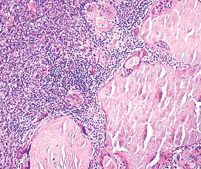
Amyloidosis of lymph node


Contributed by Nikhil Sangle, M.D.
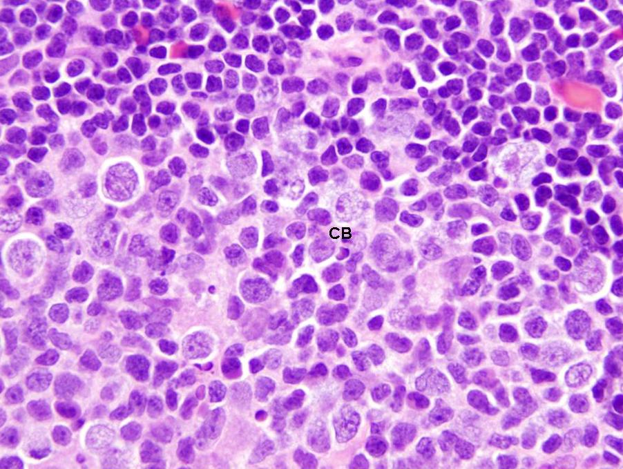
Centroblasts
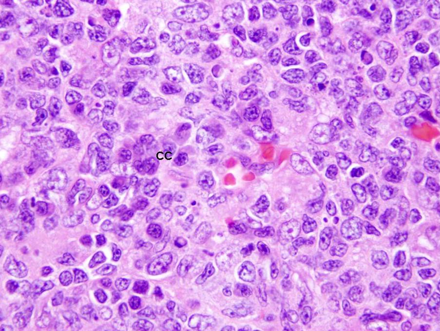
Centrocytes
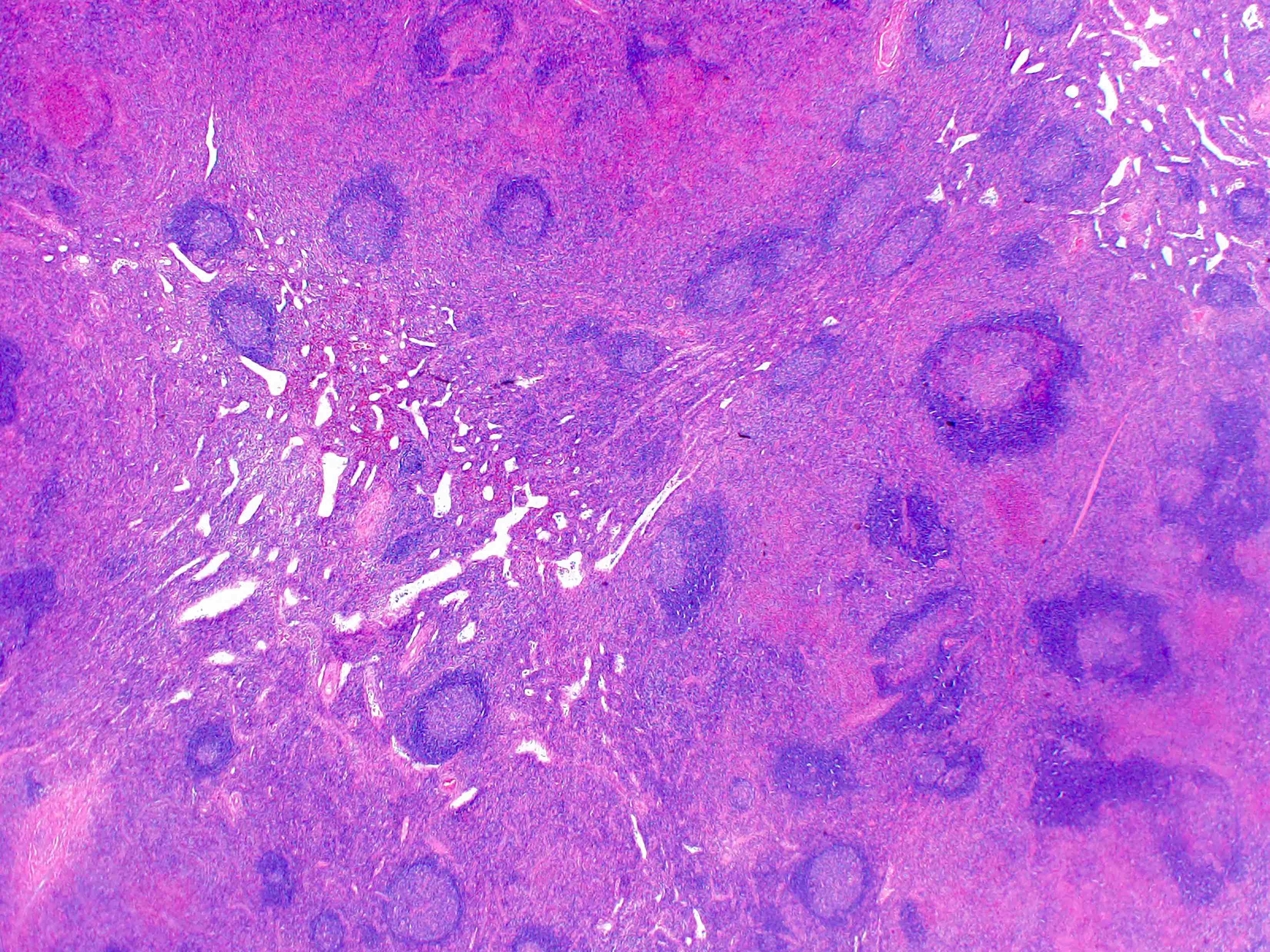
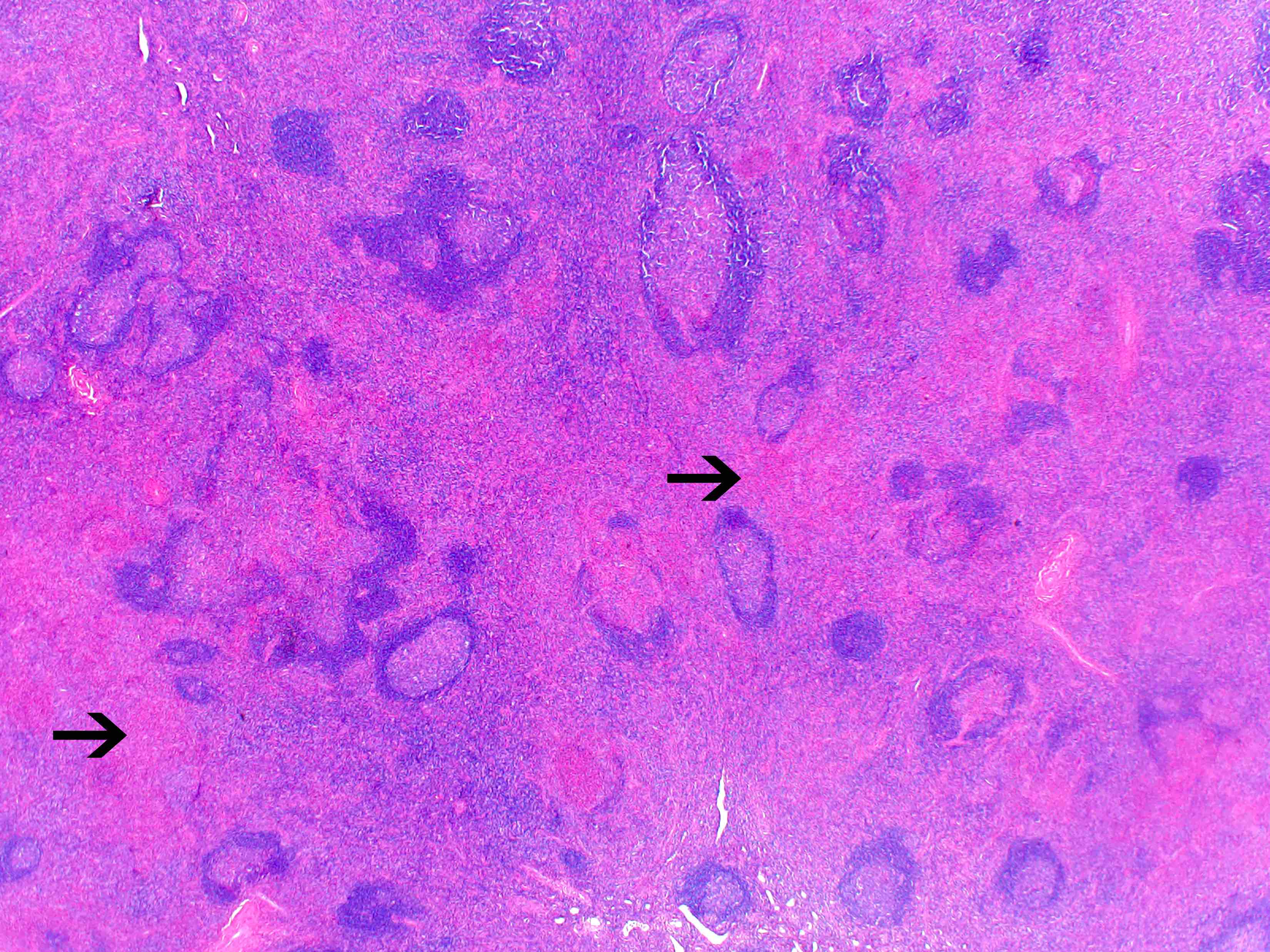
AFIP images
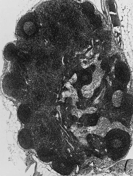
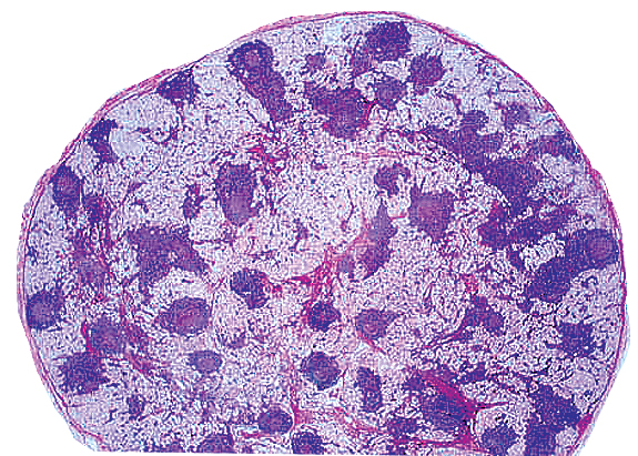
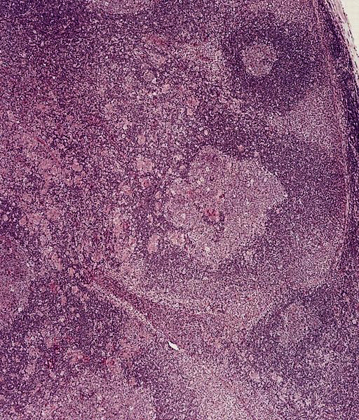
Normal lymph node

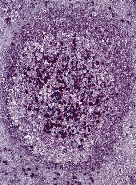
Primary follicle

Lymph node
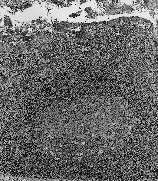
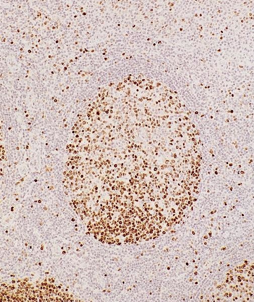
Secondary follicle
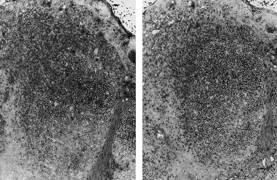
Secondary follicle
Paracortical T zone
Interdigitating dendritic cells

Smooth muscle proliferation in lymph node hilum
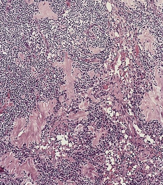
Sclerosis in an inguinal lymph node
Images hosted on other servers:

Plasma cells
Images hosted on other servers:

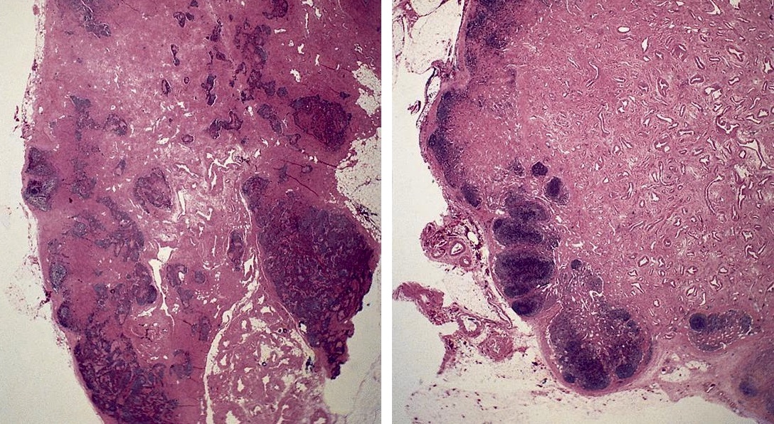
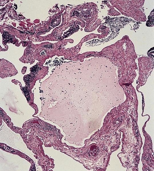
Mesenteric tumor with soft tissue parts
Images hosted on other servers:

Large smooth surfaced mass
AFIP images

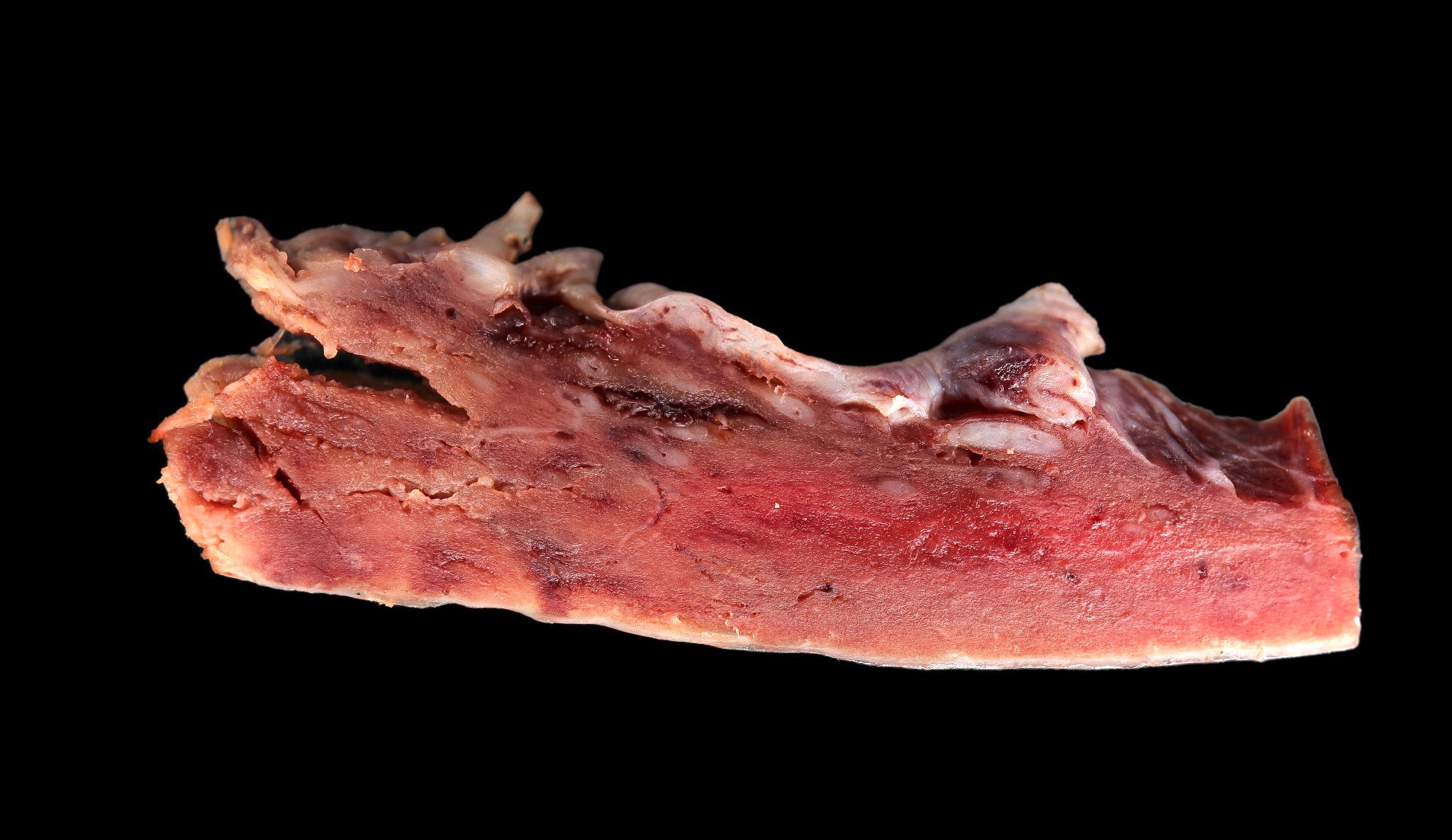
Lymph node
AFIP images
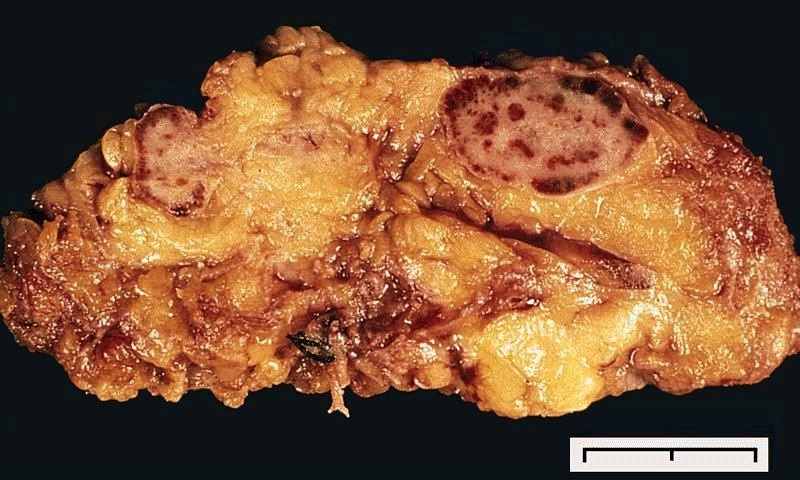
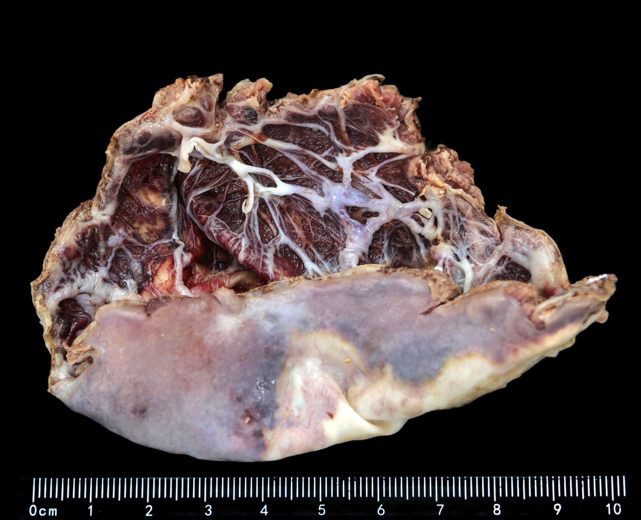
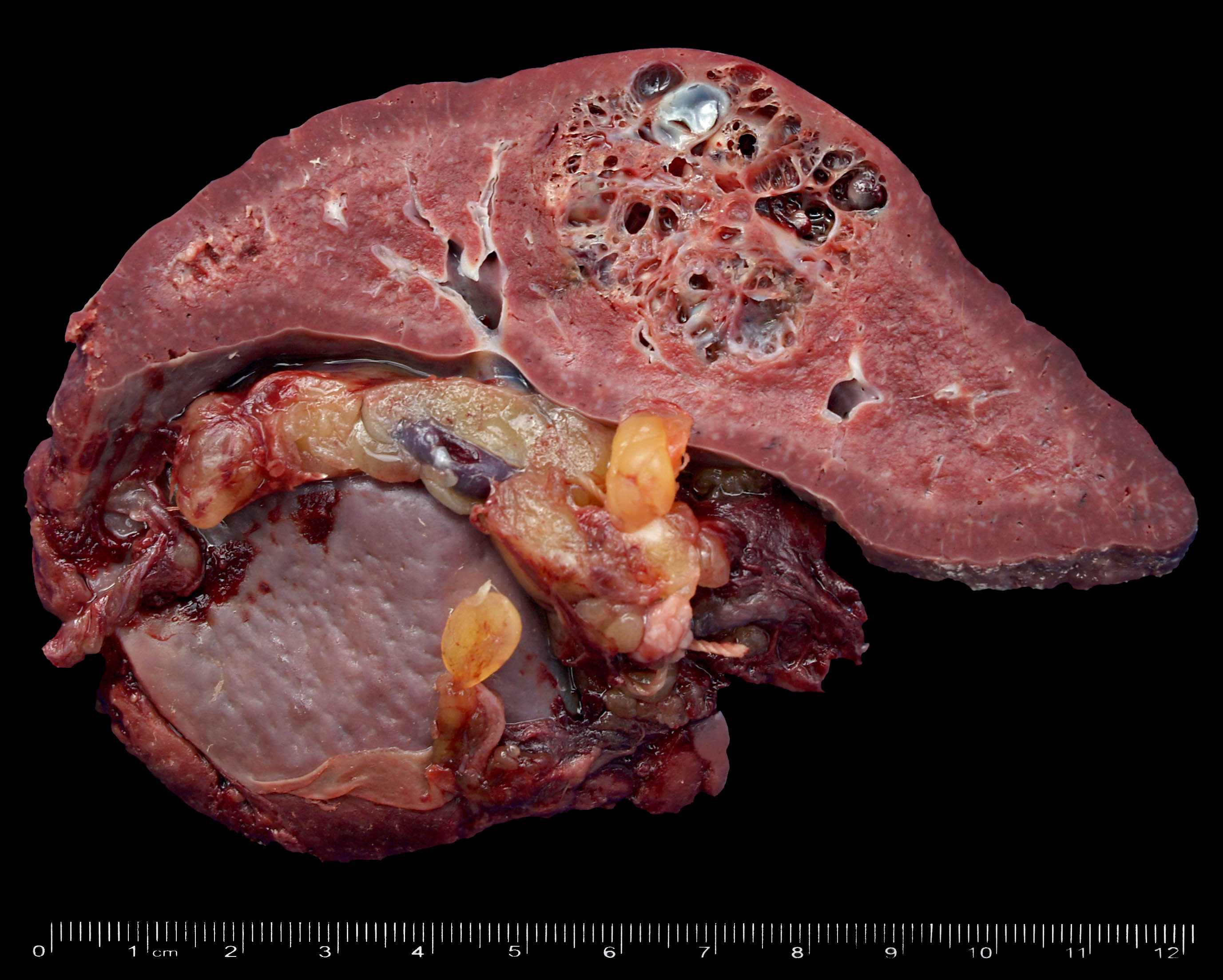
Replacement by tan, firm tissue
Contributed by Patricia Tsang, M.D., M.B.A., Vincent A. Graffeo, M.D. and AFIP
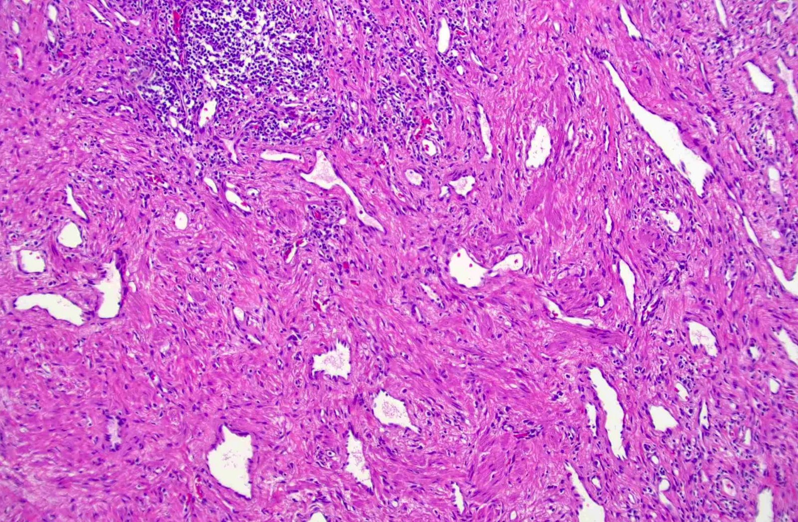
Disarrayed vascular proliferation
Angiomyomatous proliferation
Smooth muscle proliferation
Thick walled vasculature
Abnormal blood vessels

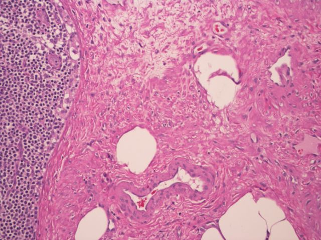
Angiomyomatous proliferation

Thick walled vessels

Disarrayed smooth muscles
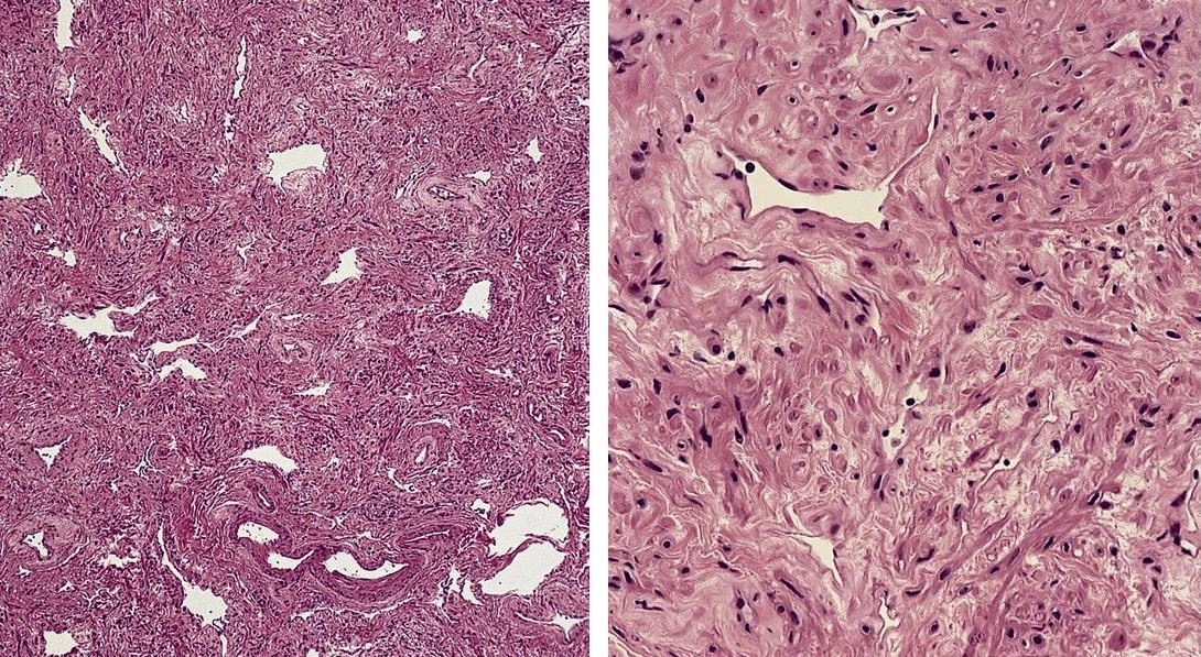
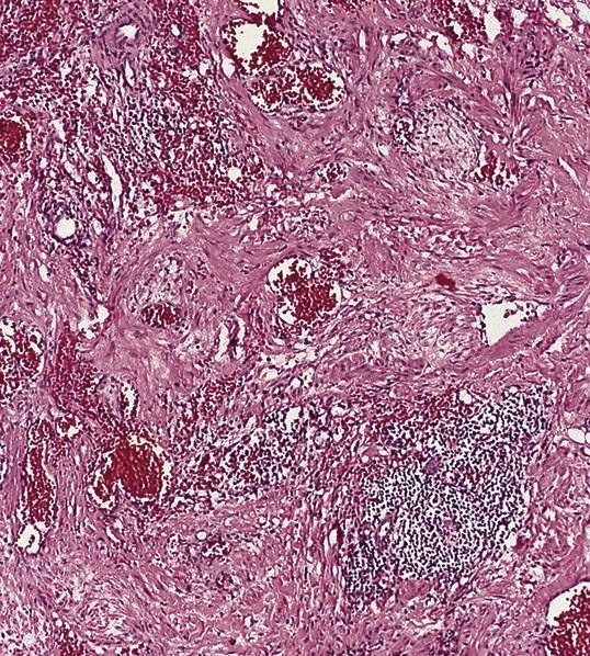
Angiomyomatous hamartoma of lymph node
Smooth muscle actin
CD31
Images hosted on other servers:

Apical lung lesion

Increased uptake in left upper lobe

FDG PET/CT scan shows
high-grade metabolic activity
in right hilar soft tissue lesion
Images hosted on other servers:

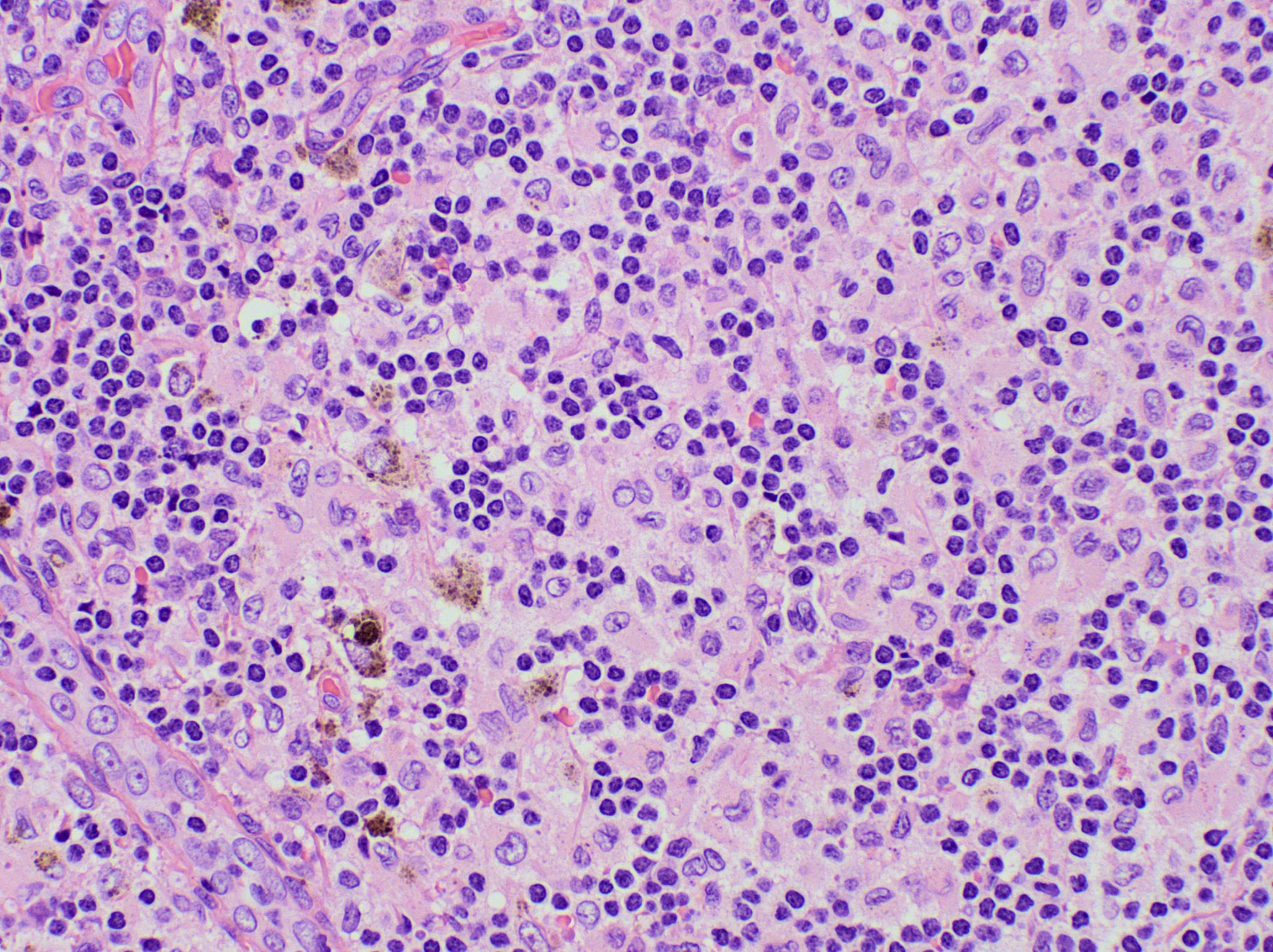
Abundant anthracotic pigment
AFIP images
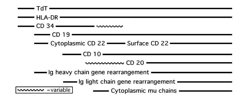
B cell biomarker expression during development
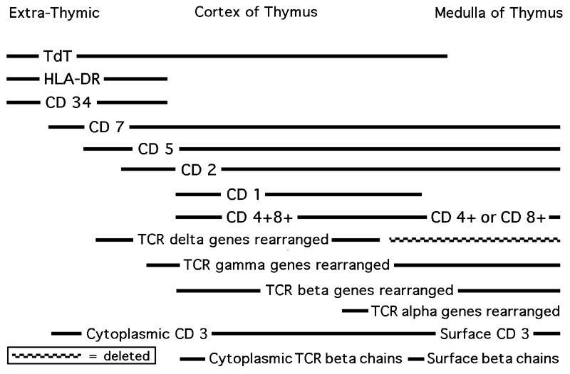
T cell biomarker expression during development
Images hosted on other servers:
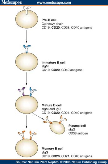
B cell development

B cell response to antigen

B cell biomarker expression during development

niches in the bone
marrow required for
B cell development

T cell activation

T cell development
Images hosted on other servers:

18F FDG PET / CT, HHV8 MCD
Contributed by Jayalakshmi Balakrishna, M.D., Amy Duffield, M.D., Ph.D., Tapan Bhavsar, M.D., Ph.D.,
Carlos Murga-Zamalloa, M.D. and Jackie D. Sublett II, M.D.
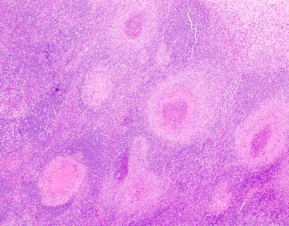
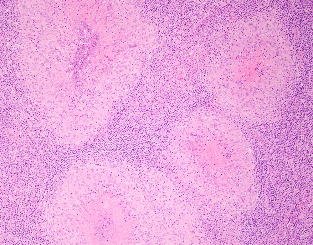
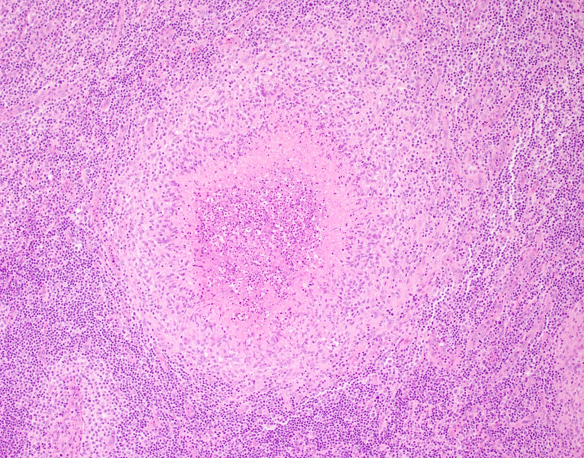
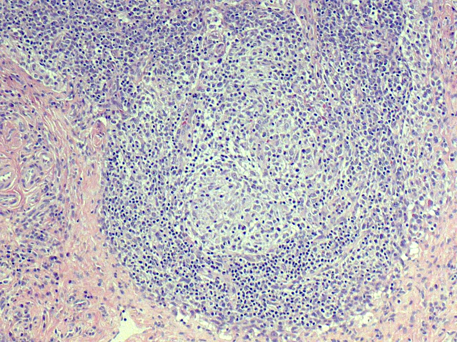
Hyaline vascular Castleman disease (HVCD)
Atretic follicles, interfollicular vascular proliferation
Atretic germinal center
Twinning of germinal center
Twinning of germinal center
Twinning of germinal center, thickened mantle zones
Lollipop follicle, thickened mantle zones
Twinning of
germinal centers,
sclerosed
vessels
Thickened mantle zones
Sclerosed vessels
Vascular proliferation
Lollipop follicle
Atretic follicle
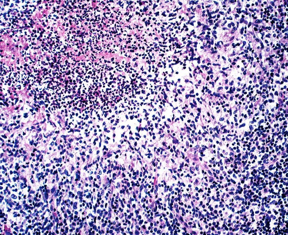
Proliferation of T lymphoblasts
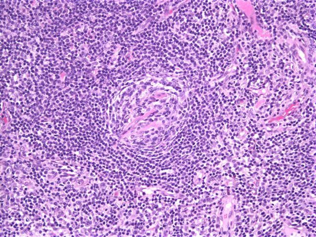
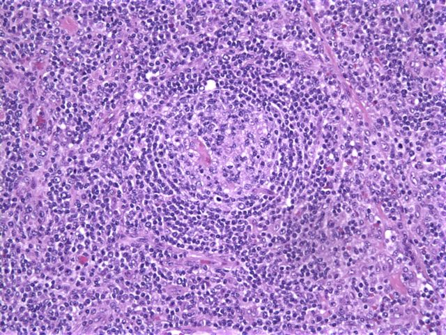
Onion skin pattern
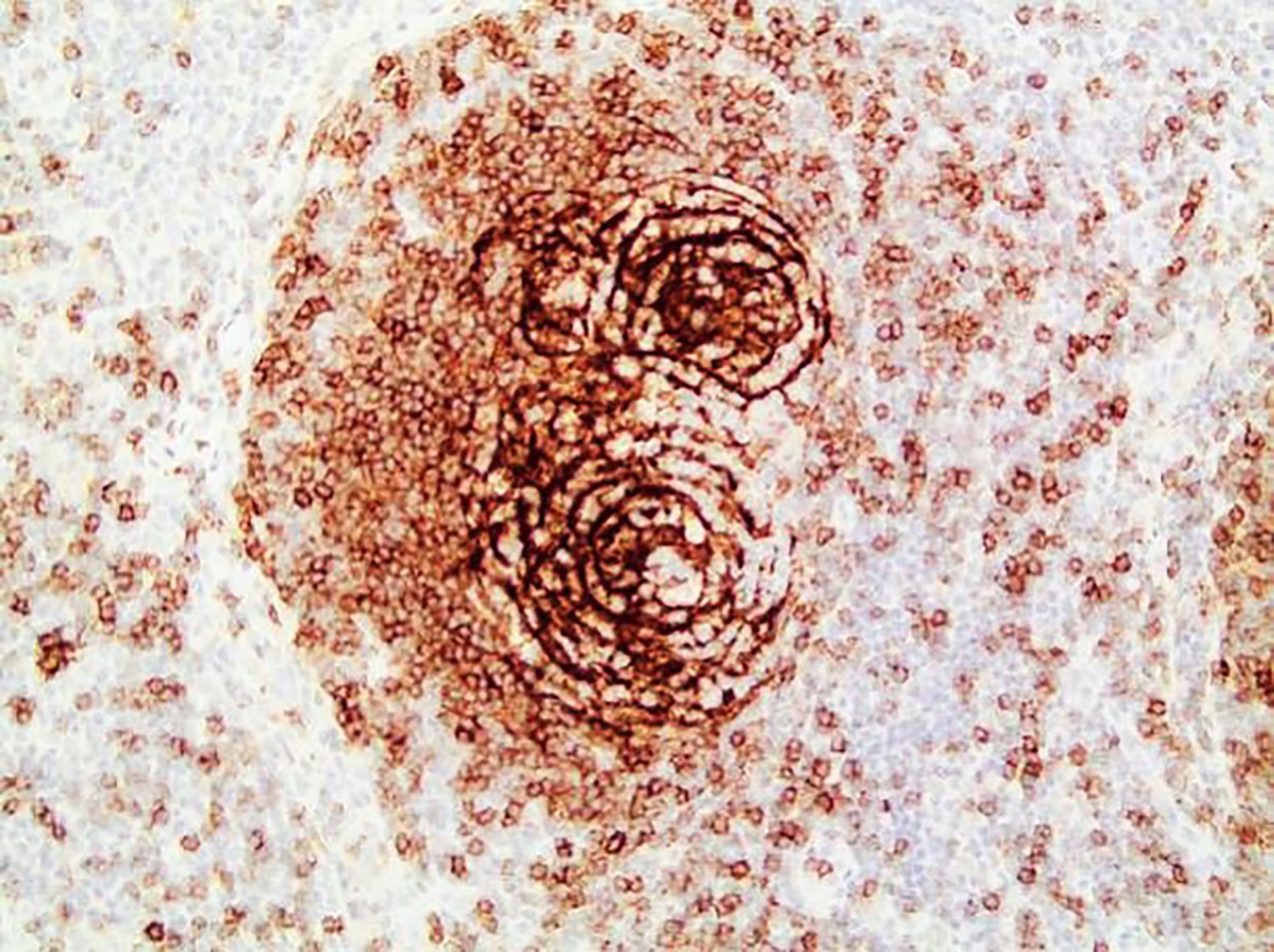
Twinning of germinal center, CD23
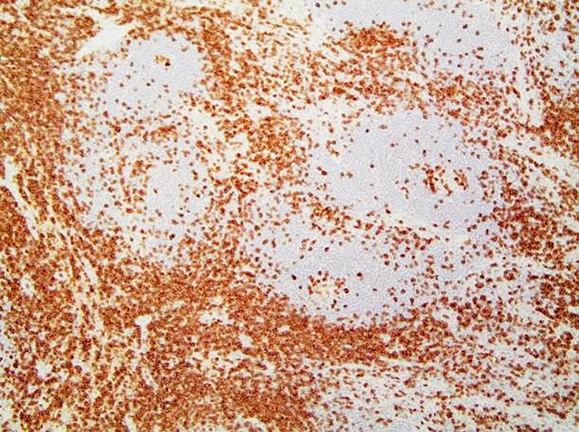
Interfollicular areas rich in T cells, CD3
Follicles and atretic germinal centers, CD20

Proliferation of TdT+ cells
Vascular proliferation, CD34
Interfollicular areas rich in T cells, CD5
Follicular dendritic cell meshworks, CD21
Low proliferation rate in interfollicular areas
Germinal centers, BCL6+
Plasma cells, CD138
Plasma cell Castleman disease (PCCD)
Aggregates of plasma cells
T cells in interfollicular areas, CD3
Atretic follicles, CD20
Aggregates of plasma cells, CD138
Vascular proliferation, ERG
Human herpesvirus 8 associated multicentric Castleman disease (HHV8 MCD)
Aggregates of plasma cells
Atretic follicle, interfollicular plasma cell proliferation
Kaposi sarcoma in a case of HHV8 MCD
Kaposi sarcoma in a case of HHV8 MCD, HHV8
Kaposi sarcoma in a case of HHV8 MCD, MUM1
HHV8+ cells
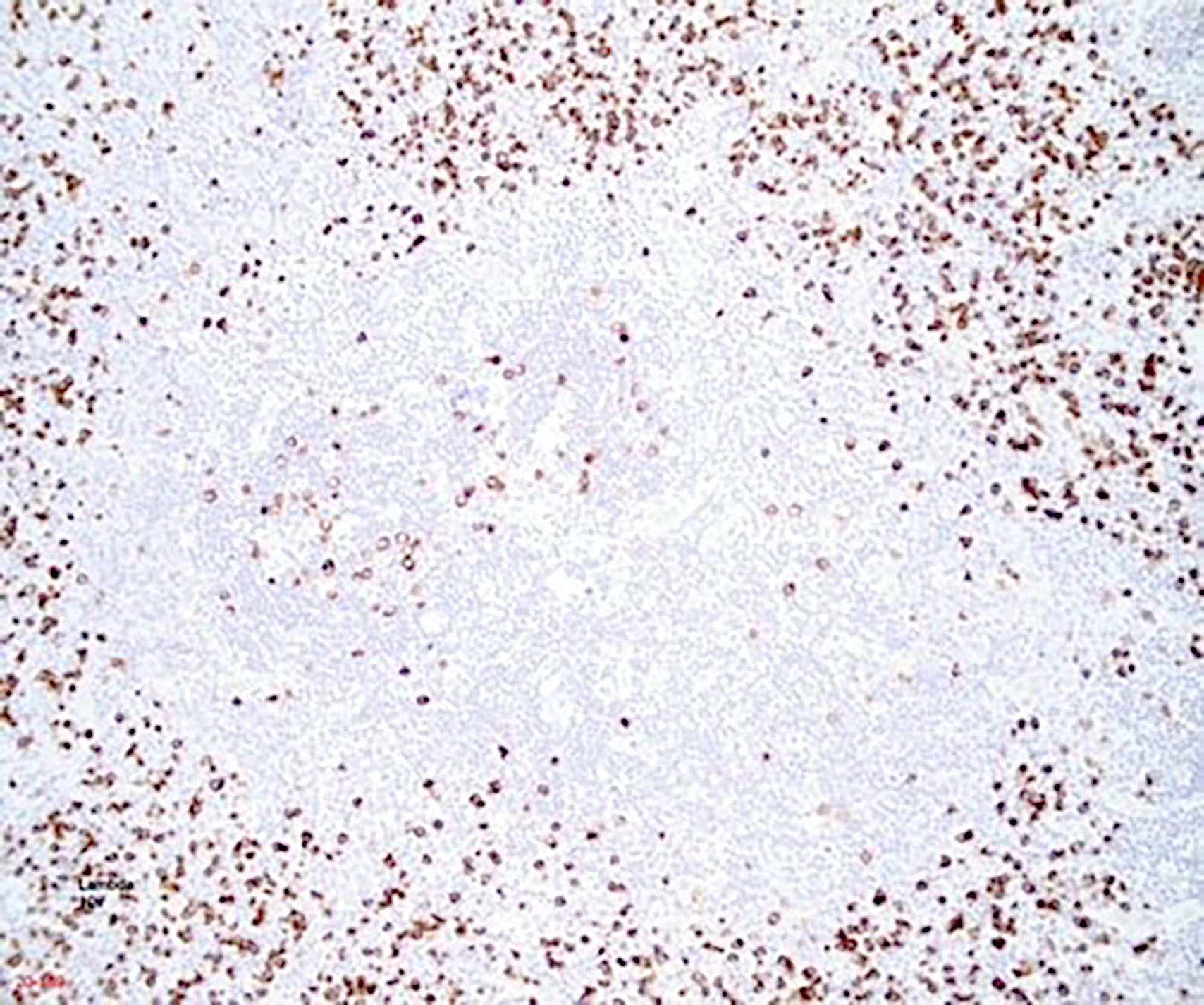
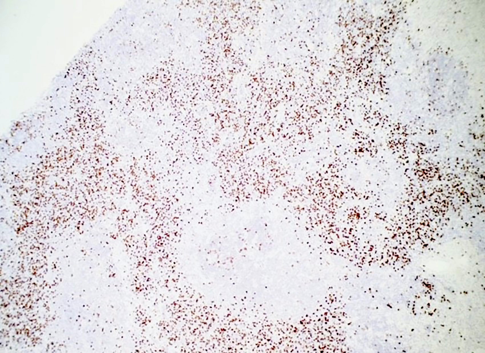
Lambda light chain predominance in plasma cells
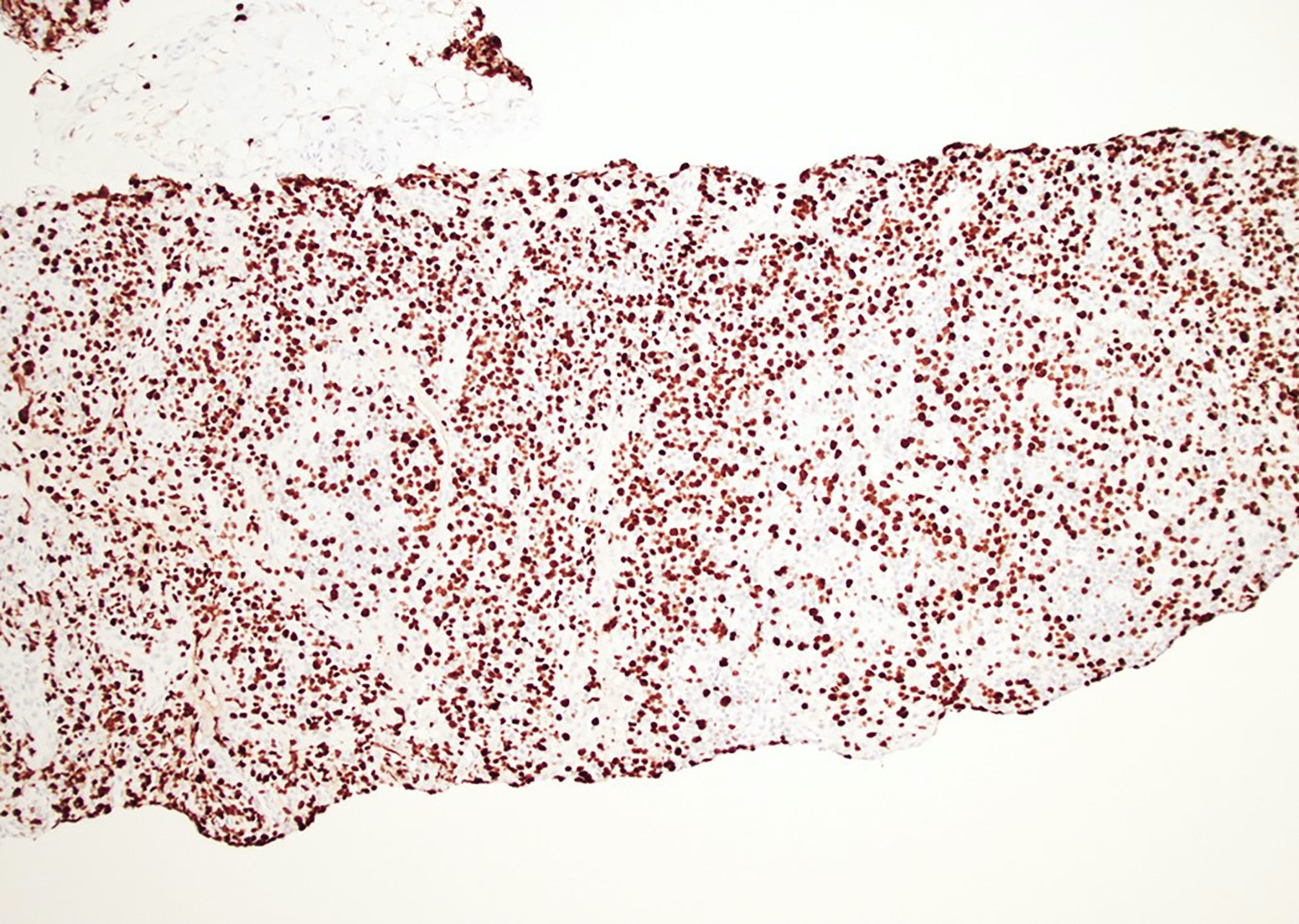
Increased proliferation in interfollicular areas, Ki67
Images hosted on other servers:

Algorithm for cat scratch disease diagnosis
Images hosted on other servers:

Retroauricular lymph node enlargement

Cervical lymphadenopathy and primary lesion
Contributed by Elizabeth Courville, M.D. and Mark R. Wick, M.D.
Suppurative granulomas

Palisading histiocytes
Lymph node biopsy
Steiner stain
Images hosted on other servers:

Lymphadenopathy
AFIP images

CML

With focal blastic transformation in liver

Involving spleen
Images hosted on other servers:

Metaphase FISH showing Philadelphia chromosome
AFIP images

Deciduosis
Images hosted on other servers:

Moderately enlarged inguinal lymphadenopathy

18FDG avid lymphadenopathy

18FDG PET / CT, transaxial PET / CT
Images hosted on other servers:

Cervical lymphadenopathy on examination
Contributed by Ingrid Tam, M.D., M.Sc., Emina Emilia Torlakovic, M.D., Ph.D. and Nikhil Sangle, M.D. (Case #396)
Vaguely nodular pattern
Polymorphous paracortical population
Cytological features, cytoplasmic pigment
Mild dermatopathic lymphadenitis
Paracortical expansion
Melanin pigment
Low Ki67 proliferative index

CD1a positive Langerhans cells
S100 positive dendritic cells
Images hosted on other servers:

Liver mass on MRI
Contributed by Elaine S. Jaffe, M.D.

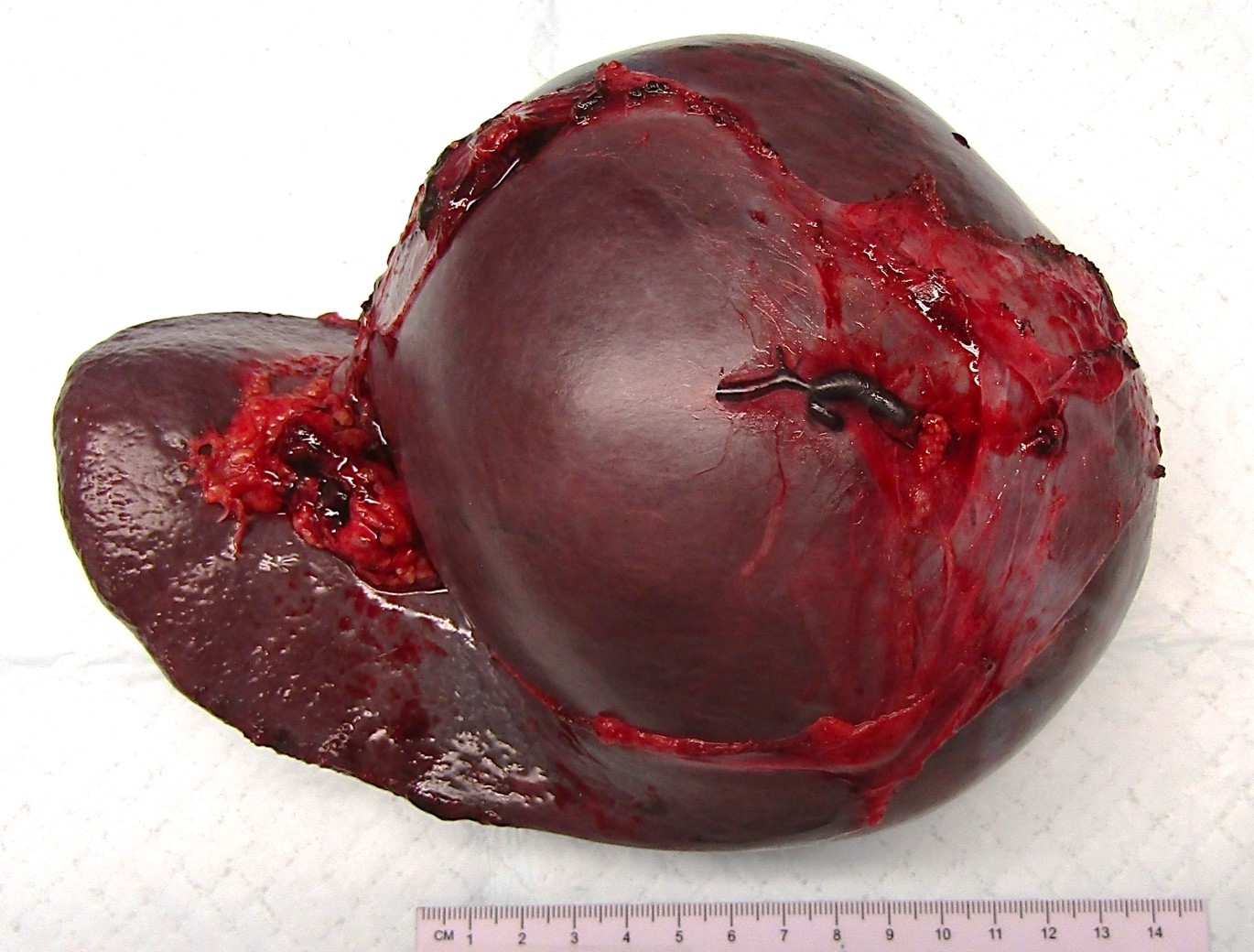
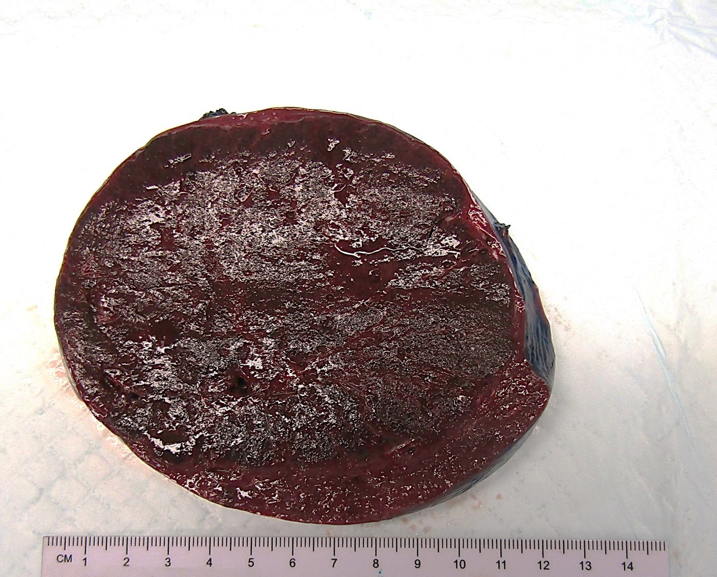
Splenic mass
Images hosted on other servers:

Solitary splenic nodule

Pancreatic nodule
Contributed by Elaine S. Jaffe, M.D., Shunyou Gong, M.D., Ph.D. and João Víctor Alves de Castro, M.D.

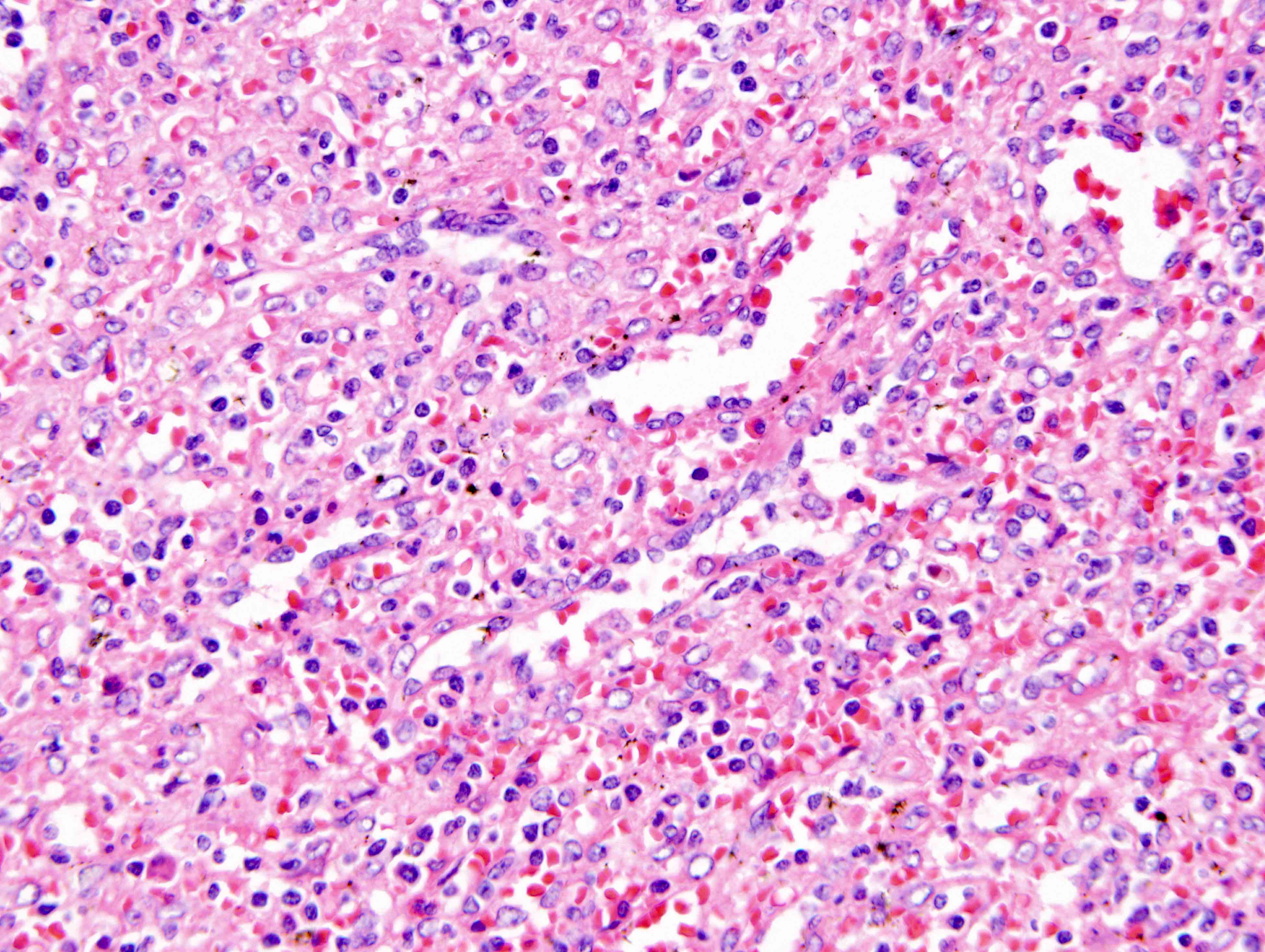
Spindle cells in an inflammatory background
Cytologic features and background
Fibrinoid necrosis

Colonic lesion

Interface between spleen and tumor
Vascular damage and granulomas
Inflammatory background obscuring neoplastic cells
SMA IHC
Double staining for SMA / EBER
Images hosted on other servers:

Syncytial group of tumor cells
Contributed by João Víctor Alves de Castro, M.D. and Elaine S. Jaffe, M.D.
EBER ISH

EBER positive spindle cells


AFIP images
inclusion in axillary
lymph node
Images hosted on other servers:

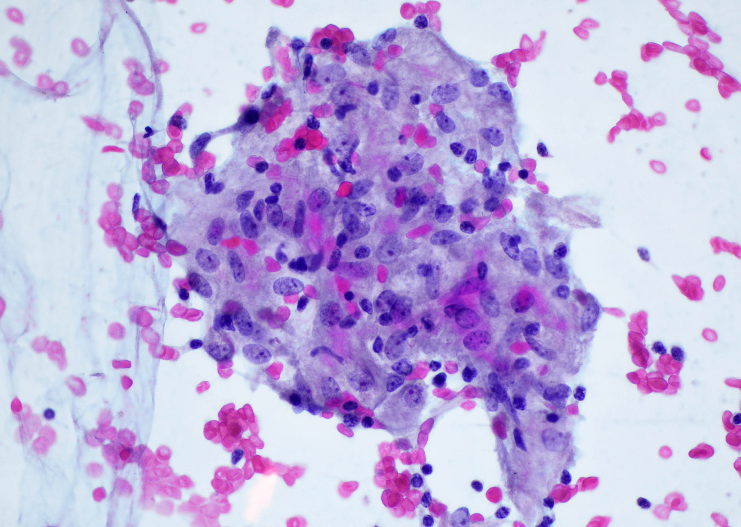
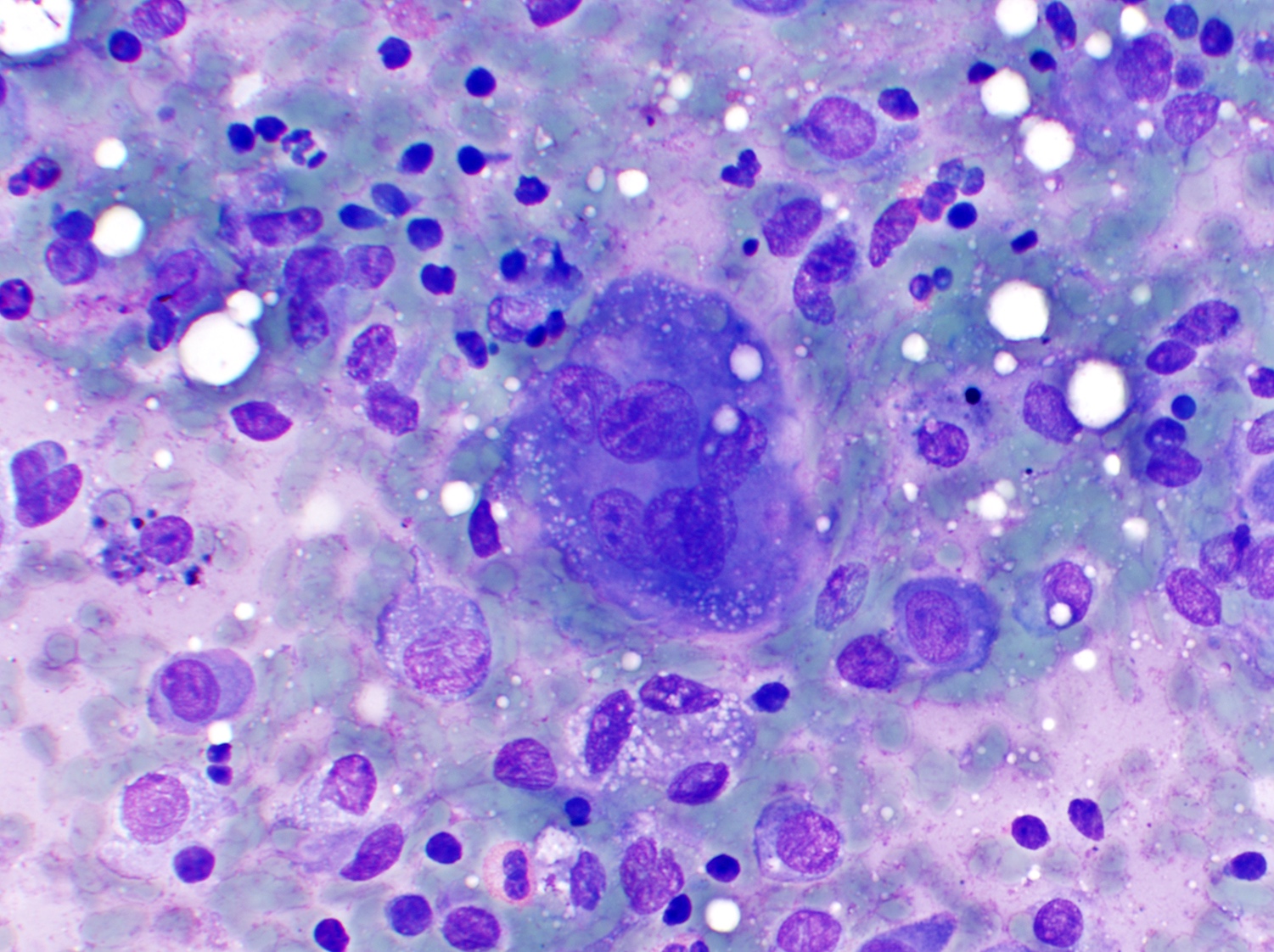
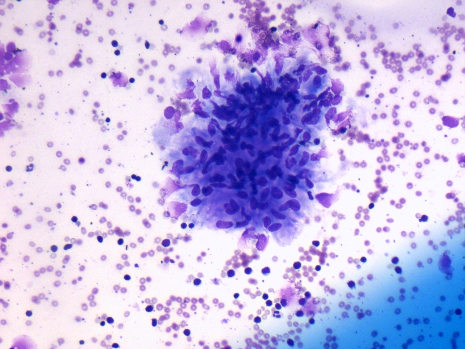
Wright stain
Images hosted on other servers:

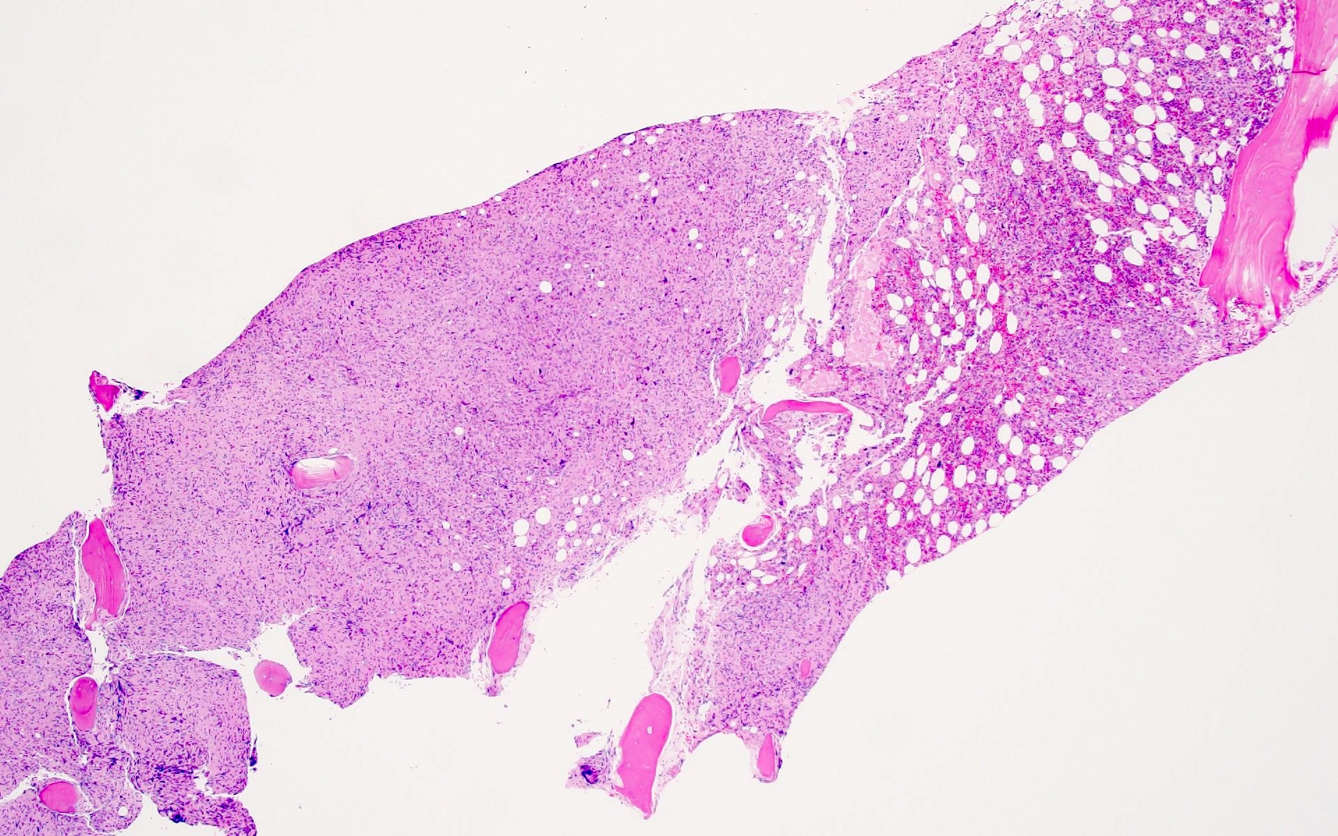
Small bowel and liver tumor

Cystic neck lesion

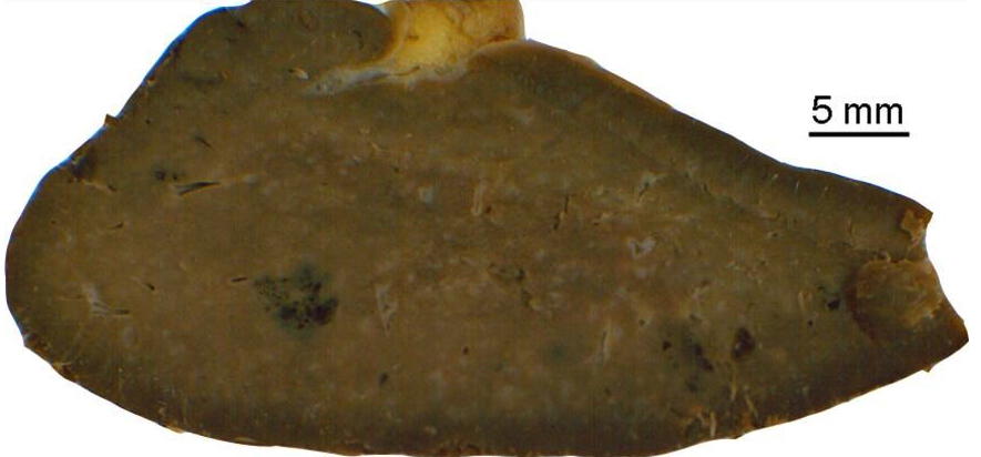
Liver mass
Images hosted on other servers:

Neck swelling

Paraneoplastic pemphigus
Images hosted on other servers:

Mesentery tumor

Small bowel and liver tumor

Well circumscribed neck tumor

Liver tumor
Contributed by Aishwarya Ravindran, M.D. and Karen L. Rech, M.D.
Storiform growth

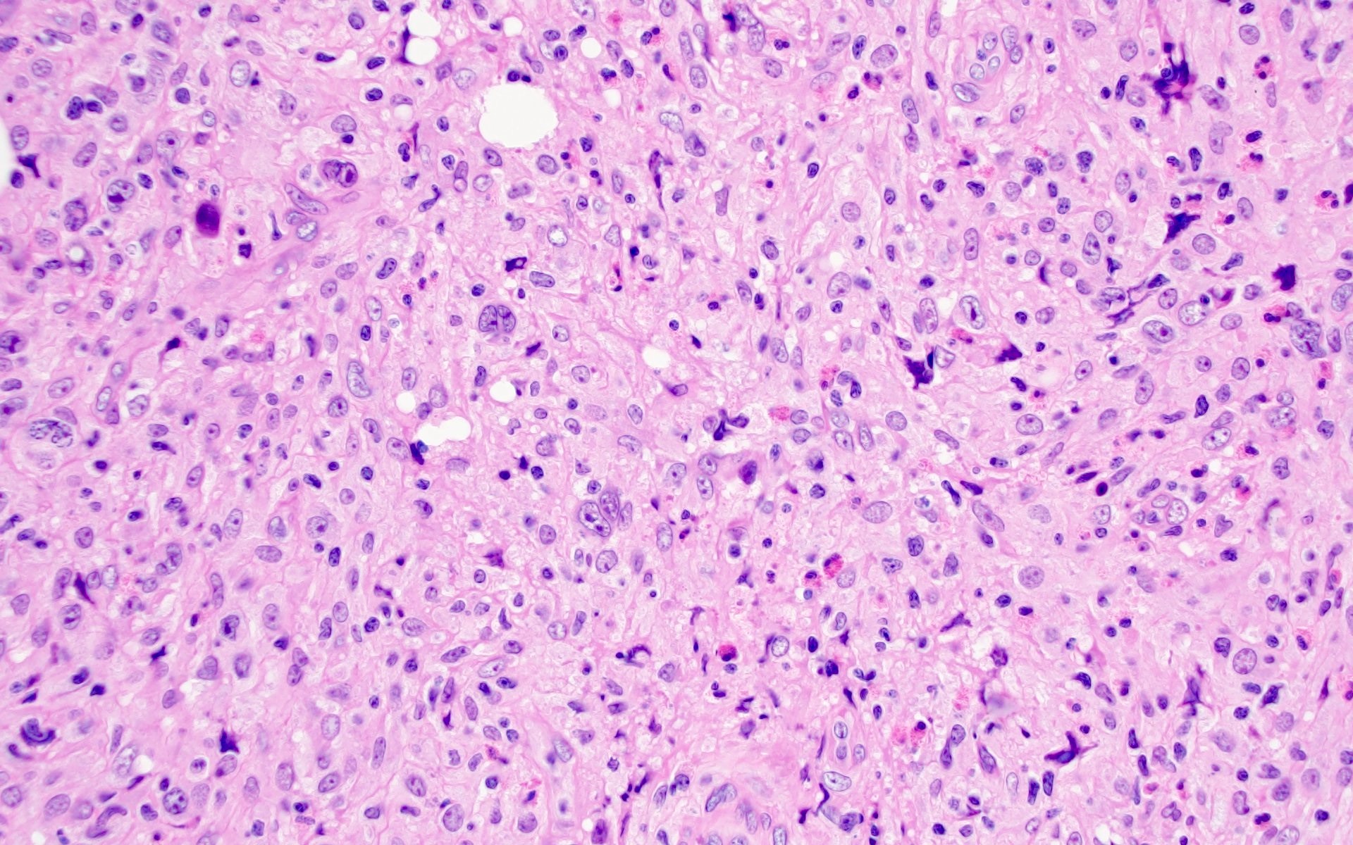
Ovoid cells mixed with lymphocytes
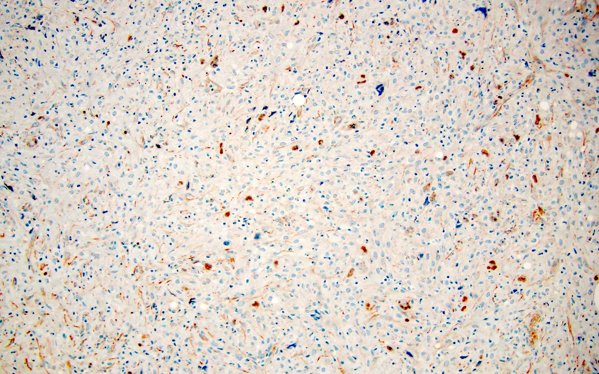
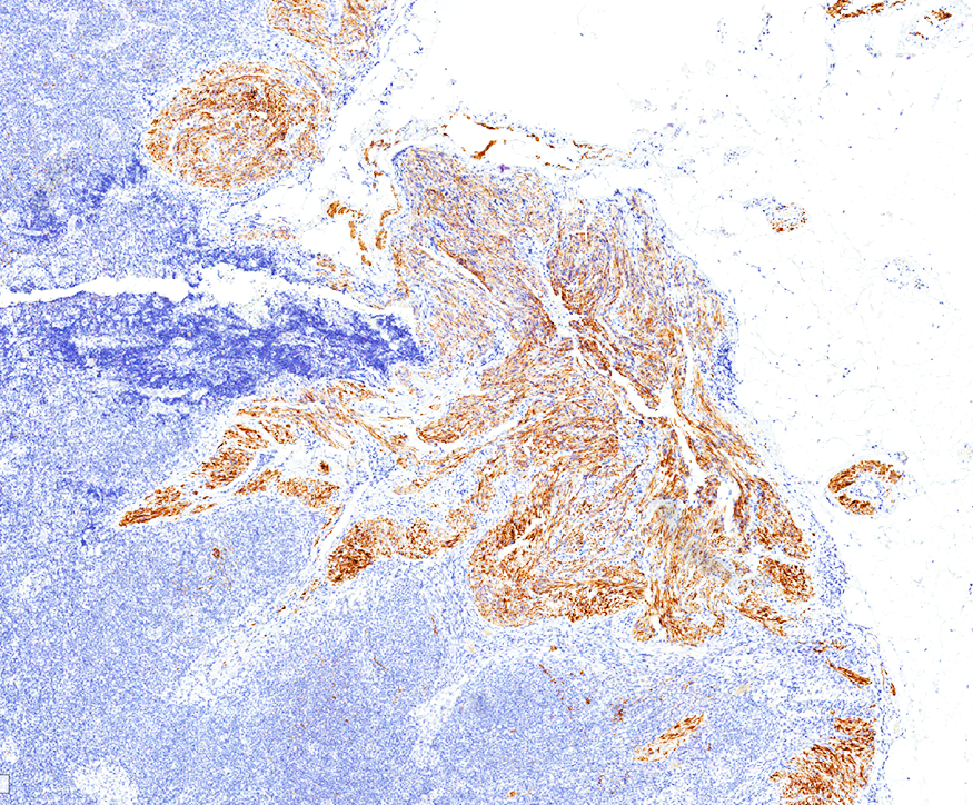
CD21
CD35
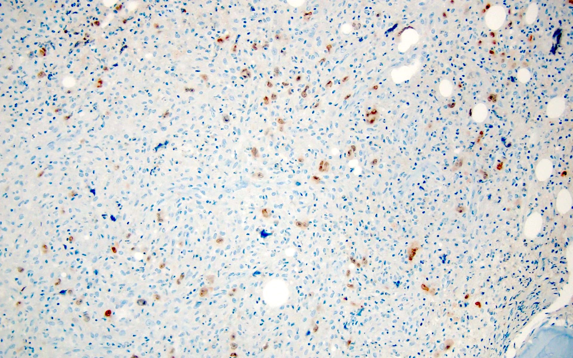
Clusterin

CXCL13
CD3
TdT
Images hosted on other servers:

Normal lymph node
Images hosted on other servers:

Lymphadenopathy
Contributed by Mariah Ferrier, M.S., PA (ASCP)
Tan-pink homogenous surface
Contributed by Jack Reid, M.D. and Sherif A. Rezk, M.D.
Hyperplastic lymphoid follicles
Polarization of germinal centers
Hyperplastic lymphoid follicles
HIV positive individual
Images hosted on other servers:

Polymorphous lymphocytes
Images hosted on other servers:

Analysis of CD38
Histopathology lymph node: follicular hyperplasia
Contributed by Ryan Hickey, PA (ASCP)
Inguinal lymph node with necrotizing granuloma
Contributed by Simon A. Backer, M.D.
Necrotizing granuloma in a cervical lymph node
Infectious (suppurative) granulomatous lymphadenitis
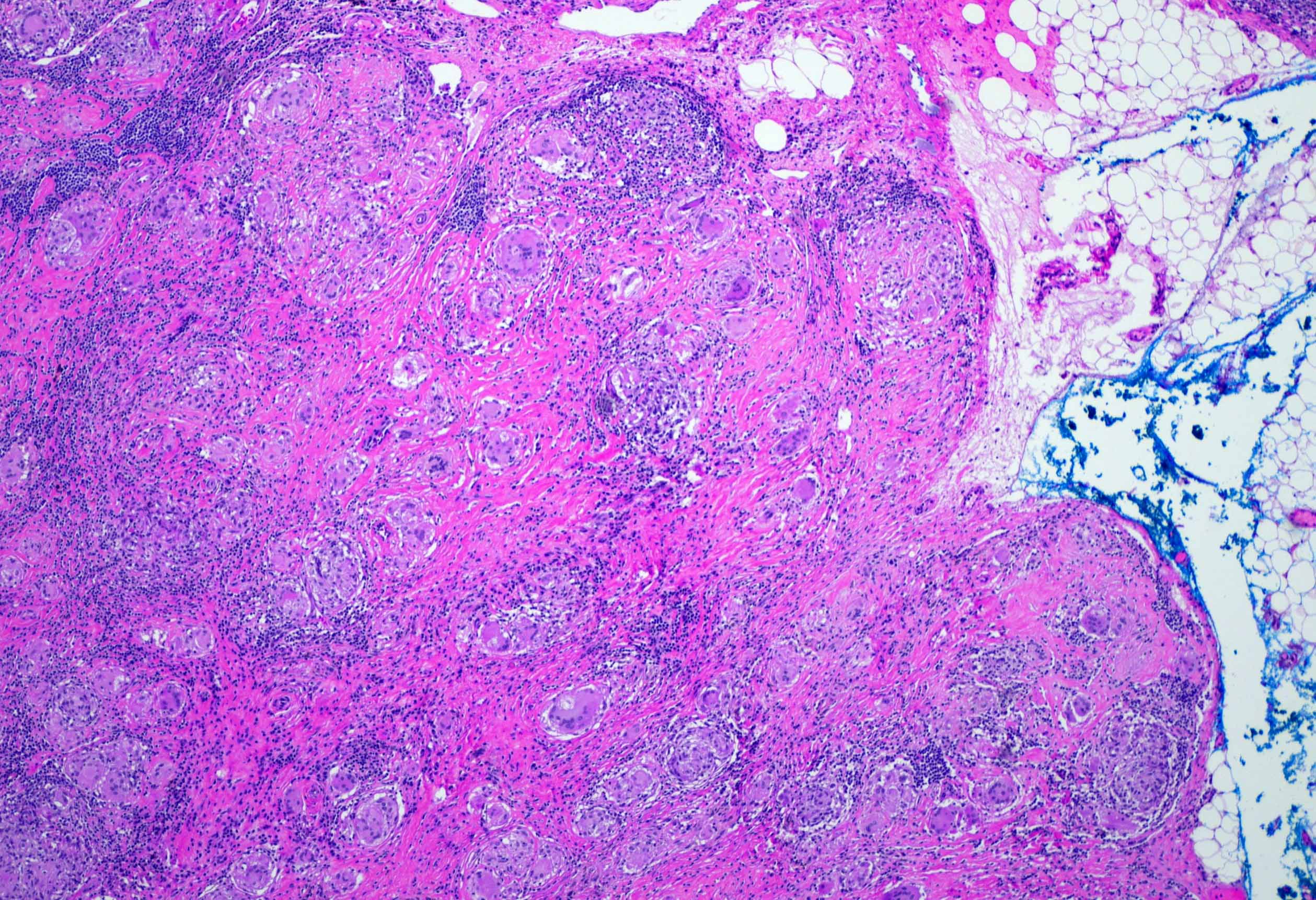
Sarcoid granuloma
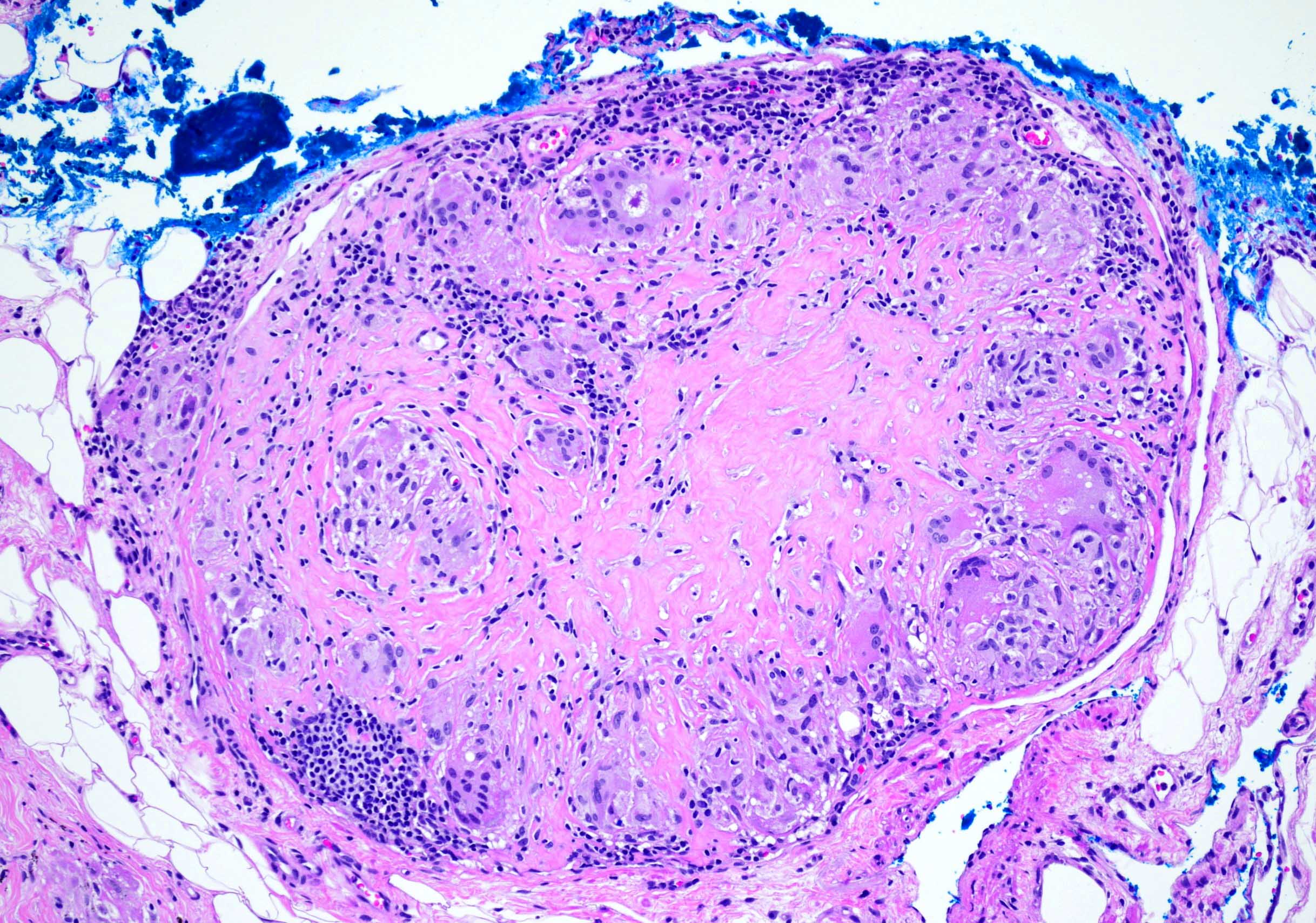
Sarcoid granuloma with asteroid body
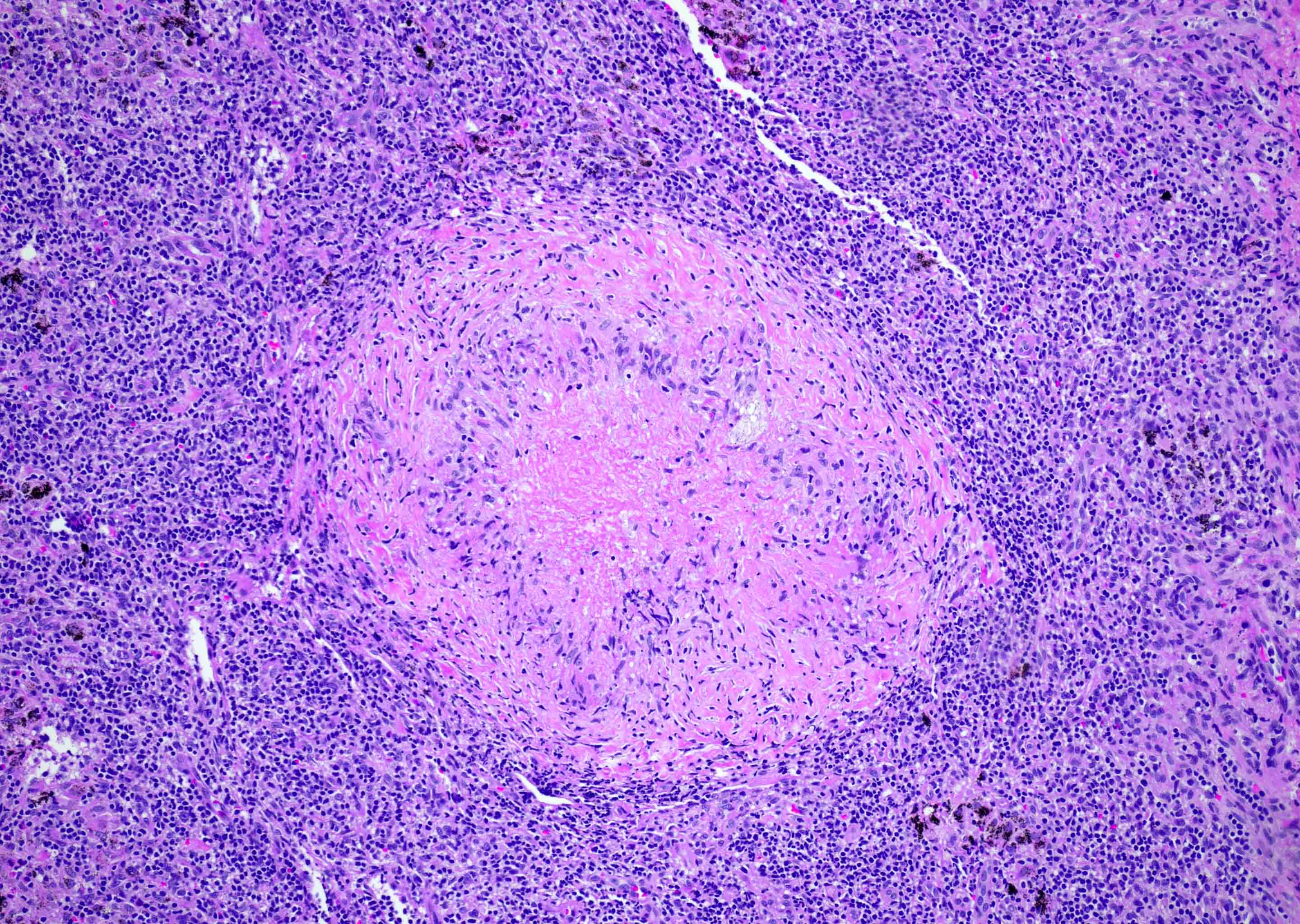
Necrotizing granuloma with palisading histiocytes
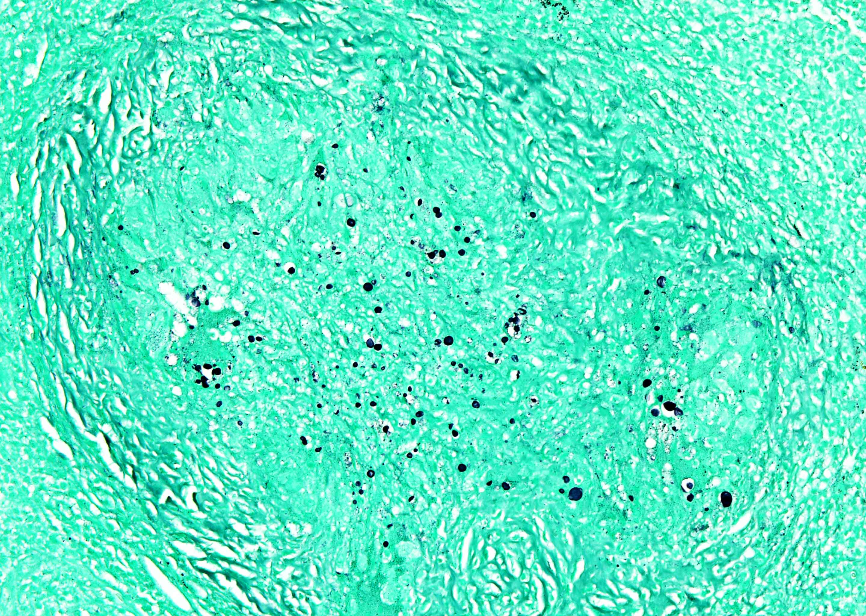
GMS with fungal forms in necrotizing granuloma
Contributed by Simon A. Backer, M.D.
Epithelioid granuloma
Images hosted on other servers:

Heterogenous enhancing splenic lesion on CT

Color Doppler showing dotted blood flow signals in mass

T1 and T2 weighted MRI of splenic mass

Isointense mass
Contributed by Nadine S. Aguilera, M.D. and Emily Gardner, M.D.
Solitary well circumscribed mass
Hamartoma with similar texture and color as the spleen
Images hosted on other servers:

Nodule of increased stiffness

Resected spleen
Contributed by Nadine S. Aguilera, M.D.

Splenic hamartoma and uninvolved spleen
Red pulp

CD8 stain

CD34 stain
CD21 stain
CD68 stain
Images hosted on other servers:

Giant splenic hemangioma

Cavernous splenic hemangioma

Splenomegaly and hemangiomas
AFIP images
Hemangioma

Capillary / cavernous hemangioma

Epithelioid hemangioma
Lobular capillary hemangioma
Cellular hemangioma
Images hosted on other servers:

Scatter diagram of CBC
Images hosted on other servers:

Small spherocytes
Contributed by Zenggang Pan, M.D., Ph.D.

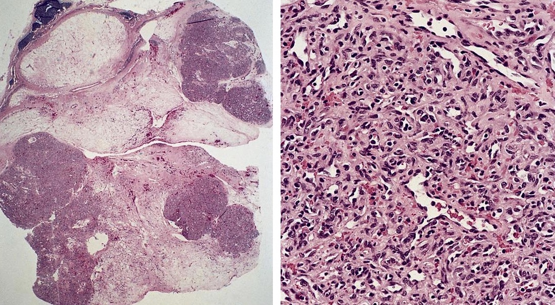
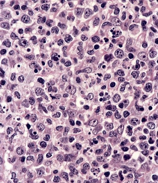
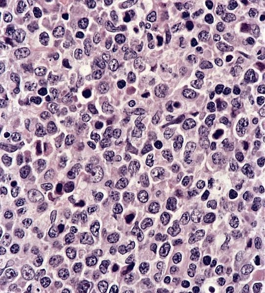
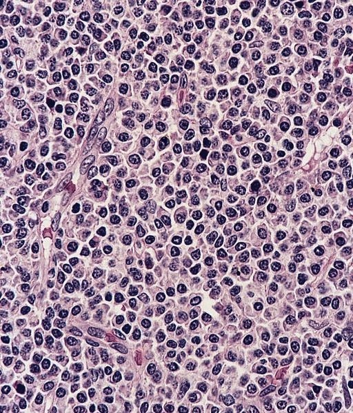
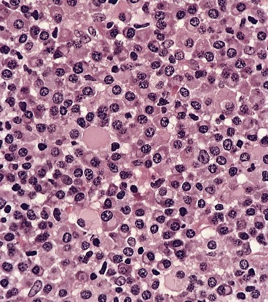
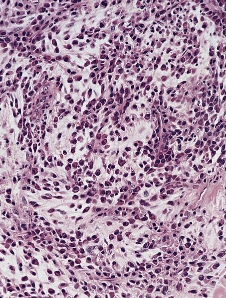
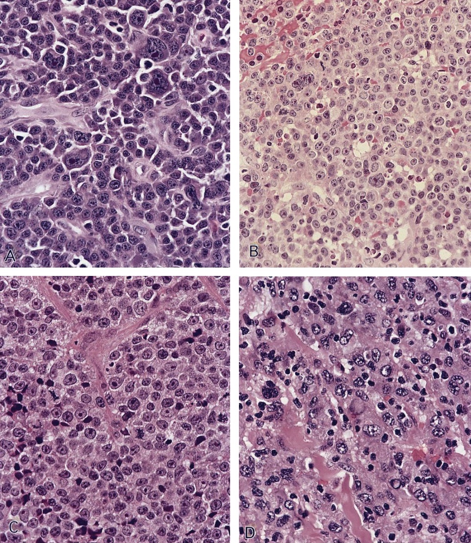
Nodal histiocytic sarcoma
AFIP images

True histiocytic lymphoma
True histiocytic lymphoma with prominent erythrophagocytosis

CD45RB and CD68
S100
Contributed by Barina Aqil, M.D.
Florid follicular hyperplasia
Monocytoid cell hyperplasia
Follicular hyperplasia, EBV+ in HIV setting
Florid follicular hyperplasia in CMV lymphadenitis
Monocytoid cell hyperplasia in CMV lymphadenitis
Paracortical hyperplasia in CMV lymphadenitis
CMV lymphadenitis
Mycobacterium avium intracellulare (MAI)
Kaposi sarcoma
Kaposi sarcoma
Burkitt lymphoma, EBV+ in HIV setting
Burkitt lymphoma, EBV+ in HIV setting
Burkitt lymphoma, EBV+ in HIV setting
Burkitt lymphoma, EBV+ in HIV setting
Classic Hodgkin lymphoma, EBV+ in HIV setting
Classic Hodgkin lymphoma, EBV+ in HIV setting
Images hosted on other servers:

Petechiae or small bruise-like markings
Contributed by Julia Geyer, M.D.

Focal germinal centers, hyperplastic marginal zones

Histiocytes, immunoblasts, neutrophils, macrophages
Expanded red pulp, atrophic white pulp
Cordal macrophages, phagocytosing platelets
Images hosted on other servers:

Erythematous papules on face and neck

Ill defined, erythematous subcutaneous nodule
Contributed by Ryanne A. Brown, M.D., M.B.A.
Dermal histiocytoid cells
Histiocytoid cells
CD68
CD1a
S100
Langerin
BRAF V600E
Contributed by Ryanne A. Brown, M.D., M.B.A.
No Birbeck granules
Contributed by Kunwar Singh, M.D. and Robert S. Ohgami, M.D., Ph.D.
Castleman disease
Follicular dendritic cell tumor
CD1a
CD3
CD4
CD8
CD99
TdT
Images hosted on other servers:

Grossly enlarged with haematoma
AFIP images

Interdigitating dendritic cell sarcoma
AFIP images
Various images from lymph node
Images hosted on other servers:
Axillary lymph node transformation from CLL / SLL
Contributed by Elizabeth Courville, M.D. and Amy Beckman, M.D.
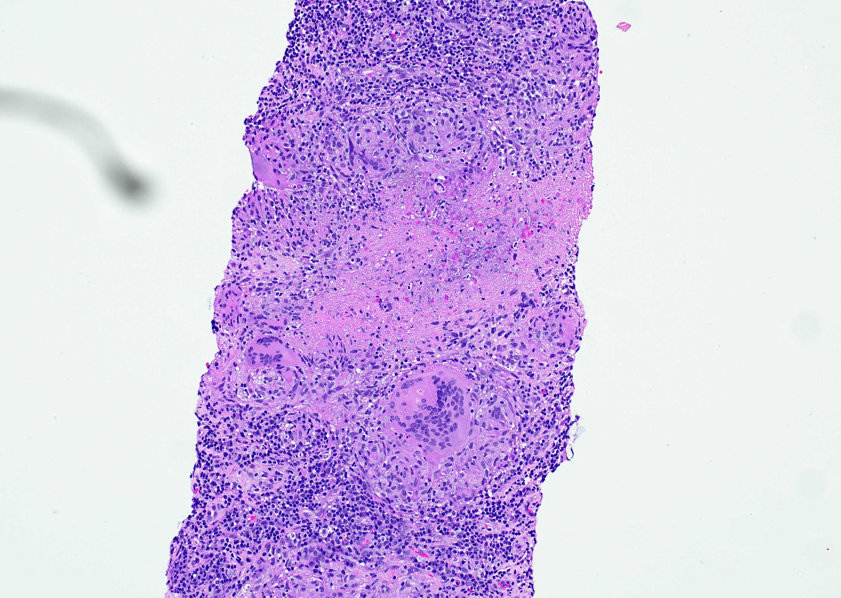
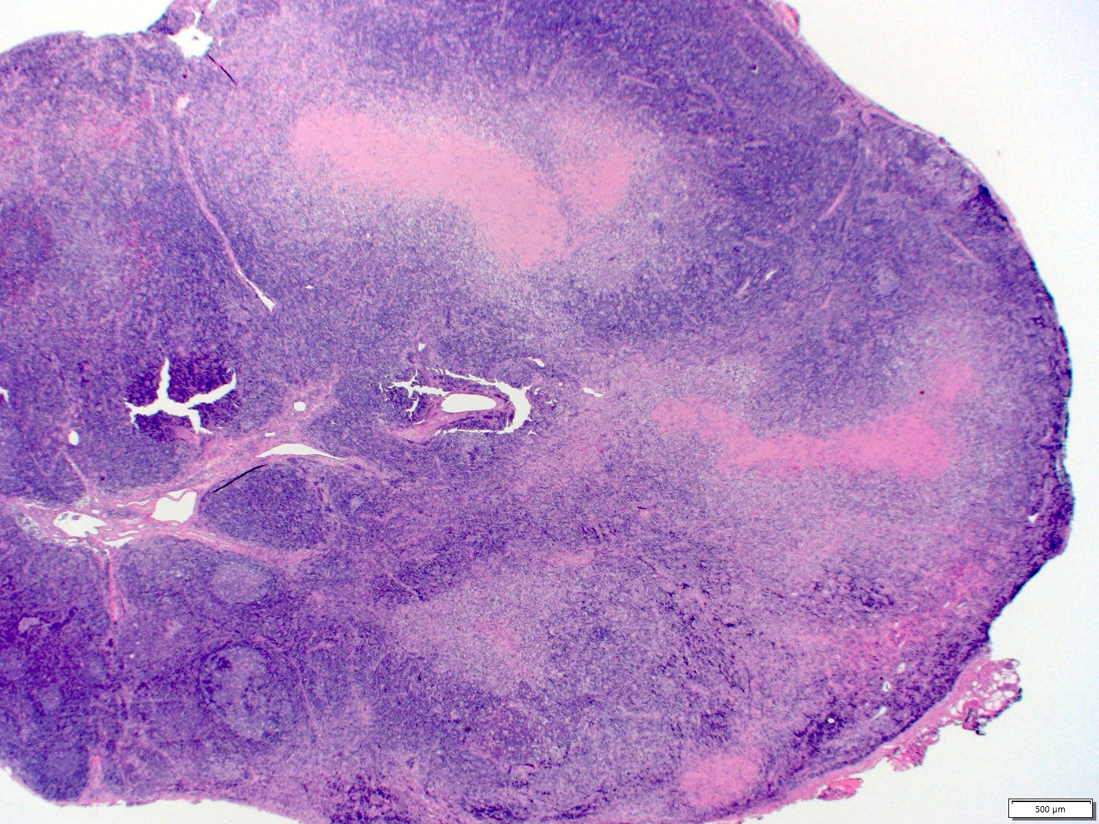
Lymph node from a 38 year old woman:
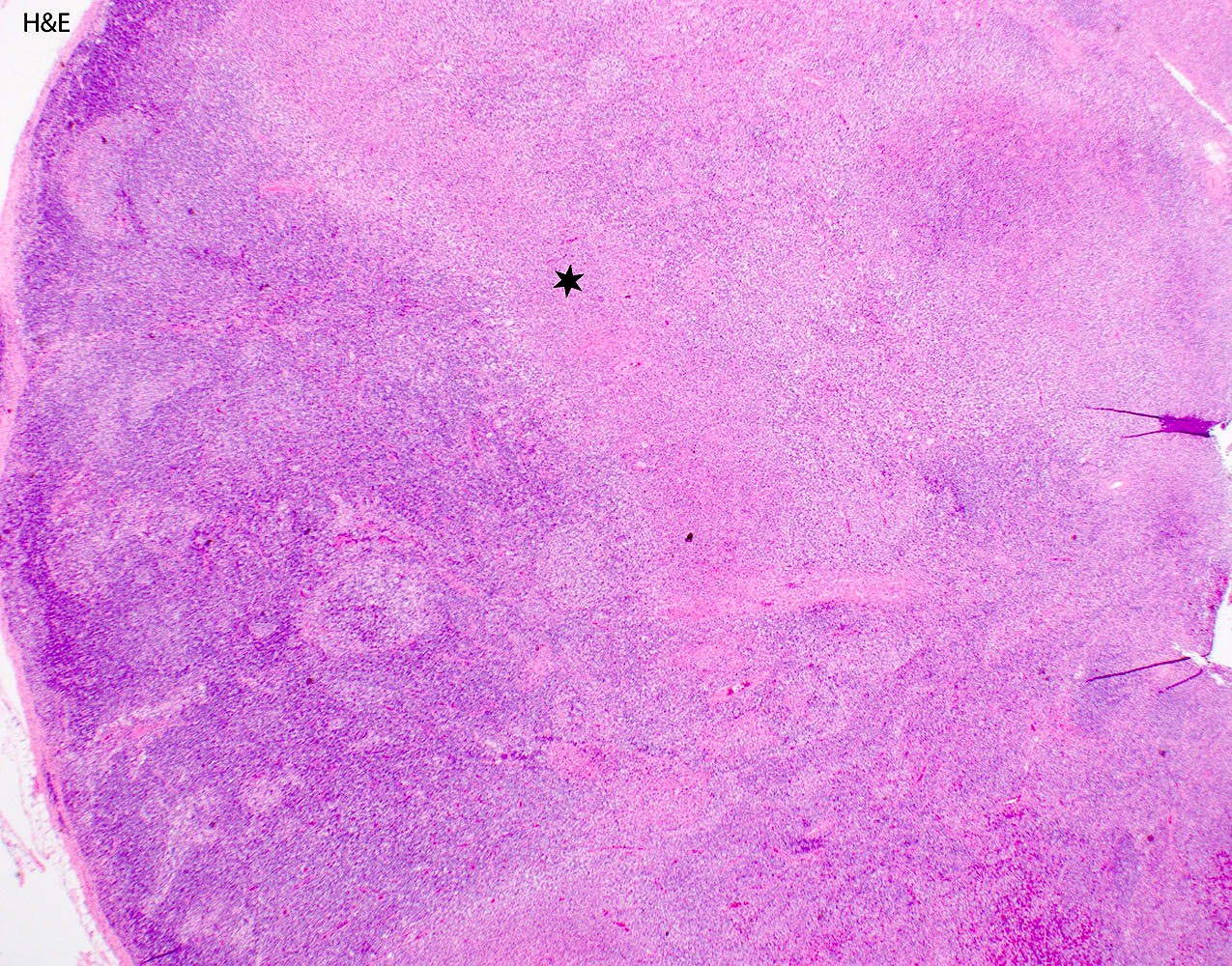
Distorted lymph node architecture
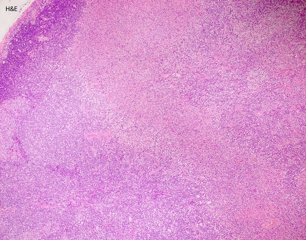
Pale staining areas

Karyorrhectic debris

Histiocytes and lymphocytes
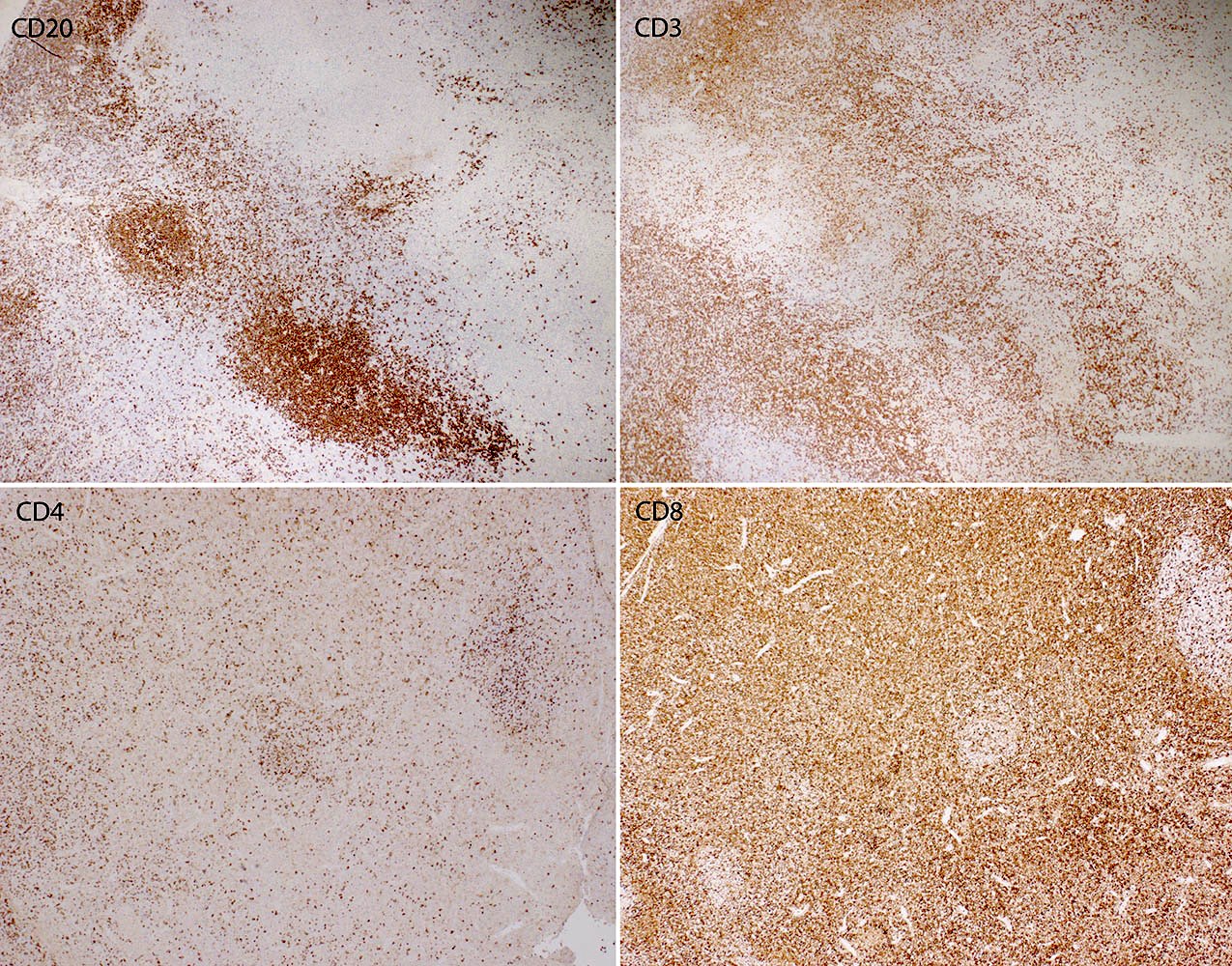
CD3, CD4, CD8, CD20
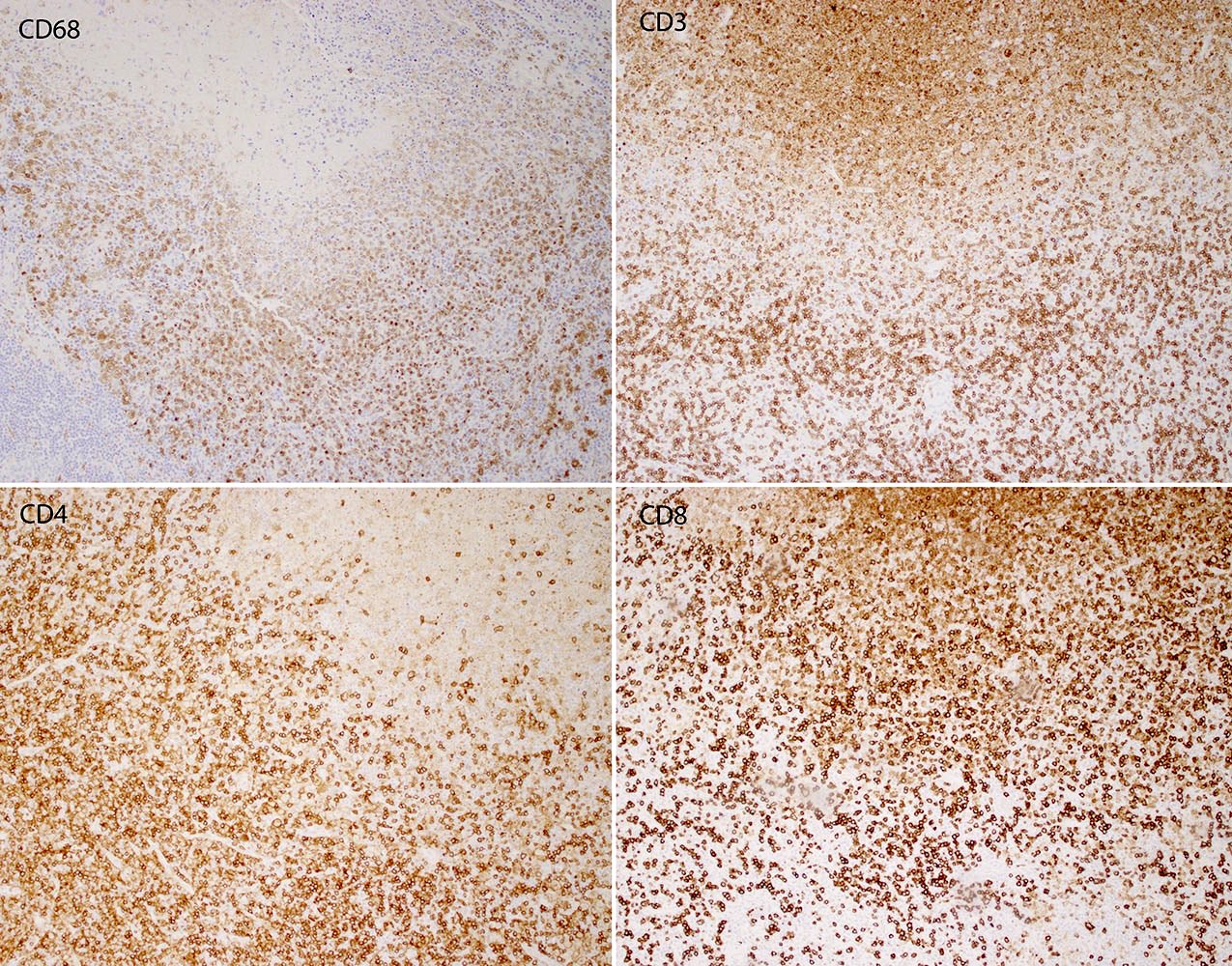
CD3, CD4, CD8, CD68
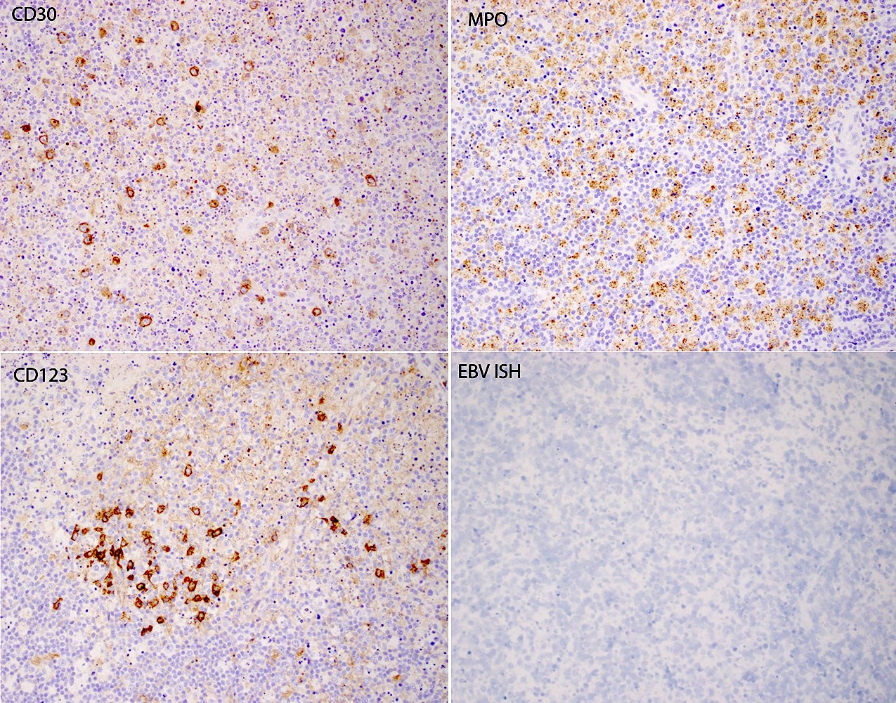
CD30, CD123, MPO, EBV ISH
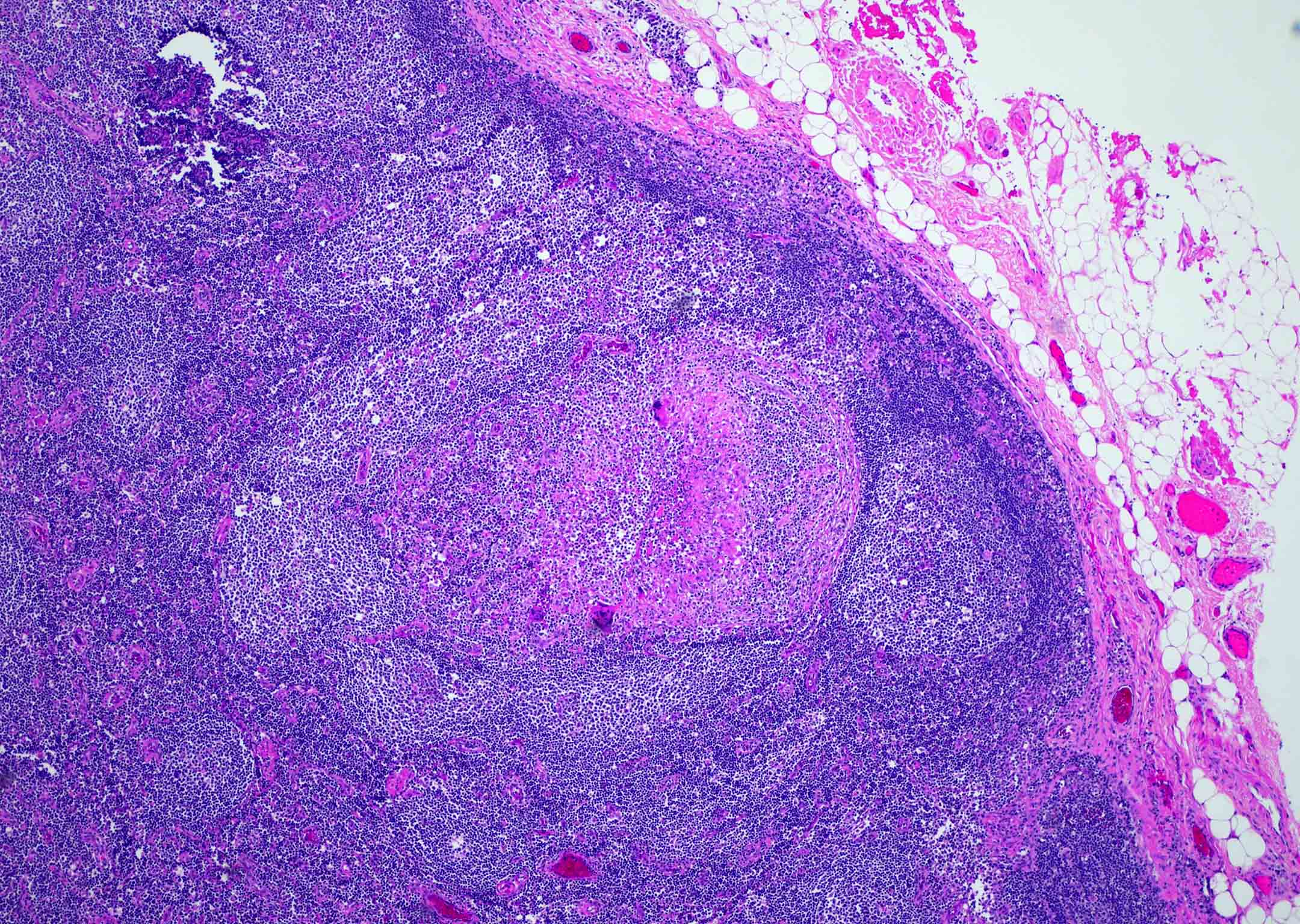
Cervical lymph node from a 40 year old woman:
Pale staining areas
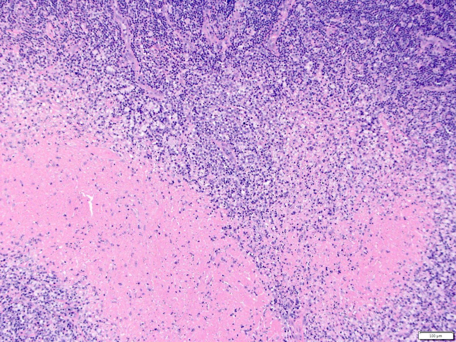
Necrosis with mononuclear infiltrate
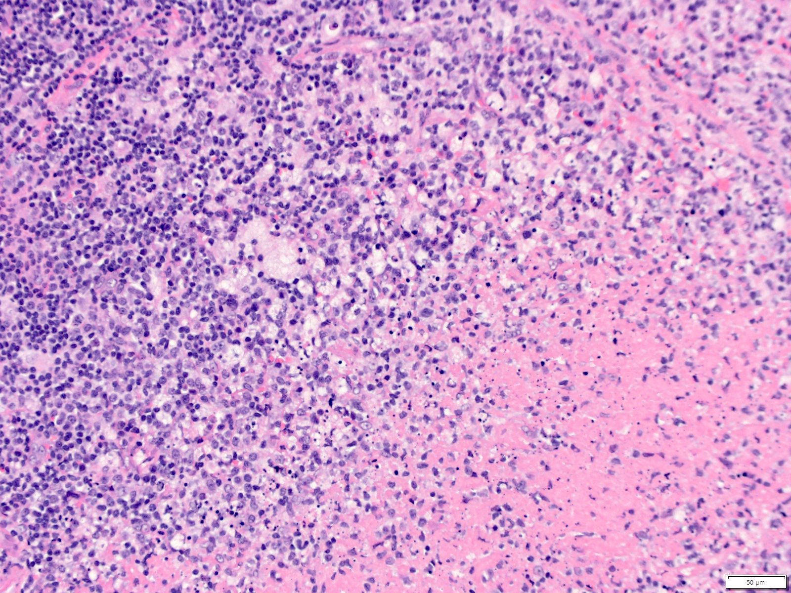
Karyorrhectic debris
Histiocytes
Lymph node
CD68

CD123
CD3
CD20
CD30
AFIP images
Kikuchi necrotizing lymphadenitis
Images hosted on other servers:

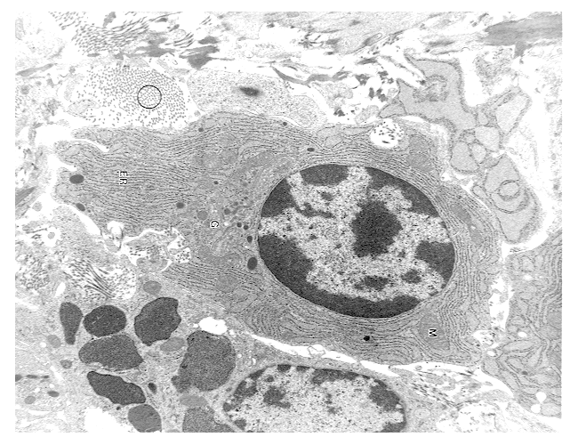
Histiocytes with myelin figures

#2 with tubuloreticular structures
Images hosted on other servers:

Ultrasound findings


CT and MRI findings
Images hosted on other servers:

Lesion on buttock
Contributed by Aishwarya Ravindran, M.B.B.S. and Julie Teruya-Feldstein, M.D.
Excisional biopsy of lymph node
Eosinophilic infiltrates
Eosinophilic infiltrates
Inflammatory milieu
Polykaryocytes of Warthin-Finkeldey type
Images hosted on other servers:

Eosinophils in a reactive background
Contributed by Vignesh Shanmugam, M.D.

Misguided myeloid / dendritic cell model
Contributed by Vignesh Shanmugam, M.D.

Sagittal view

Coronal view
Images hosted on other servers:

LCH
Contributed by Vignesh Shanmugam, M.D.
Preservation of follicle centers

Sinusoidal infiltration
Infiltrate of subcapsular sinus
Frequent admixed eosinophils
Prominent nuclear grooves
Admixed multinucleated giant cells
Langerin
CD1a
S100
Cyclin D1
Contributed by Catherine Hagen, M.D.
Langerhans cell histiocytosis

CD1a positive LCH
S100 positive LCH
AFIP images


With Hodgkin lymphoma
Involving spleen
Malignant
S100
Nuclear and cytoplasmic staining S100
Case #314
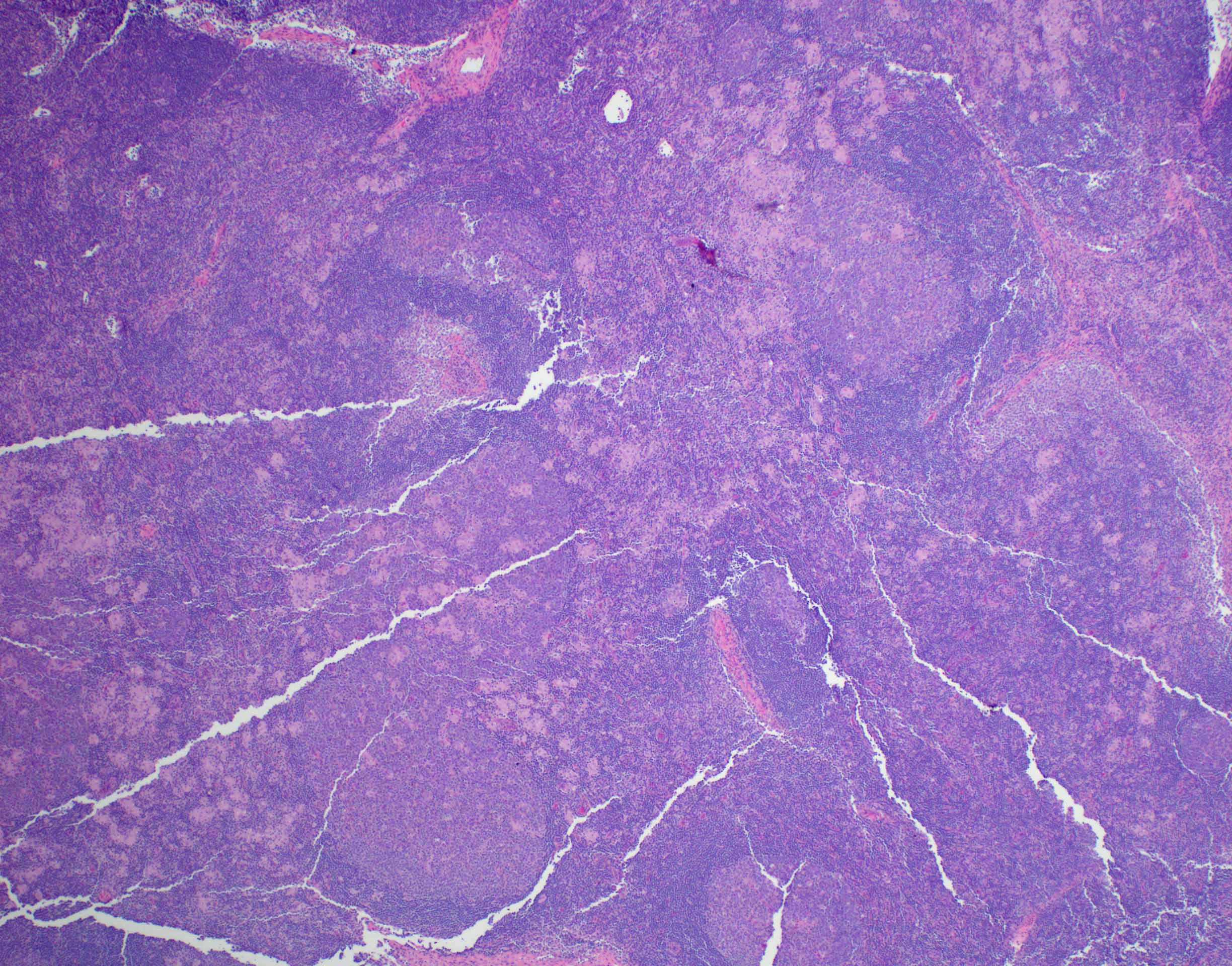
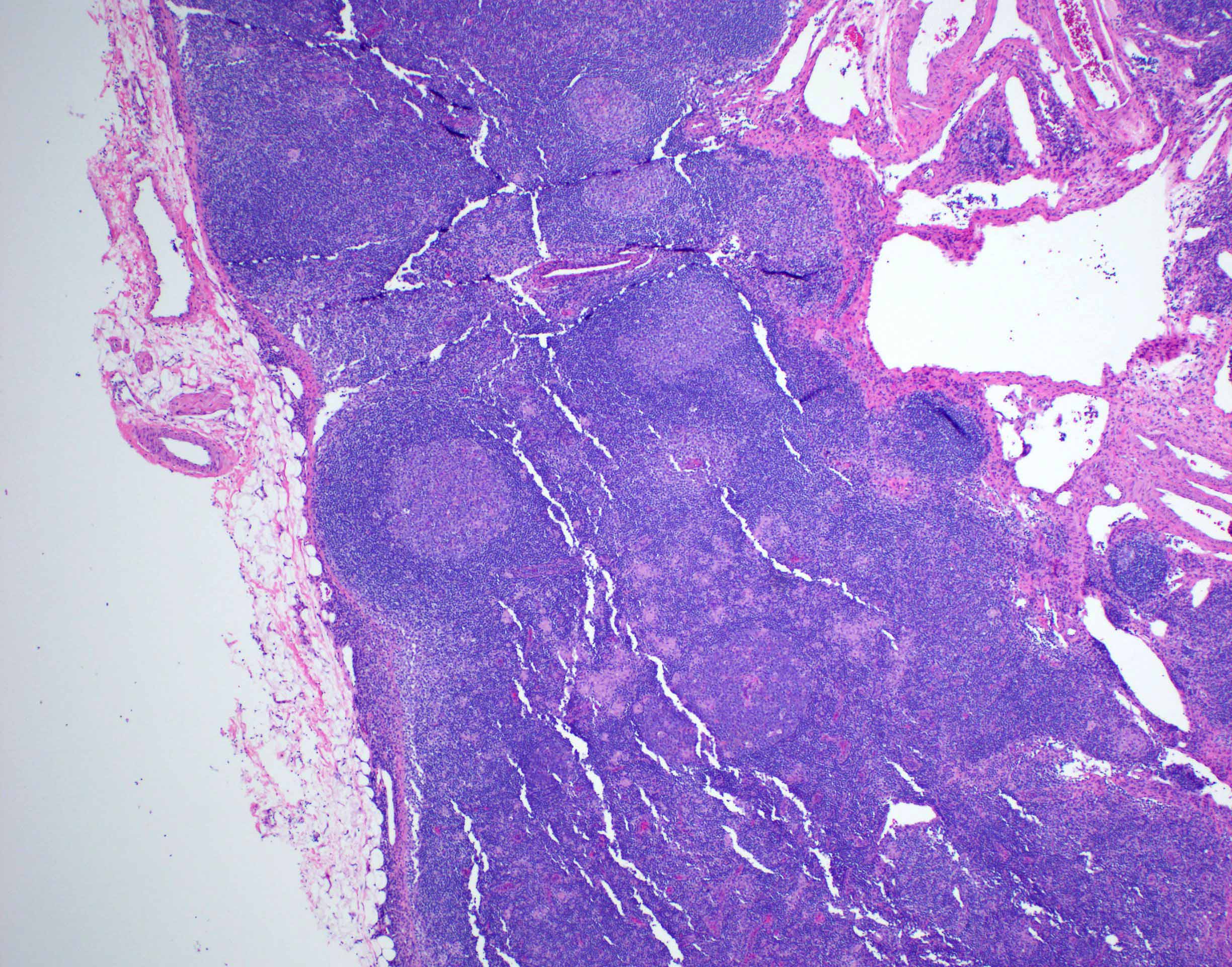
Diffuse involvement
Sinusoidal and pericapsular involvement
Pericapsular involvement
Admixed eosinophils
Nuclear grooves

Mitotic activity

CD1a
CD5
CD20
CD23
Langerin
S100
Contributed by Vignesh Shanmugam, M.D.

Prominent nuclear grooves and admixed eosinophils
Scattered multinucleated giant cells
AFIP images
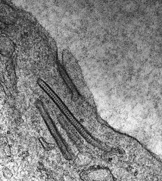
Birbeck granules
Images hosted on other servers:

Birbeck granules
Images hosted on other servers:

PAS

Black granules - Orcein

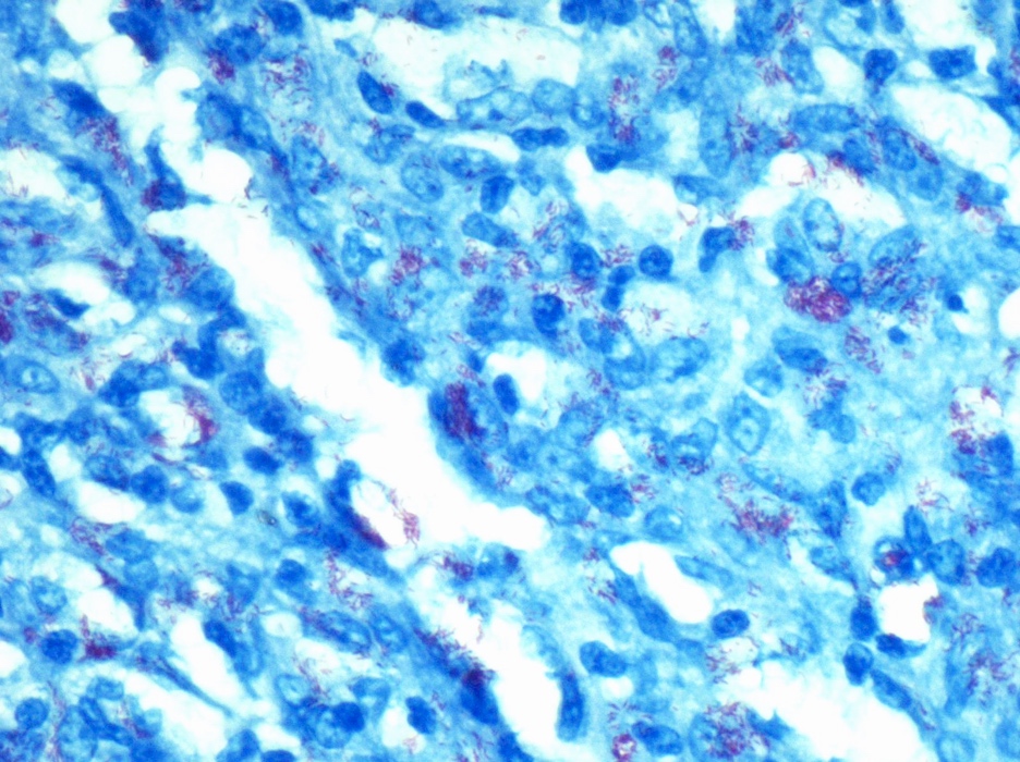
L: expansion of medullary sinuses; R: Long Ziehl-Neelsen stain


Contributed by Patricia Tsang, M.D., M.B.A.
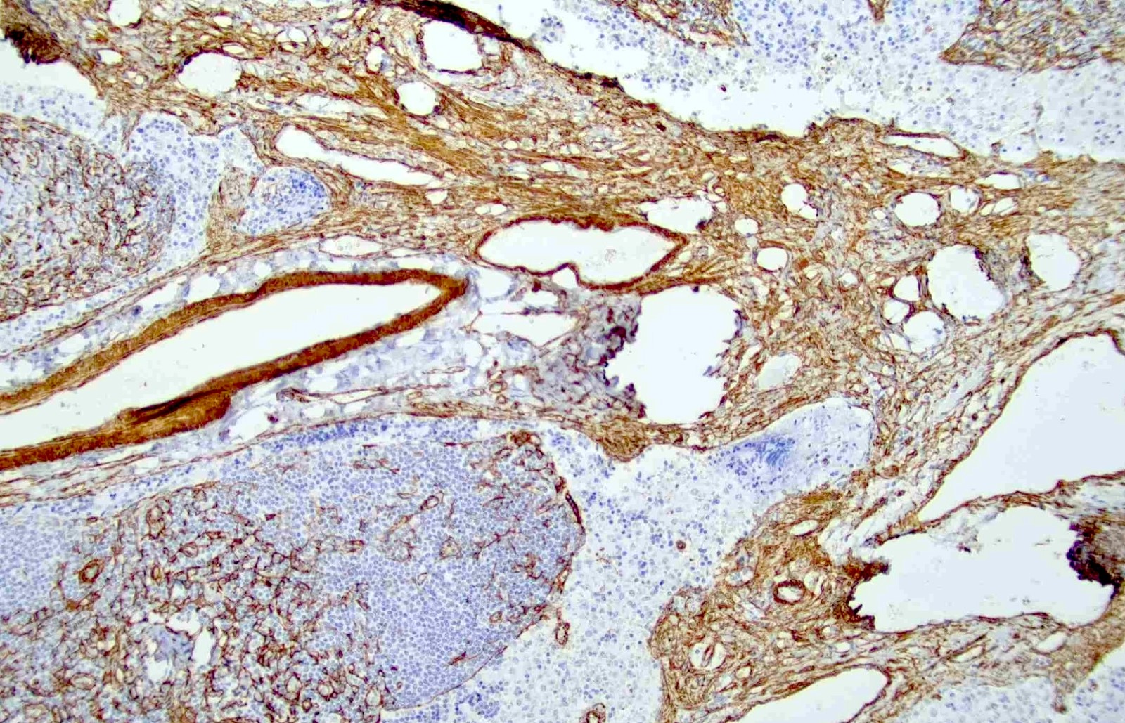
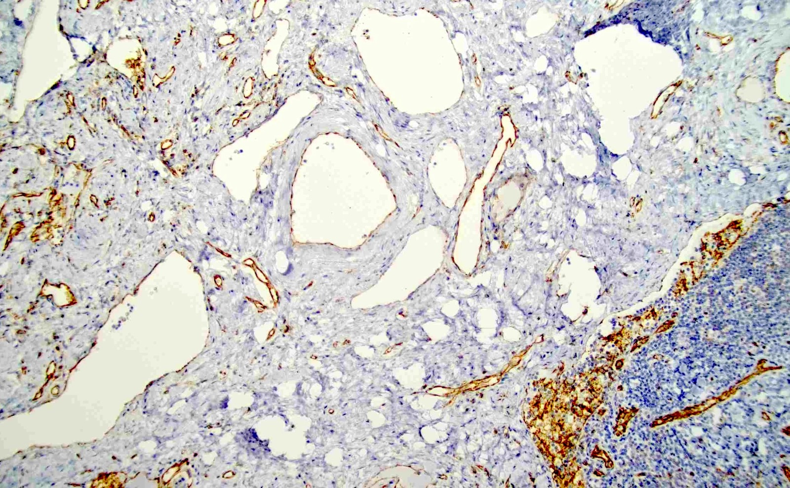
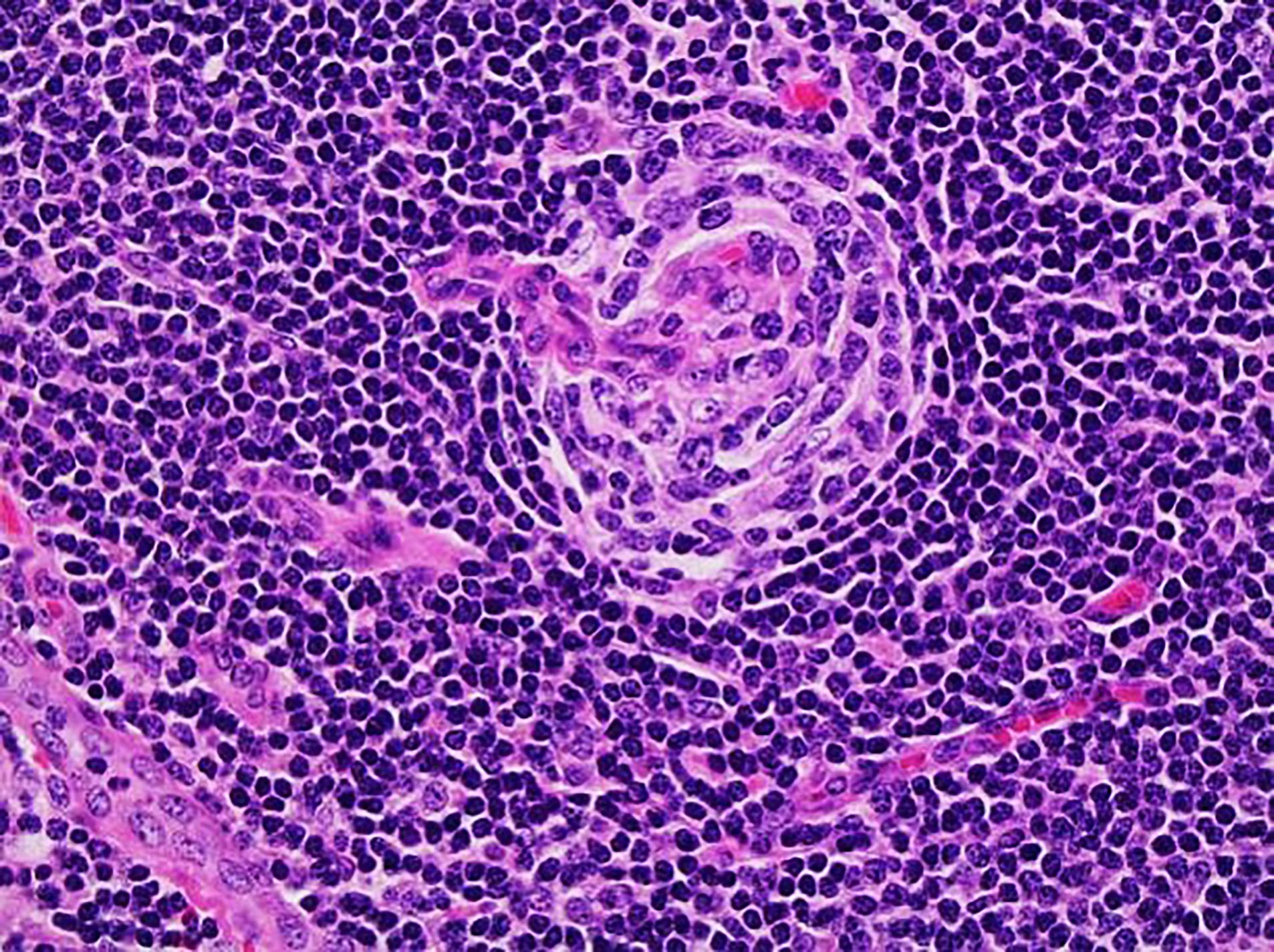
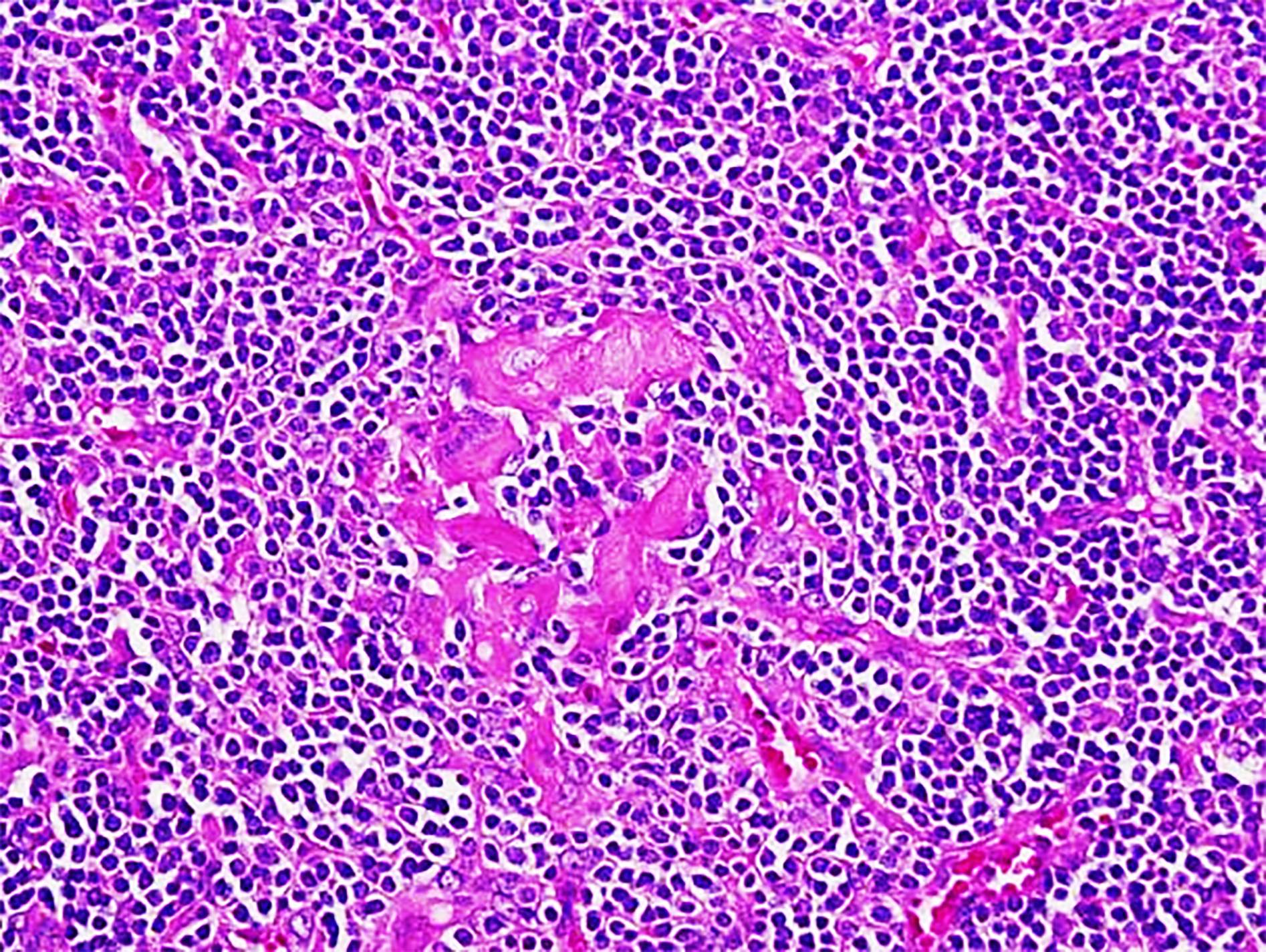
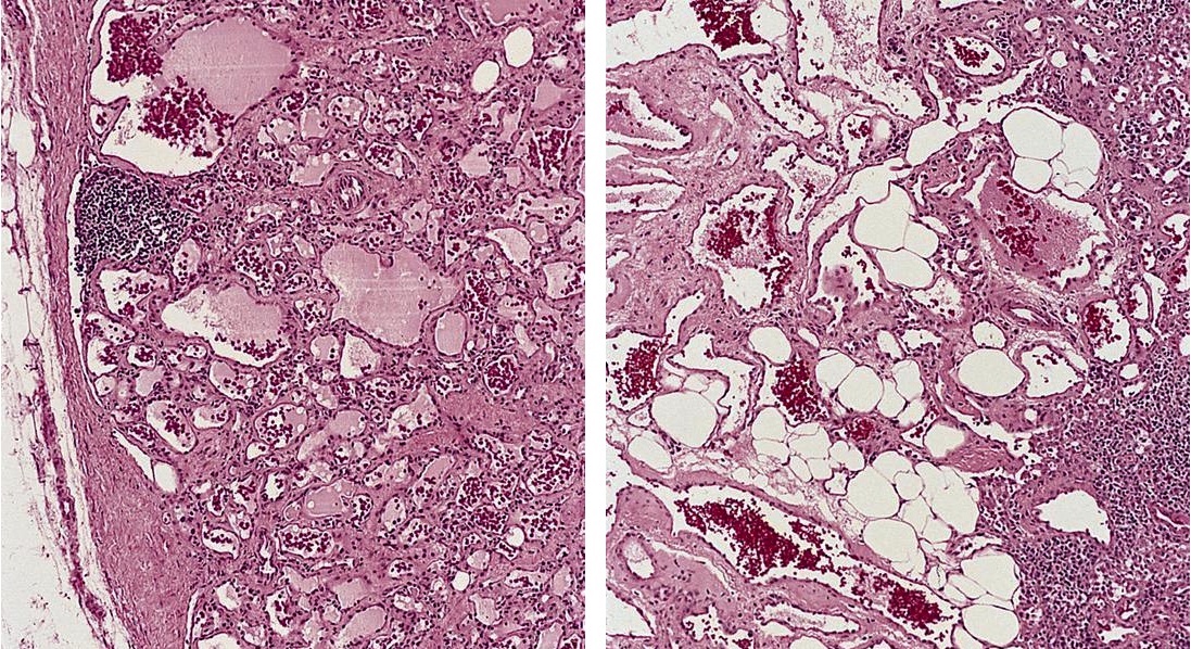
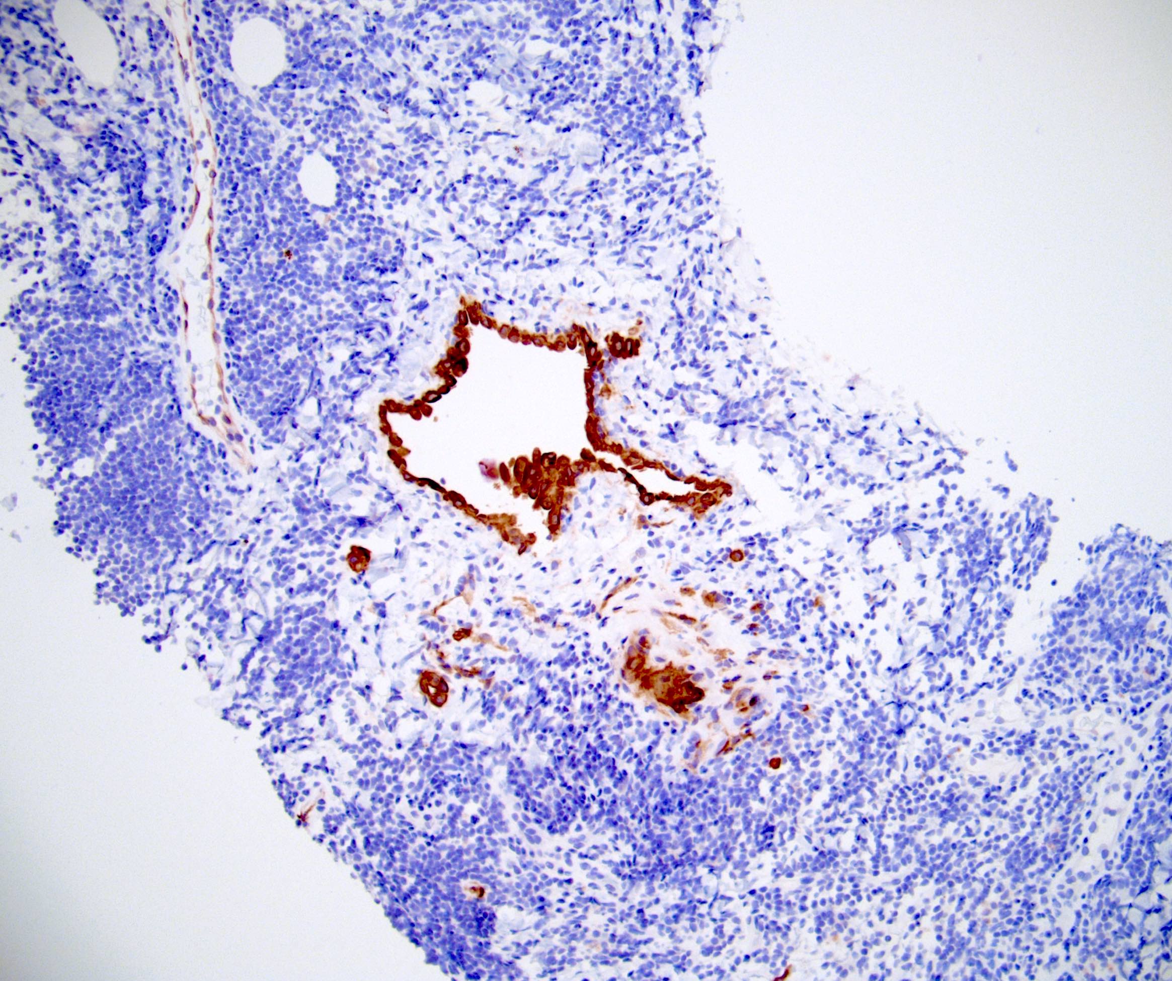
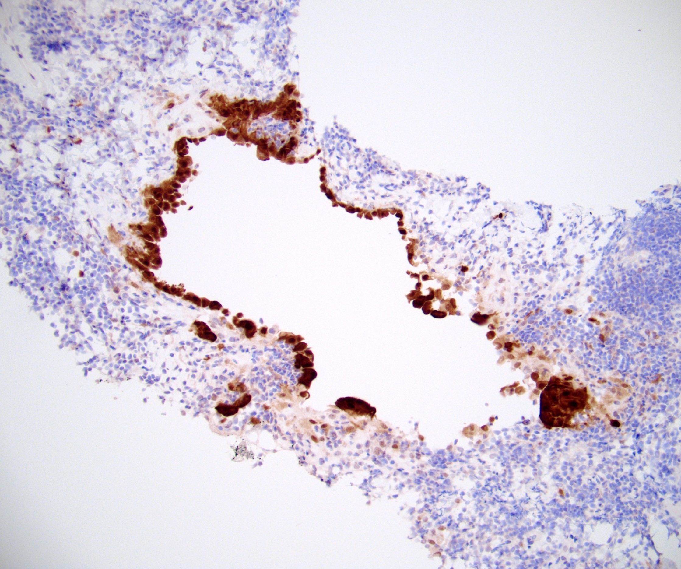
Tortuous vascular channels

Anastomosing vascular channels
Vascular channels appear papillary
CD31
CD34
CD68
Contributed by Genevieve M. Crane, M.D., Ph.D.

Thickened capsule

Thickened fibrotic capsule
Inflammatory cells in capsule
T. pallidum organisms

Plasma cells in capsule
T. pallidum organisms

Ki67
Images hosted on other servers:

Spiral shaped bacteria
AFIP images
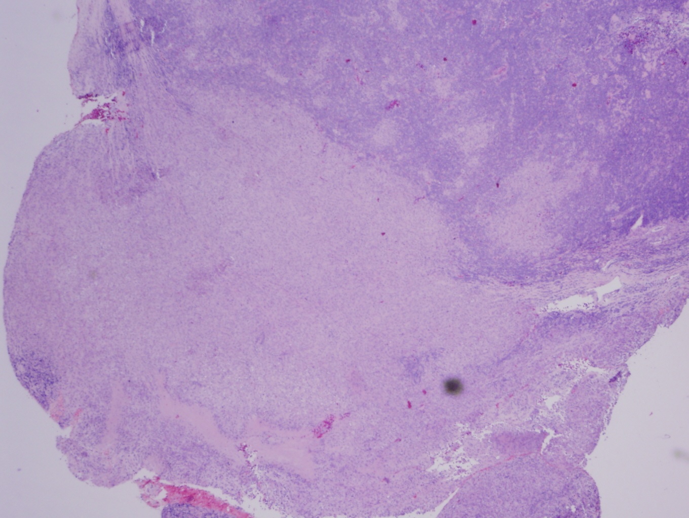
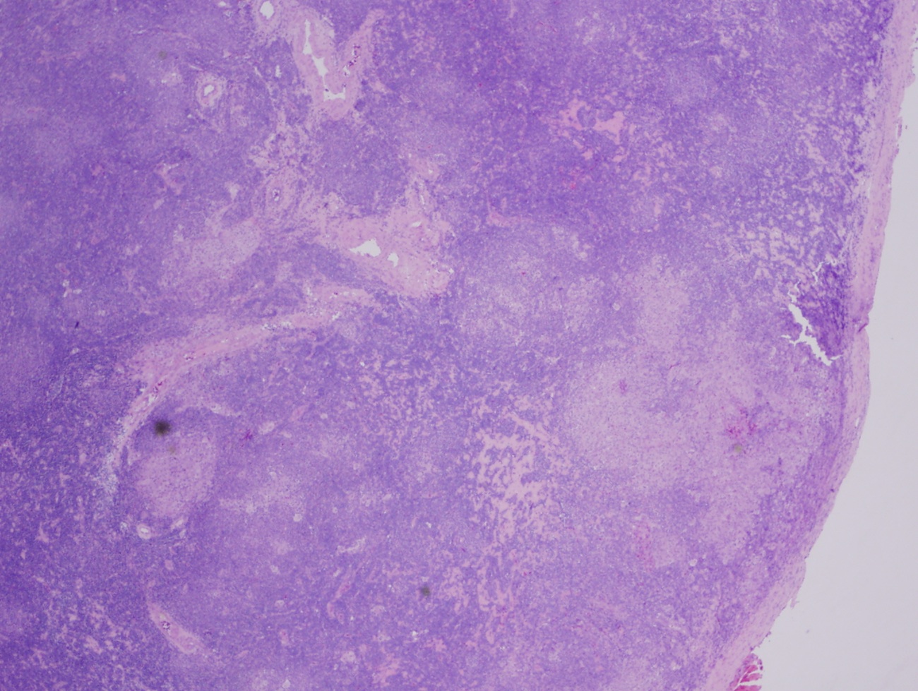
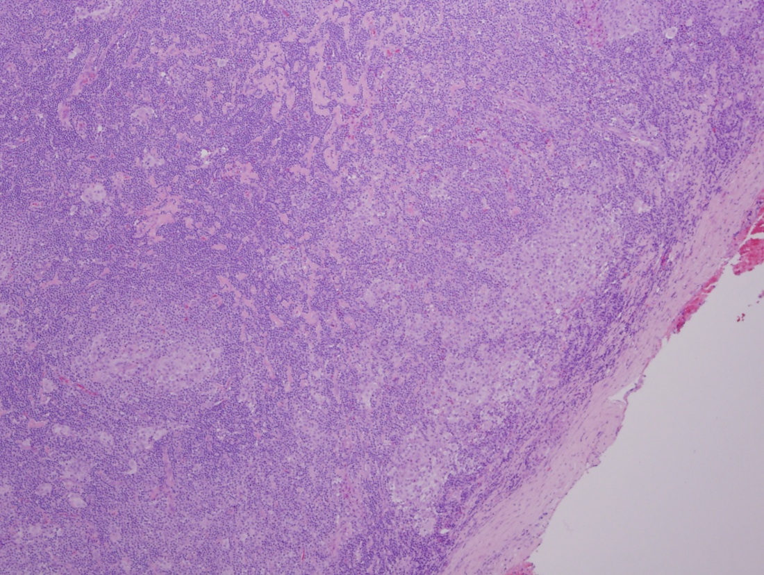
Lymph node
Images hosted on other servers:

Diagnostic criteria
Contributed by Anna Sarah Erem, M.D. and Gulisa Turashvili, M.D., Ph.D. (Case #522), @katcollmd on Twitter and AFIP


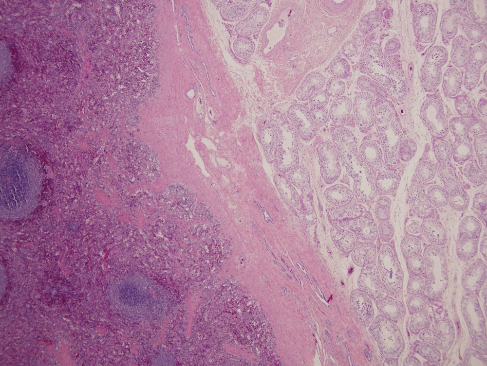
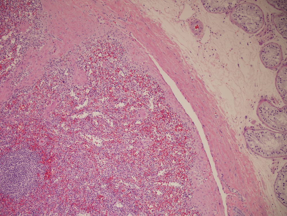
Extrapulmonary lymphangioleiomyomatosis
Lymphangiomyomatosis


Lymphangiomyomatosis
Lymph node
Involving pelvic lymph node
Contributed by Kristin Dittmar, M.D.
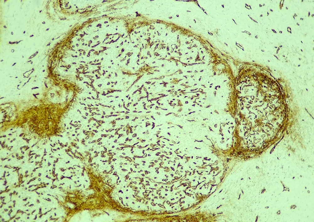
Enlarged lymph node due to mesothelial inclusion
Contributed by Debra L. Zynger, M.D.
Core biopsy

High power
CK7

CK5 / 6
Calretinin
AFIP images

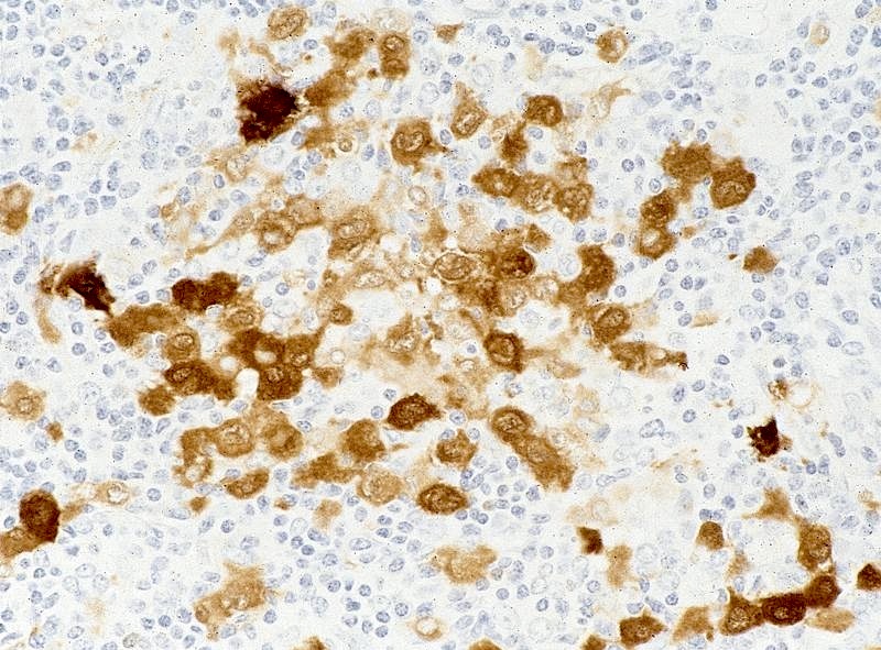
Metastatic Merkel cell carcinoma from skin
Metastatic Merkel cell carcinoma
Metastatic neuroblastoma
Metastatic seminoma
Metastatic carcinoma
simulating Hodgkin
lymphoma
Images hosted on other servers:

F-FDG PET images of splenic metastasis

CT images of splenic metastasis
Images hosted on other servers:


Parotid gland: carcinoma ex pleomorphic adenoma
Images hosted on other servers:

Submandibular lymphadenopathy
Images hosted on other servers:

Central caseation
in node involved
by M. avium
intracellulare
Contributed by Ahmed Alrajjal, M.D.
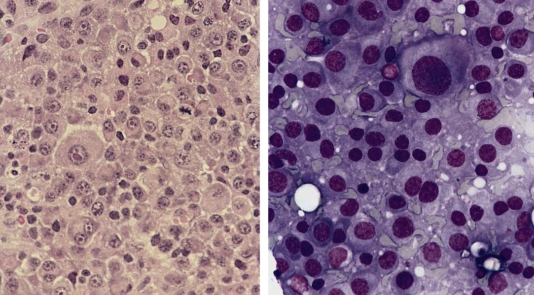
Sheets of foamy histiocytes
Nonnecrotizing granuloma
Reactive histiocytes
Sheets of histiocytes
Lymph node, AFB stain
Contributed by Ahmed Alrajjal, M.D.
Touch imprint, lymph node granuloma
Images hosted on other servers:

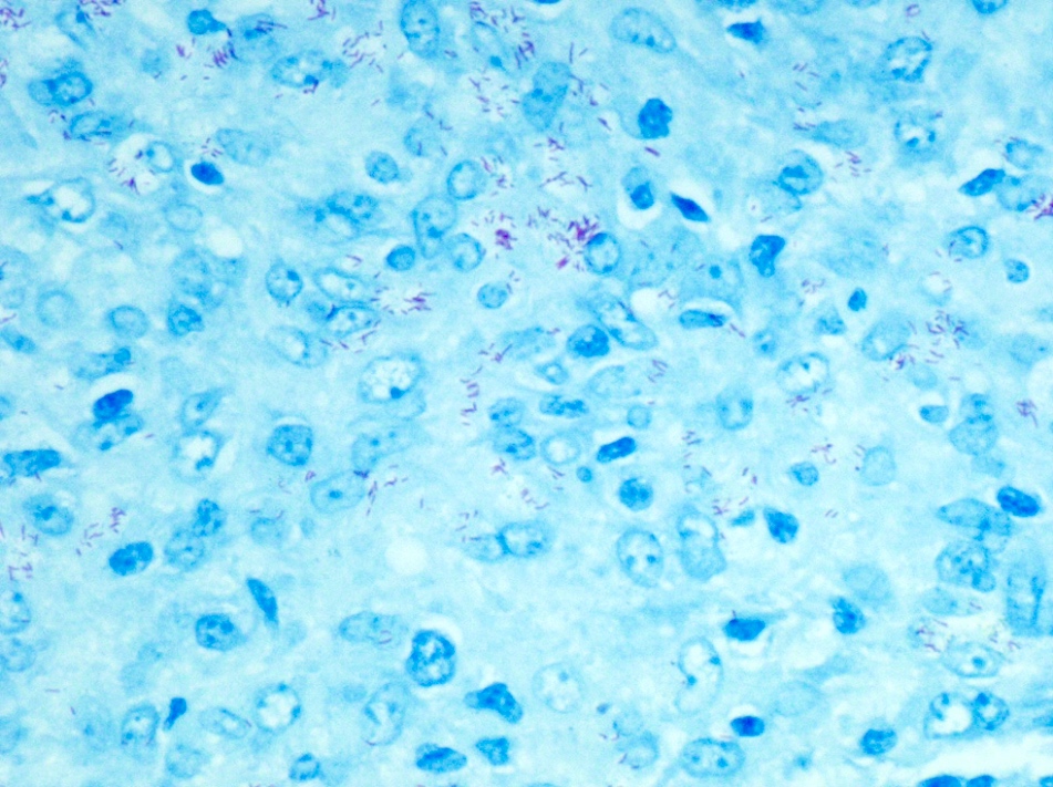
Mycobacterium avium complex (MAC), Ziehl-Neelsen stain
Images hosted on other servers:

PCR for 7 different strains
Images hosted on other servers:

Splenic peliosis
AFIP images

Plasmacytoma

Plasmacytoma
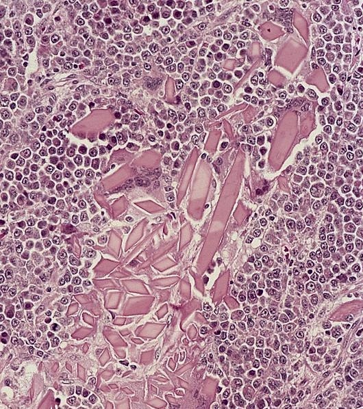
With crystalloids
With amyloid deposition

With blood lakes
With myxoid stroma
Plasmablastic plasmacytoma of nasopharynx
Anaplastic plasmacytoma
Immunohistochemistry
Contributed by Roaa Aljuaid, M.B.B.S.

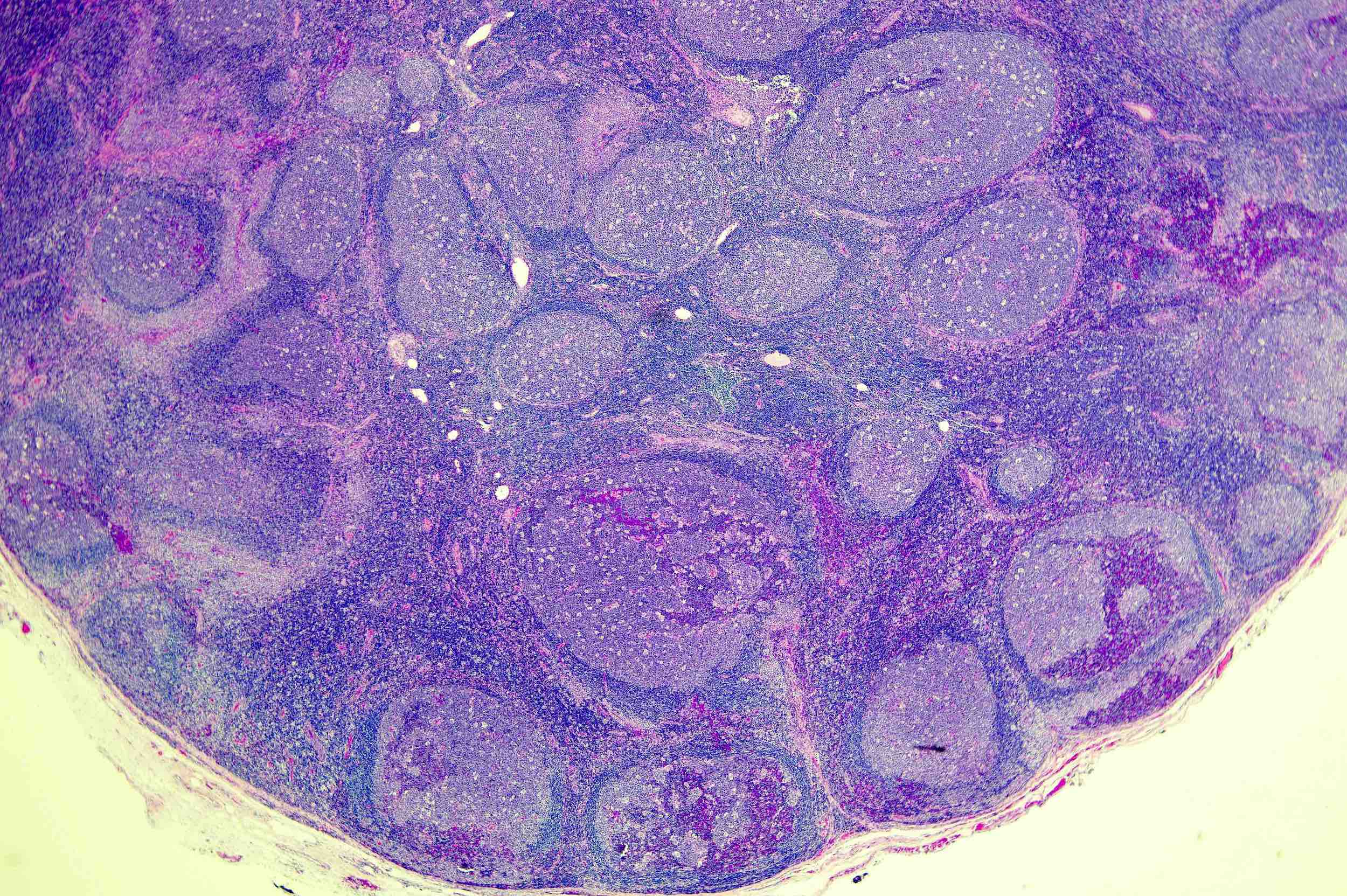
Follicular hyperplasia
Nodules at different stages

Macronodules
Expansile lymphoid follicles

Reactive follicles
Ingrowth of mantle zone B cells
IgD in PTGC

BCL2 in PTGC nodules
BCL6 in PTGC nodules
CD10 in PTGC nodules

Atypical lymphocytes versus immunoblasts
CD15
EMA

PAX5
Contributed by Roberto N. Miranda, M.D. and Roman Segura-Rivera, M.D.
Reactive follicular hyperplasia

Polarized Ki67
Follicular lymphoma
Nonpolarized Ki67
Nodal marginal zone lymphoma

Monocytoid cytology
Mantle cell lymphoma (MCL) with mantle zone pattern

Cyclin D1 (mantle pattern) in MCL
Progressive transformation of germinal centers
CD20 in PTGC
NLPHL
LP cells
Rosetting CD3+ lymphocytes
Castleman disease, hyaline vascular variant
Germinal center in HVCD
Interfollicular expansion
Immunoblasts
Infectious mononucleosis, EBER
EBV positive diffuse large B cell lymphoma
EBER positivity in EBV positive diffuse large B cell lymphoma
Mixed cellularity classic Hodgkin lymphoma (MCCHL)

EBER positivity in MCCHL
Images hosted on other servers:

Reactive lymphoid hyperplasia
AFIP images
Reactive lymphadenopathy of rheumatoid arthritis
Images hosted on other servers:

Follicular hyperplasia
with eosinophilic
material in
interfollicular areas

Extensive replacement
by focally calcified
eosinophilic material

Rounded, well
separated follicles
with expanded
paracortex

Follicular center composed
primarily of centrocytes,
with no / rare tingible body
macrophages or mitotic figures

Interdigitating dendritic
cells (CD1+) are associated
with small lymphocytes
Images hosted on other servers:

Imaging findings in RDD
Contributed by Aishwarya Ravindran, M.B.B.S. and Karen L. Rech, M.D.
Excisional biopsy of
subcutaneous mass
CD163
S100
OCT2
Cyclin D1
Images hosted on other servers:

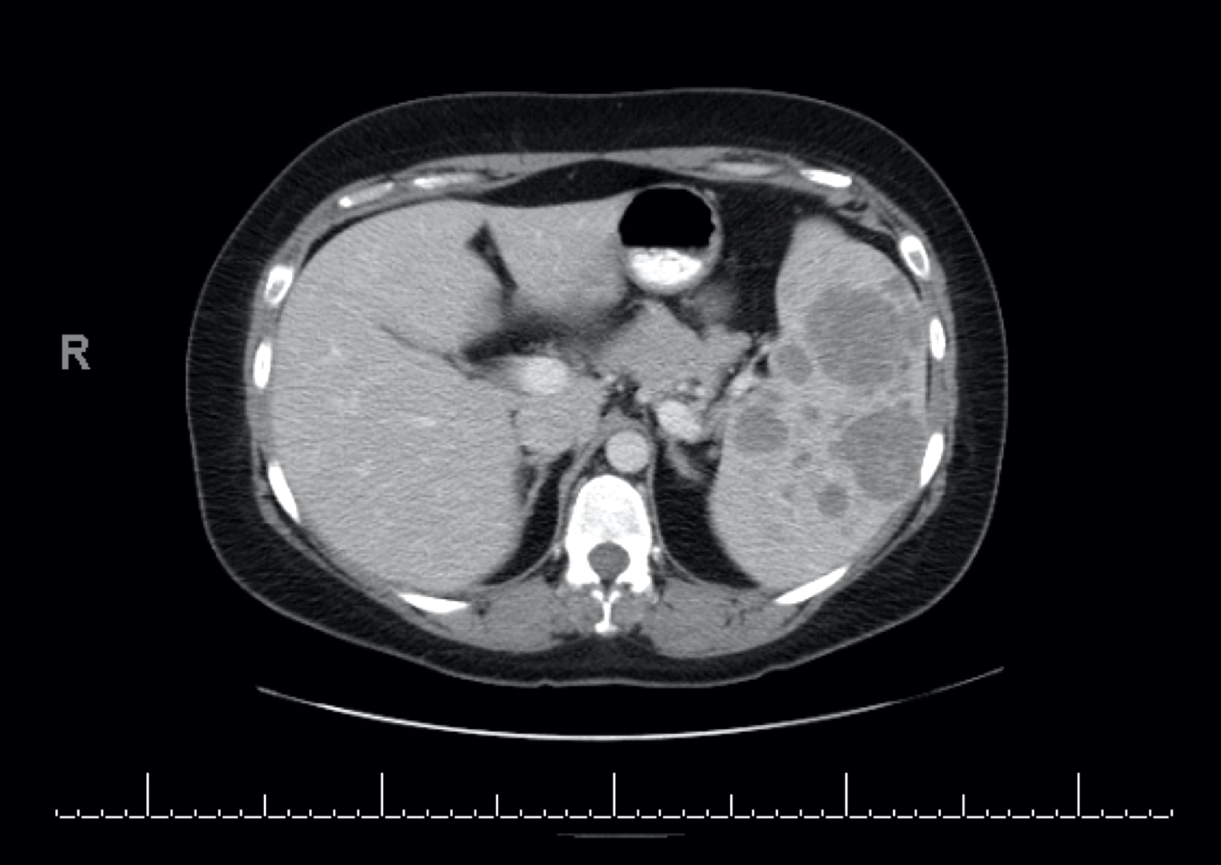
CT findings

Expansible low density mass

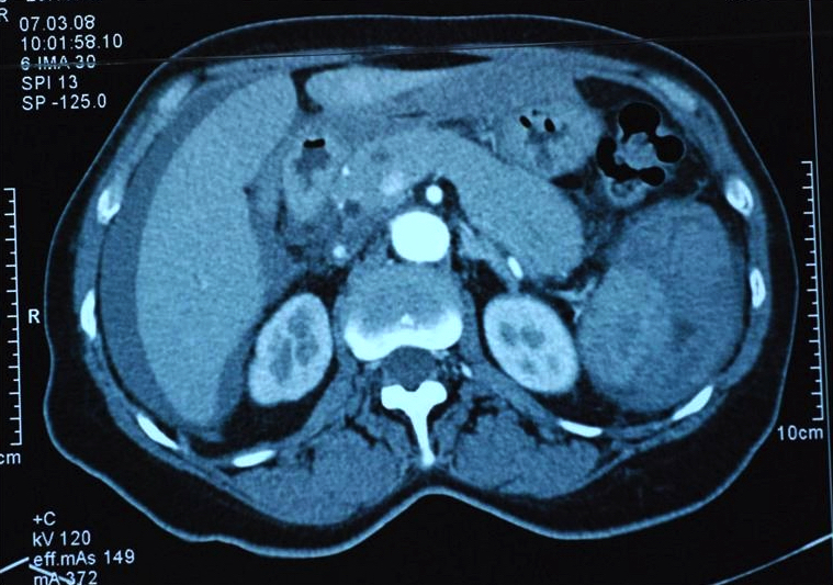
5 cm tumor in hilus

Hypointensity of tumor

Splenic lesion at medial aspect

Lesion in lower spleen

Hypoenhancing mass
Images hosted on other servers:

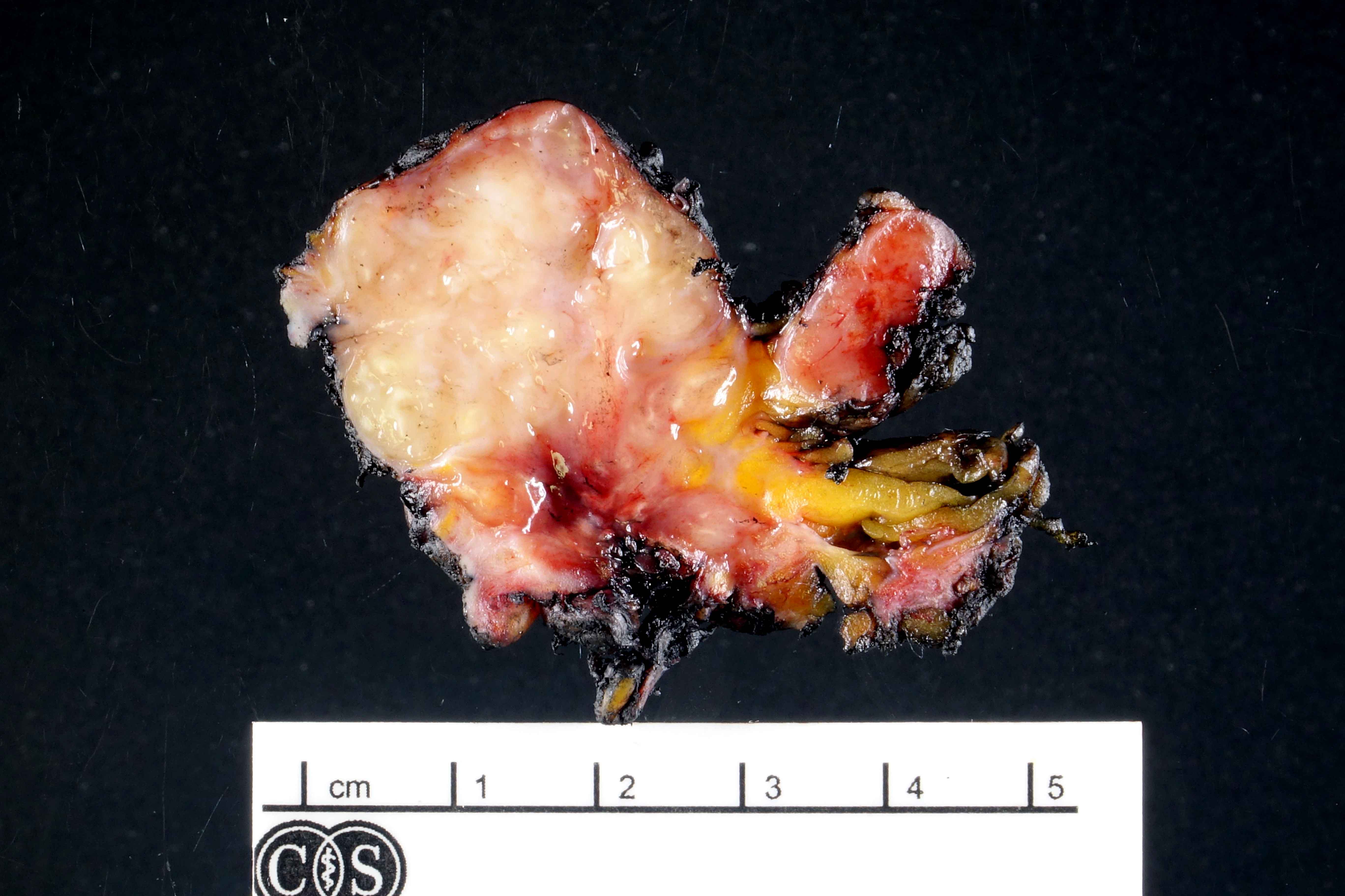
Macroscopic findings

Laparoscopic view
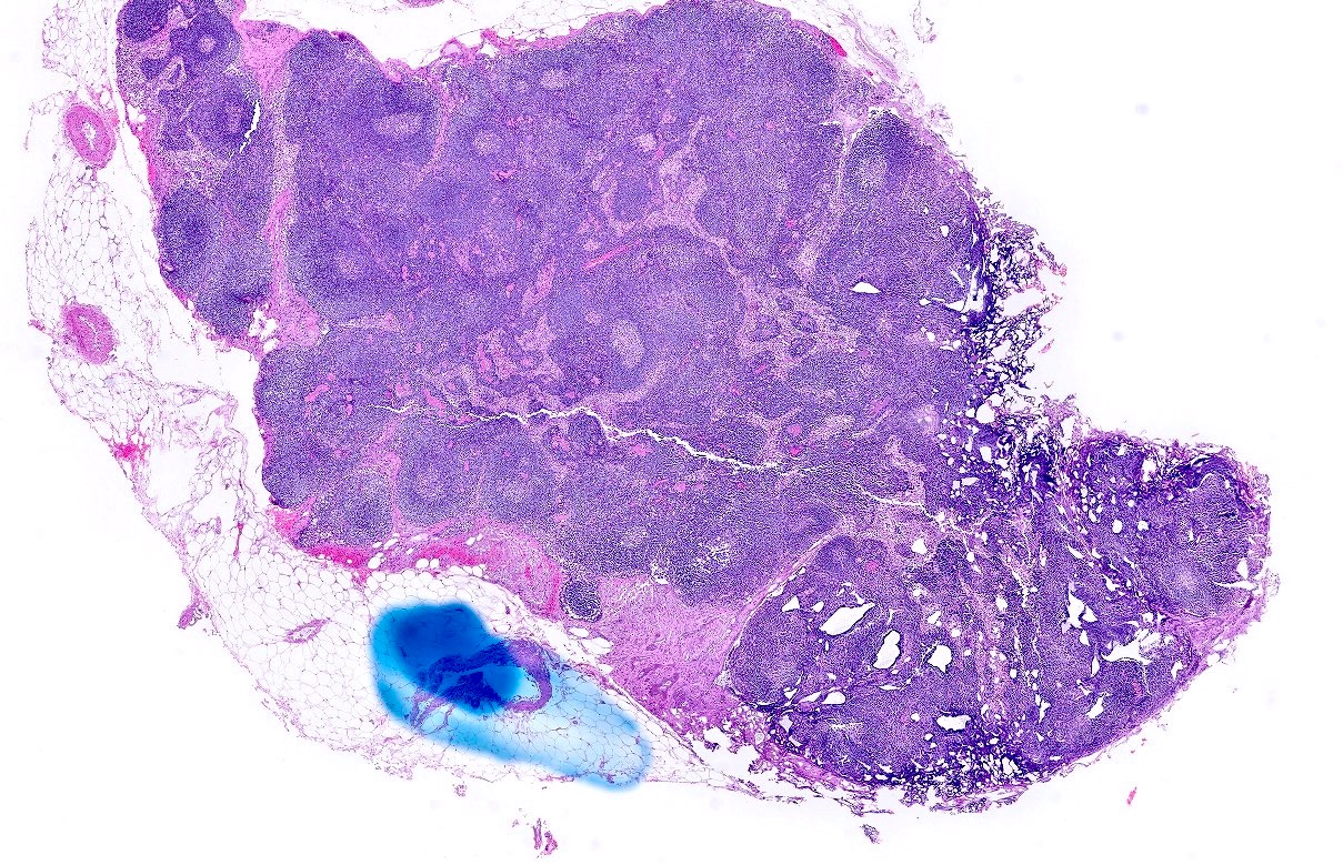
Contributed by Shafinaz Hussein, M.D. and Huifei Liu, M.D., Ph.D. (Case #364)
Circumscribed, bosselated, firm, tan colored mass
Images hosted on other servers:

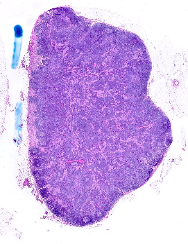
Well demarcated, unencapsulated lesion

Splenic resection

Well defined, nonencapsulated lesion

Red-brown, multinodular lesions
Contributed by Valentina Sangiorgio, M.D., Shafinaz Hussein, M.D. and Huifei Liu, M.D., Ph.D. (Case #364)
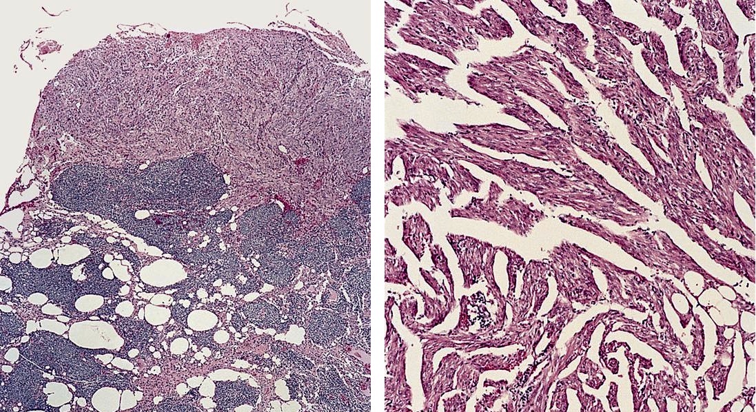
Multiple vascular nodules
Predominantly small capillaries
Bland endothelial cells
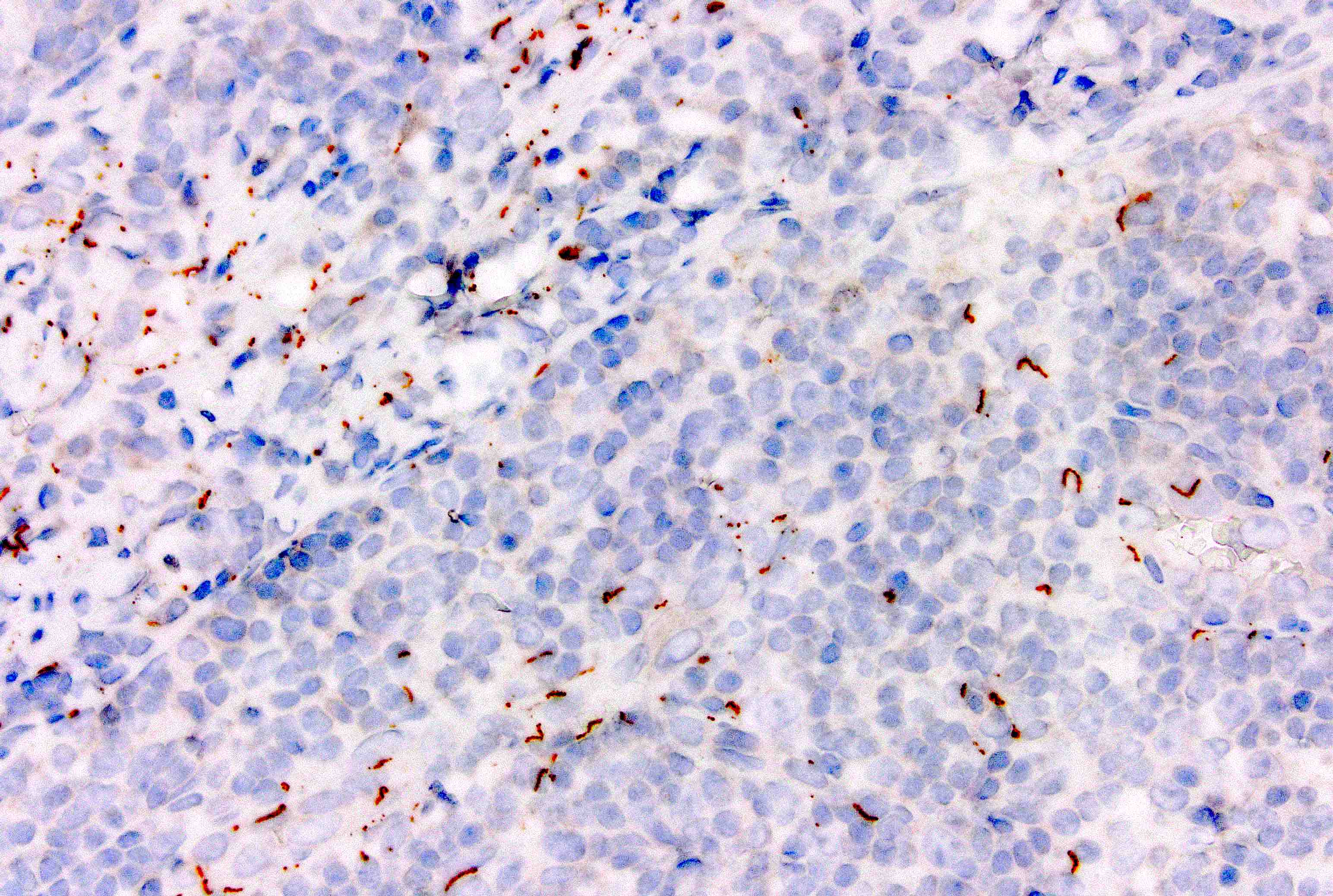
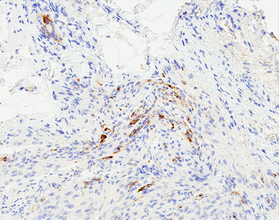
CD8
CD31
CD34
Images hosted on other servers:

Rupture of implant


Various images
Contributed by Dr. Mark R. Wick
Silicone in axillary lymph node
Images hosted on other servers:

Material consistent with silicone

Liquid silicone droplets

Various images

Subcapsular sinus diffusely expanded

Vacuoles which contain refractile material

Foreign body granuloma

Germinal centers surrounded by sinuses

Silicone leakage

Silicone particles in cystic spaces
Case #309

Splenic gonadal fusion
Images hosted on other servers:


Necrotizing lymphadenopathy
Images hosted on other servers:

Life cycle
Contributed by Tayler A. van den Akker, M.D.
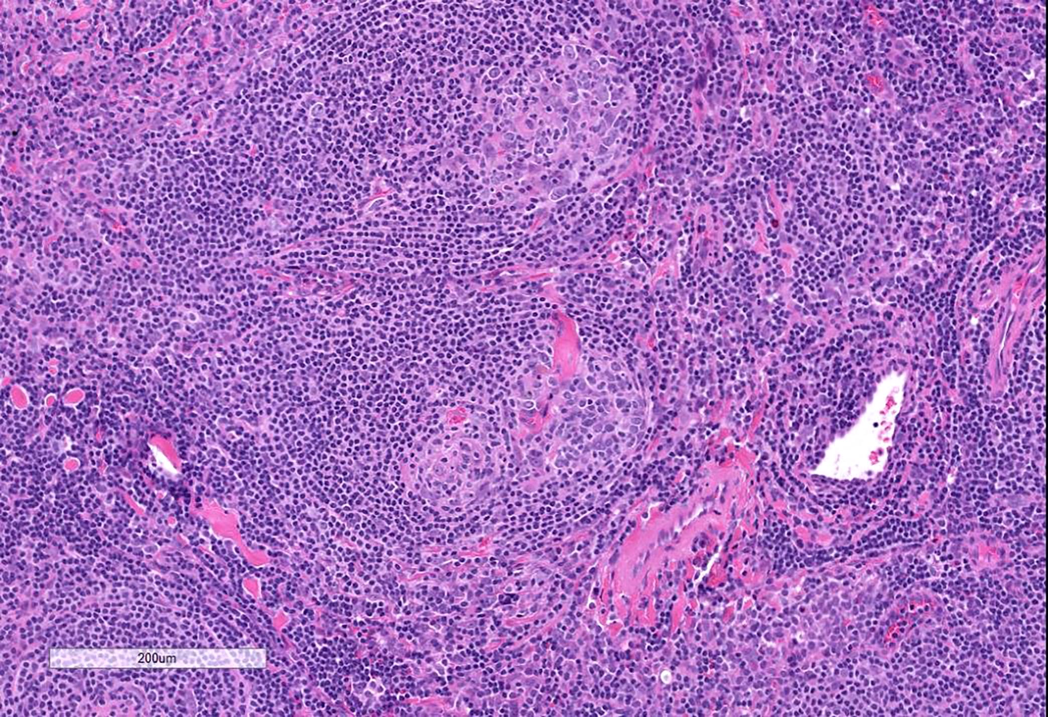
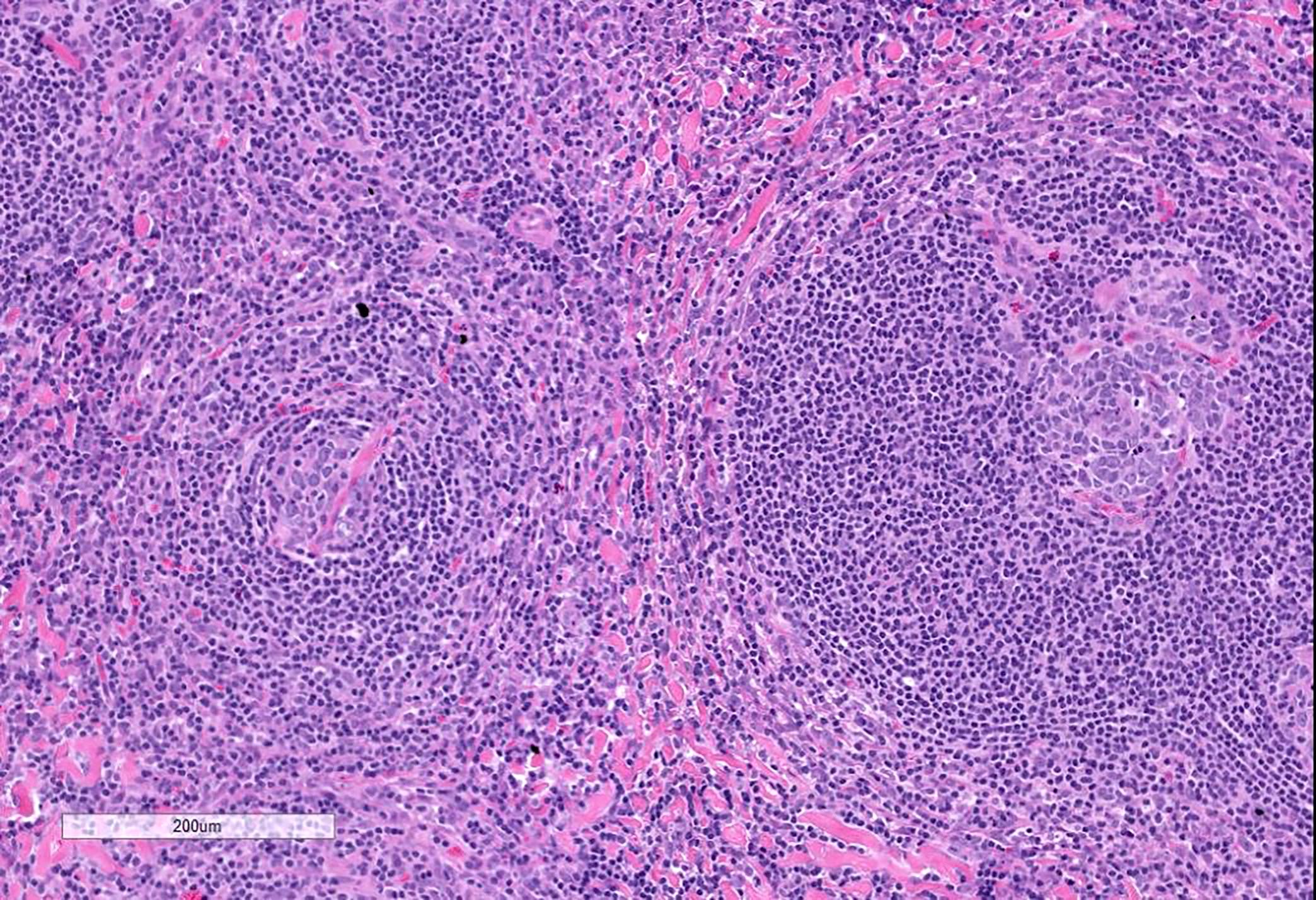
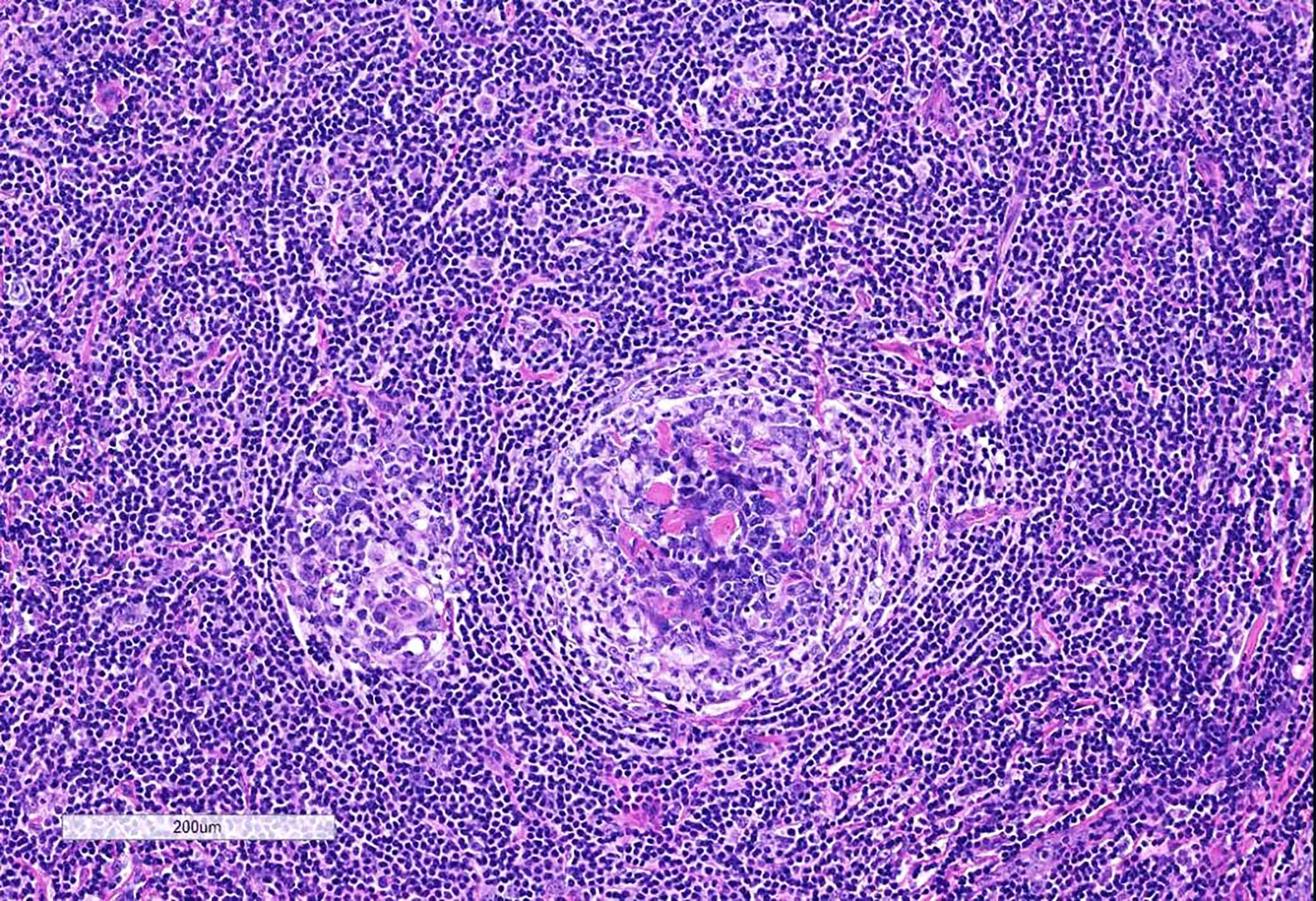
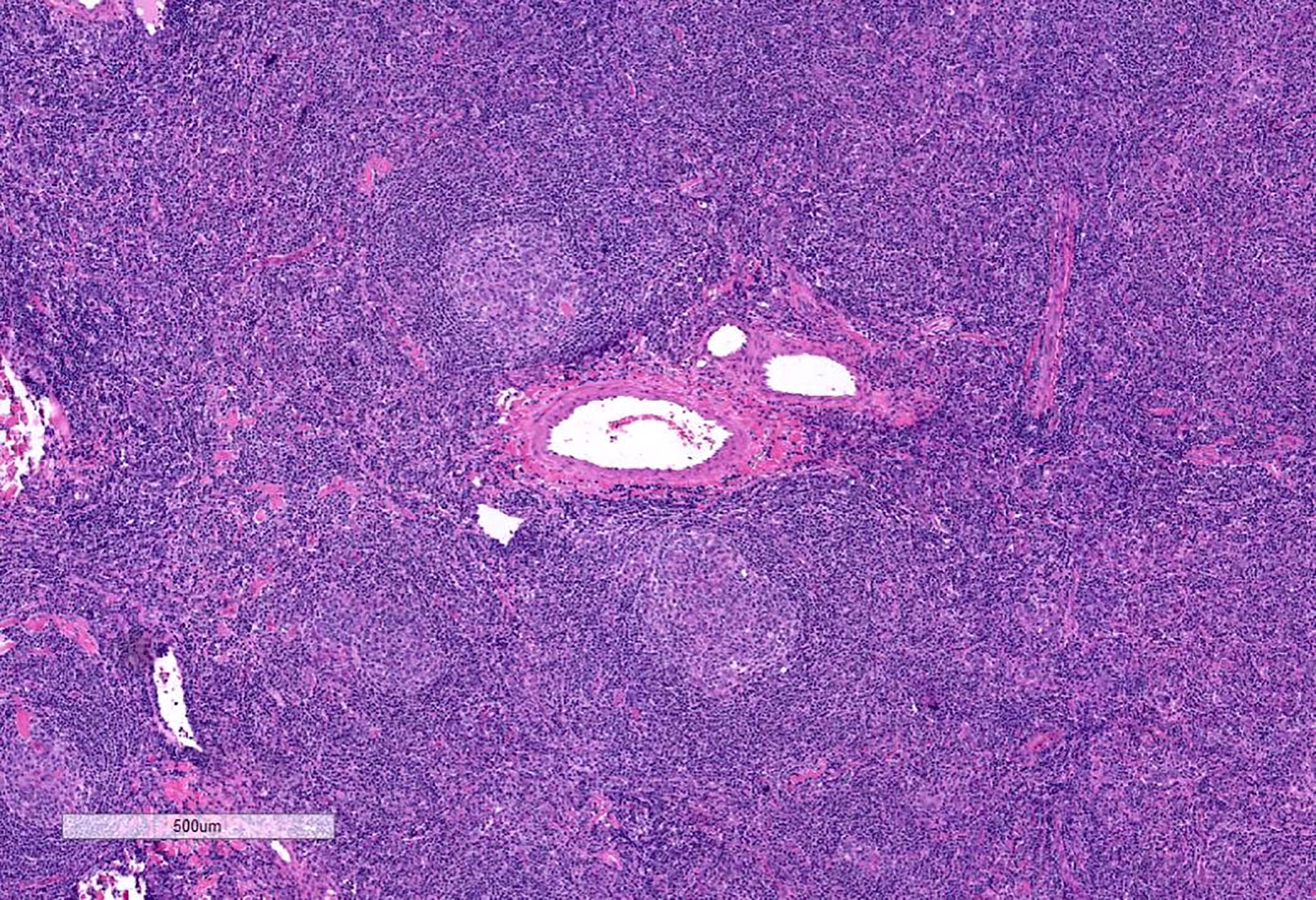
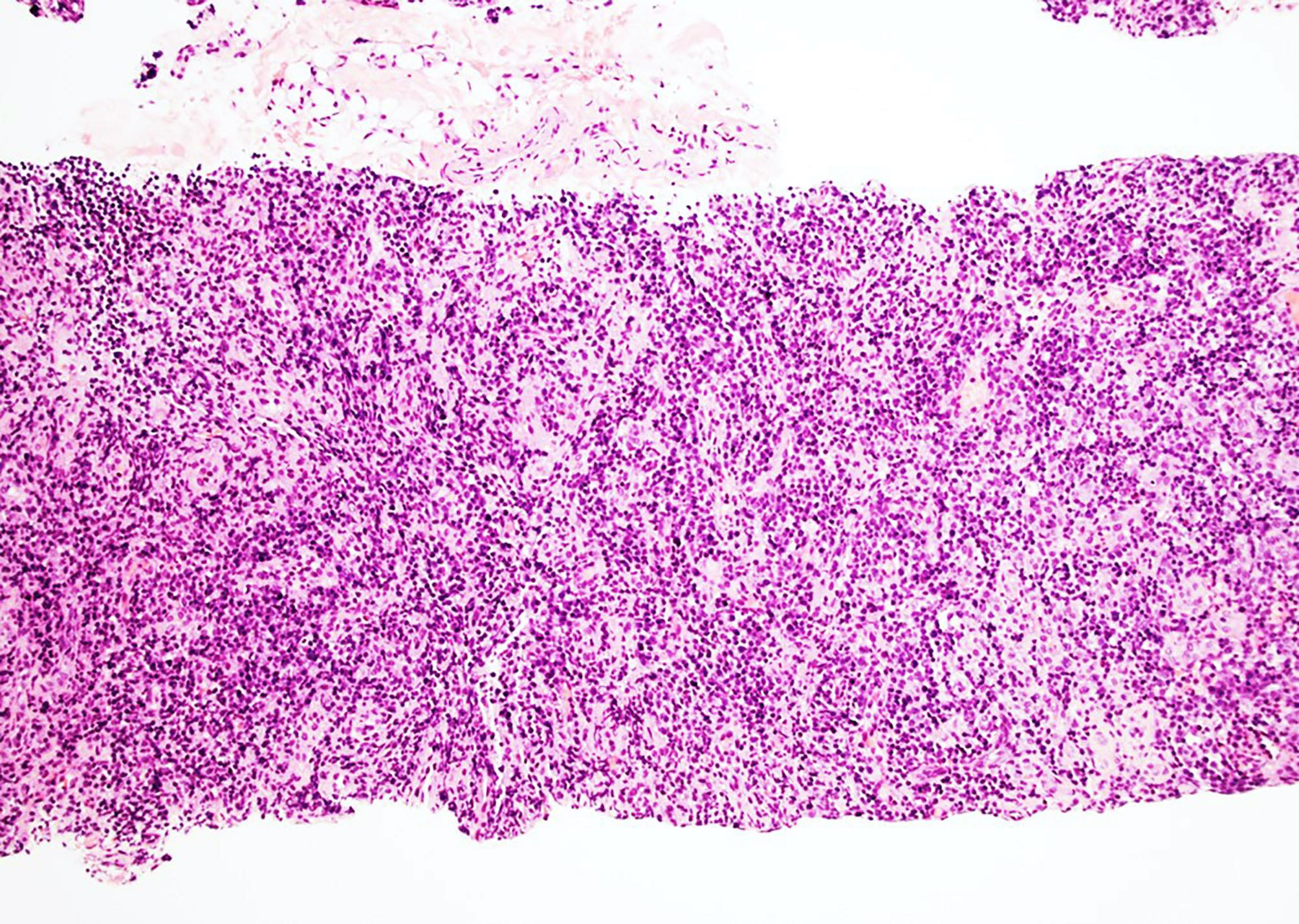
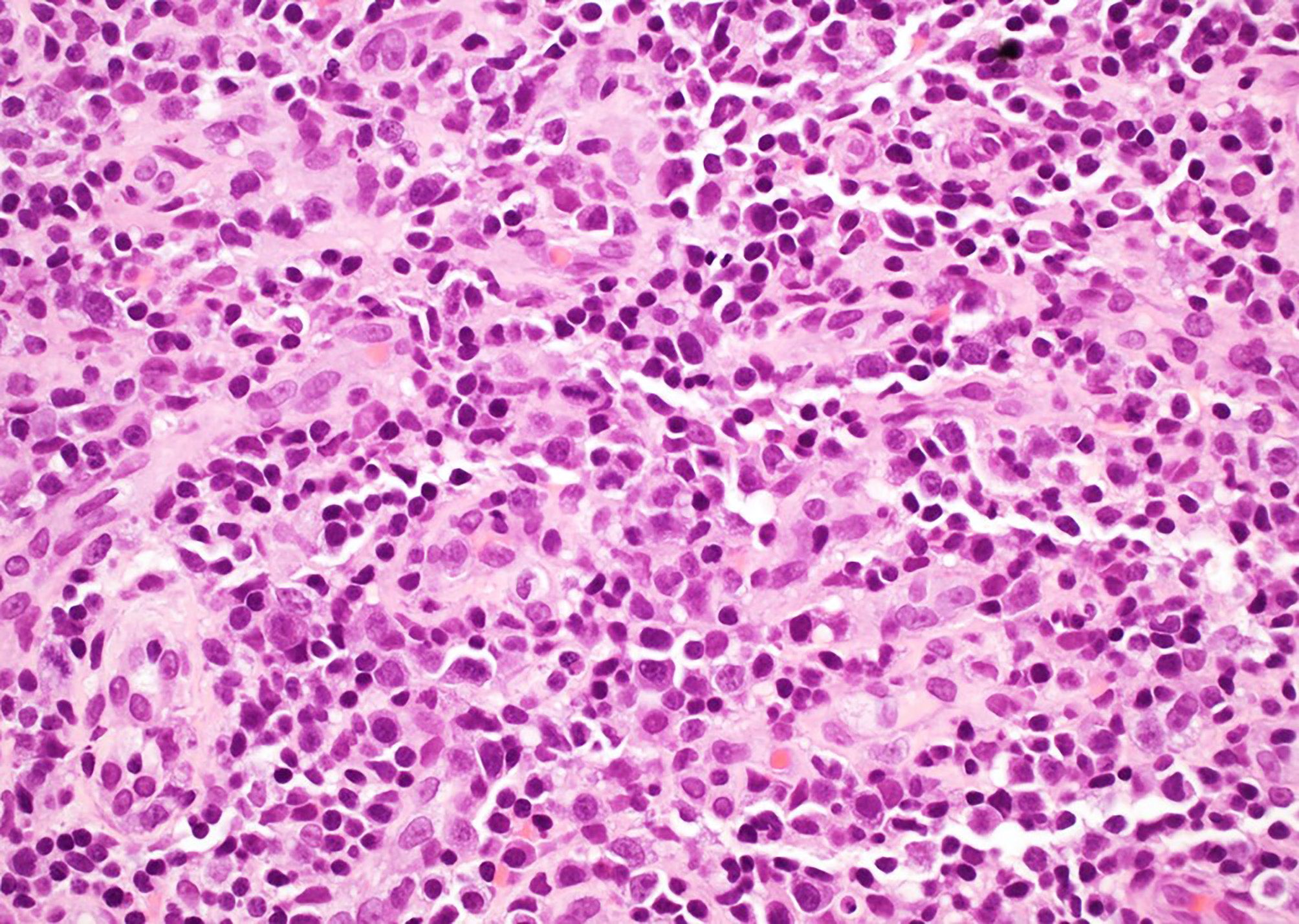
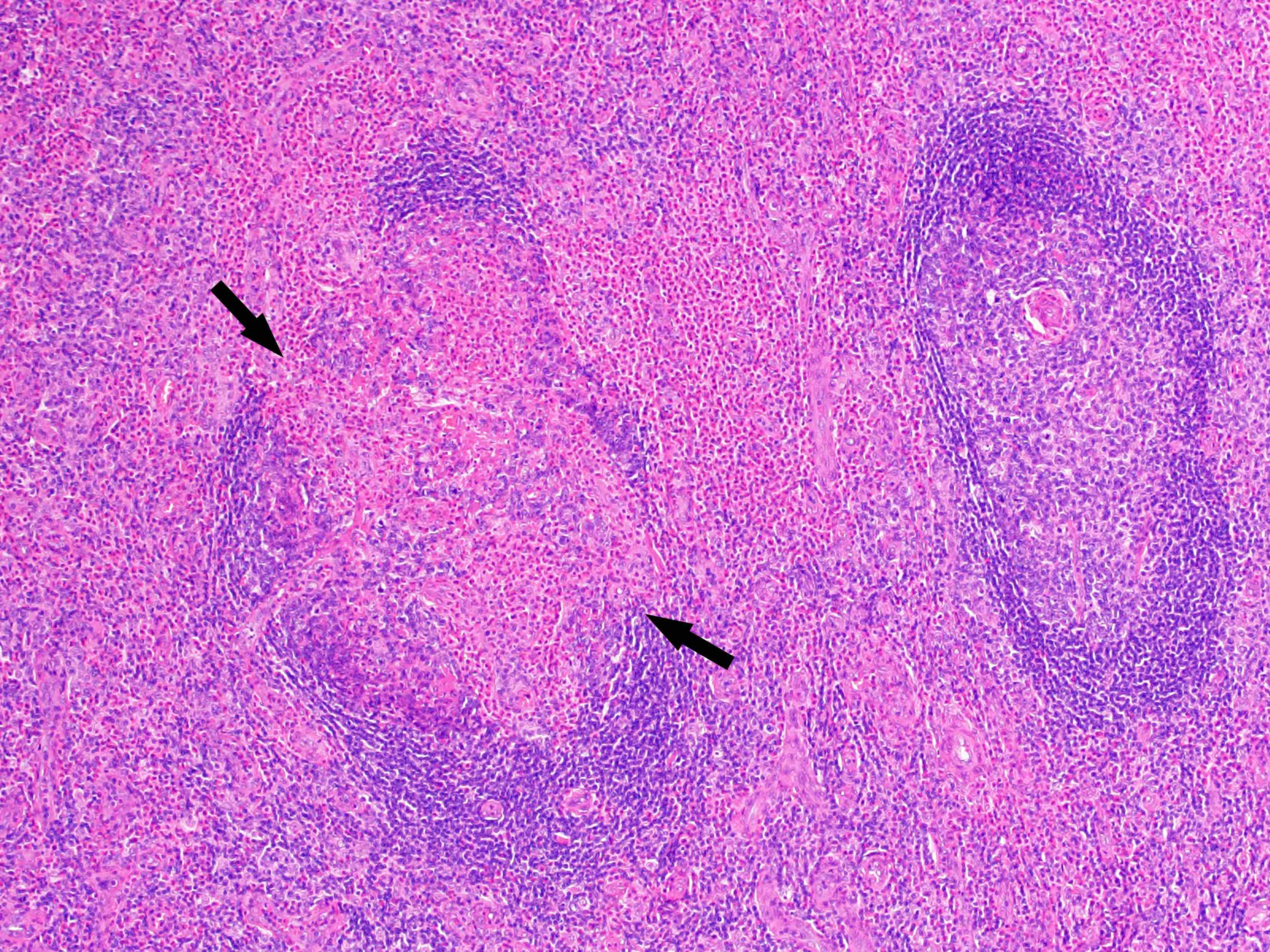
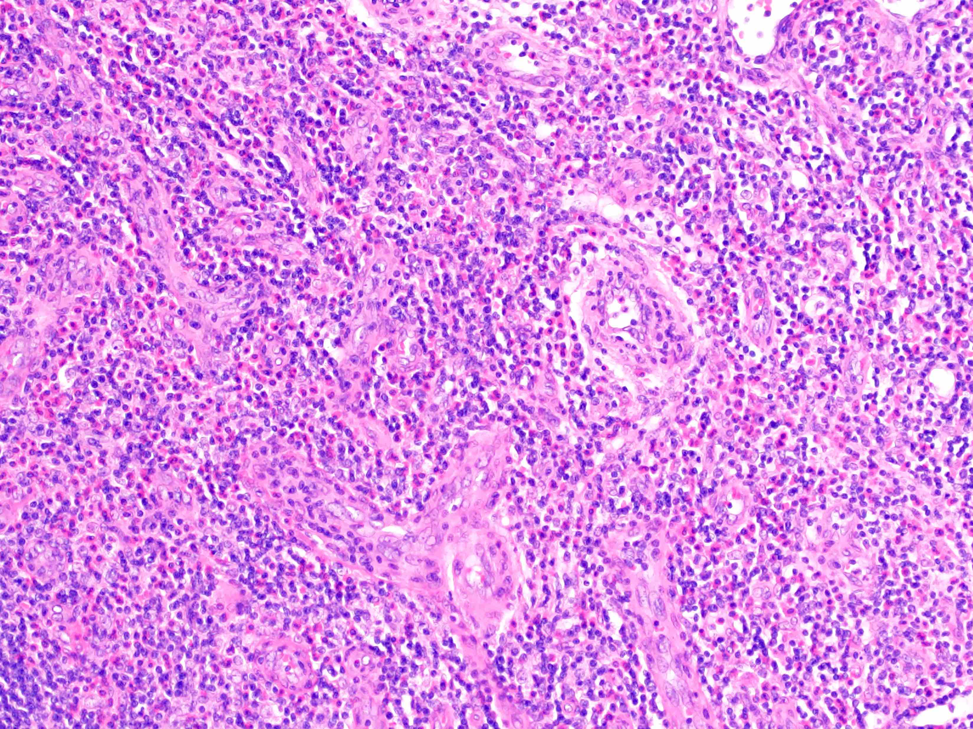
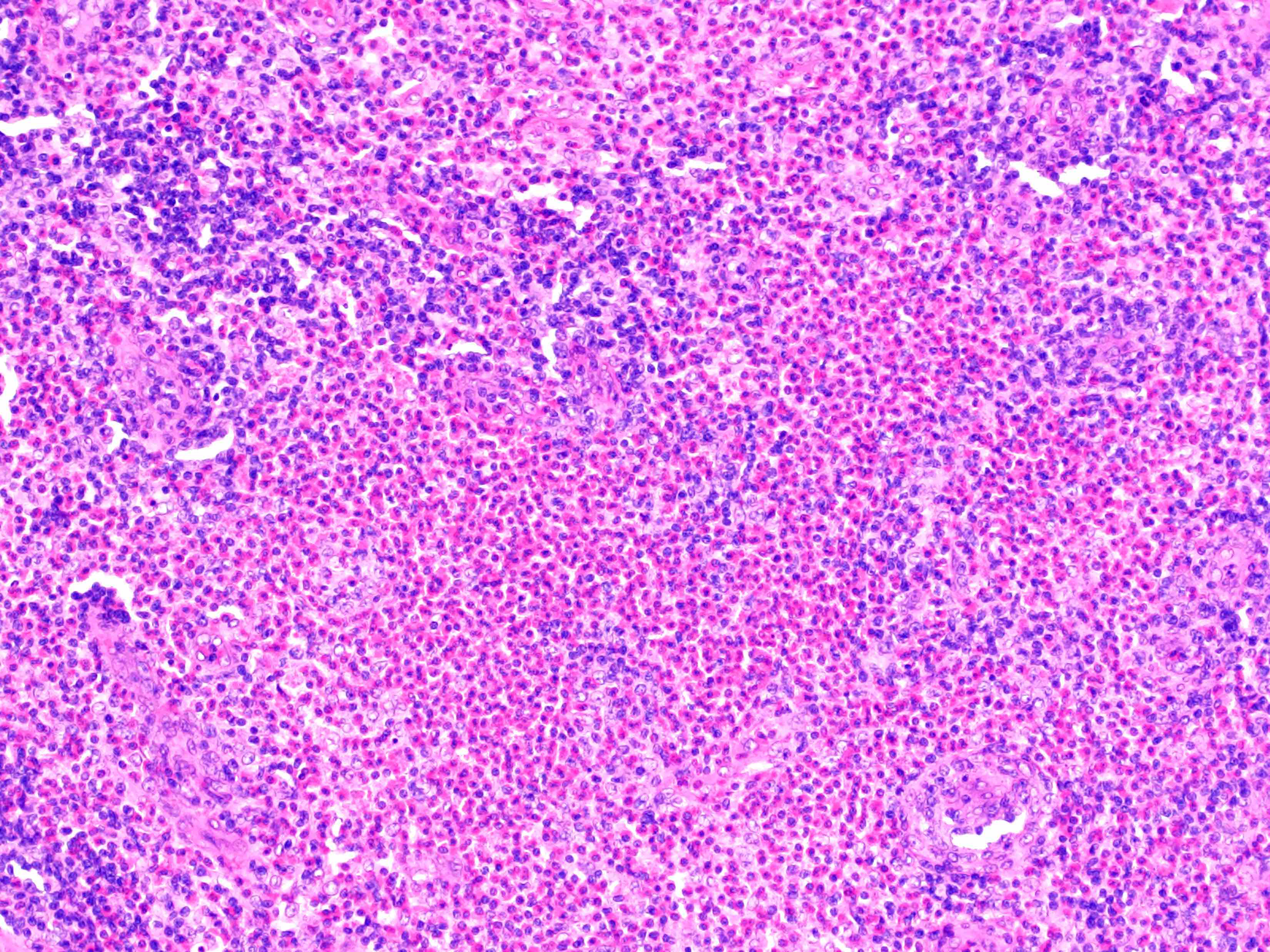
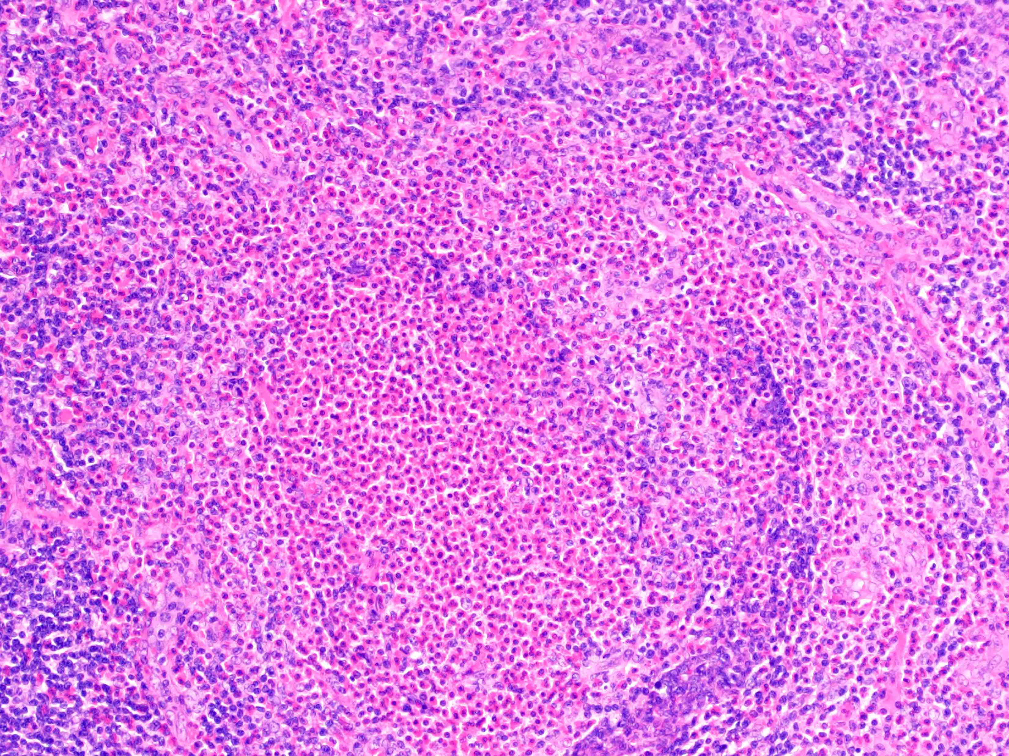
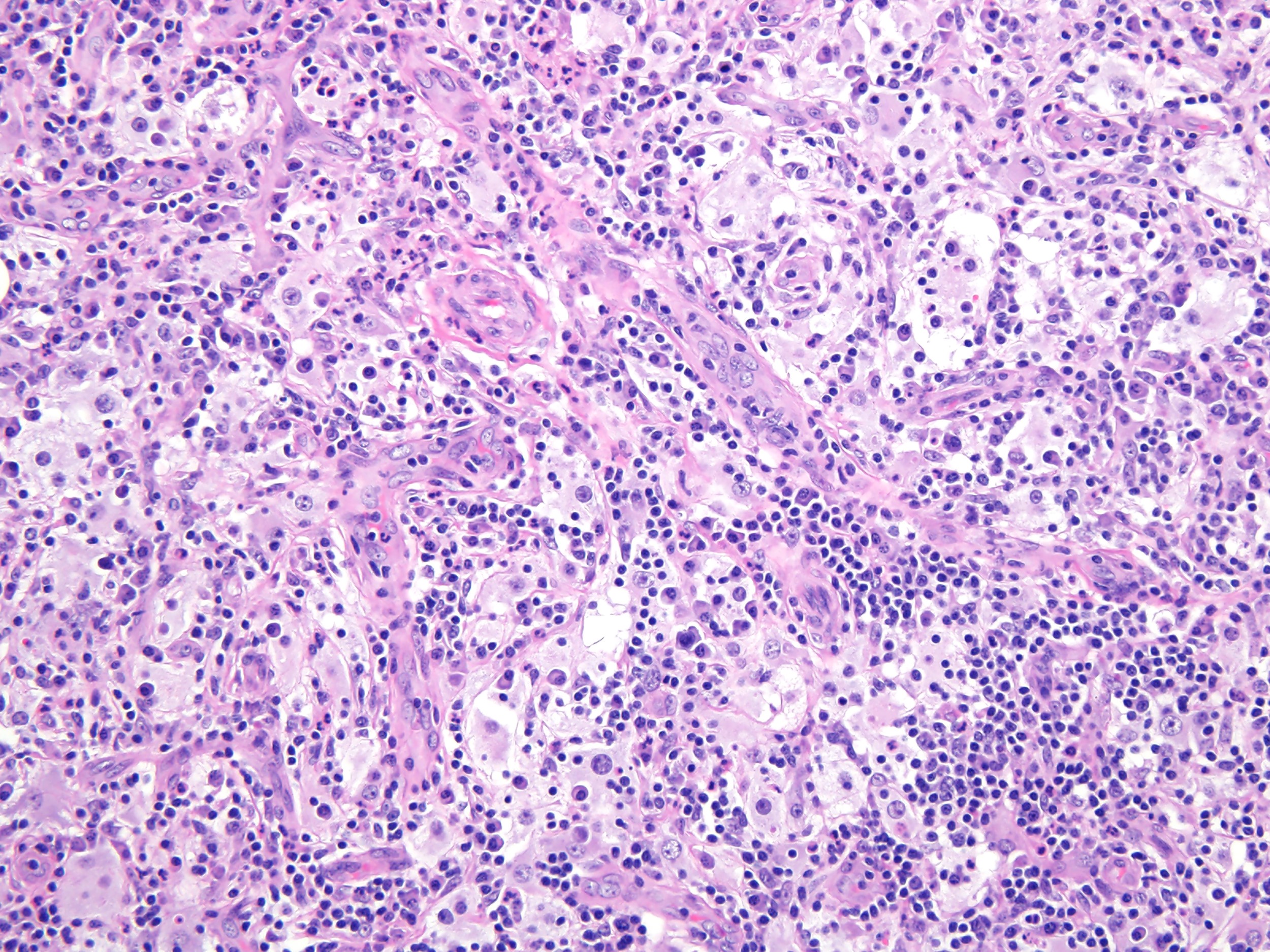
Histologic triad
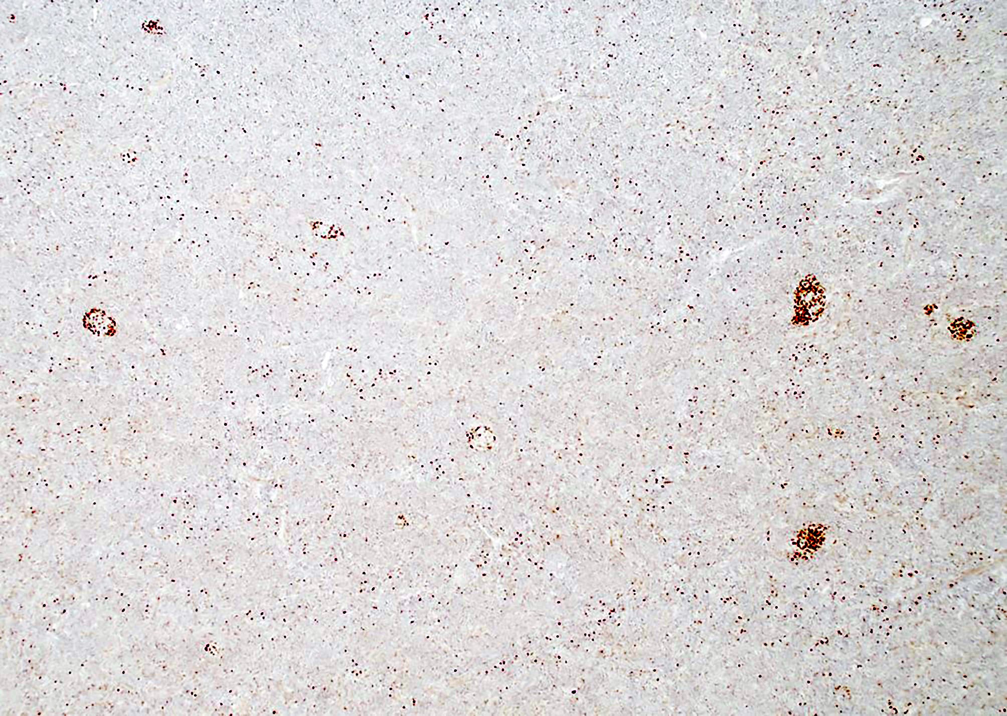
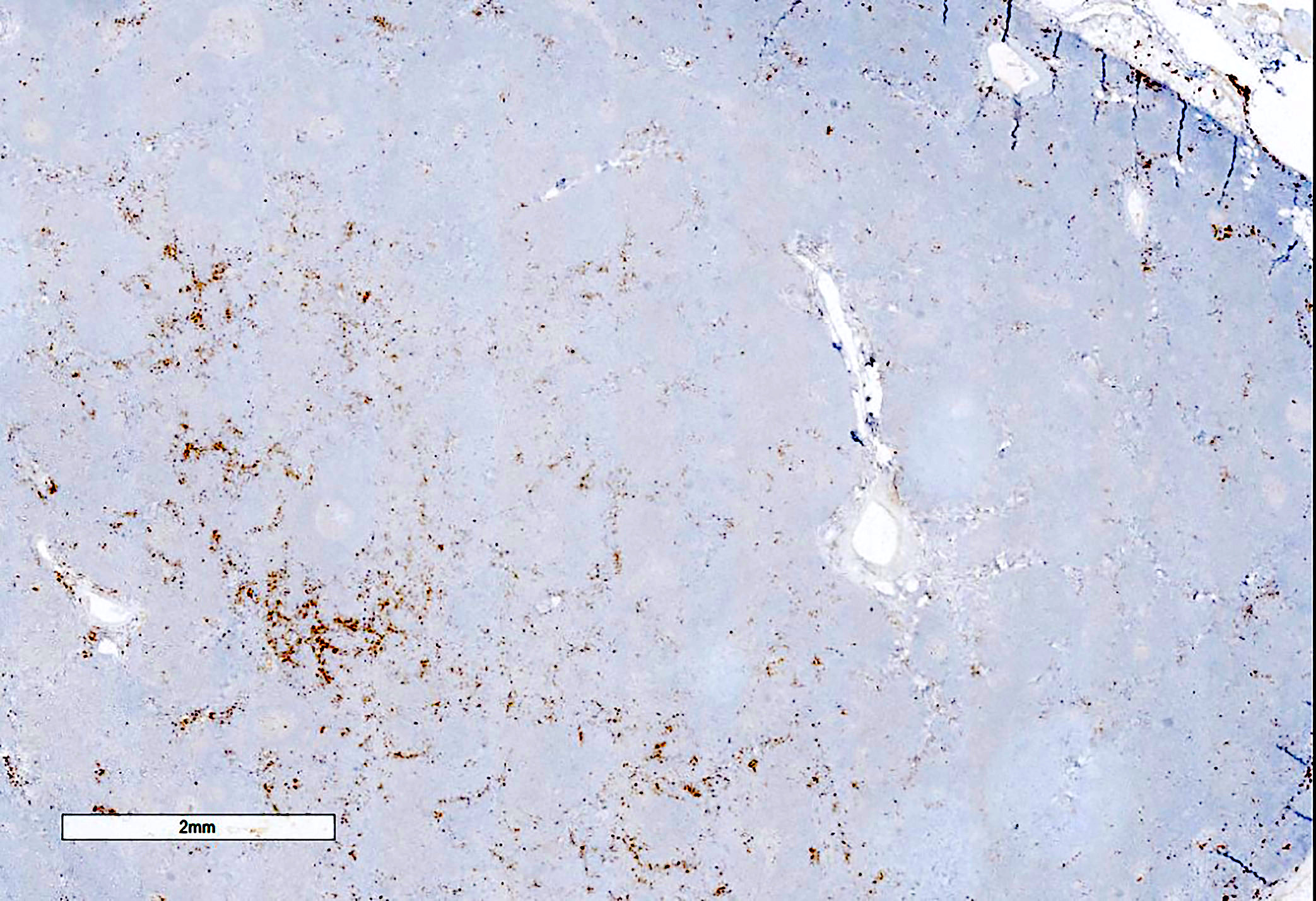
Follicular hyperplasia

Reactive follicles, monocytoid cells
Epithelioid histiocytes
Epithelioid histiocytes, monocytoid B cells
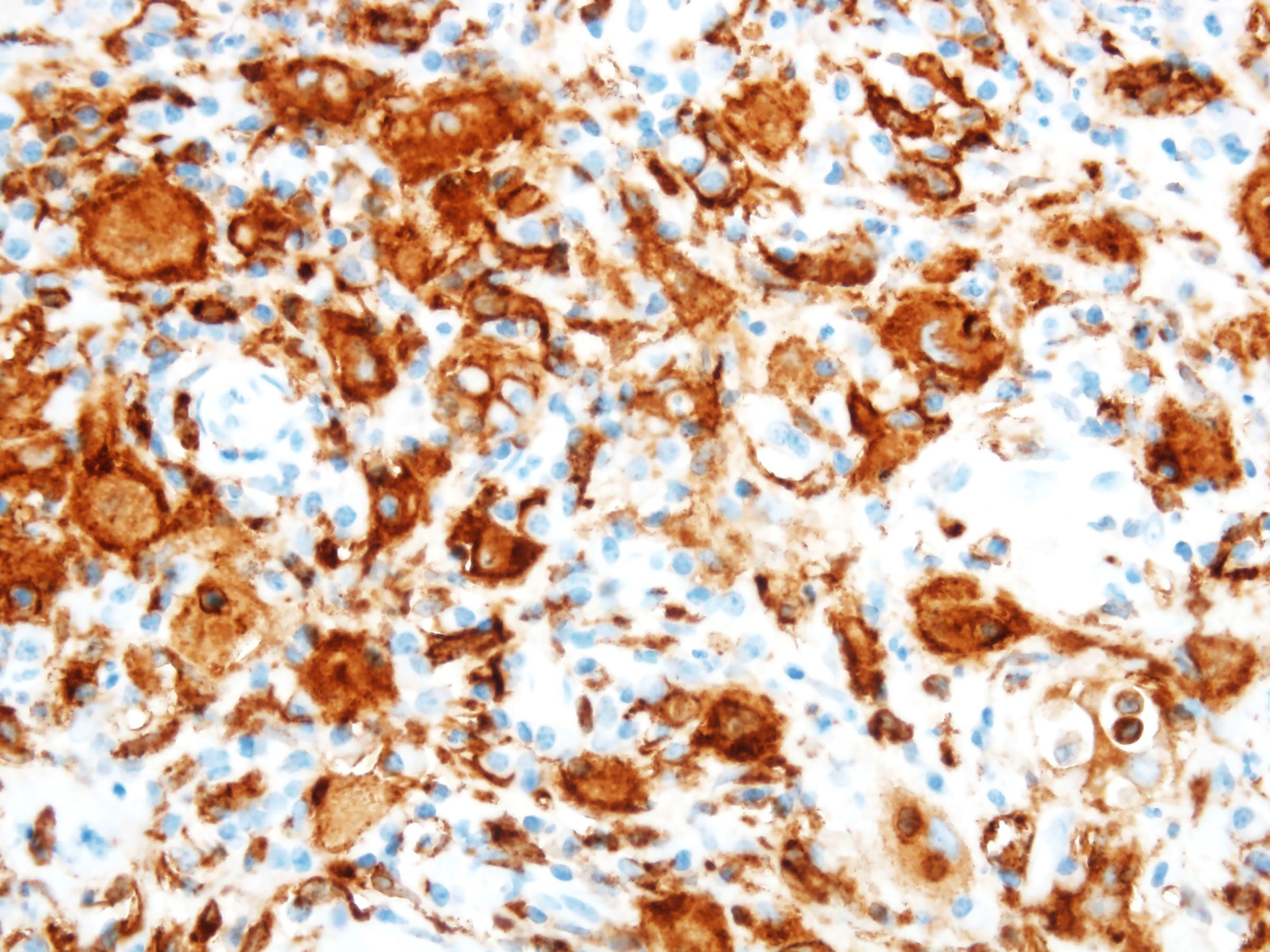
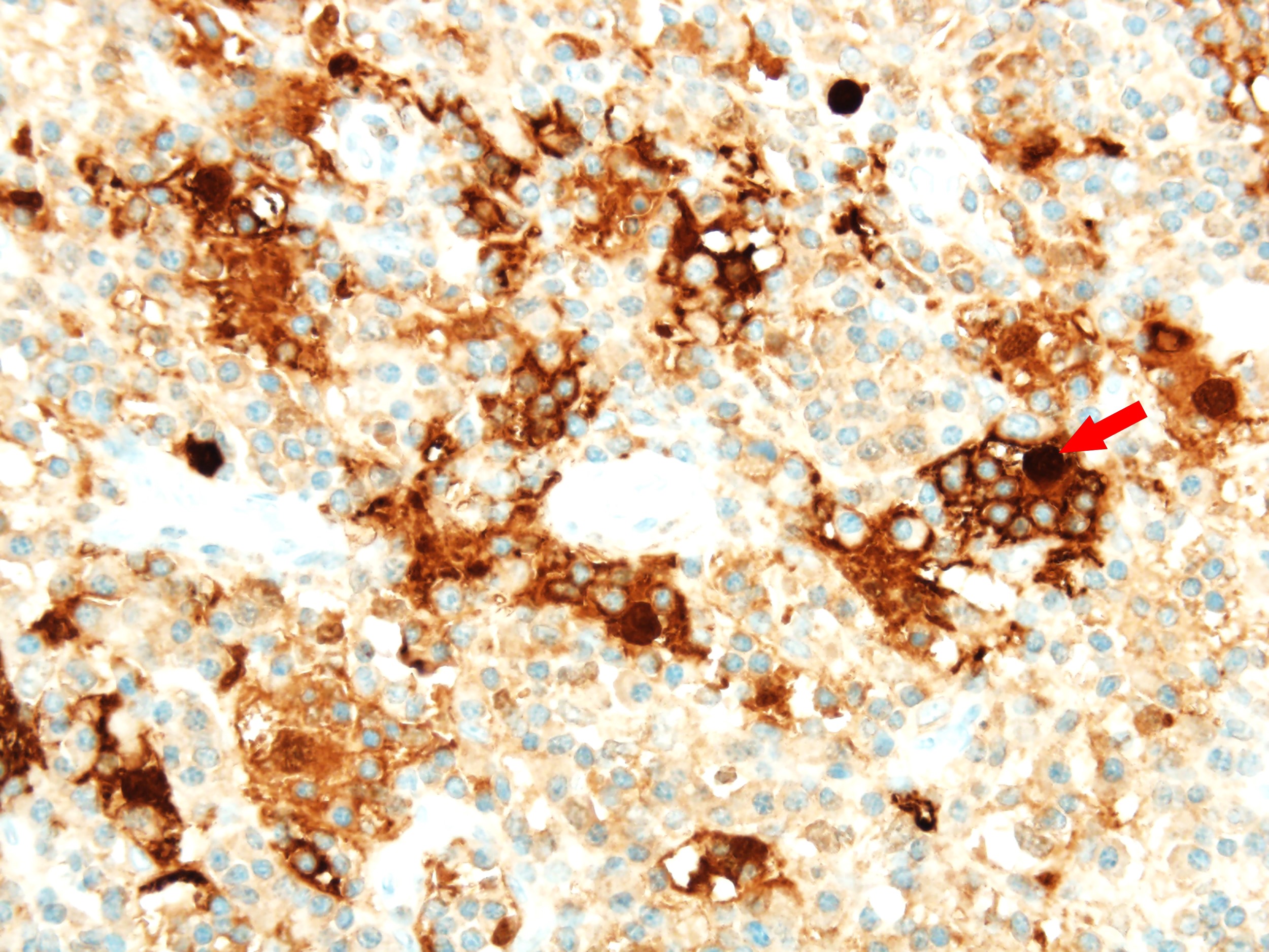
Toxoplasma special stain
AFIP images
Toxoplasmic lymphadenitis
Monocytoid B cells
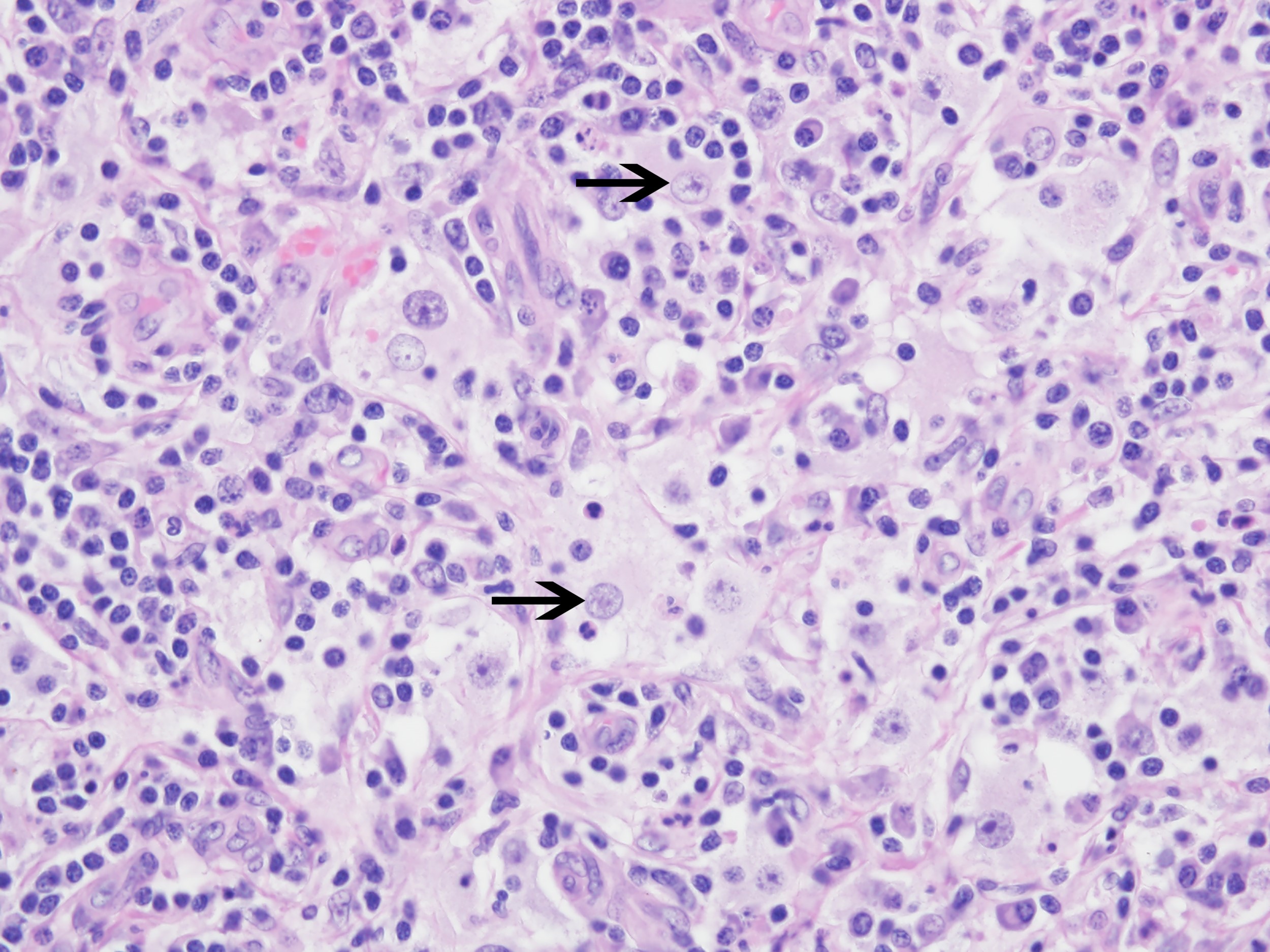
Contributed by Bobbi Pritt, M.D.
Tachyzoites of Toxoplasma gondii
Images hosted on other servers:

Toxoplasma tissue cyst within an intact neurone
Classic histomorphology of toxoplasma lymphadenitis
Great teaching case of cerebral toxoplasmosis
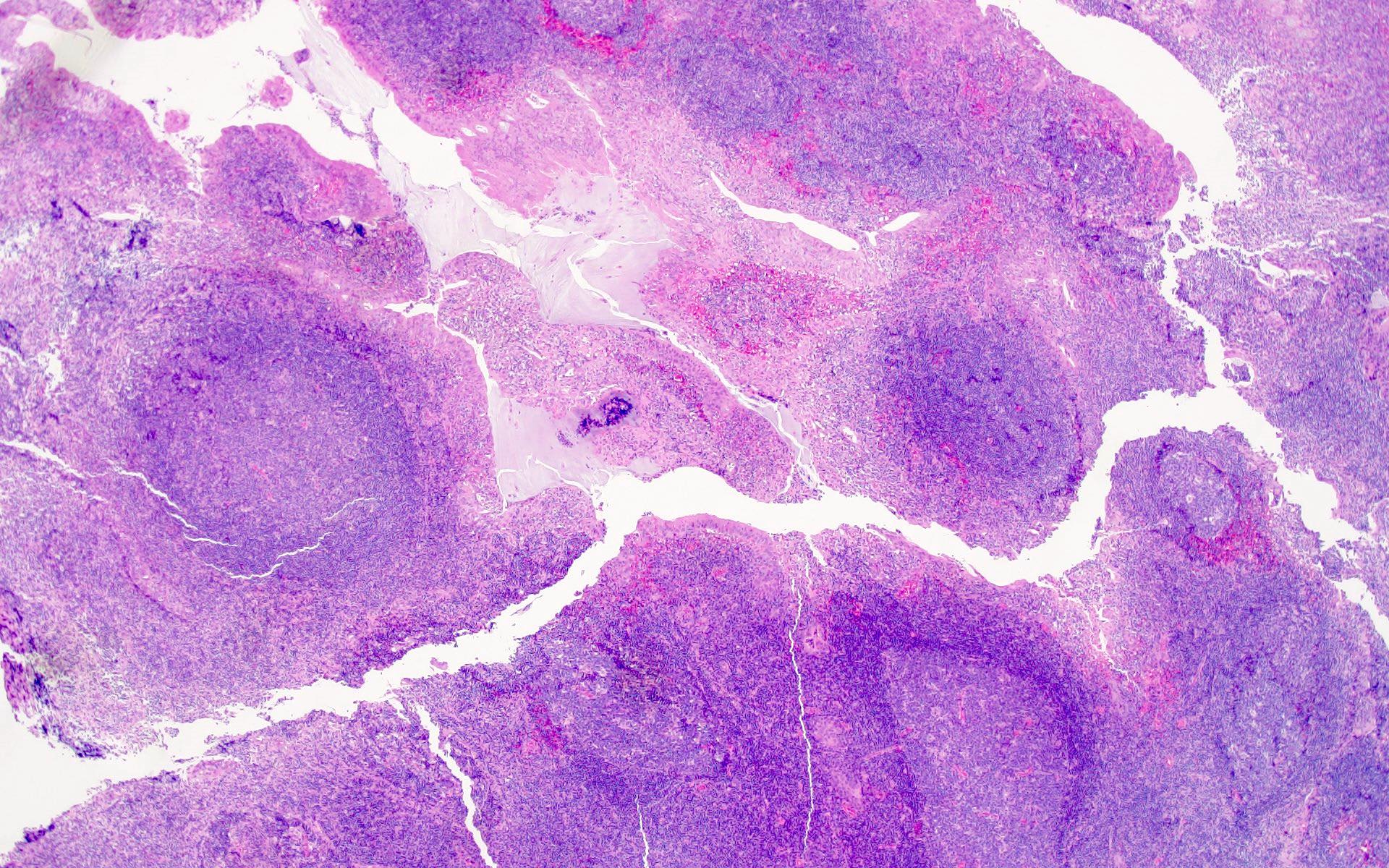
Case #405
AFIP images


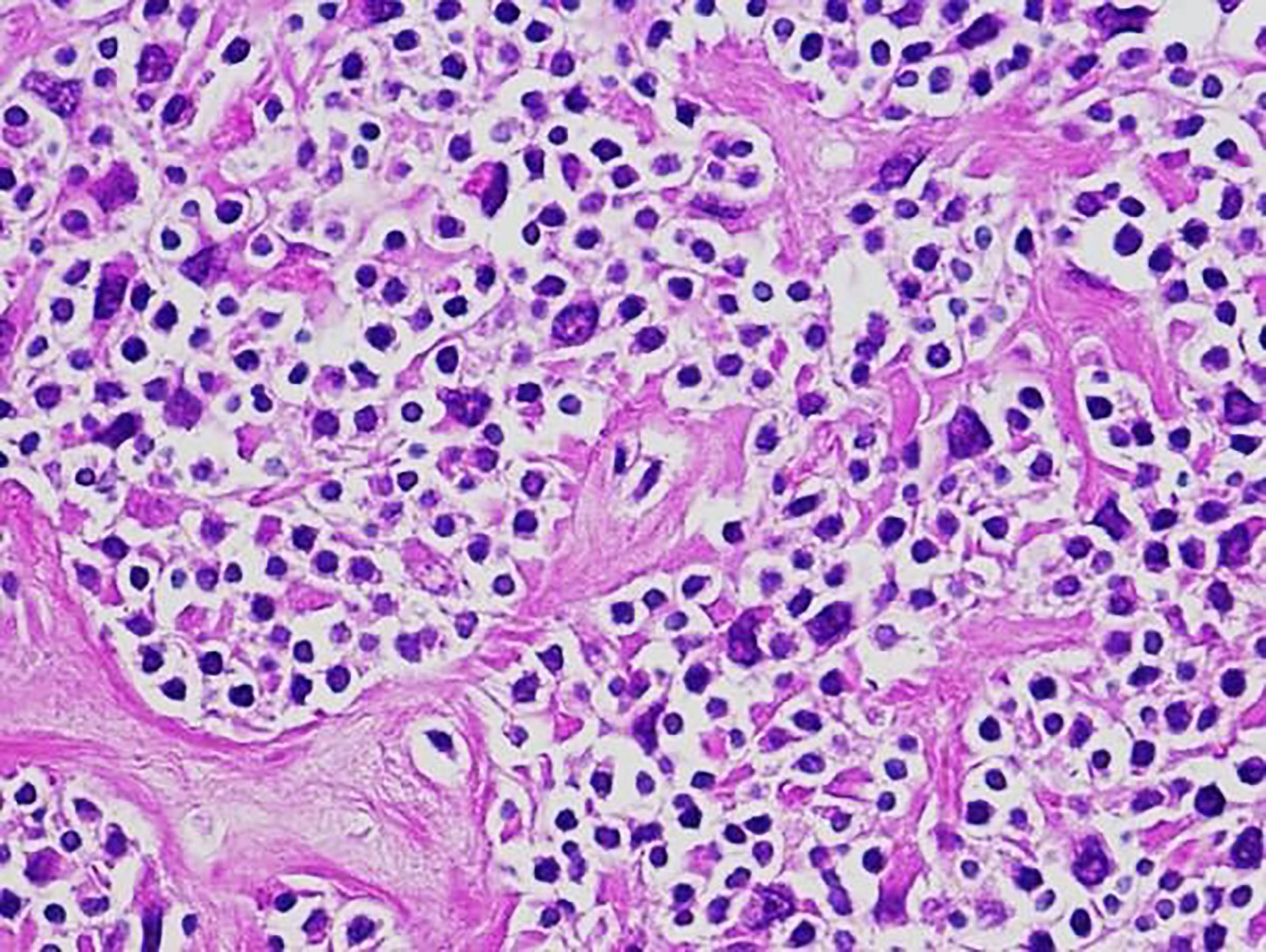
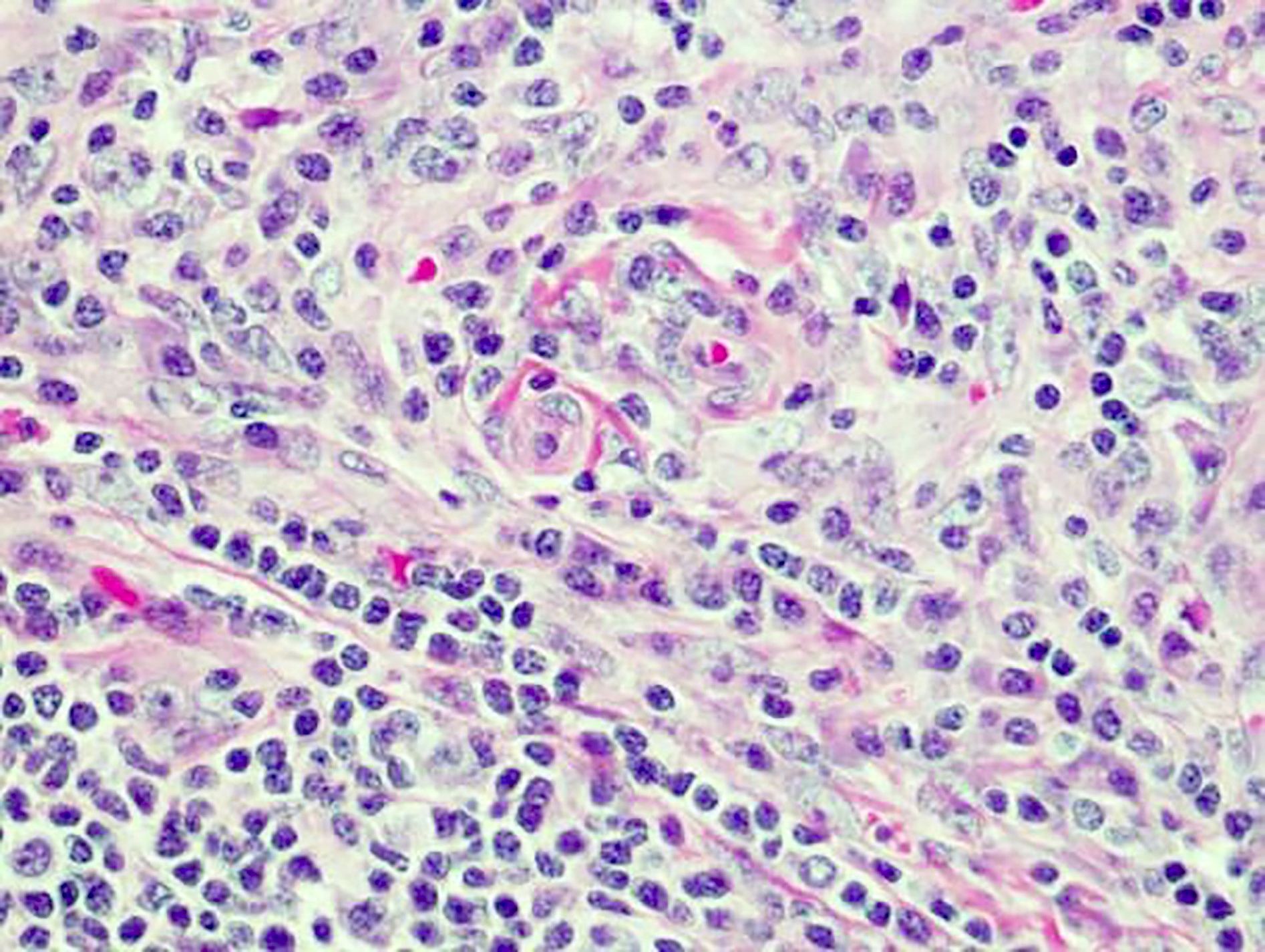
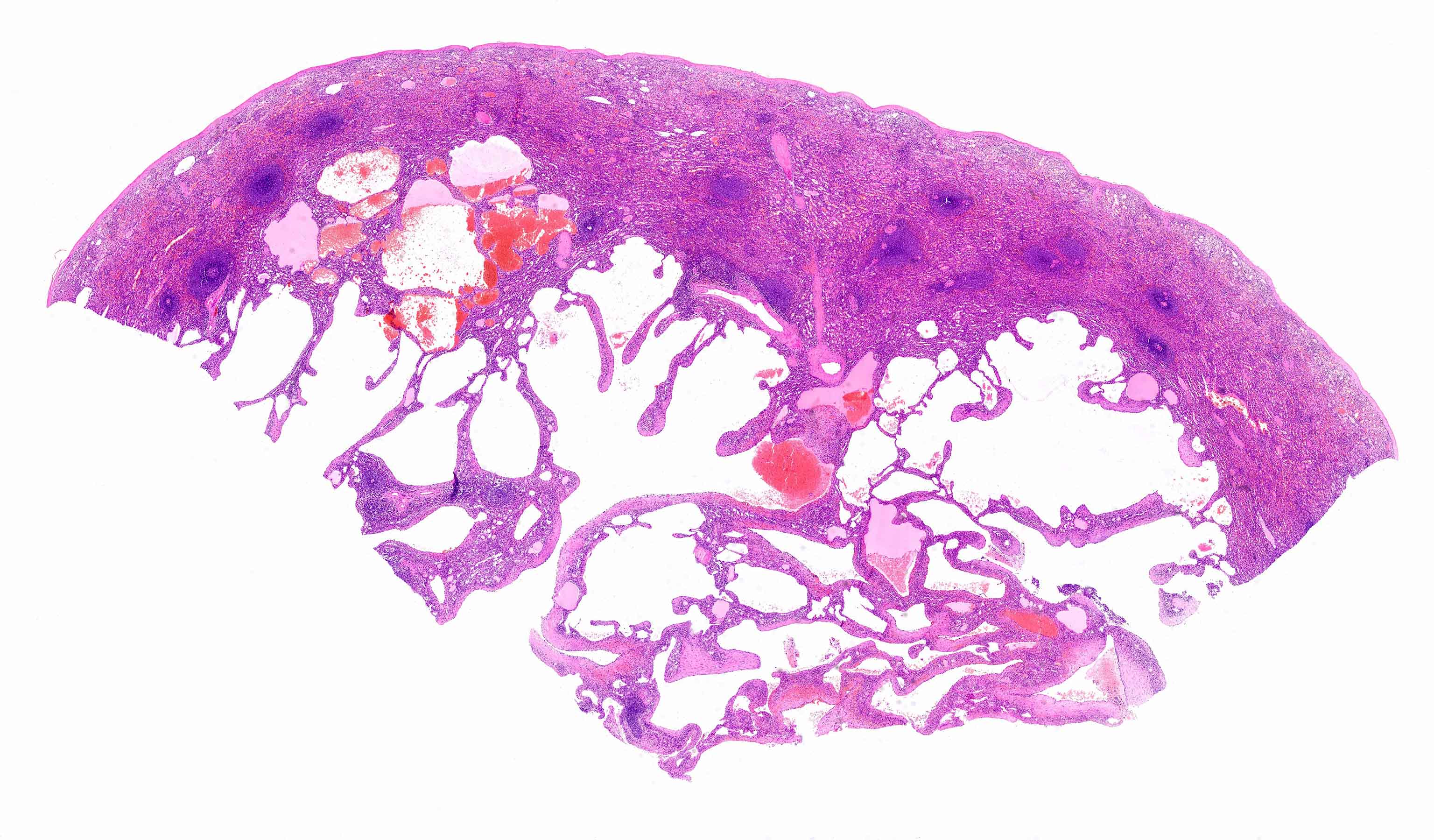
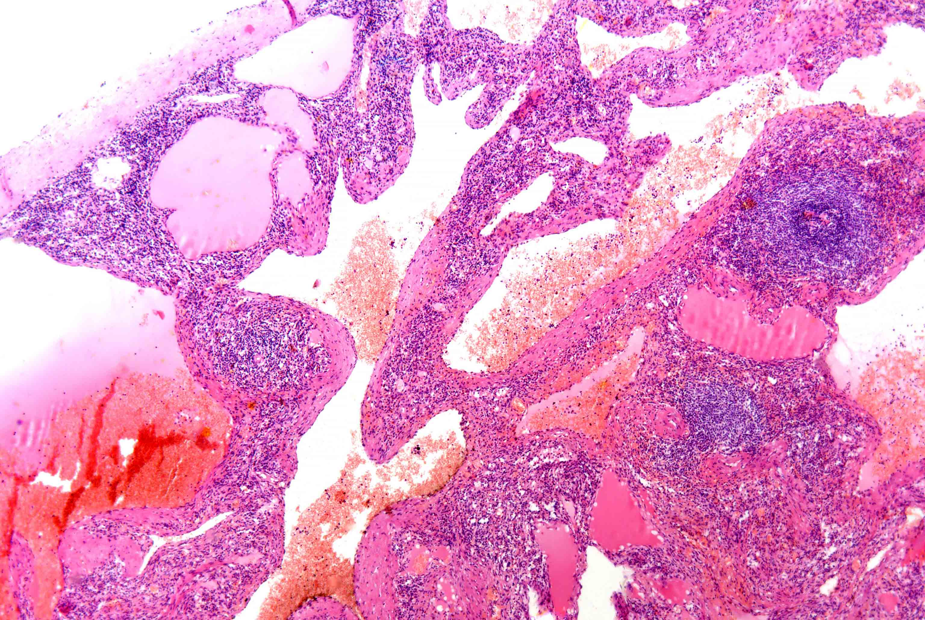
Vascular transformation of sinuses

Intra-abdominal lymph node
Nodular spindle cell vascular transformation
Ashton-Key: 2016

Auerbach: 2022
Crane: 2021
Duffield: 2020
Hsi: 2017
Hudnall: 2019
Jaffe: 2016
Jiang: 2023
Medeiros: 2017
Medeiros: 2021
Medeiros: 2023
Naeim: 2018
Wang: 2020
Find related Pathology books: cytopathology, hematopathology
