
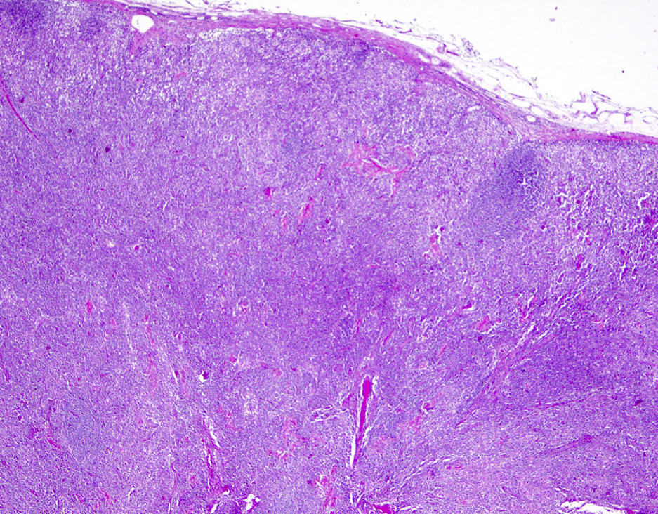
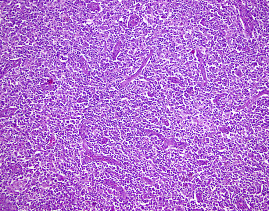
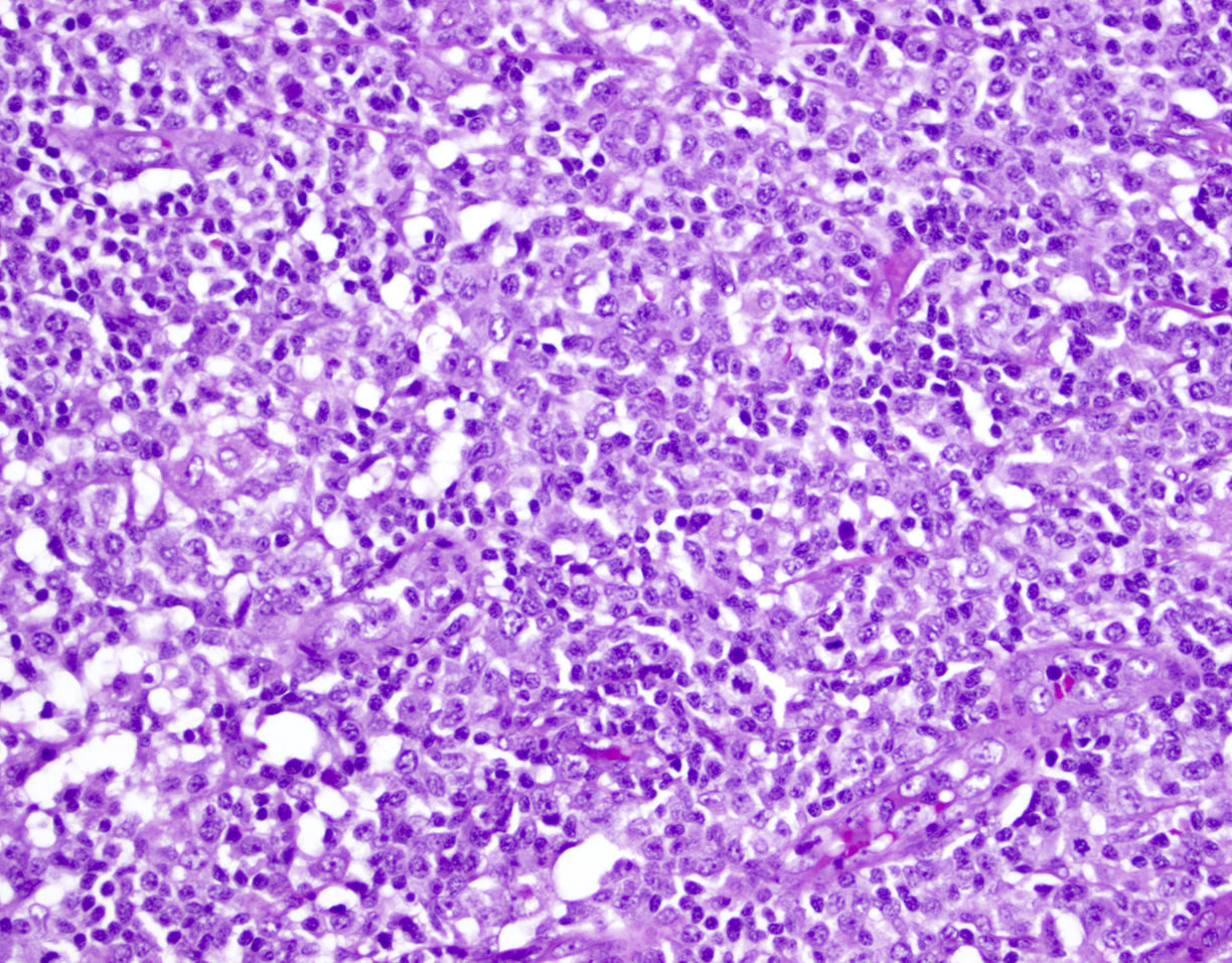
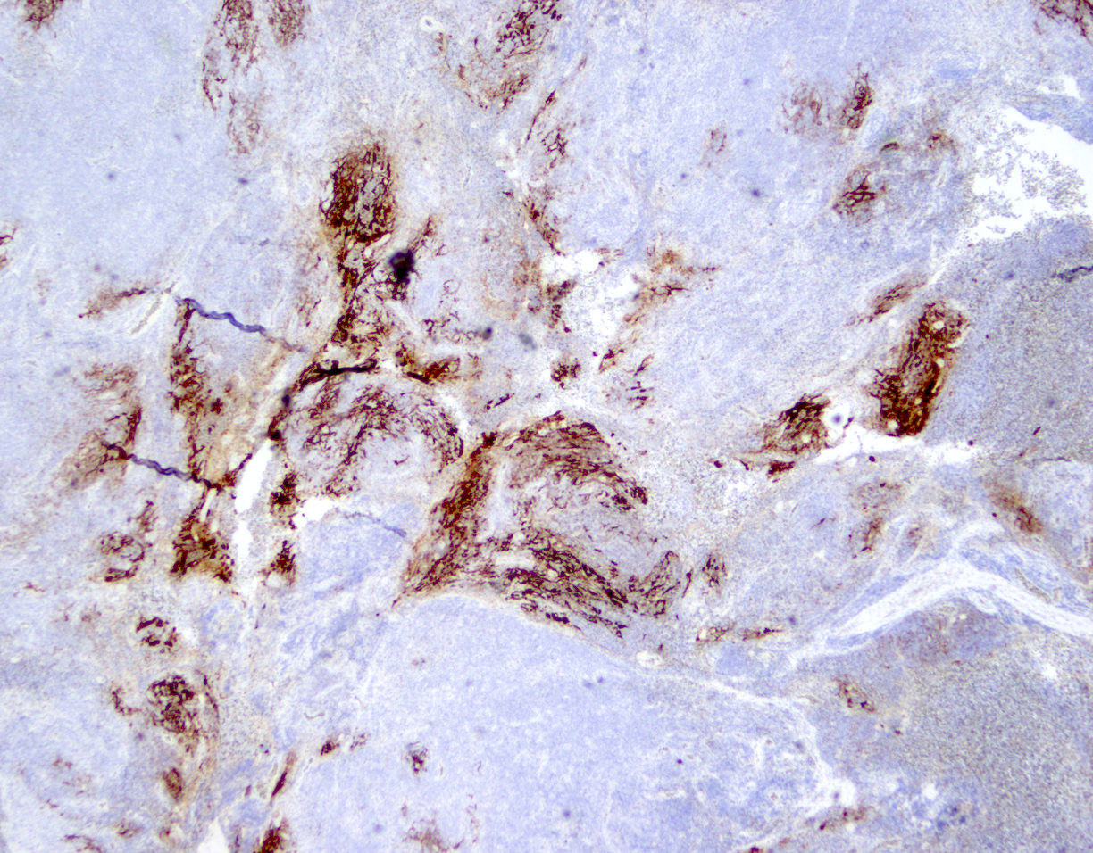
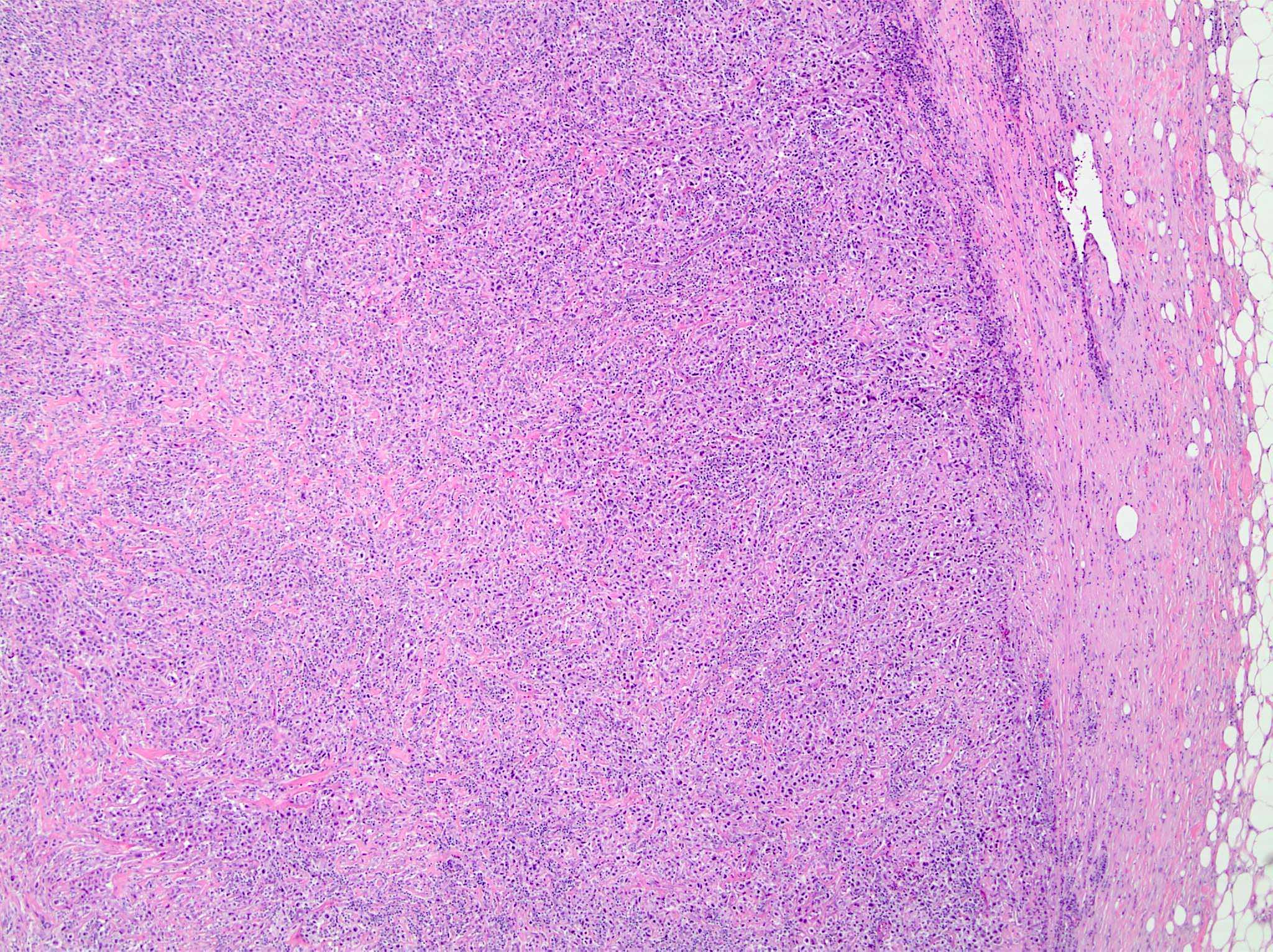
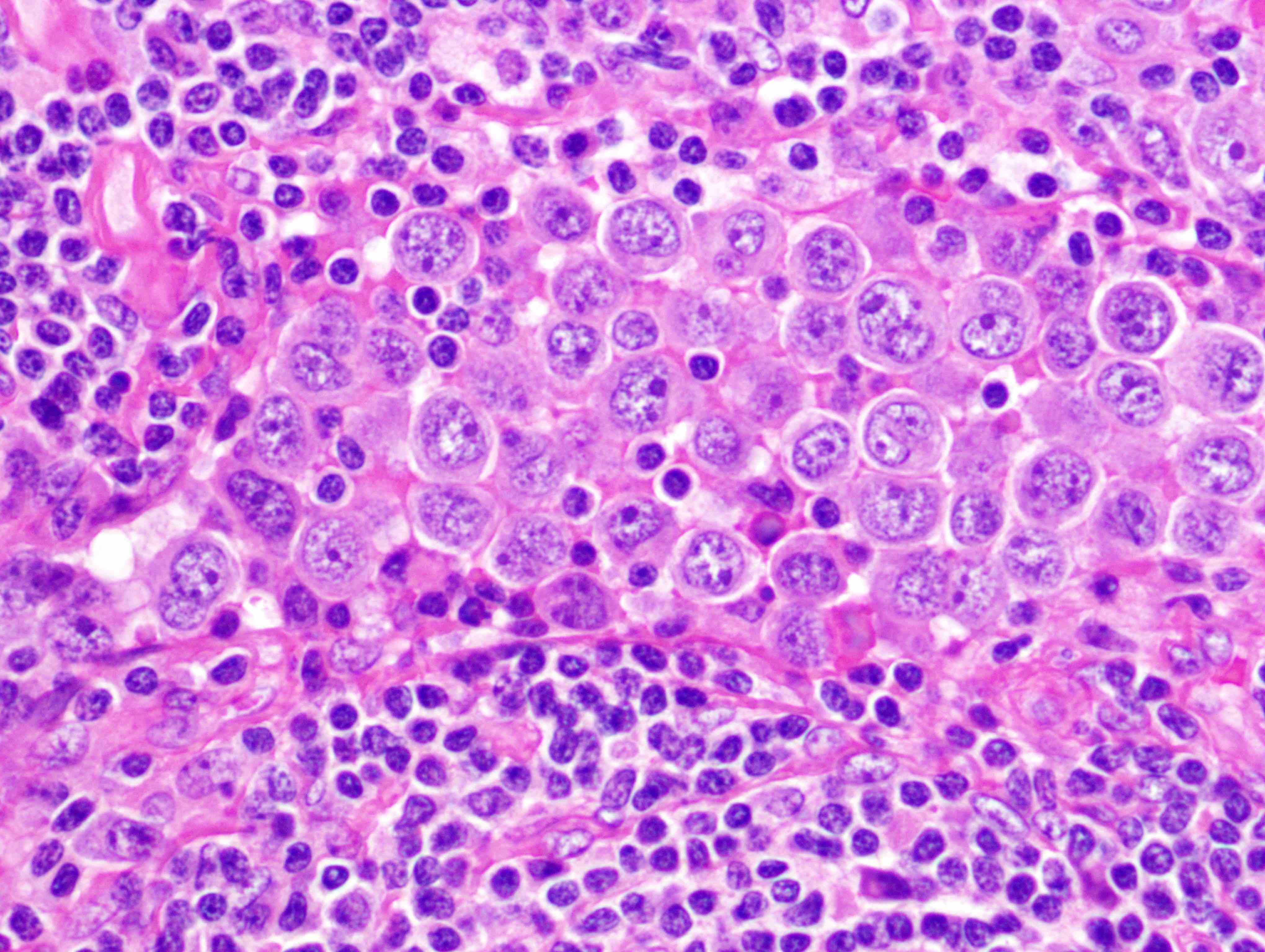
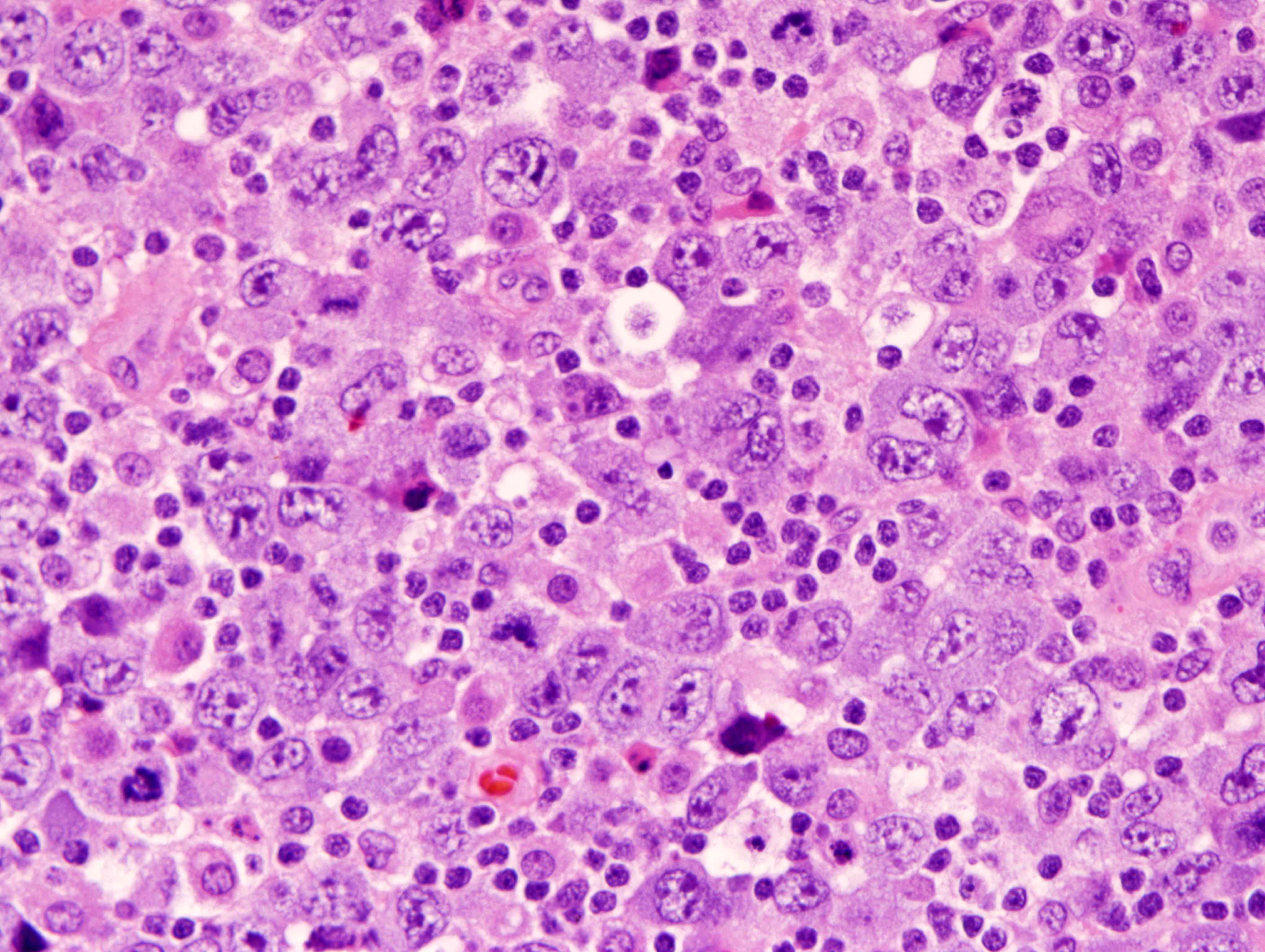
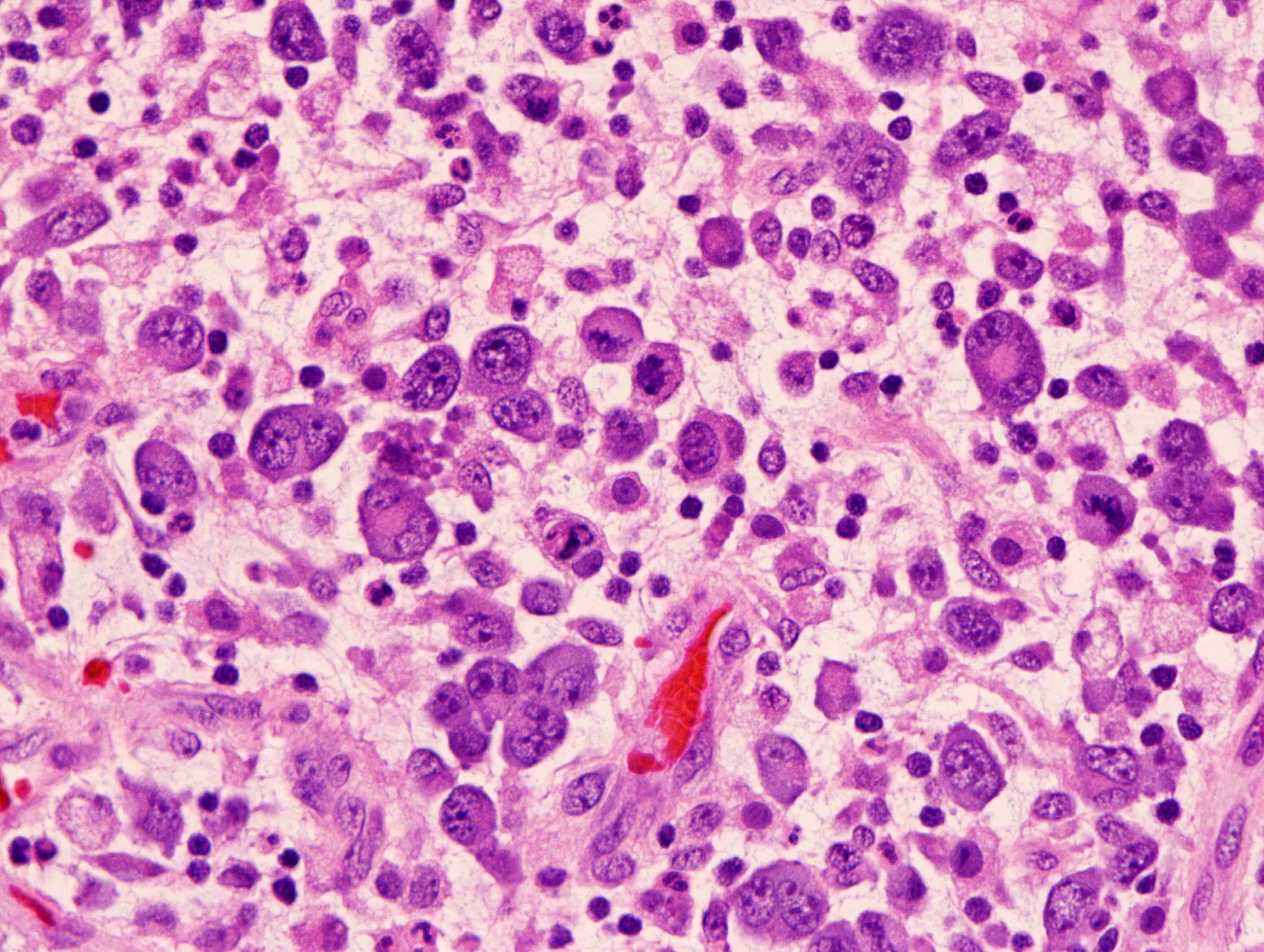
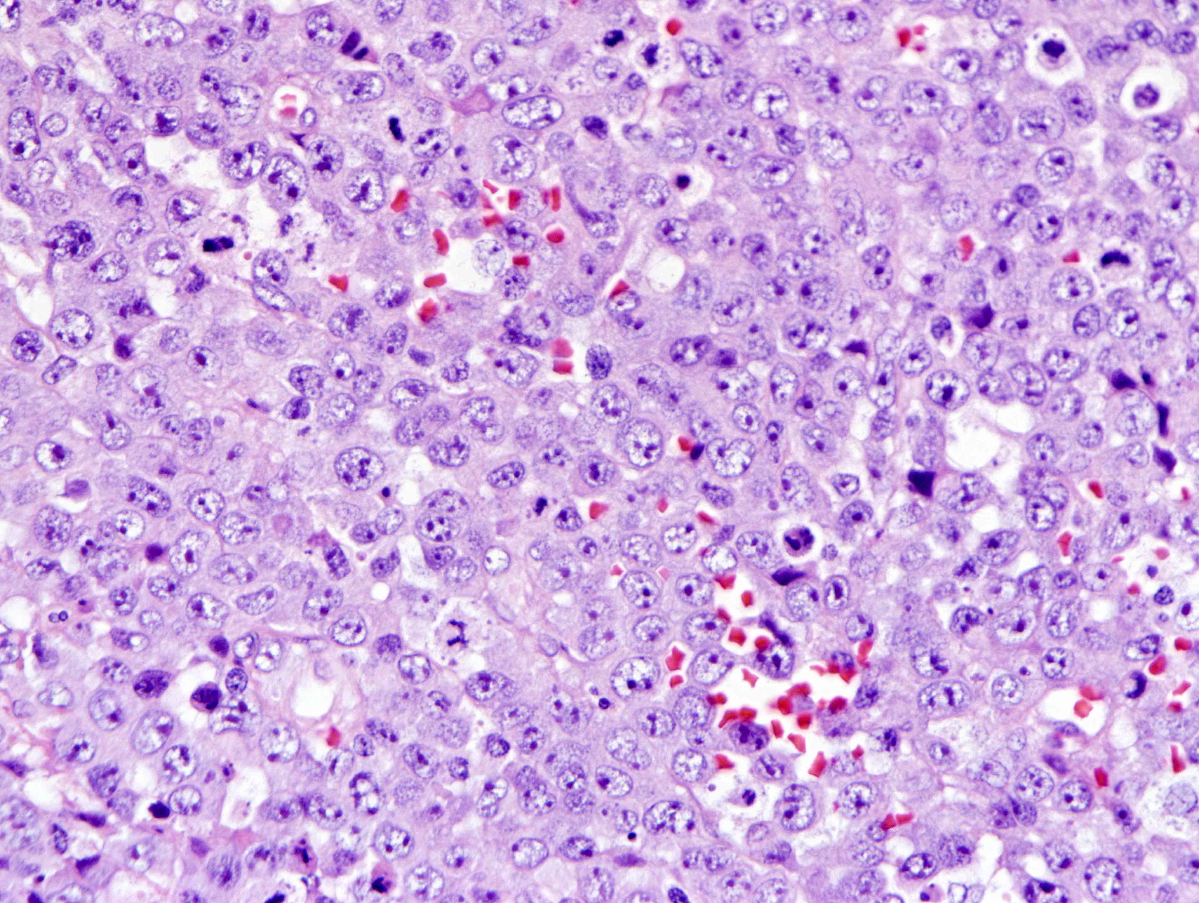
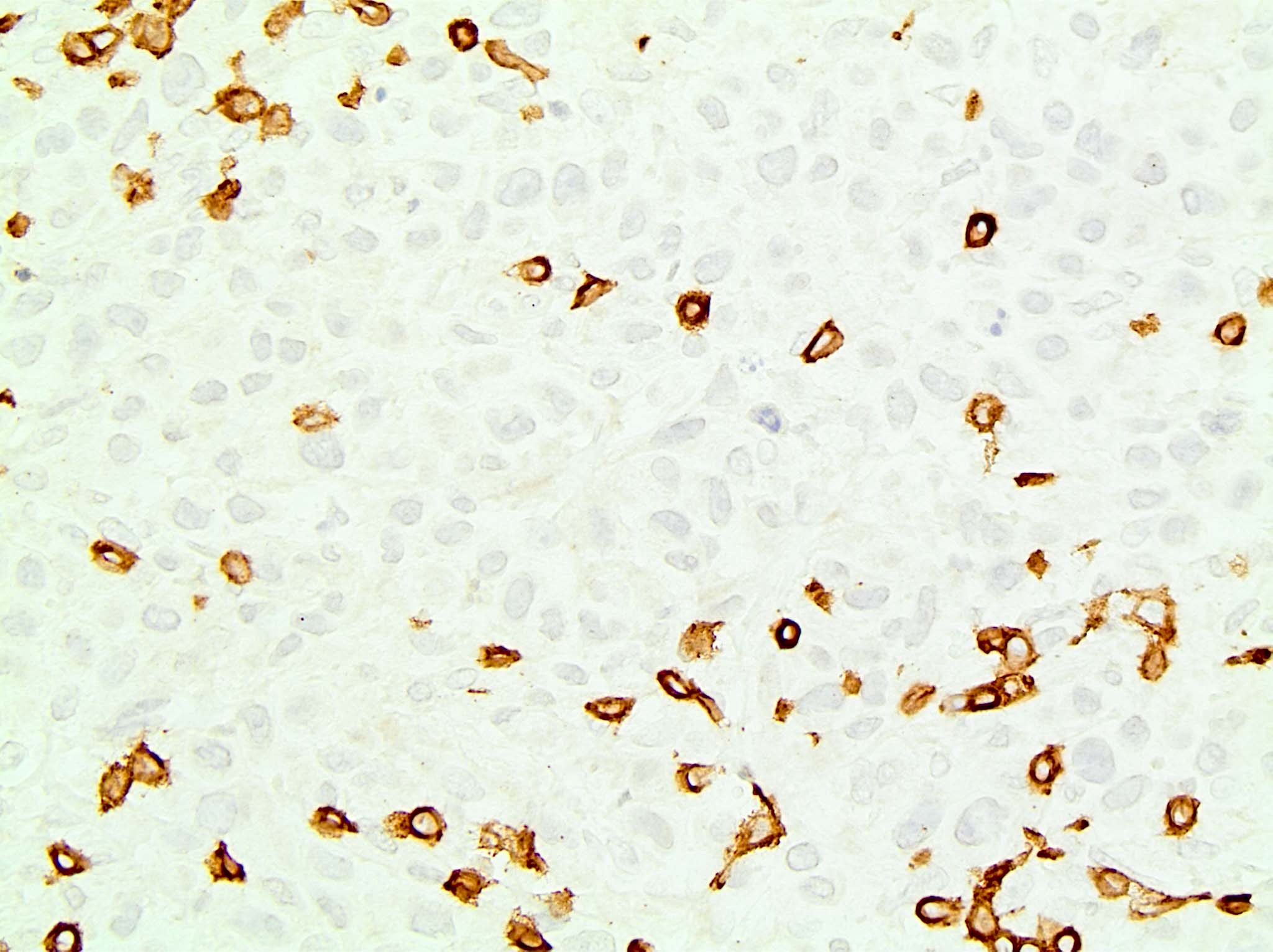
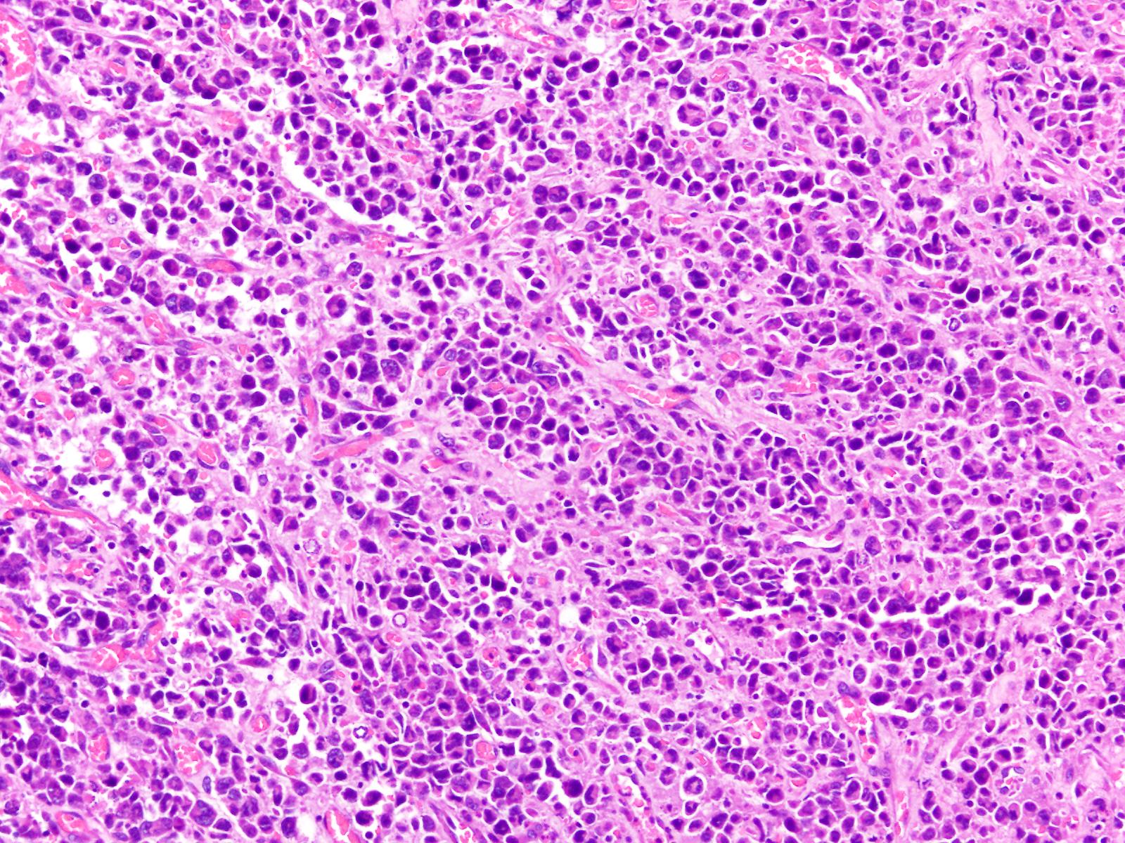
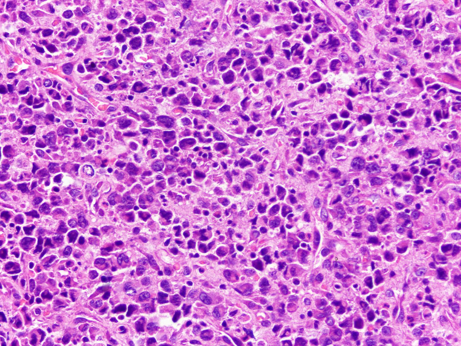
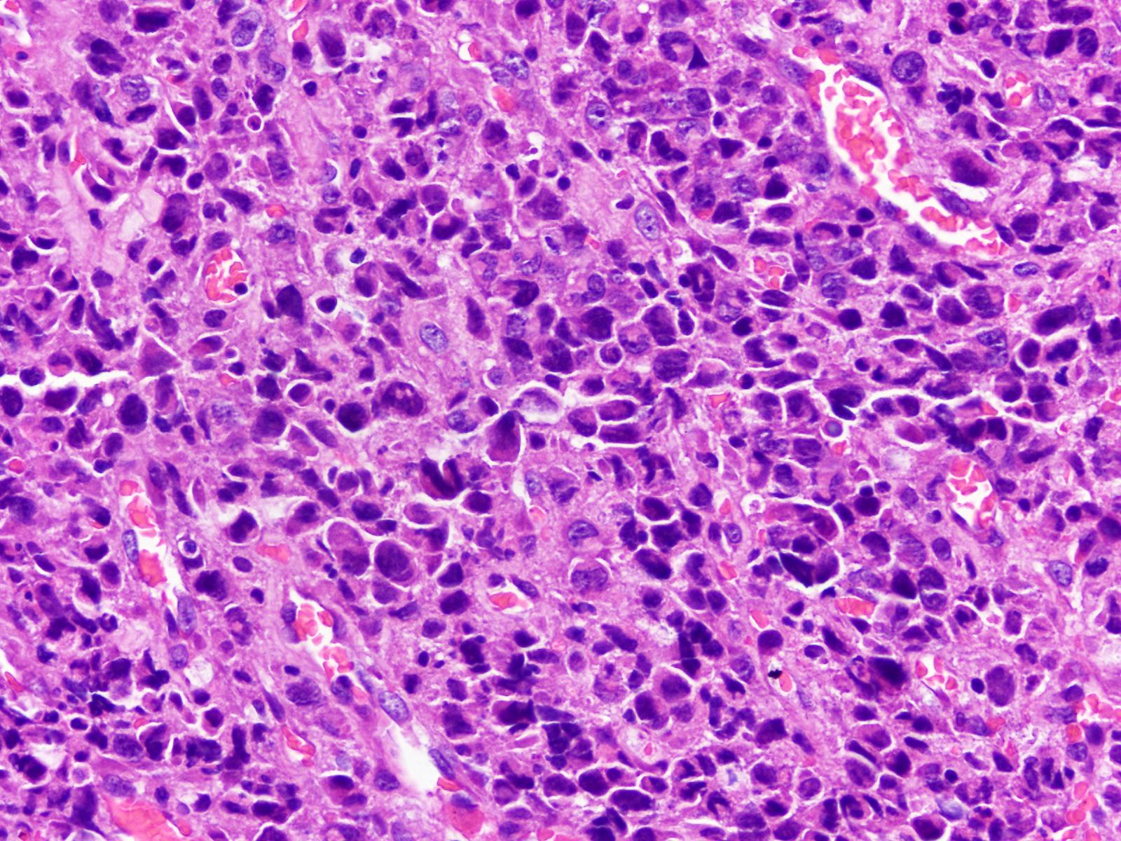
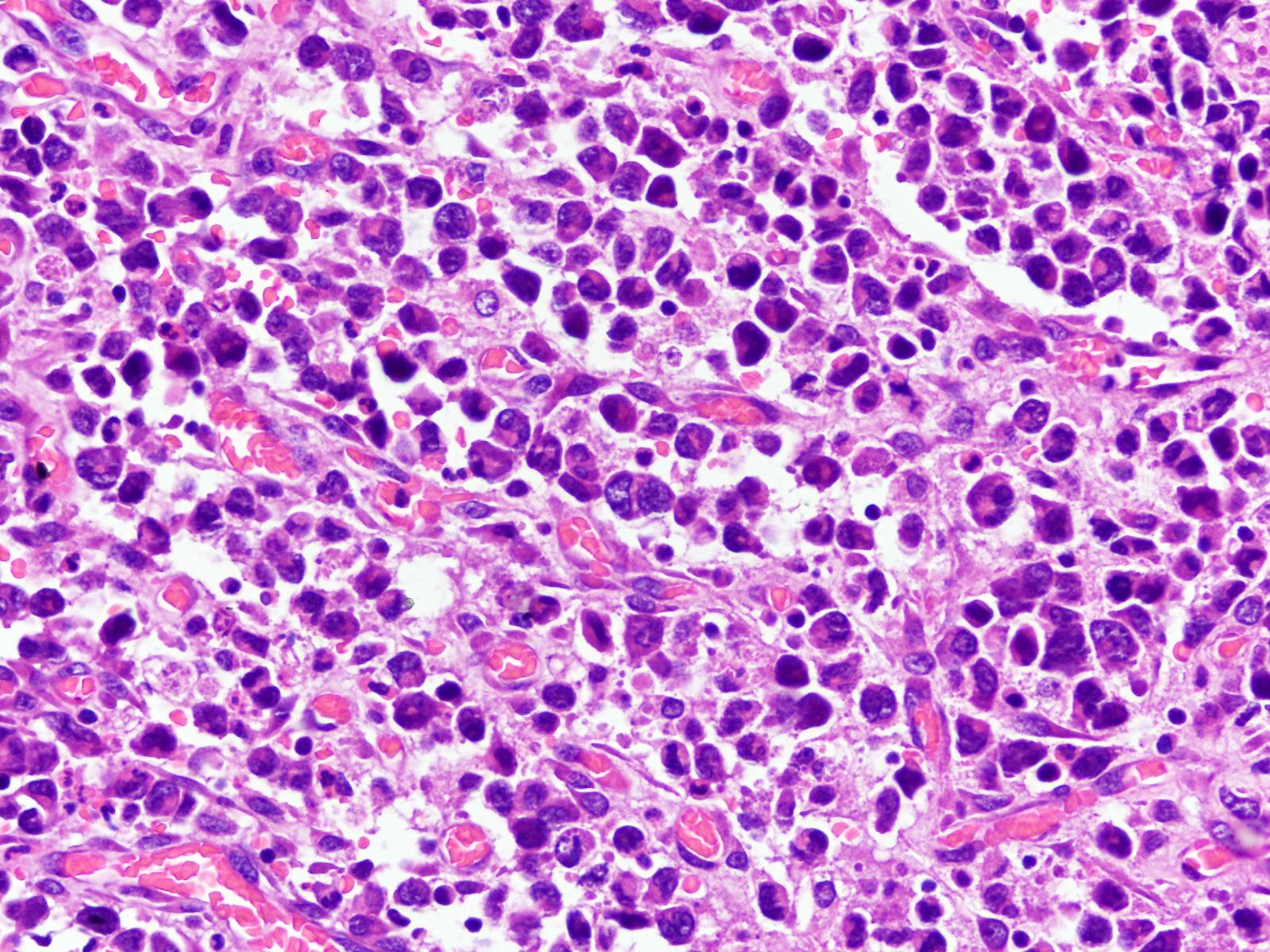
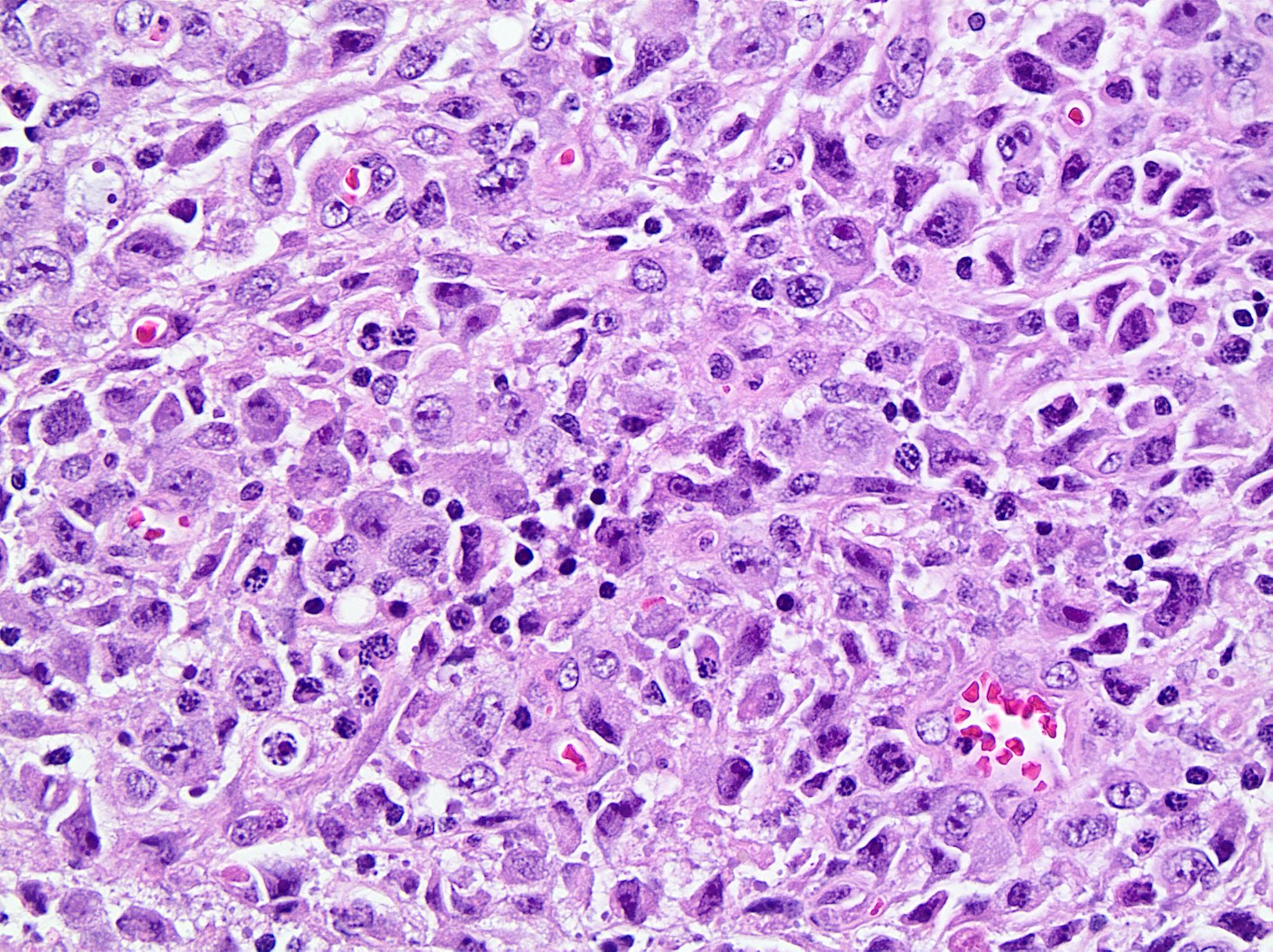
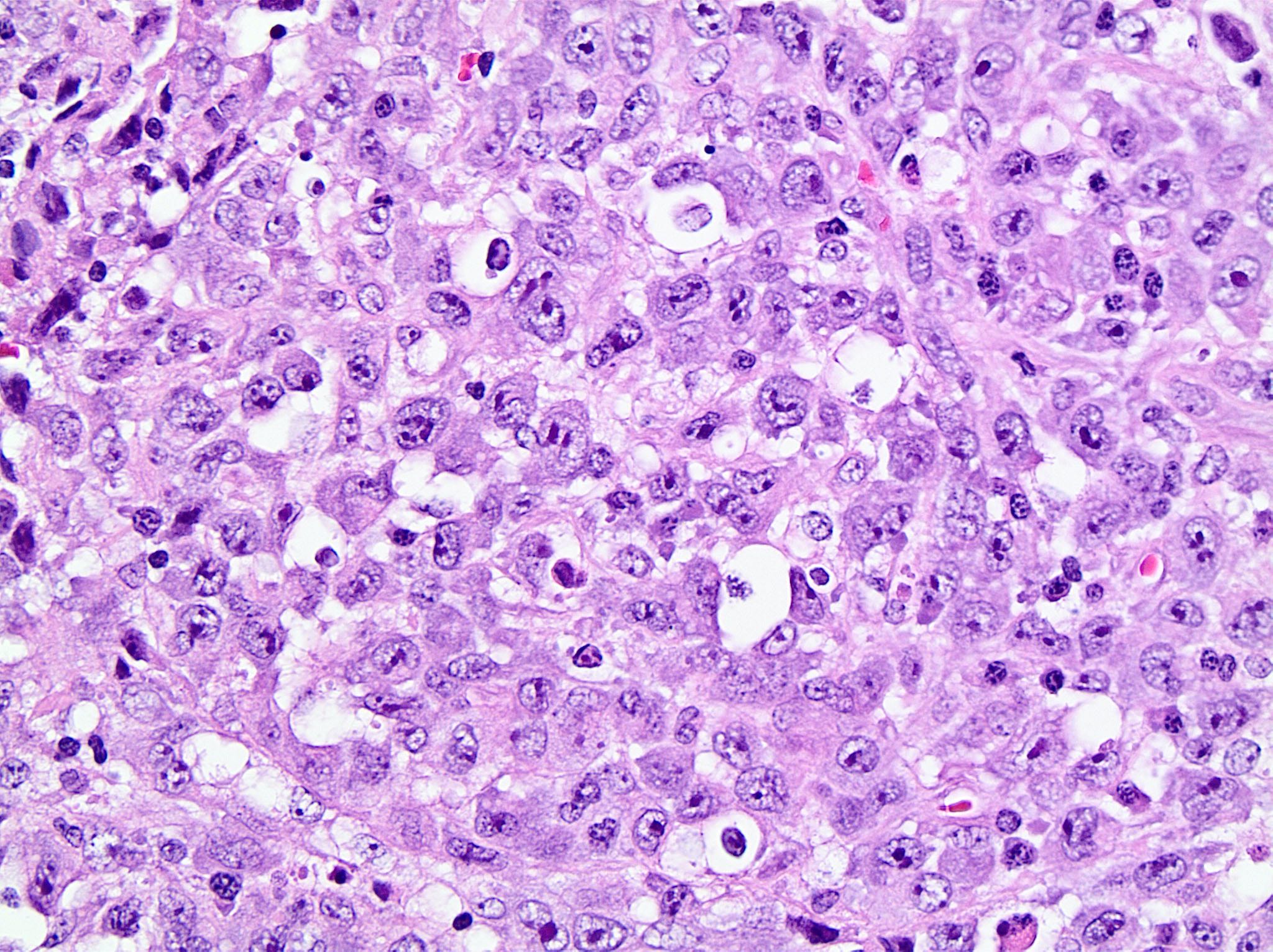
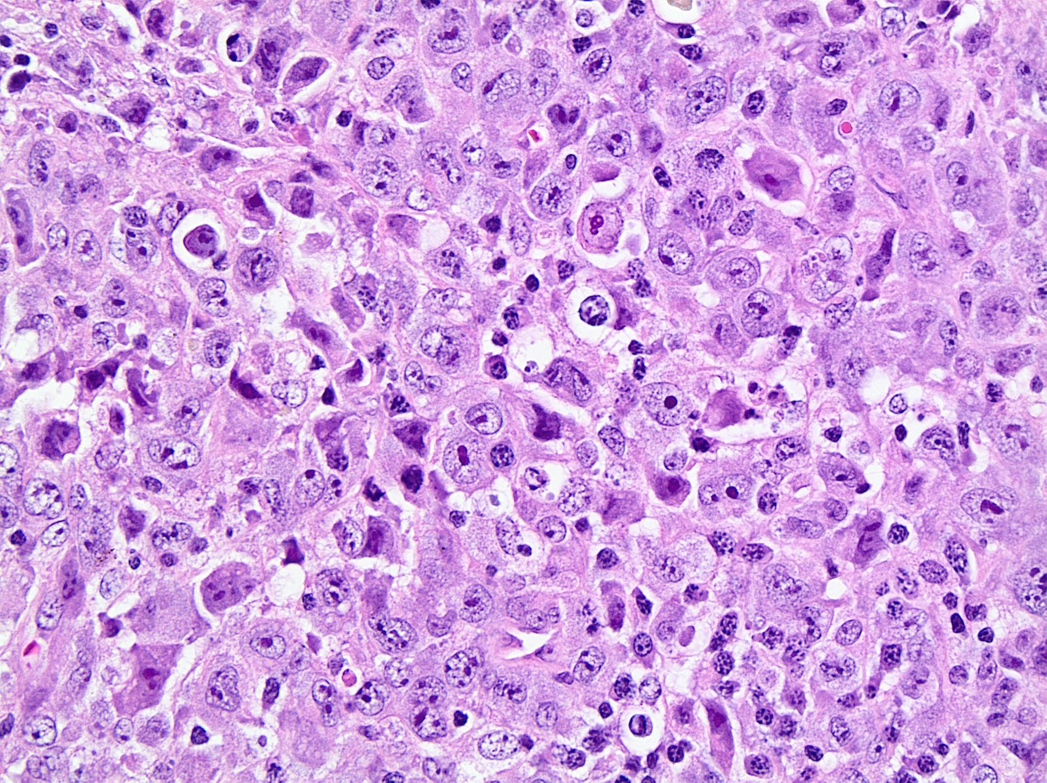
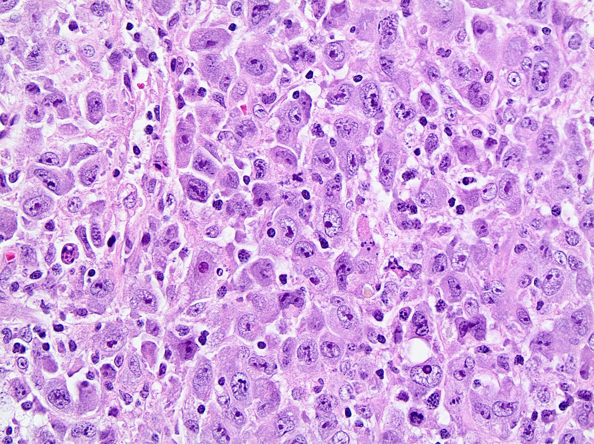
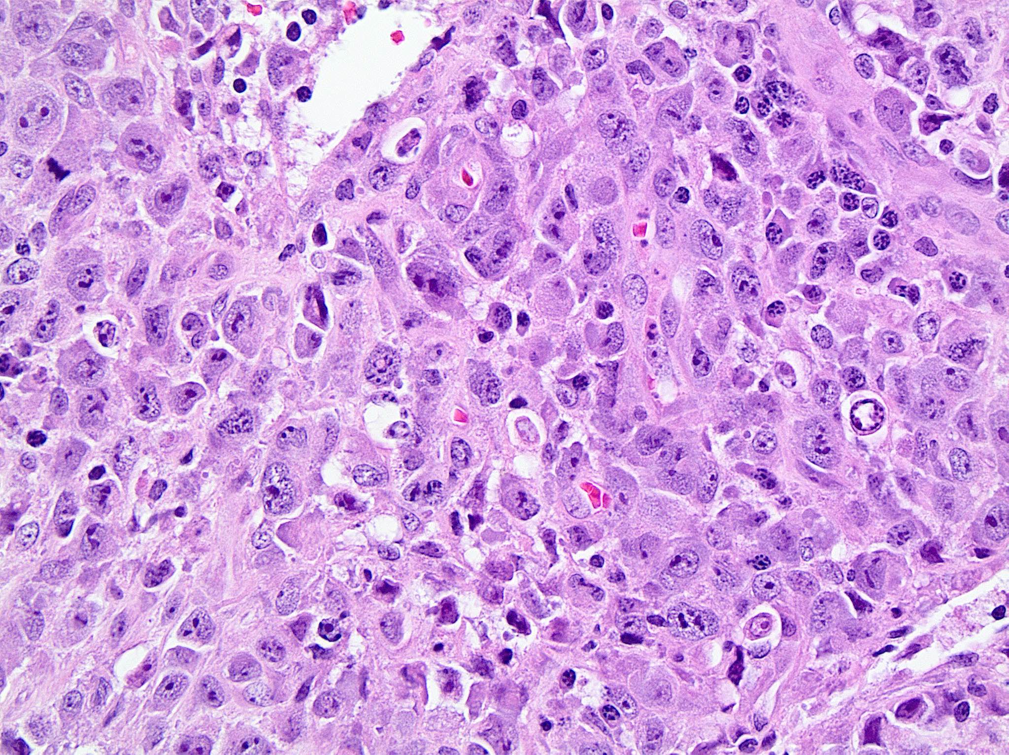
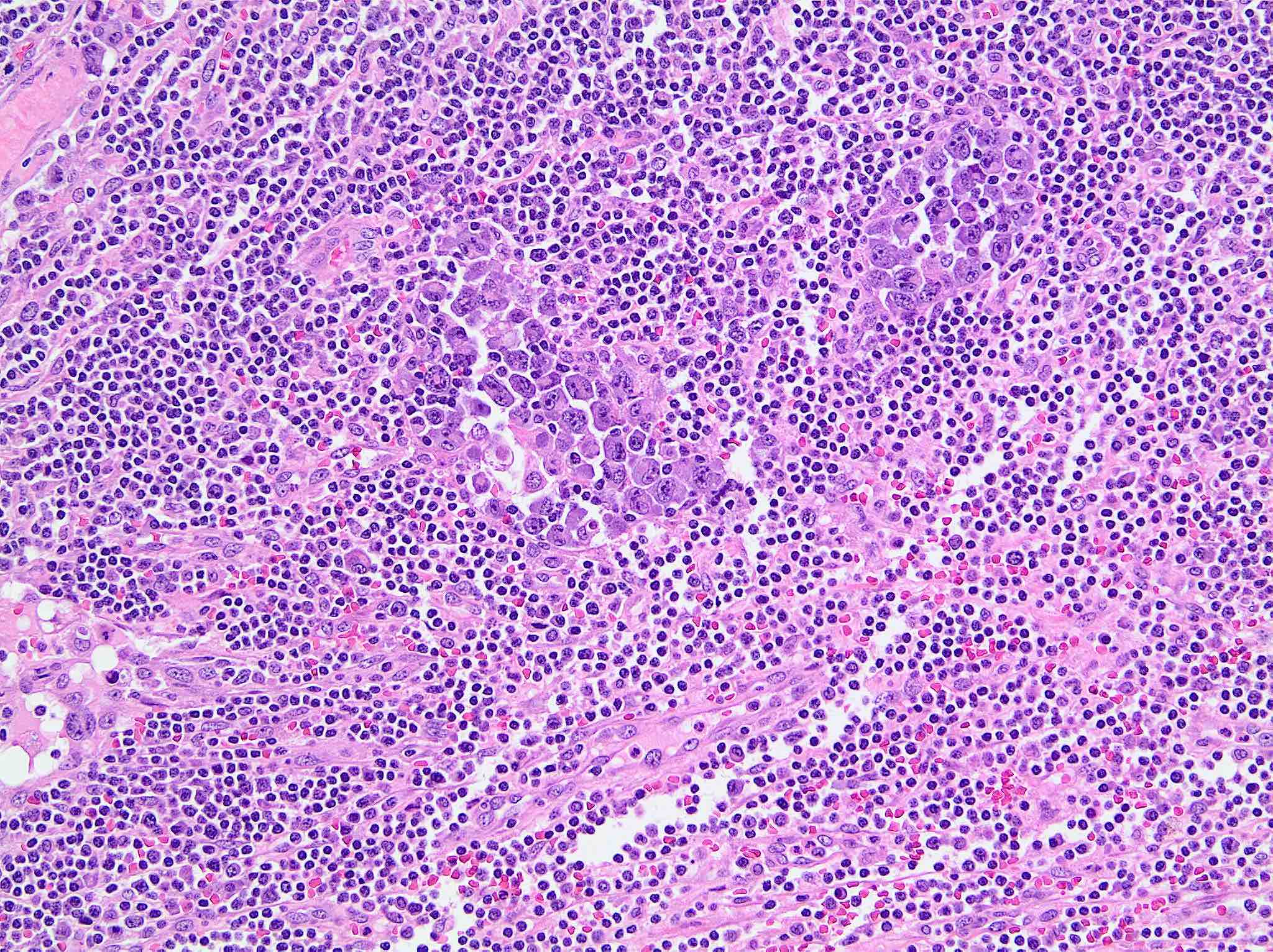
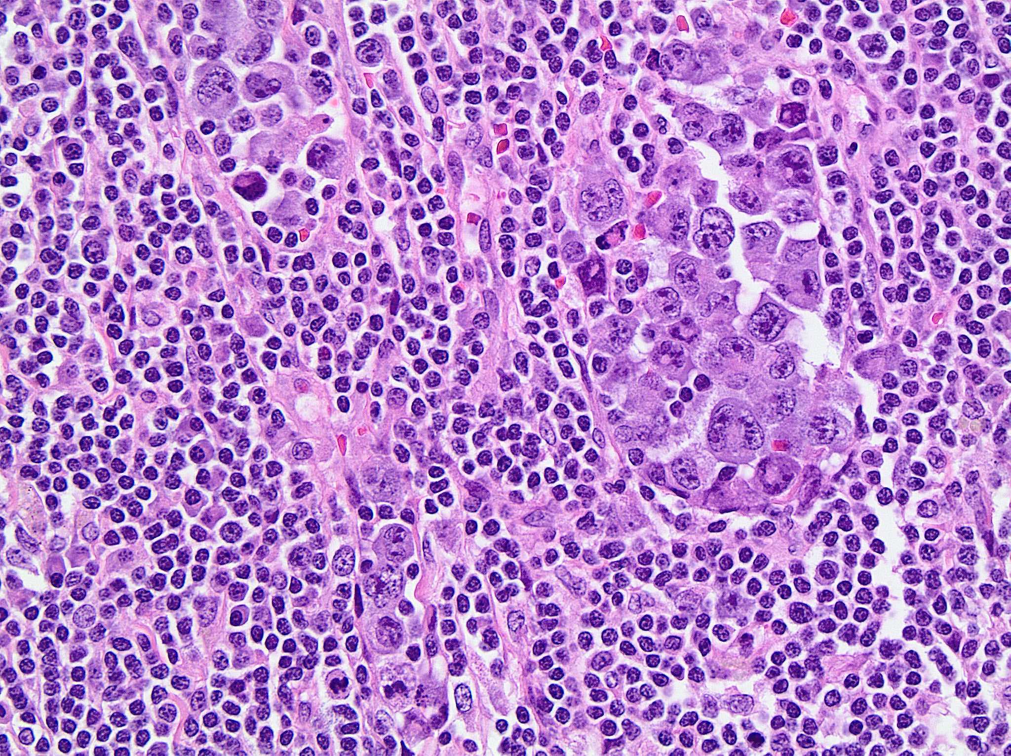
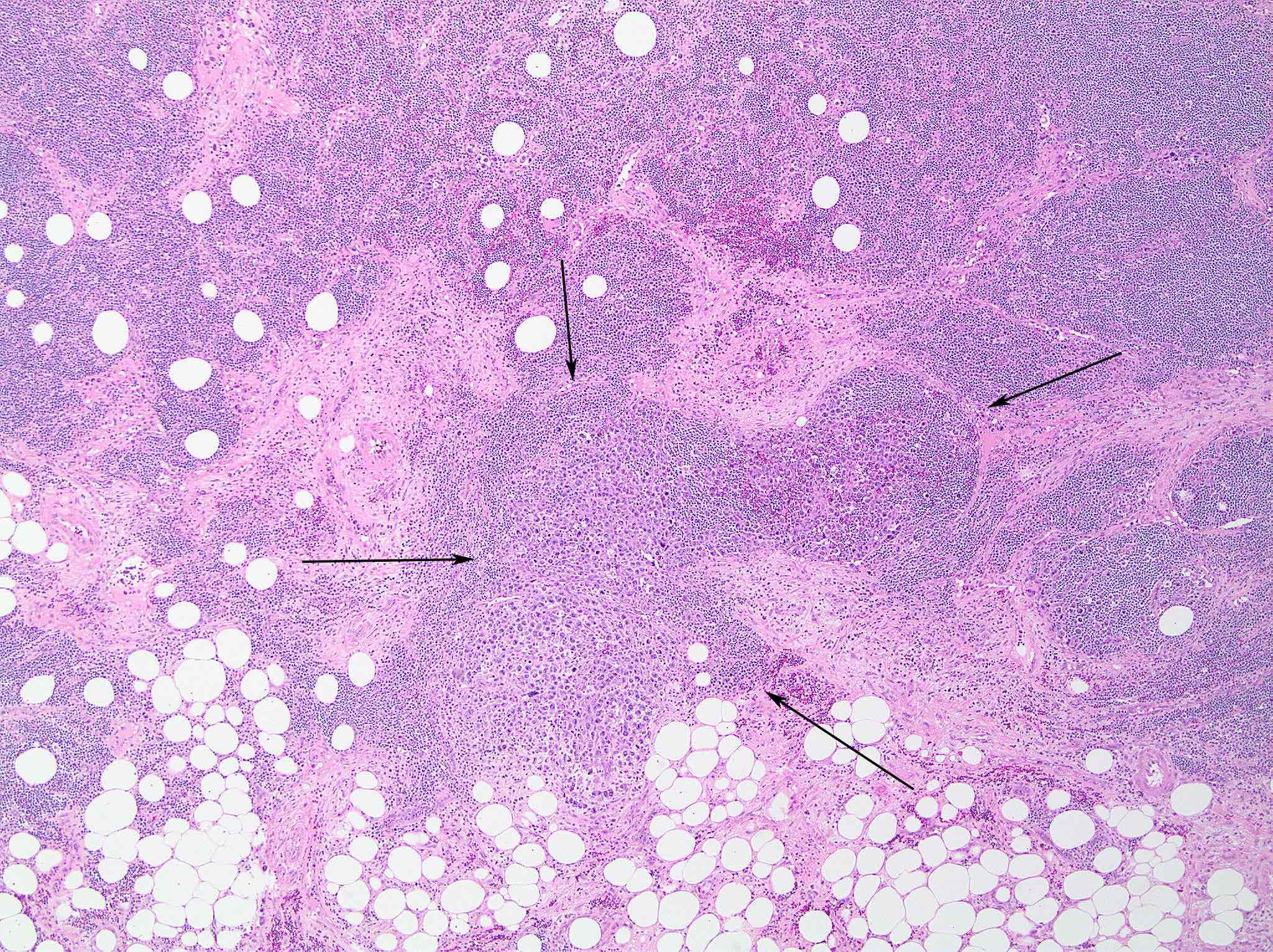
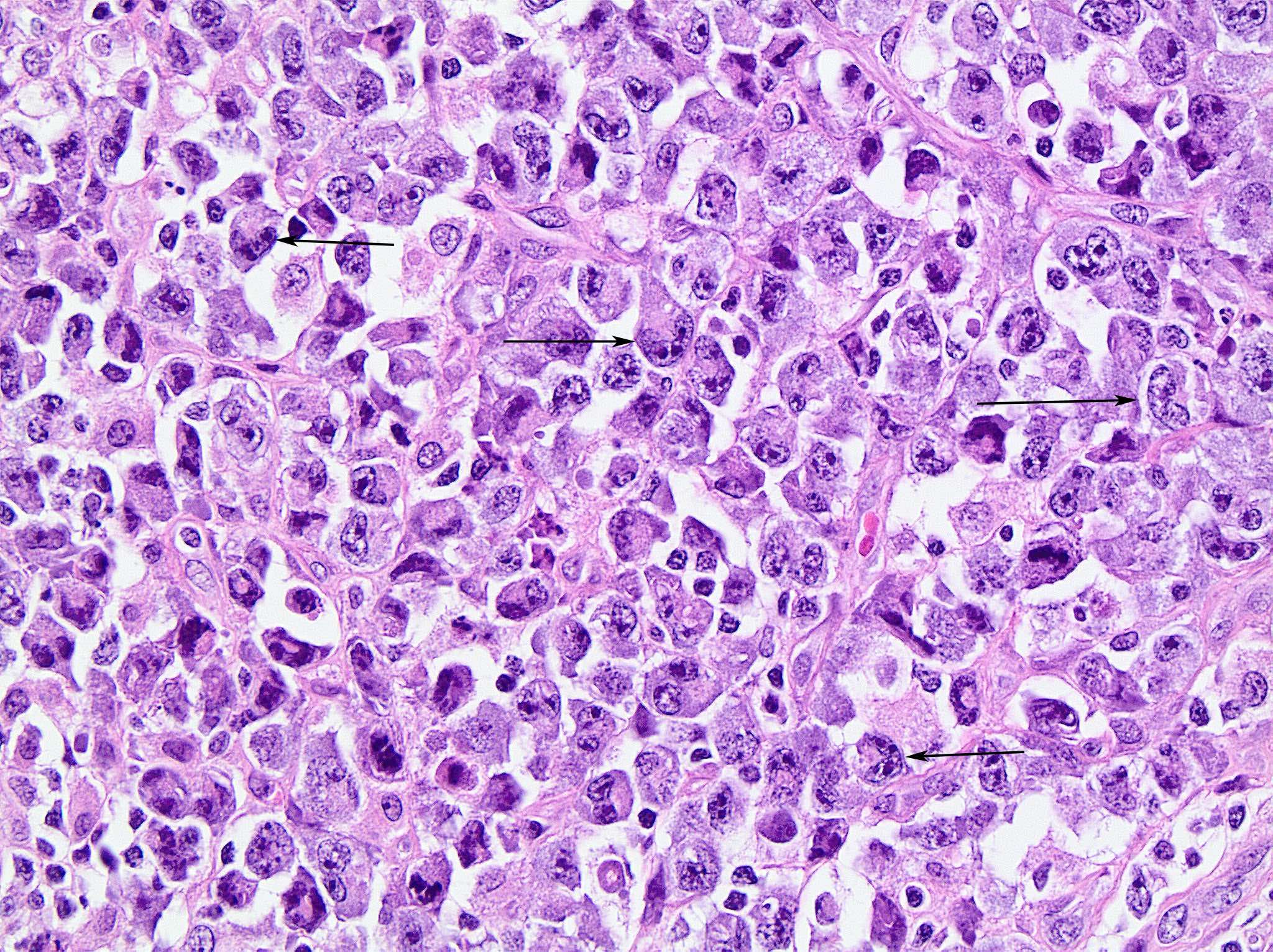
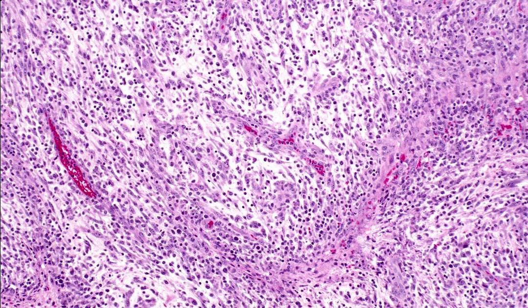
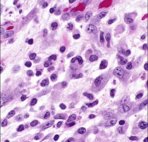
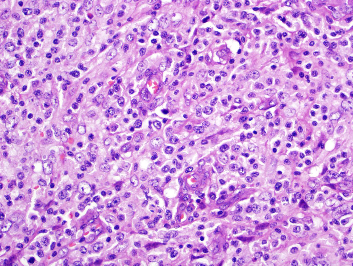
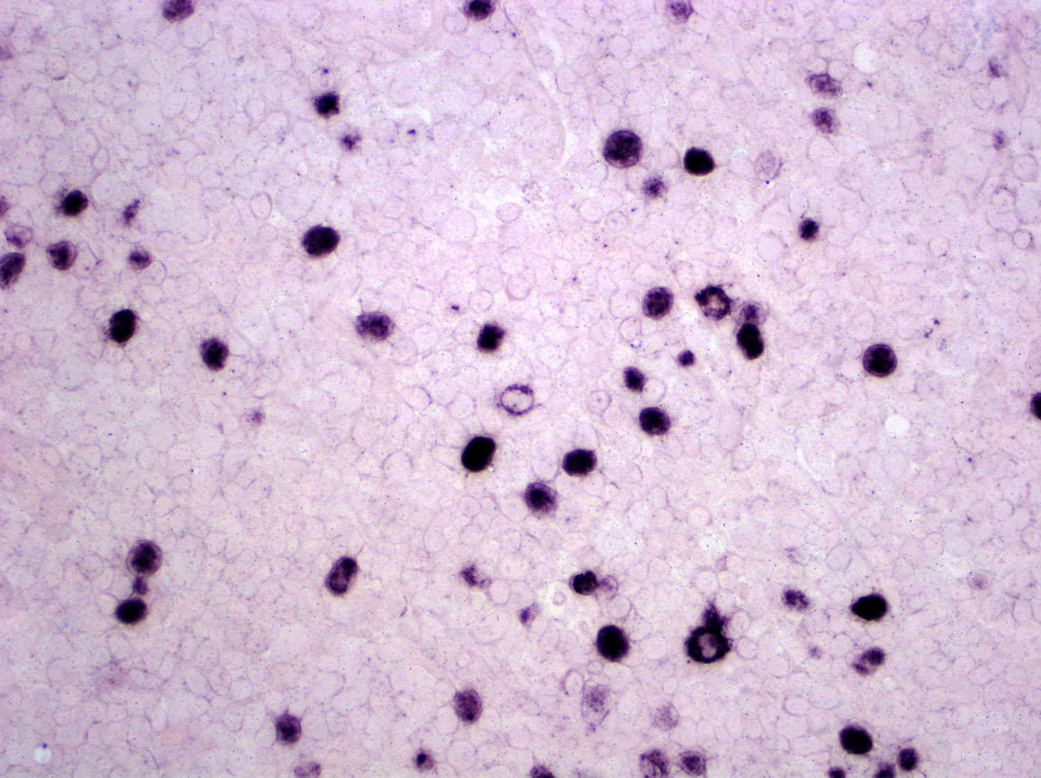
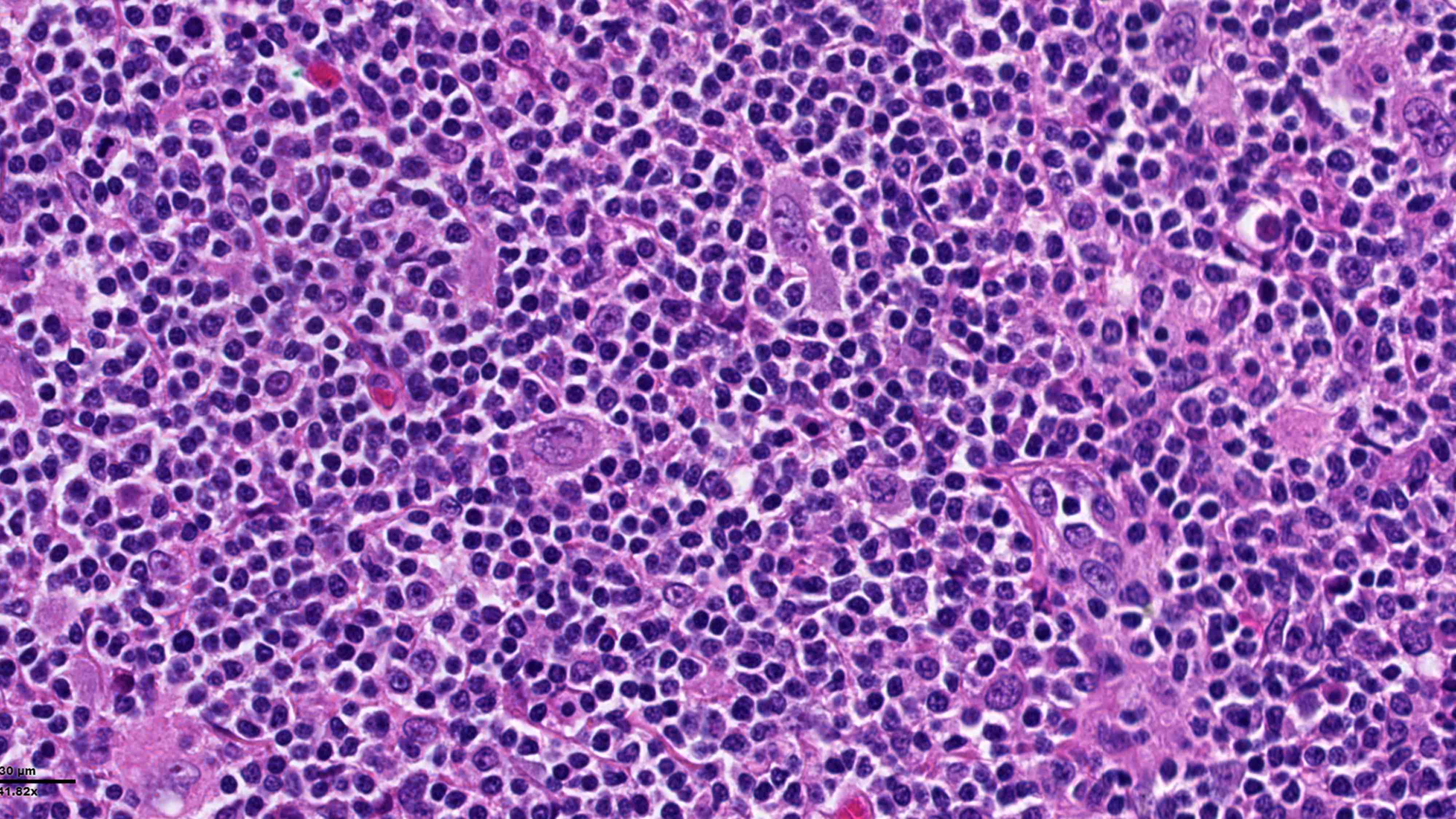
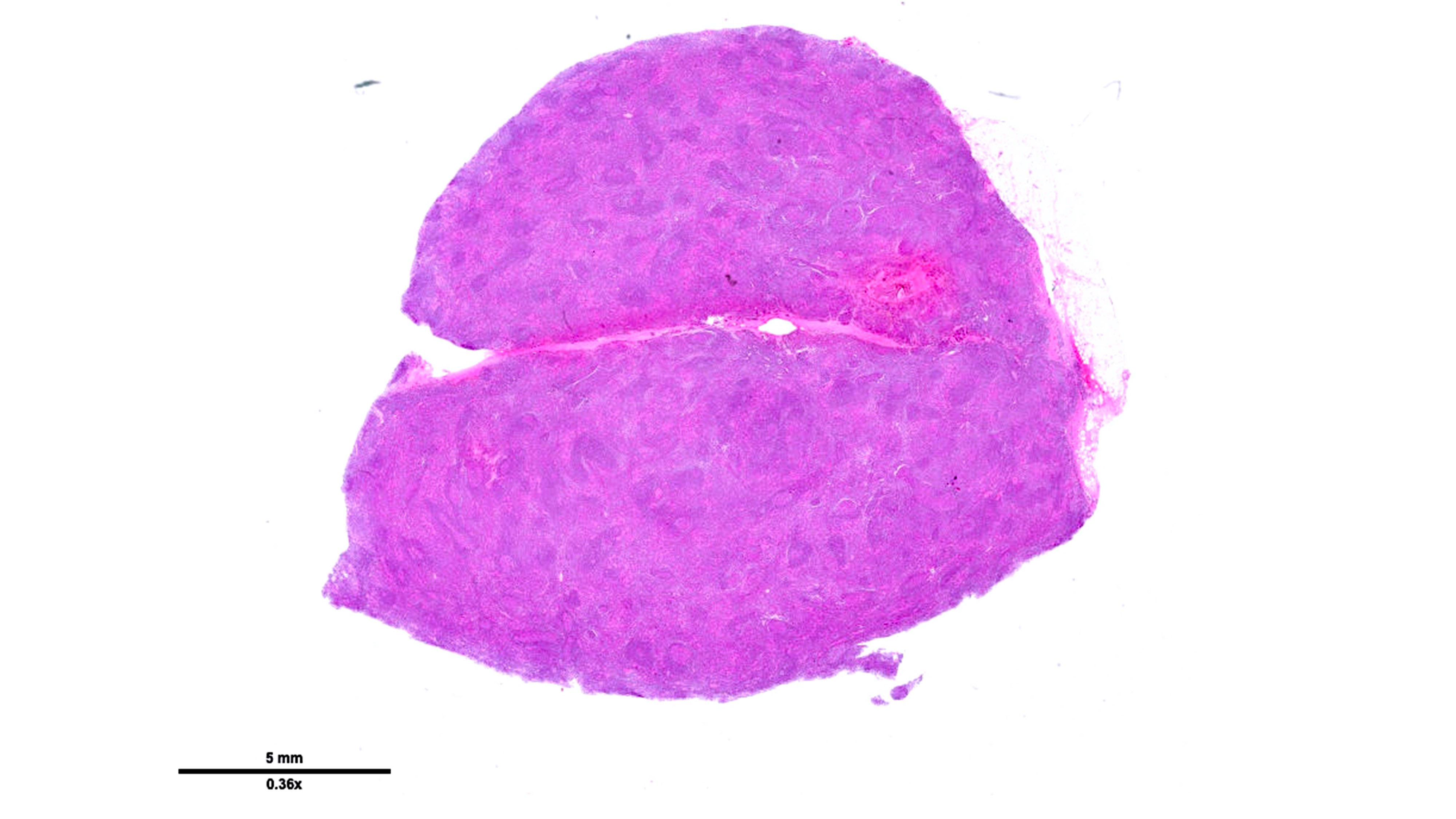
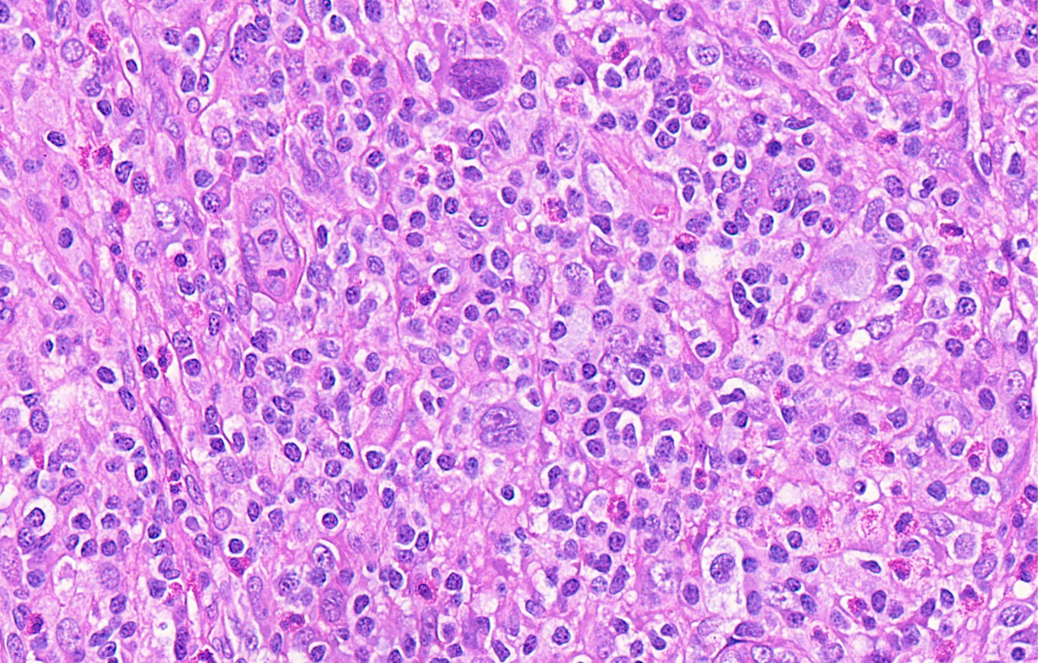
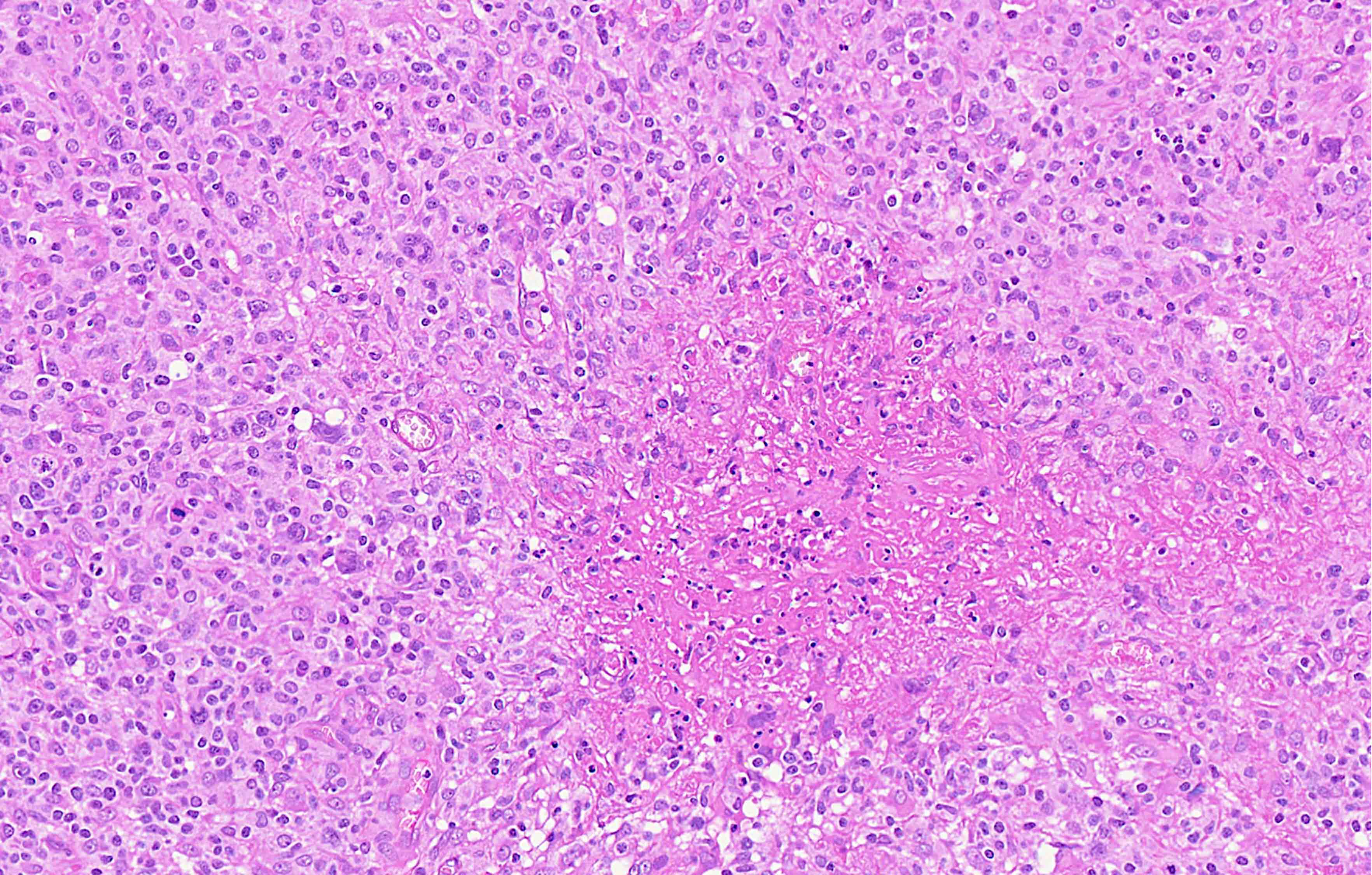
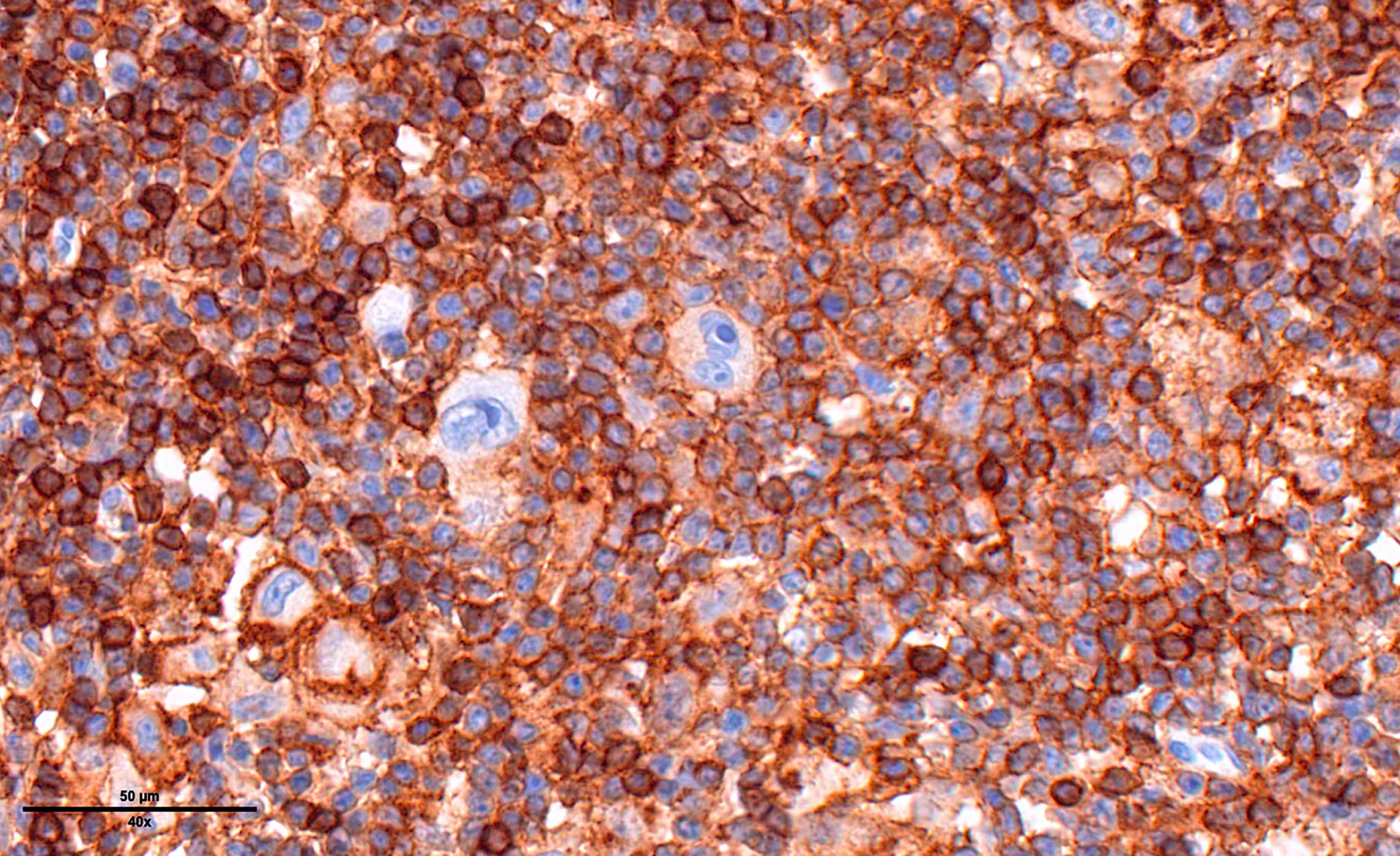
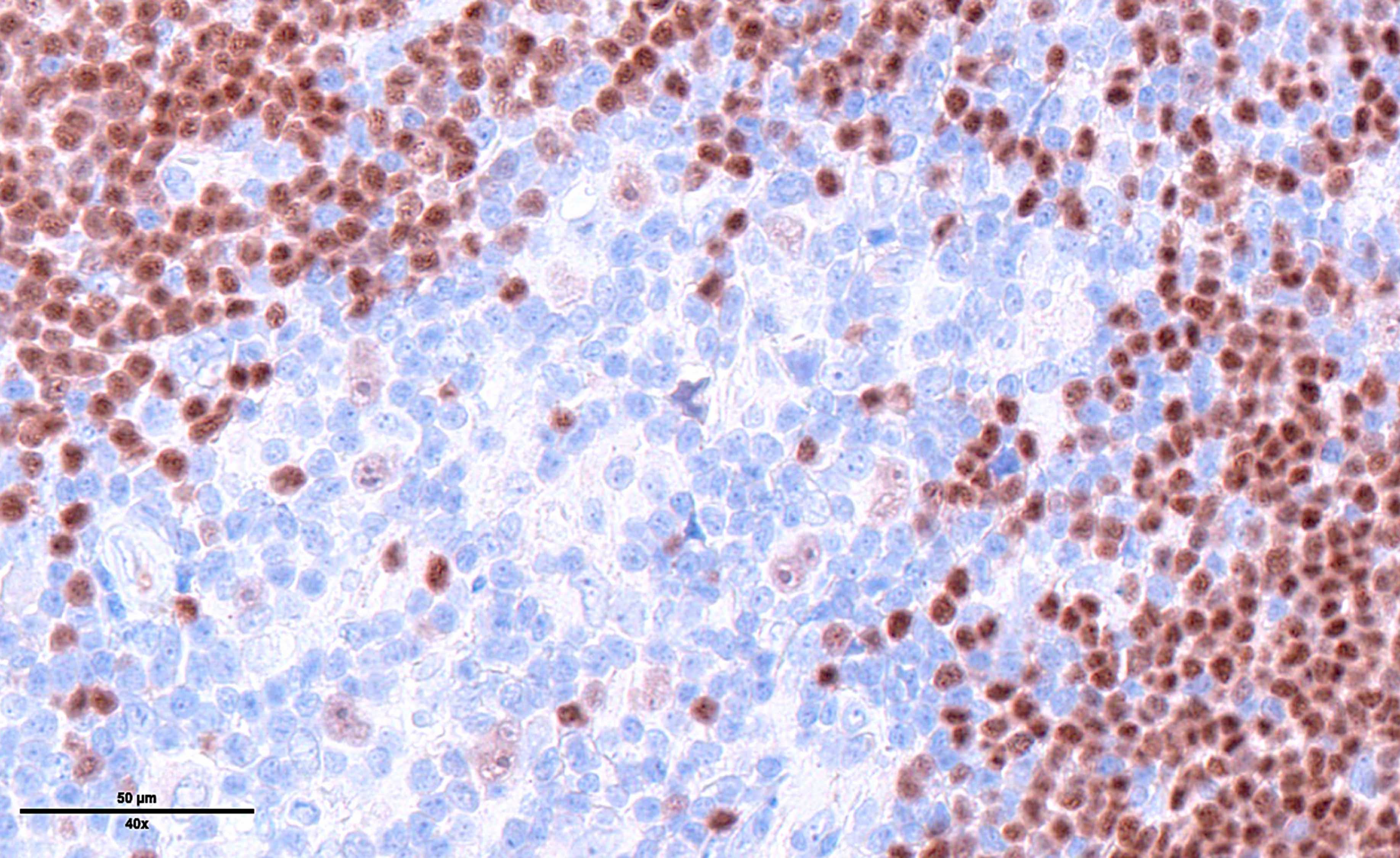
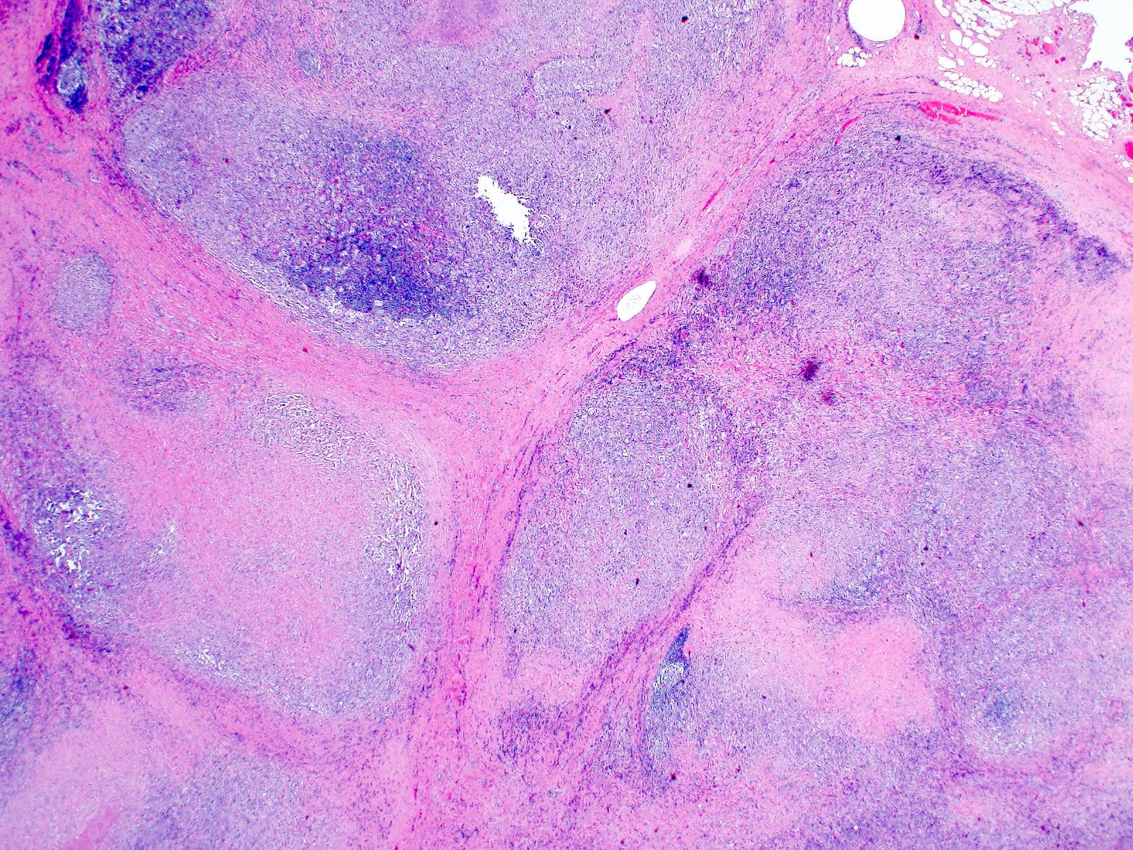
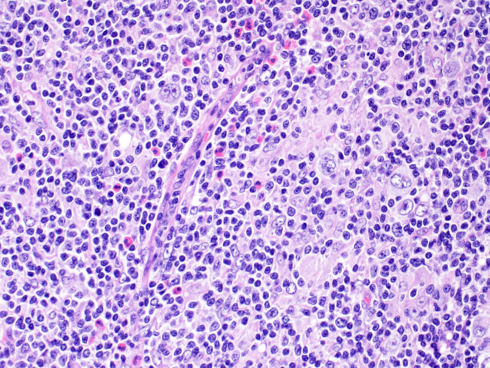
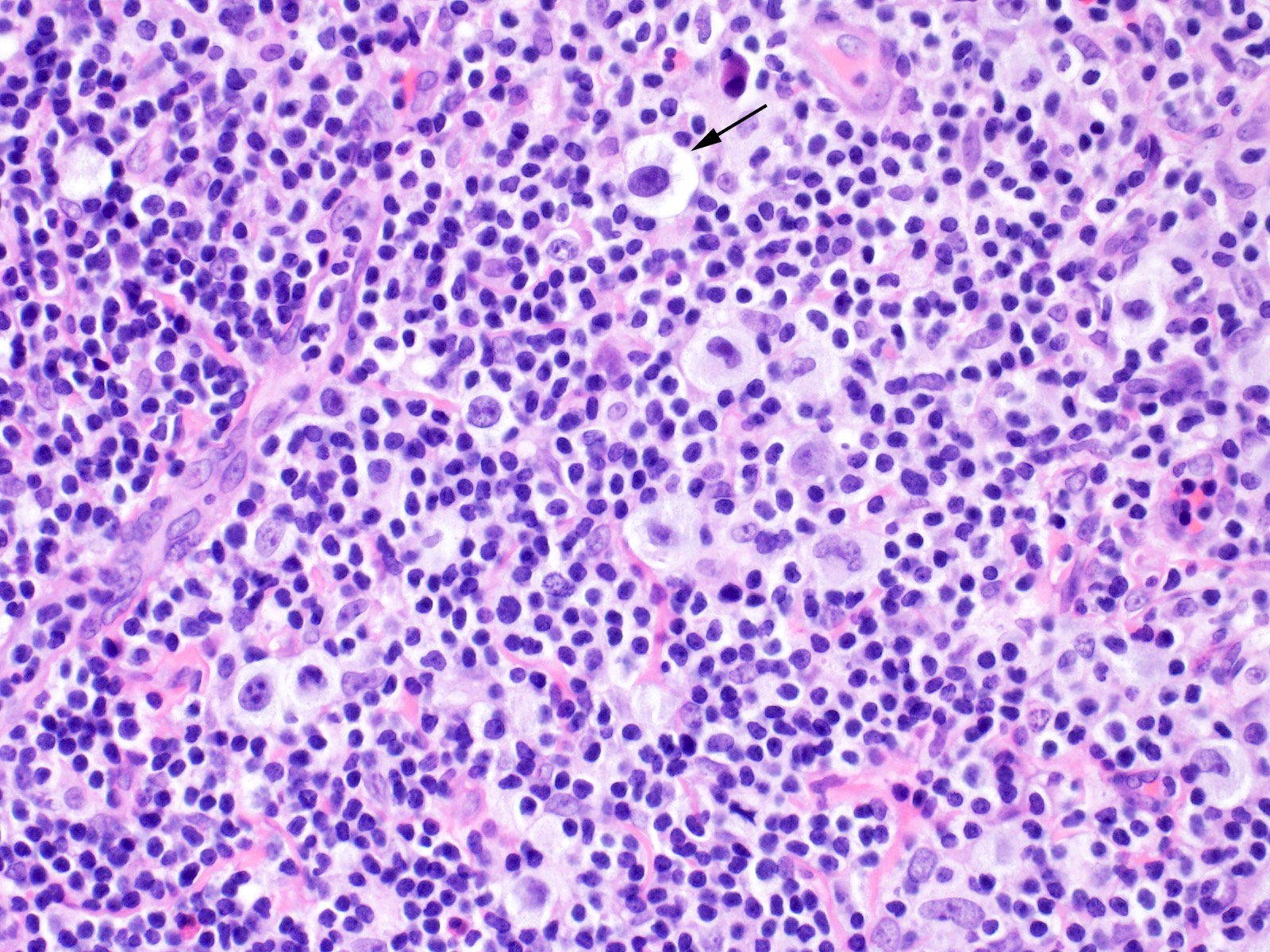
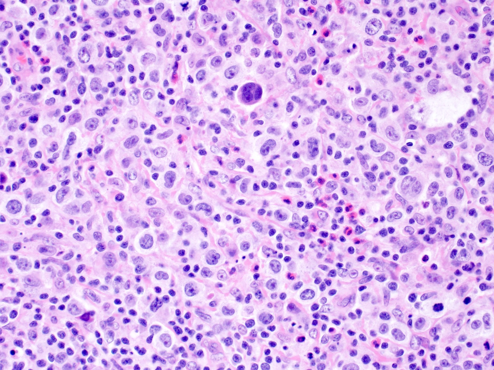
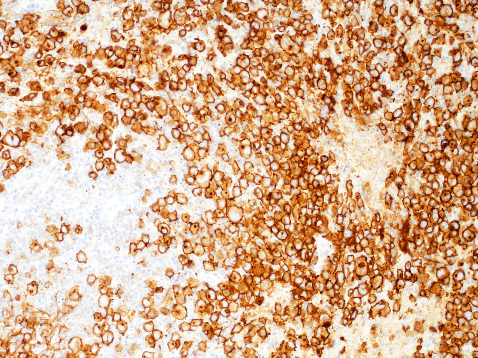
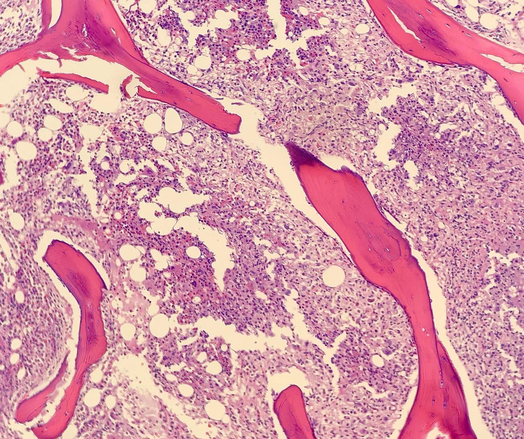
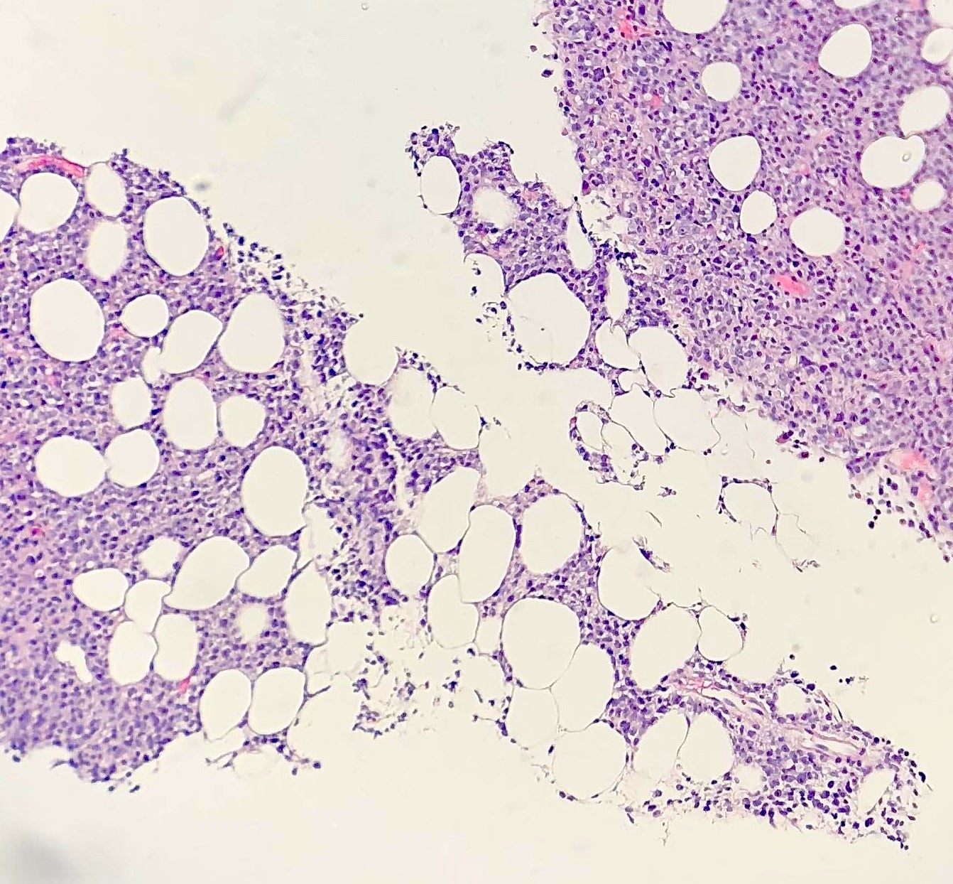
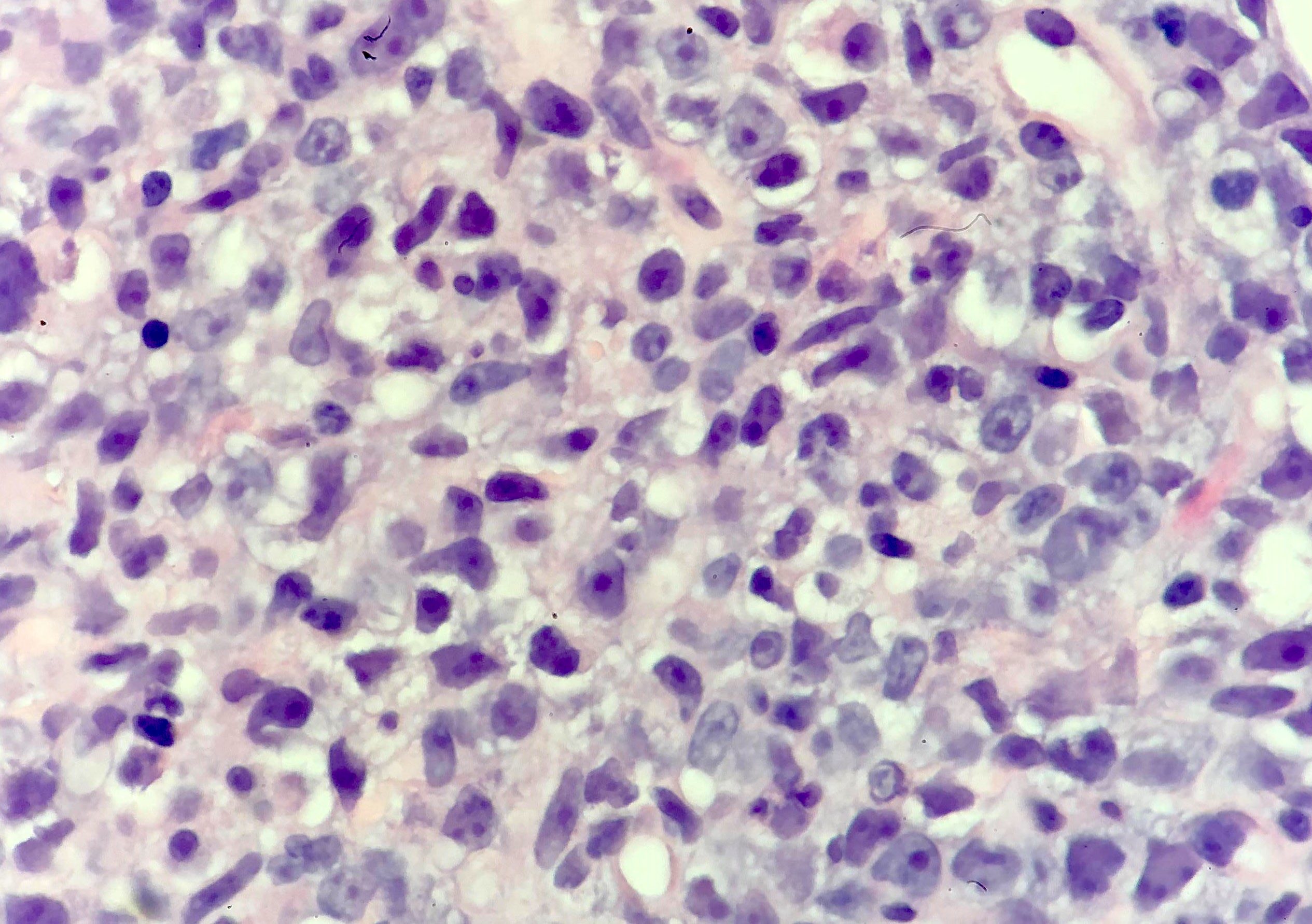
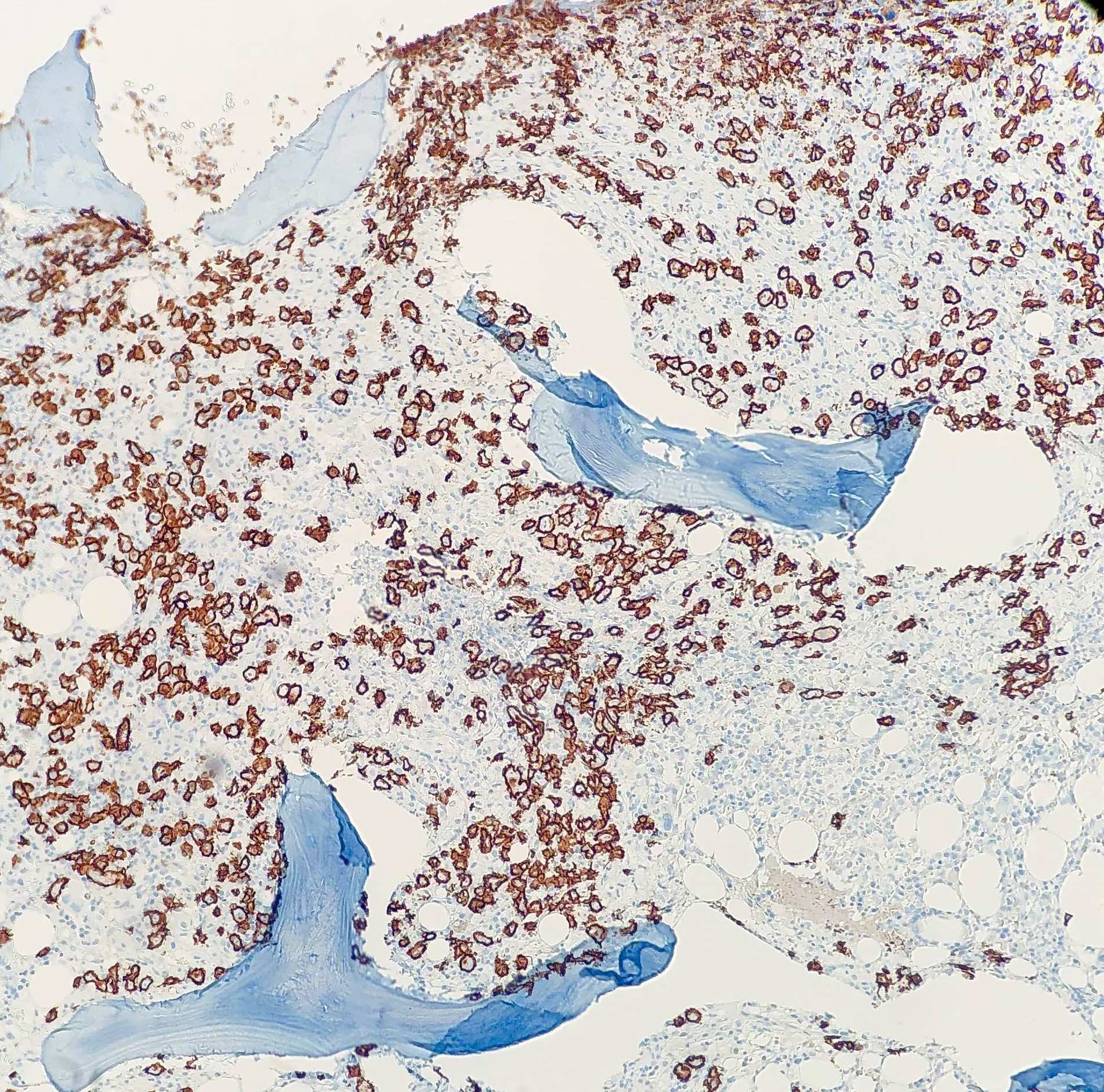
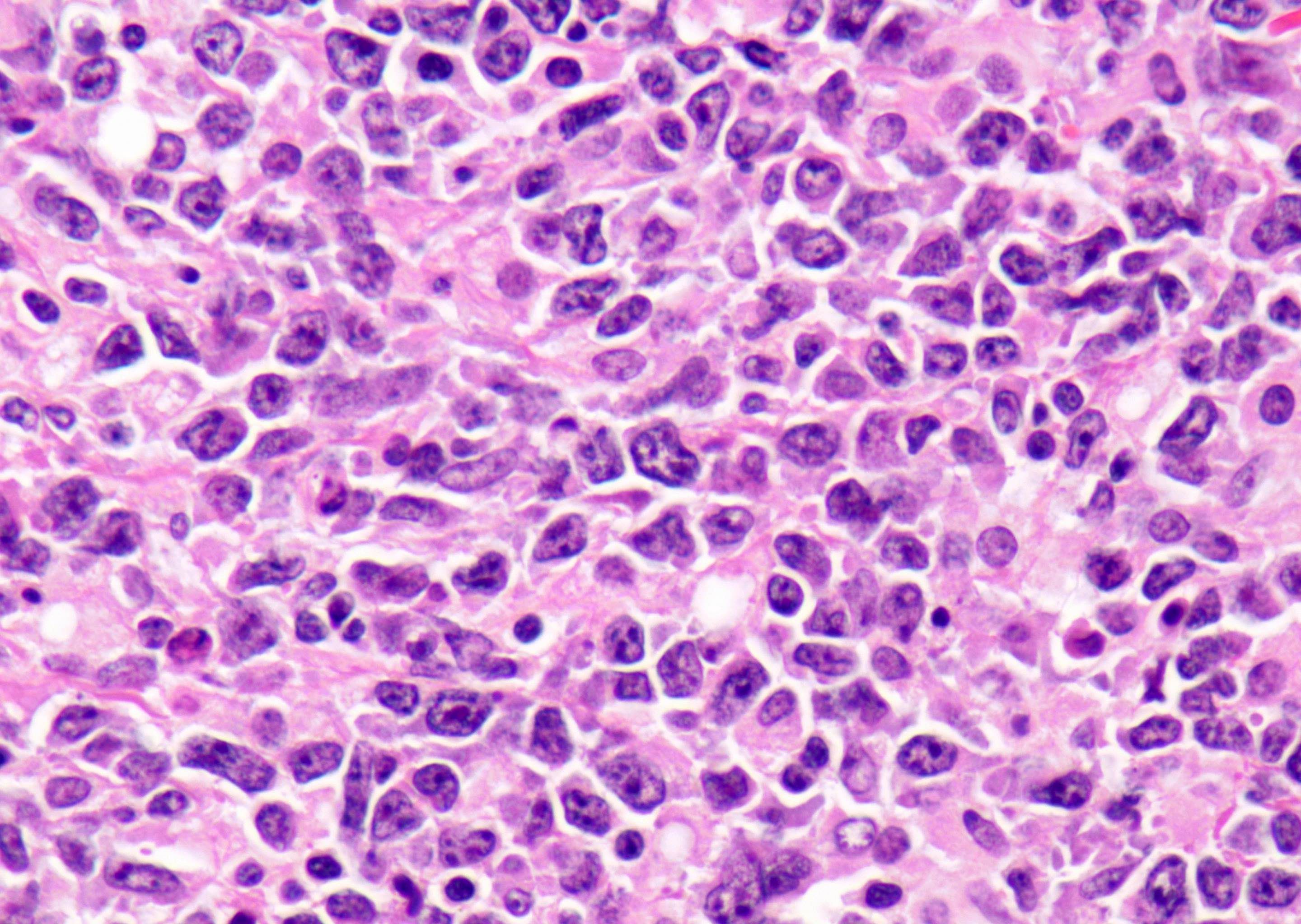
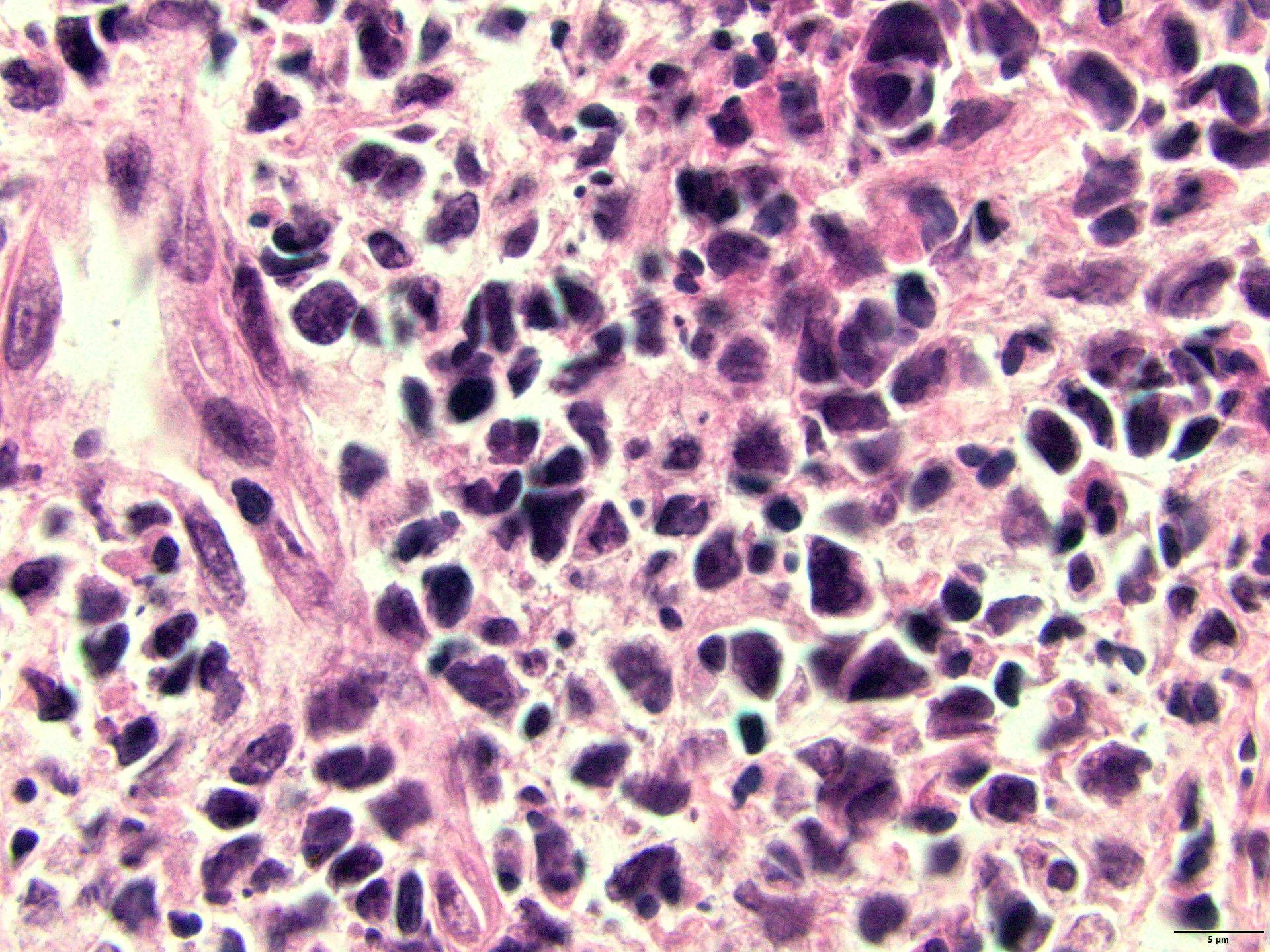
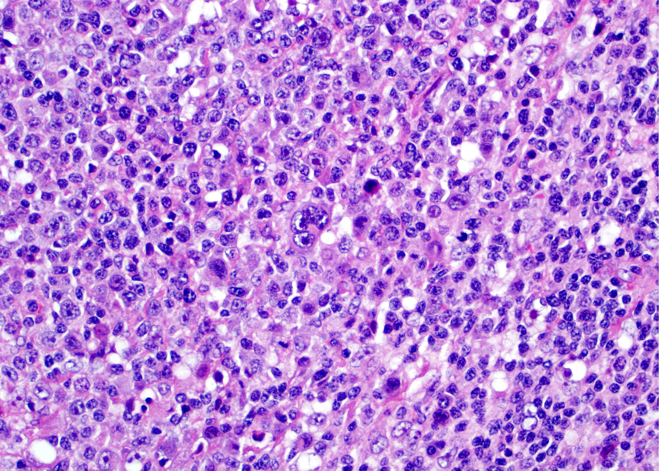
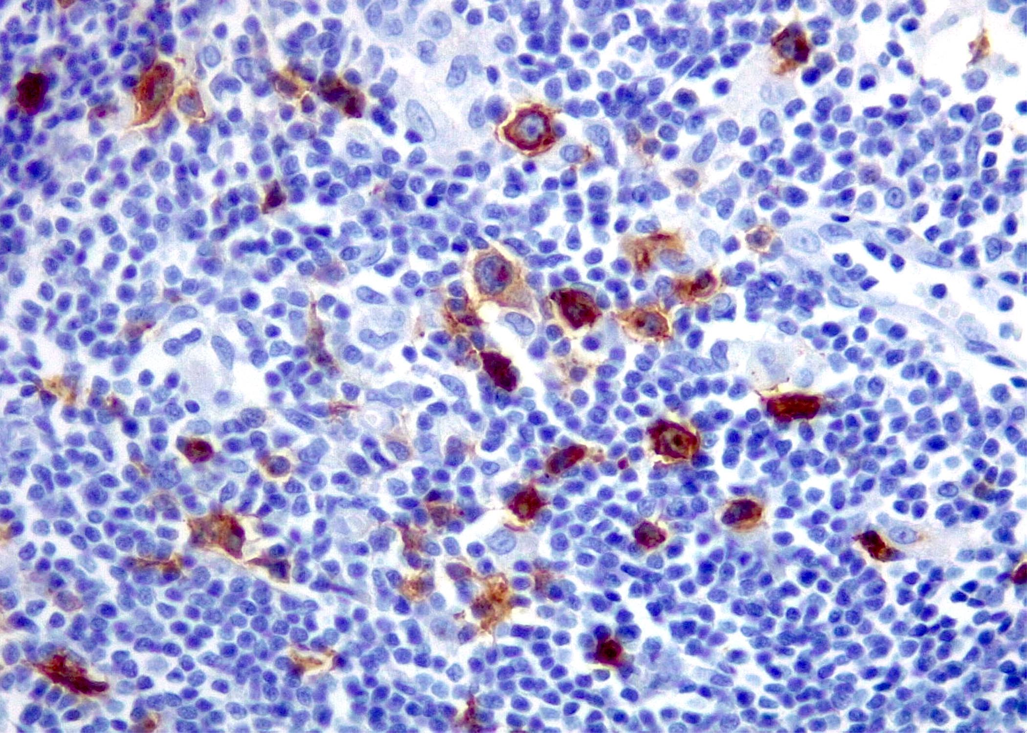
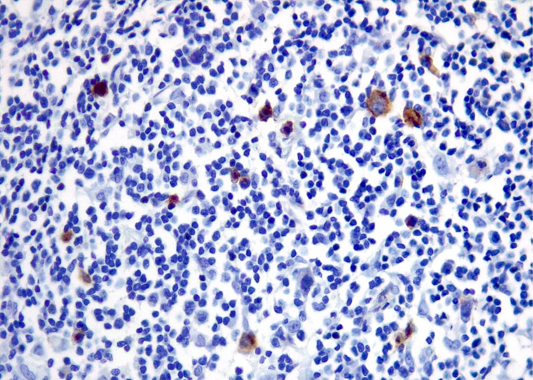
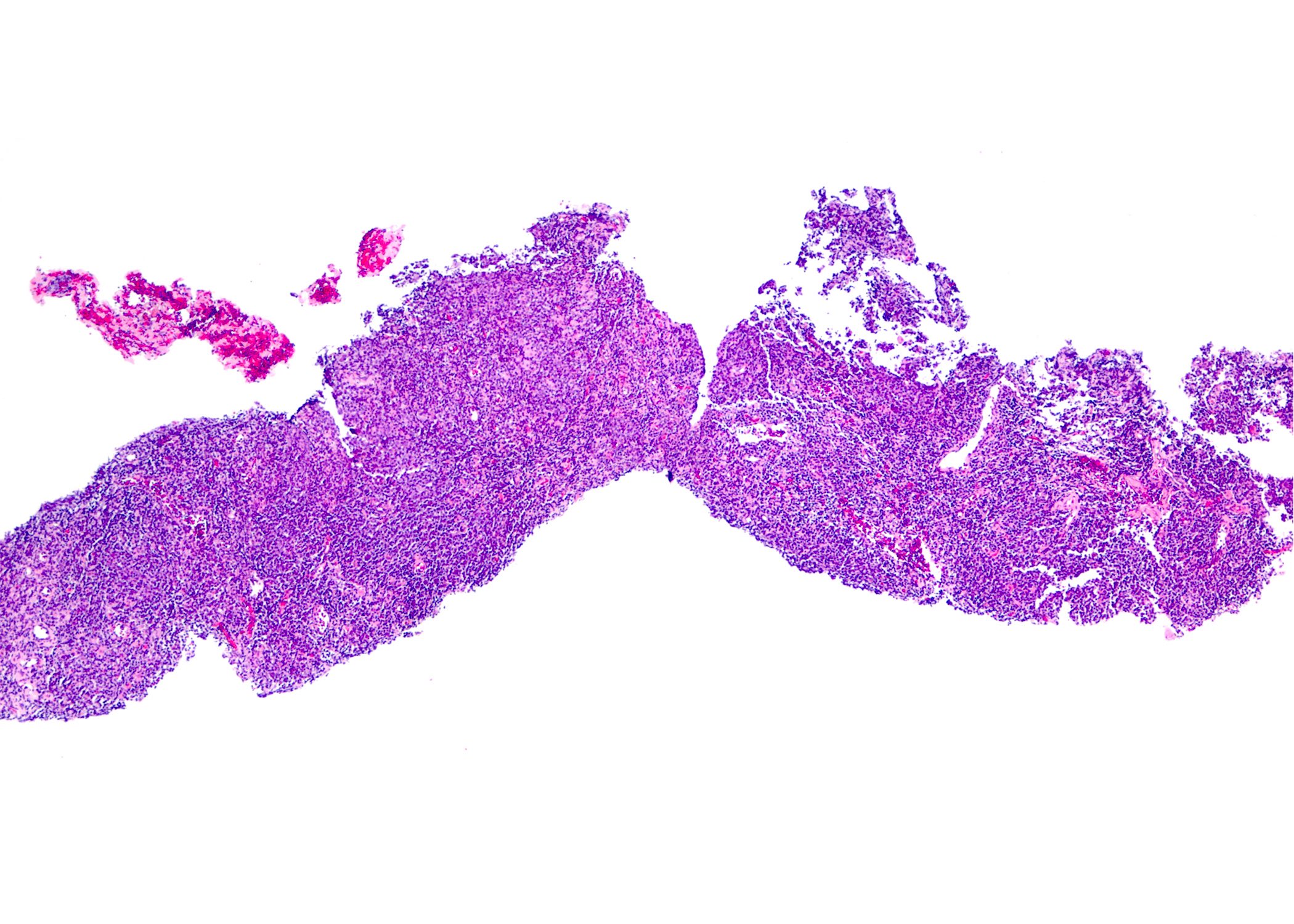
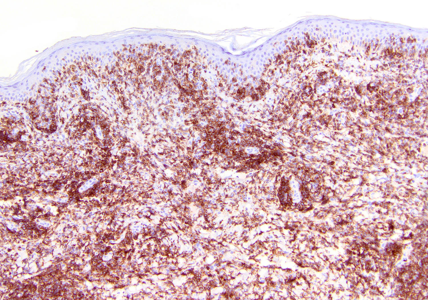
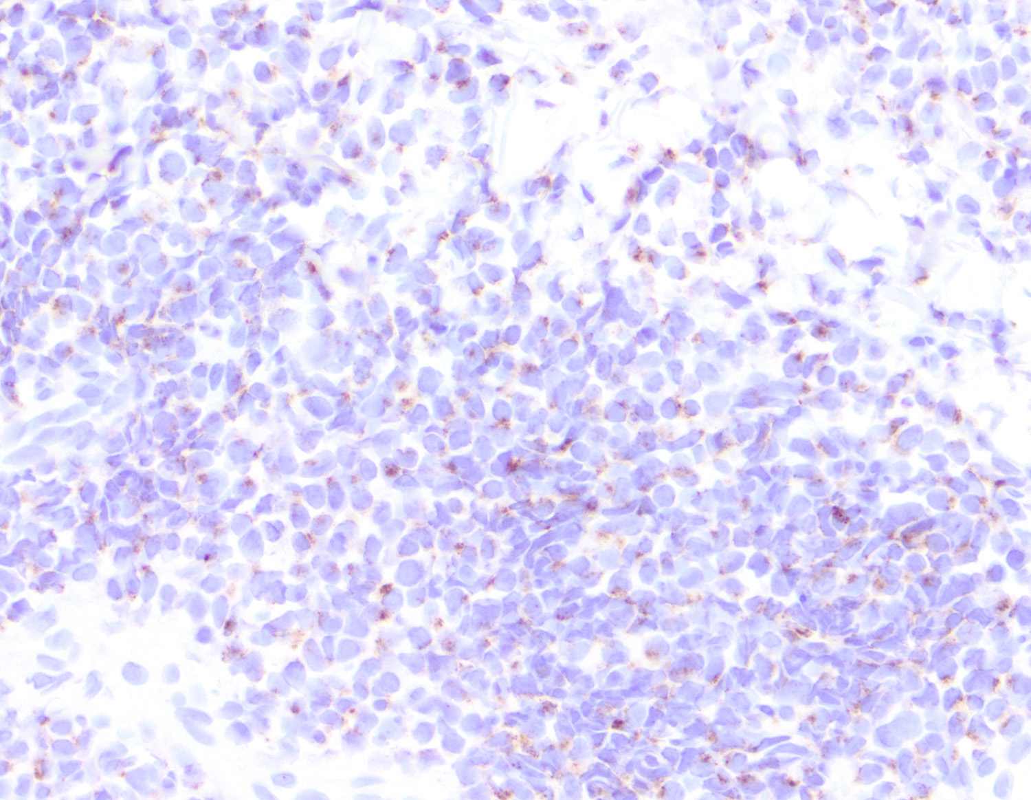
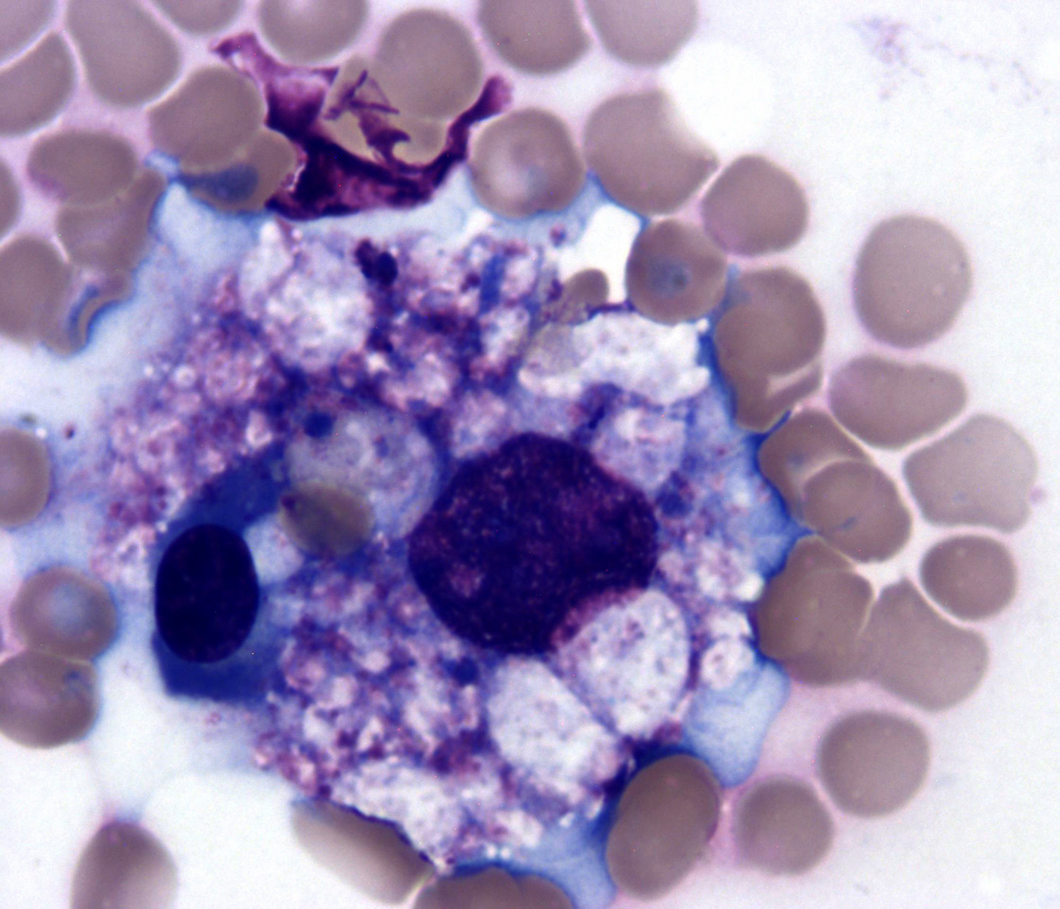
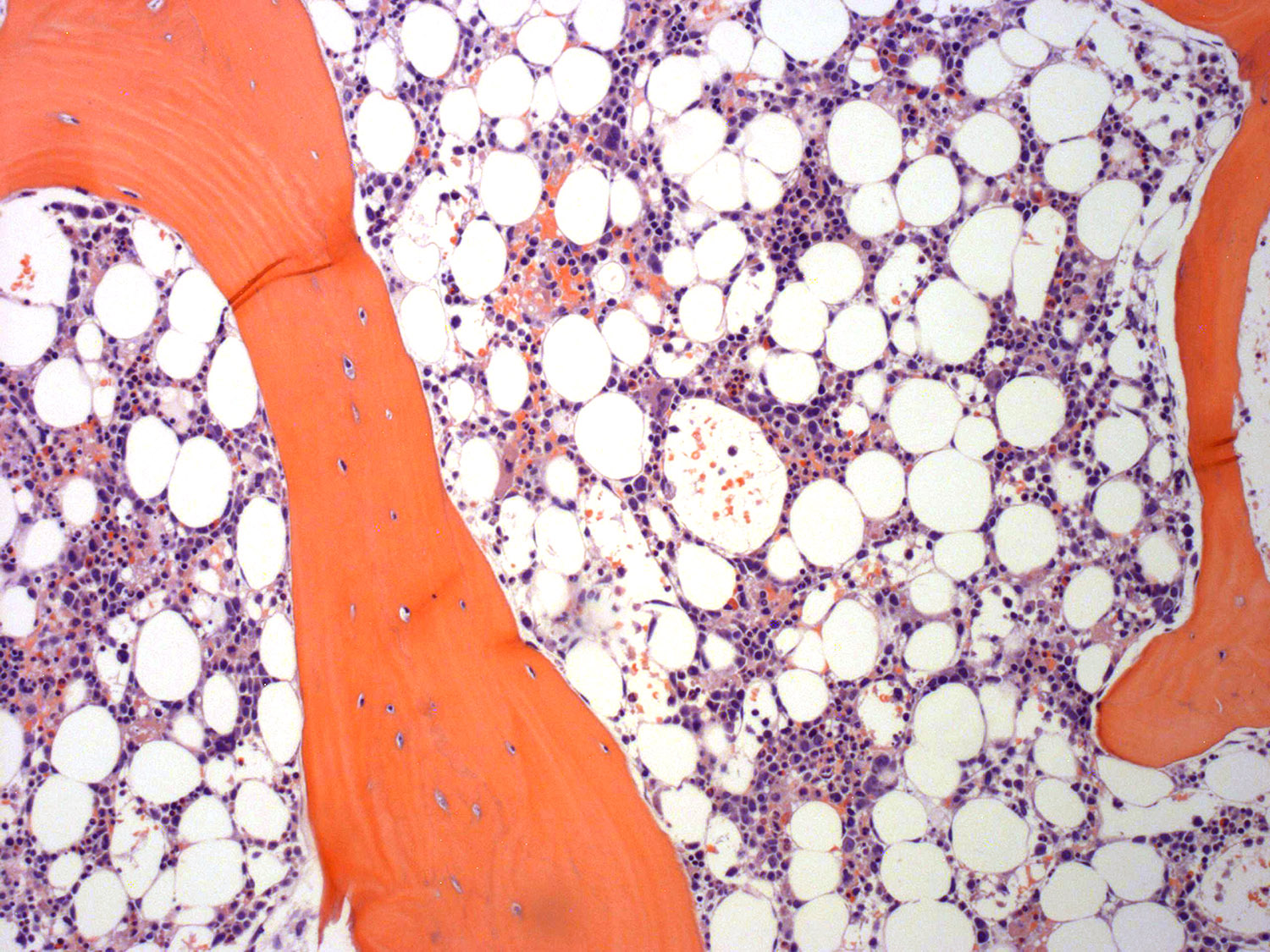
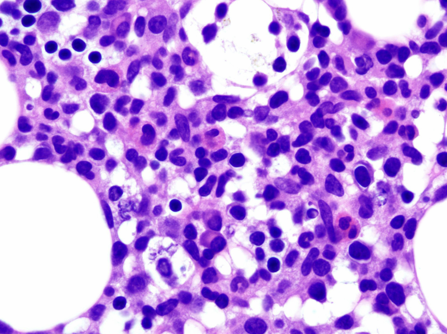
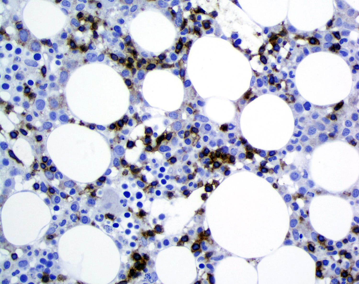
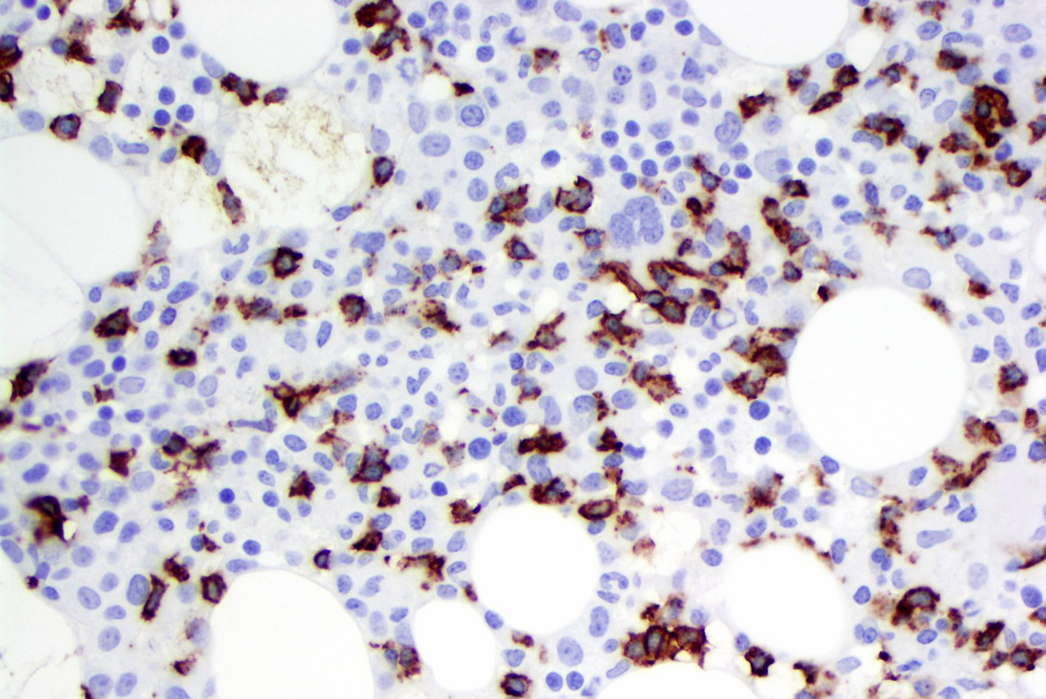
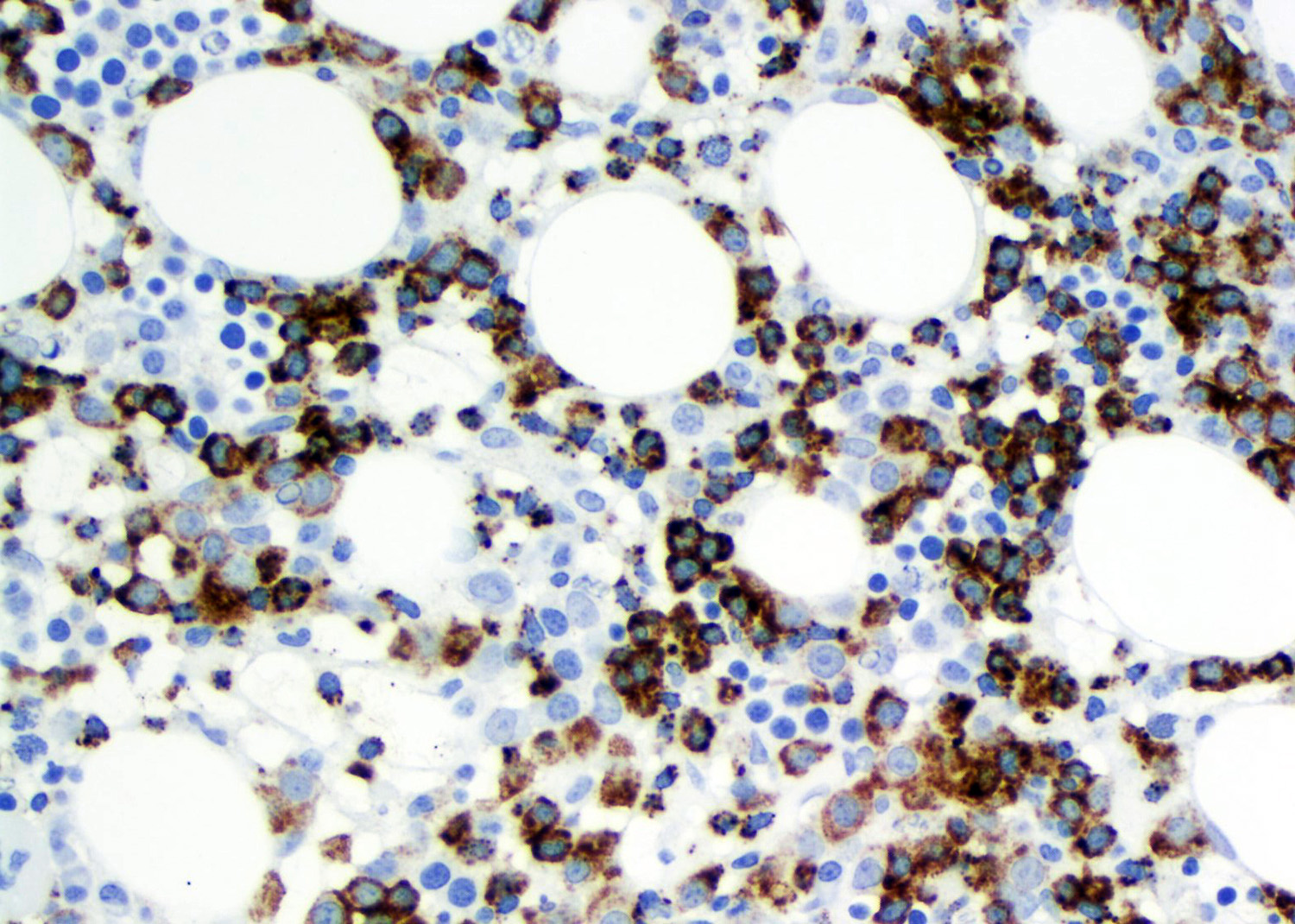
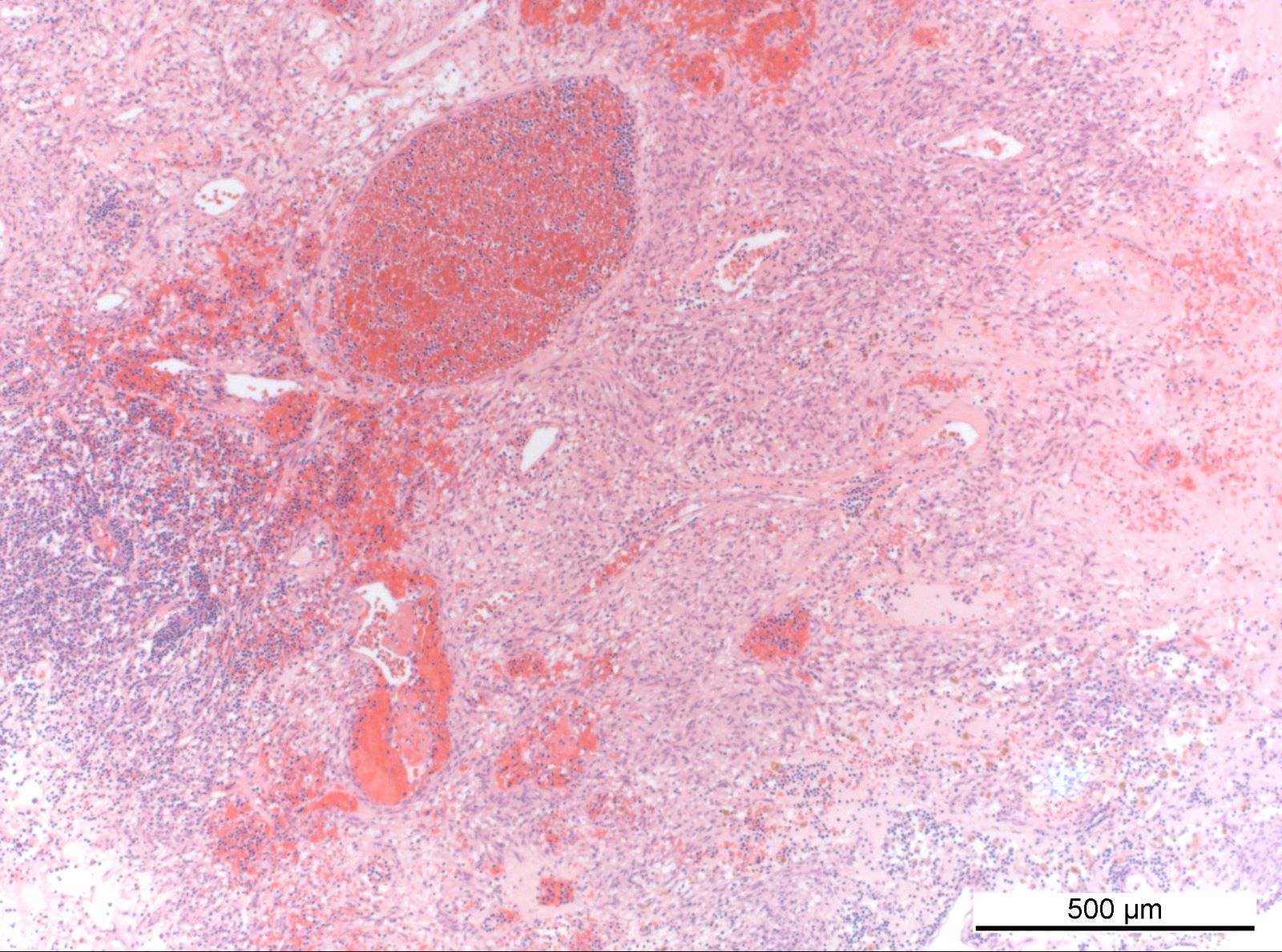
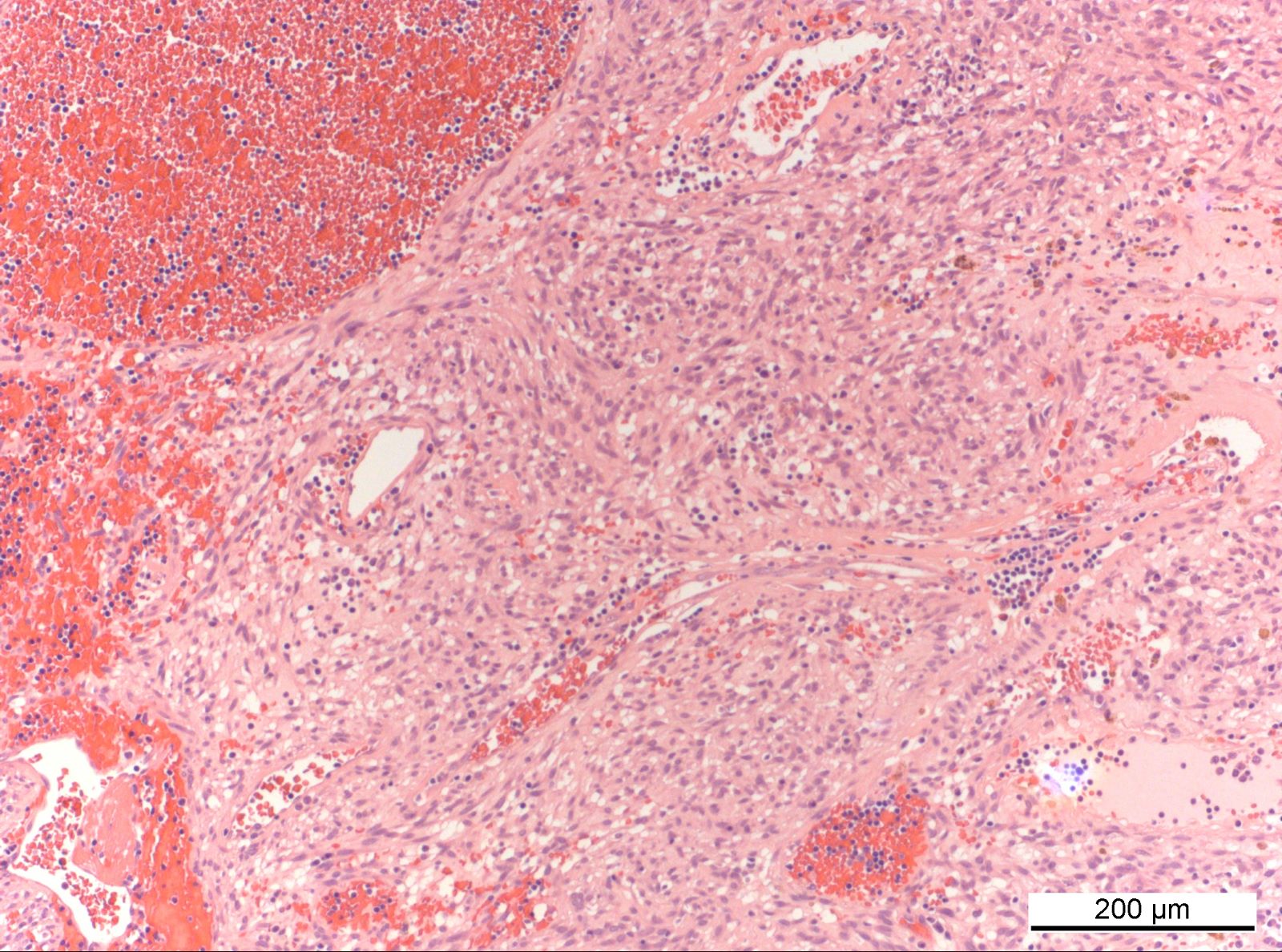
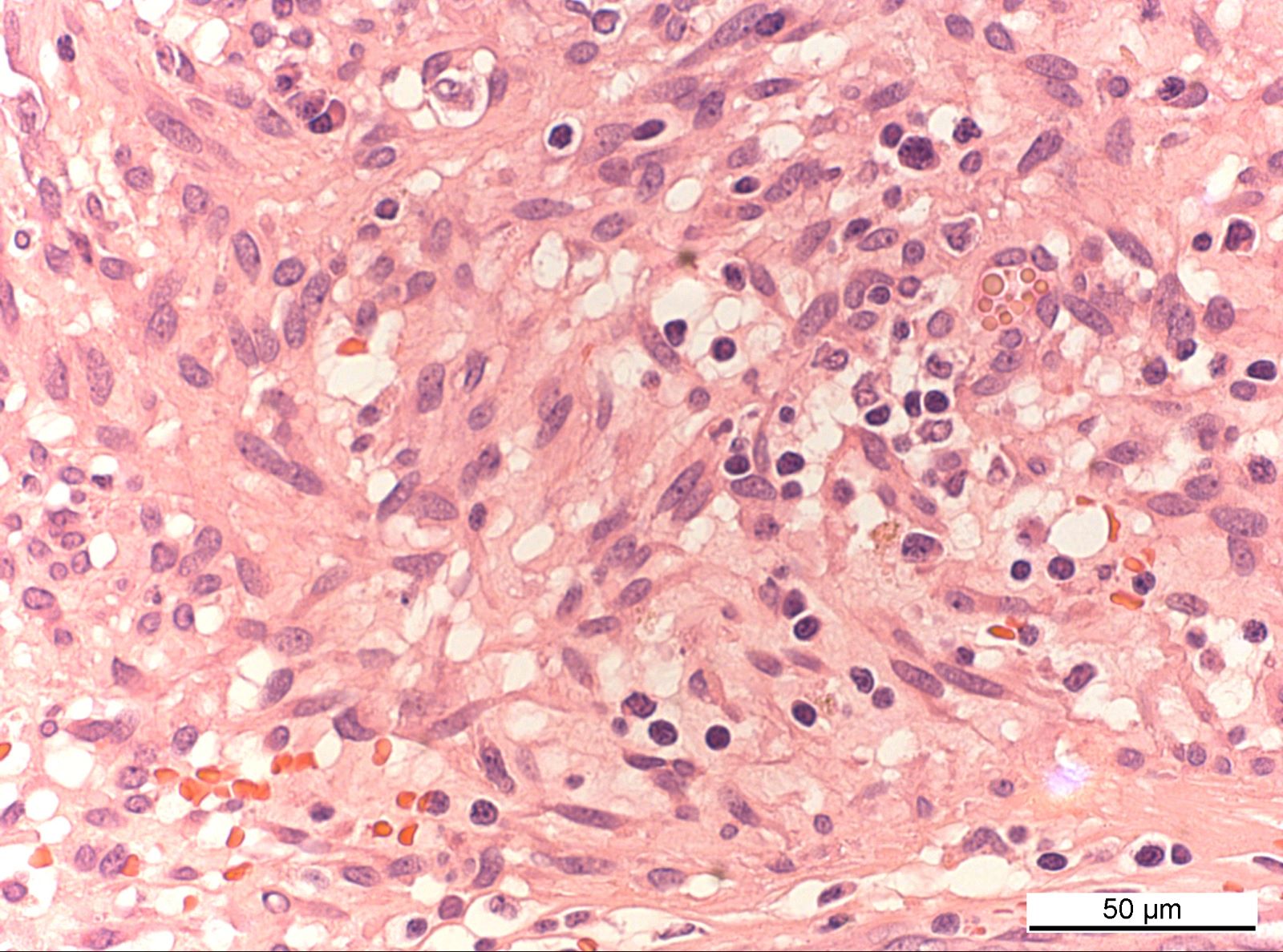
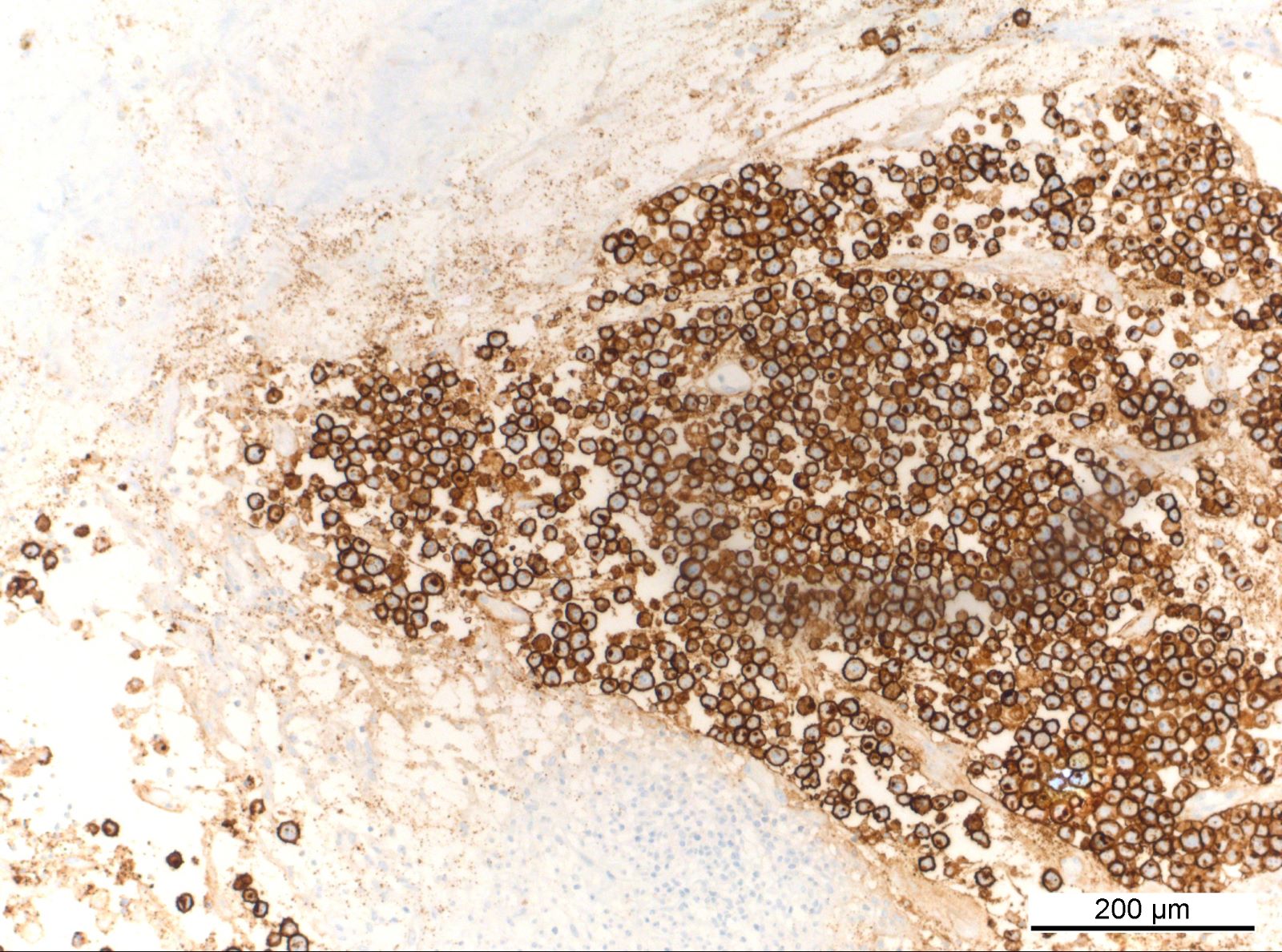
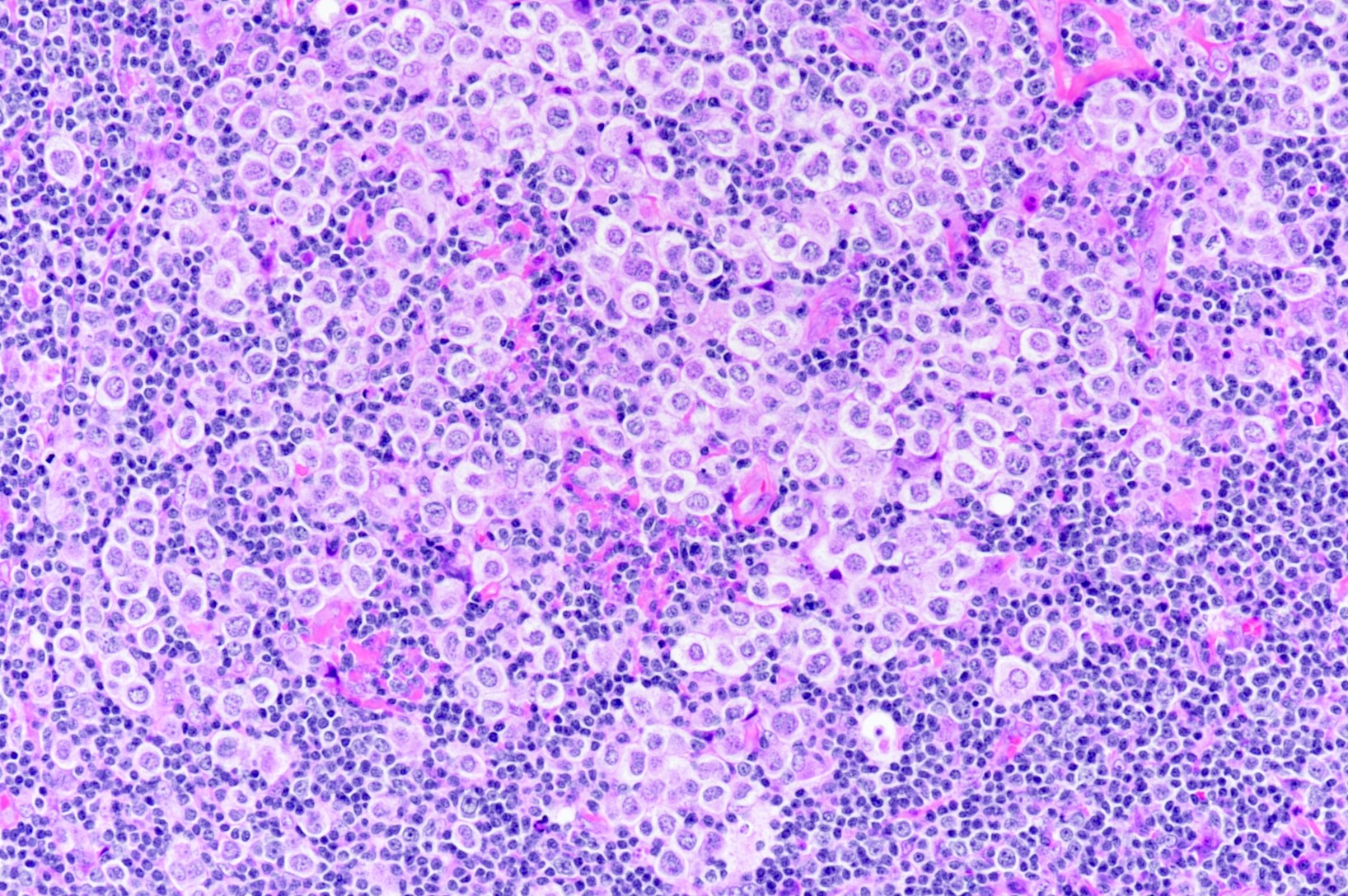
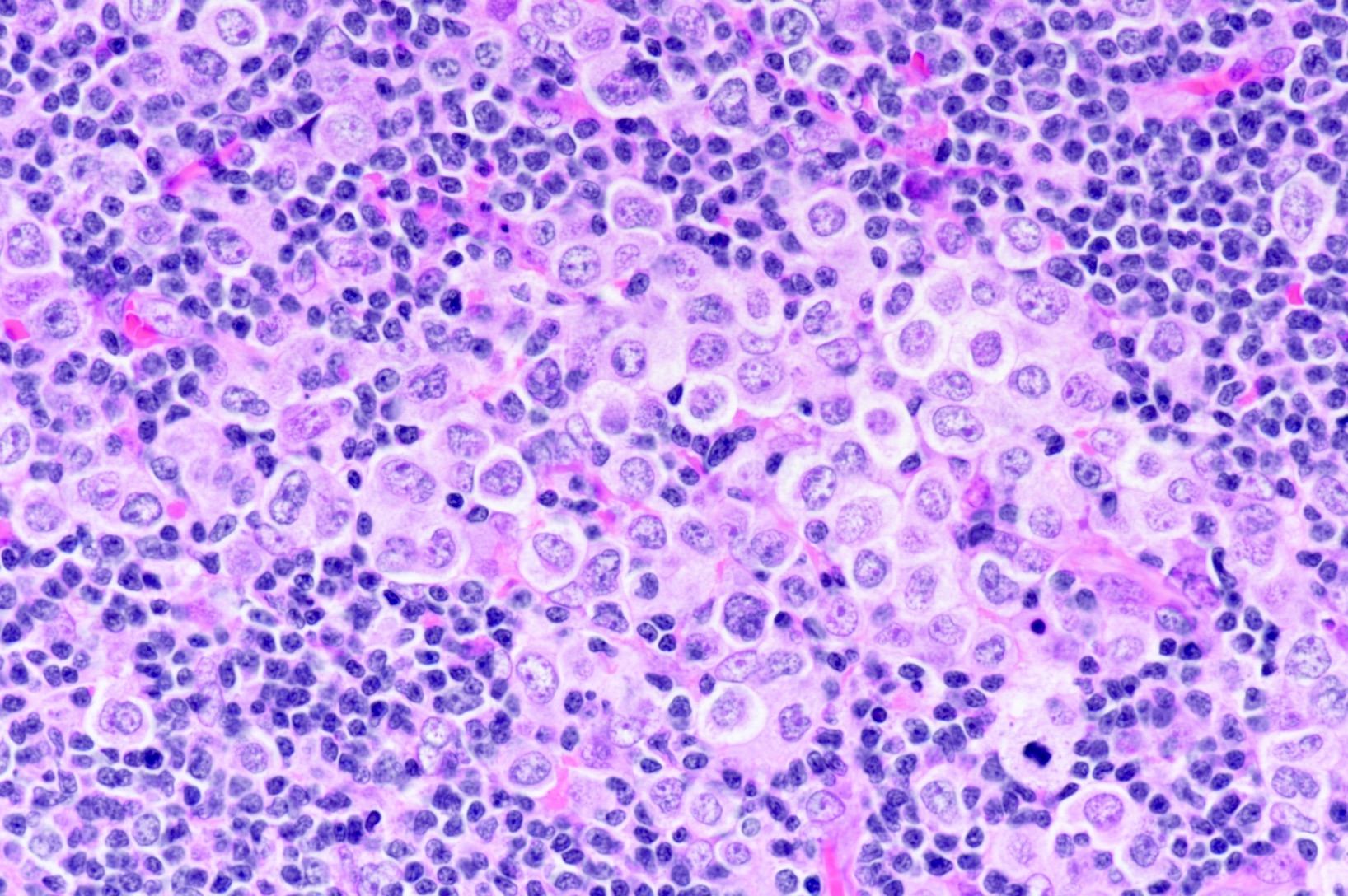
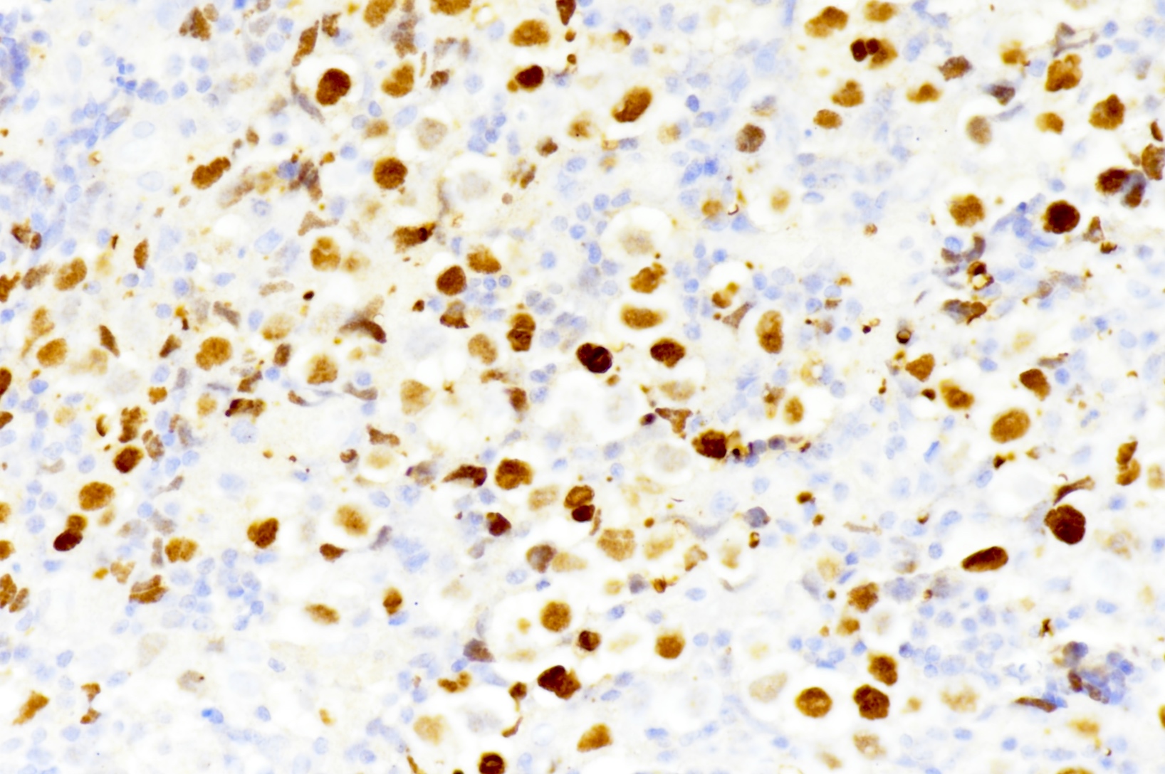
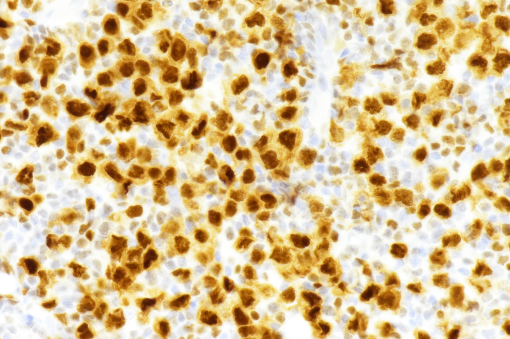
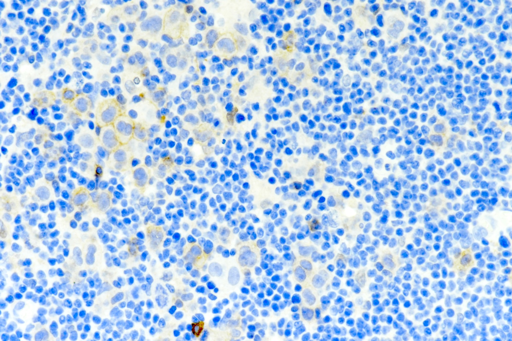
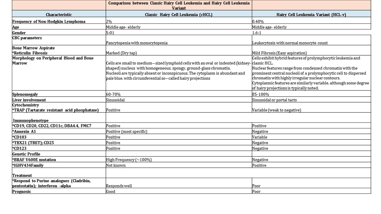
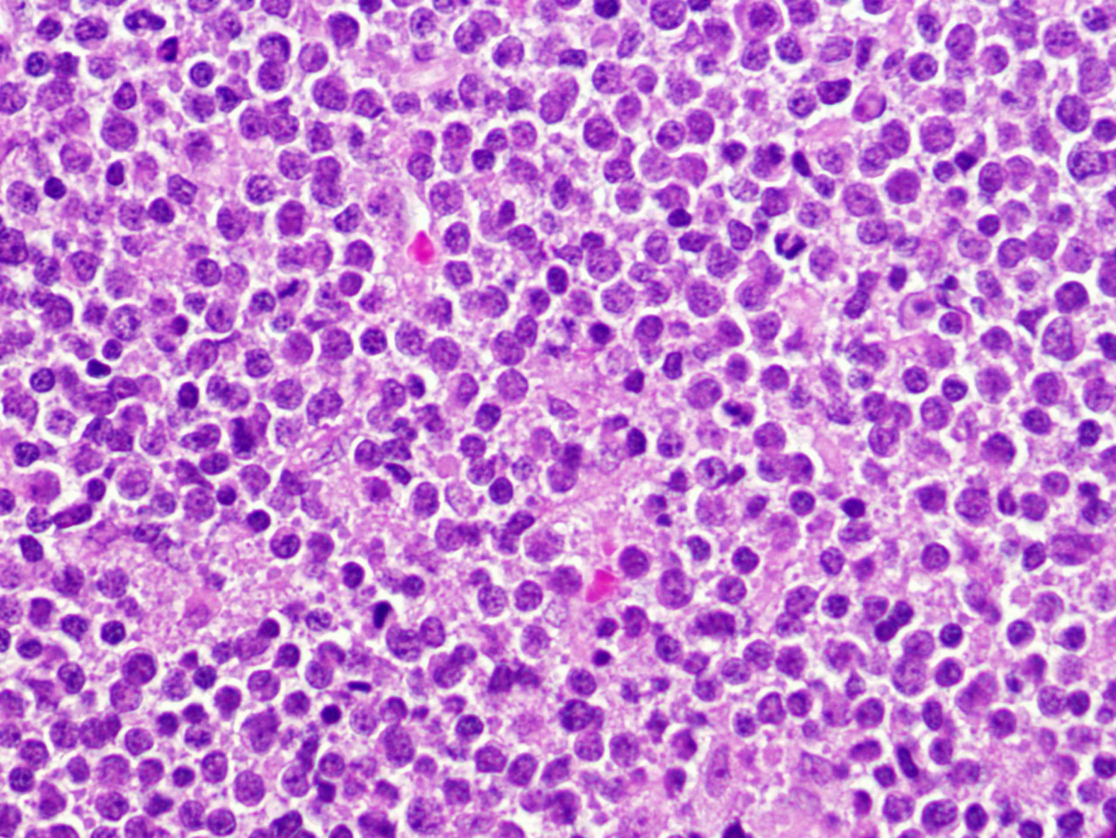
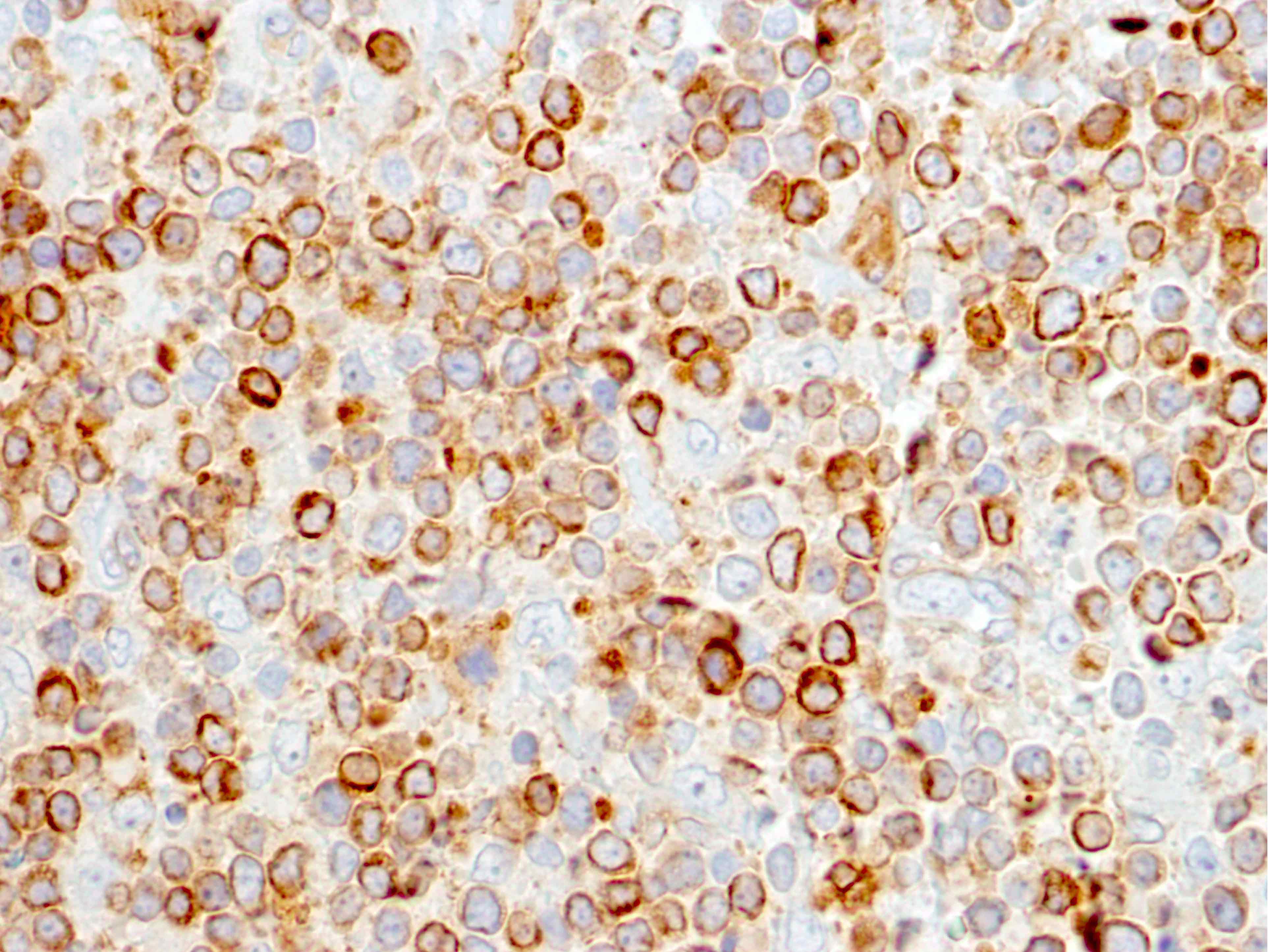
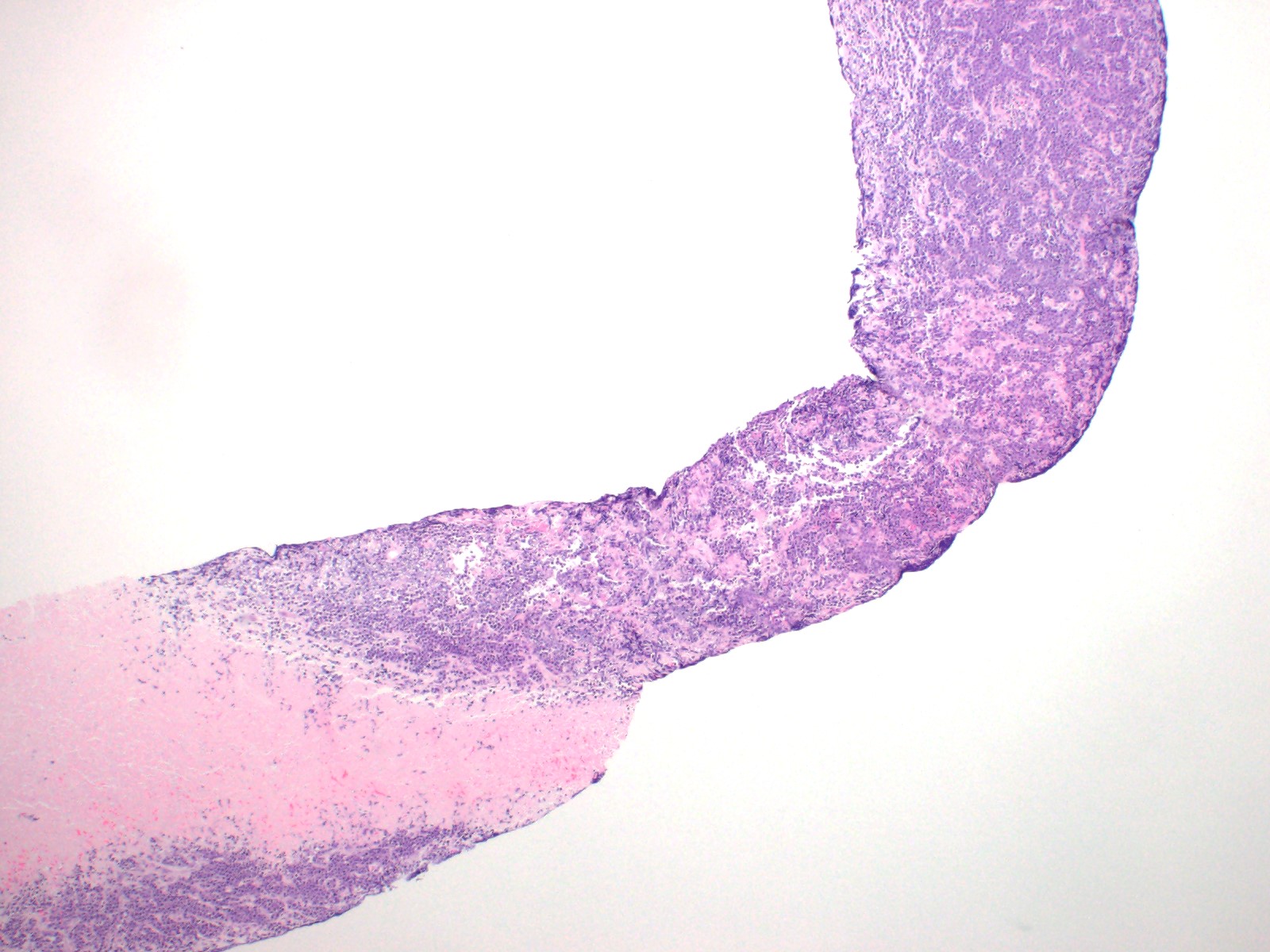
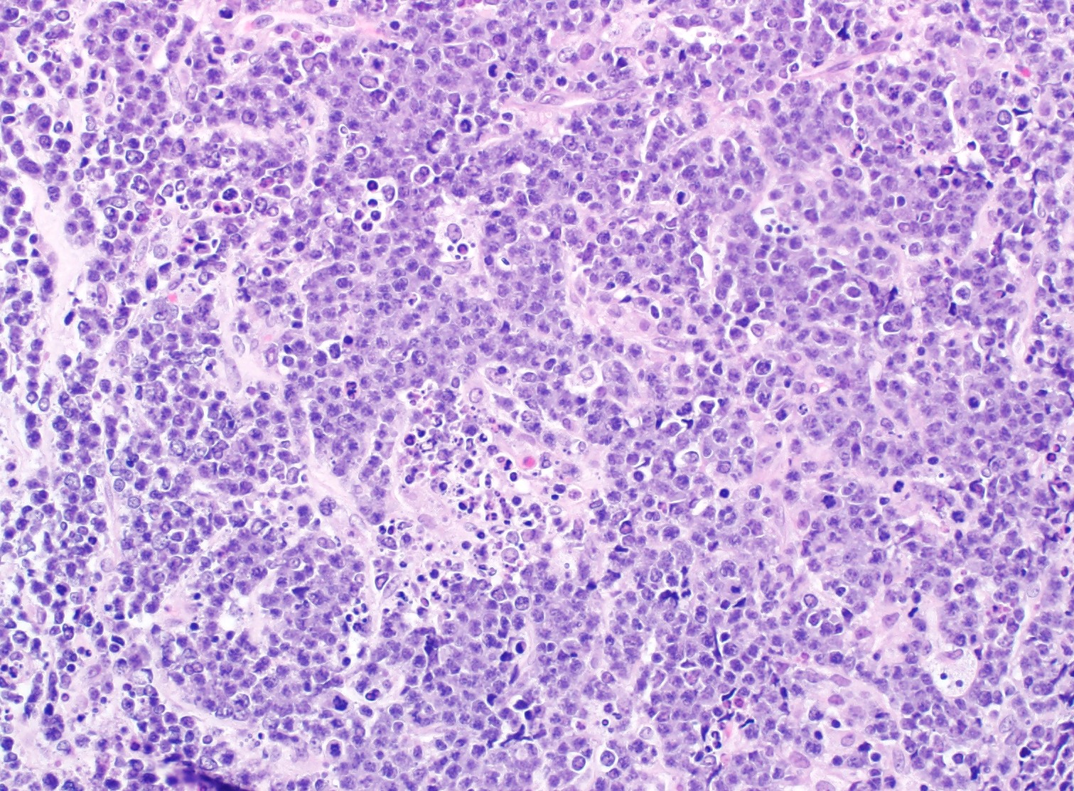
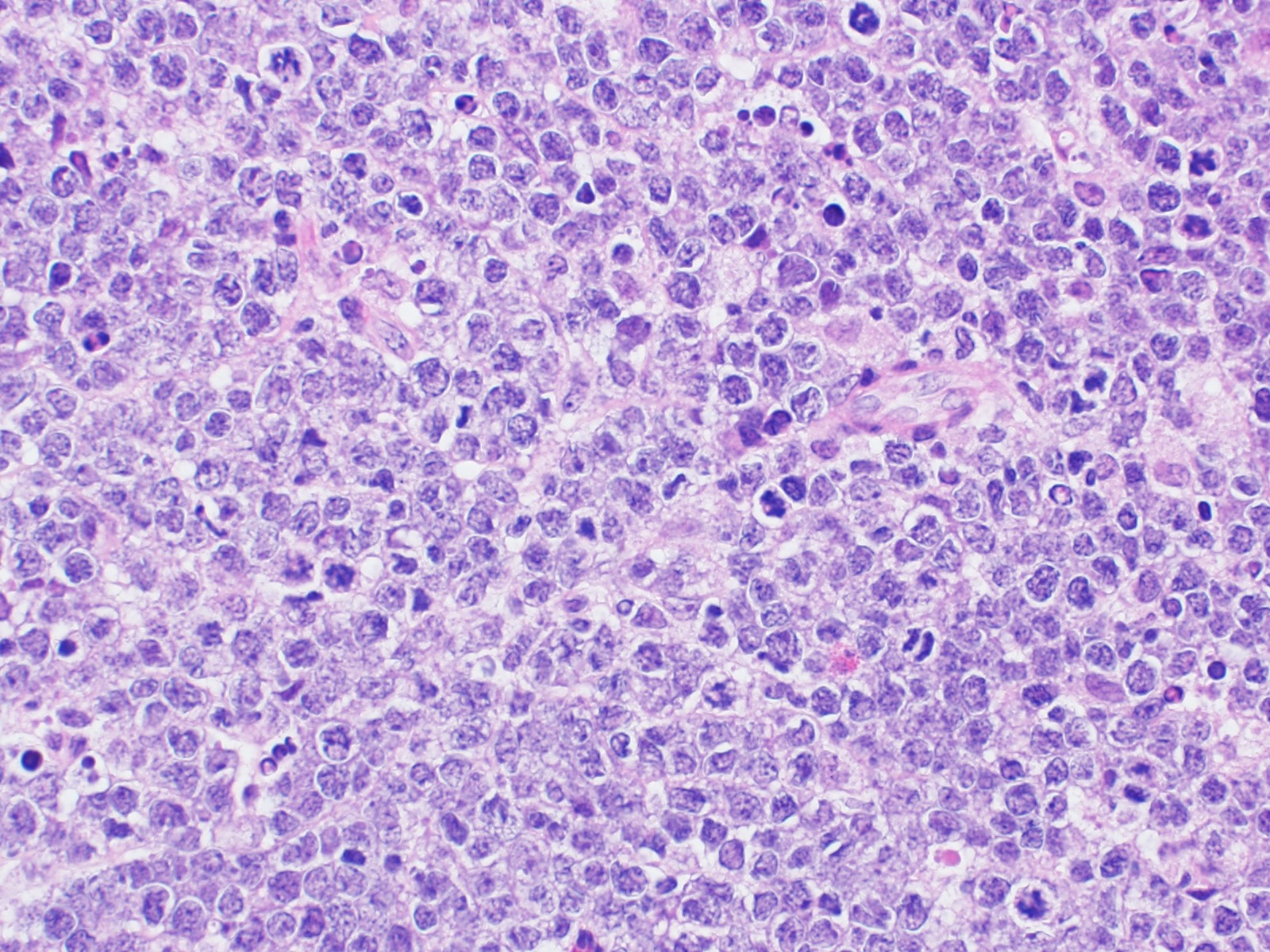
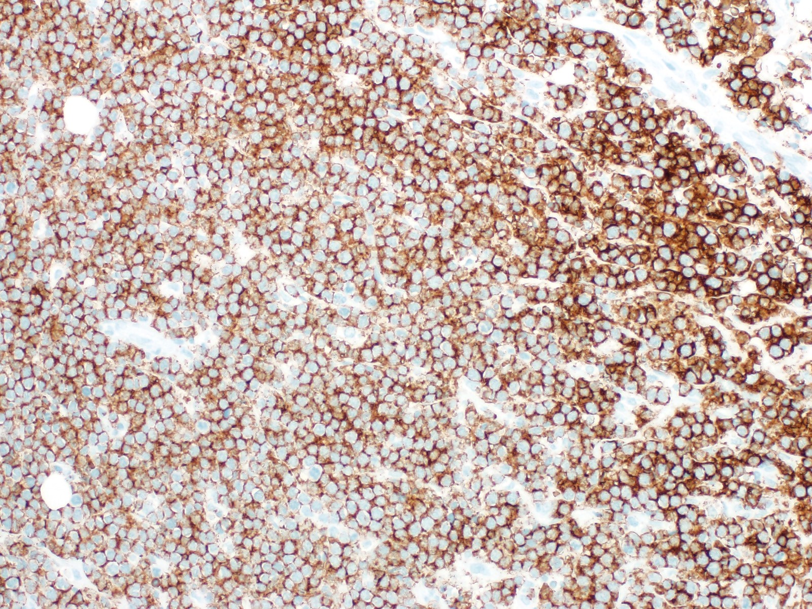
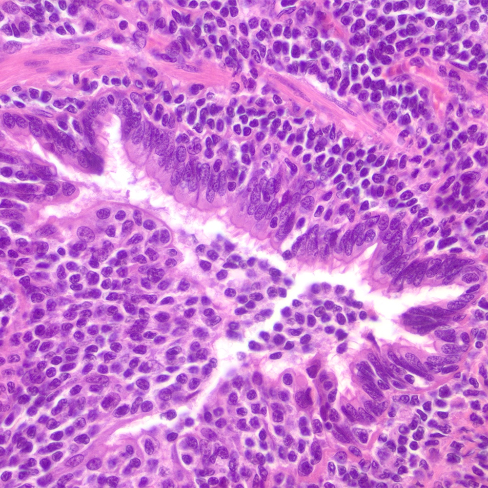
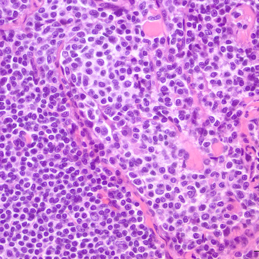
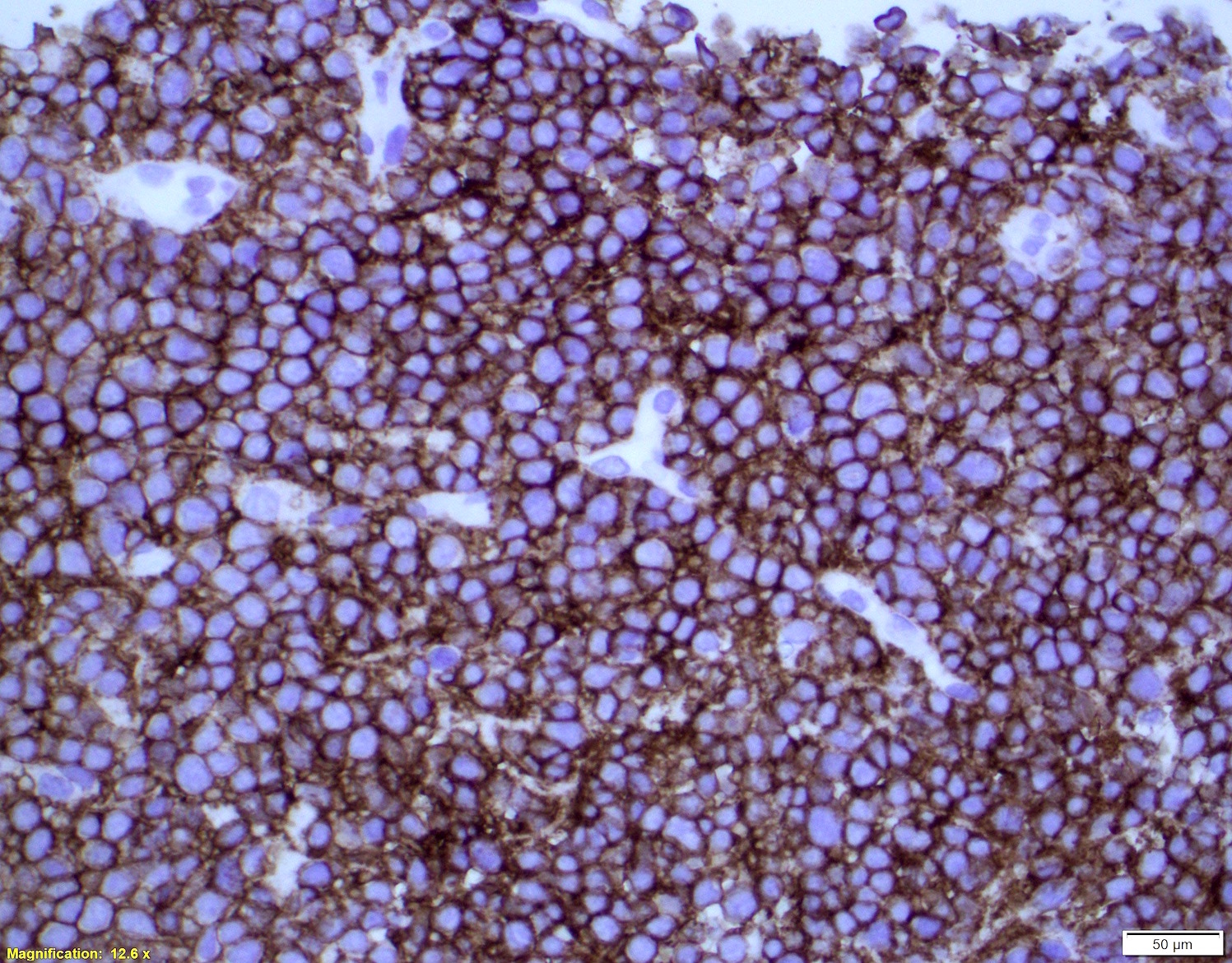
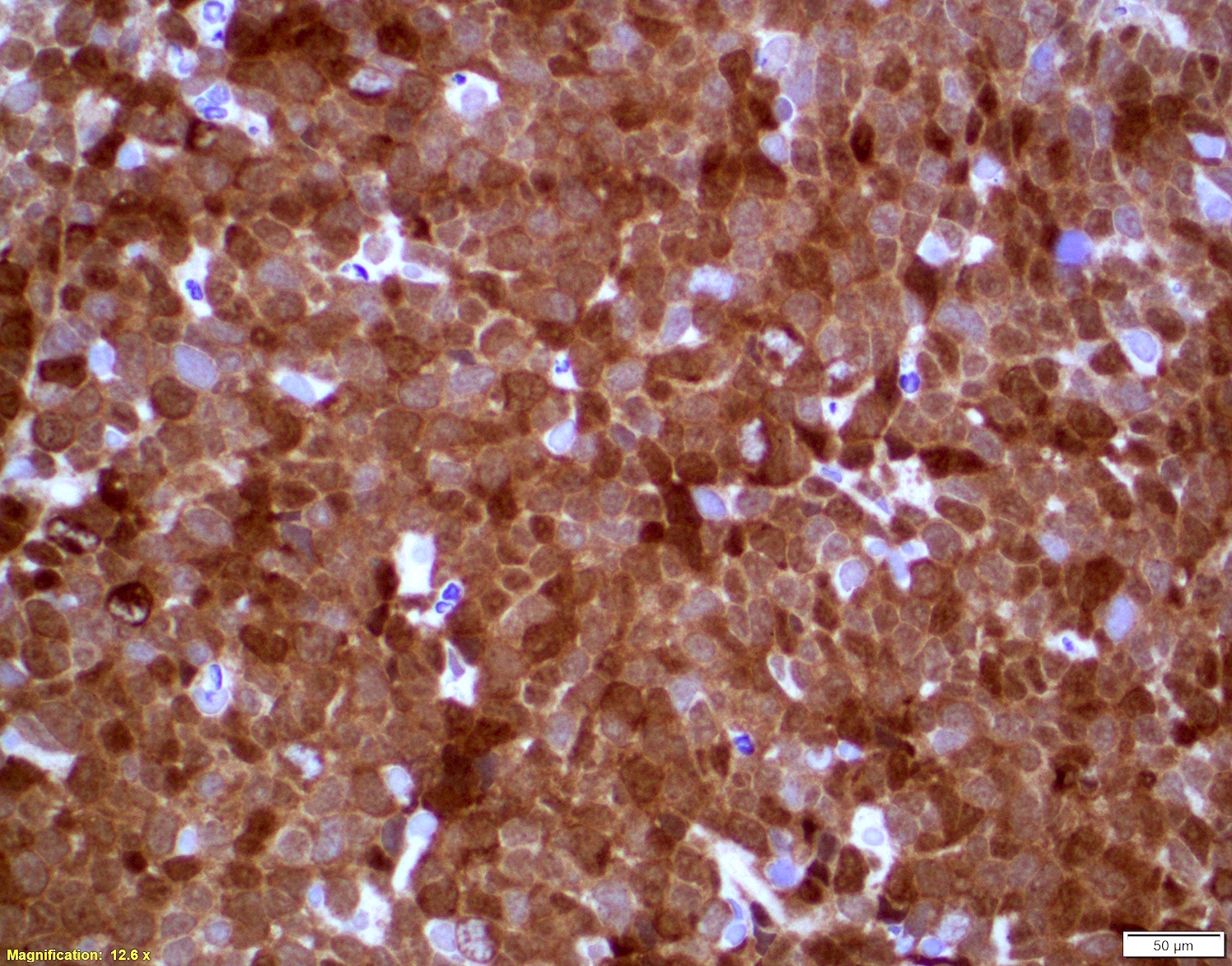
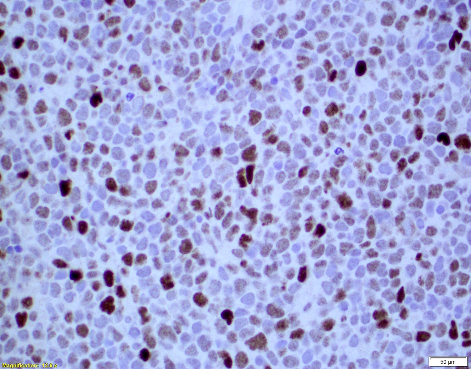
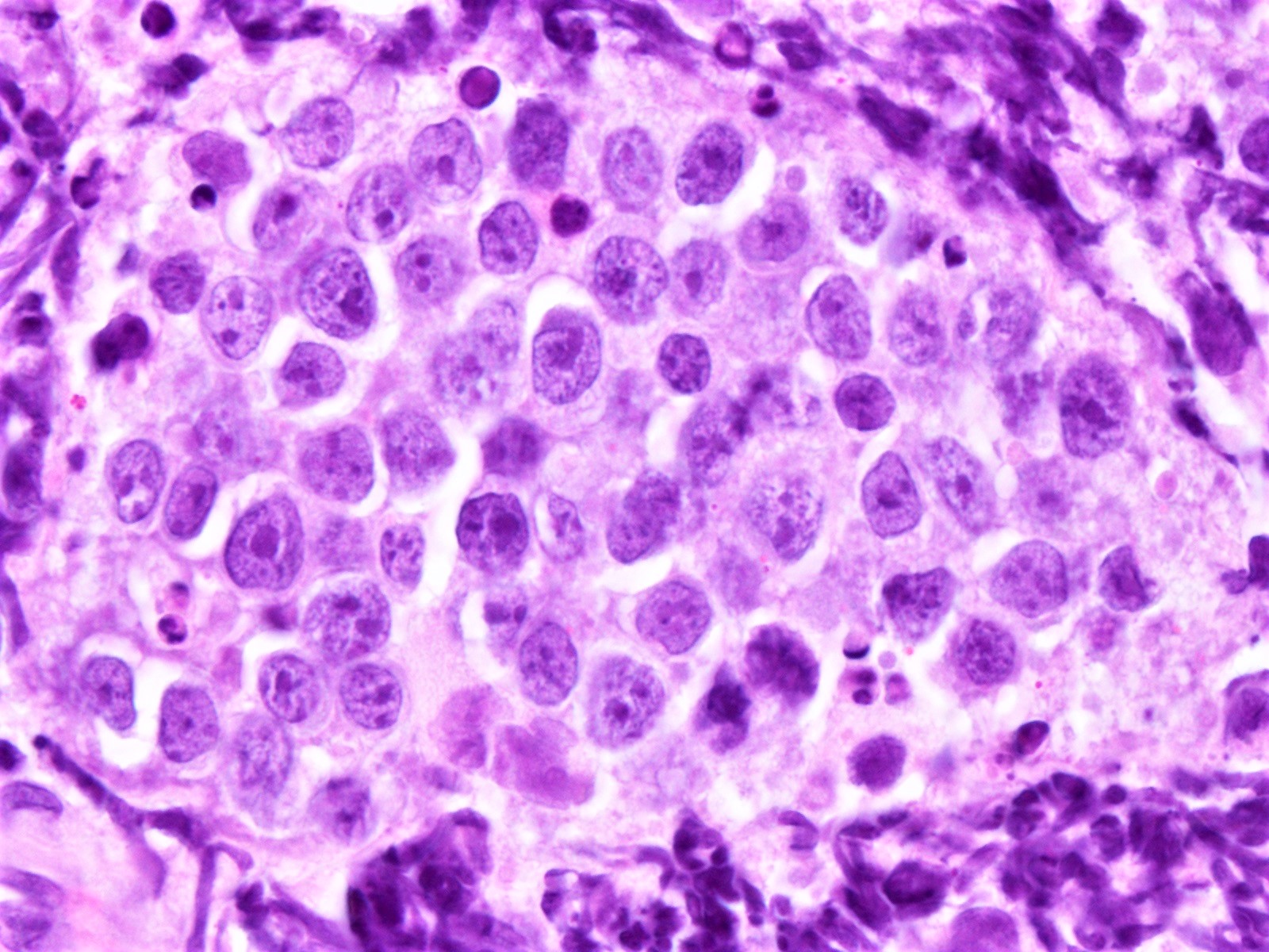
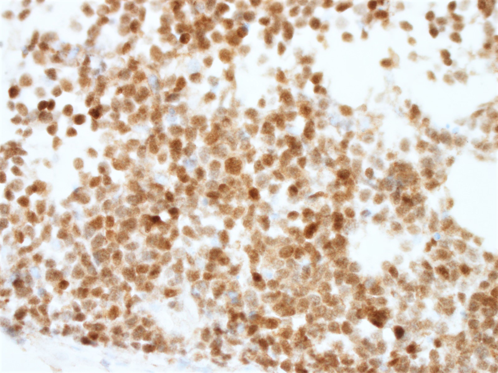
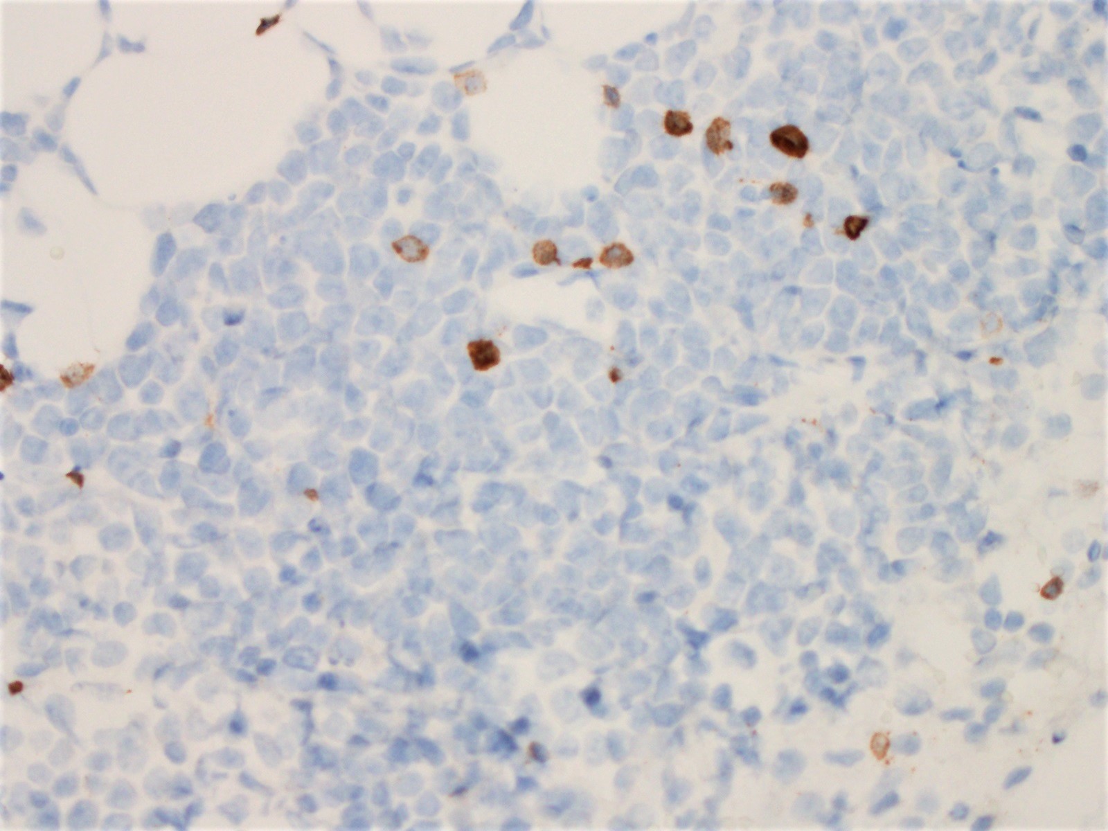
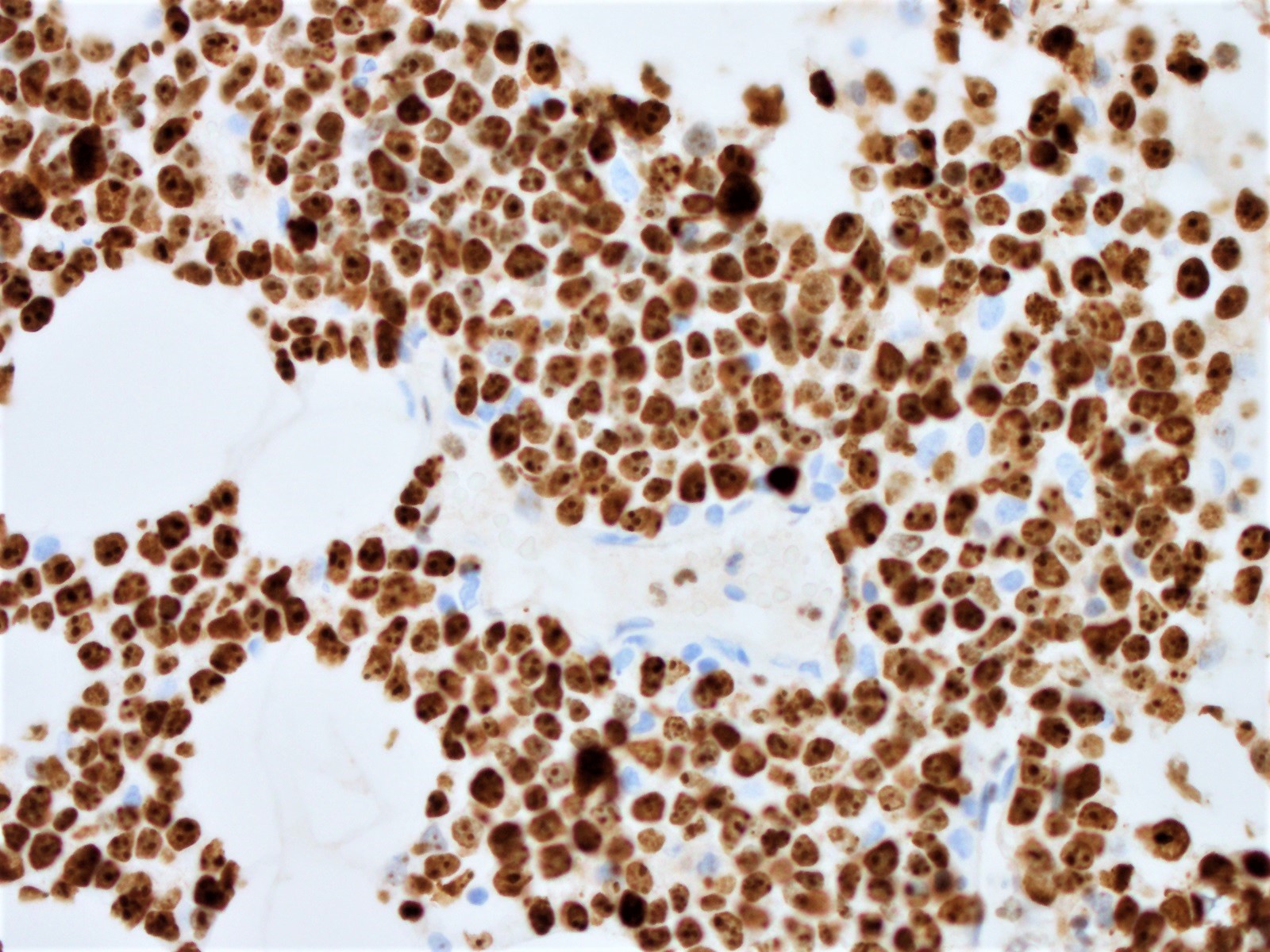
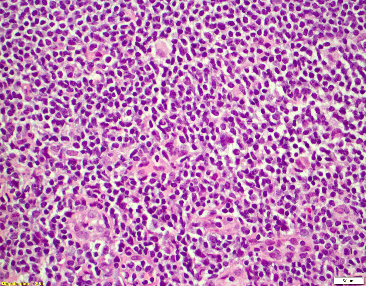
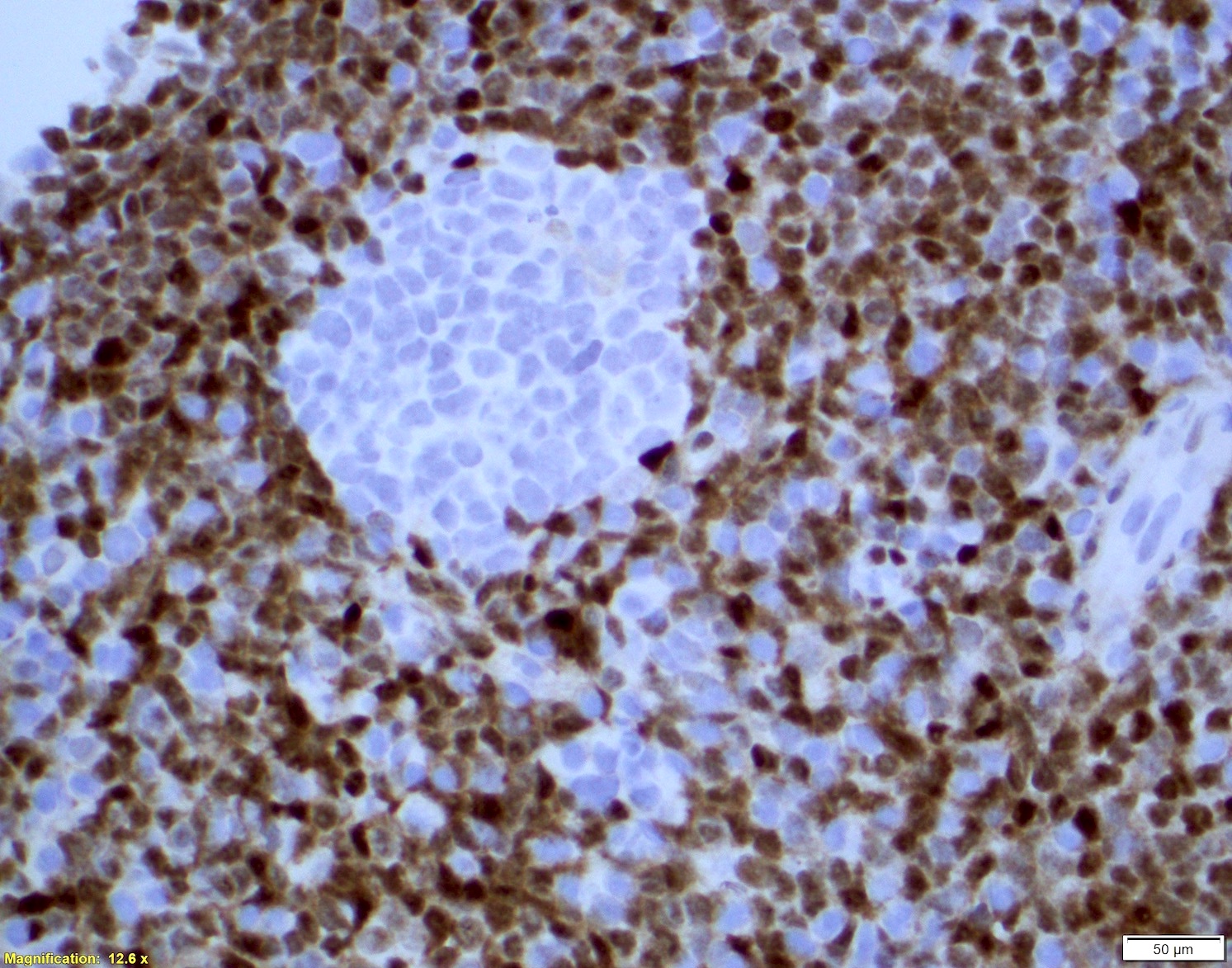
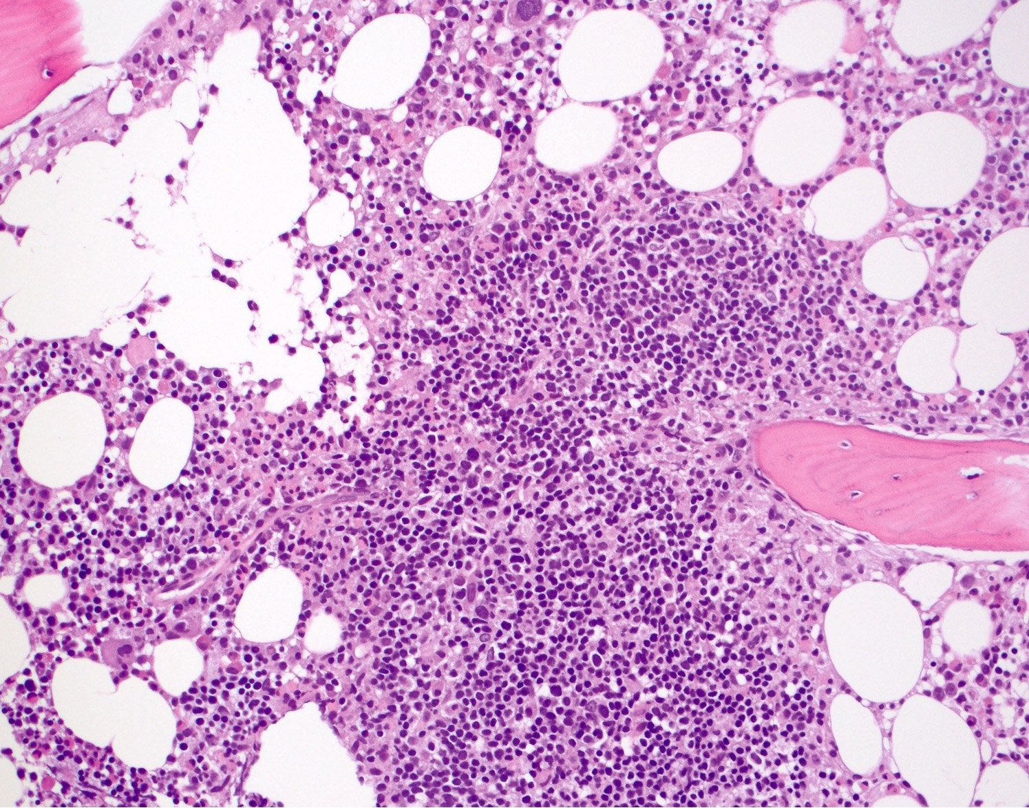
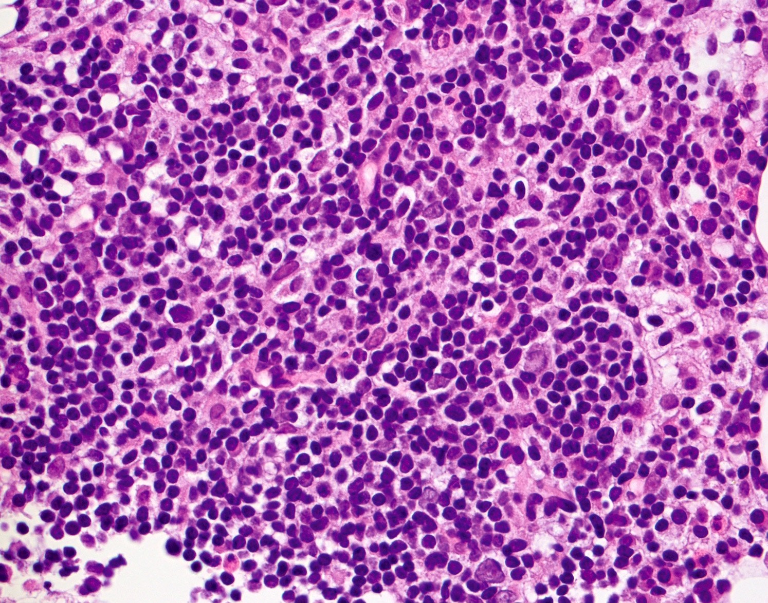
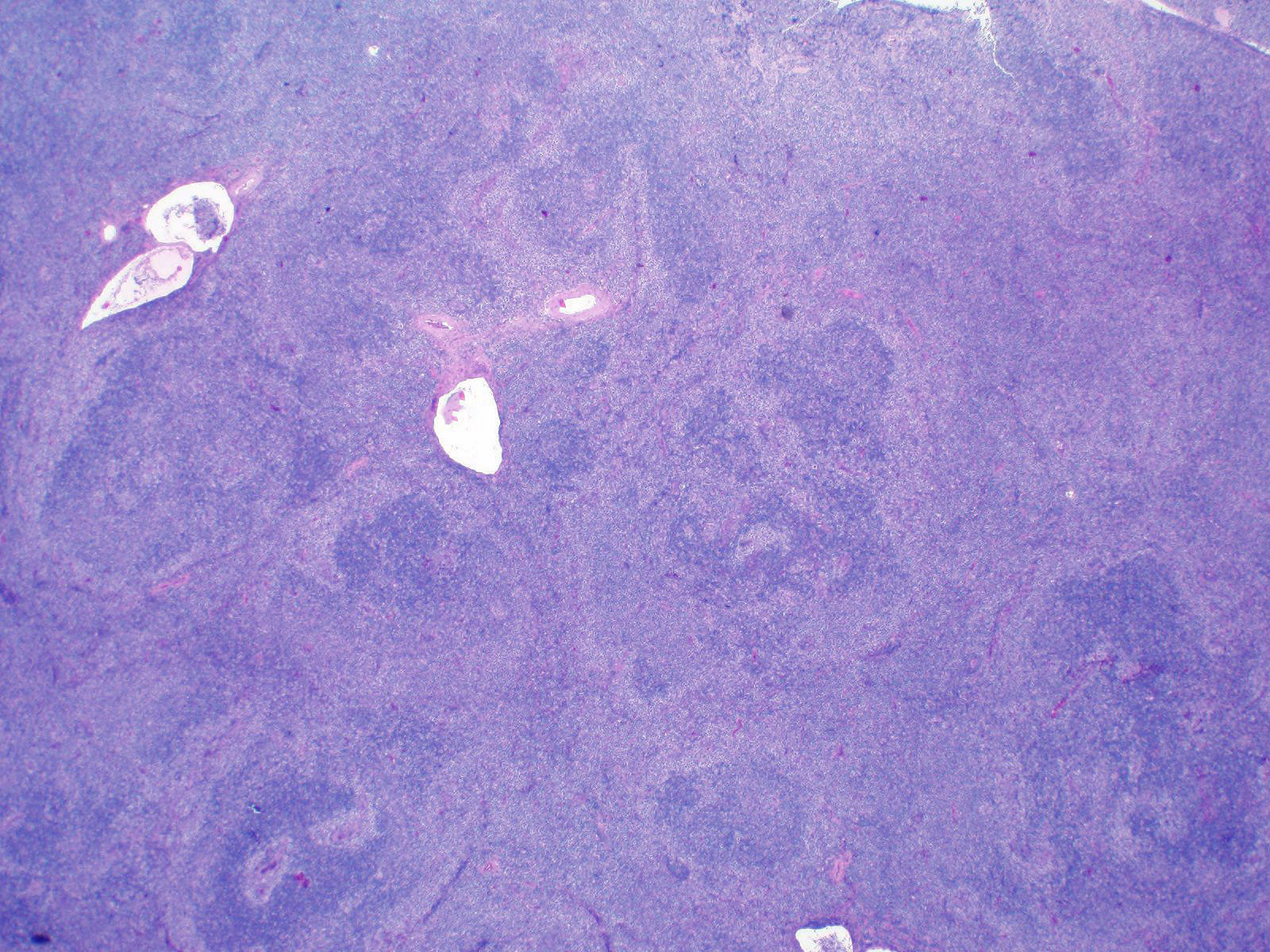
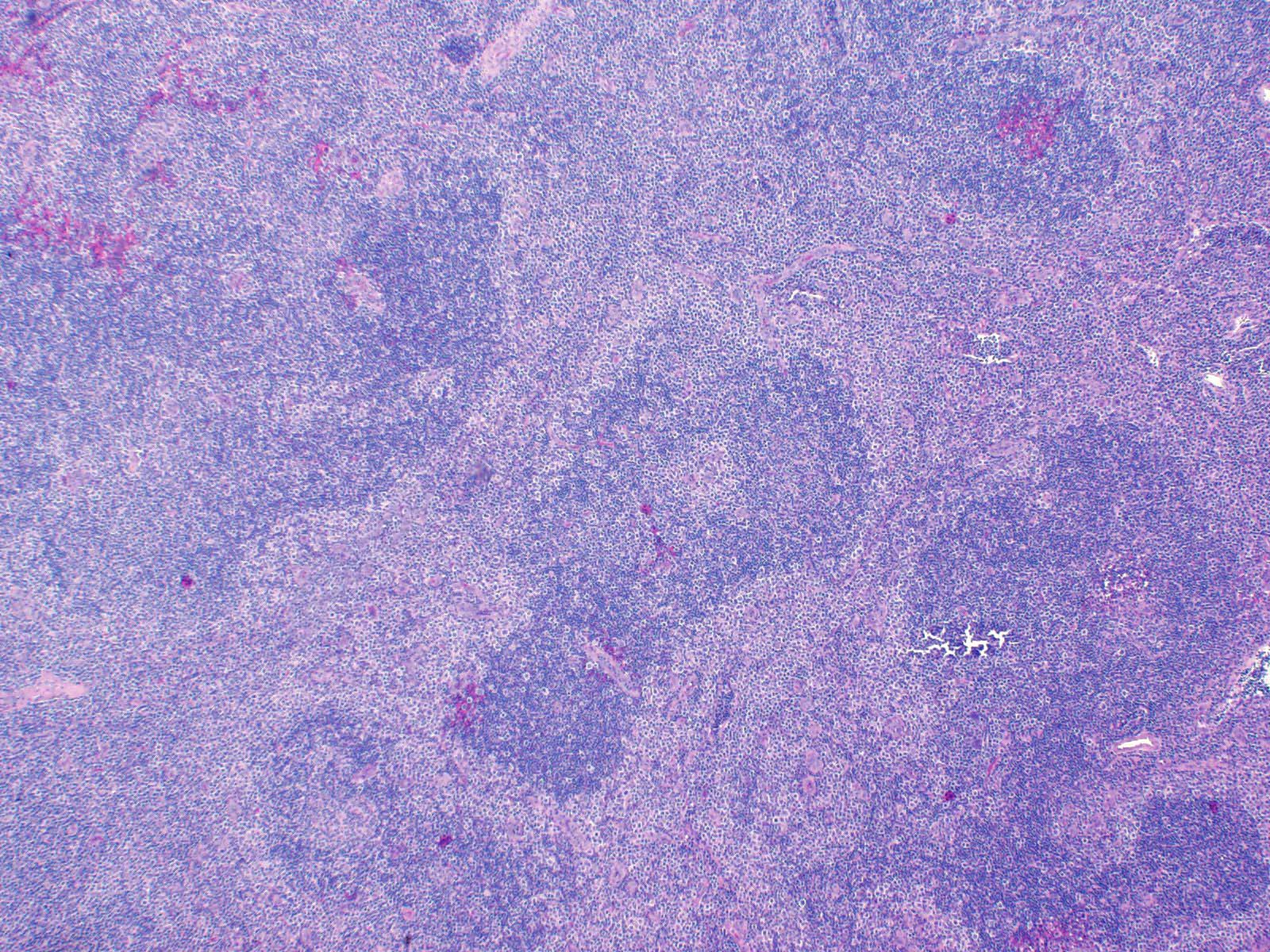
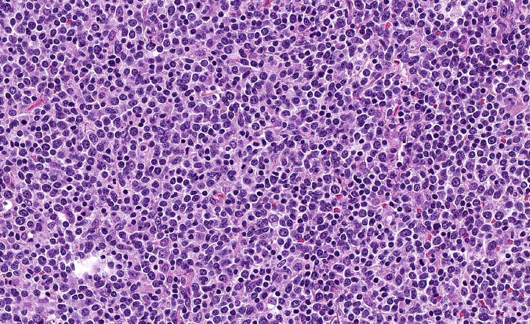
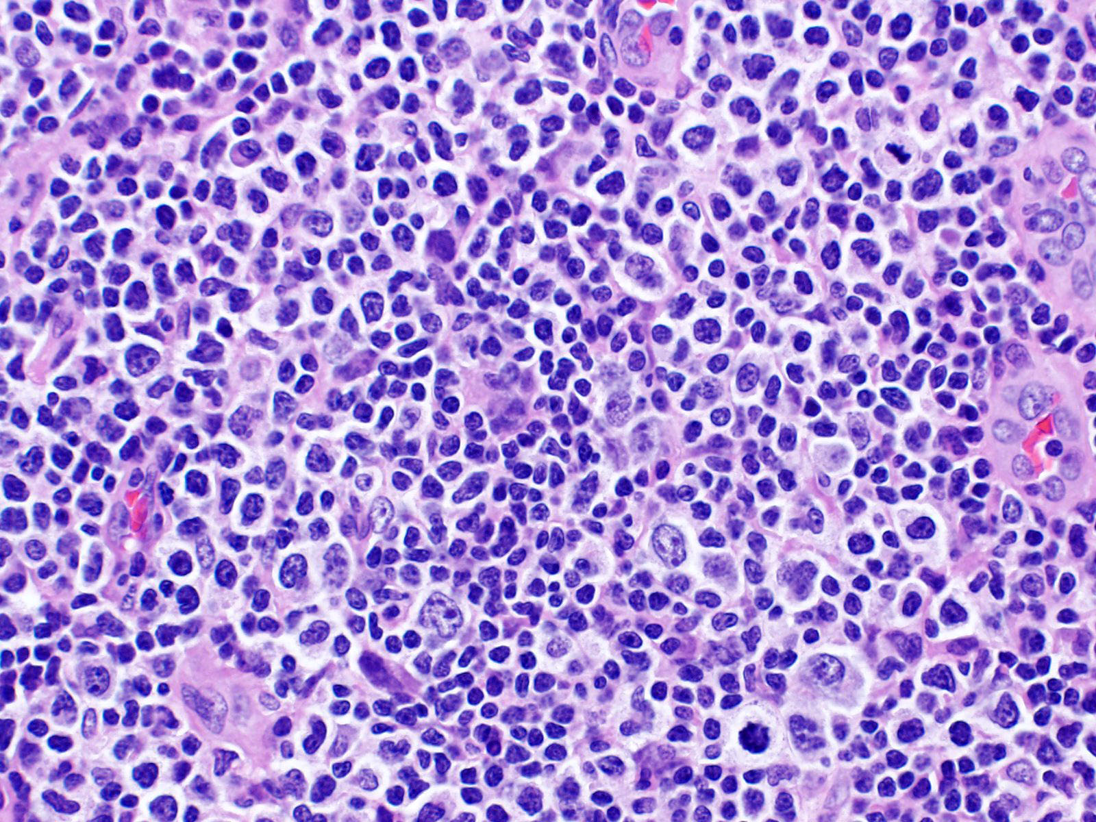
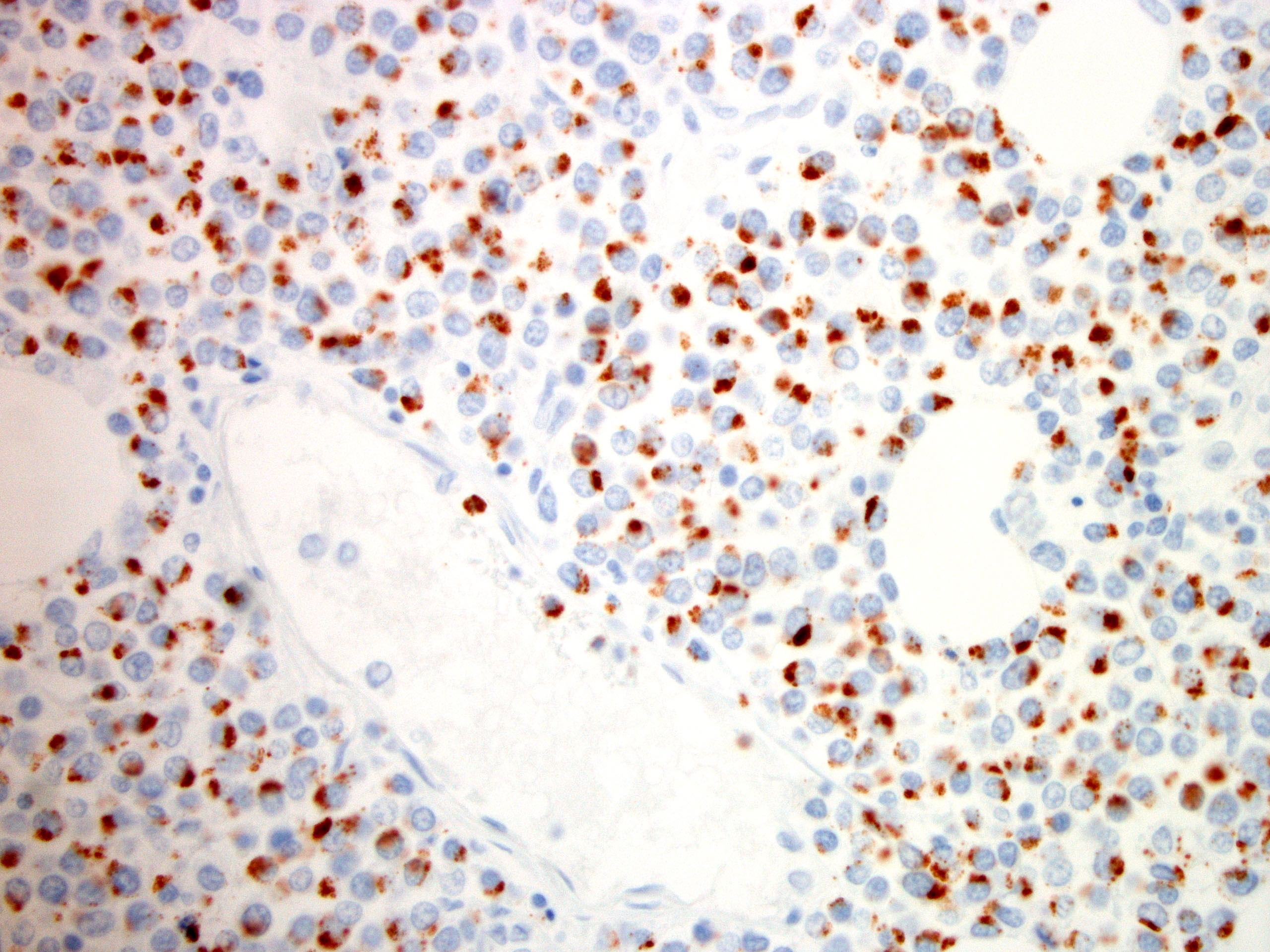
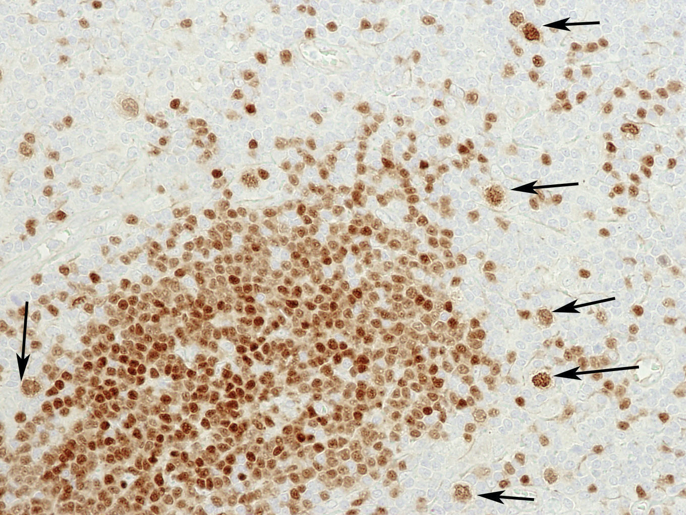
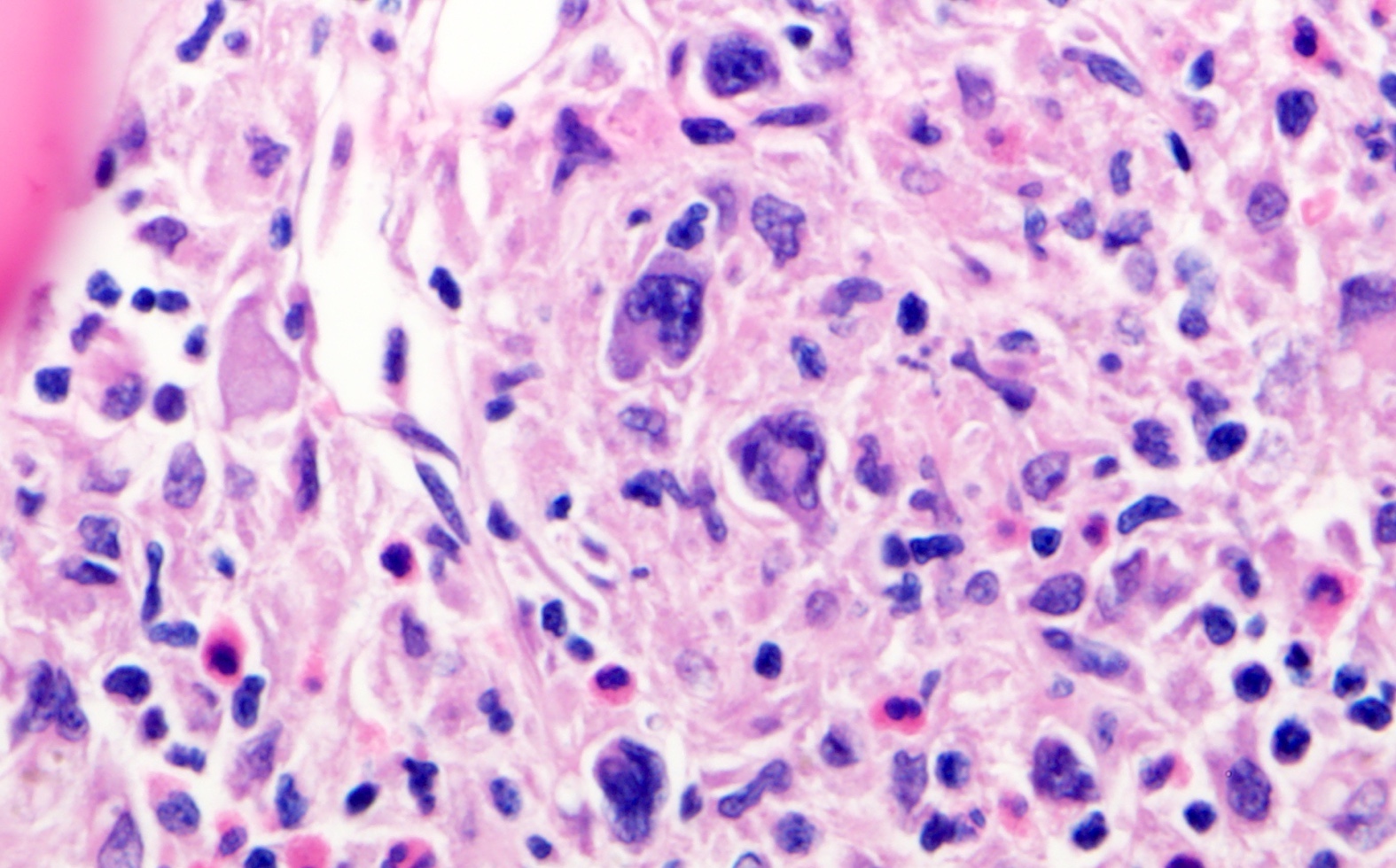
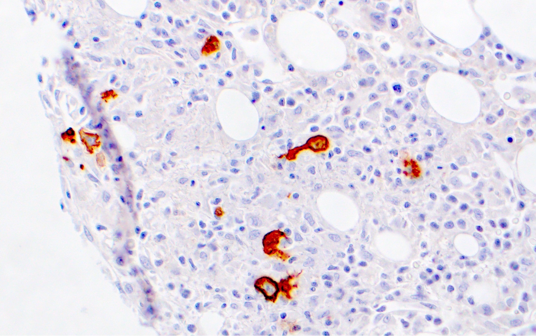
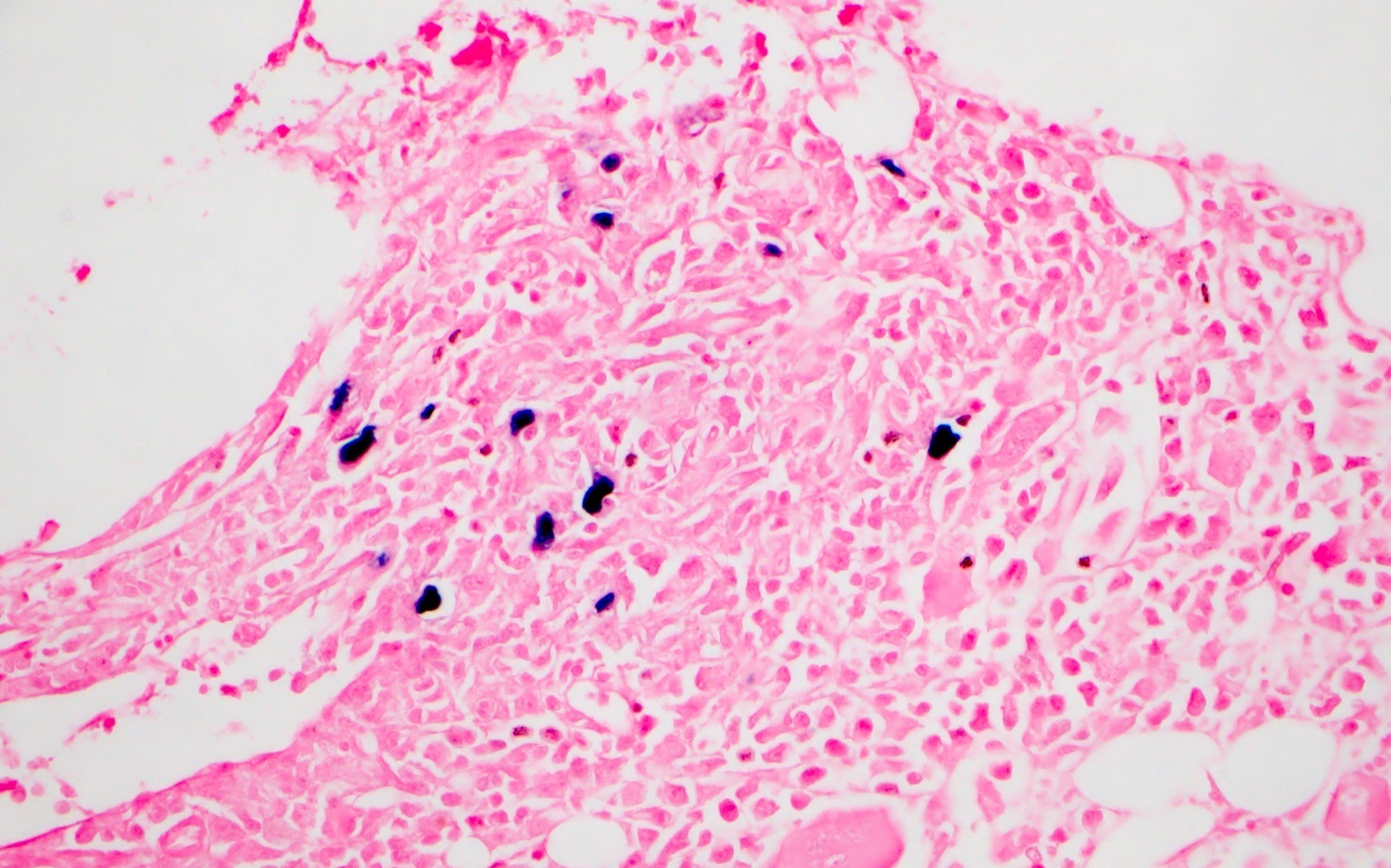
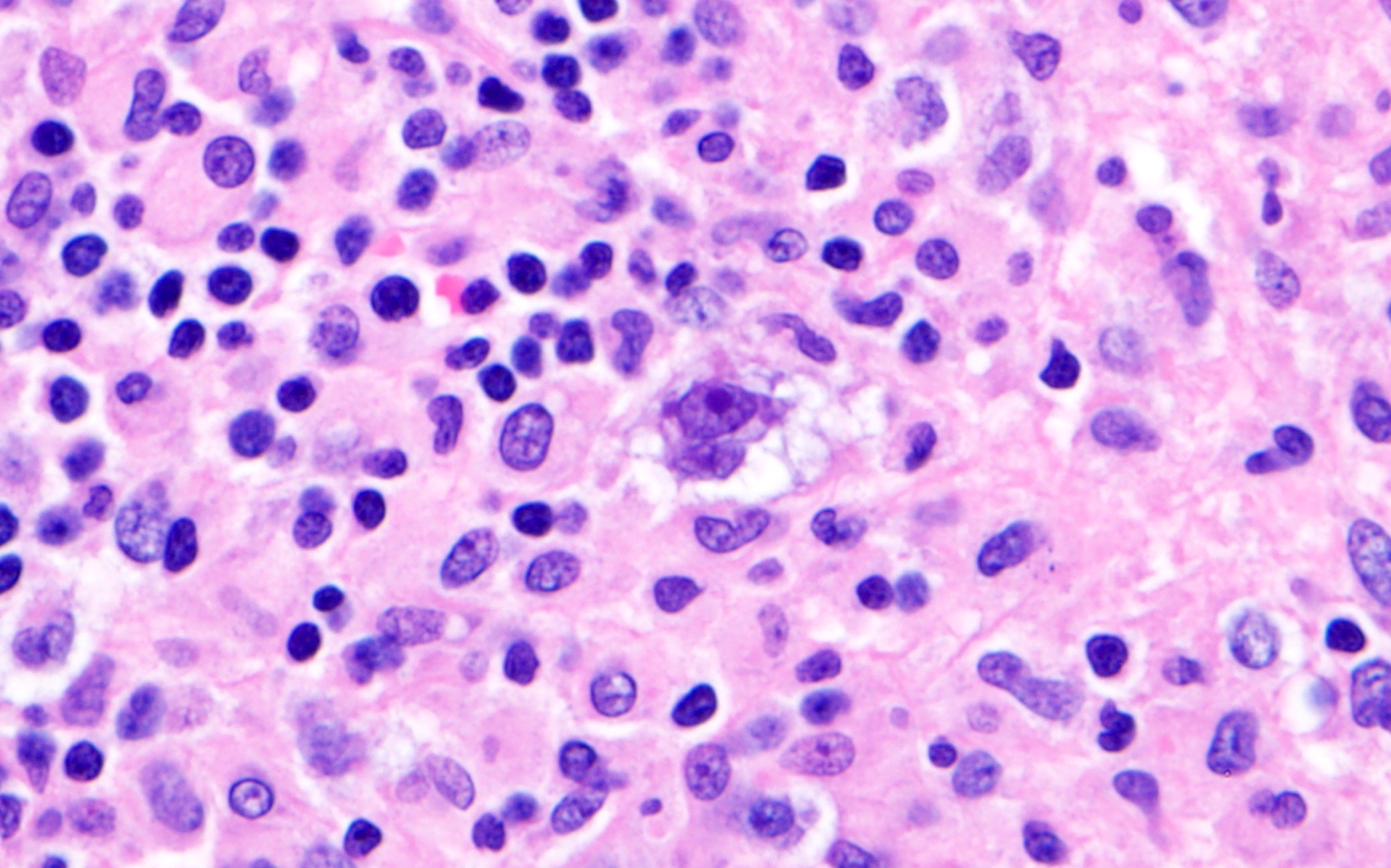
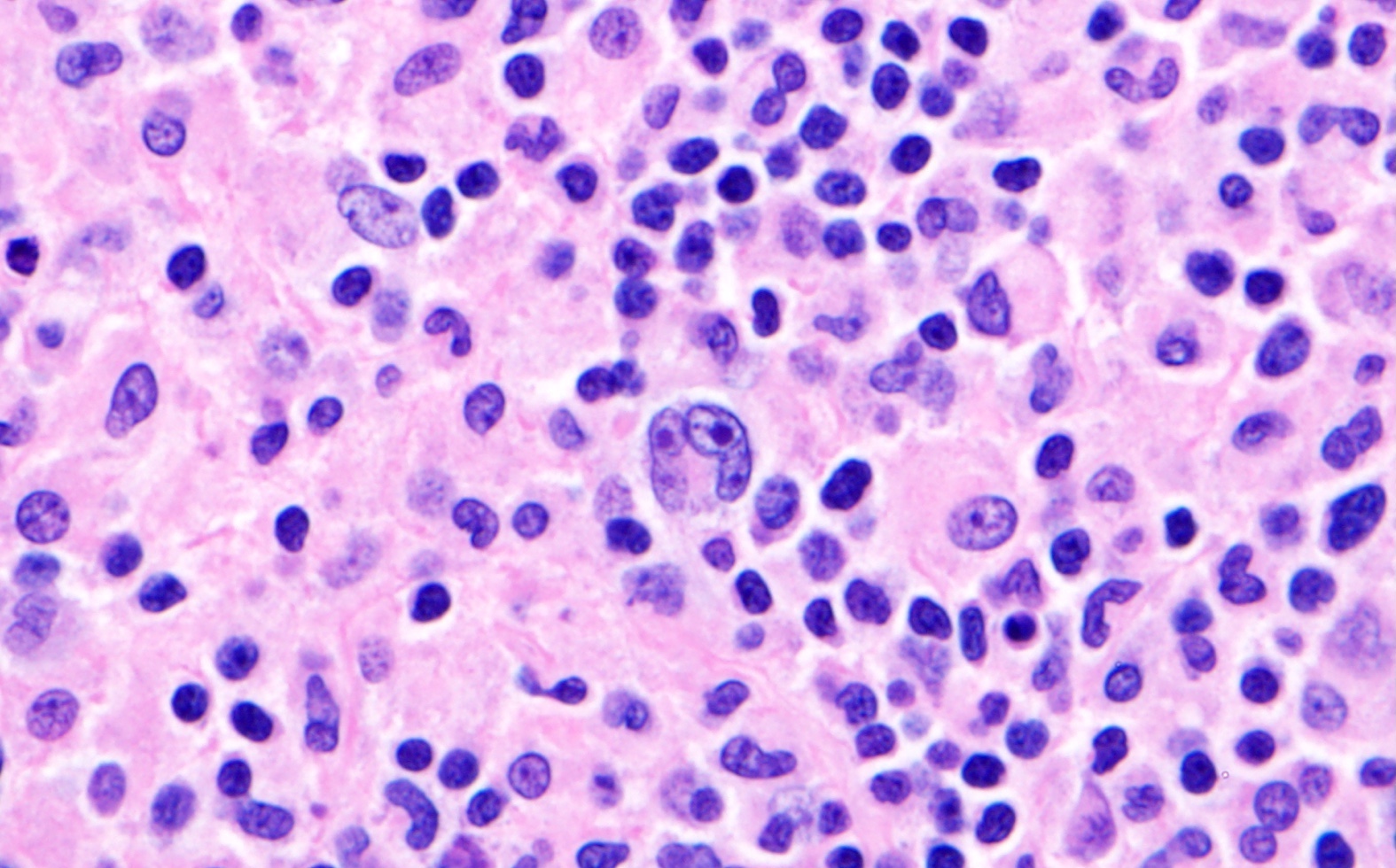
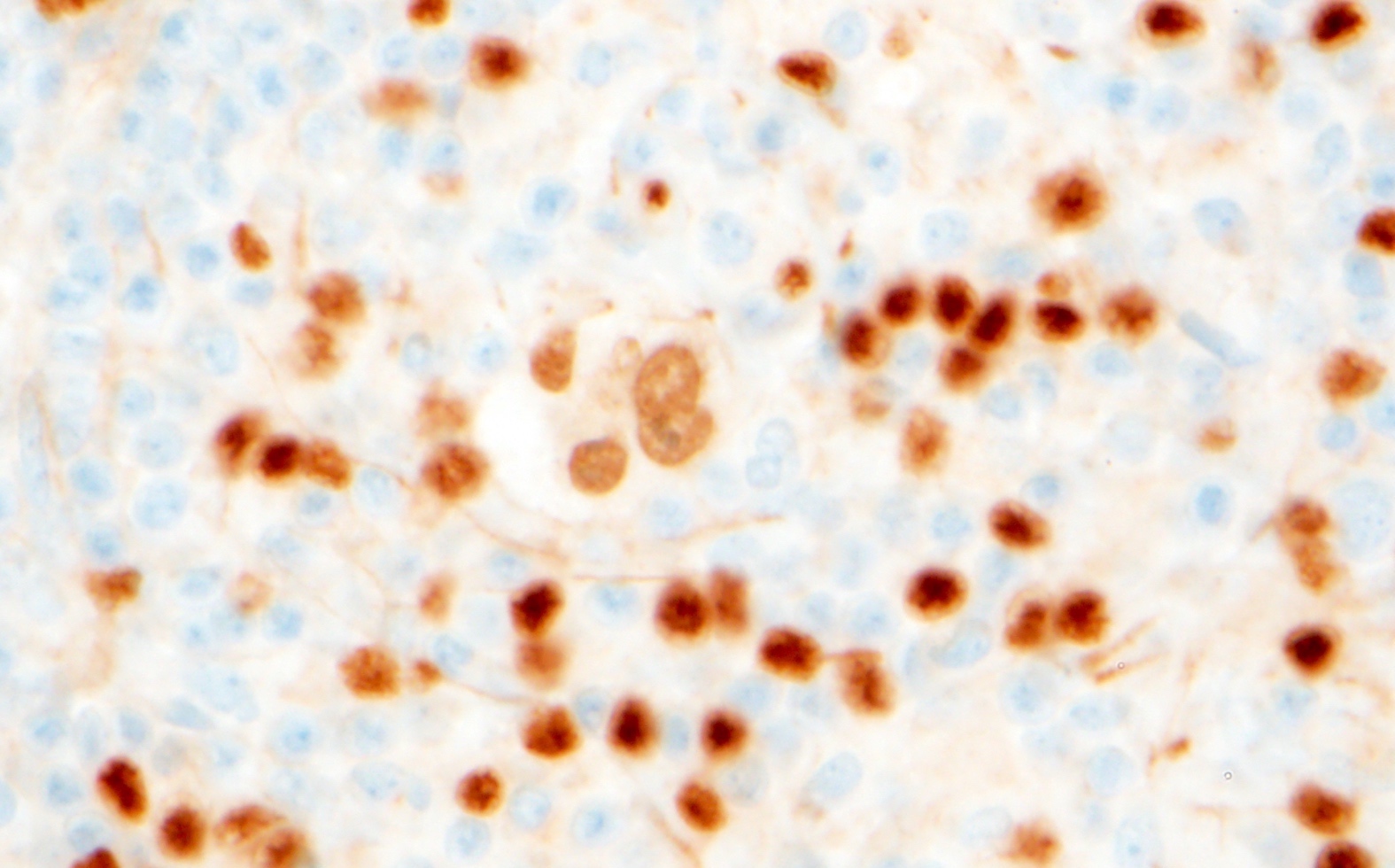
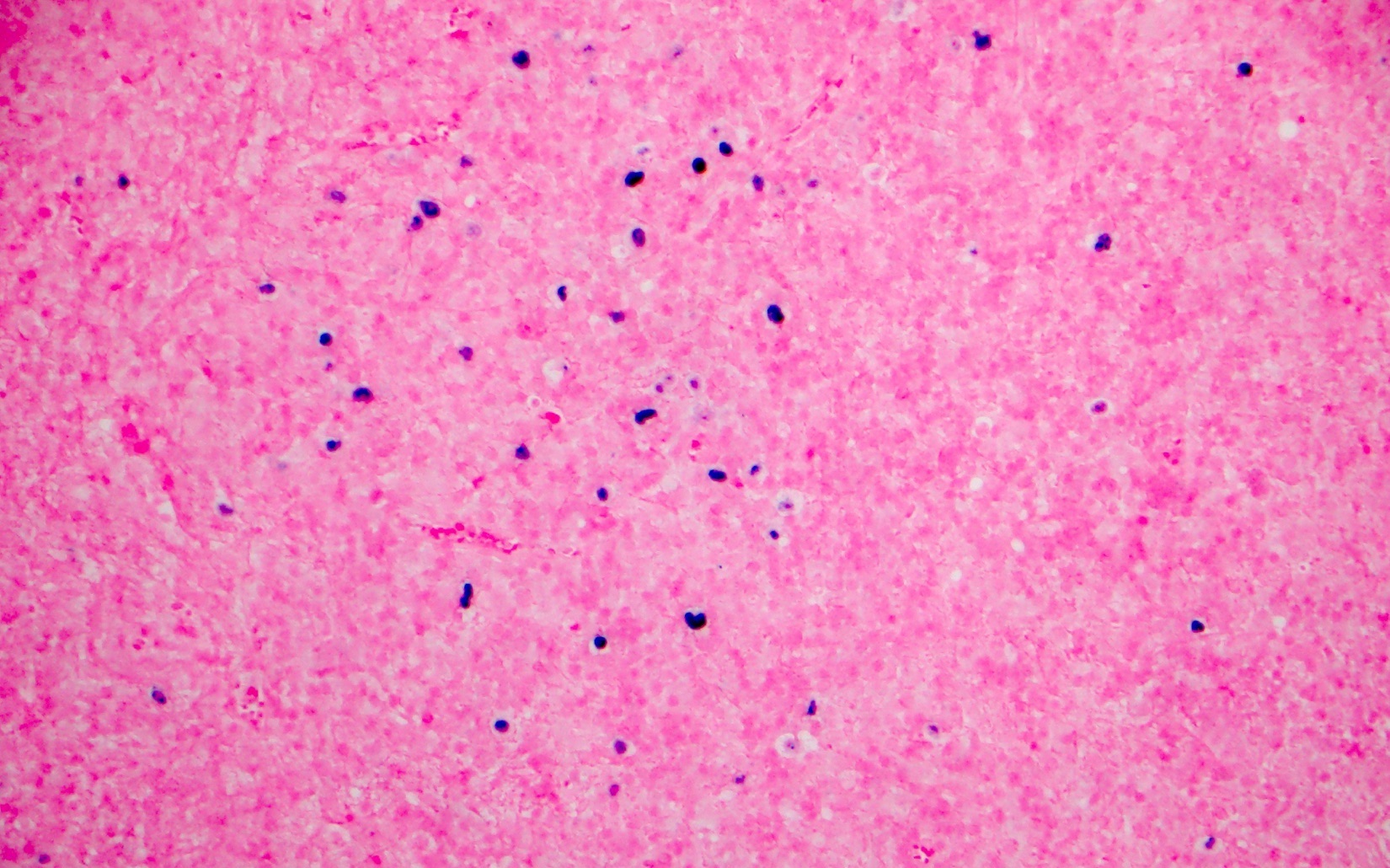
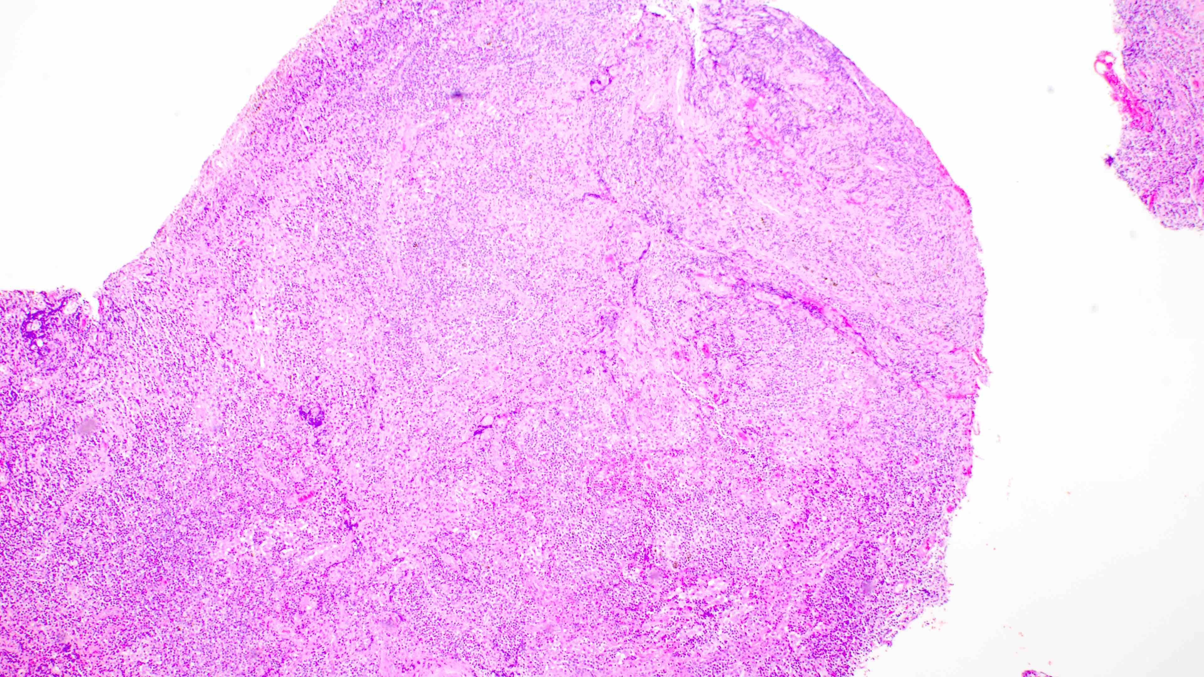
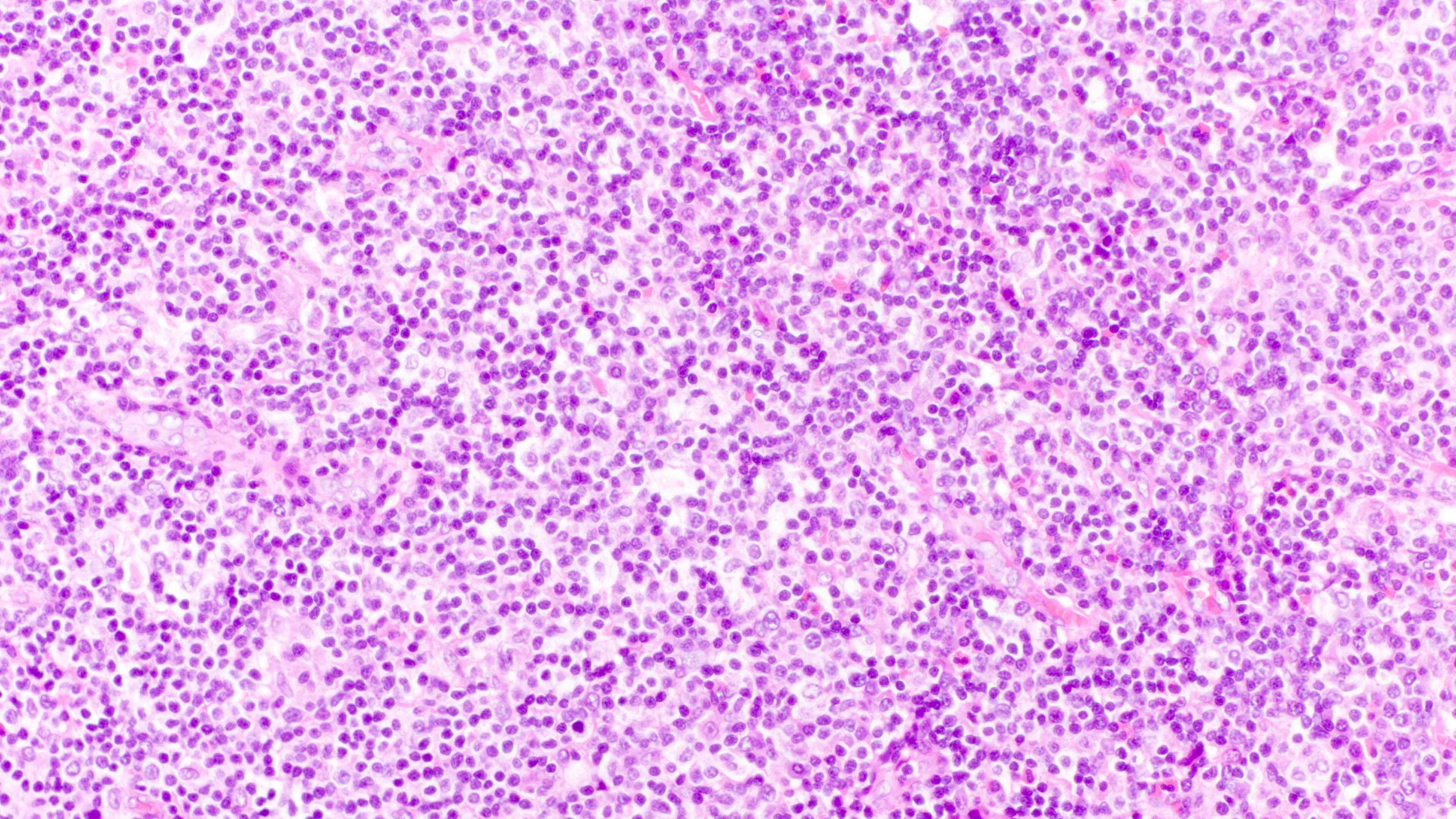
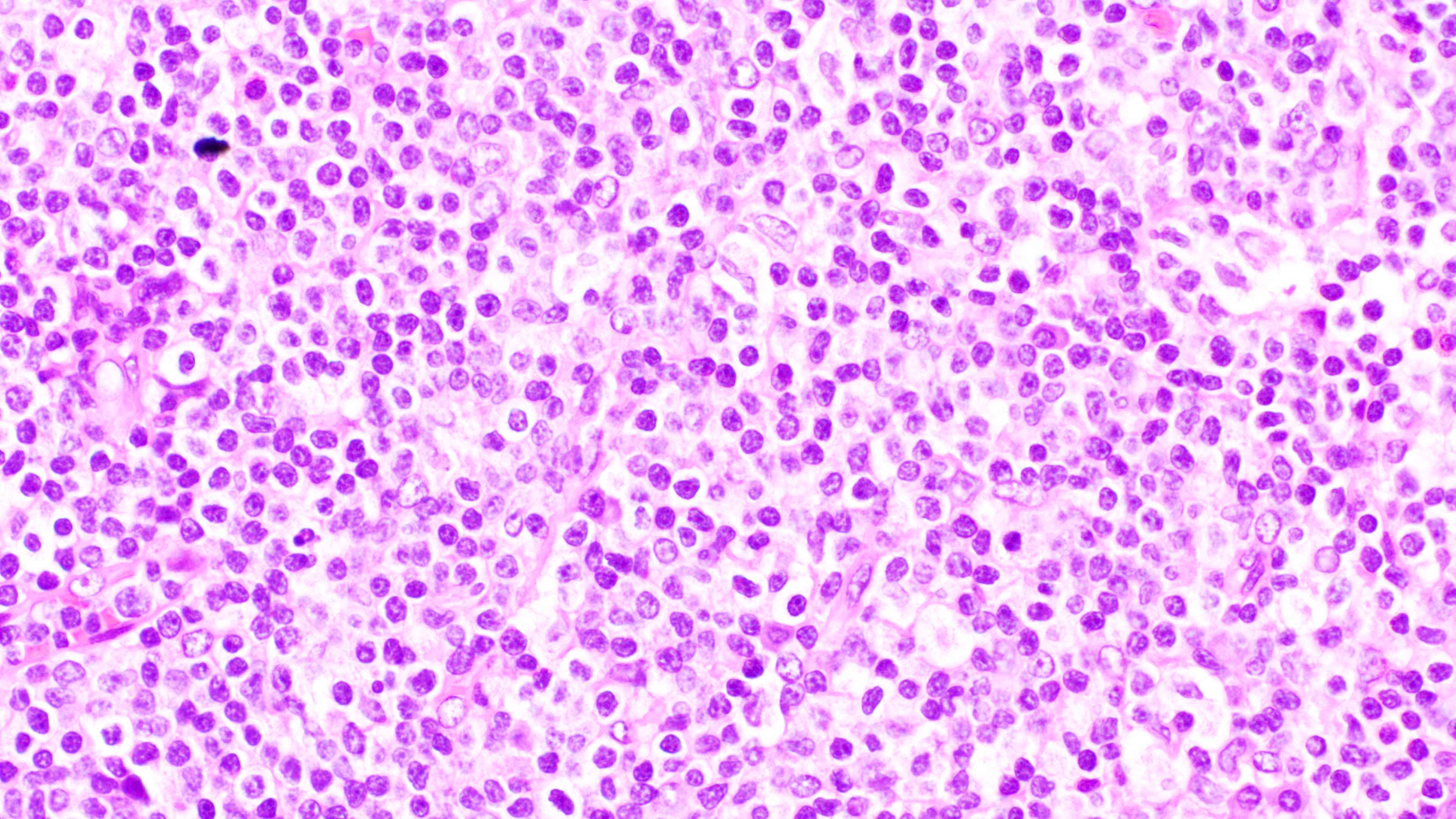
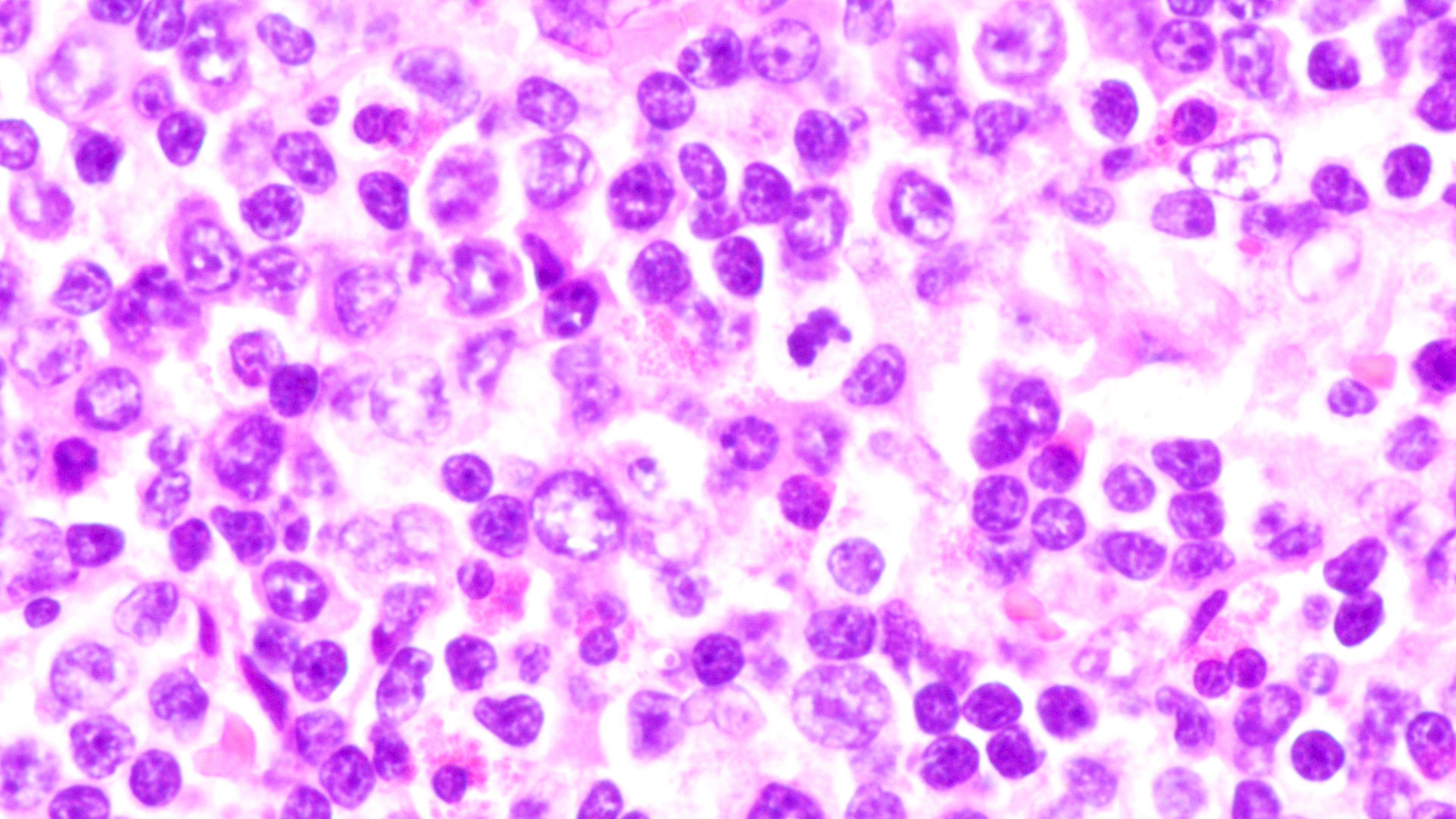
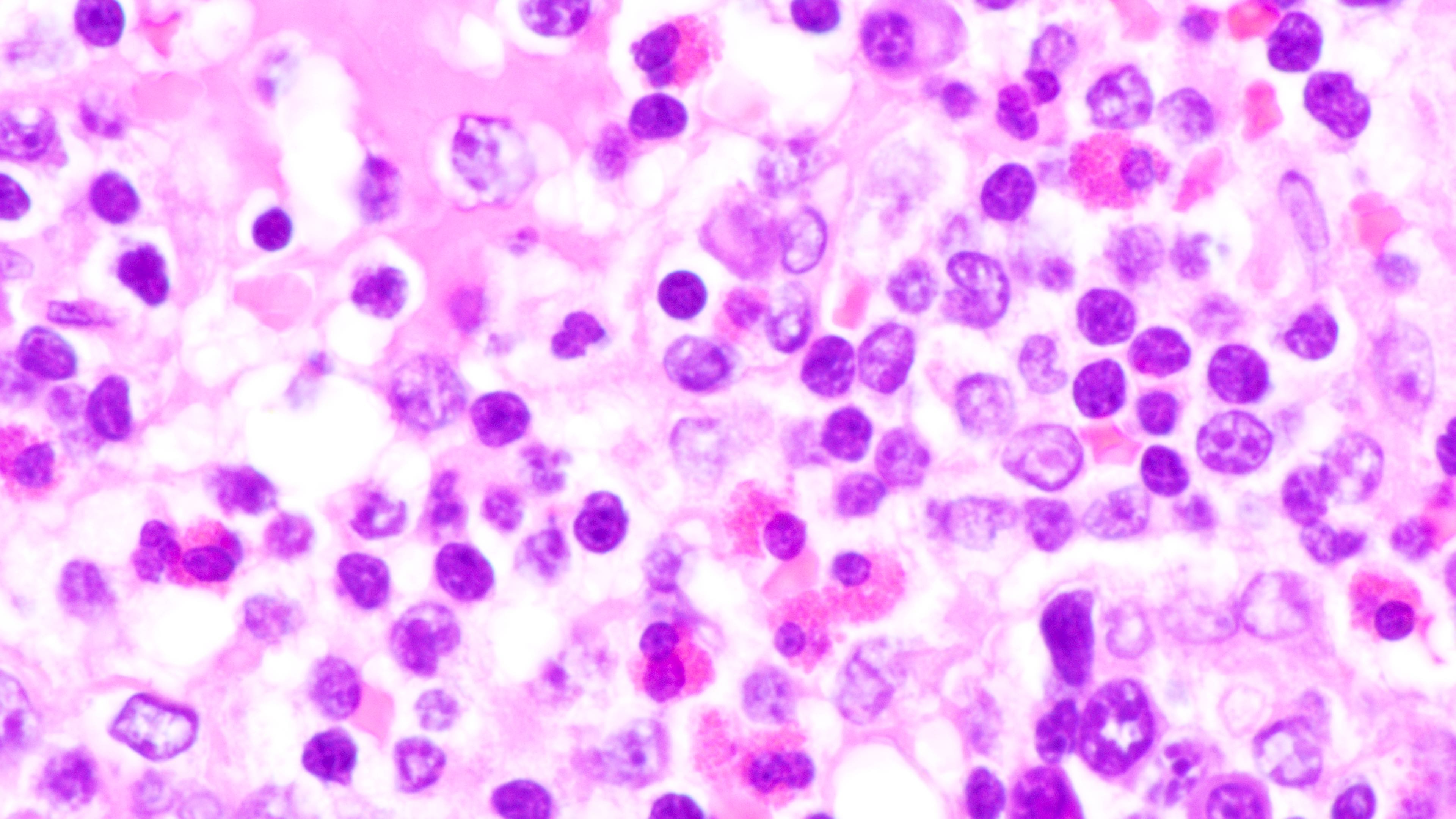
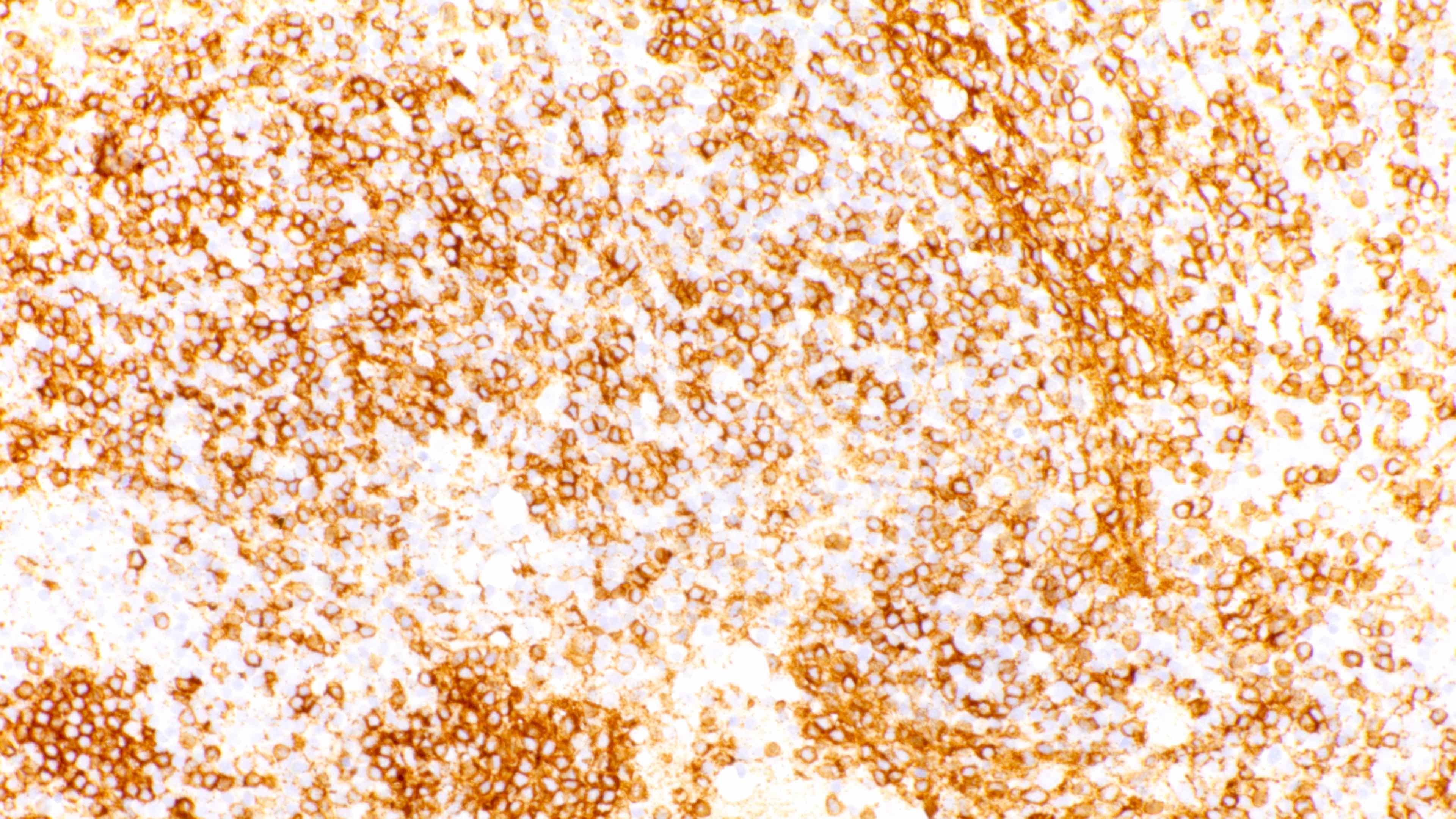
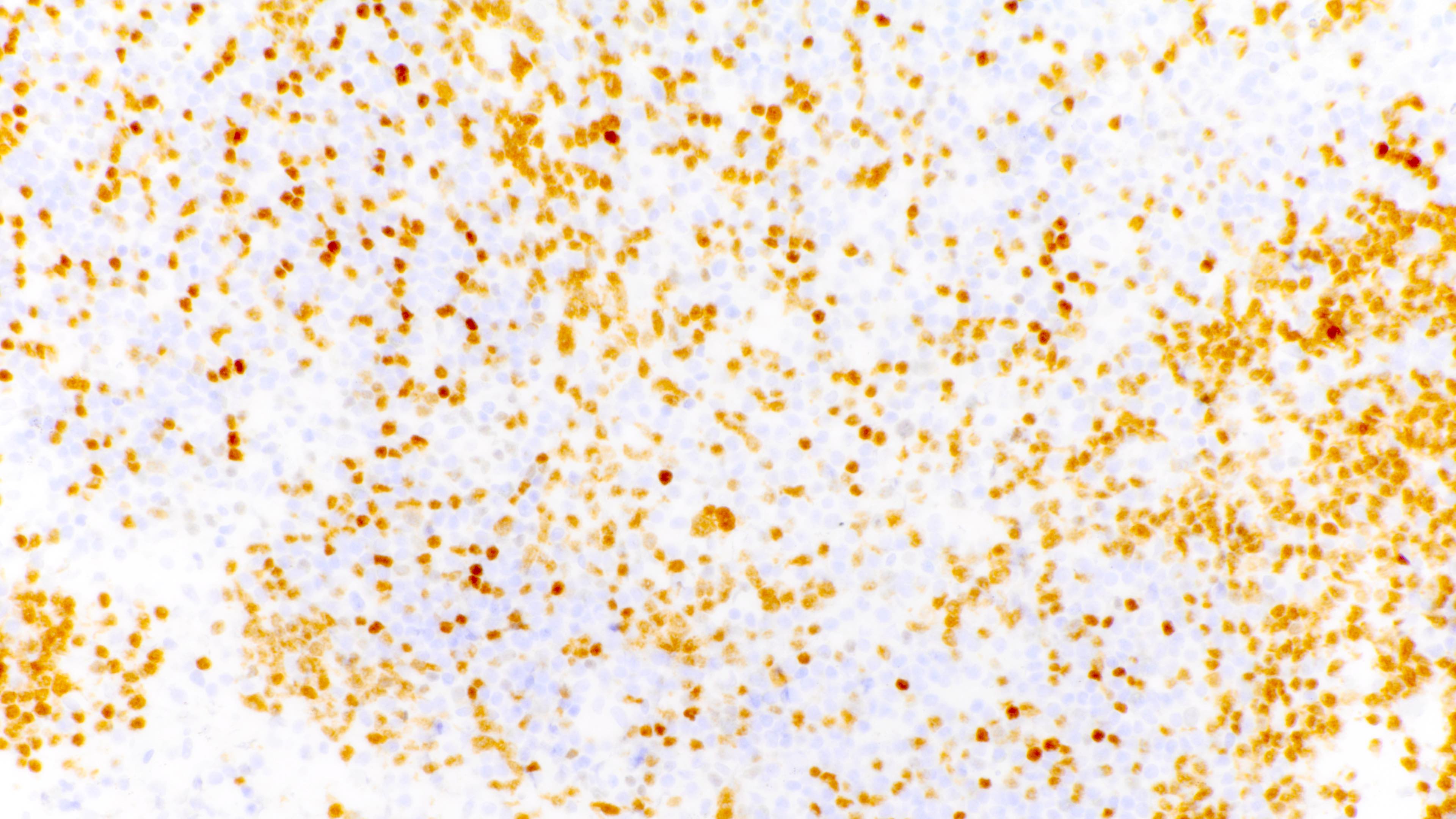
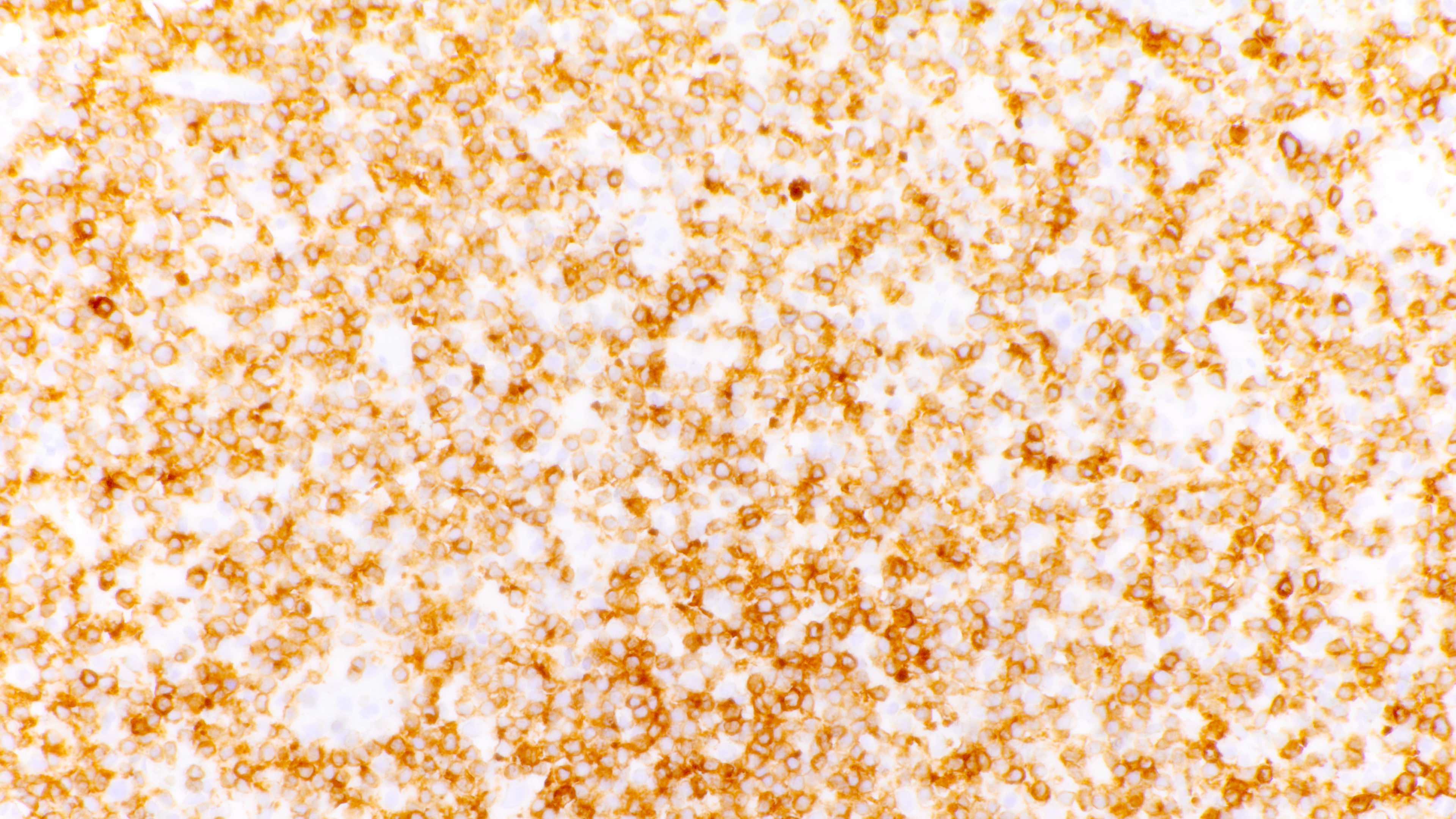
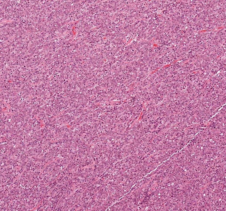
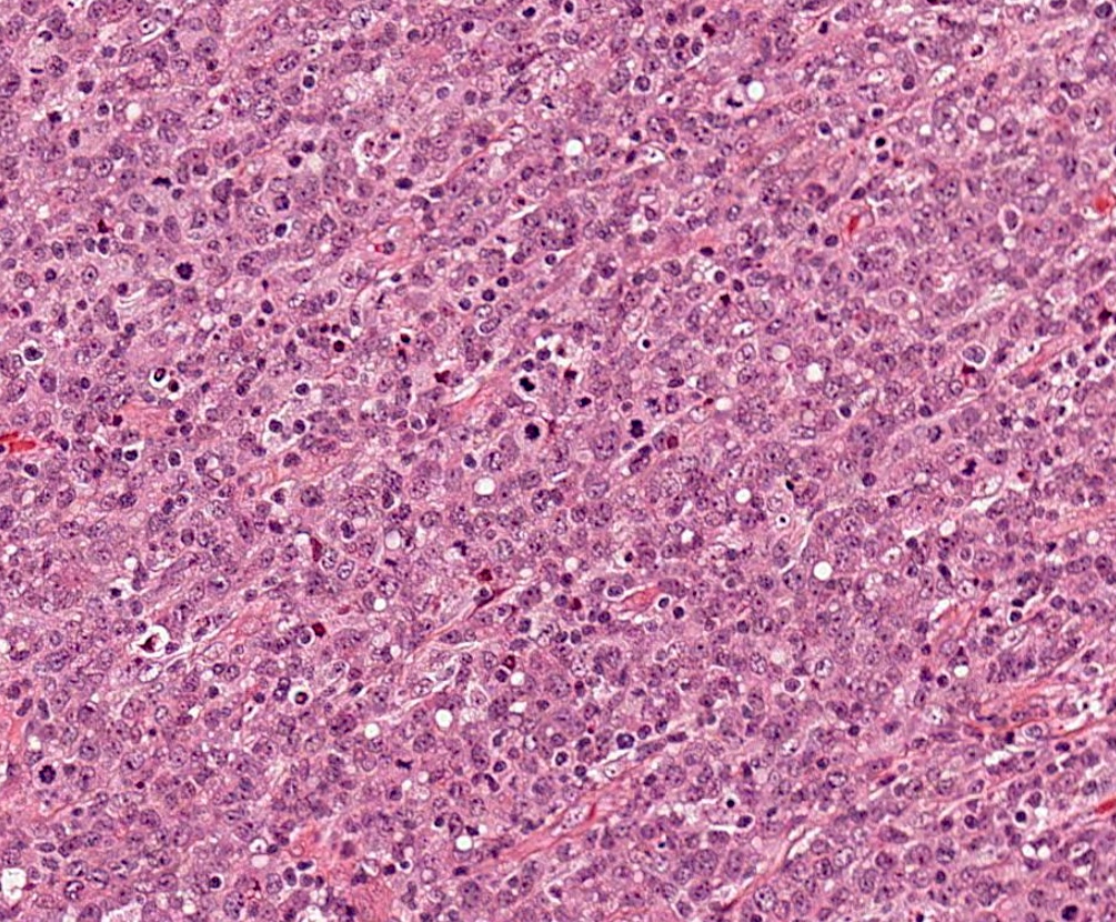
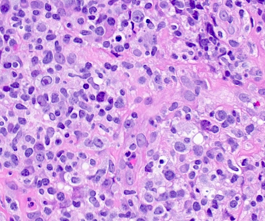
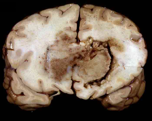
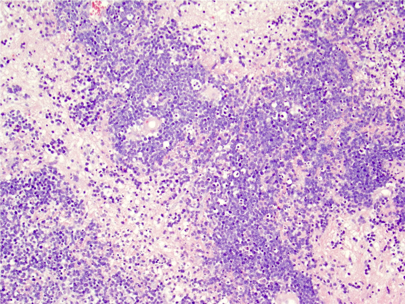
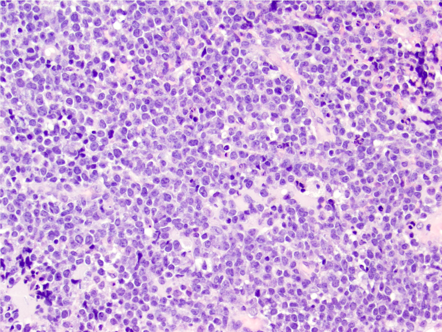
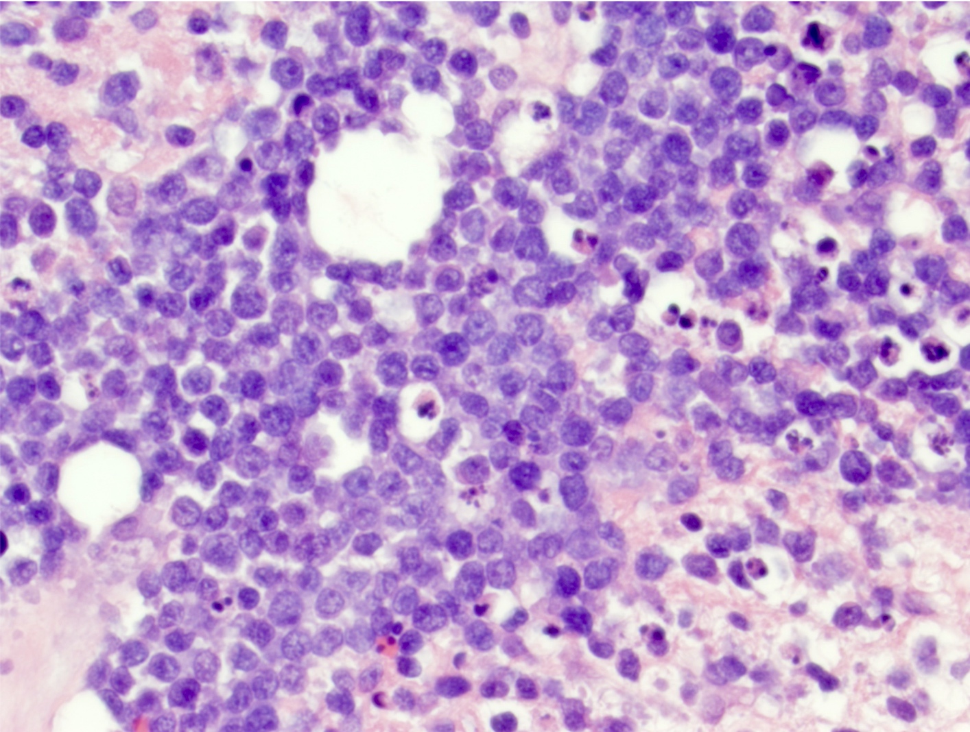
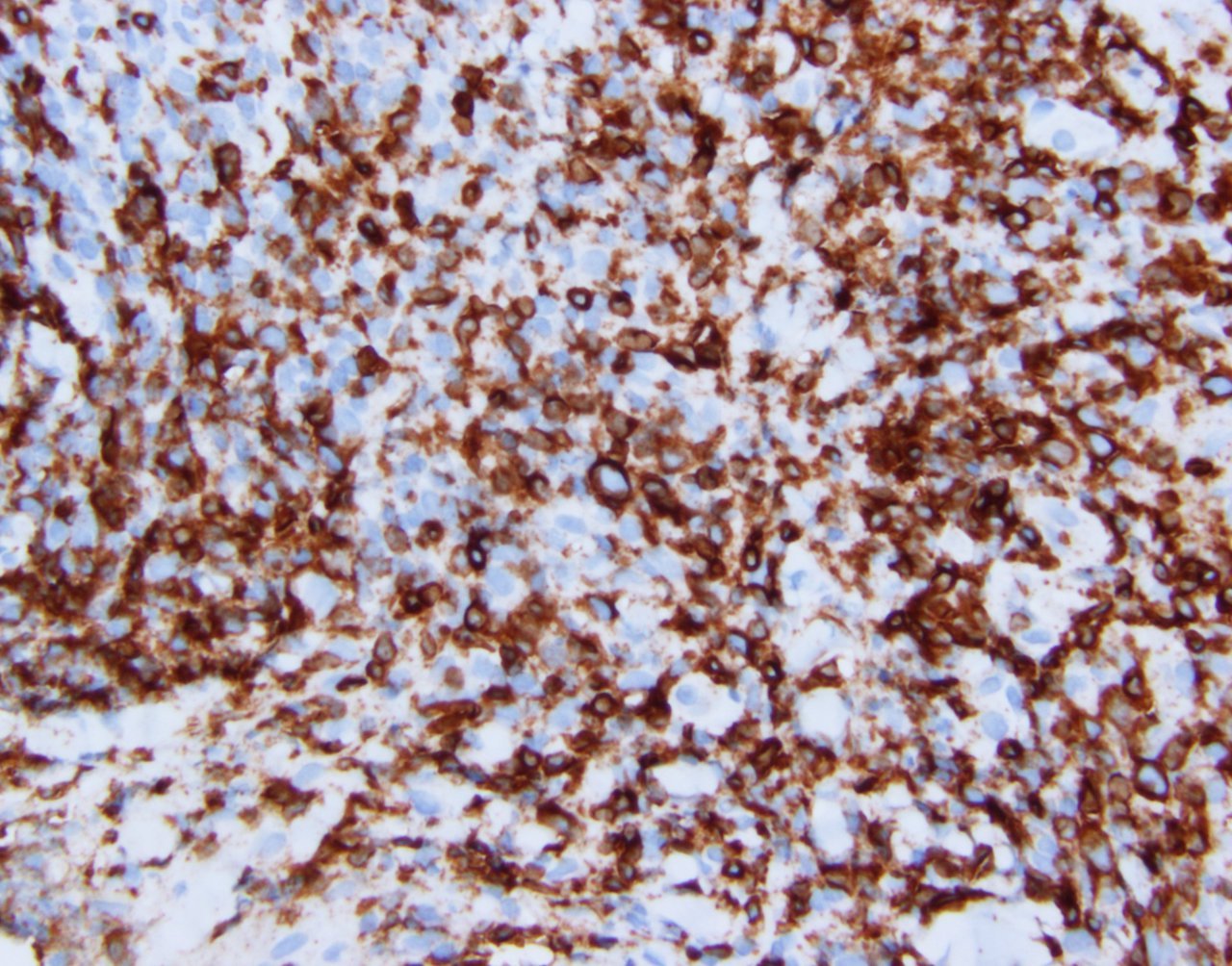
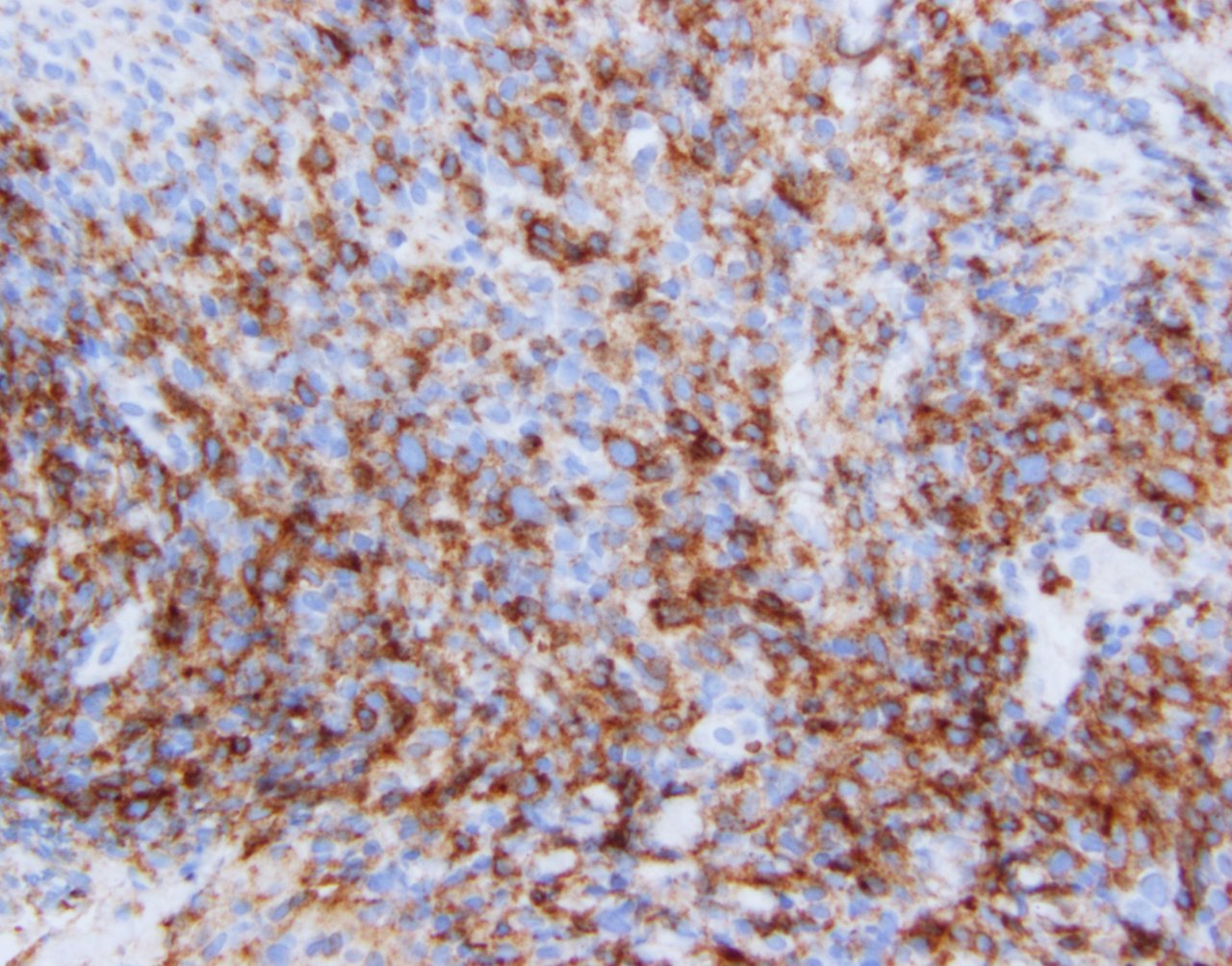
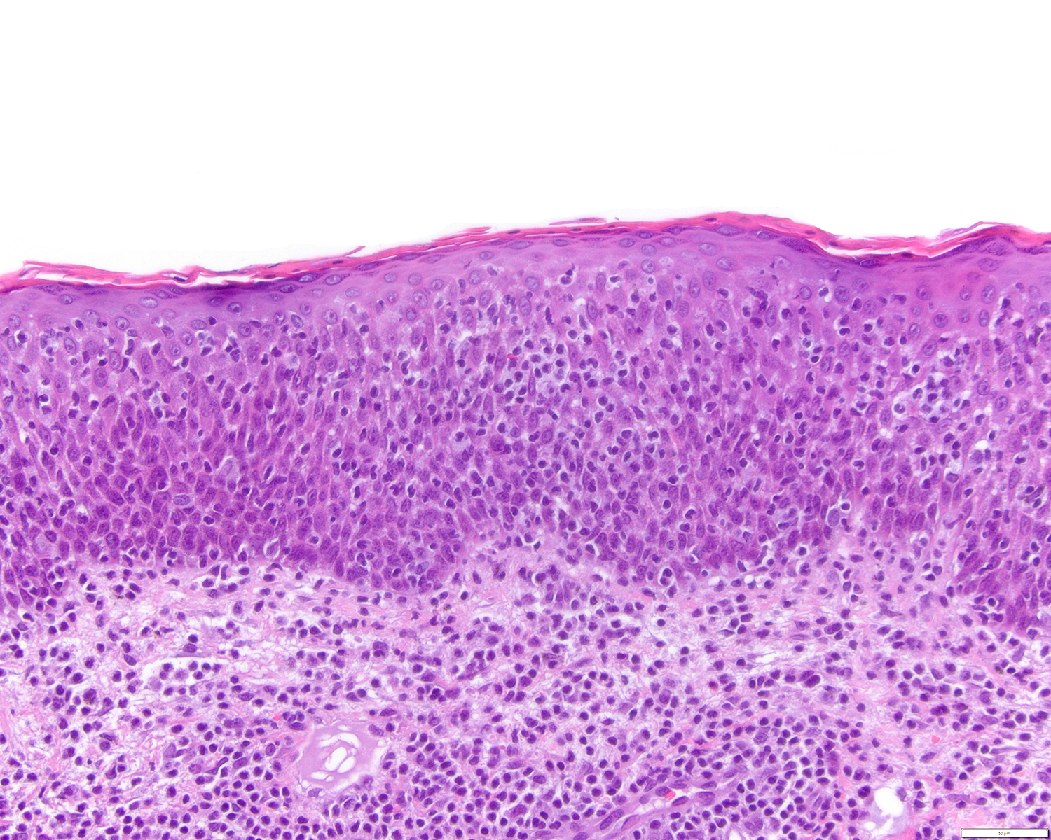
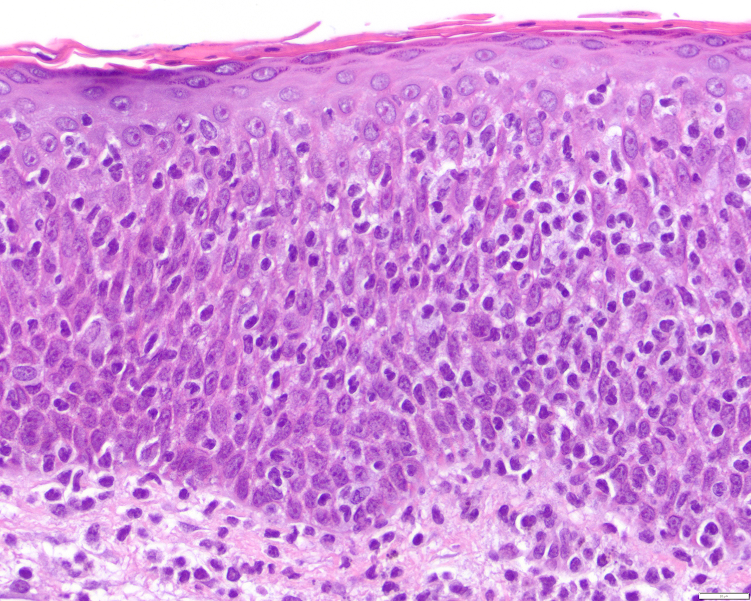
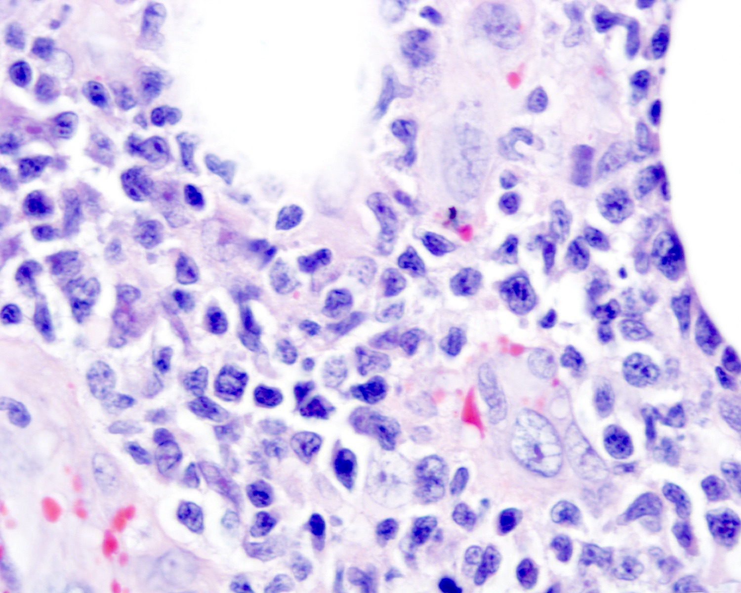
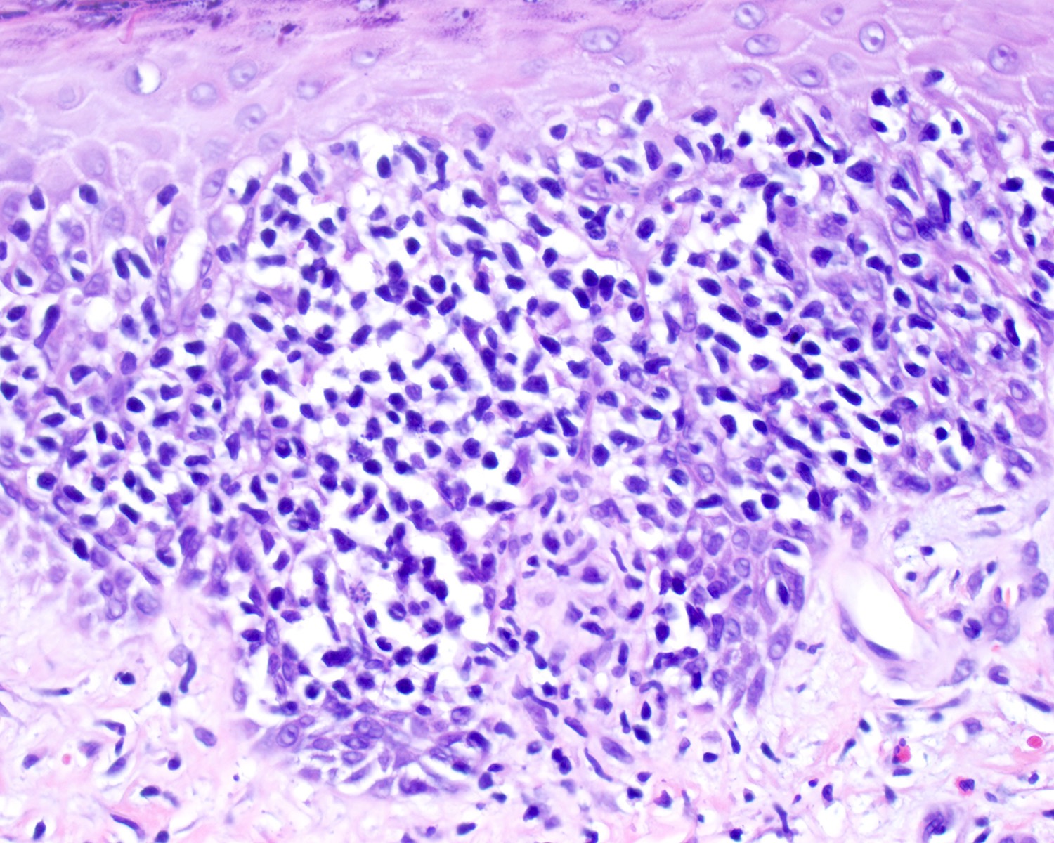
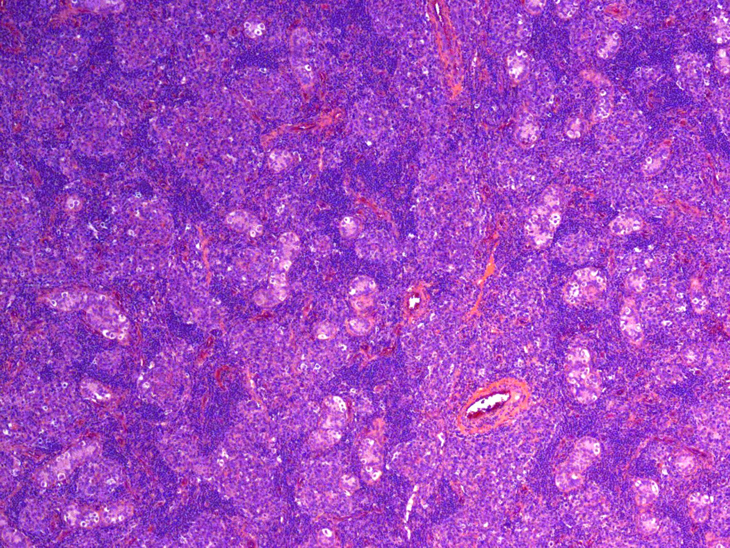
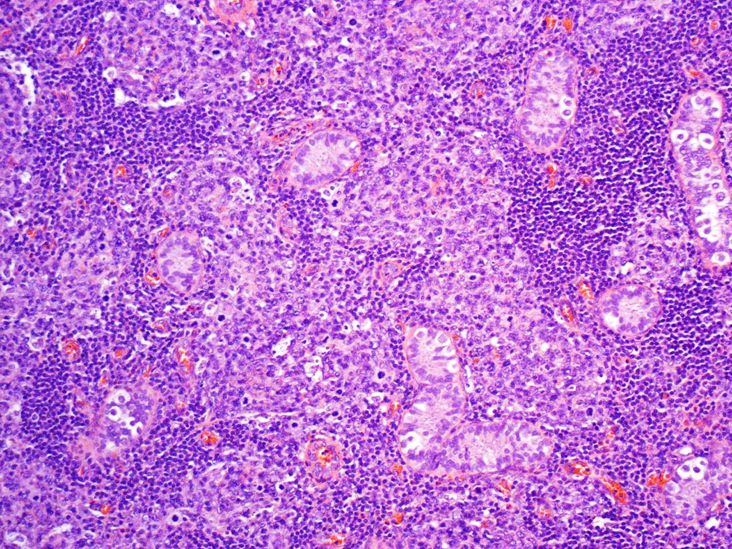
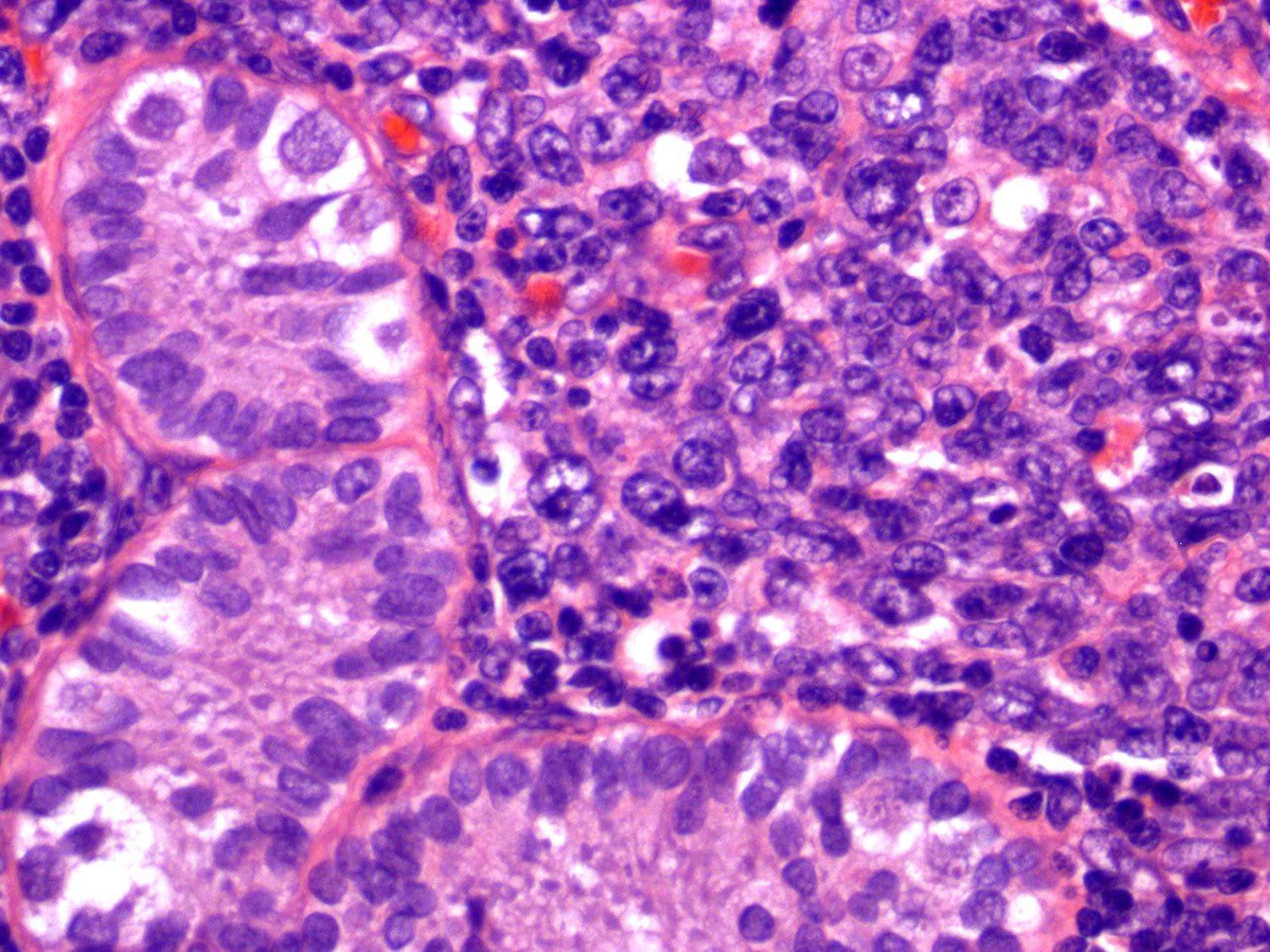
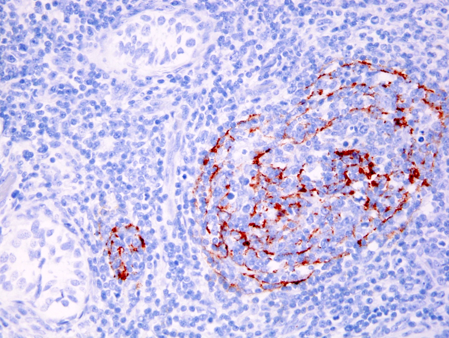
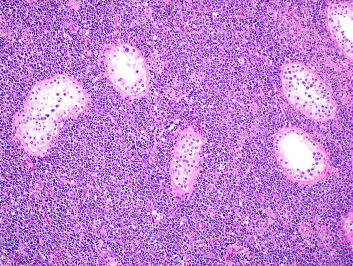
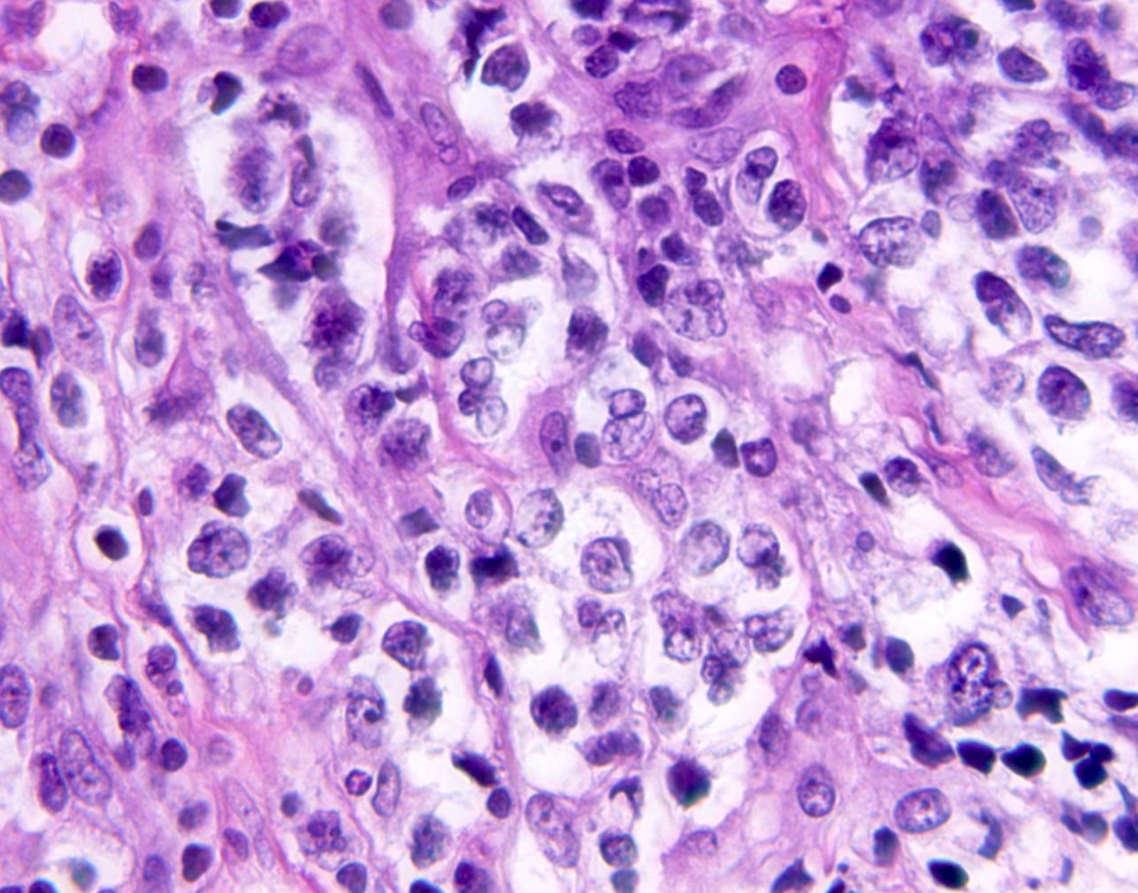
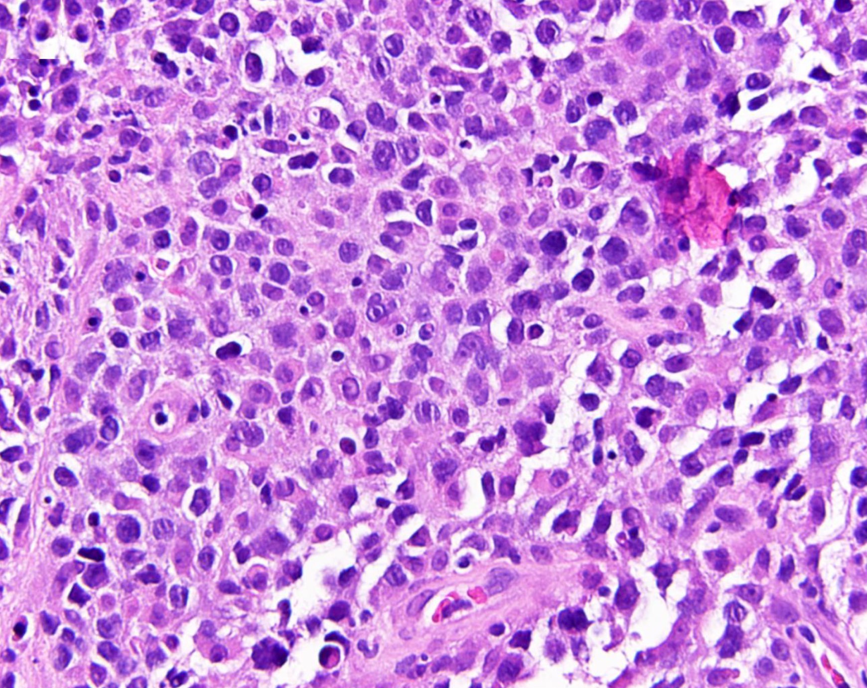
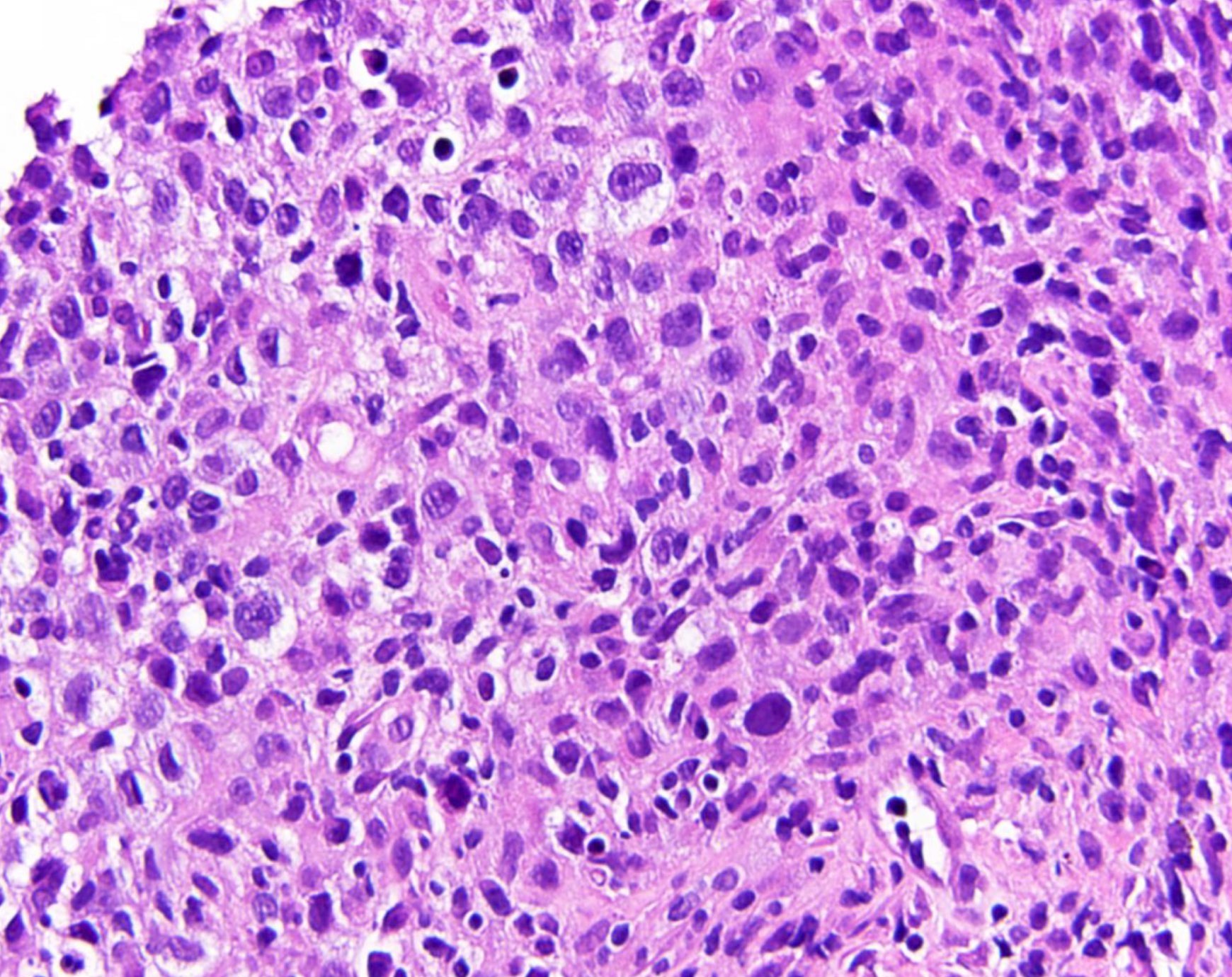
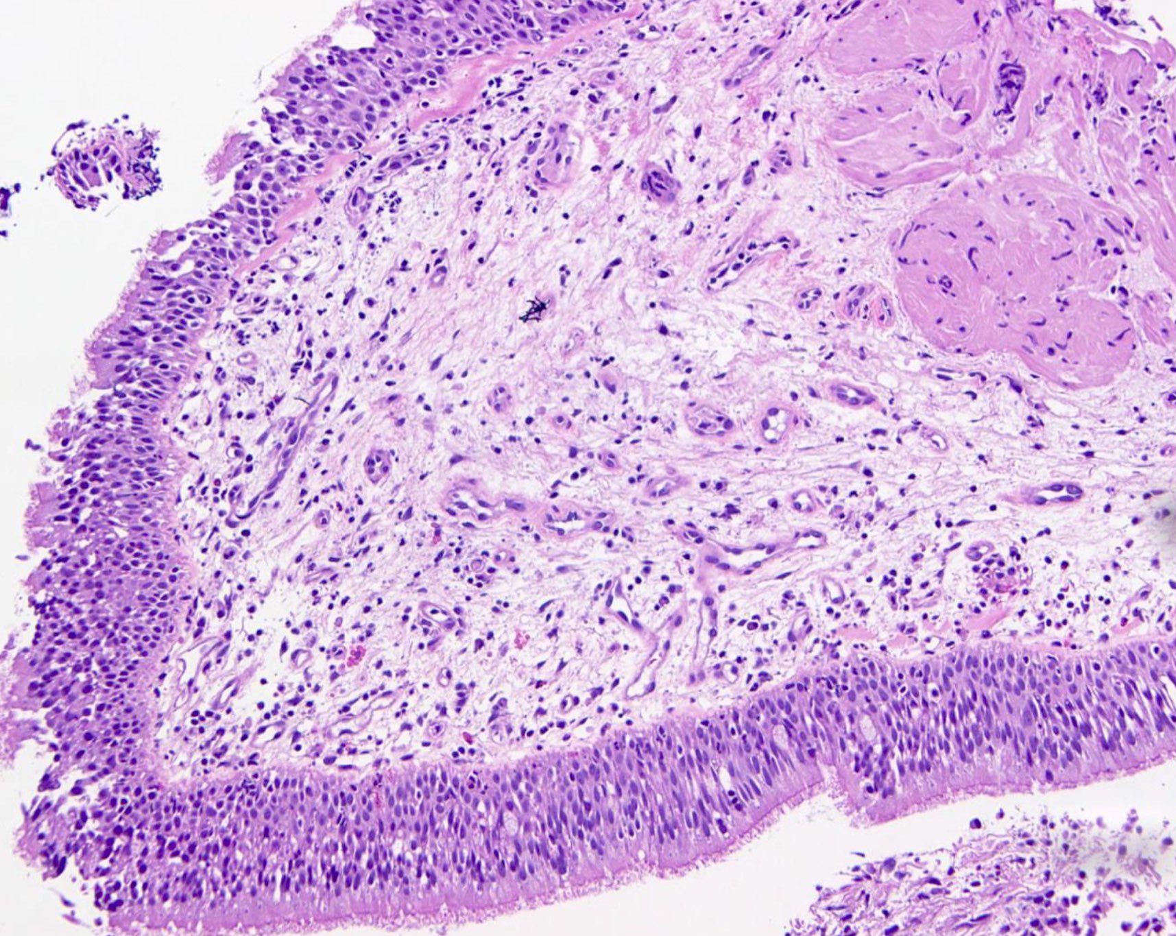
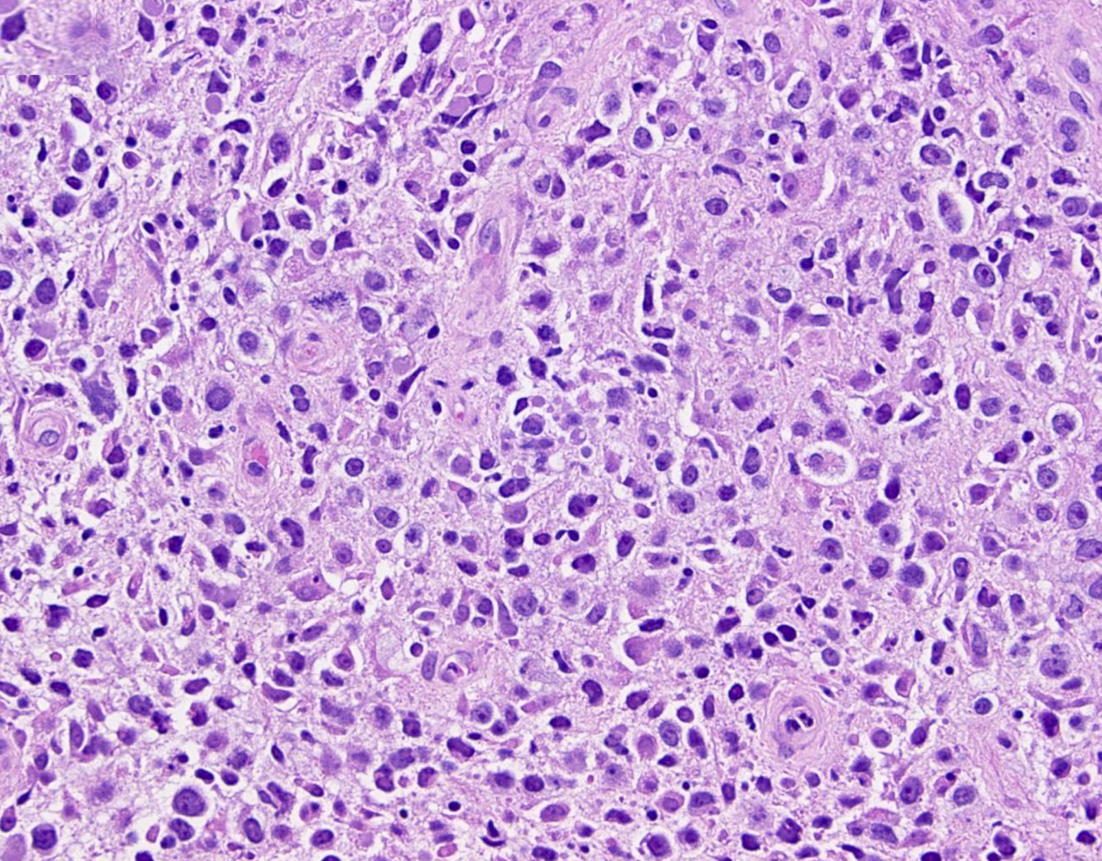
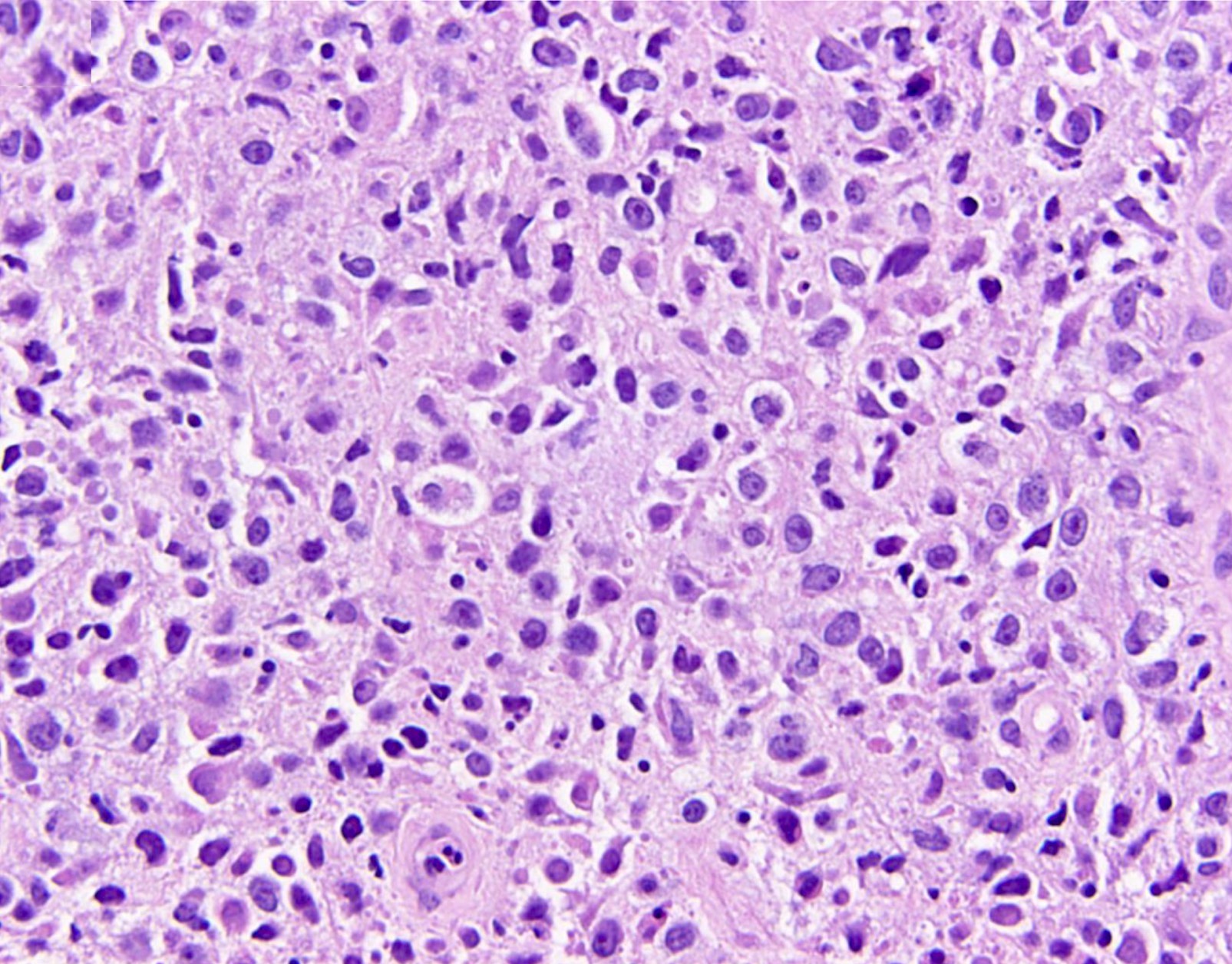
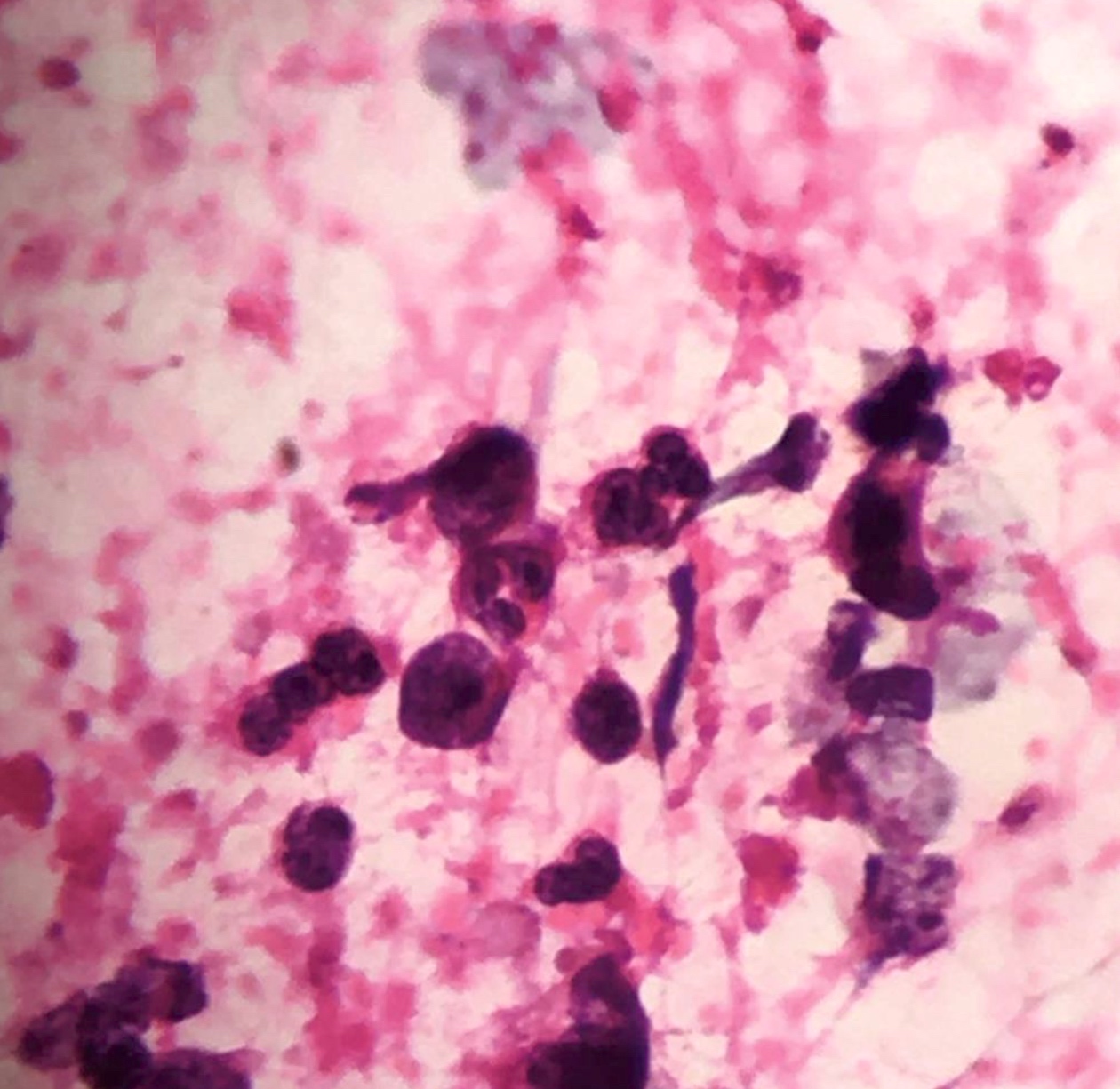
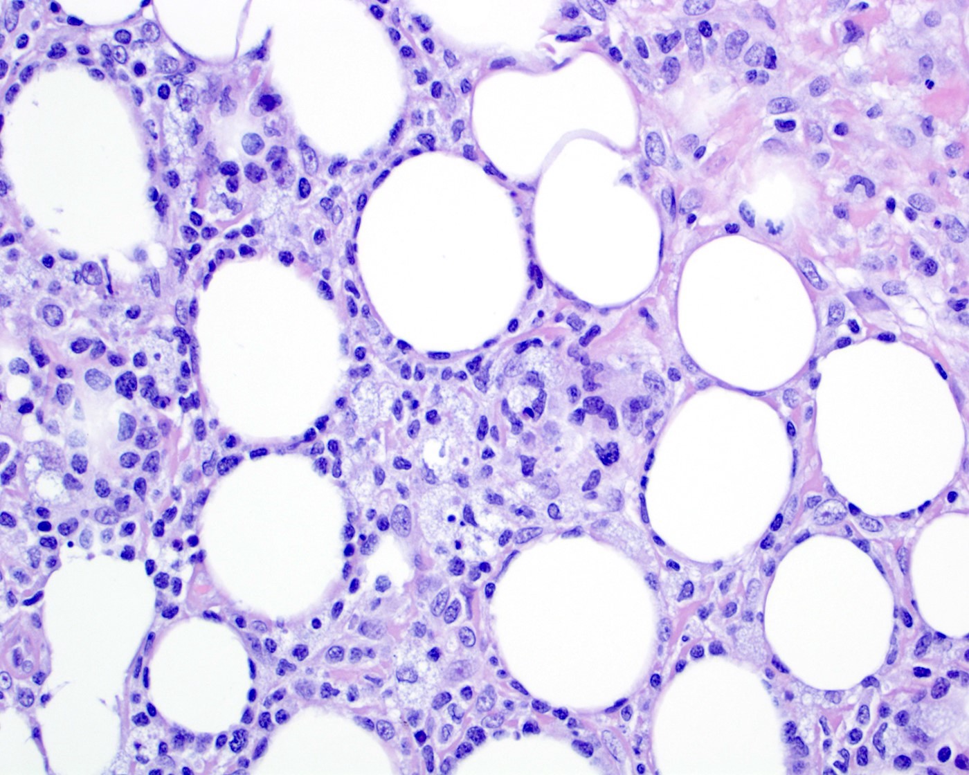
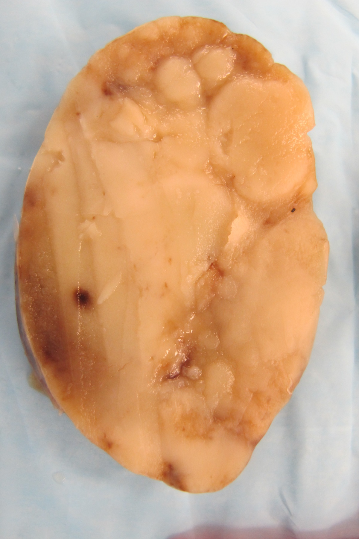
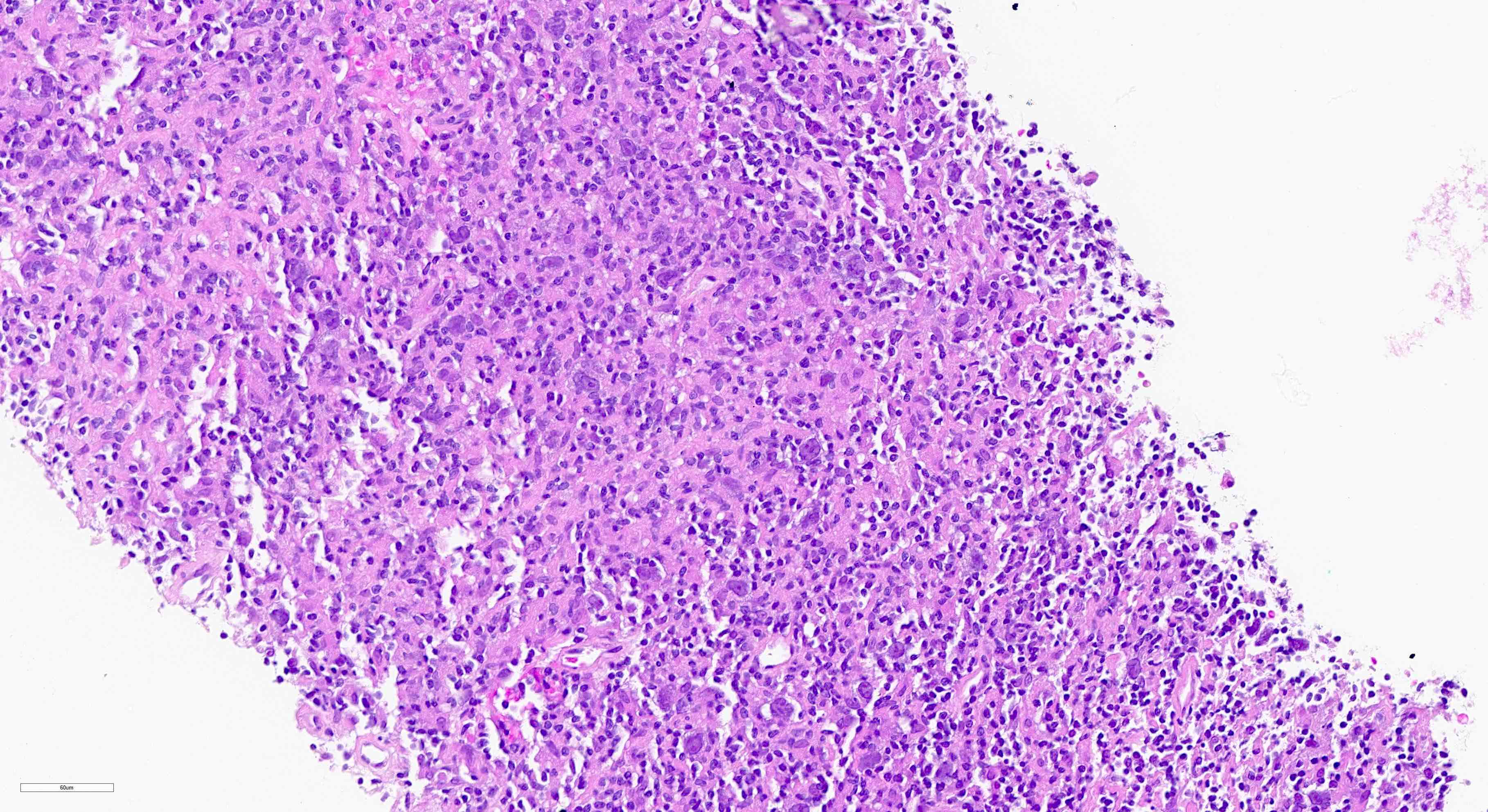
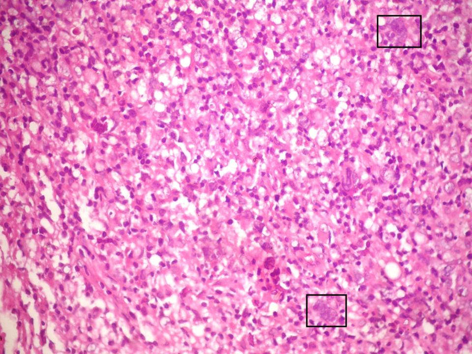
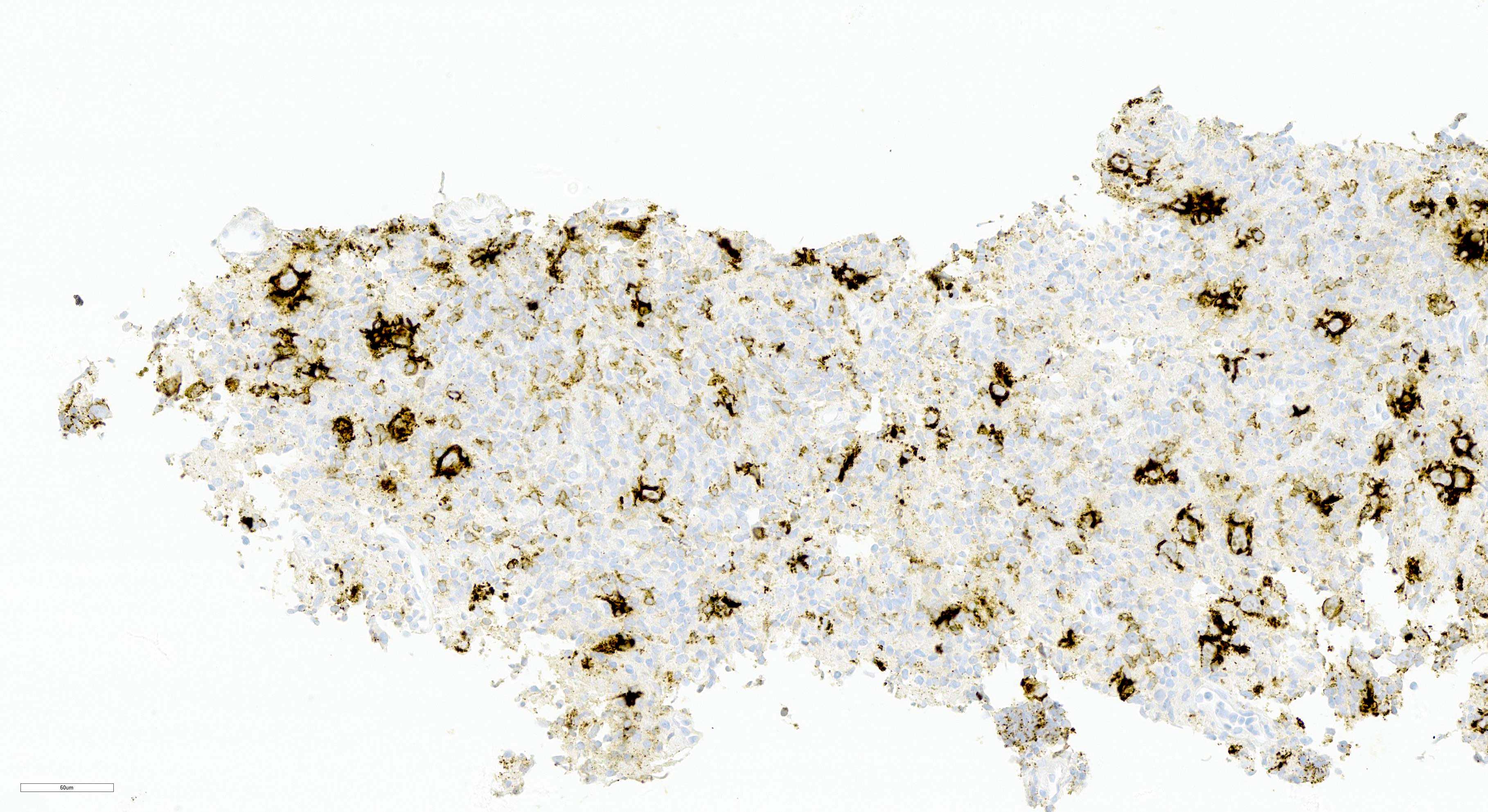
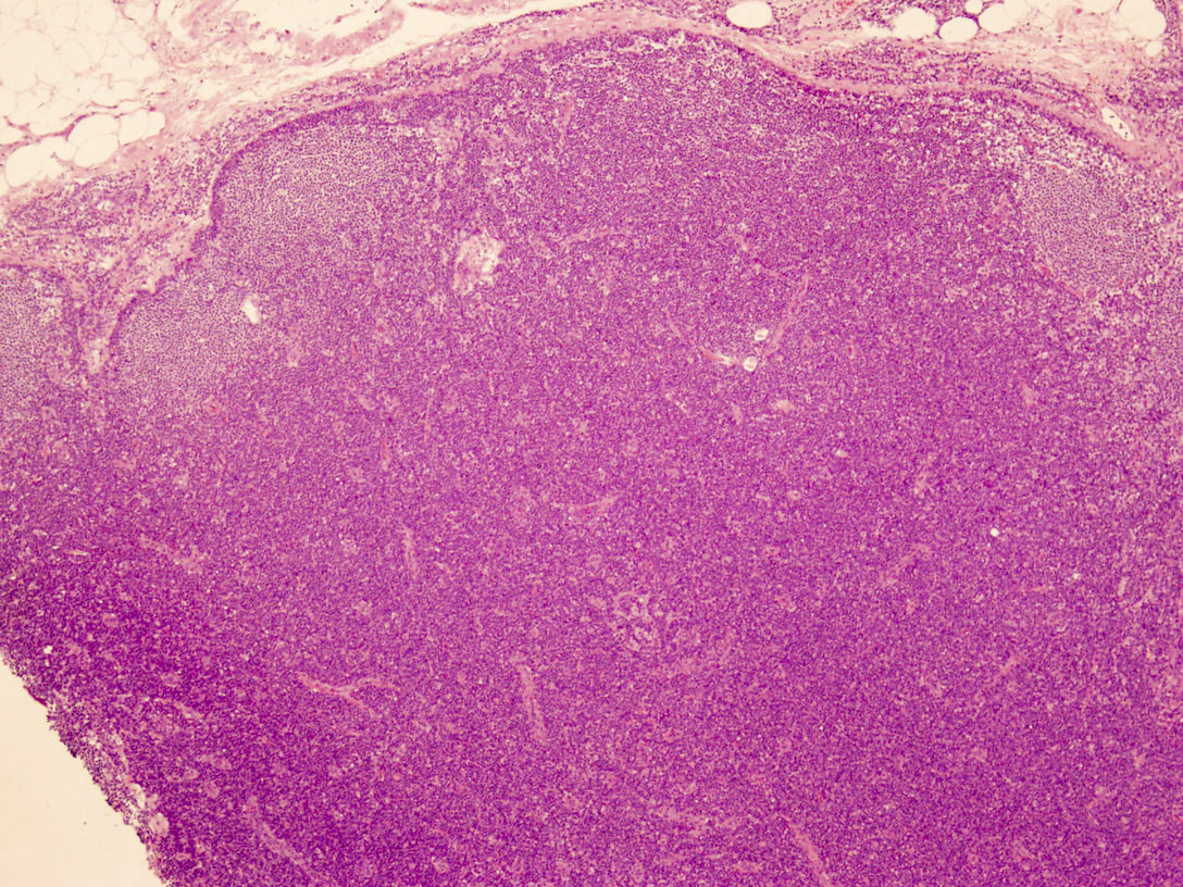
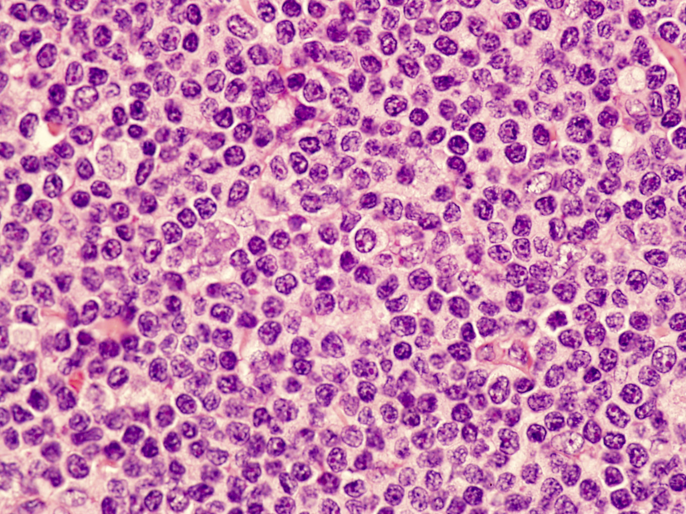
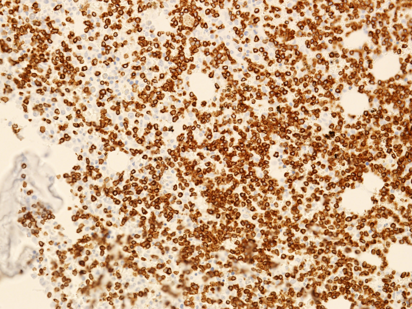
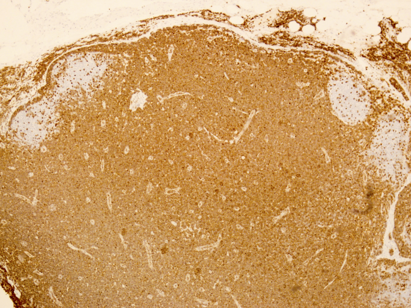
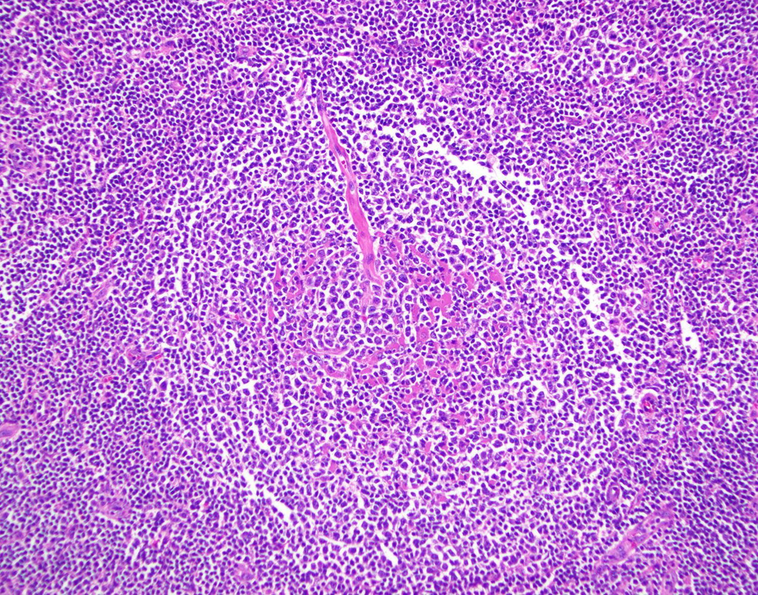
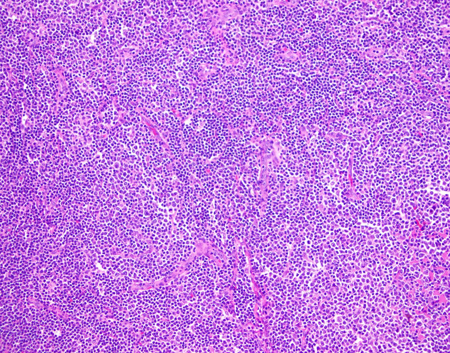
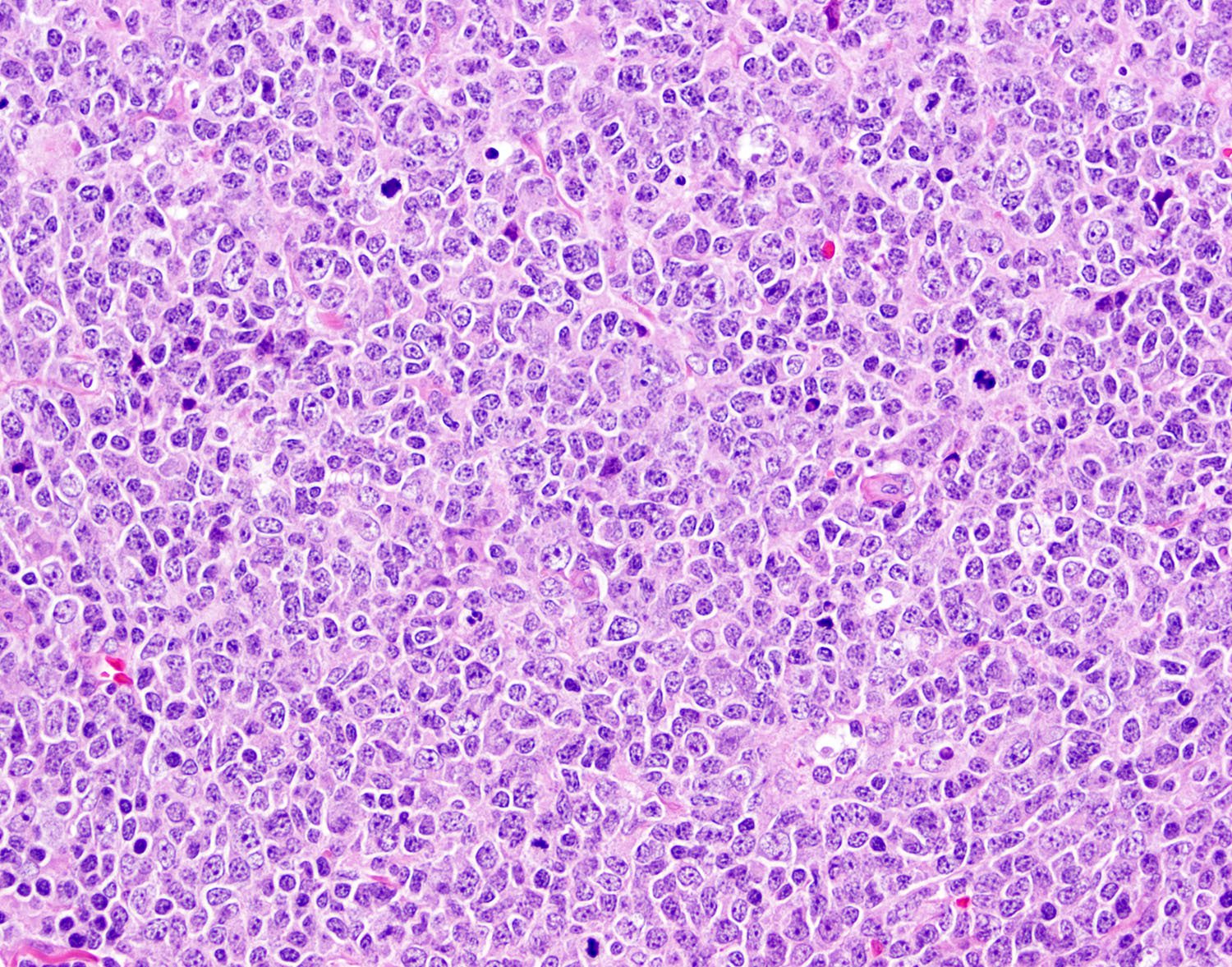
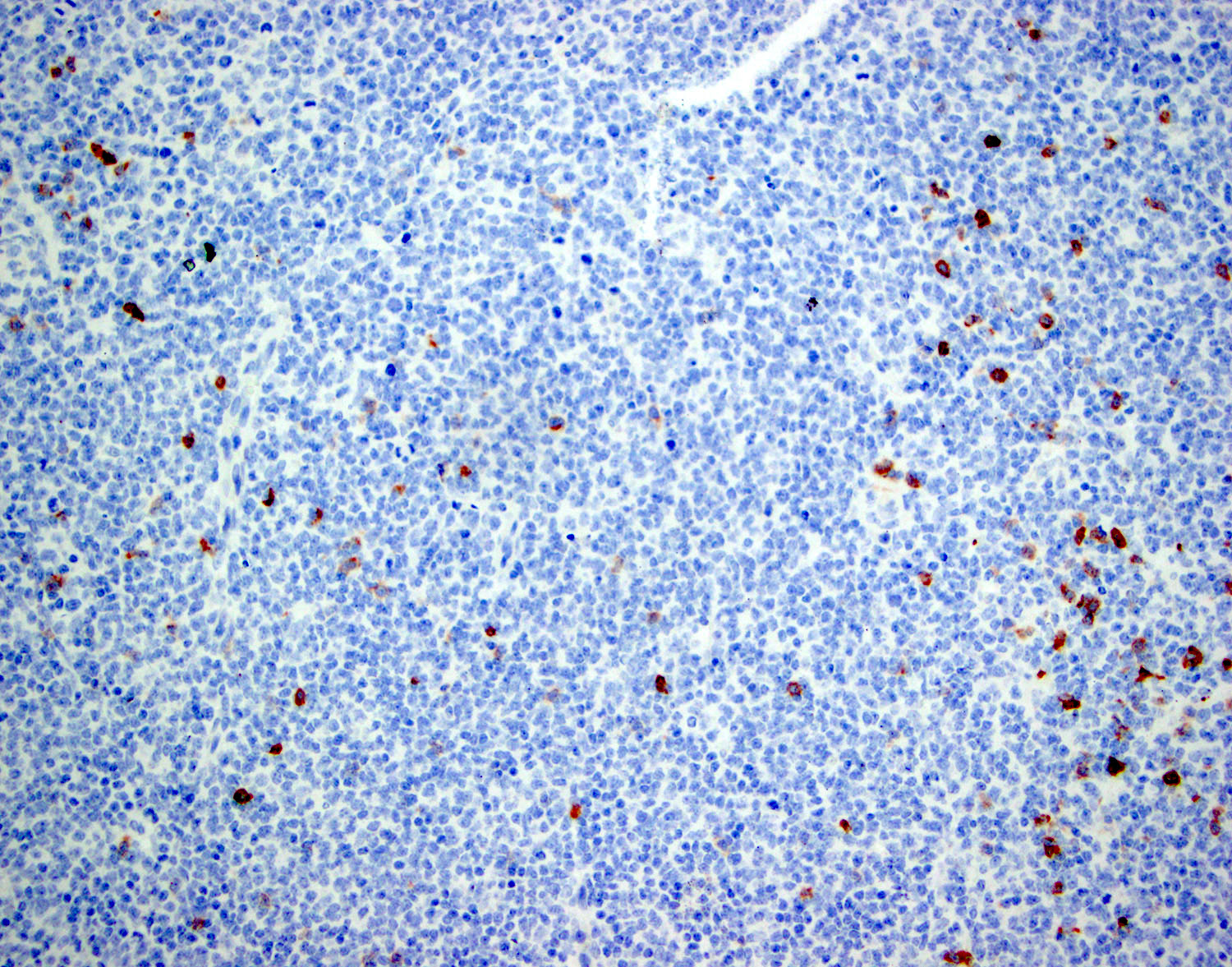
Contributed by Marie Therese Manipadam, M.B.B.S., M.D.
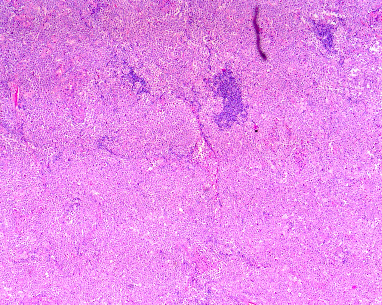
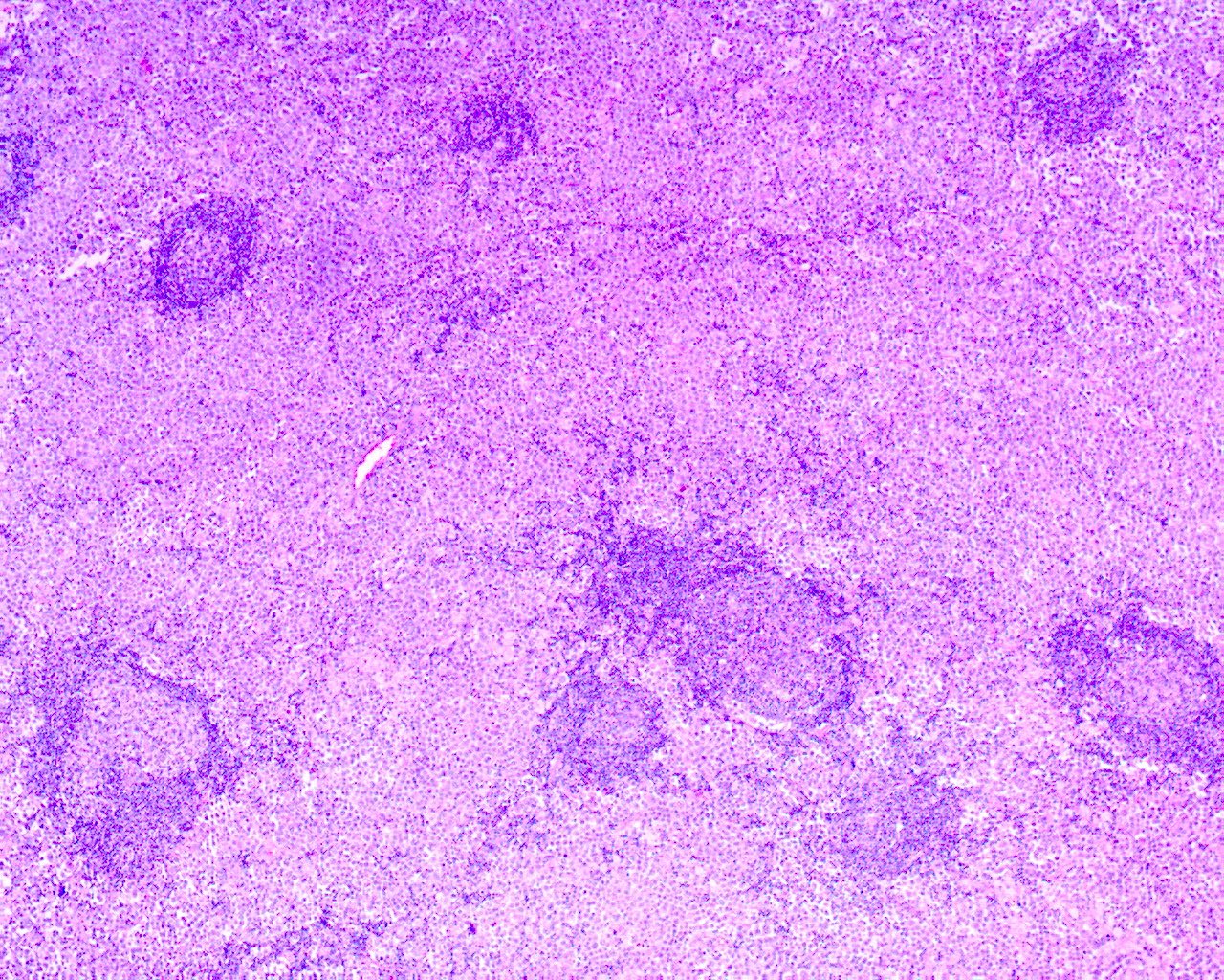
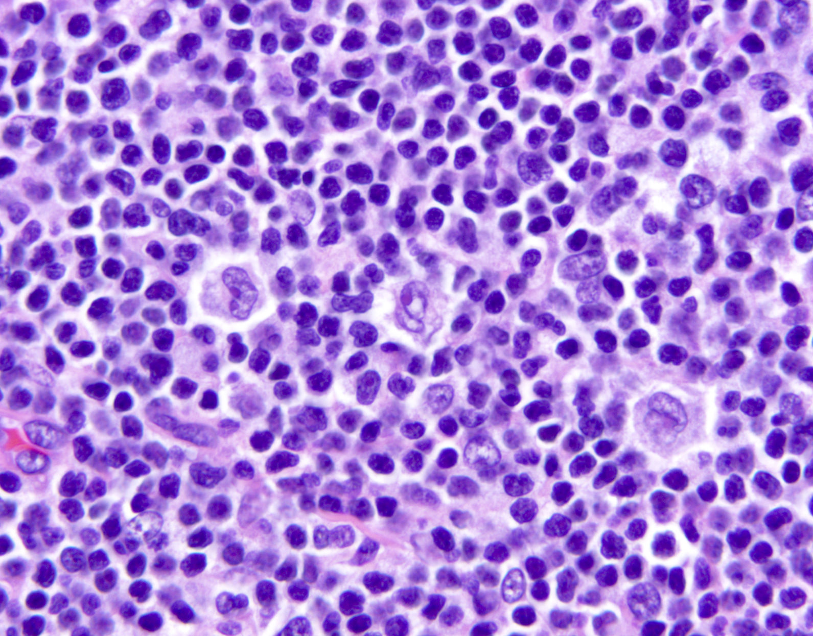
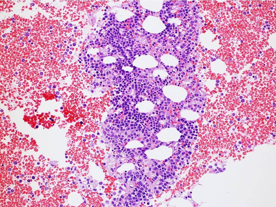
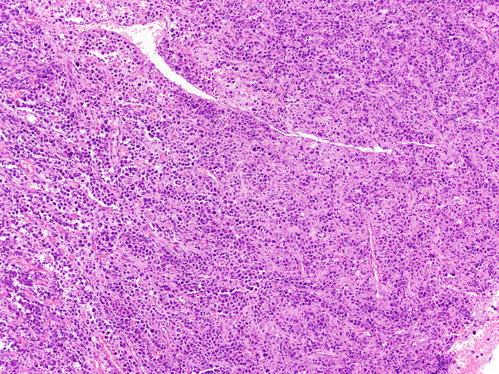
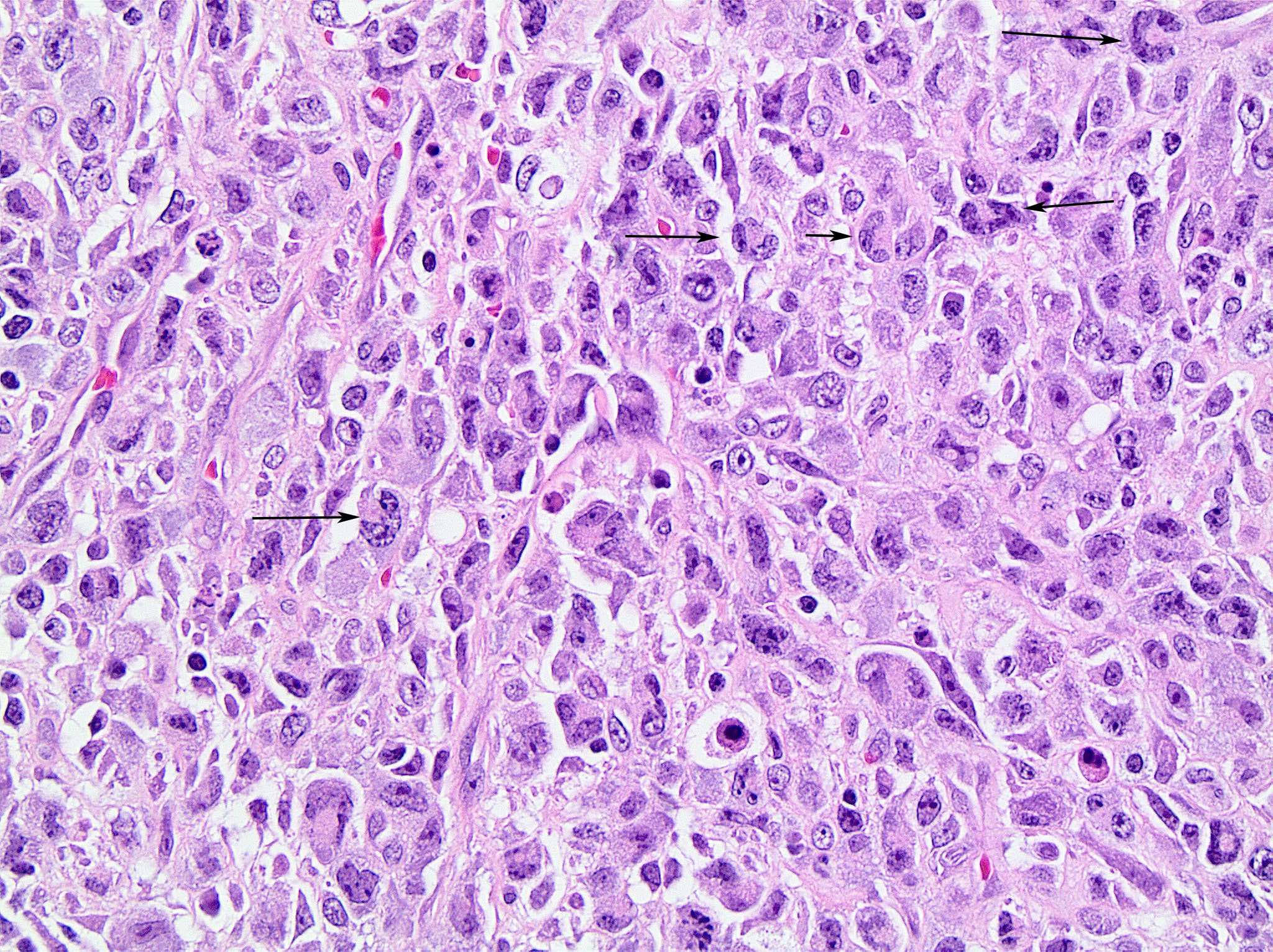
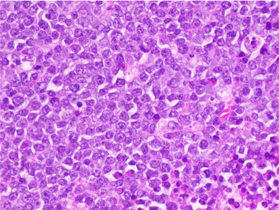
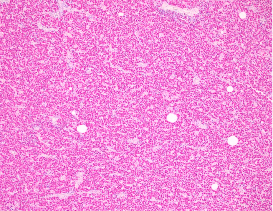
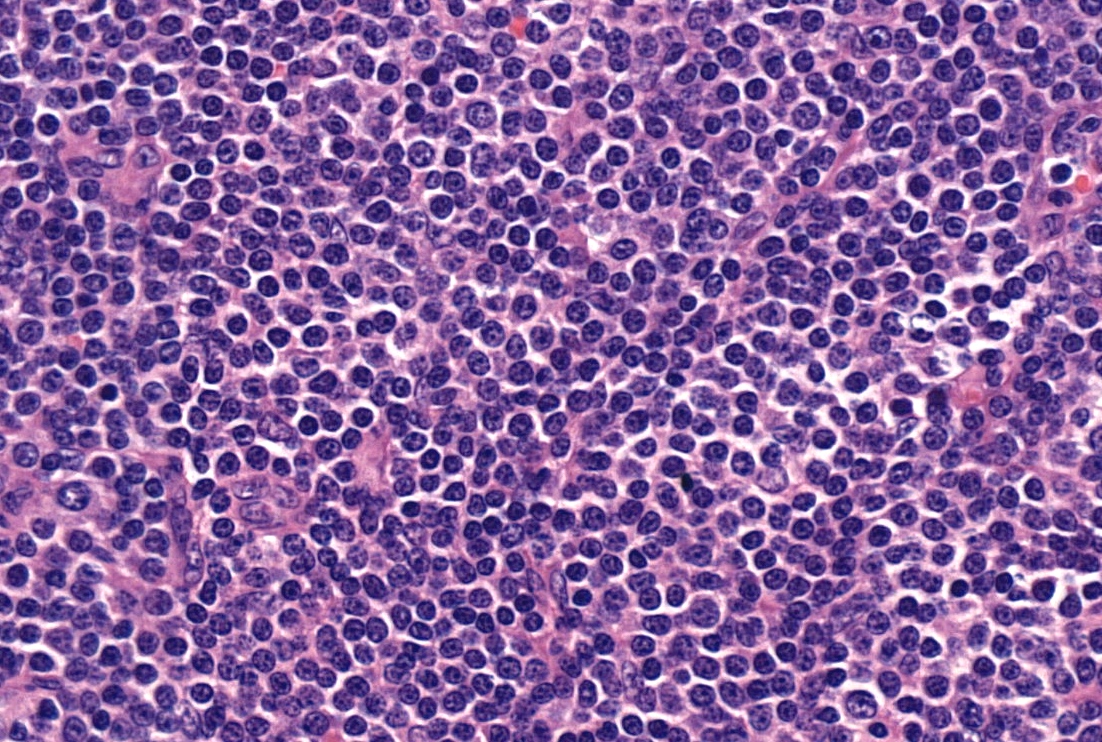
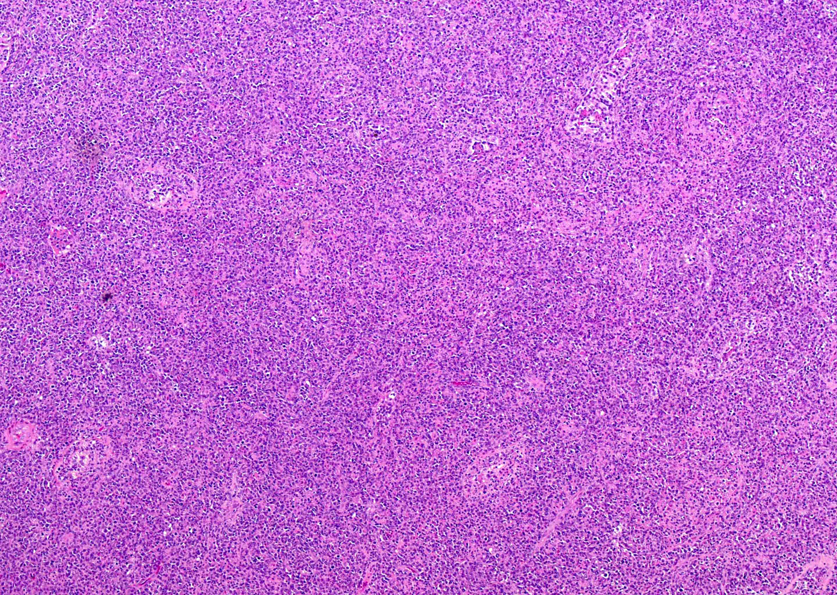
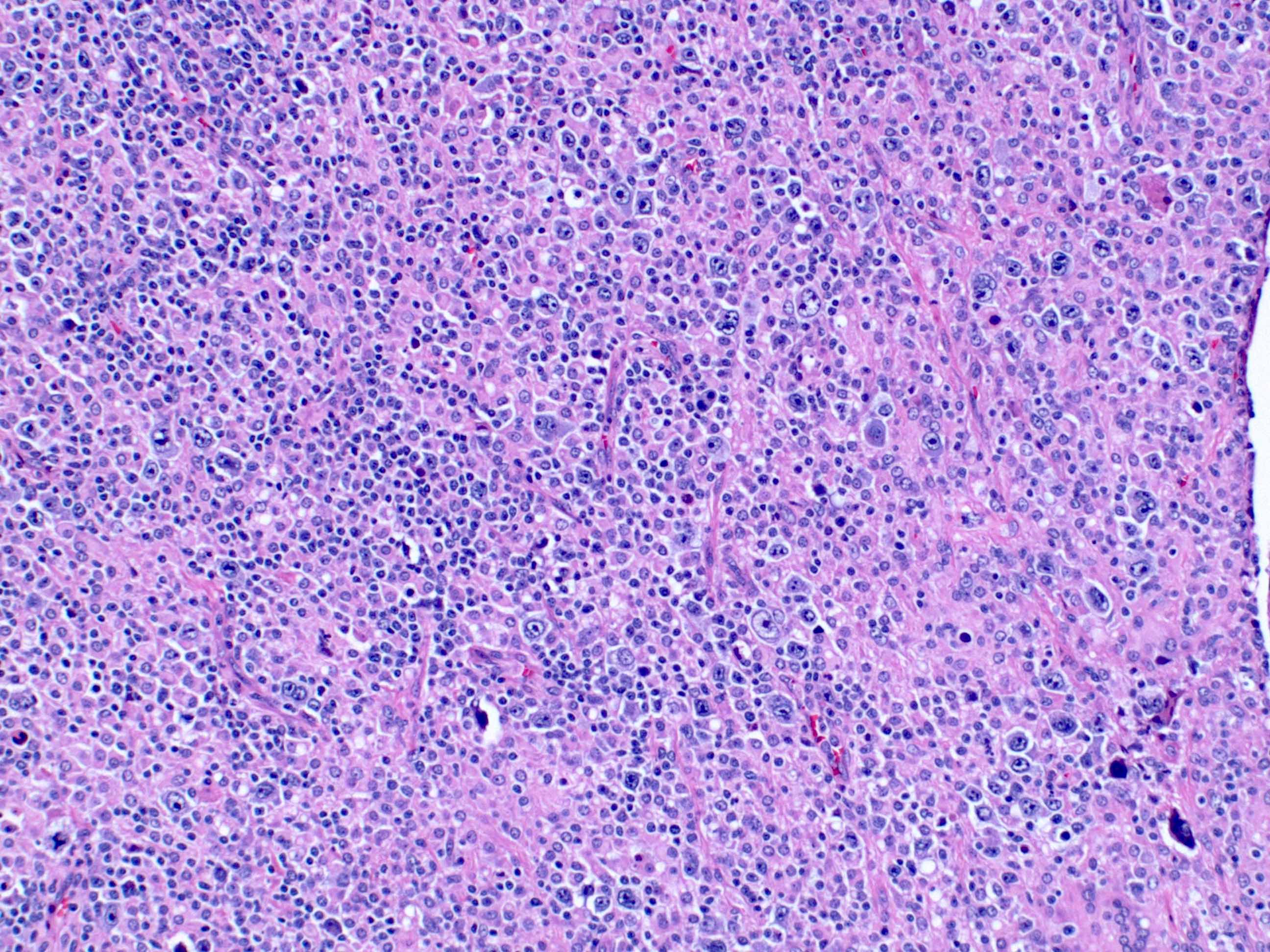
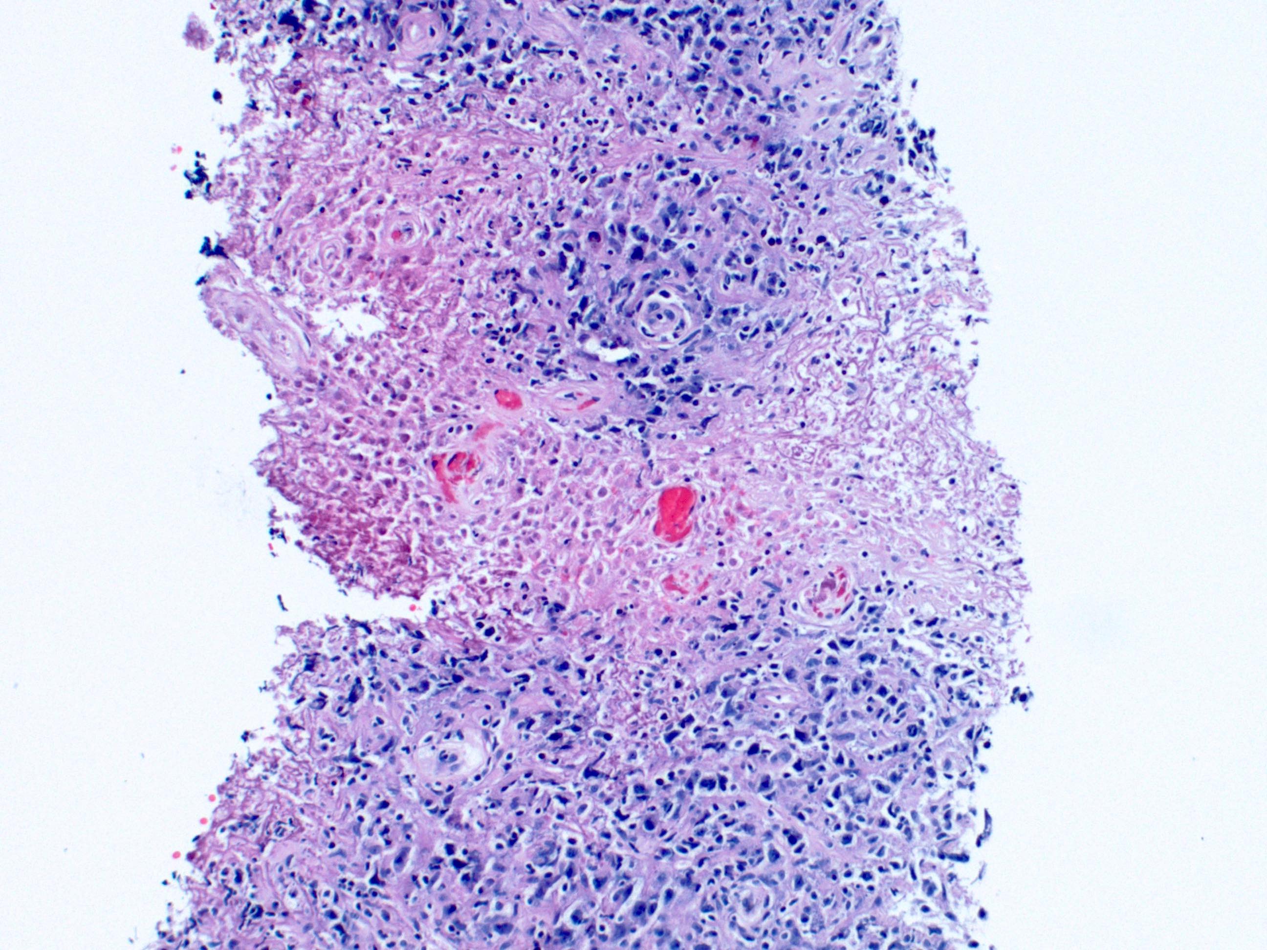
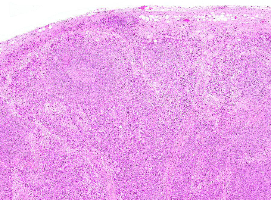
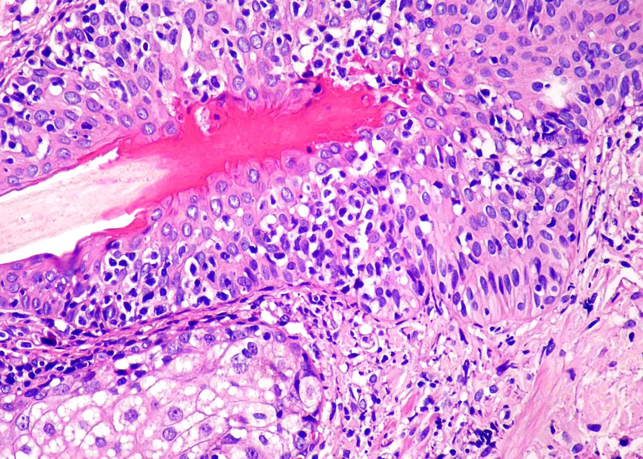
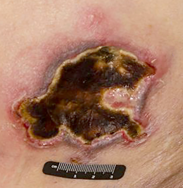
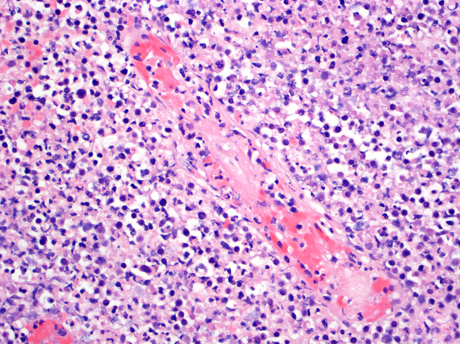
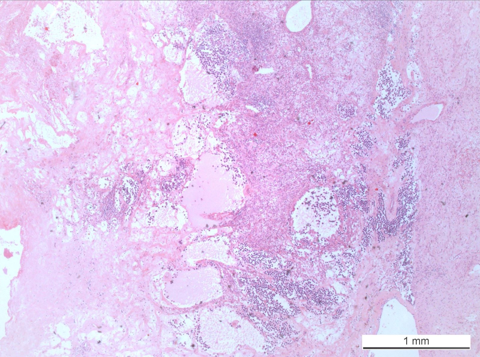
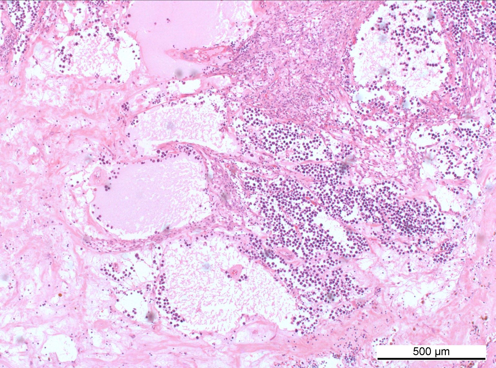
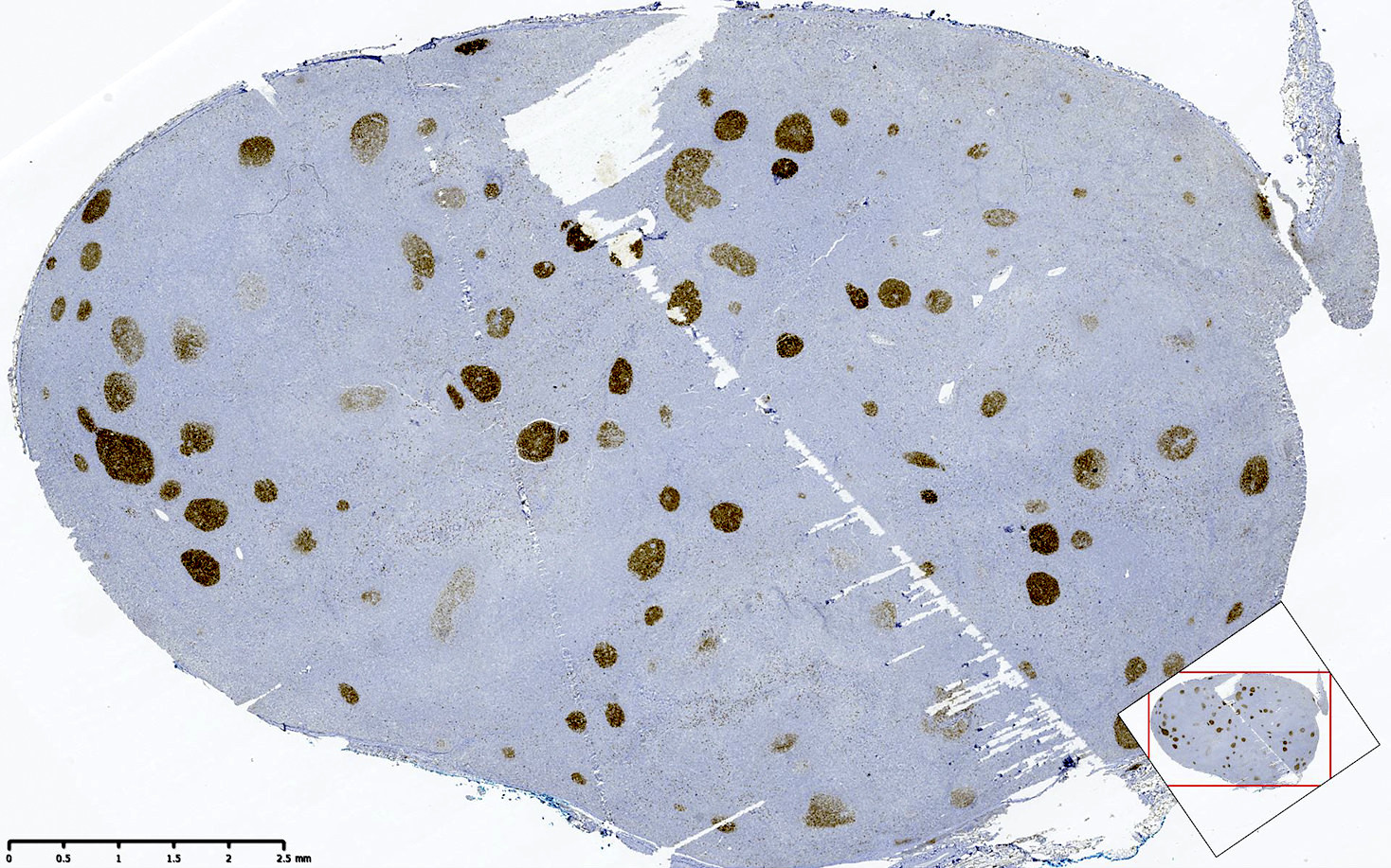
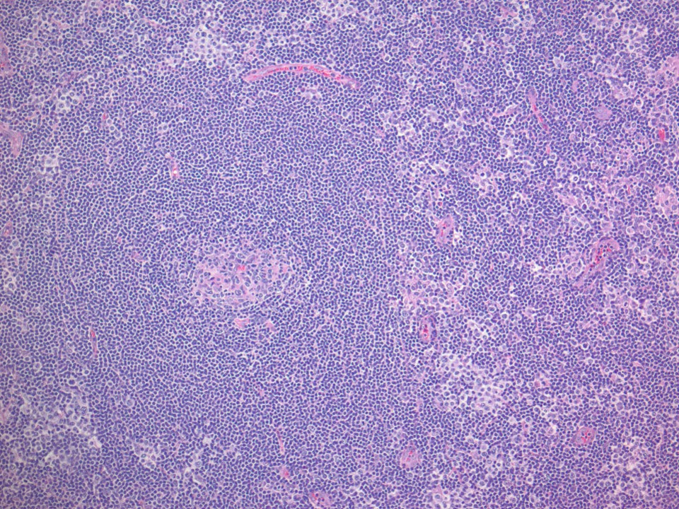
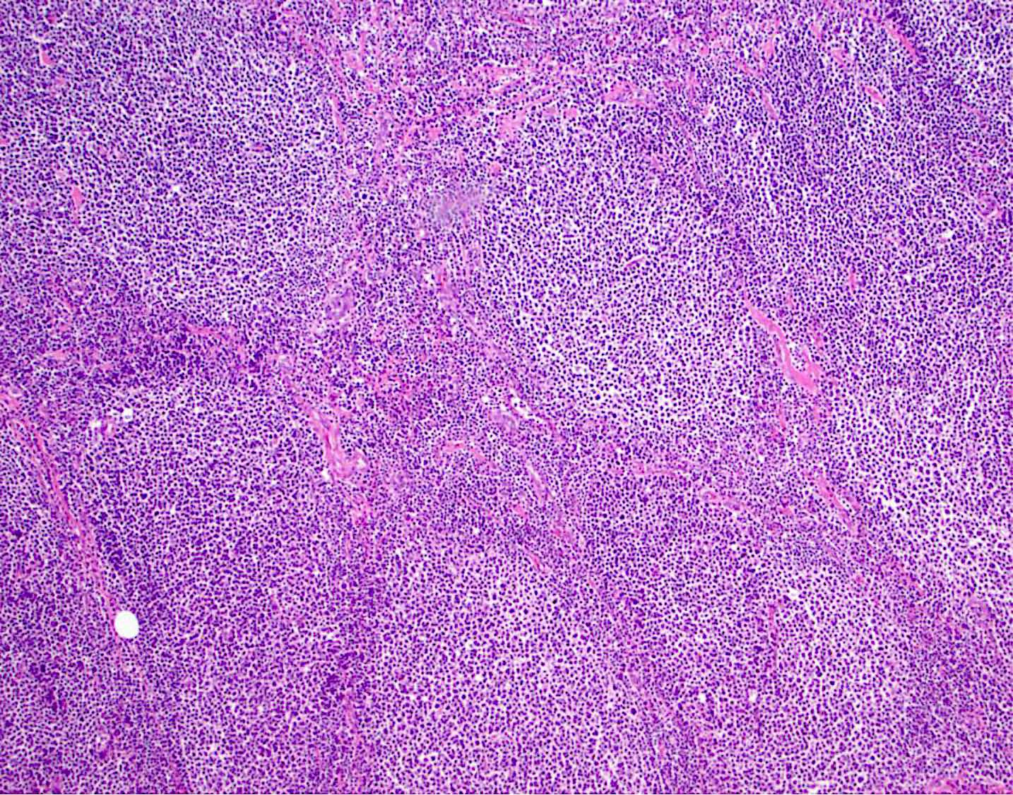
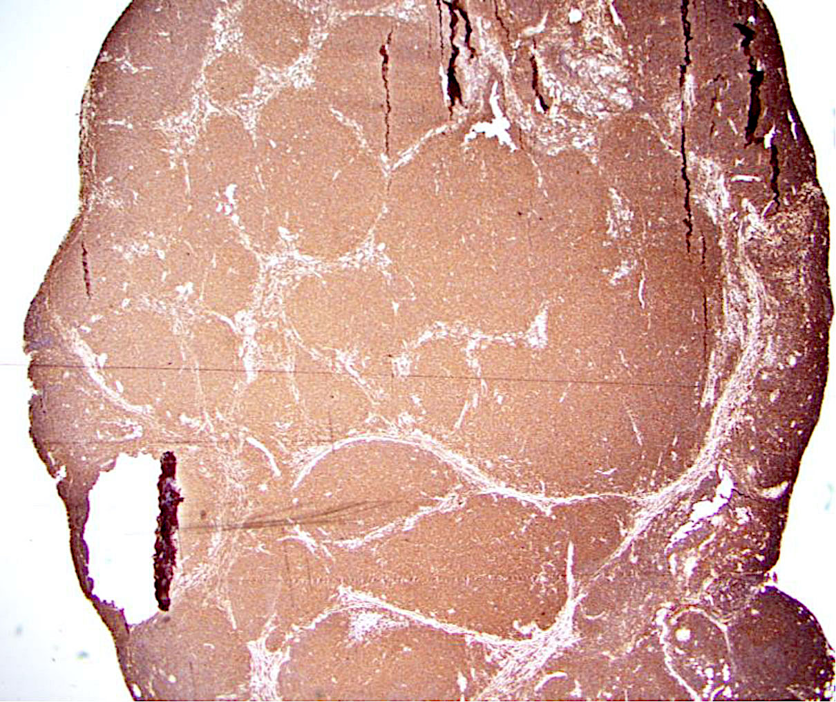
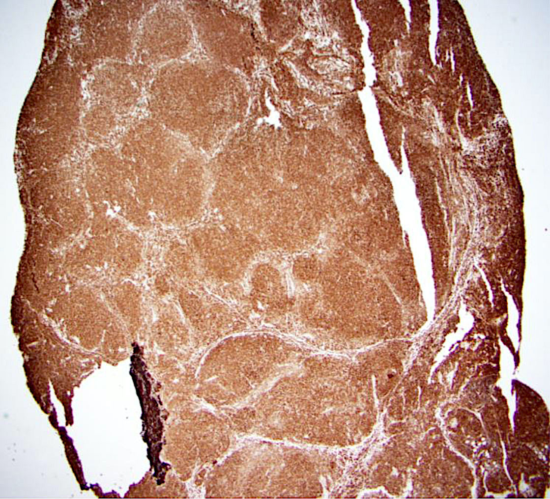
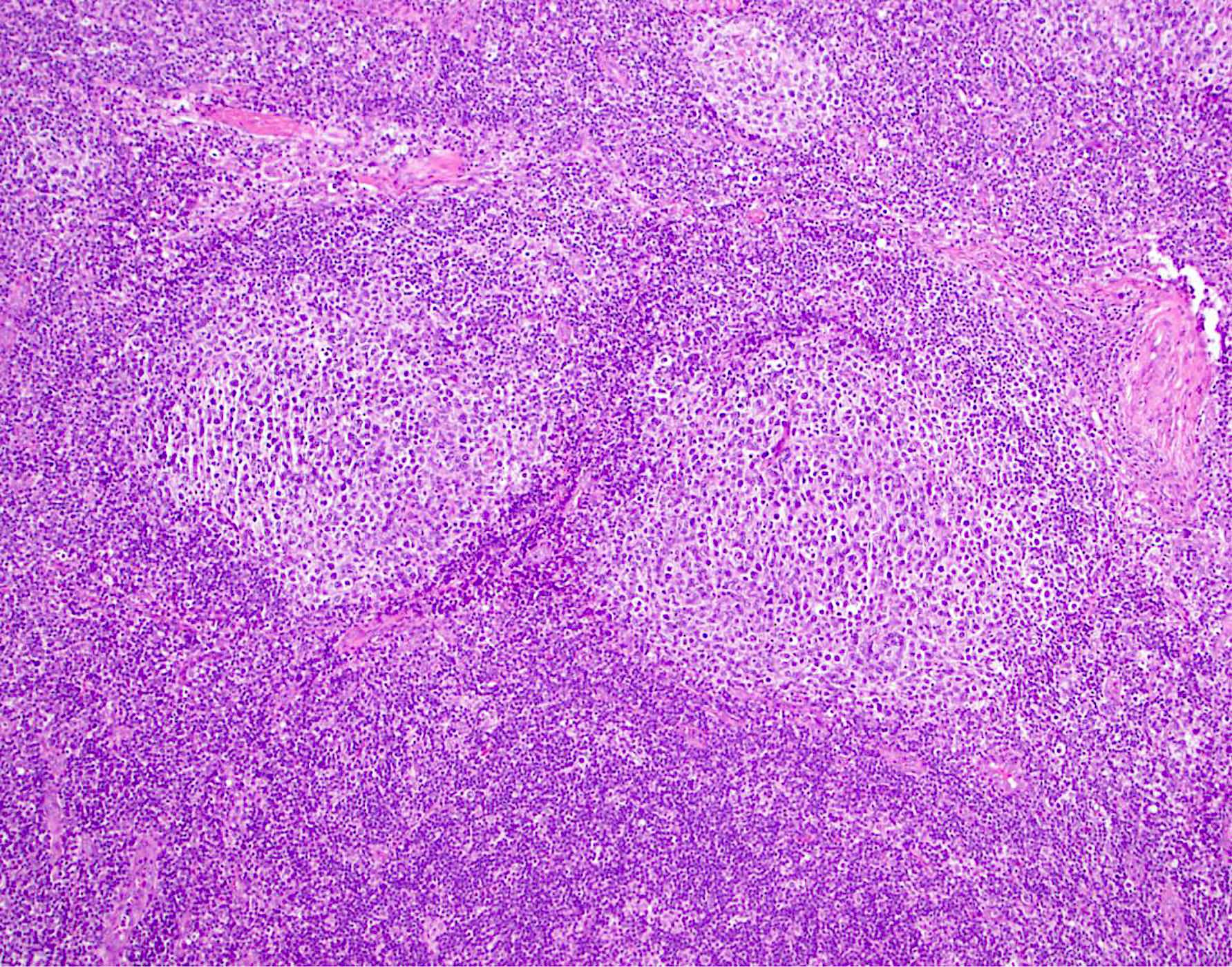
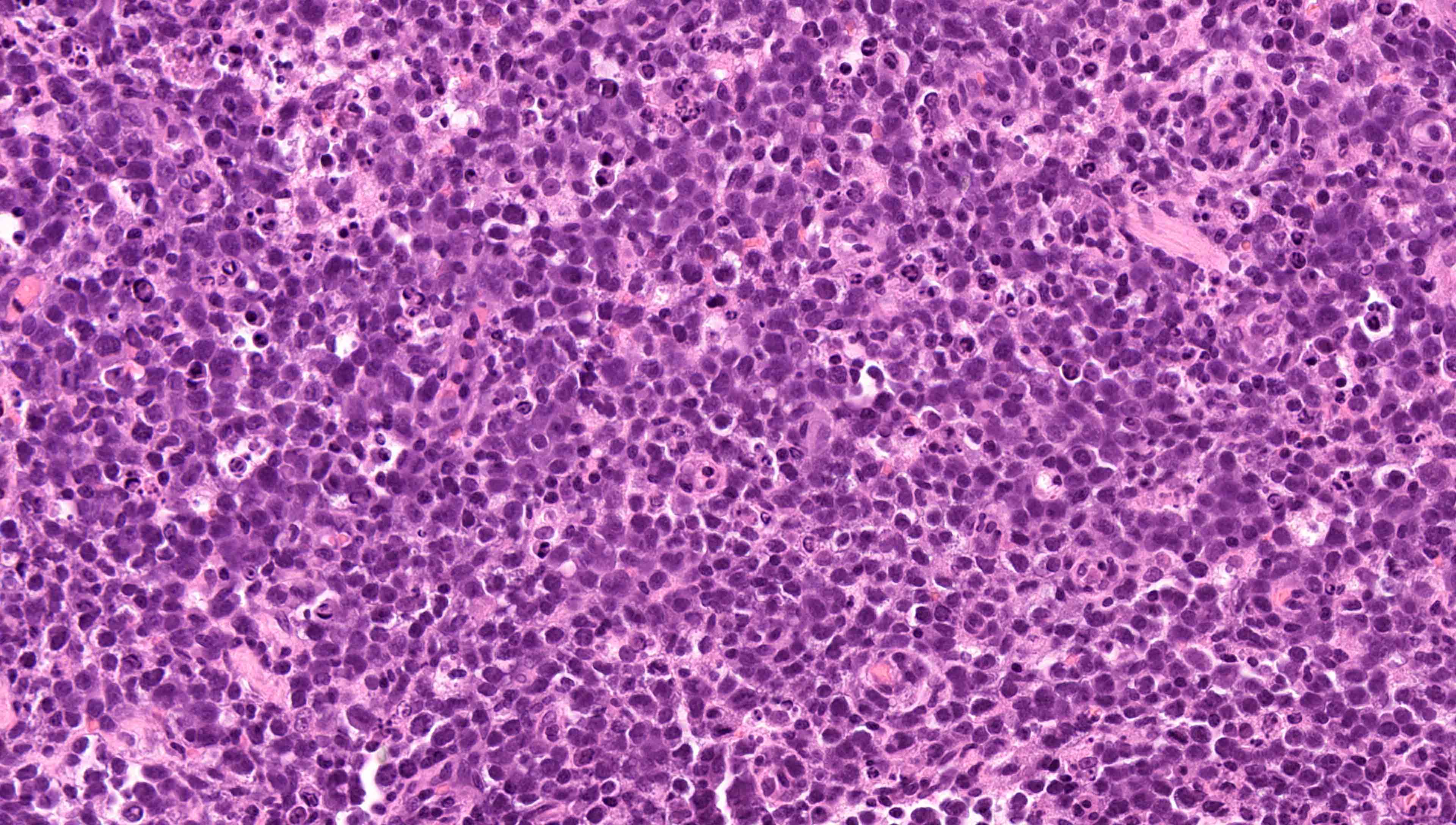
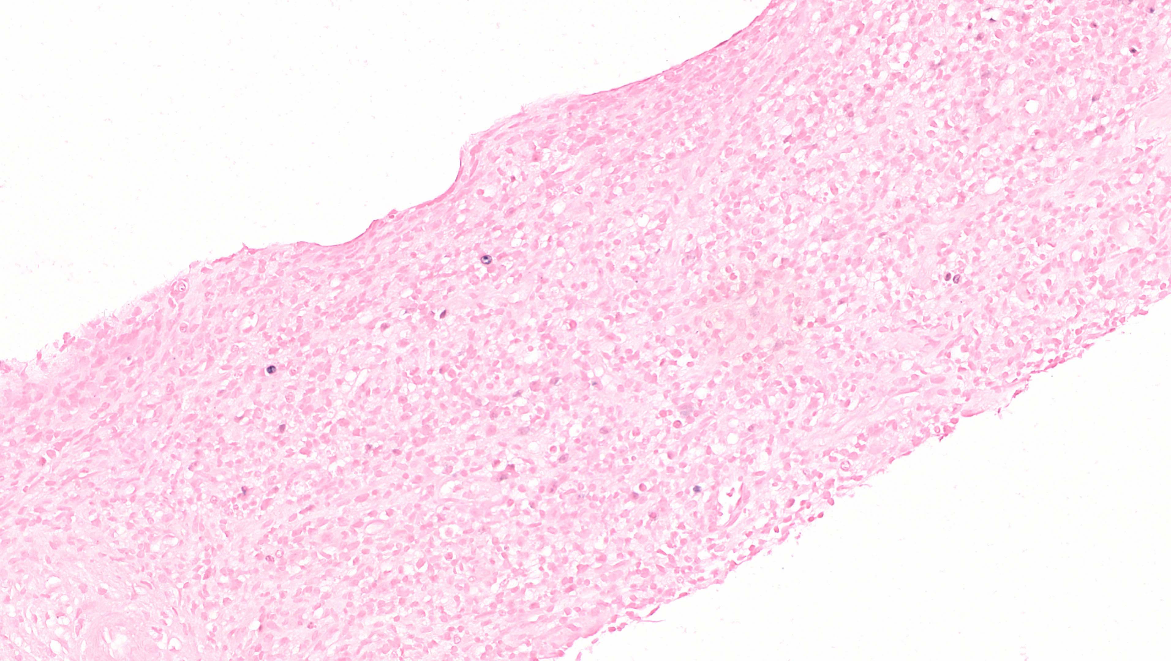
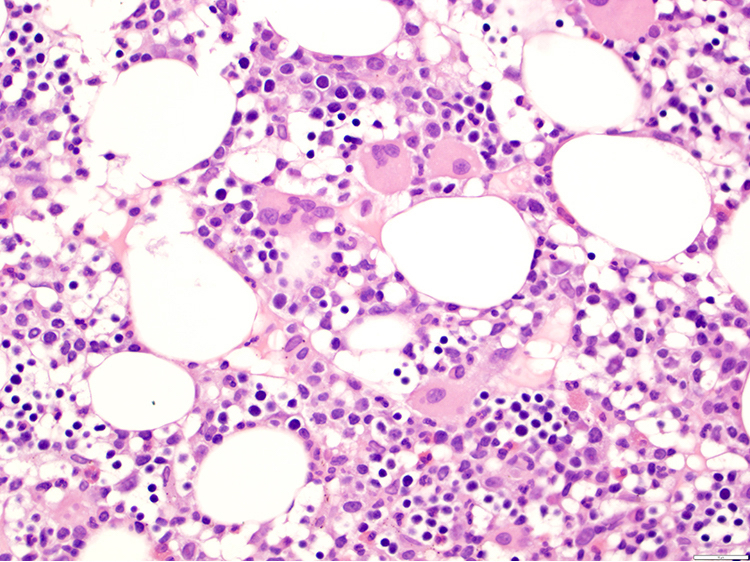
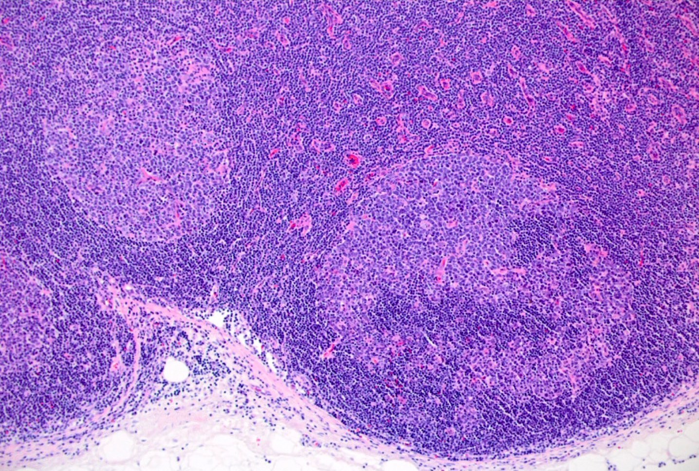
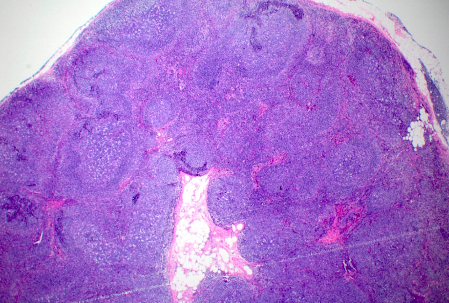
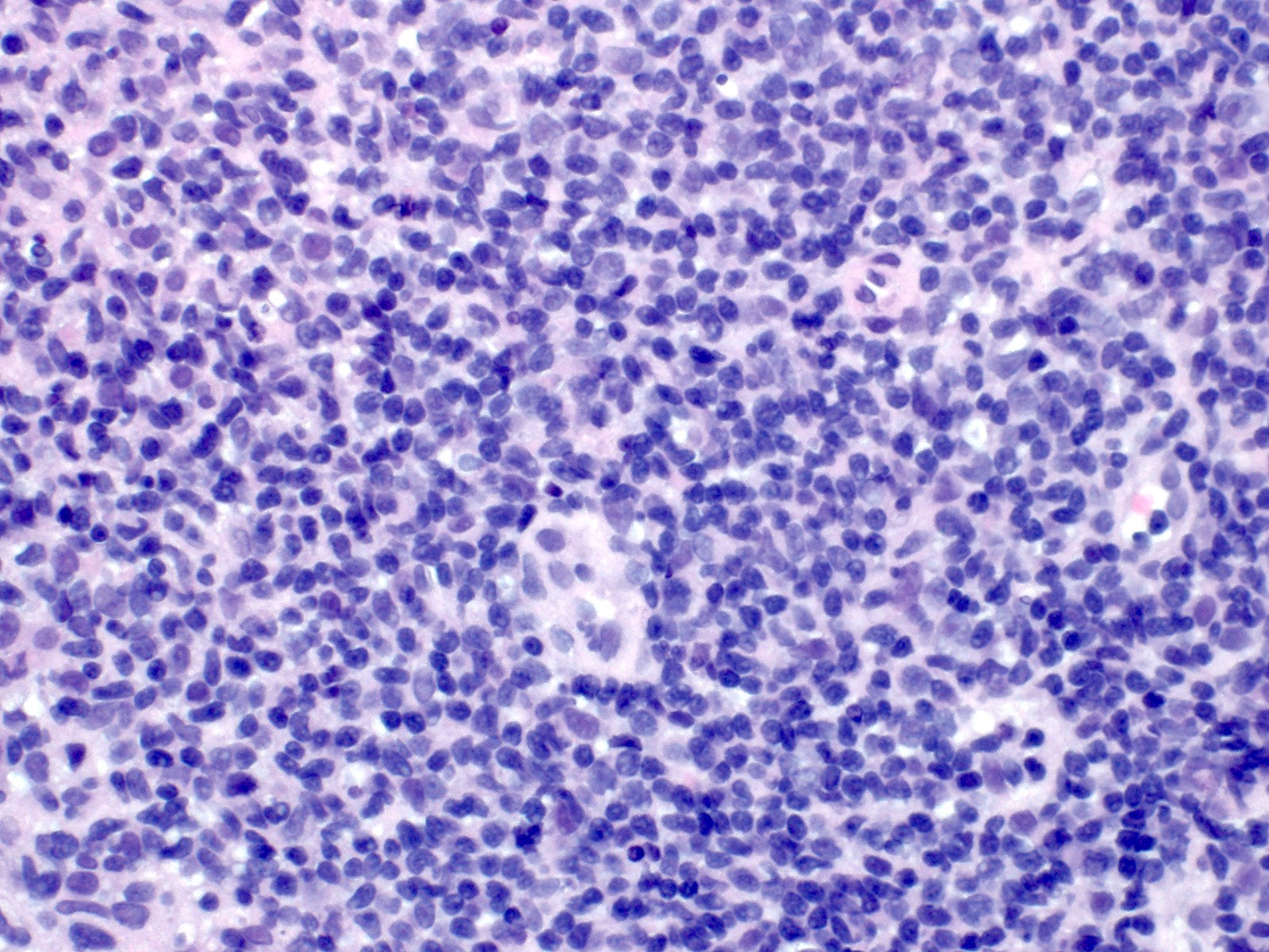
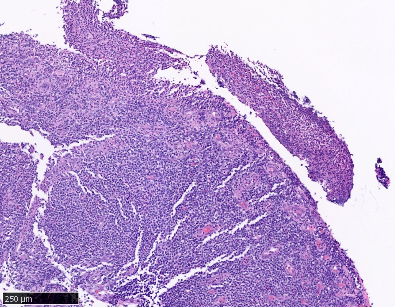
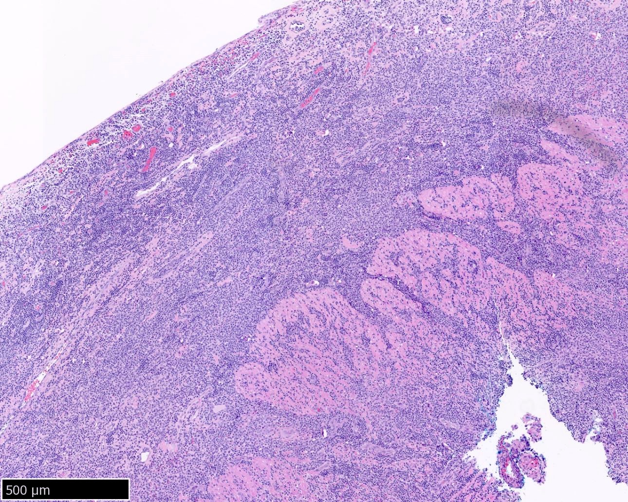
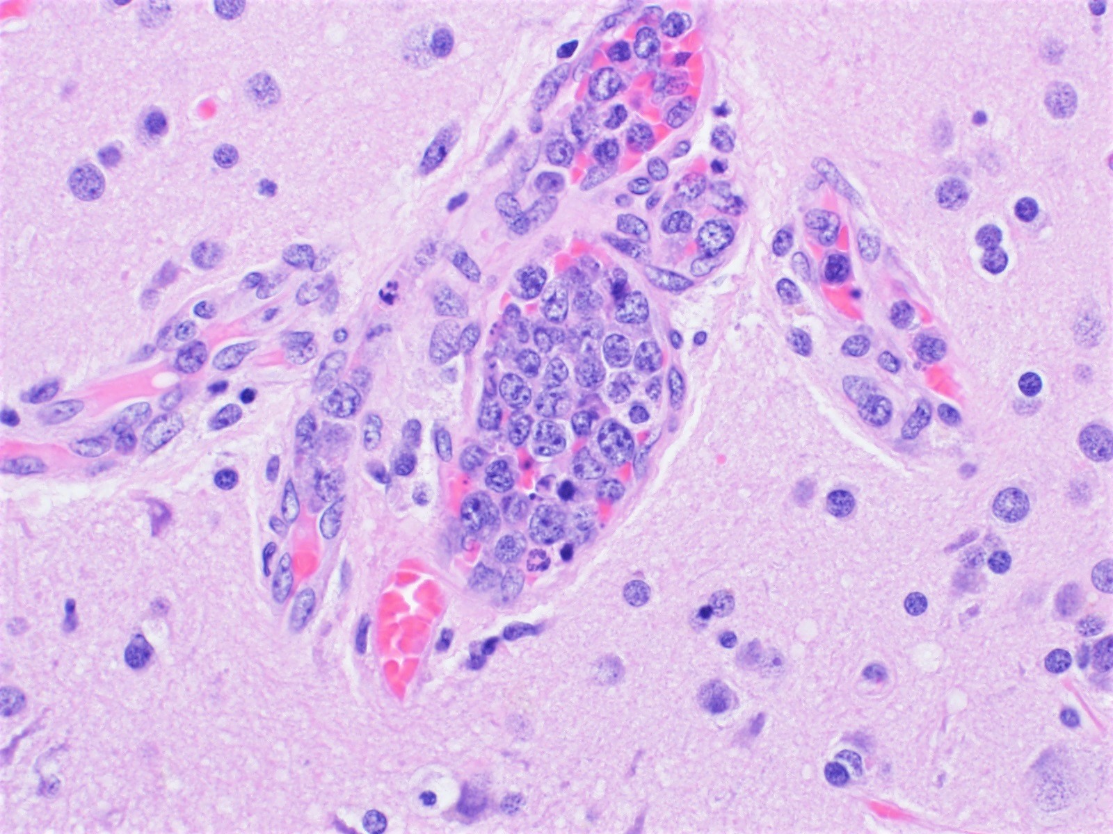
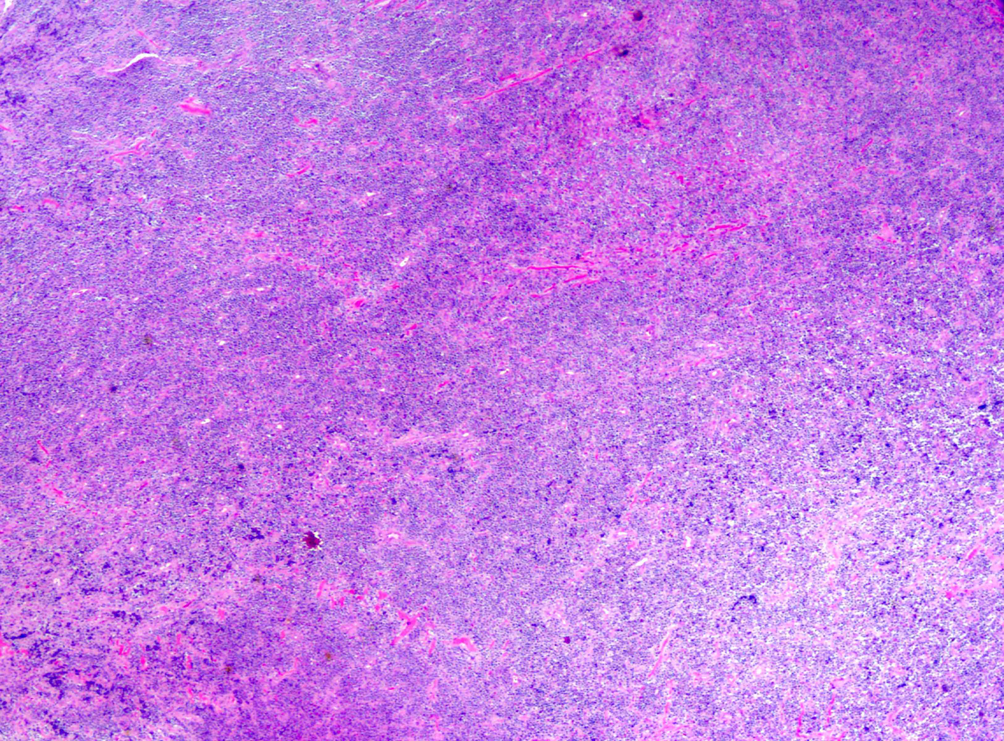
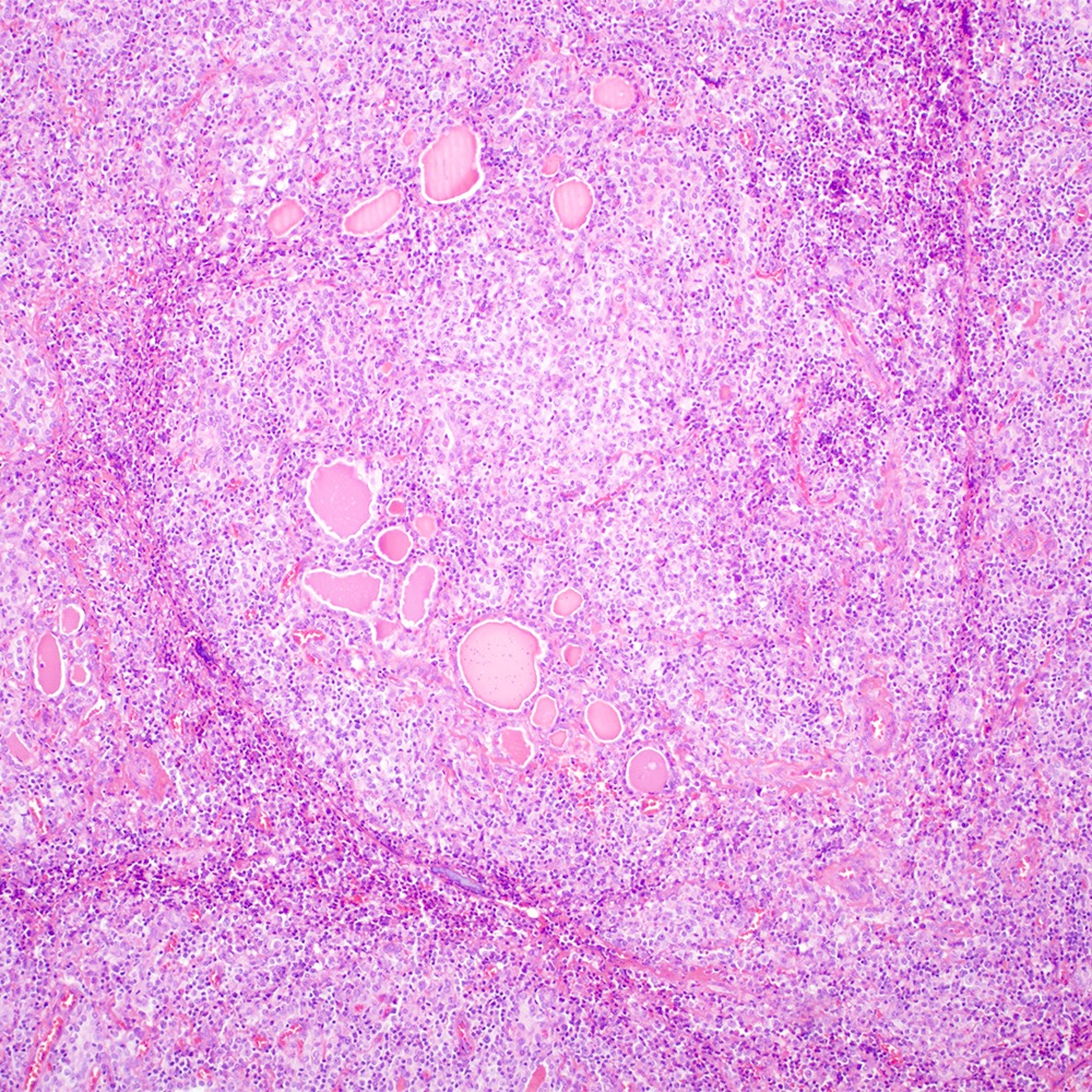
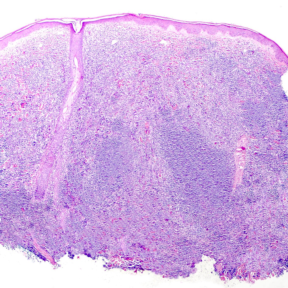
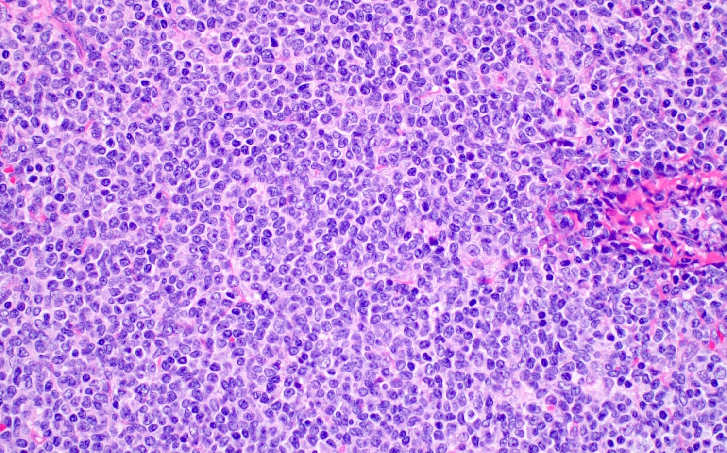
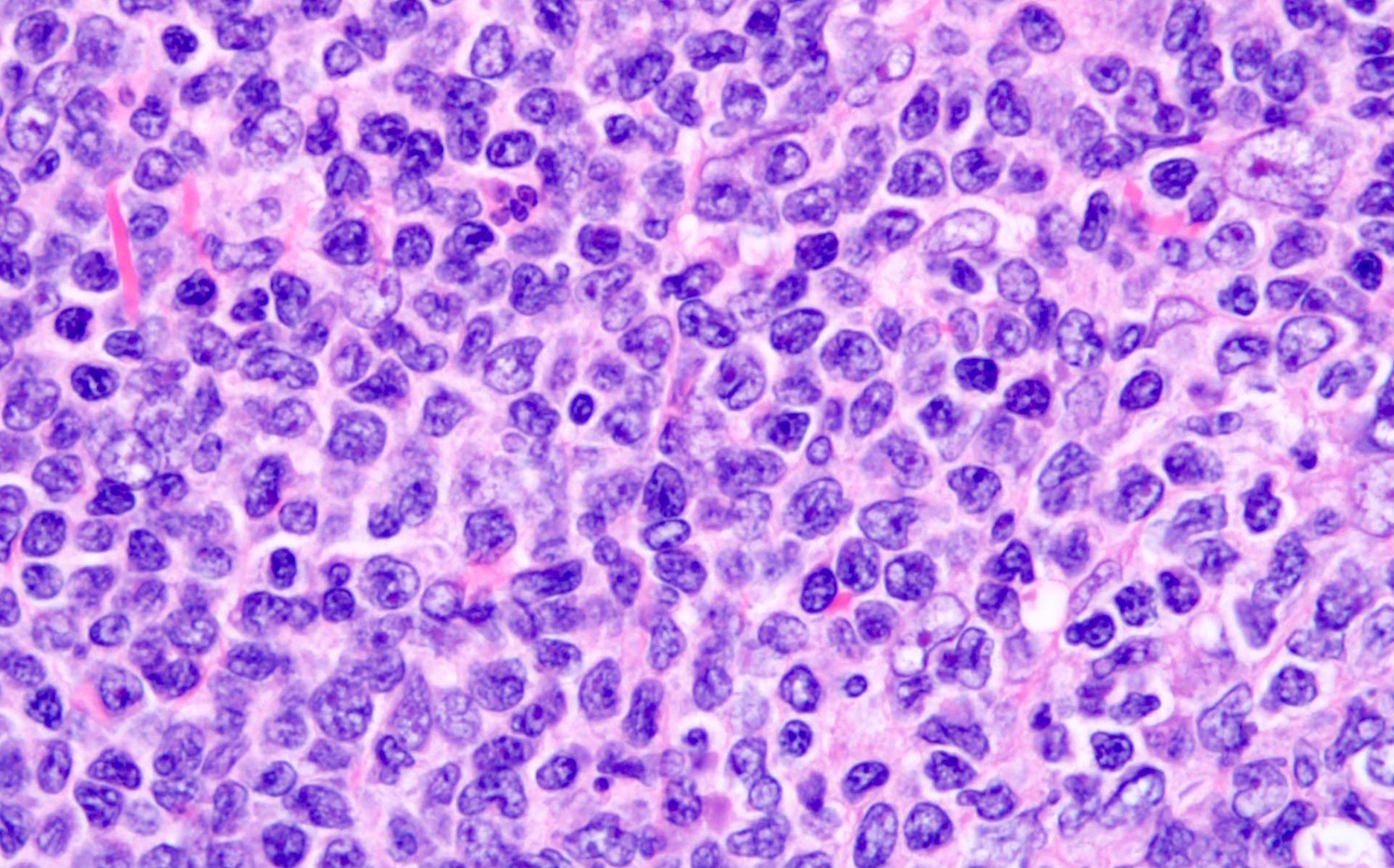
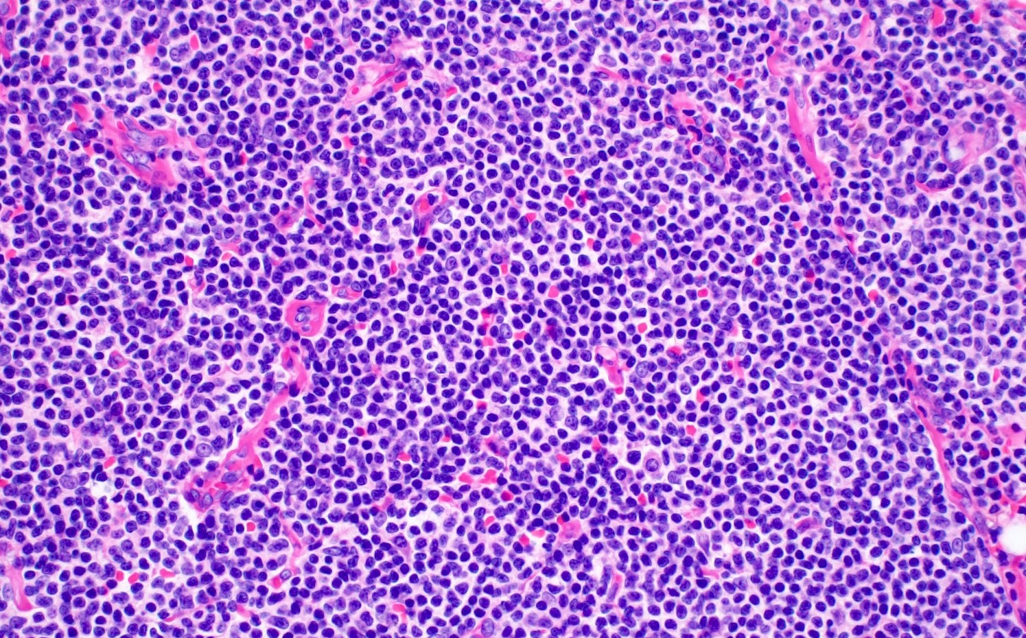
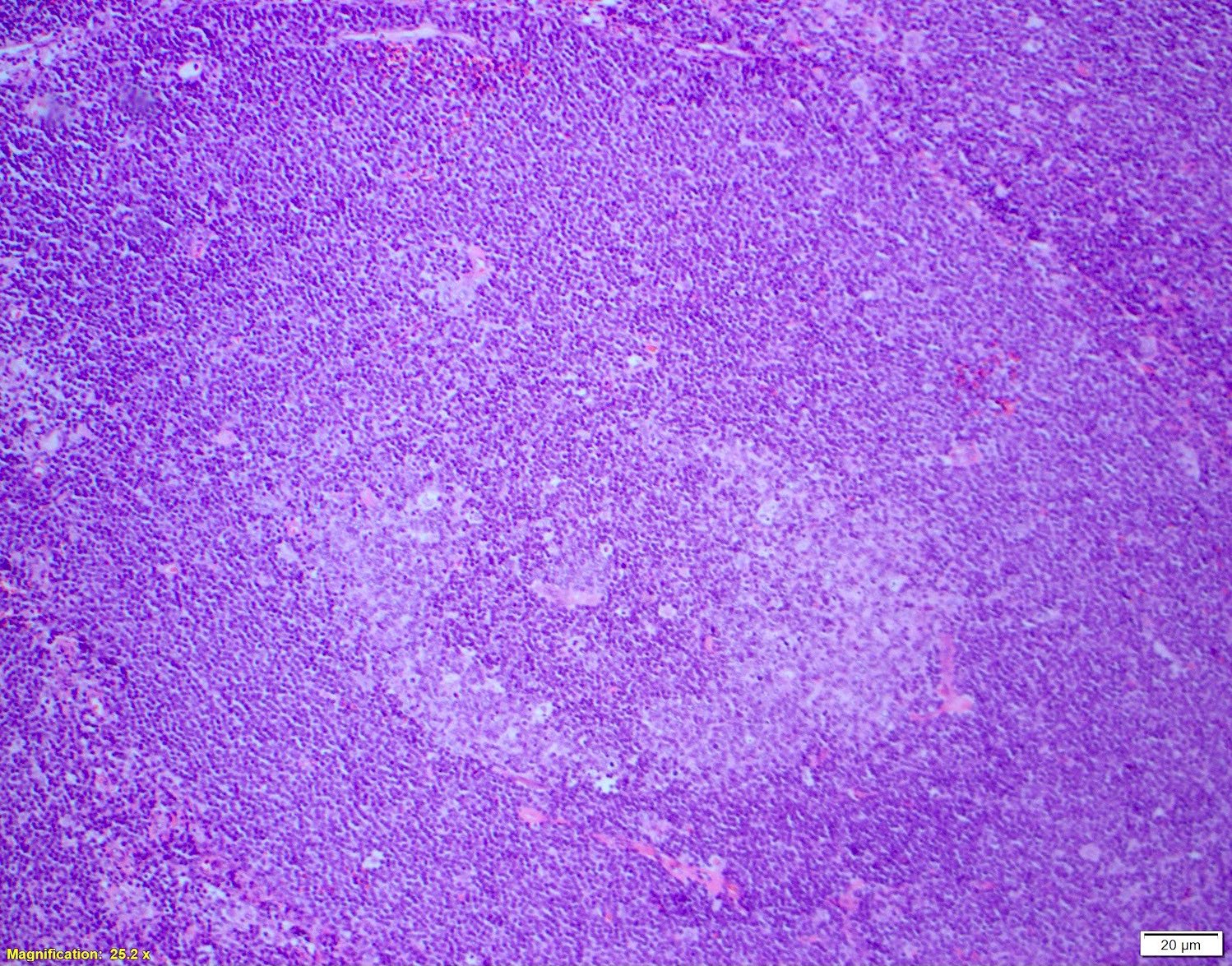
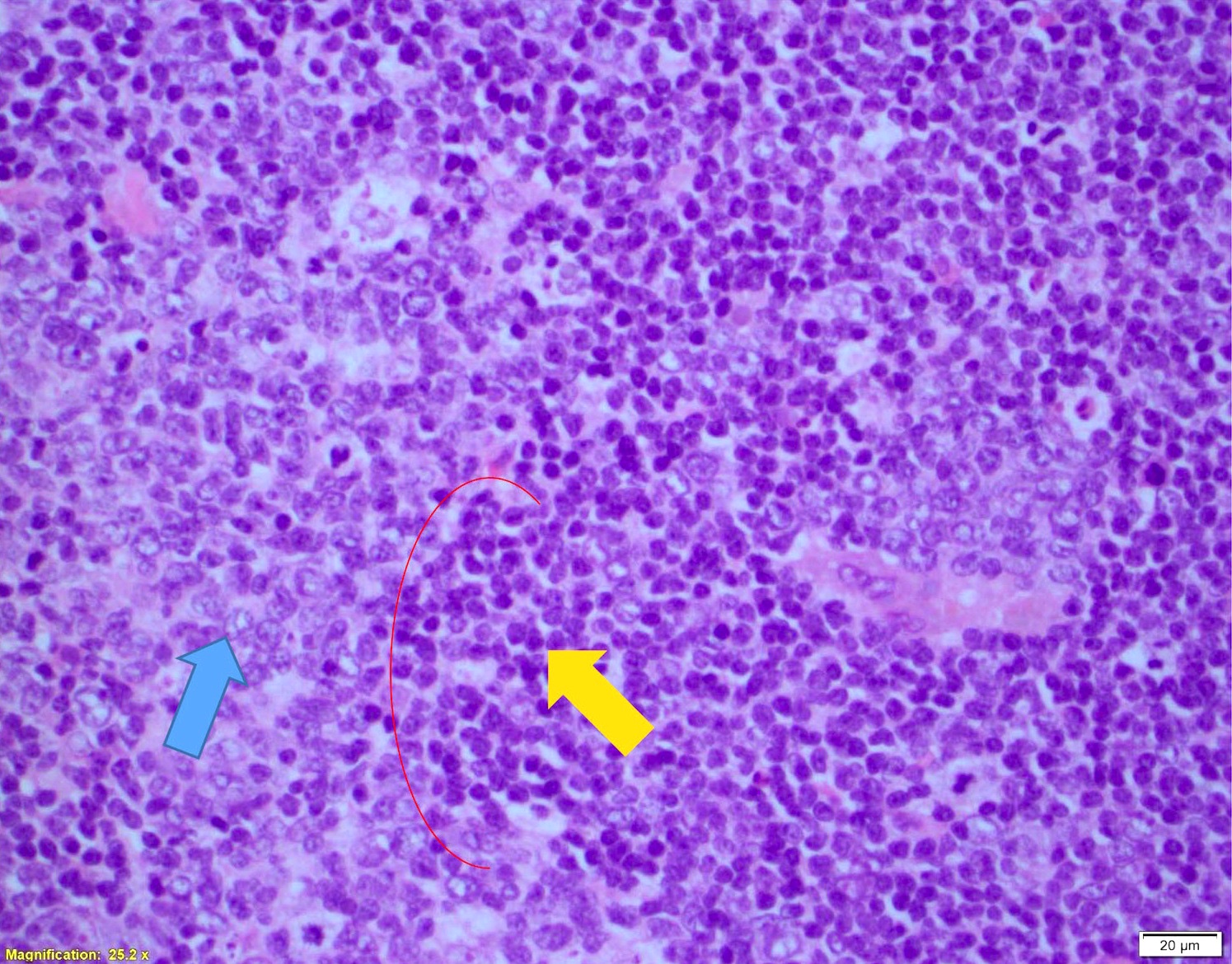
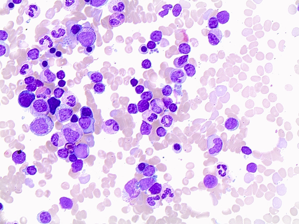
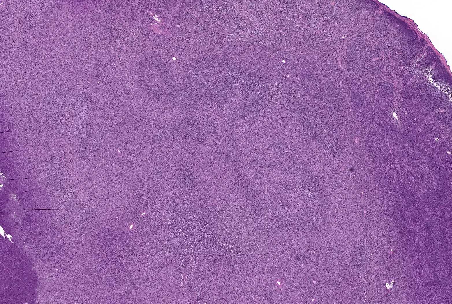
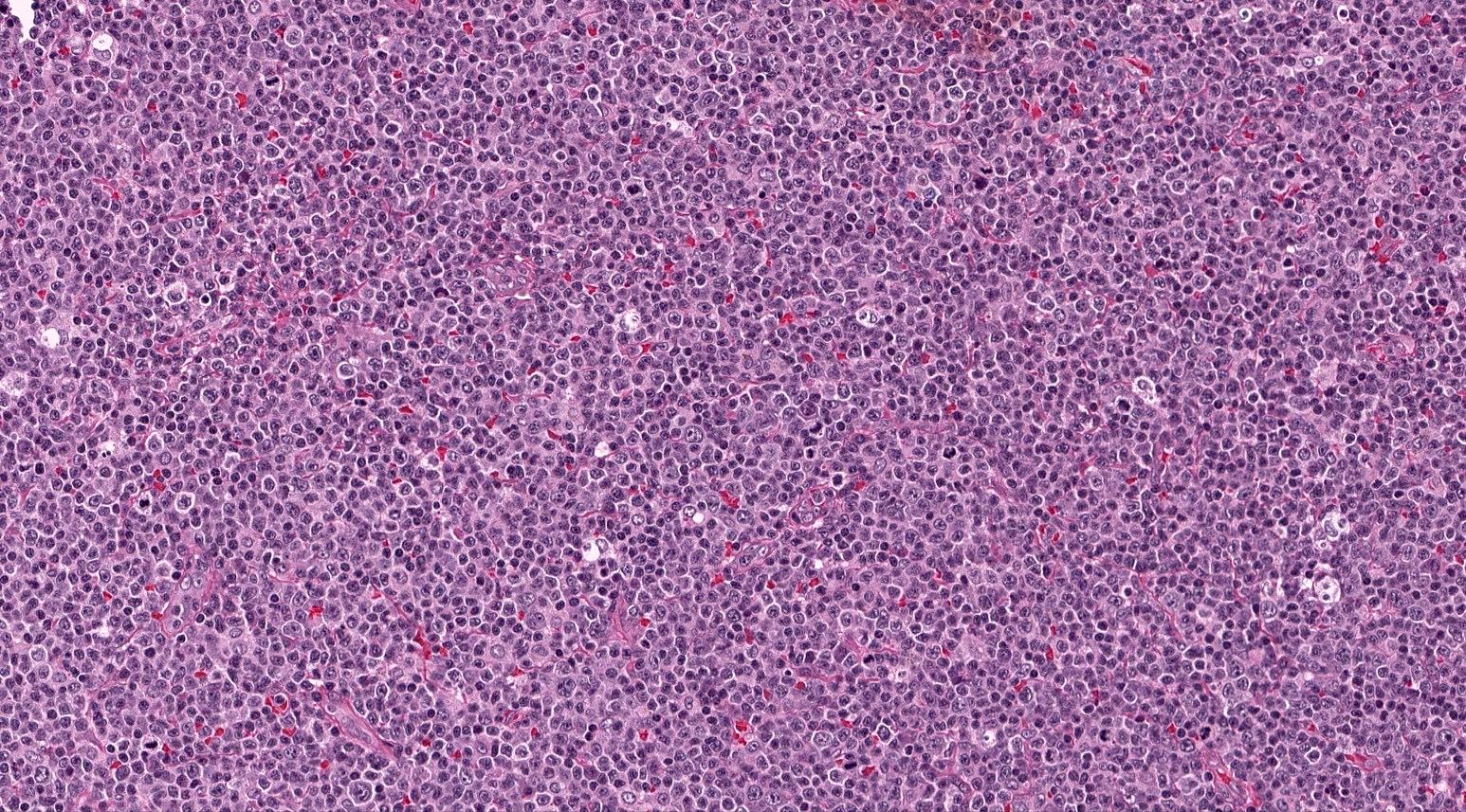
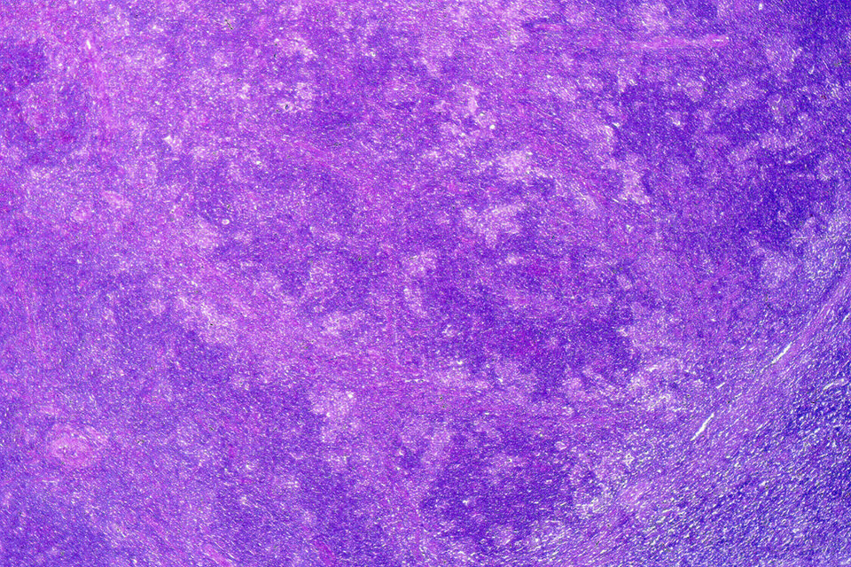
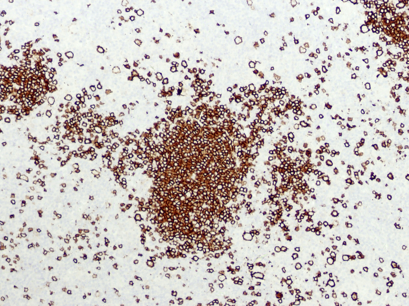
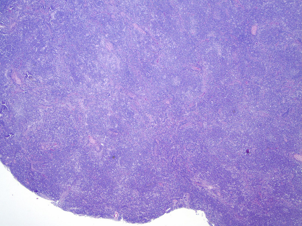
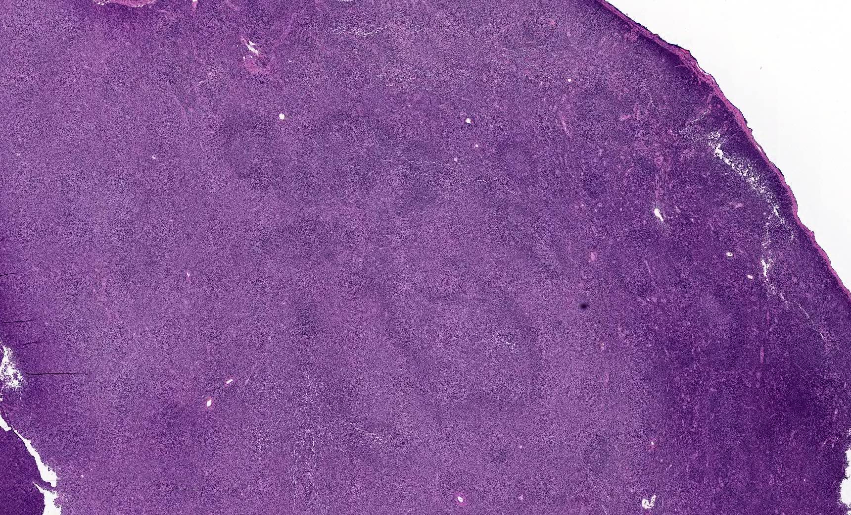
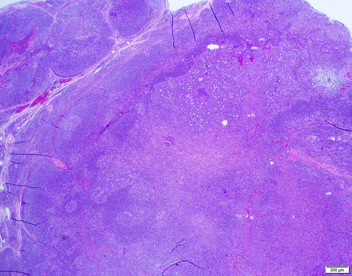
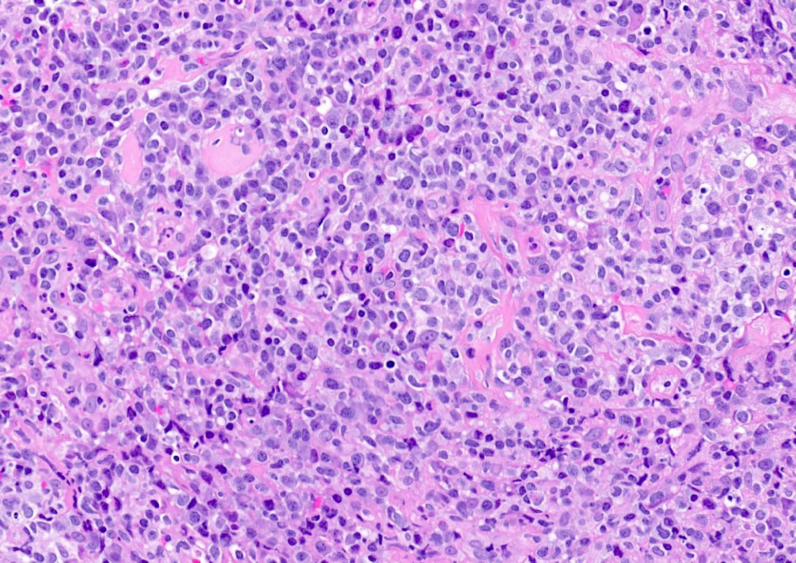
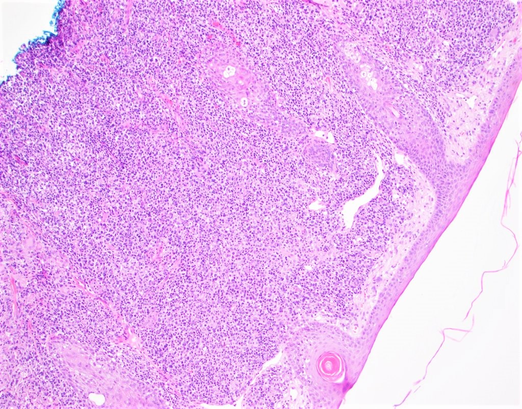
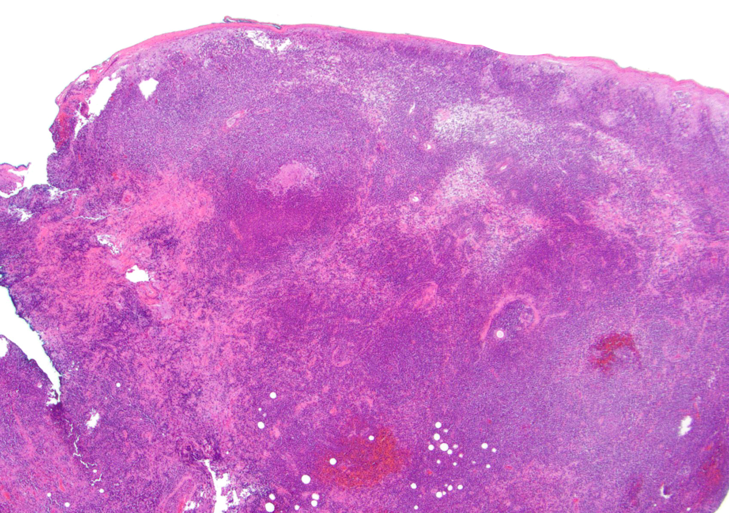
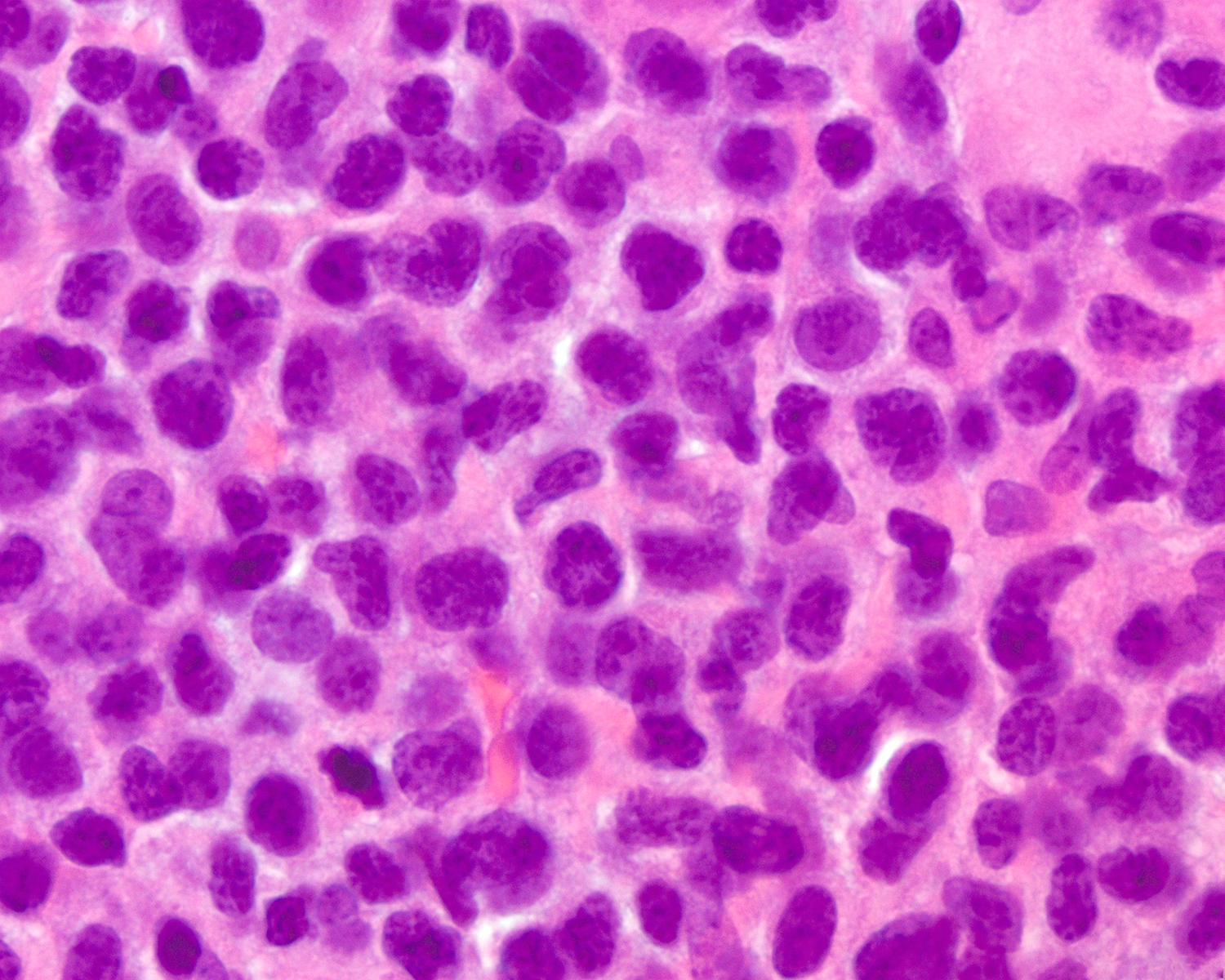
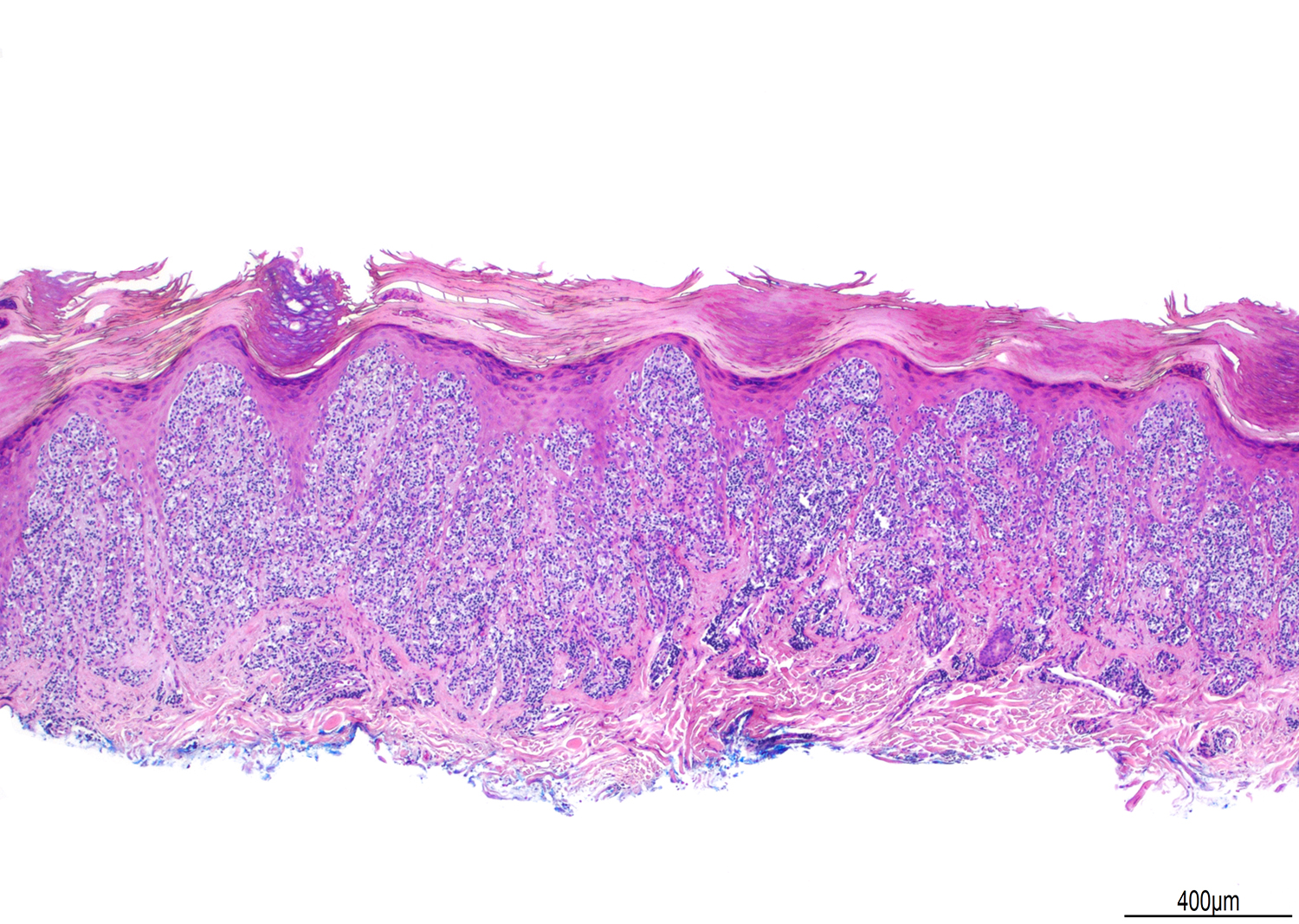
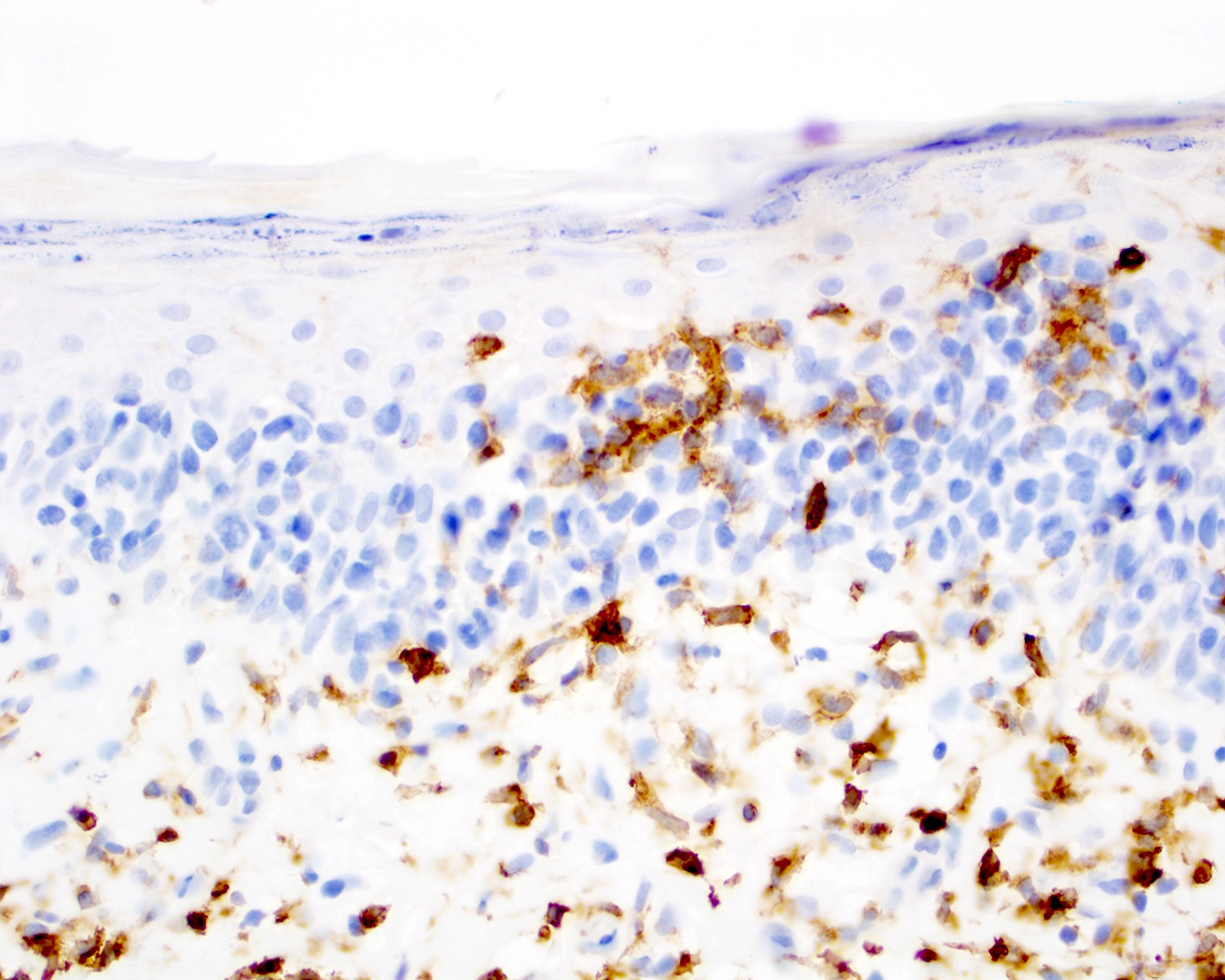
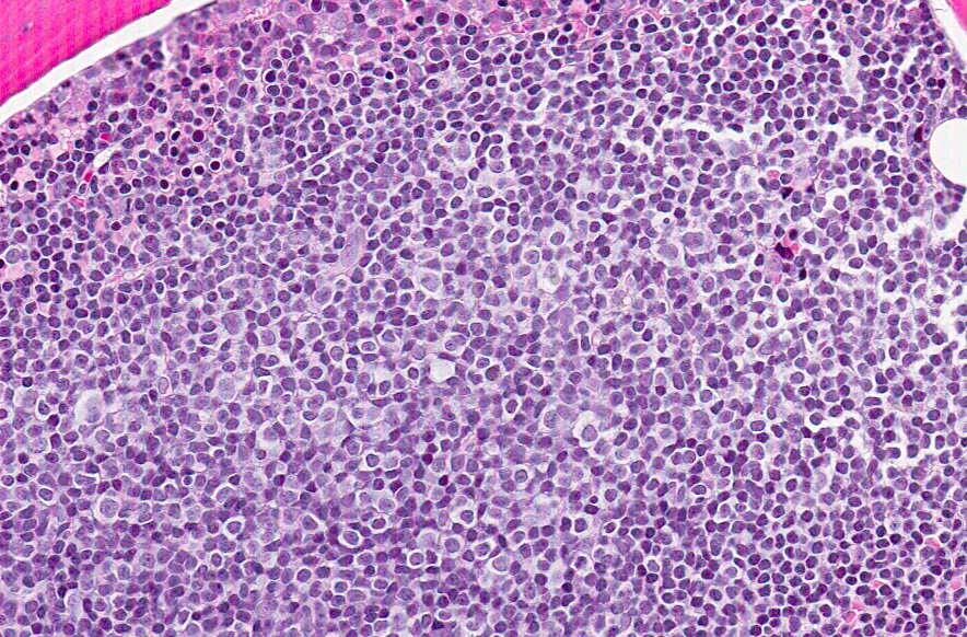
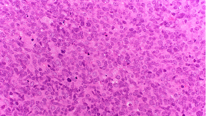
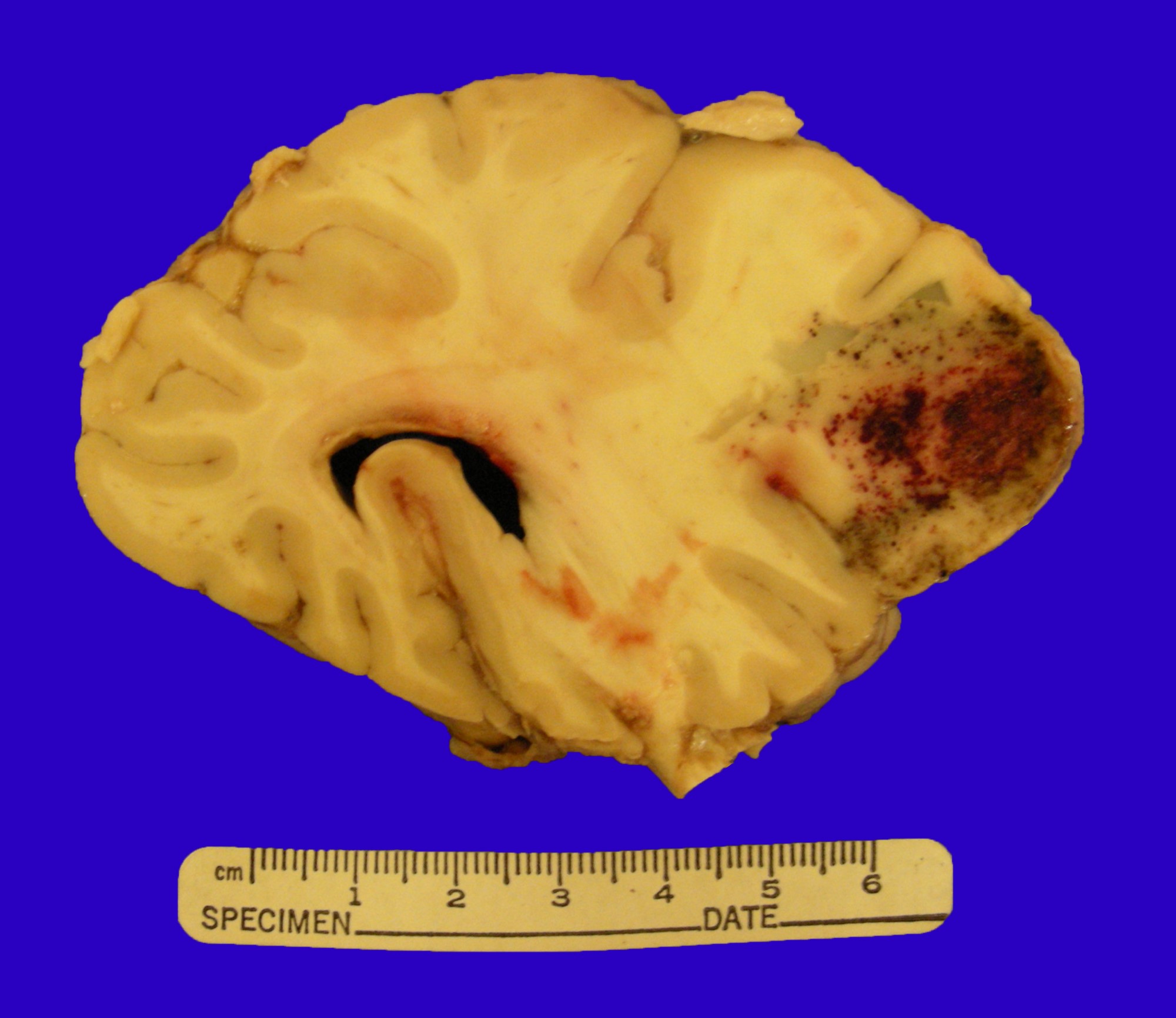
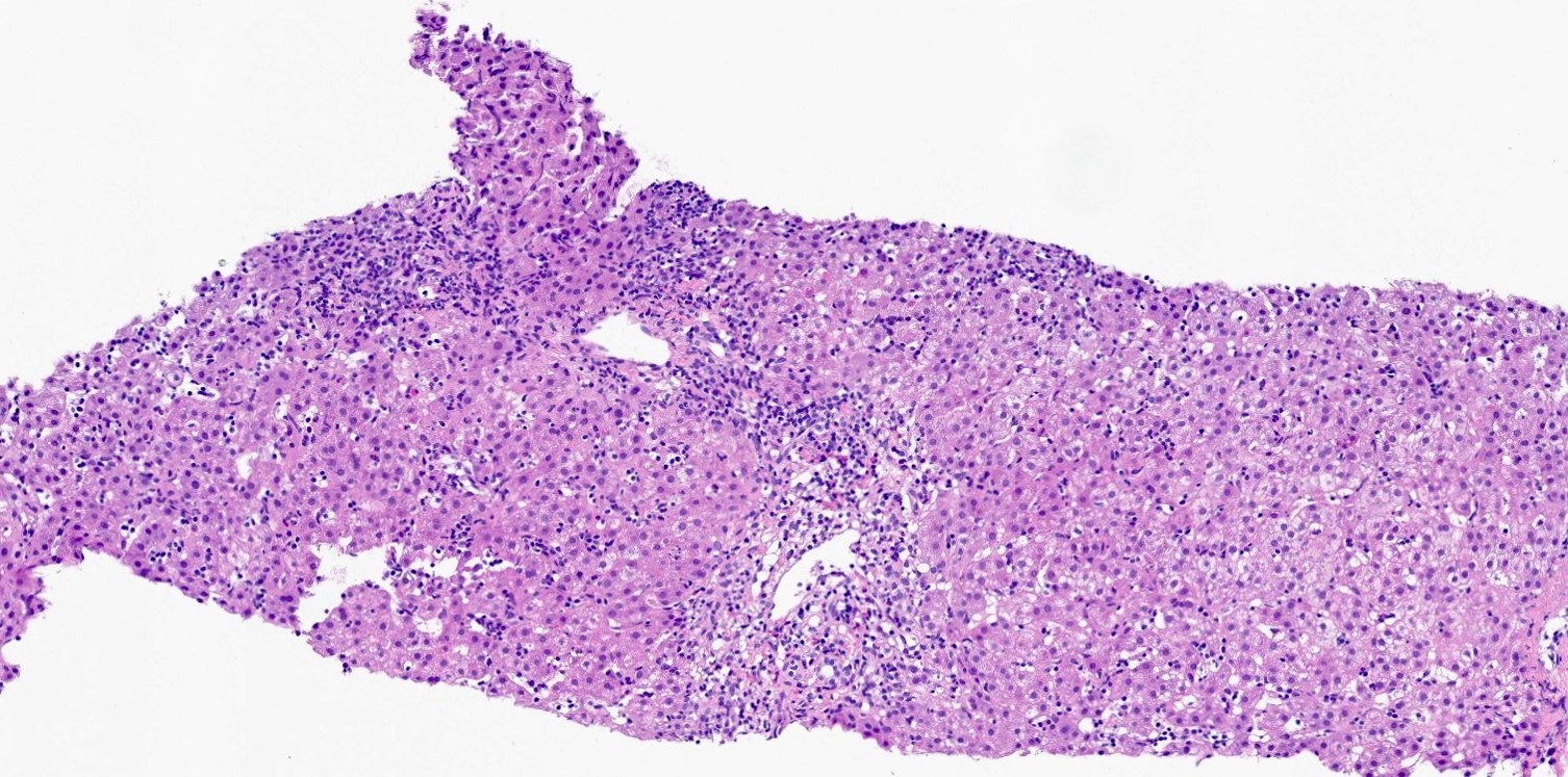
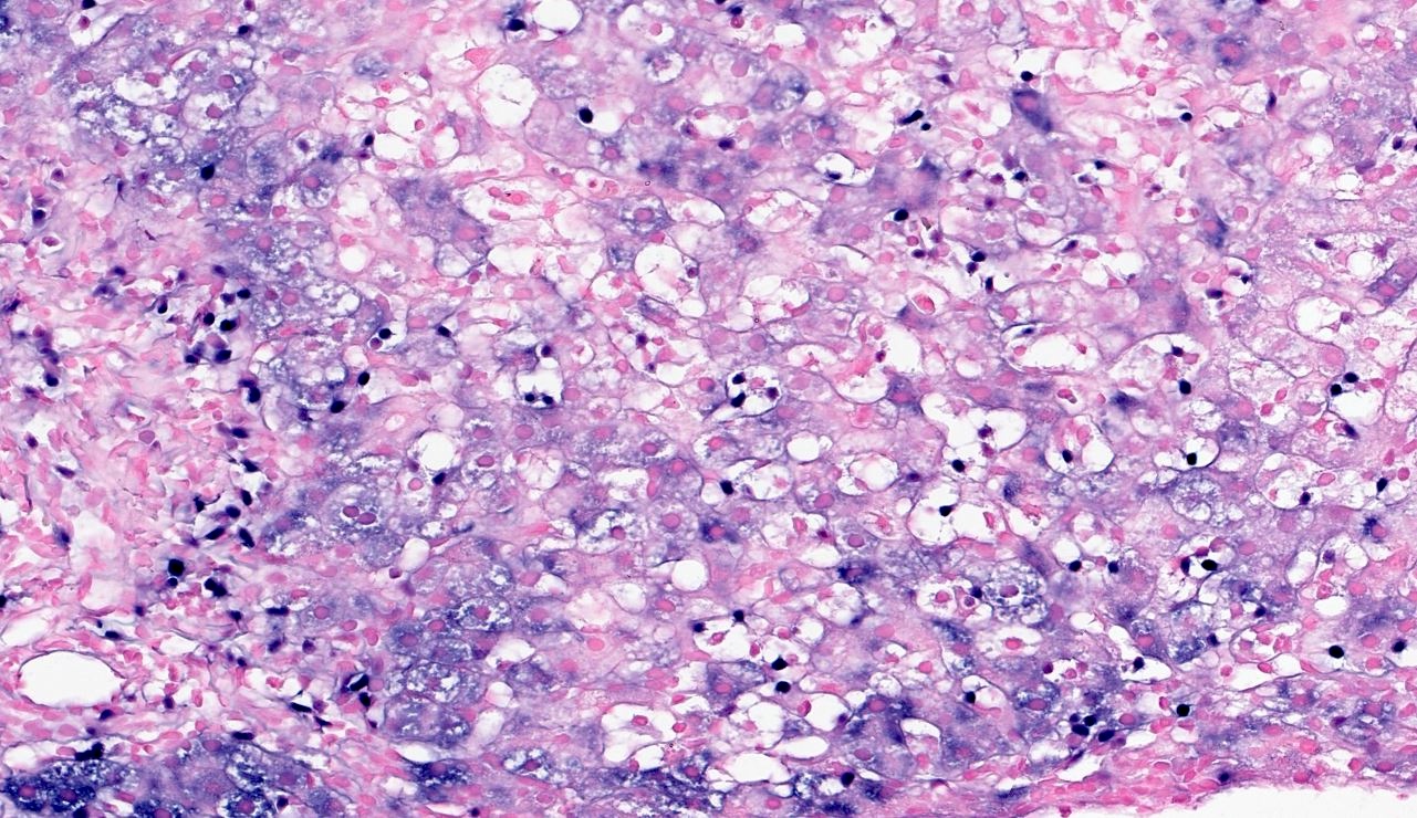
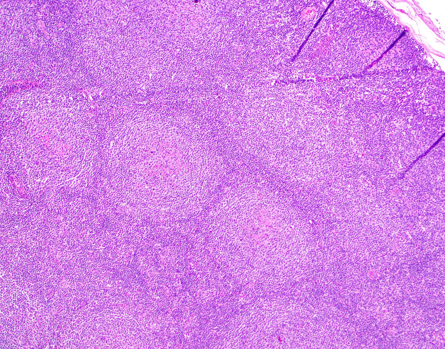
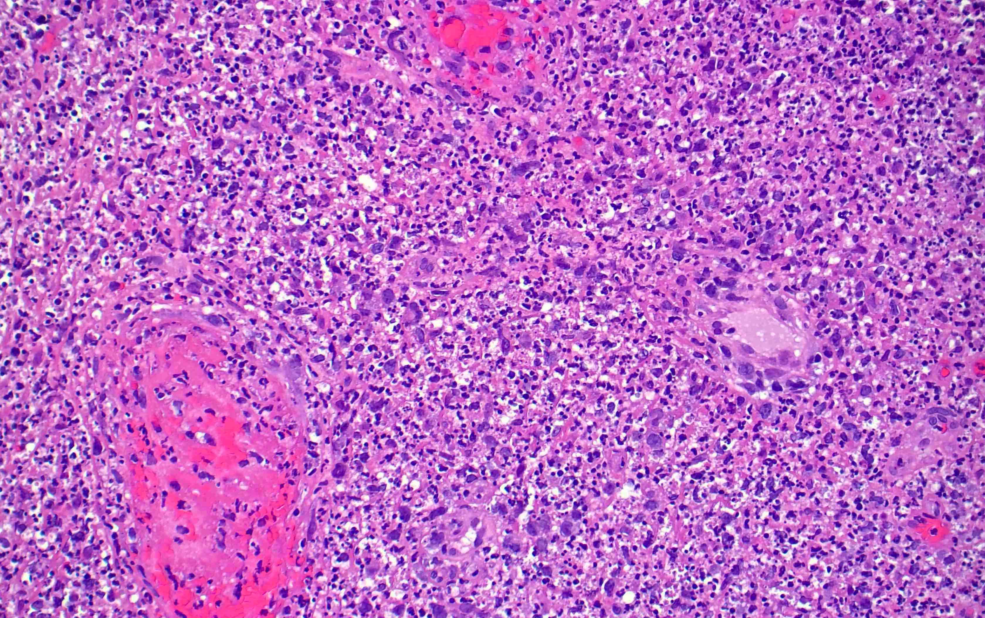
Tumor with diffuse involvement
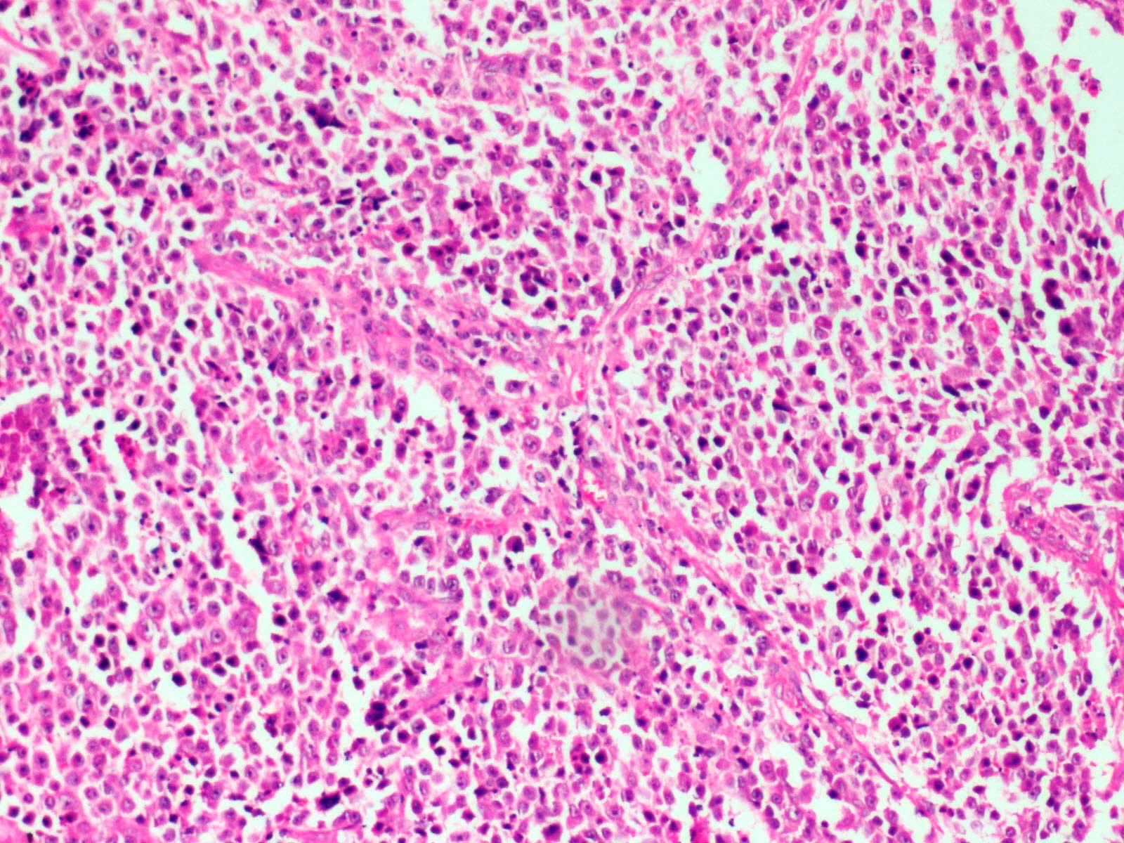
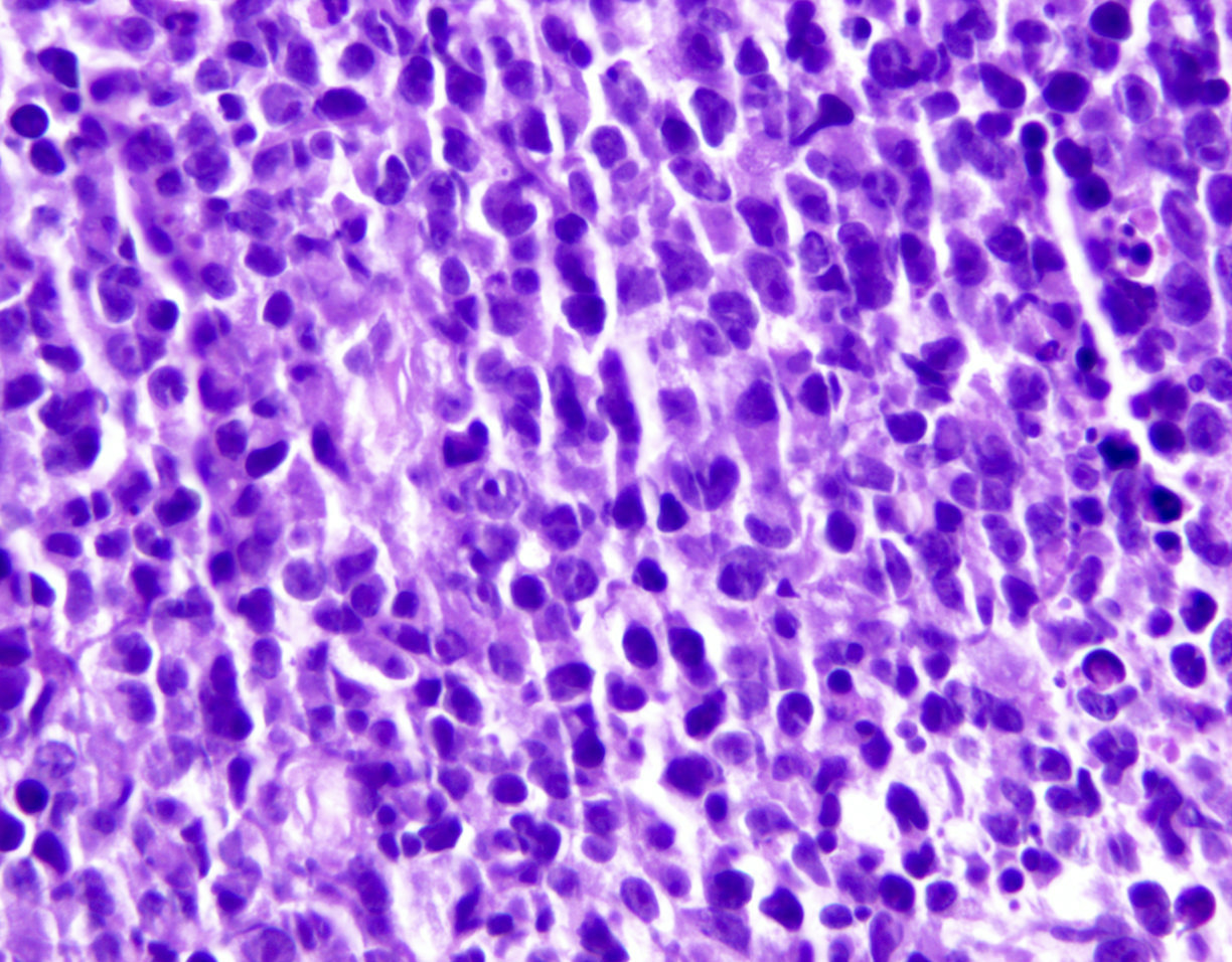
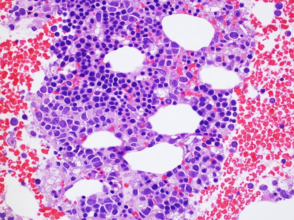
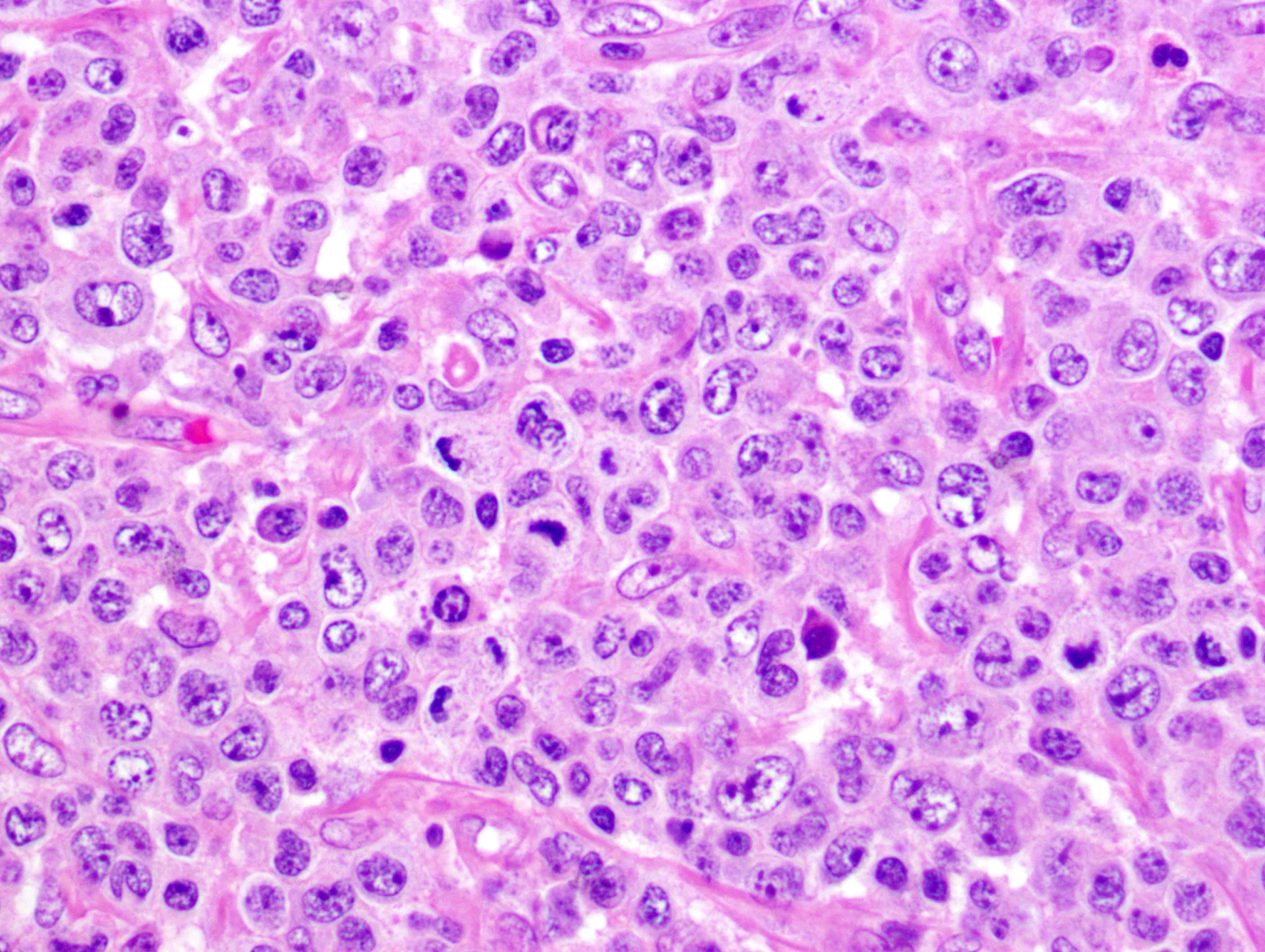
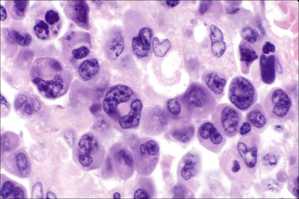
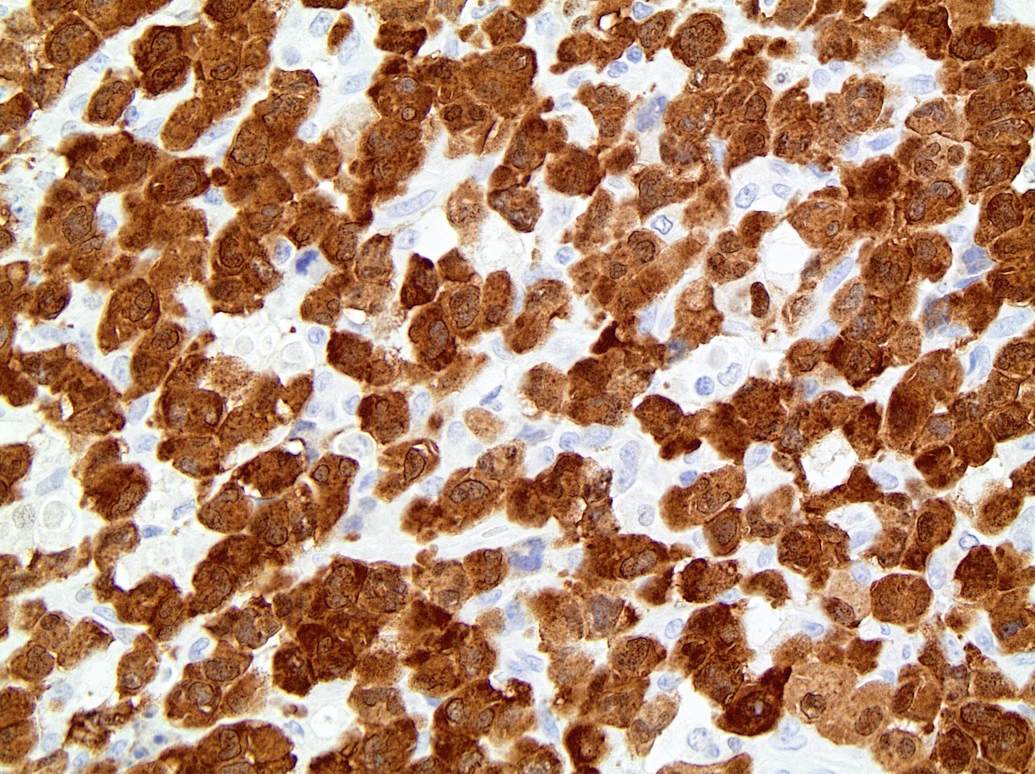
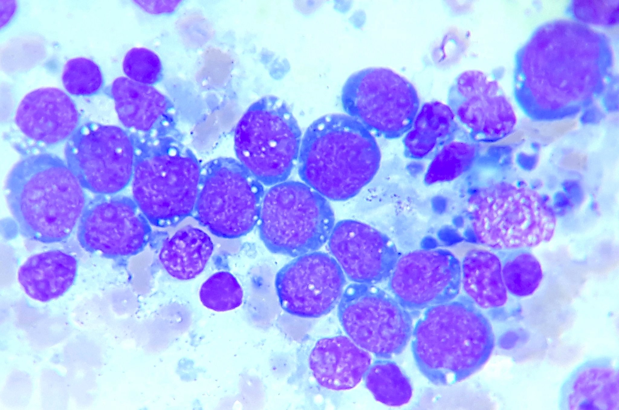
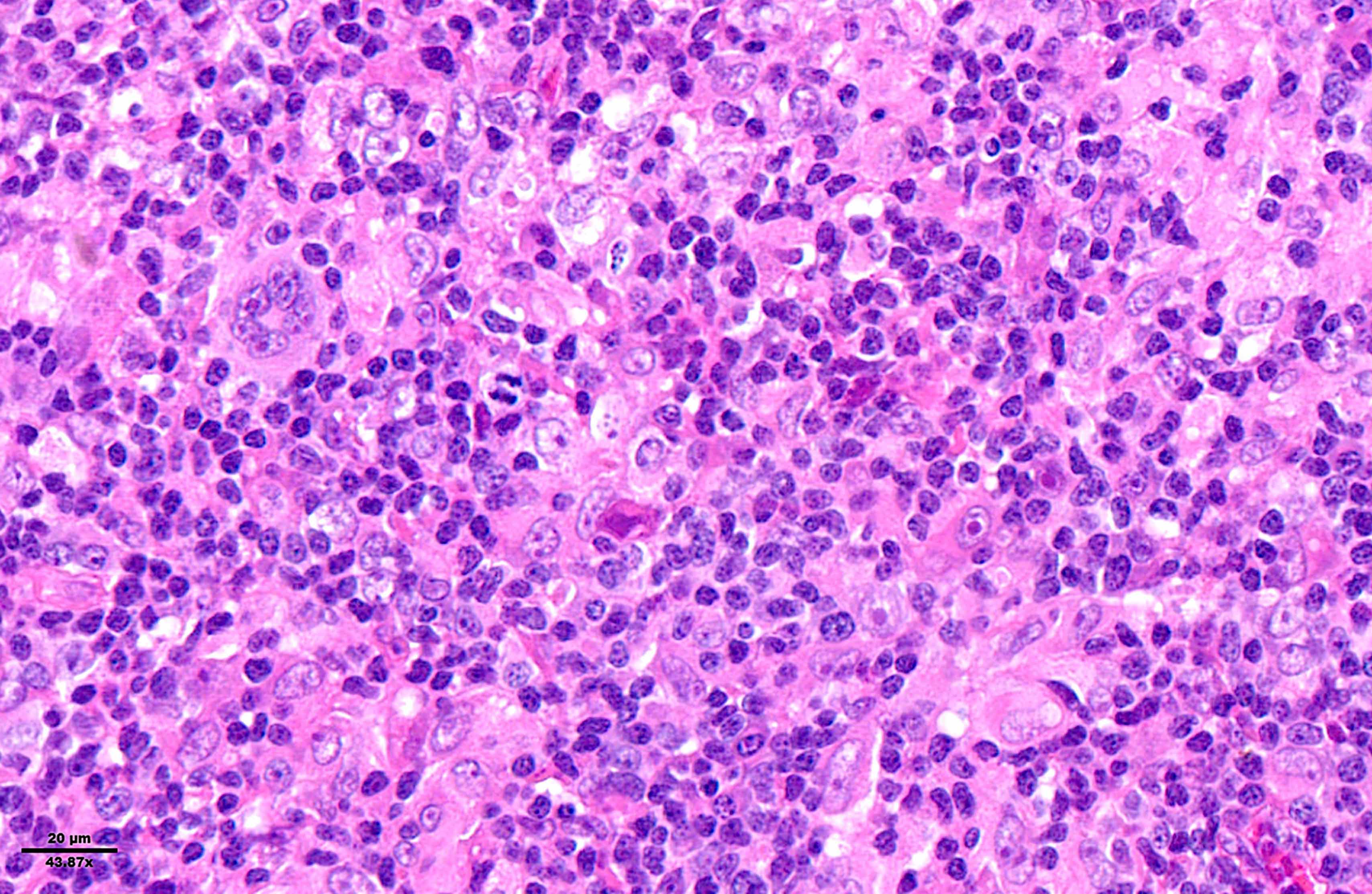
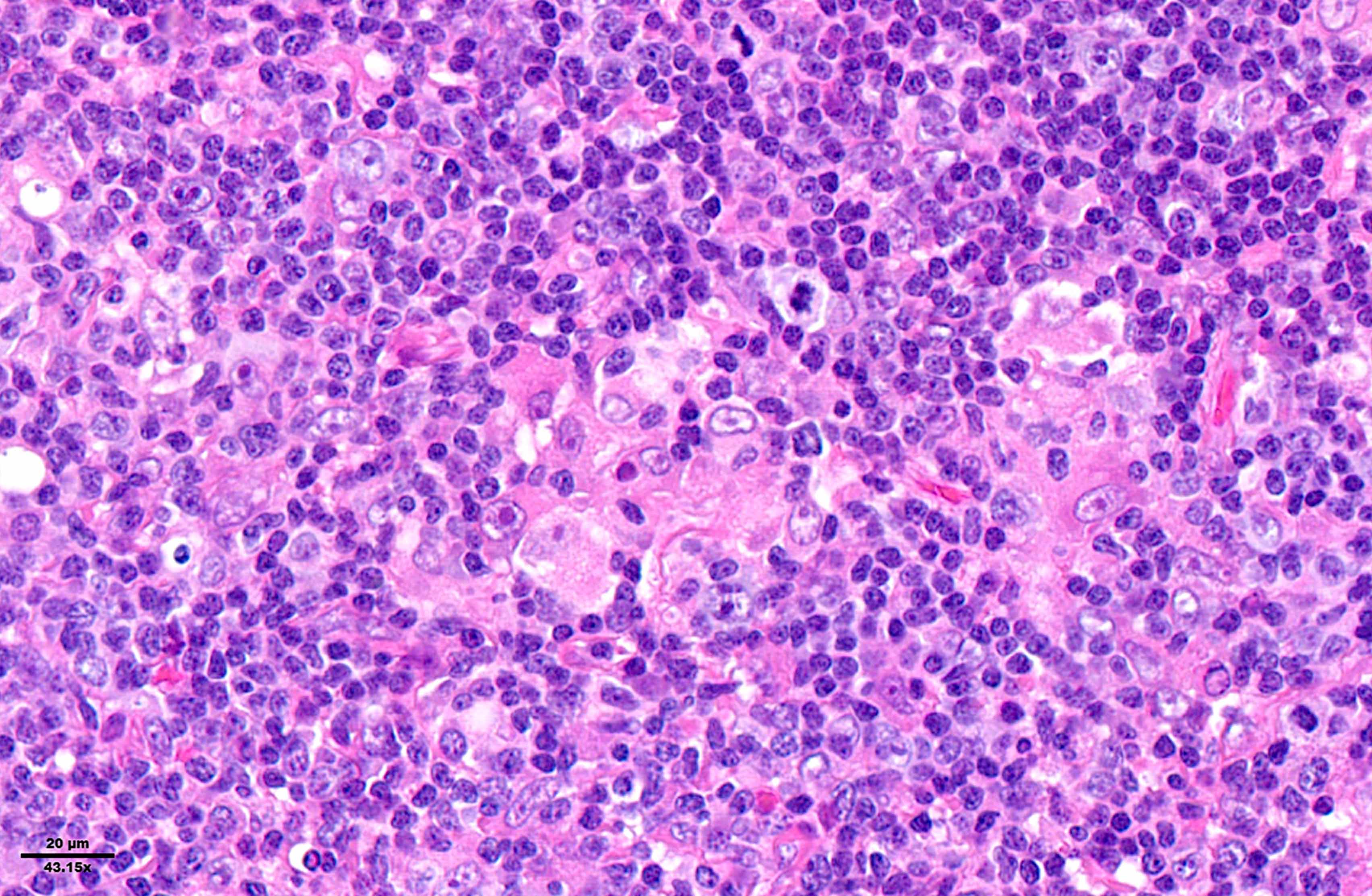
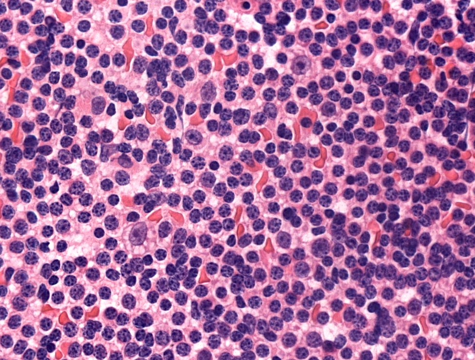
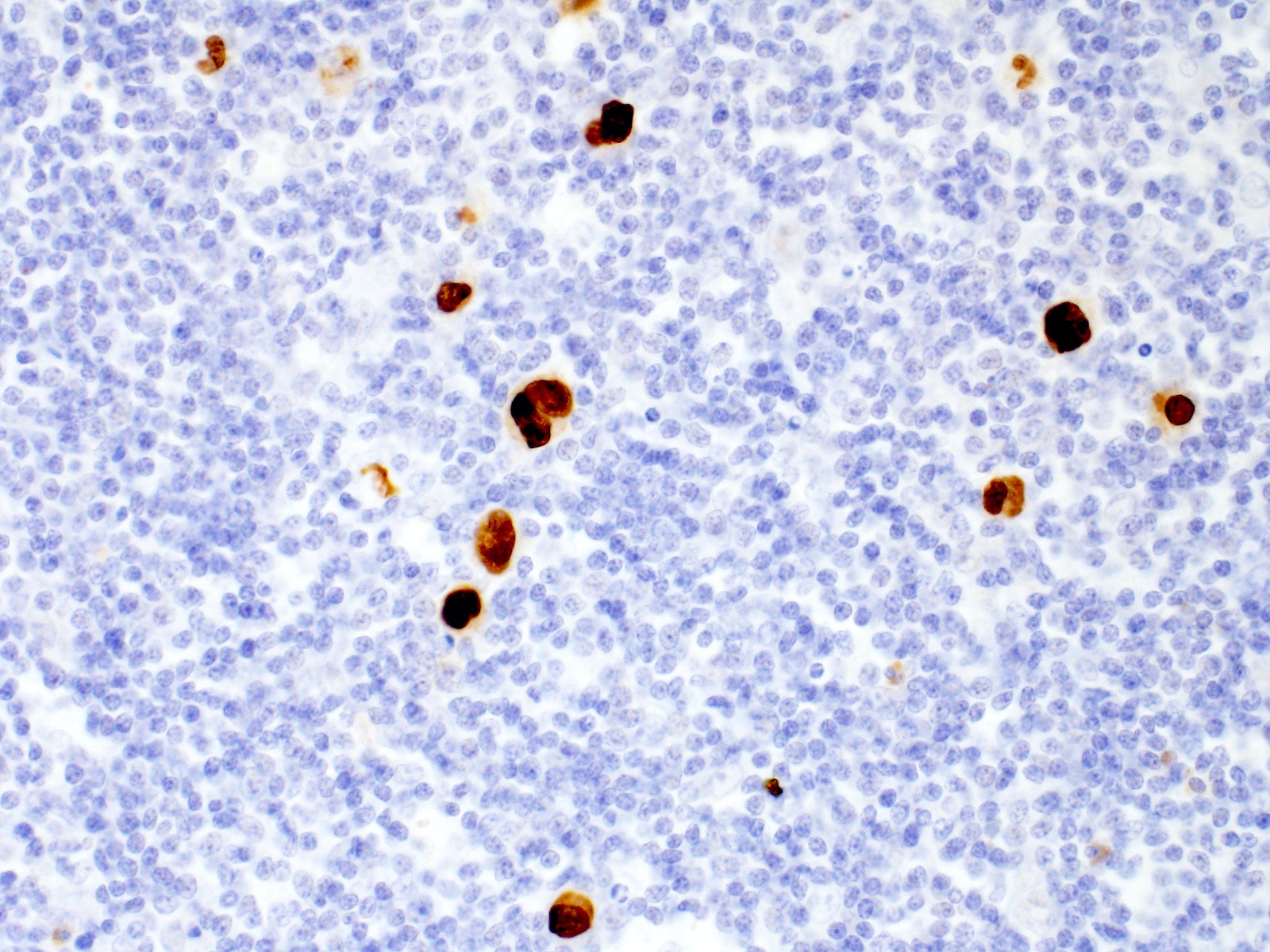
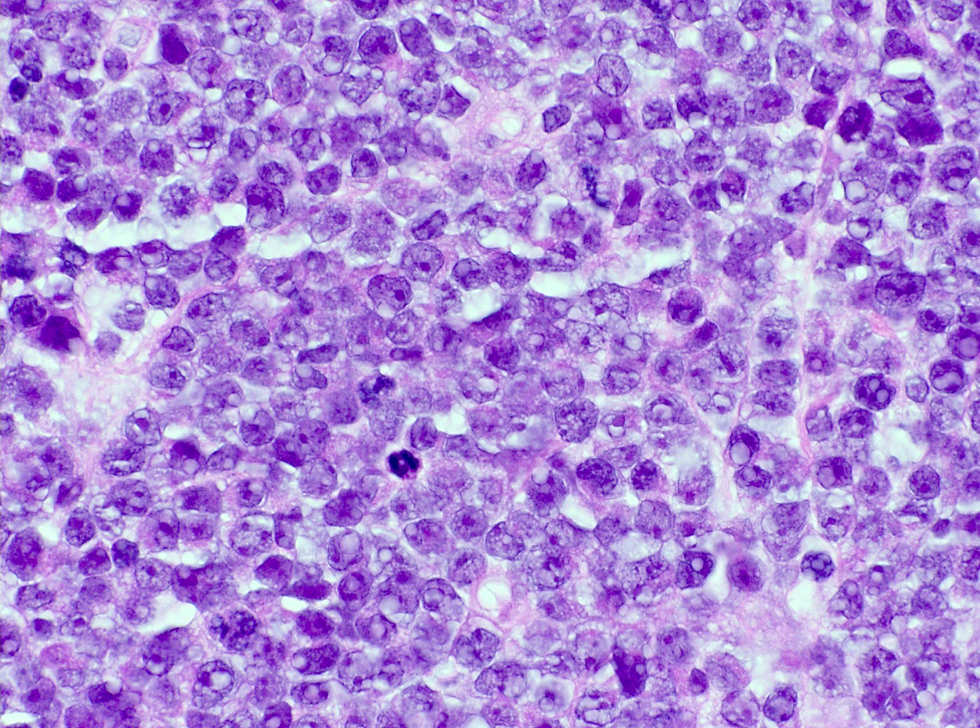
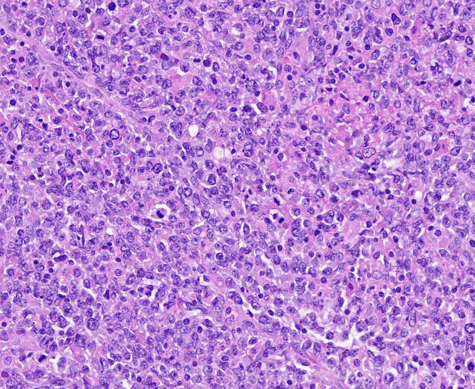
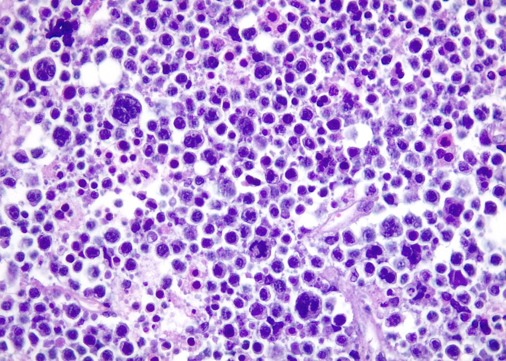
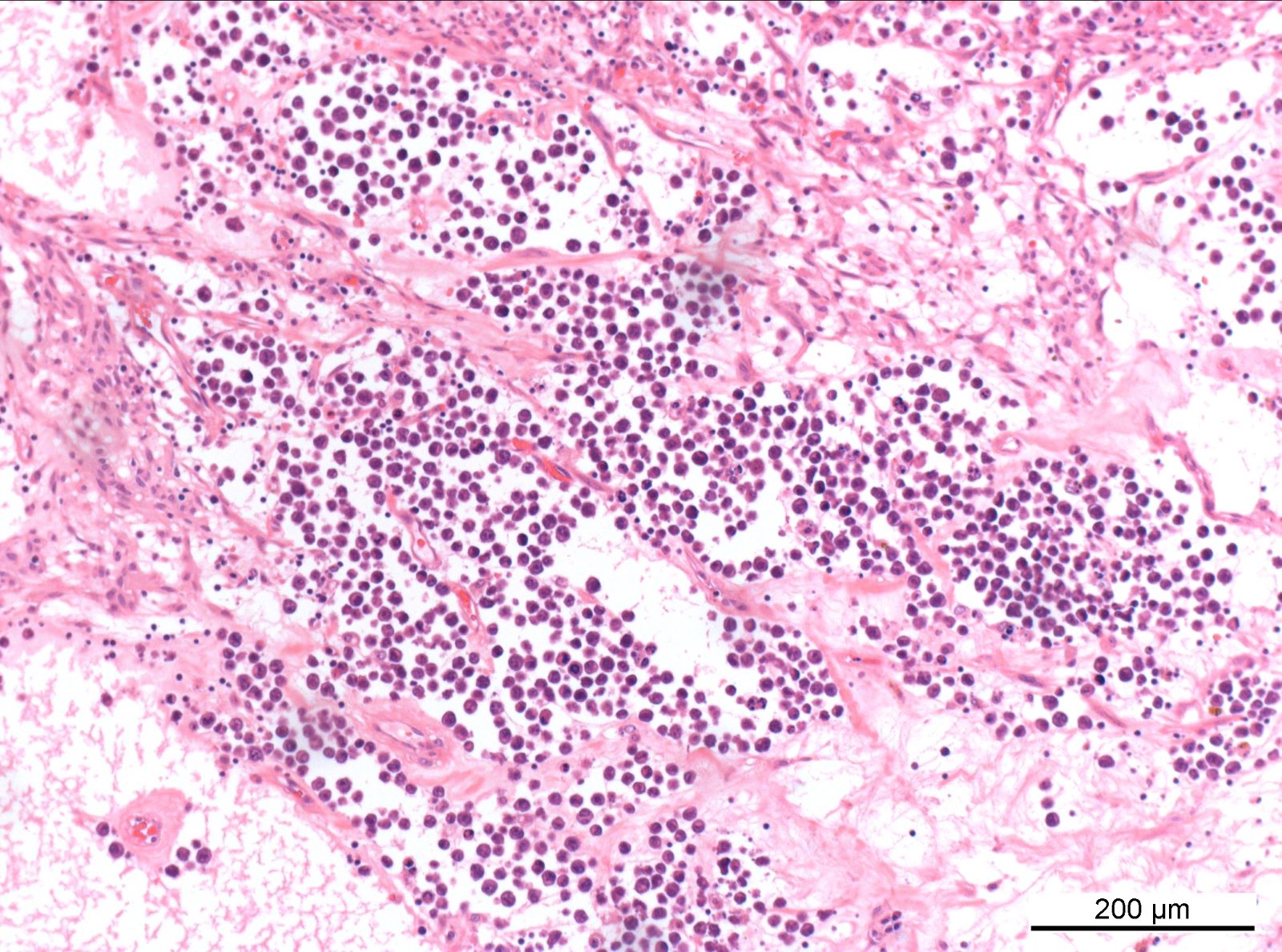
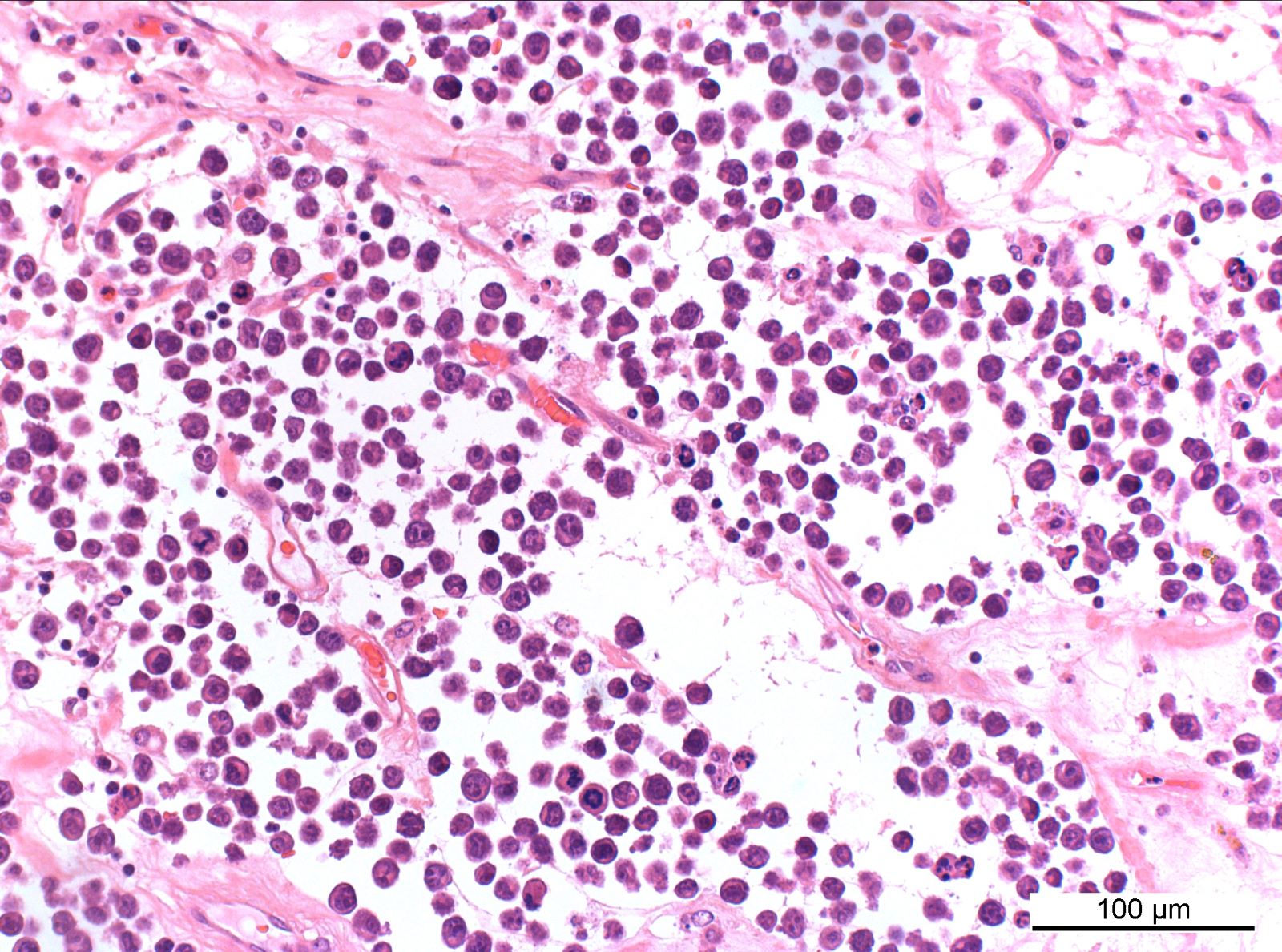
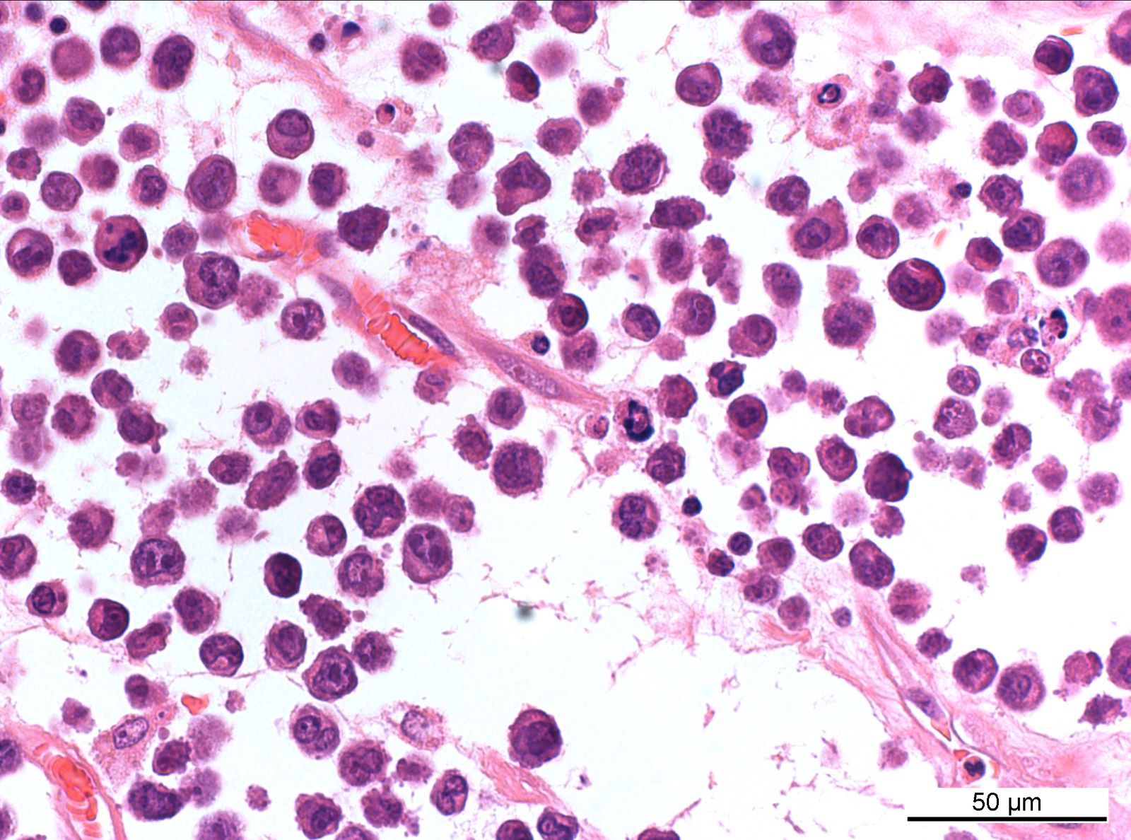
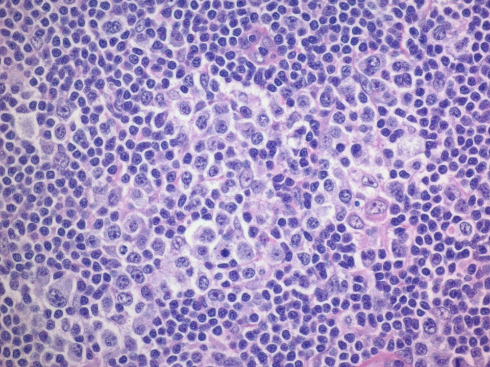
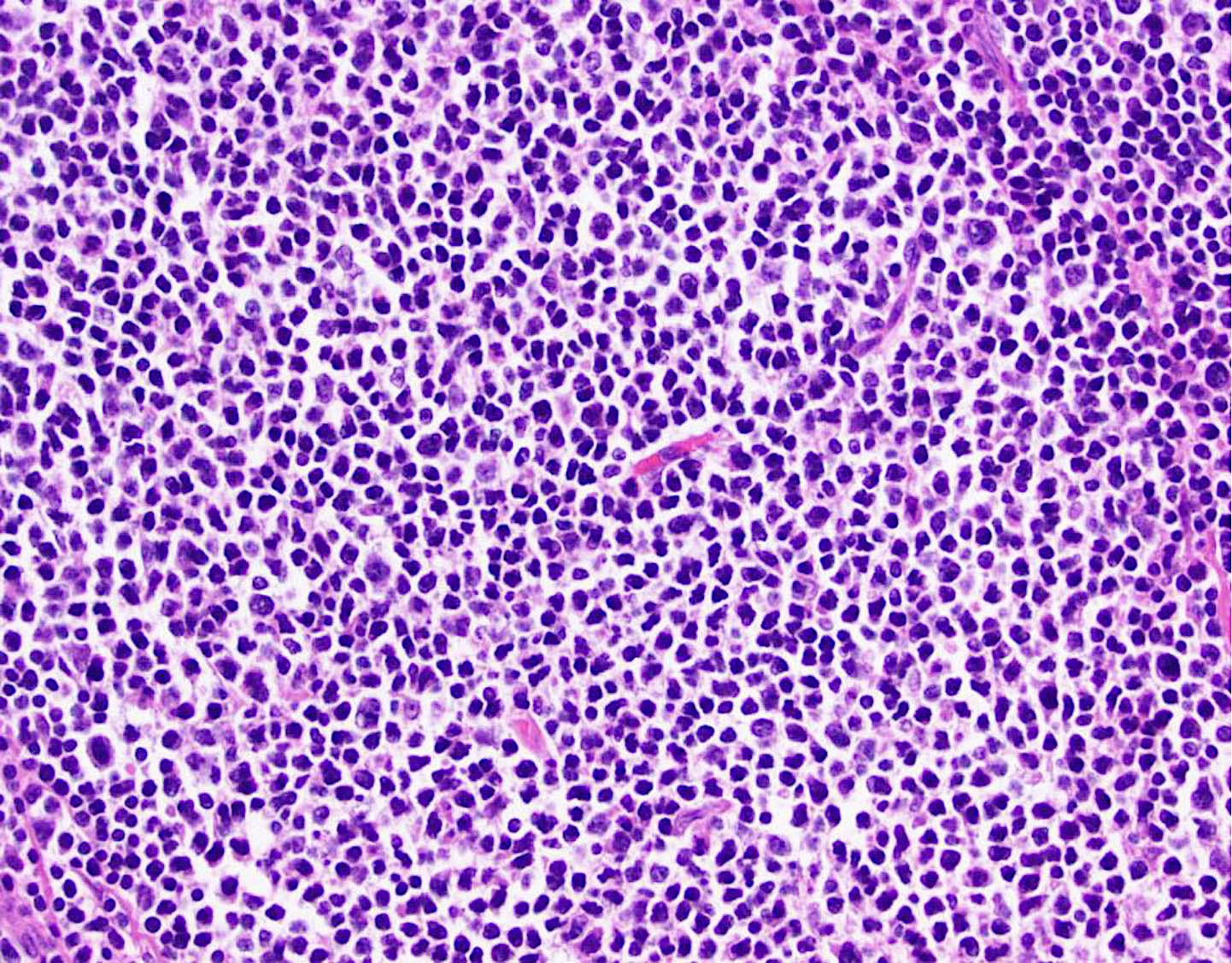
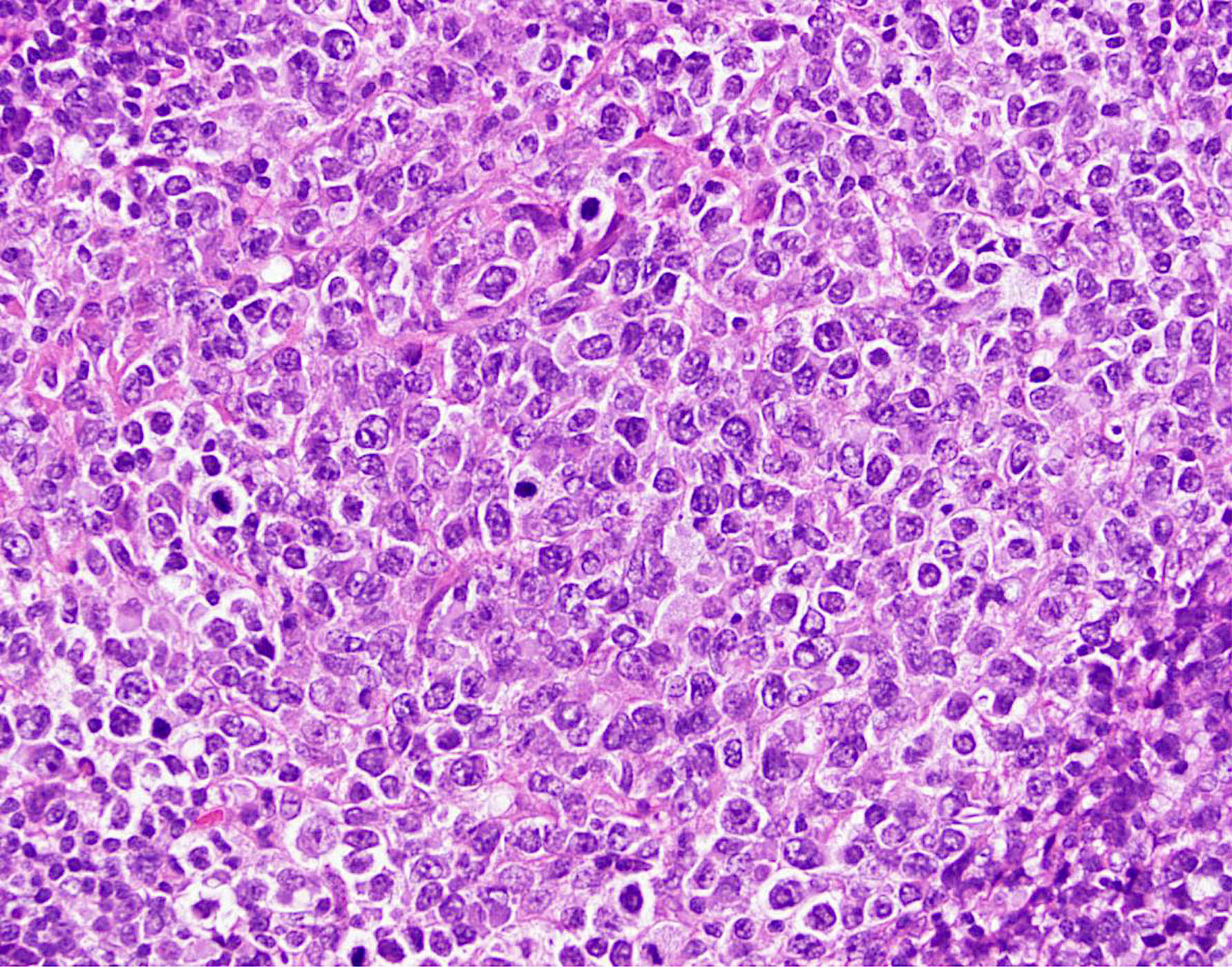
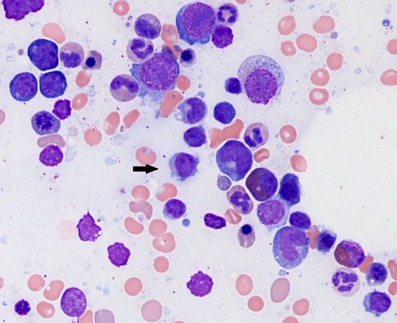
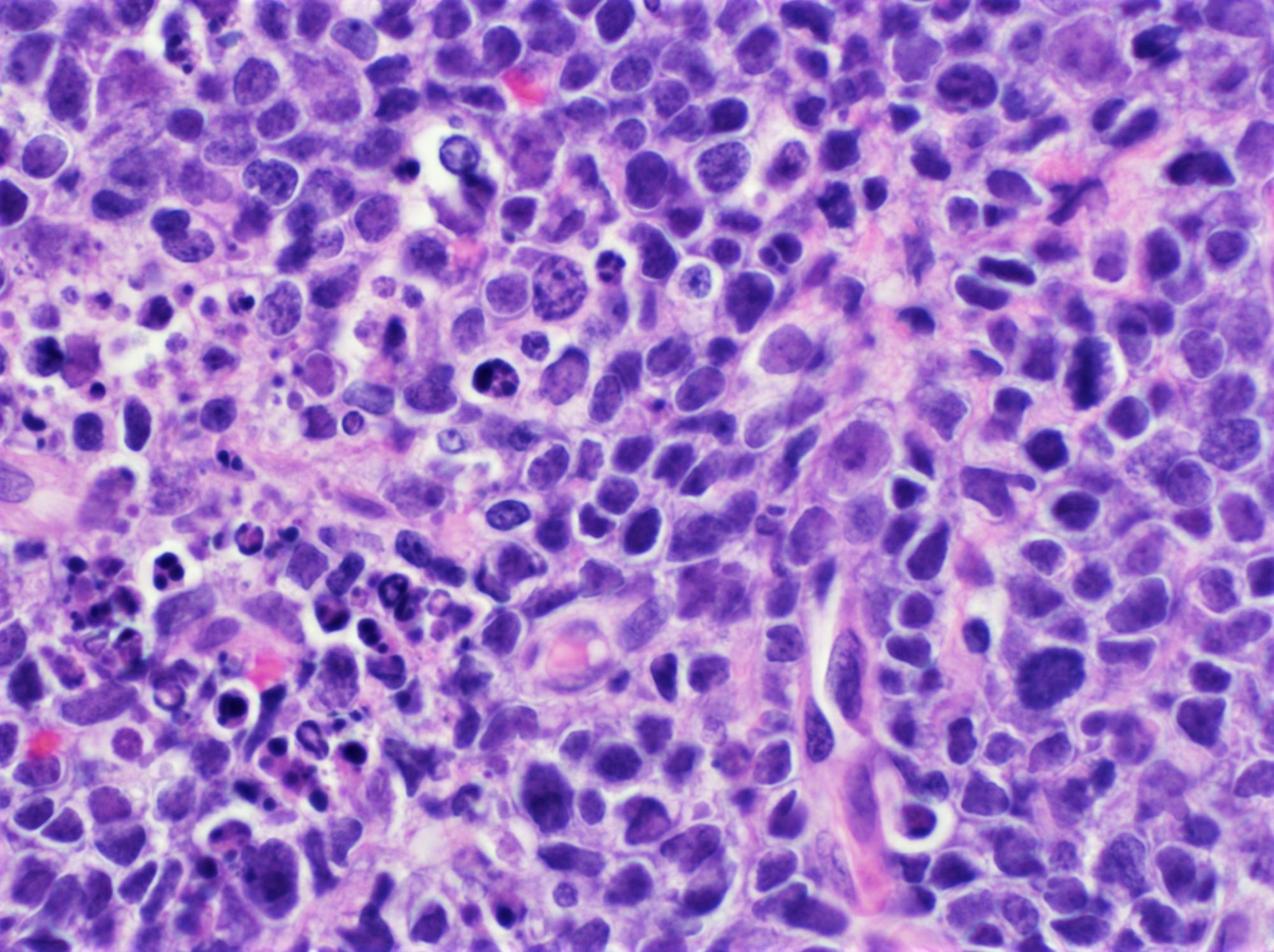
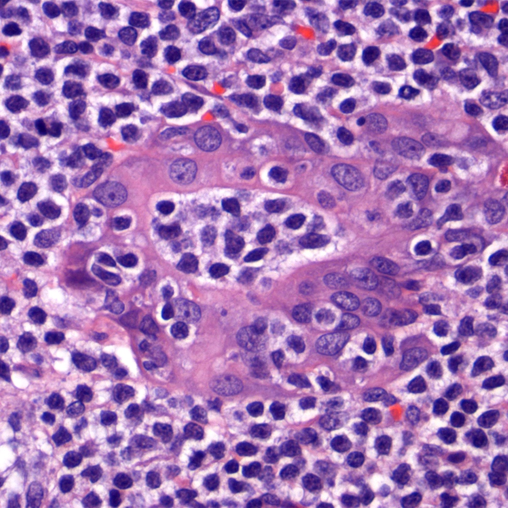
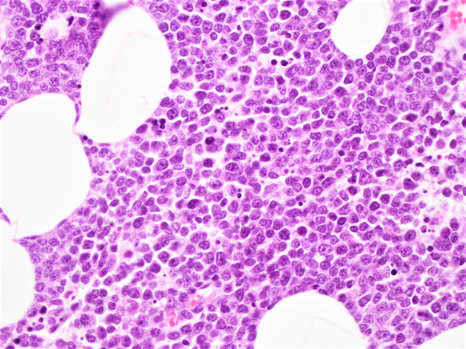
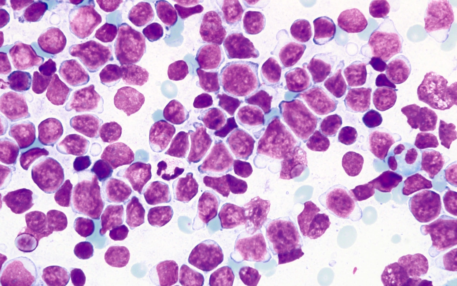
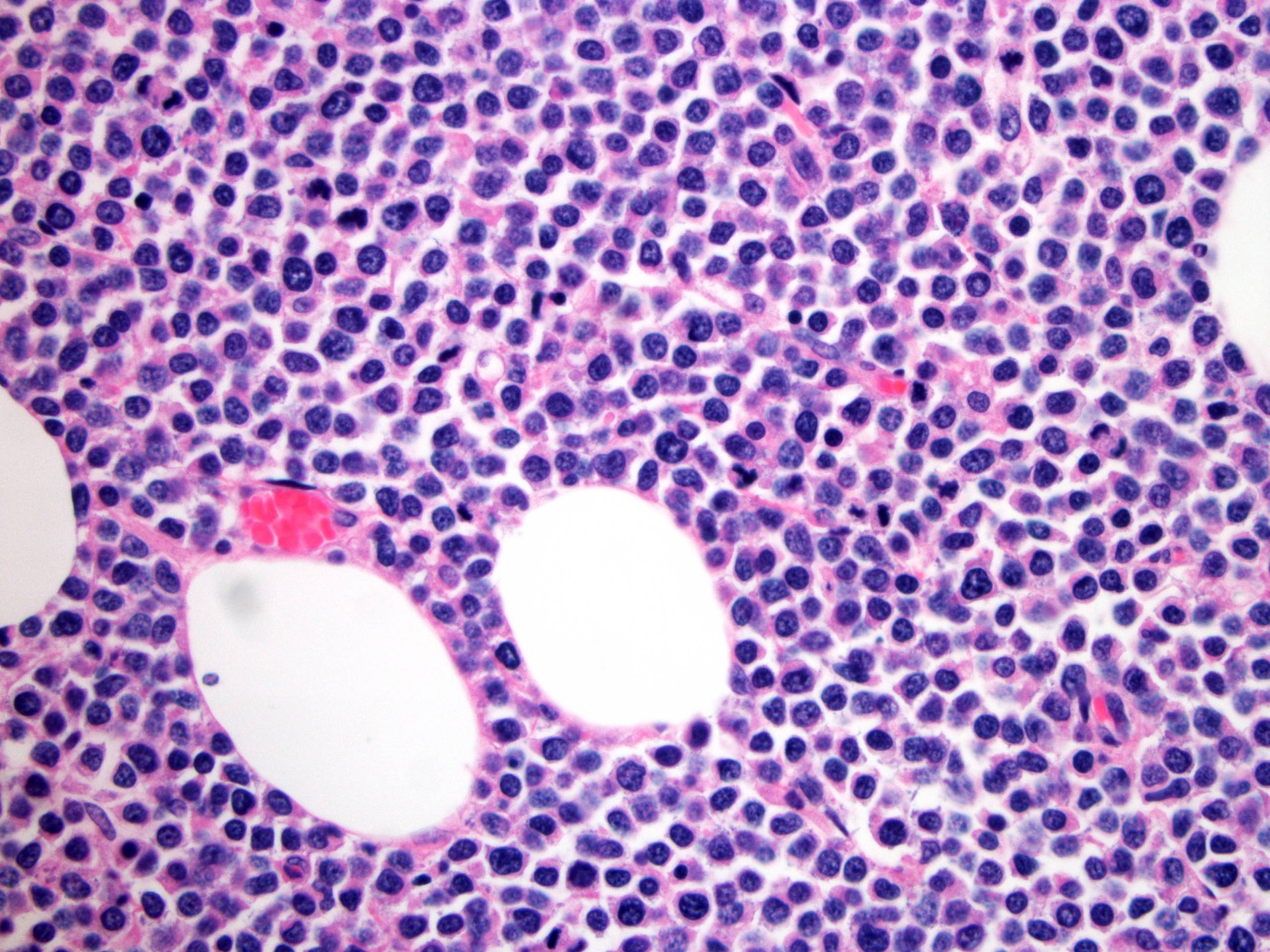
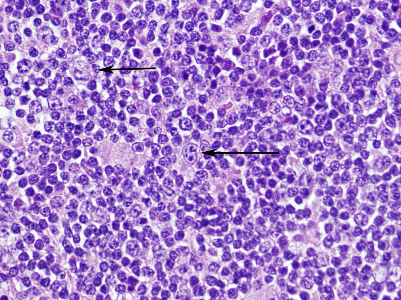
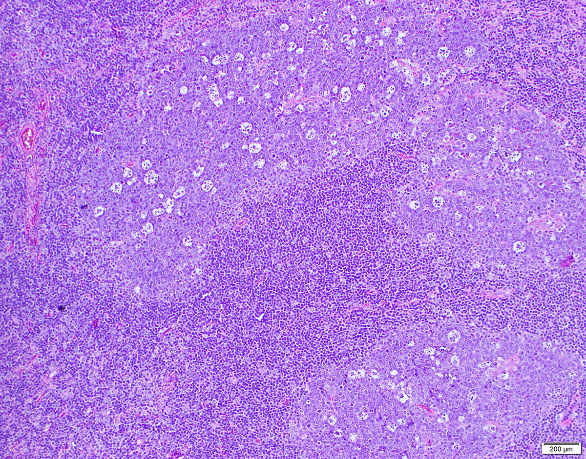
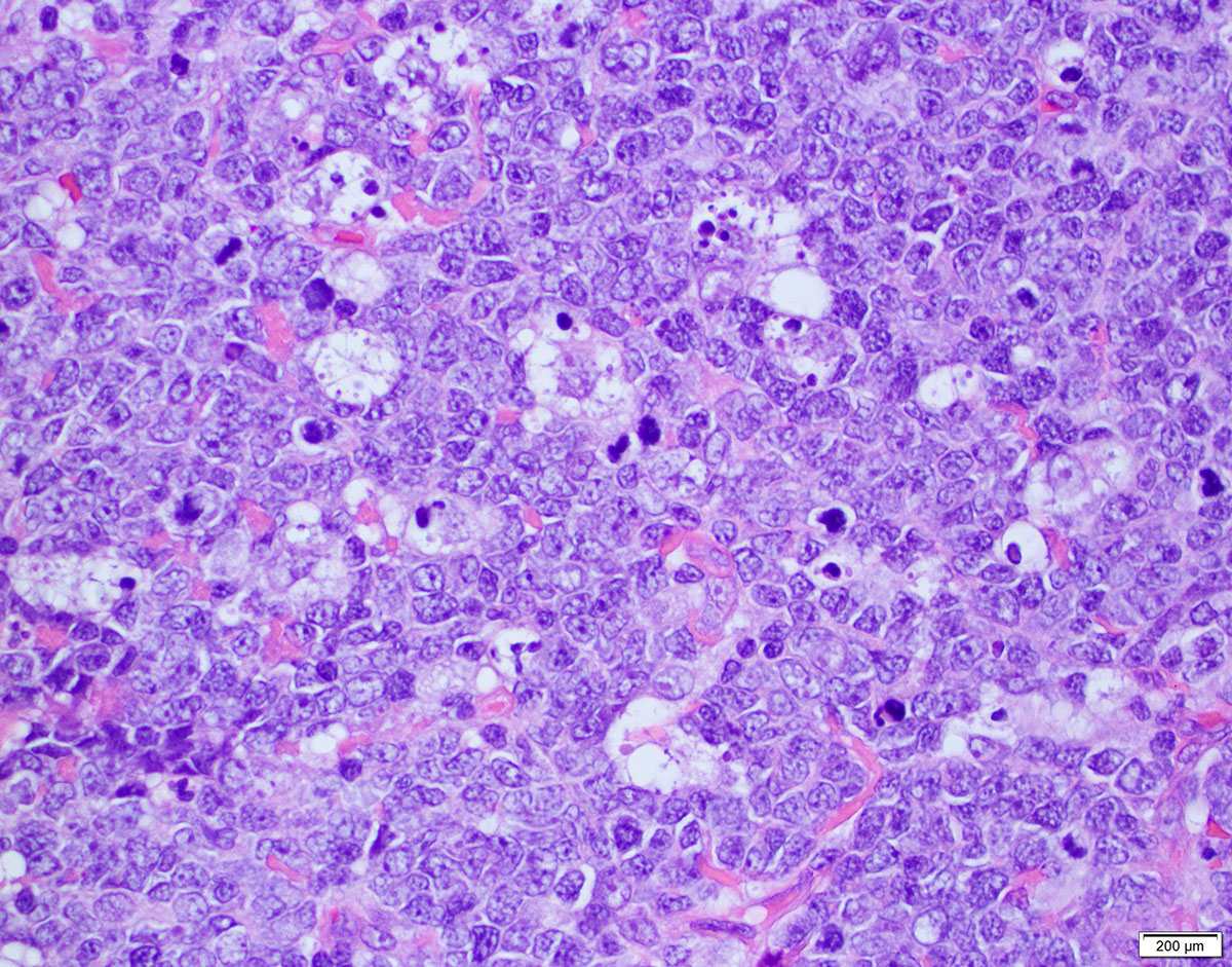
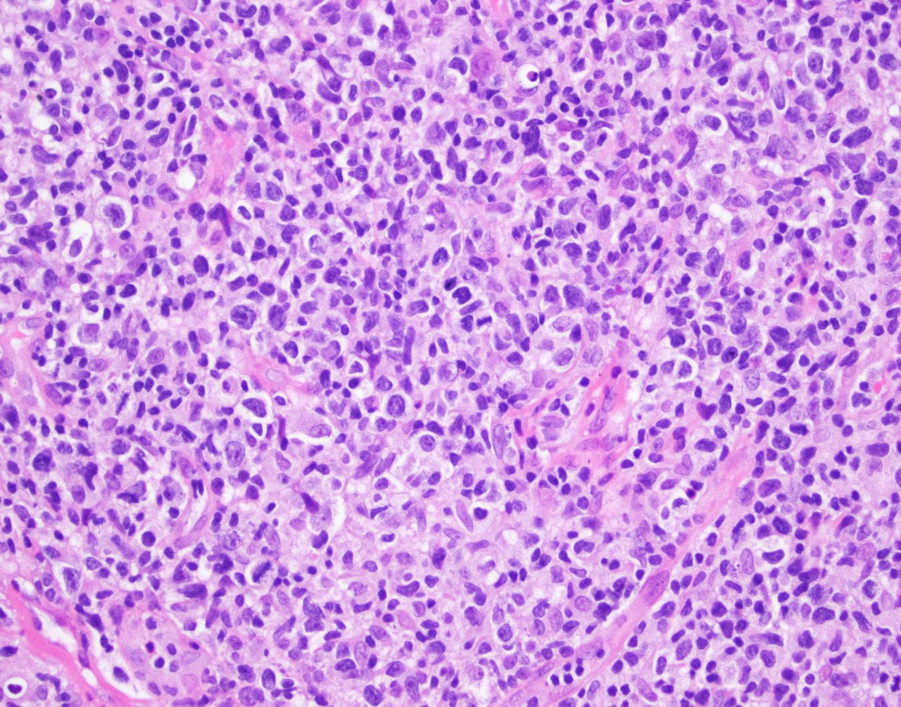
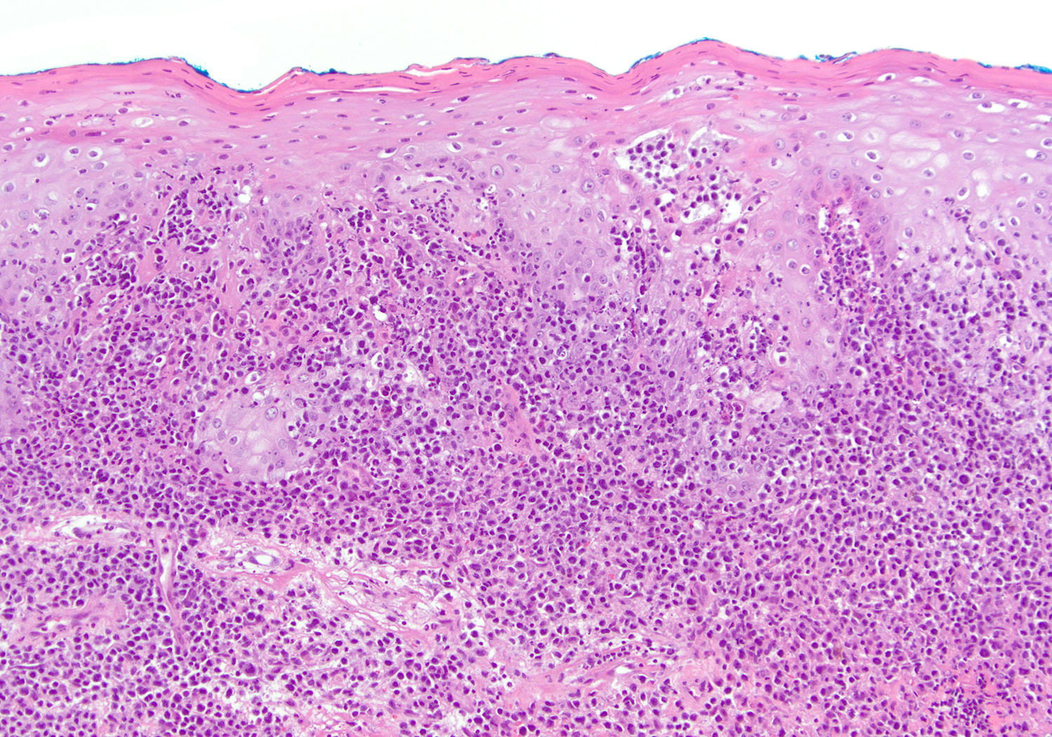
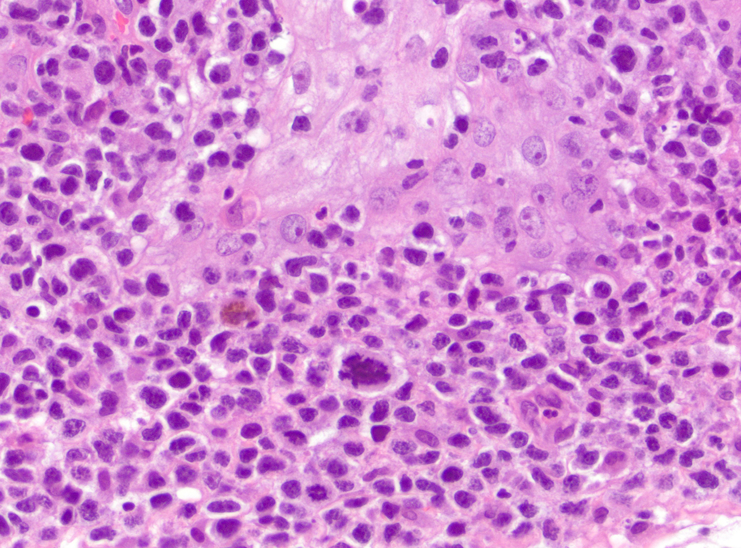
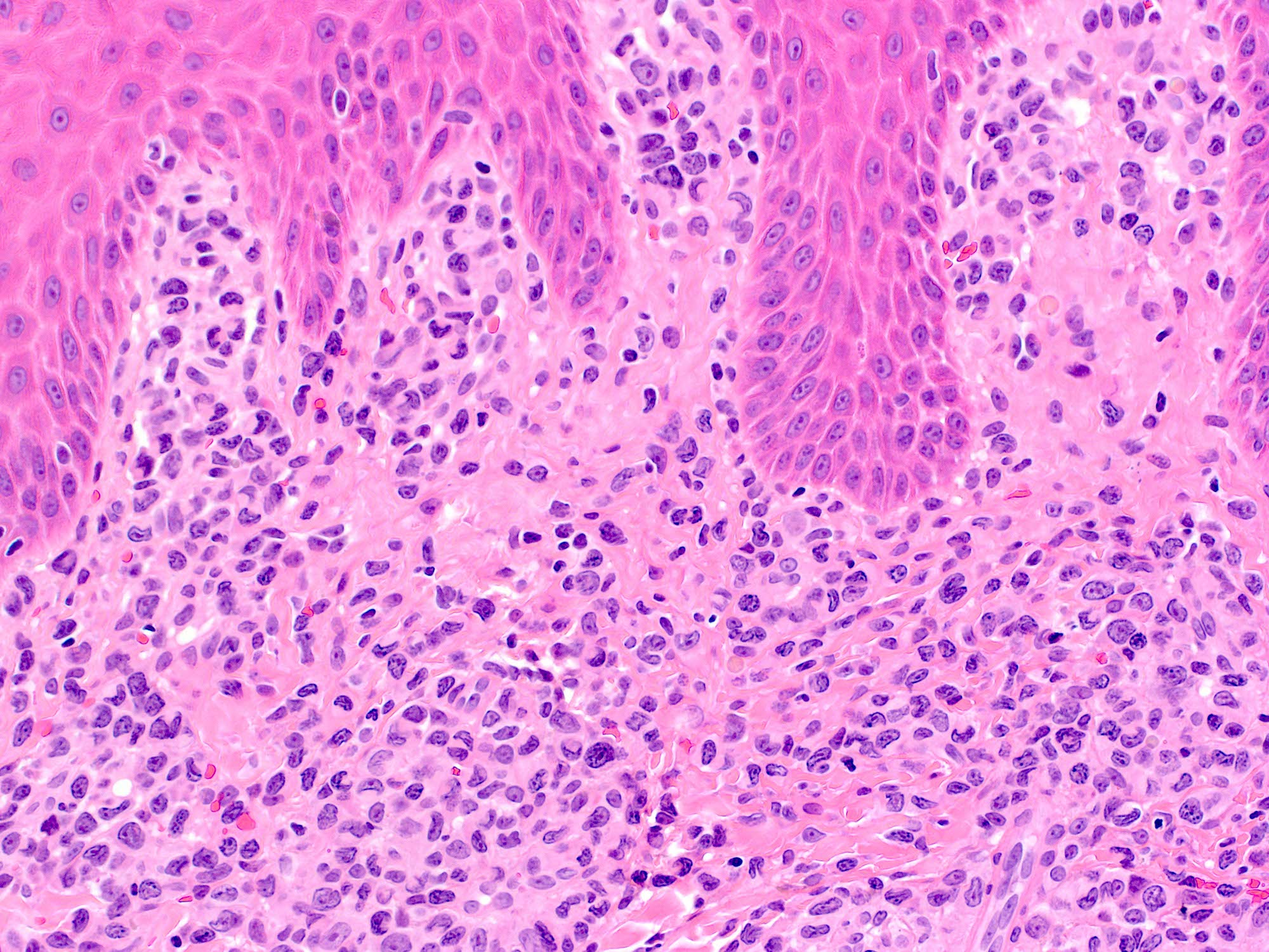
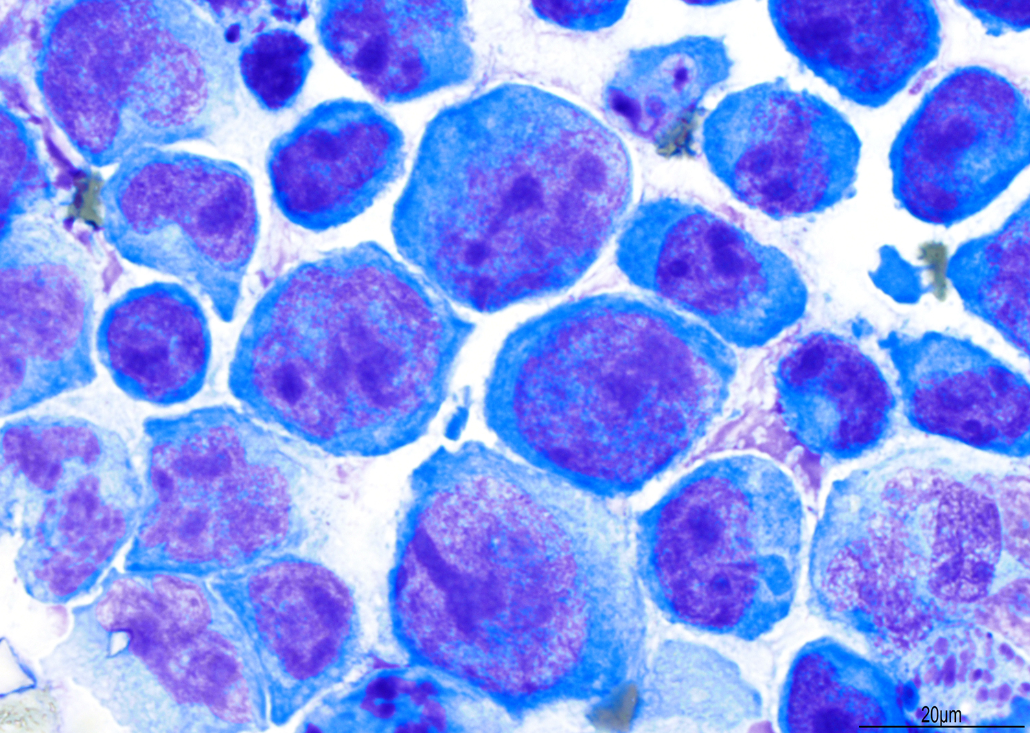
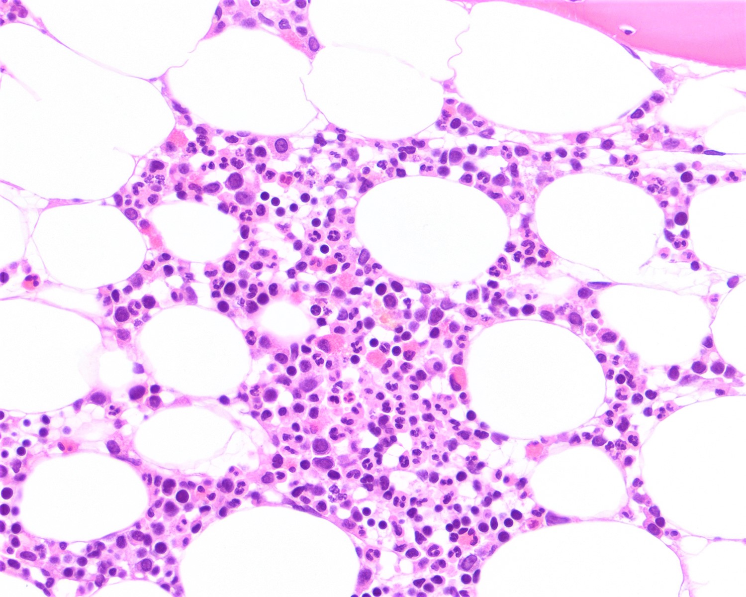
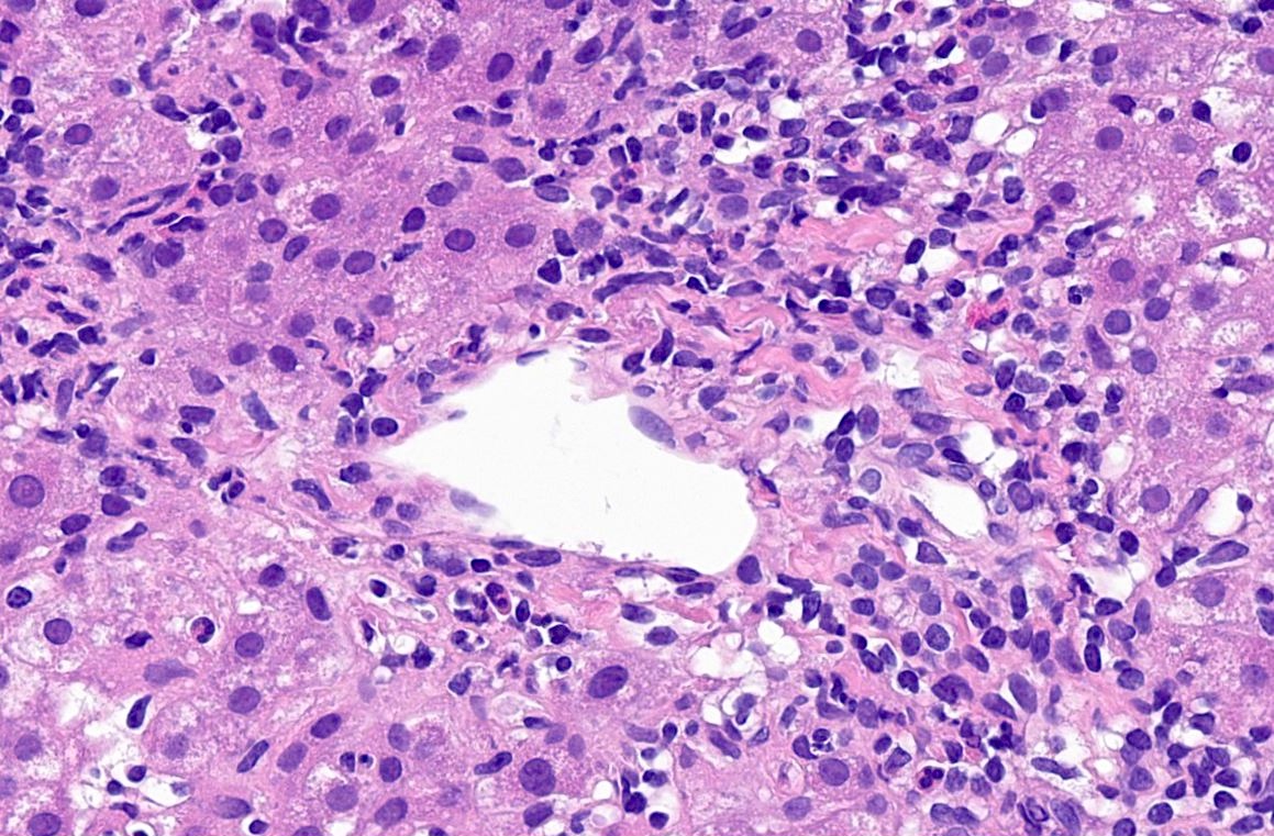
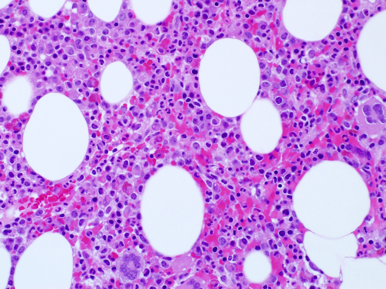
Large lymphoid tumor cells
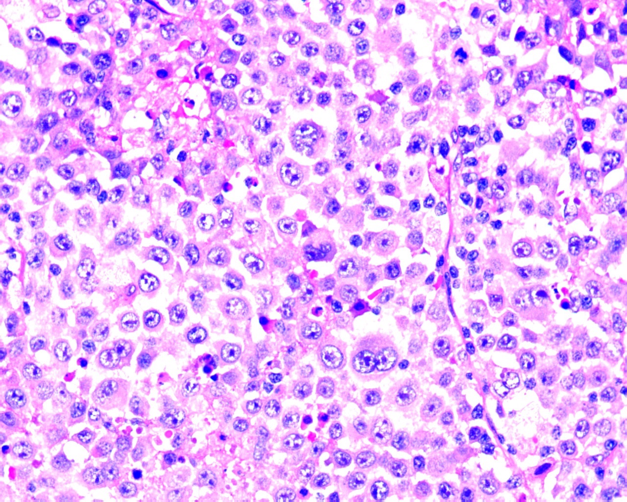
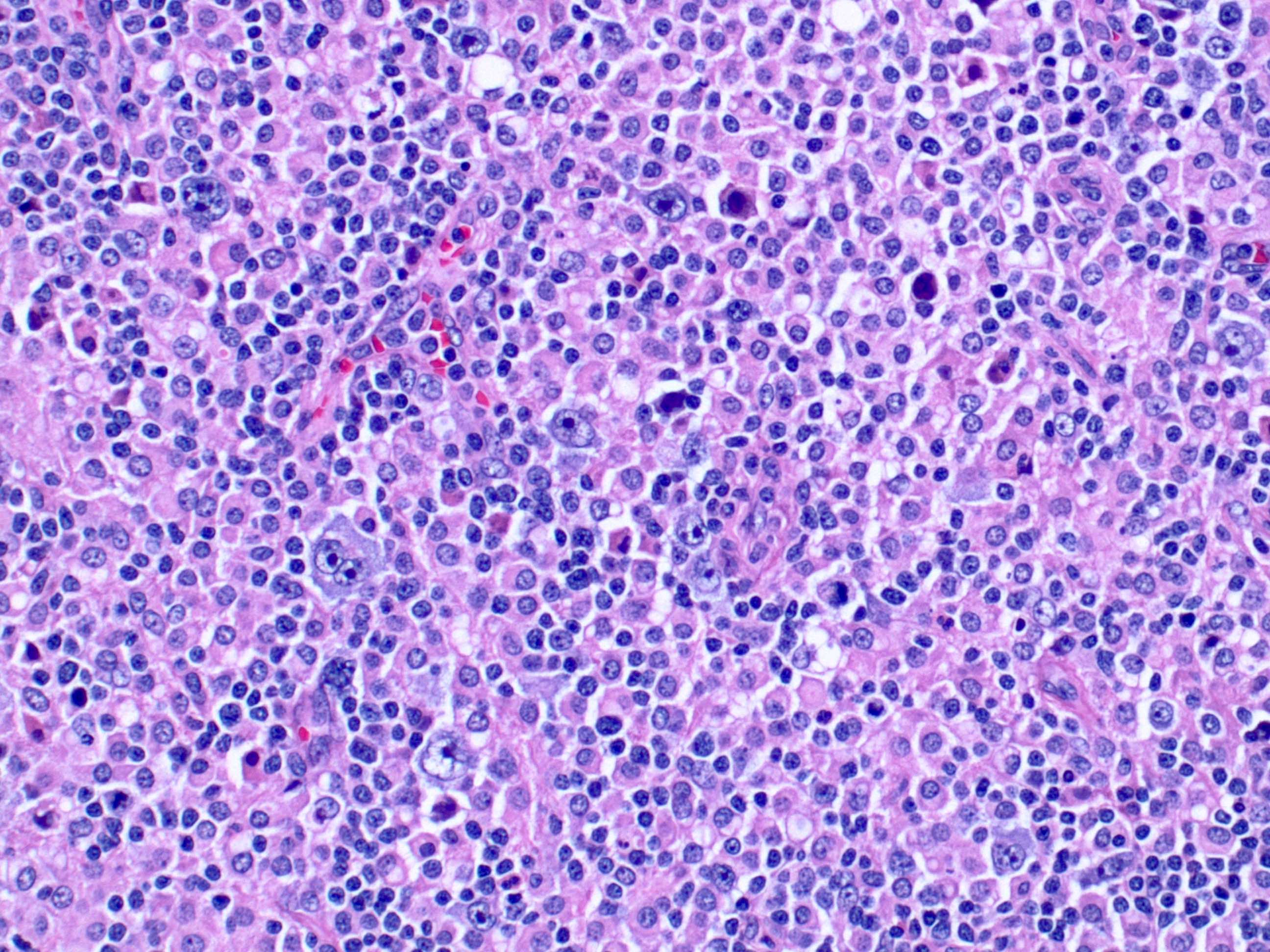
Immunoblast-like
tumor cells and
few plasmablasts
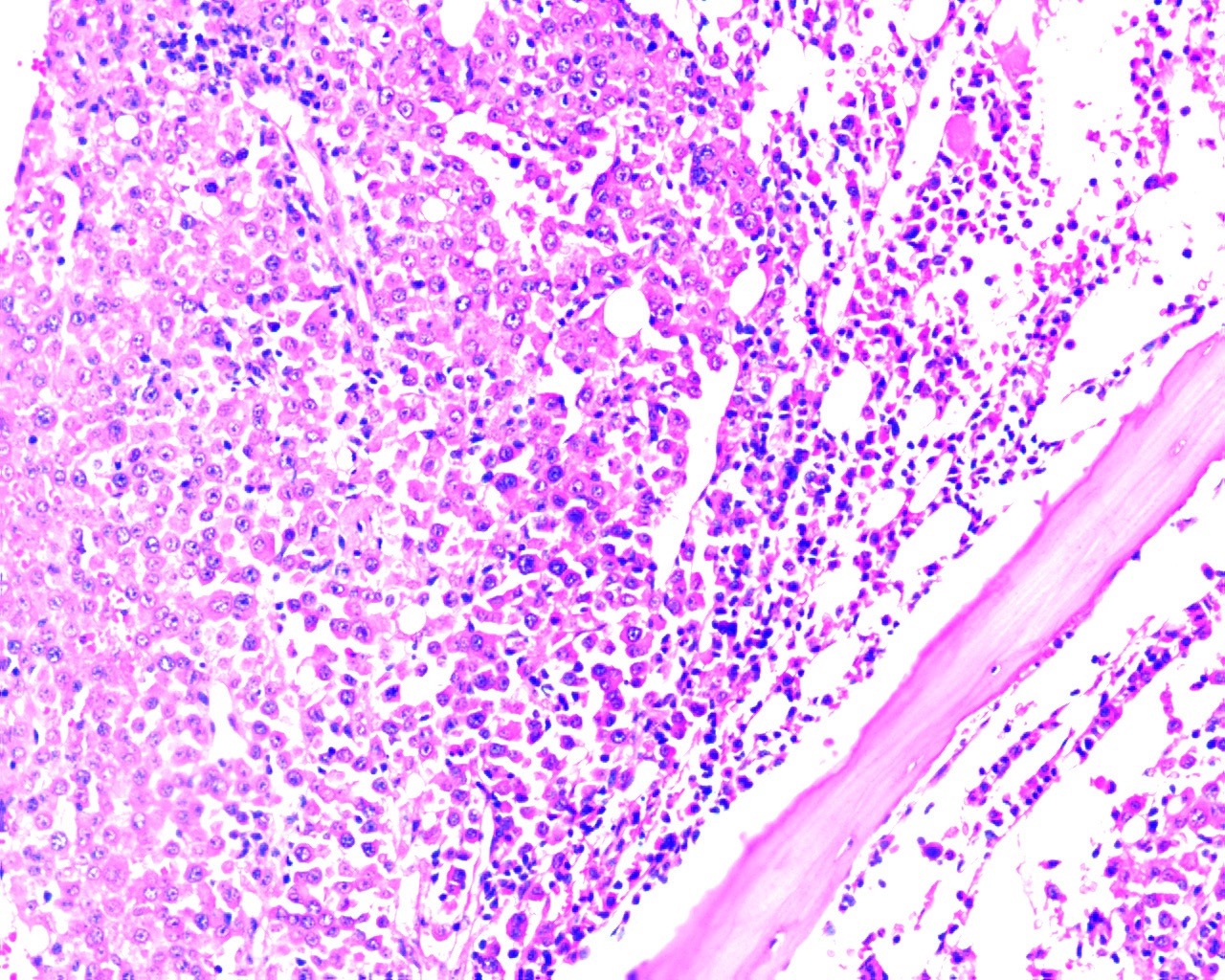
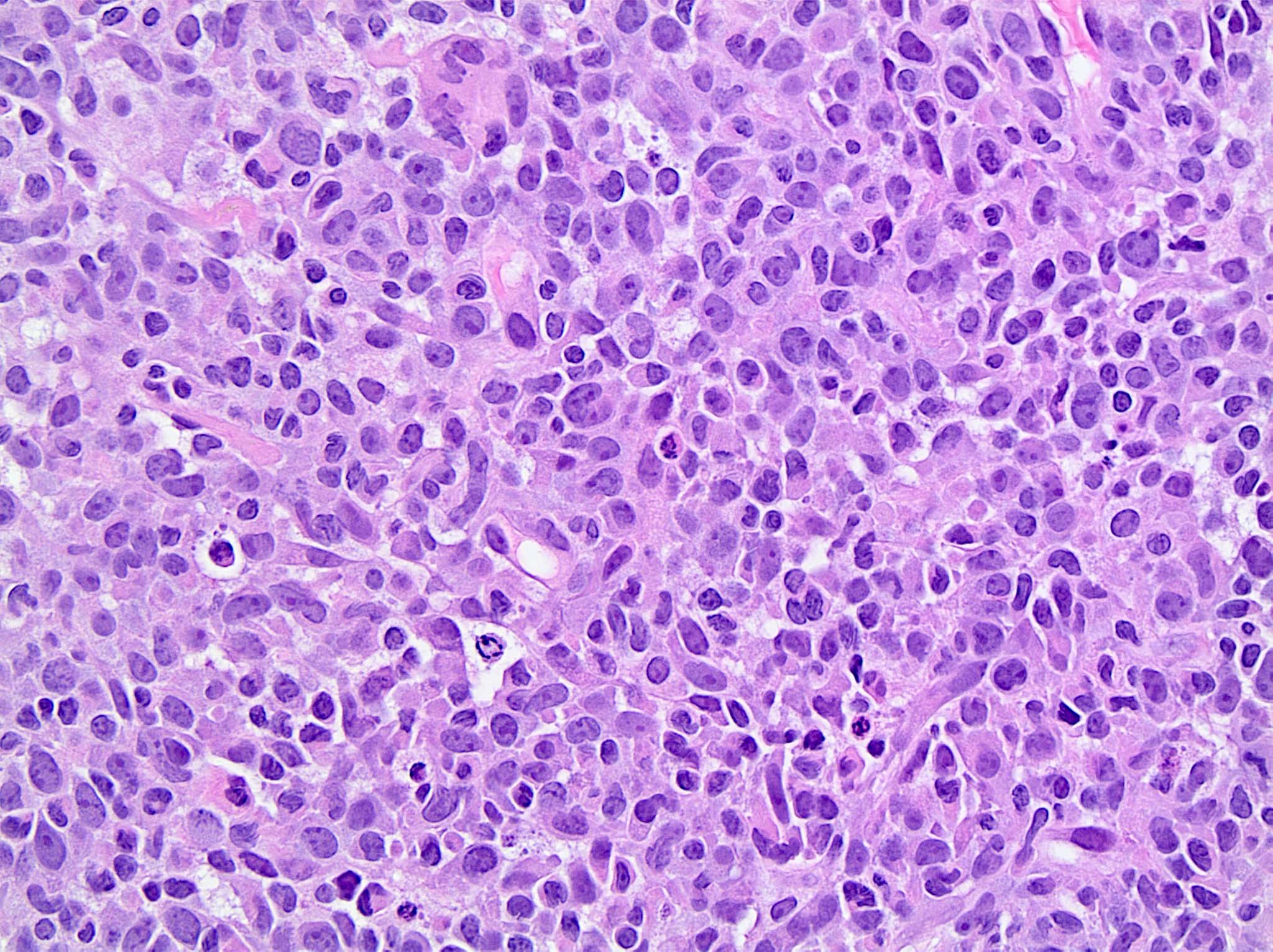
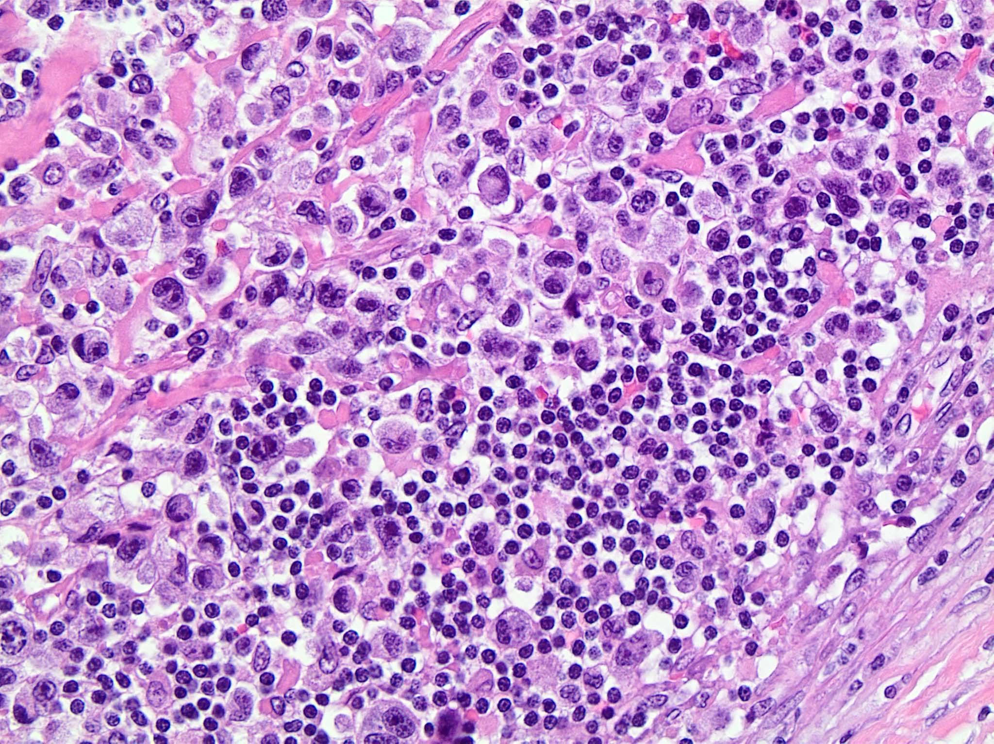
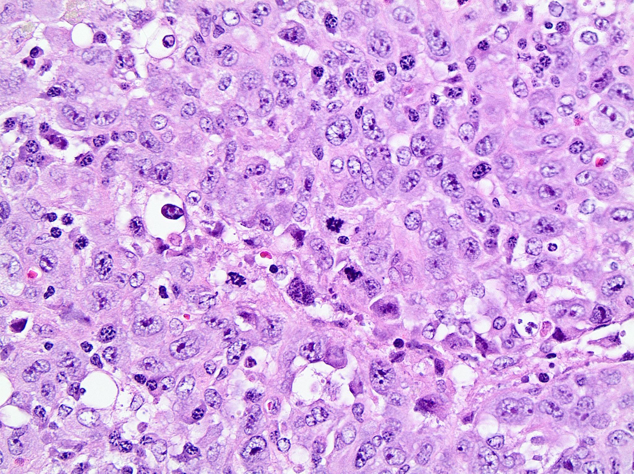
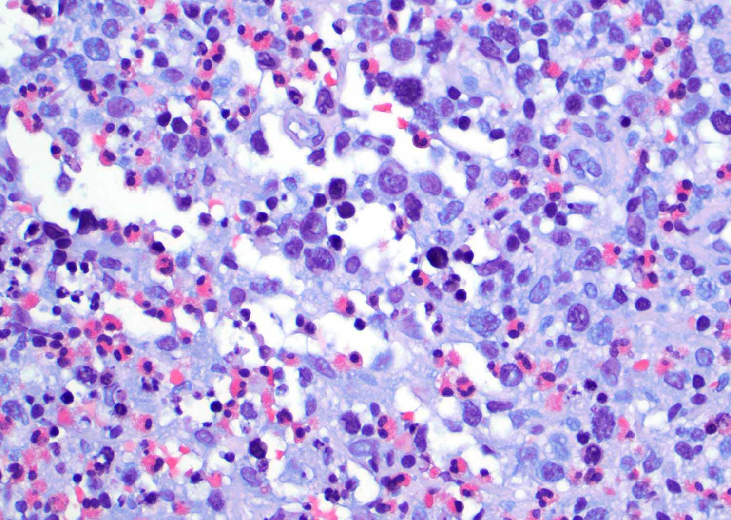
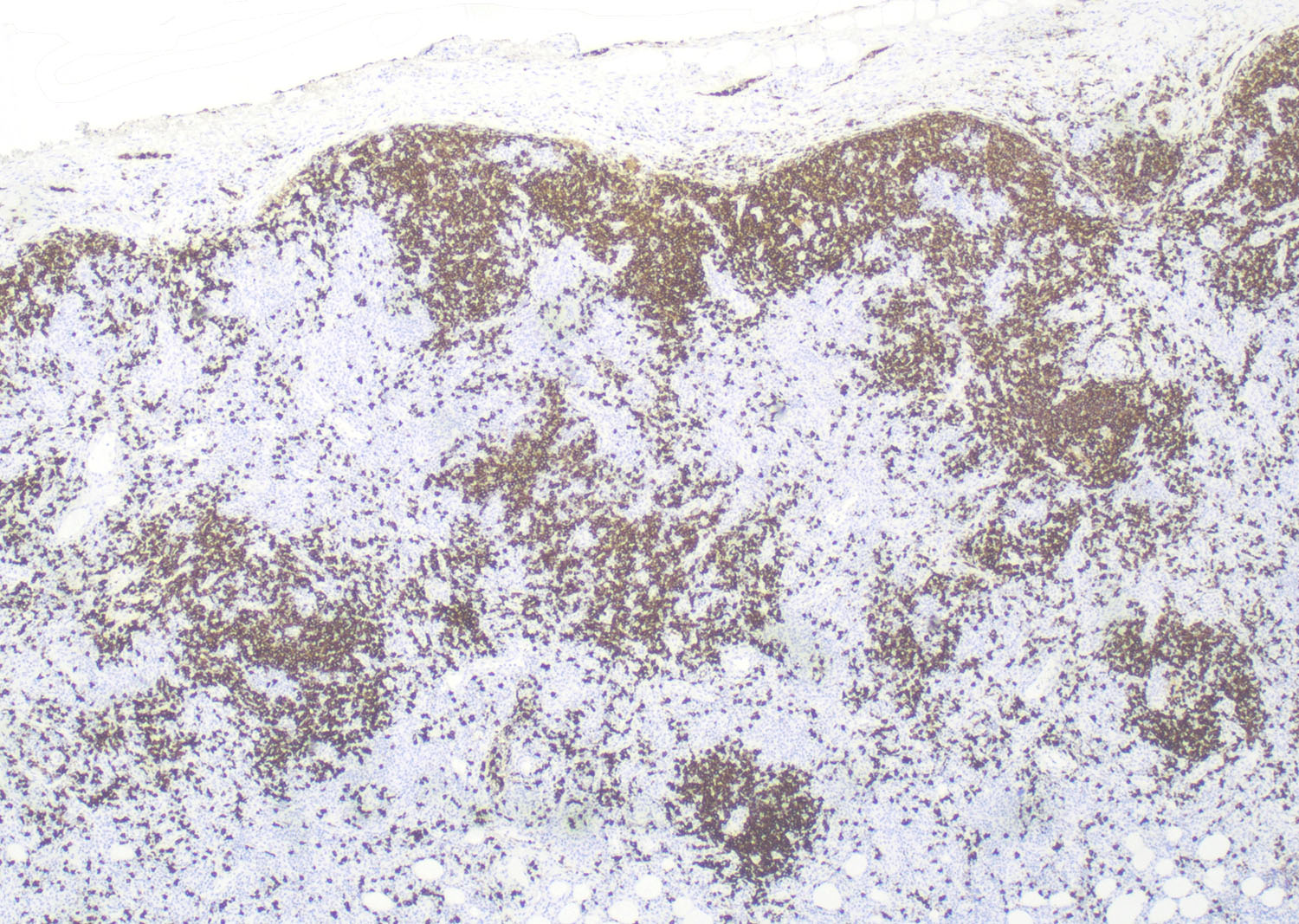
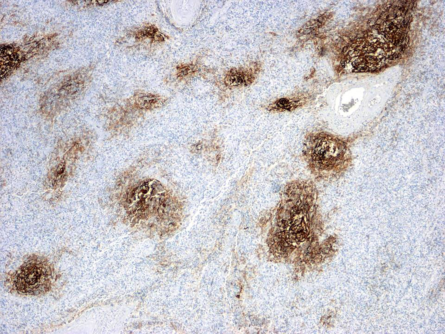
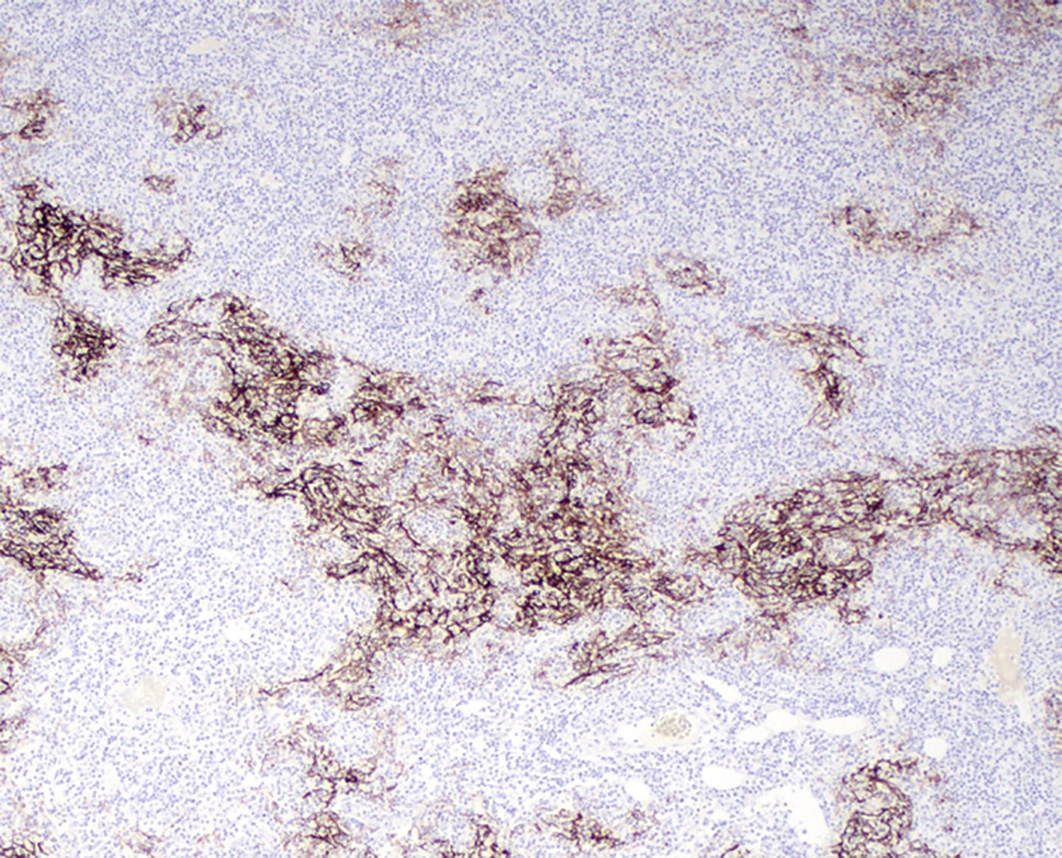
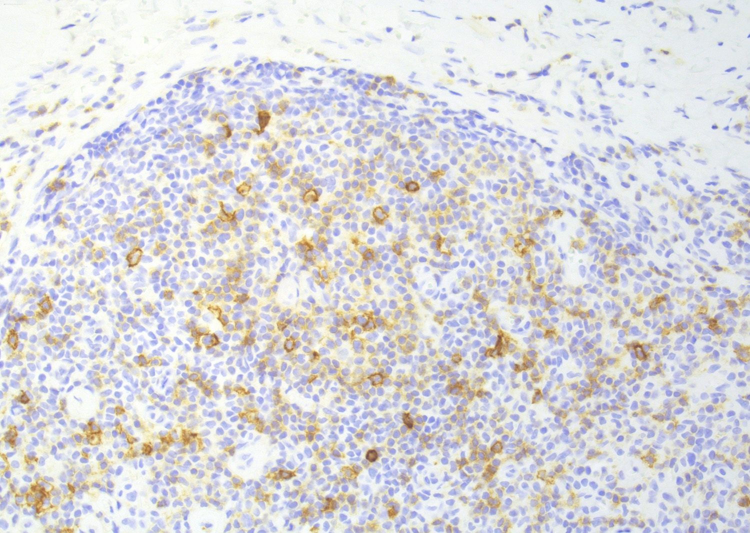
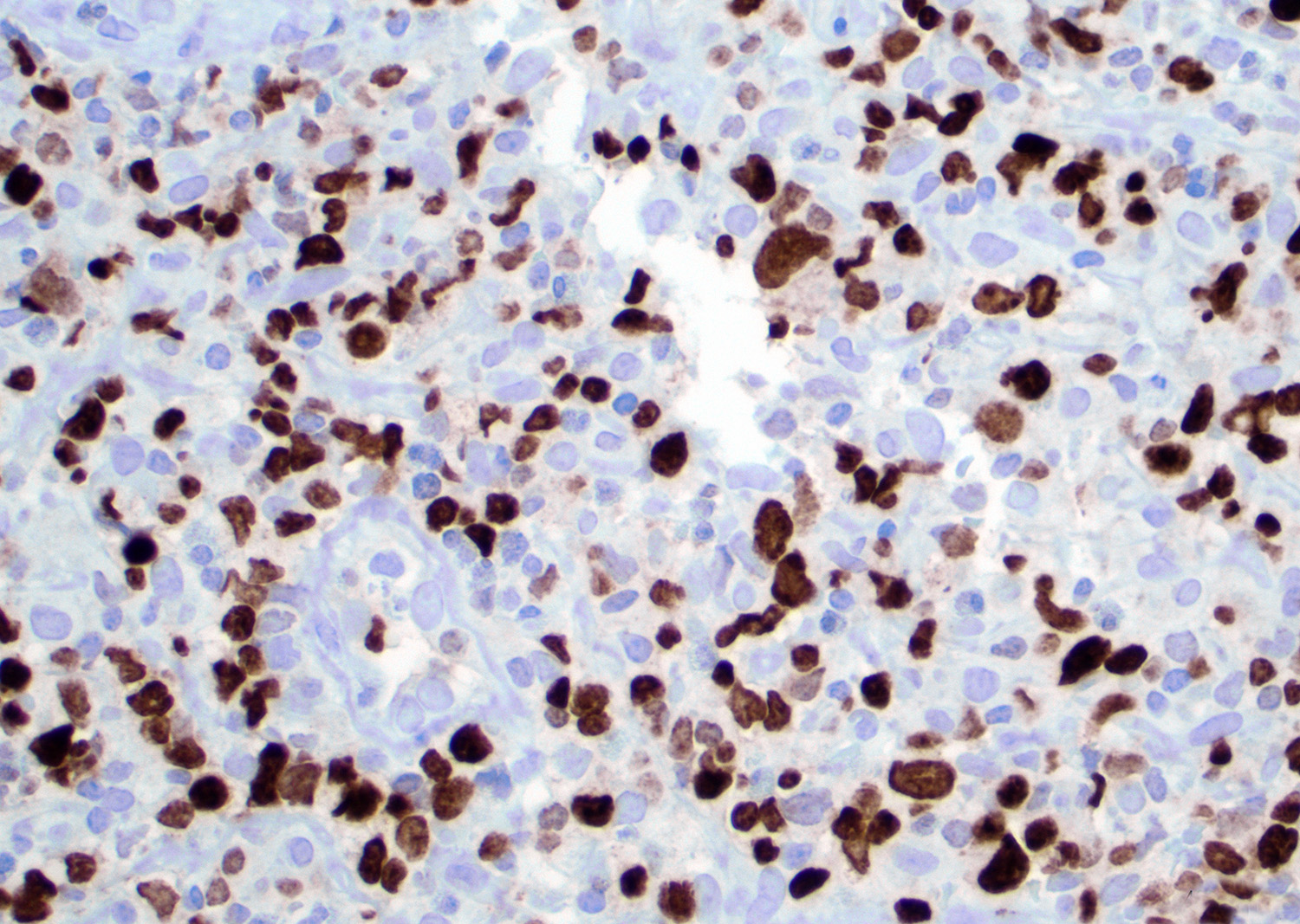
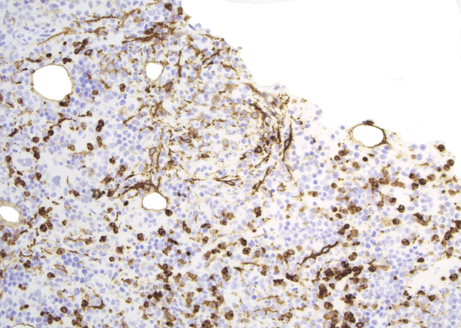
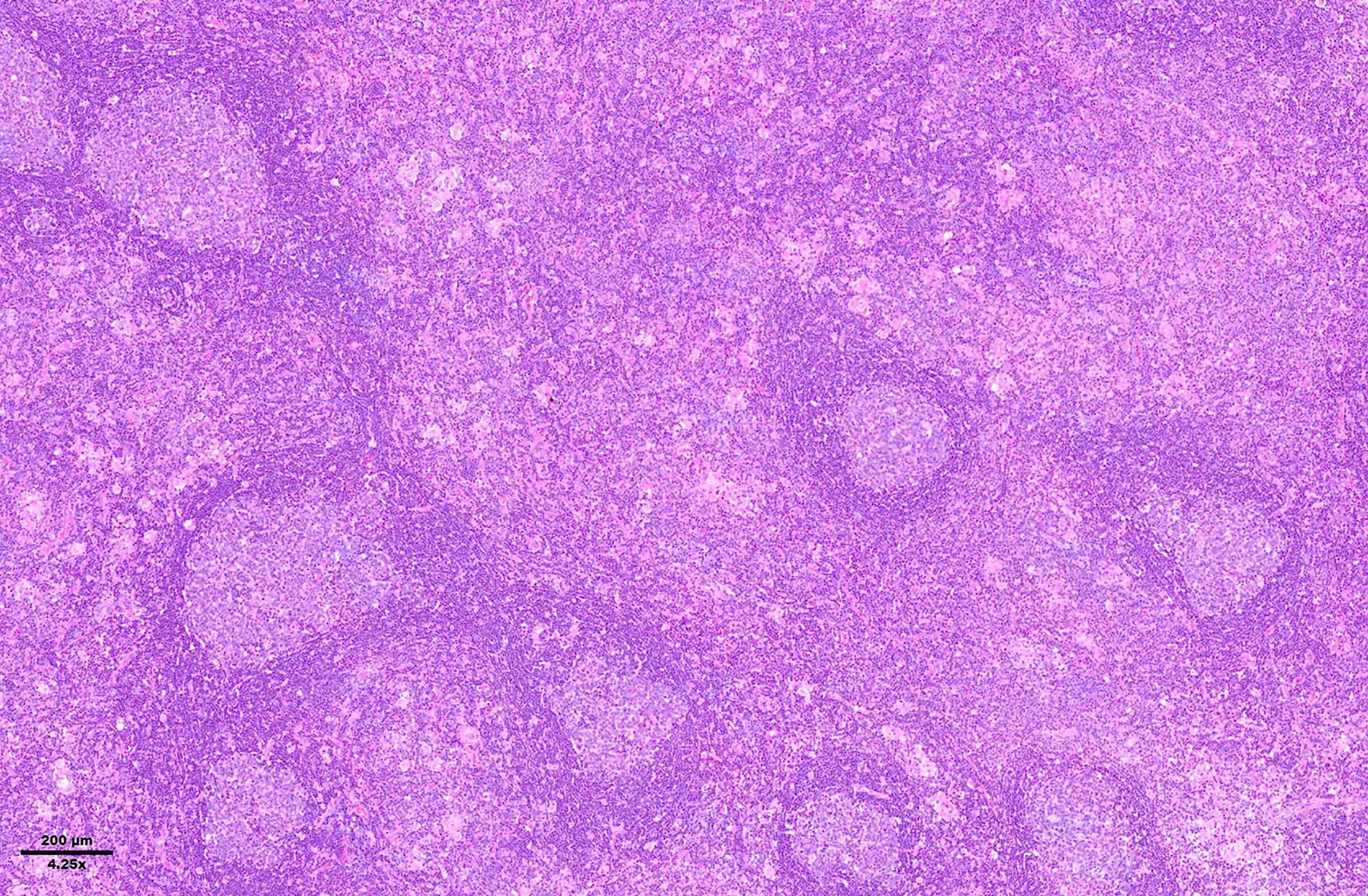
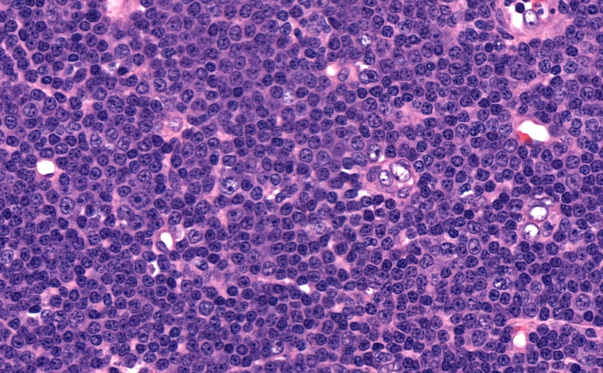
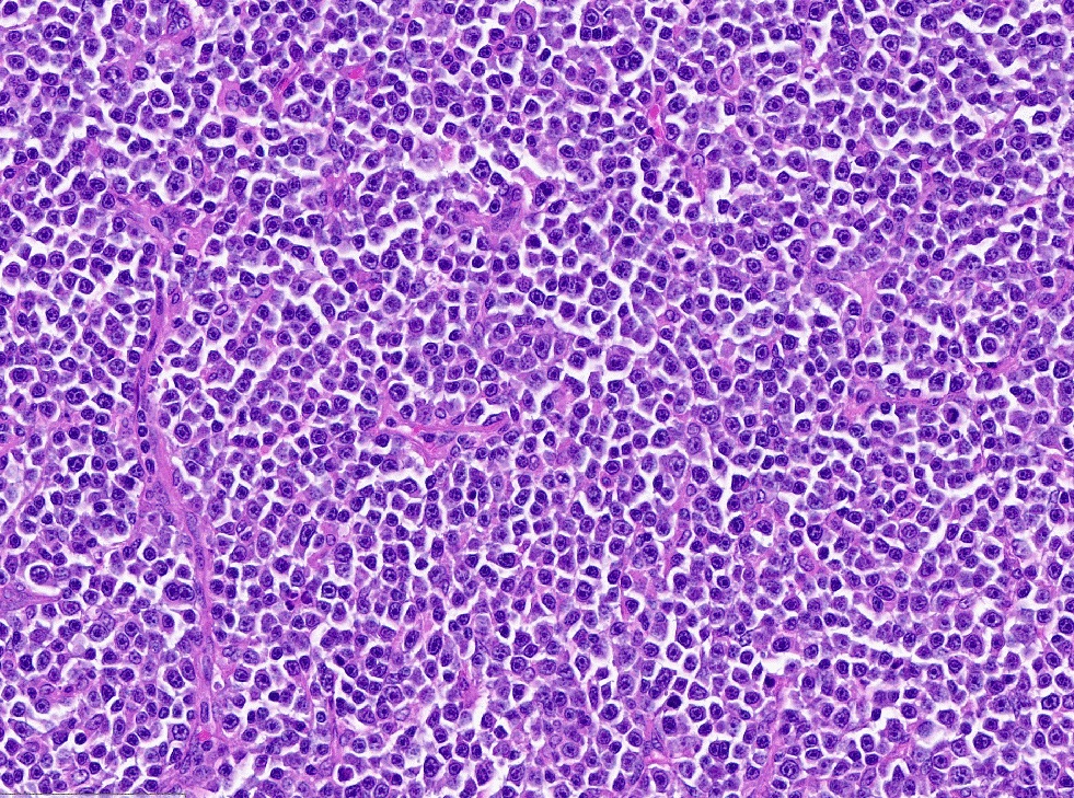
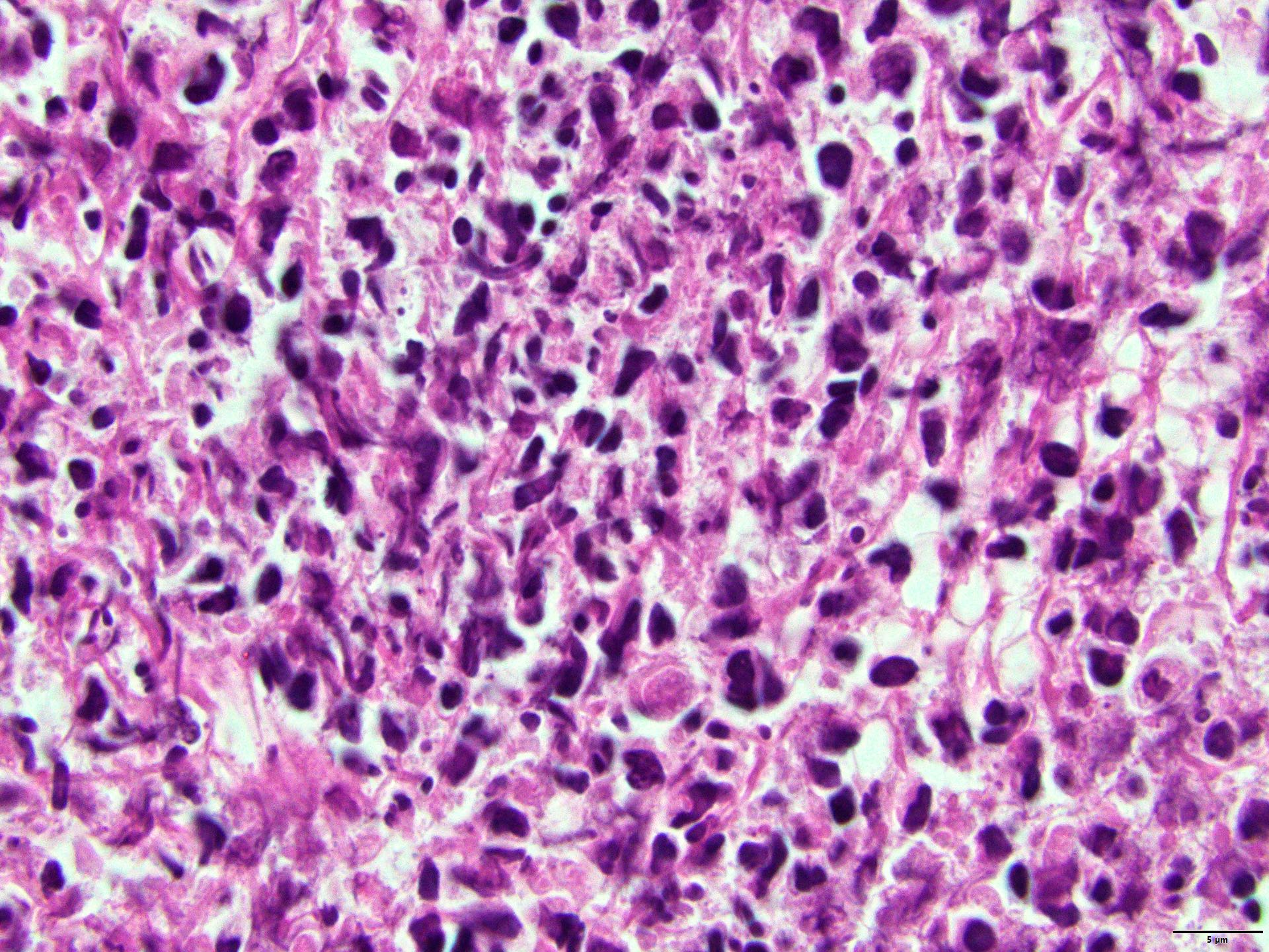
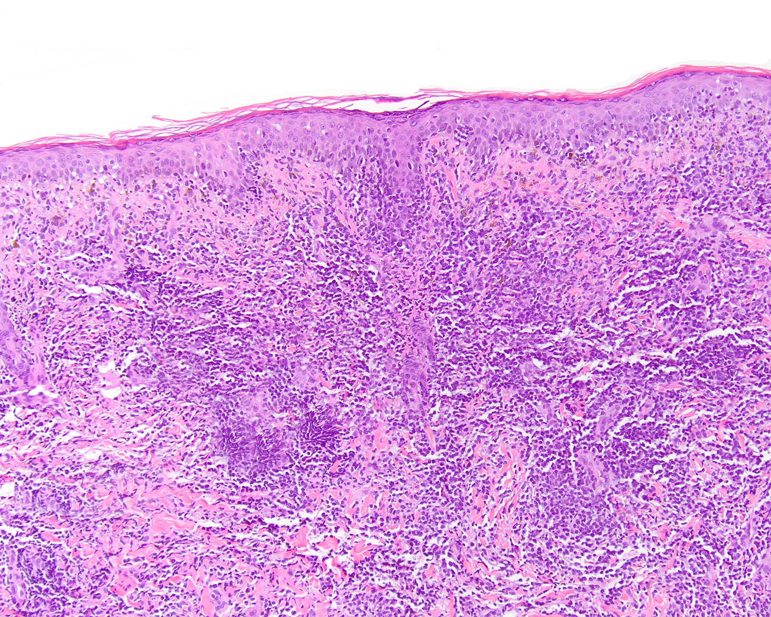
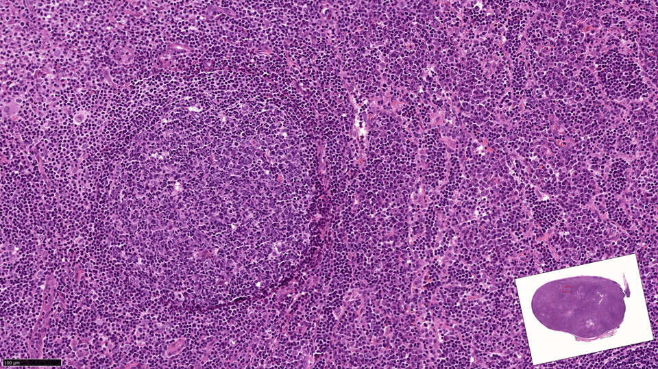
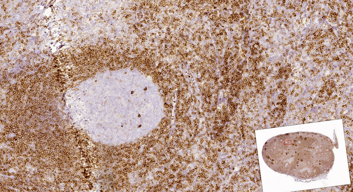
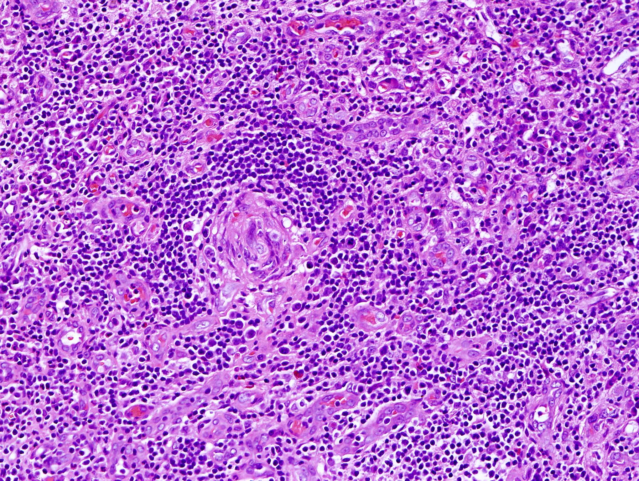
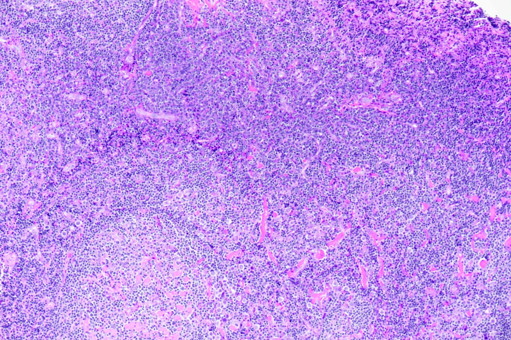
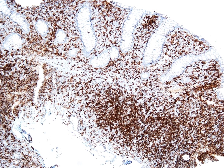
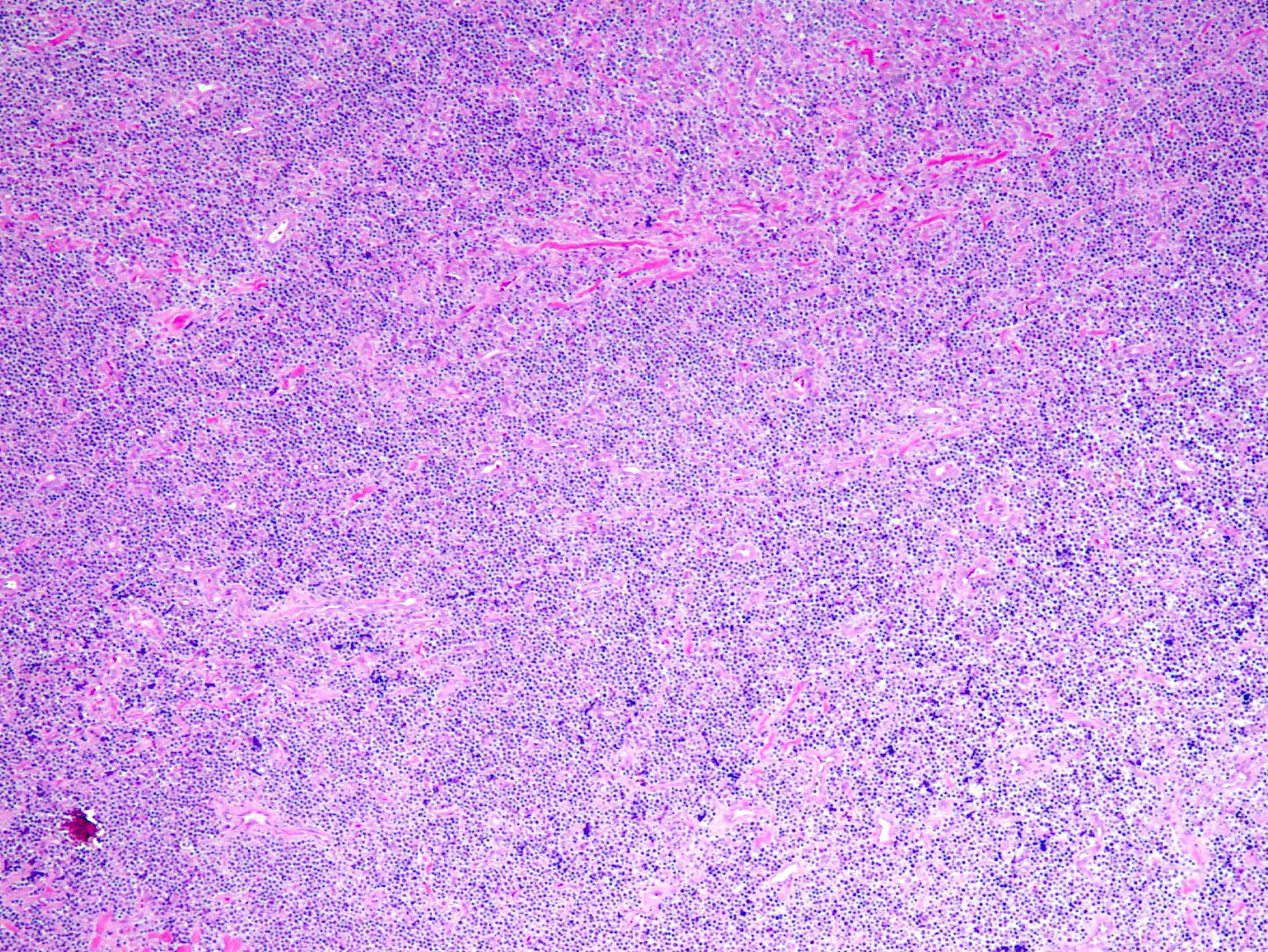
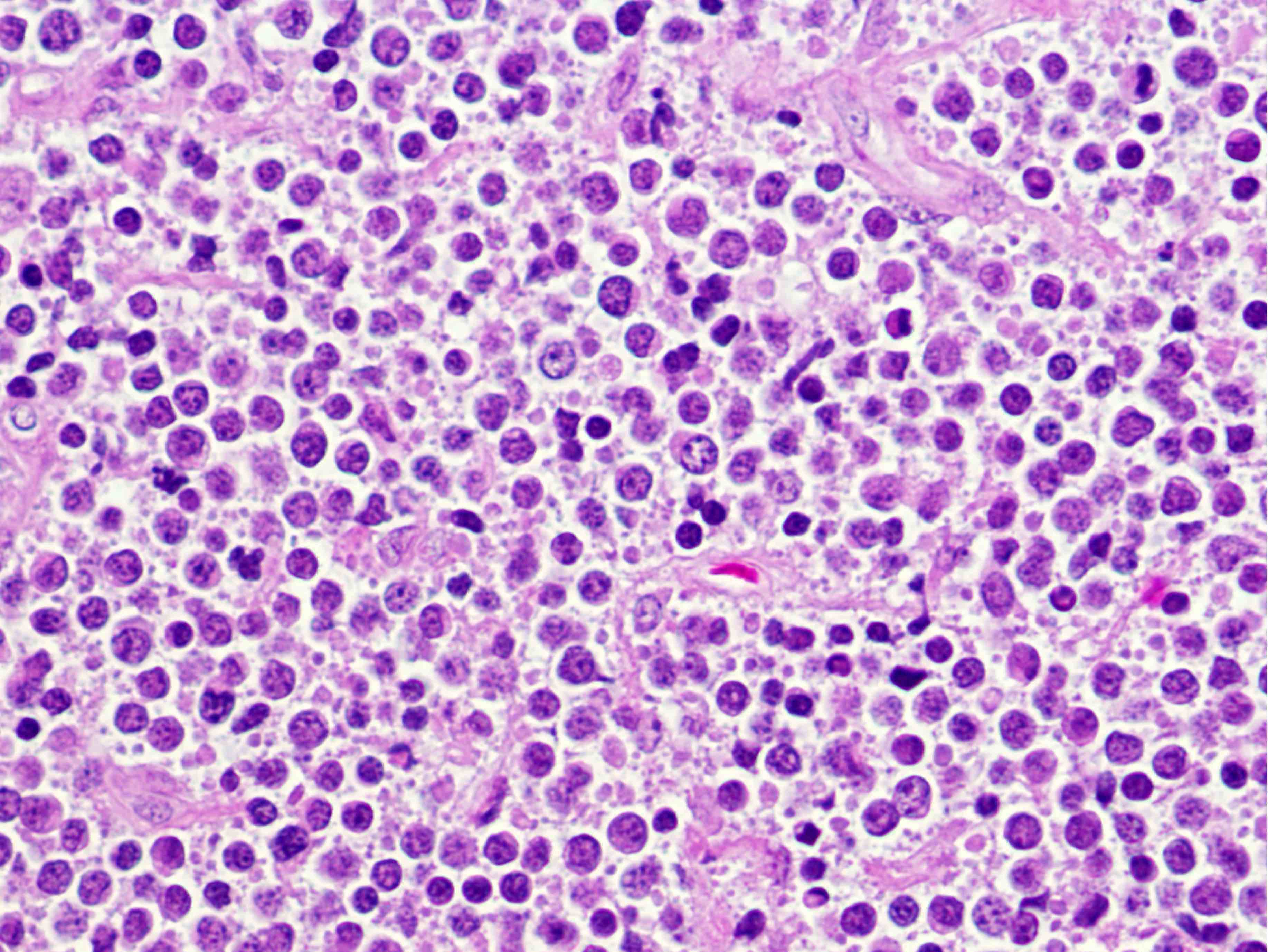
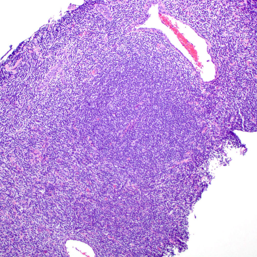
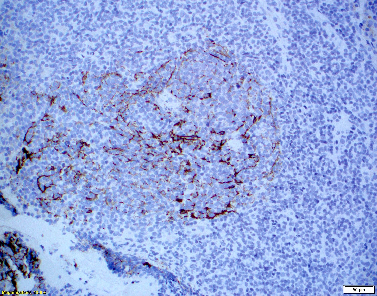
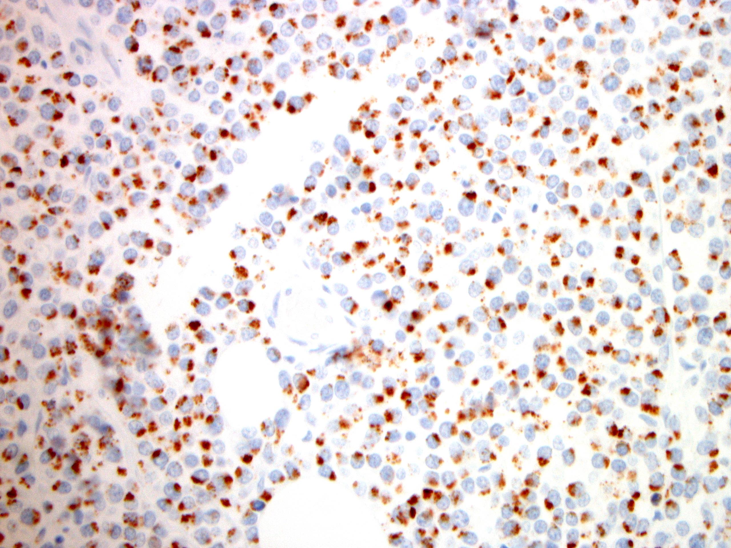
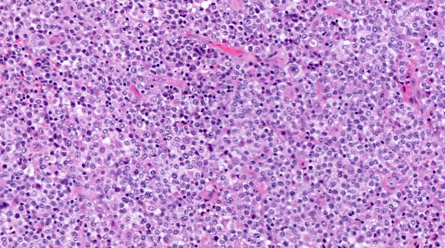
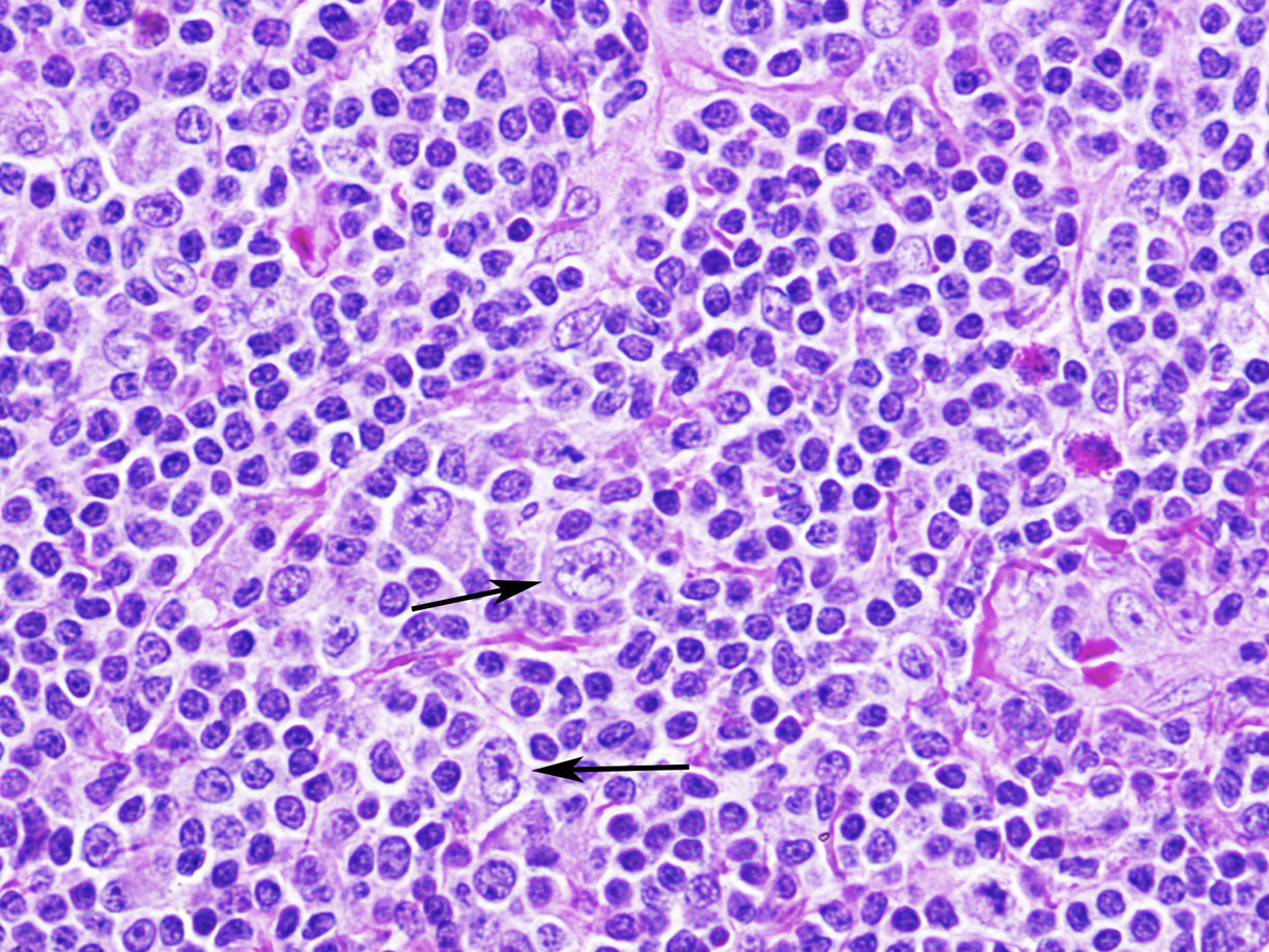
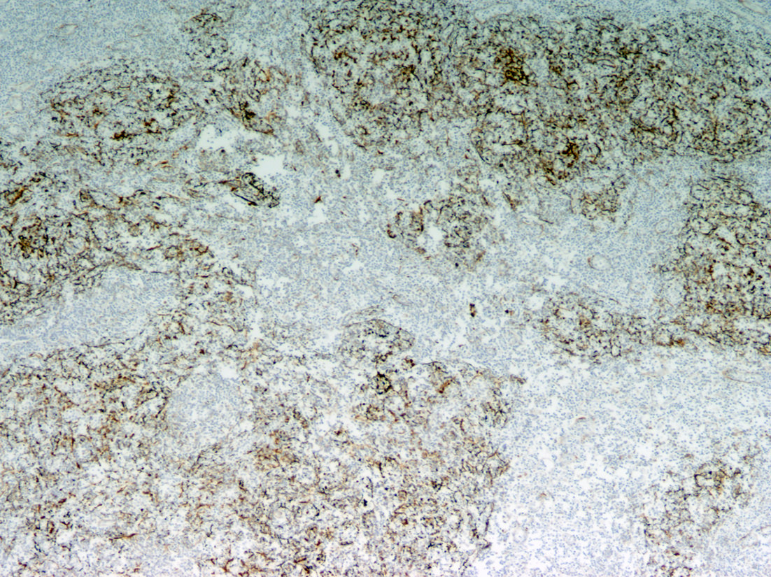
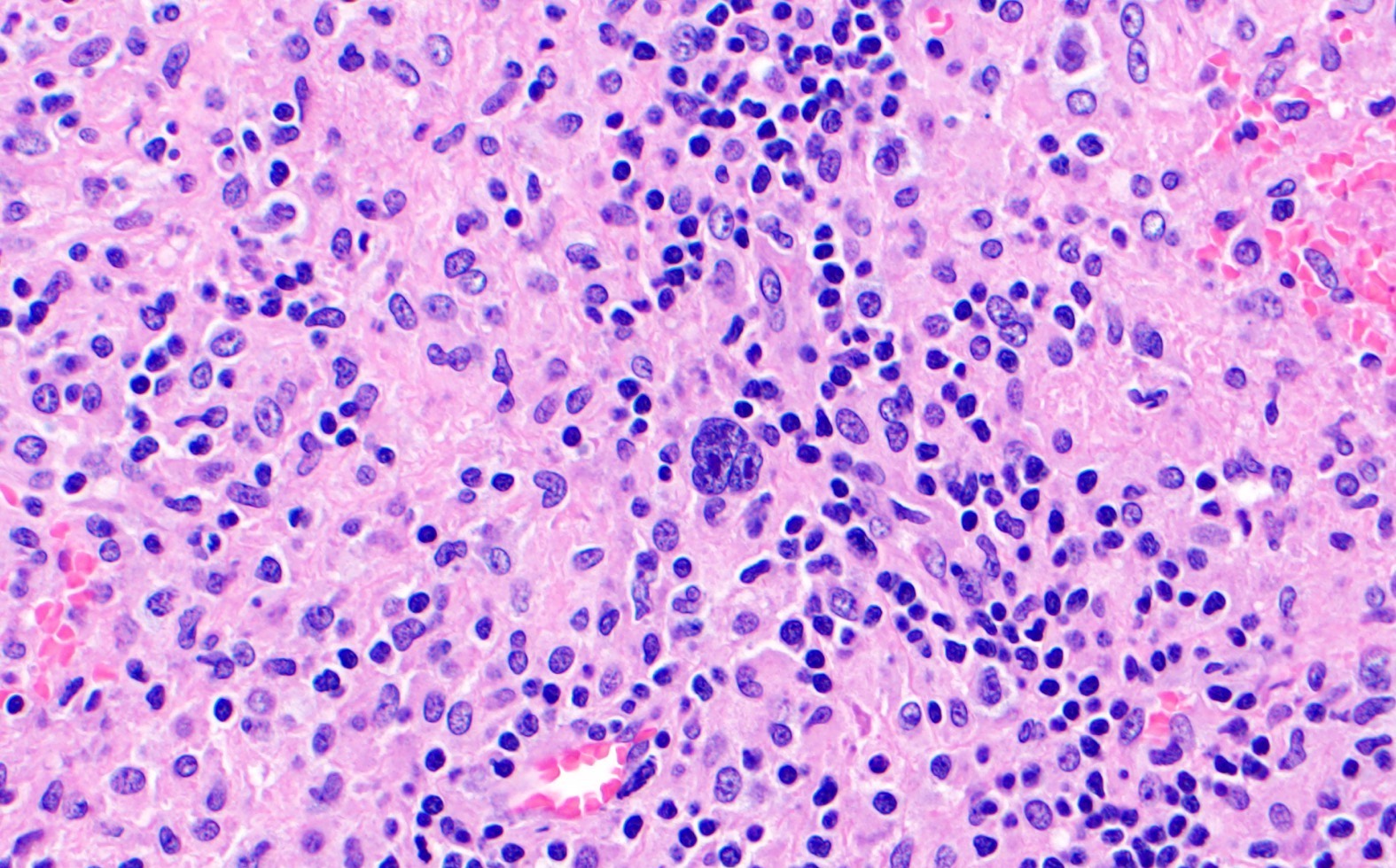
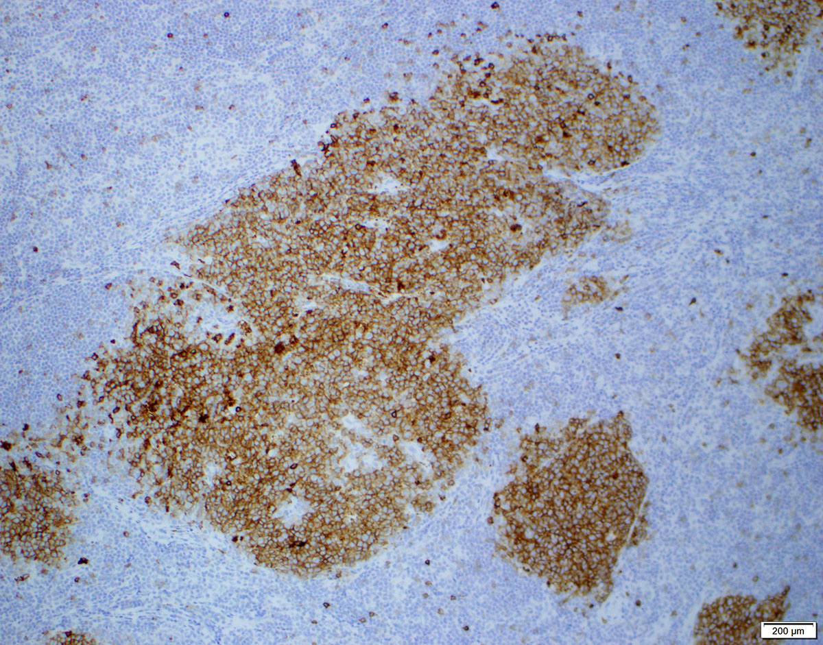
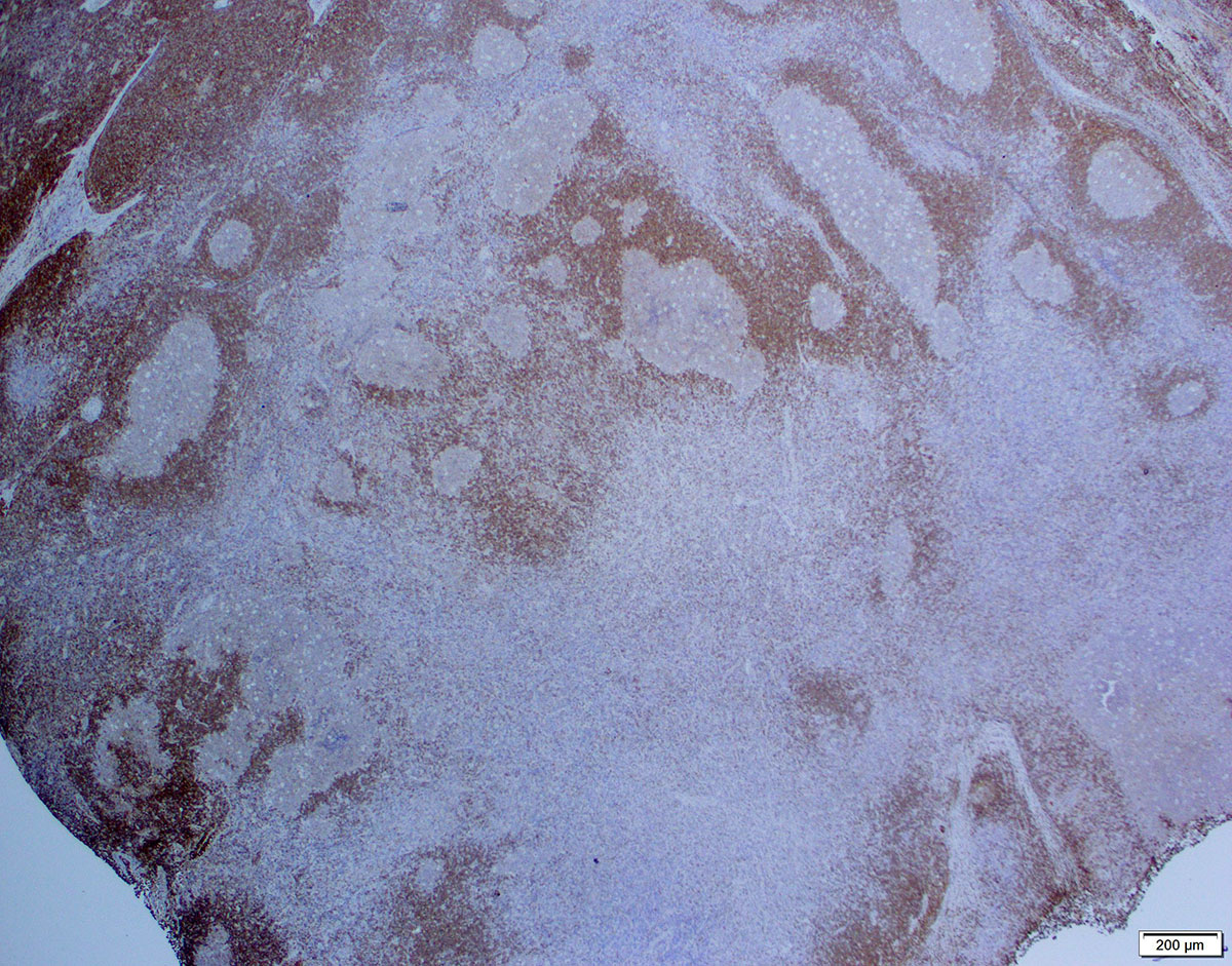
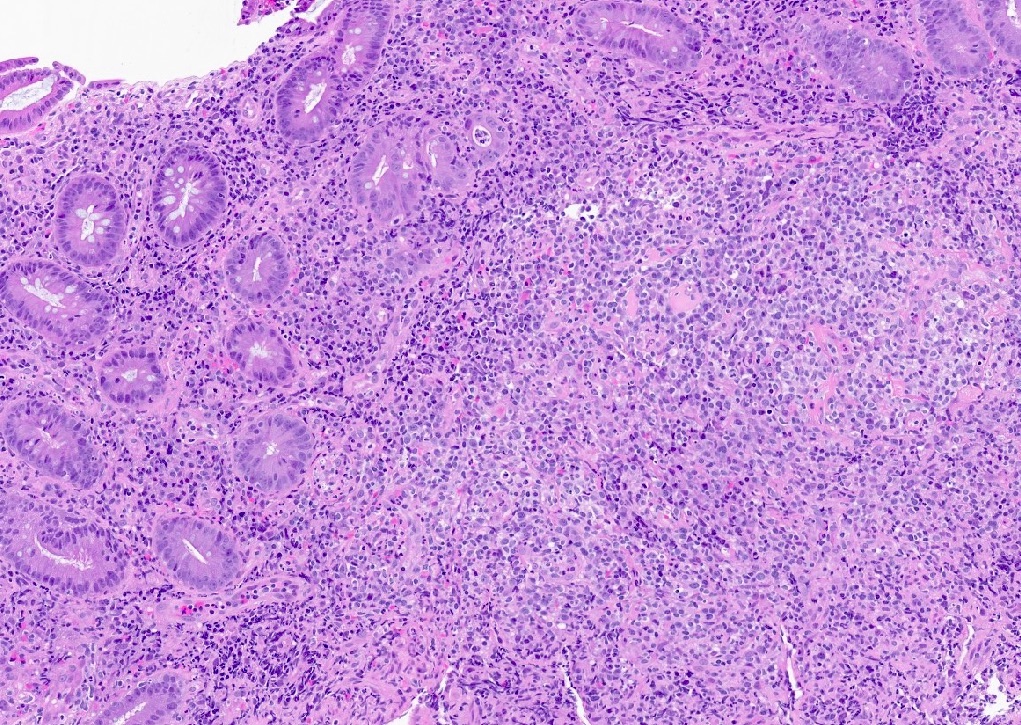
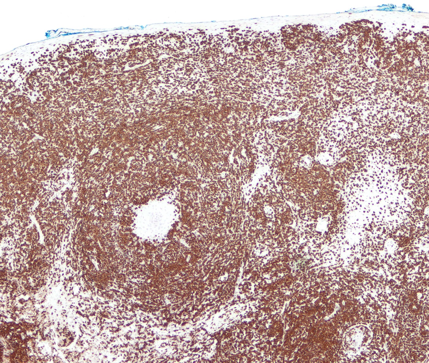
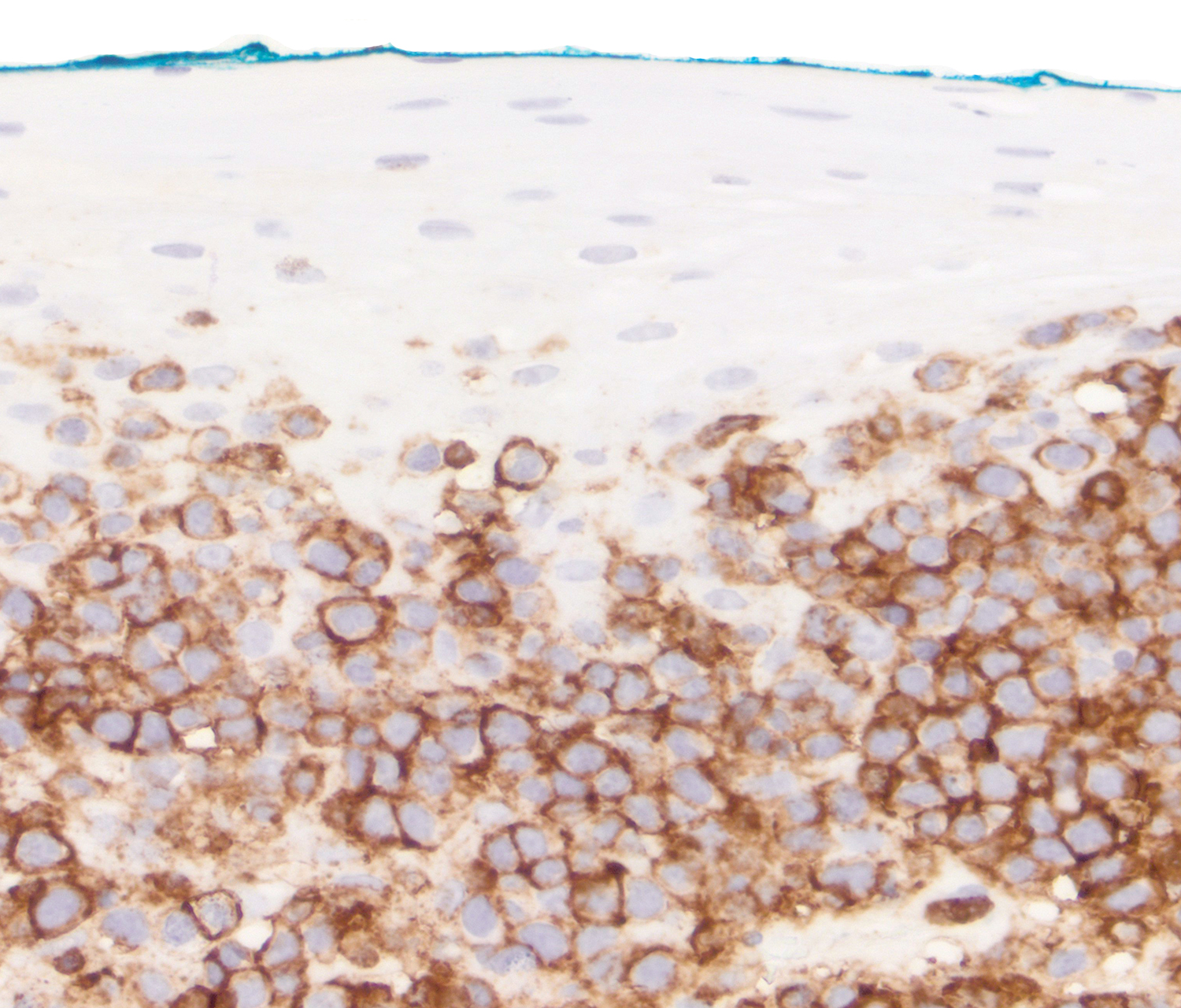
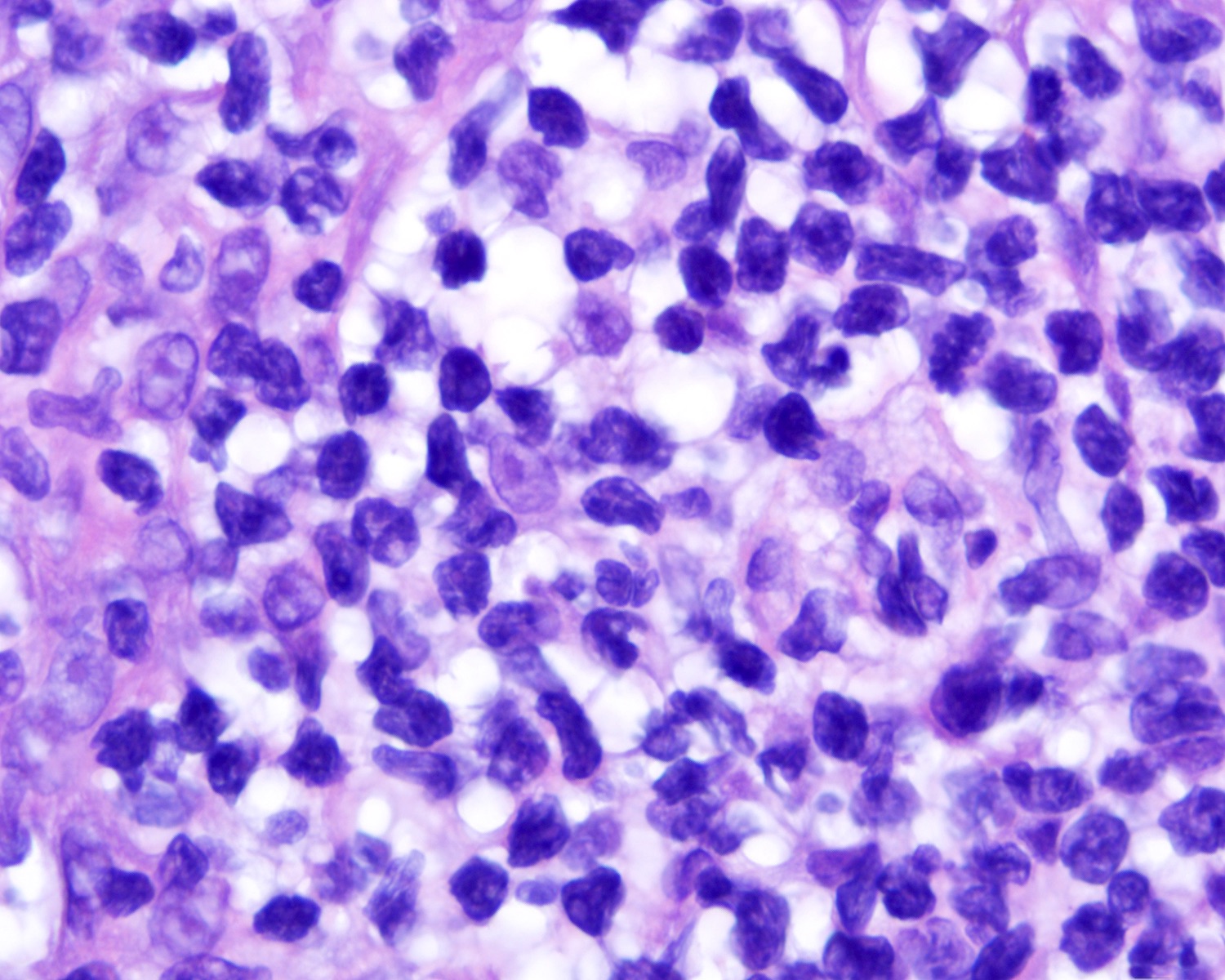
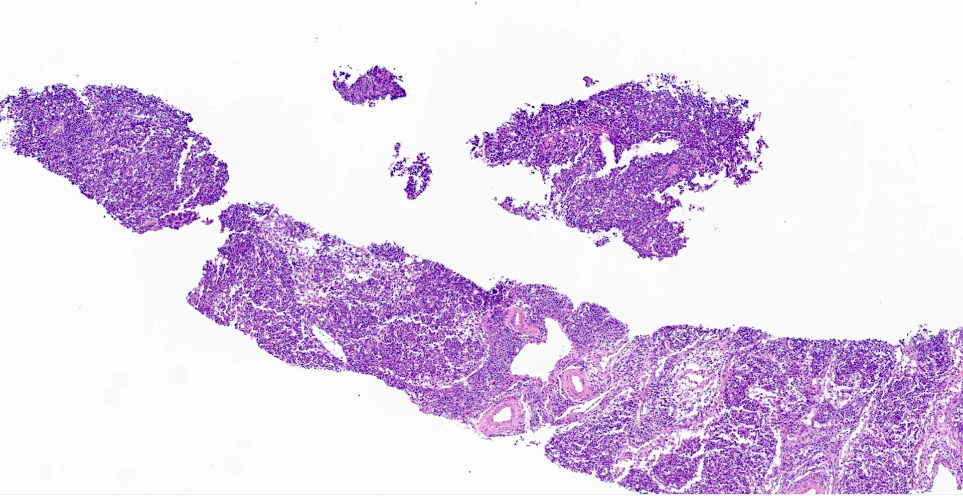
Interfollicular expansion by tumor cells

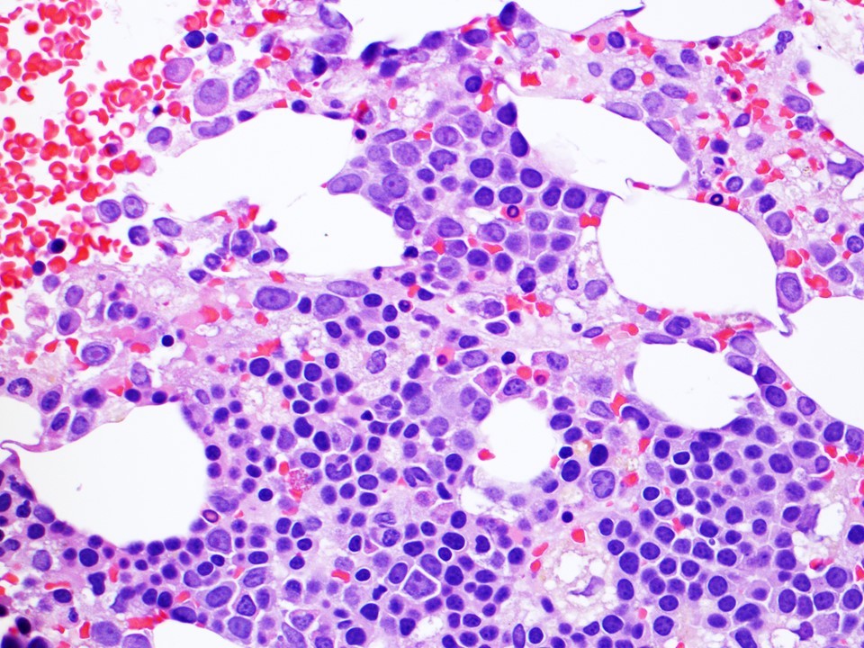
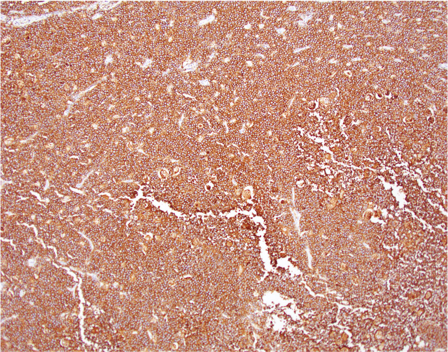
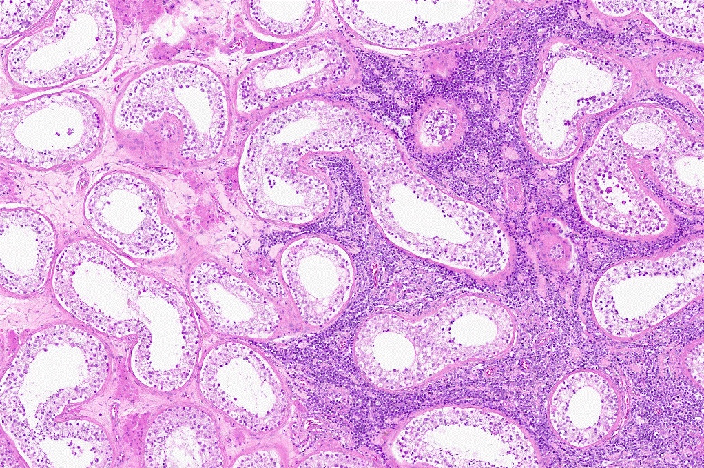
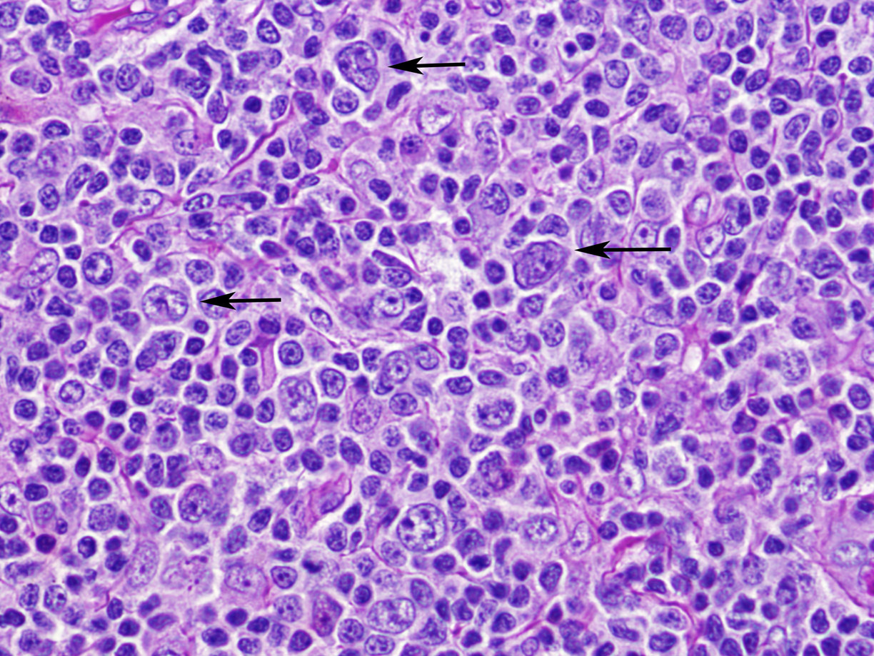
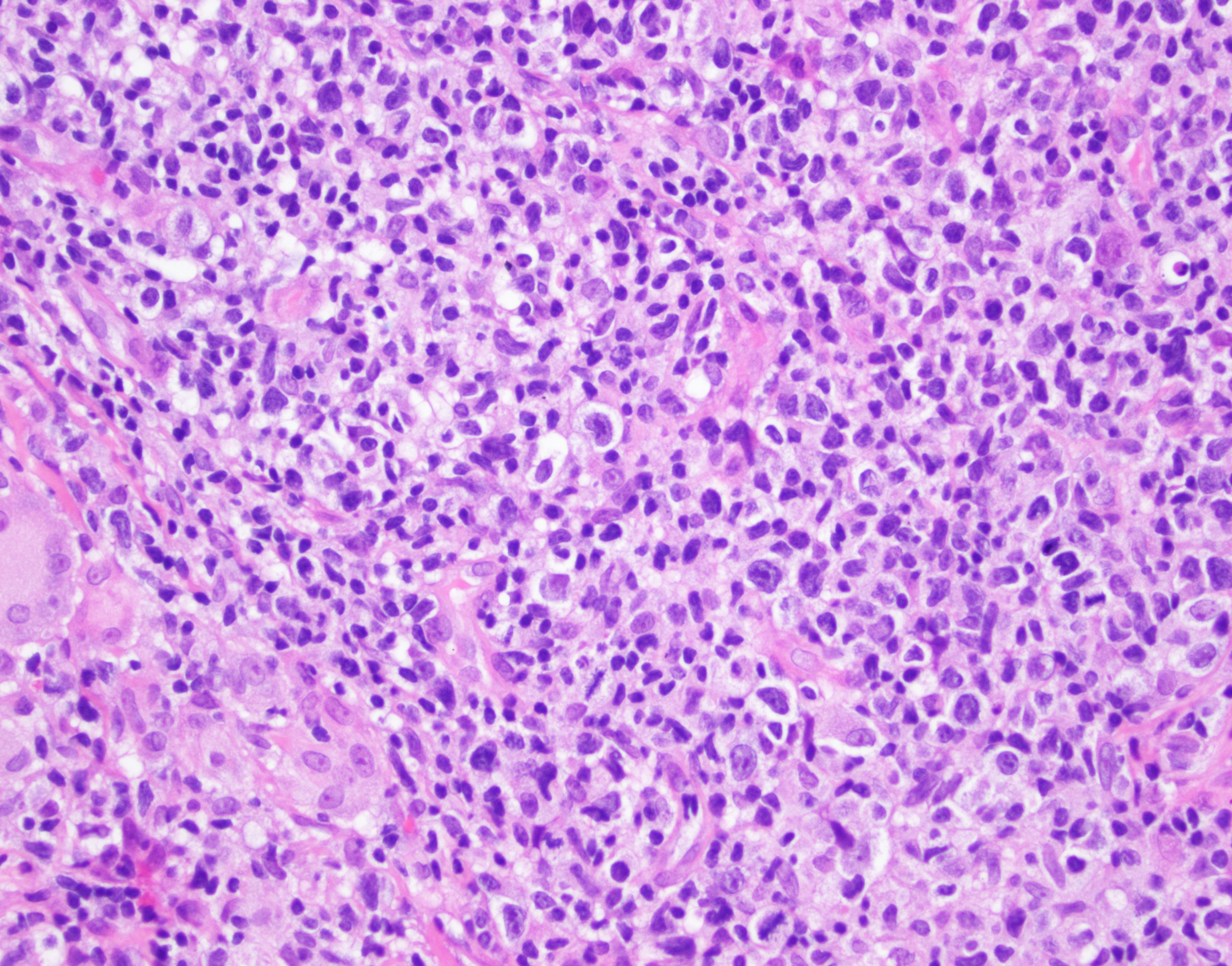
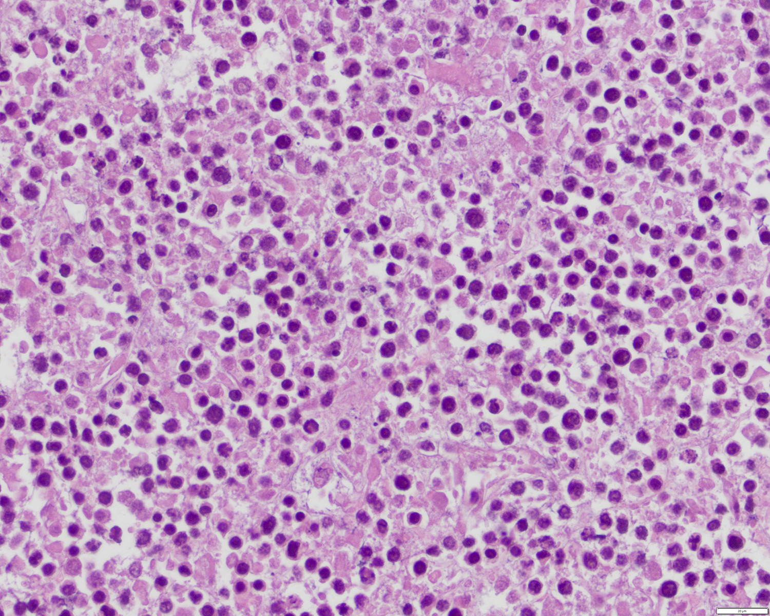
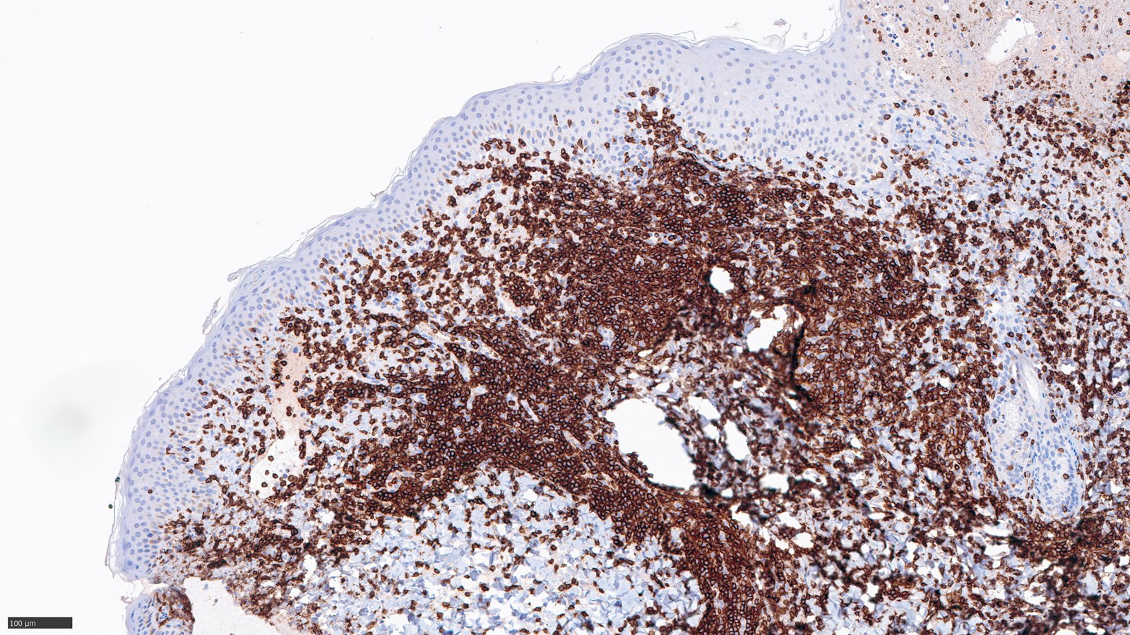
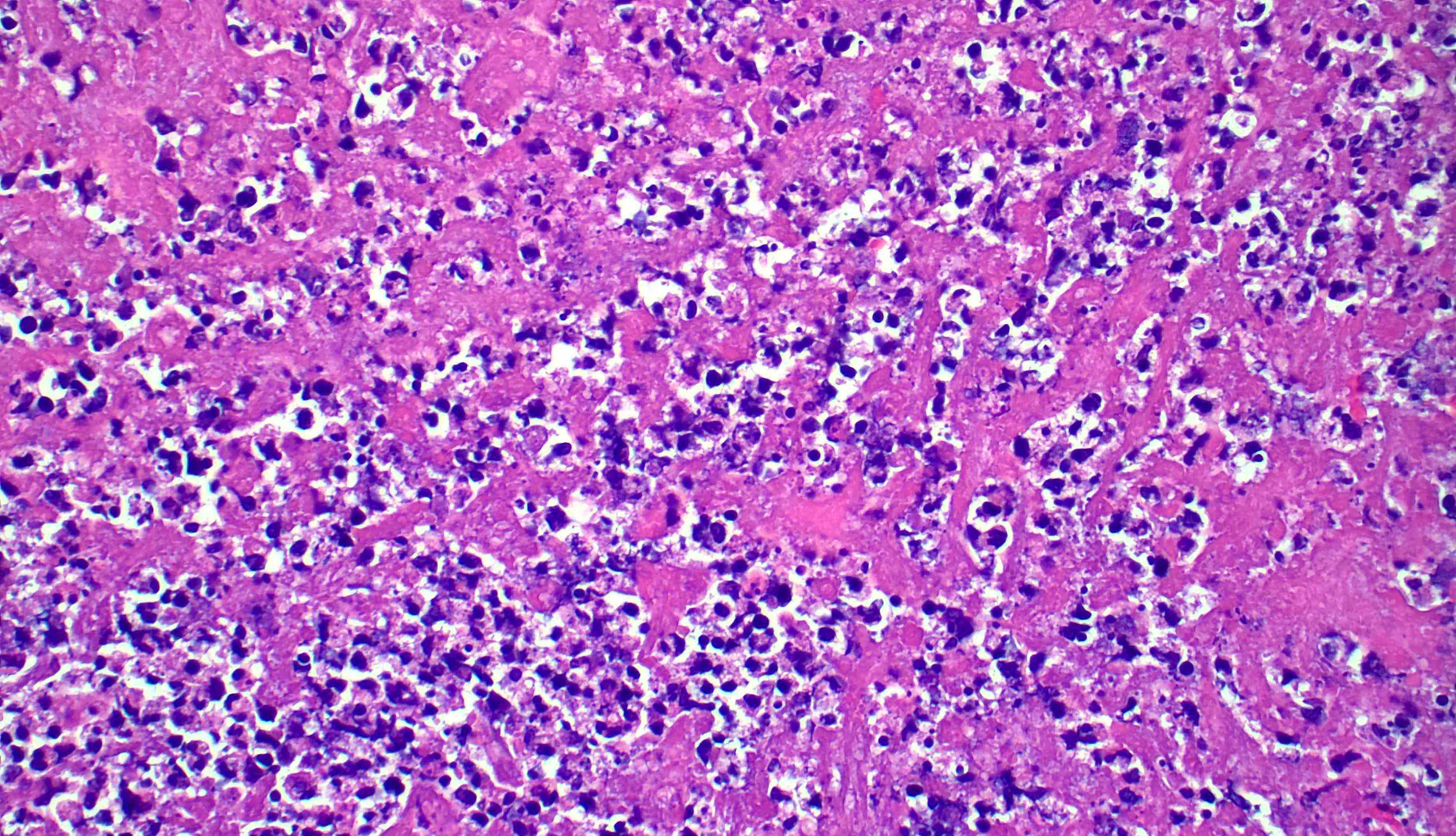
Intrasinusoidal growth of tumor
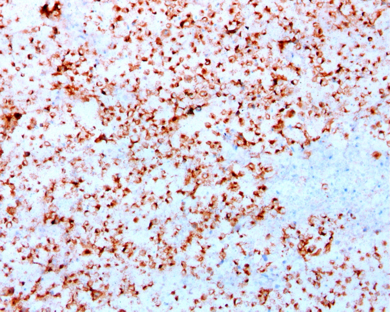
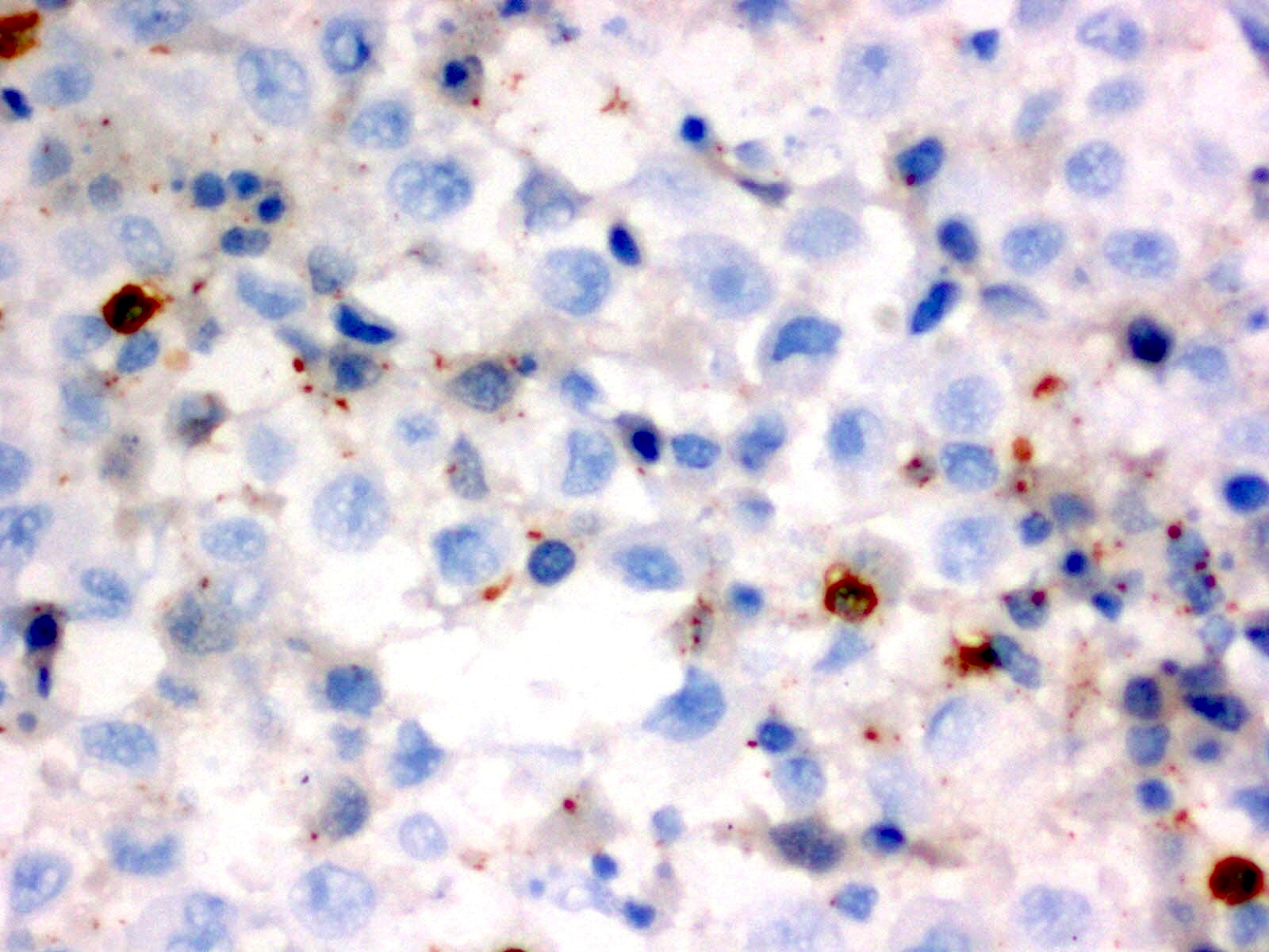
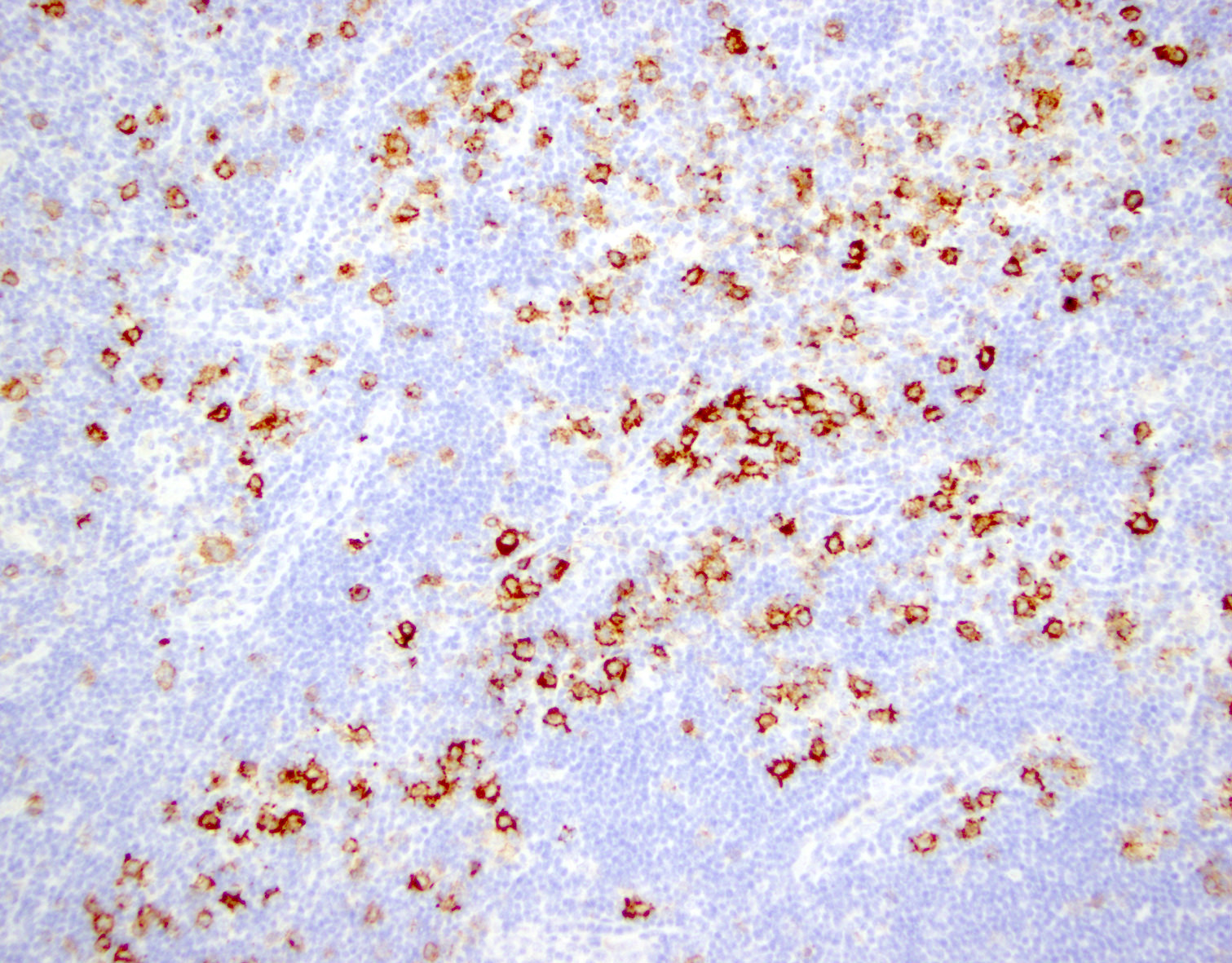
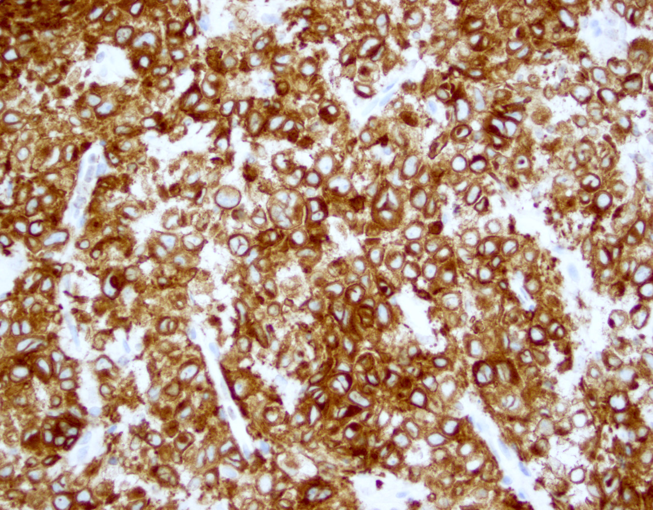
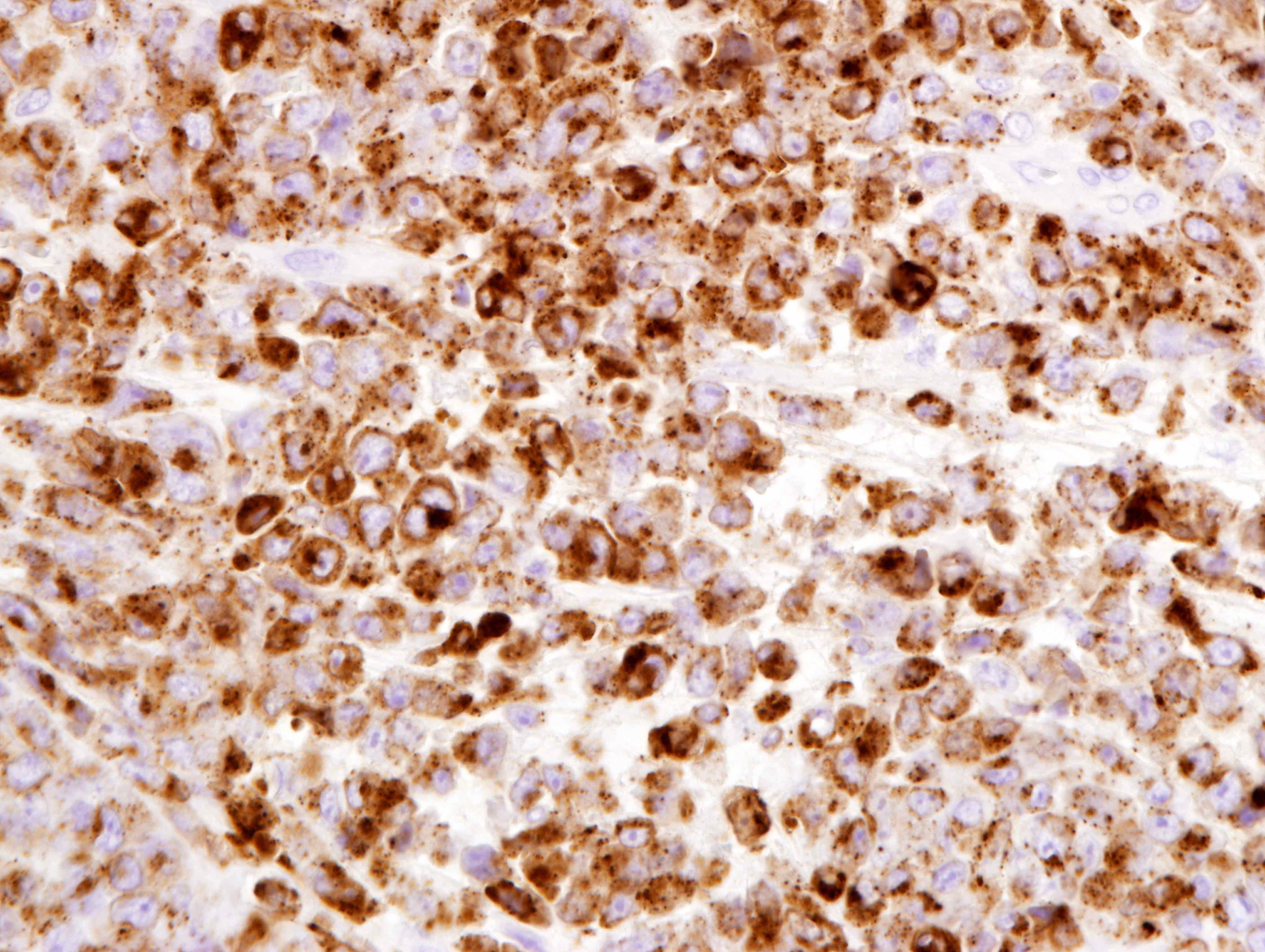
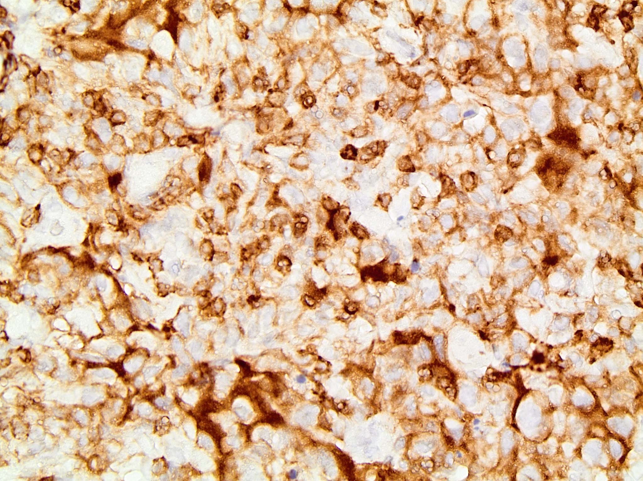
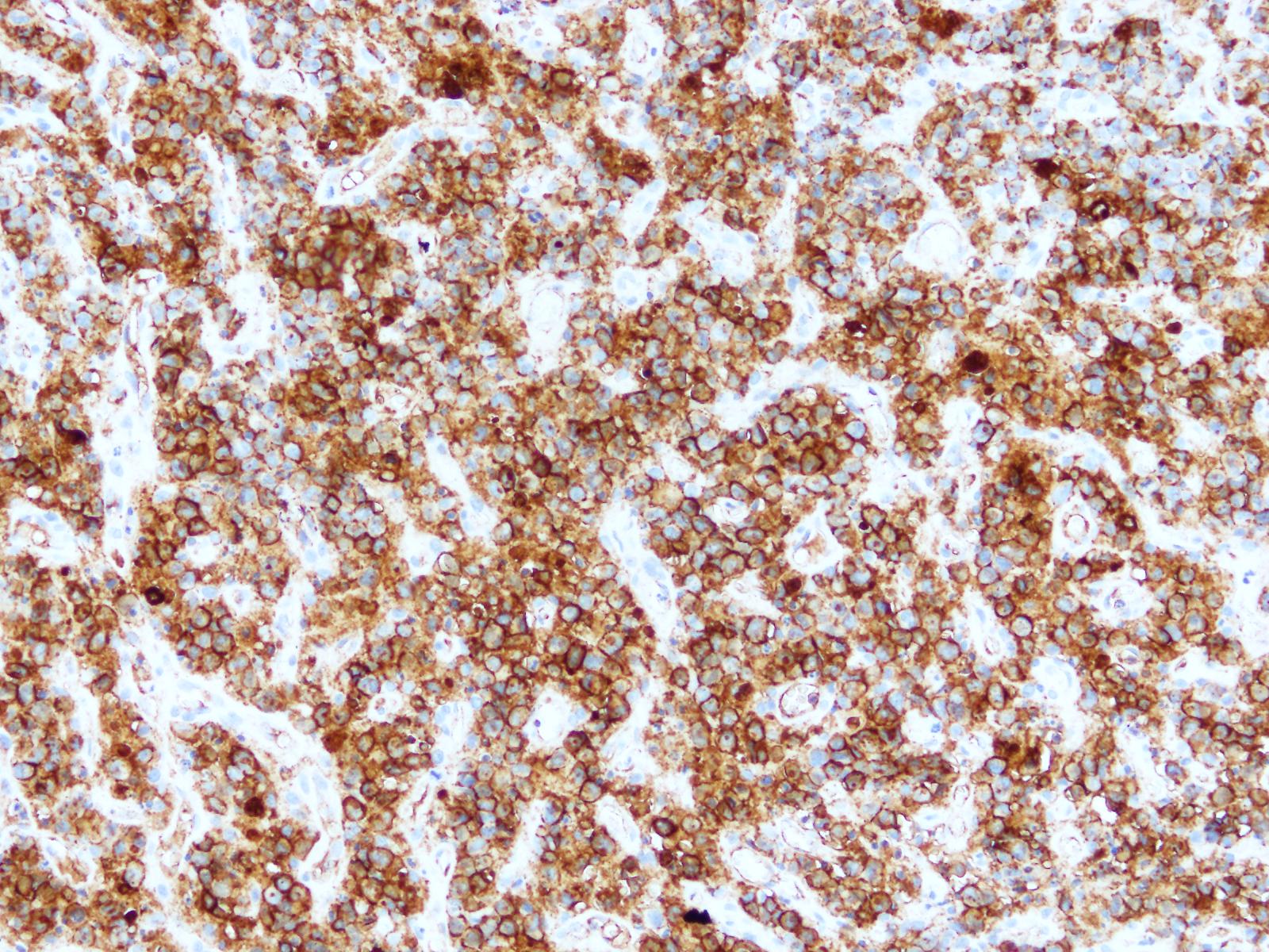
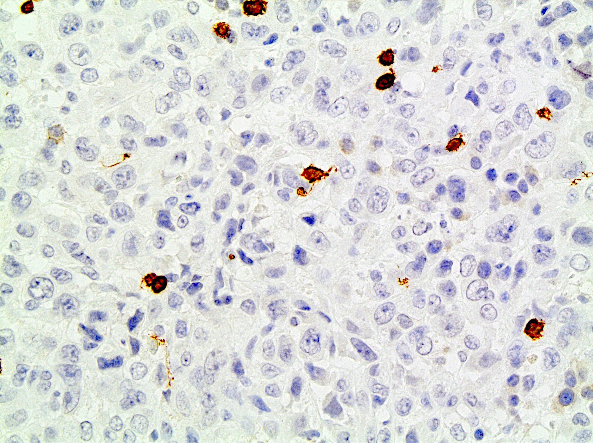
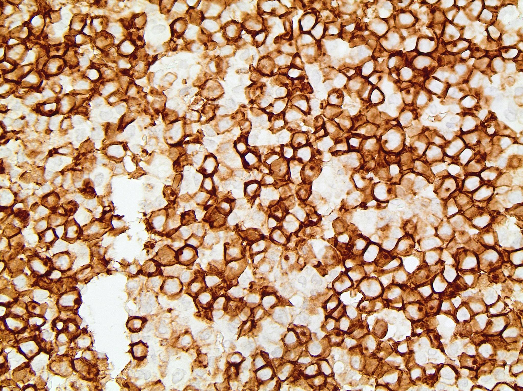
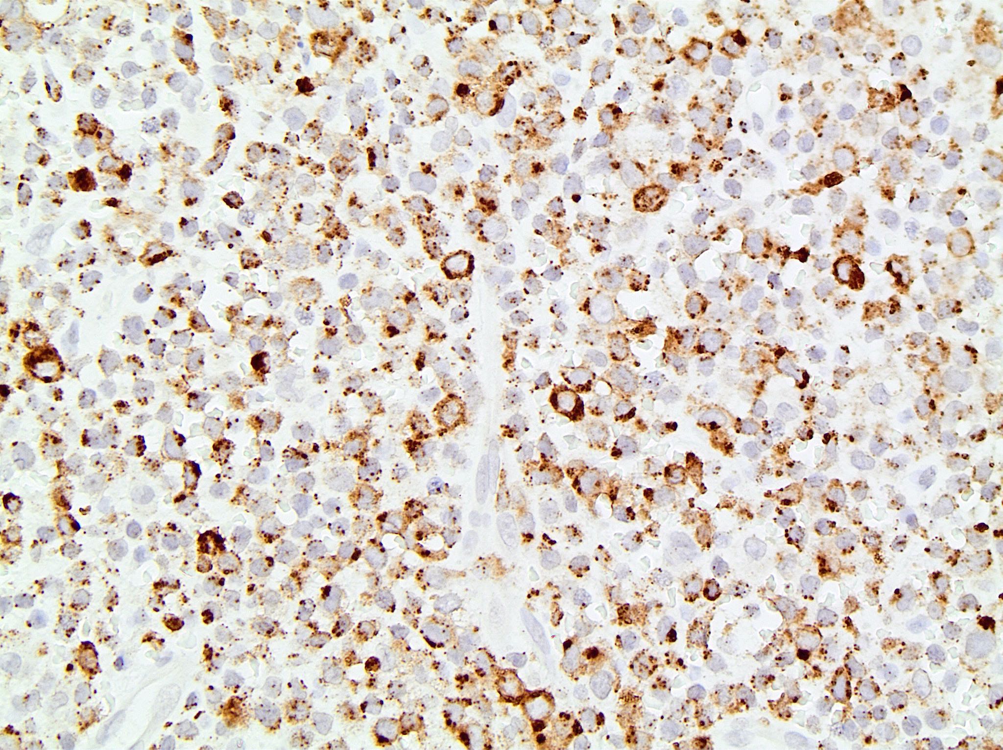
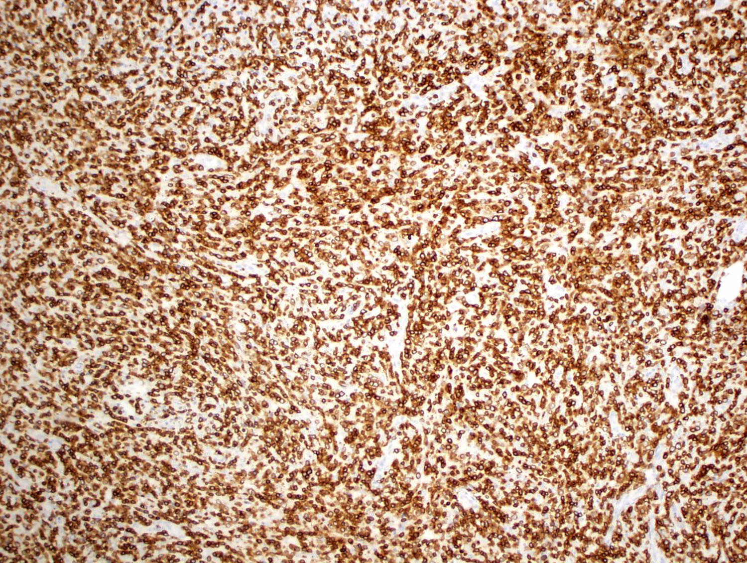
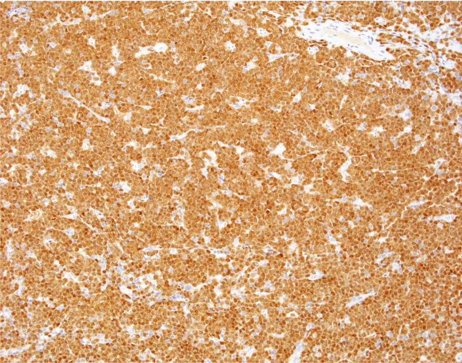
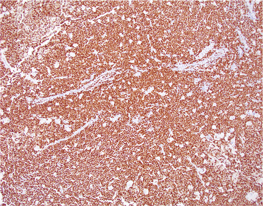
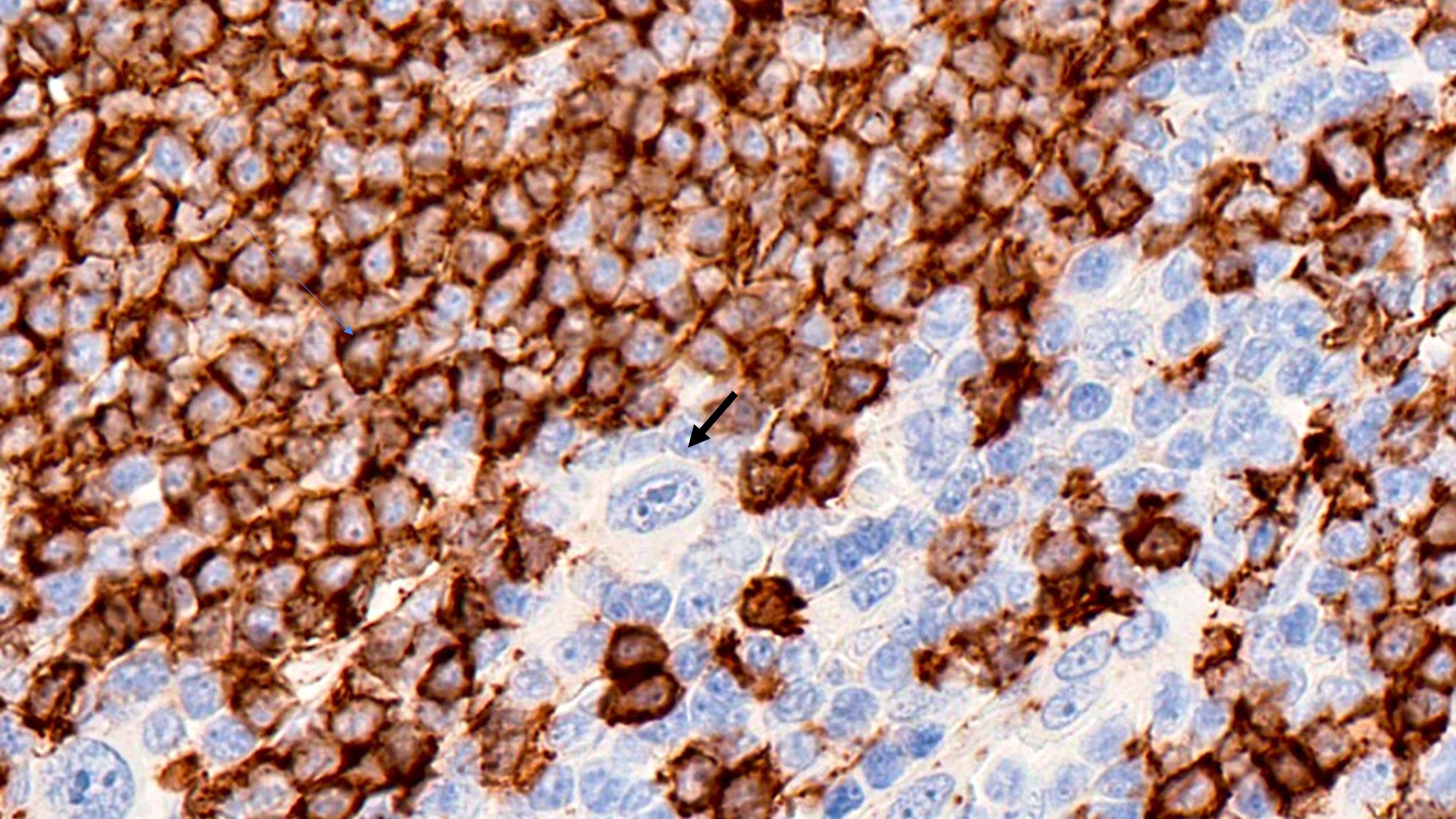
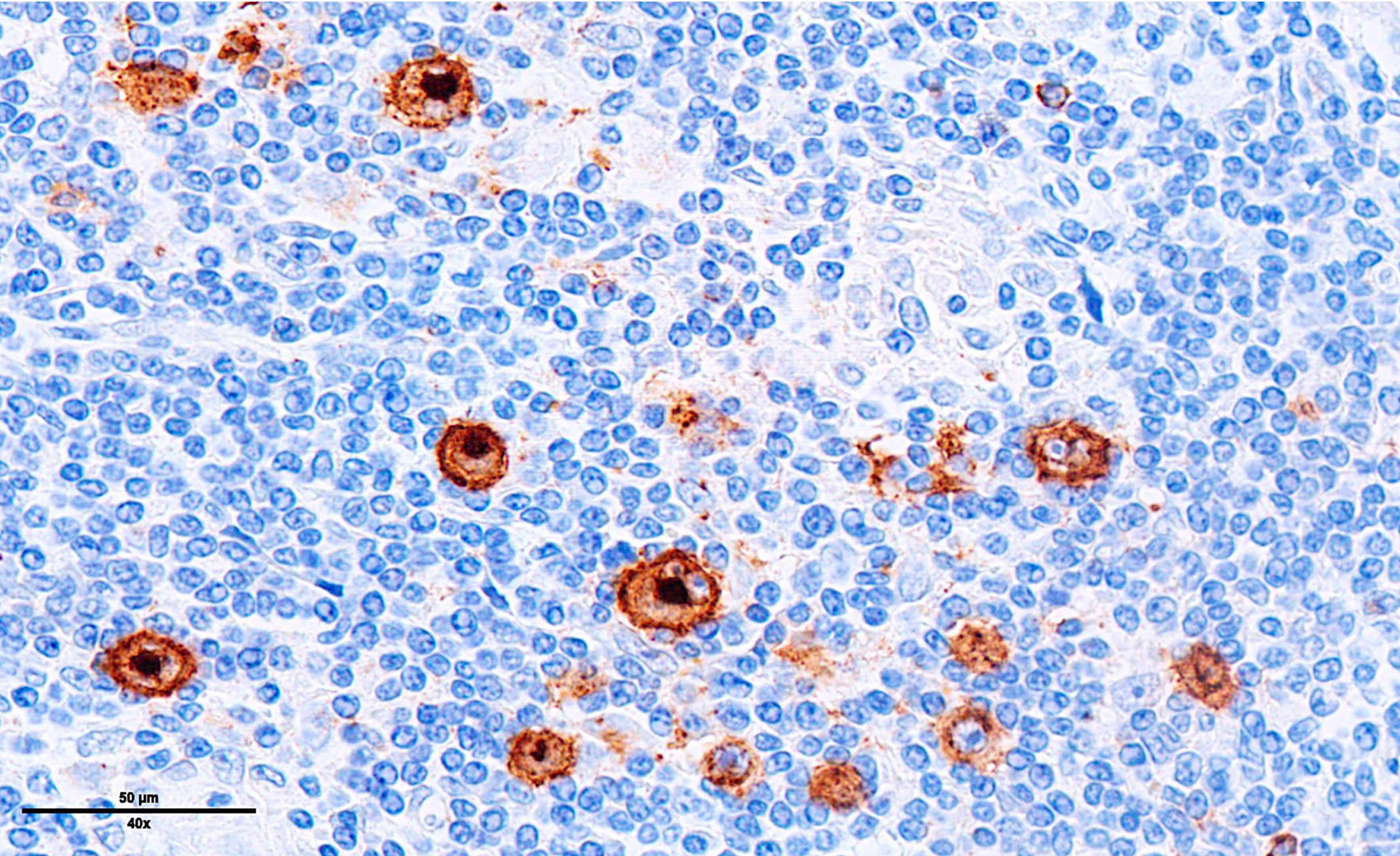
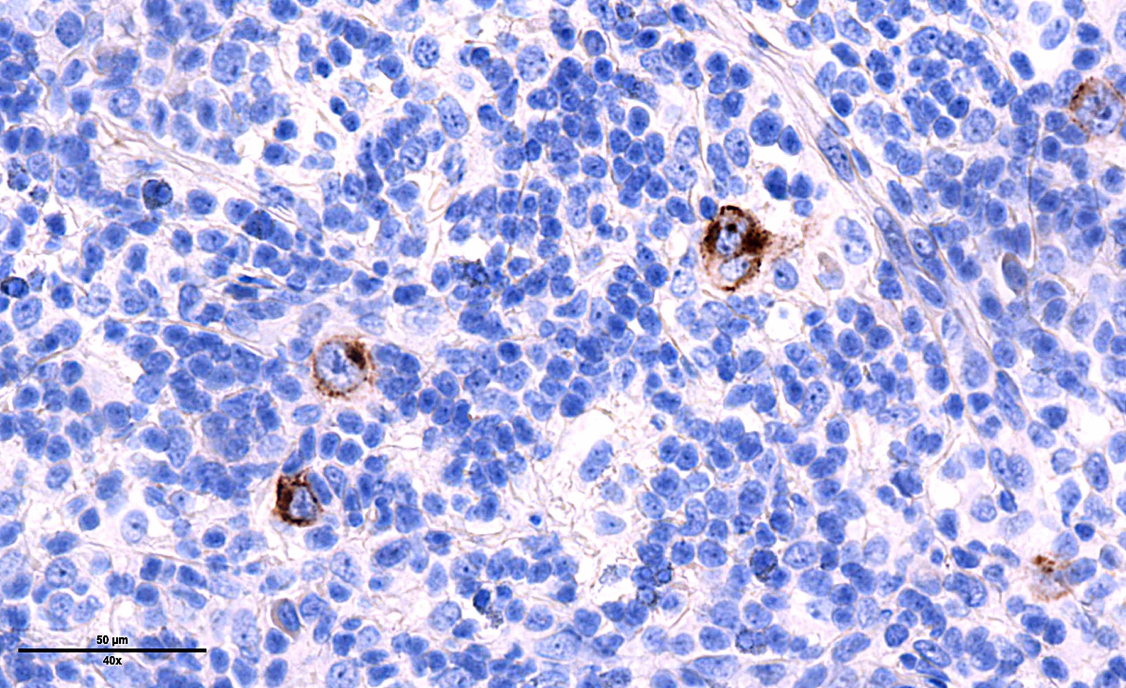
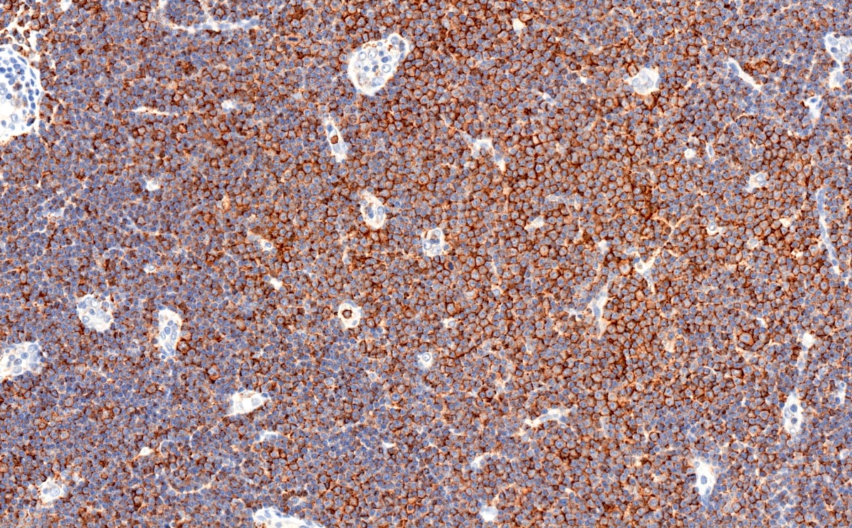
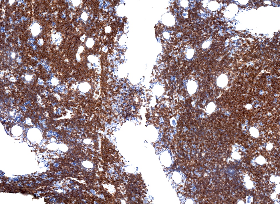
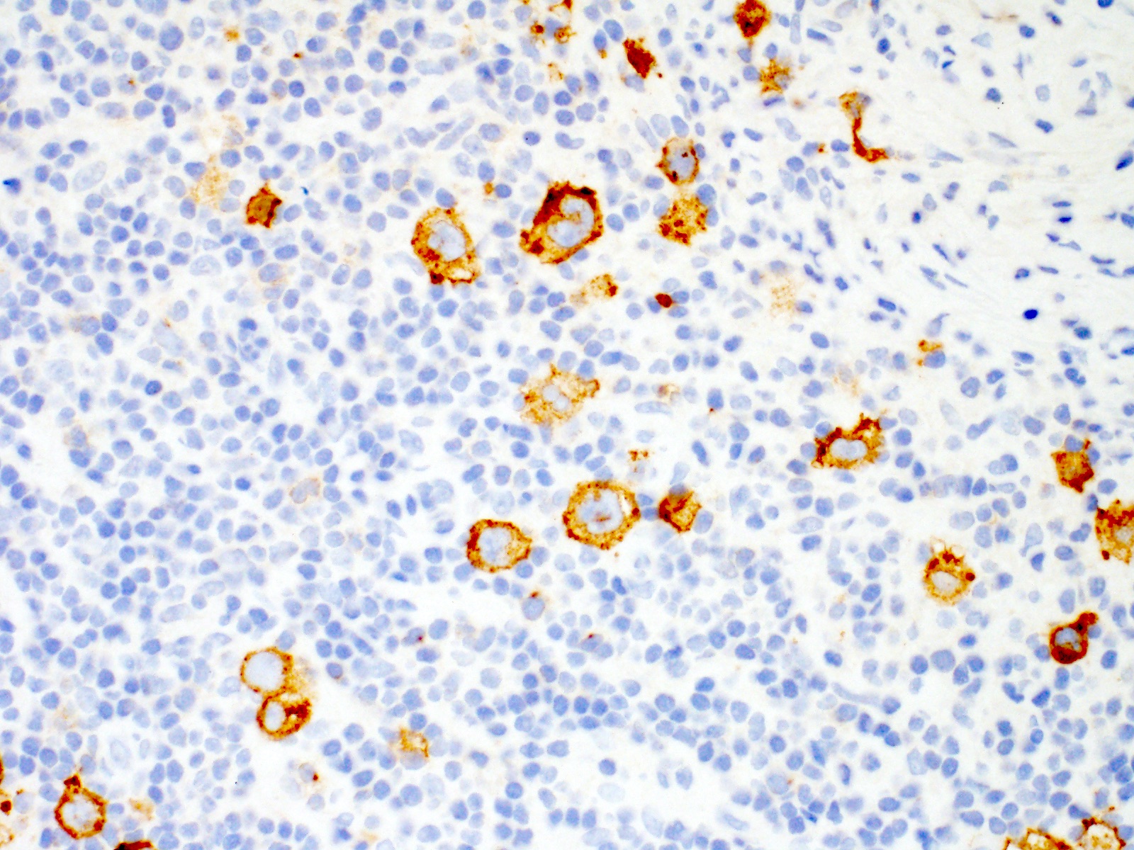
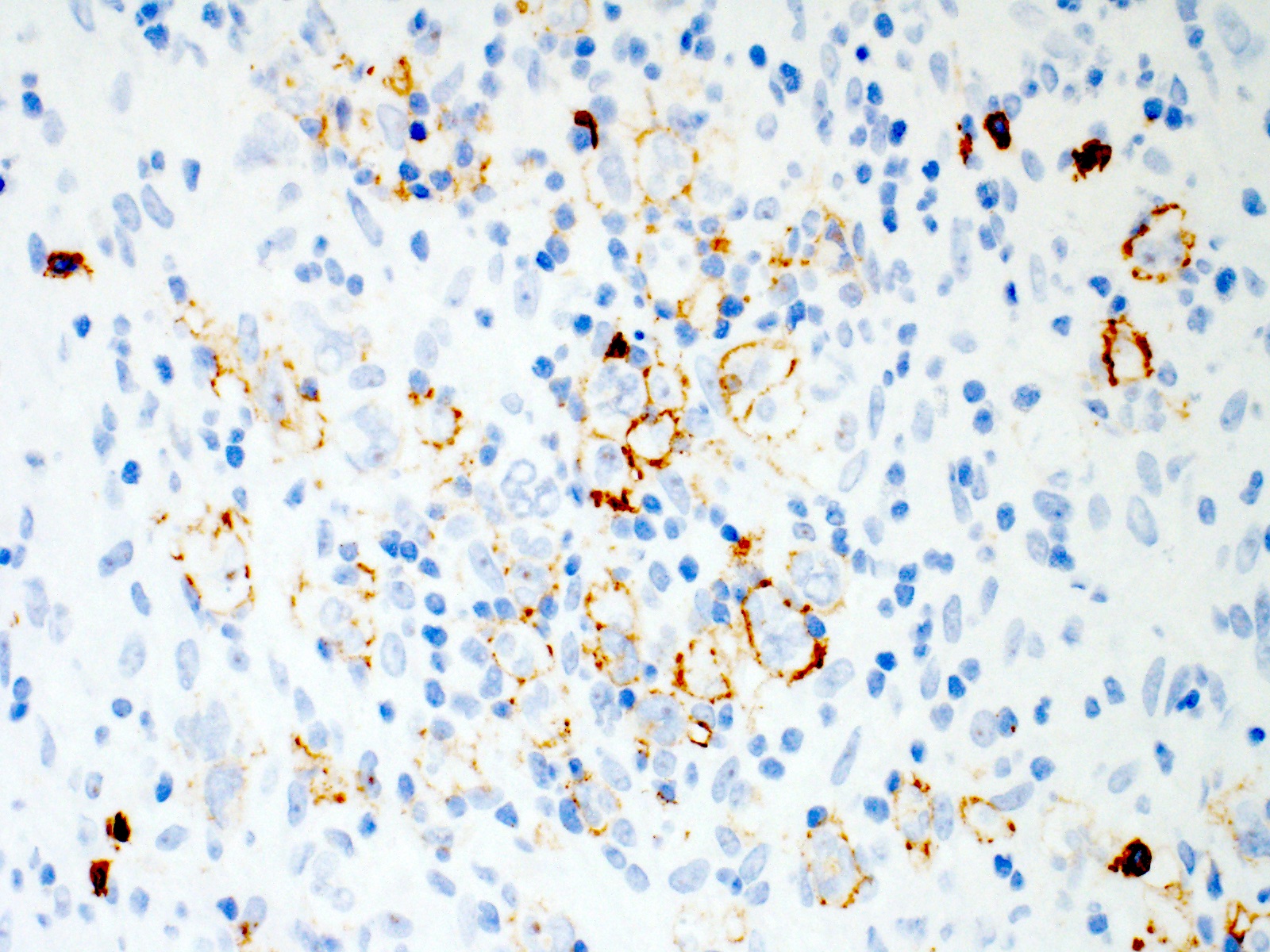
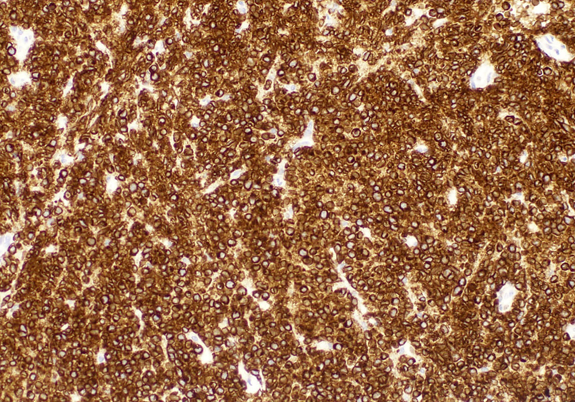
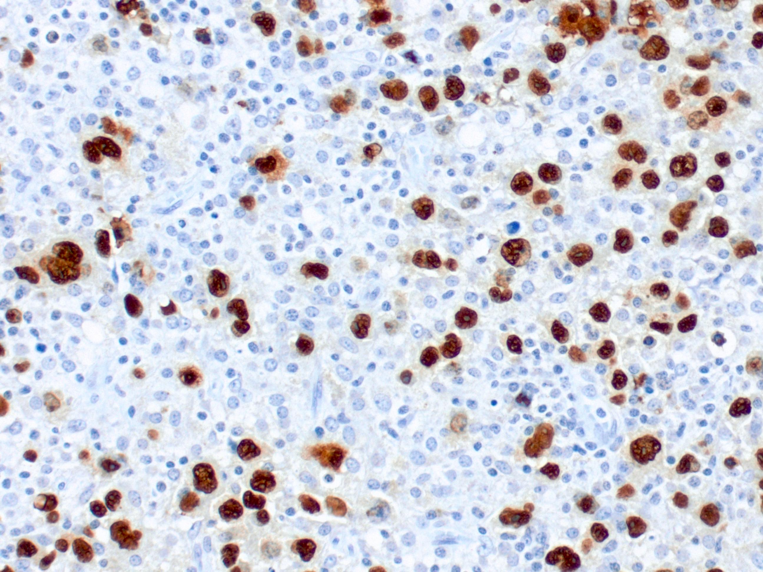
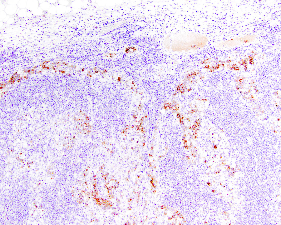
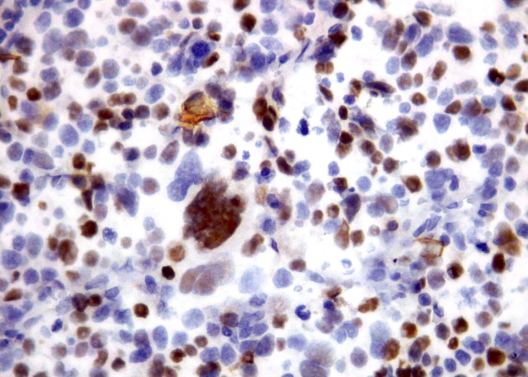
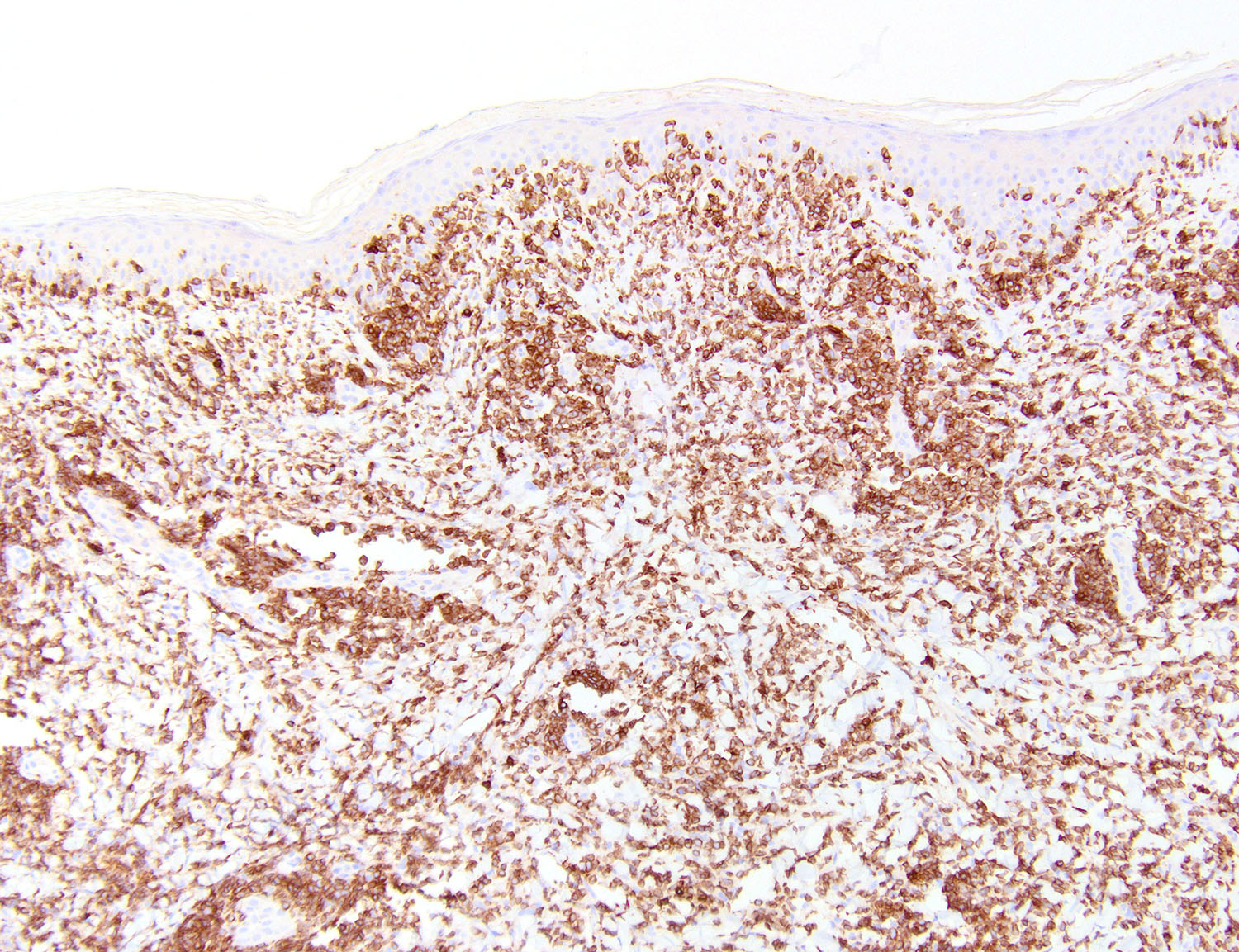
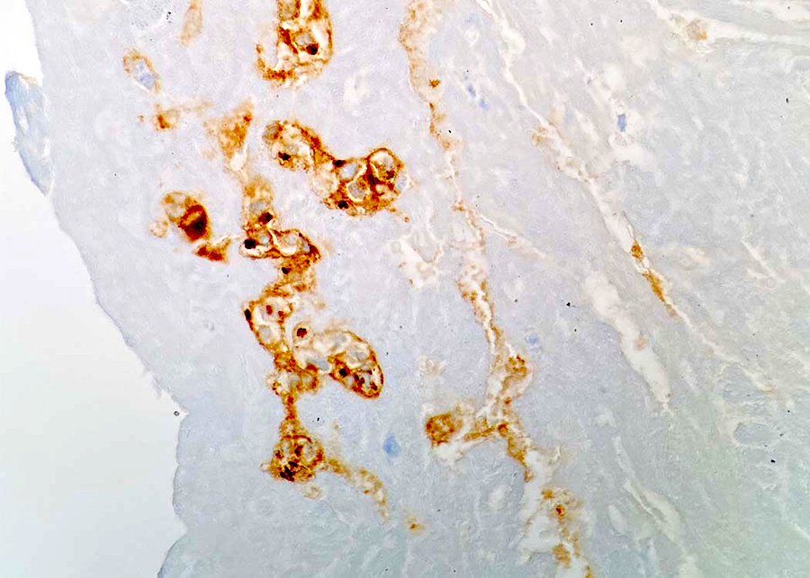
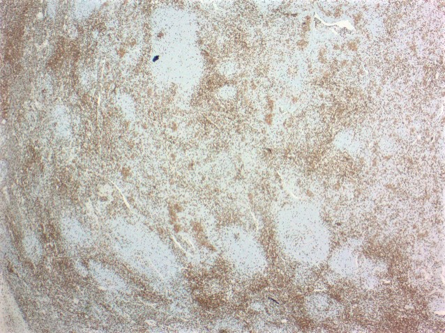
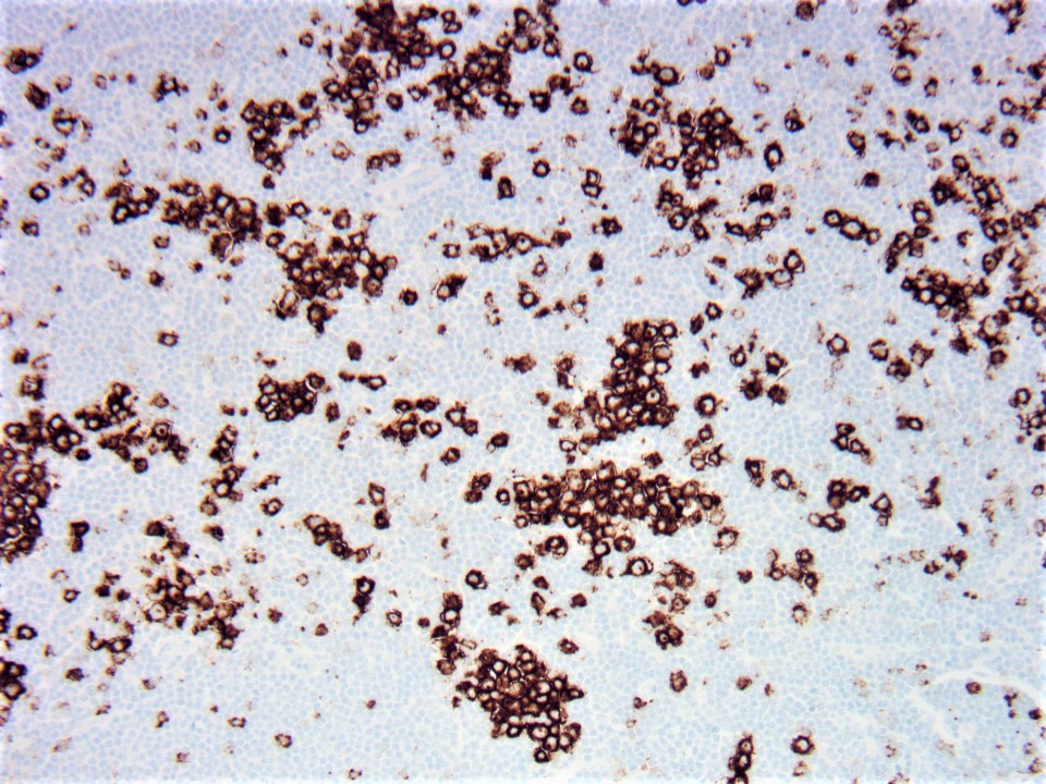
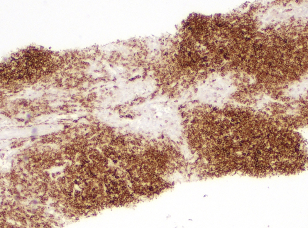
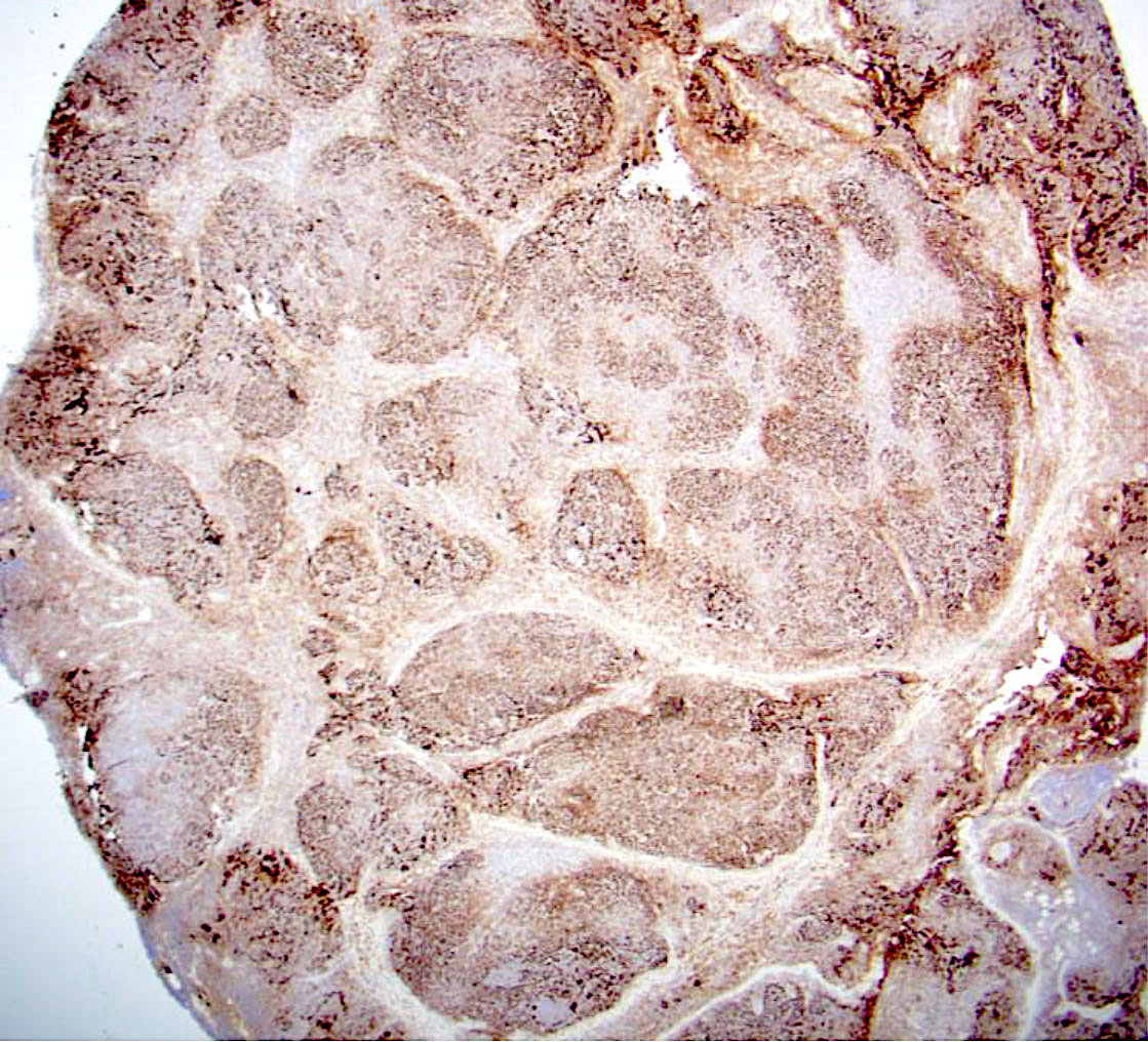
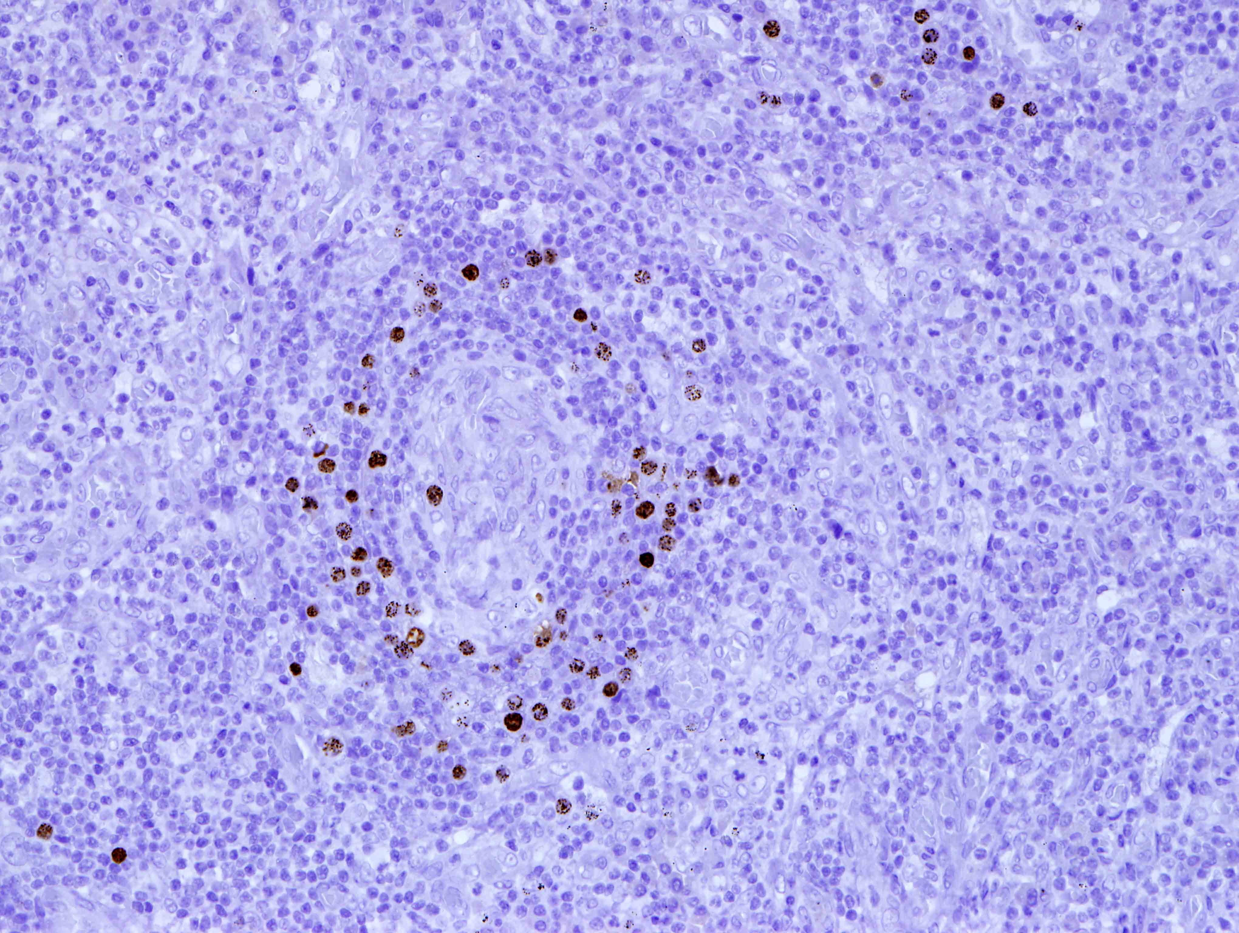
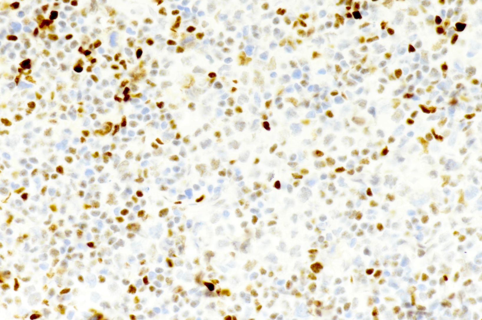
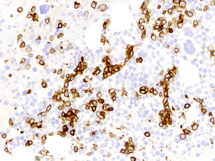
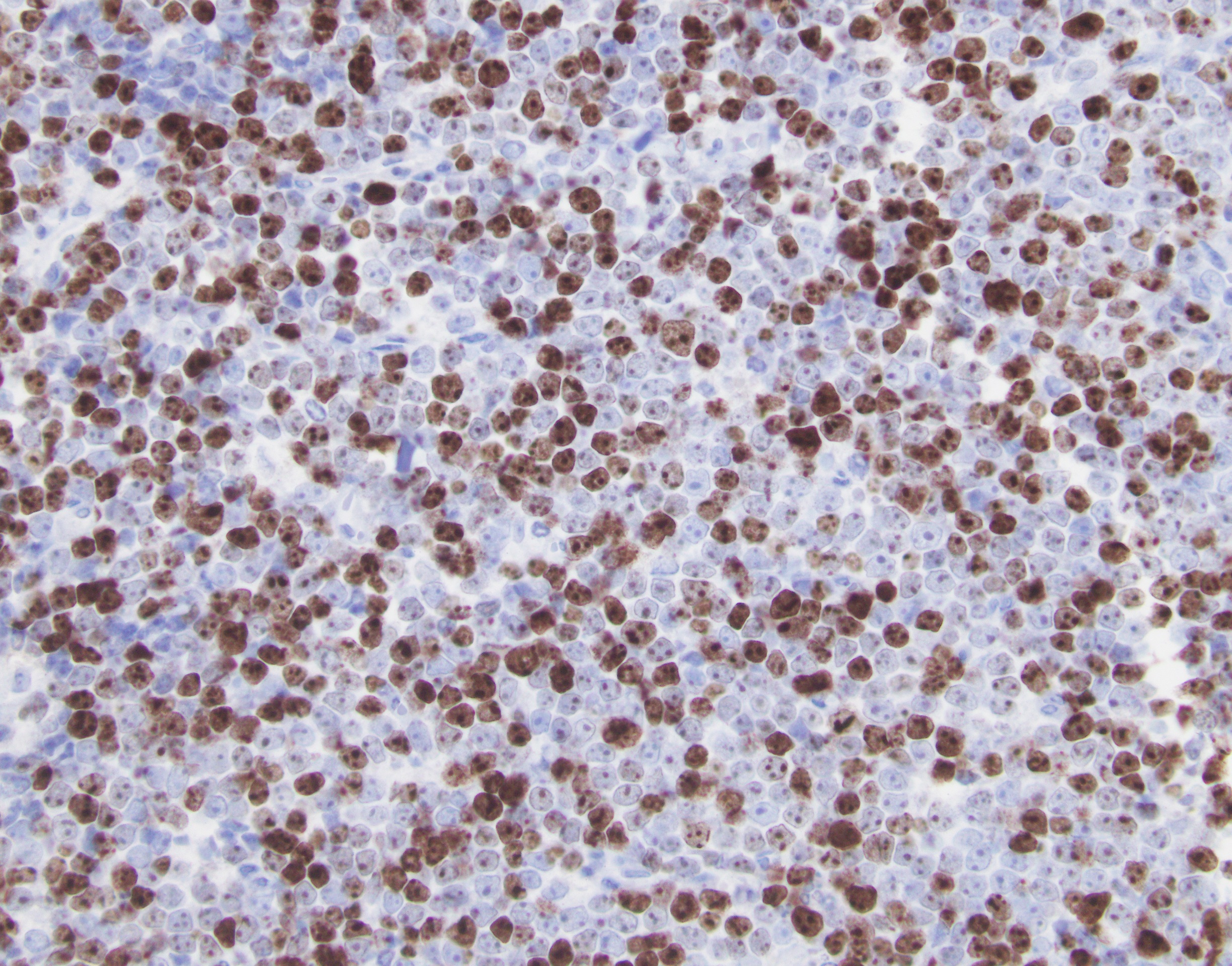
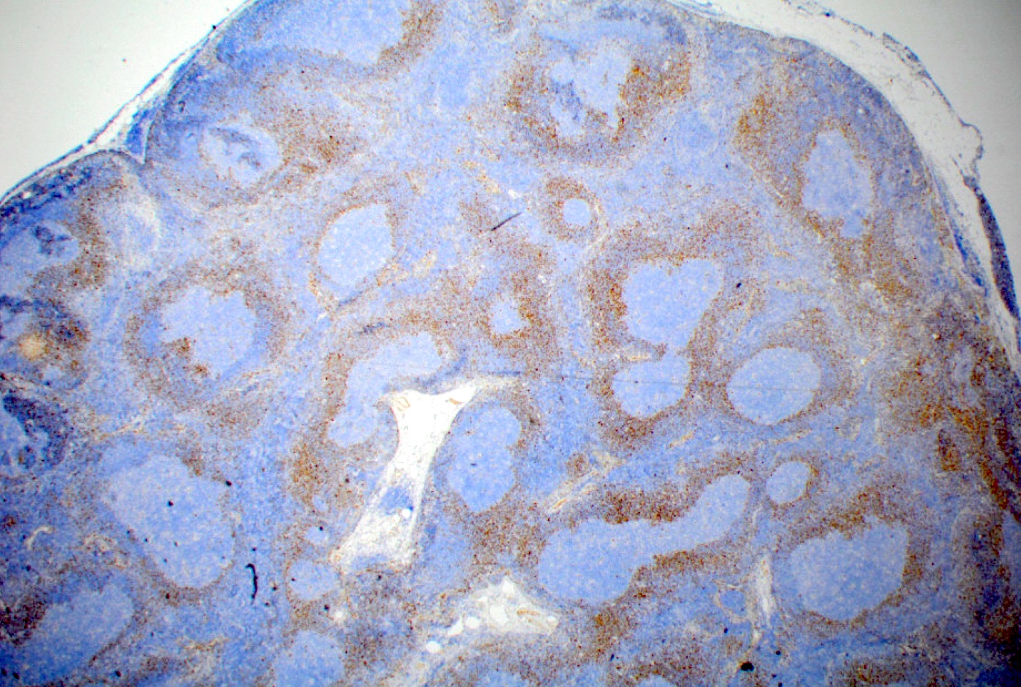
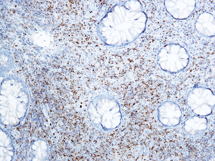
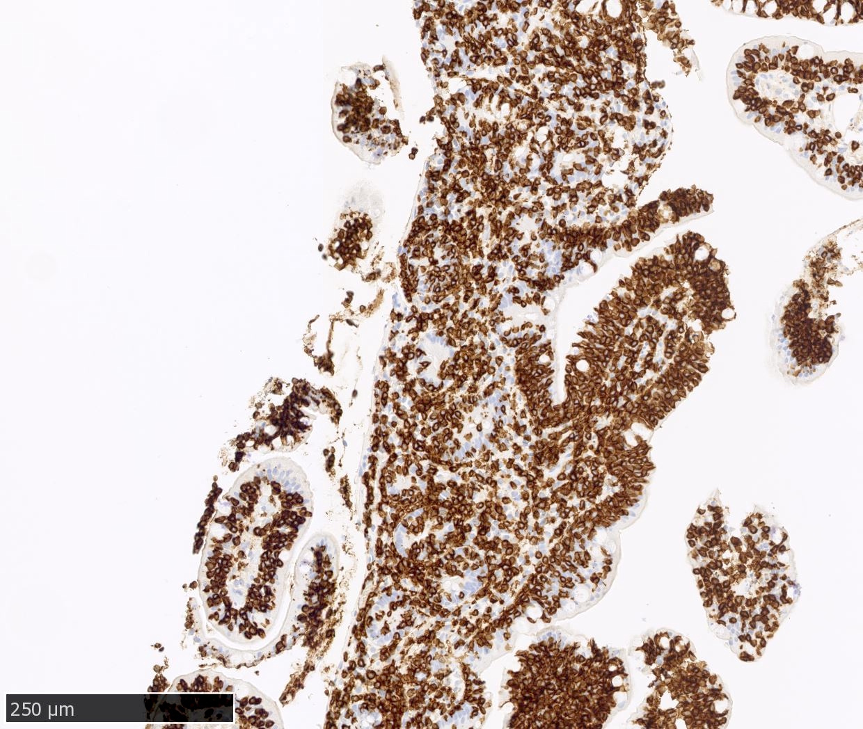
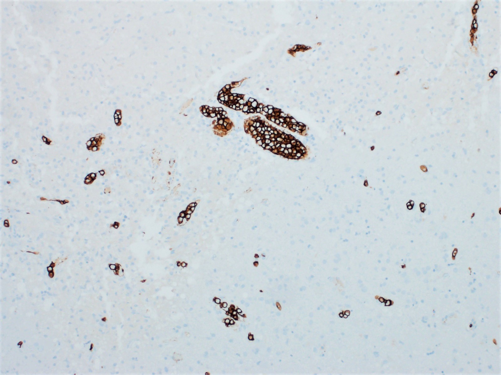
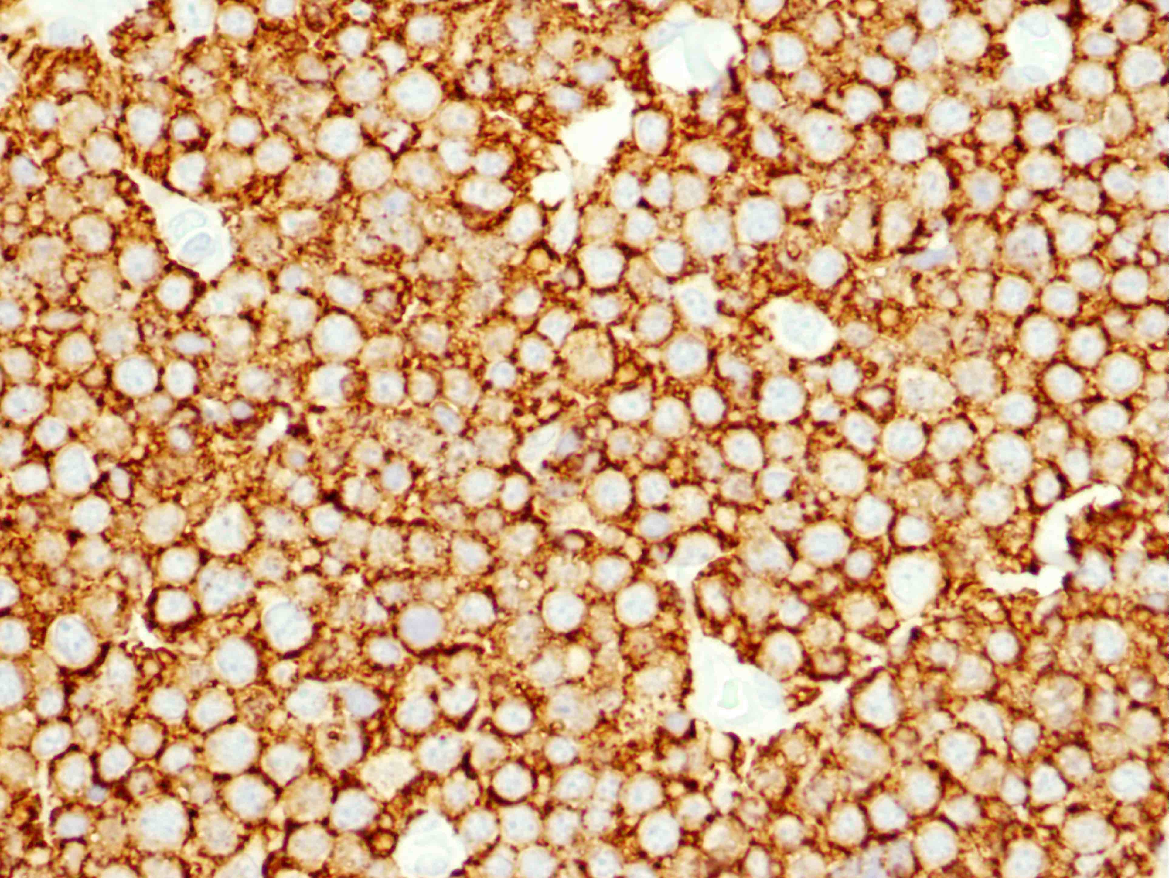
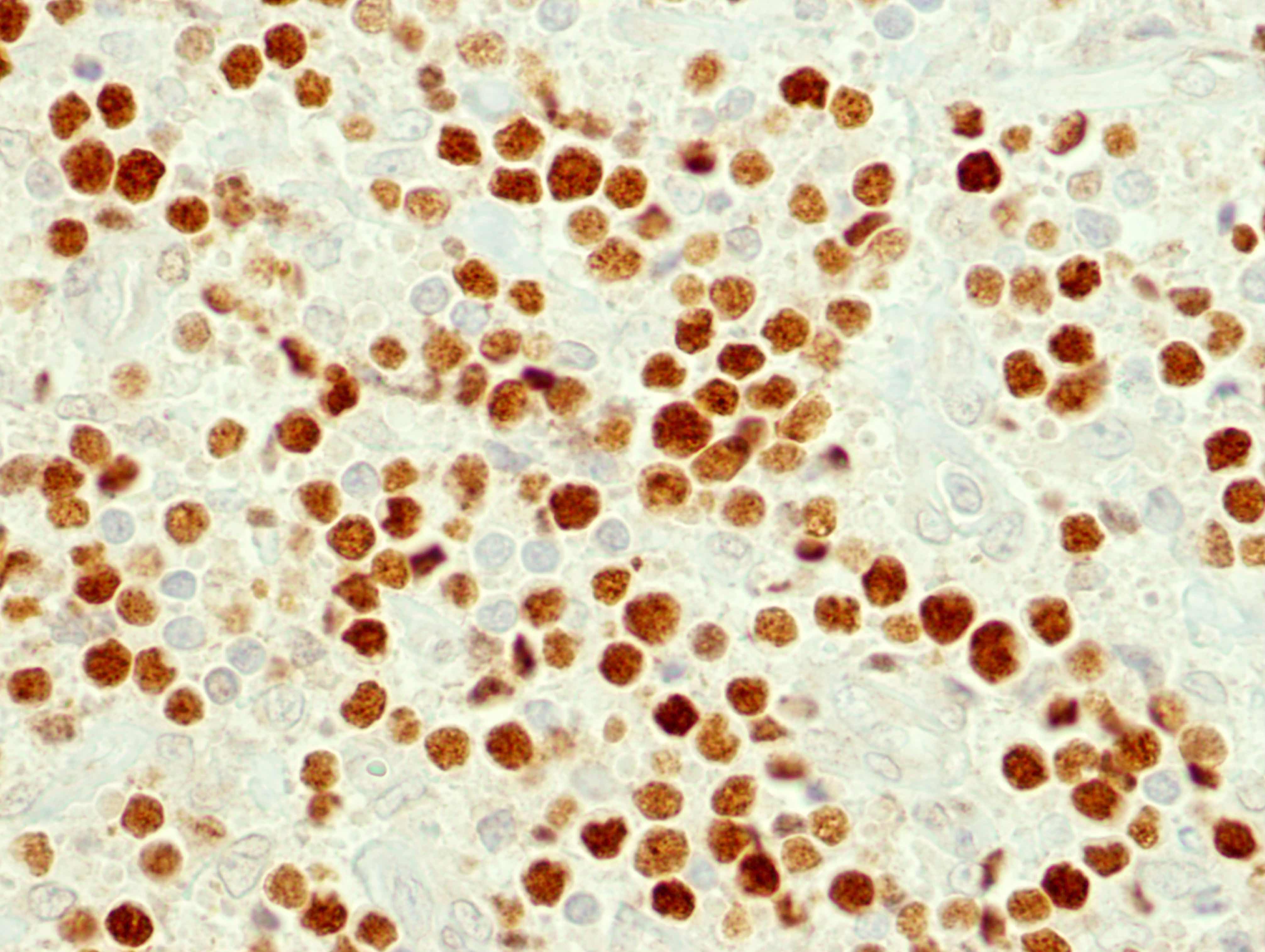
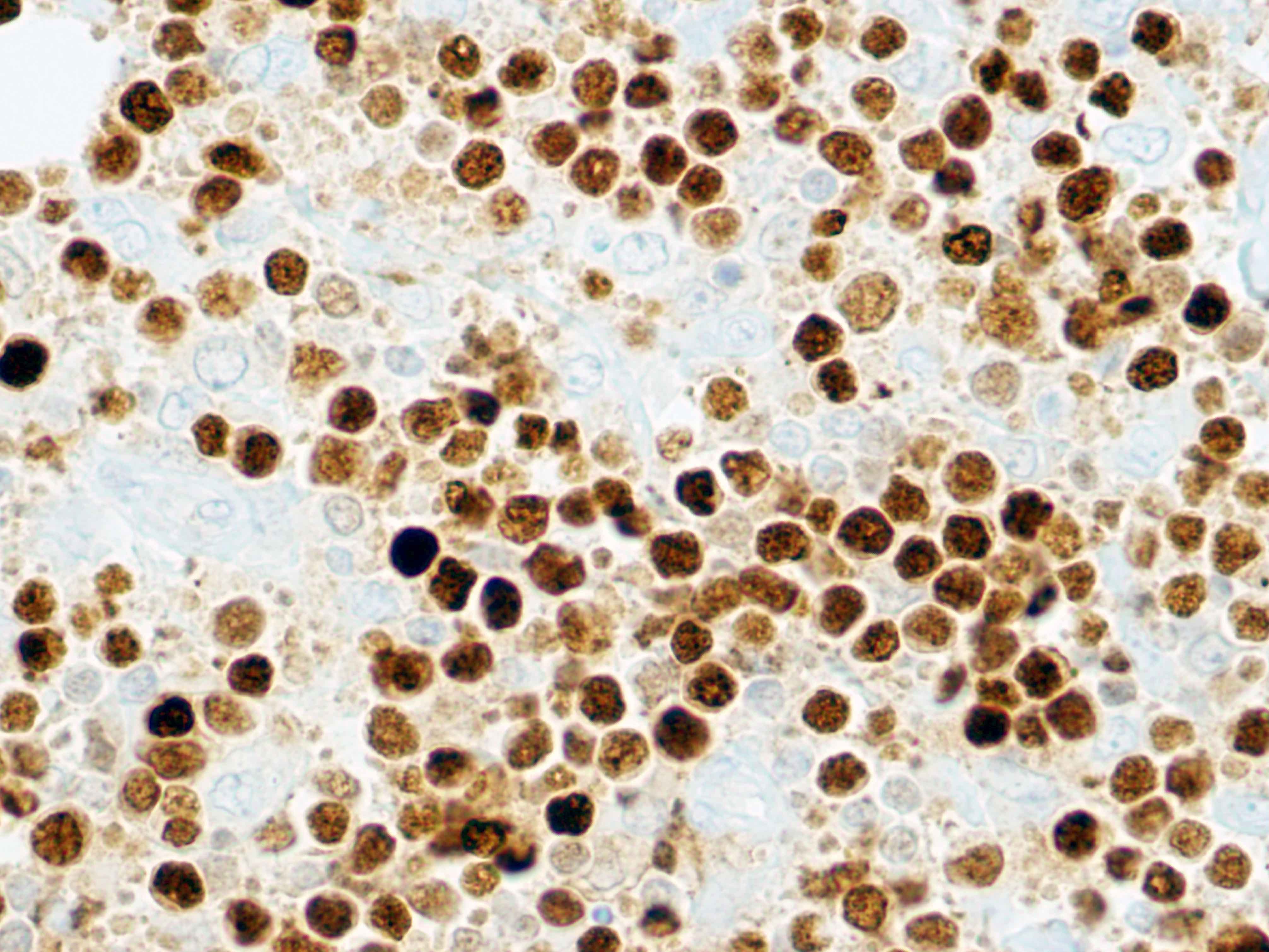
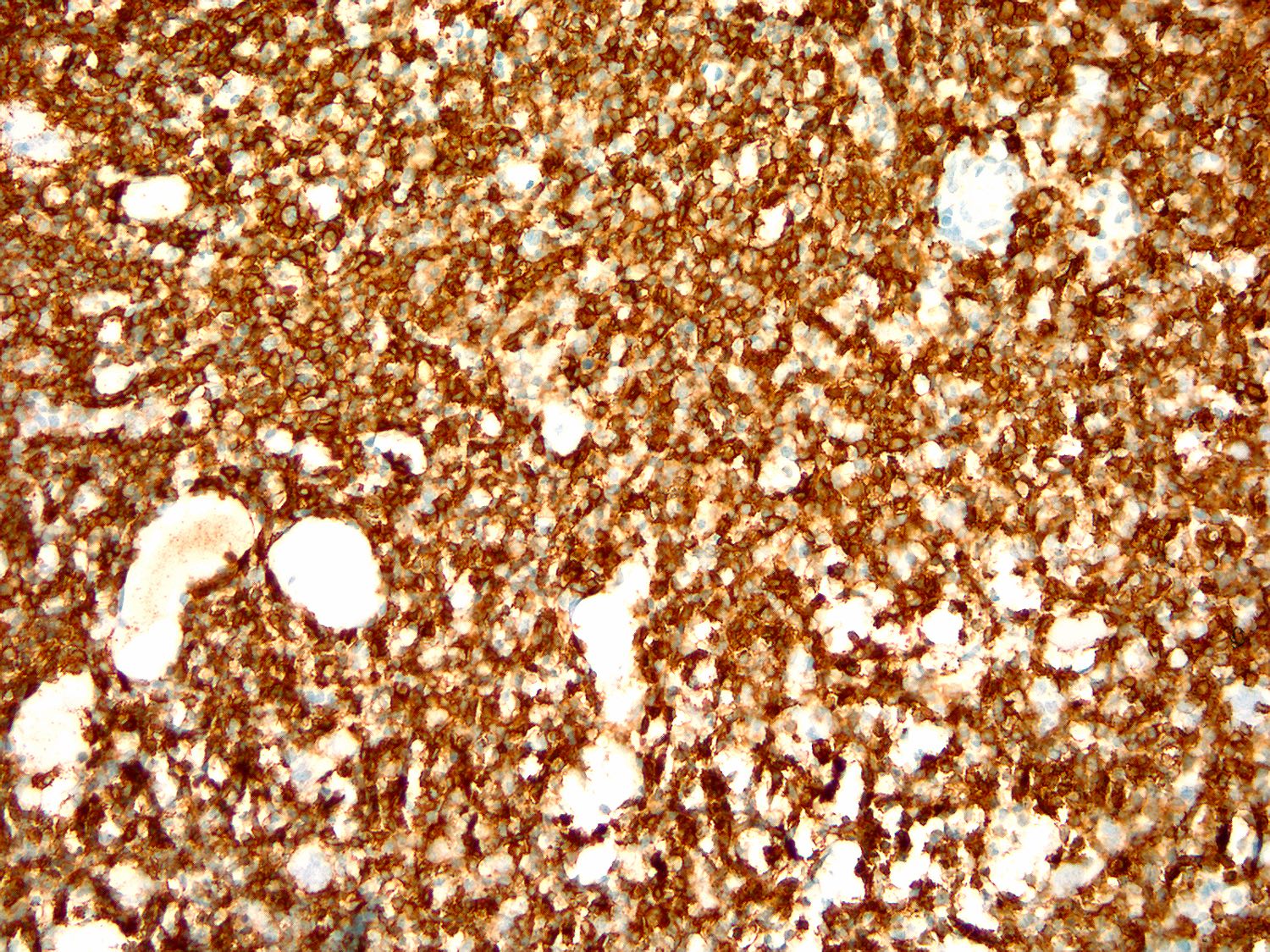
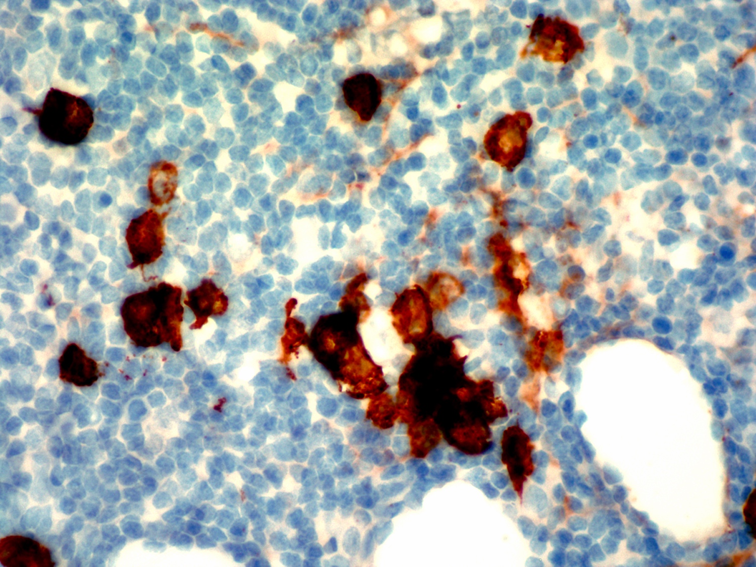
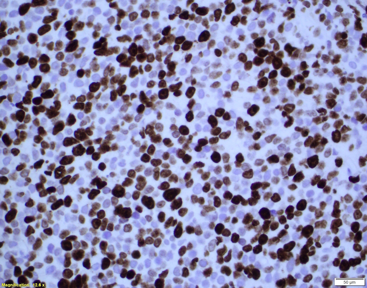
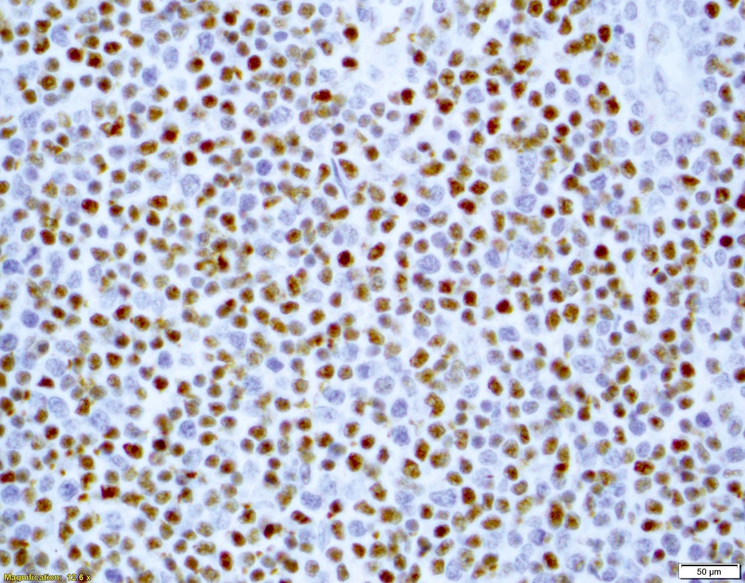
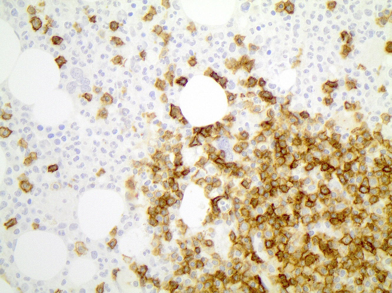
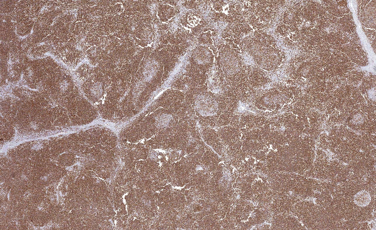
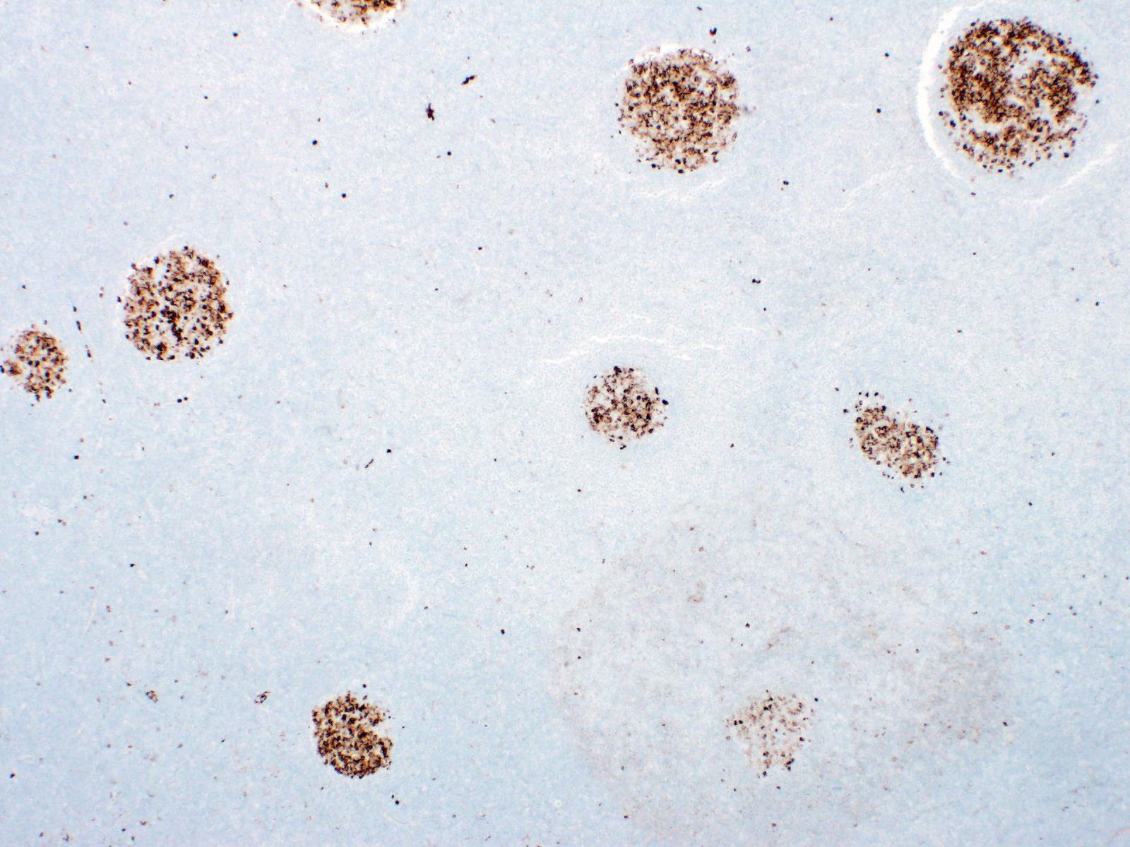
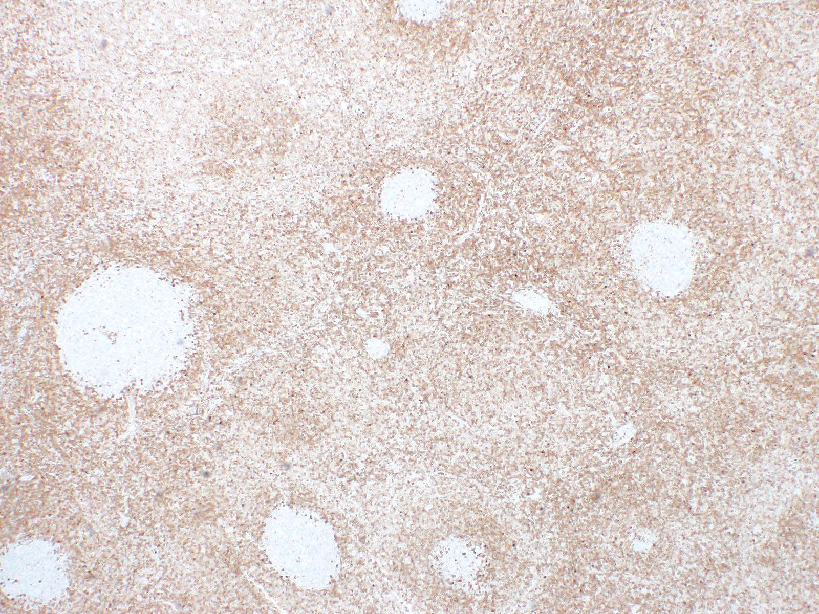
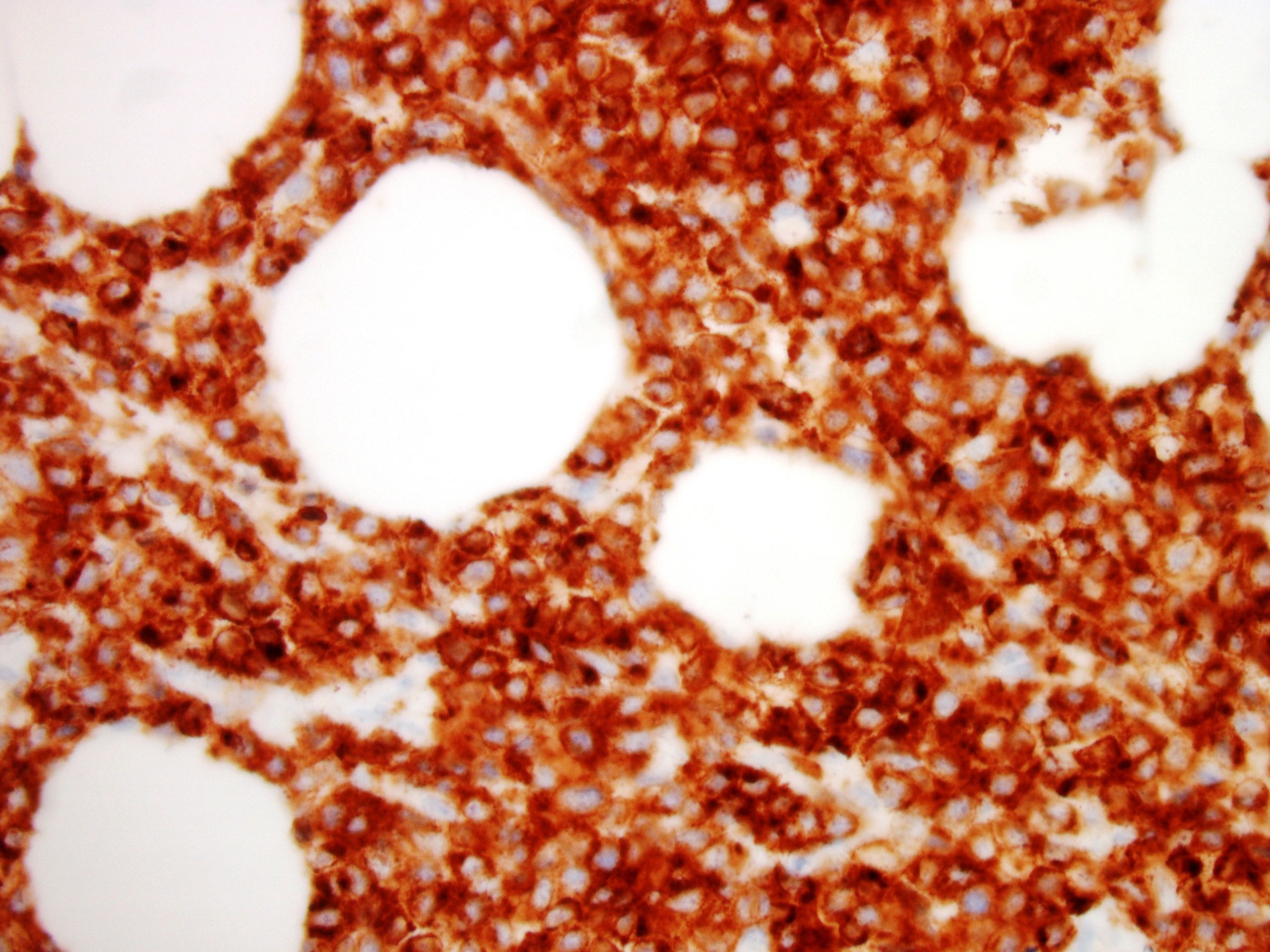
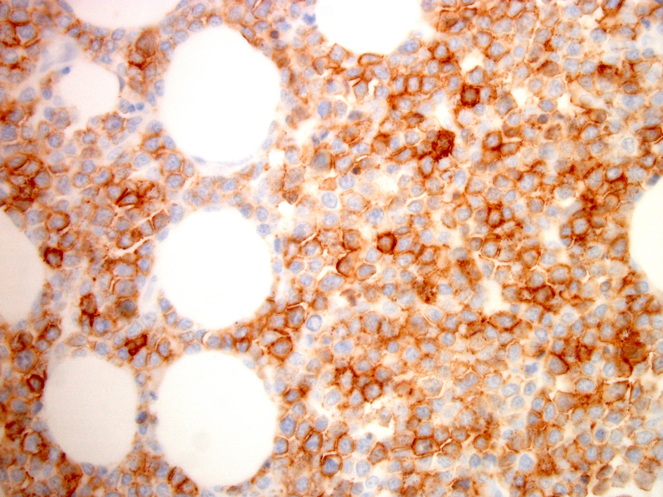
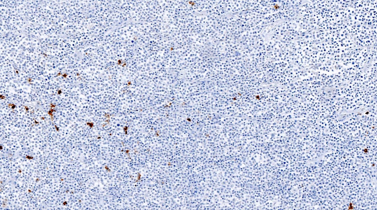
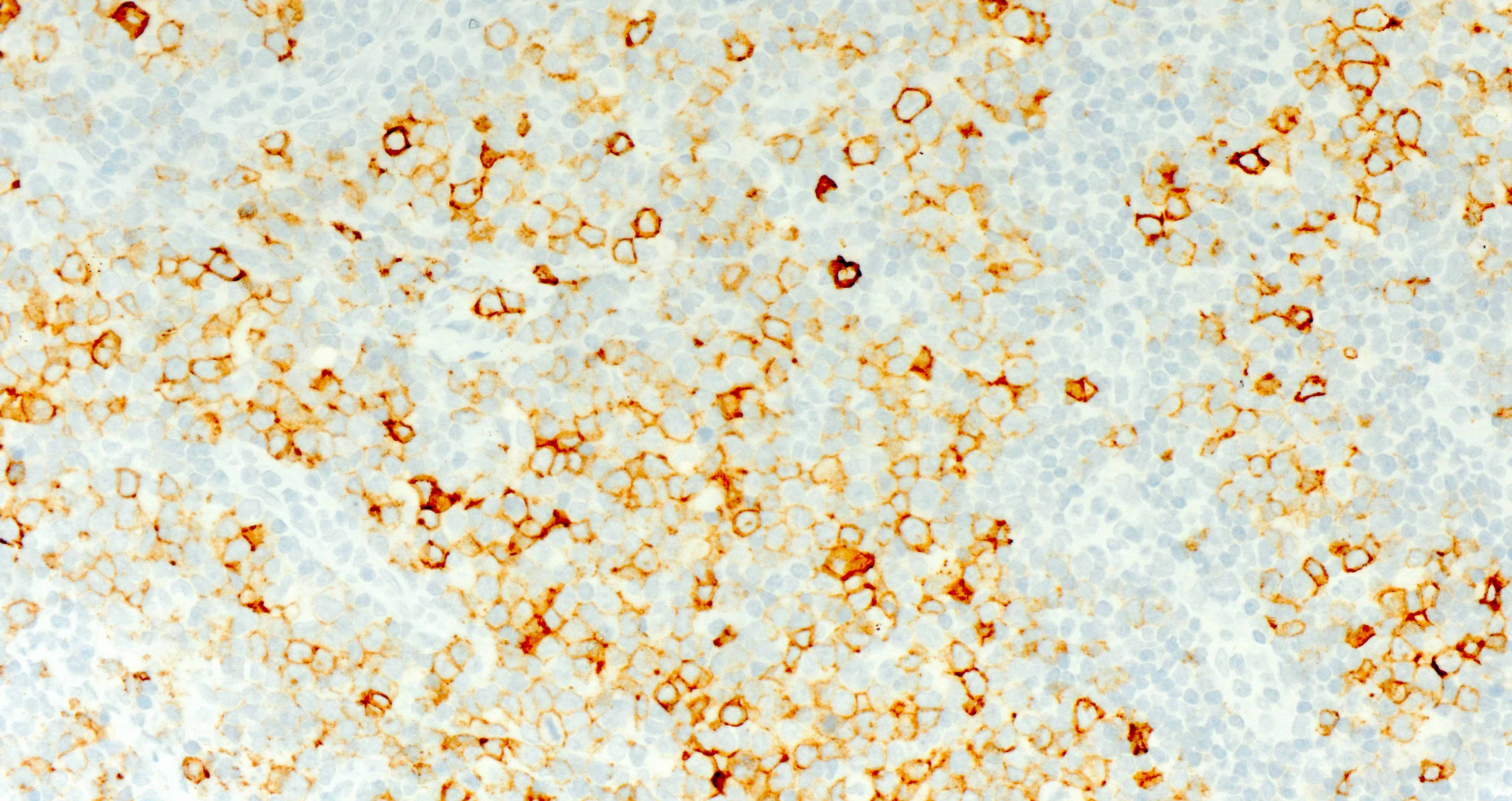
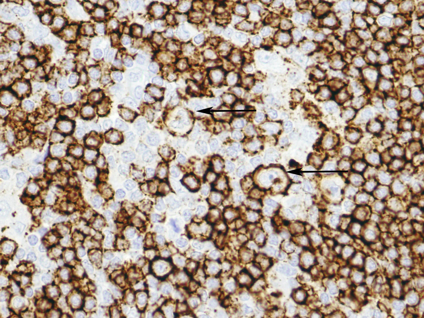
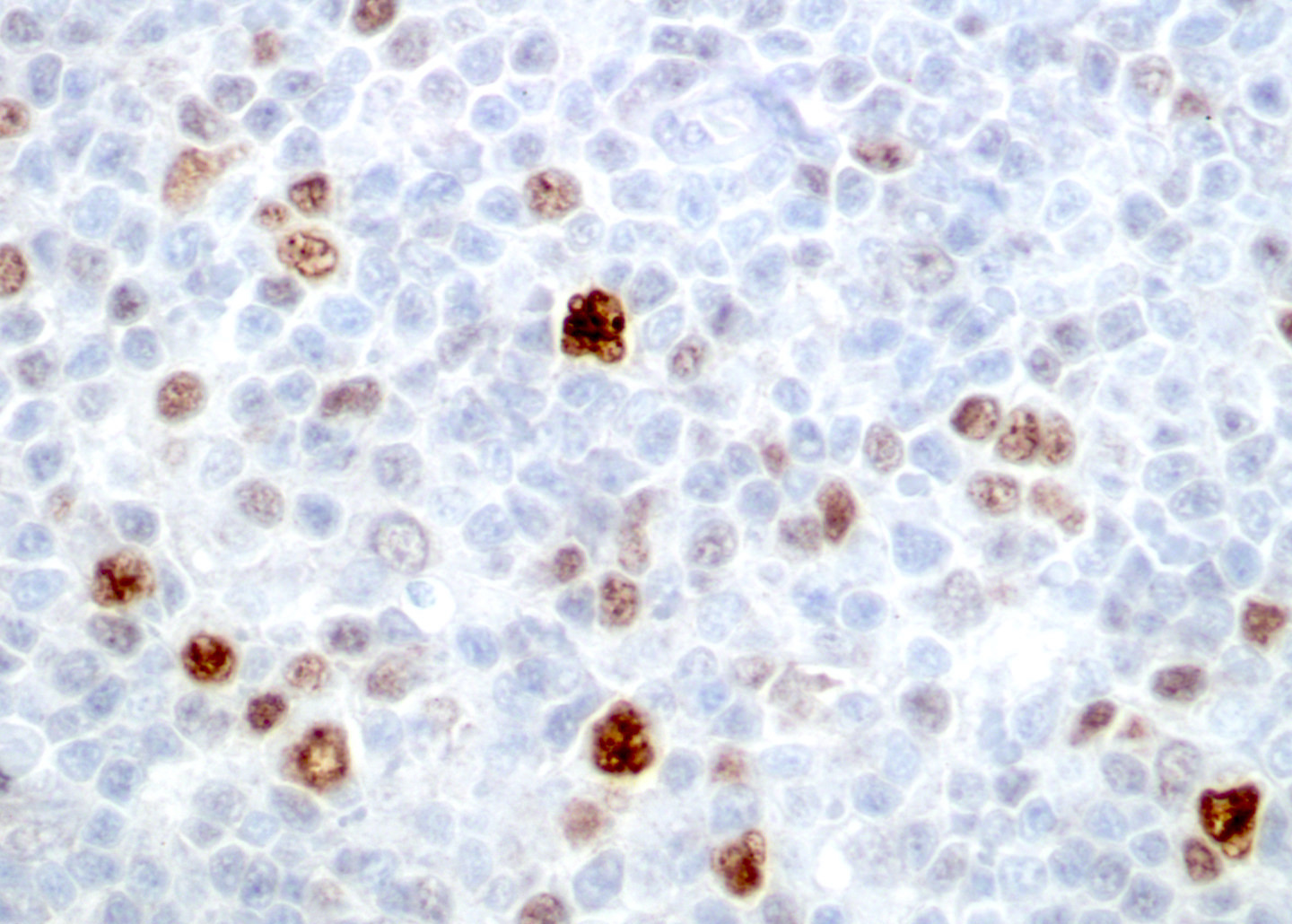
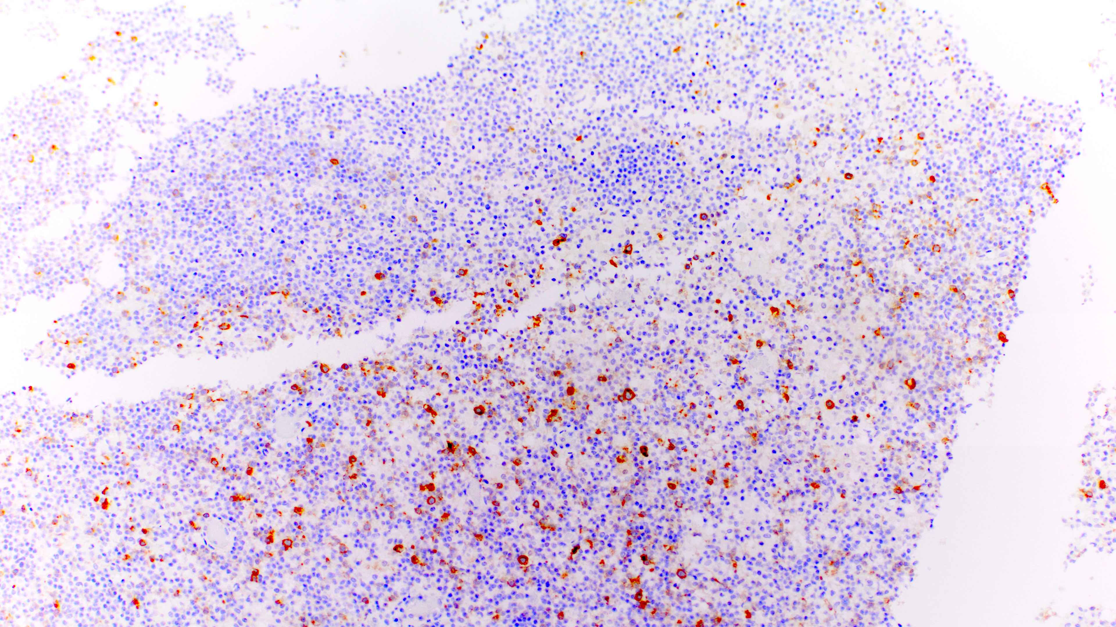
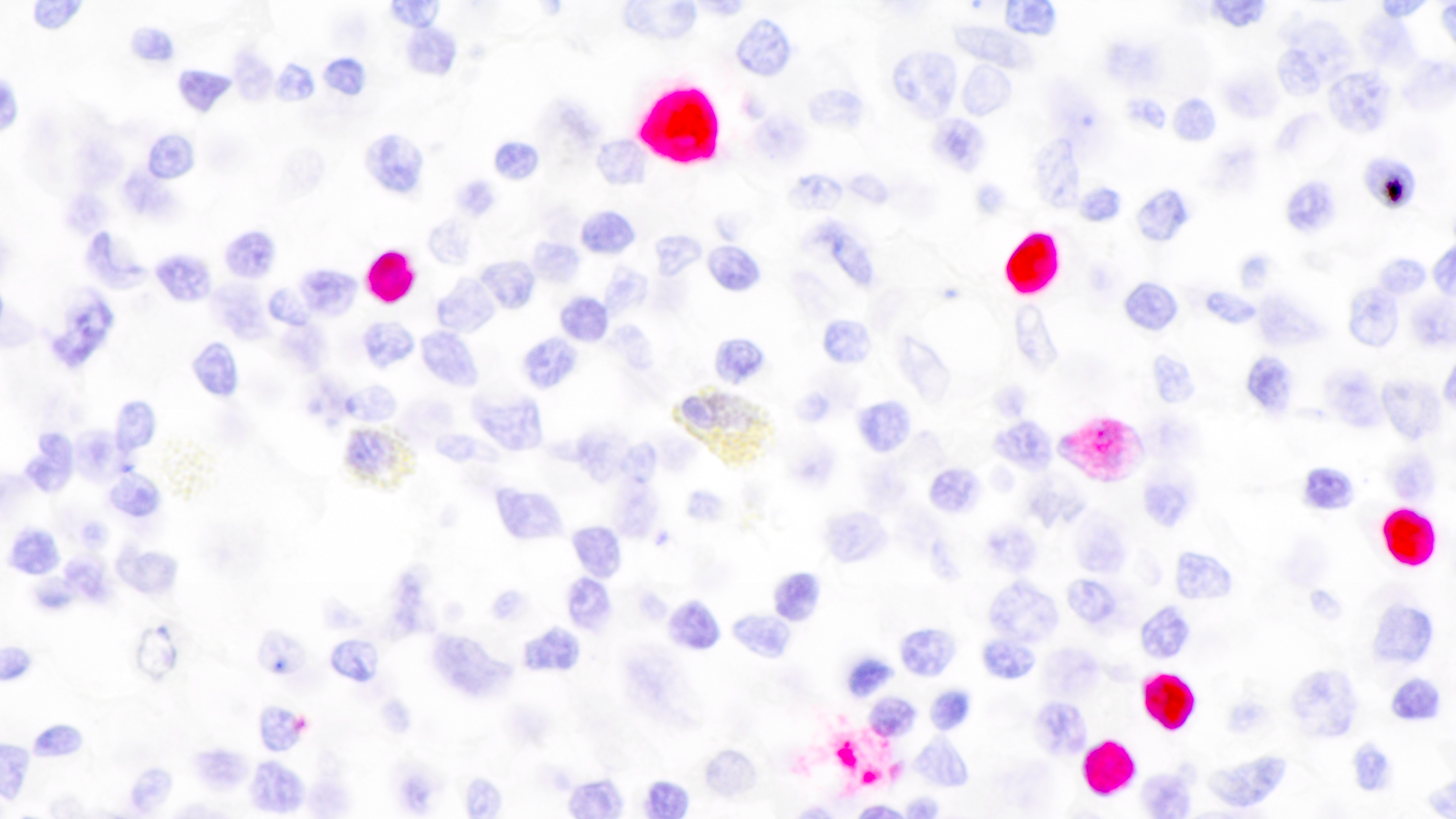
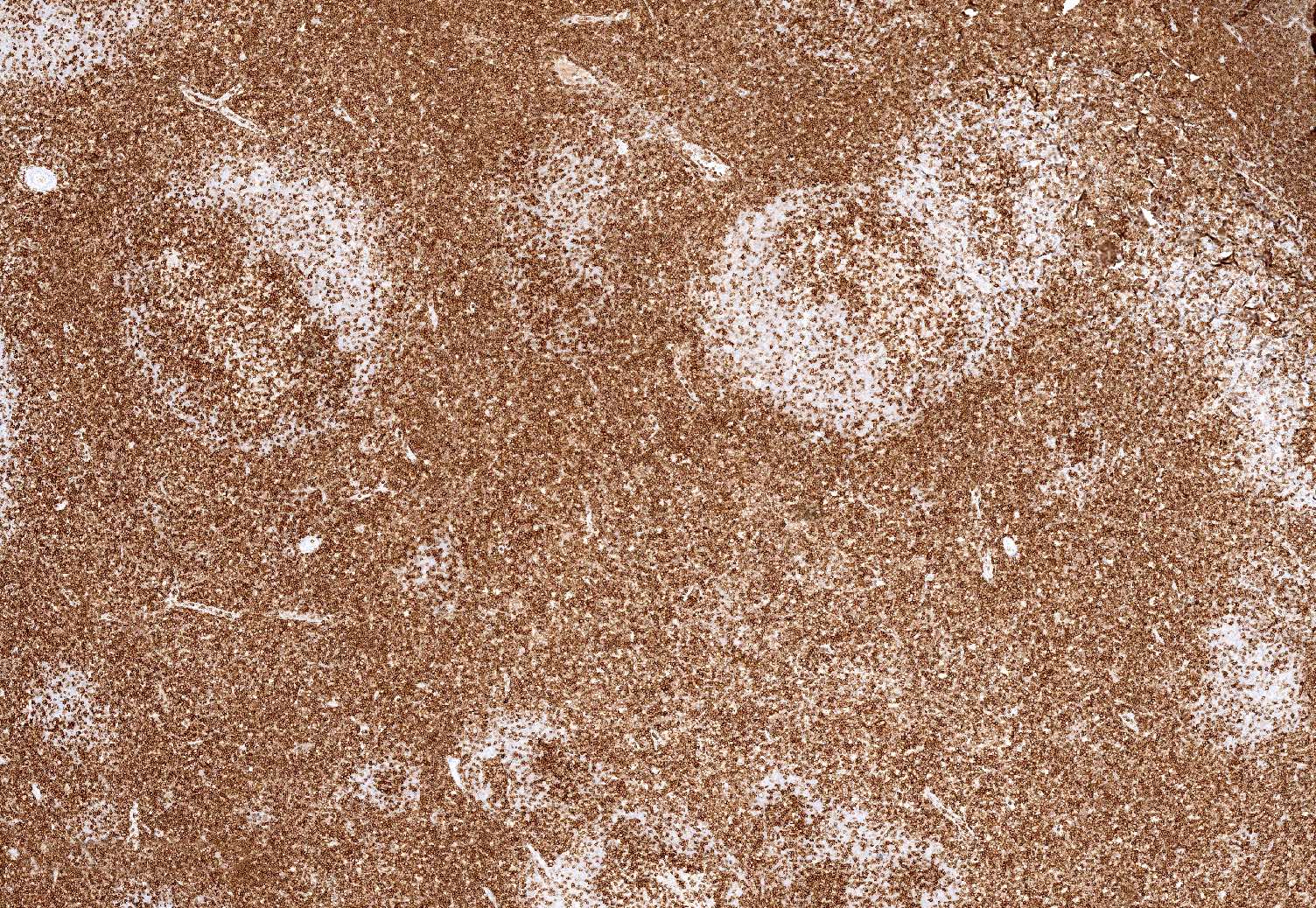
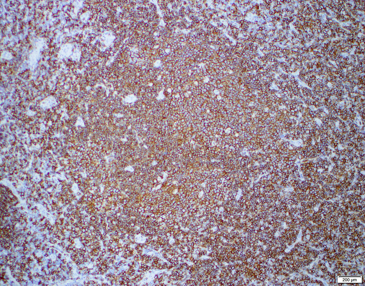
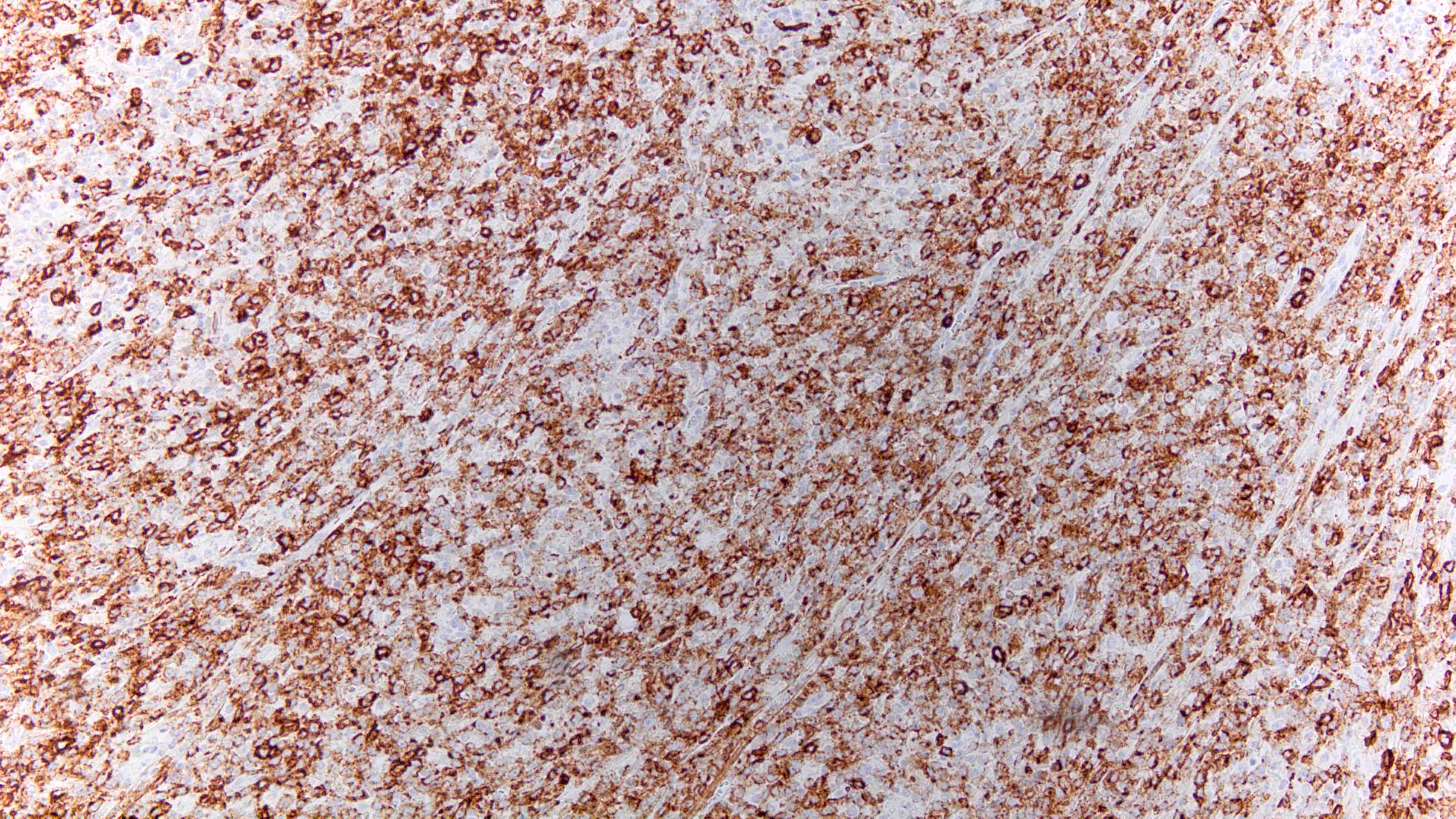
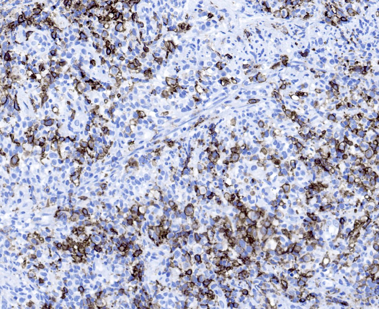
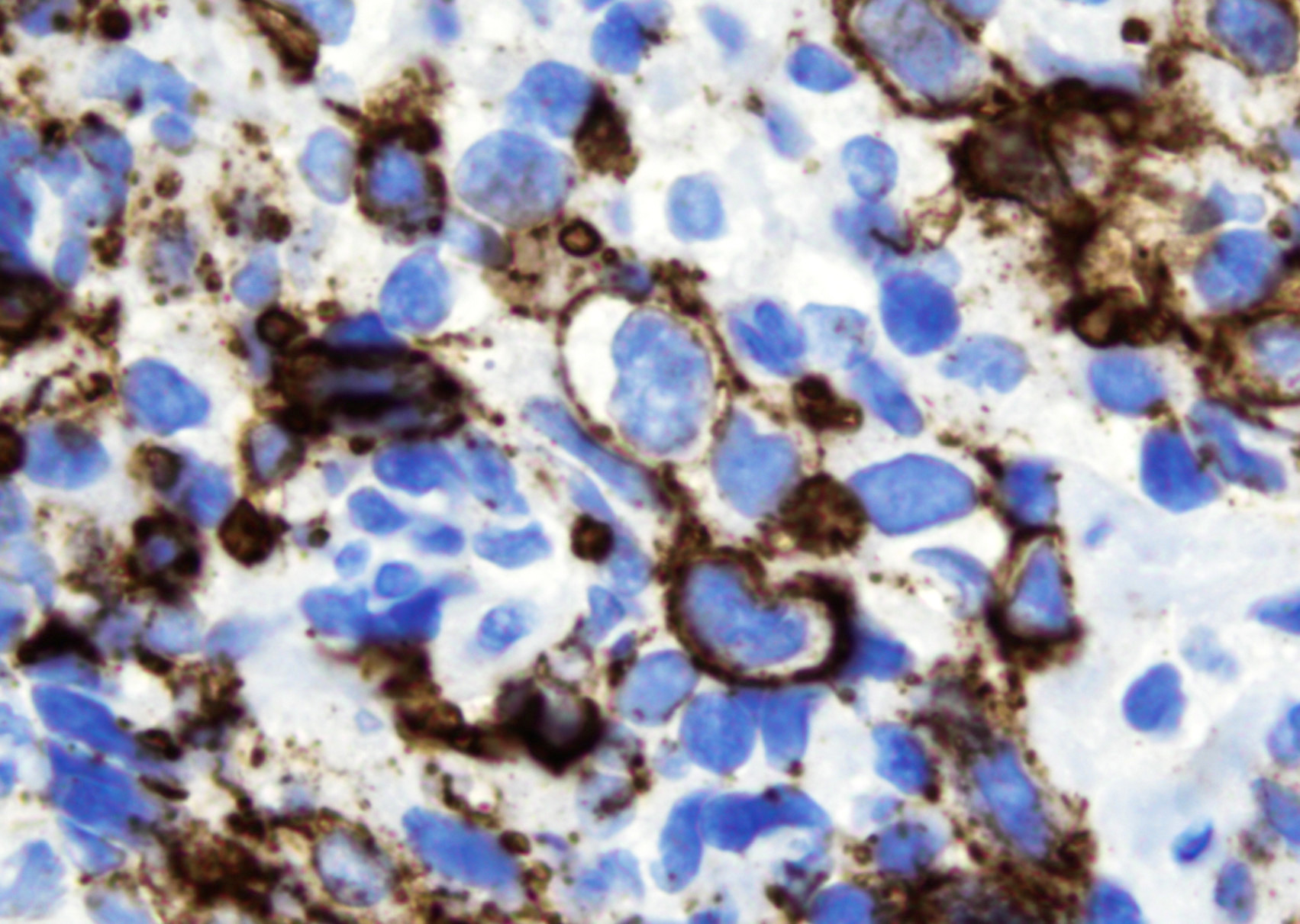
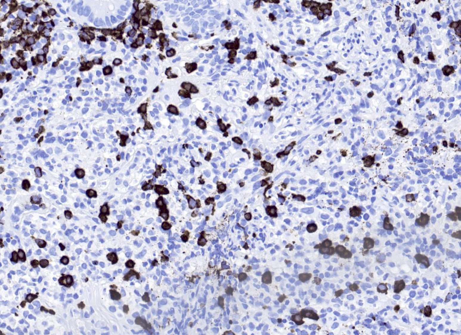
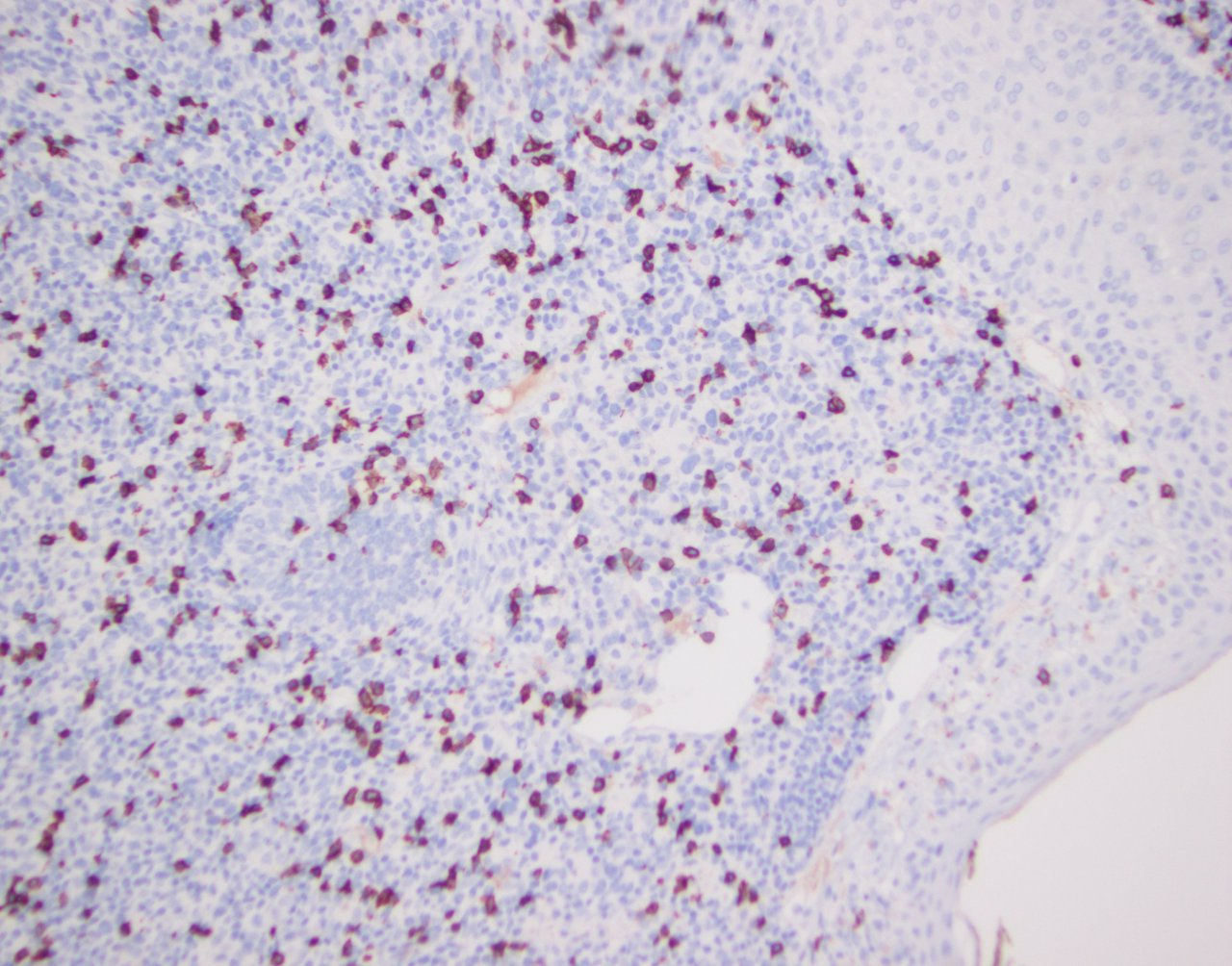
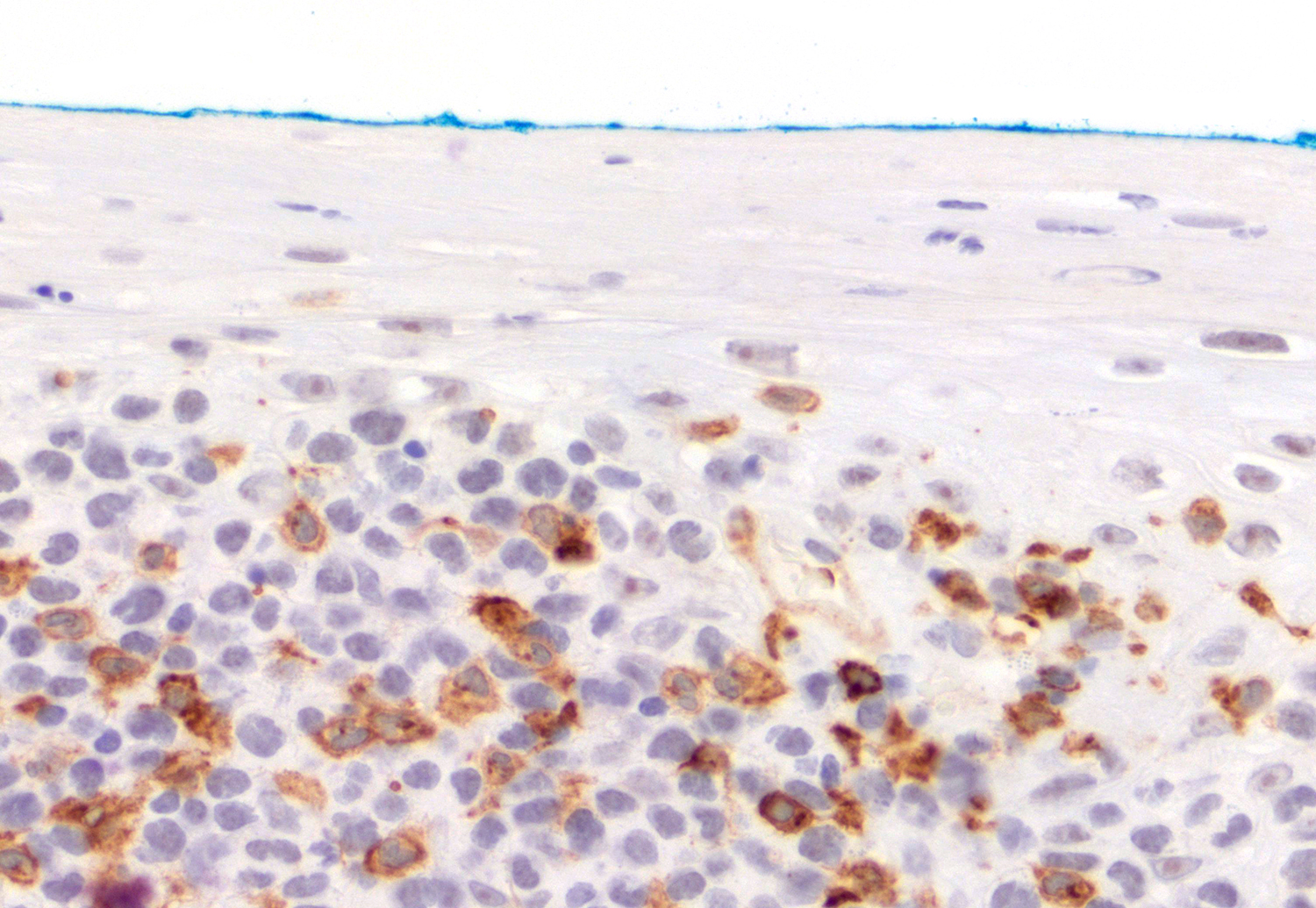
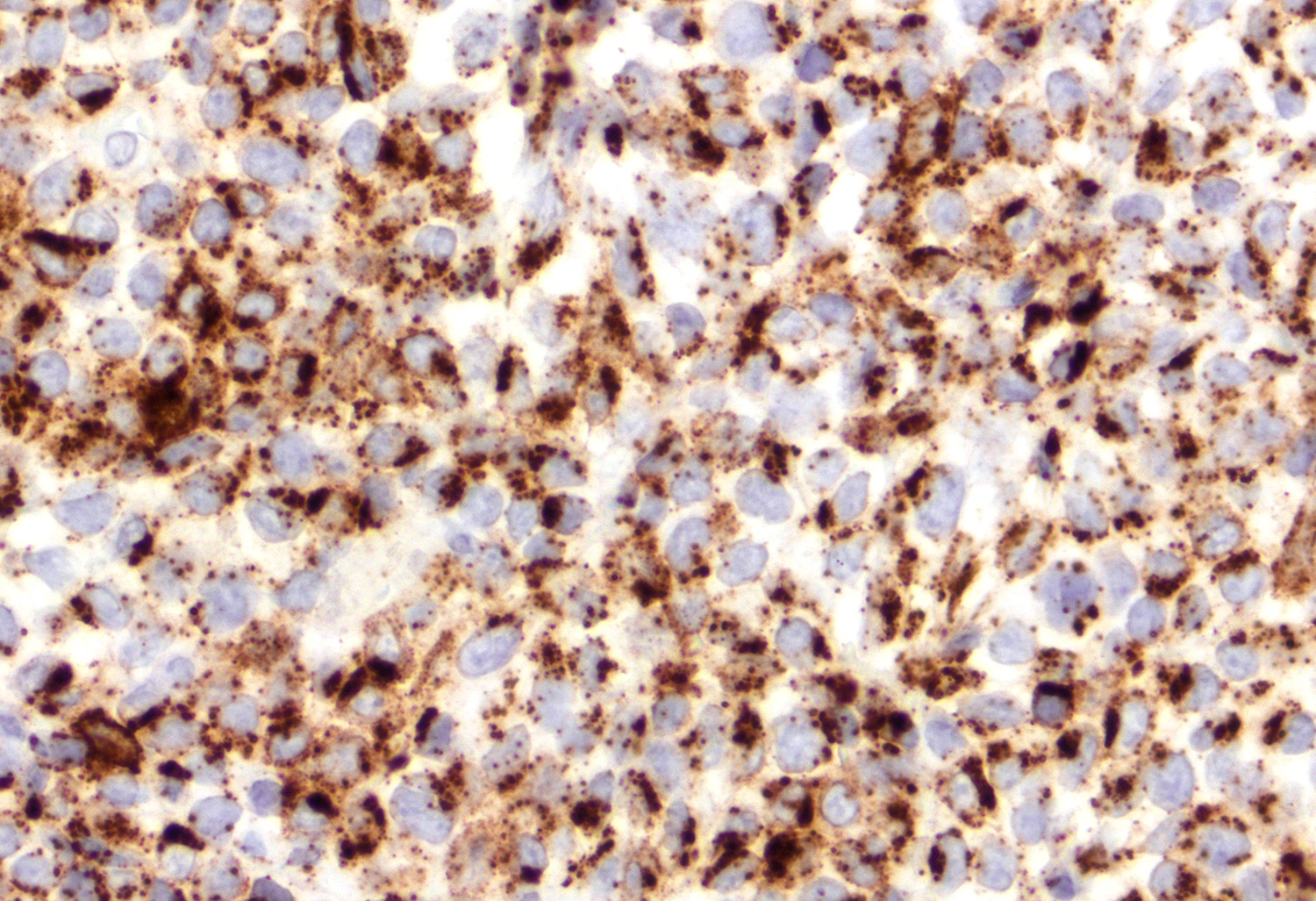
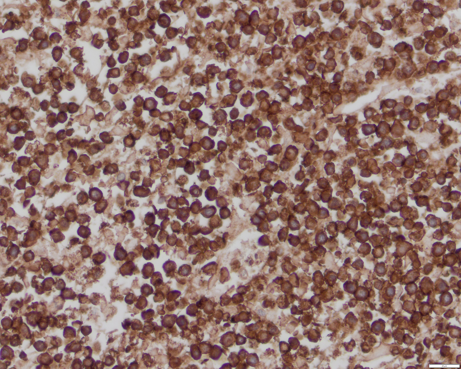
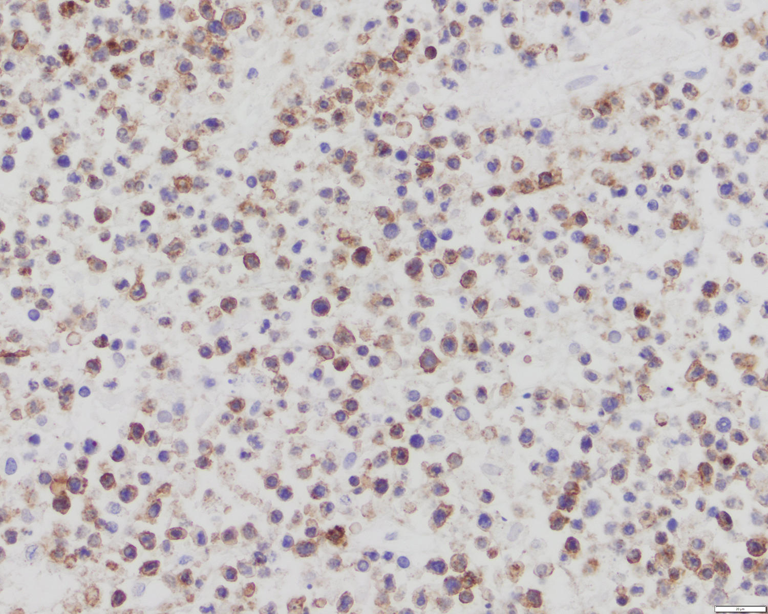
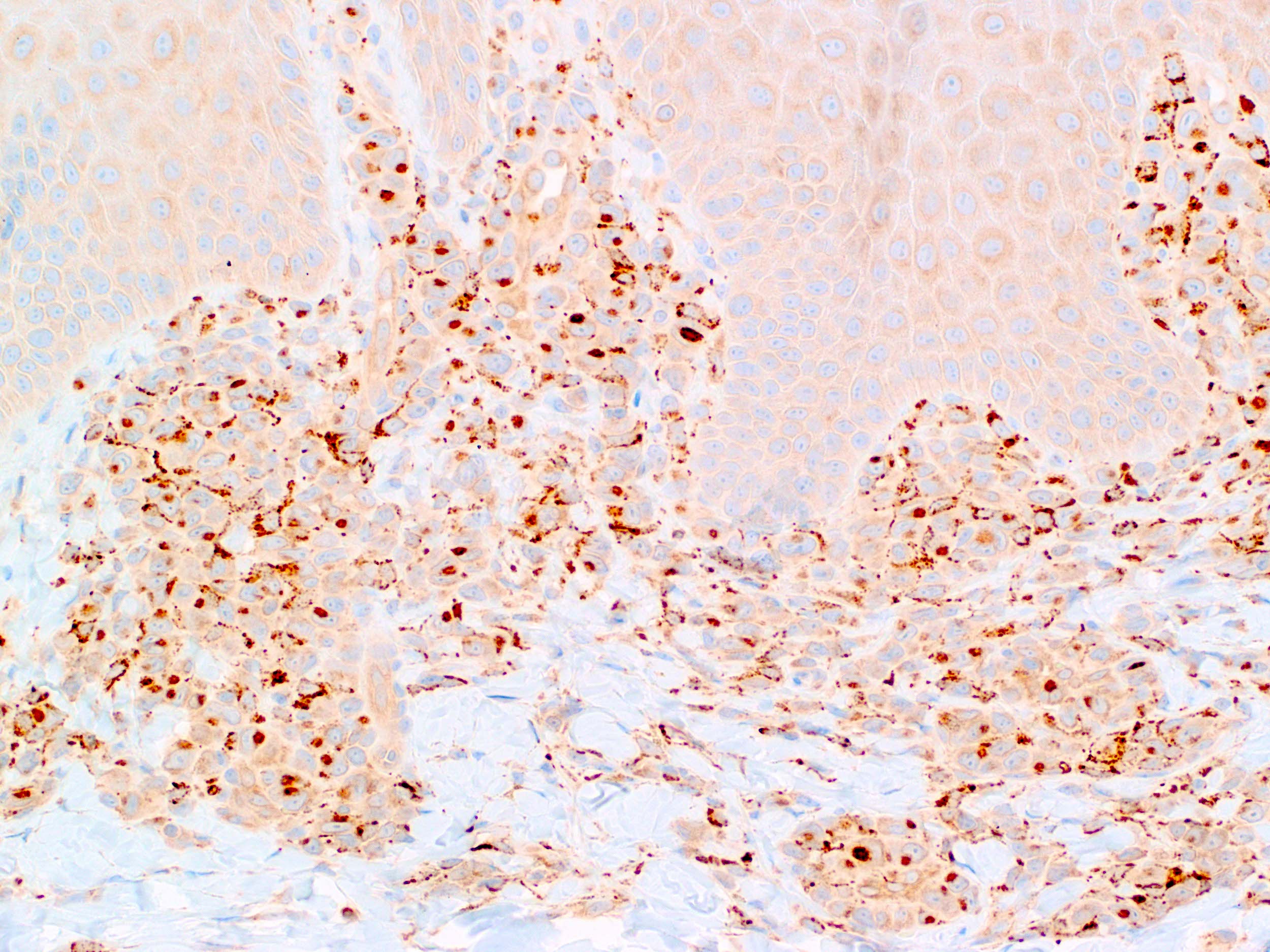
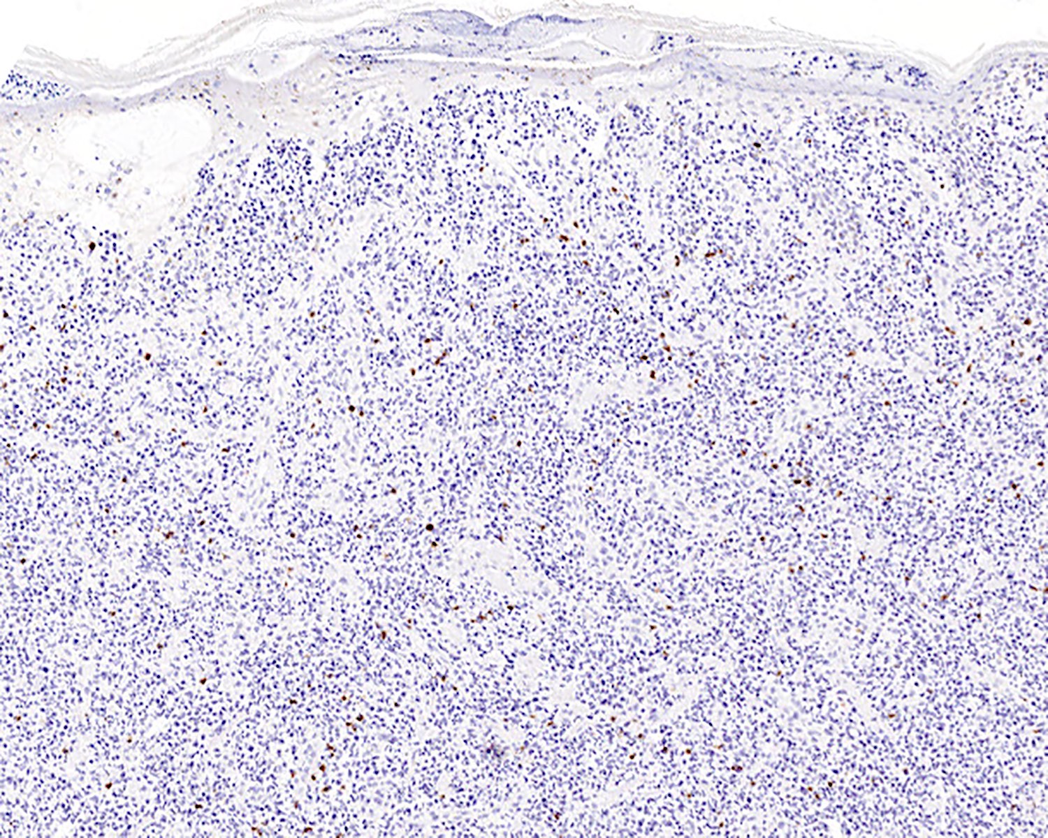
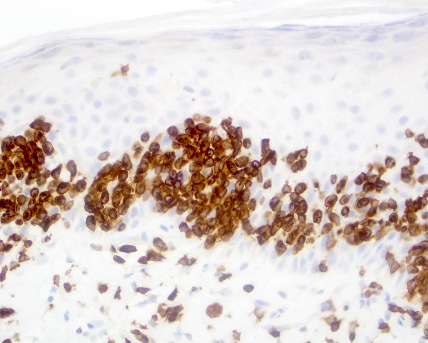
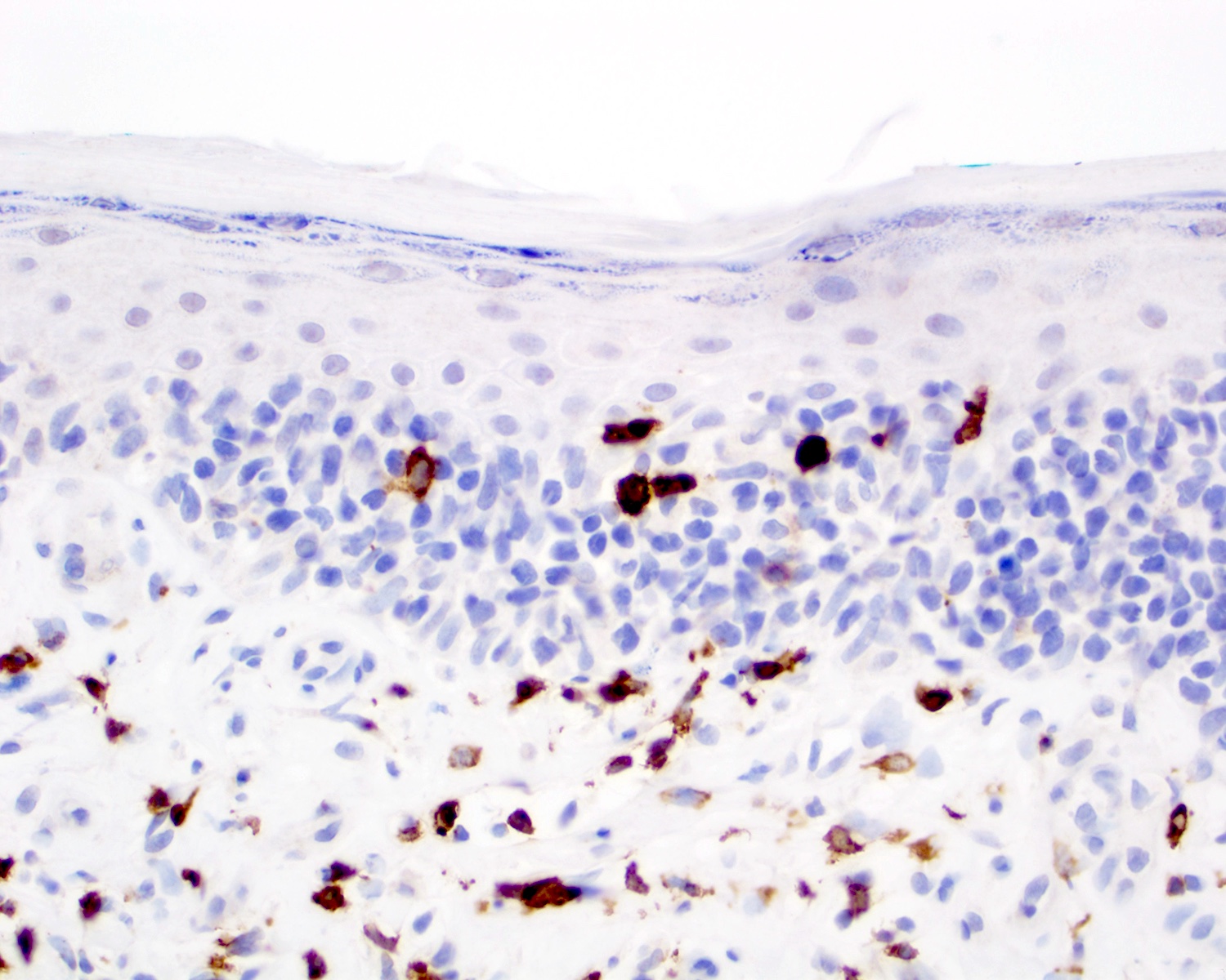
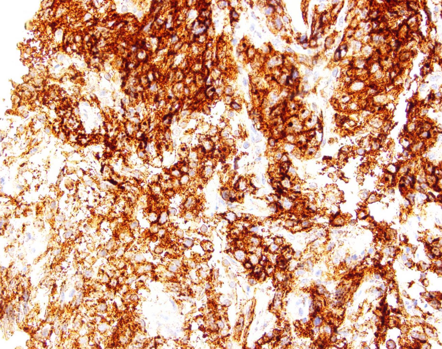
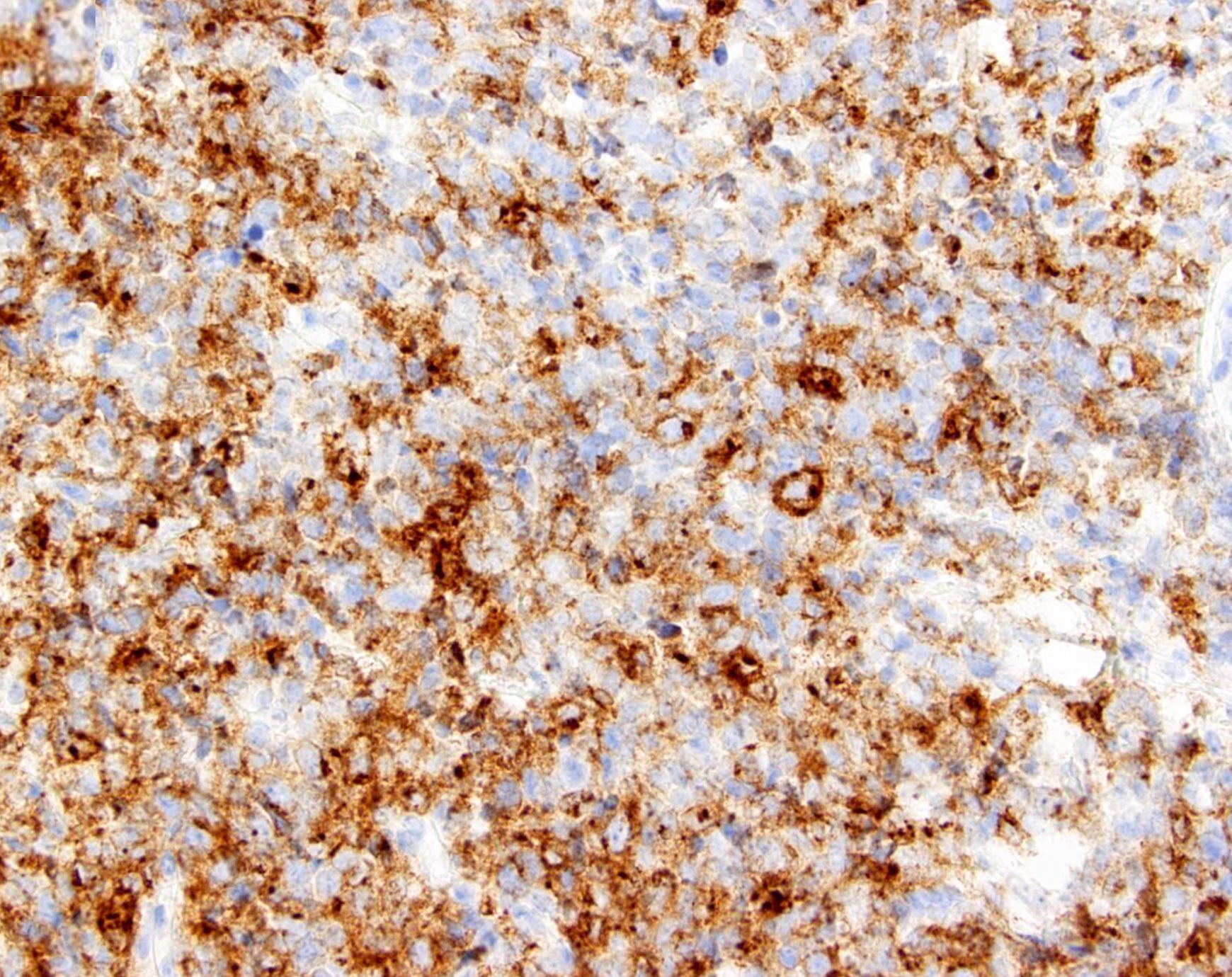
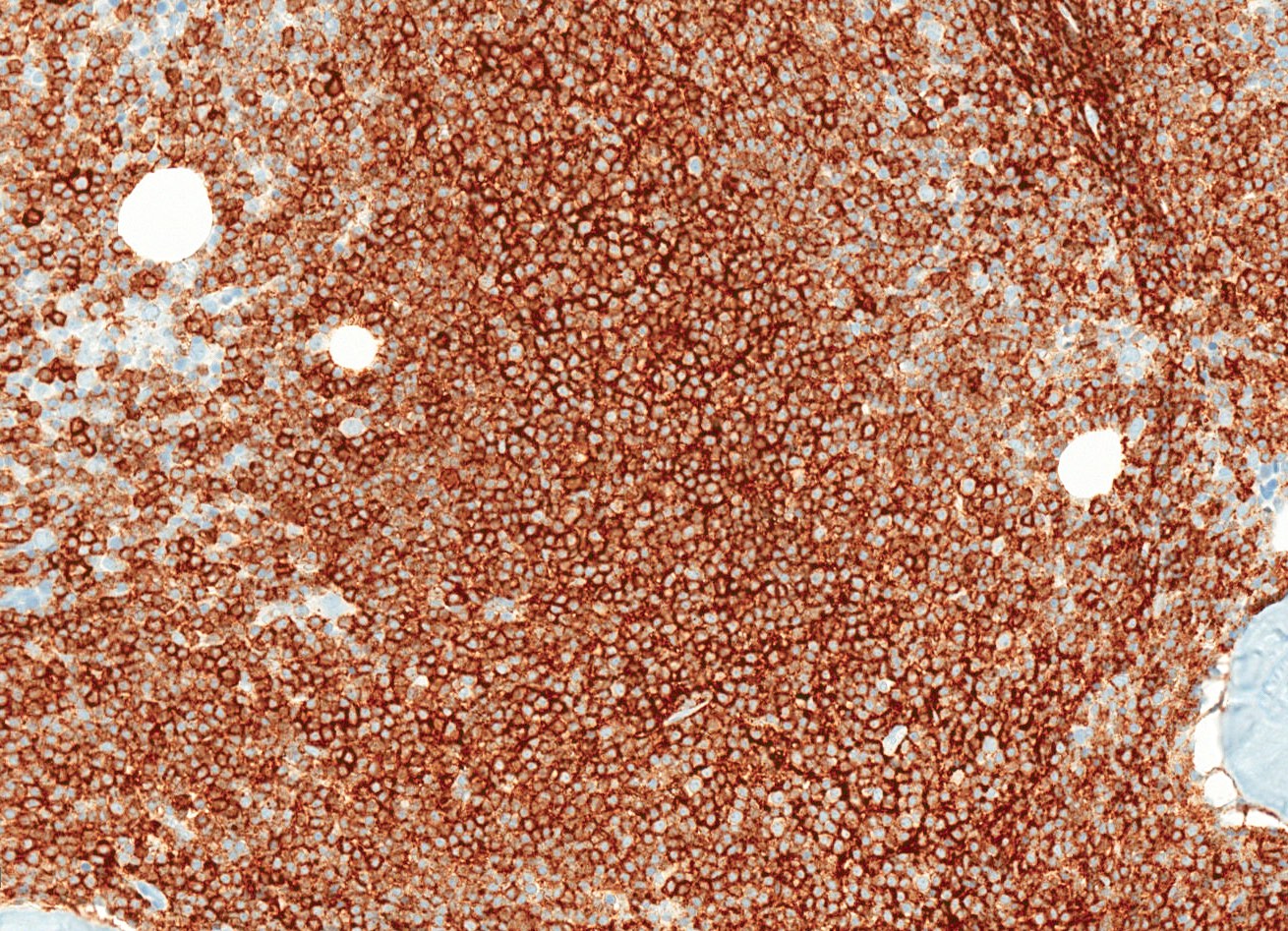
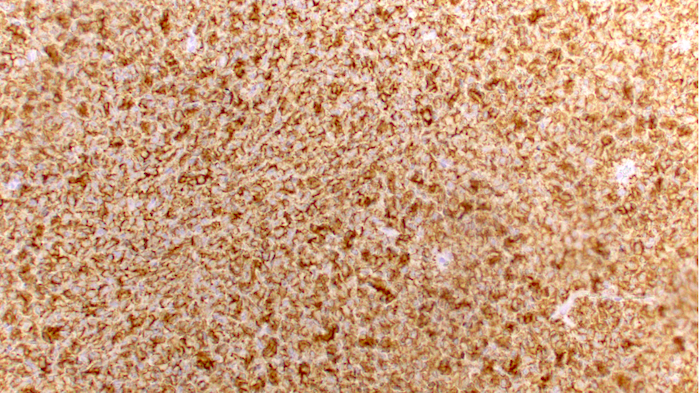
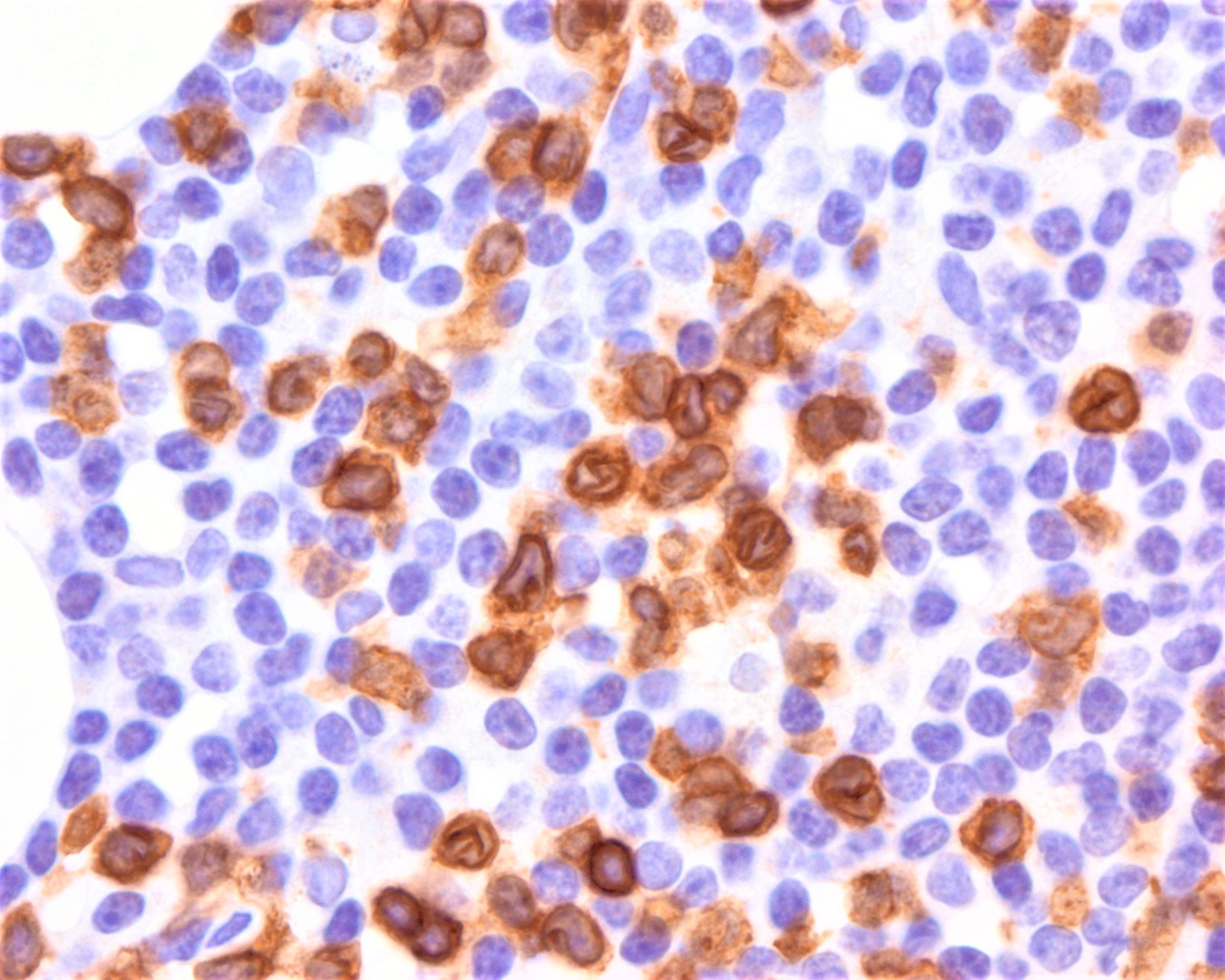
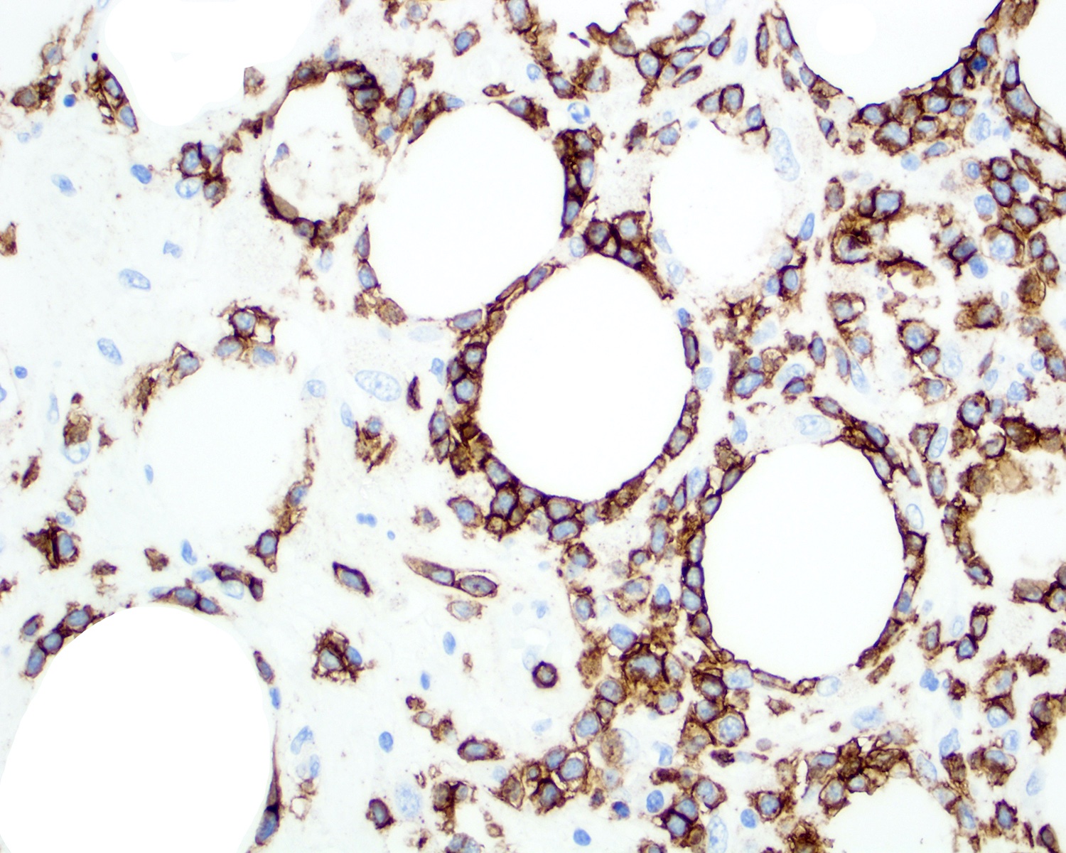
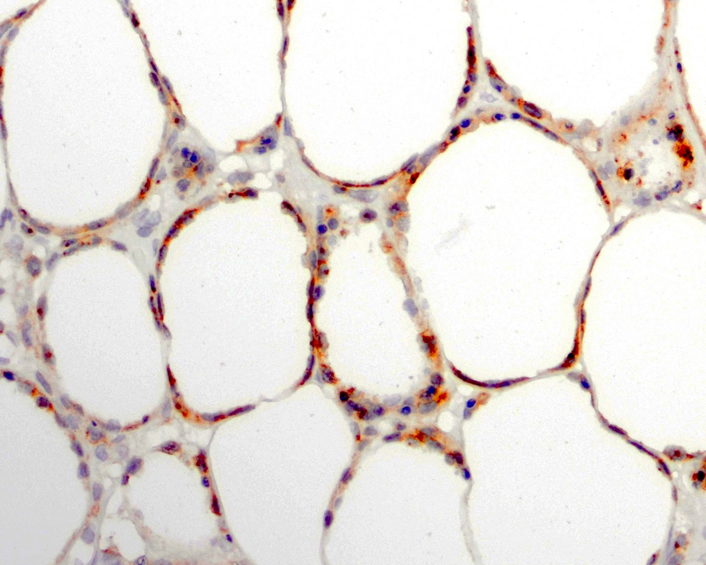
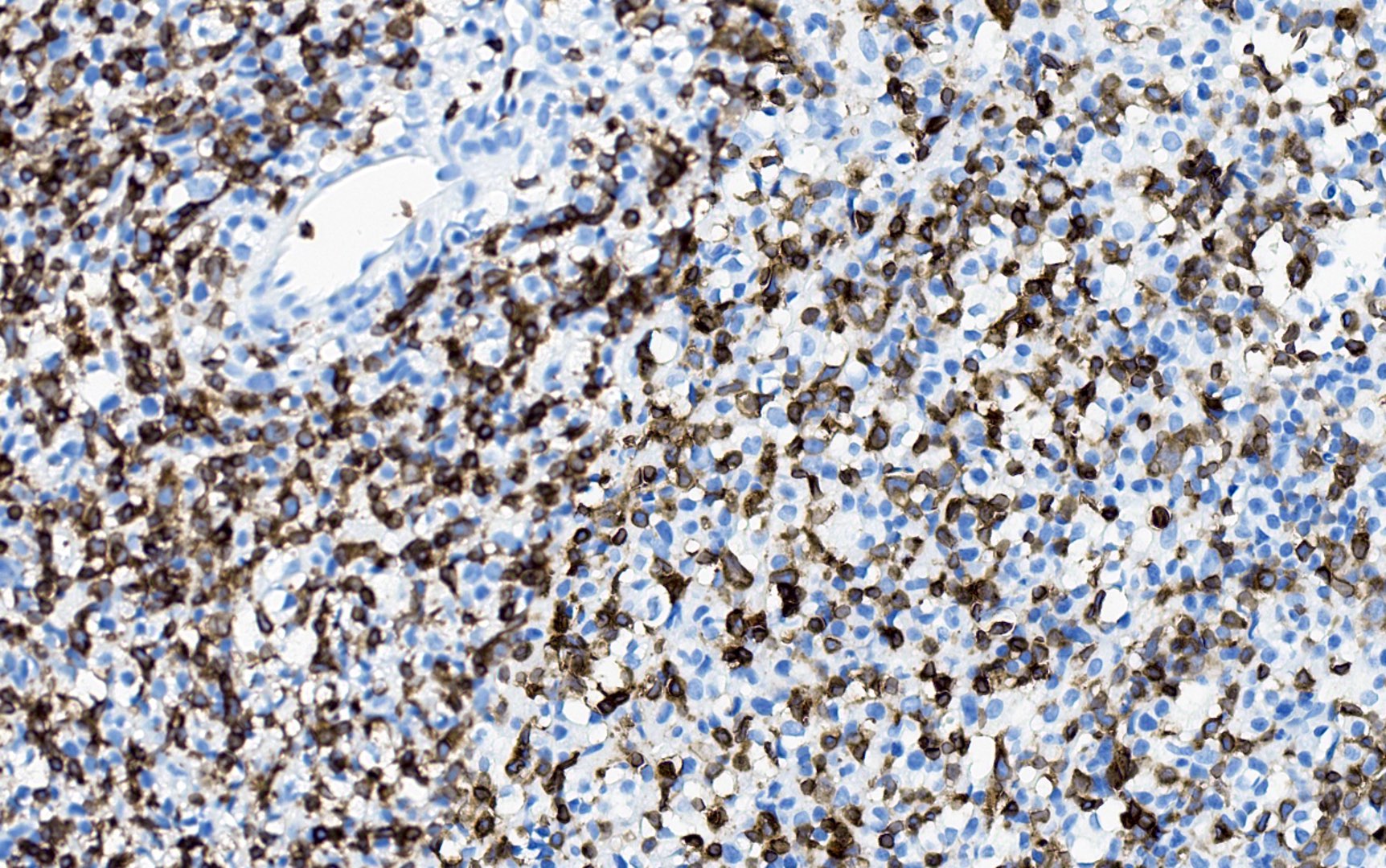
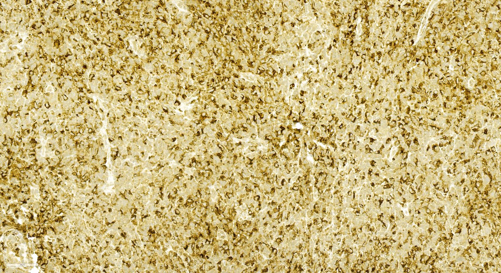
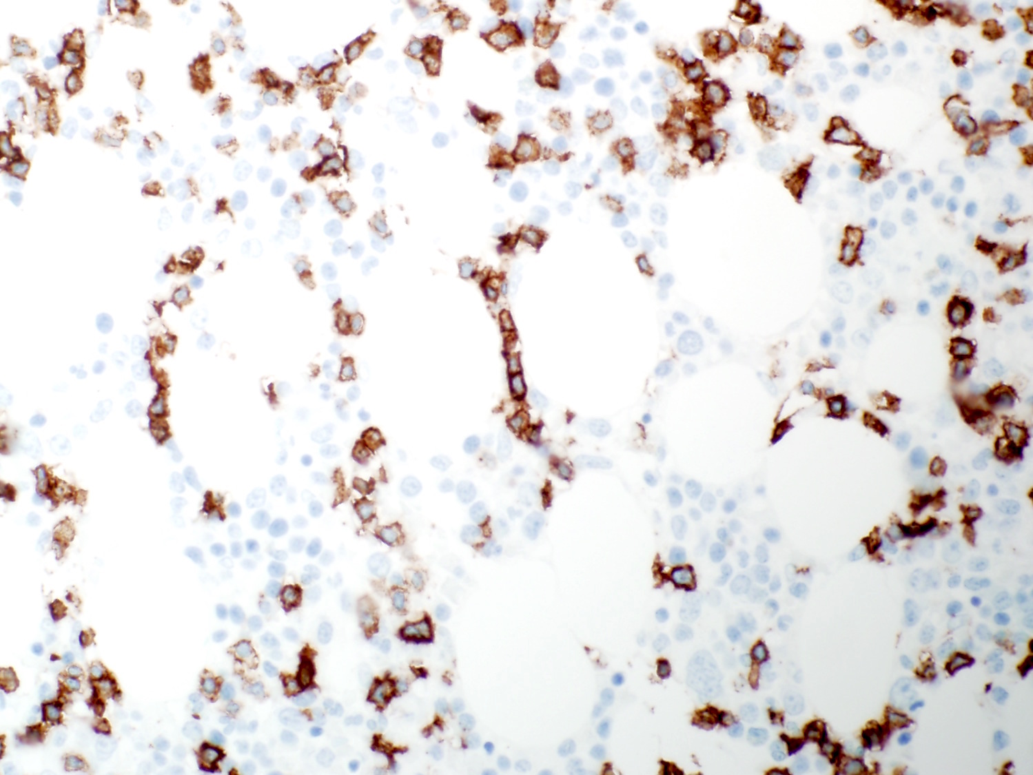
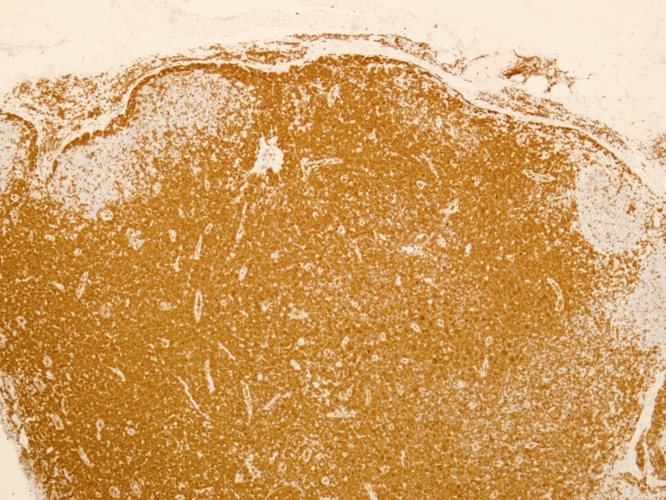
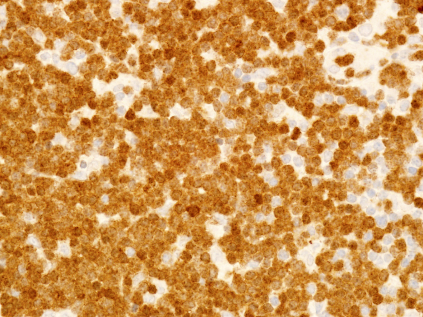
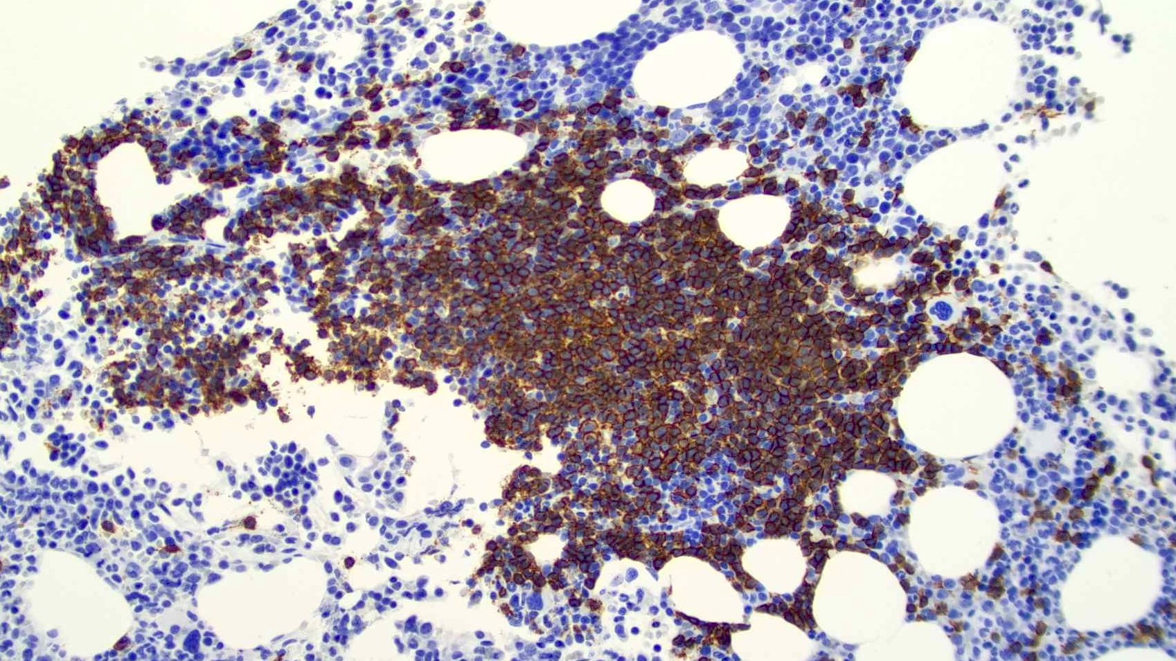
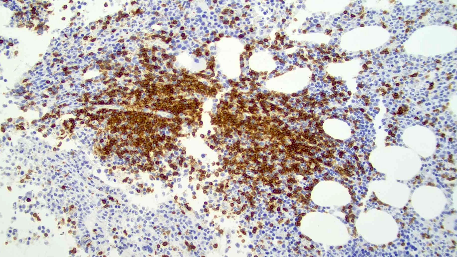
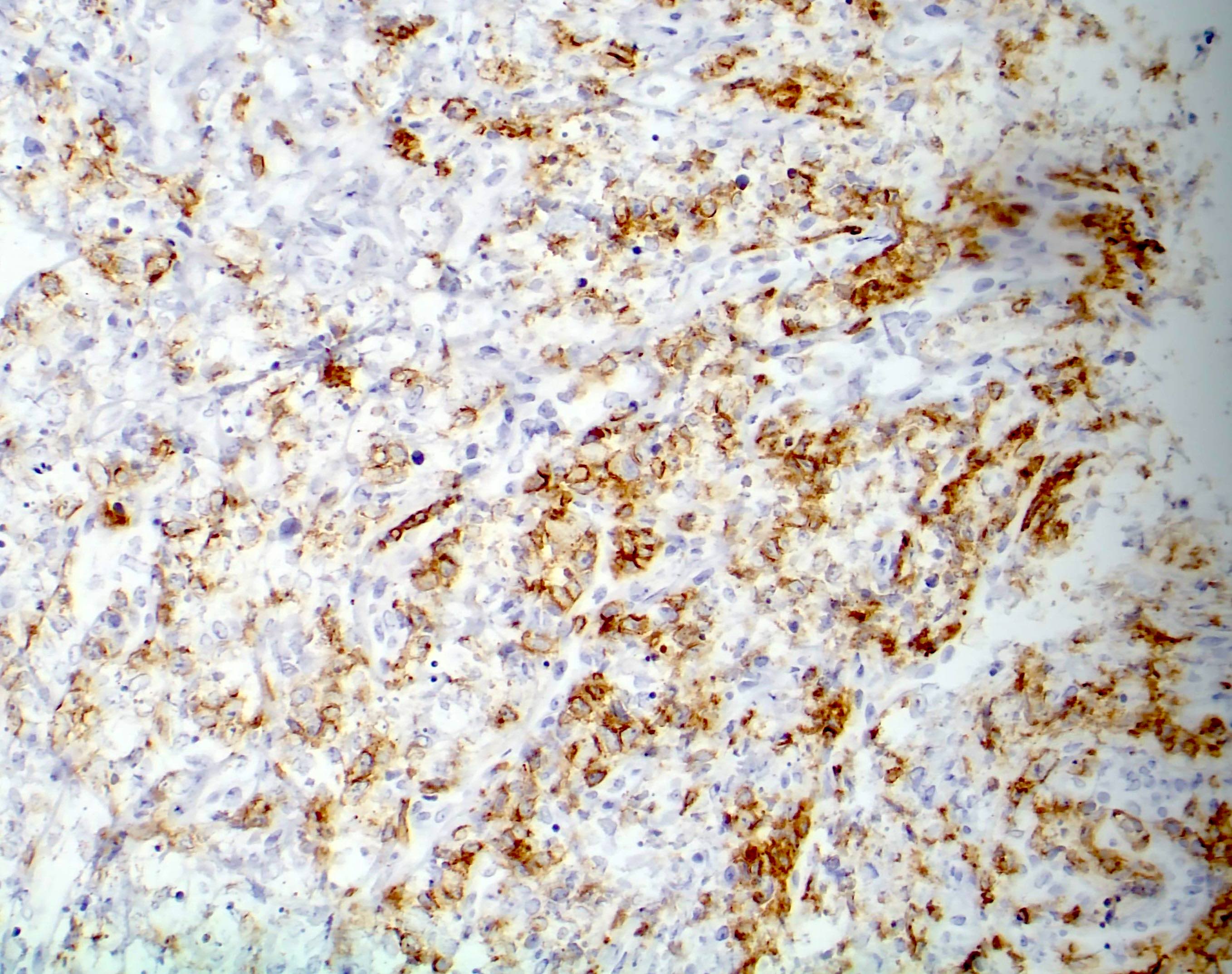
Cytoplasmic granular ALK positivity

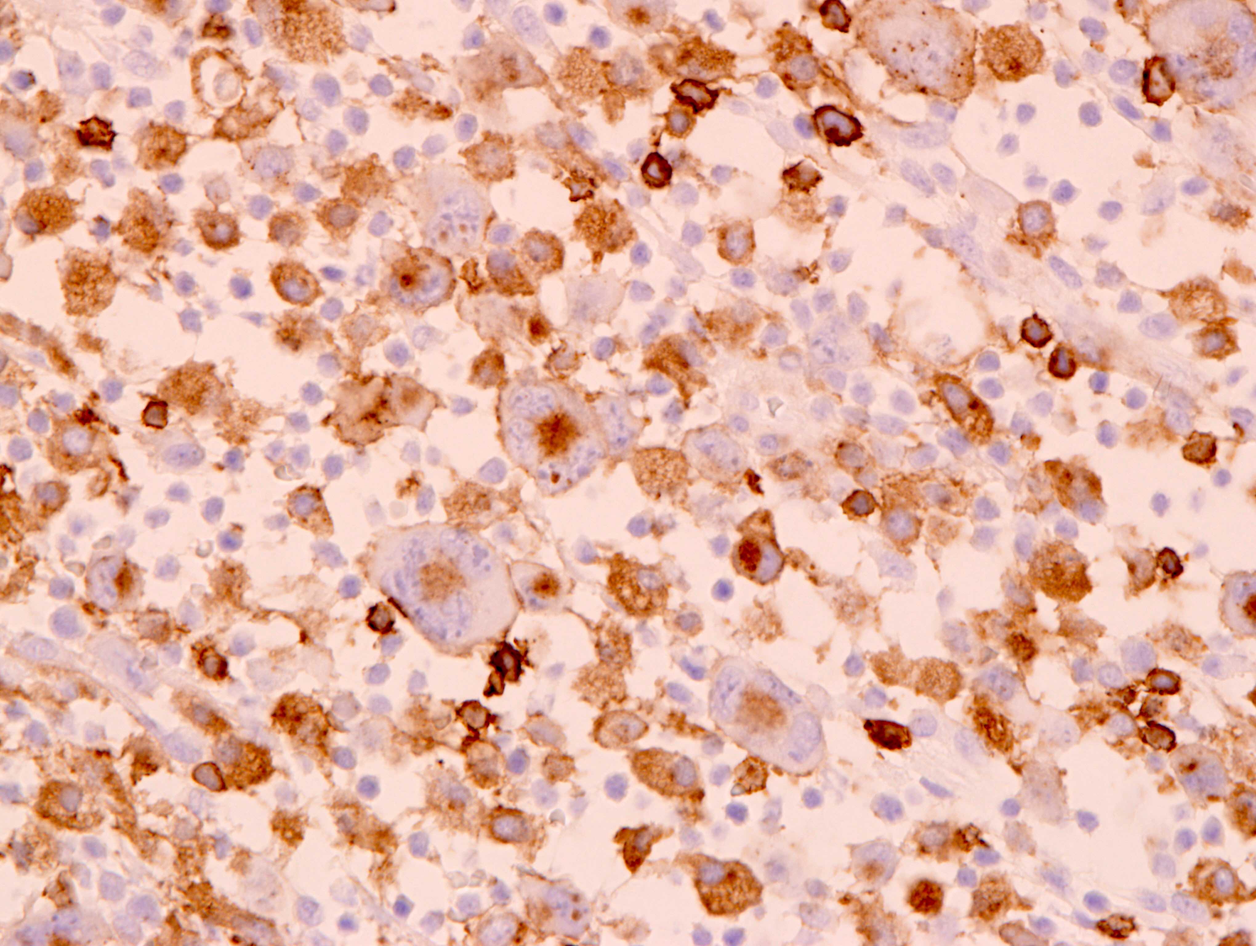
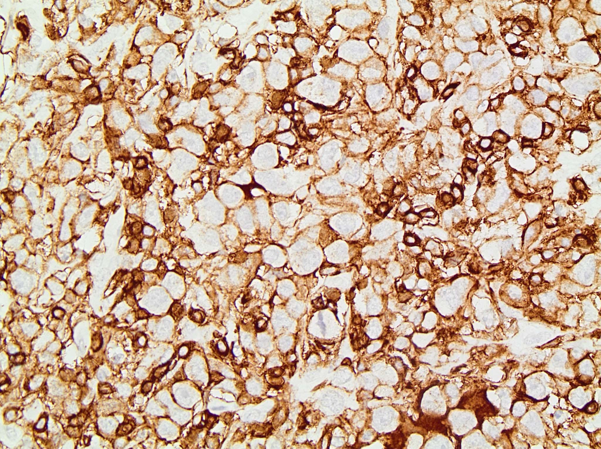
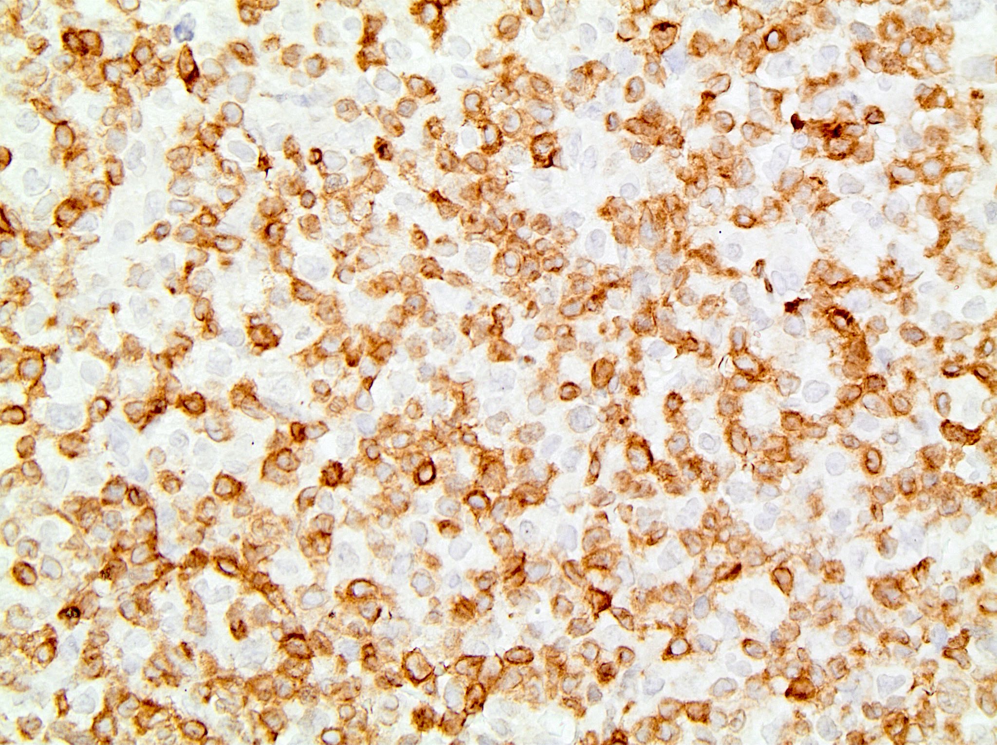
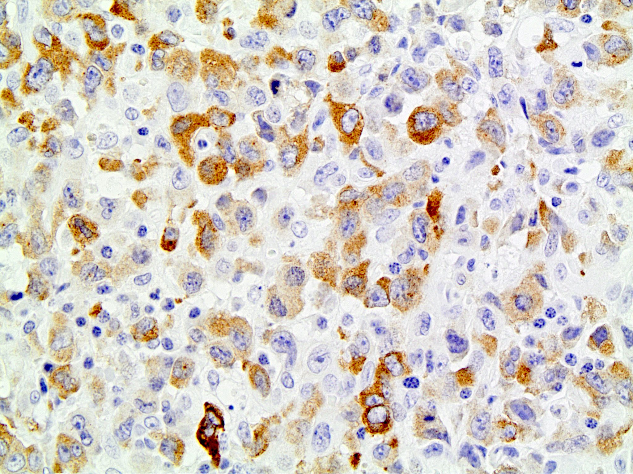
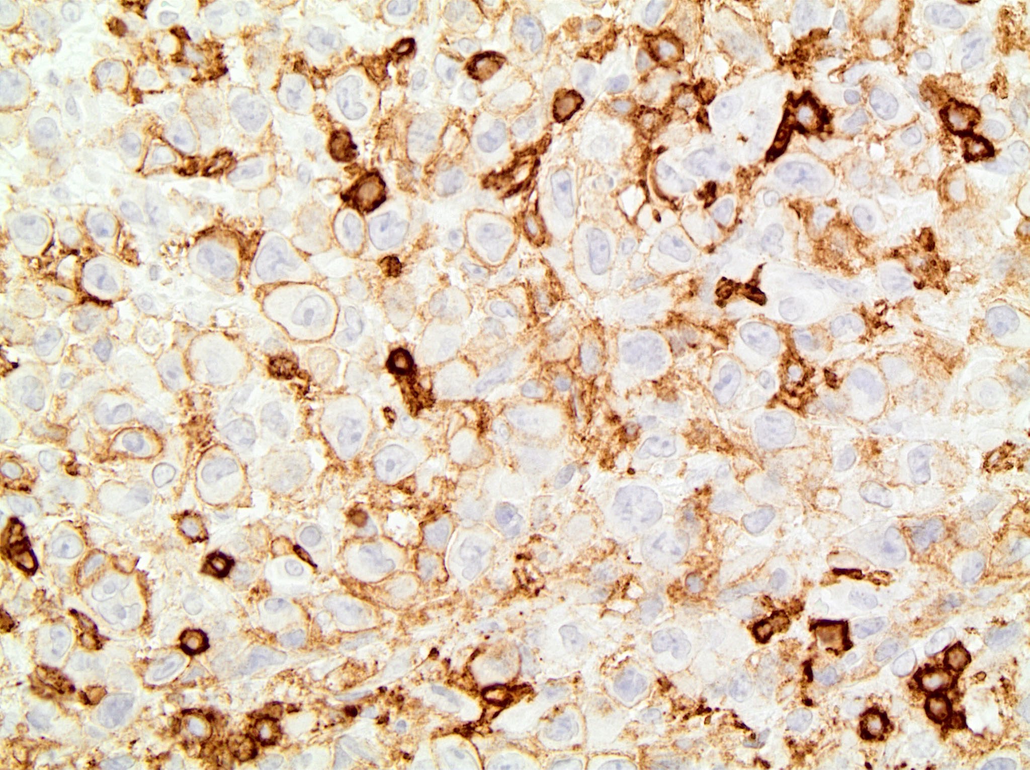
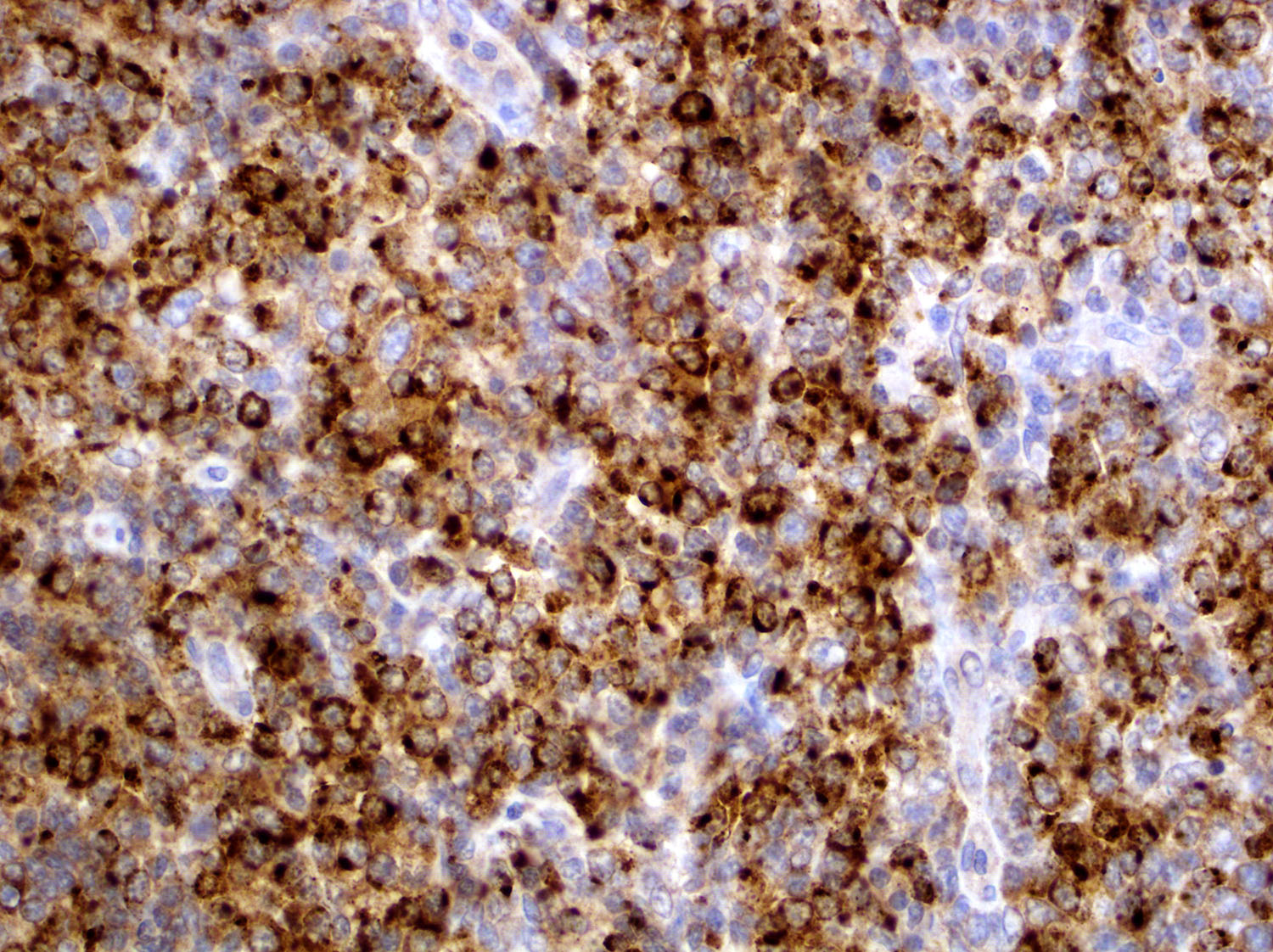
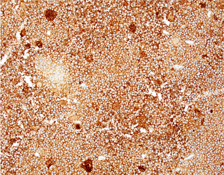
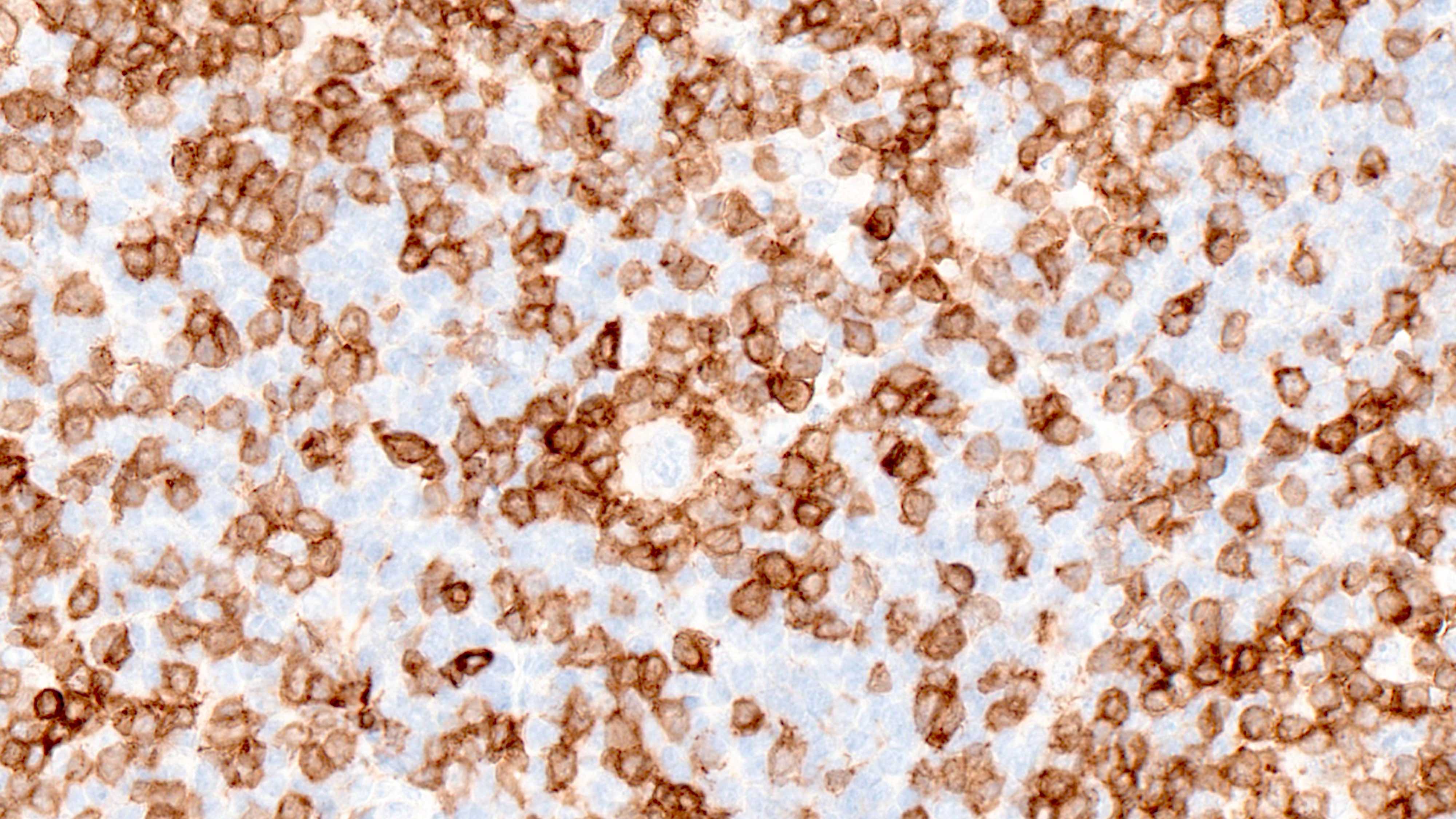
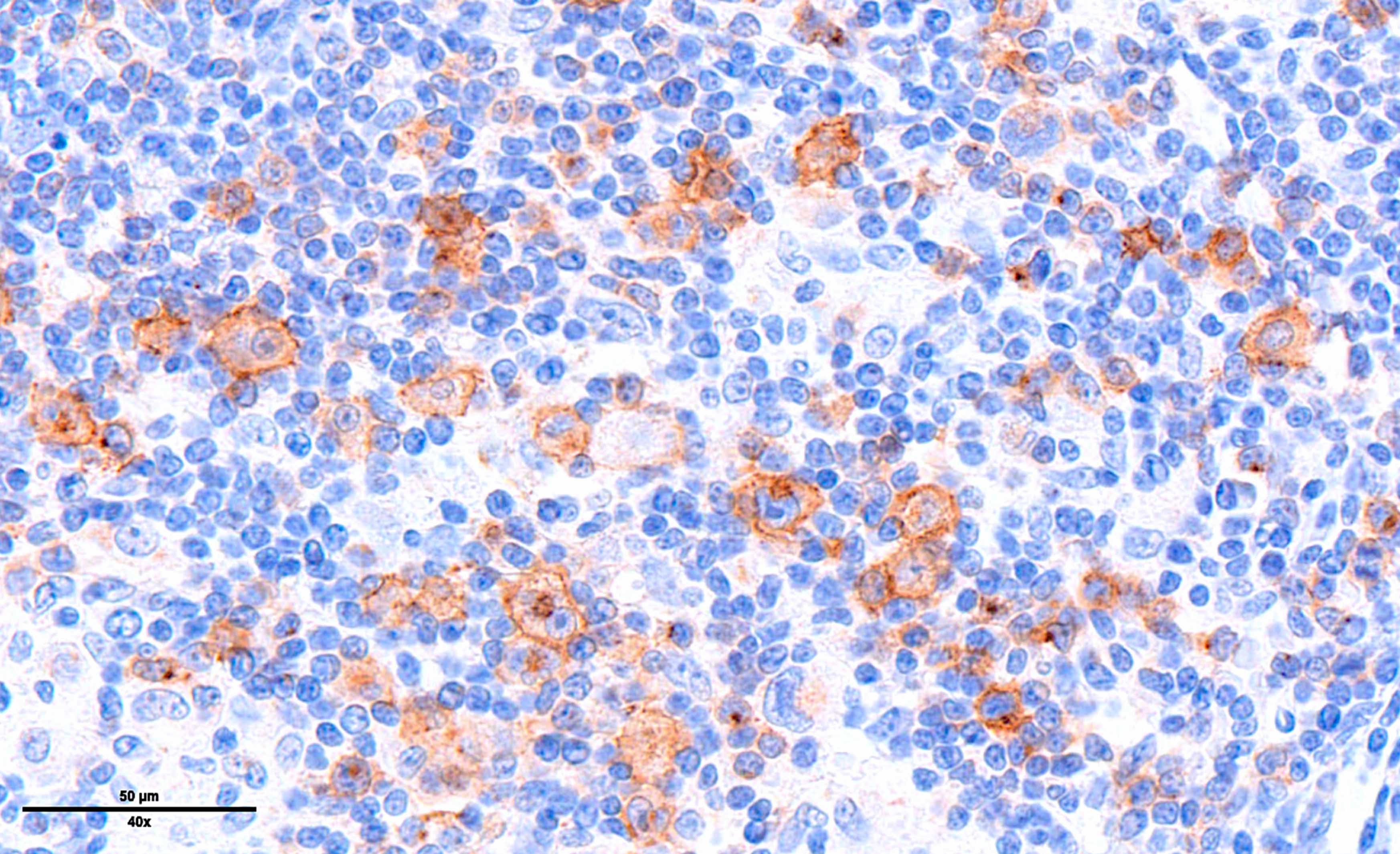
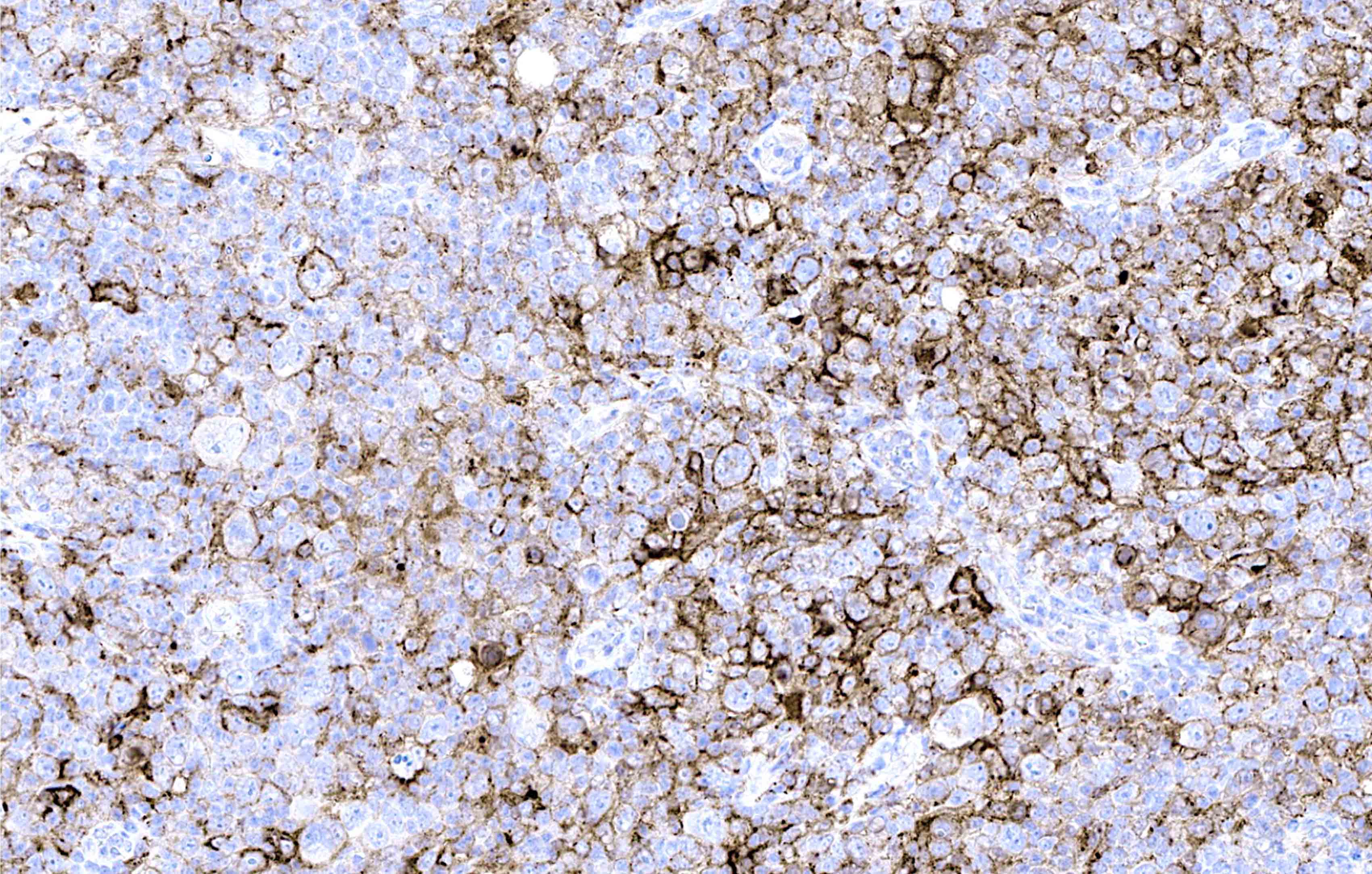
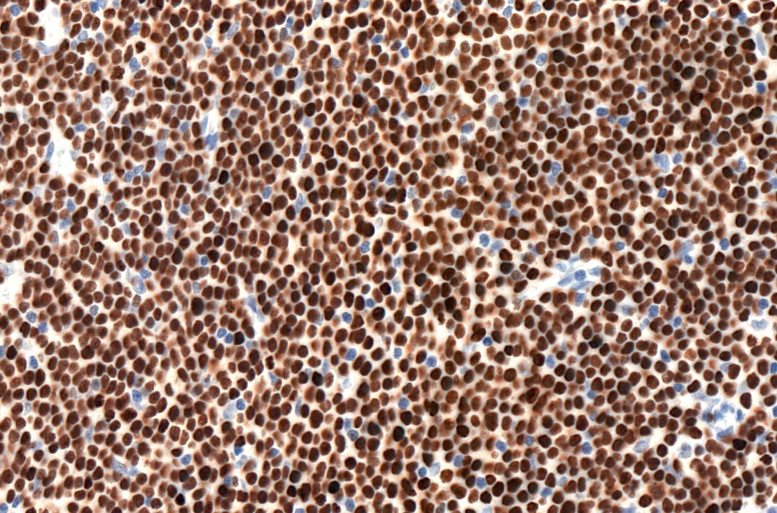
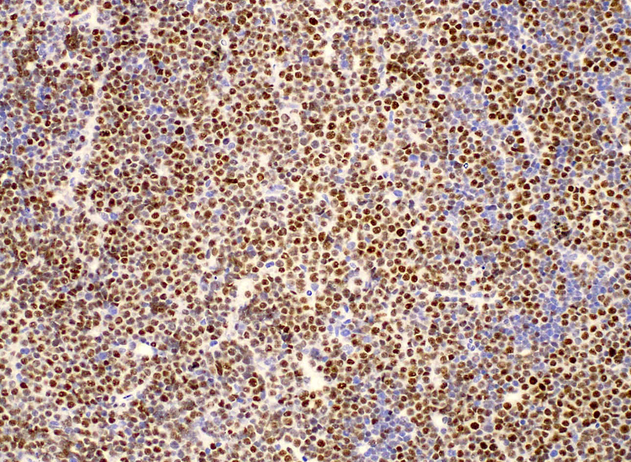
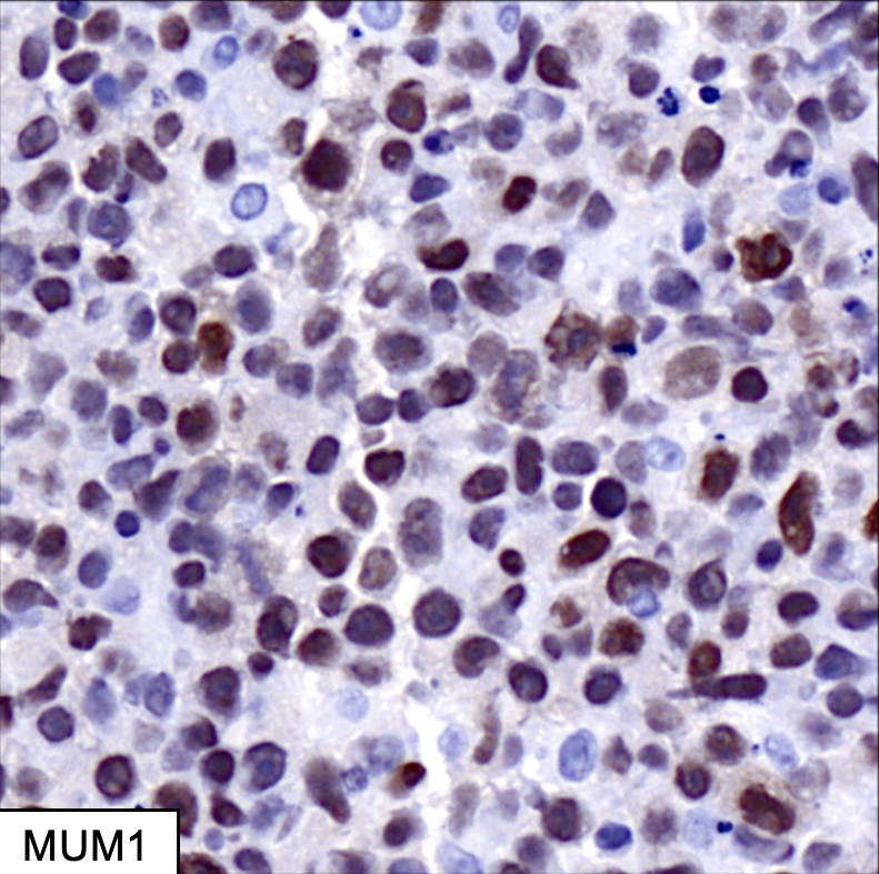
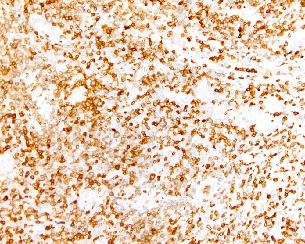
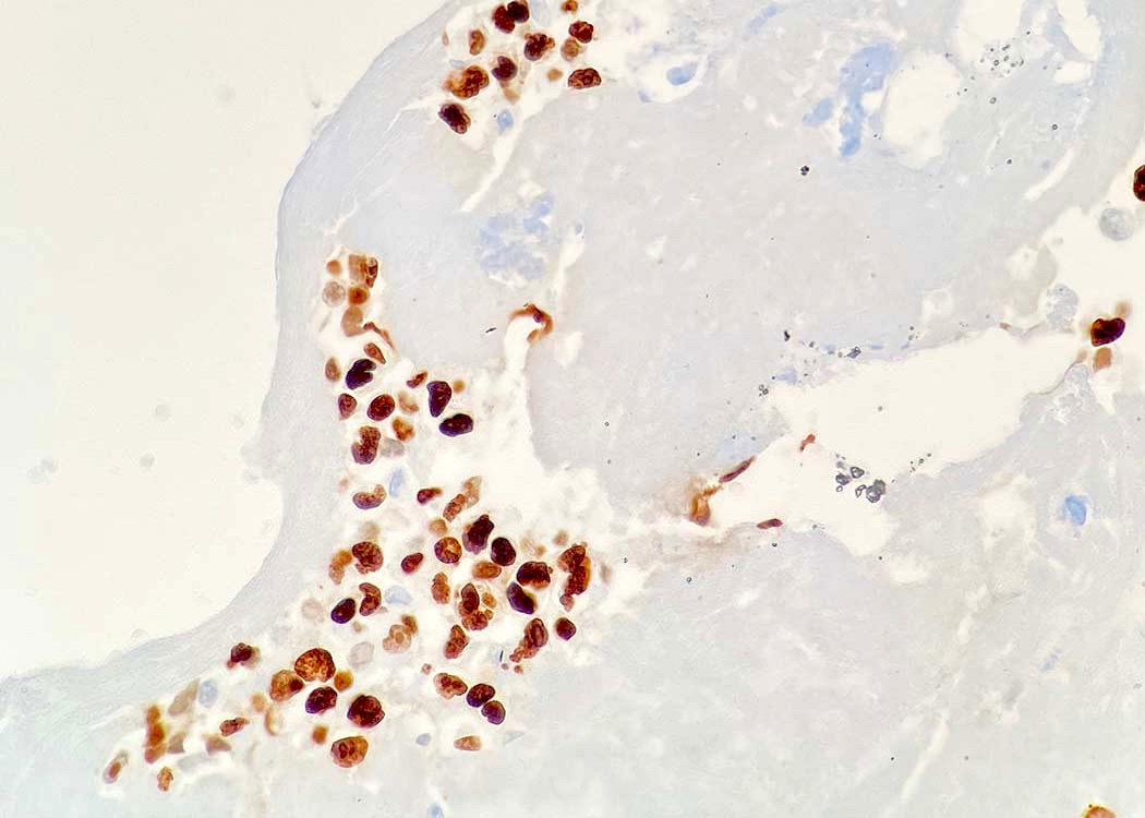
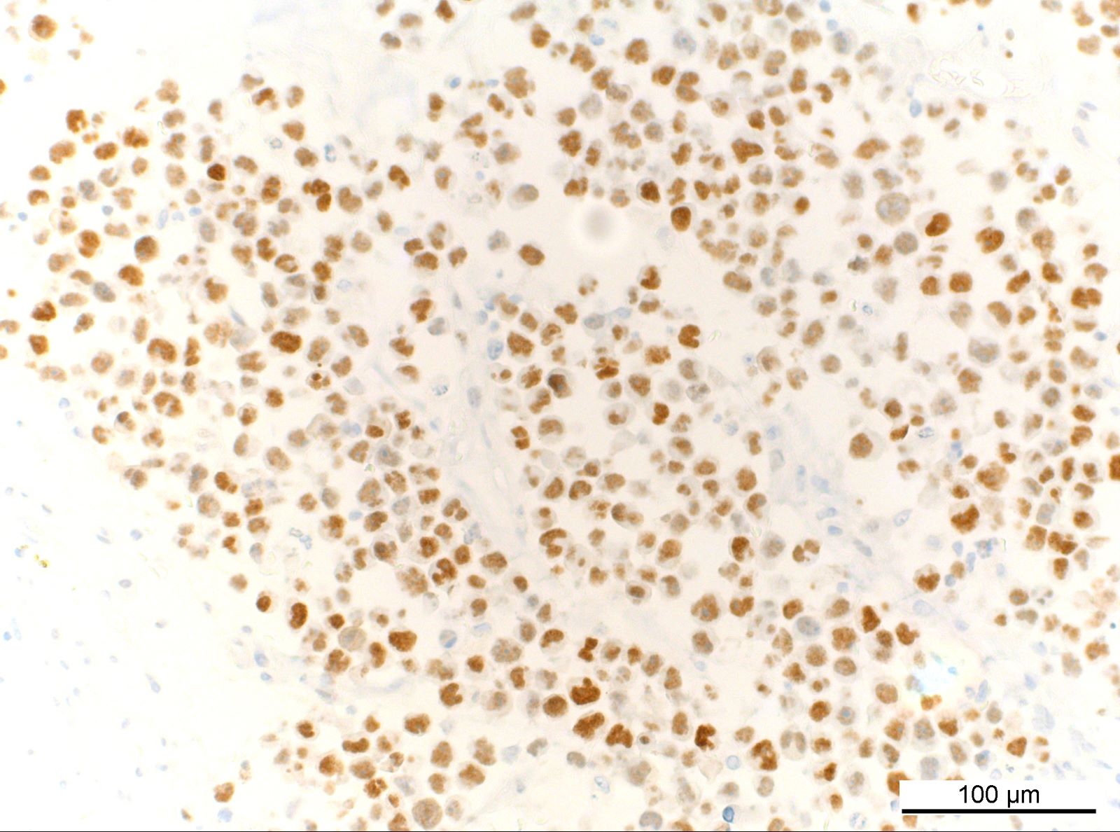
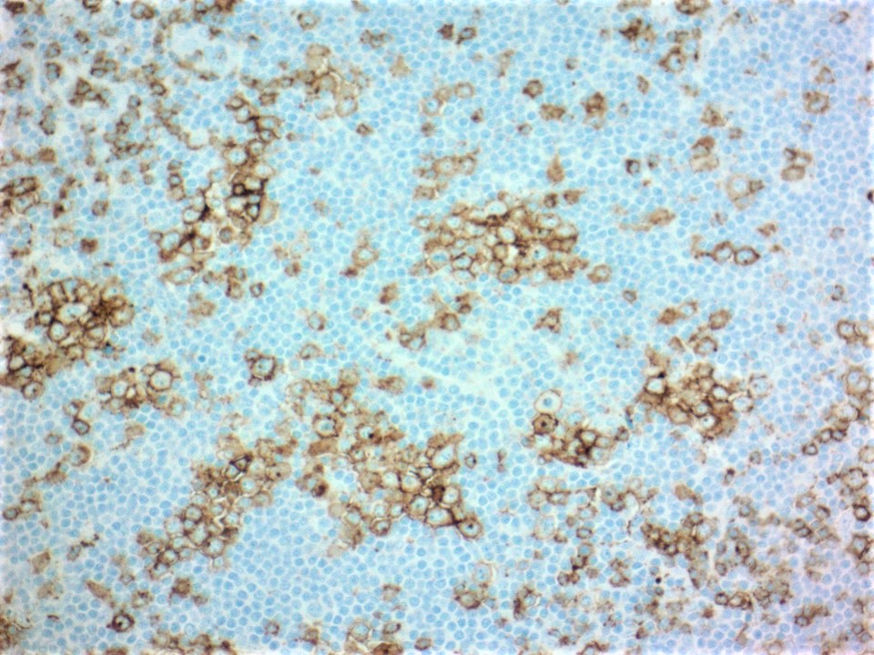
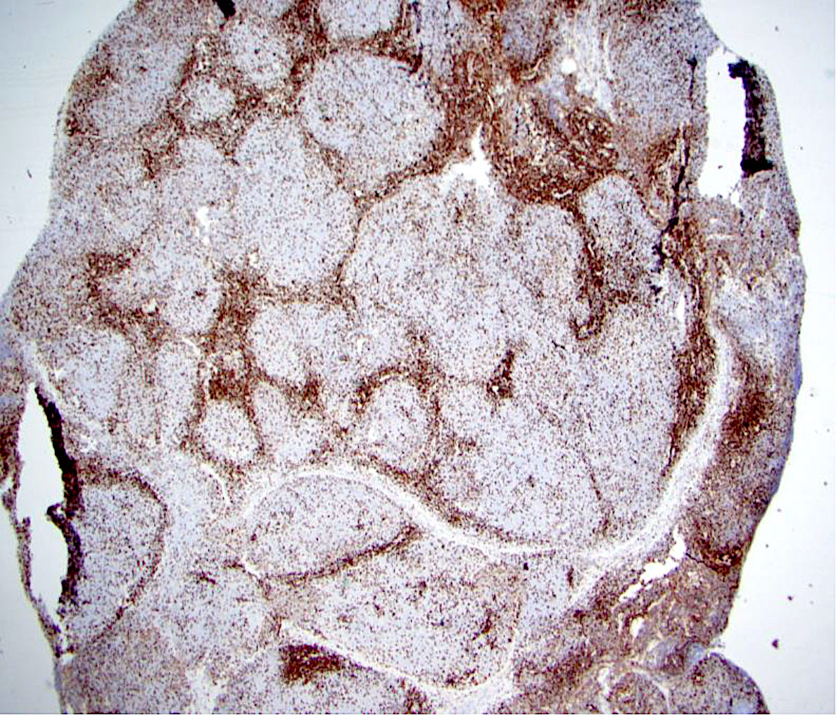
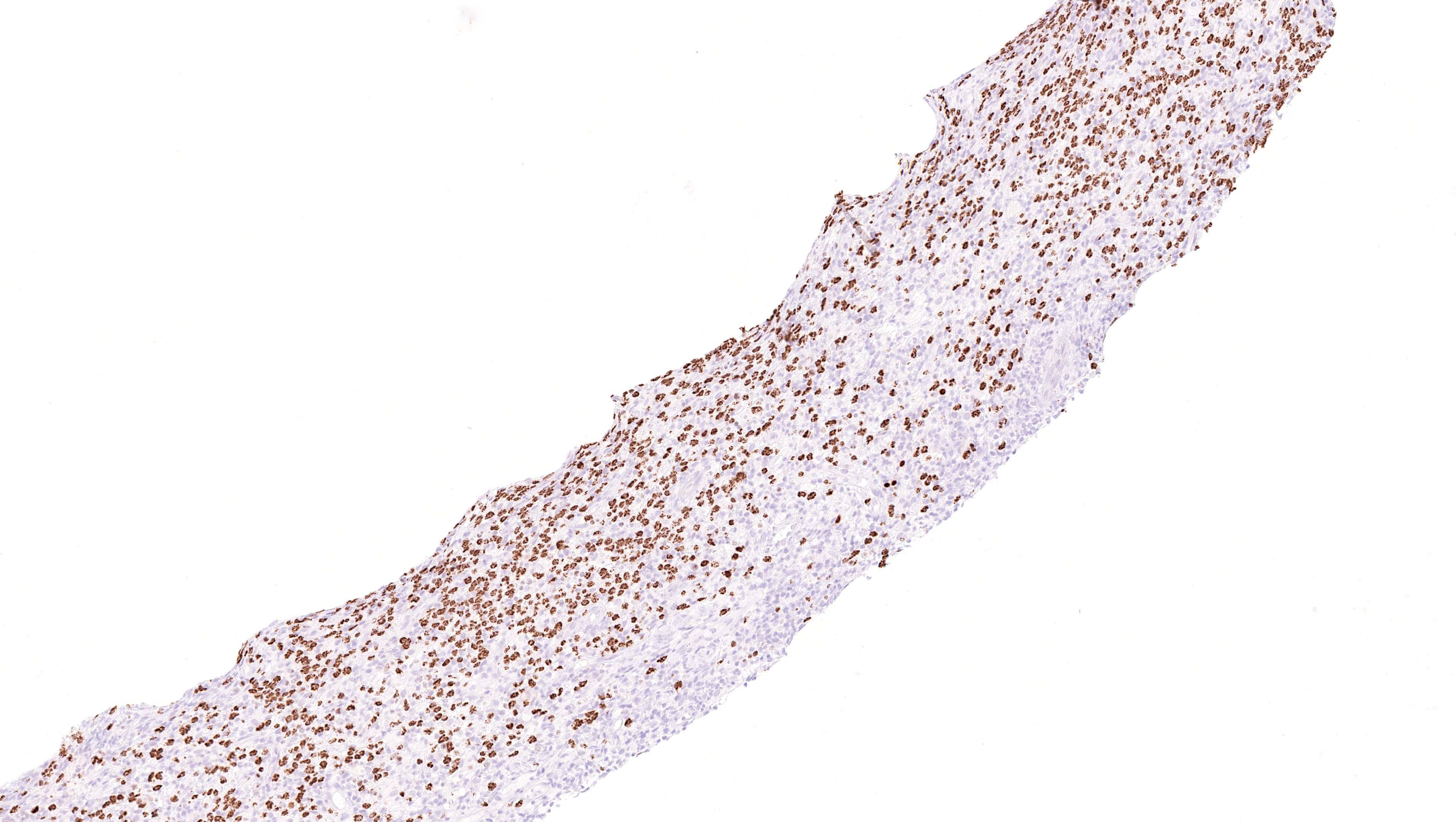
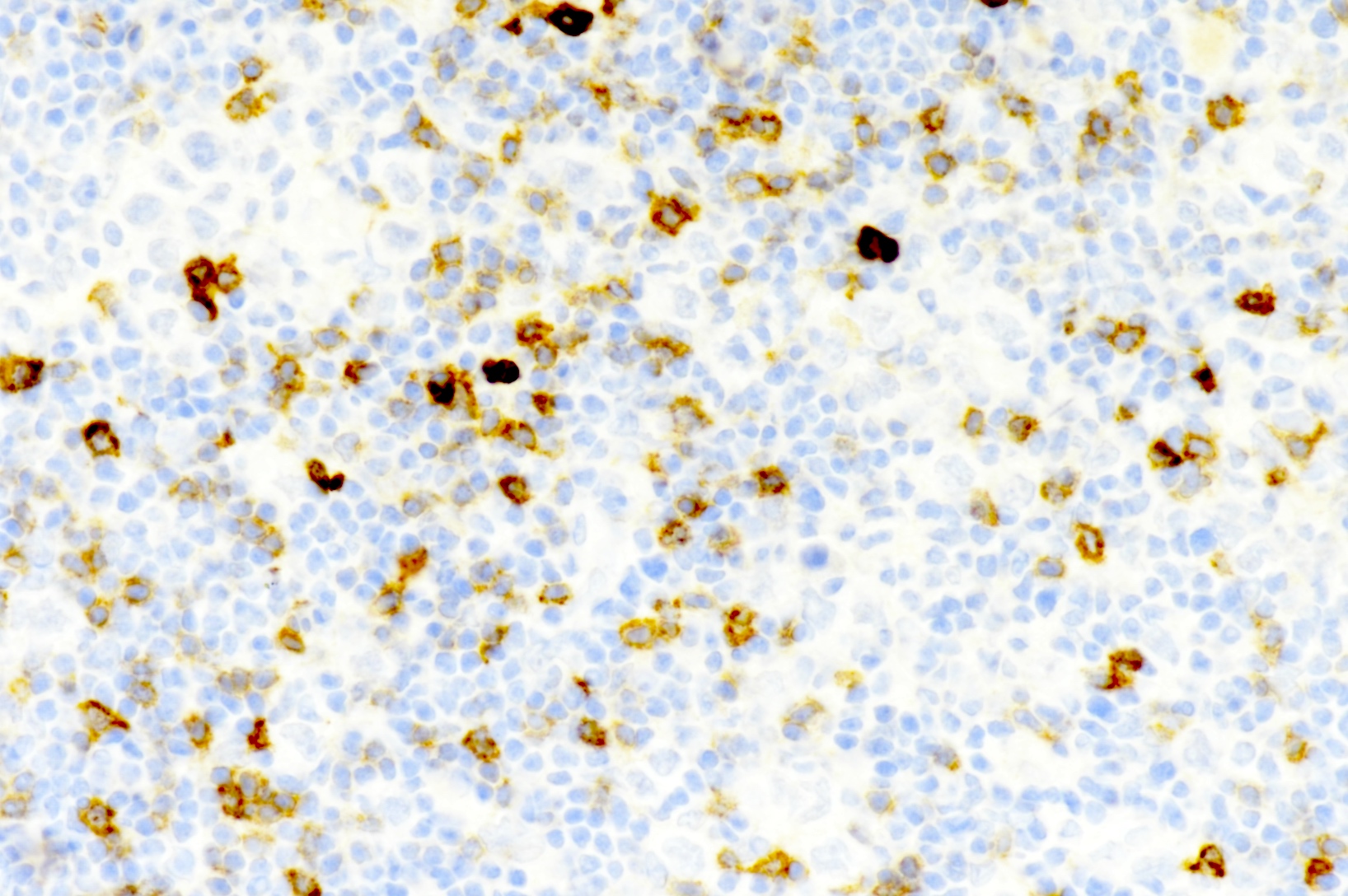
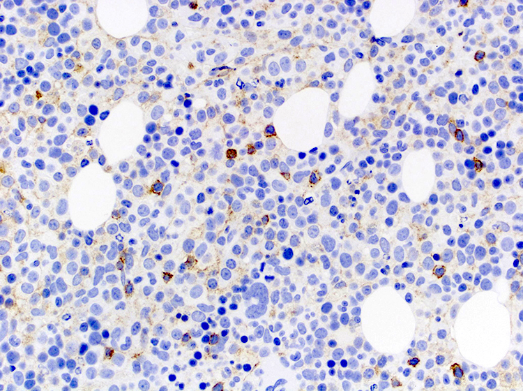
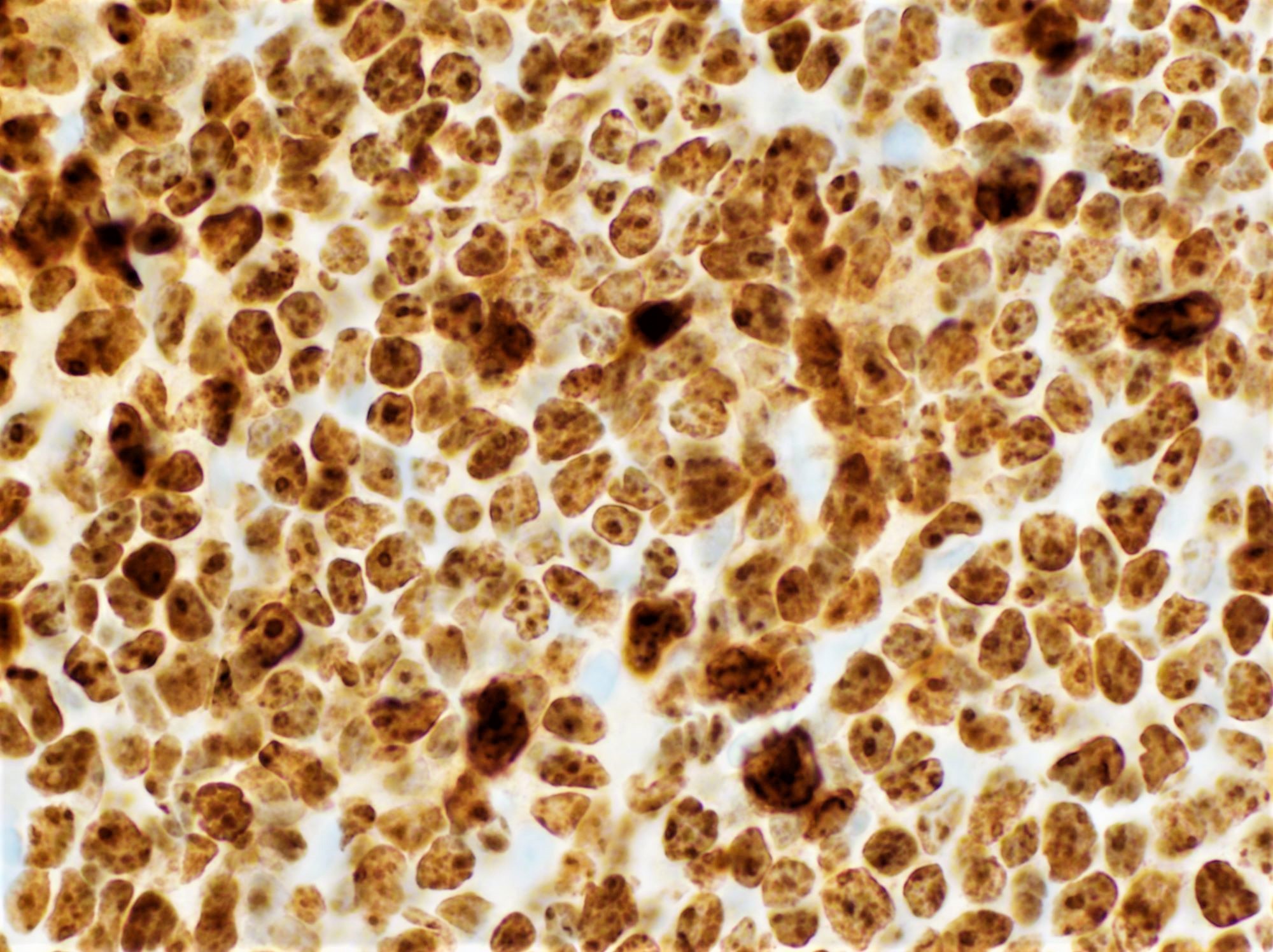
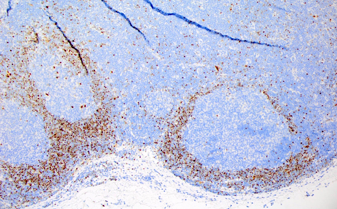
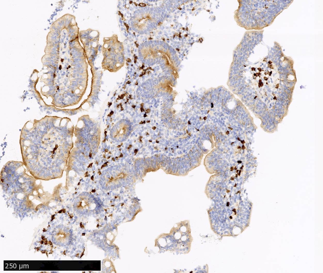
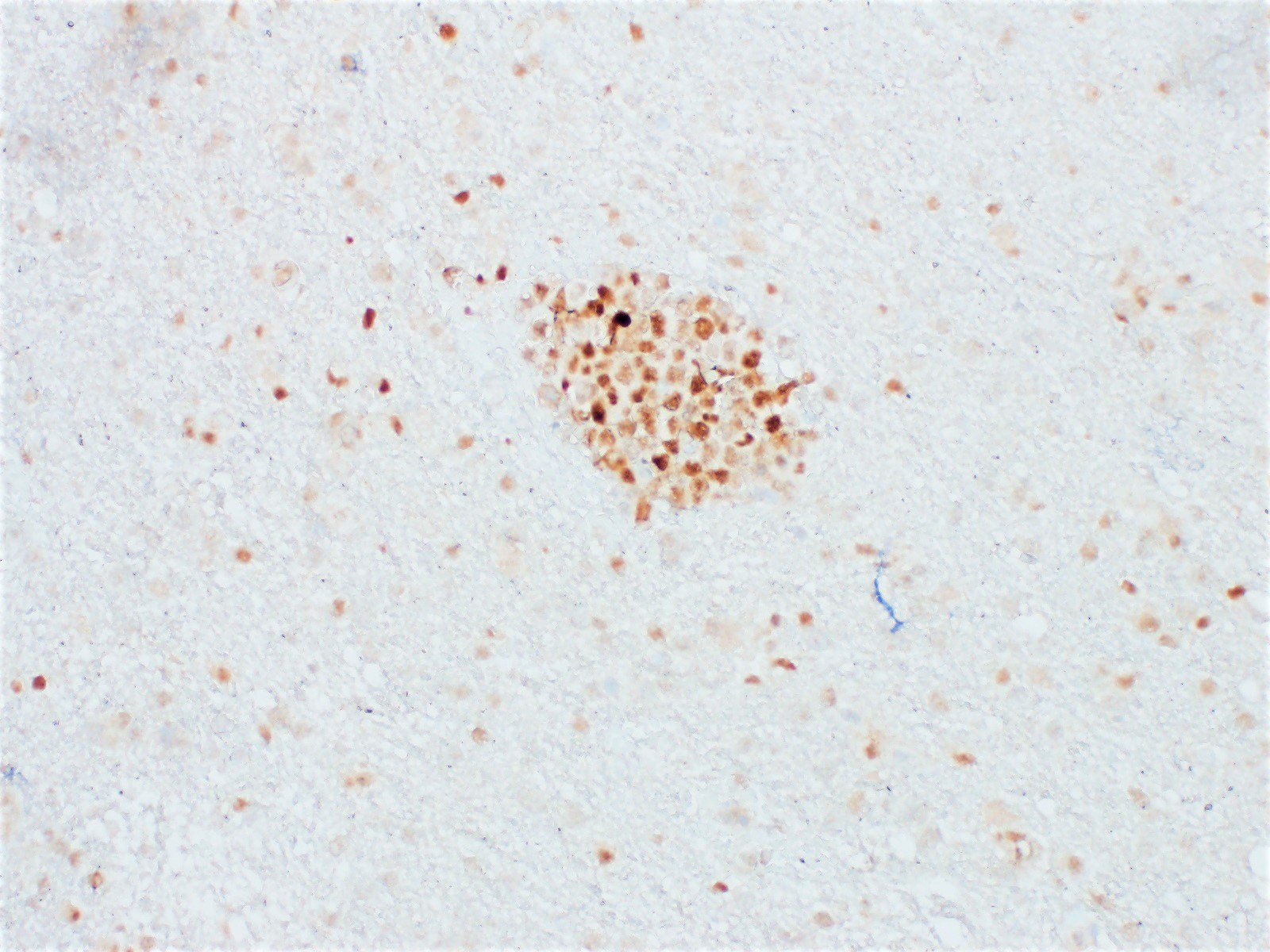
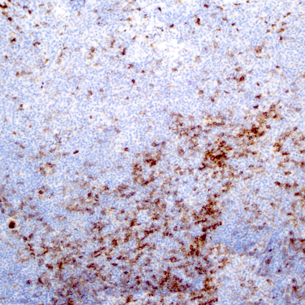
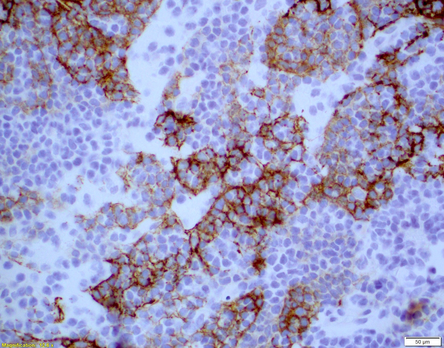
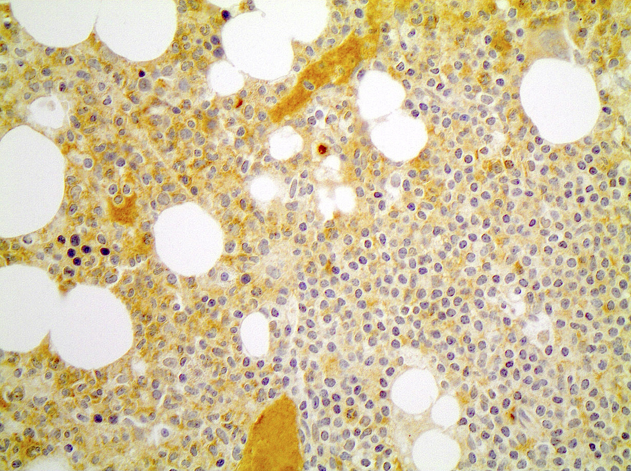
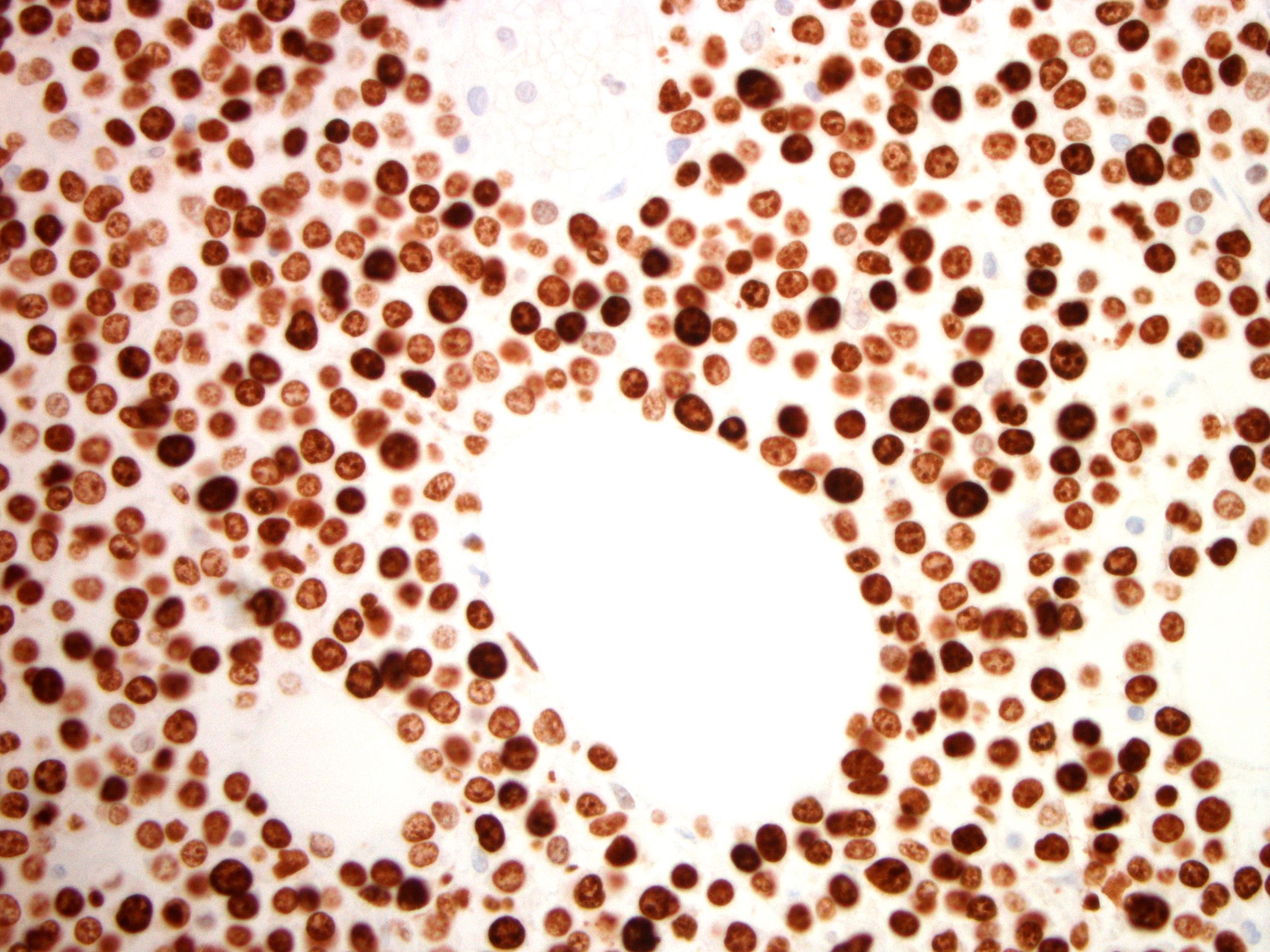
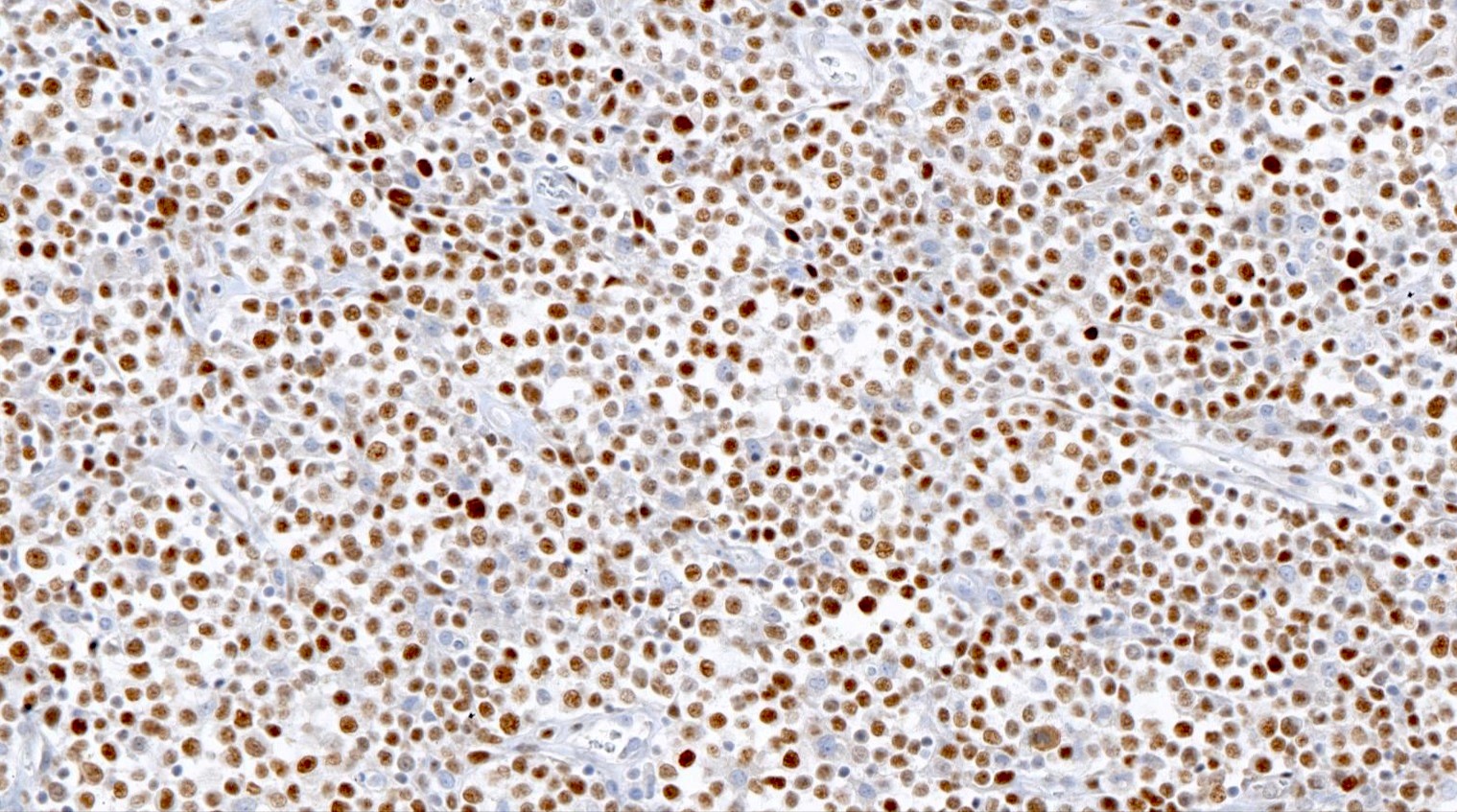
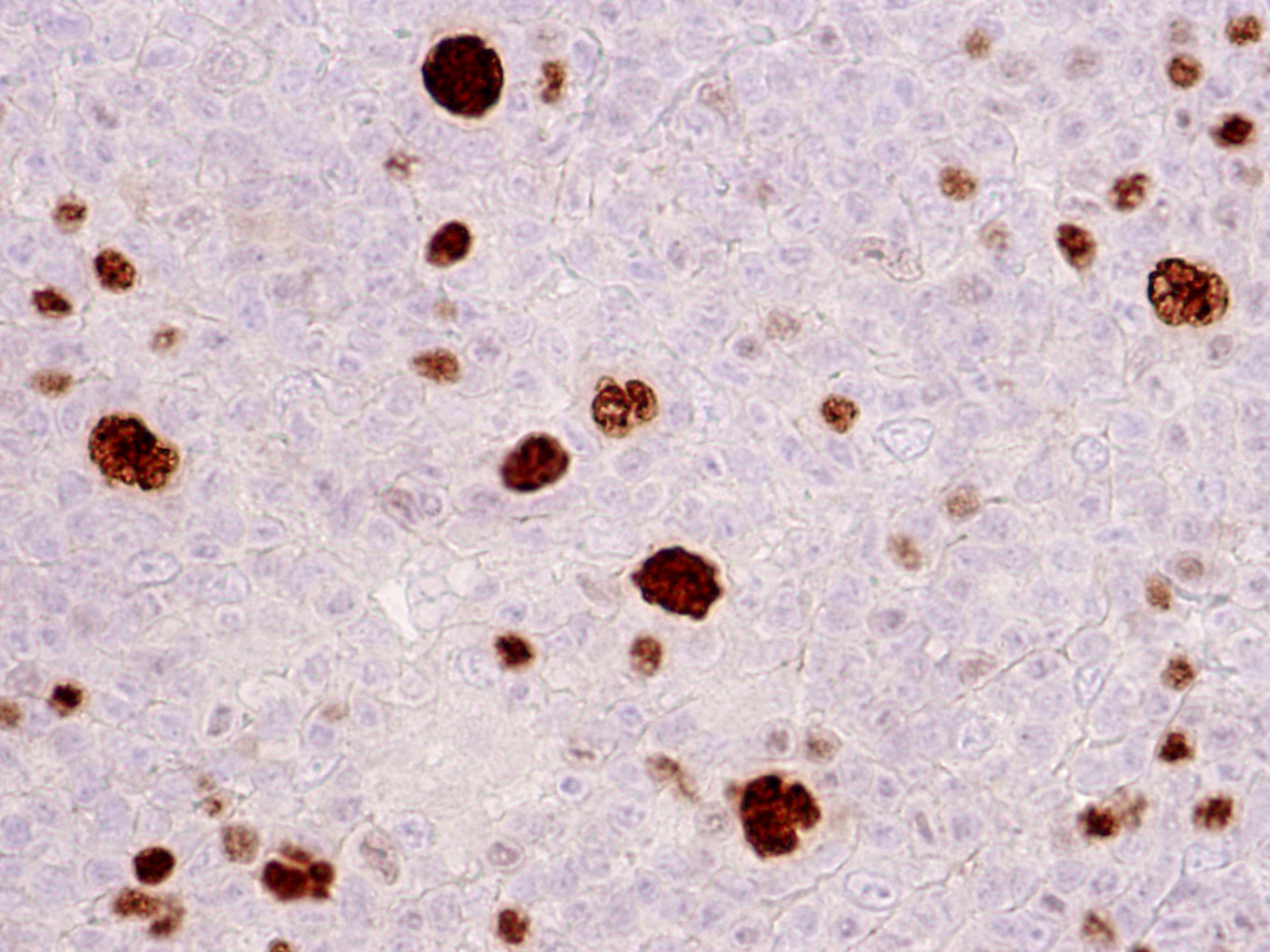
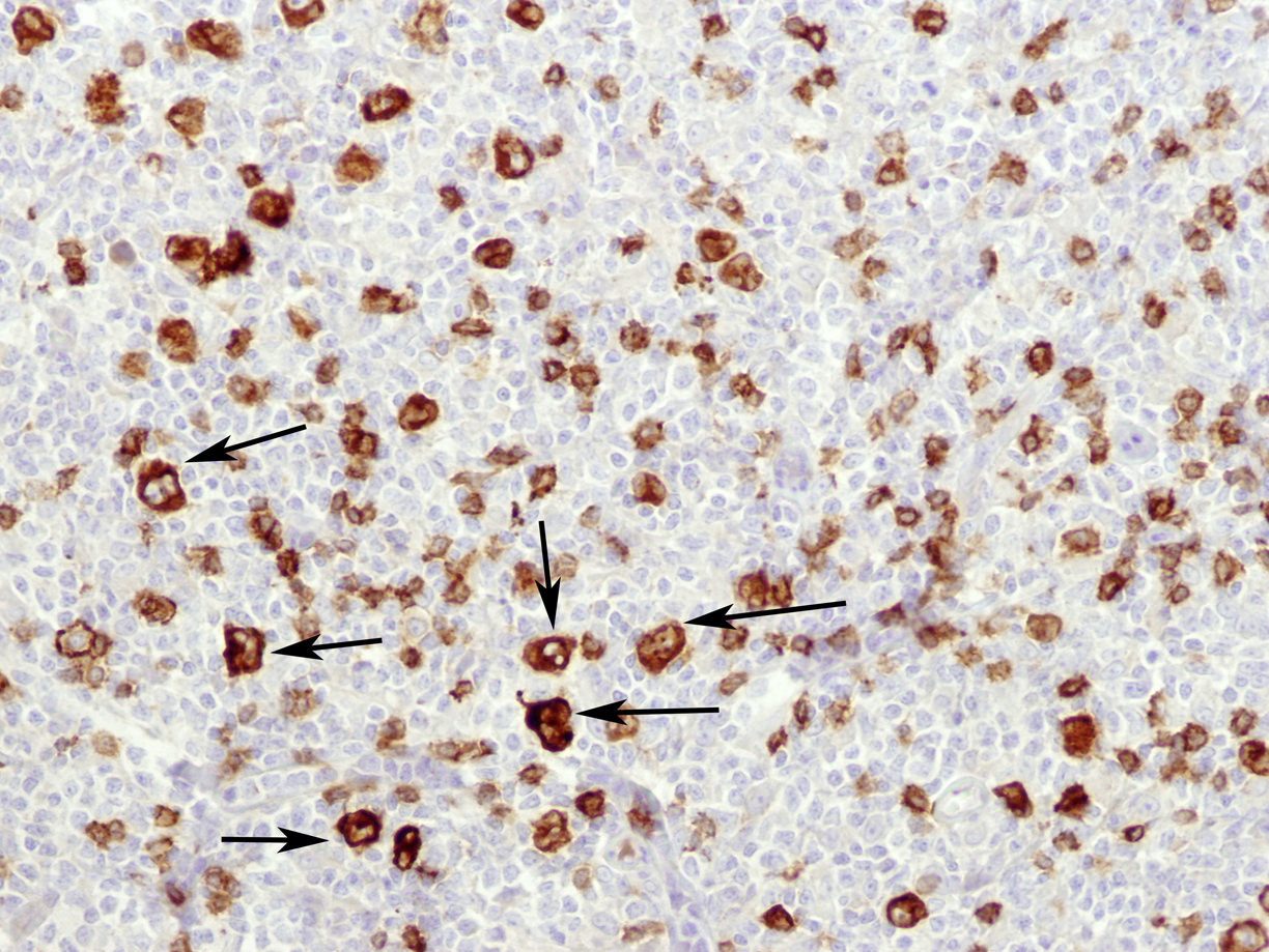
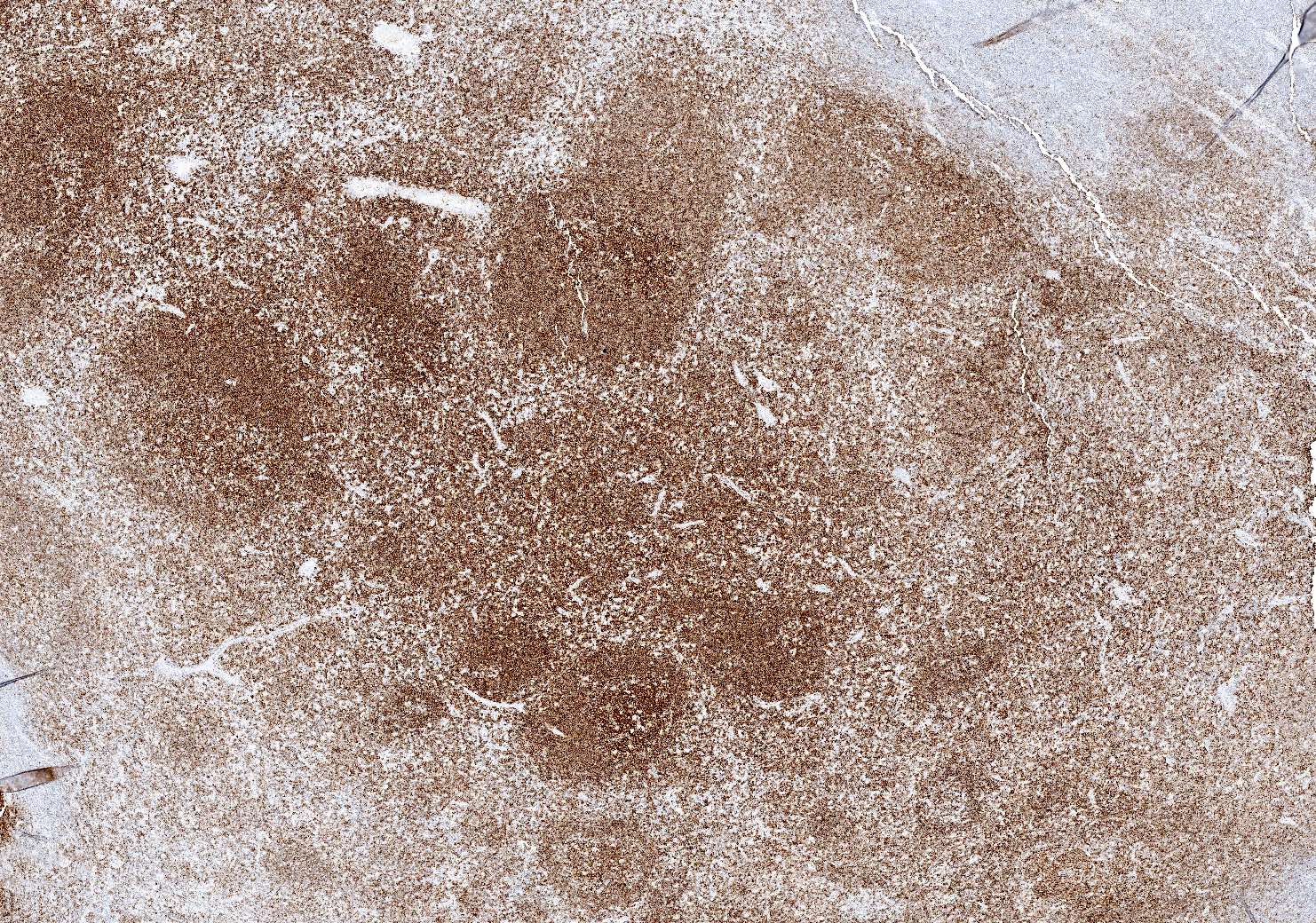
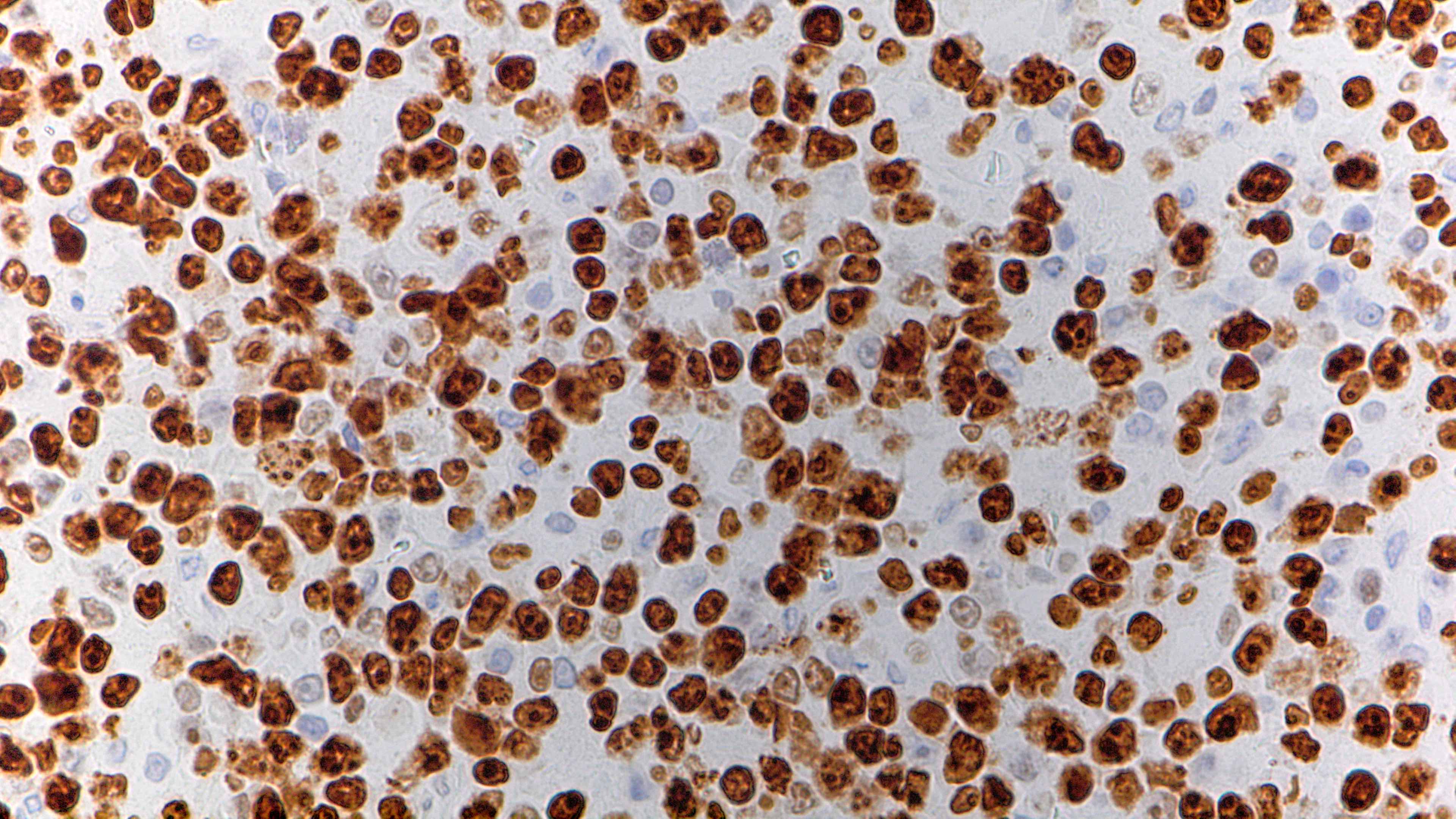
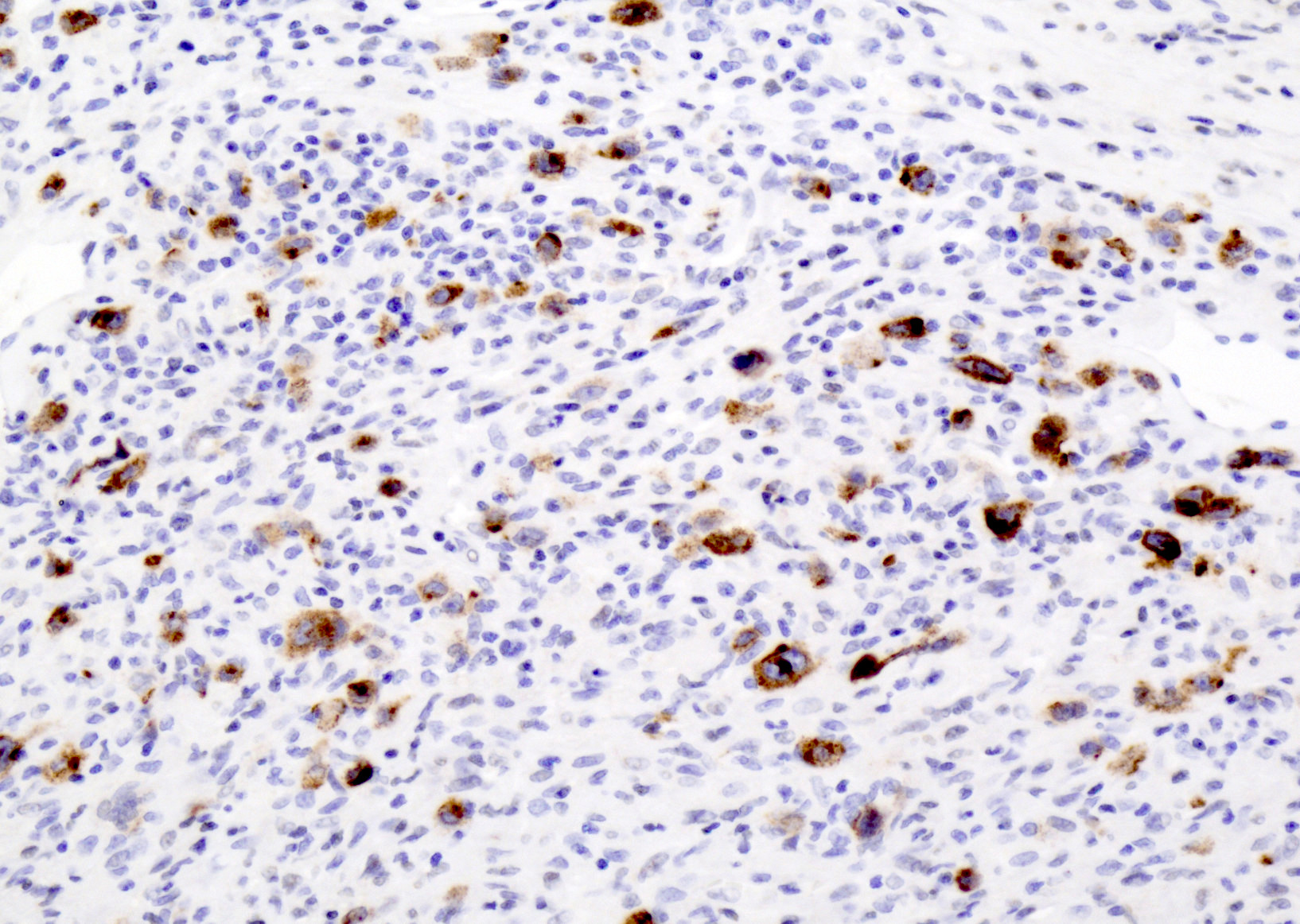
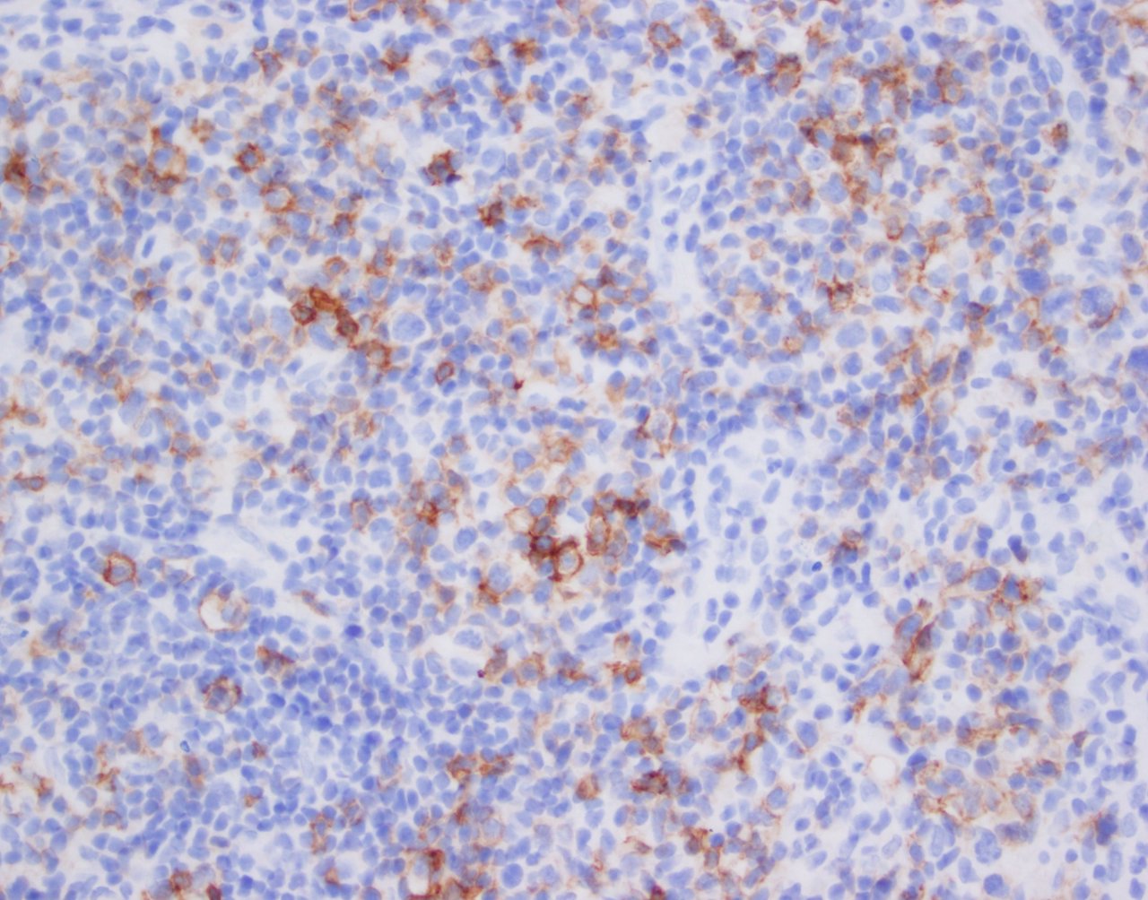
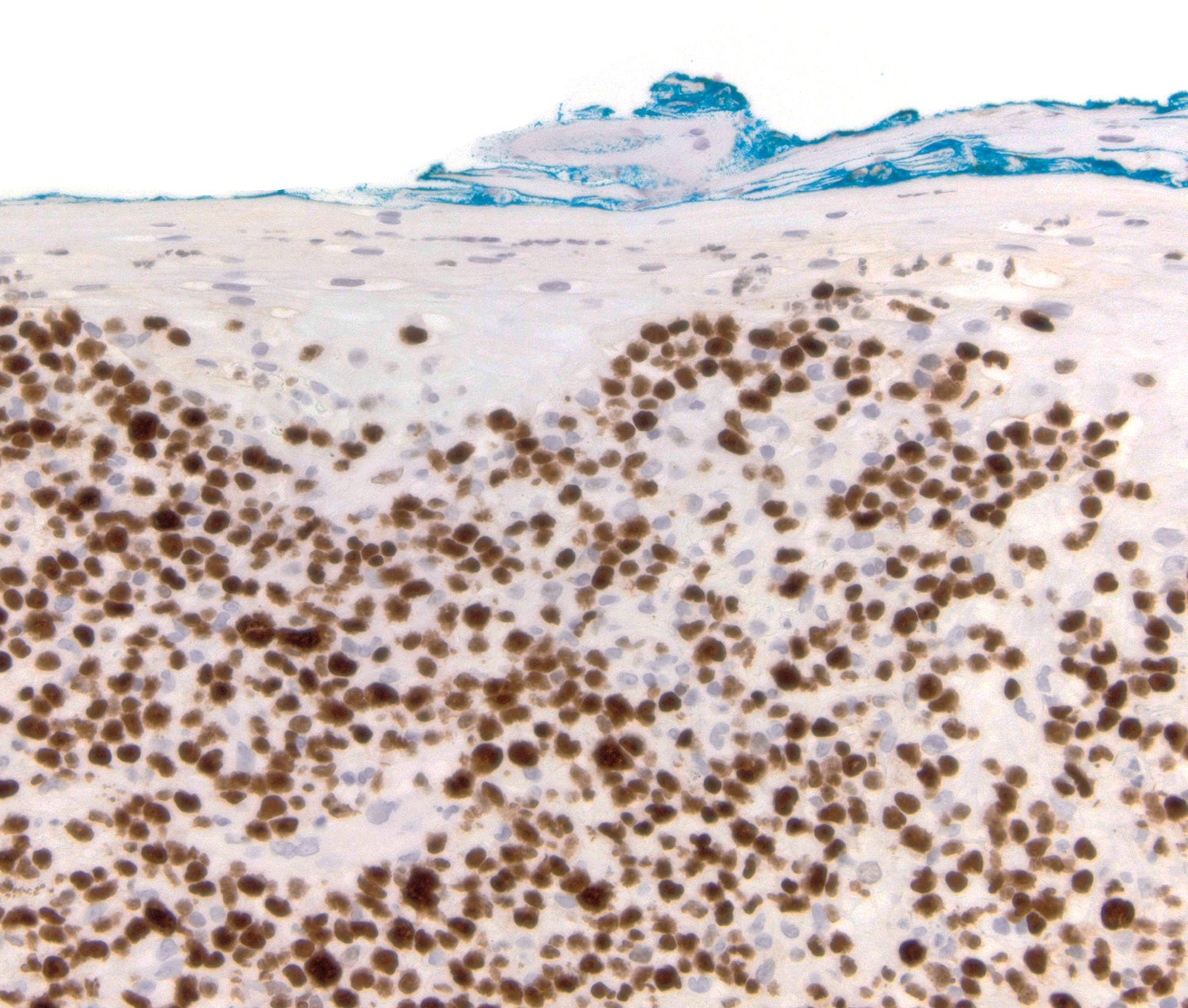
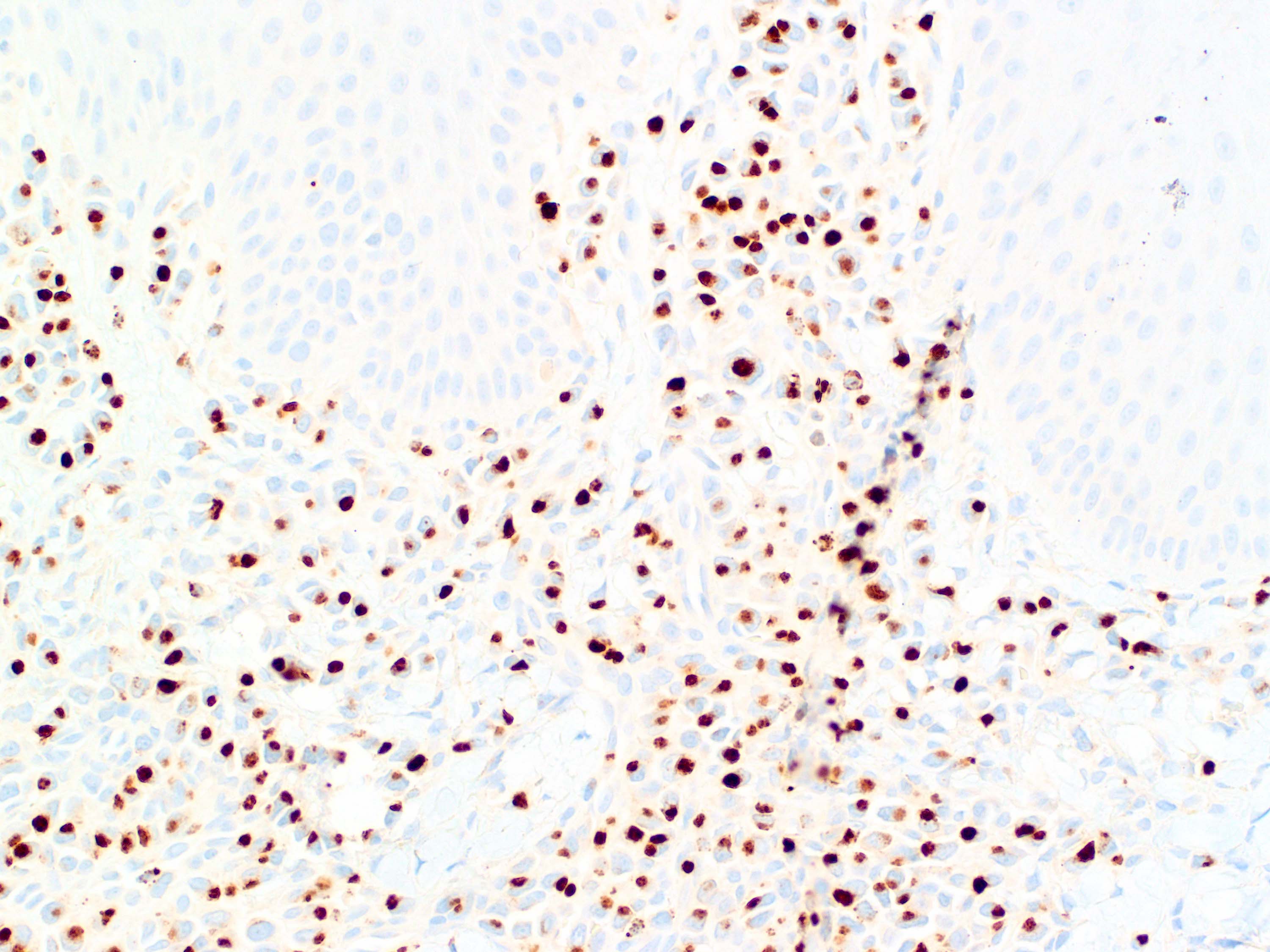
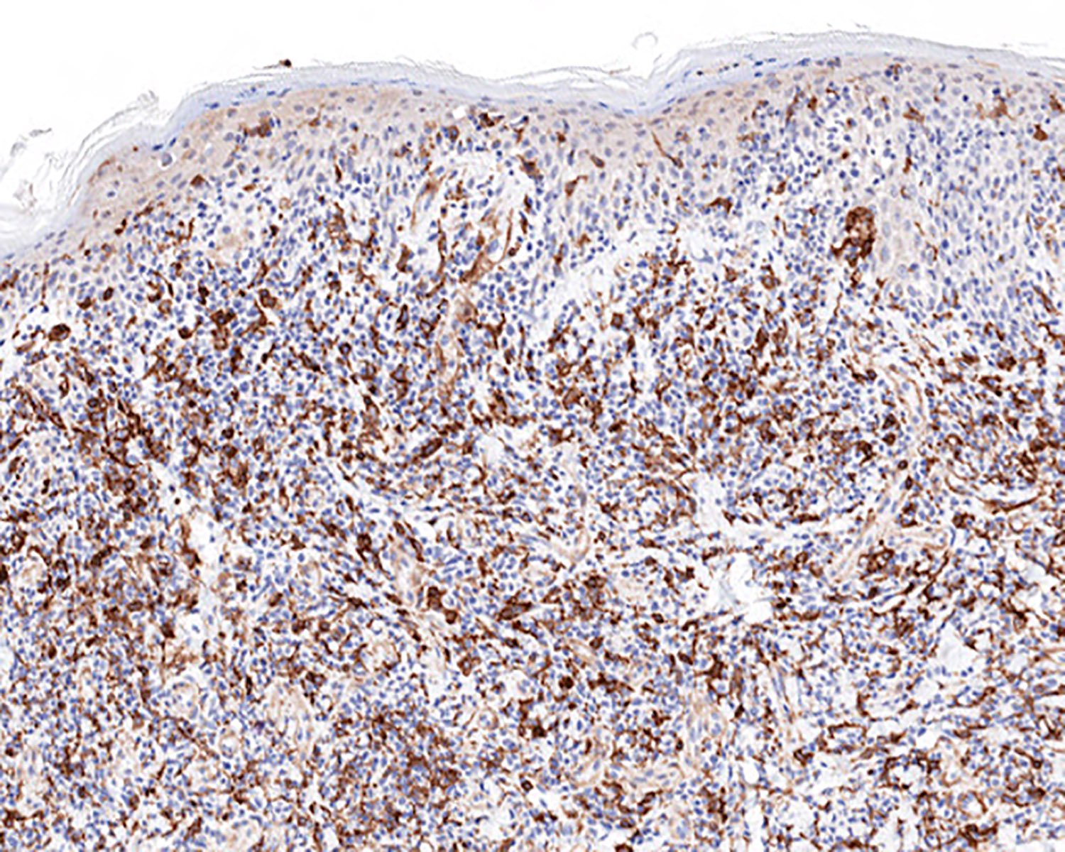
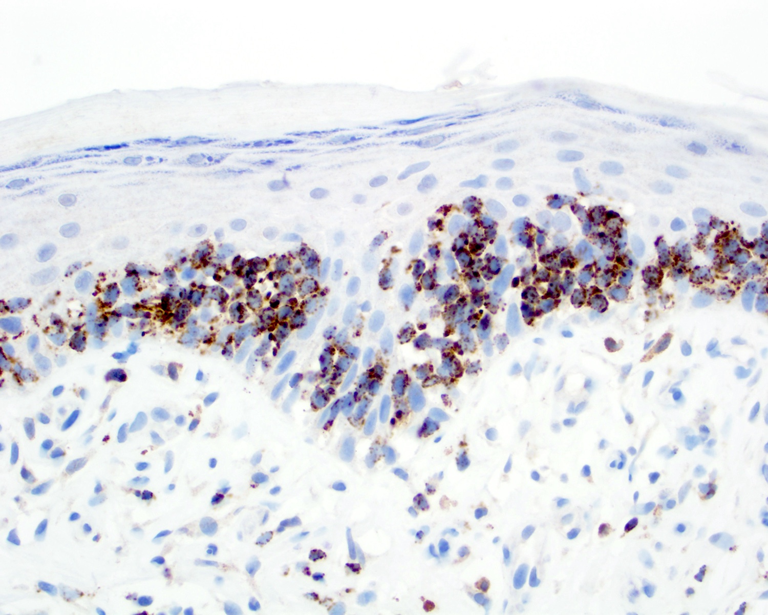
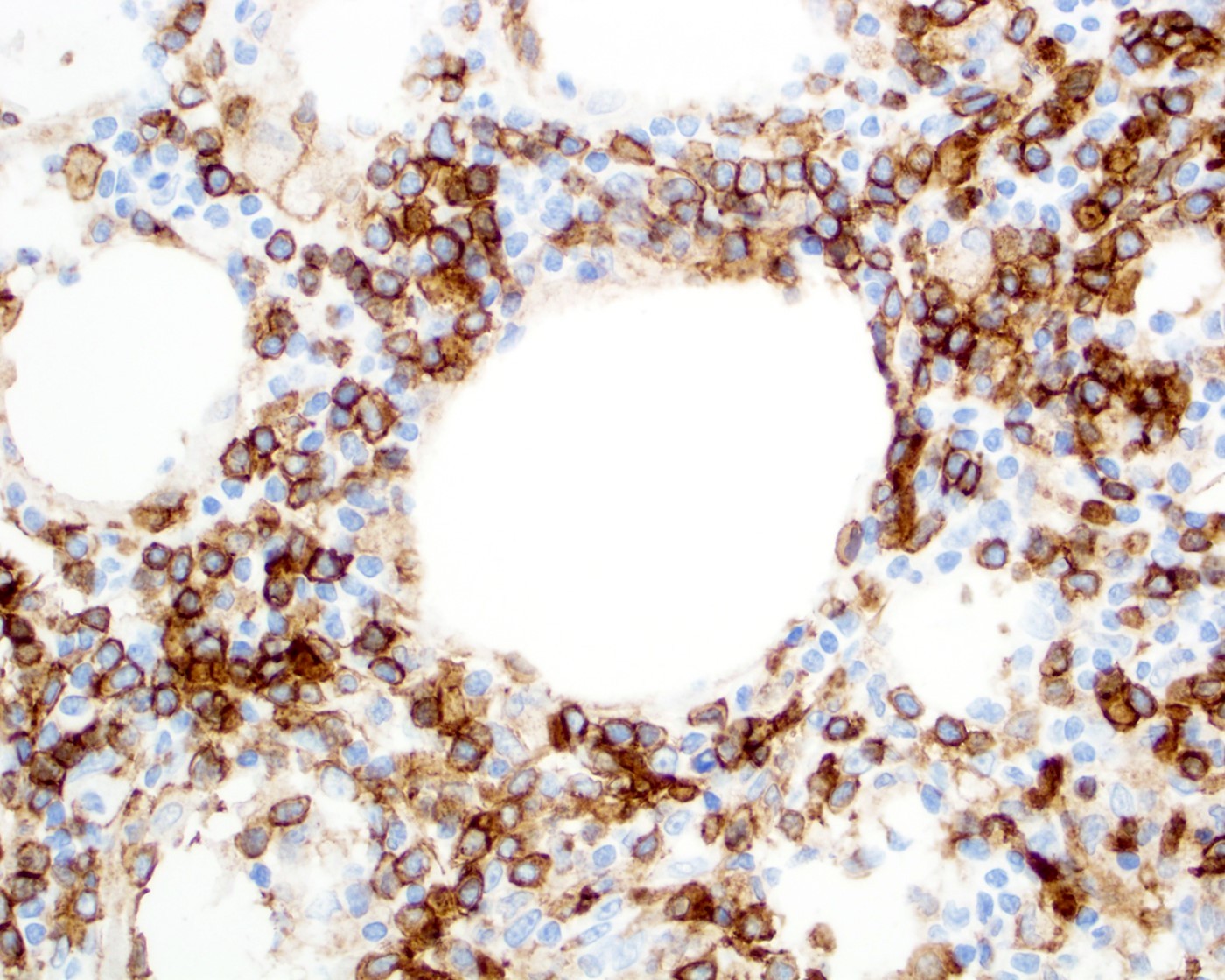
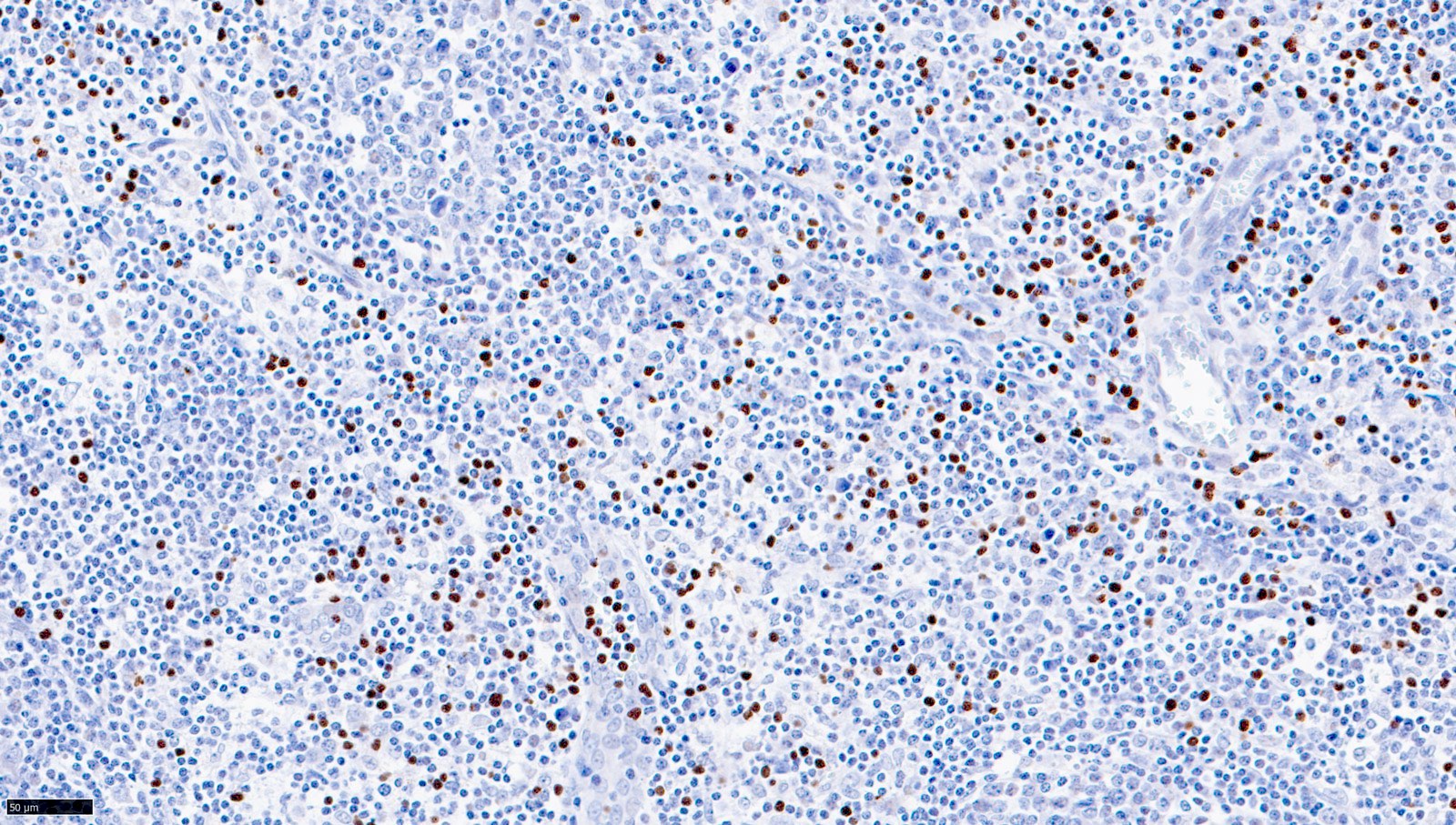
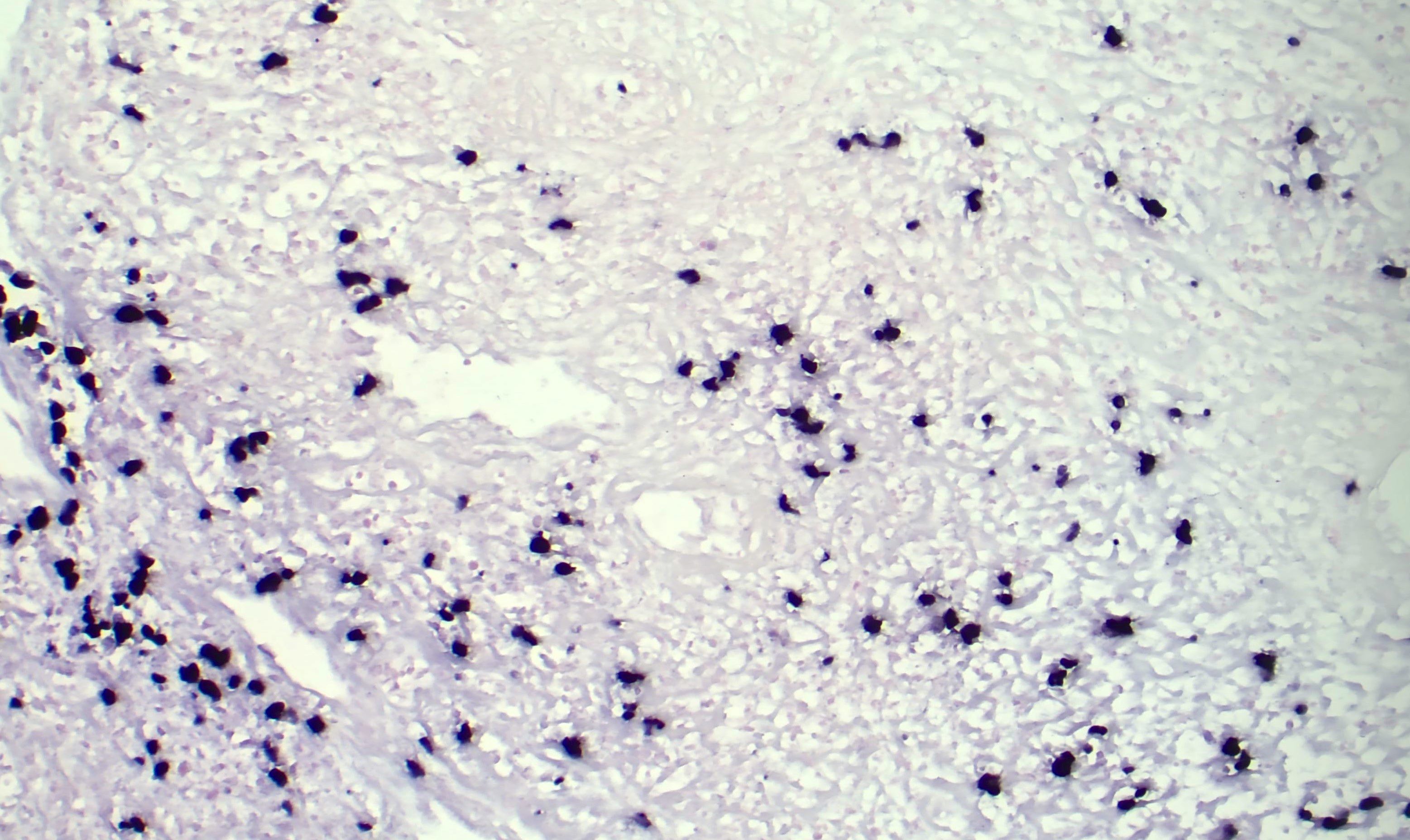
MUM1 positivity
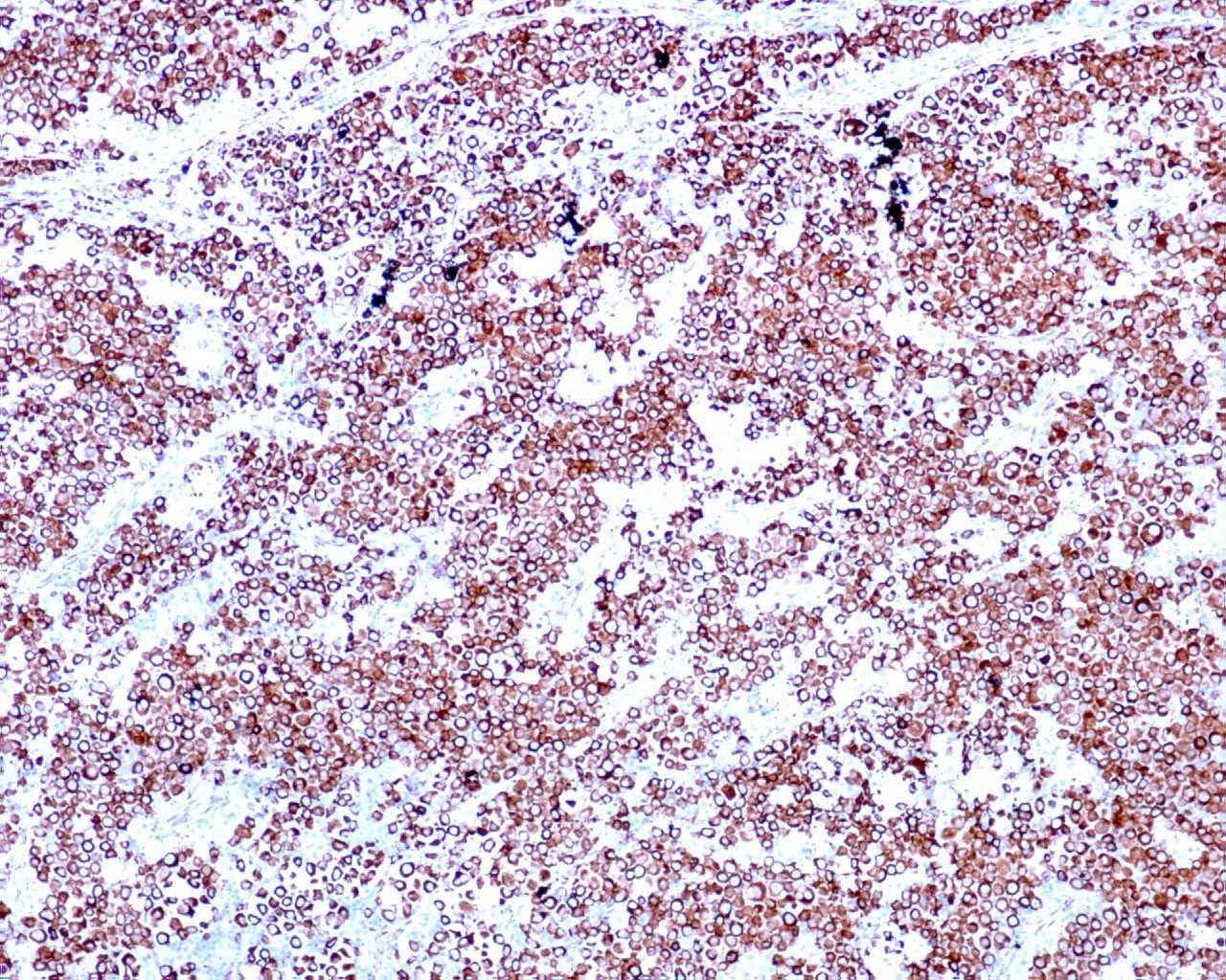
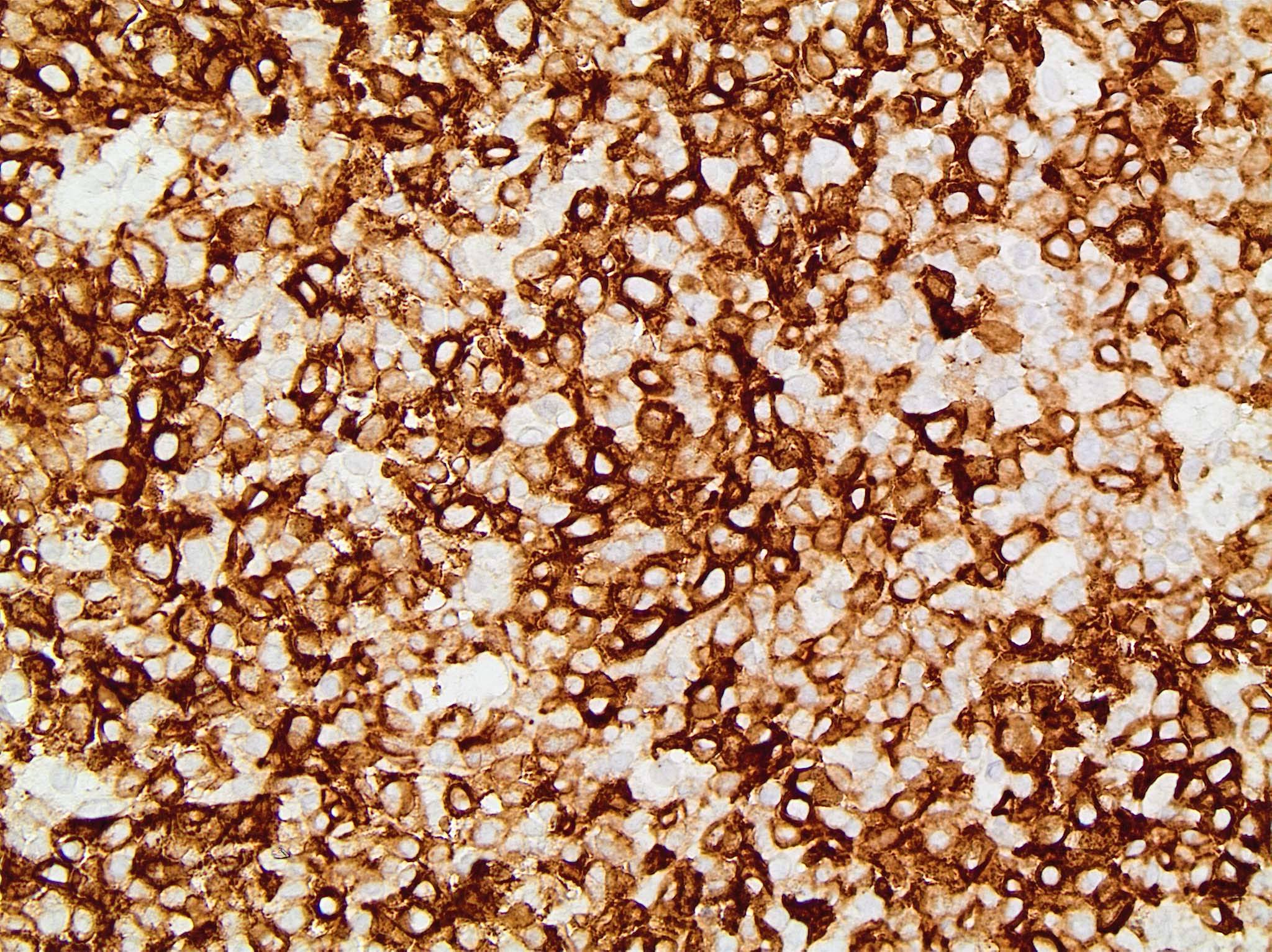
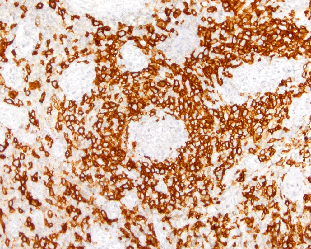
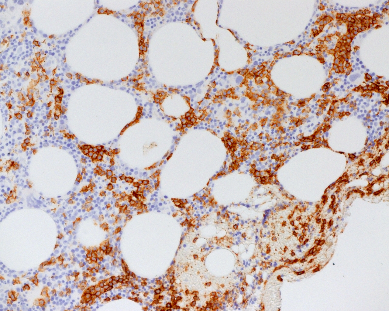
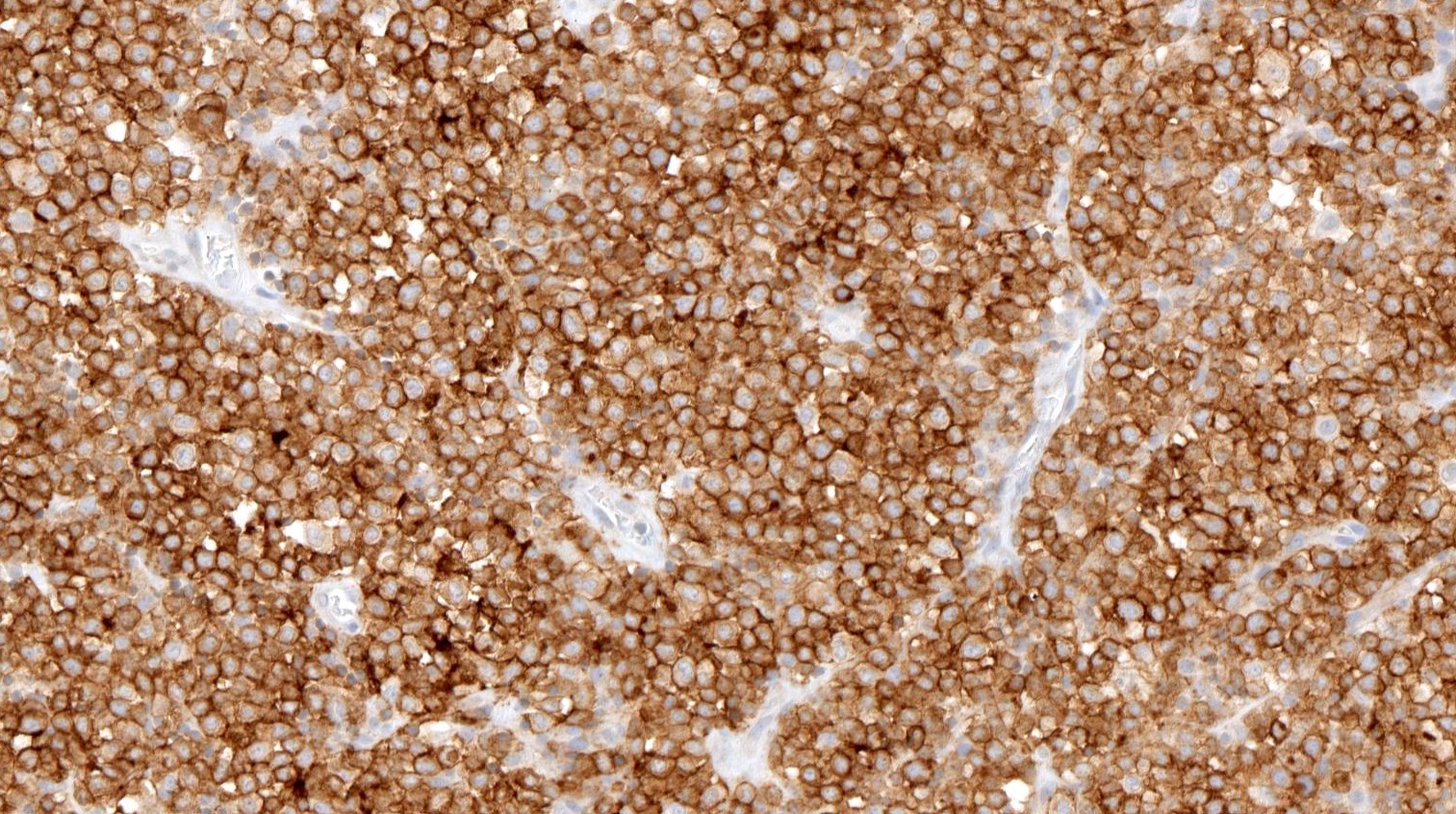
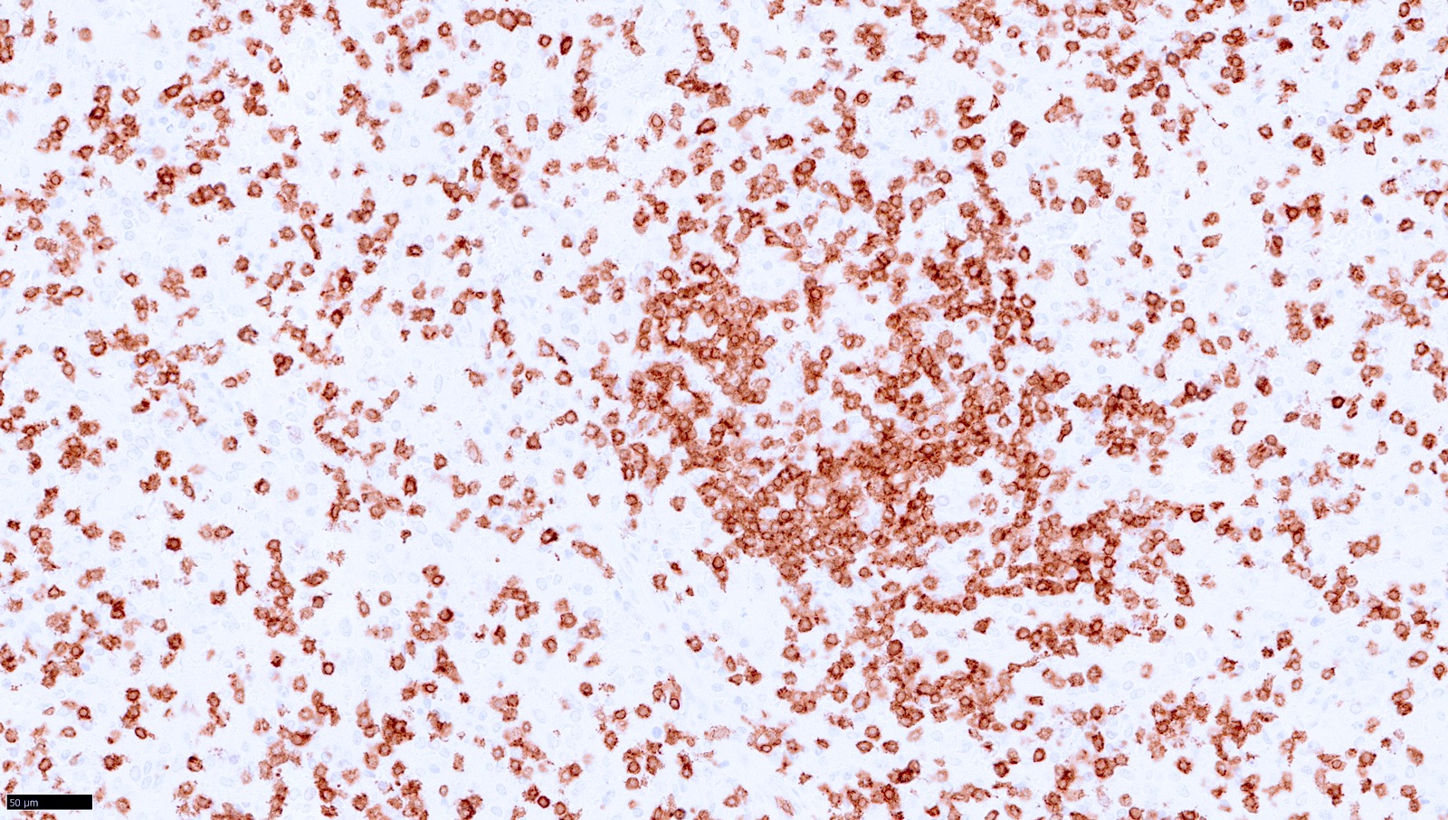
CD138 positivity
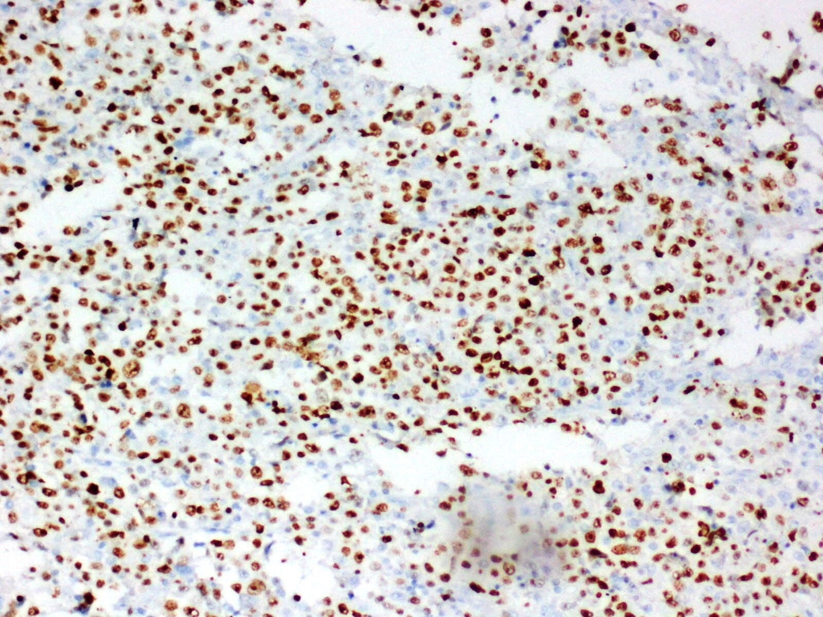
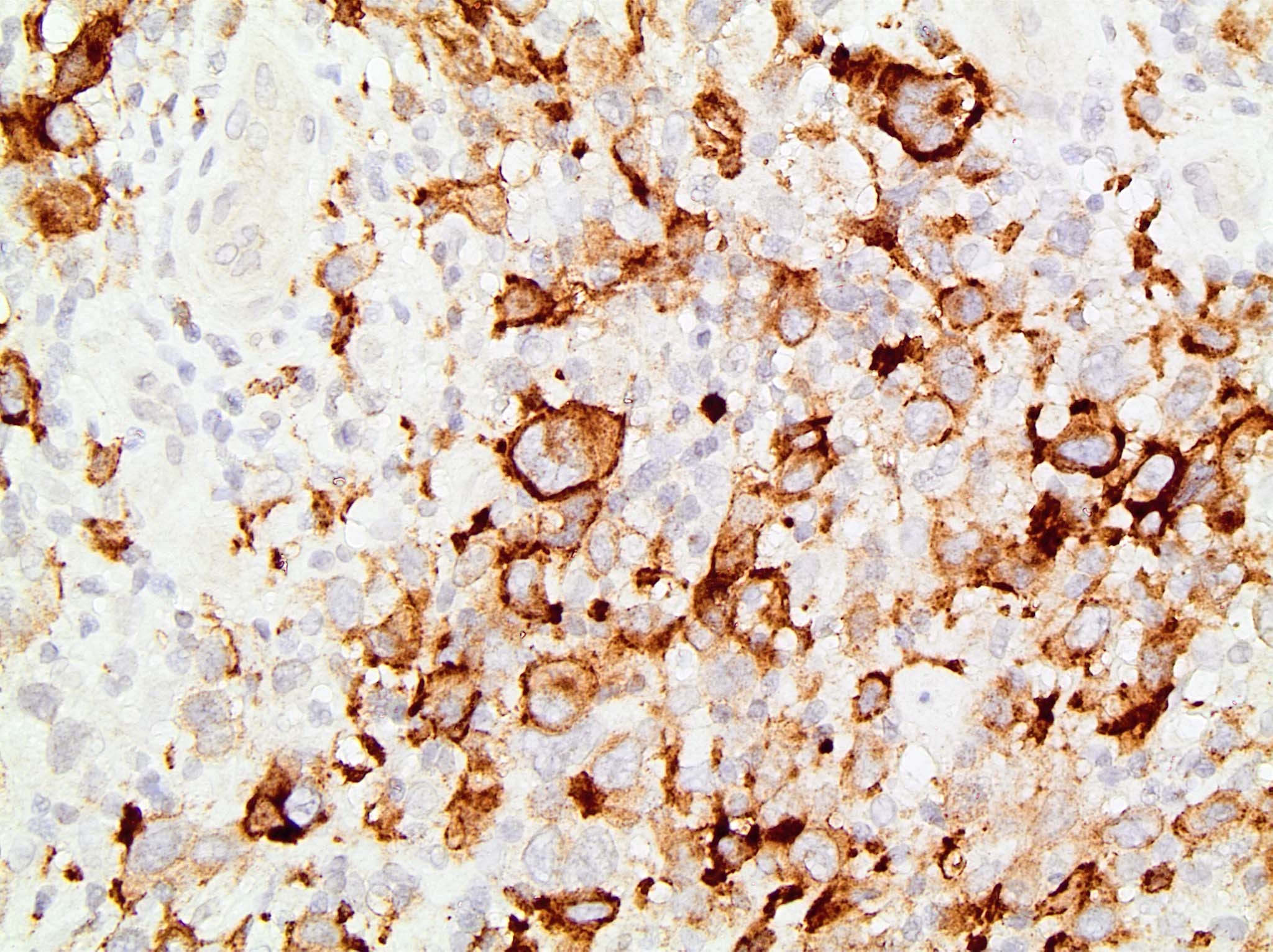
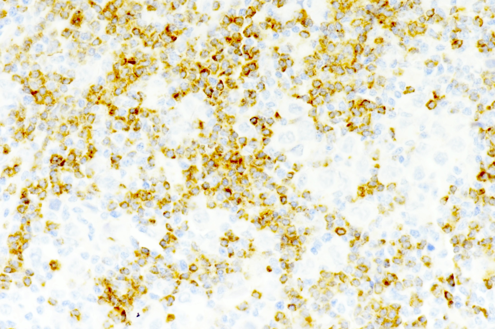
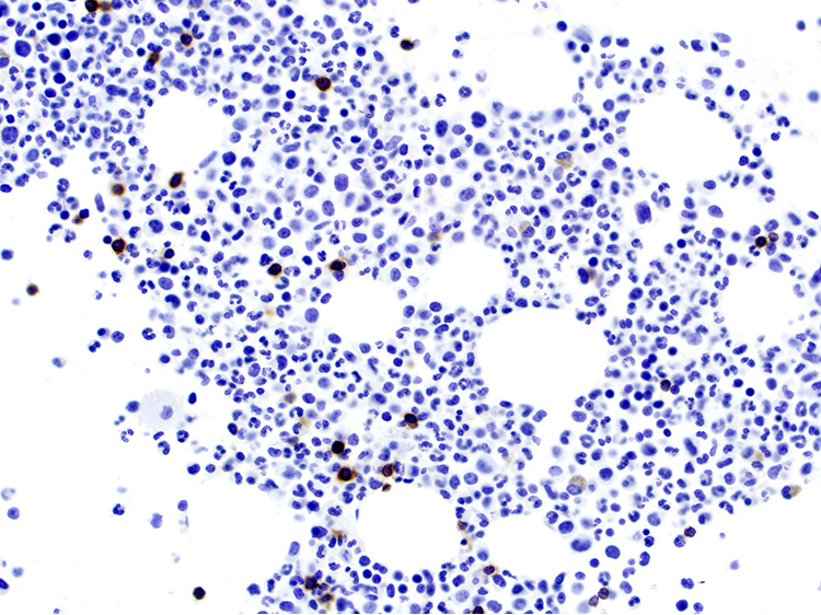
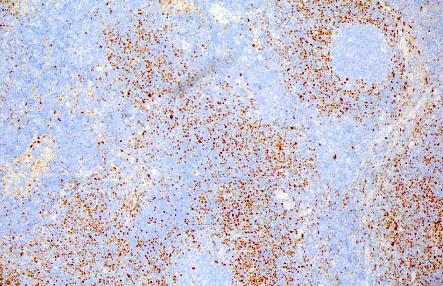
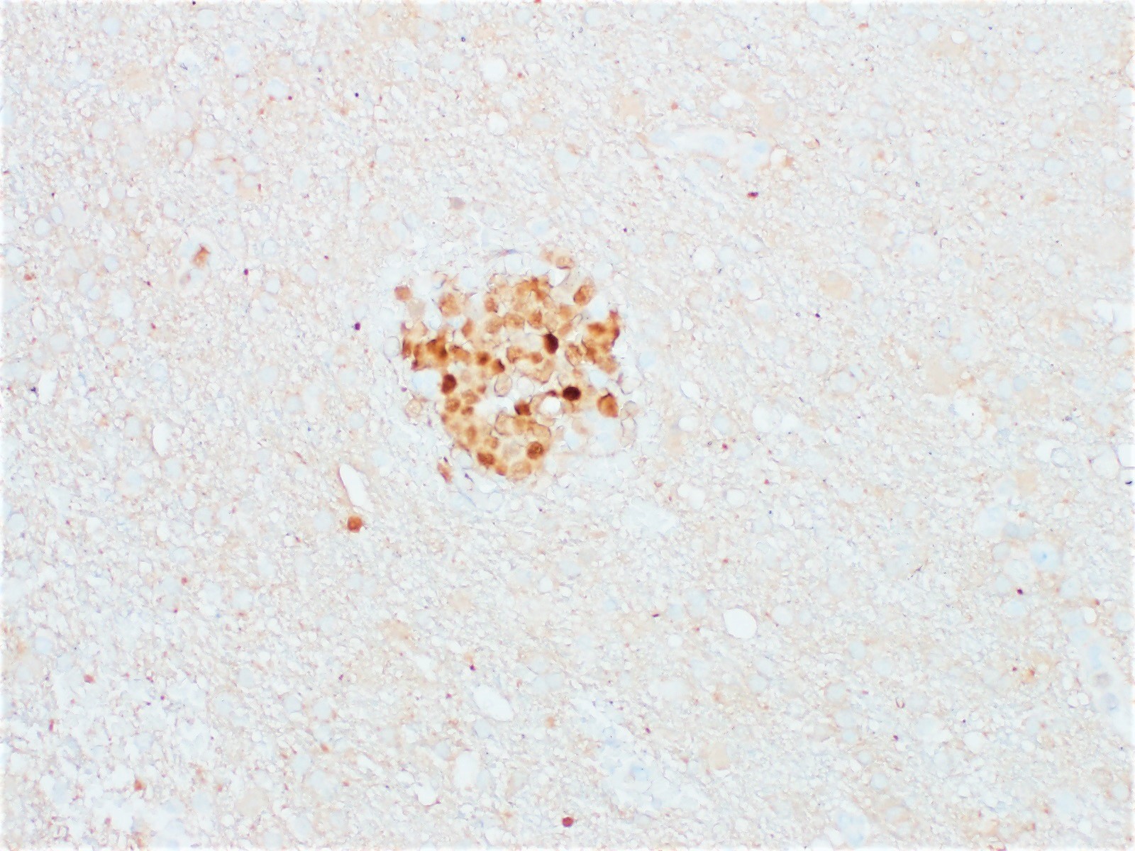
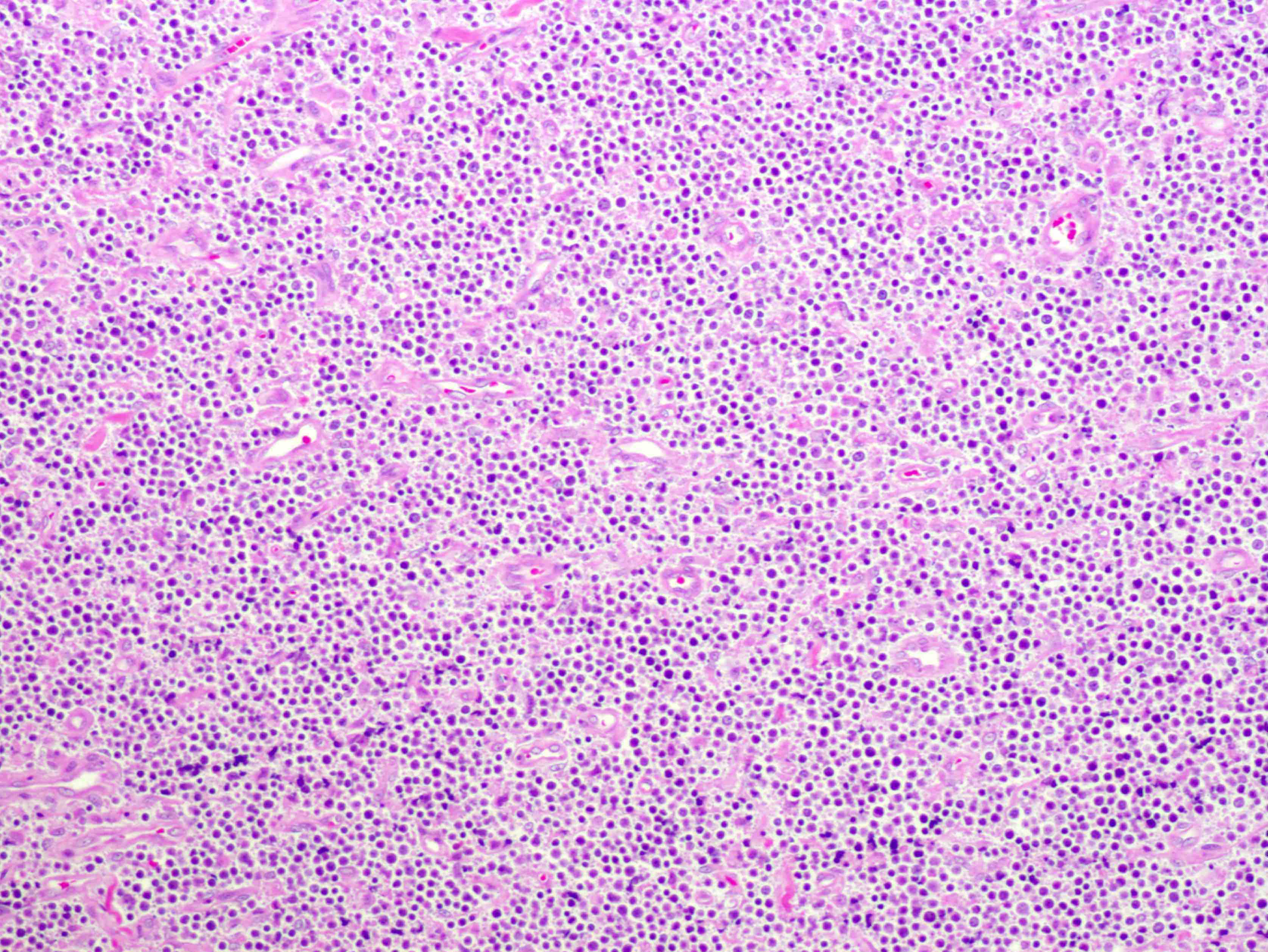
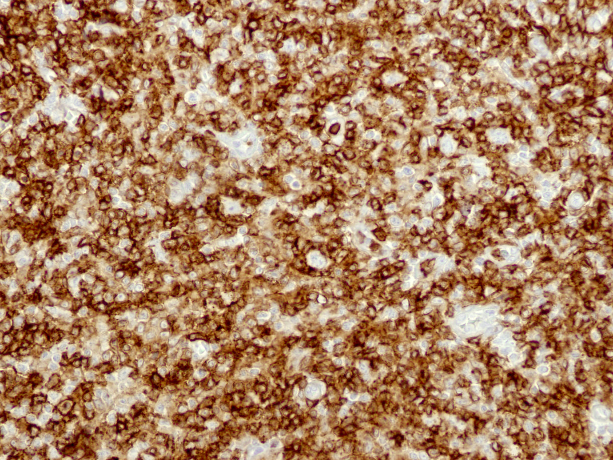
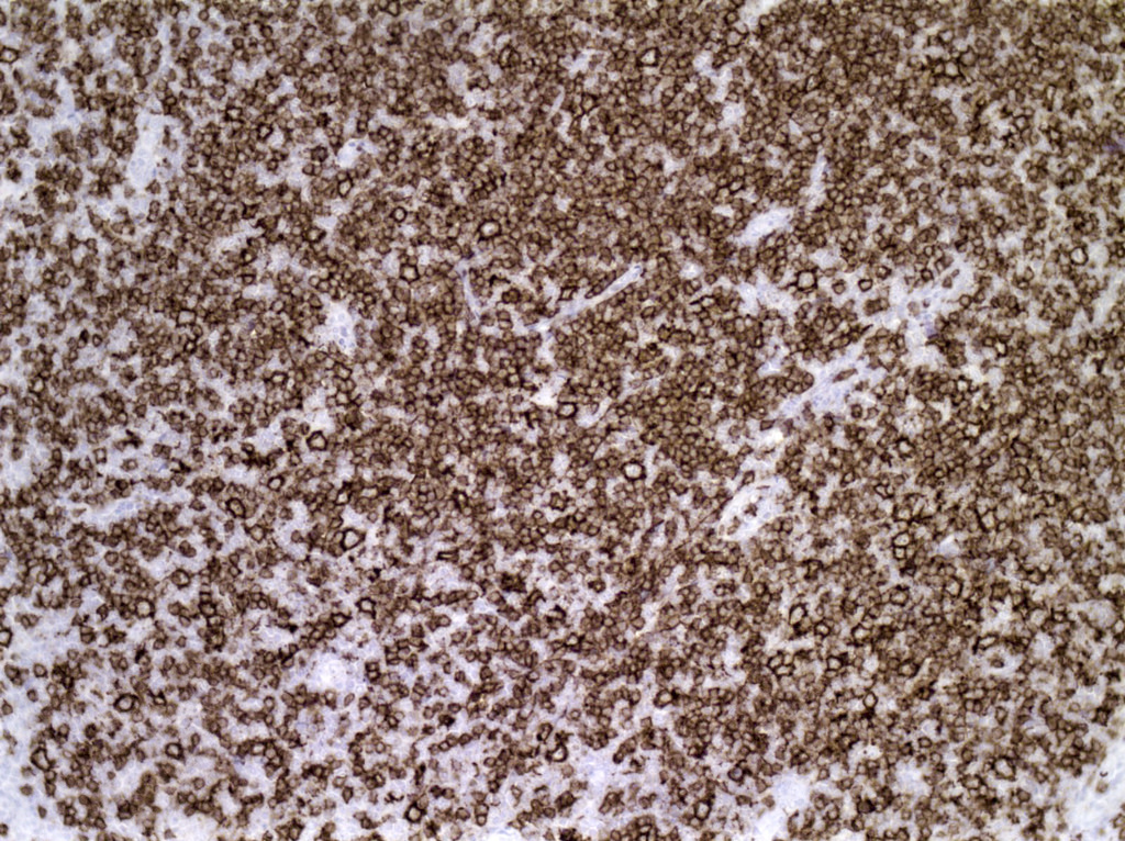
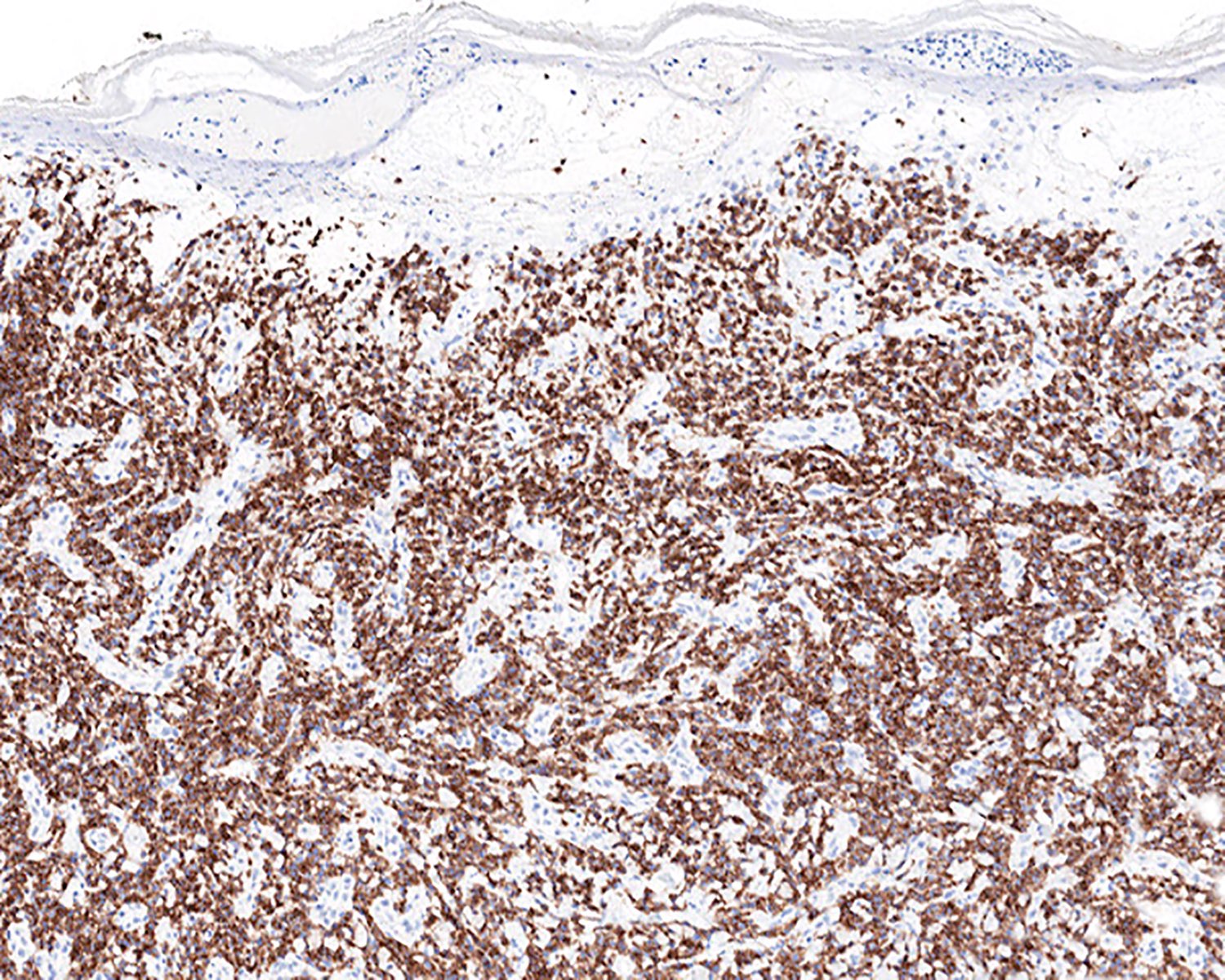
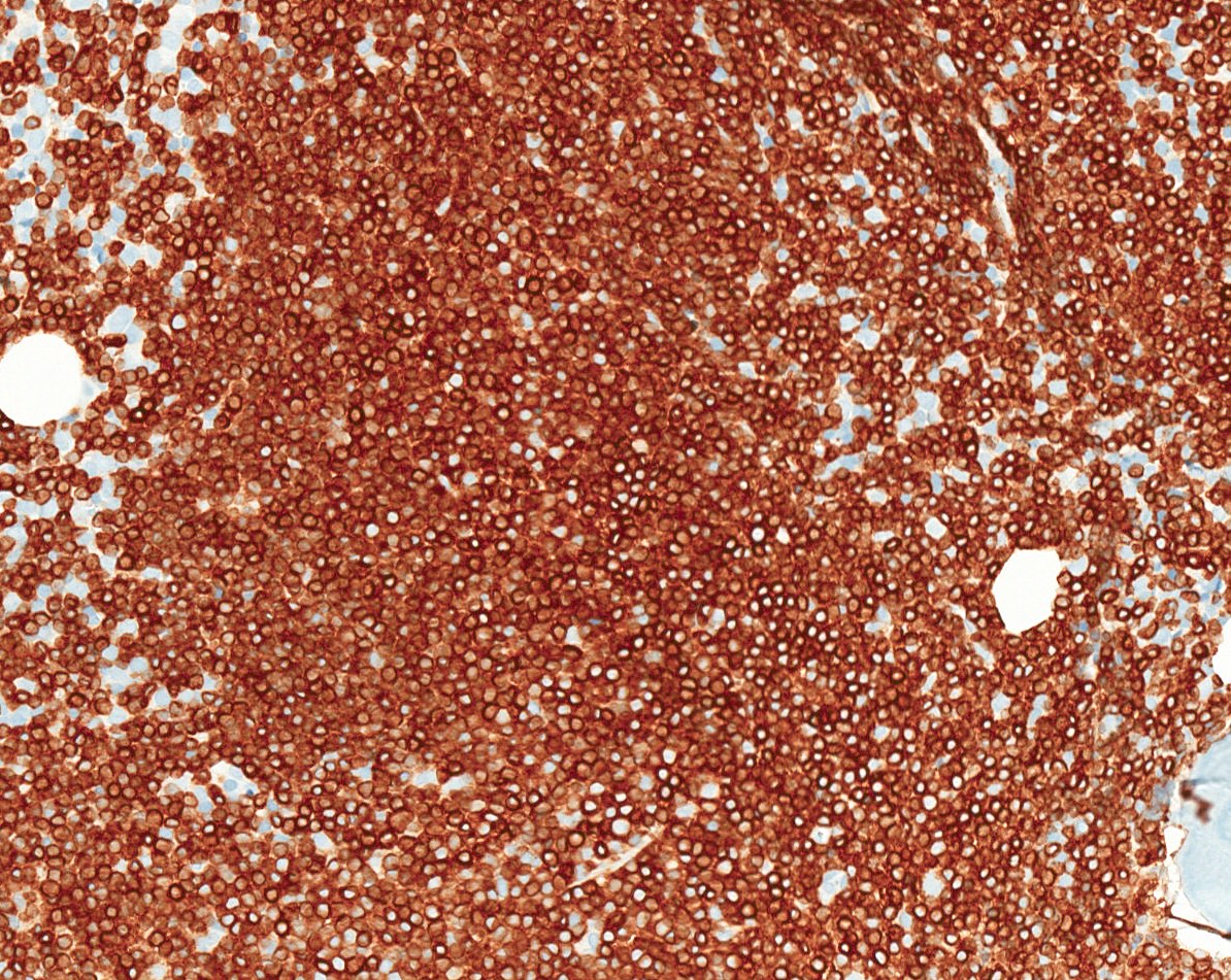
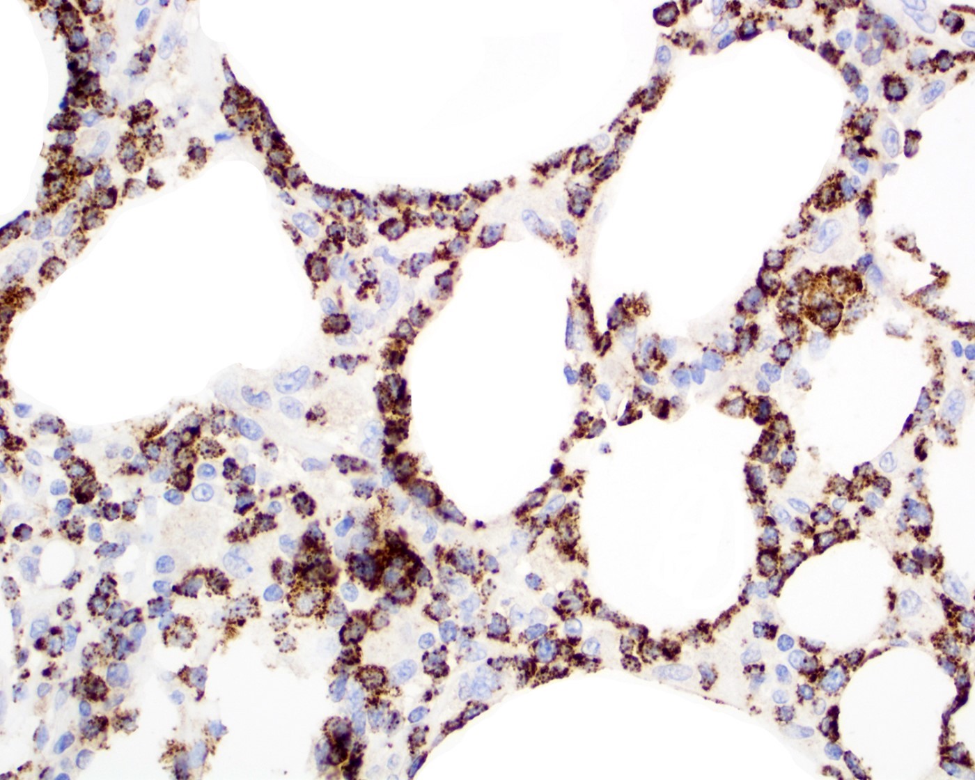
MIB1 index of 80 - 90%
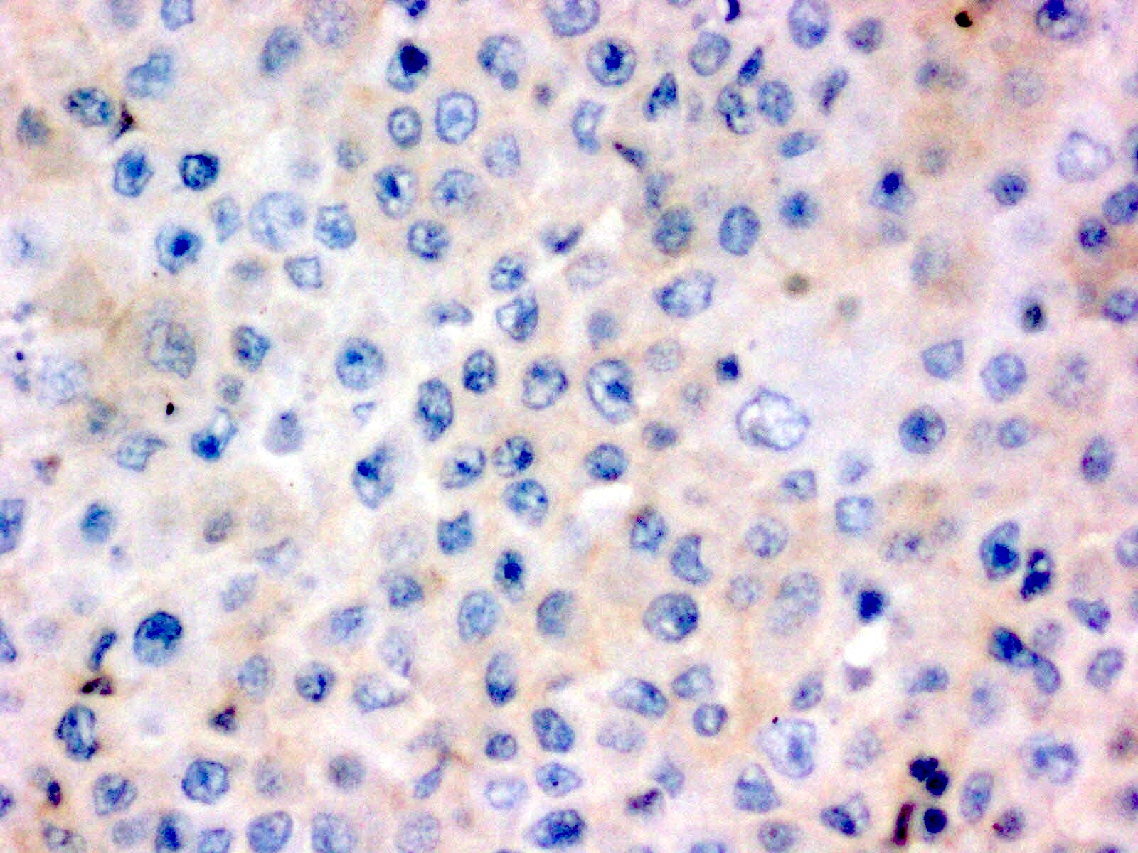
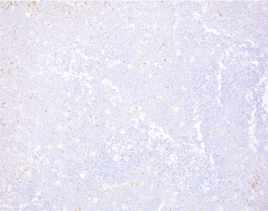
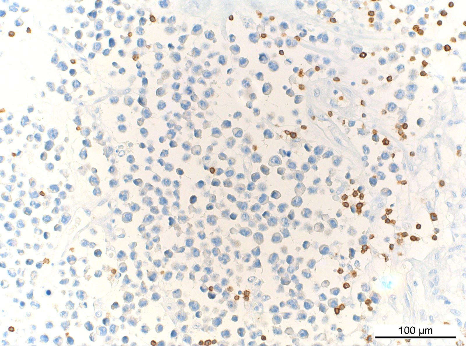
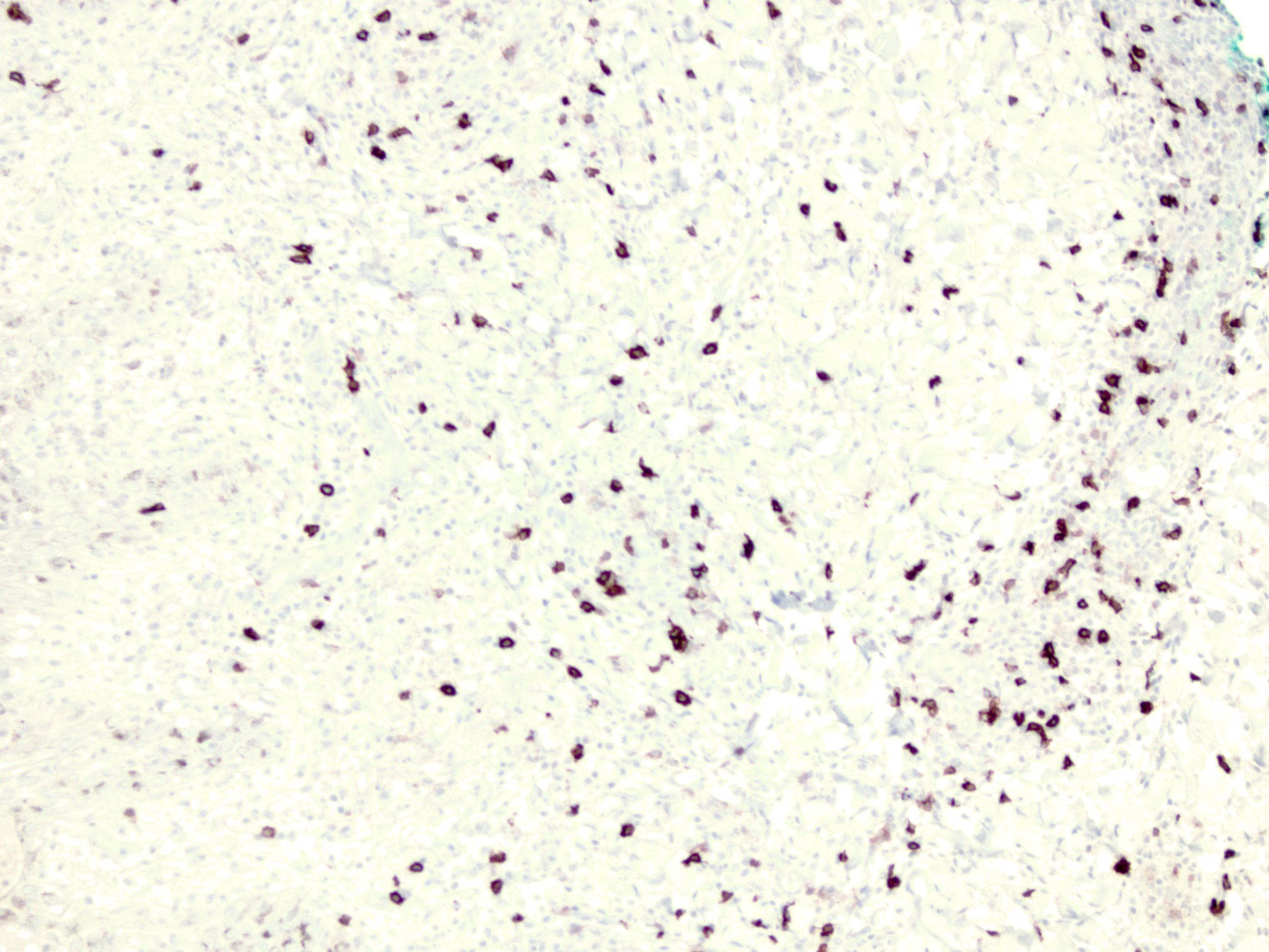
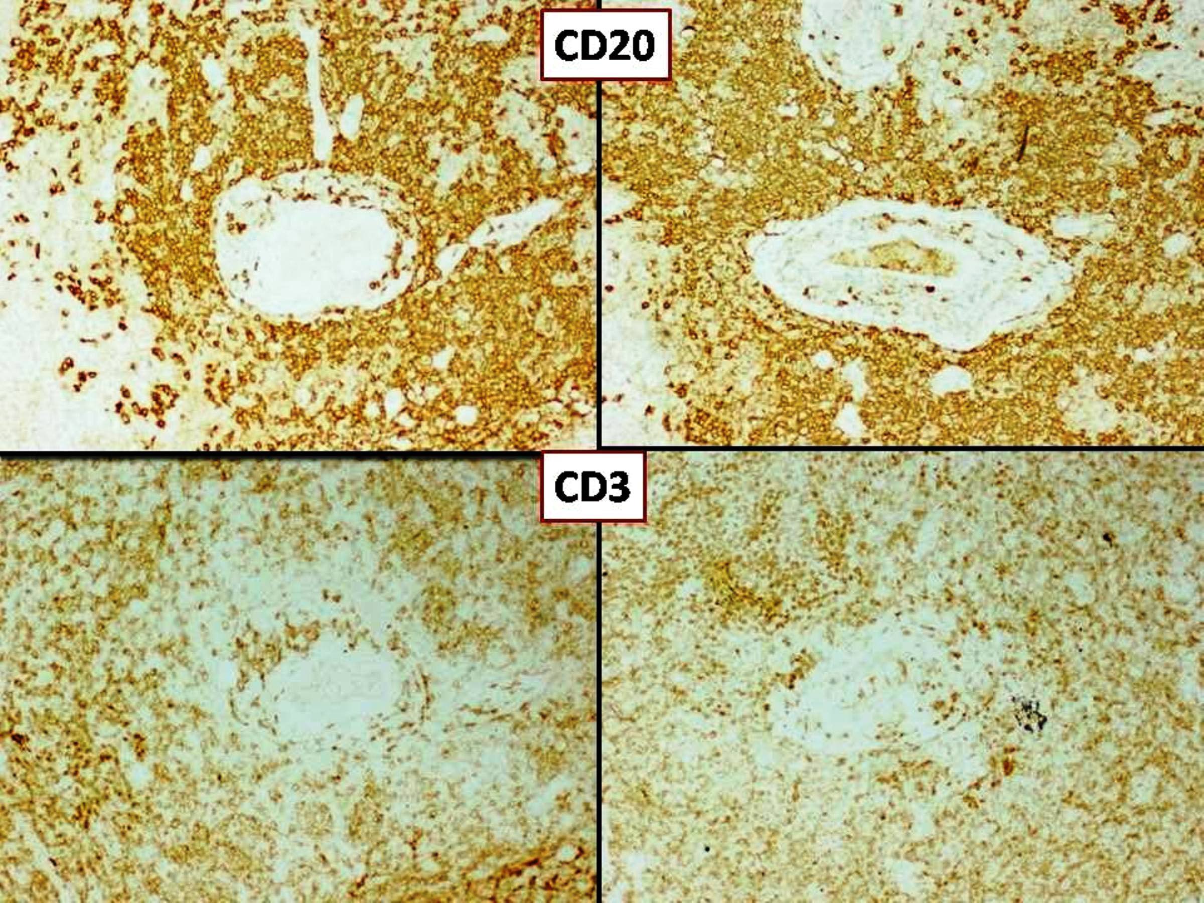
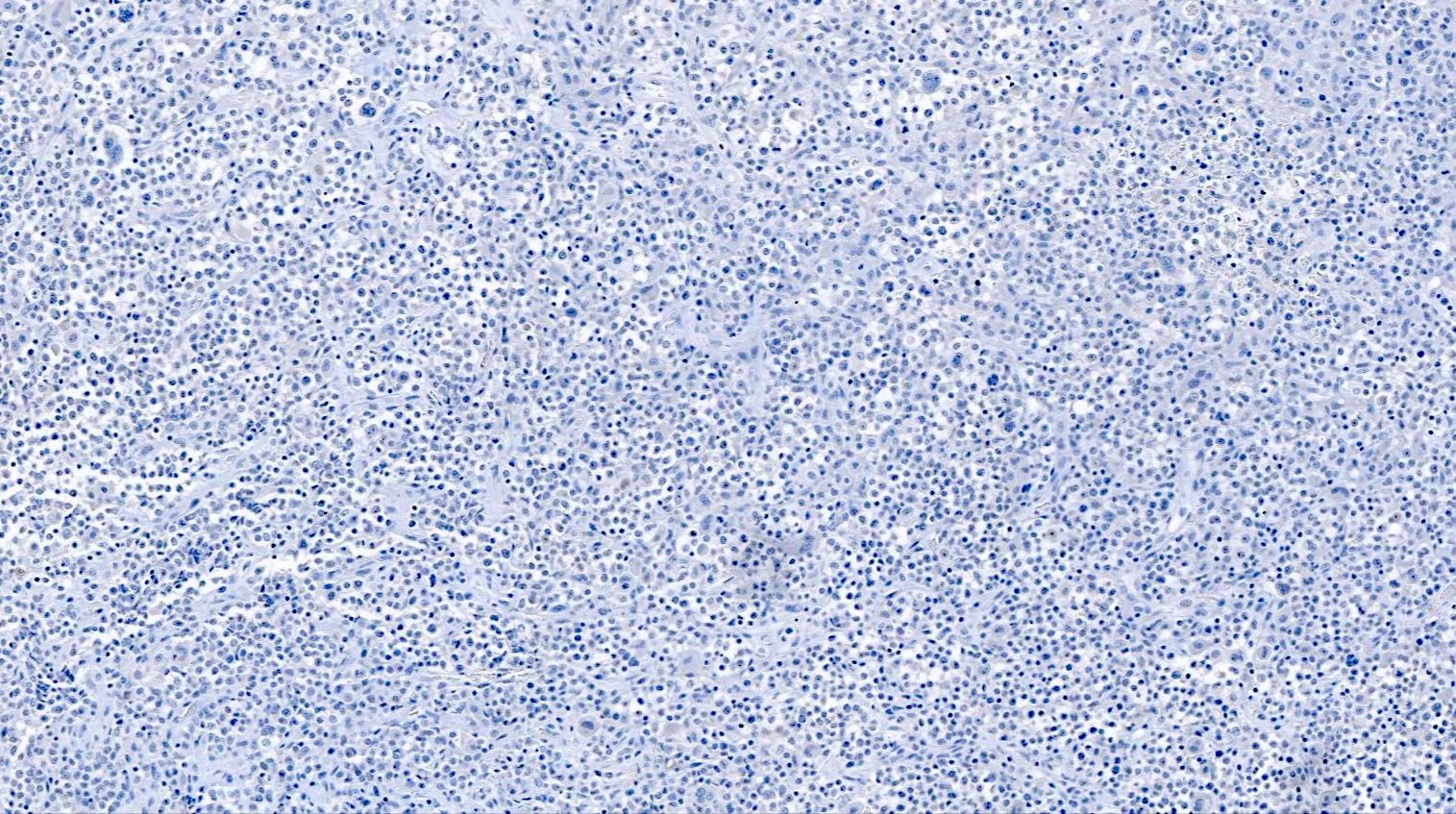
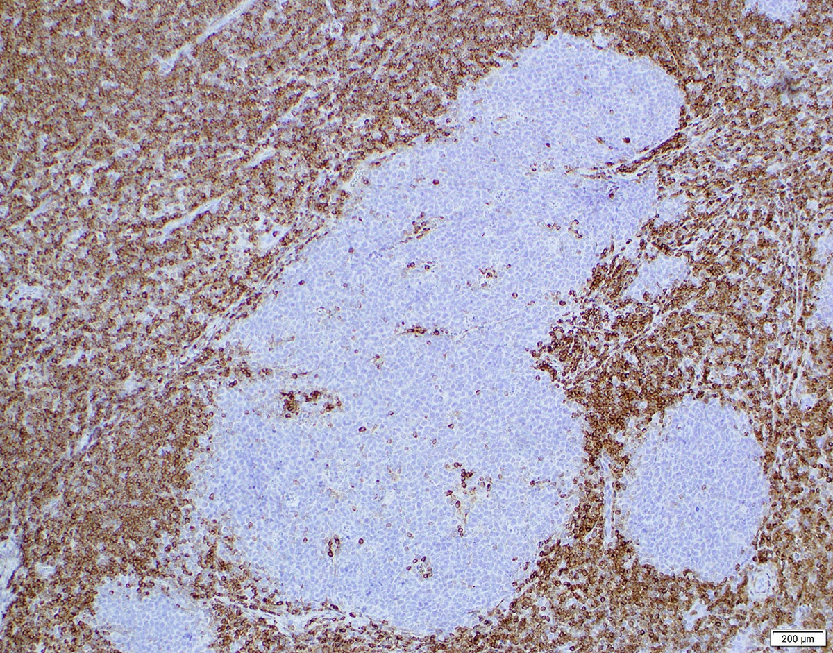
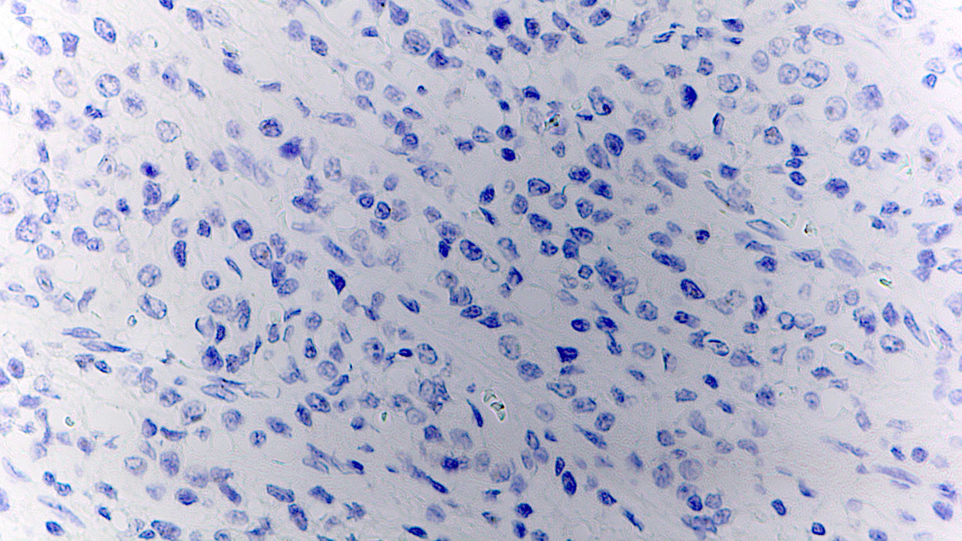
CD30 negativity in tumor cells
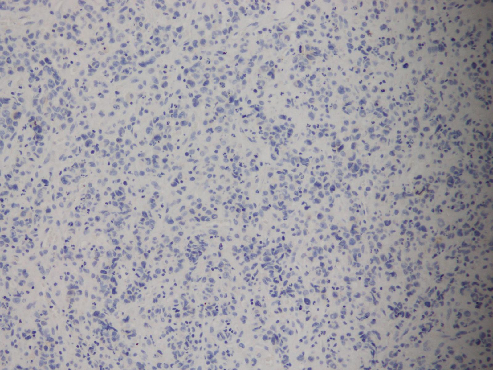
CD3 negativity in tumor cells
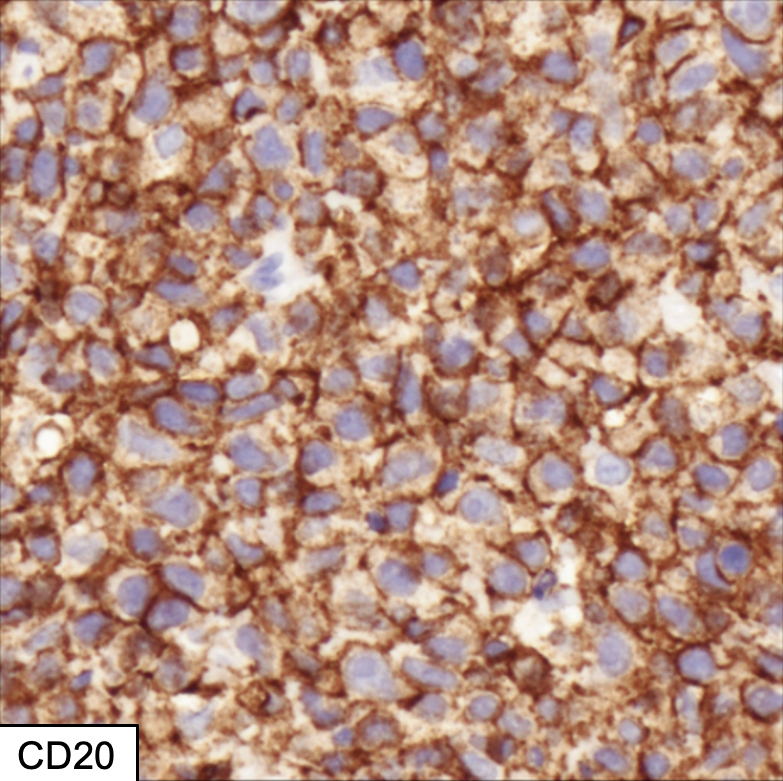
CD20 negativity in tumor cells
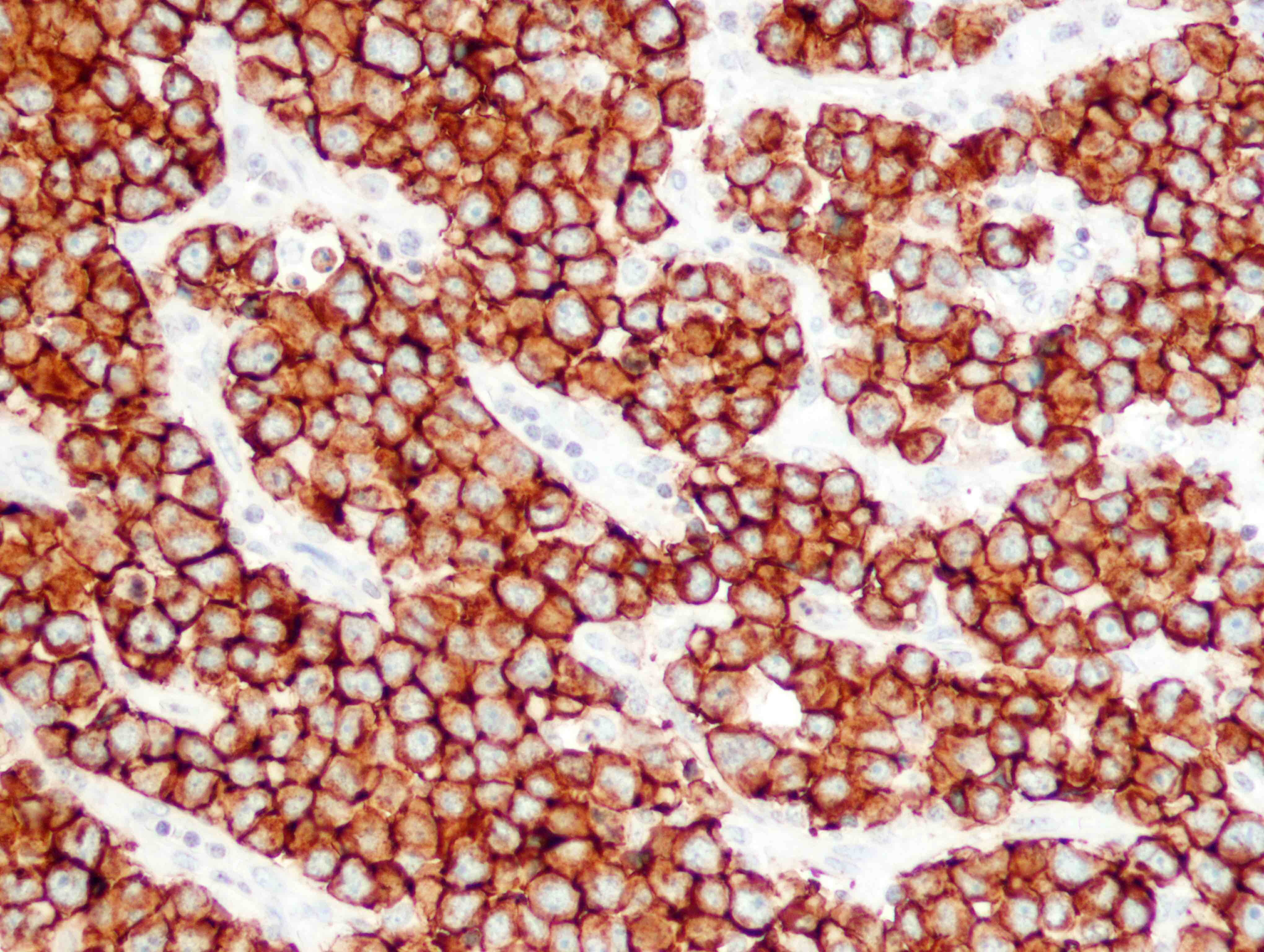
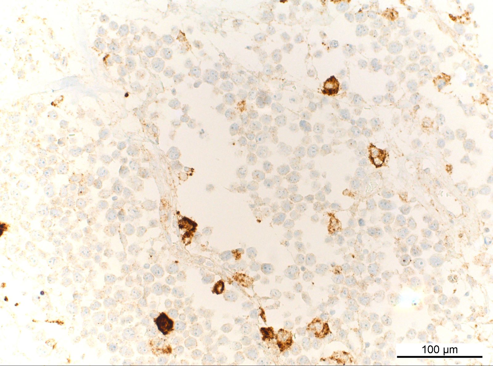
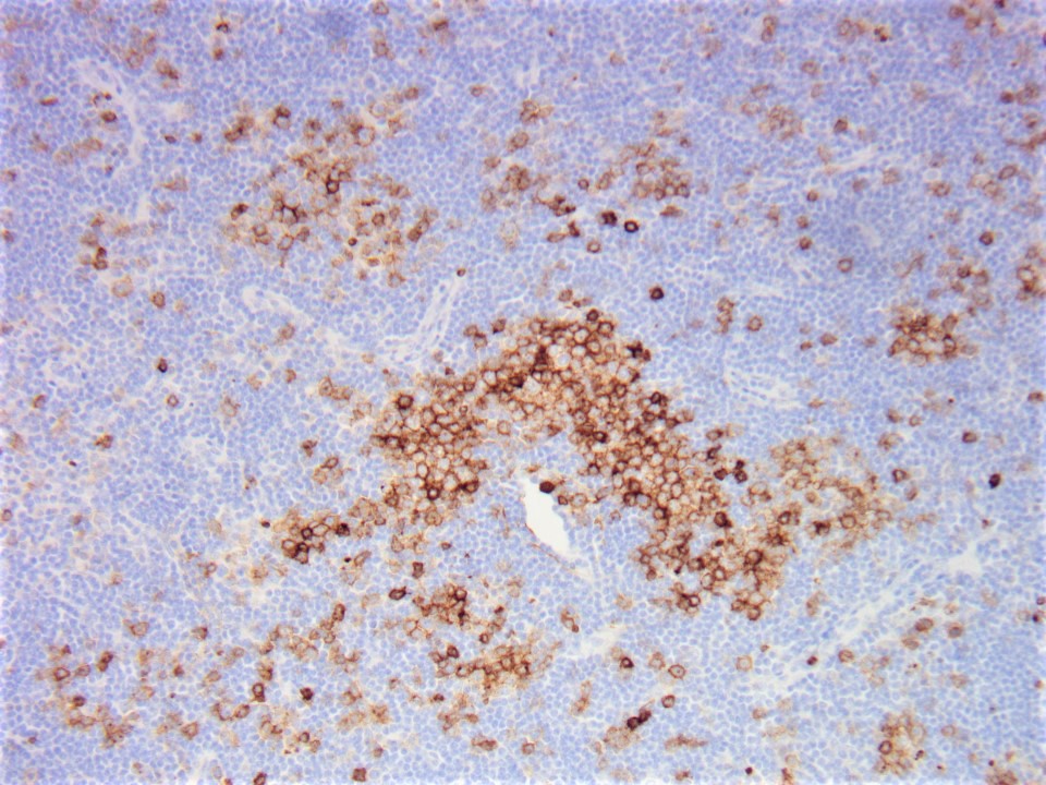
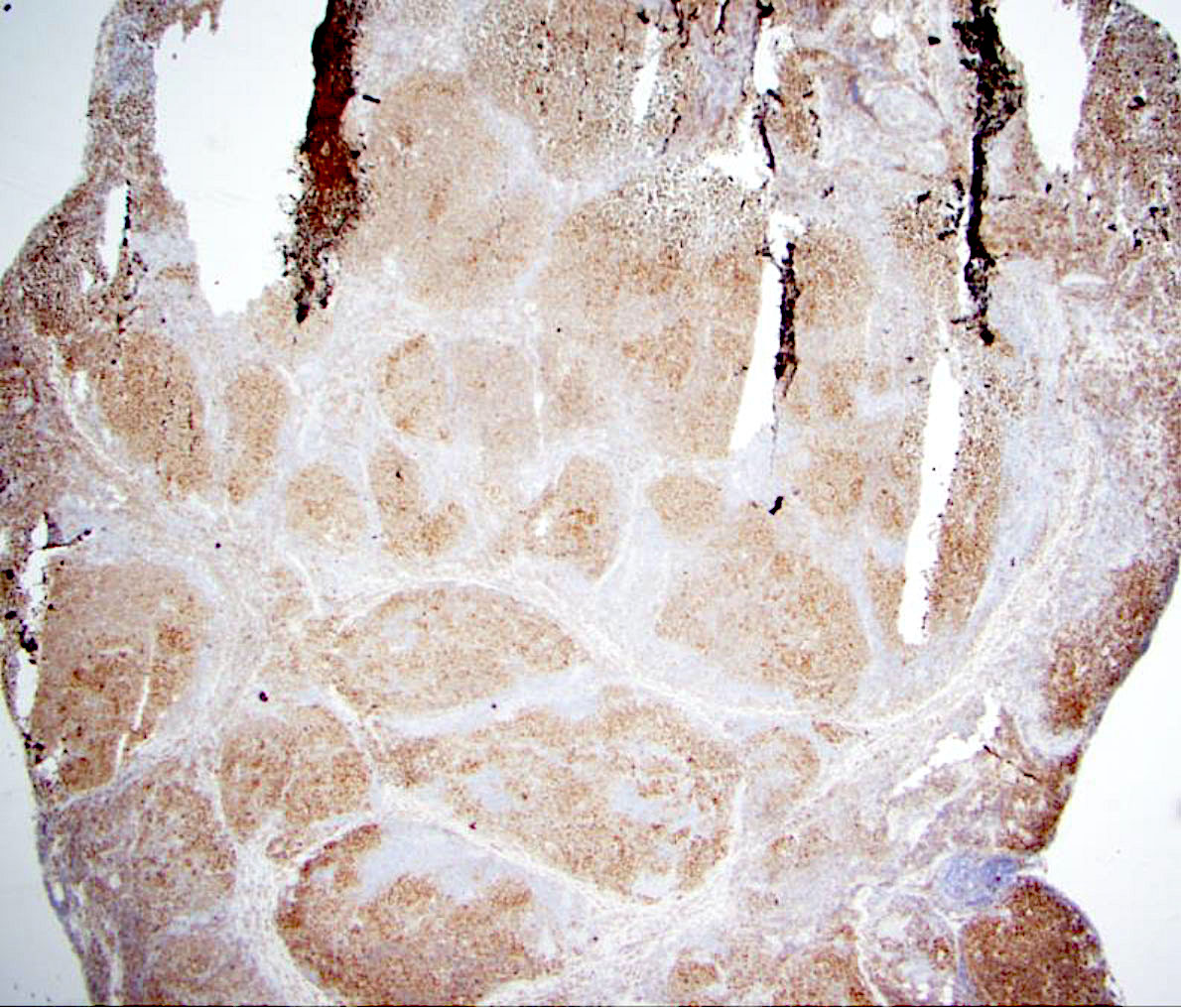
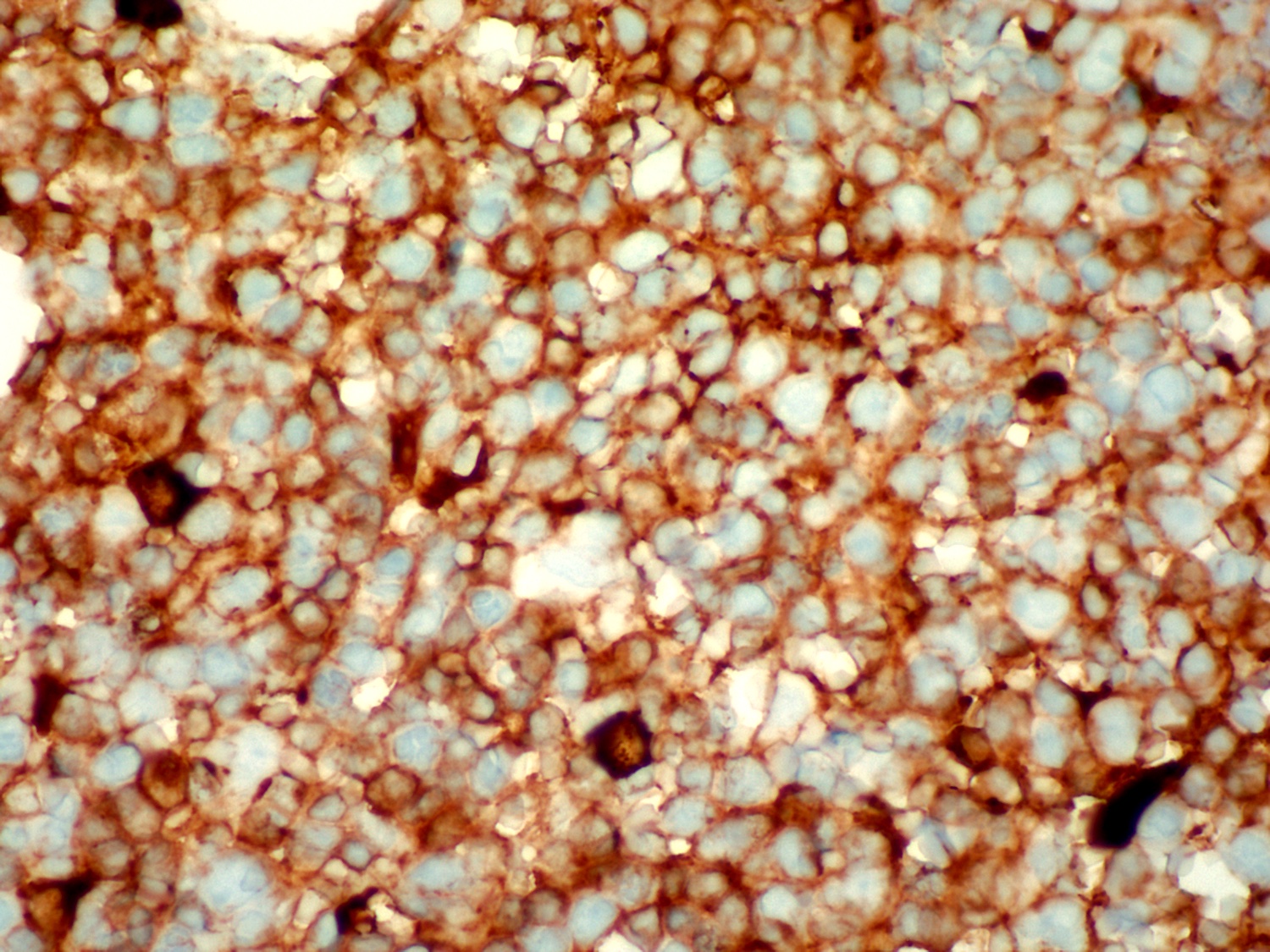
Weak positivity for CD79a
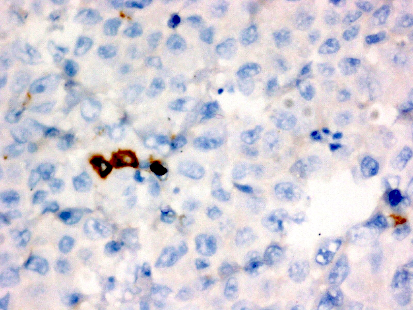
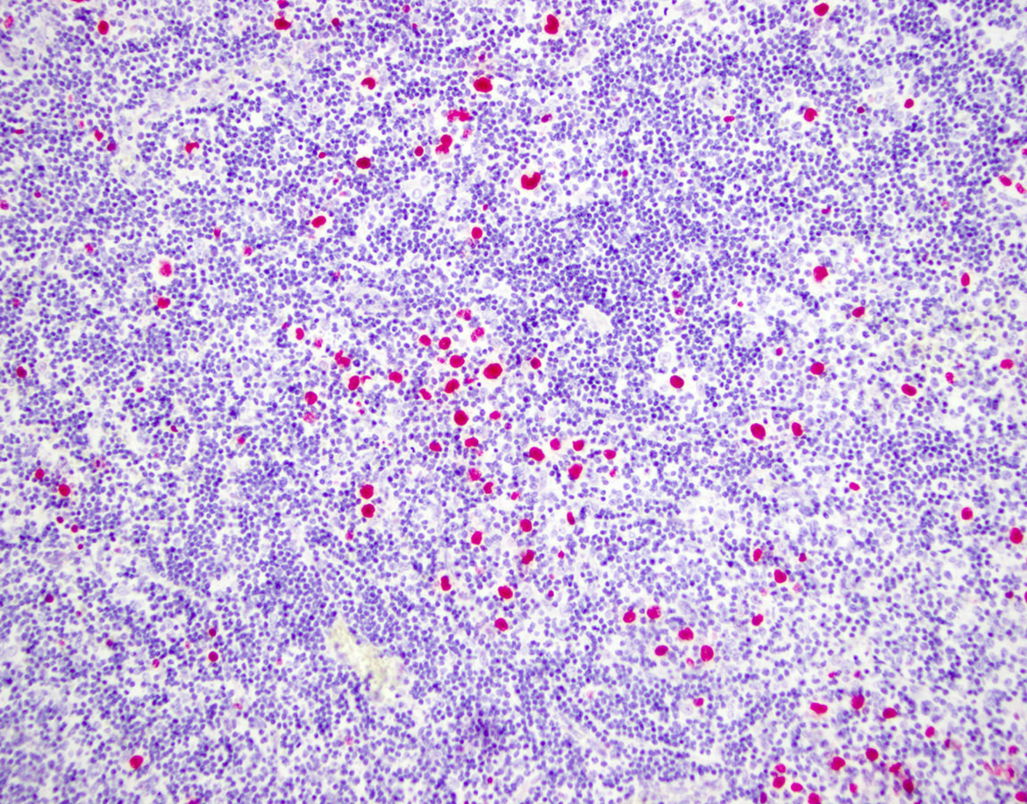
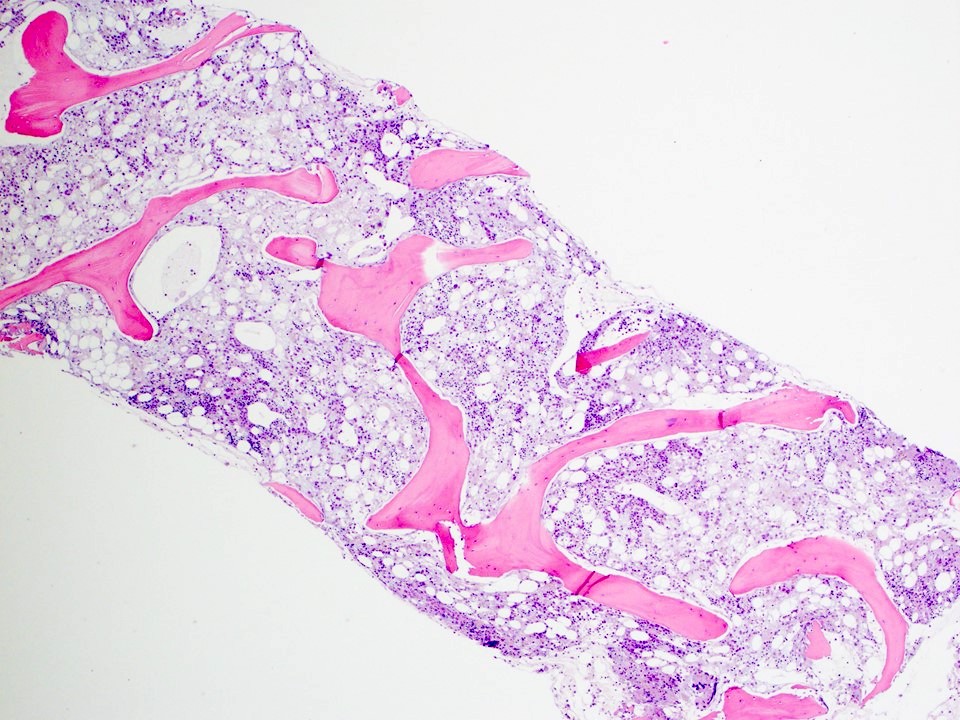
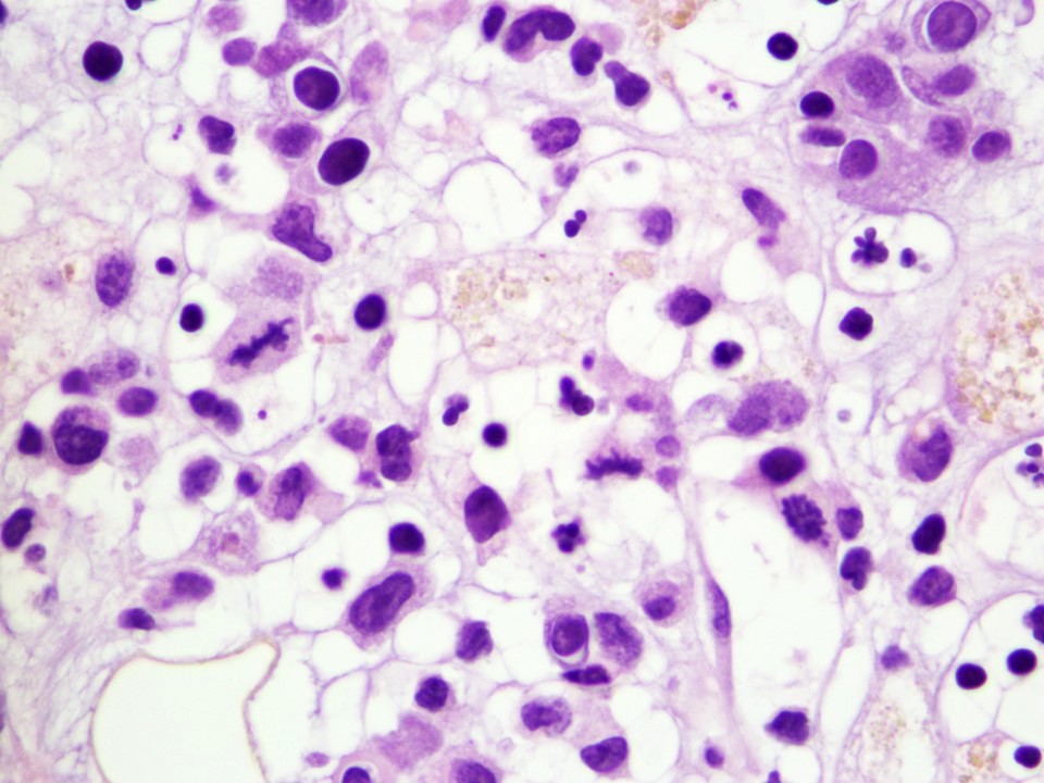
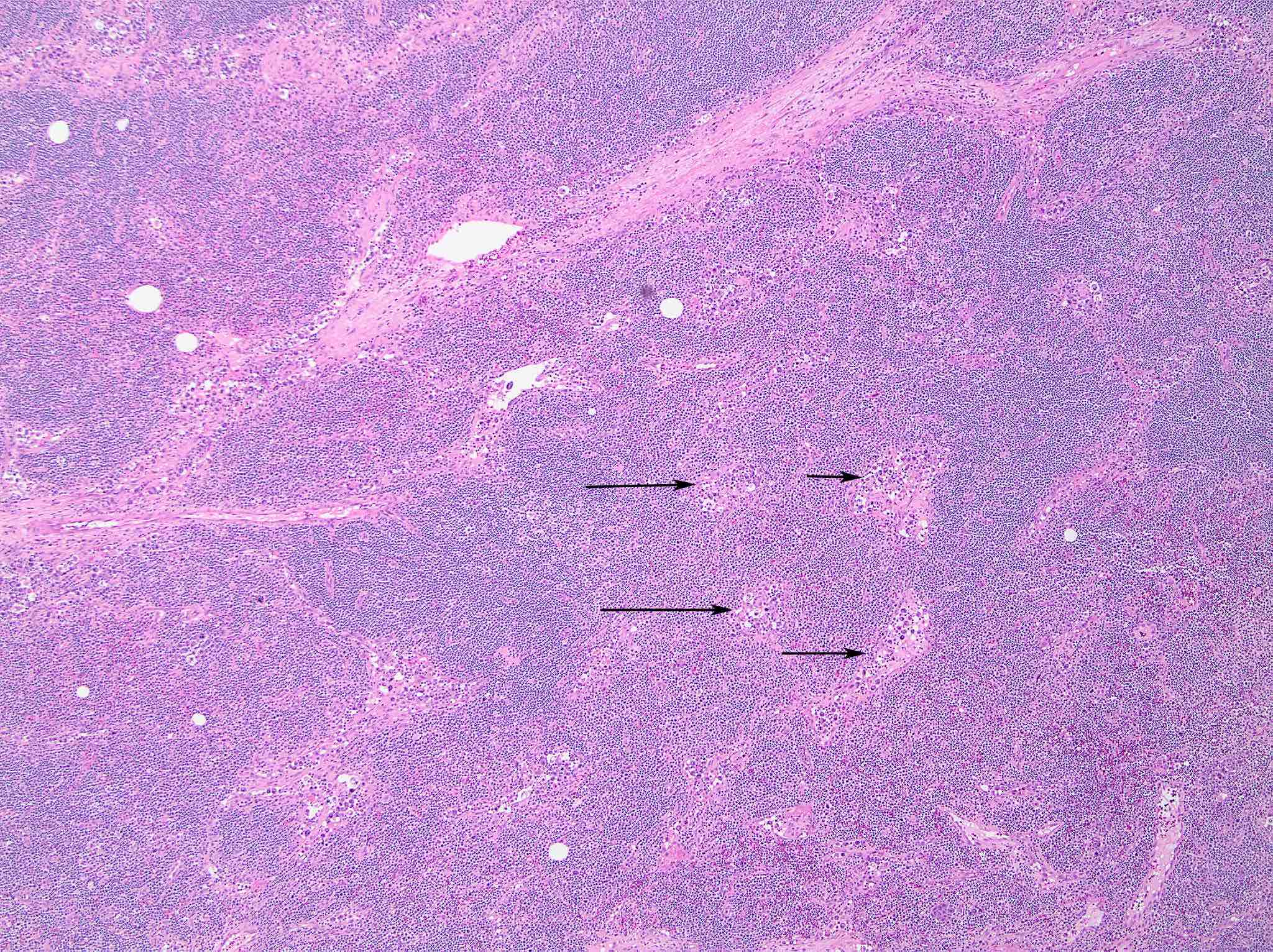
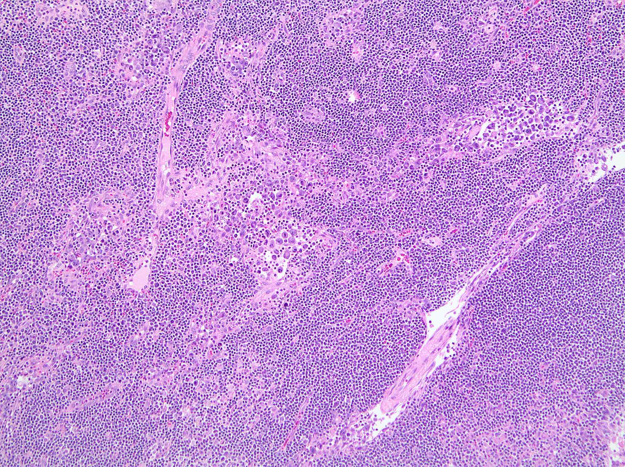
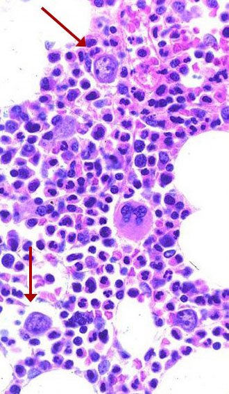
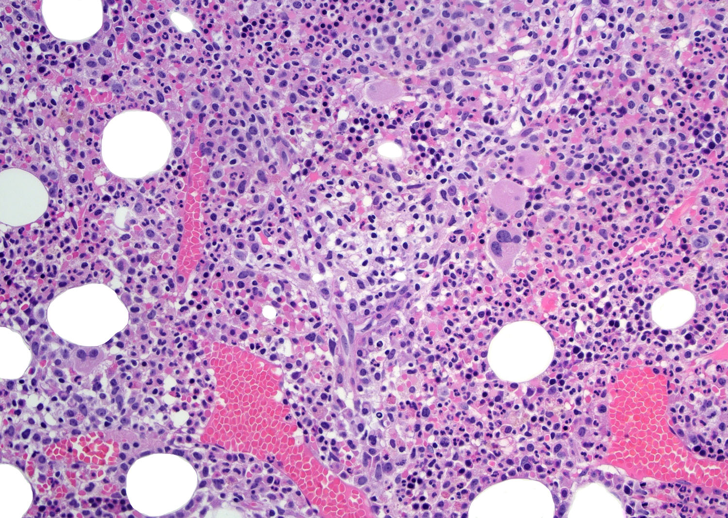
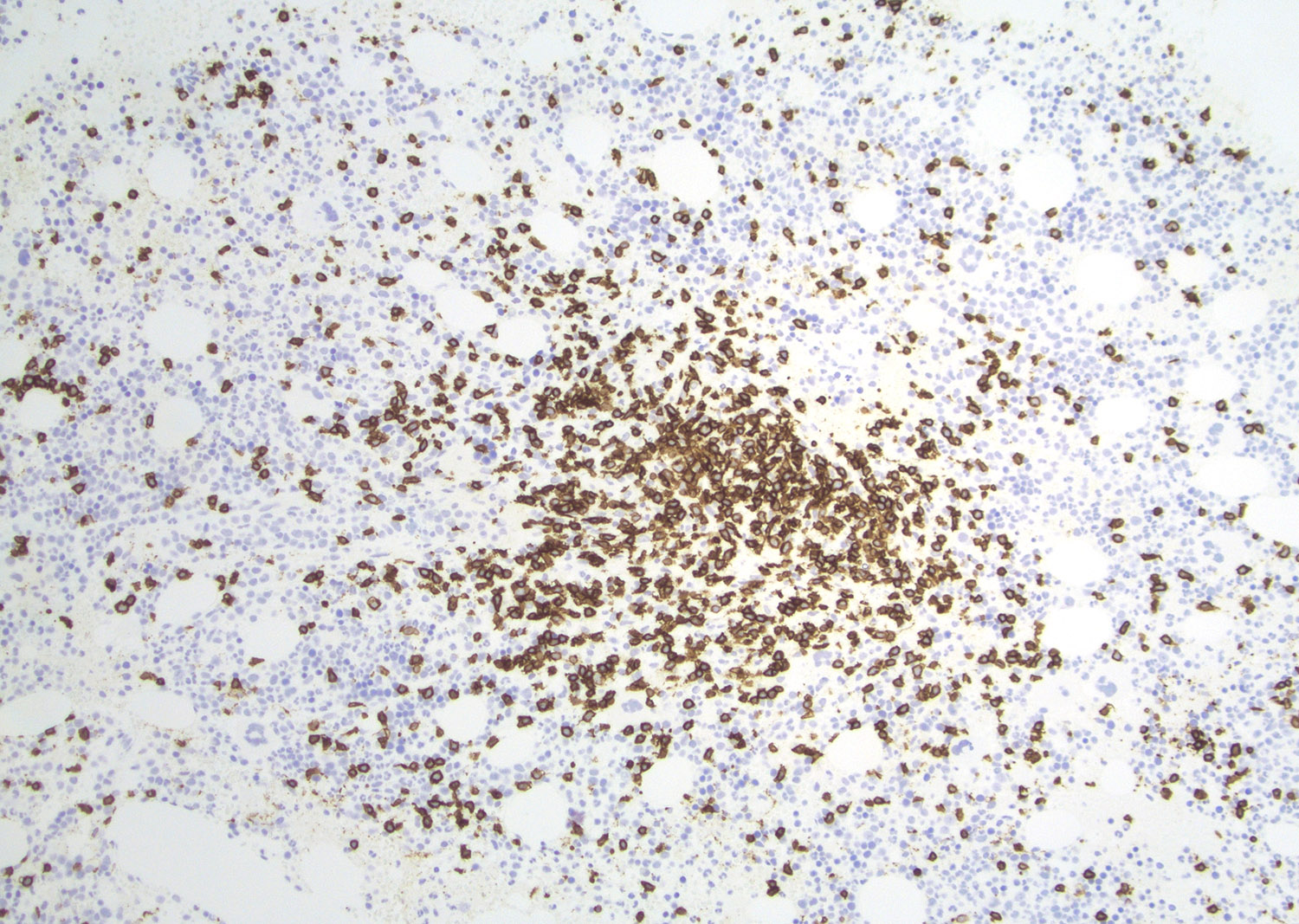
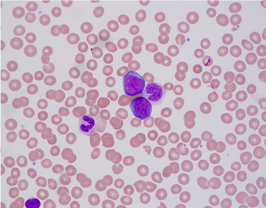
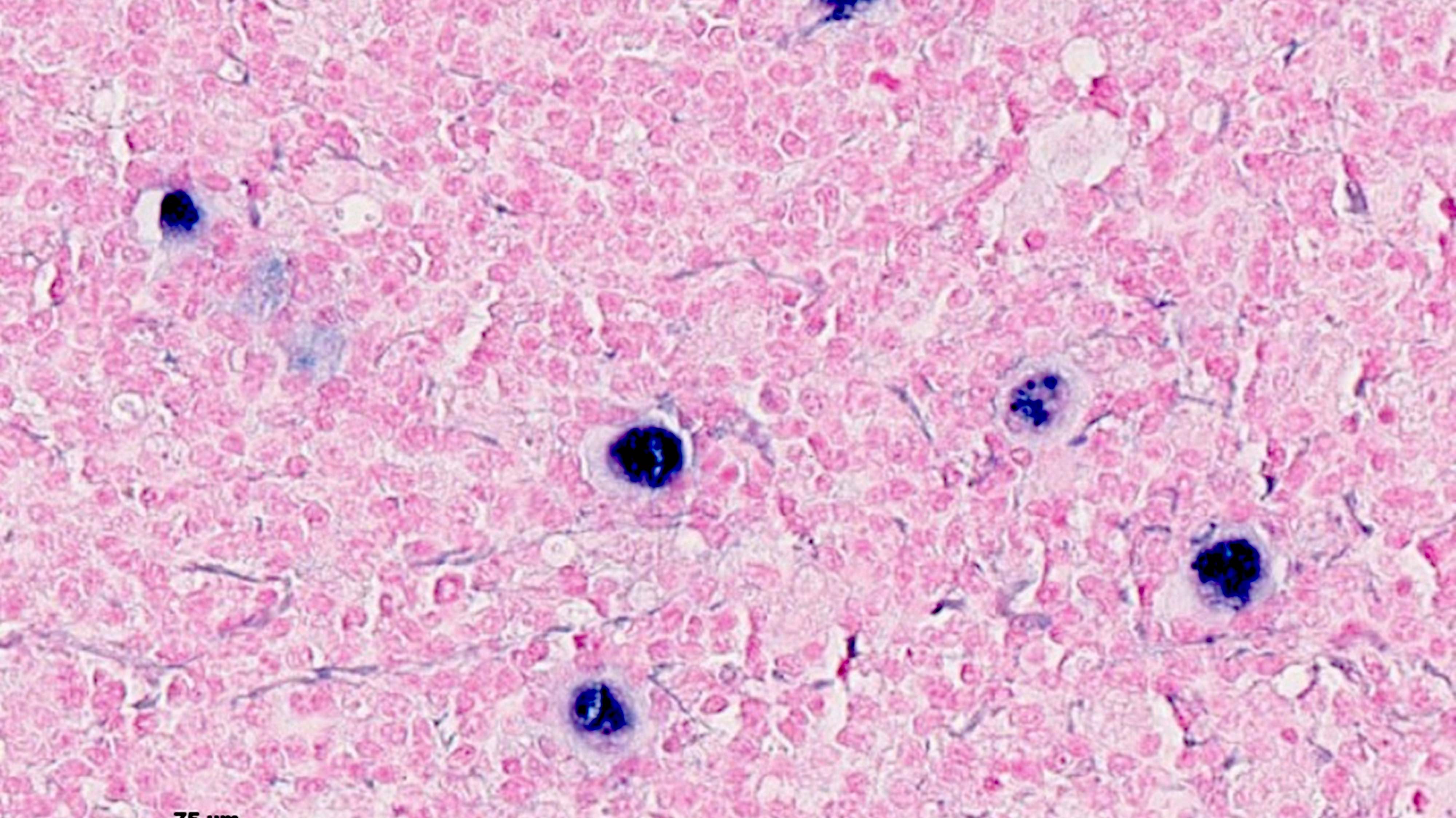
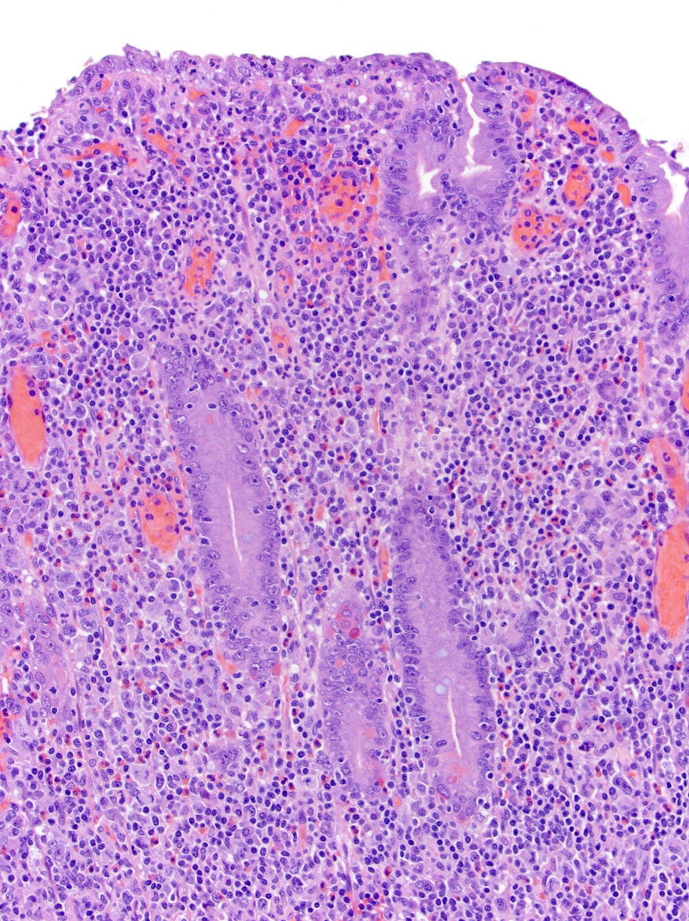
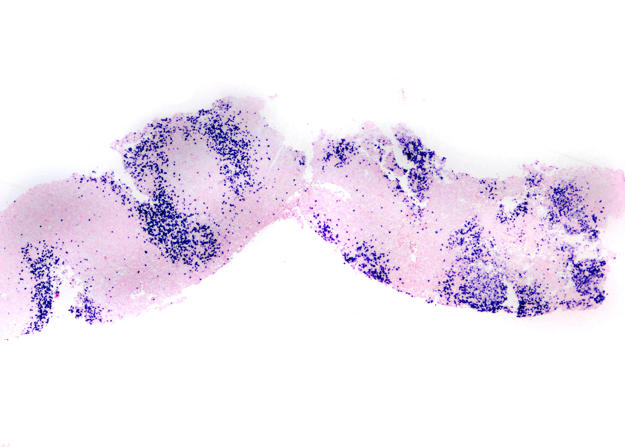
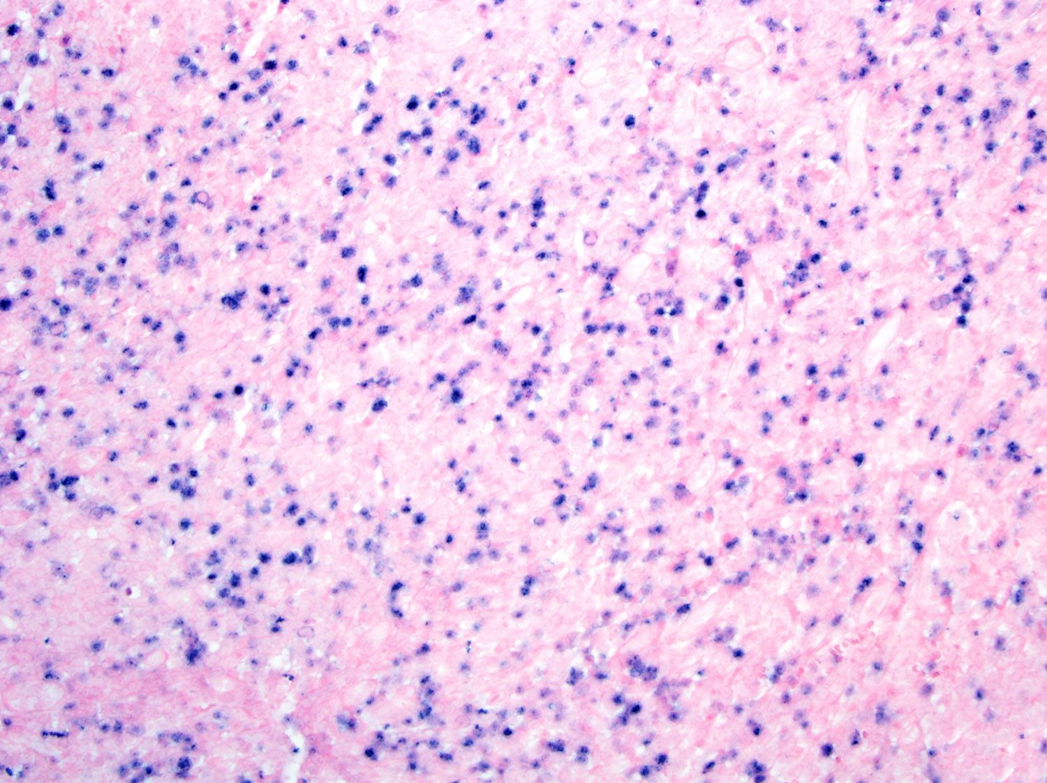
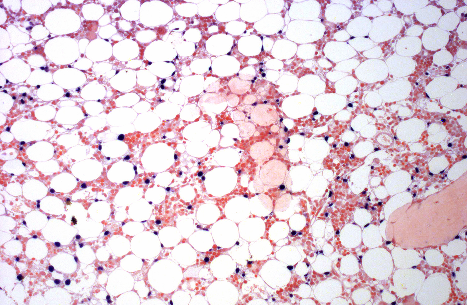
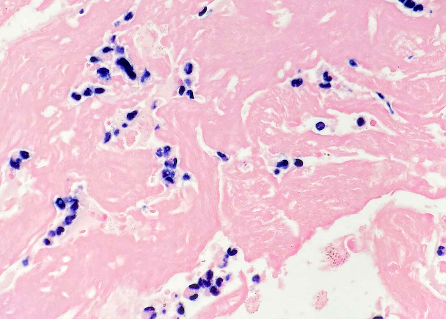
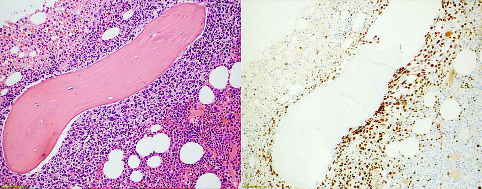
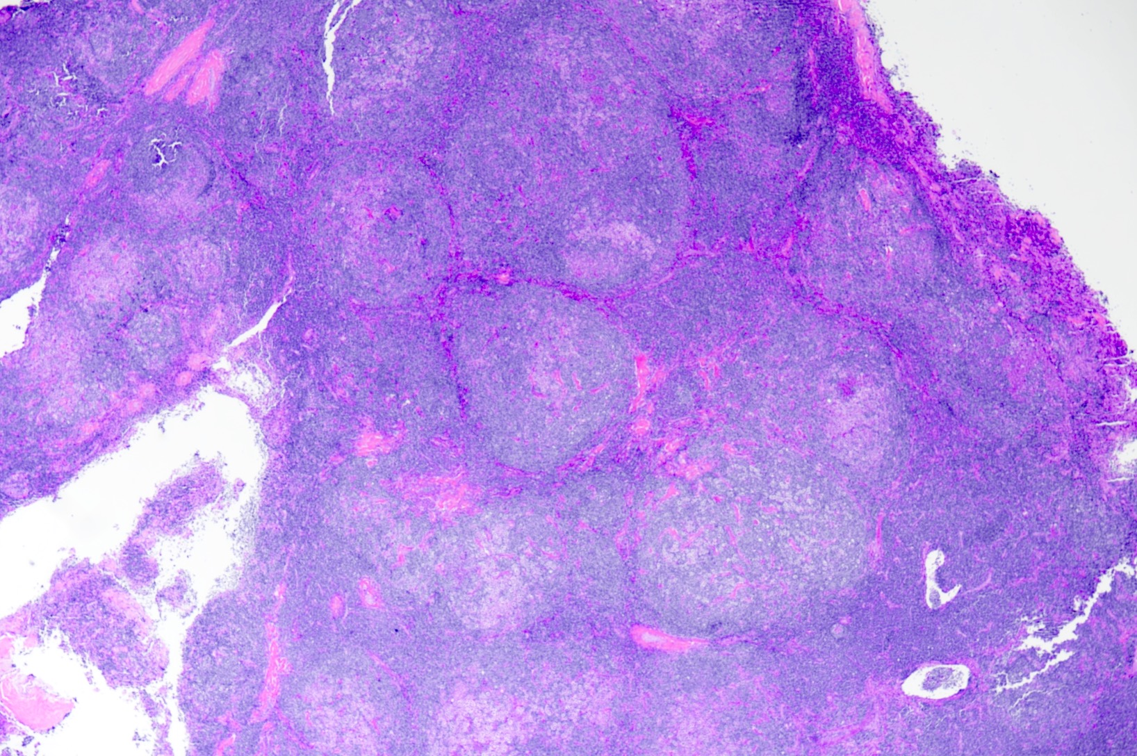
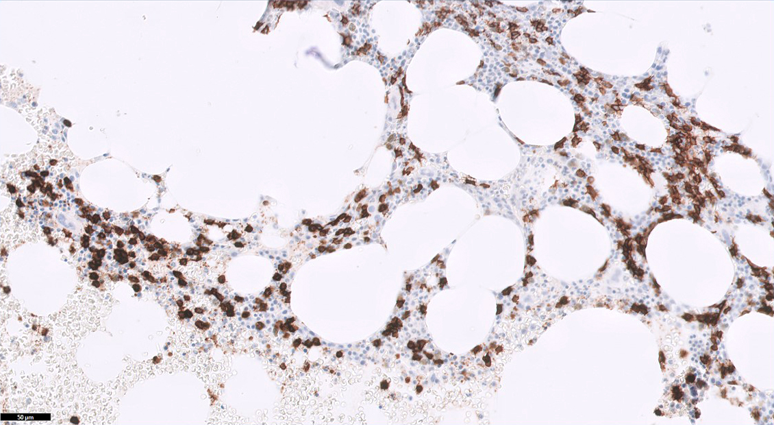
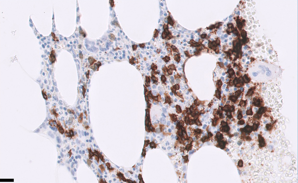
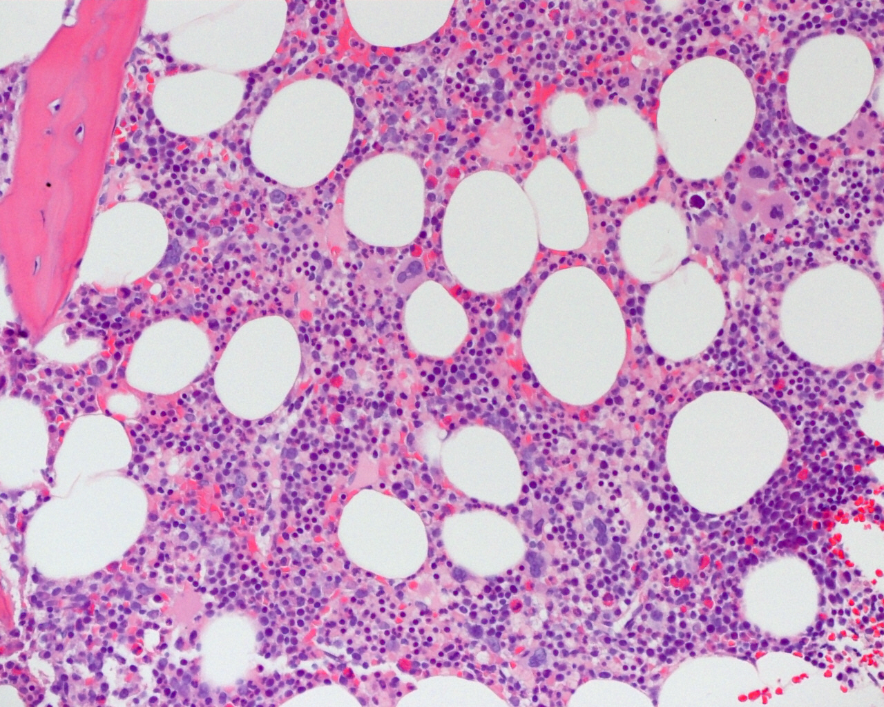
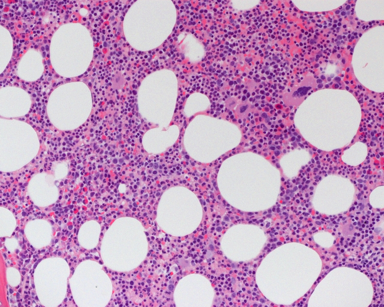
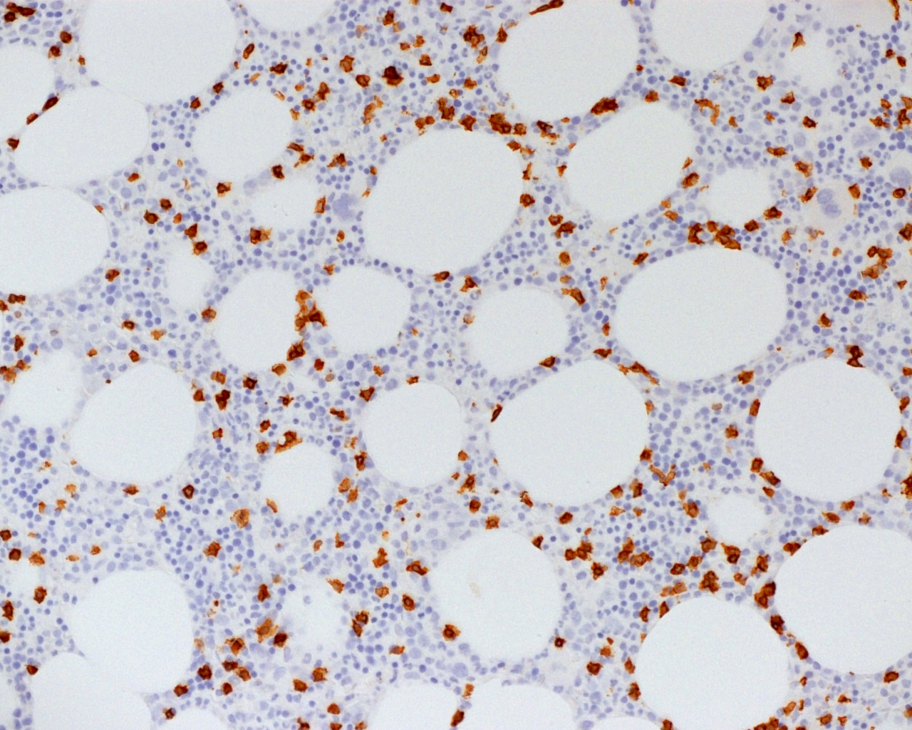
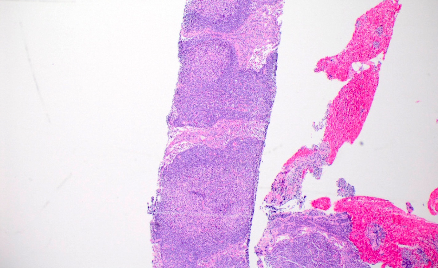
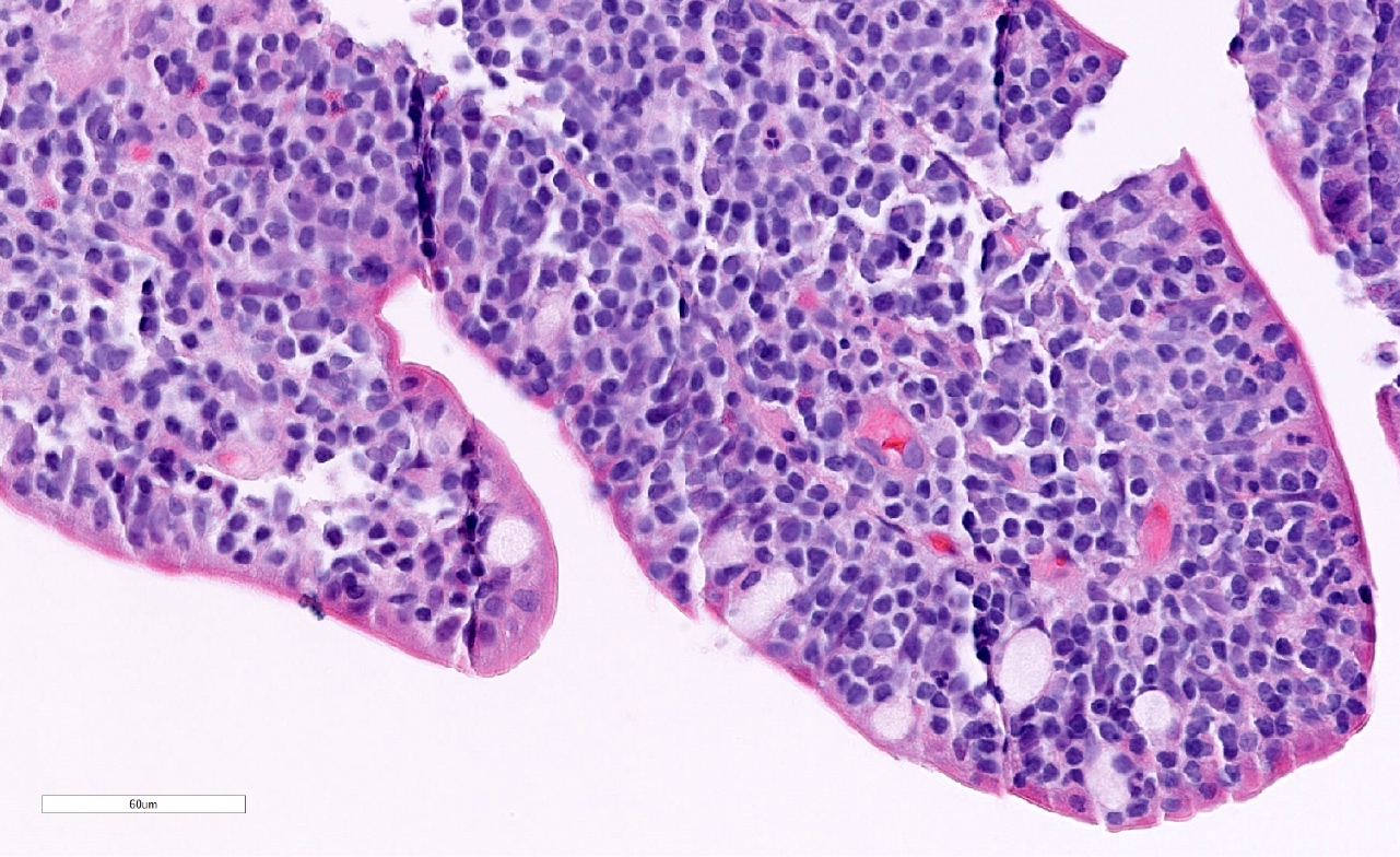
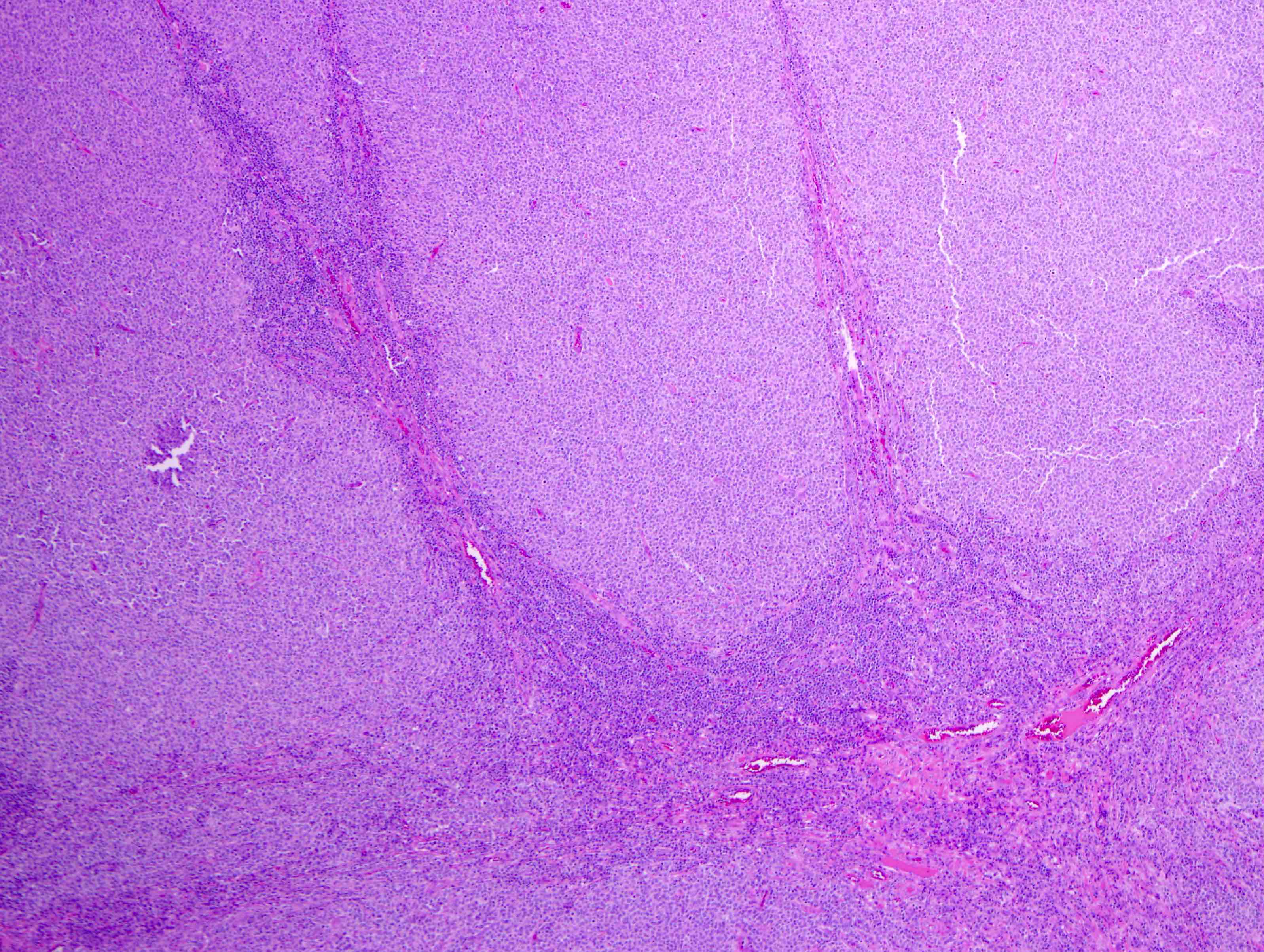
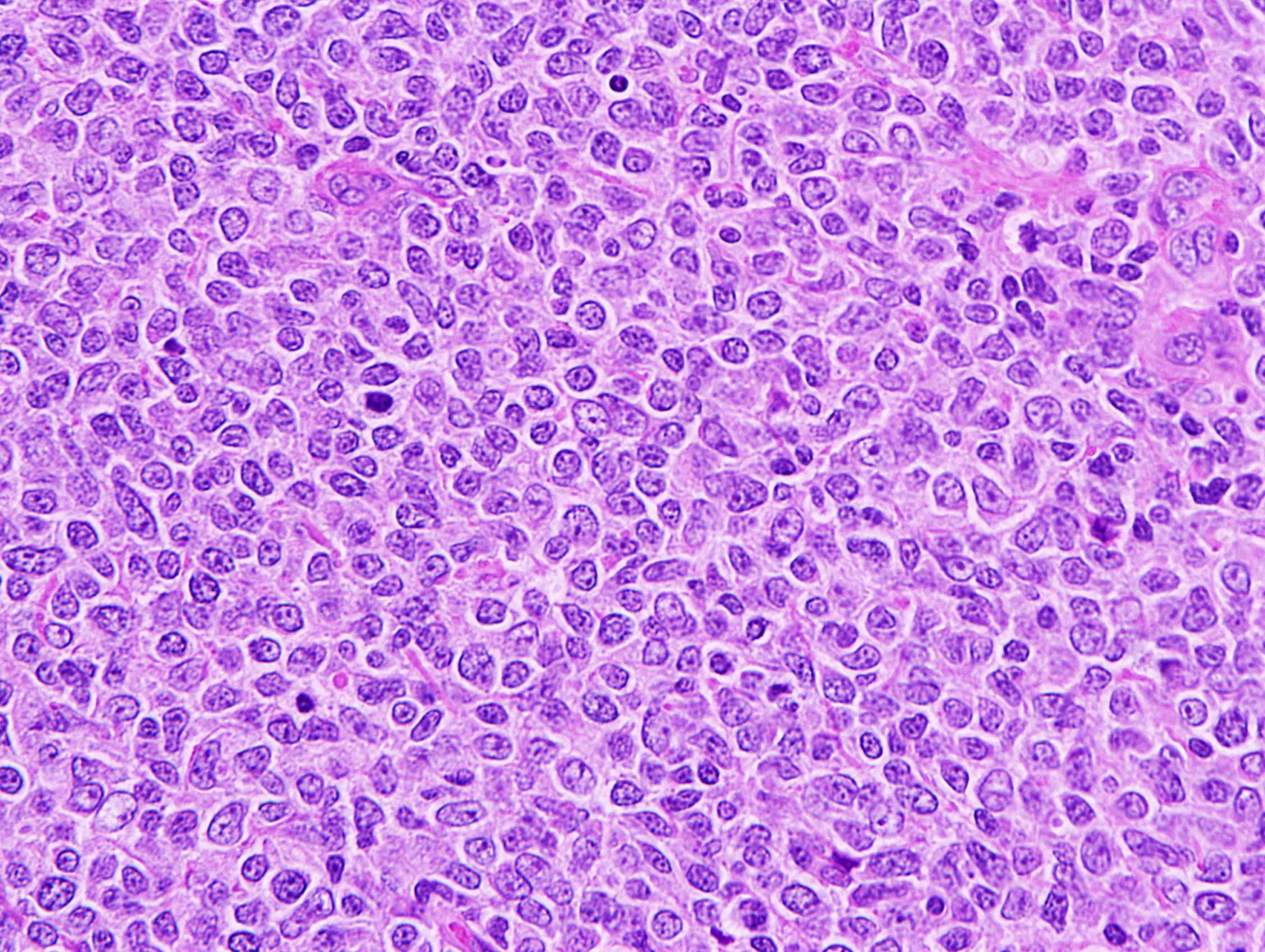
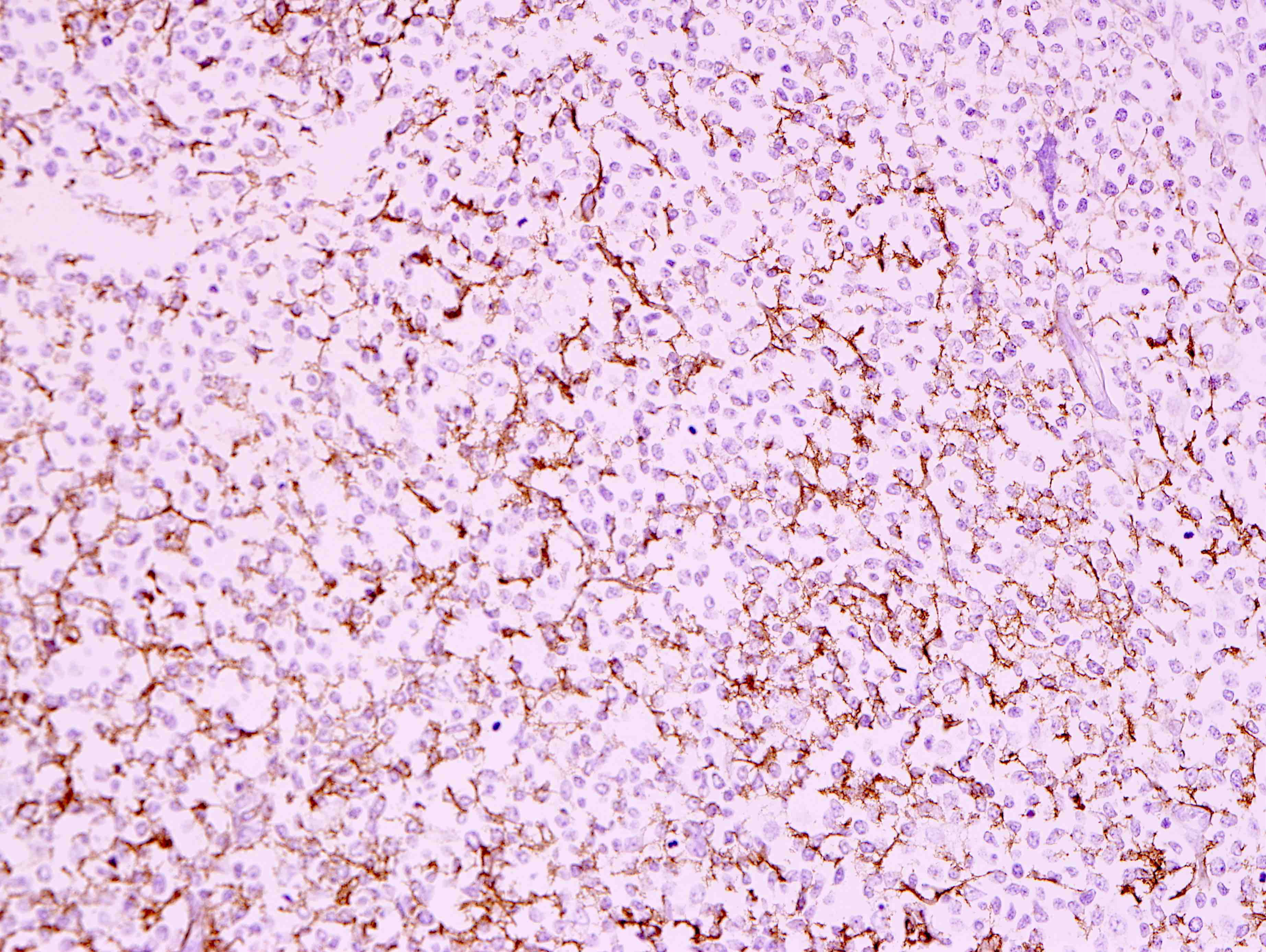
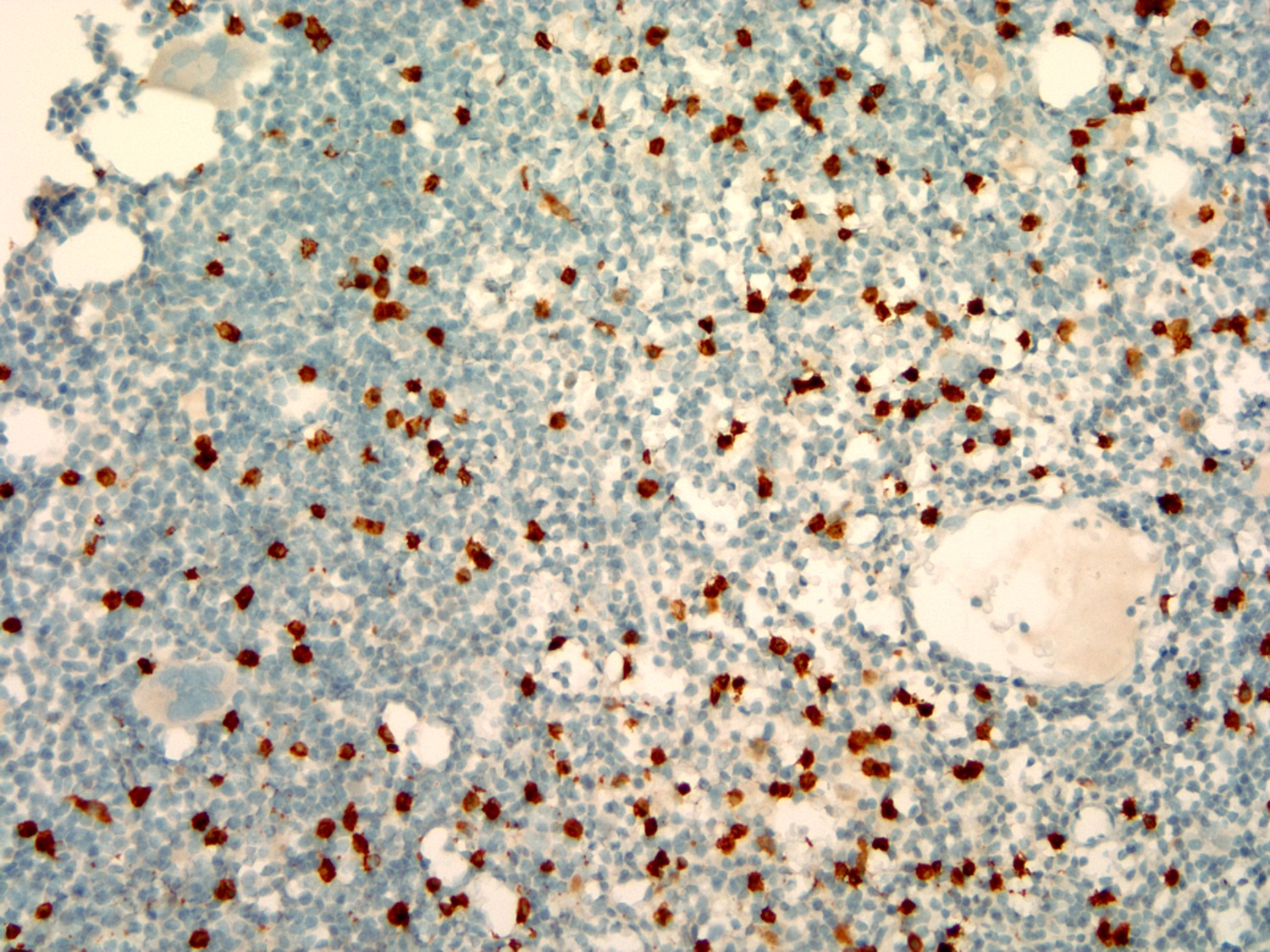
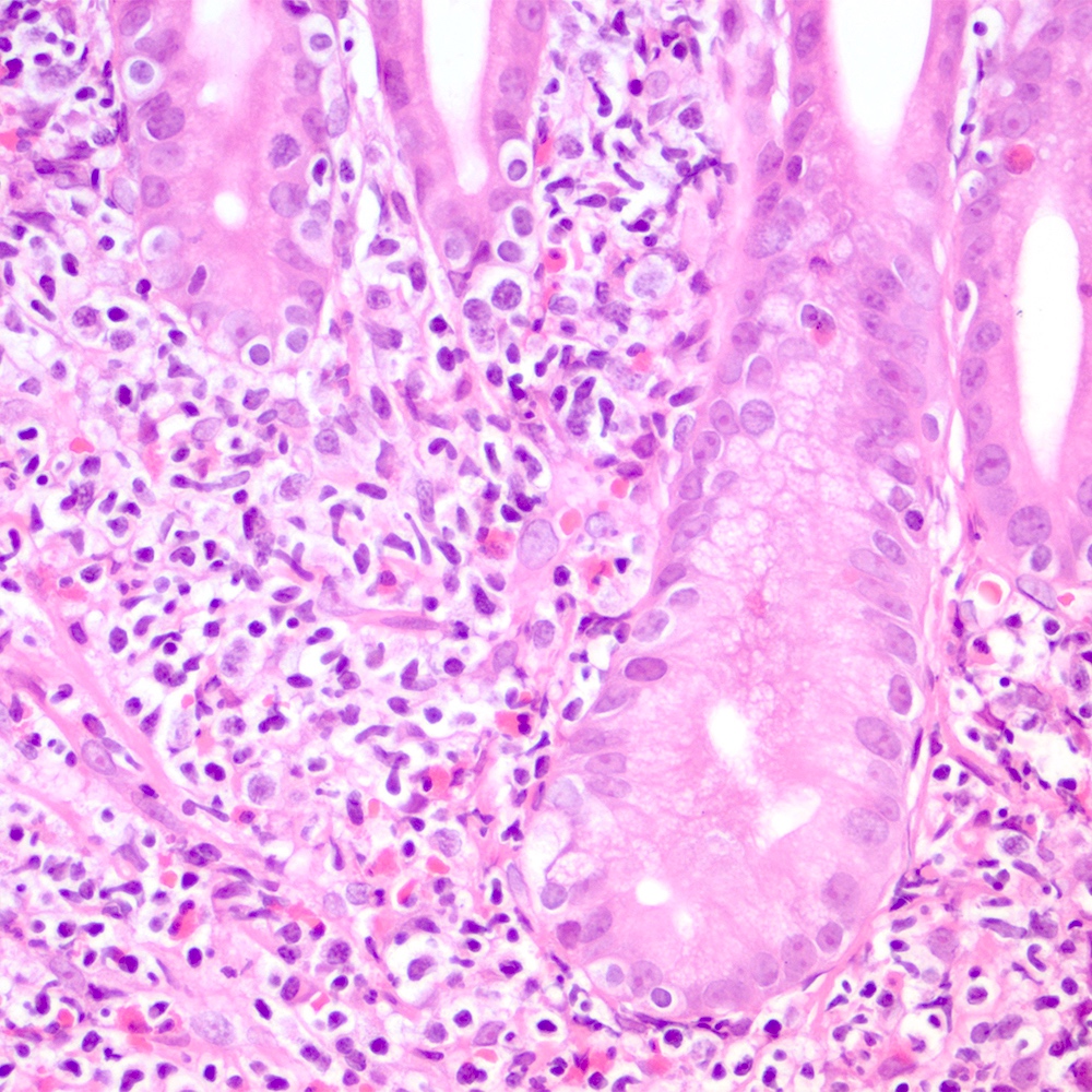
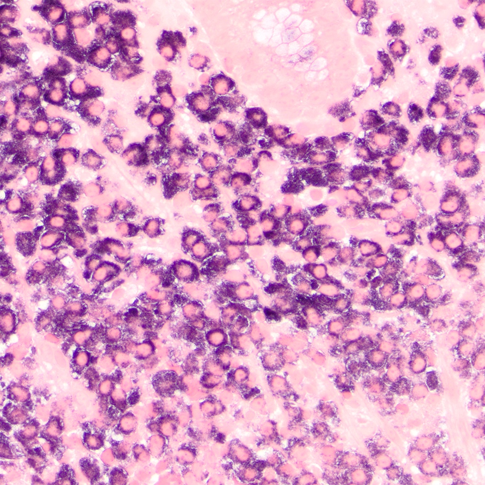
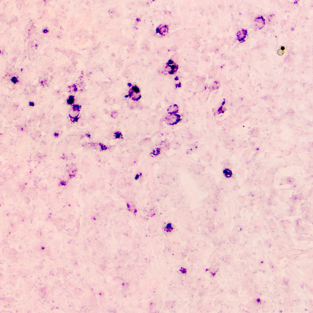
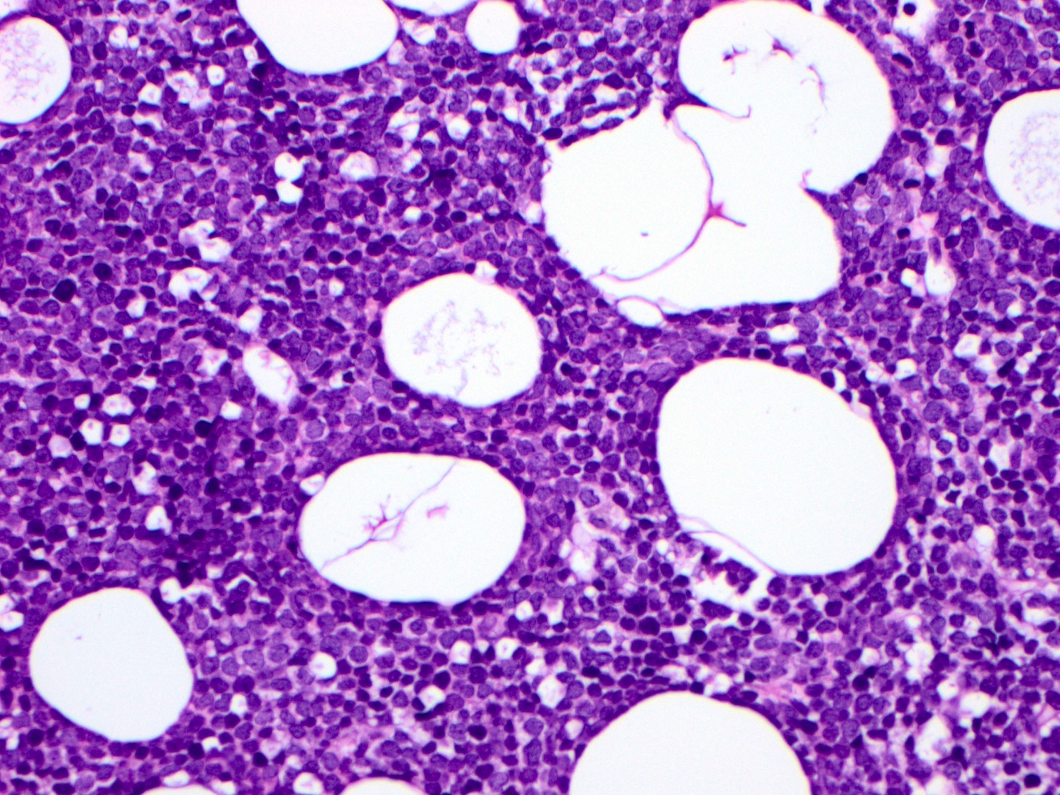
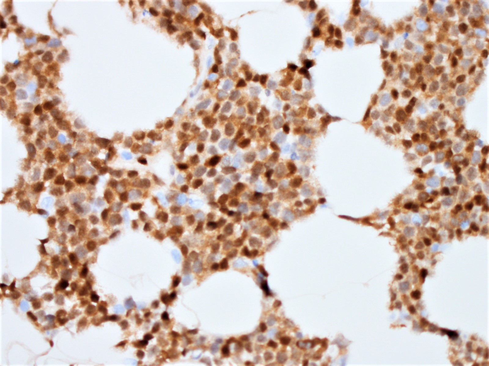
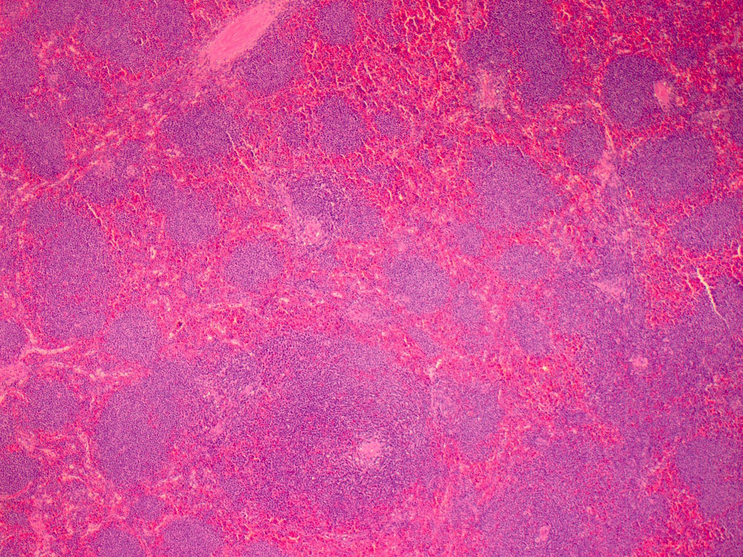
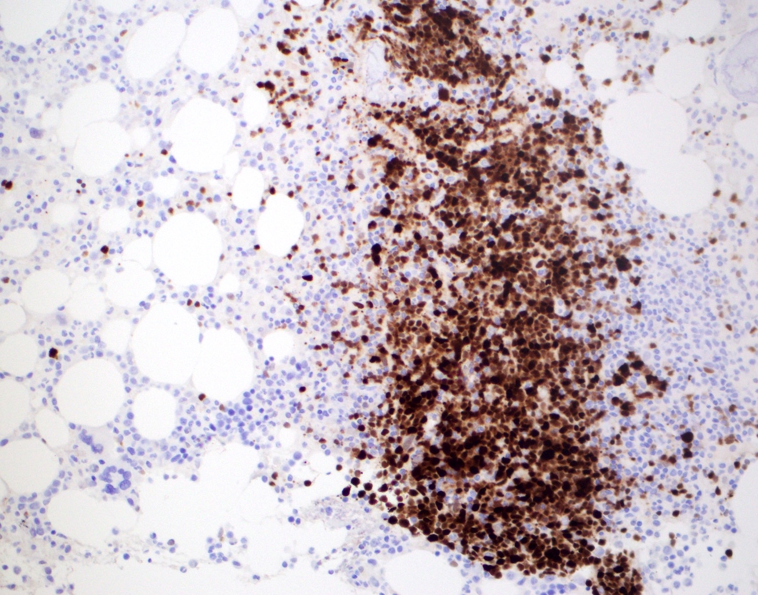
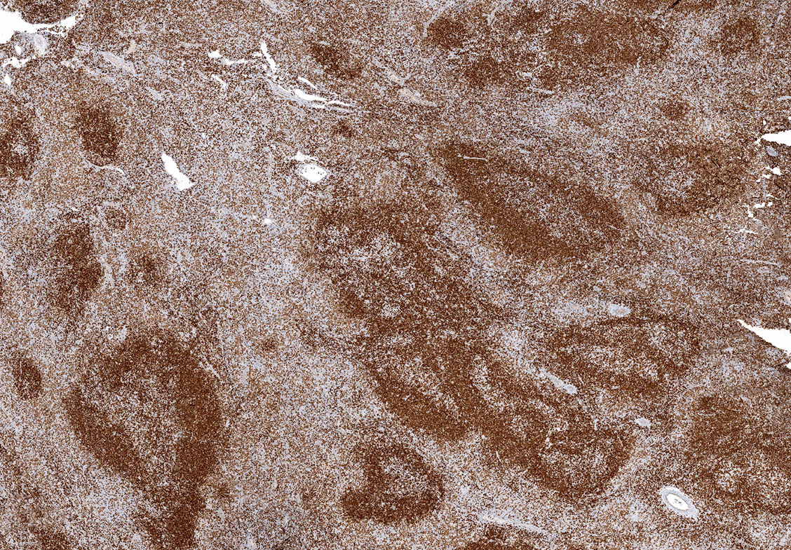
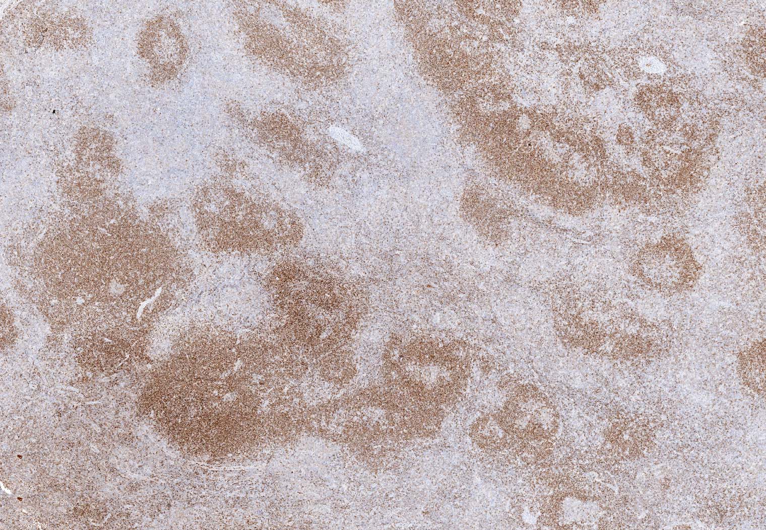
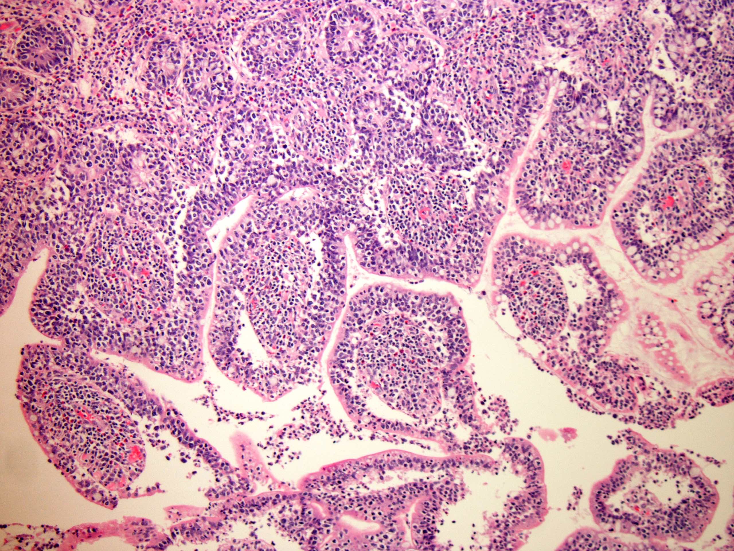
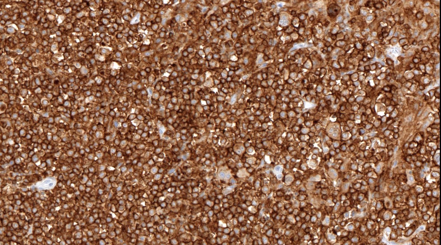
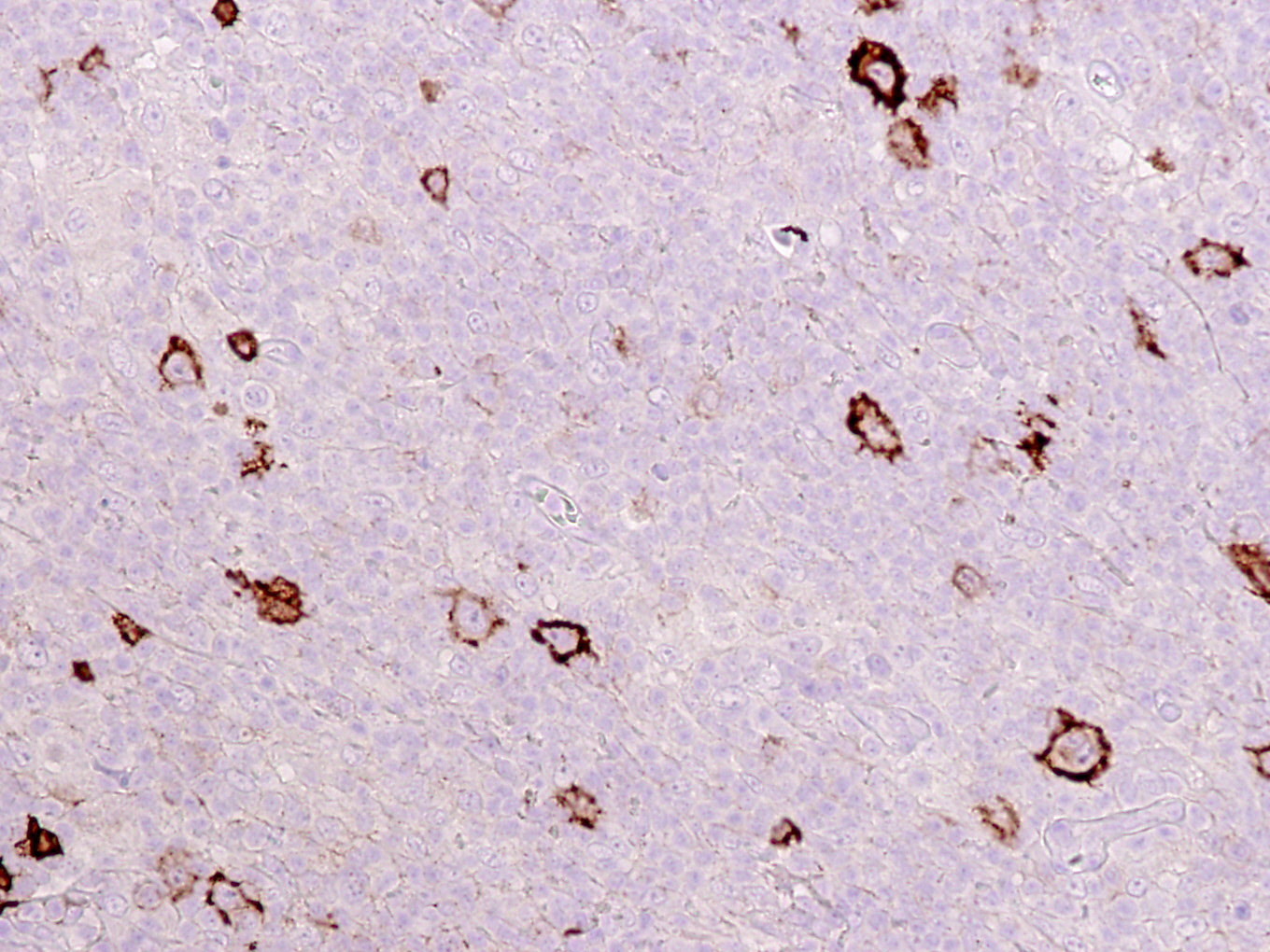
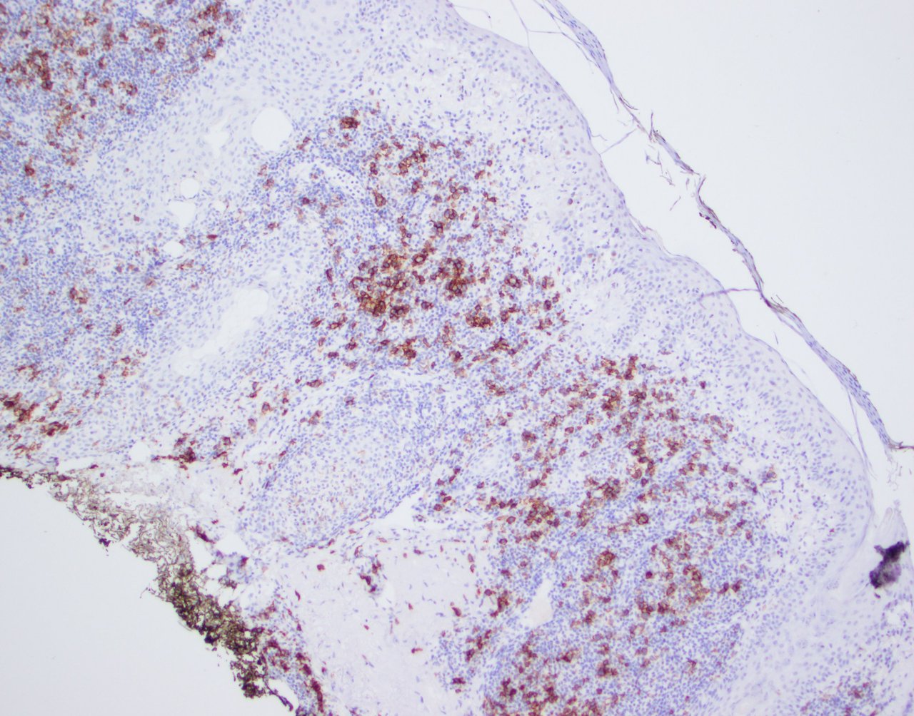
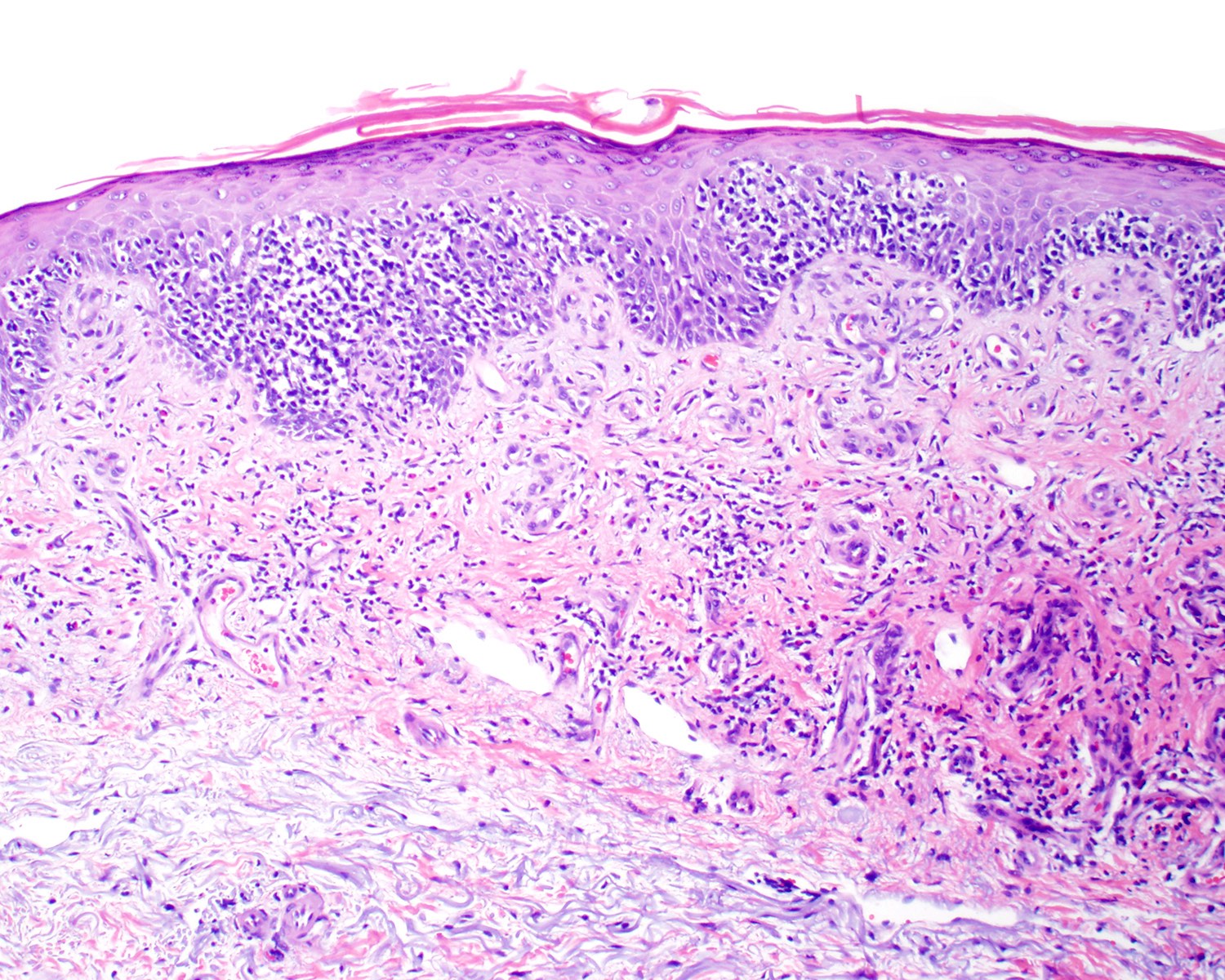
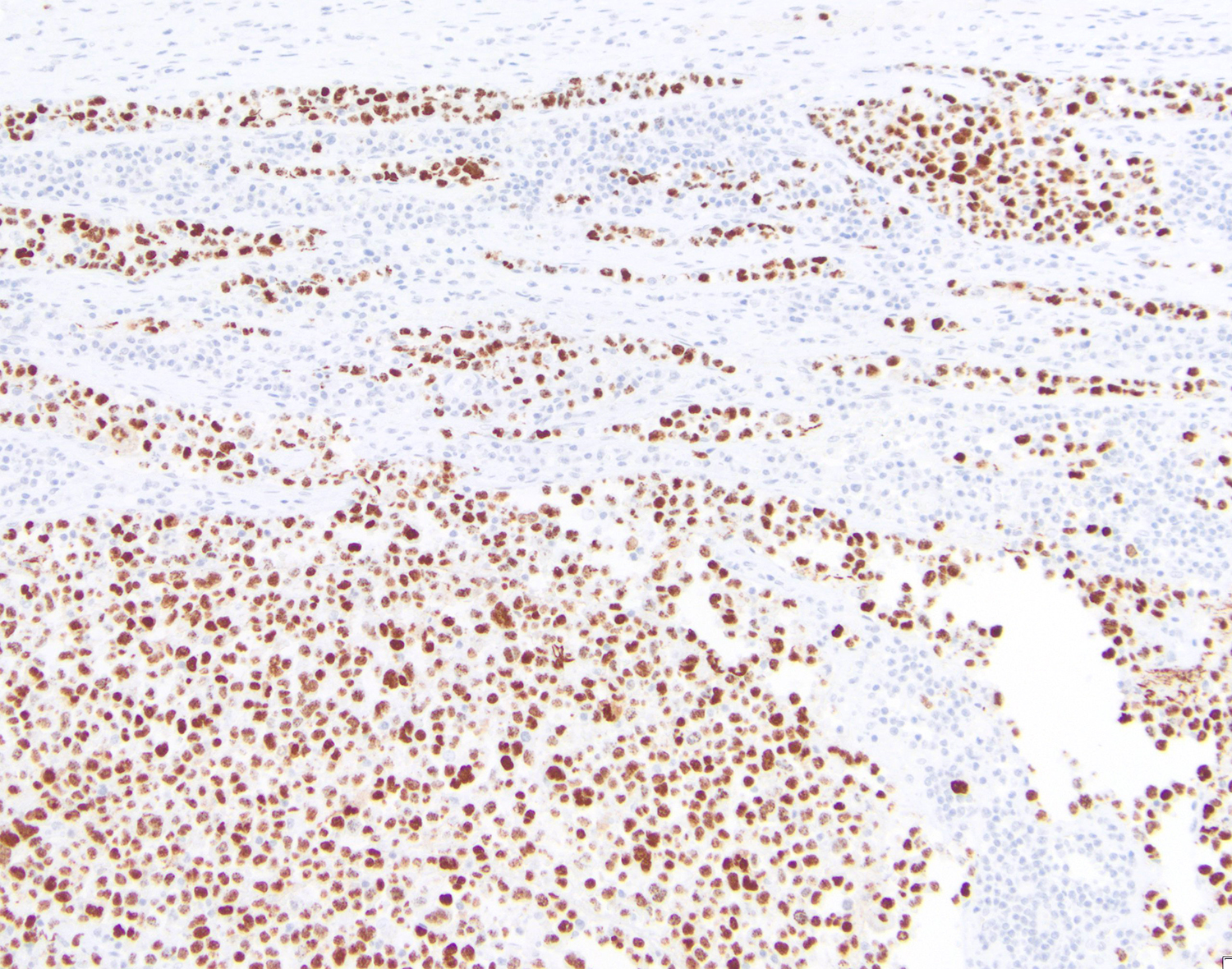
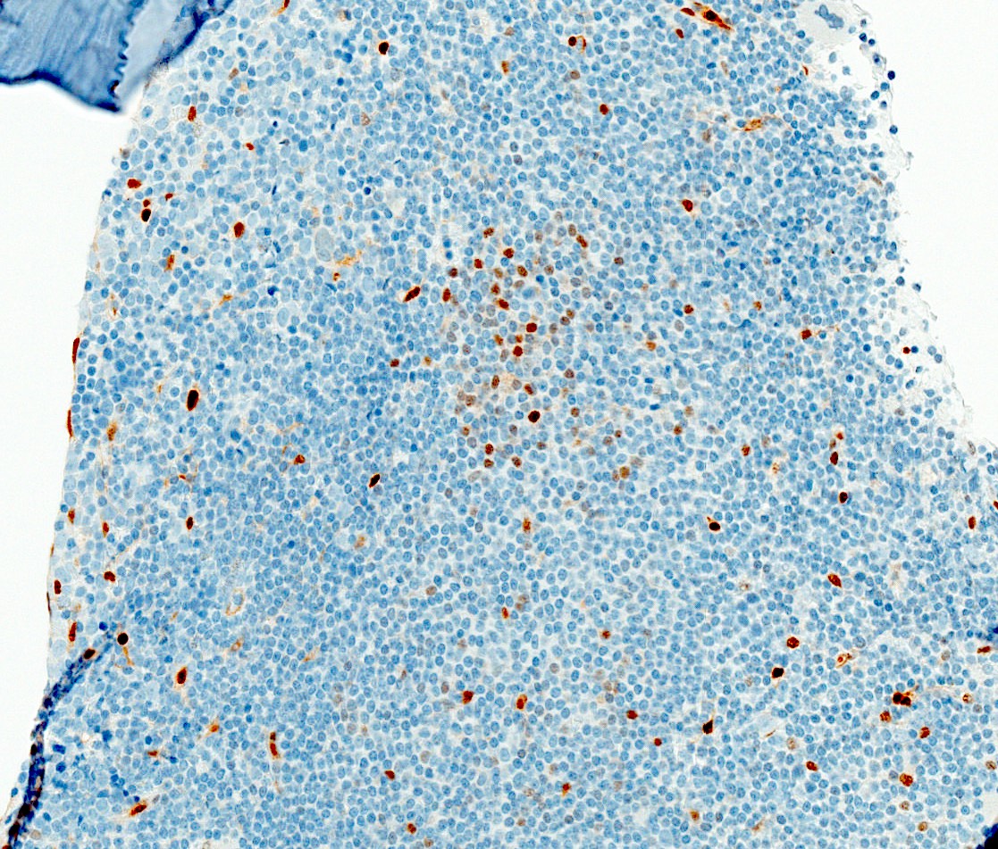
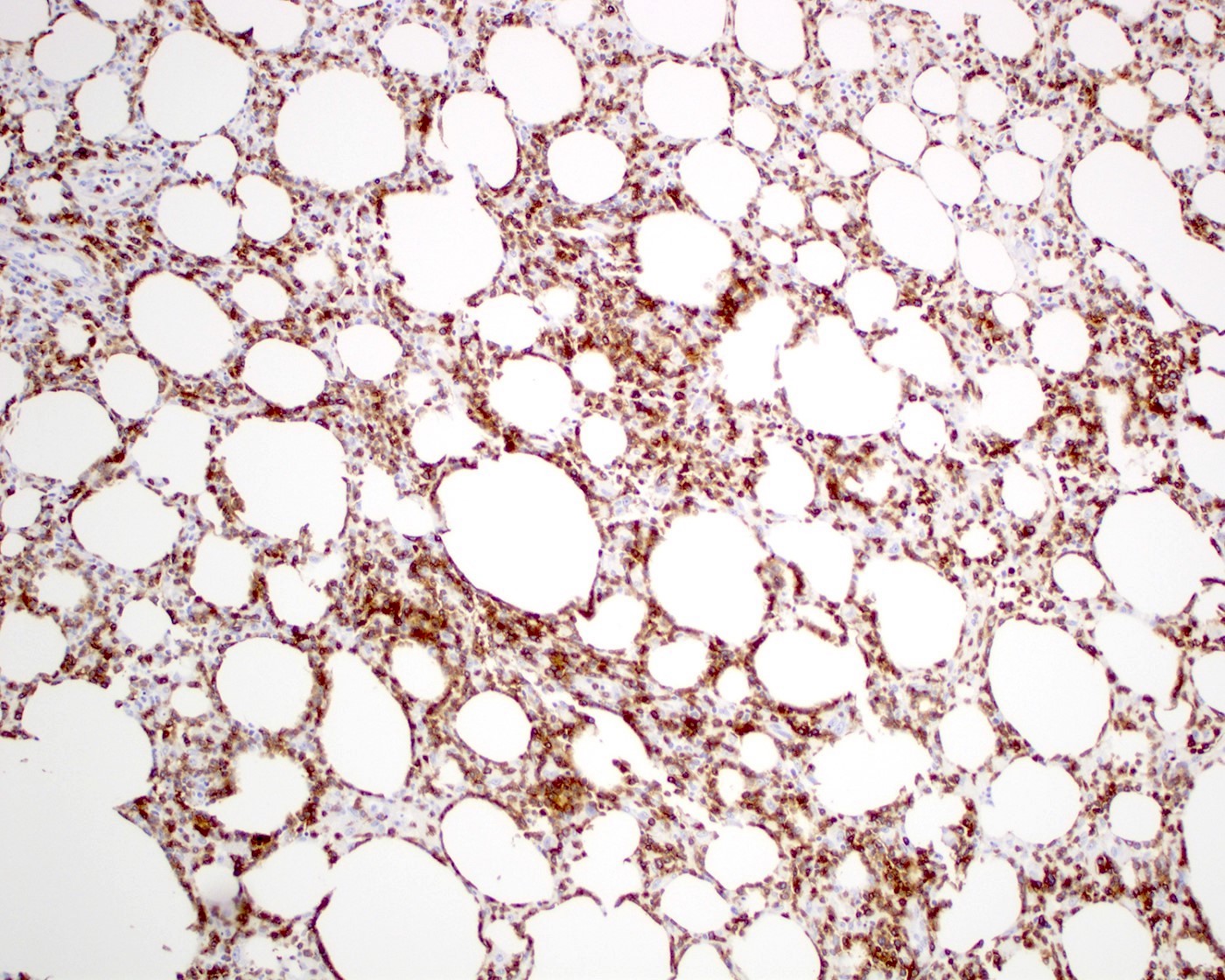
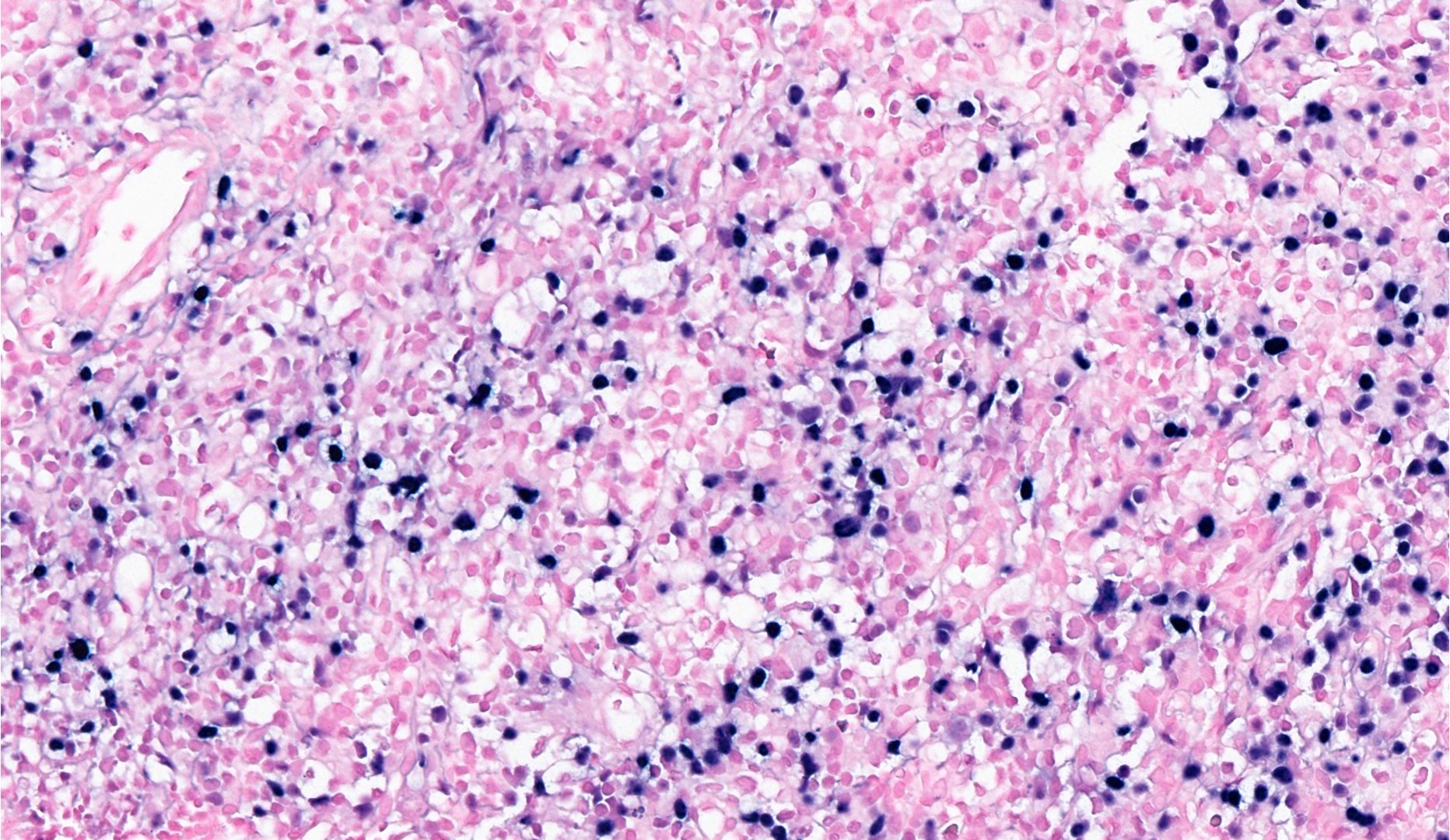
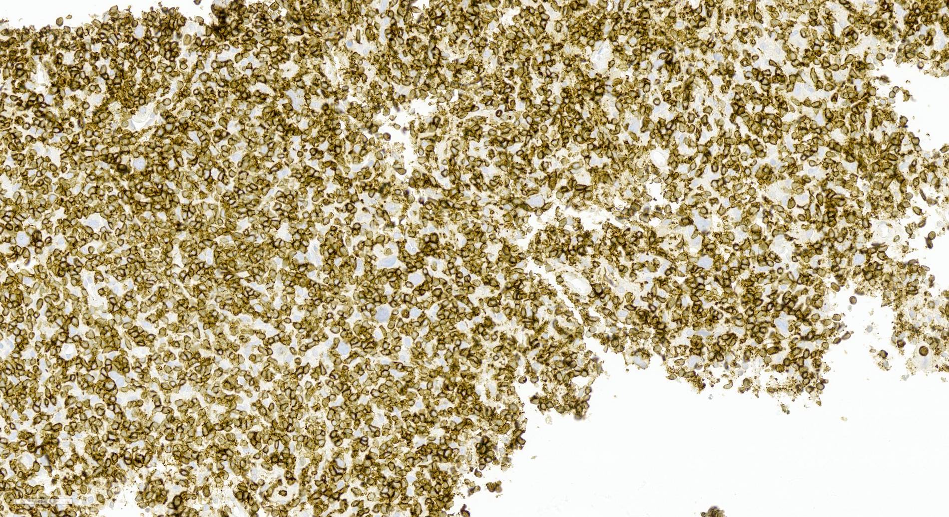
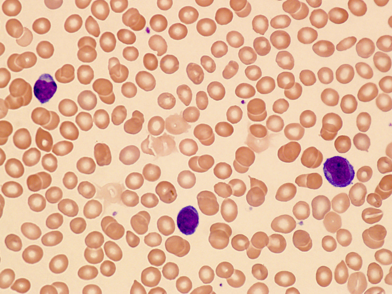
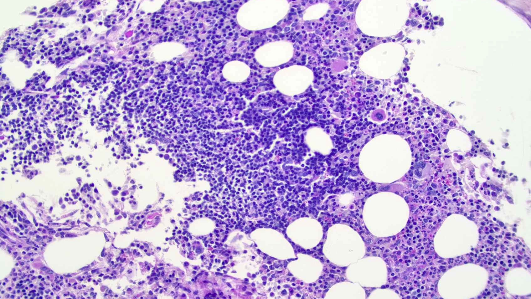
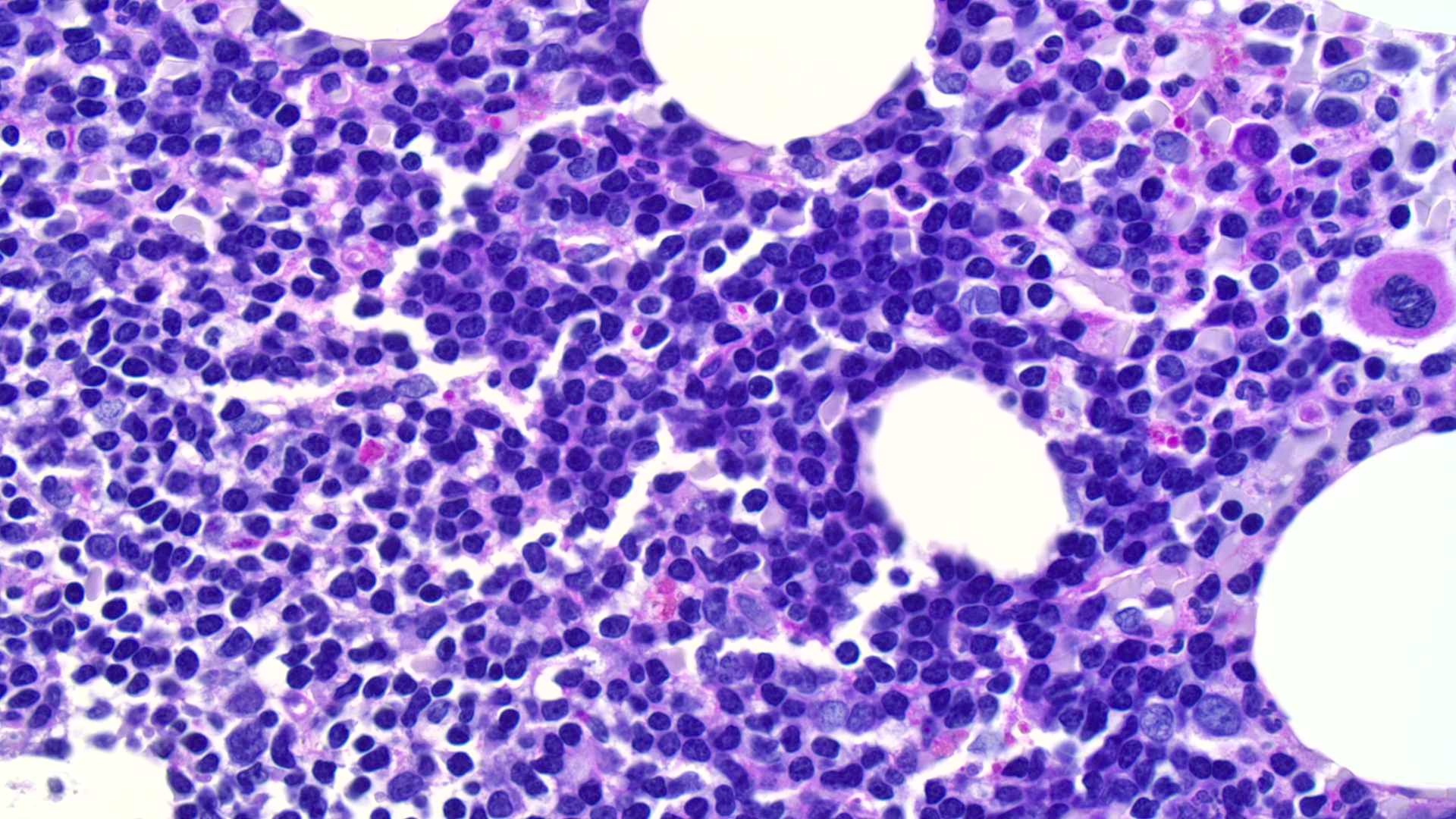
Lymphoma involving bone marrow biopsy
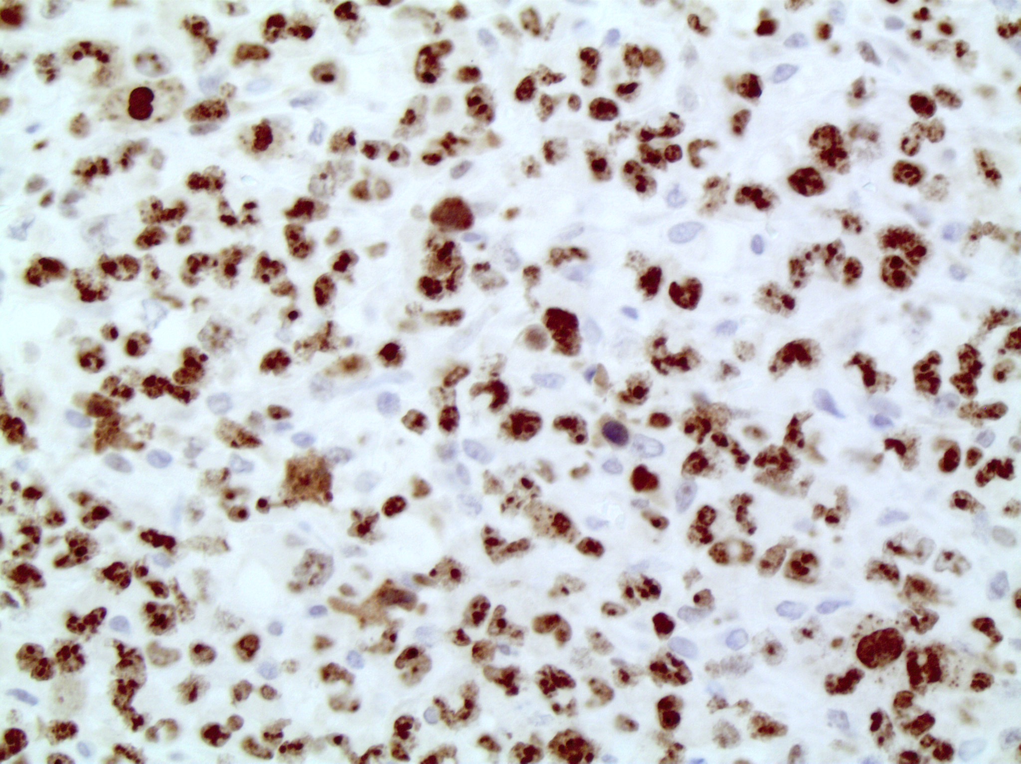
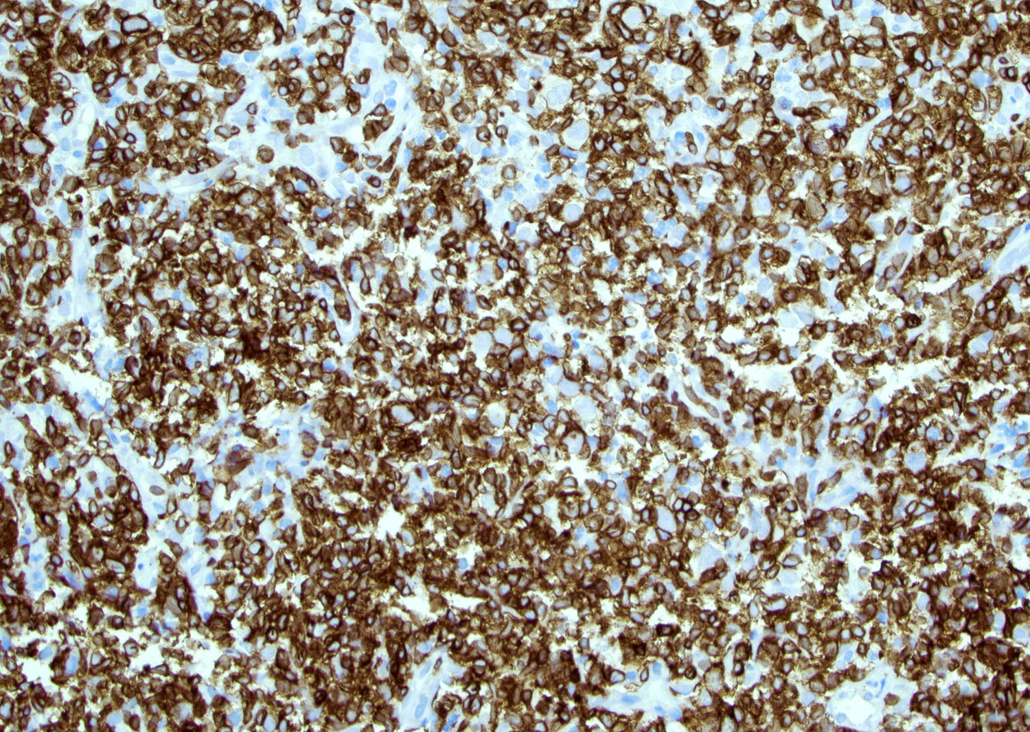
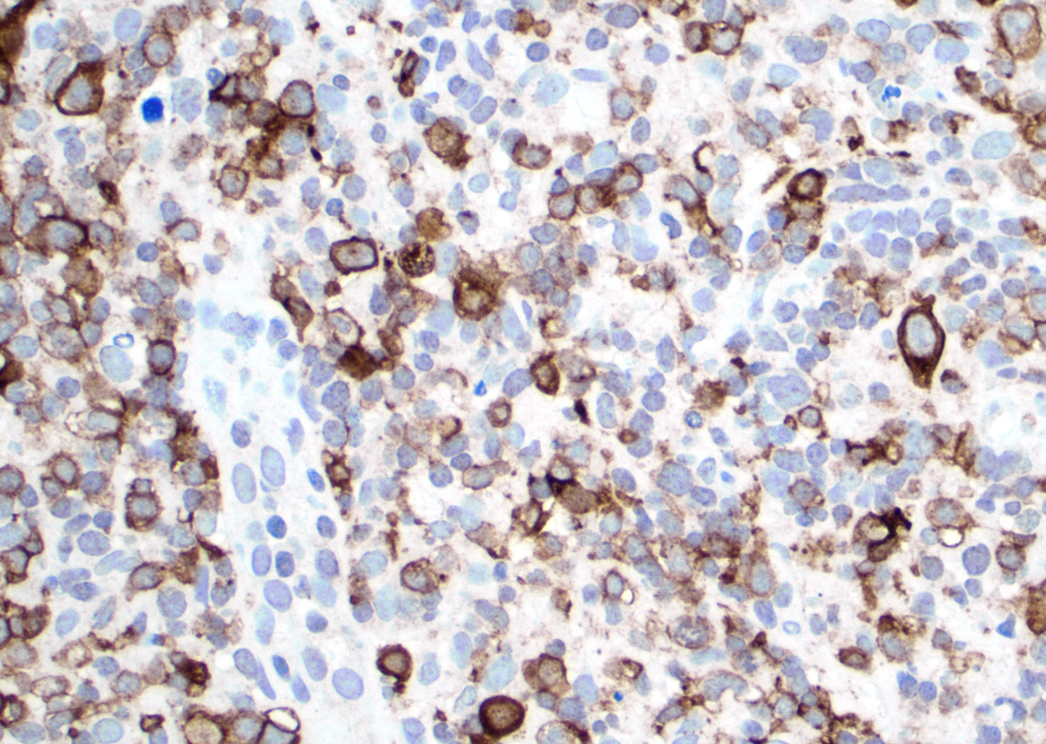
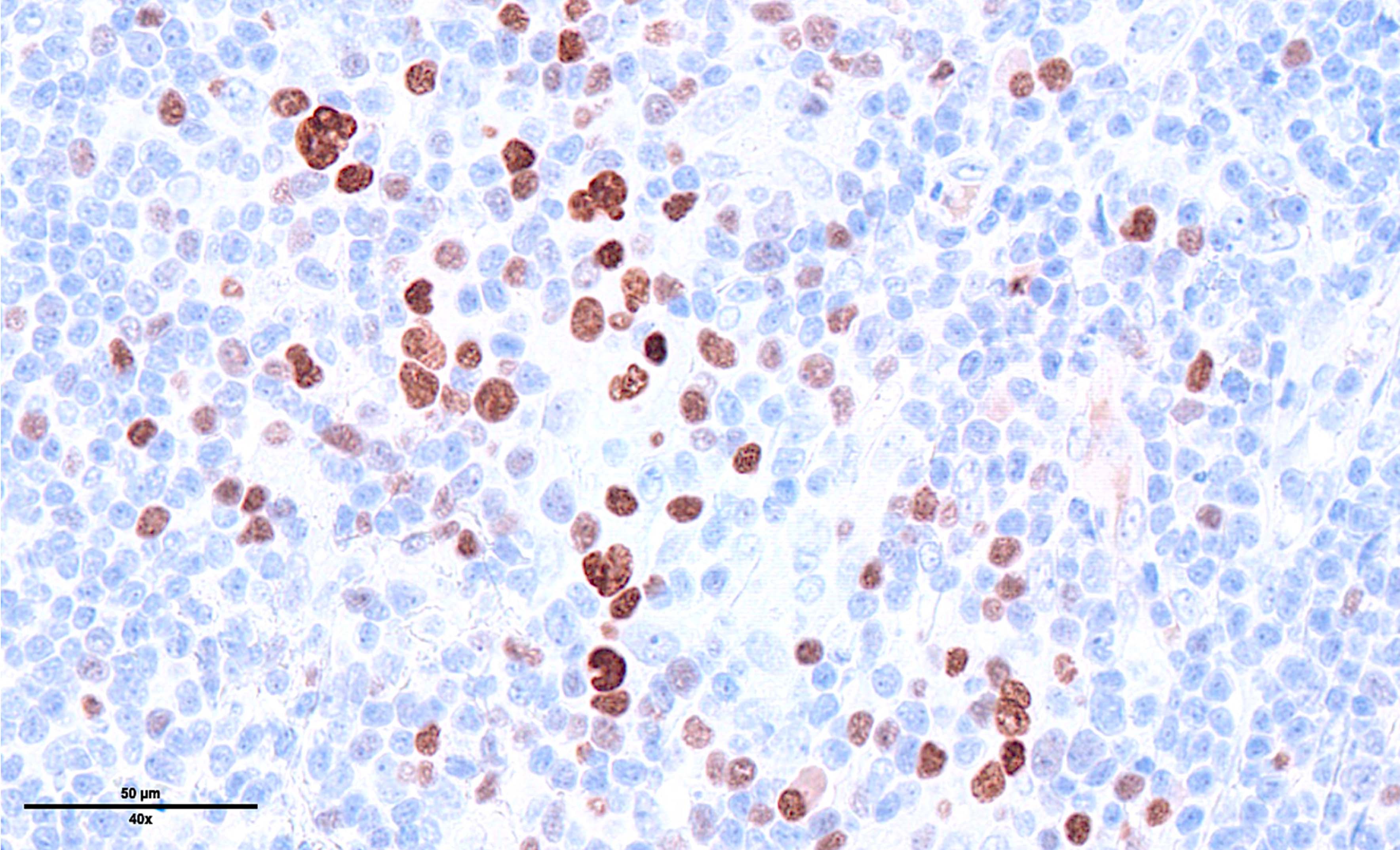
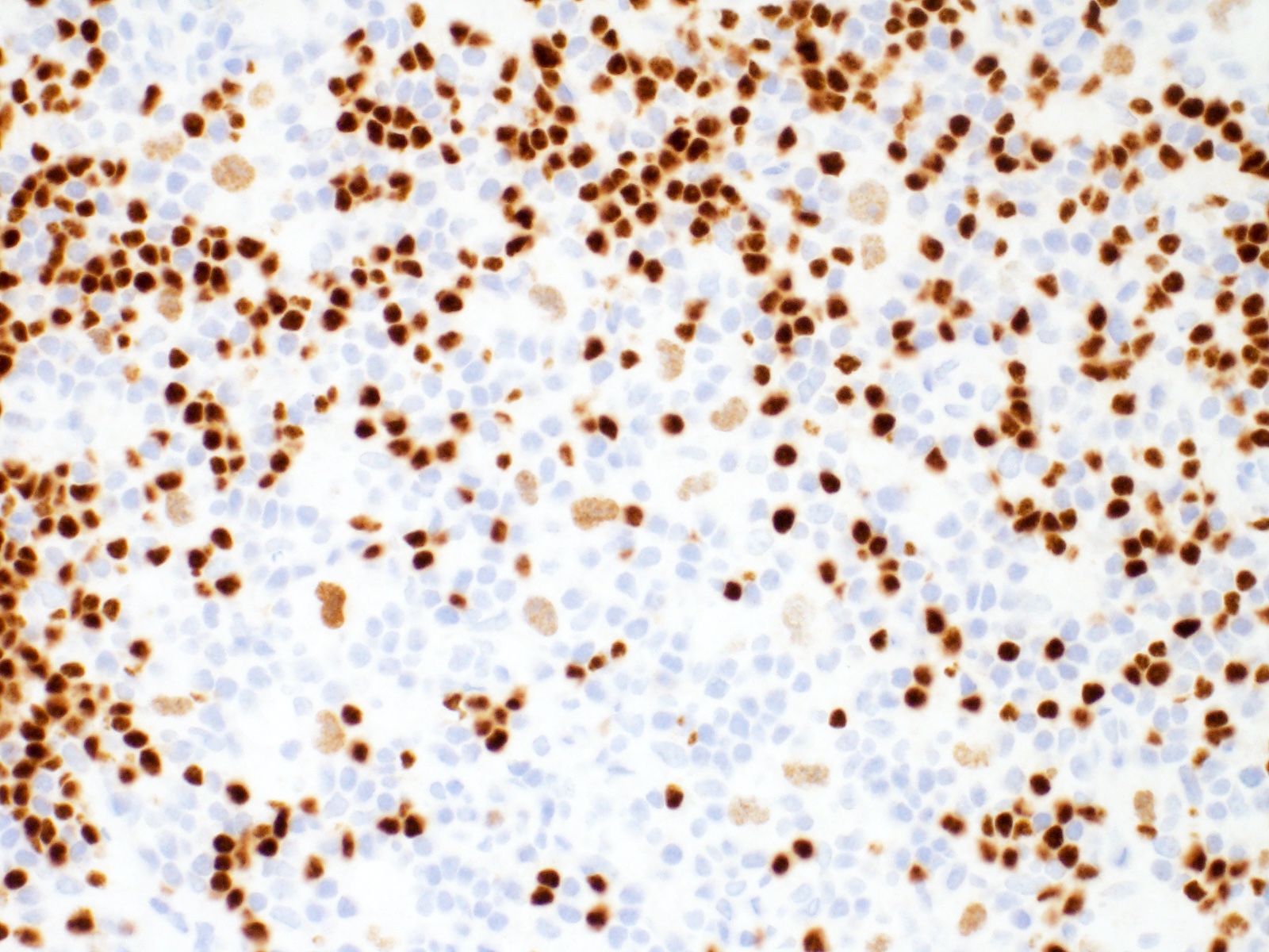
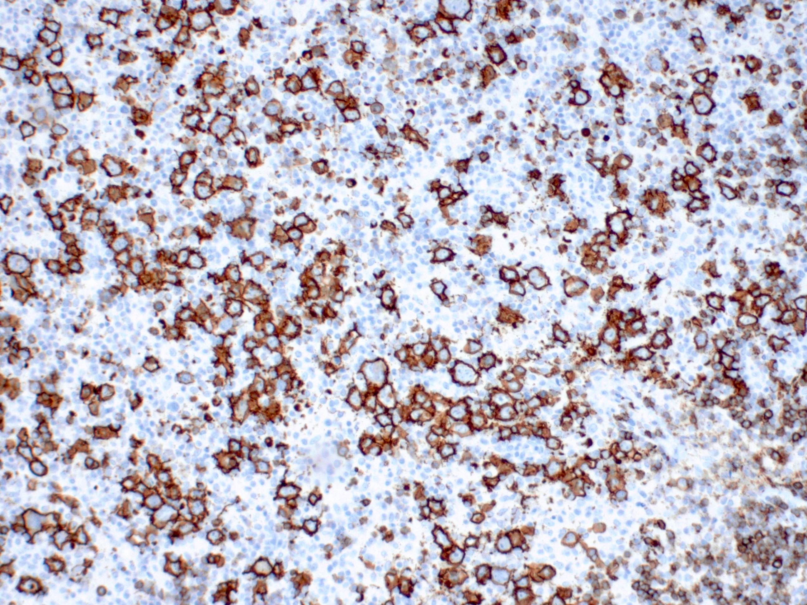
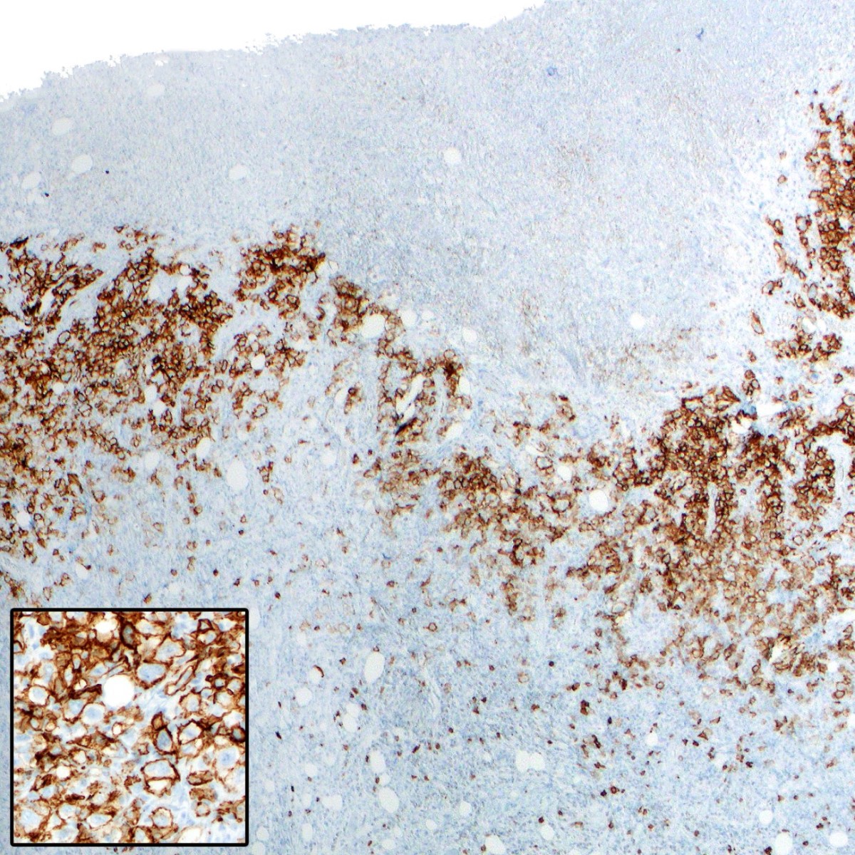
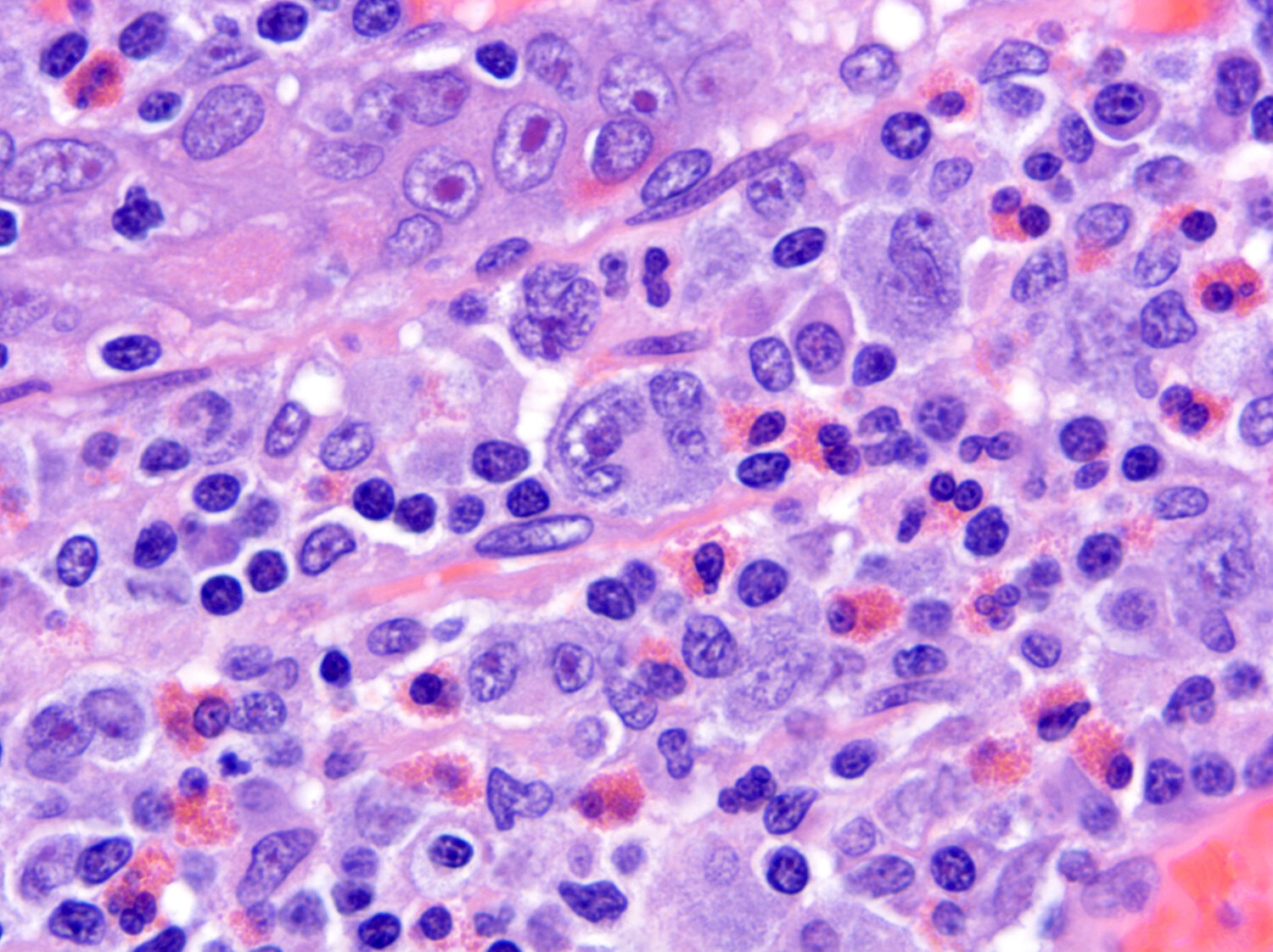
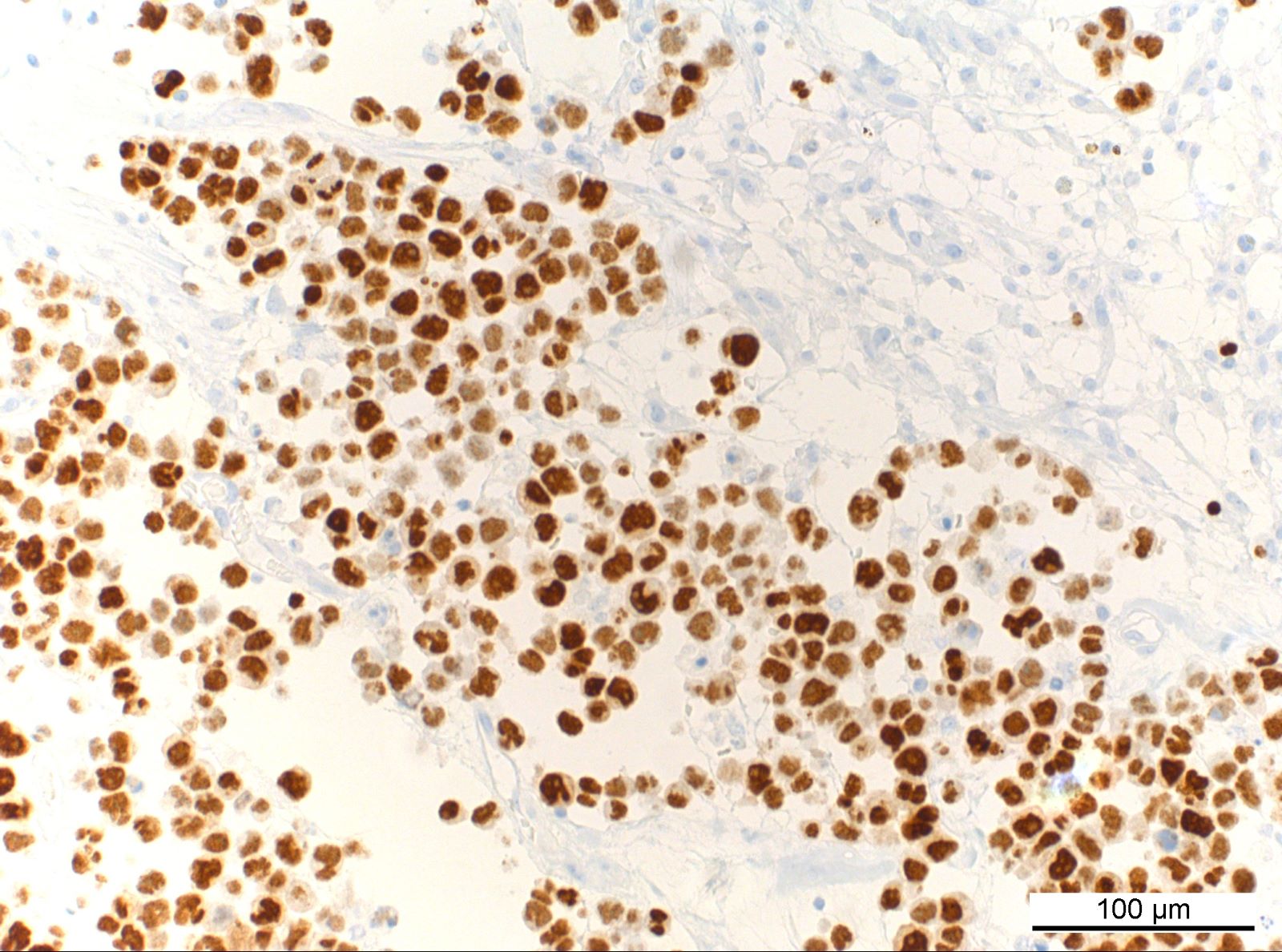
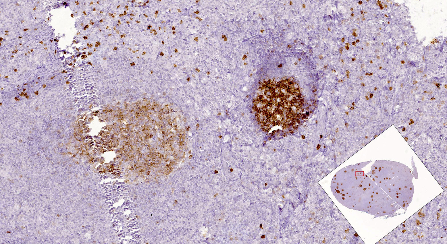
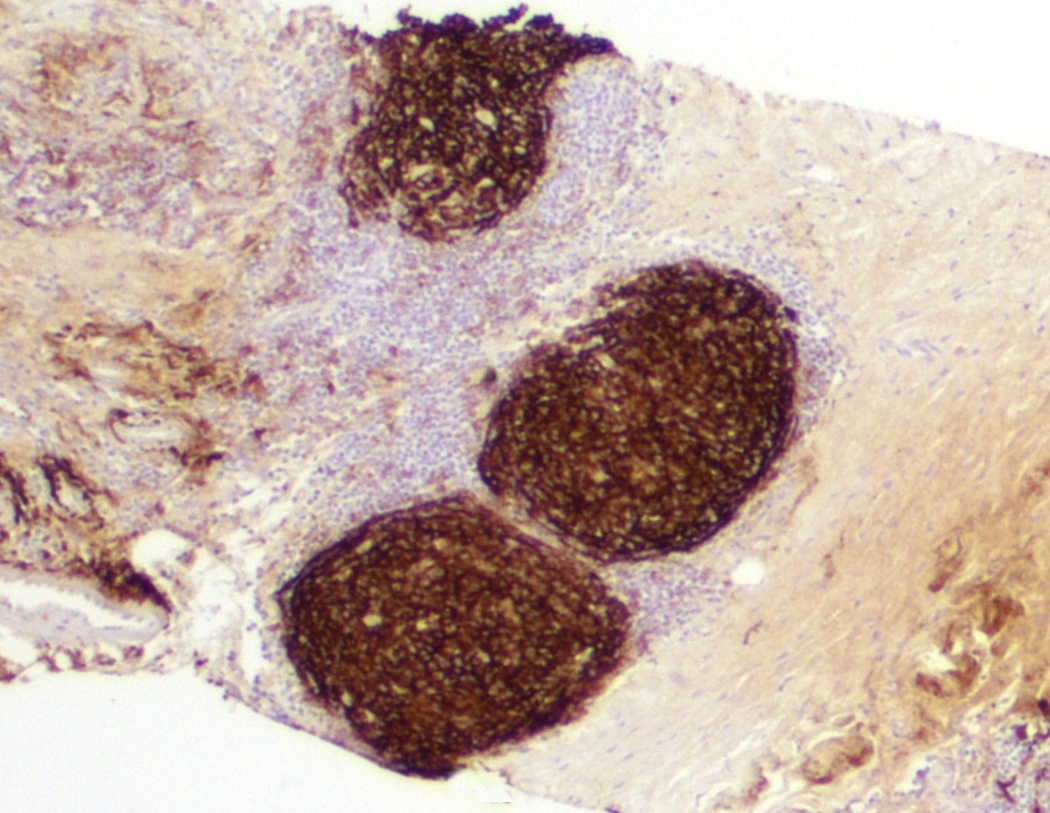
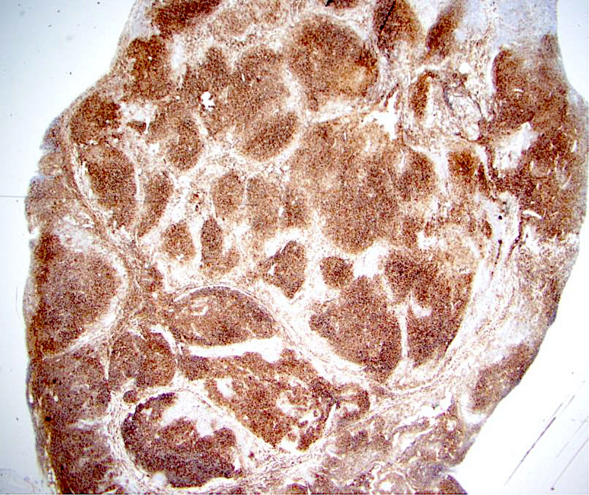
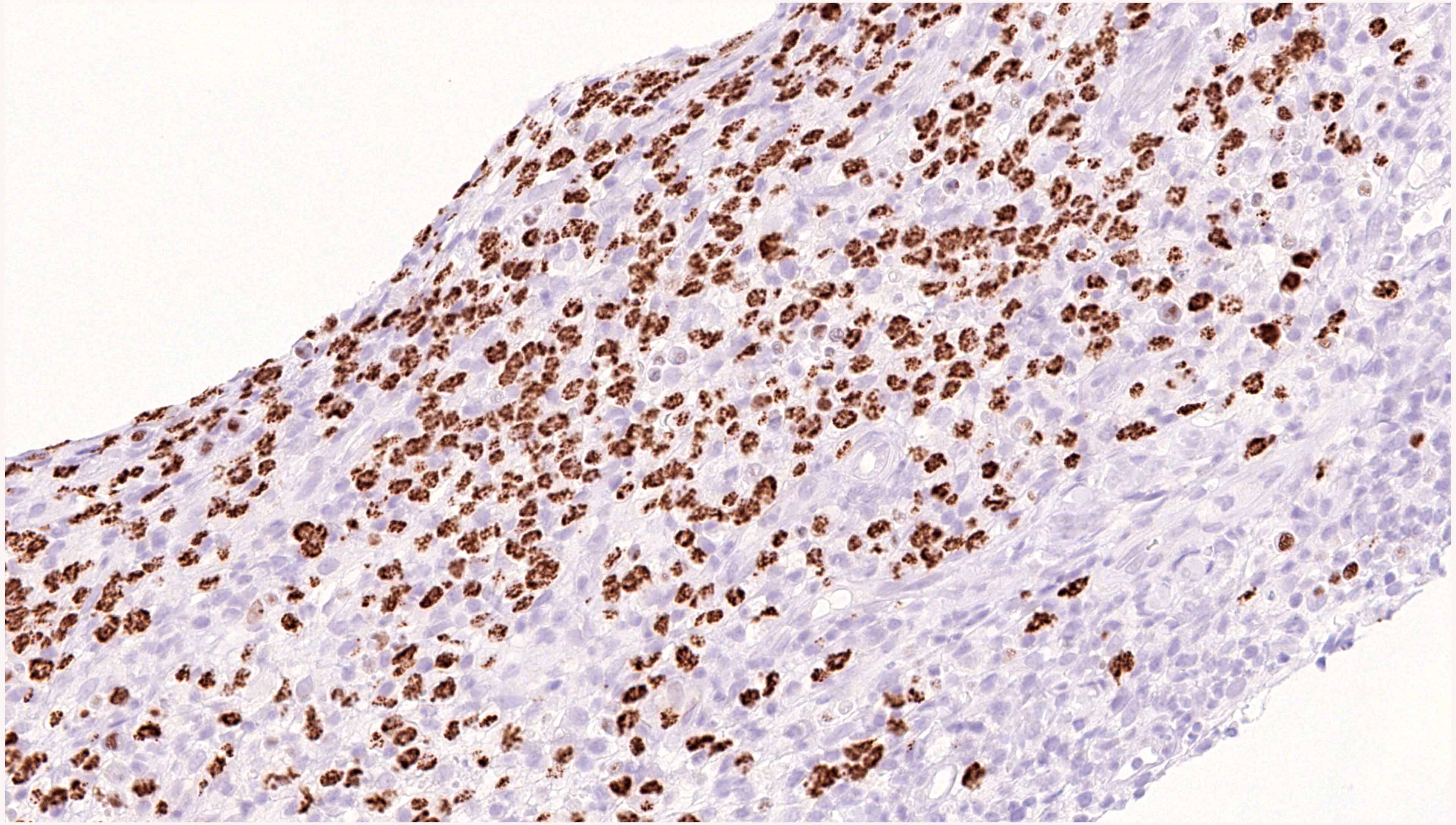
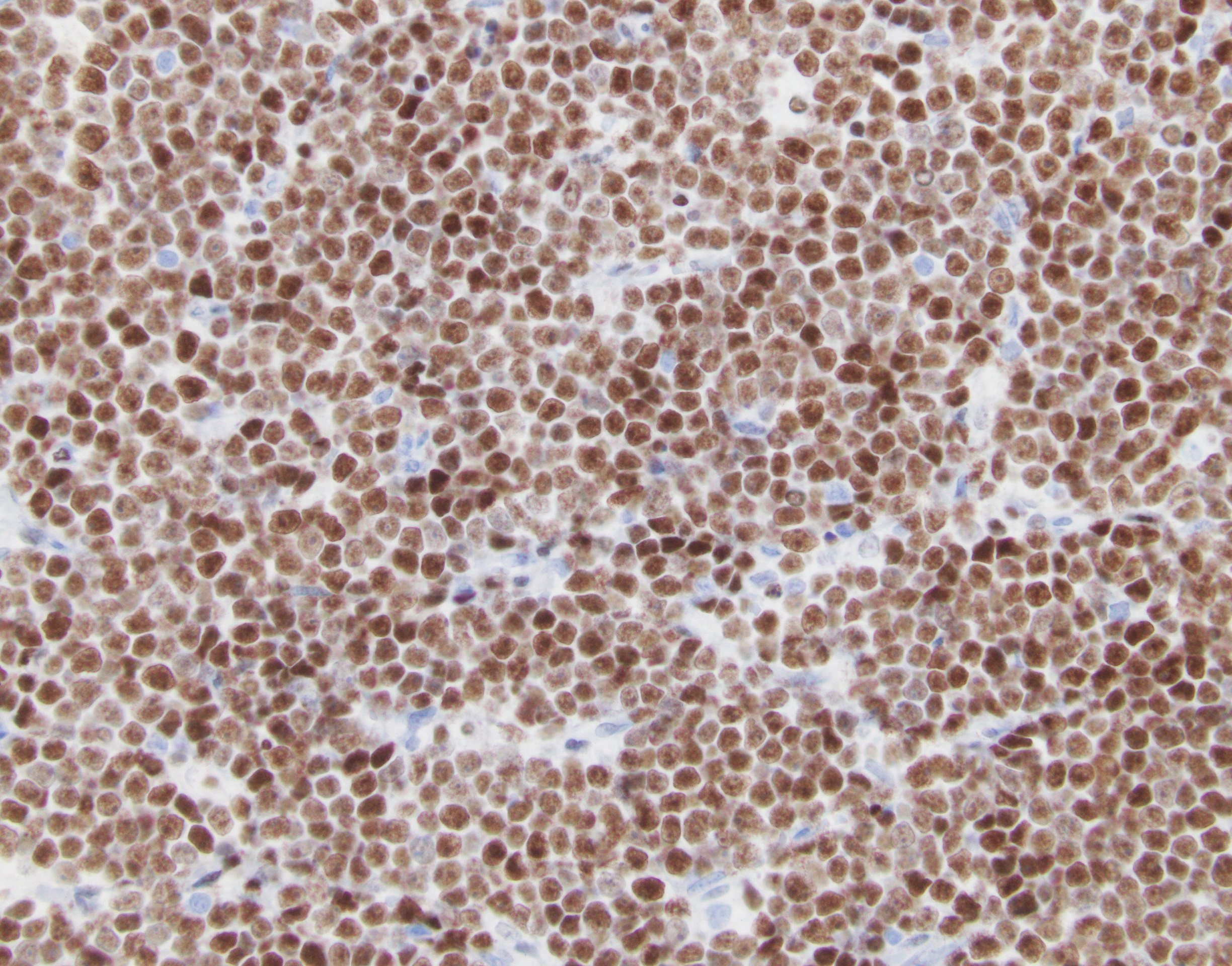
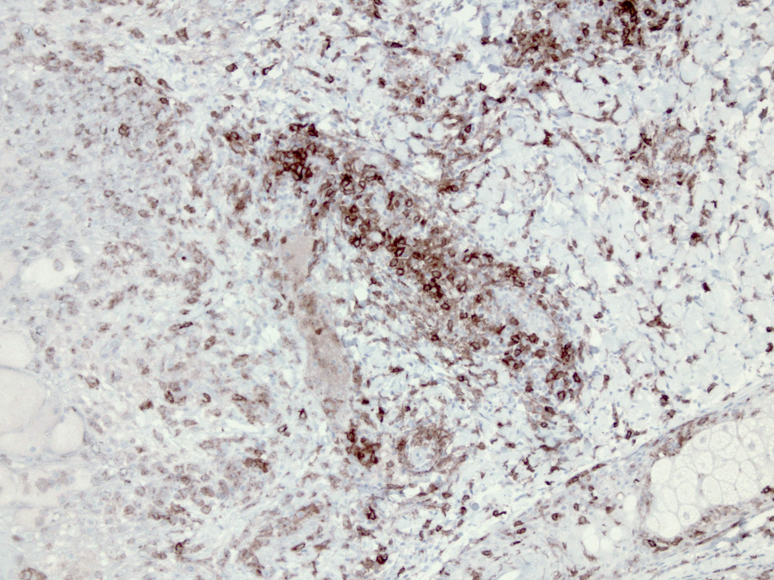
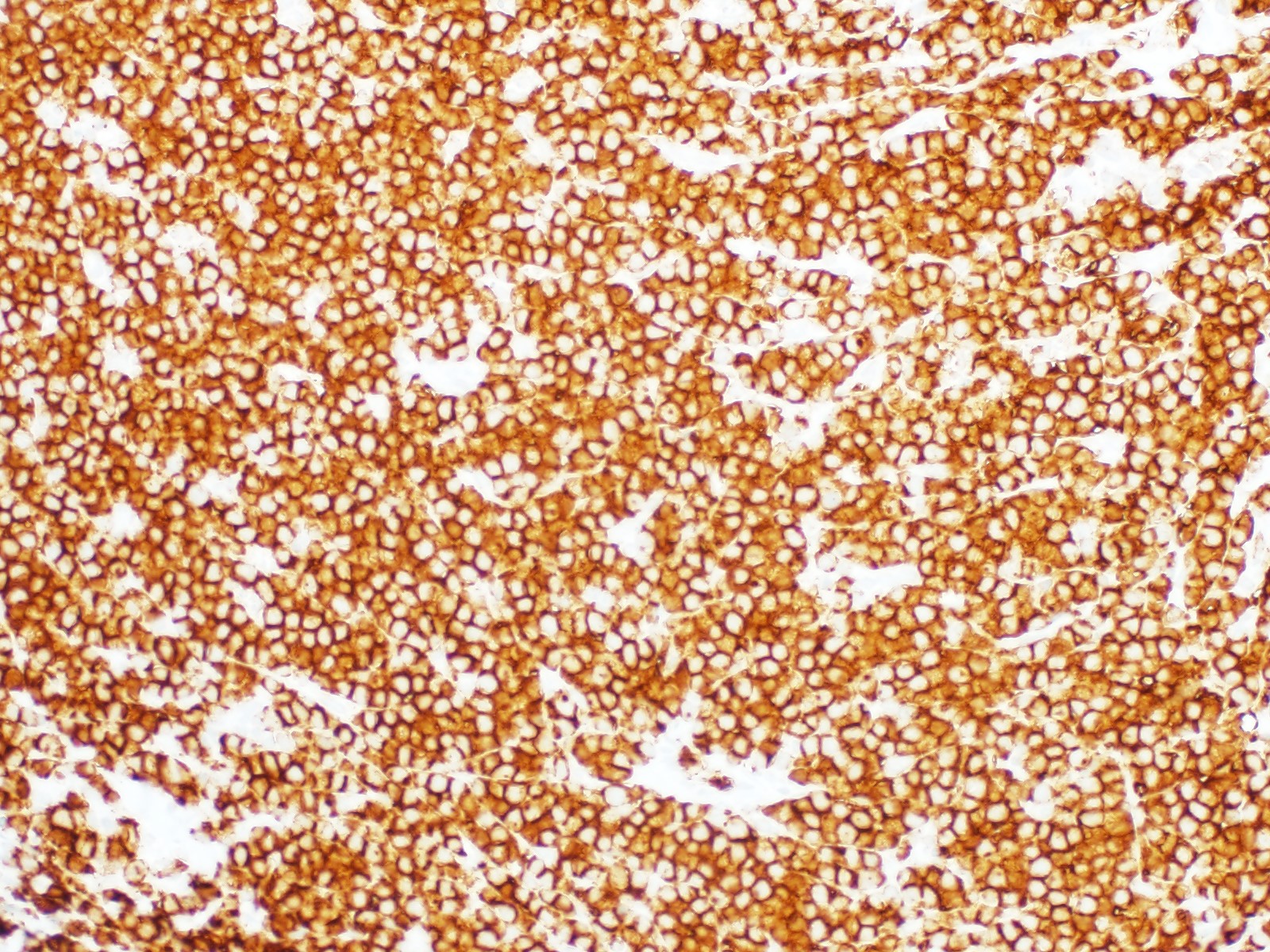
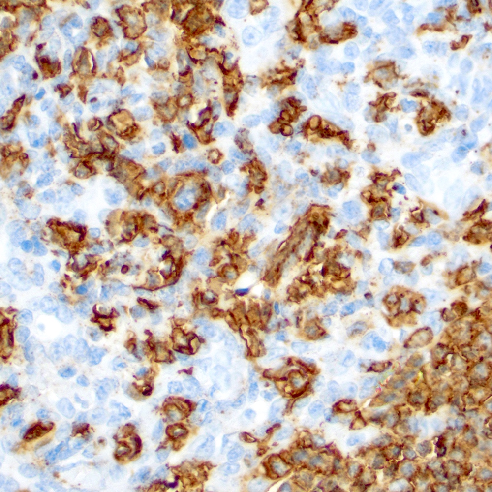
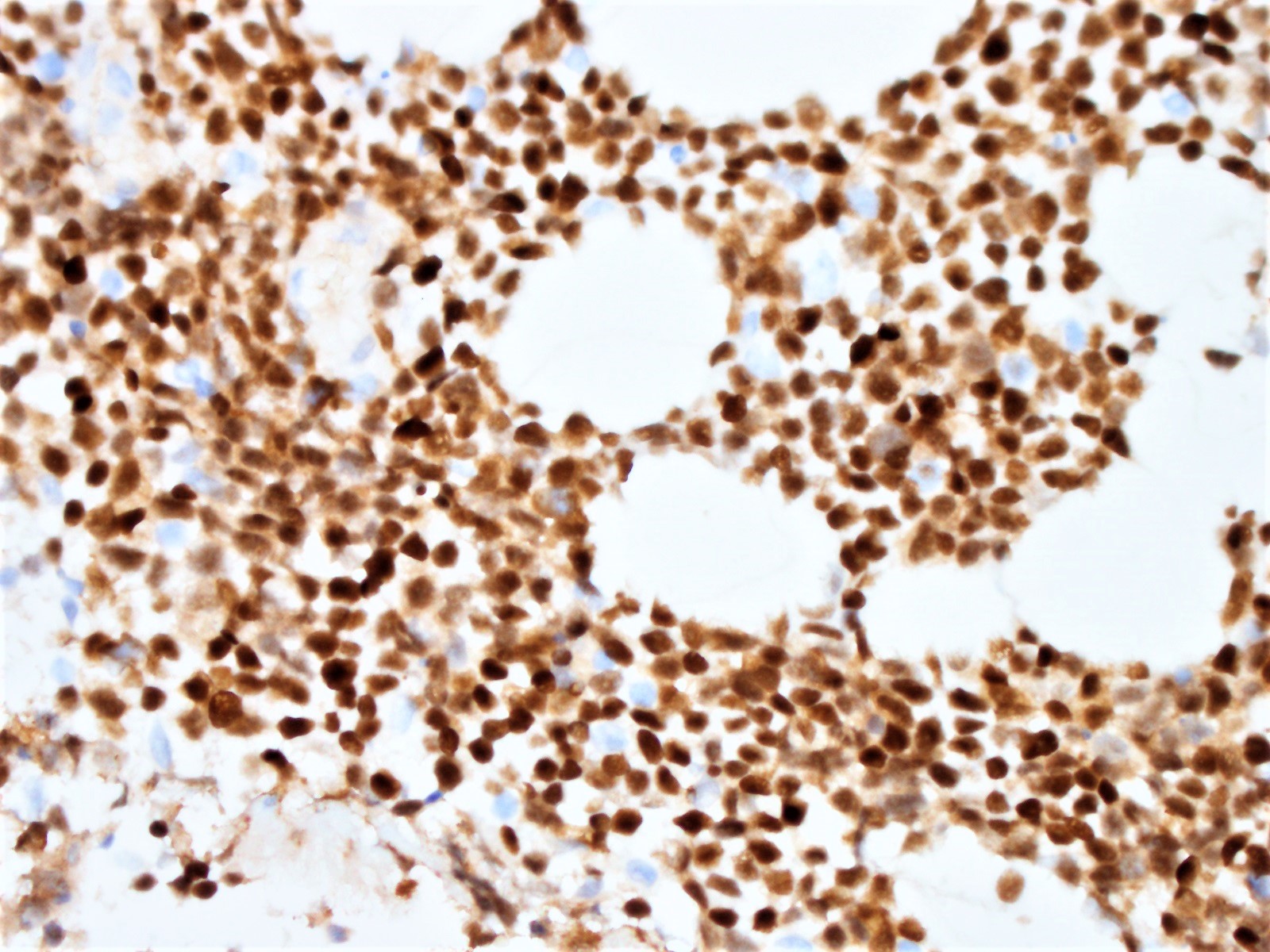
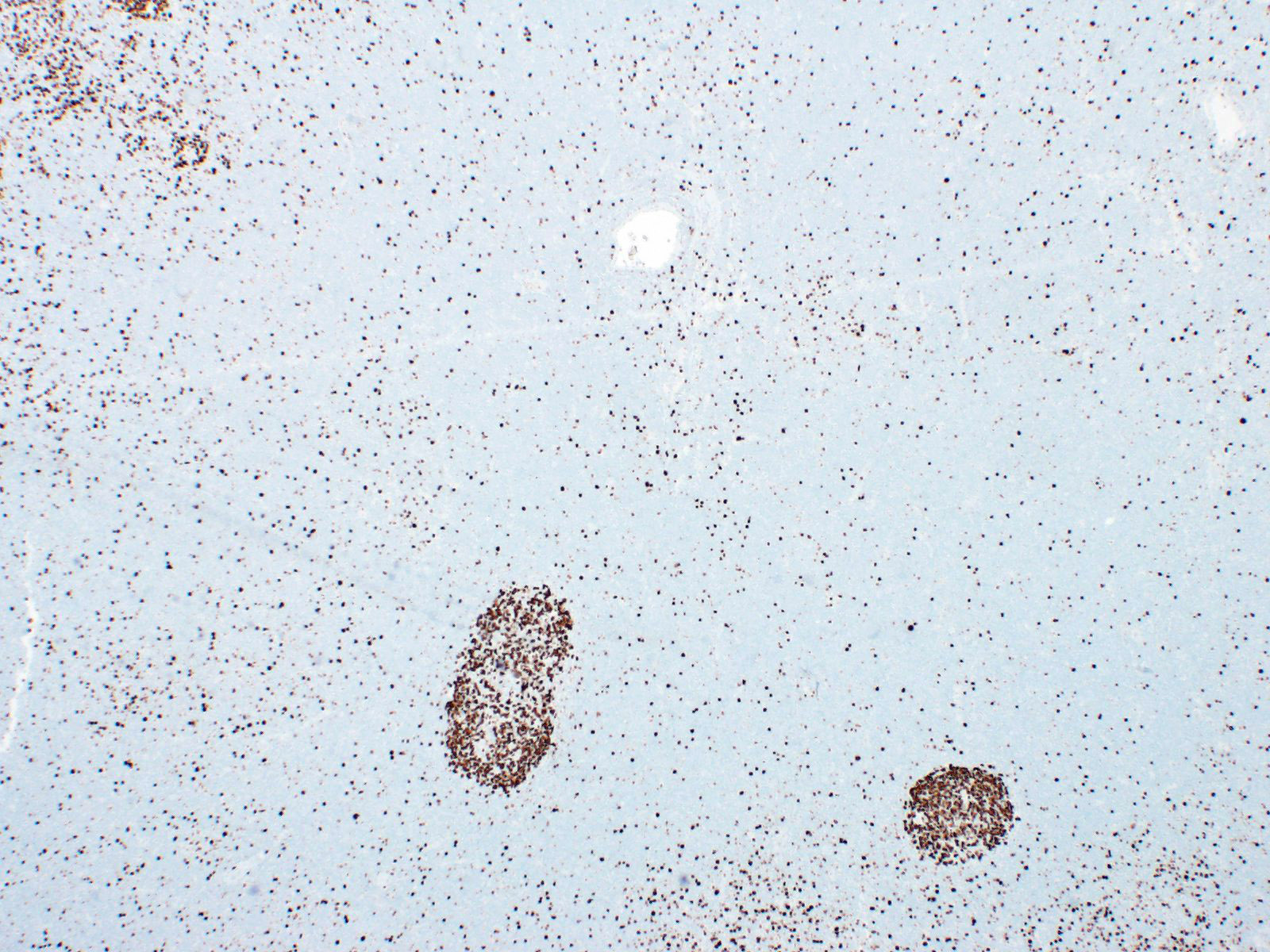
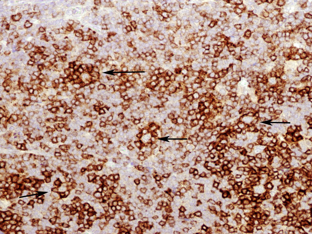
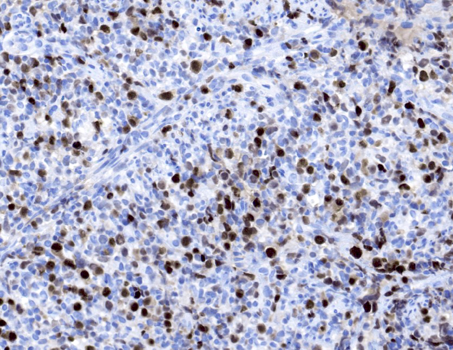
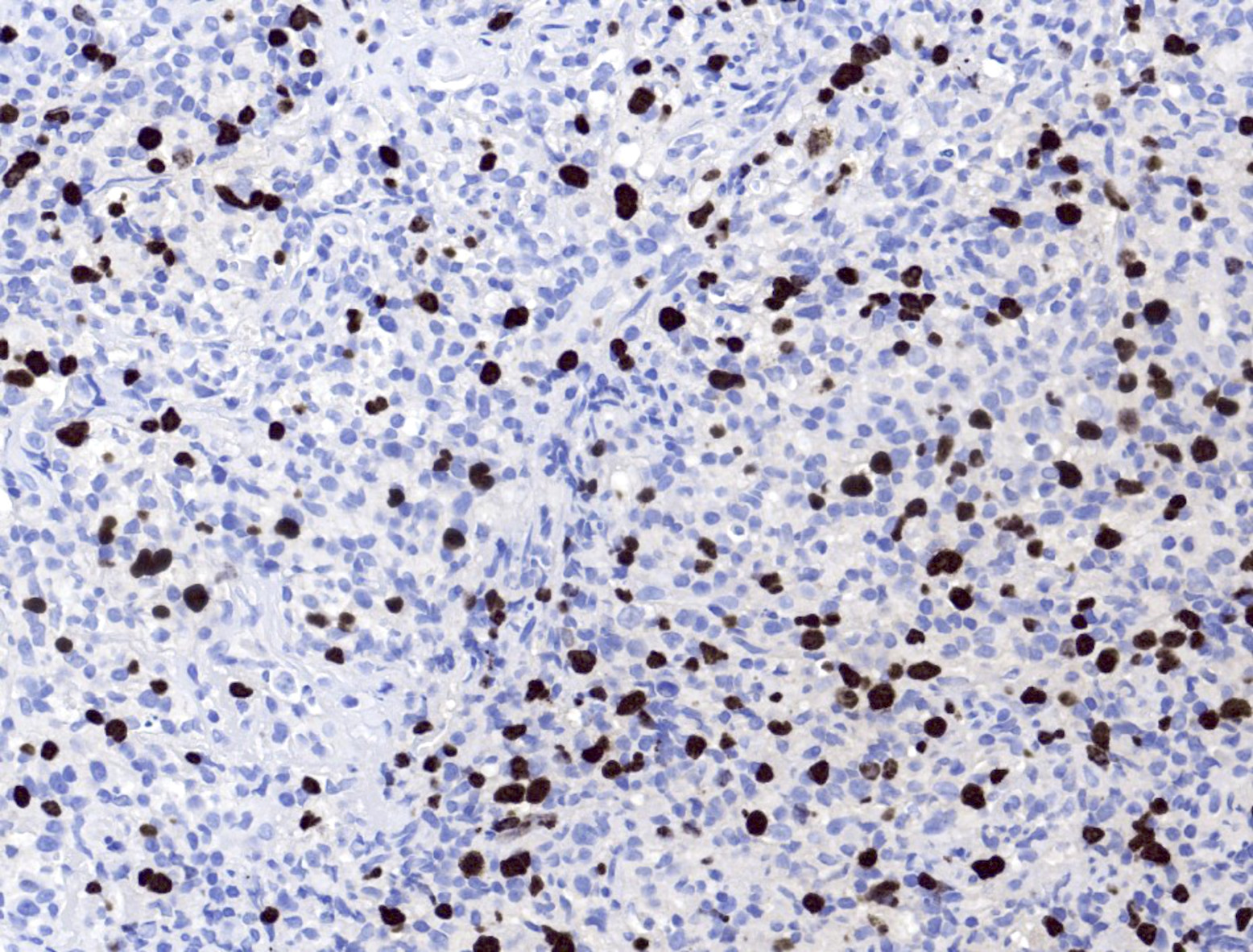
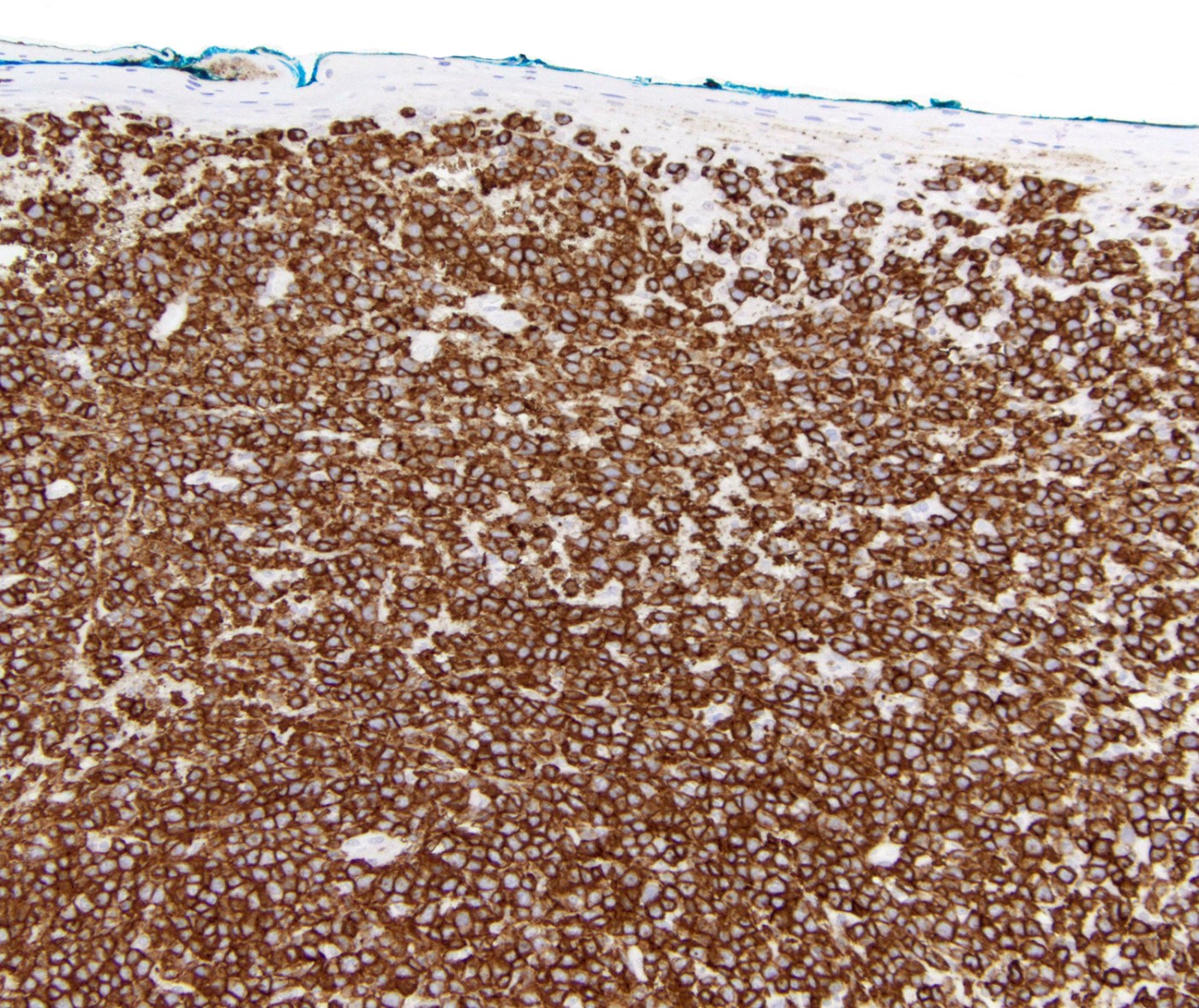
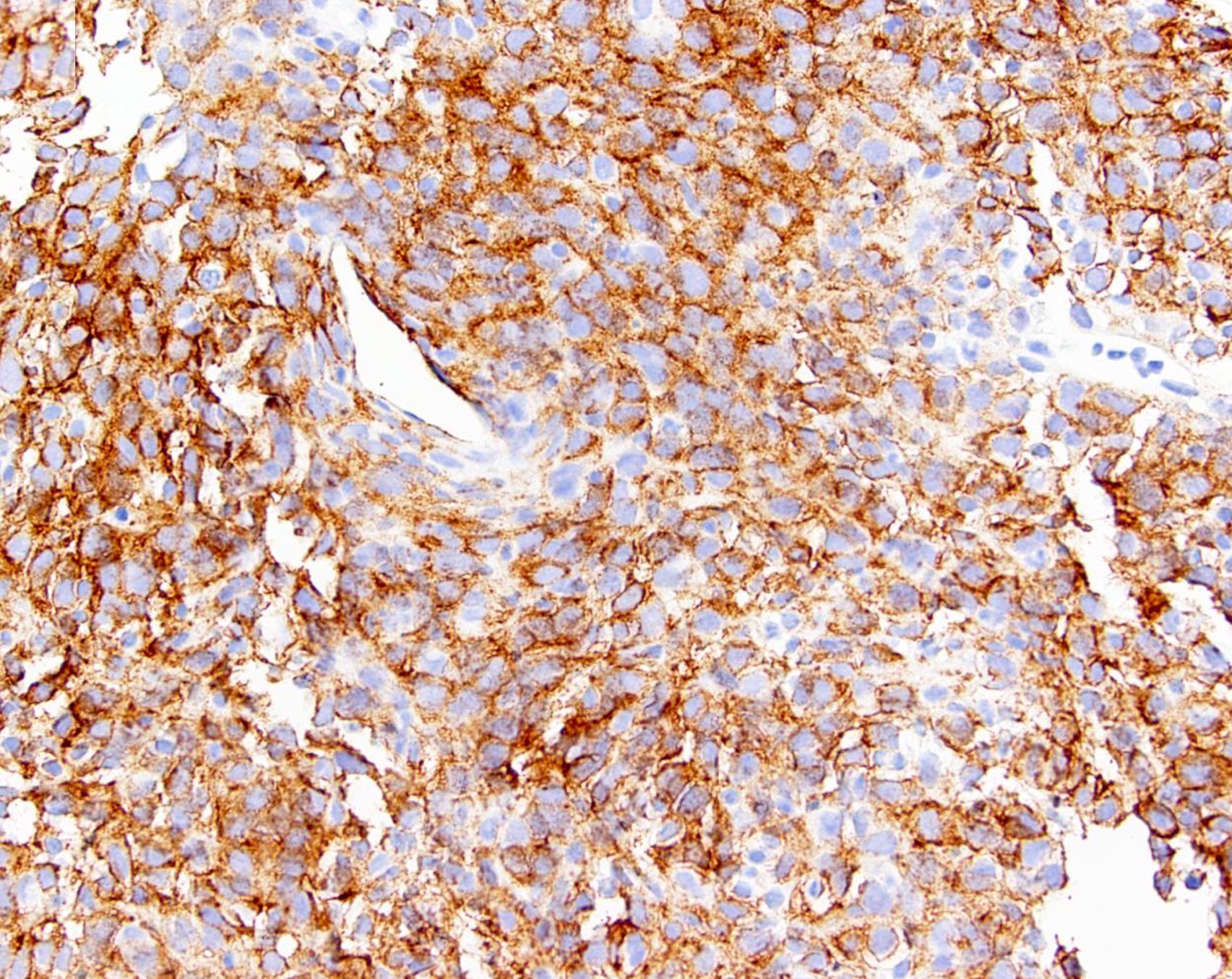
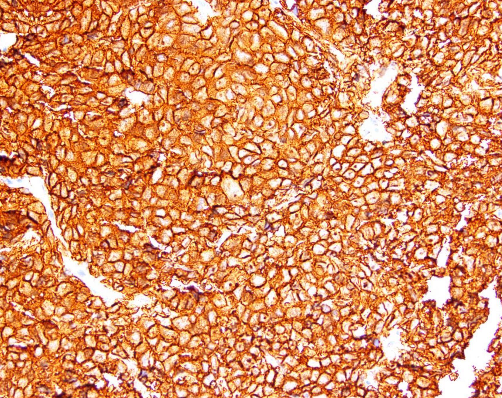
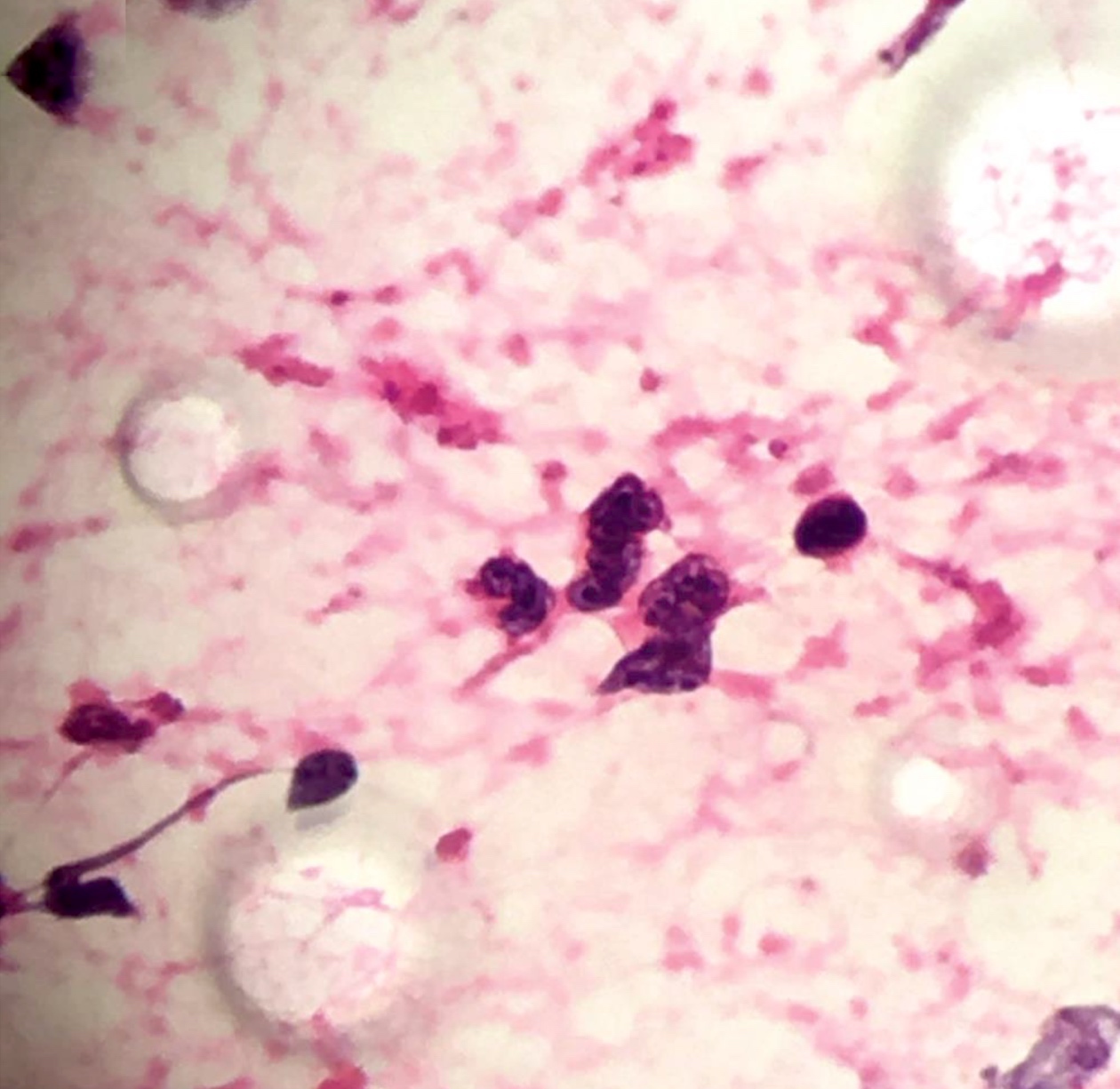
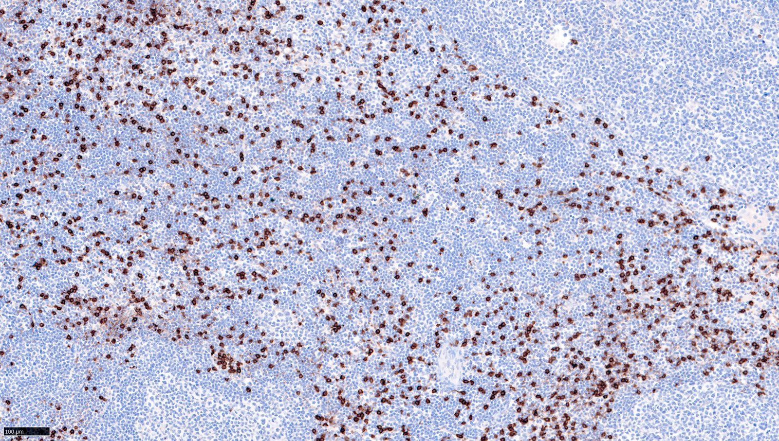
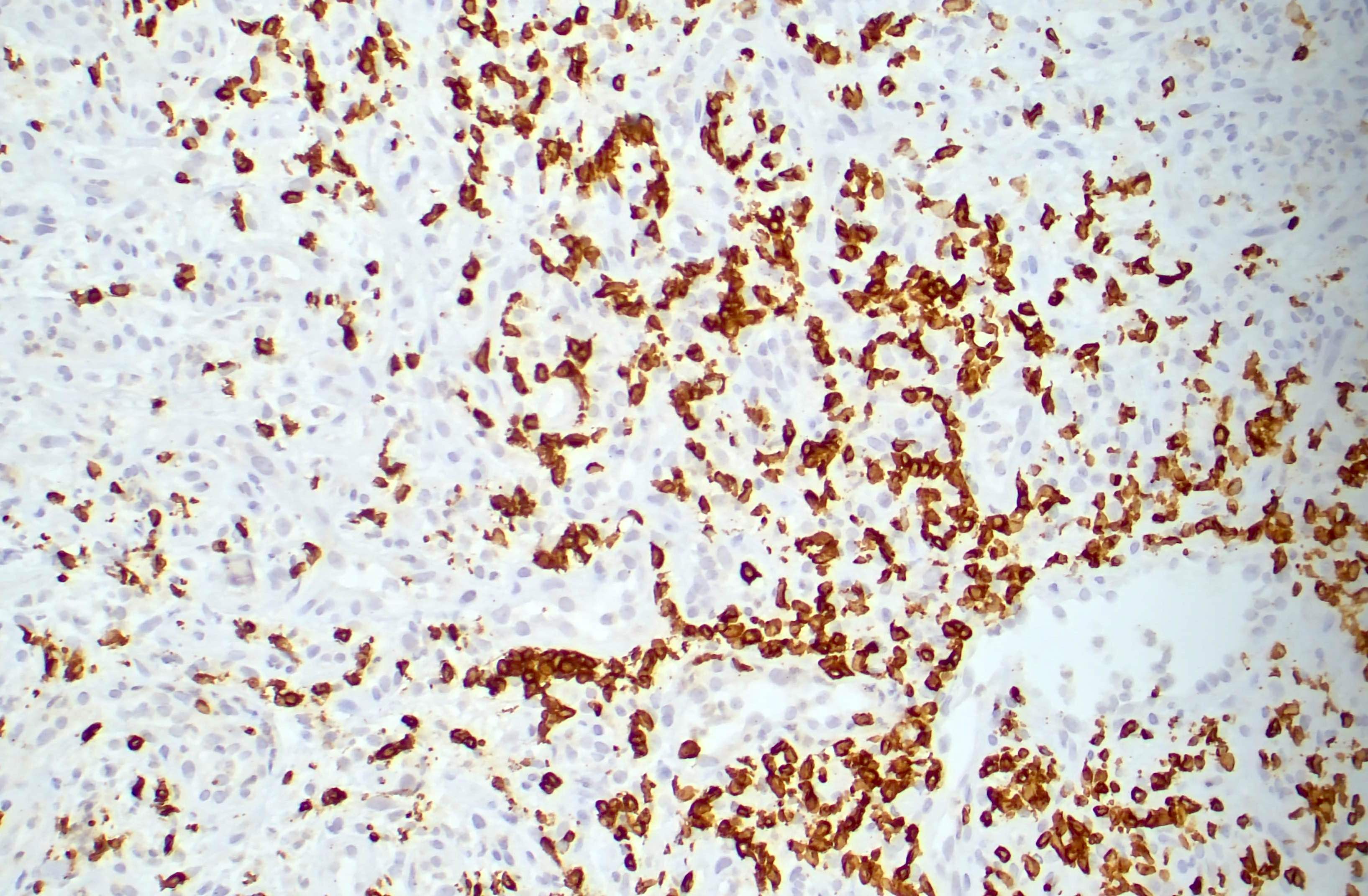
ALK positivity in tumor cells
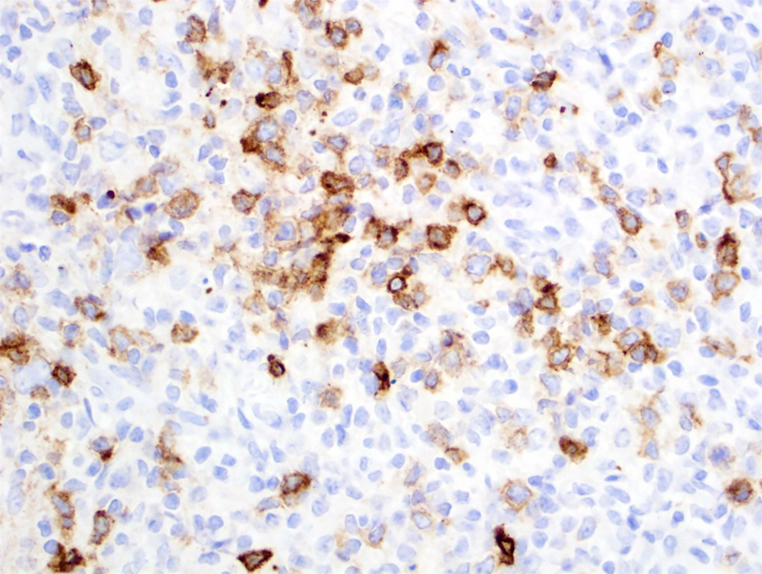
Granular ALK positivity in tumor cells
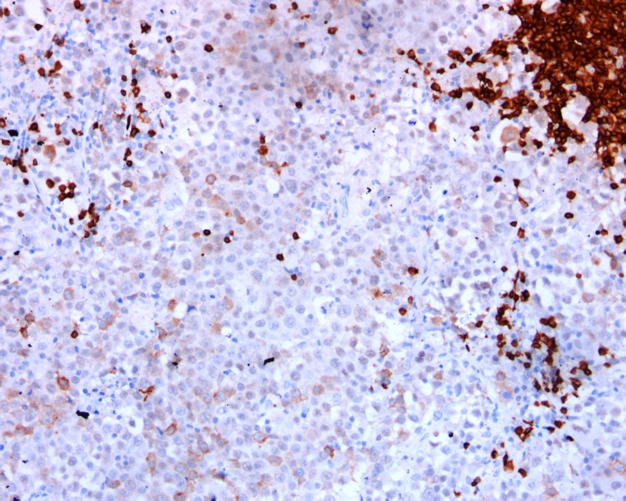
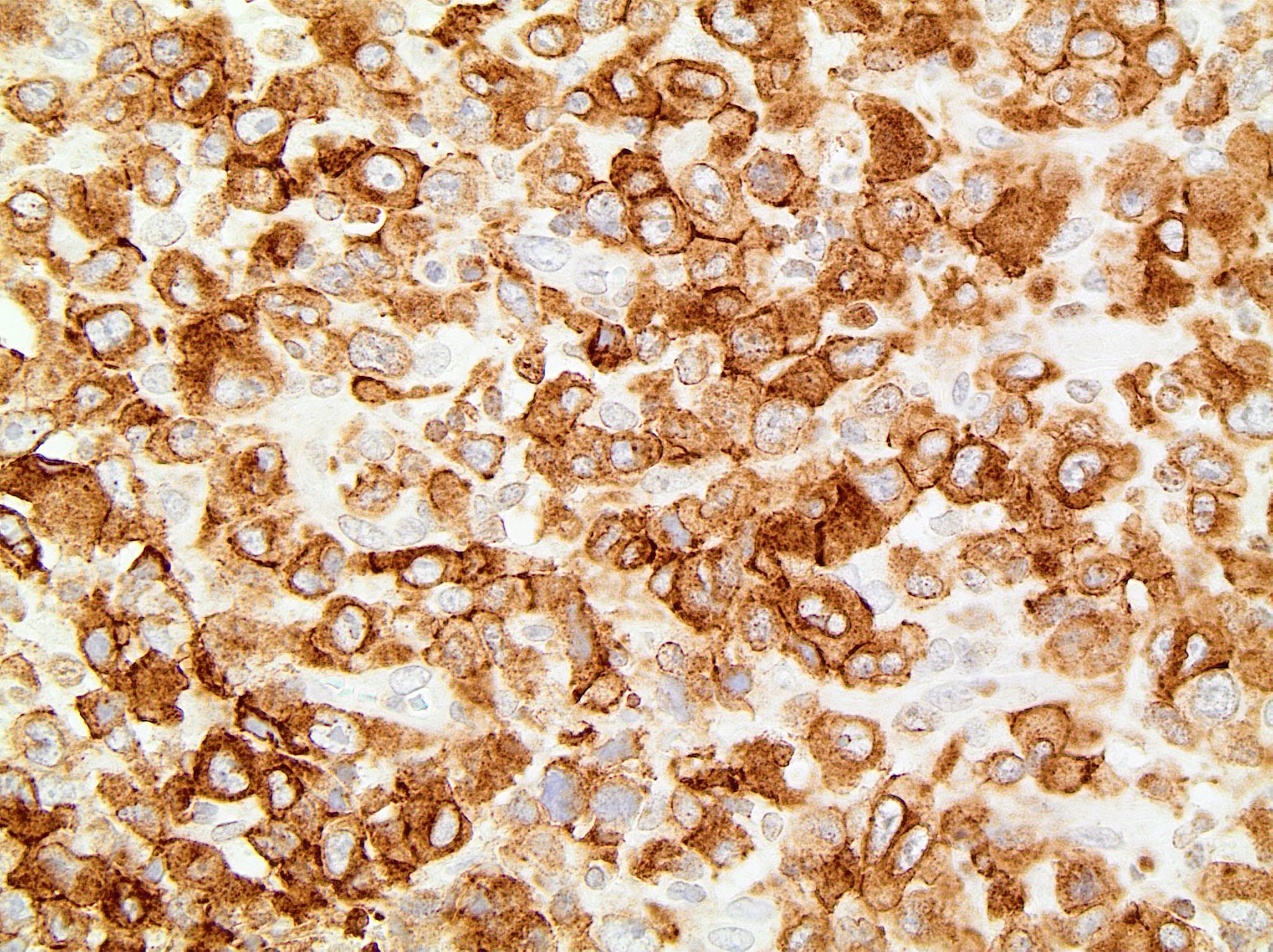
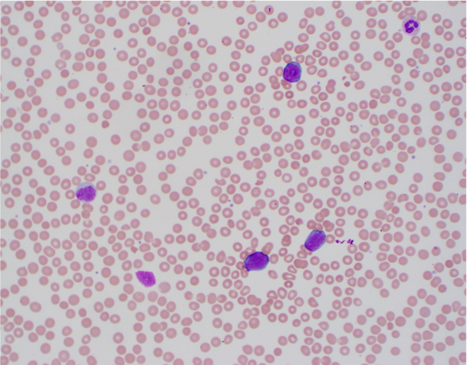
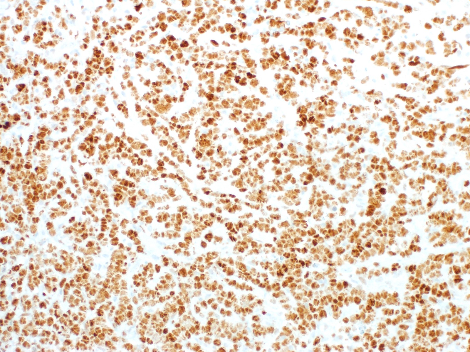
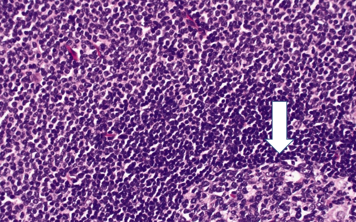
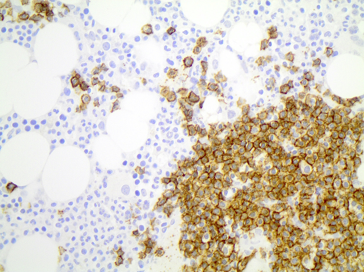
ALK positive large B cell lymphoma cells
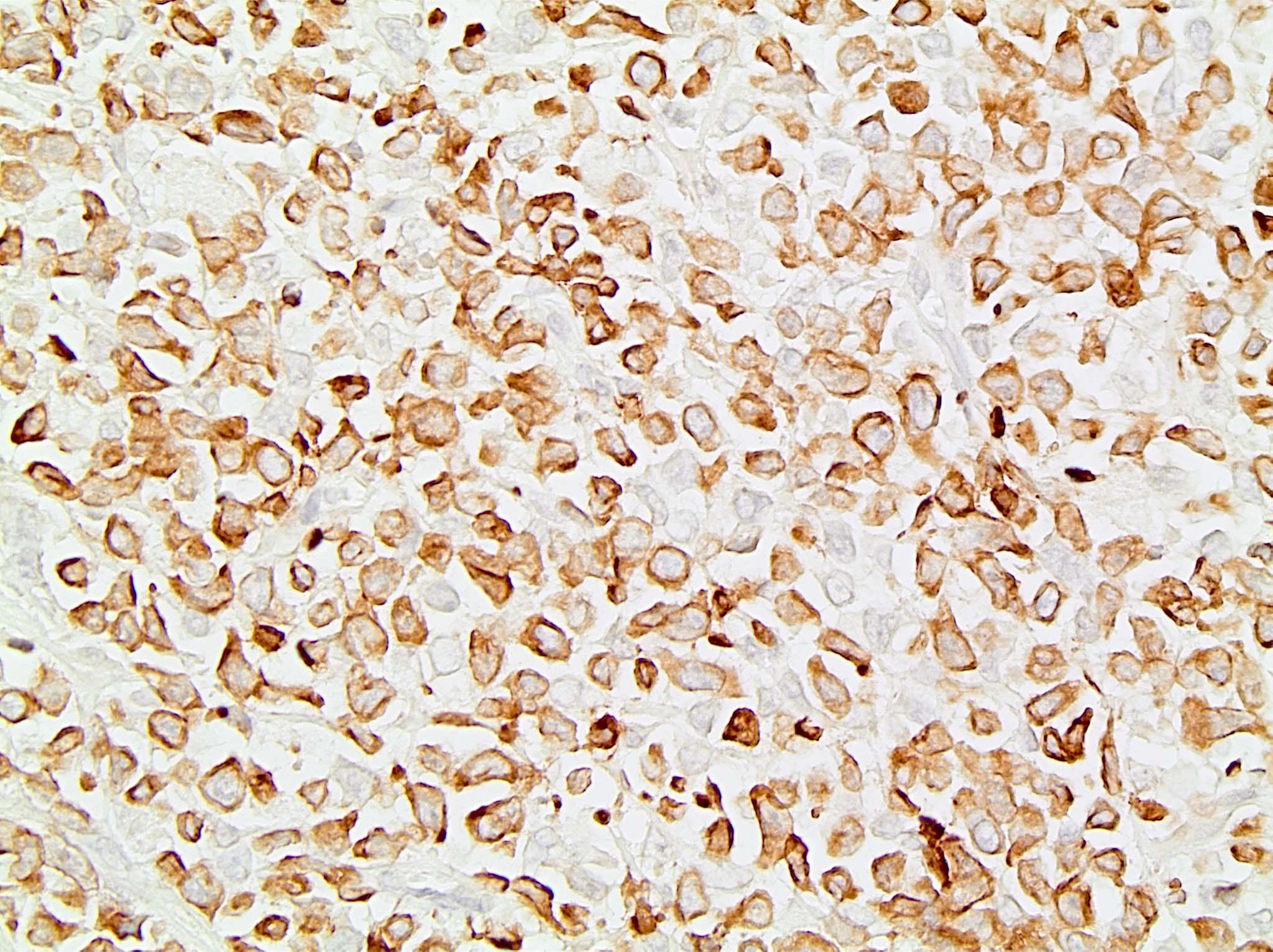
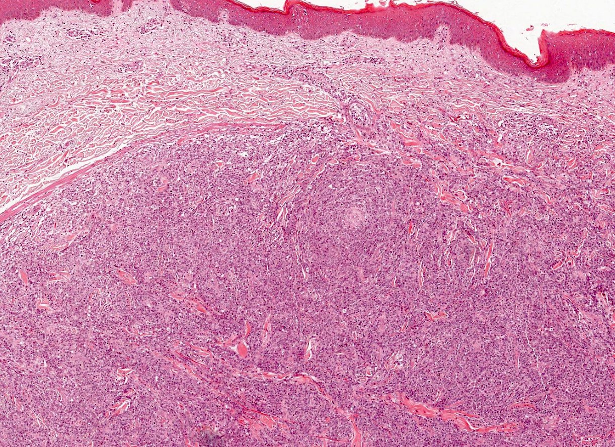
Contributed by Jennifer Chapman, M.D.
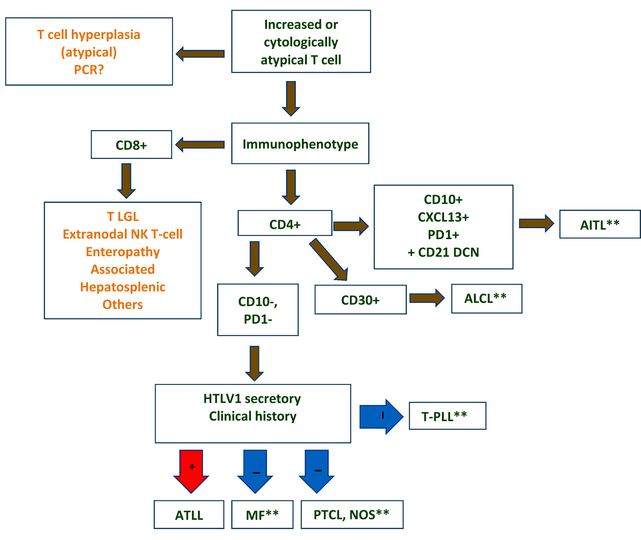
Differential diagnostic algorithm
Images hosted on other servers:

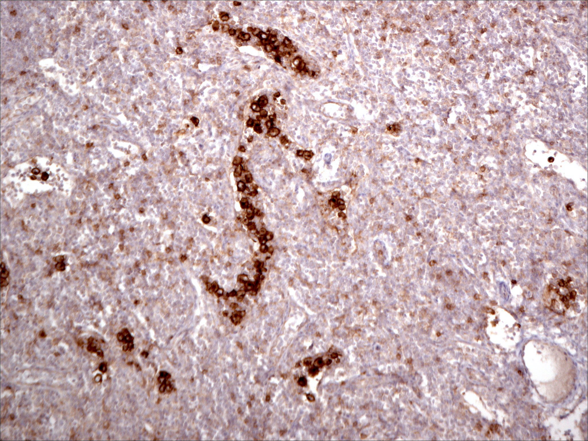
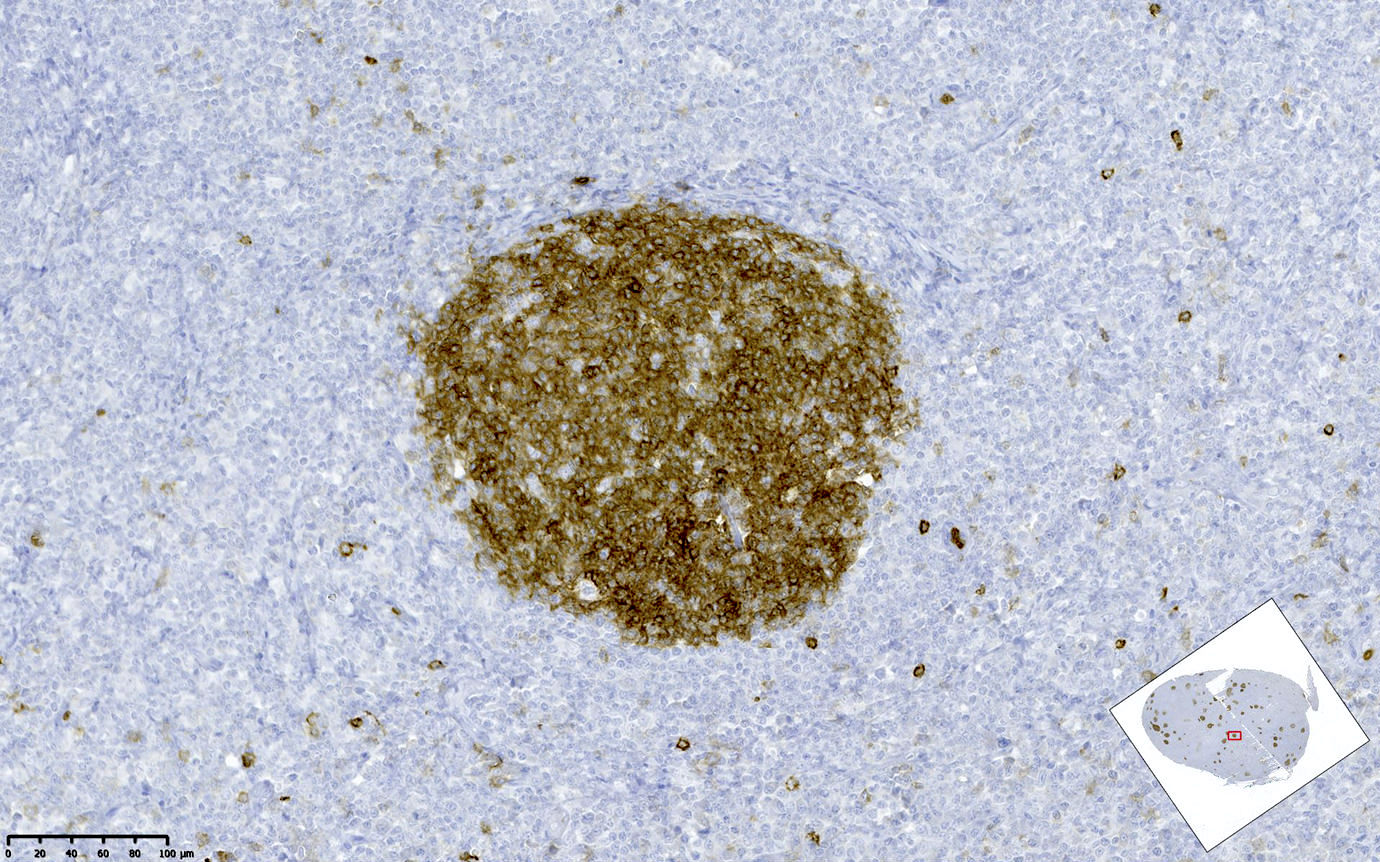
Nodules
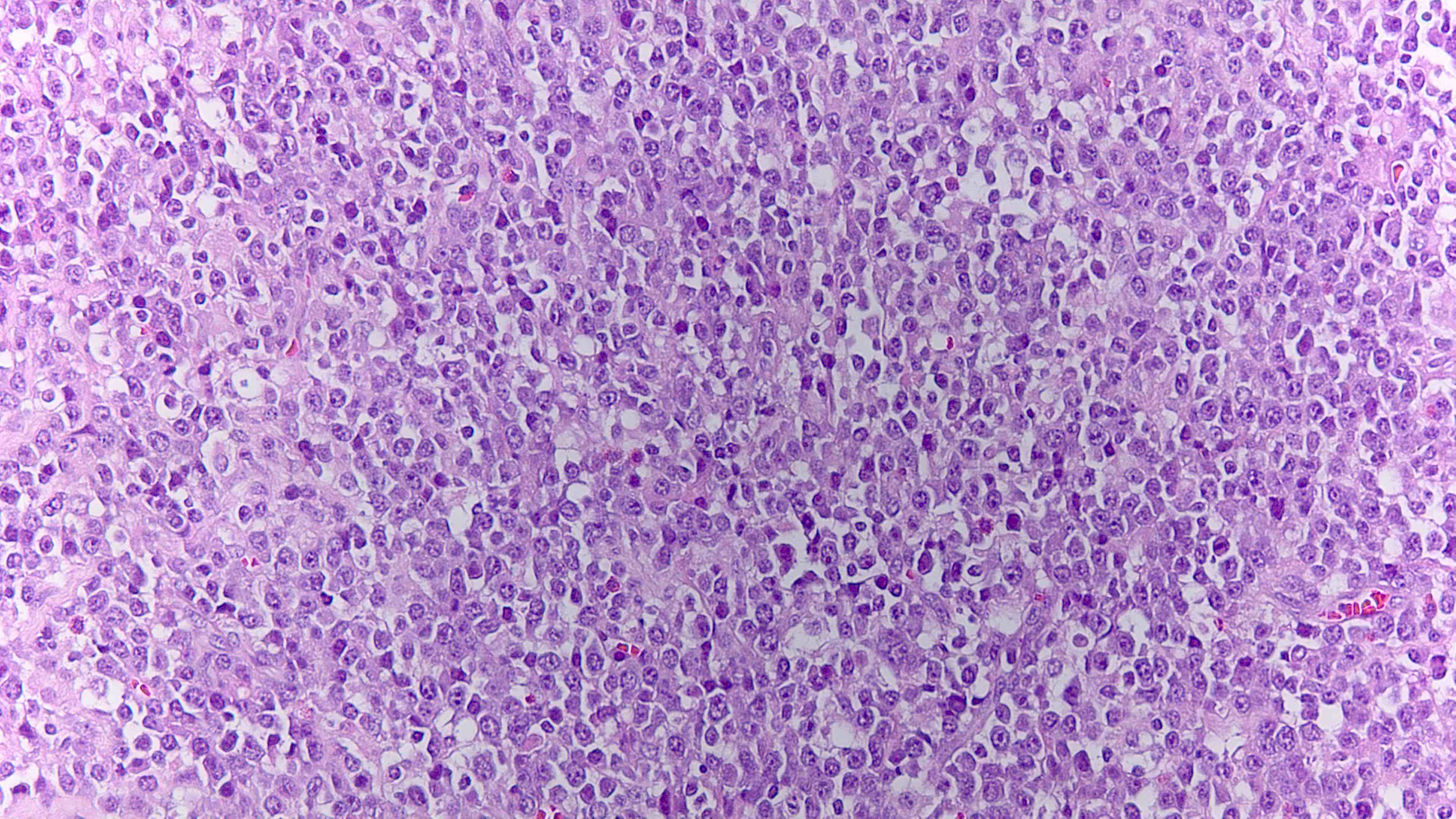
Contributed by Jennifer Chapman, M.D.
ATLL mimicking angioimmunoblastic T cell lymphoma
ATLL with Hodgkin-like cells mimicking classical Hodgkin lymphoma
ATLL mimicking mycosis fungoides

ATLL, lymphomatous type, mimicking anaplastic large cell lymphoma
Contributed by Jennifer Chapman, M.D.
ATLL, acute variant,
mimicking
T prolymphocytic
lymphoma
Images hosted on other servers:

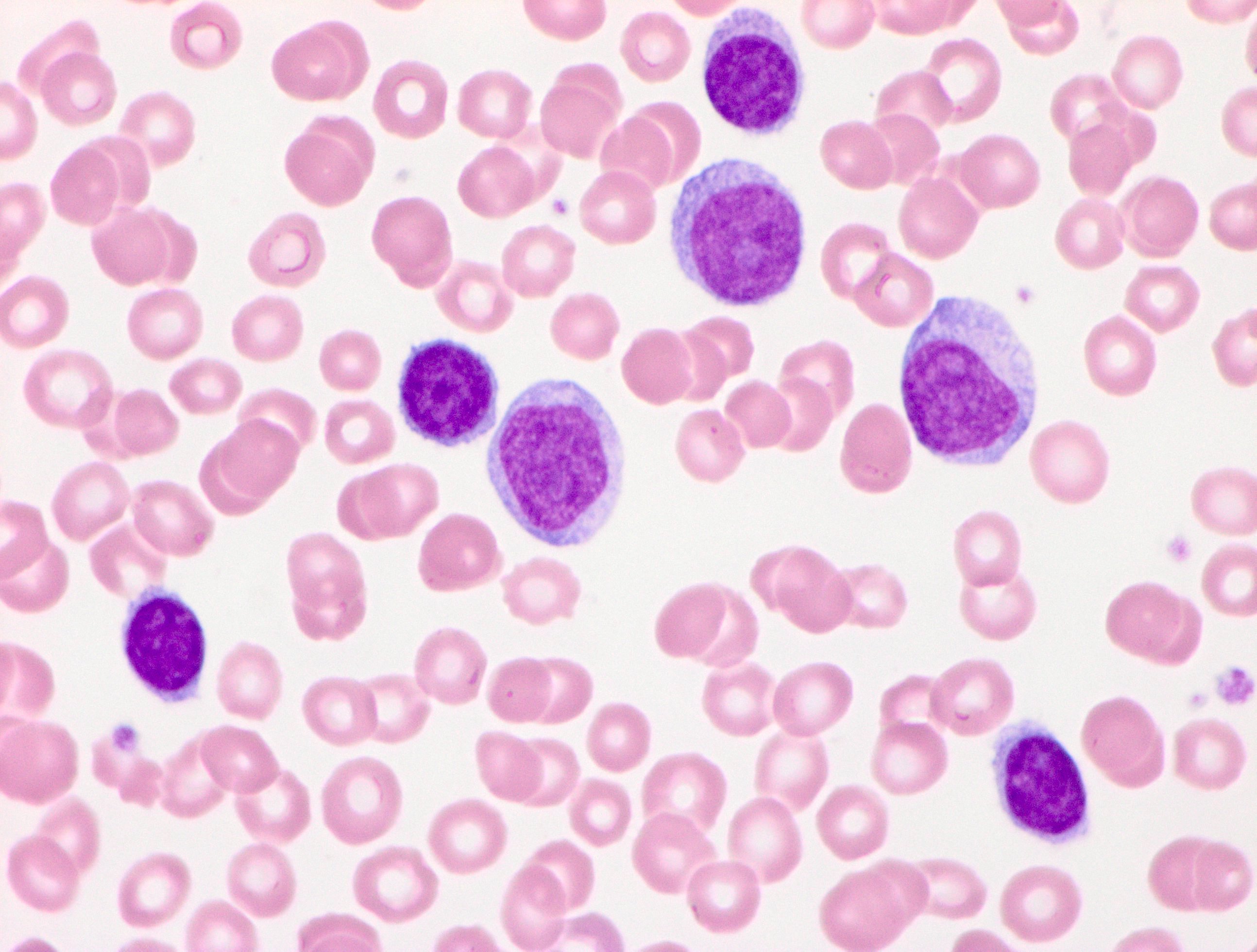
Flower cells

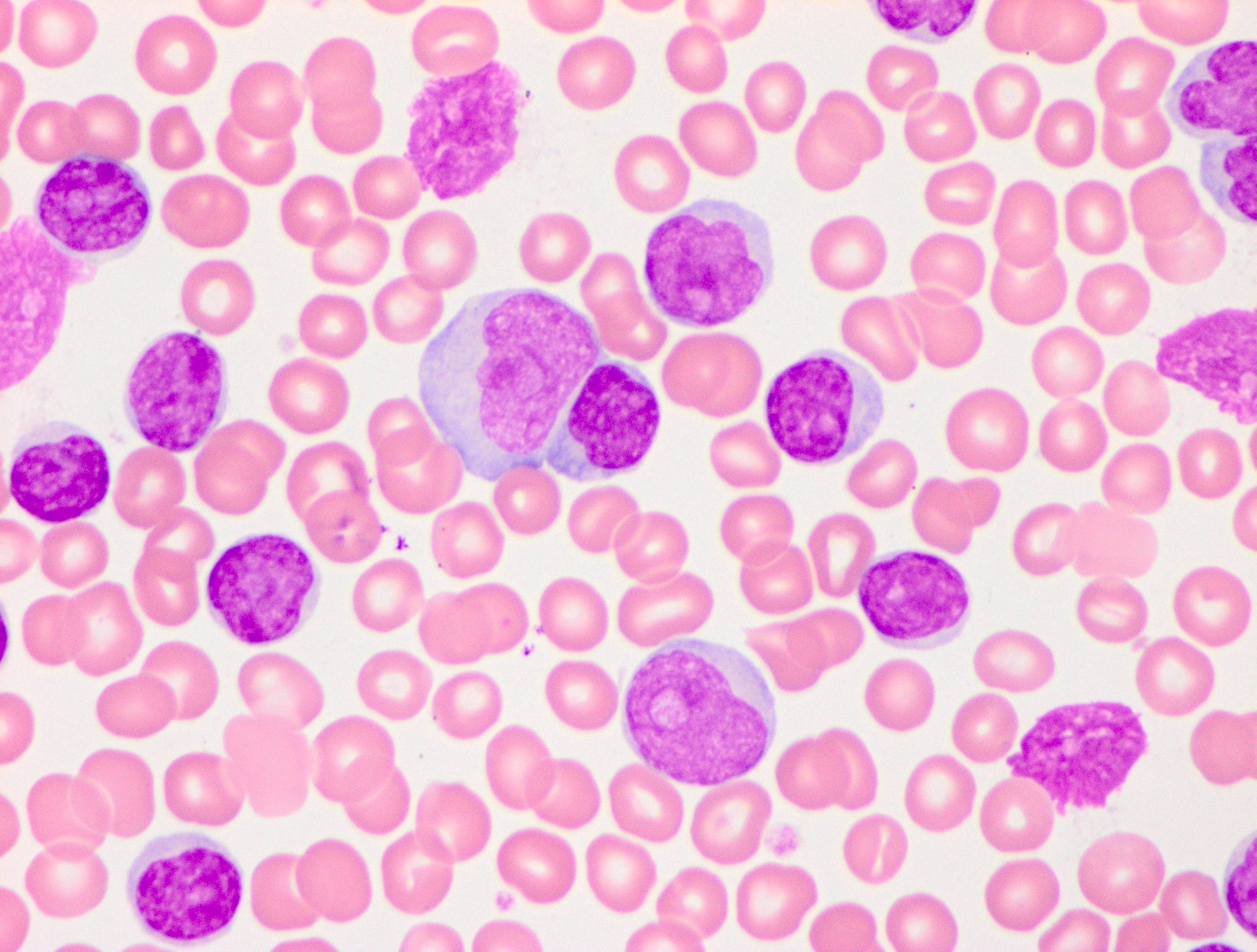
CLL-like morphology
Contributed by Jennifer Chapman, M.D.
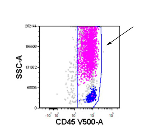
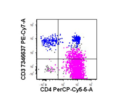
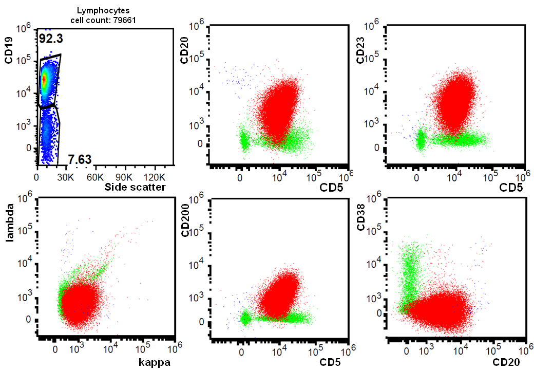
CD45
CD4

CD56
CD7
CD25
Images hosted on other servers:

CD3+, CD4+, CD25+, CD7-, CD8-
Images hosted on other servers:

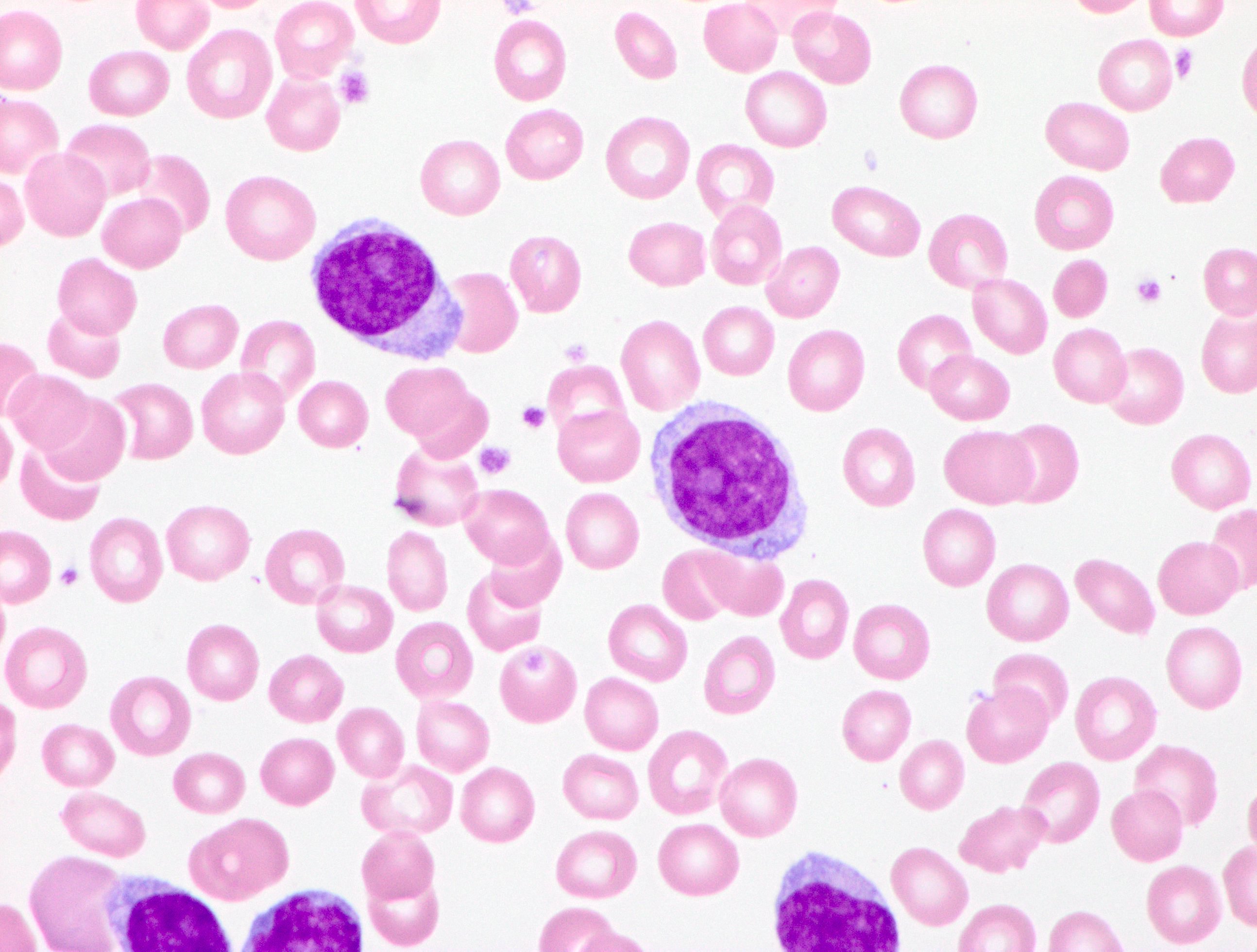
ATLL cell
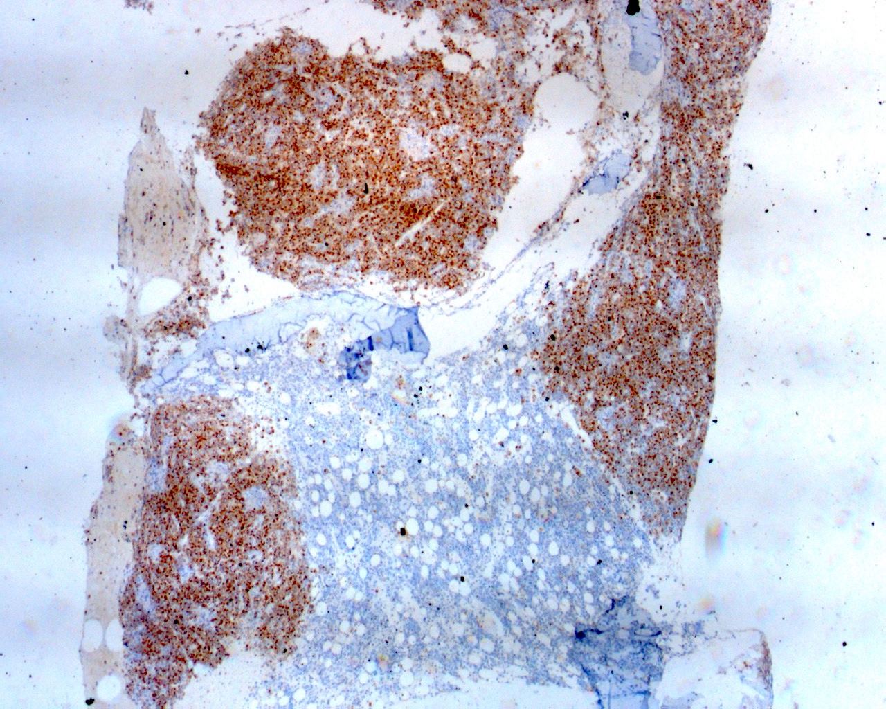
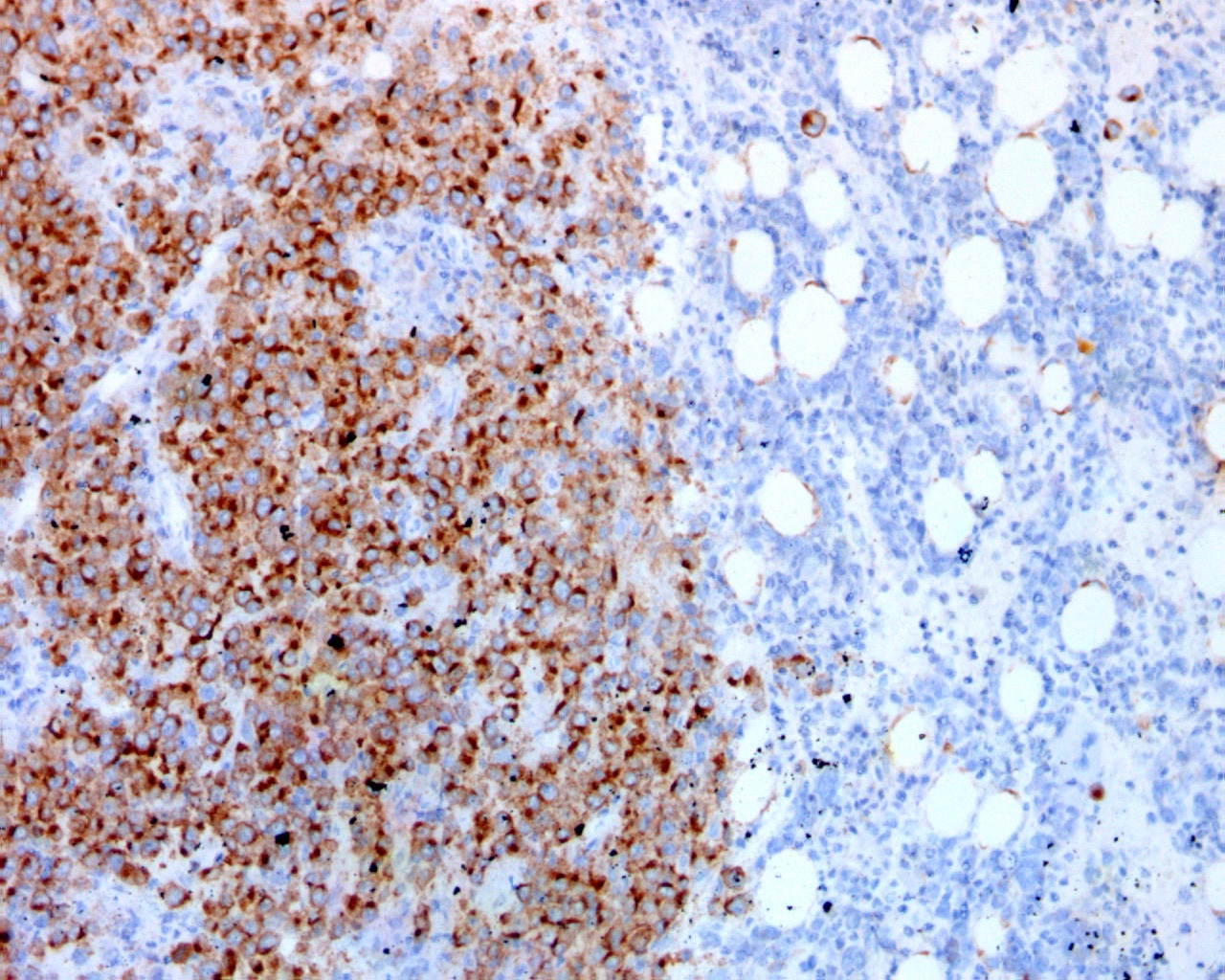
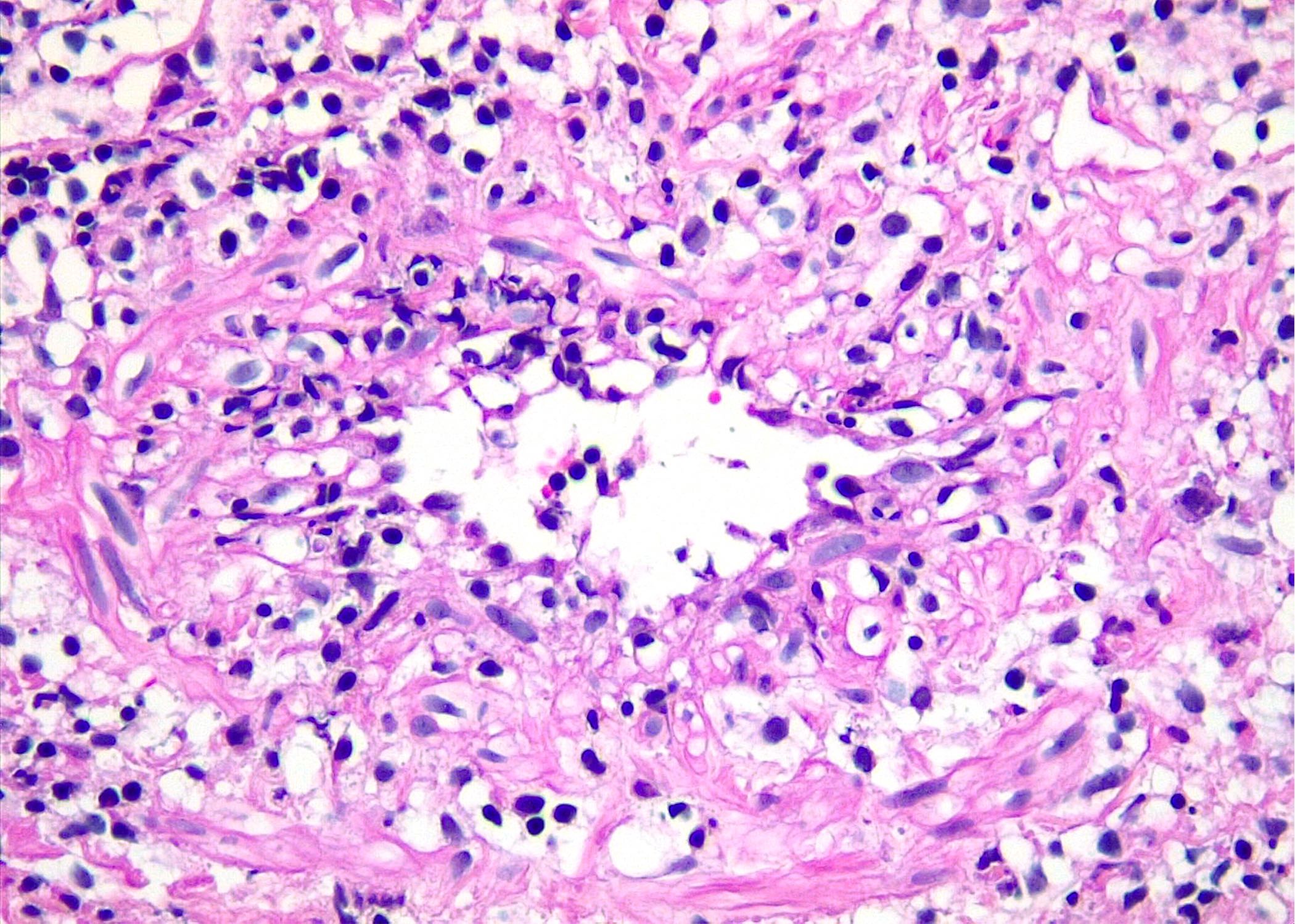
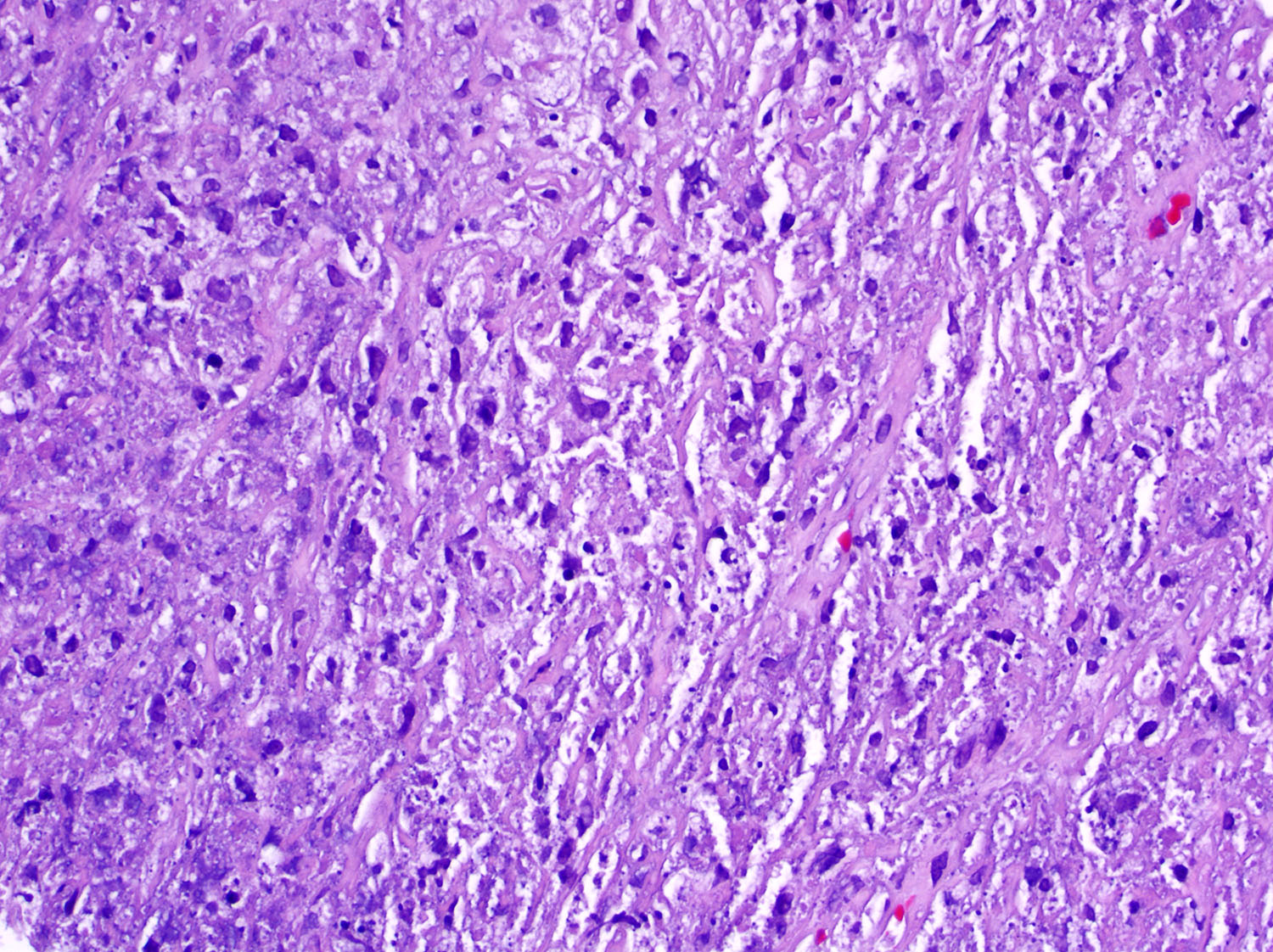
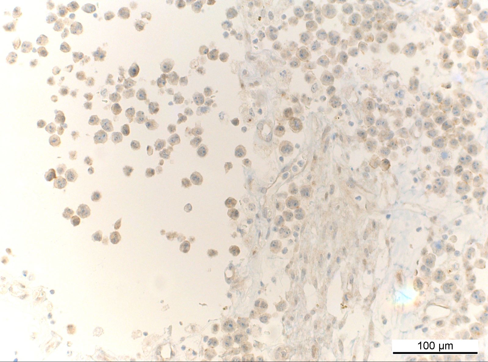
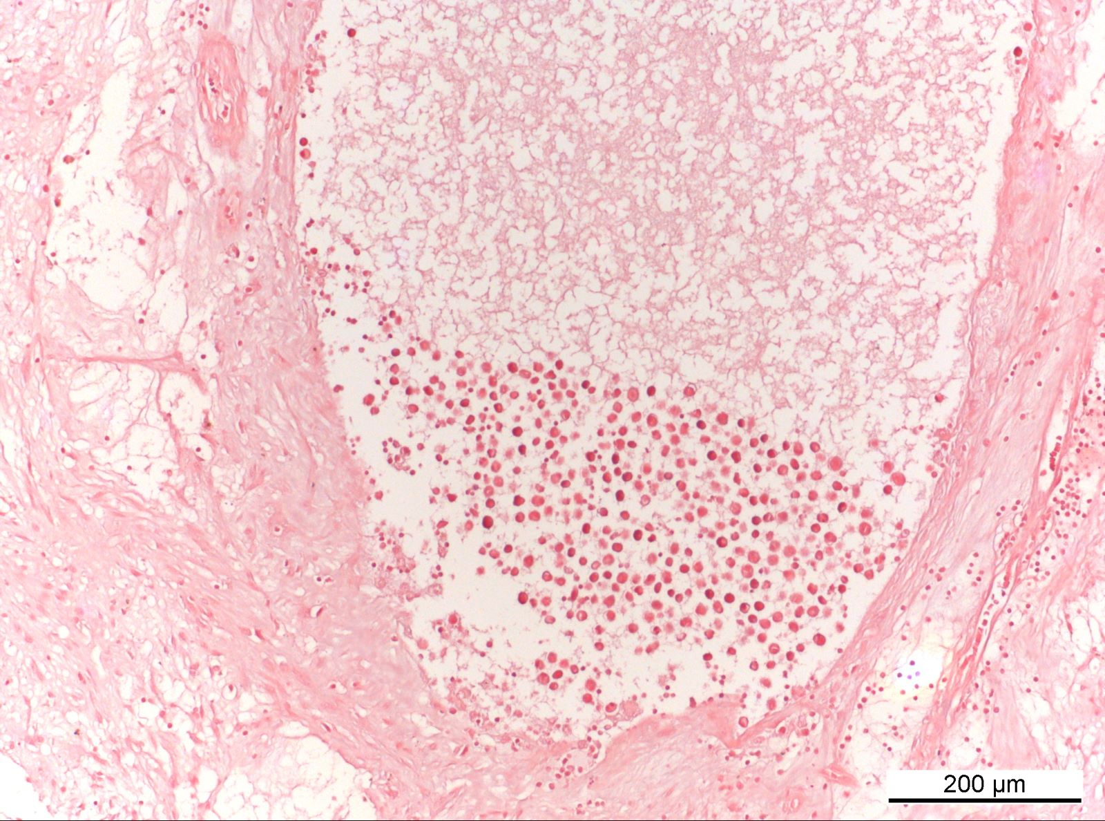
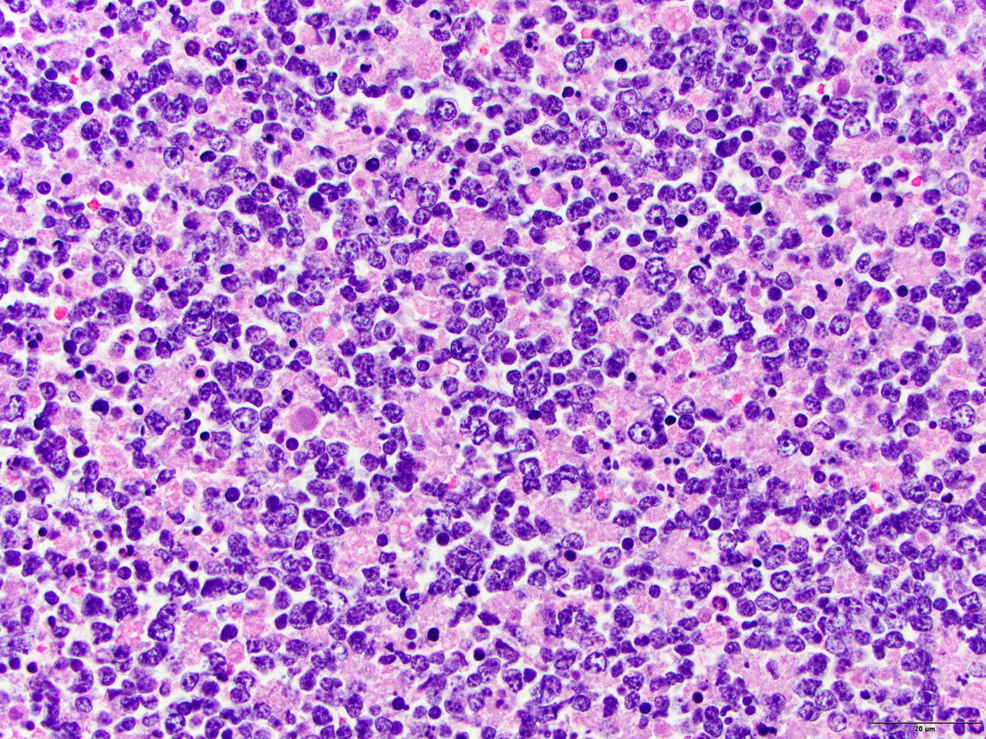
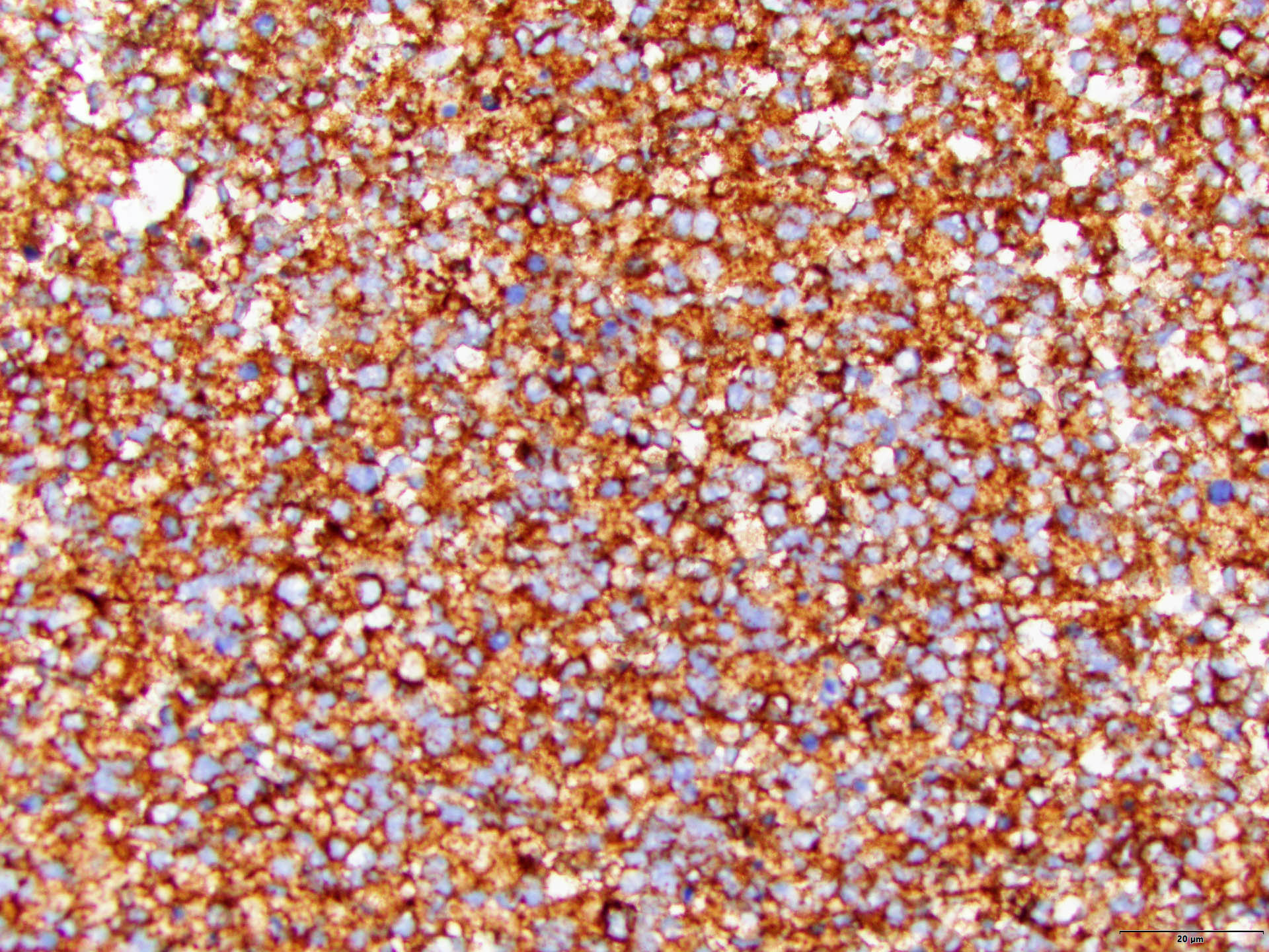
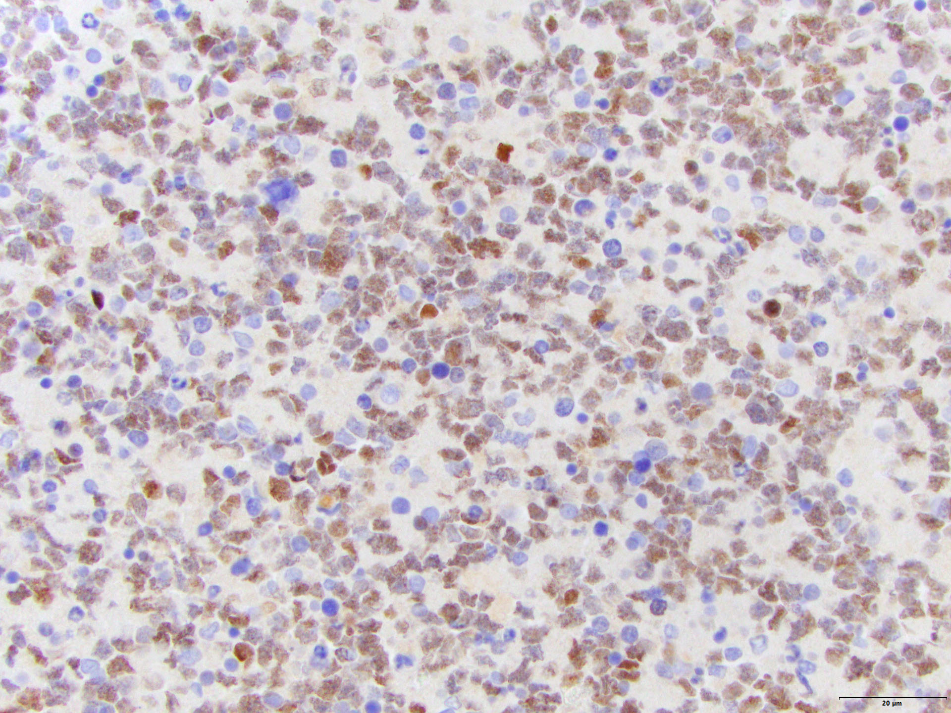
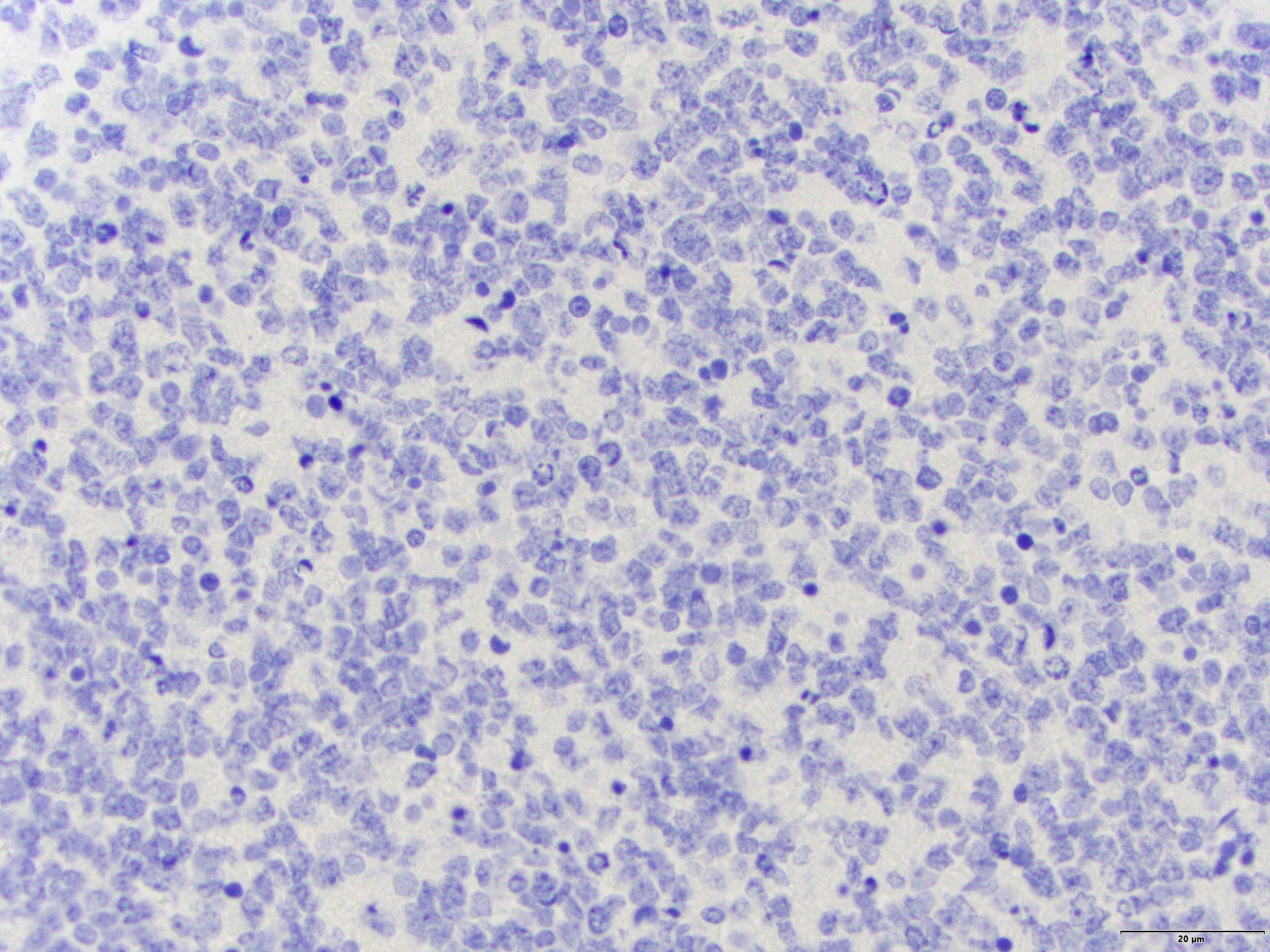
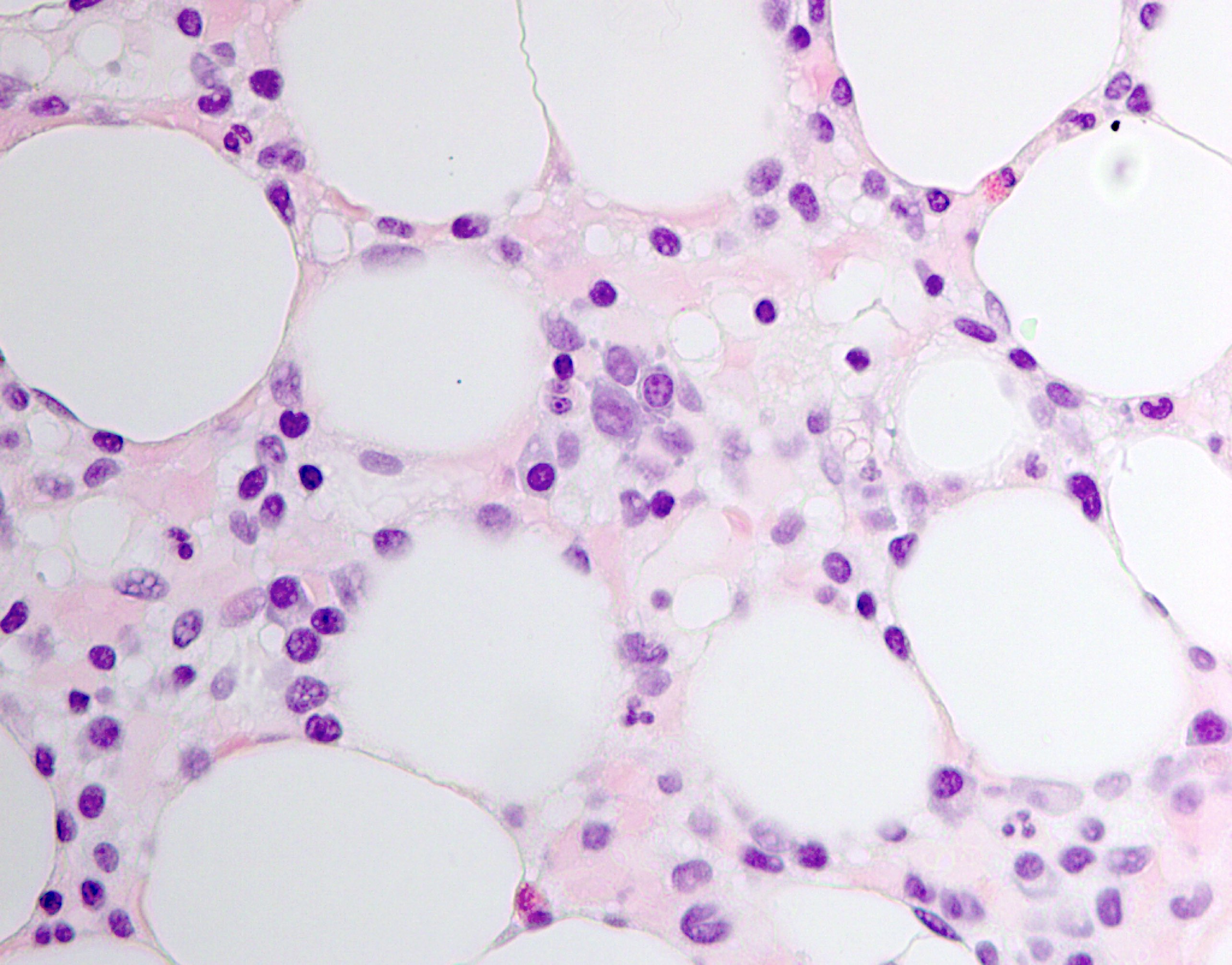
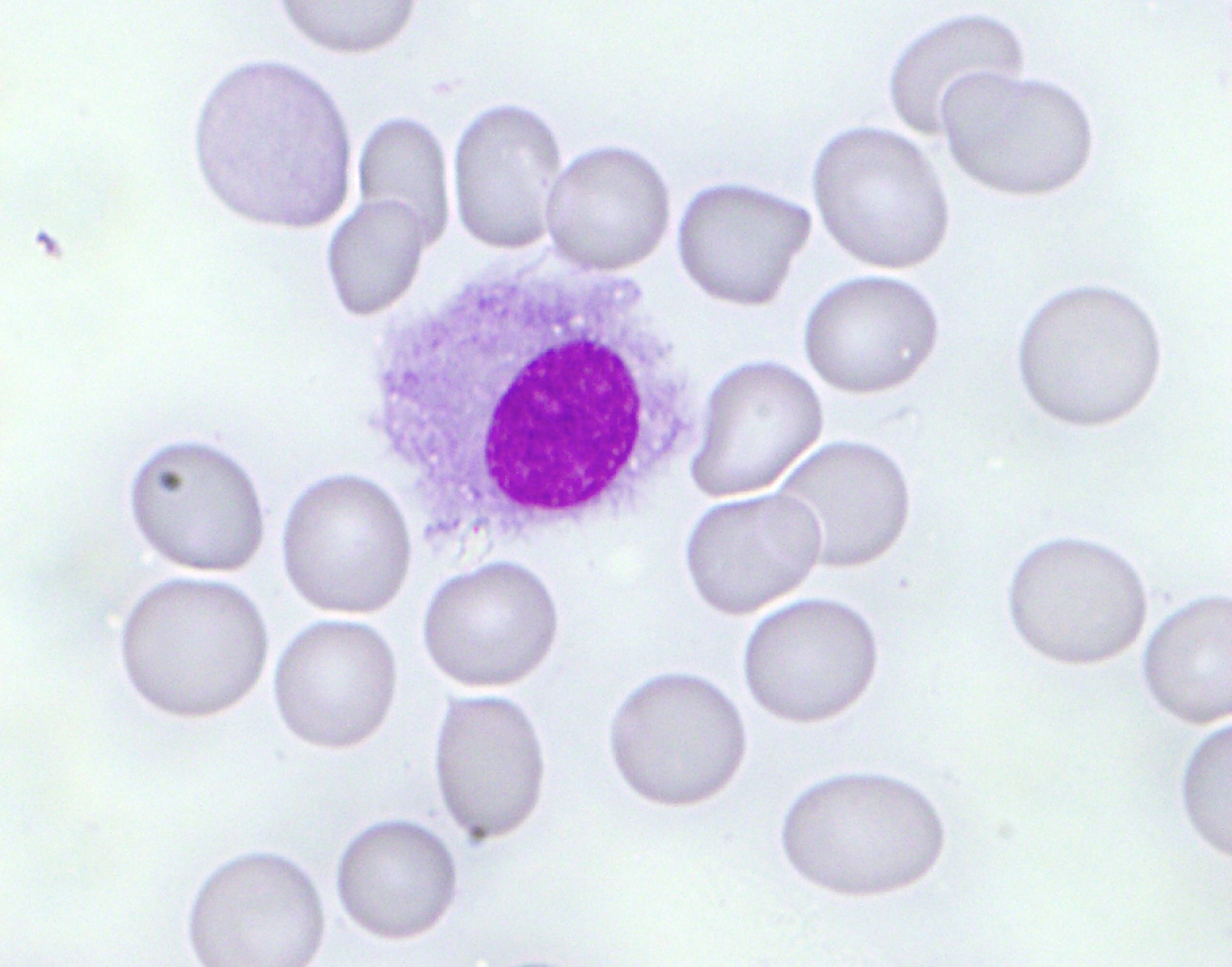
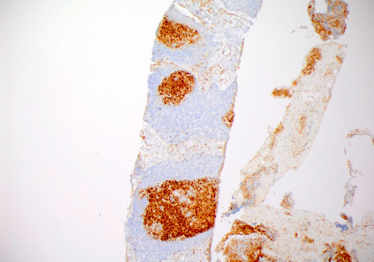
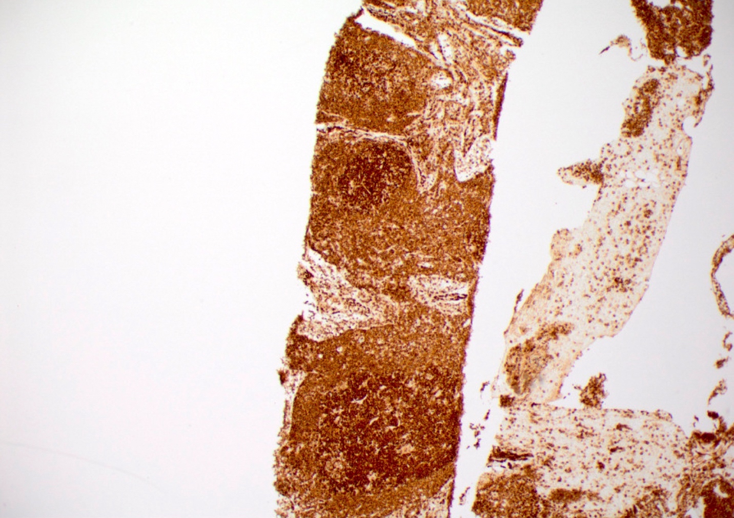
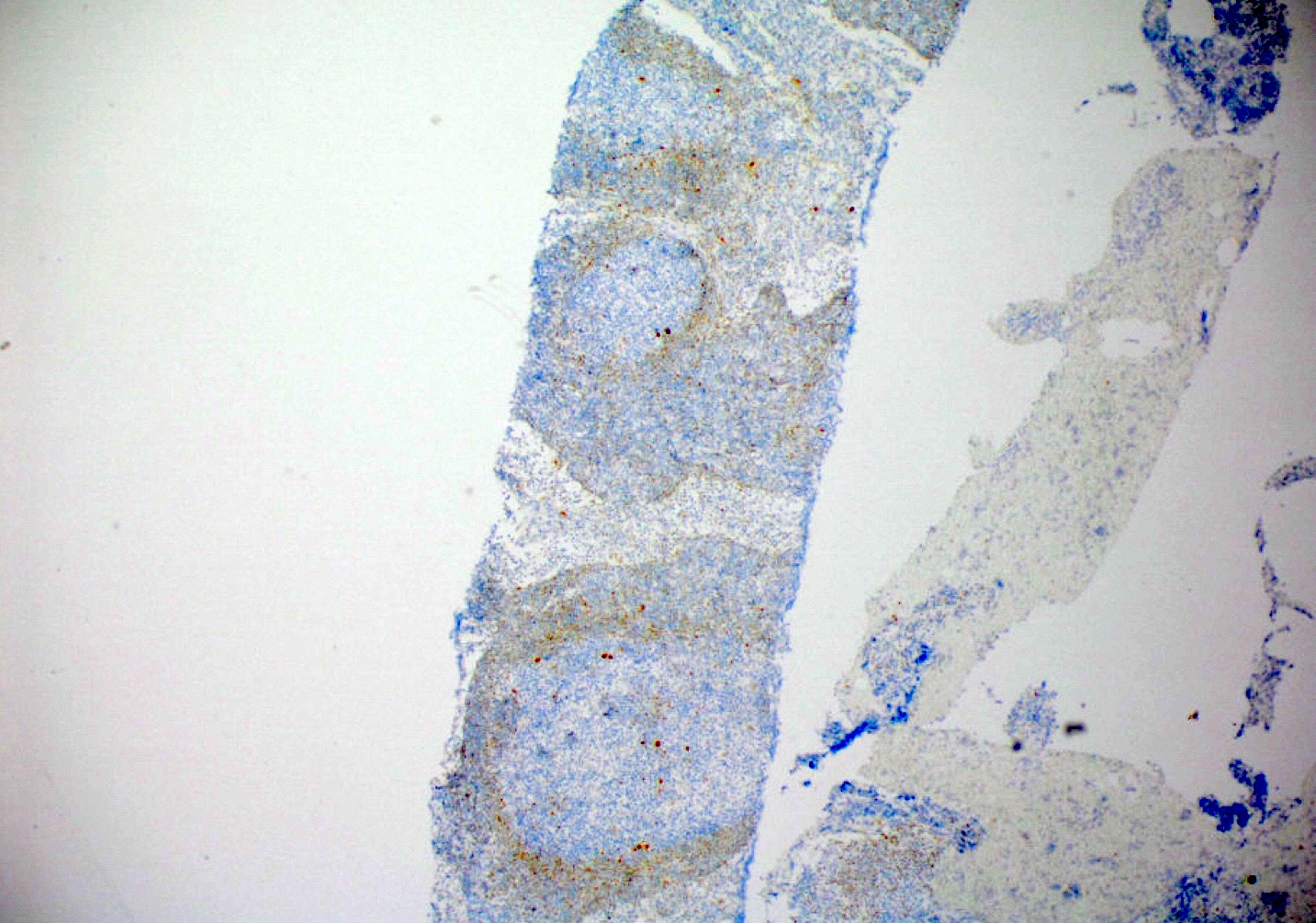
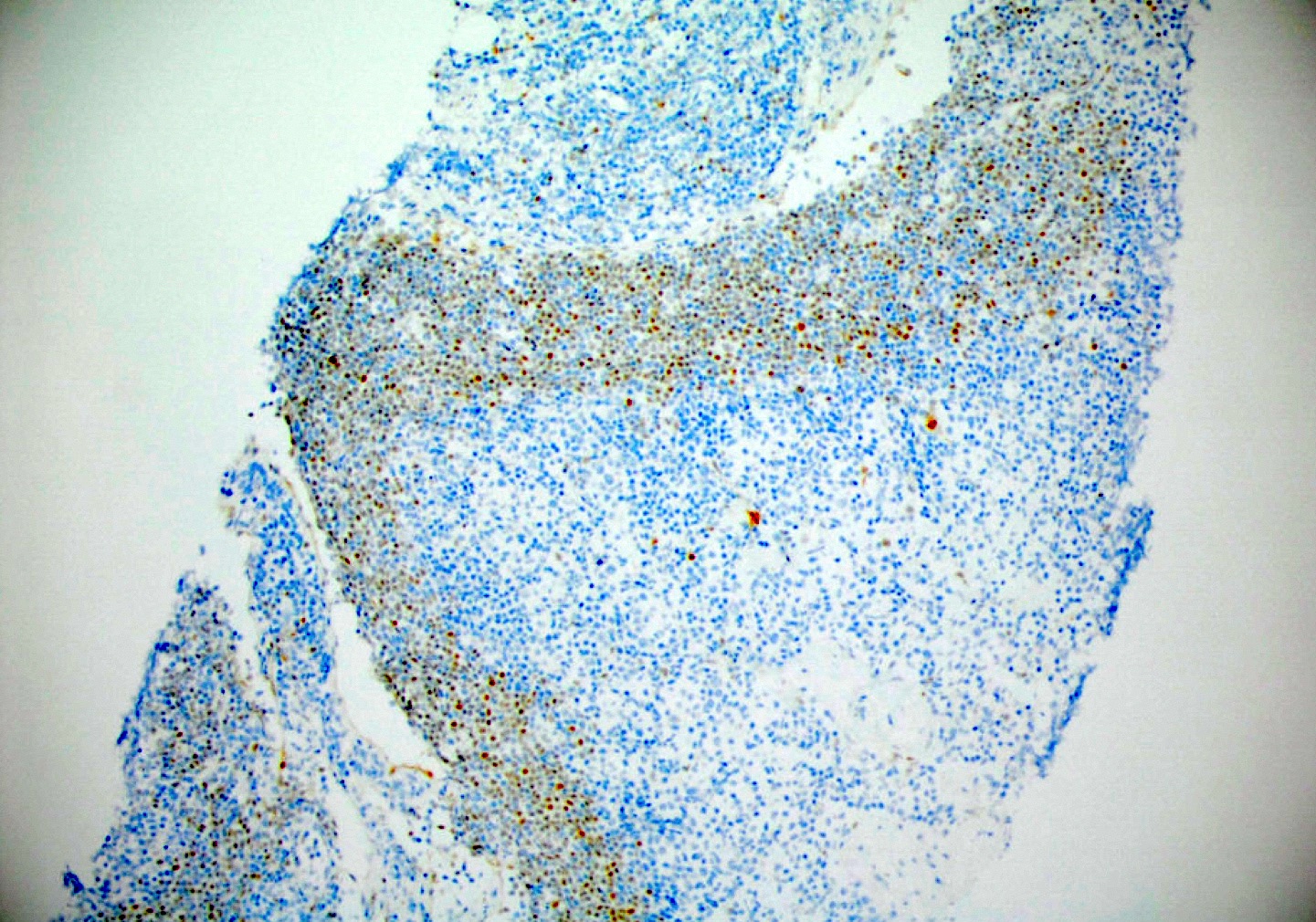
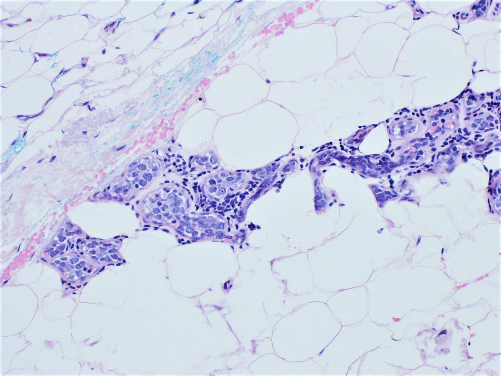
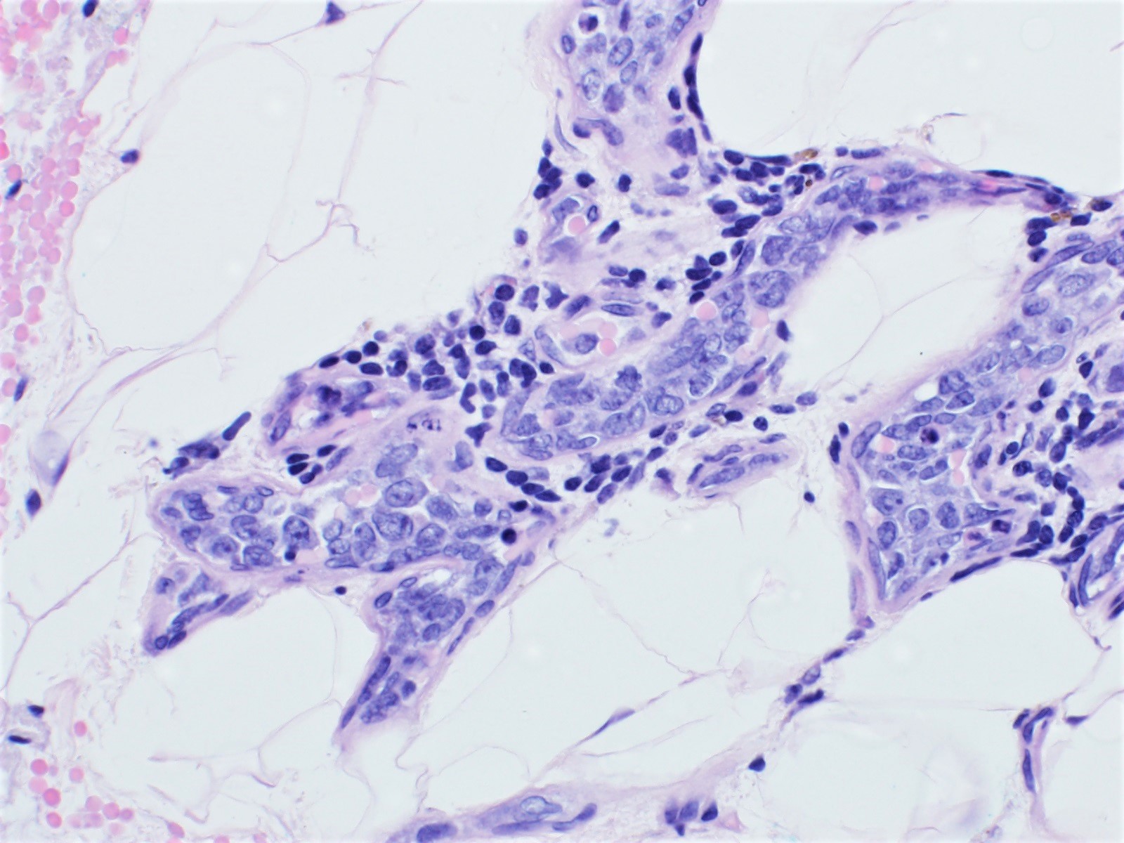
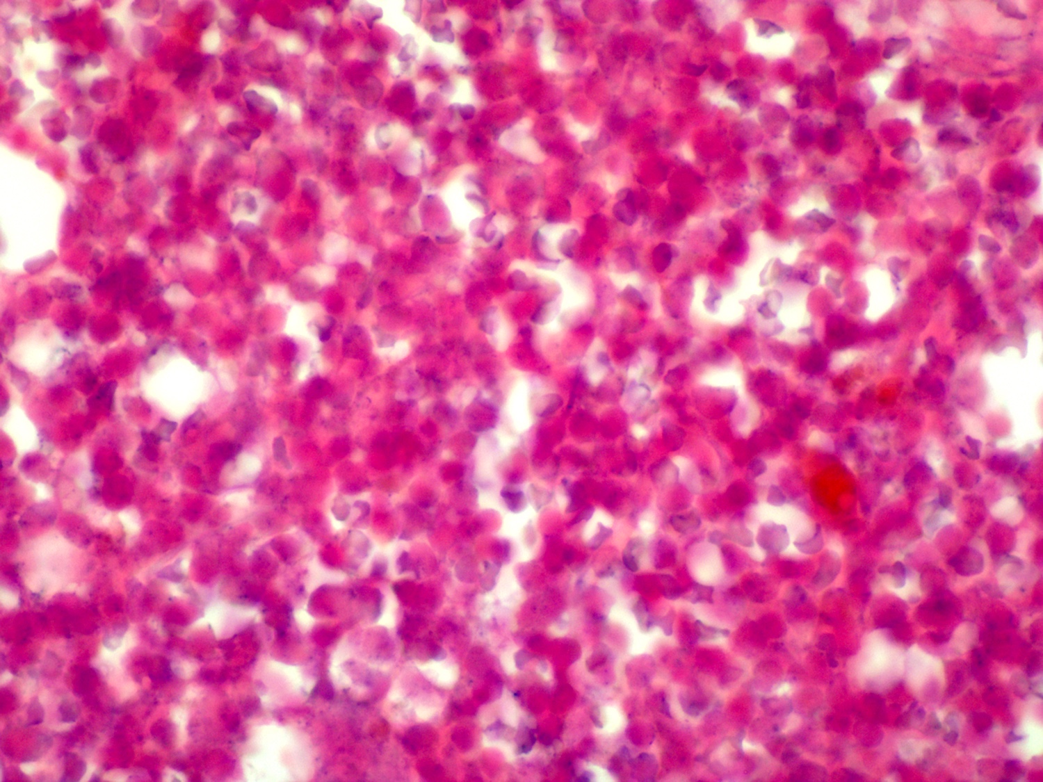
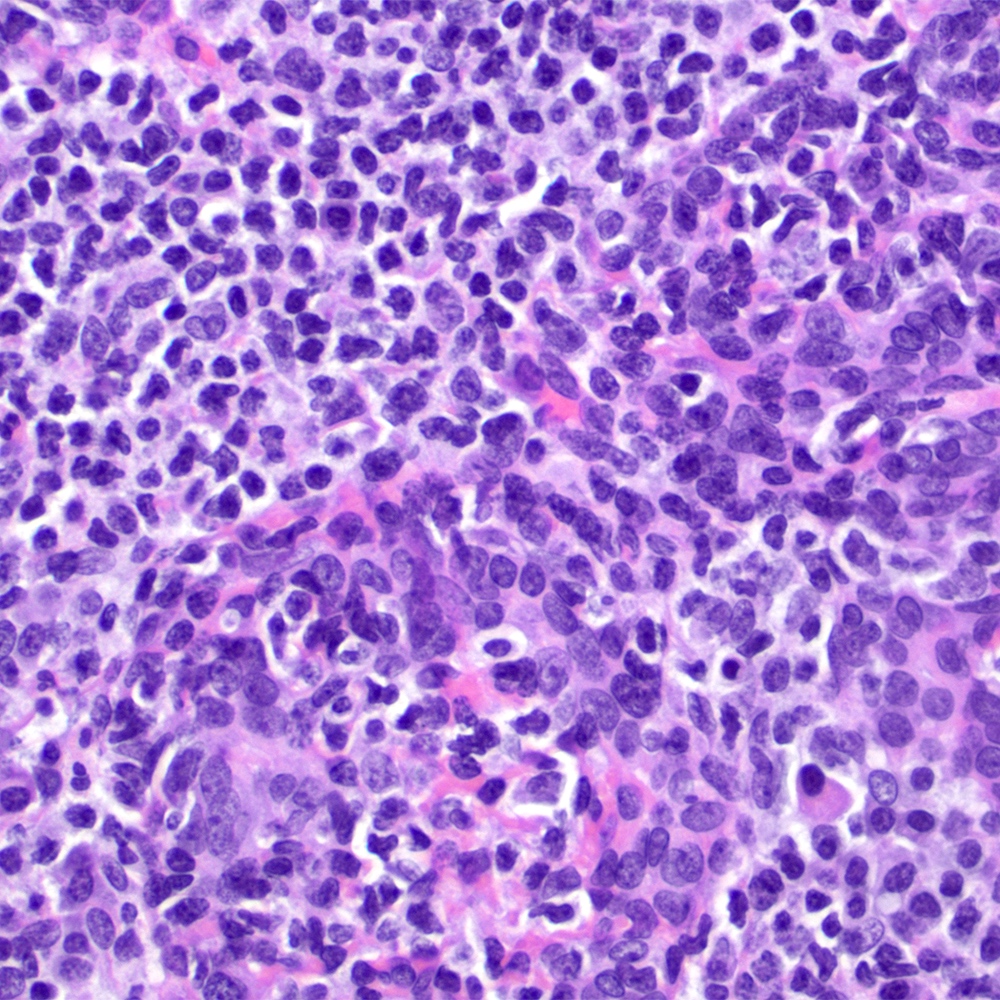
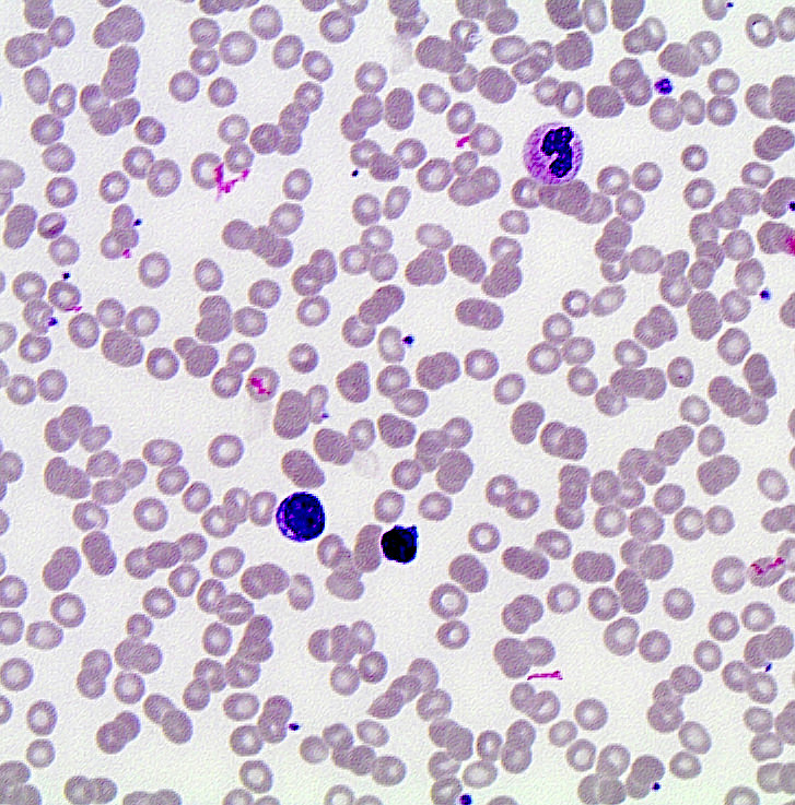
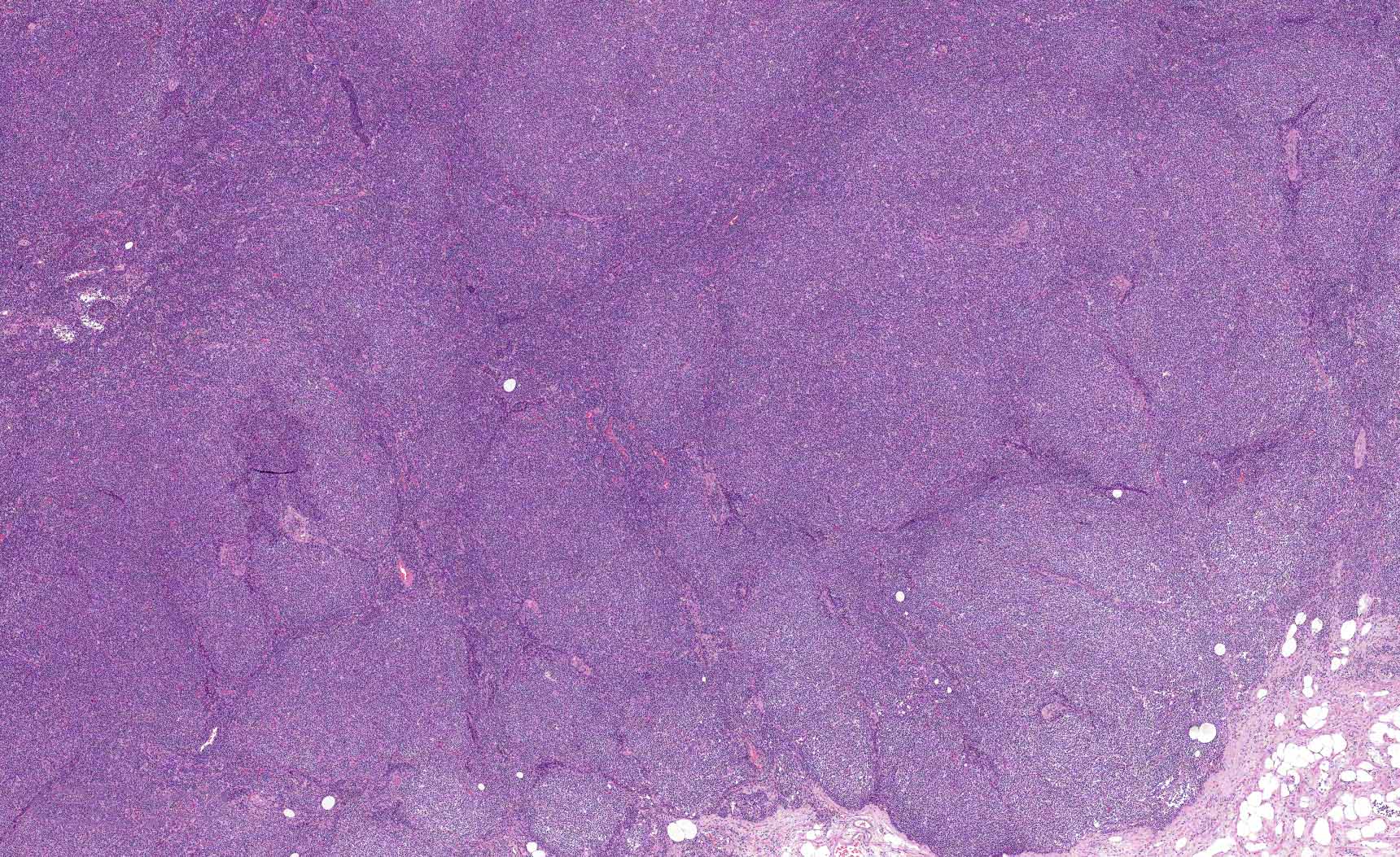
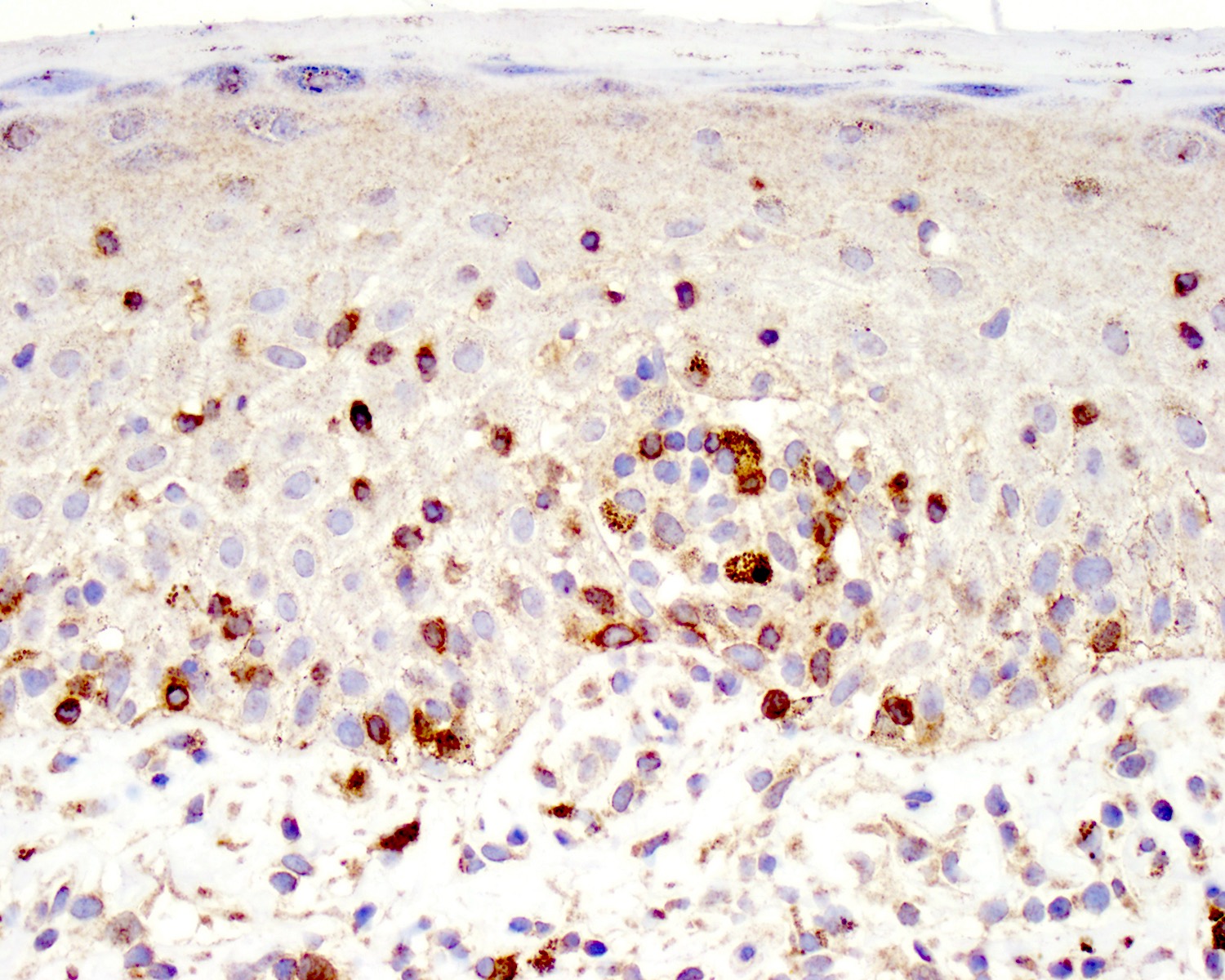
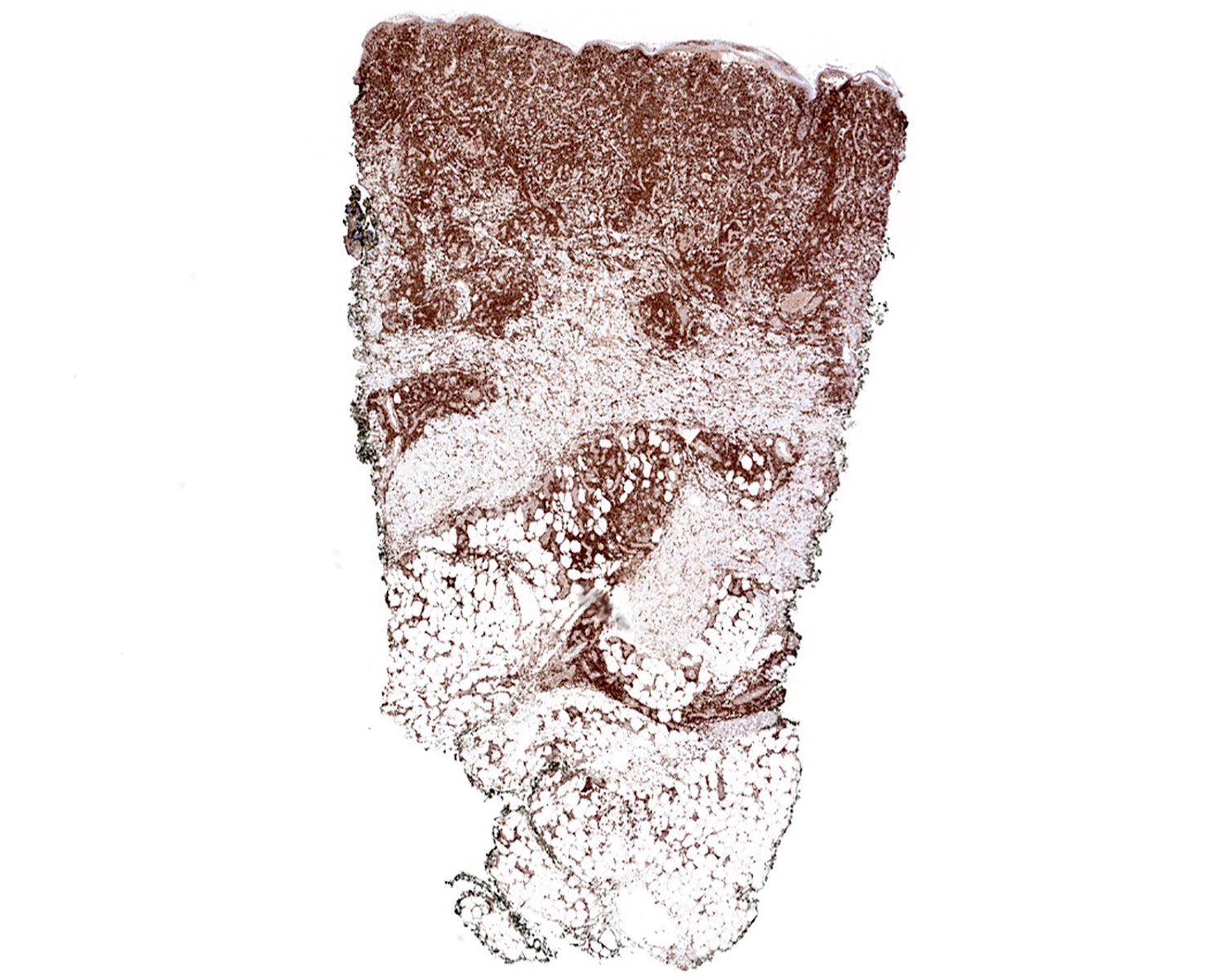
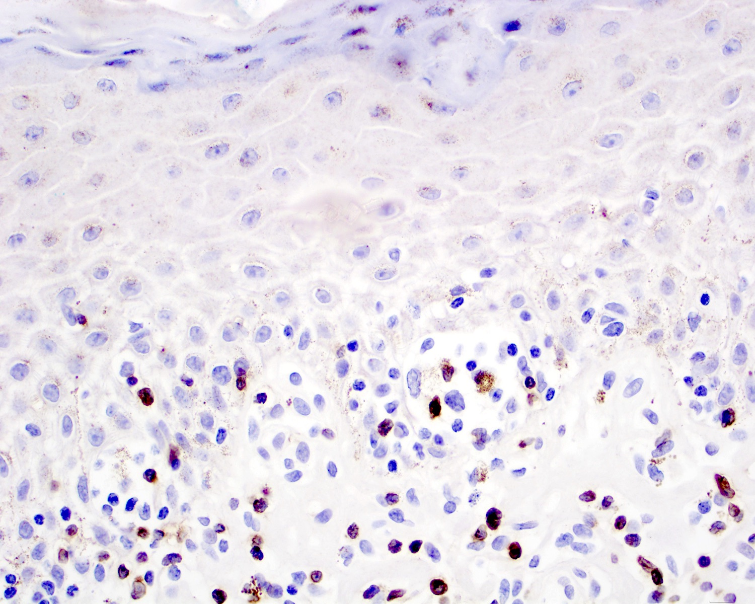
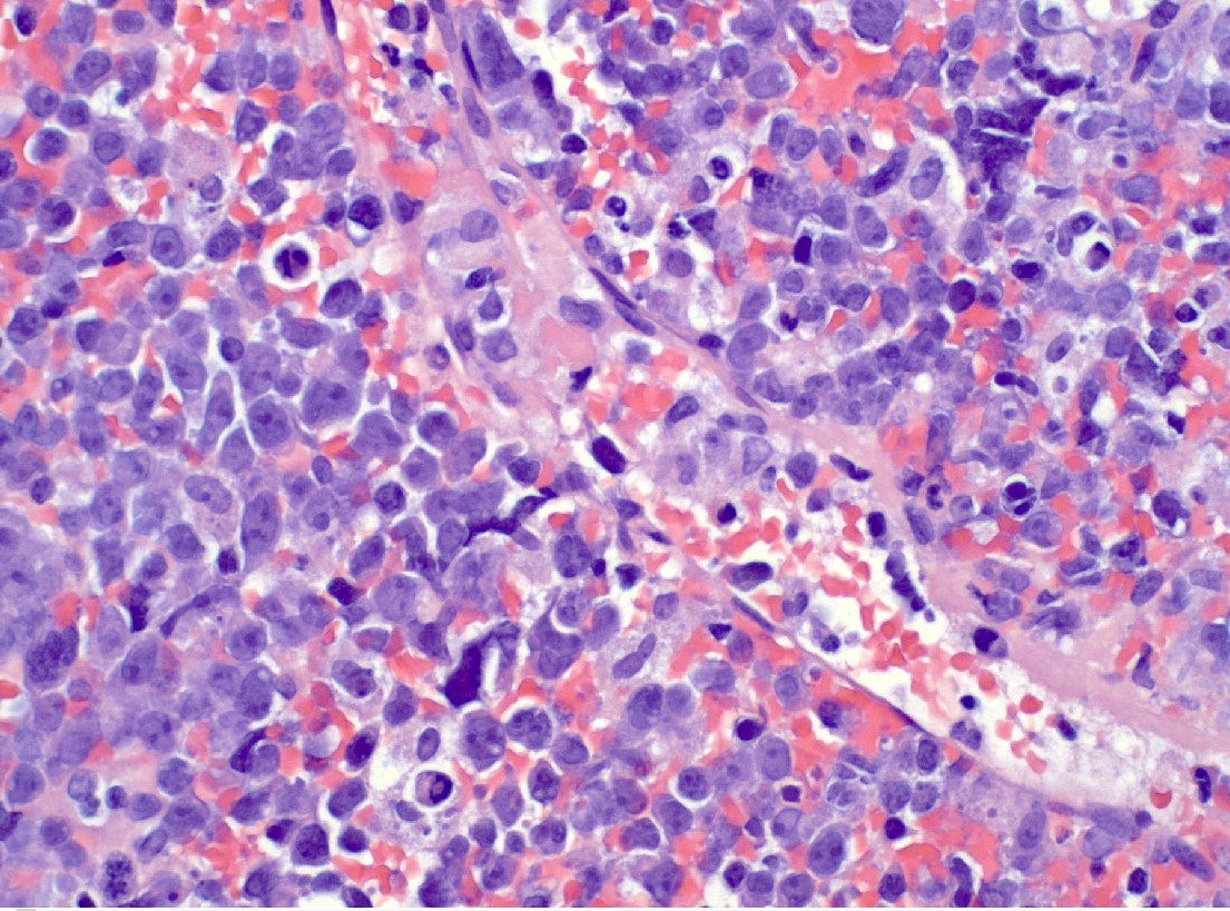
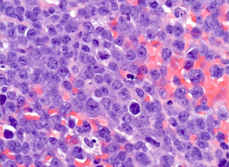
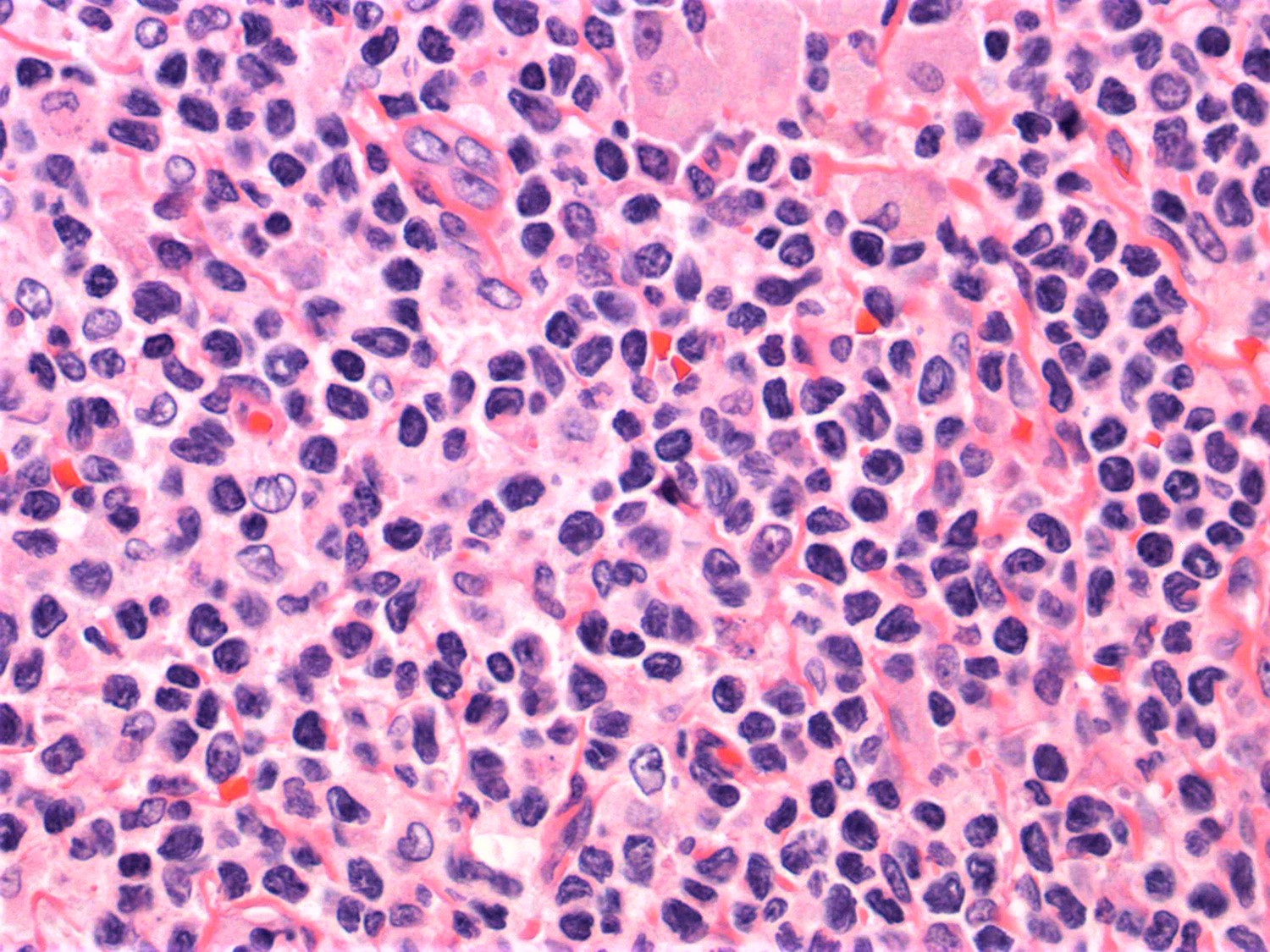
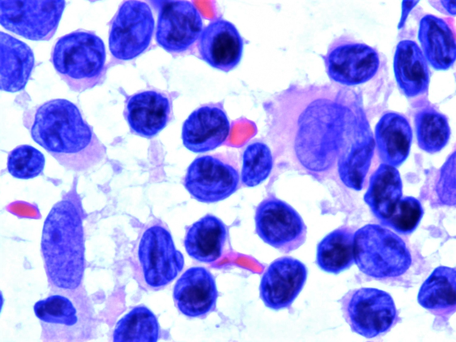
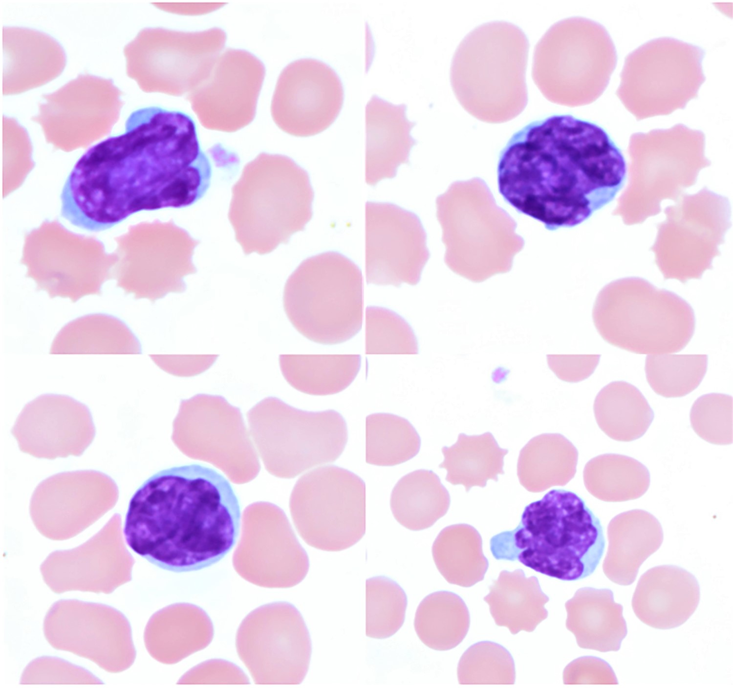
Contributed by Siba El Hussein, M.D. and Joseph Khoury, M.D.
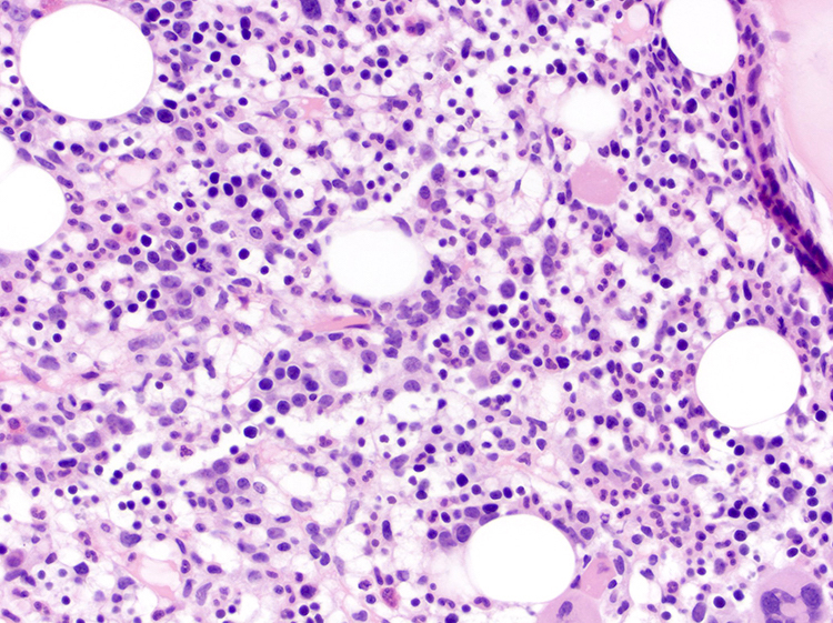
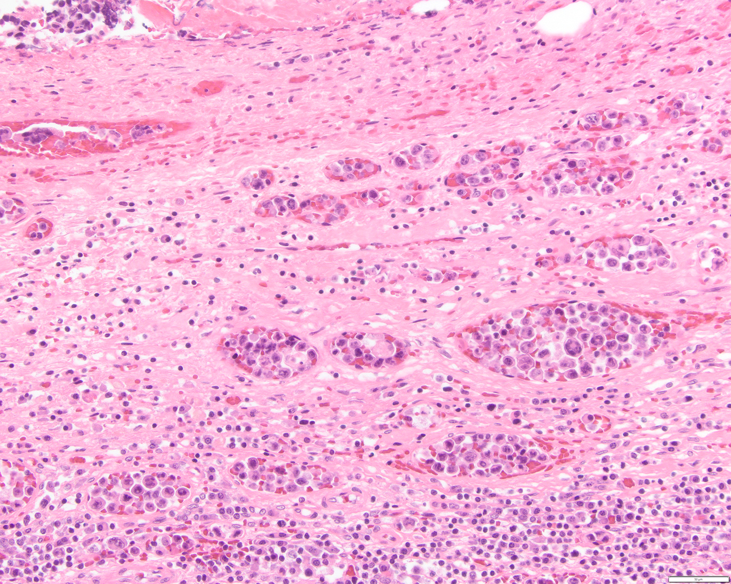
Neoplastic cells involving the bone marrow
Neoplastic cells involving the bone marrow
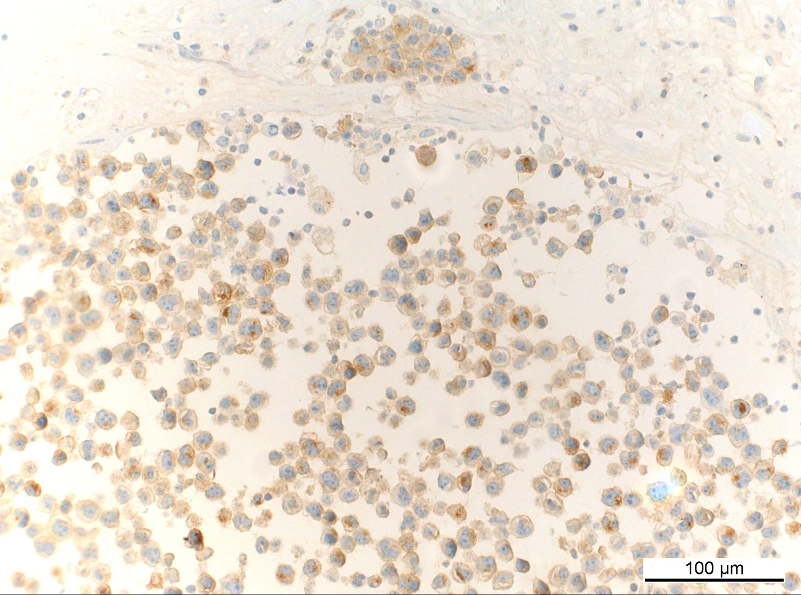
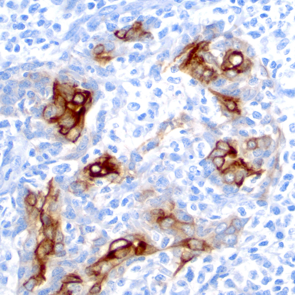
Immunohistochemical stains
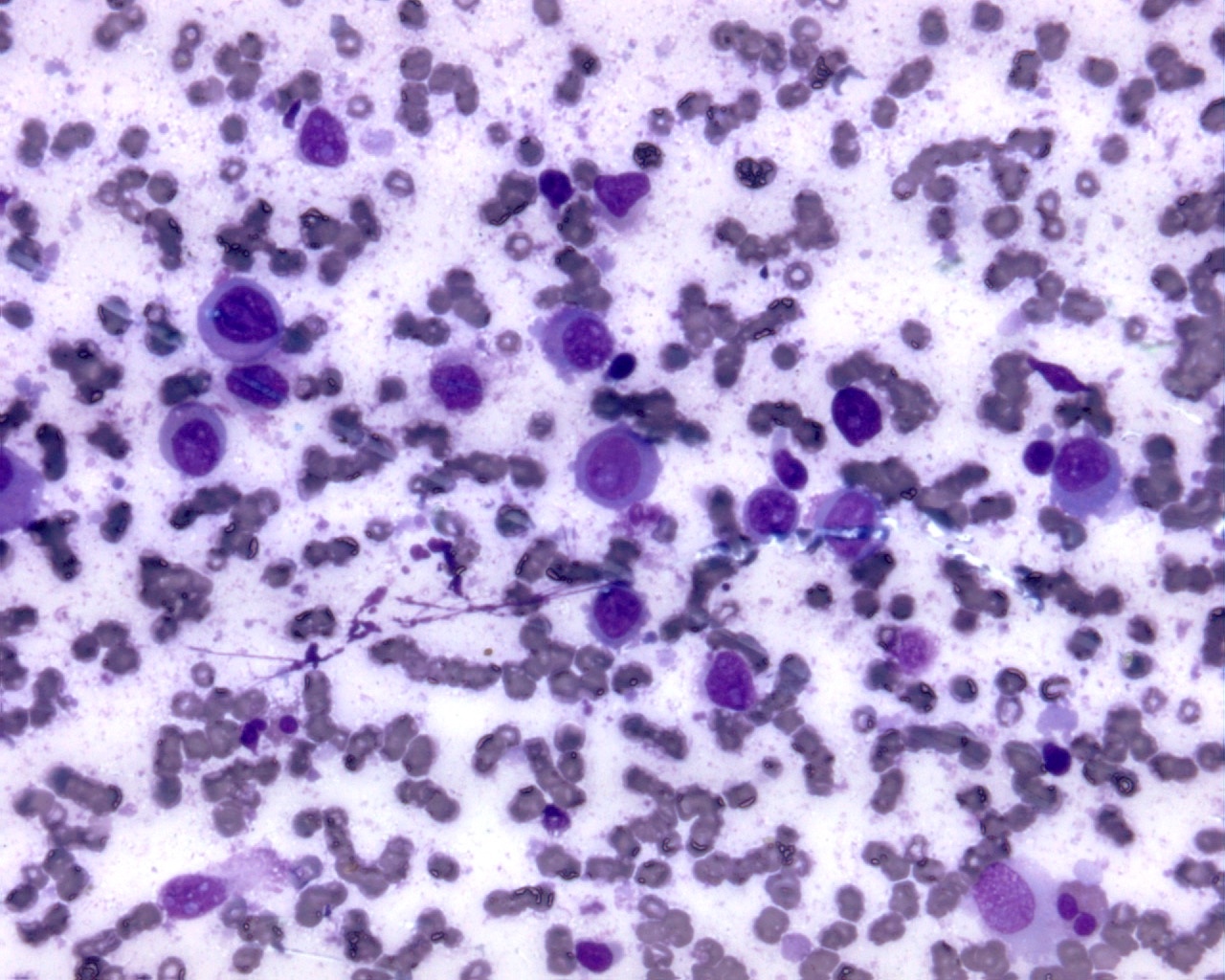
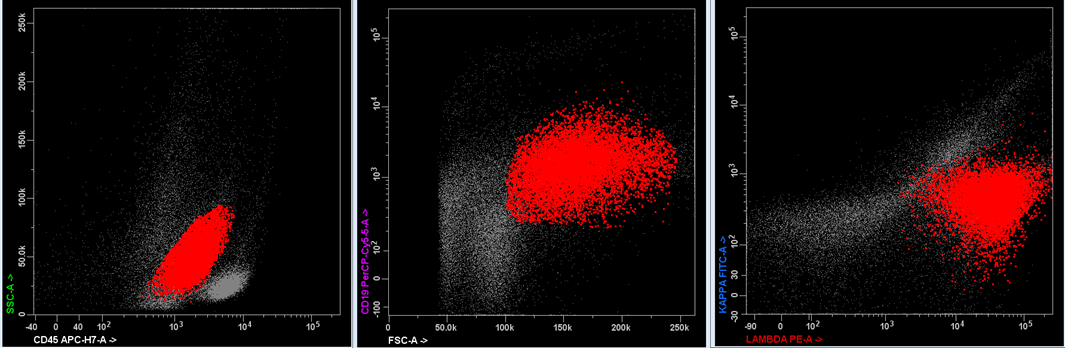
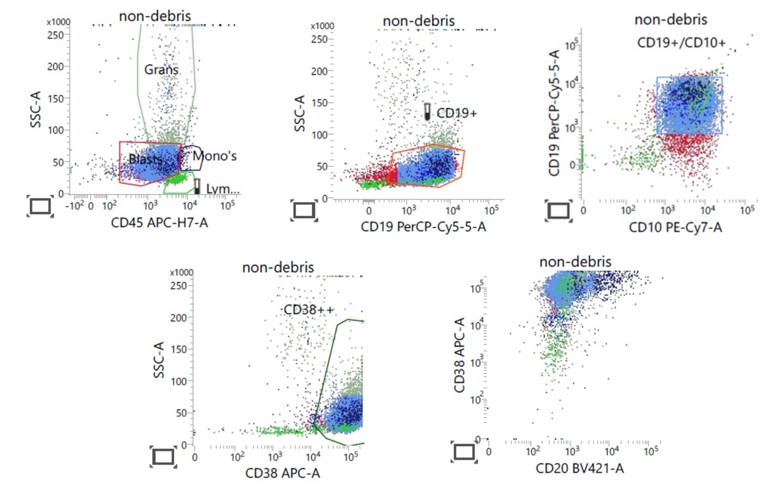
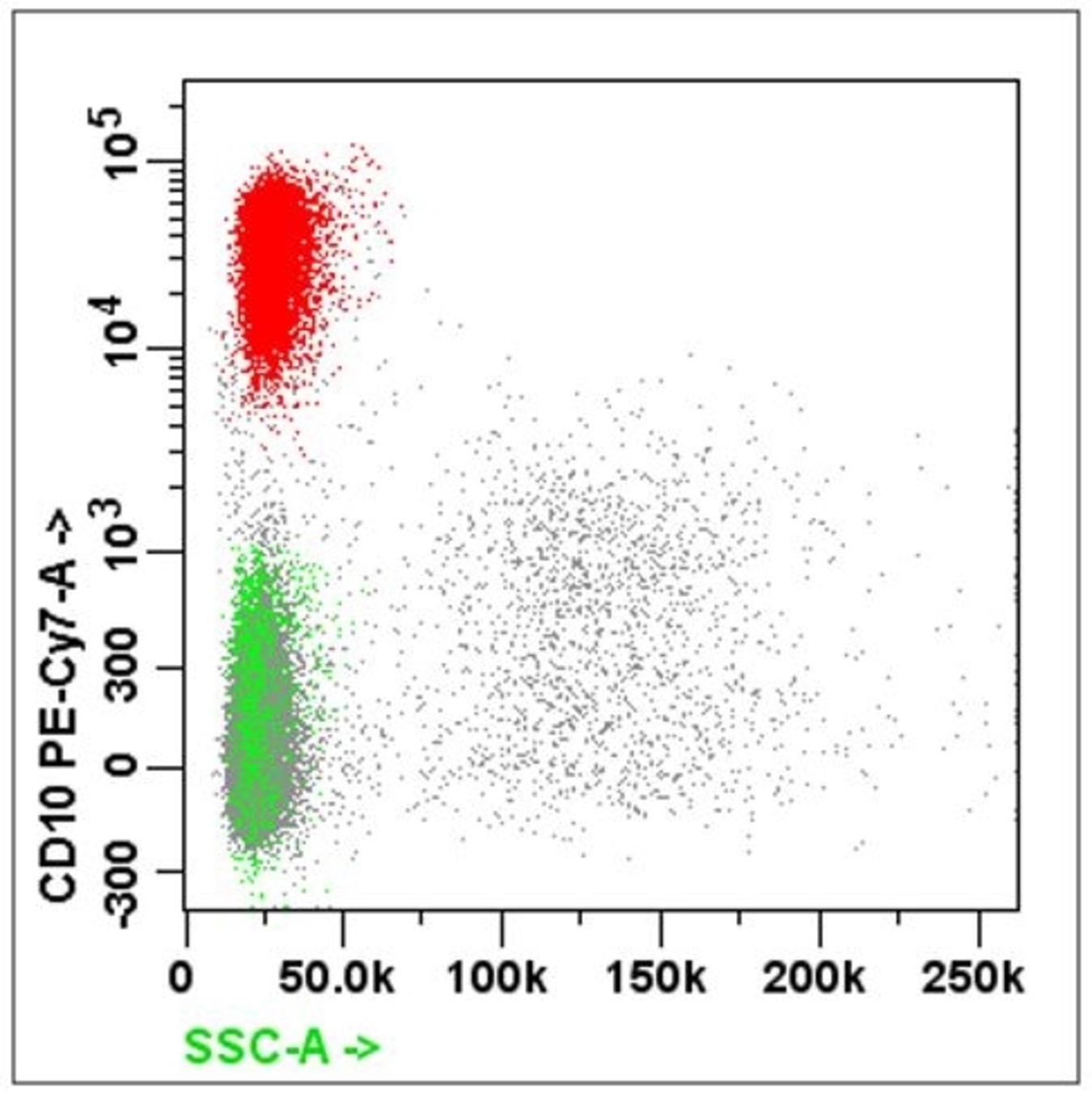
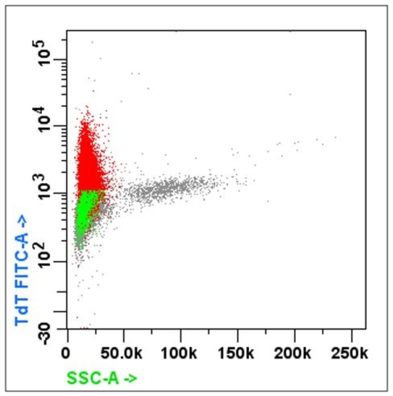
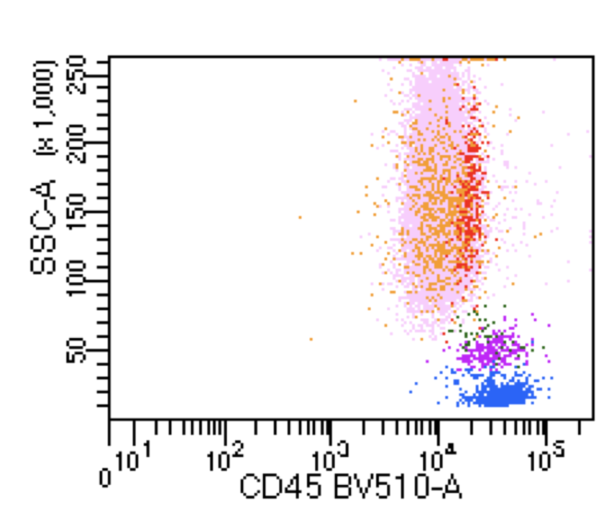
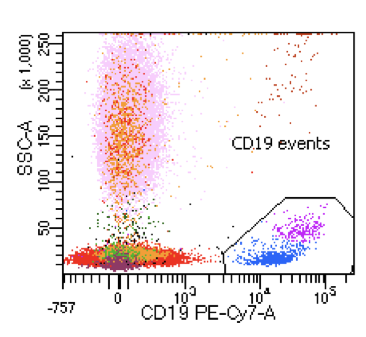
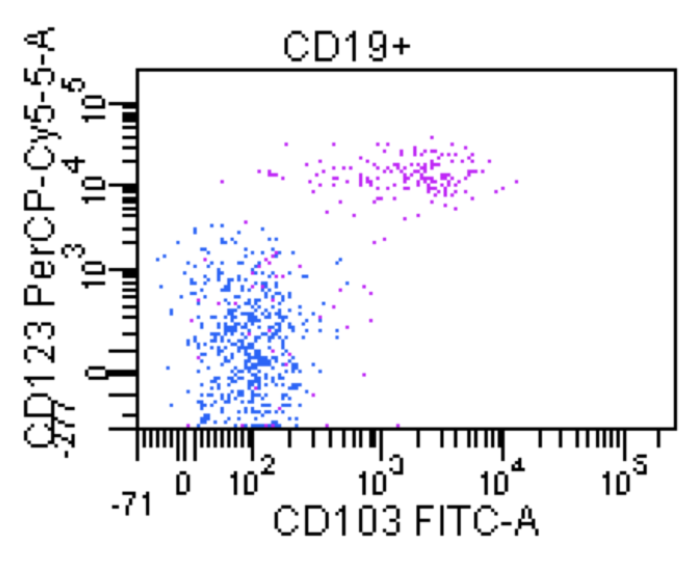
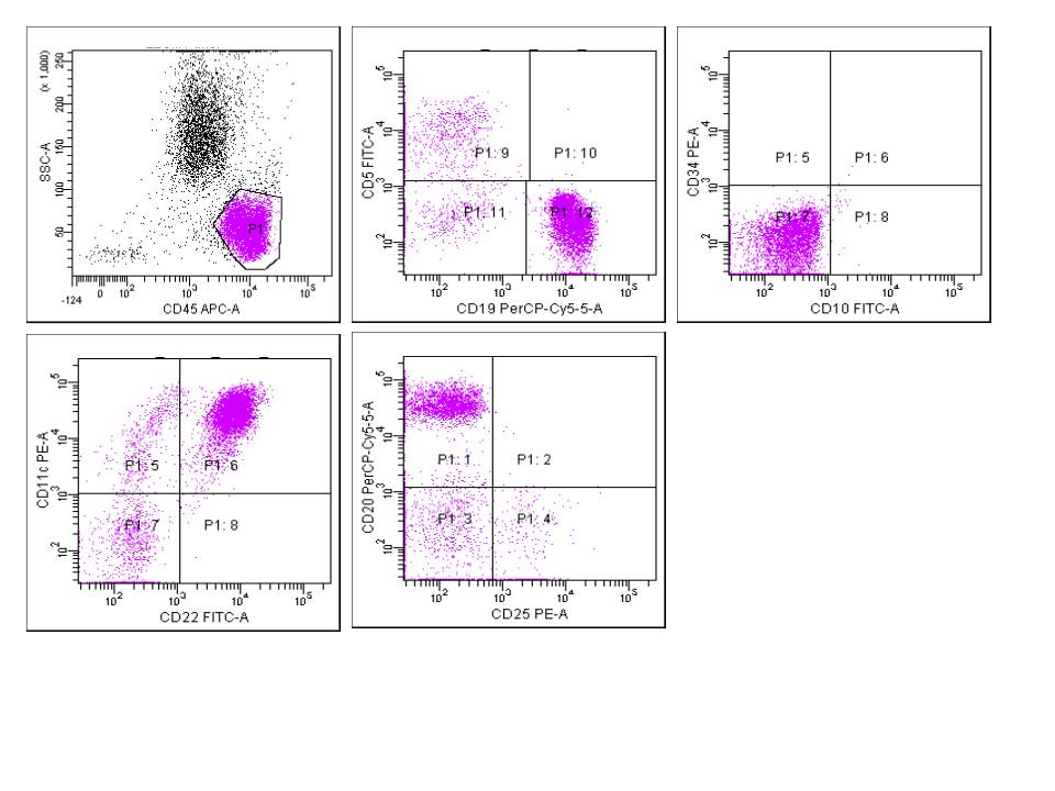
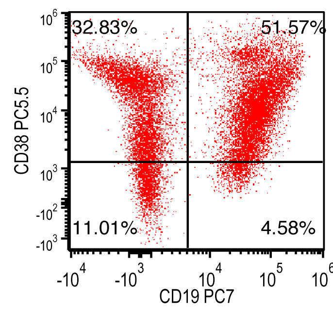
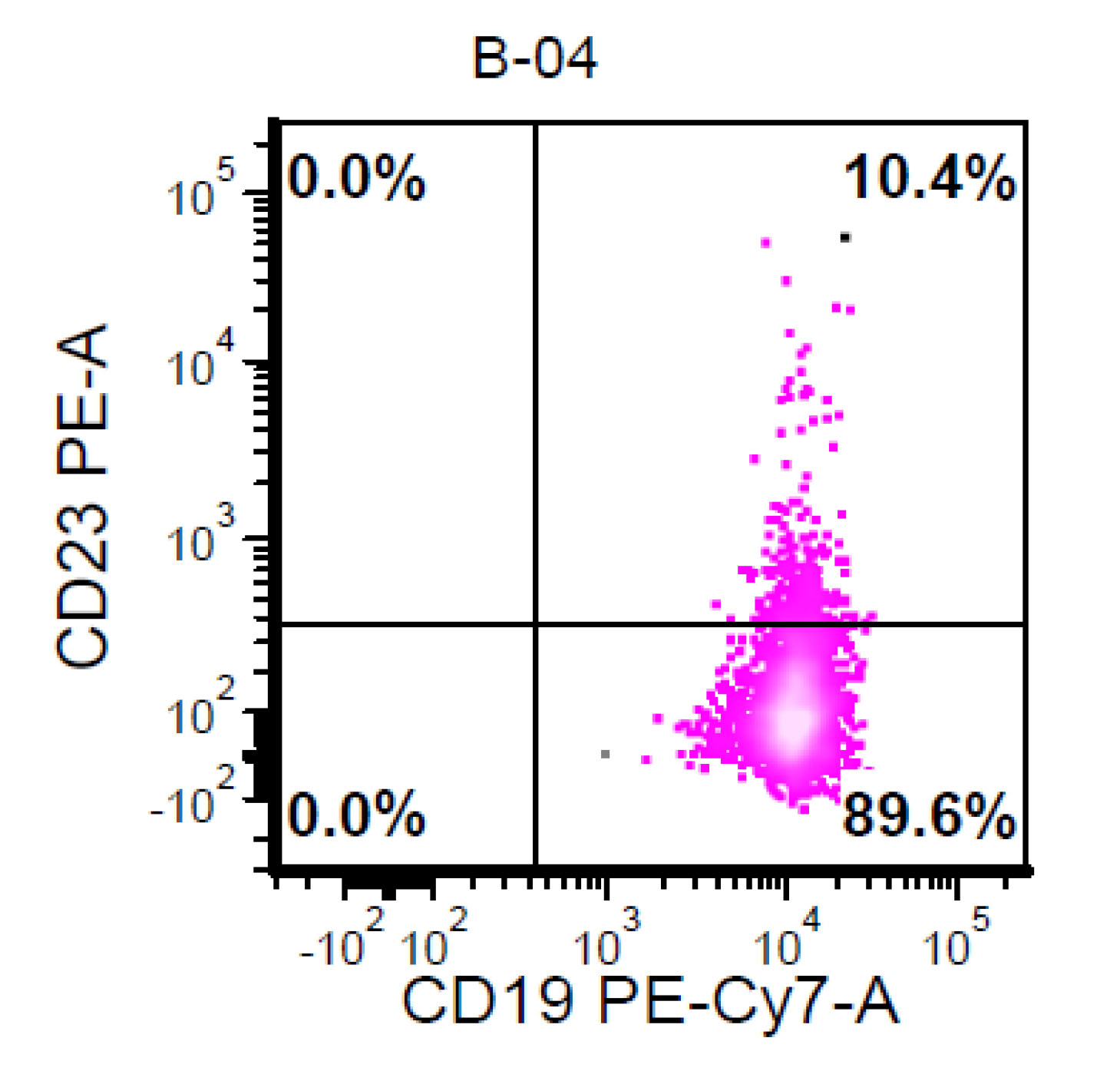
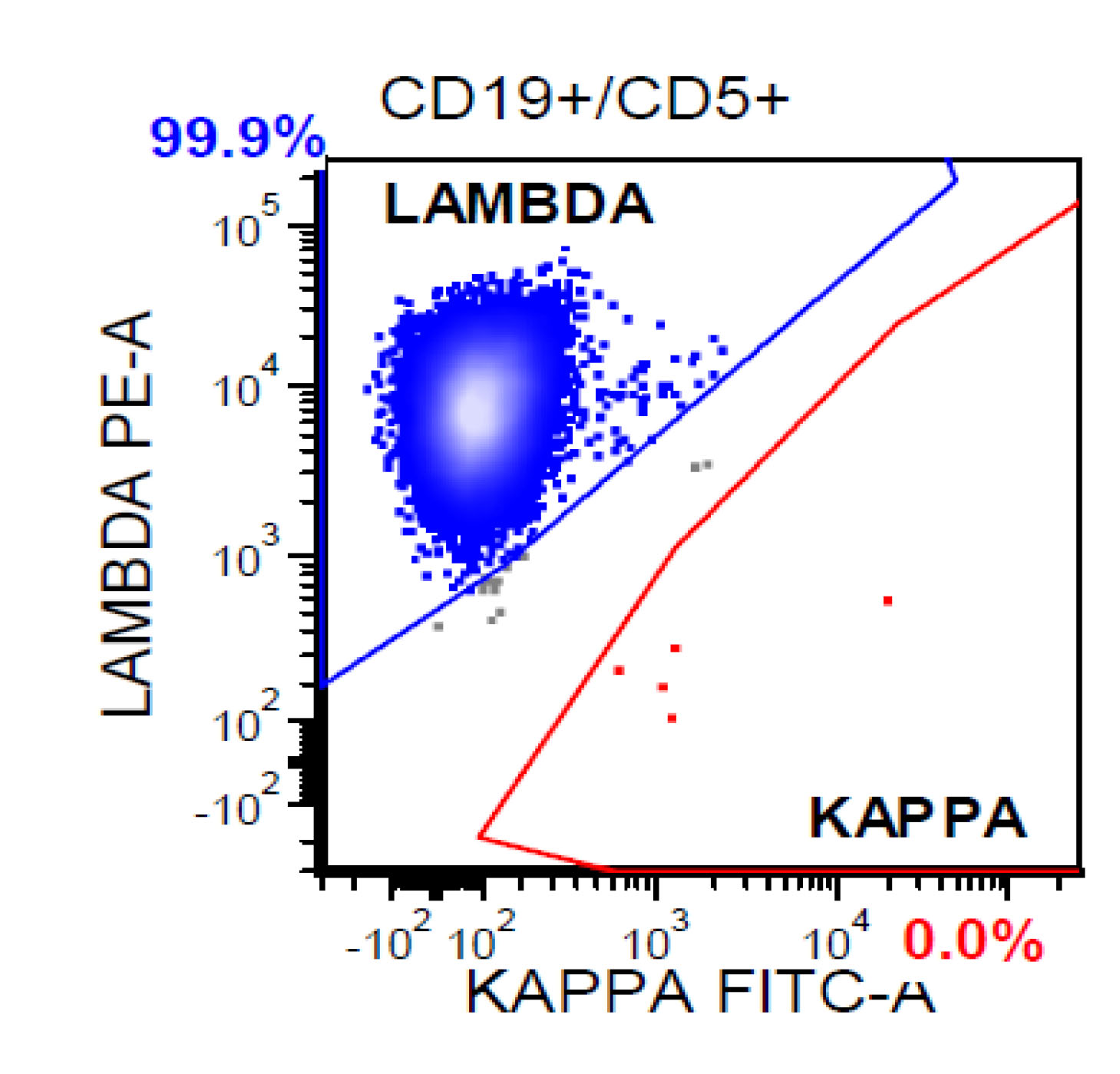
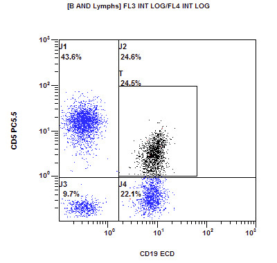
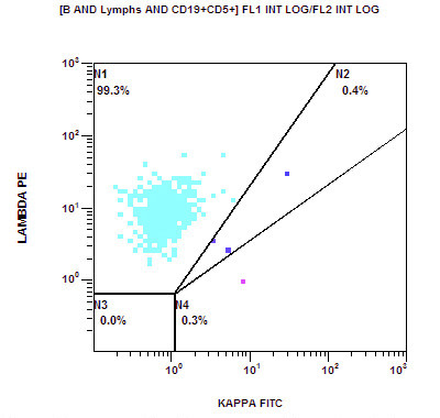
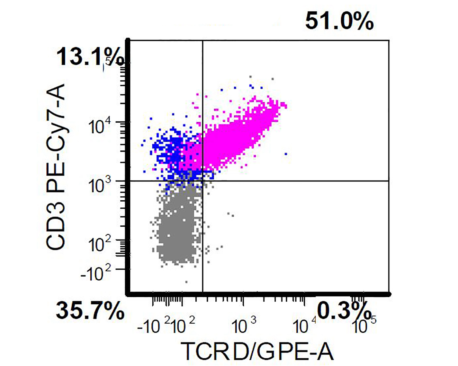
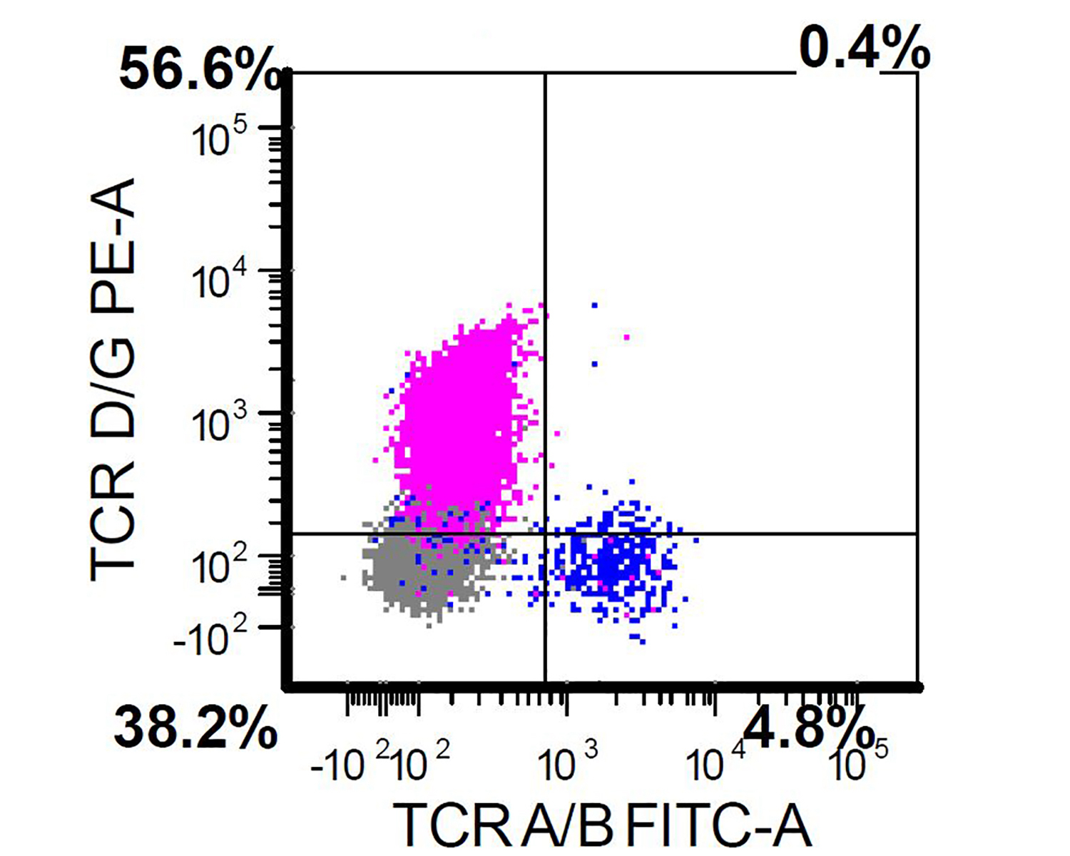
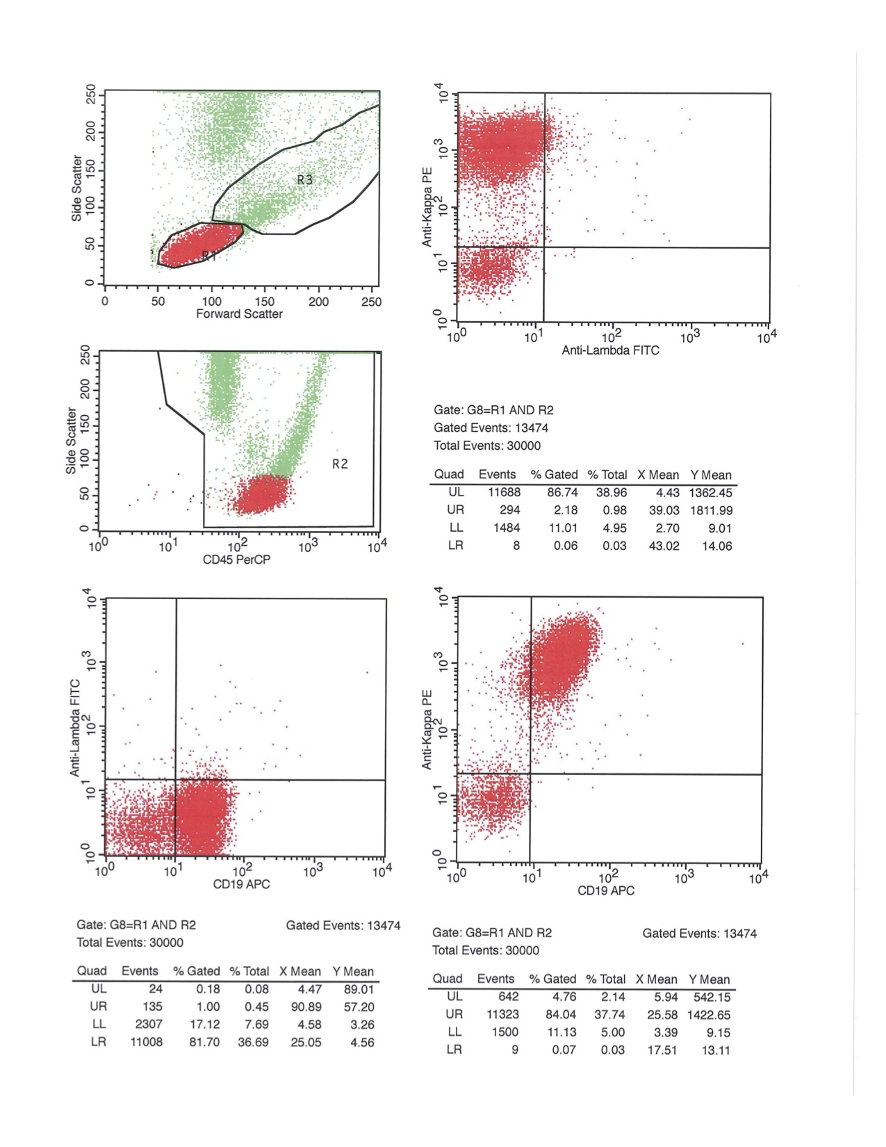
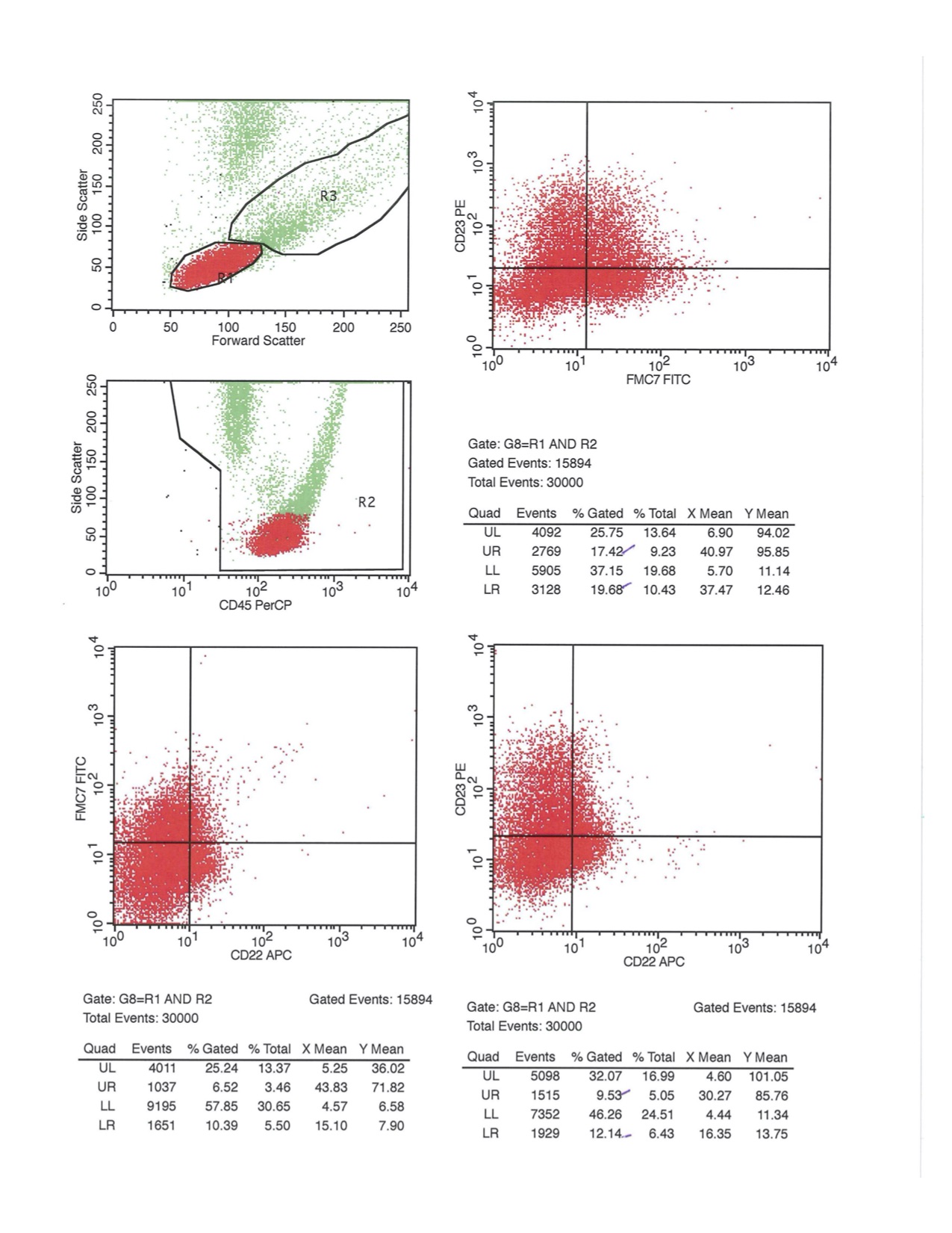
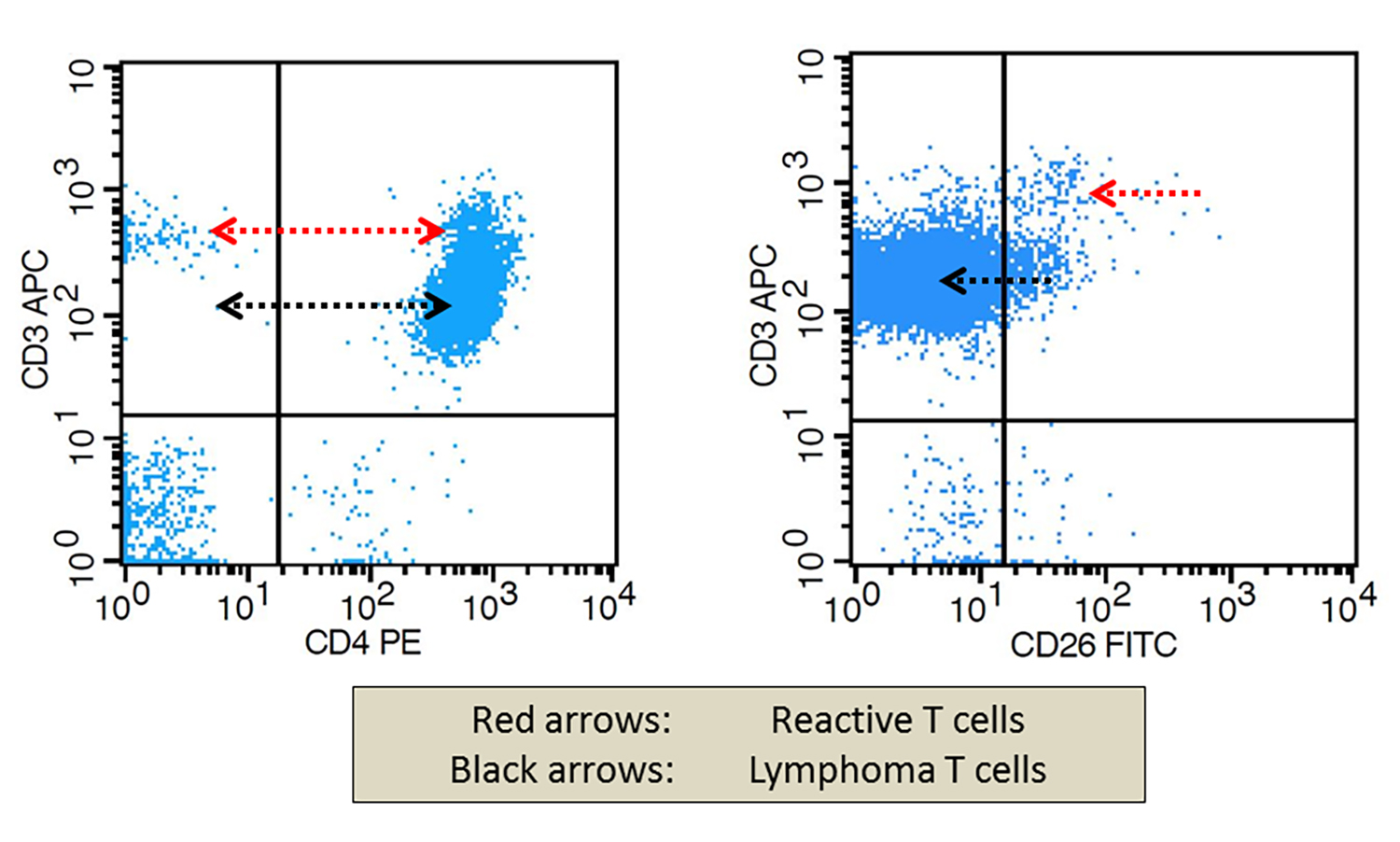
Contributed by Siba El Hussein, M.D. and Joseph Khoury, M.D.
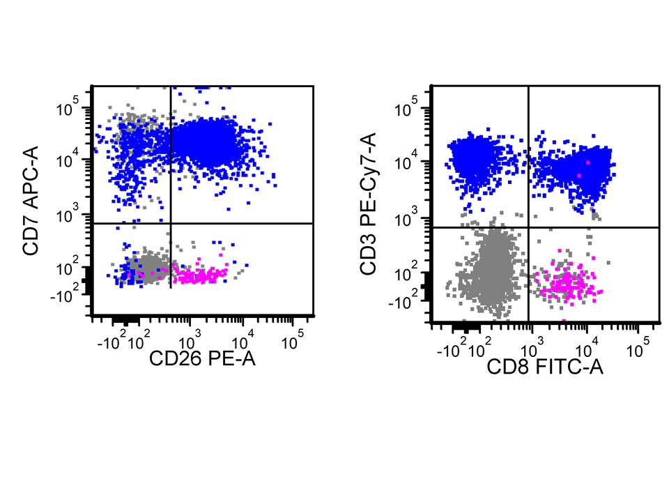
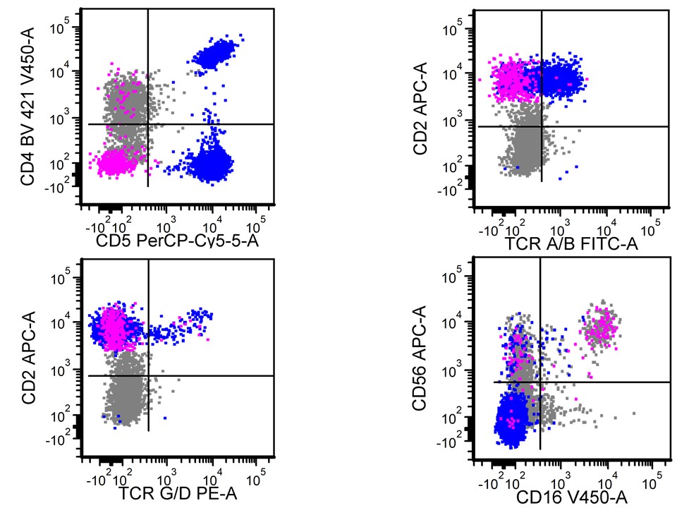
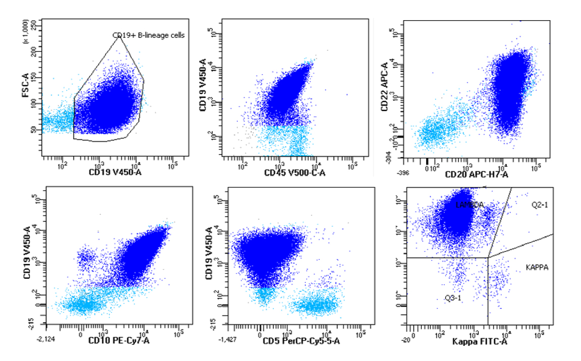
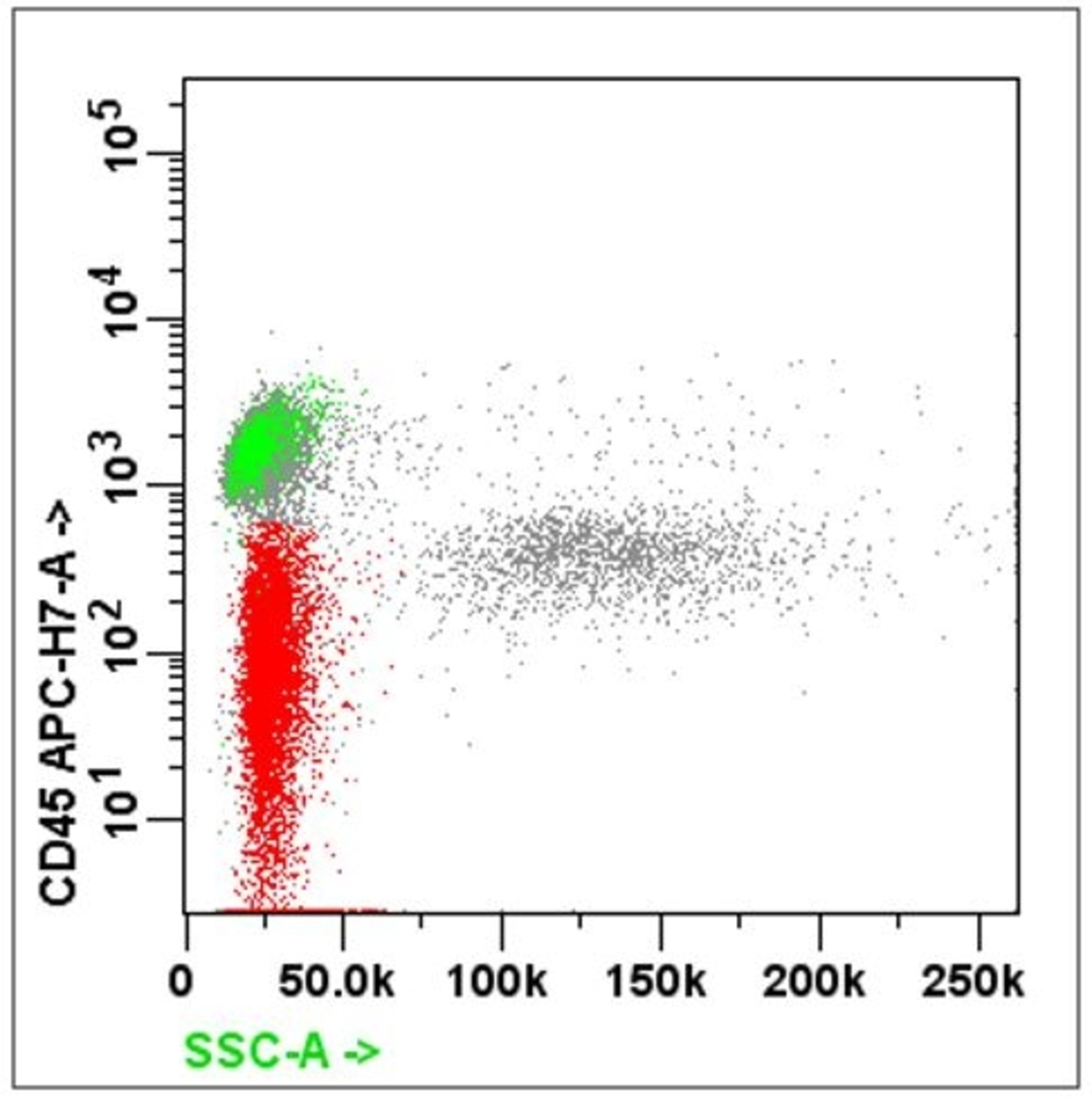
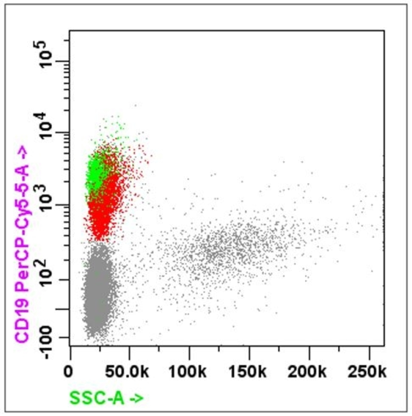
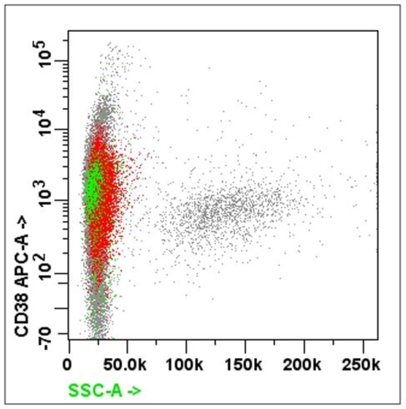
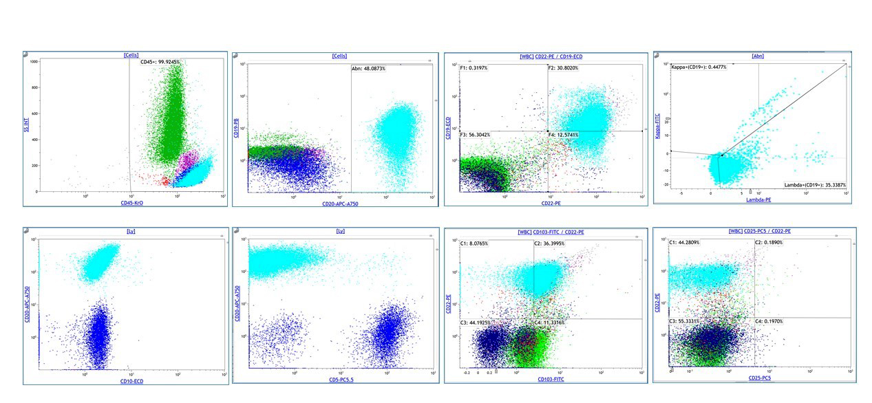
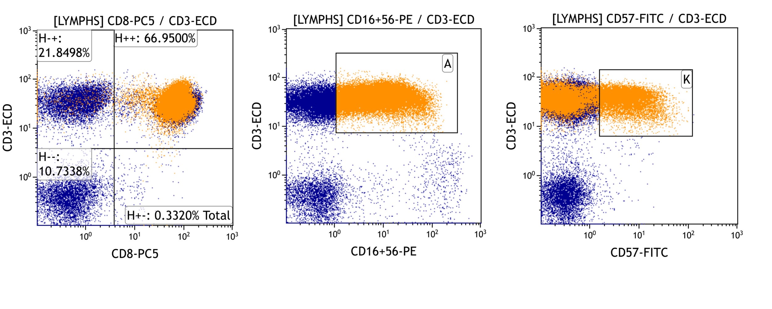
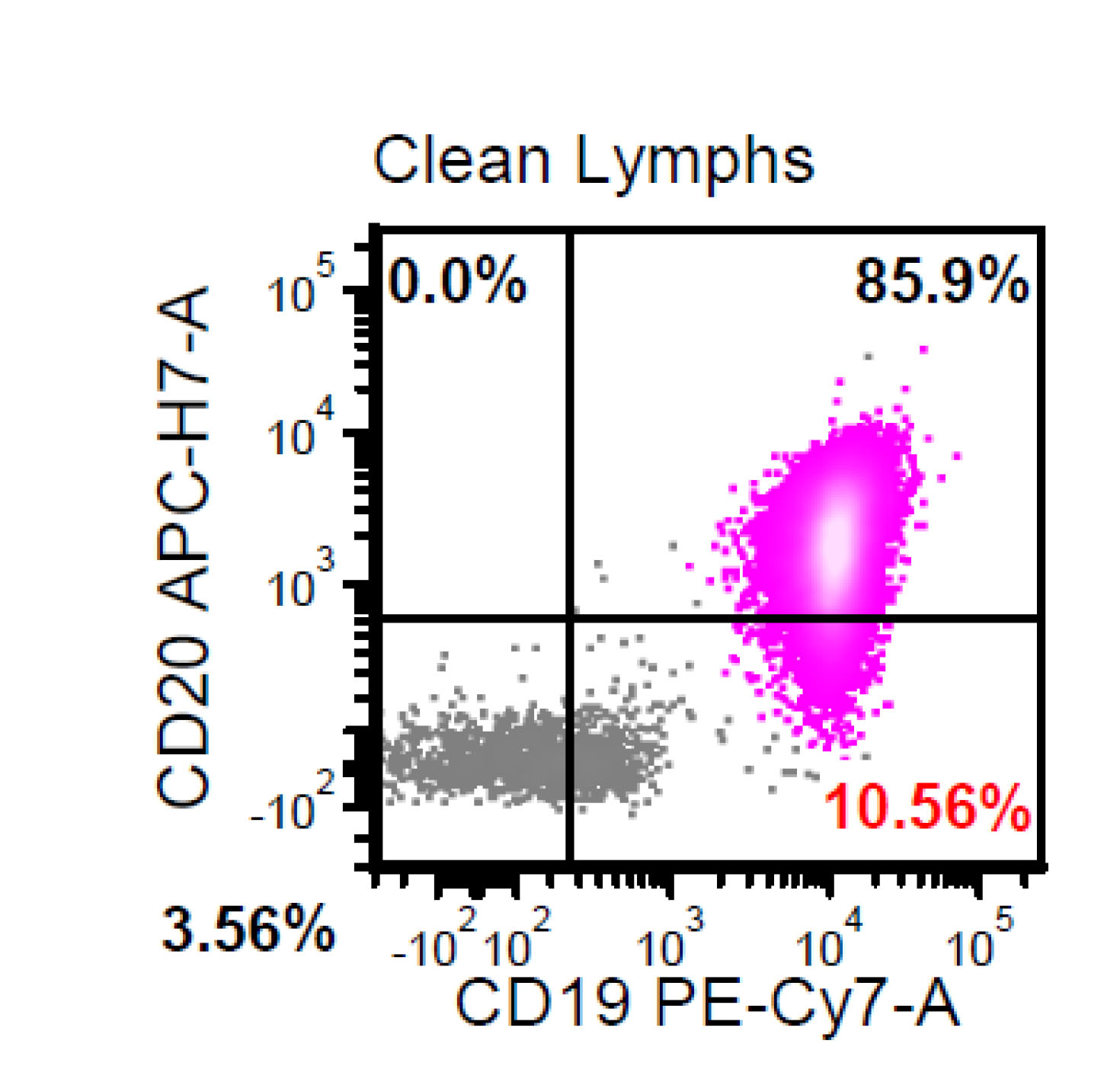
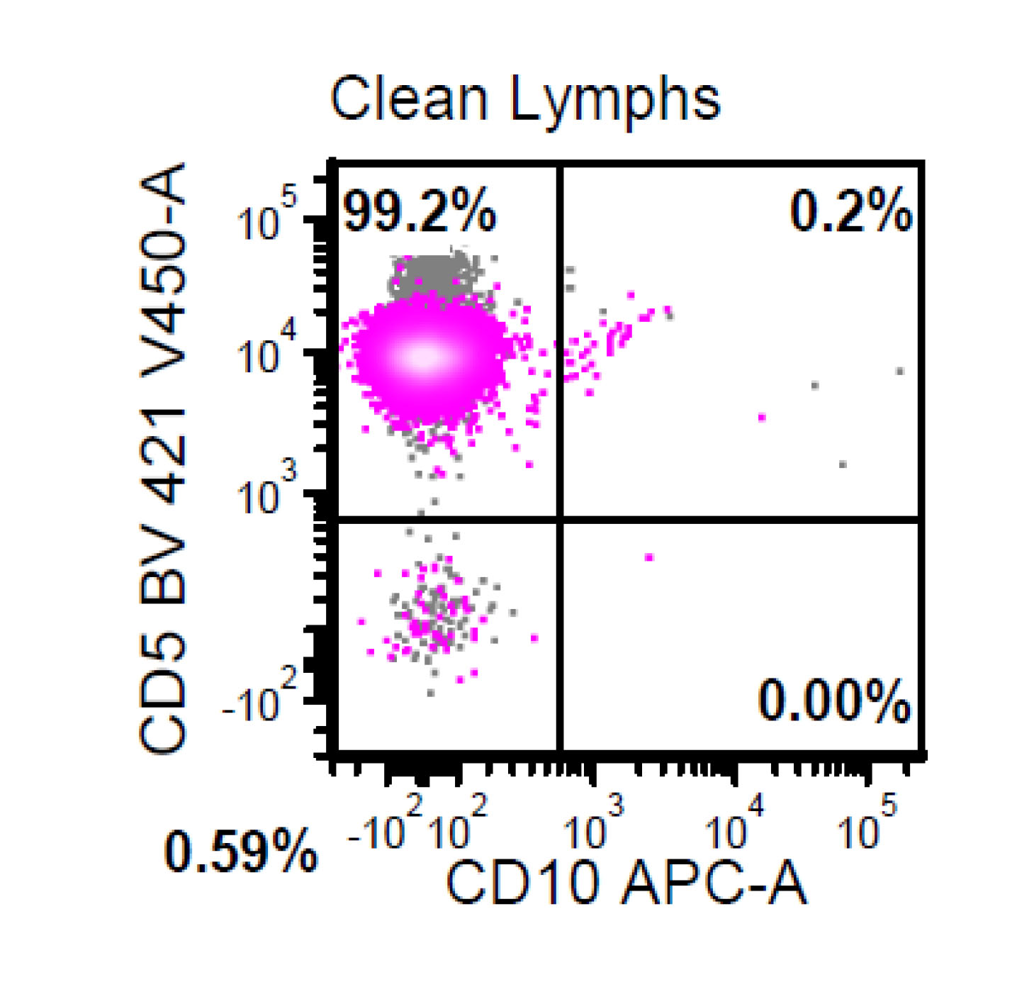
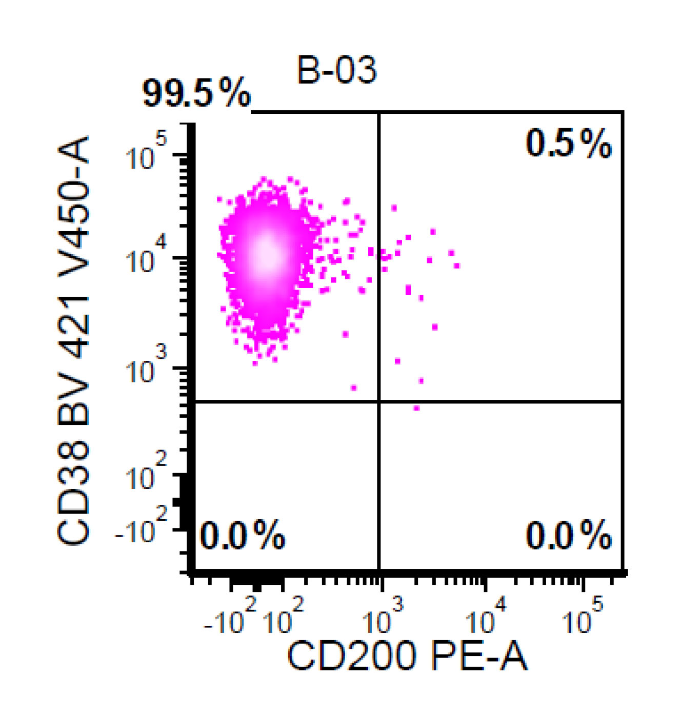
Flow cytometry characteristic
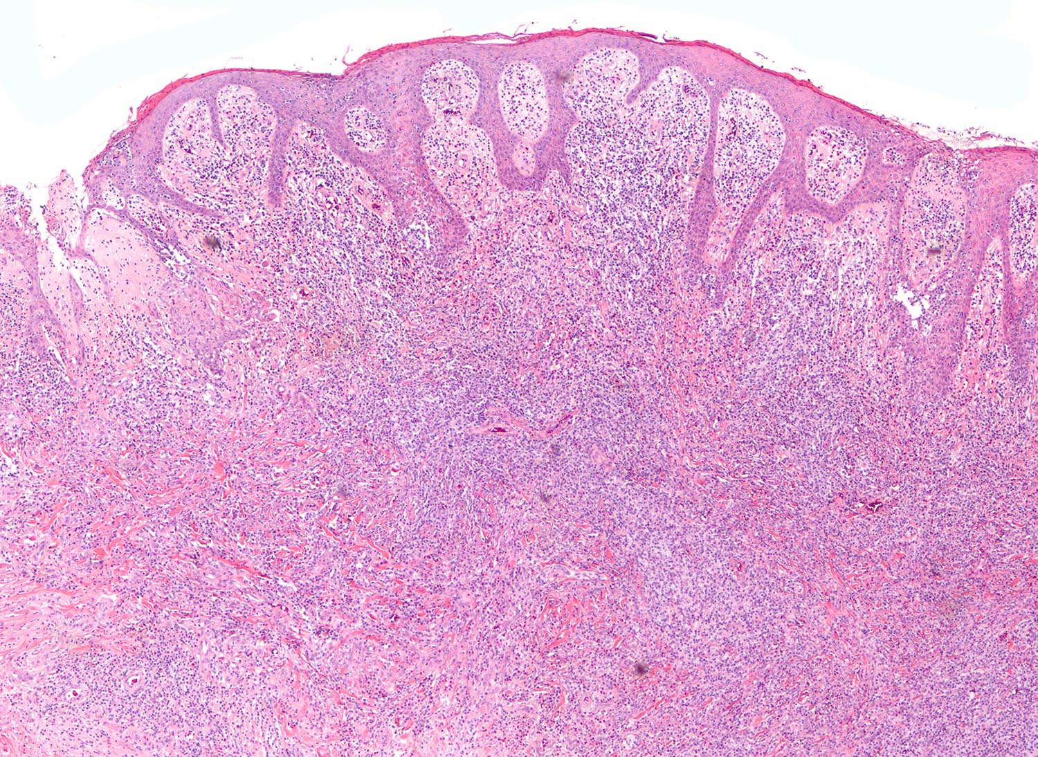
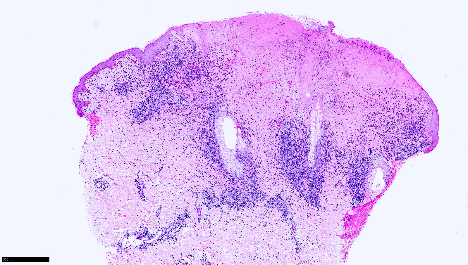
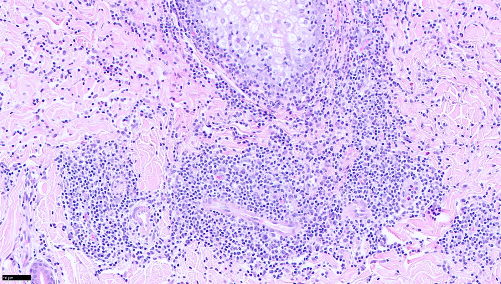
Contributed by Jayalakshmi Balakrishna, M.D. and Elaine S. Jaffe, M.D.
Diffuse infiltrate
Invasion of sinus
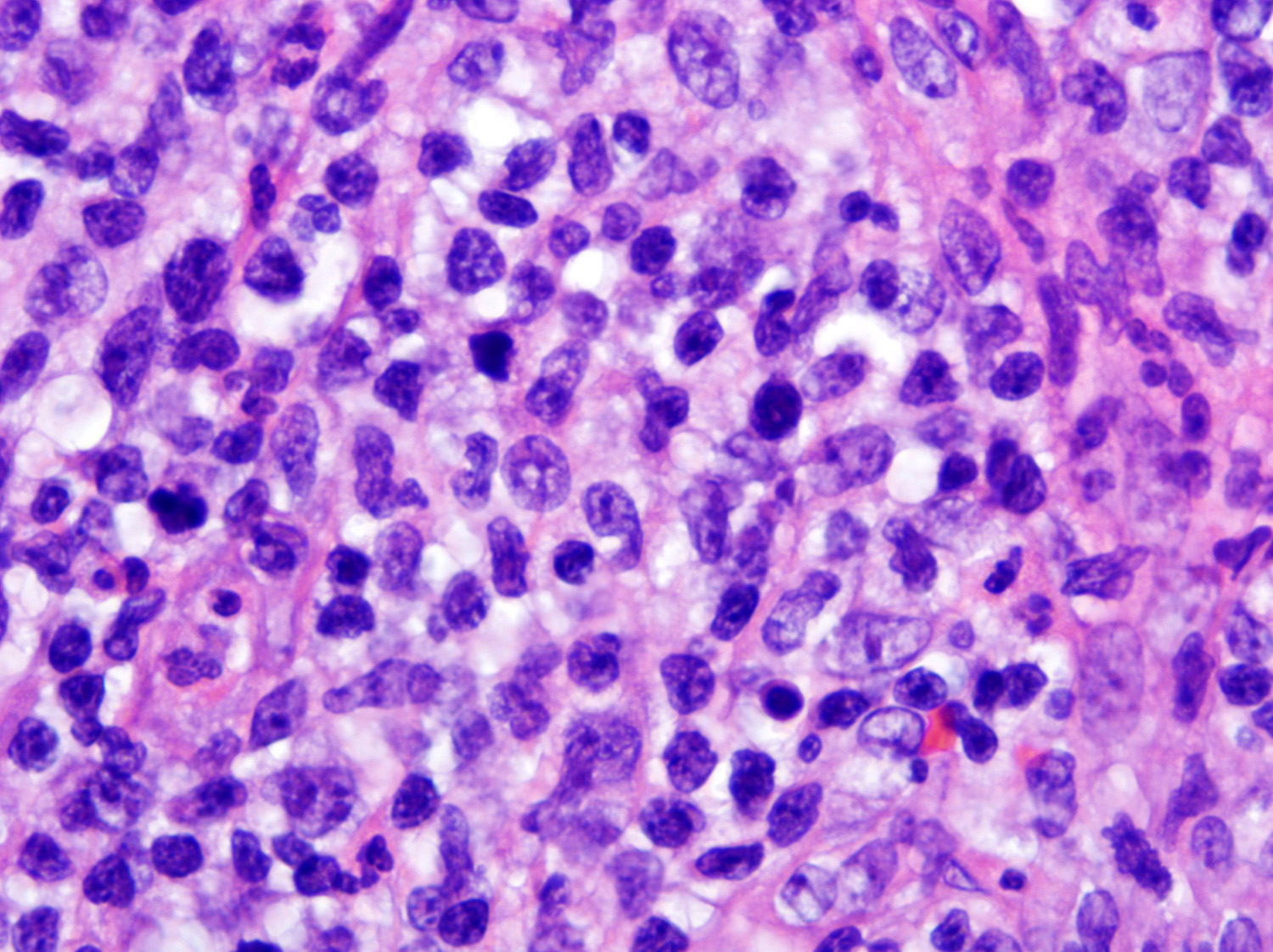
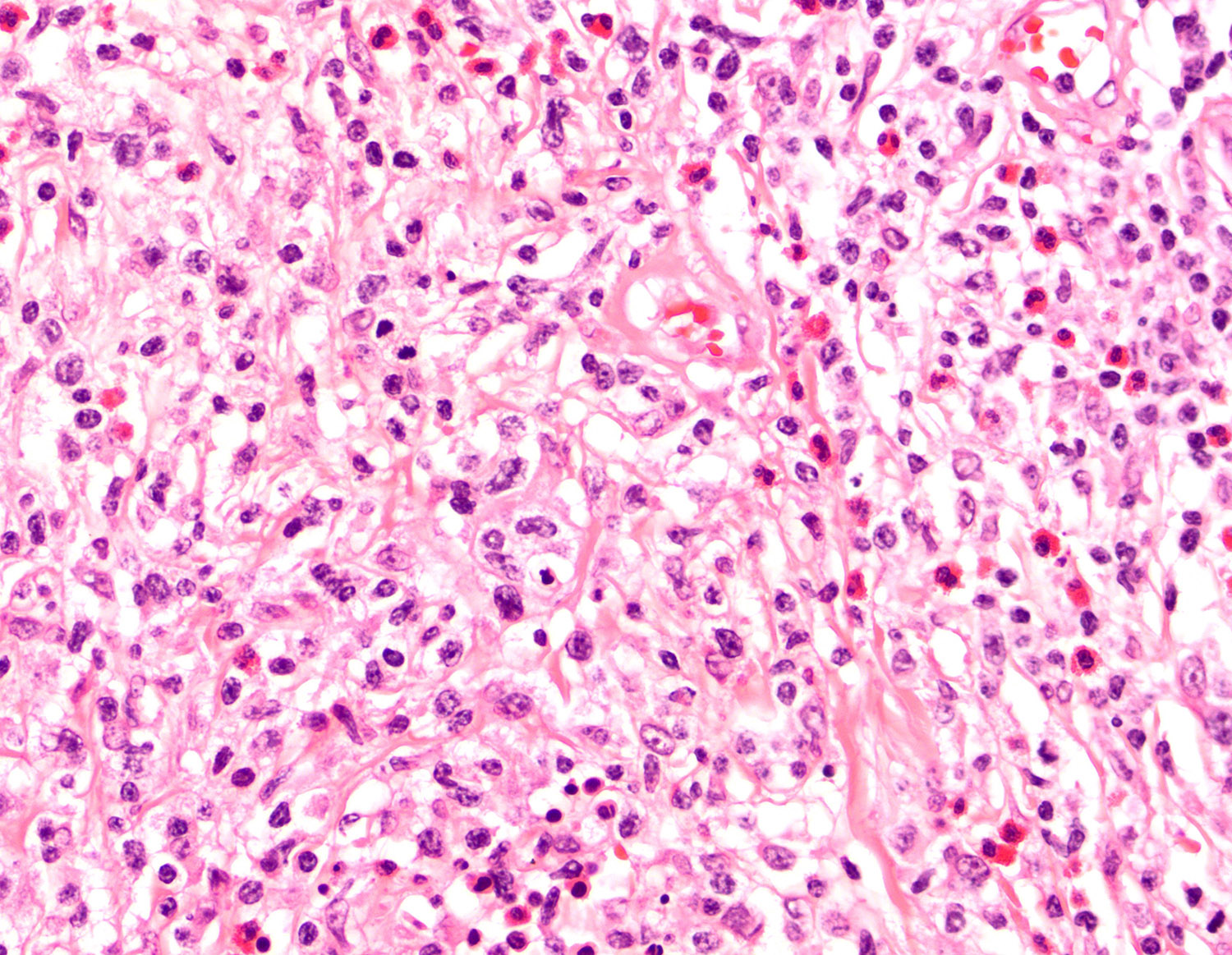
Large atypical cells
Mitotic figures and apoptotic bodies
Large atypical cells
Large atypical cells
Hallmark cells
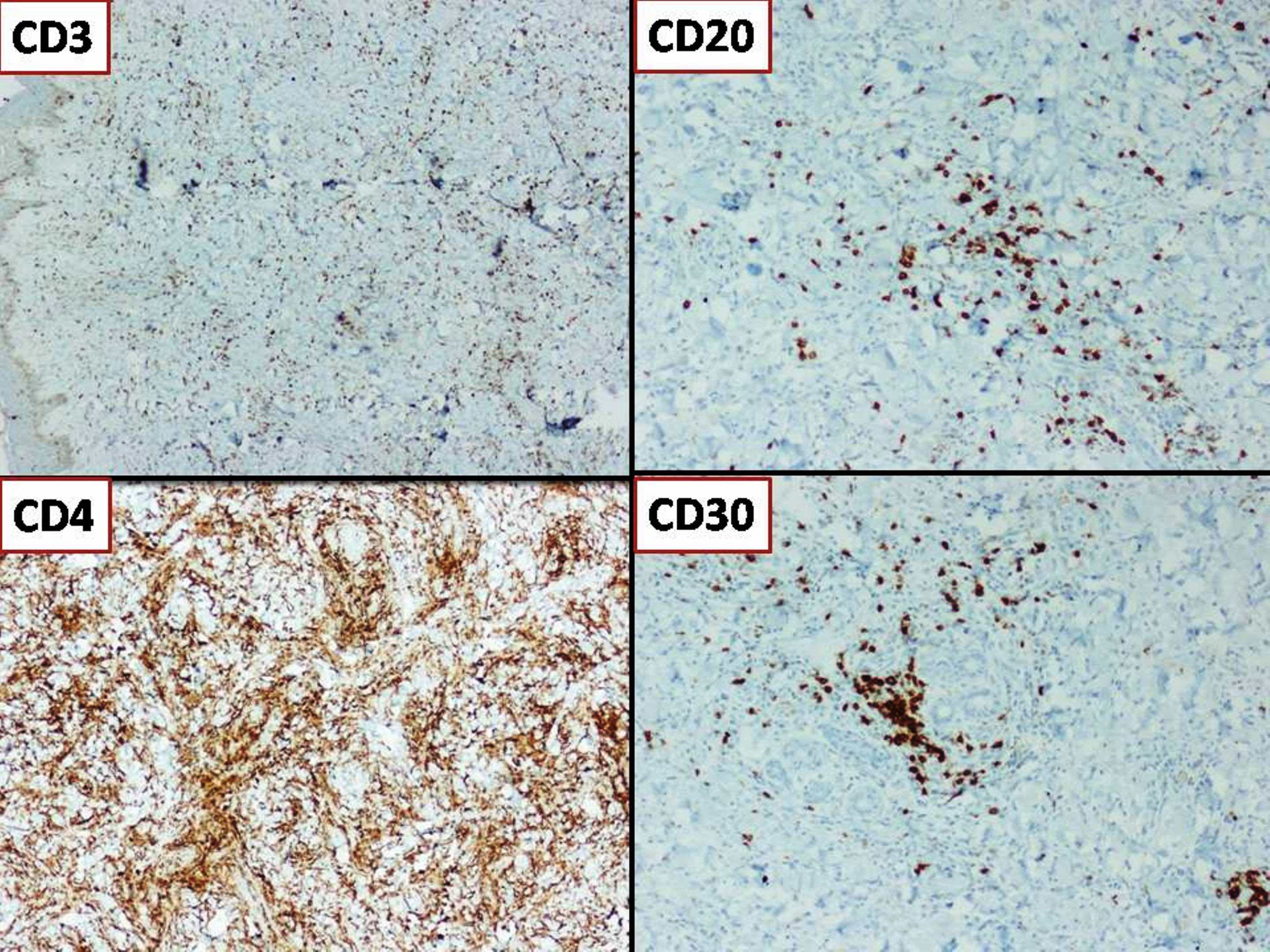
IHC stain, CD3
IHC stain, CD4
IHC stain, CD3
IHC stain, CD25
IHC stain, Ki67

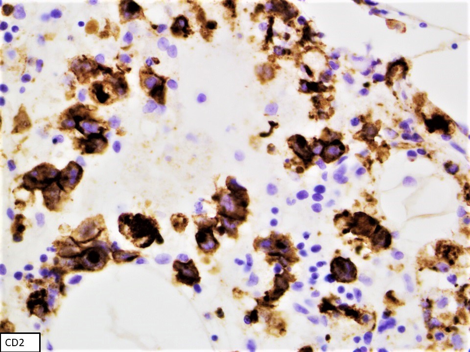
IHC stain, CD2
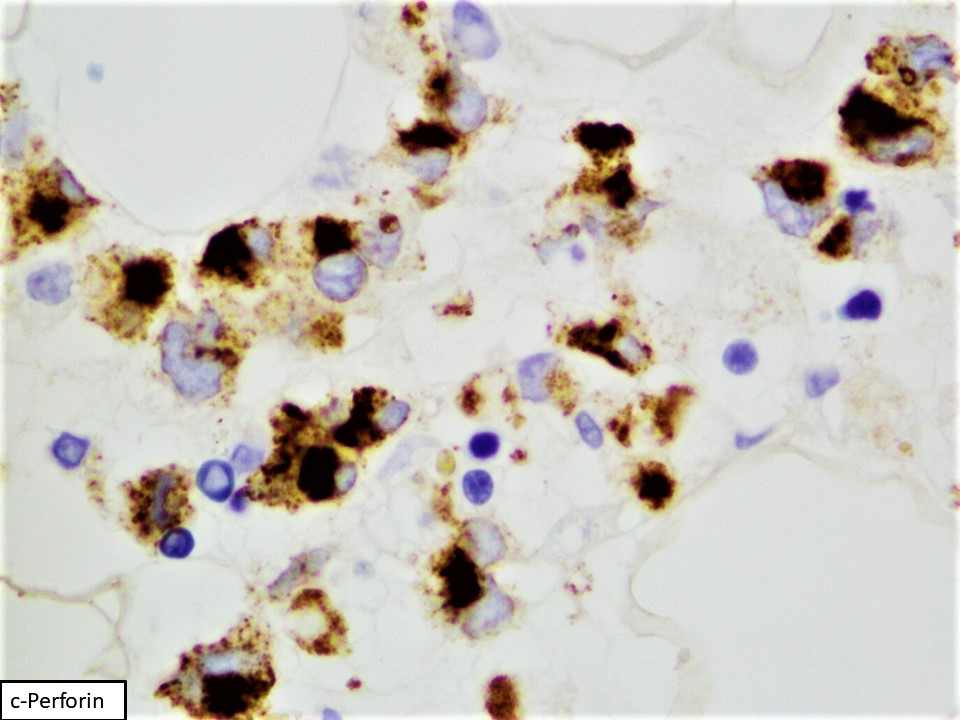
IHC stain, perforin
IHC stain, CD30
IHC stain, CD43
IHC stain, CD45
IHC stain, EMA

IHC stain, granzyme
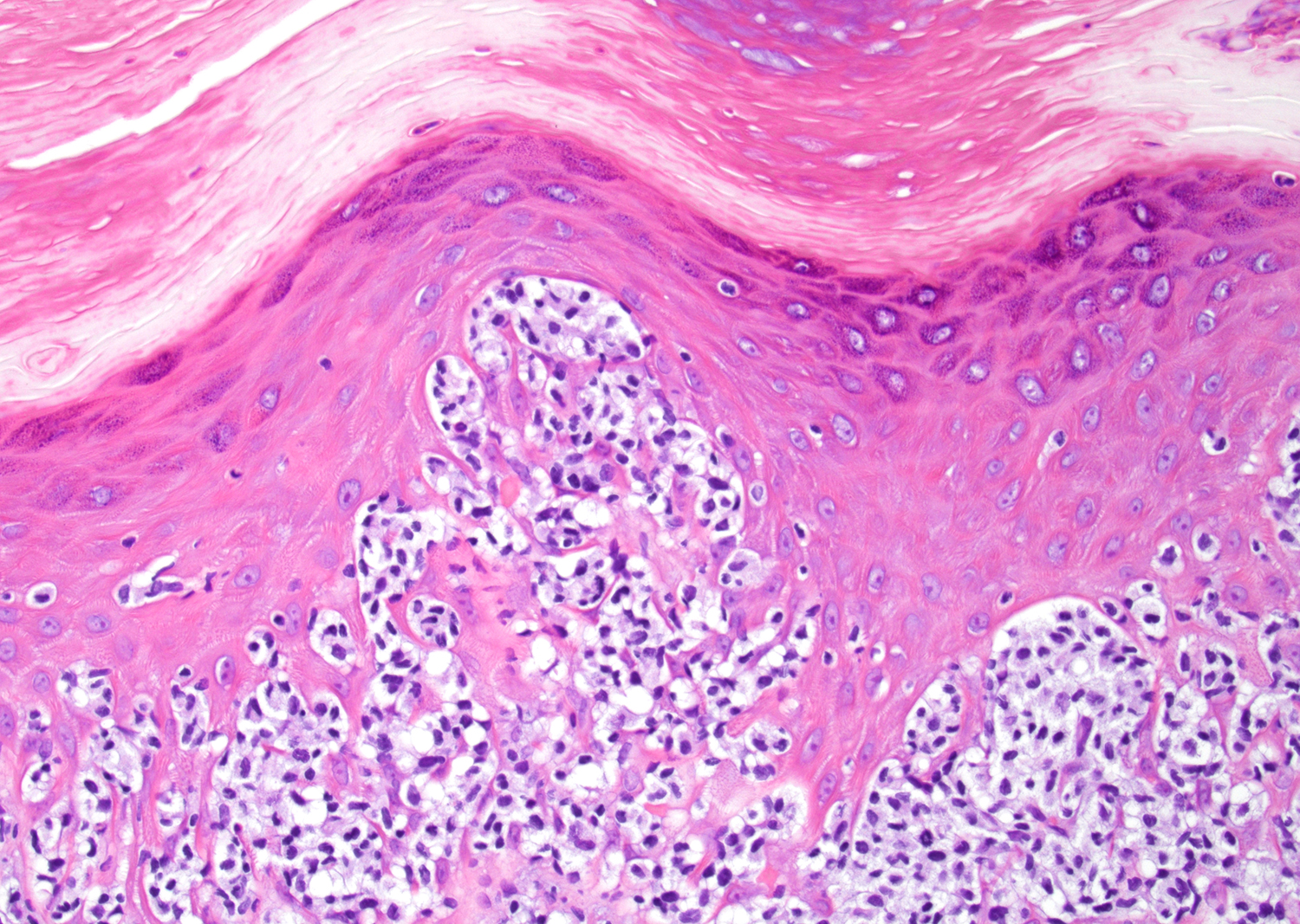
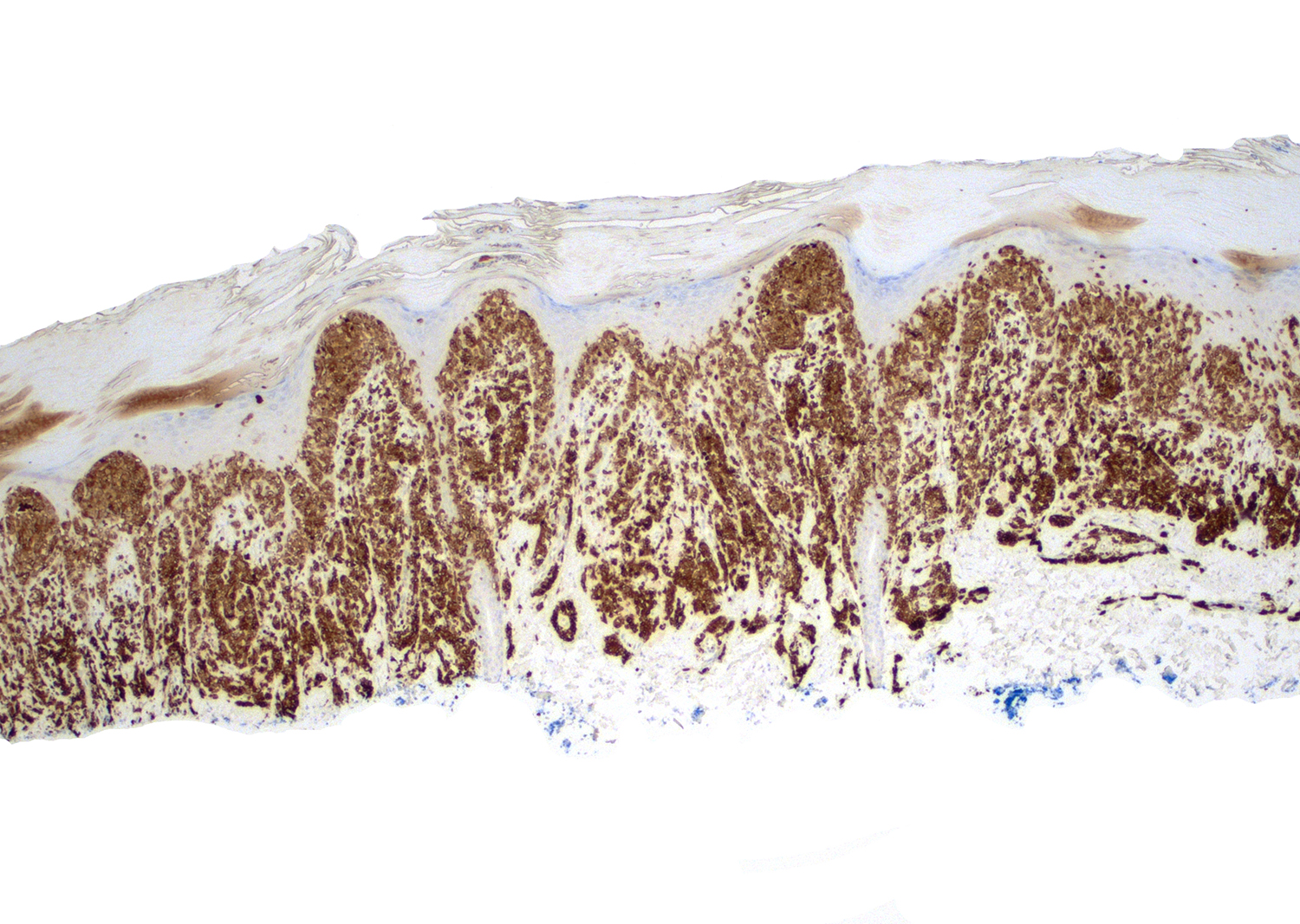
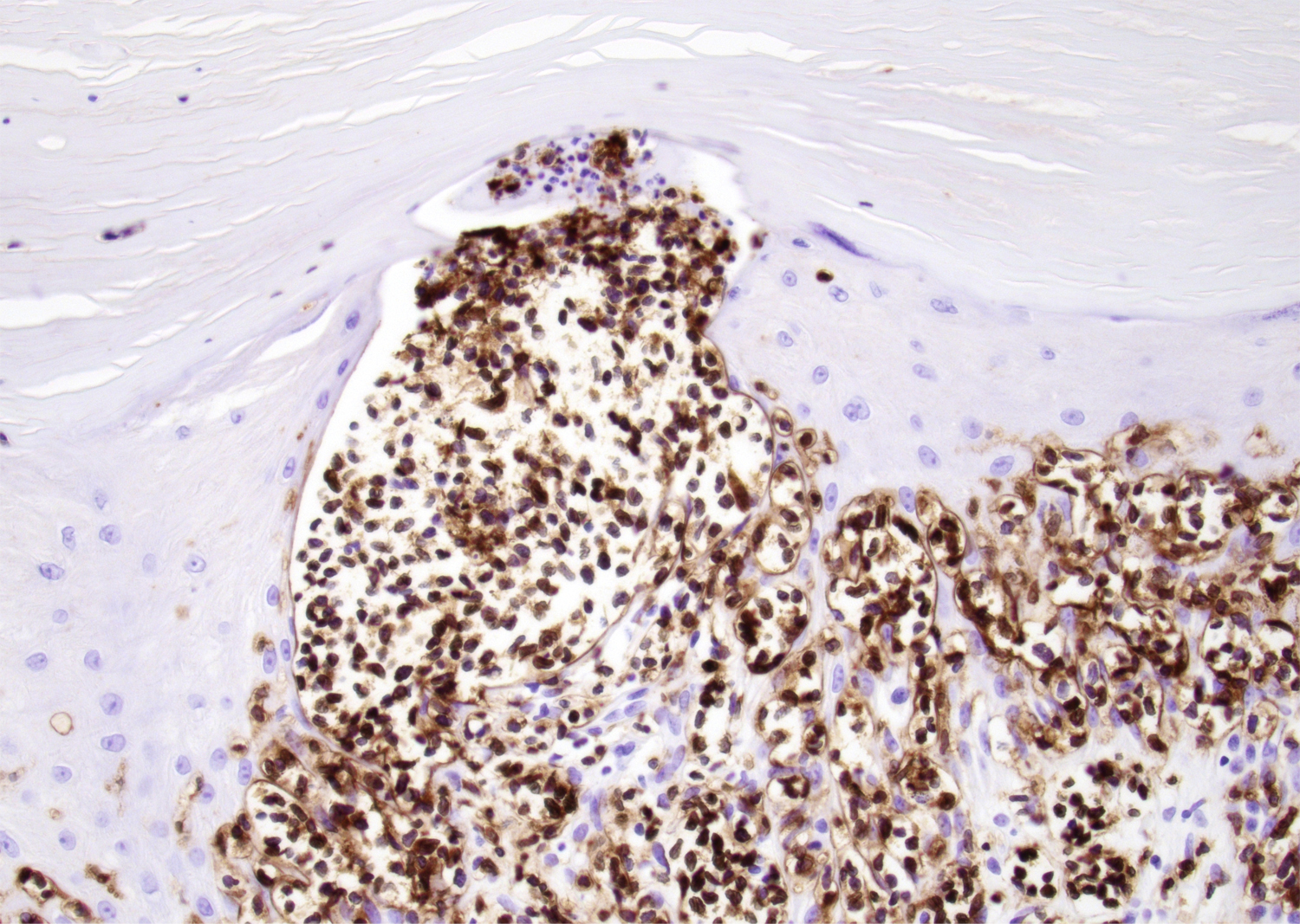
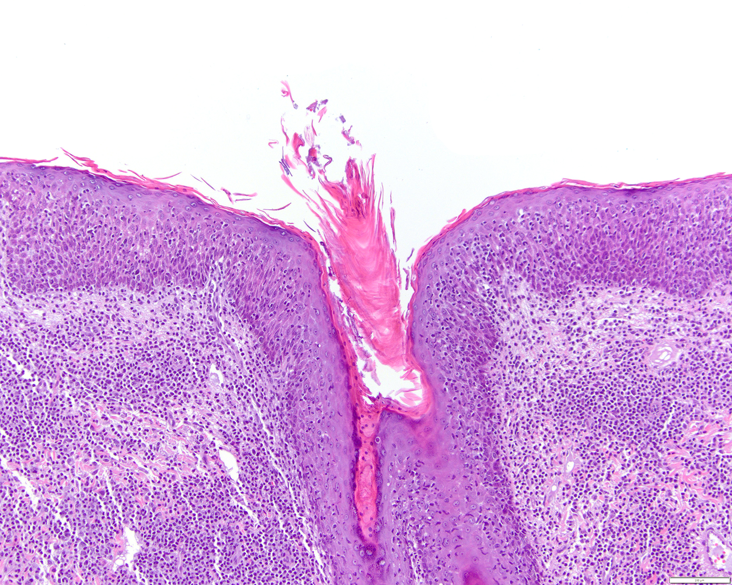
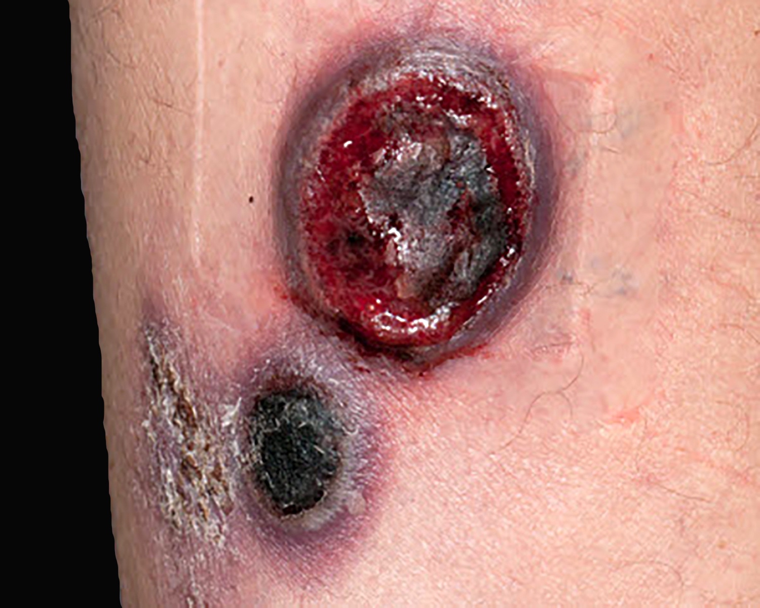
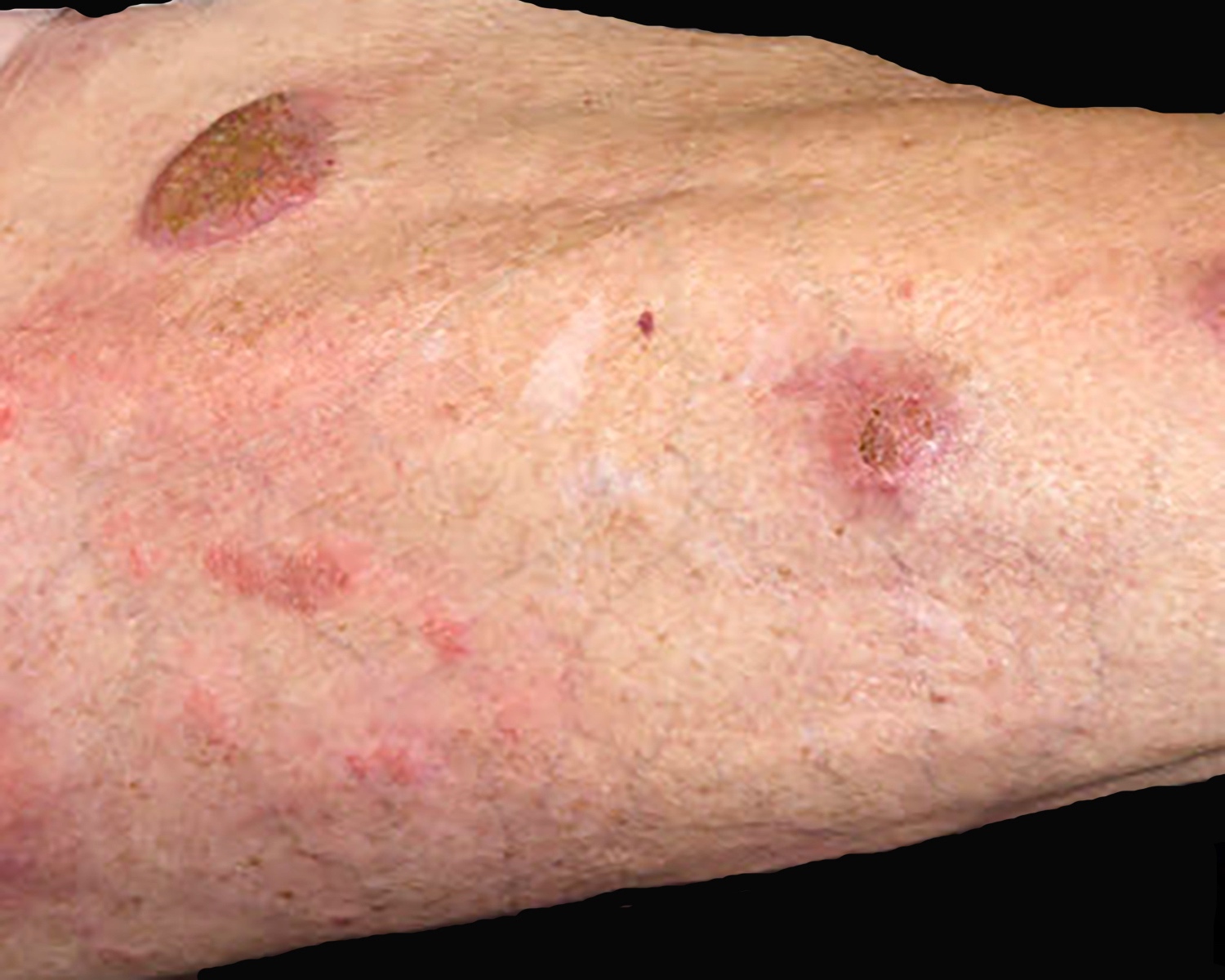
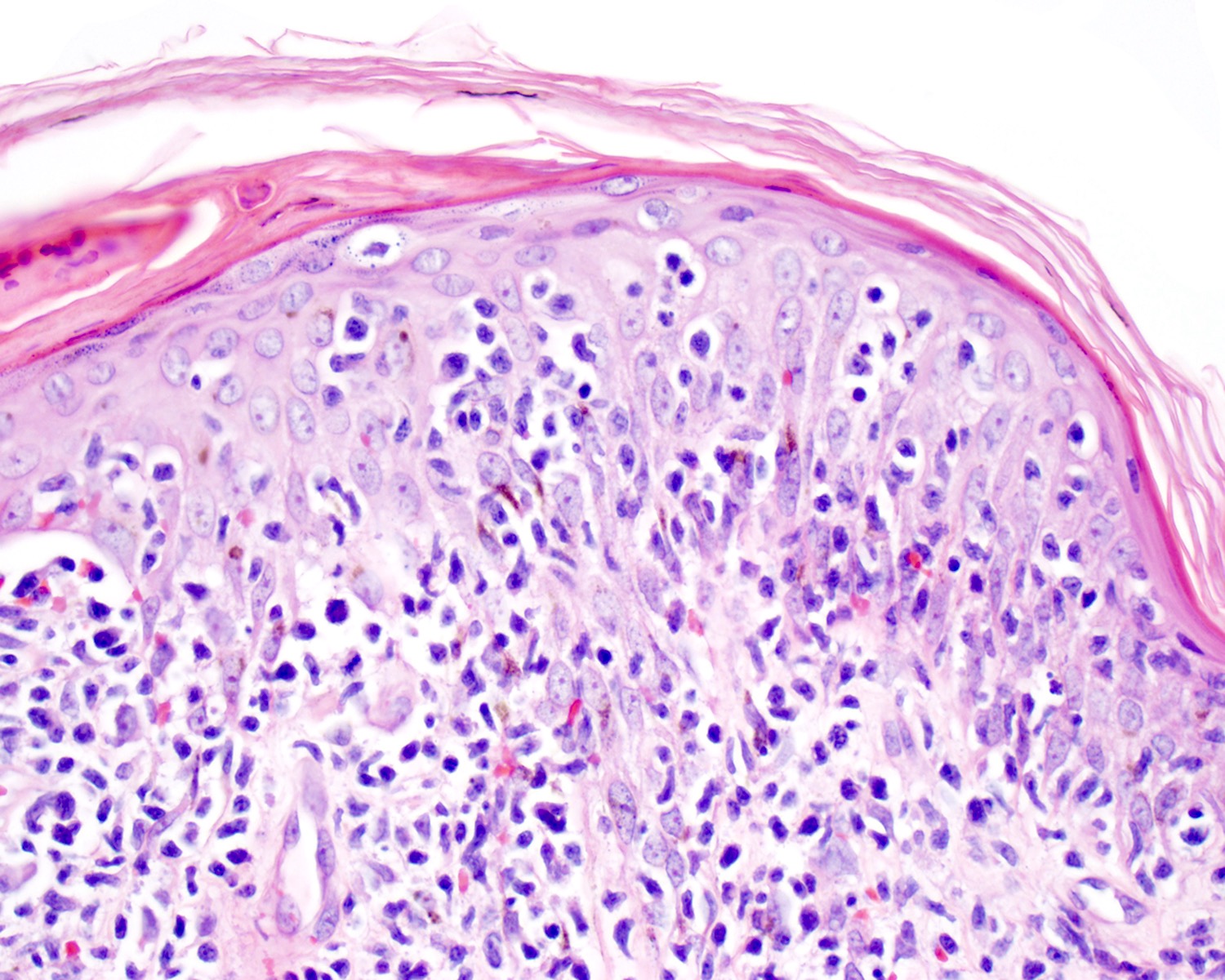
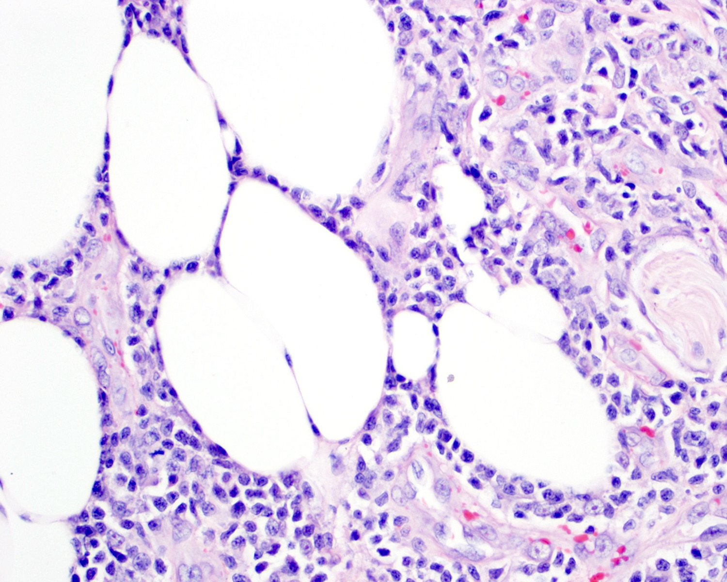
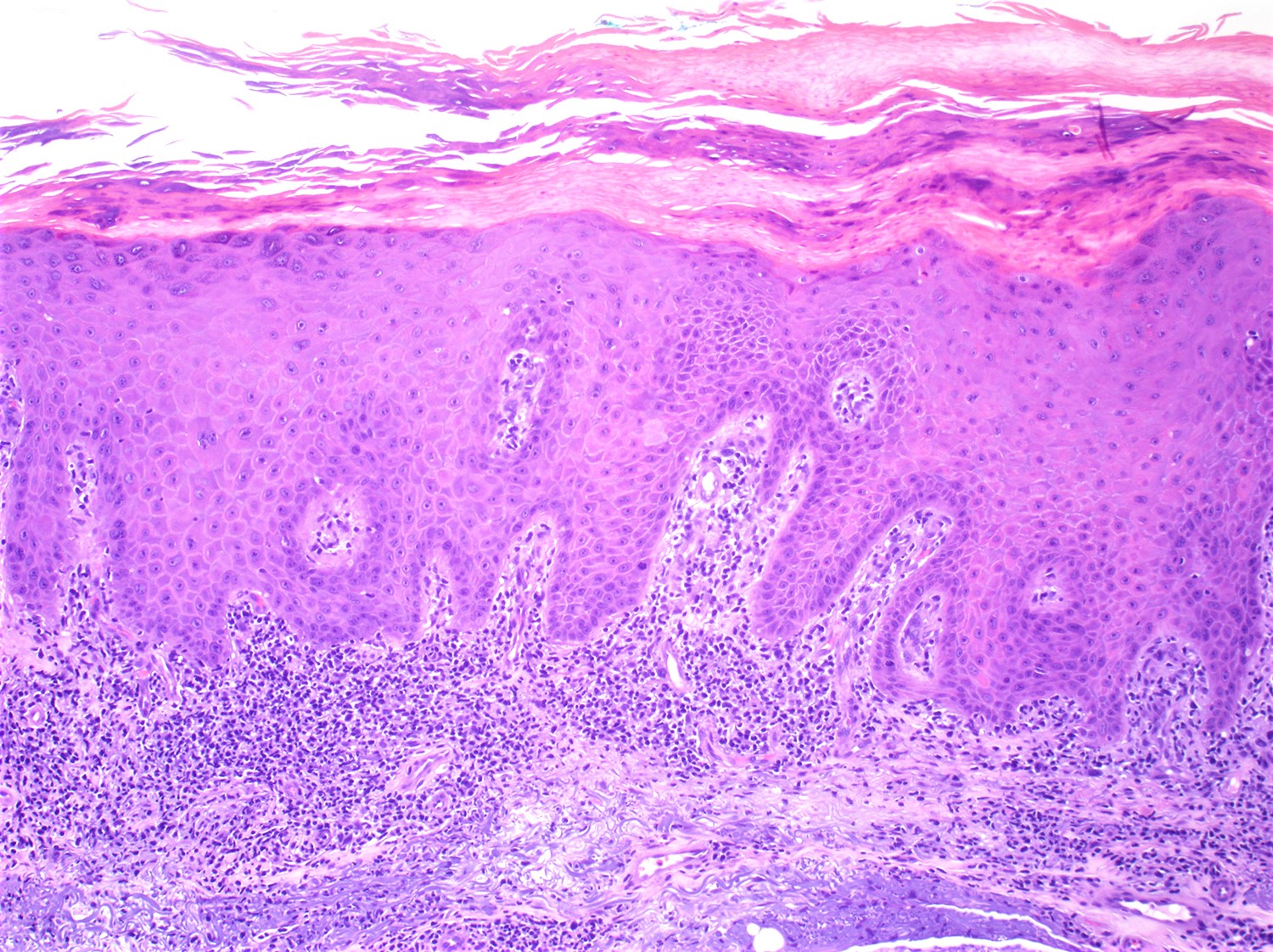
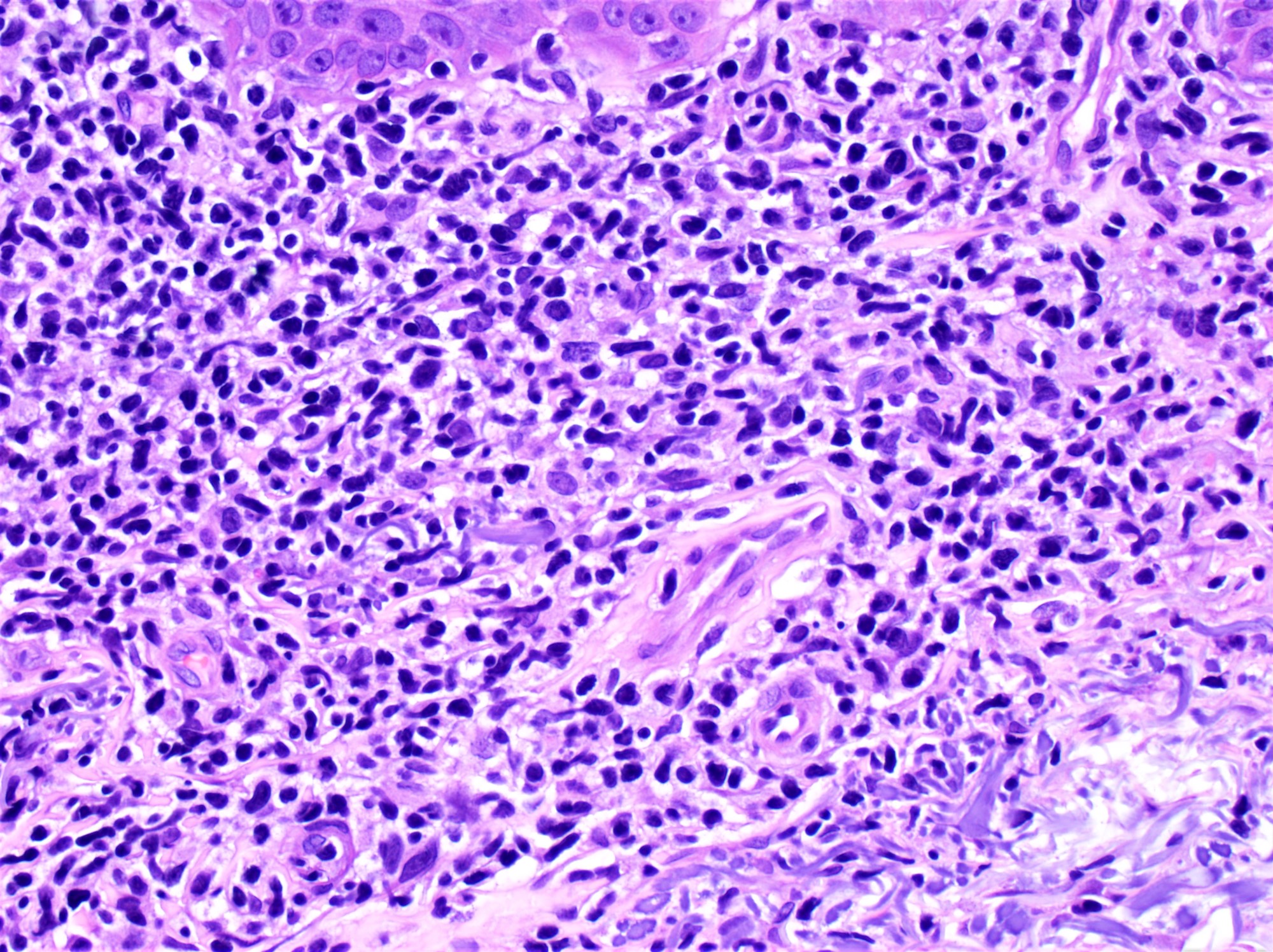
Contributed by Doan Minh Khuy, M.D.
23 year old man with a cervical lymphoid nodule, no B symptoms
CK-
EMA+
ALK-
CD15-
CD20-

CD5+
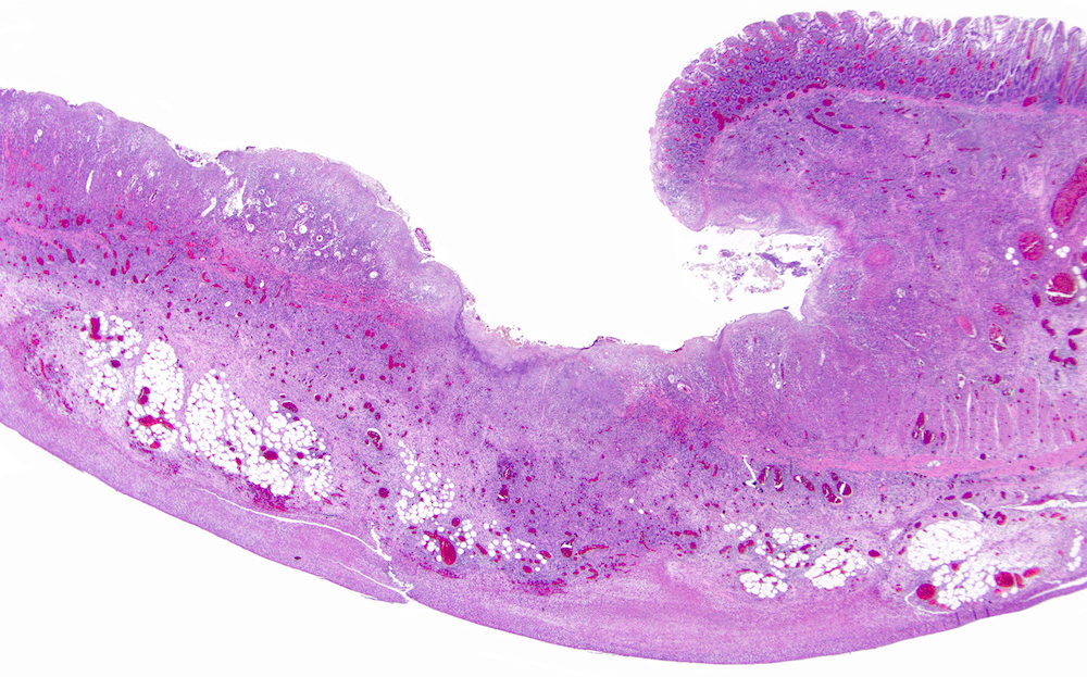
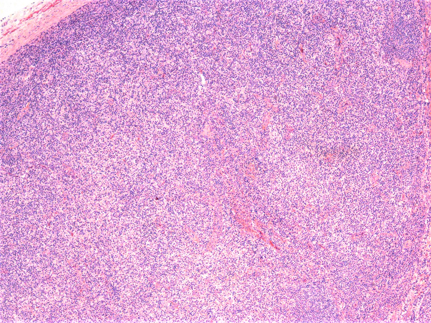
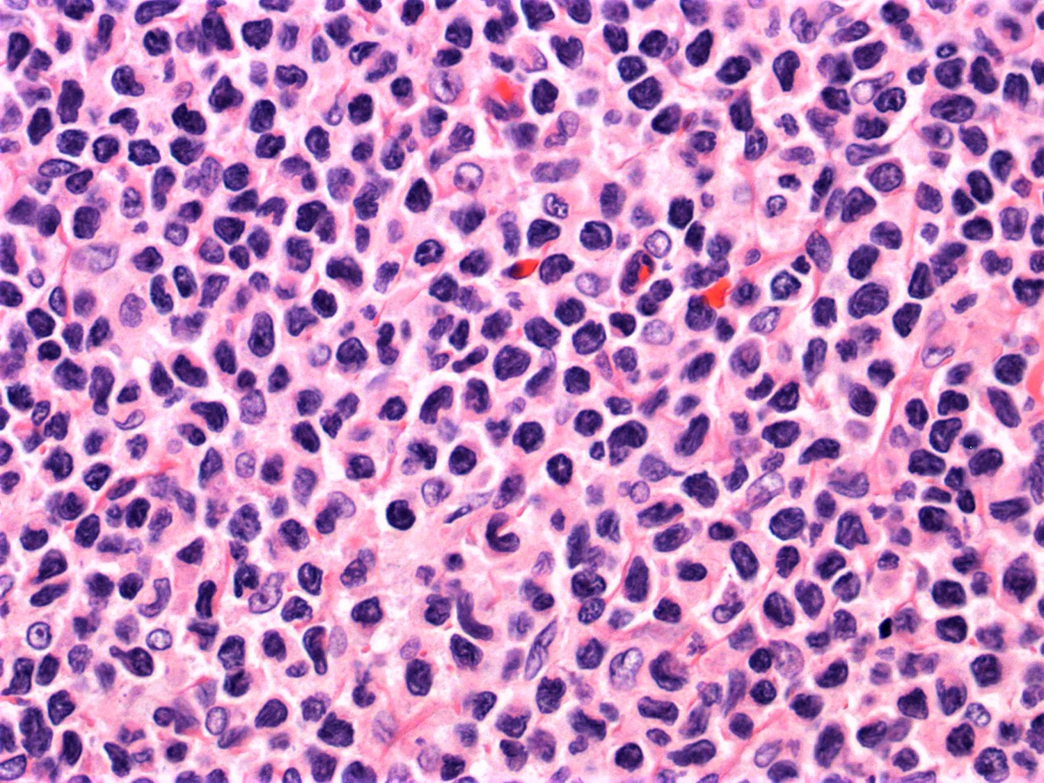
Contributed by Jayalakshmi Balakrishna, M.D.
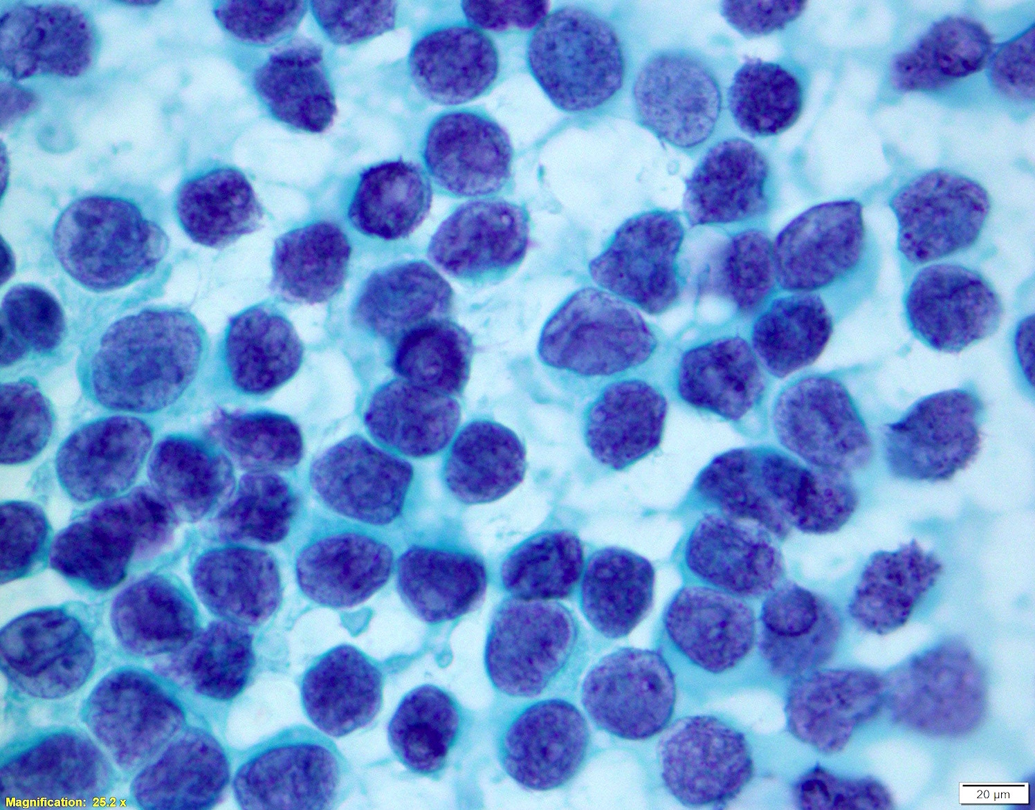
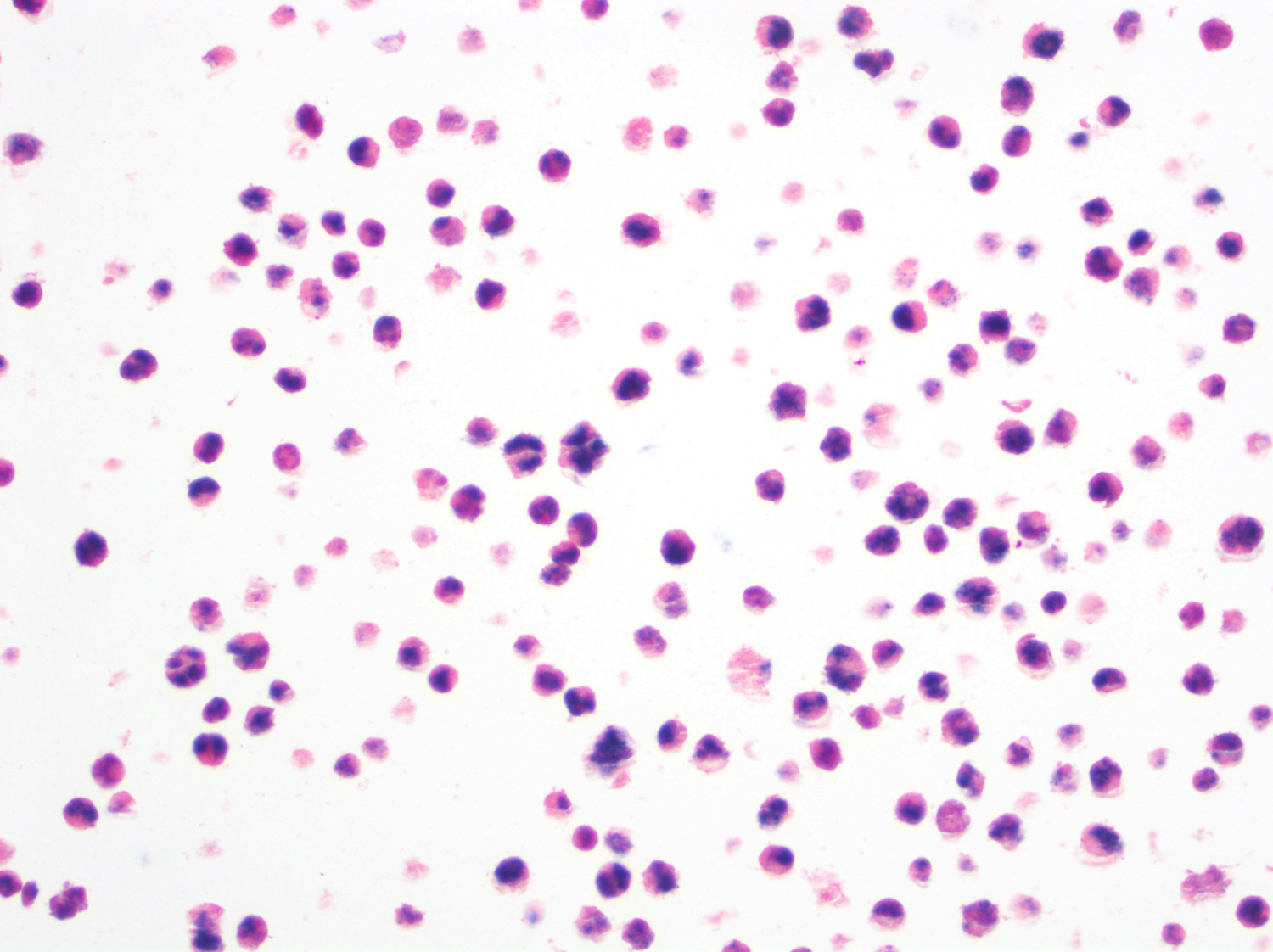
Lymph node biopsy touch preparation
Contributed by Sanam Loghavi, M.D.
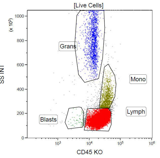
CD45 and side light scatter
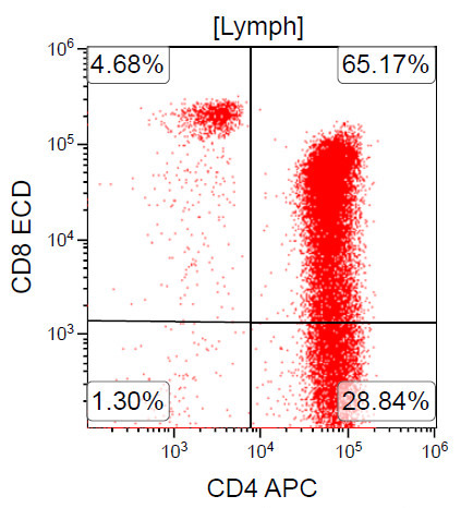
CD3 and CD4

CD4 and CD8
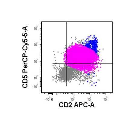
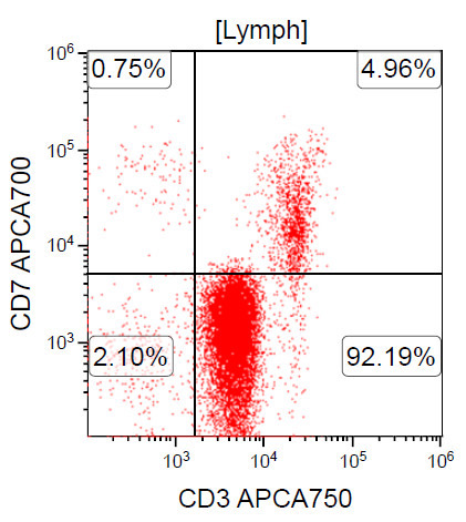
CD5 and CD2
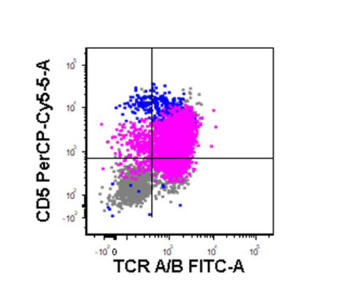
TCR A / B

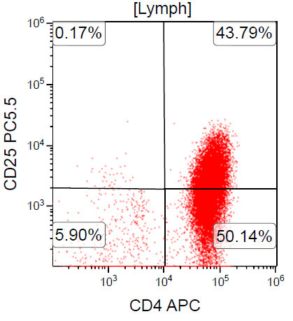
CD3 and CD30
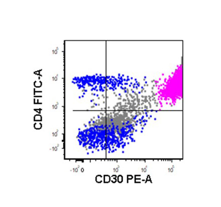
CD4 and CD30
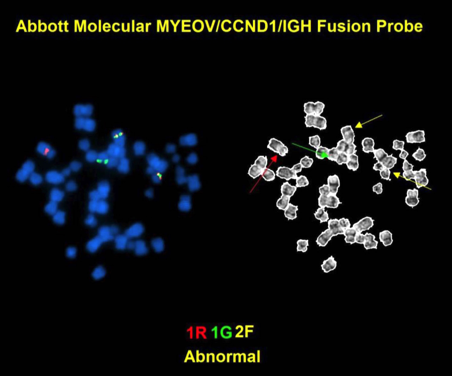
Contributed by Andrew Feldman, M.D.
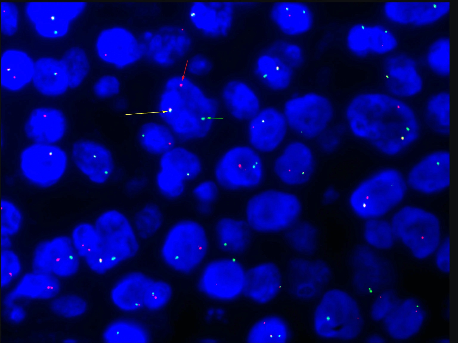
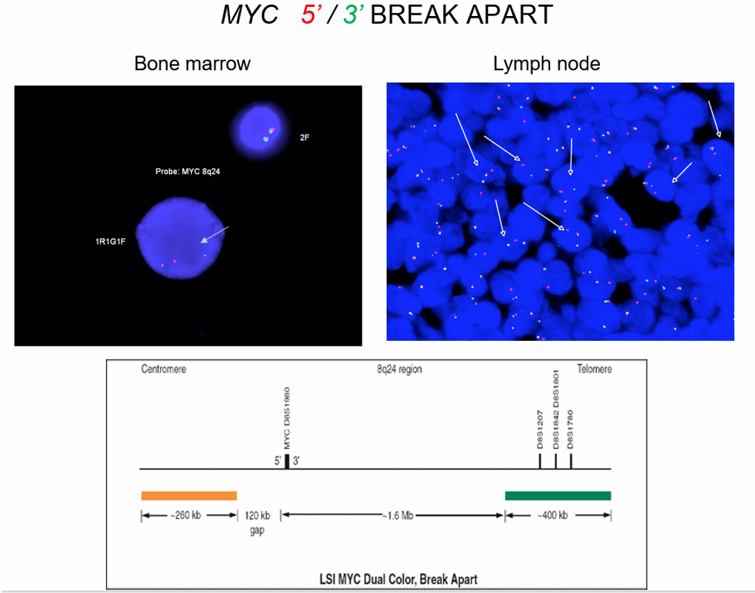
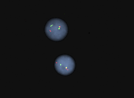
Break apart probe FISH for chromosome region 6p25.3
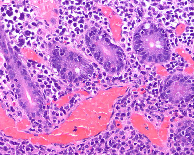
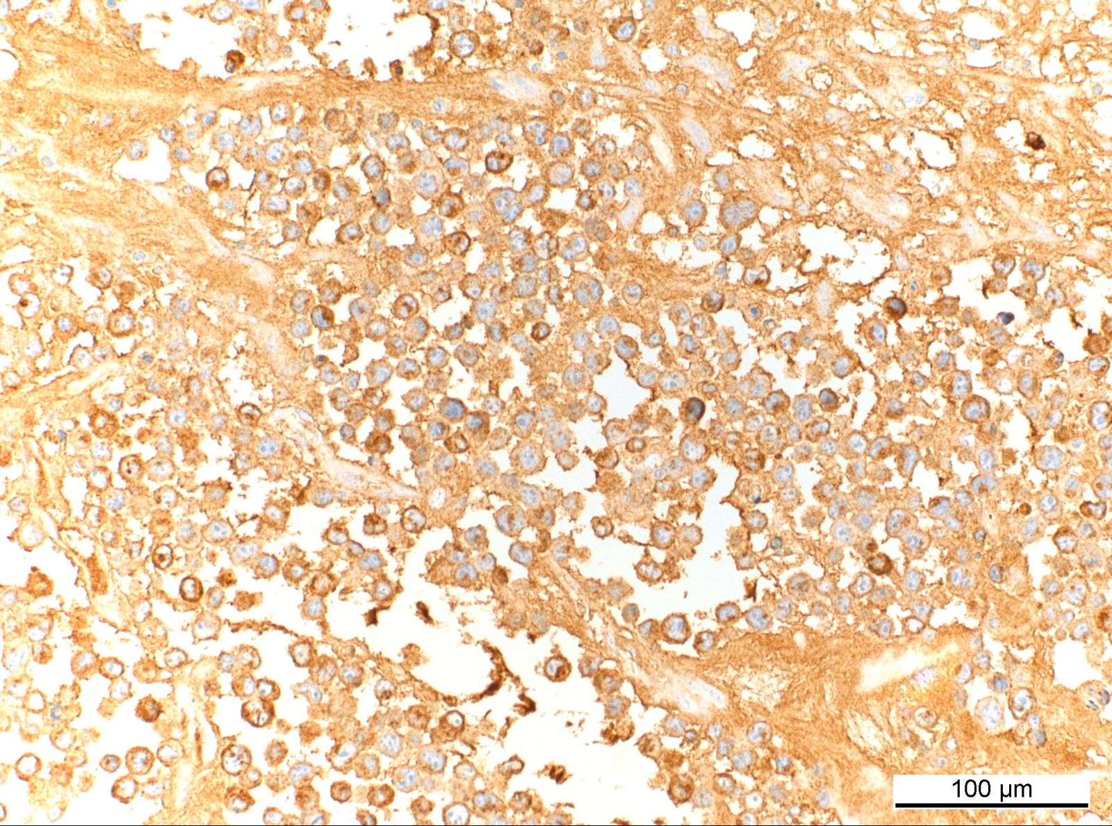
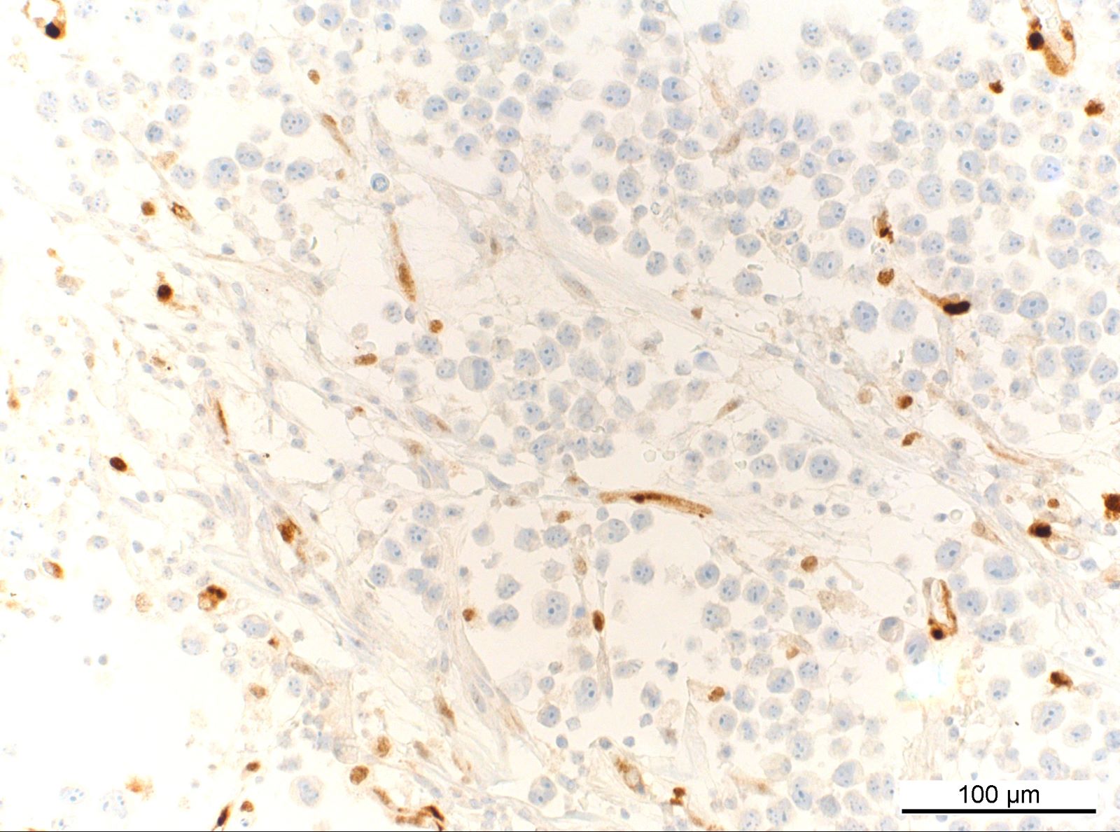
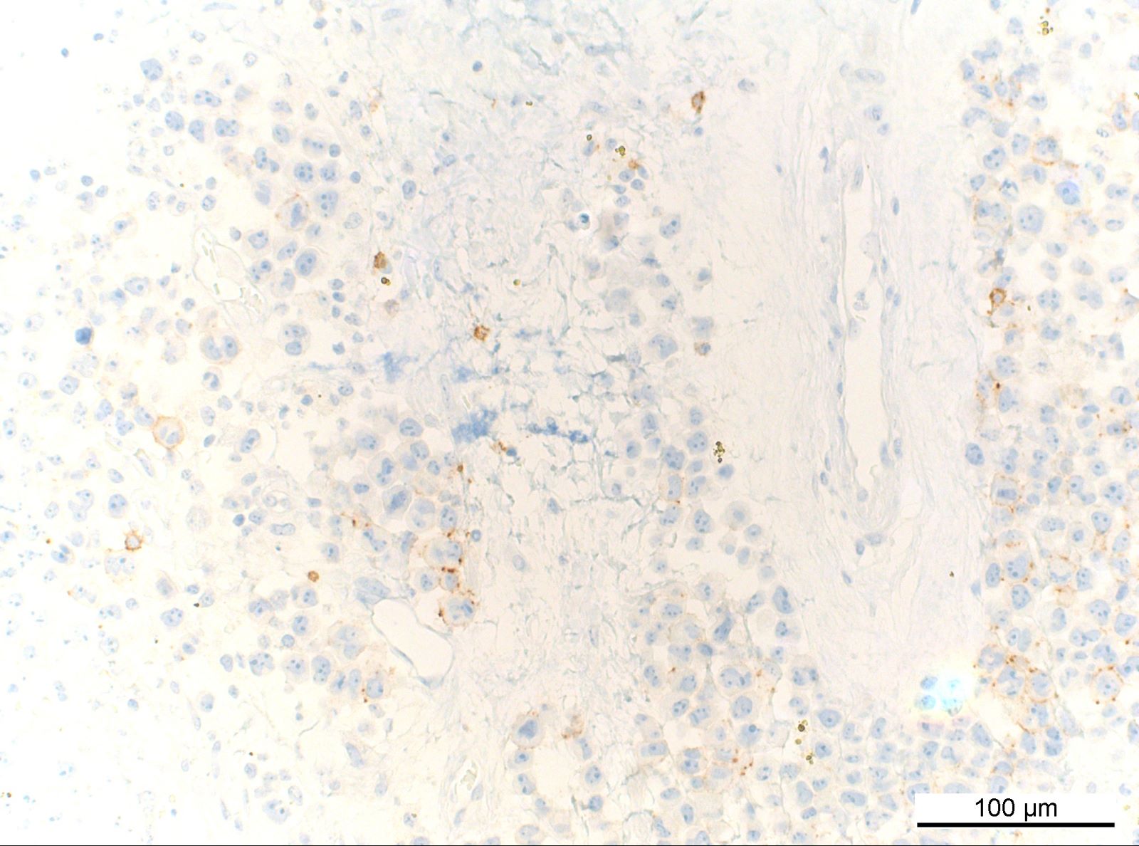
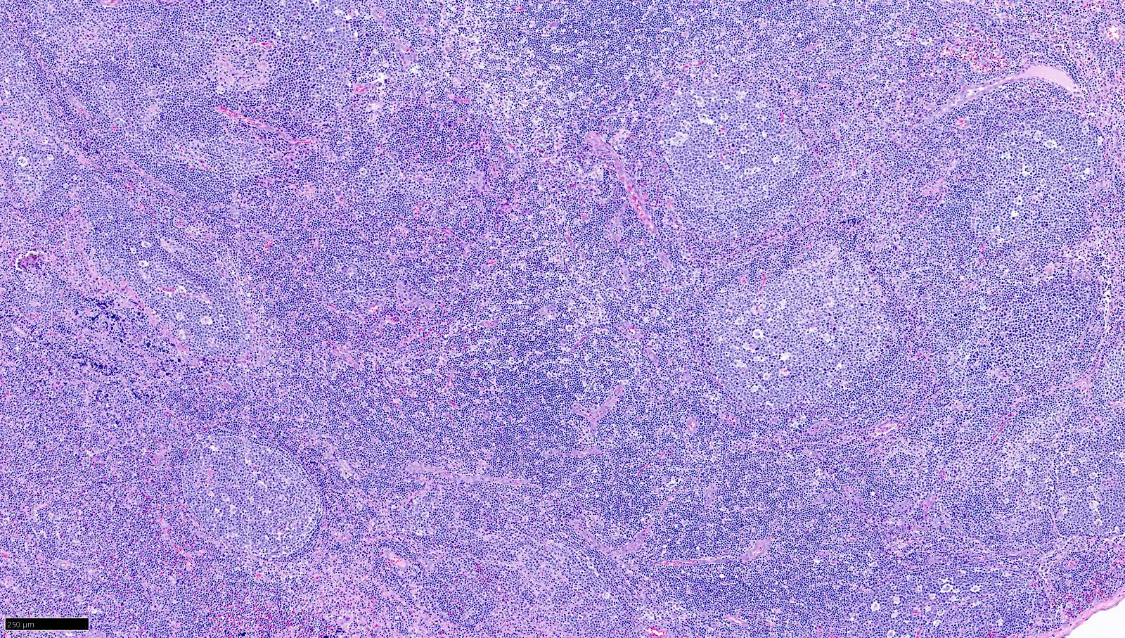
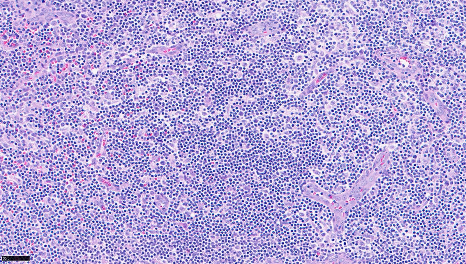
Contributed by Jayalakshmi Balakrishna, M.D., Elaine S. Jaffe, M.D.

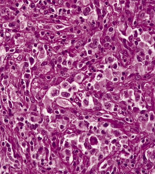
Diffuse infiltrate

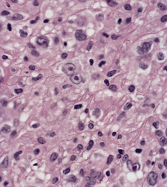
Large pleomorphic cells
Large cells with pleomorphic nuclei
Large cells with pleomorphic nuclei
Large cells with pleomorphic nuclei
Mitoses and apoptosis
Intrasinusoidal infiltrate
Intrasinusoidal infiltrate
Focal infiltrate
Hallmark cells
Hallmark cells

Polymorphous background
CD3
CD3
CD30
ALK1
ALK1

ALK1
EMA
Granzyme B
CD45
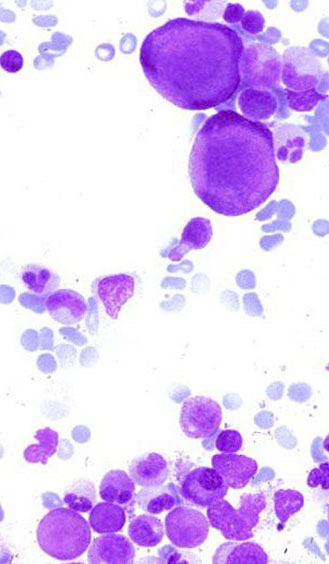
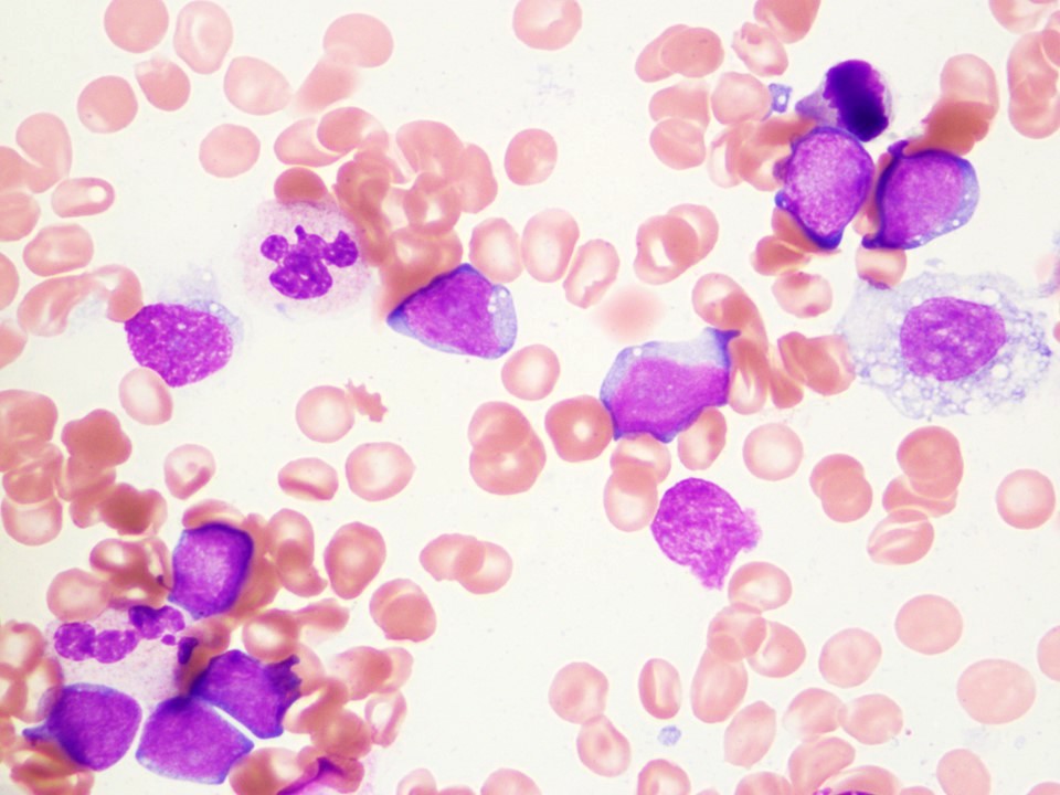
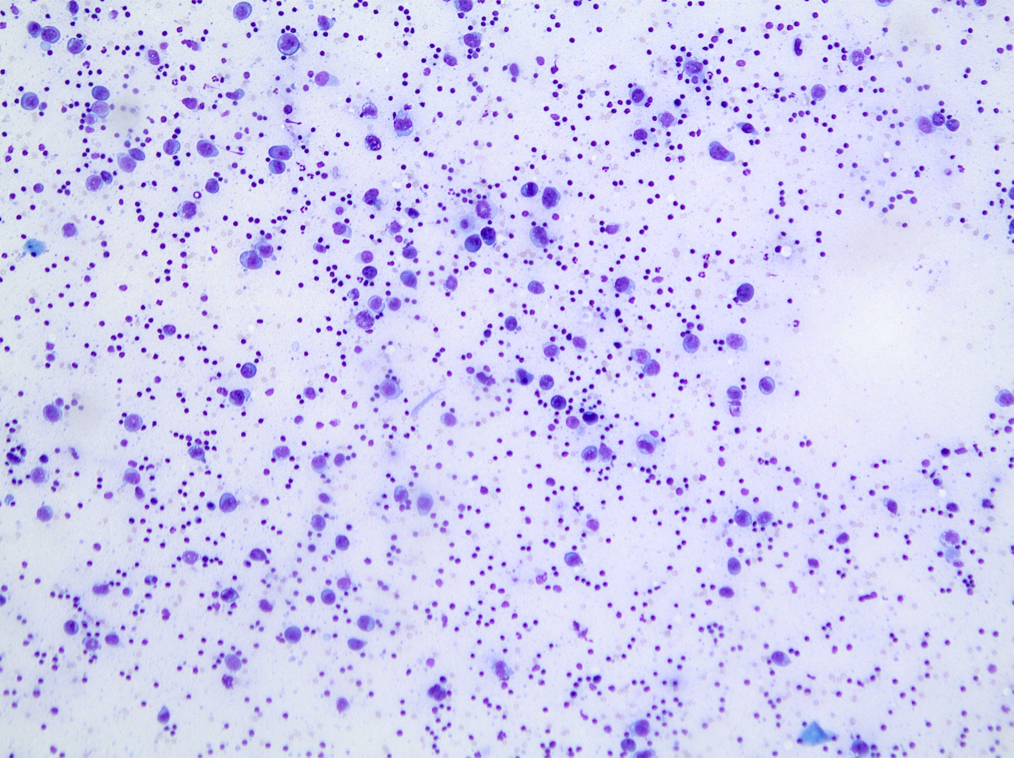
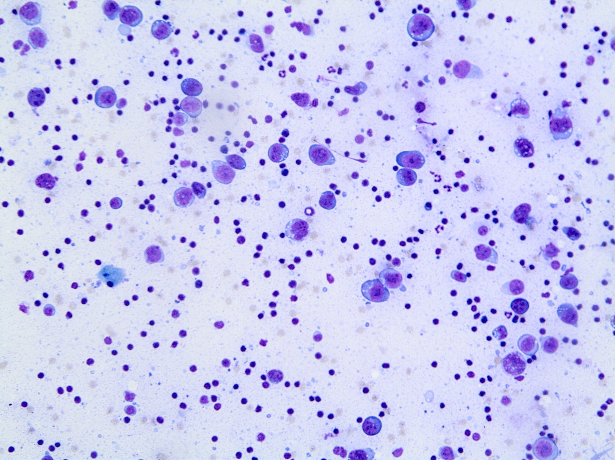
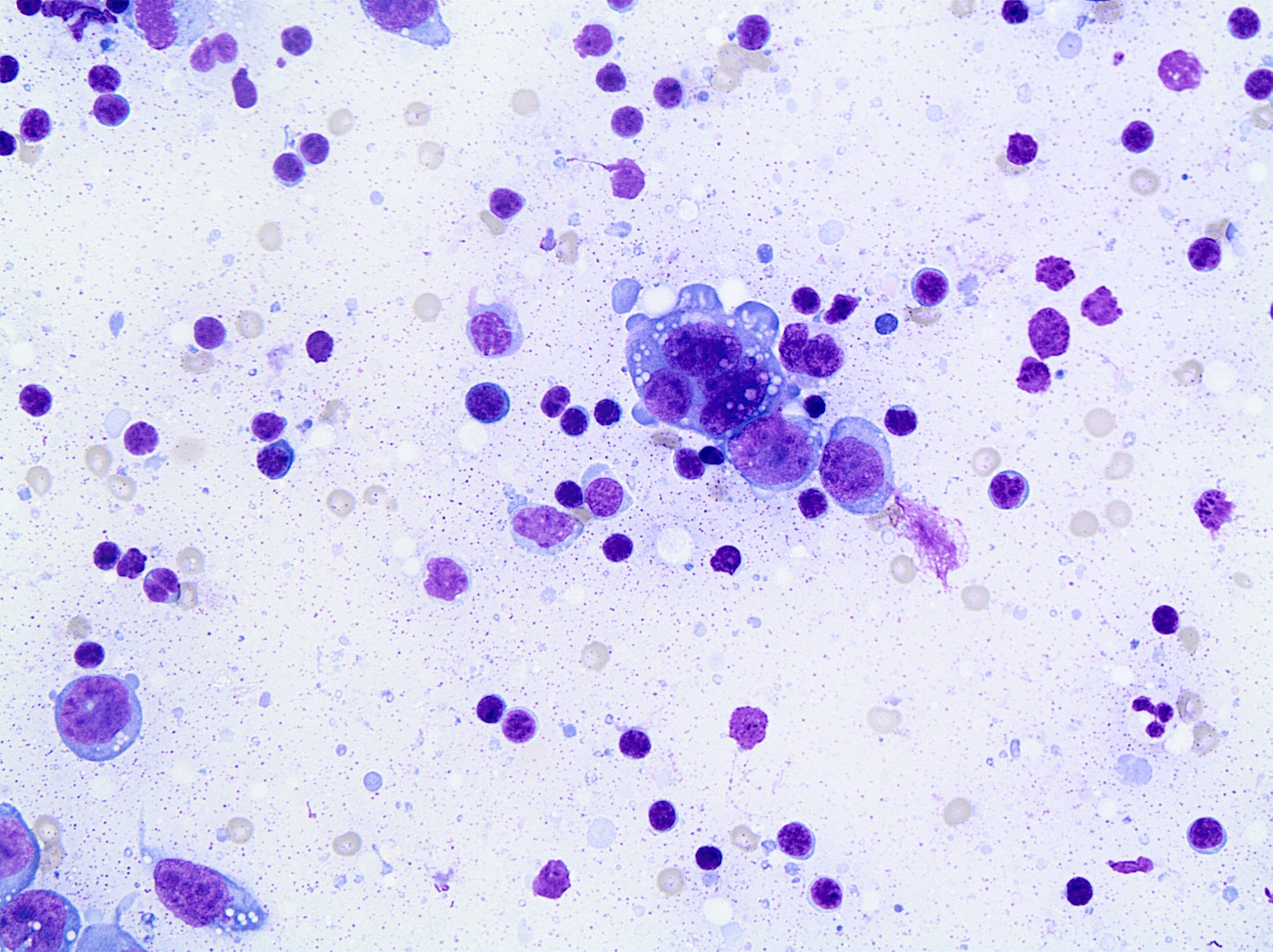
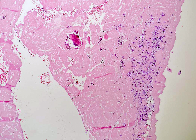
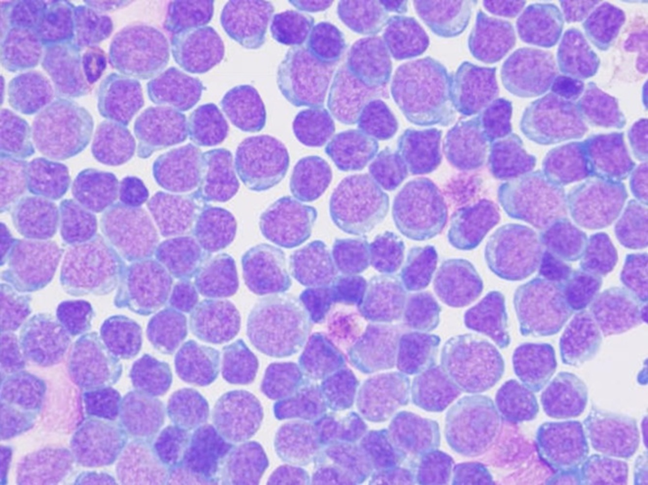
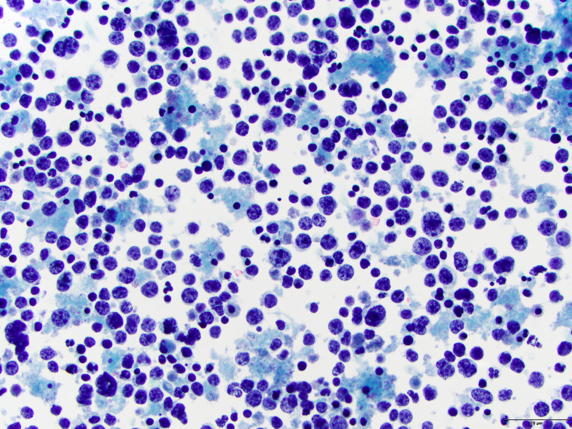
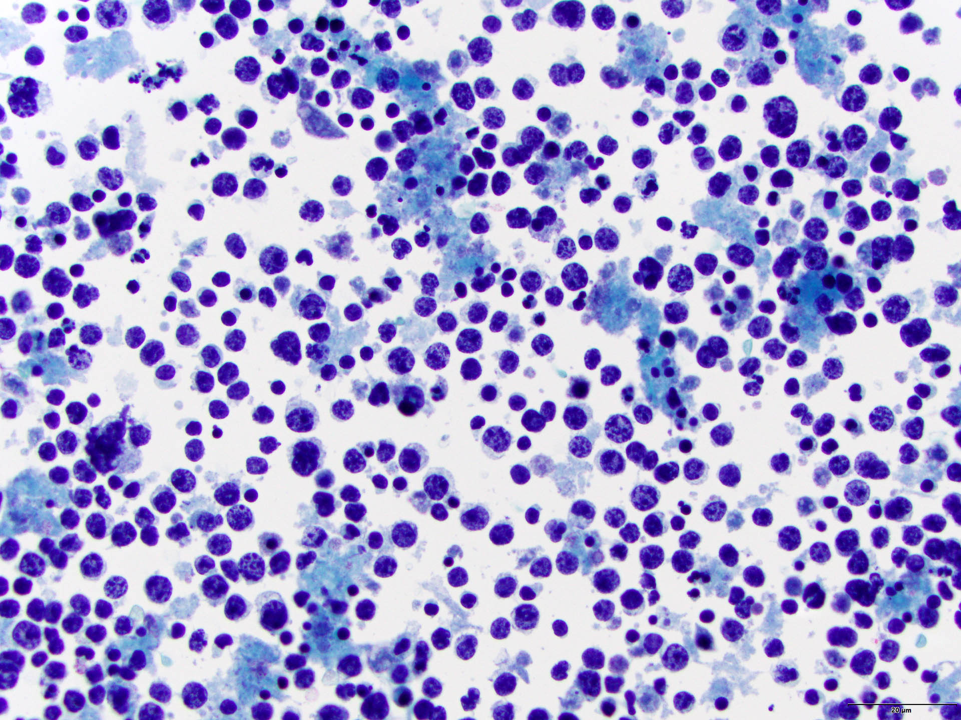
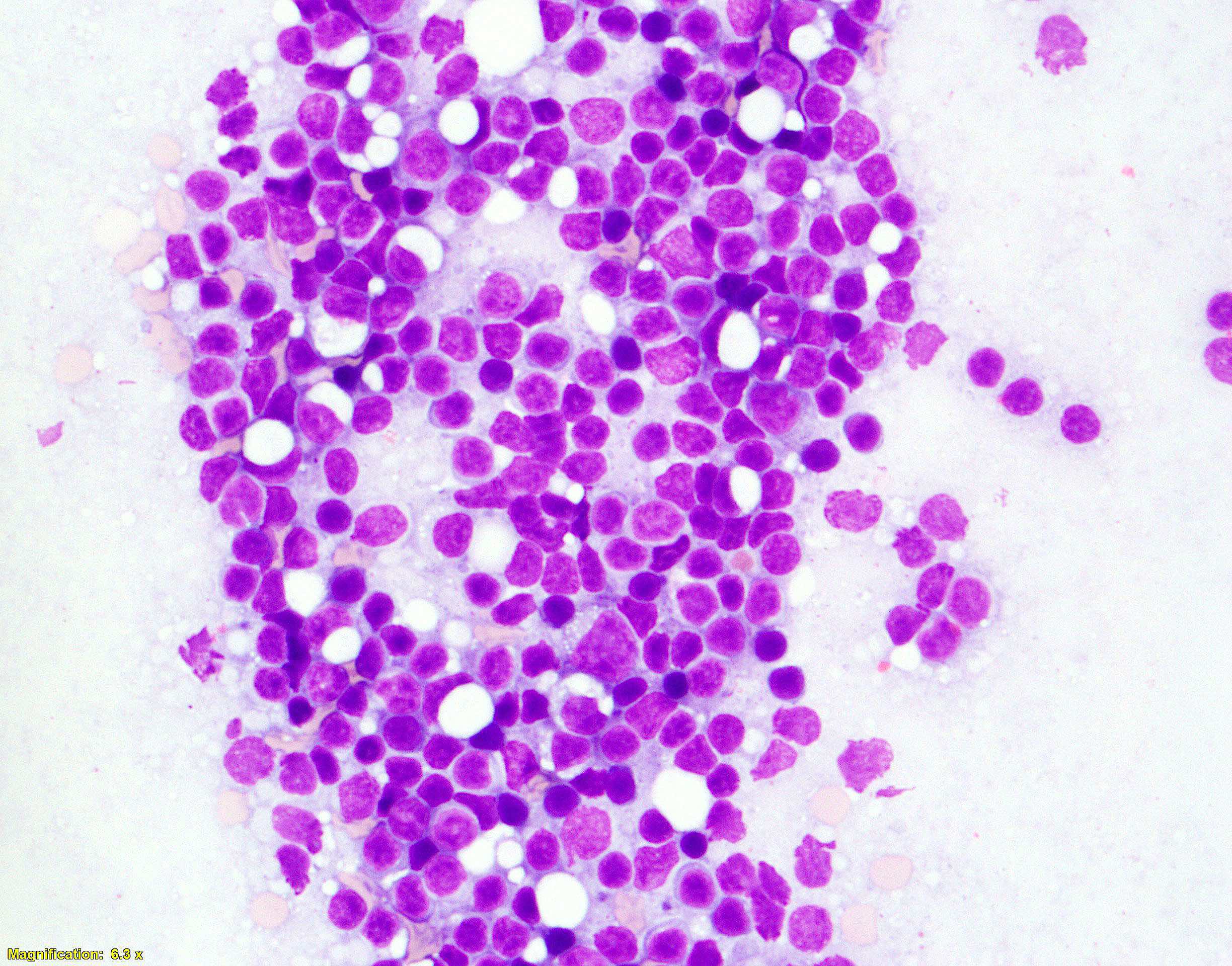
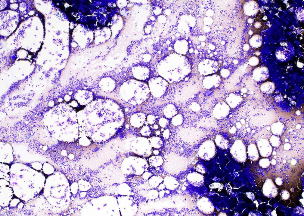
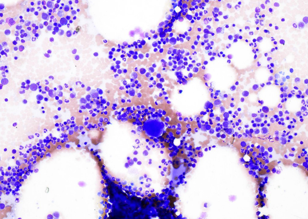
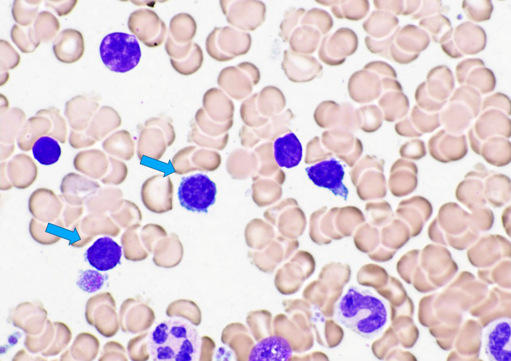
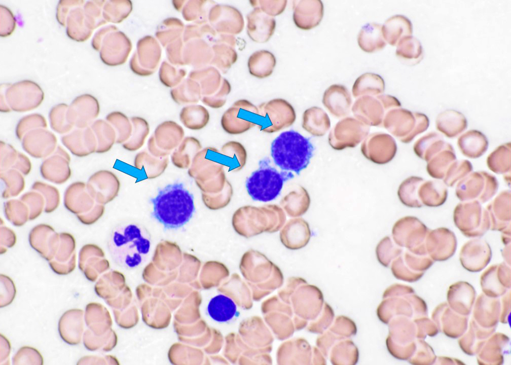
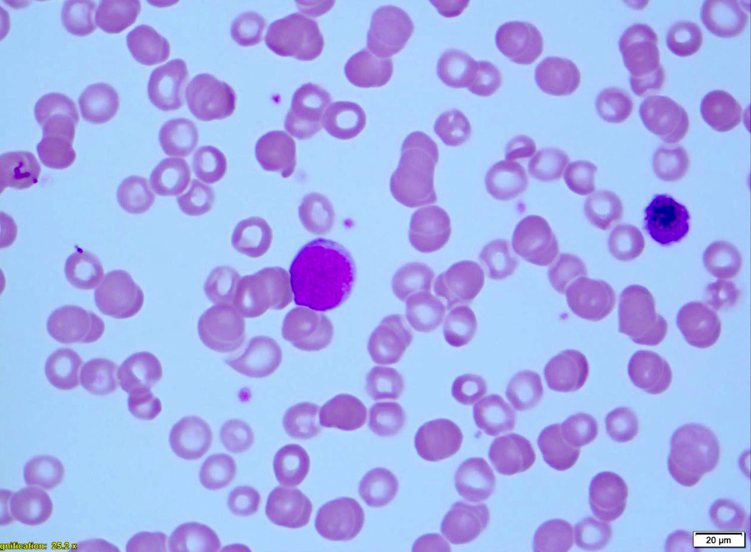
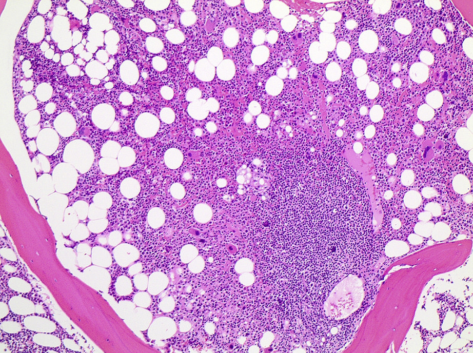
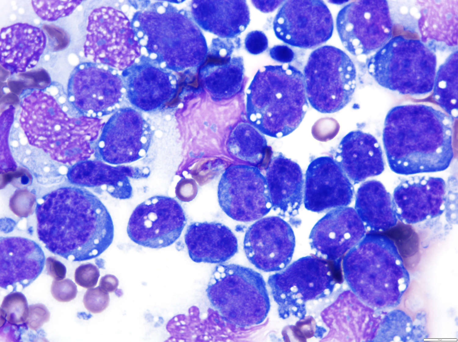
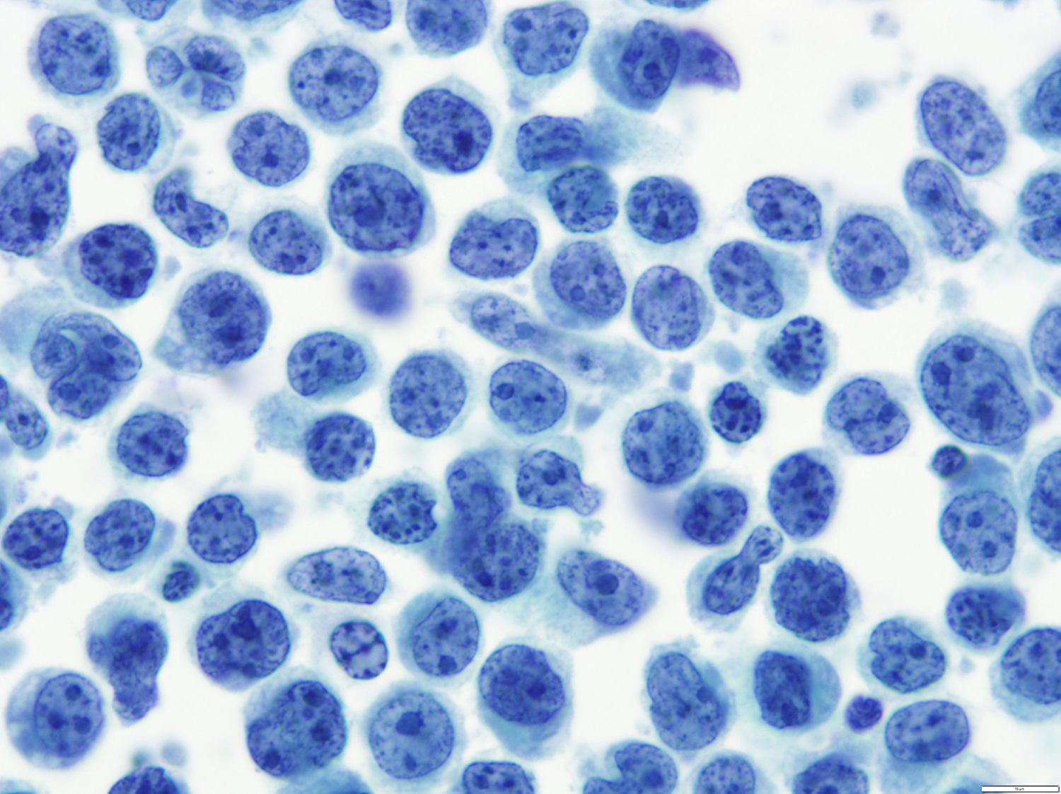
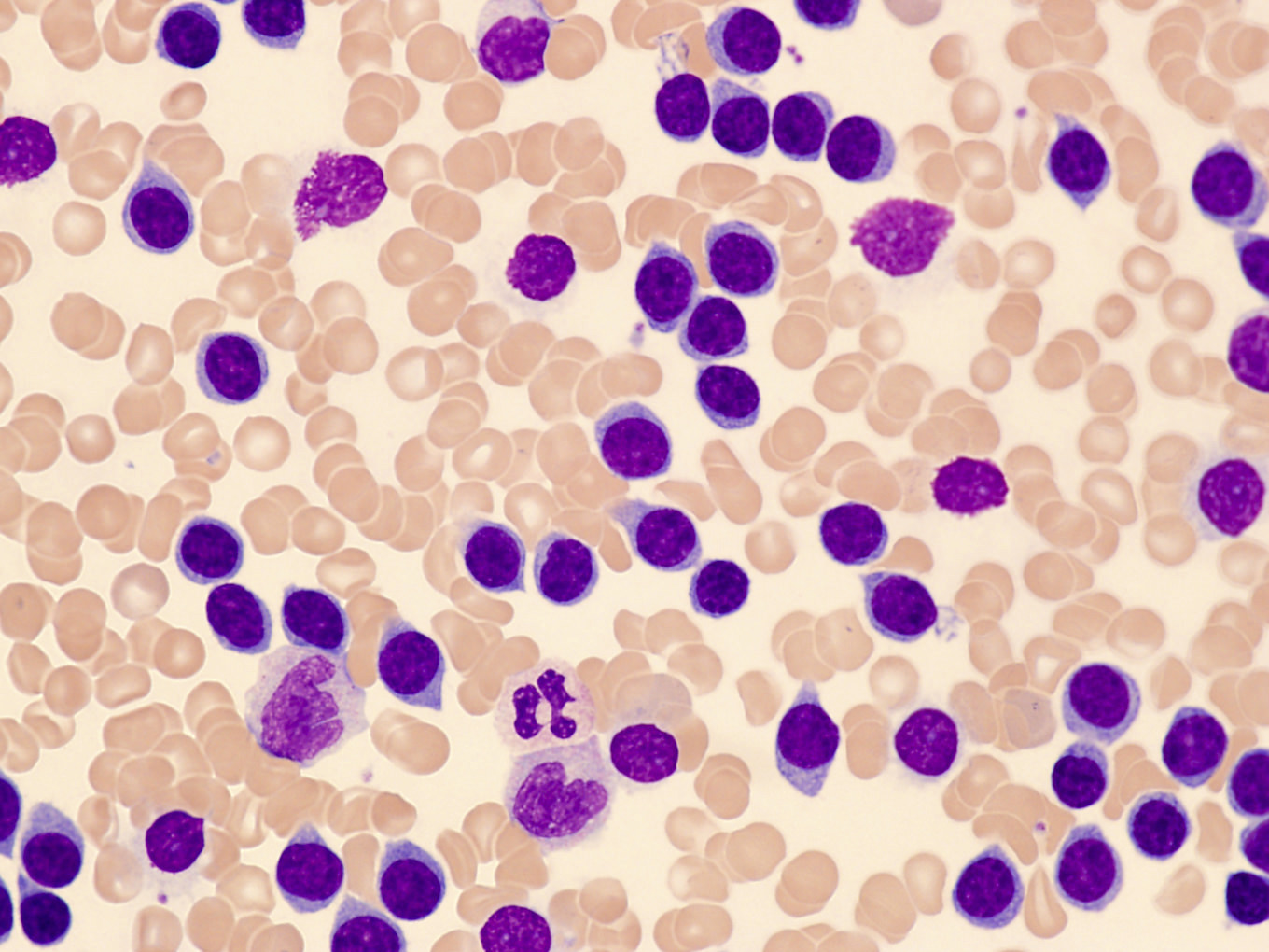
Bone marrow aspirate smear
Bone marrow biopsy


Lymphohistiocytic variant


Small cell variant
Sarcomatoid variant
Contributed by Jayalakshmi Balakrishna, M.D.
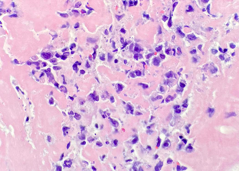
Large neoplastic cells

Large neoplastic cells in polymorphous background
Contributed by Roberto N. Miranda, M.D.
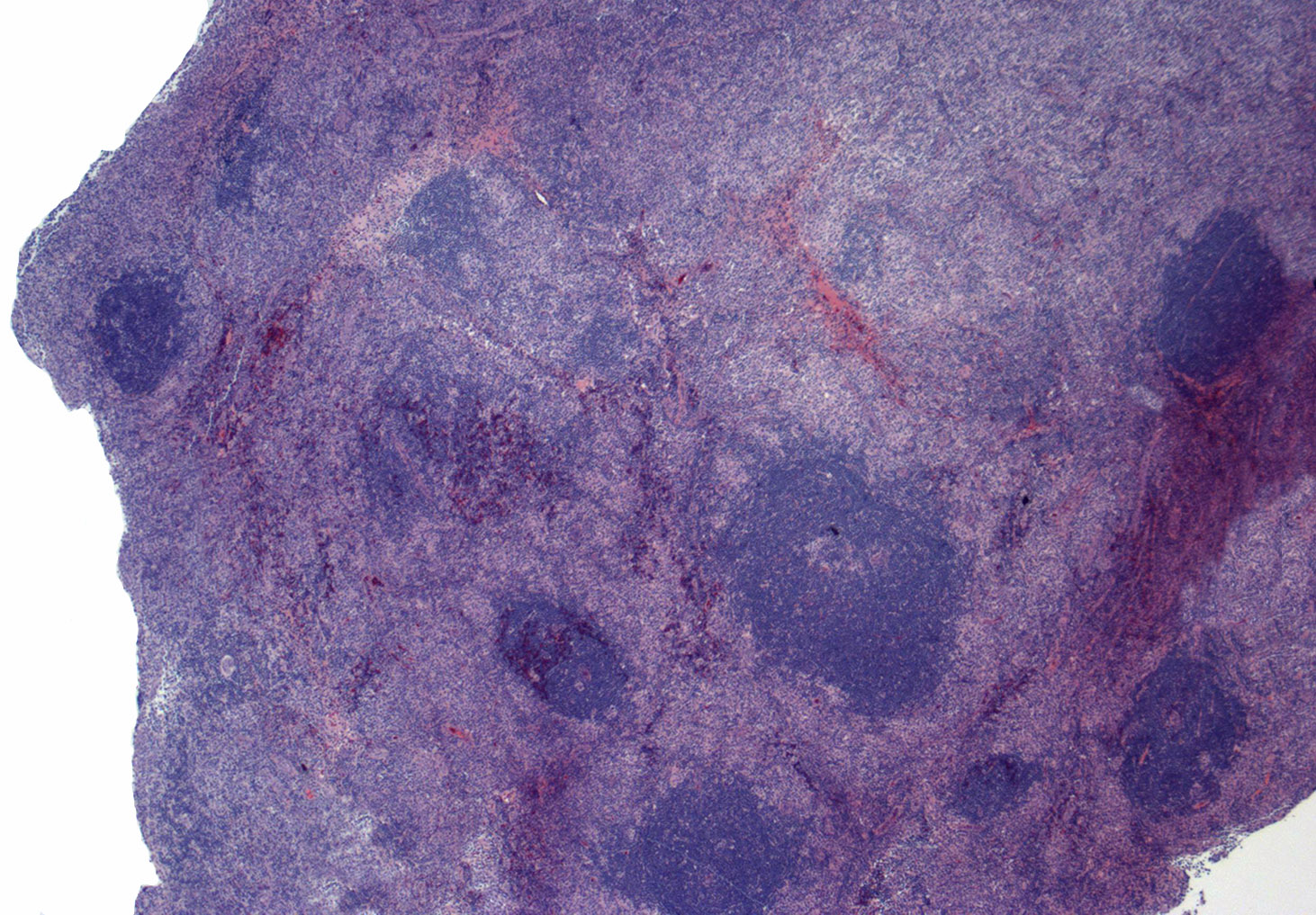
Pattern I: partial nodal involvement
Pattern III: complete effacement
Pattern III: capsular involvement
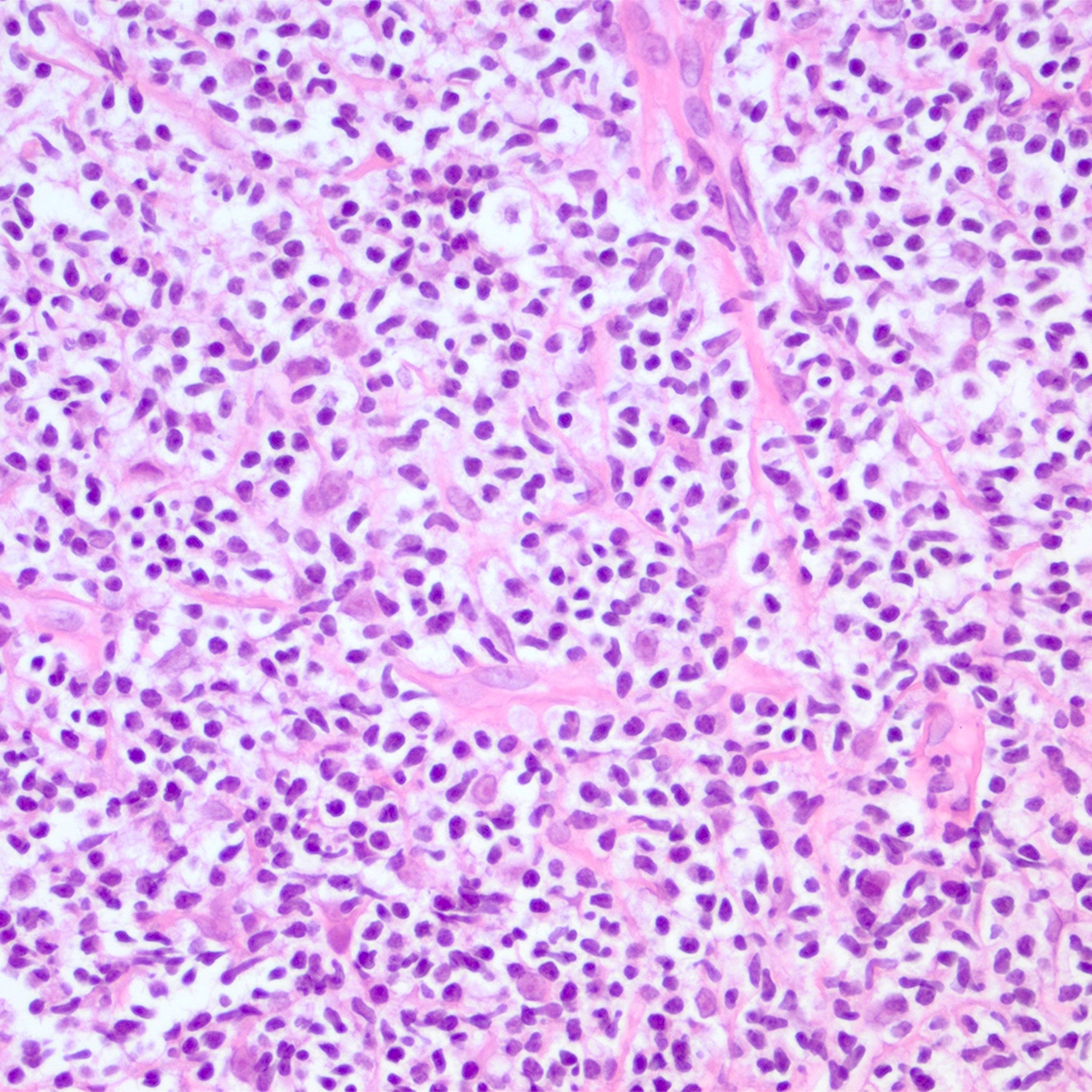
Polymorphic infiltrate
Polymorphic infiltrate with eosinophilia
Bone marrow involvement
Diffuse reactivity with CD3
CD20
CD21, pattern I
CD21, pattern III
PD-1 positivity
CXCL13 positivity
CD10 in small and intermediate size lymphoma cells
CD30 positivity
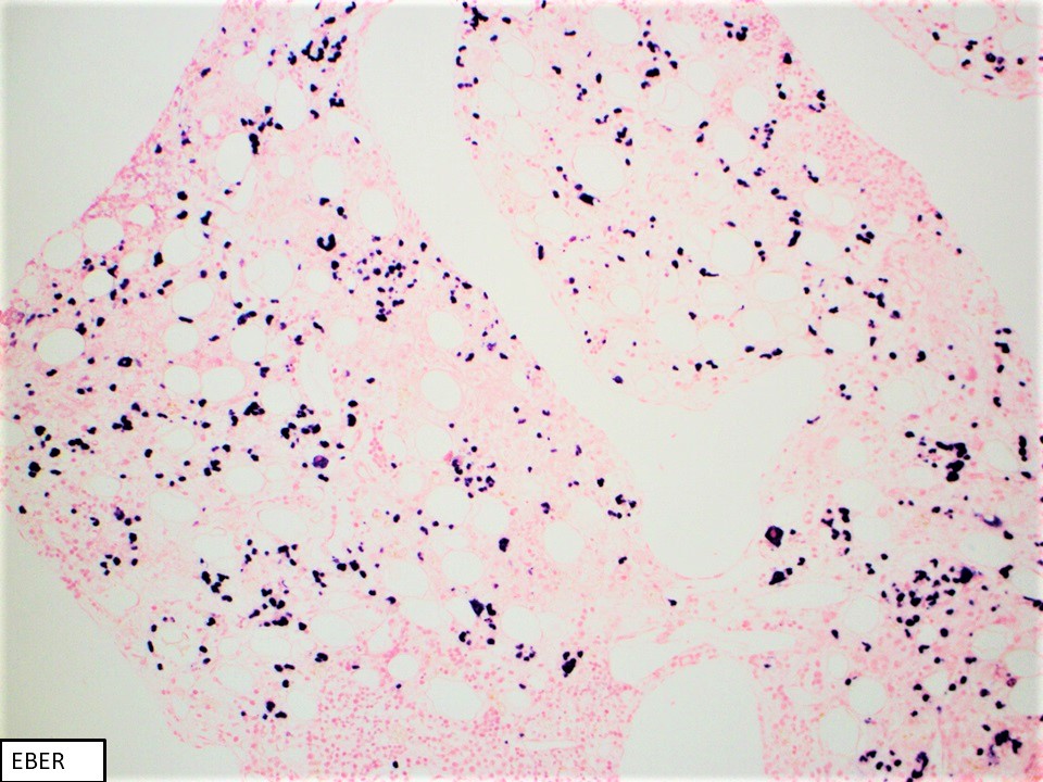
Epstein-Barr virus (EBER) reactivity
Proliferation marker Ki67
CD3 reactivity in bone marrow
CD10 reactivity
Contributed by Elizabeth Courville, M.D.
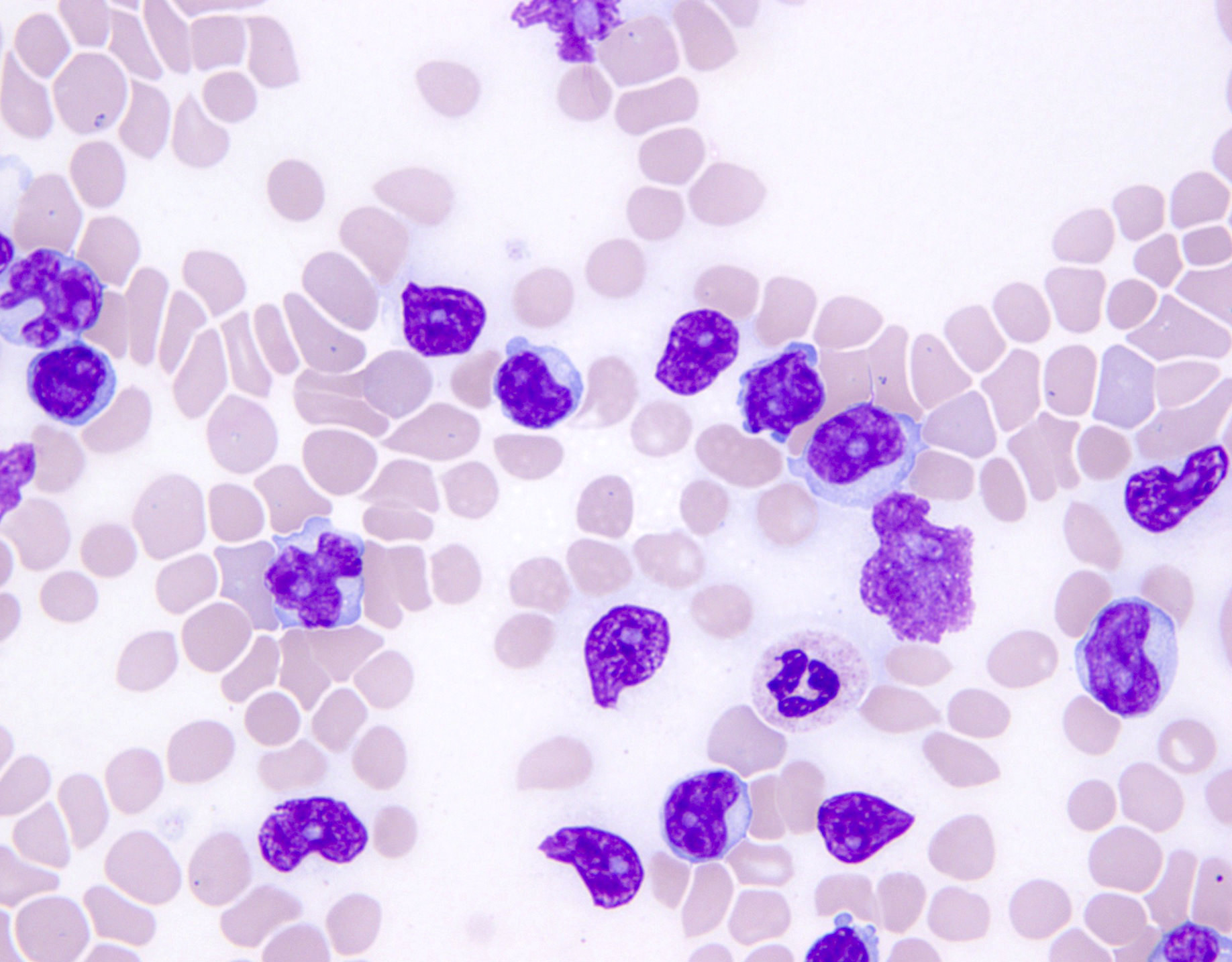
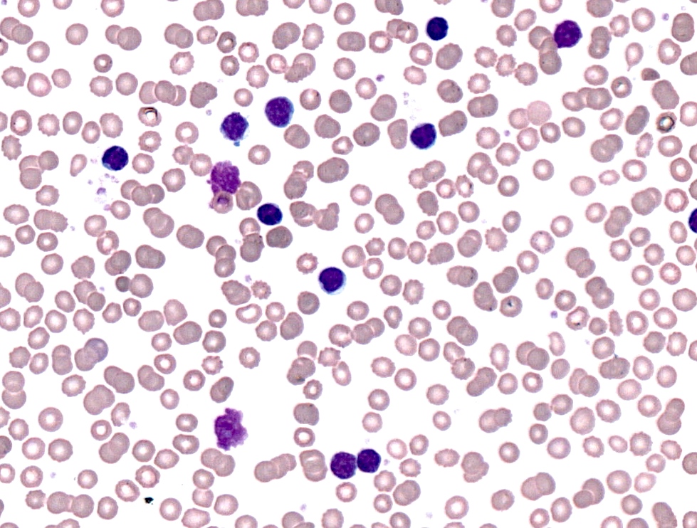
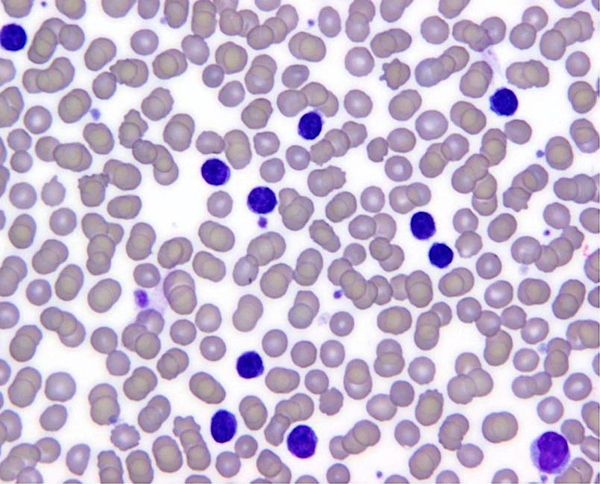
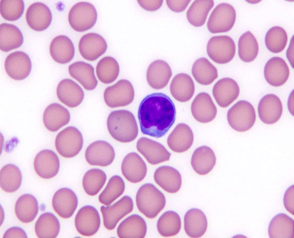
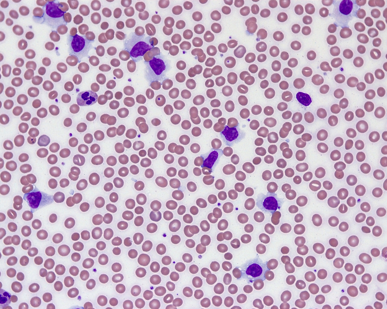
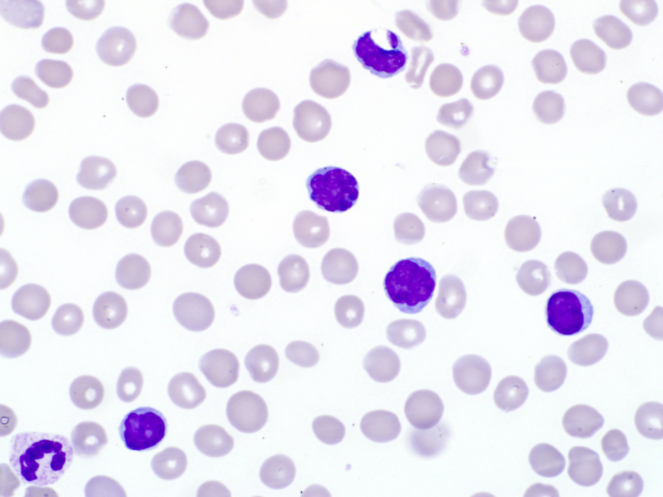
Atypical monotonous lymphocyte population
Mature circulating atypical lymphocytes
Contributed by Meghan Hupp, M.D.
CLL-like

Non-CLL phenotype
Images hosted on other servers

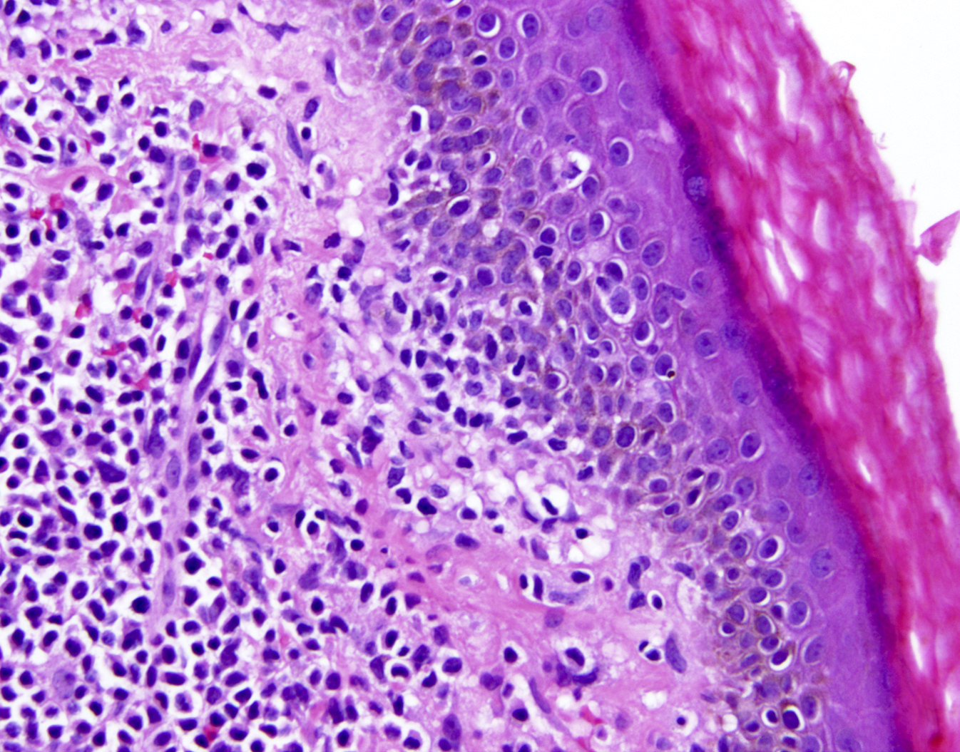
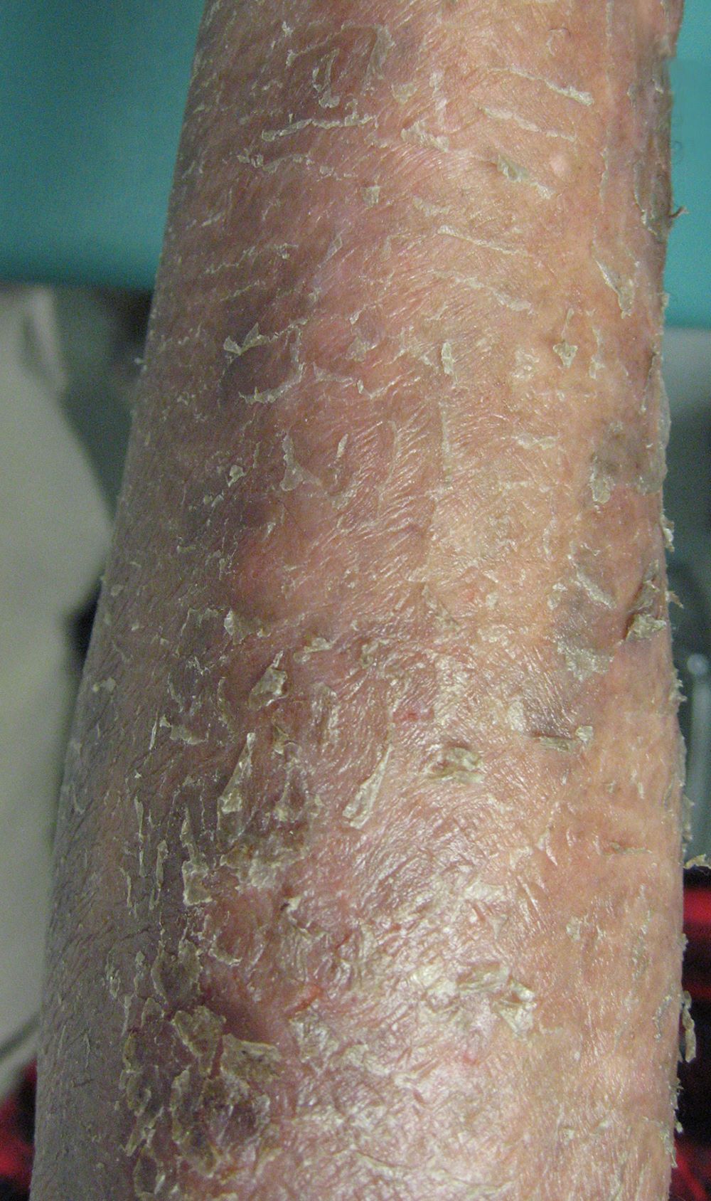
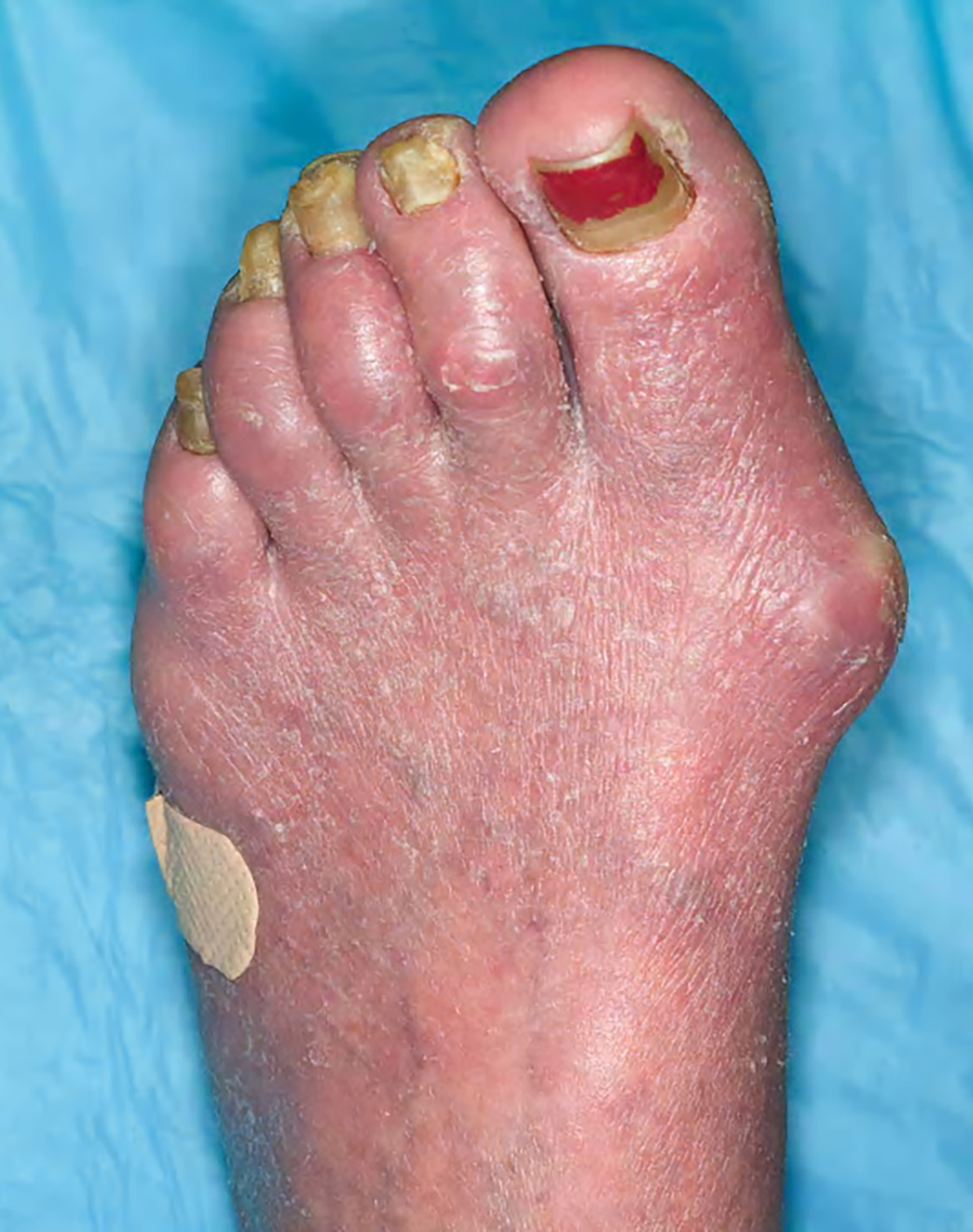
Skin involvement

Classic jaw involvement
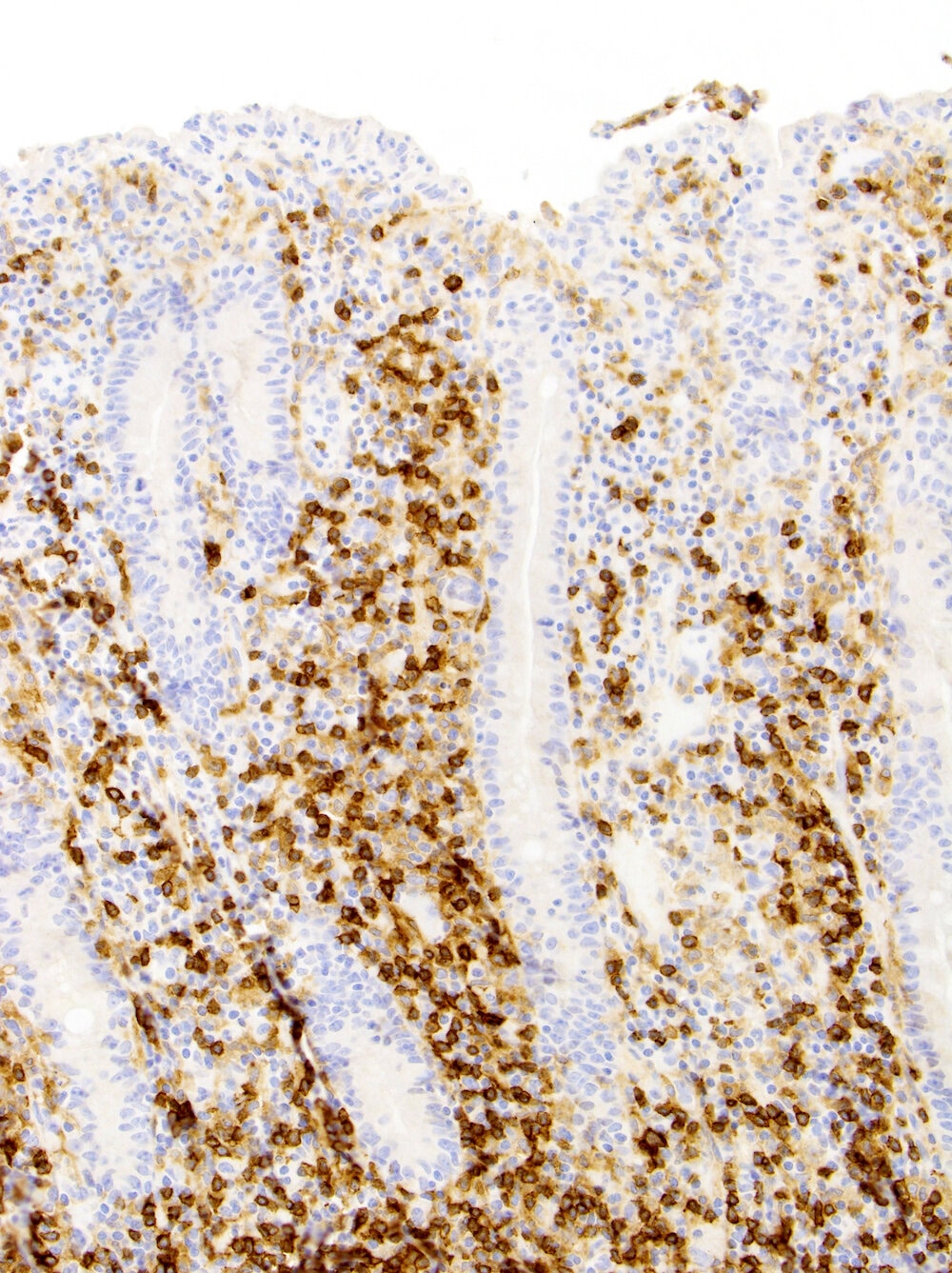
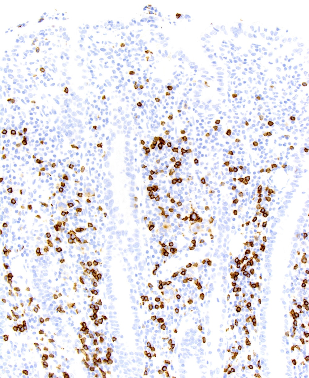
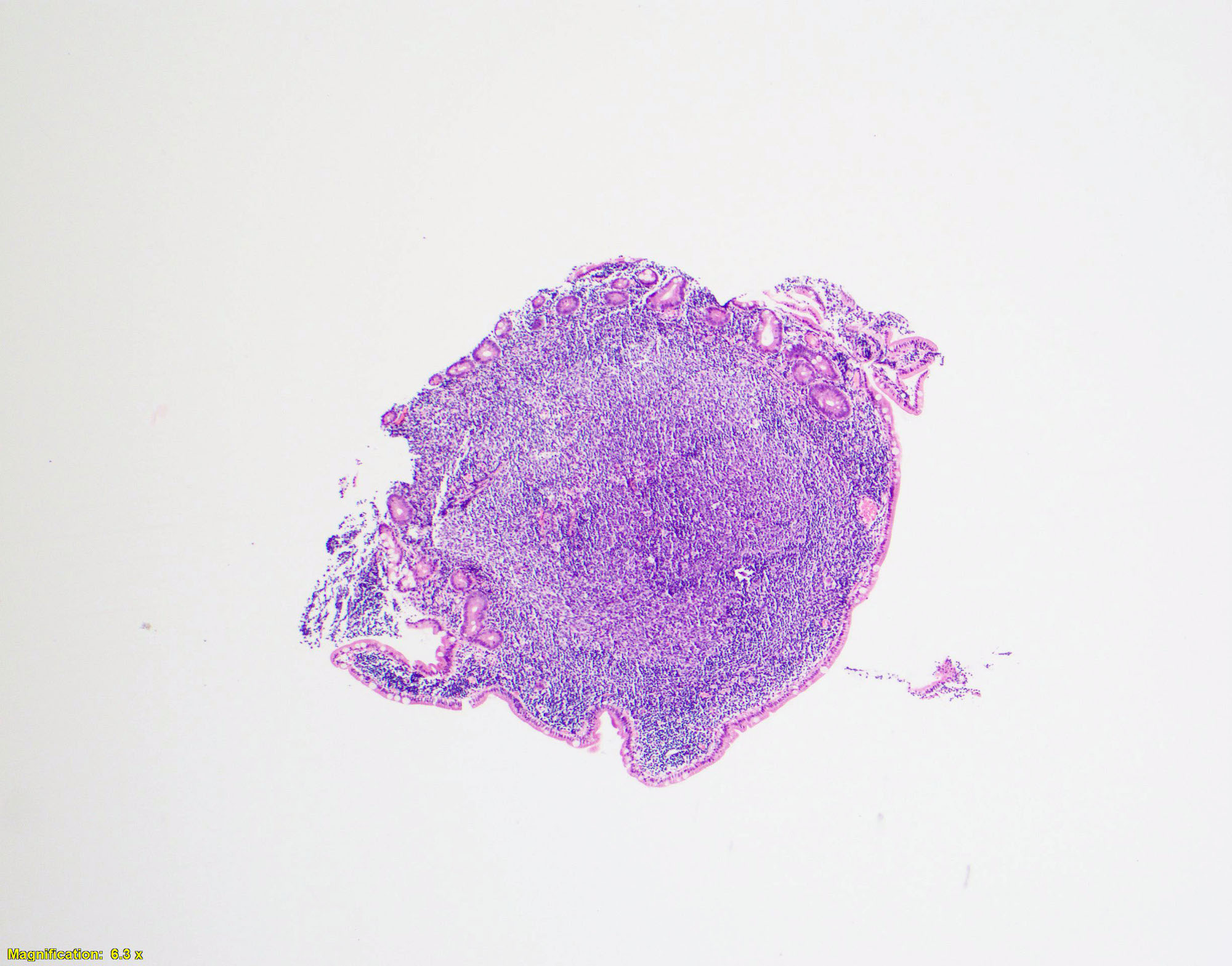
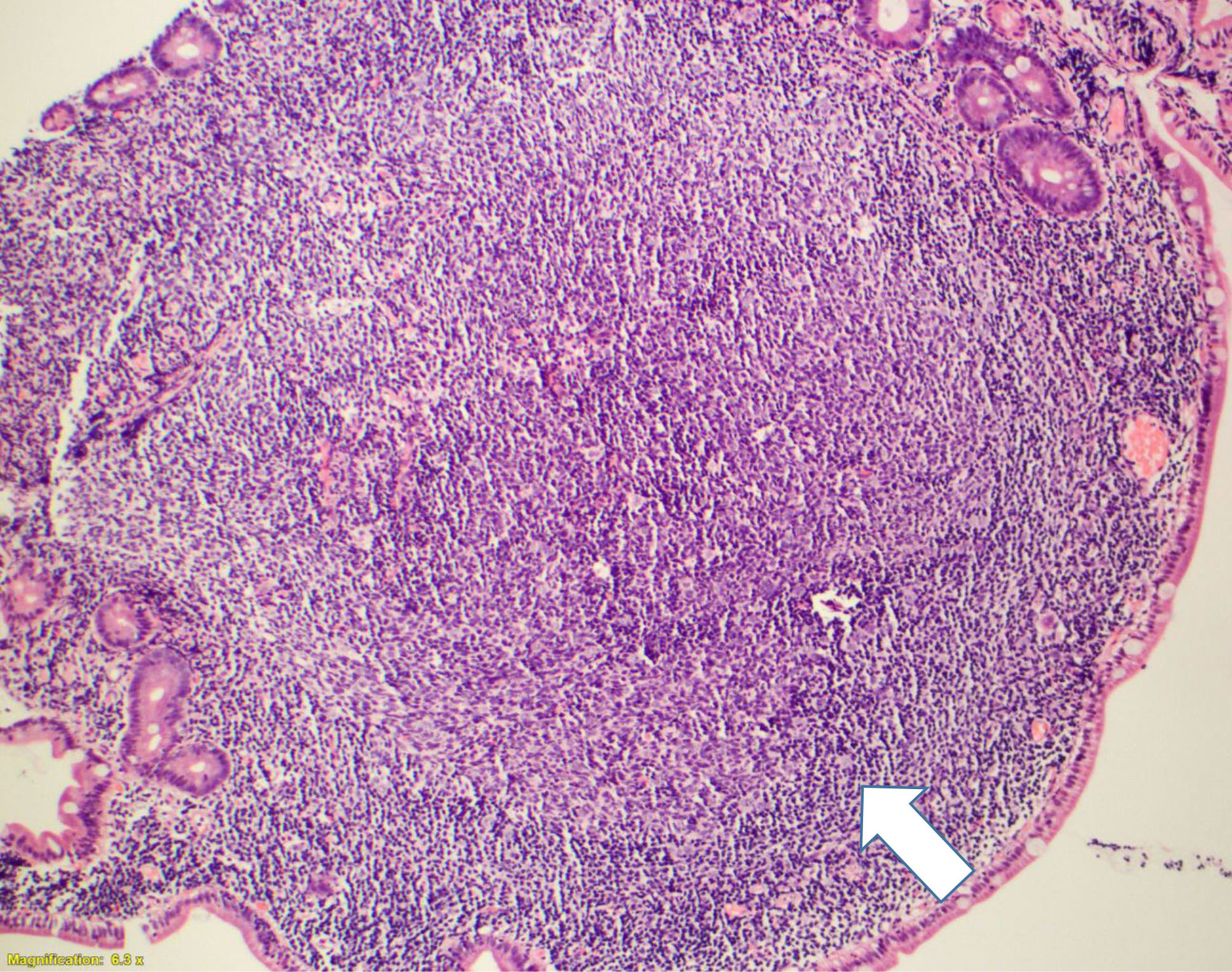
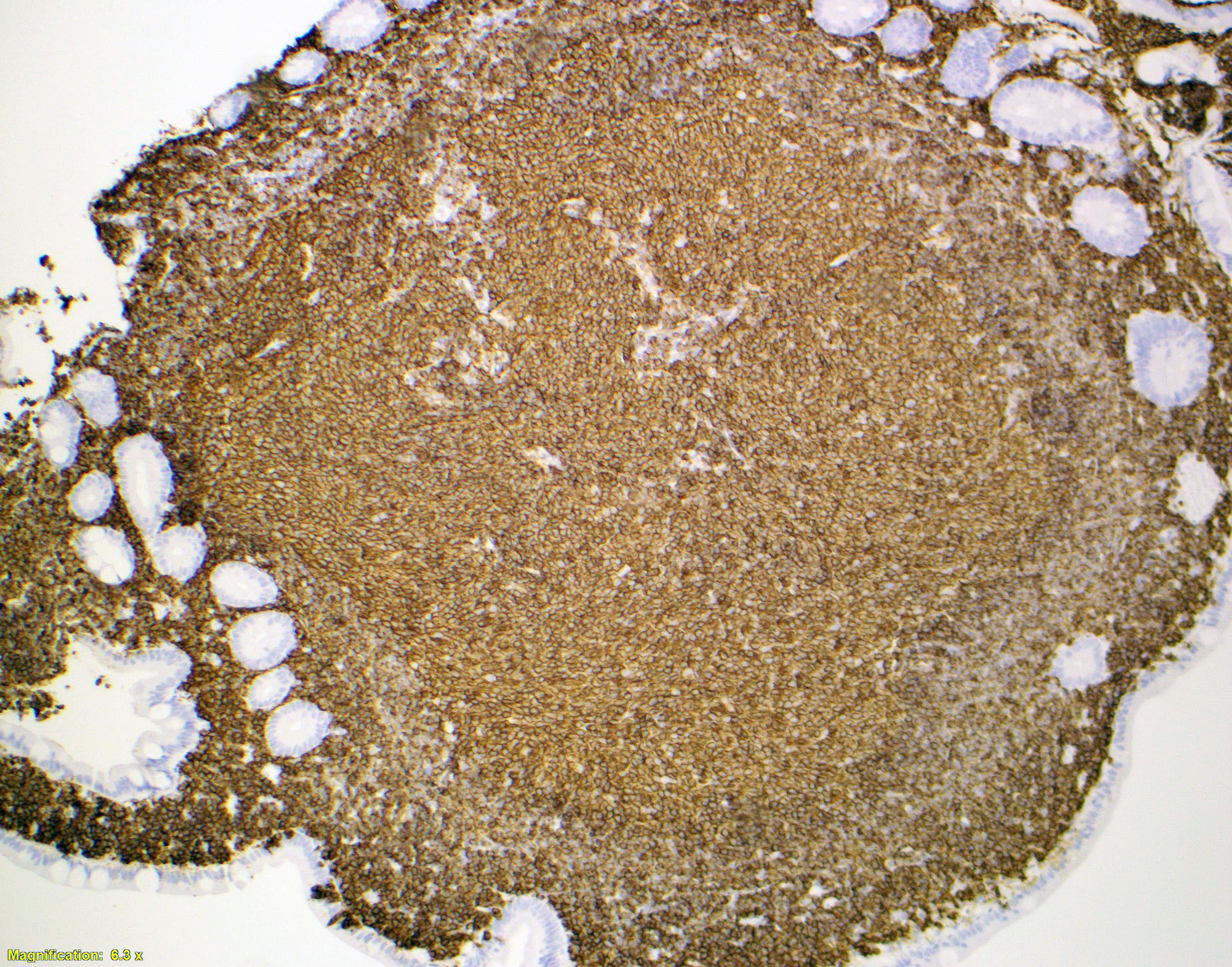
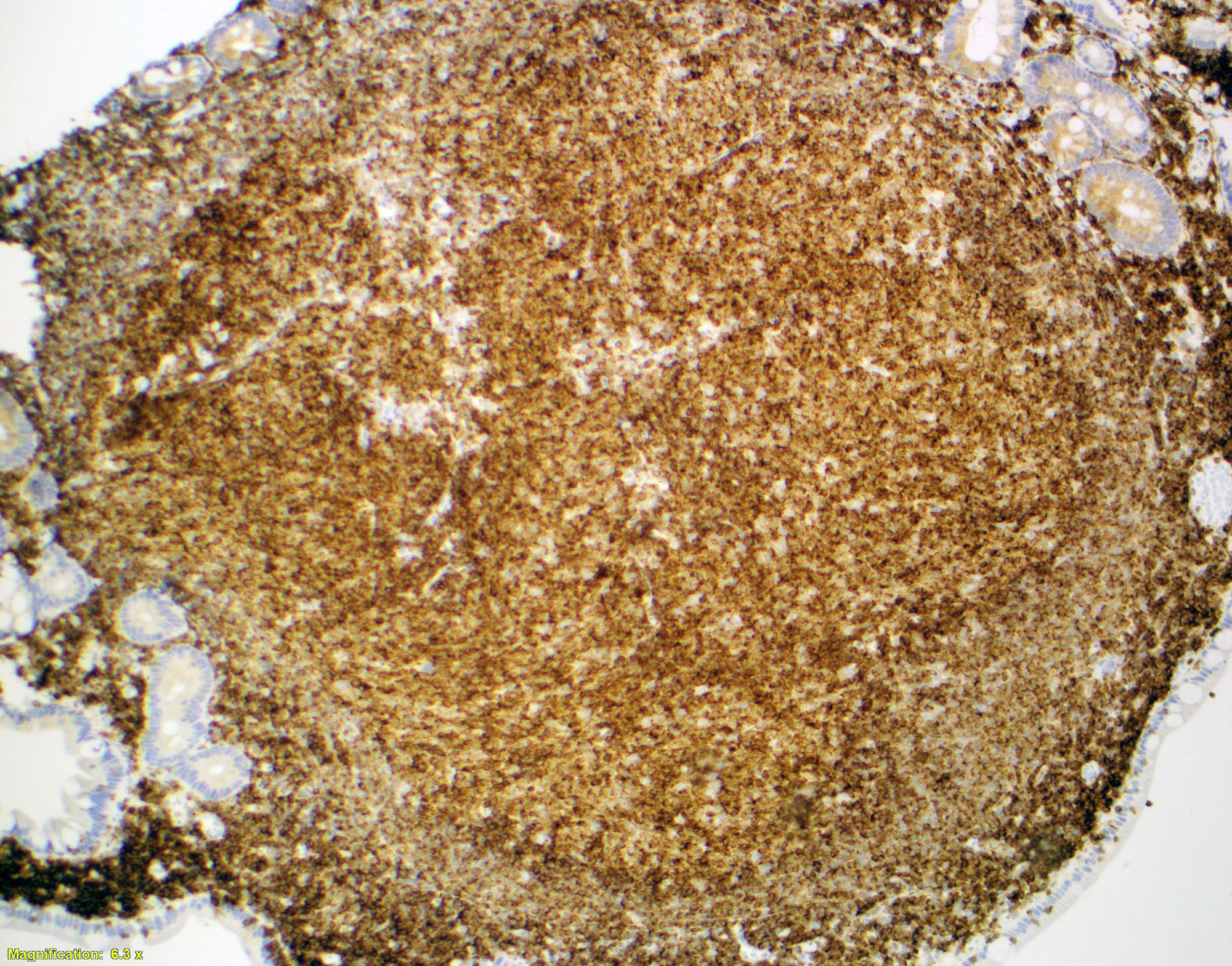
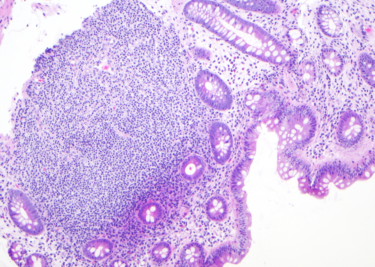
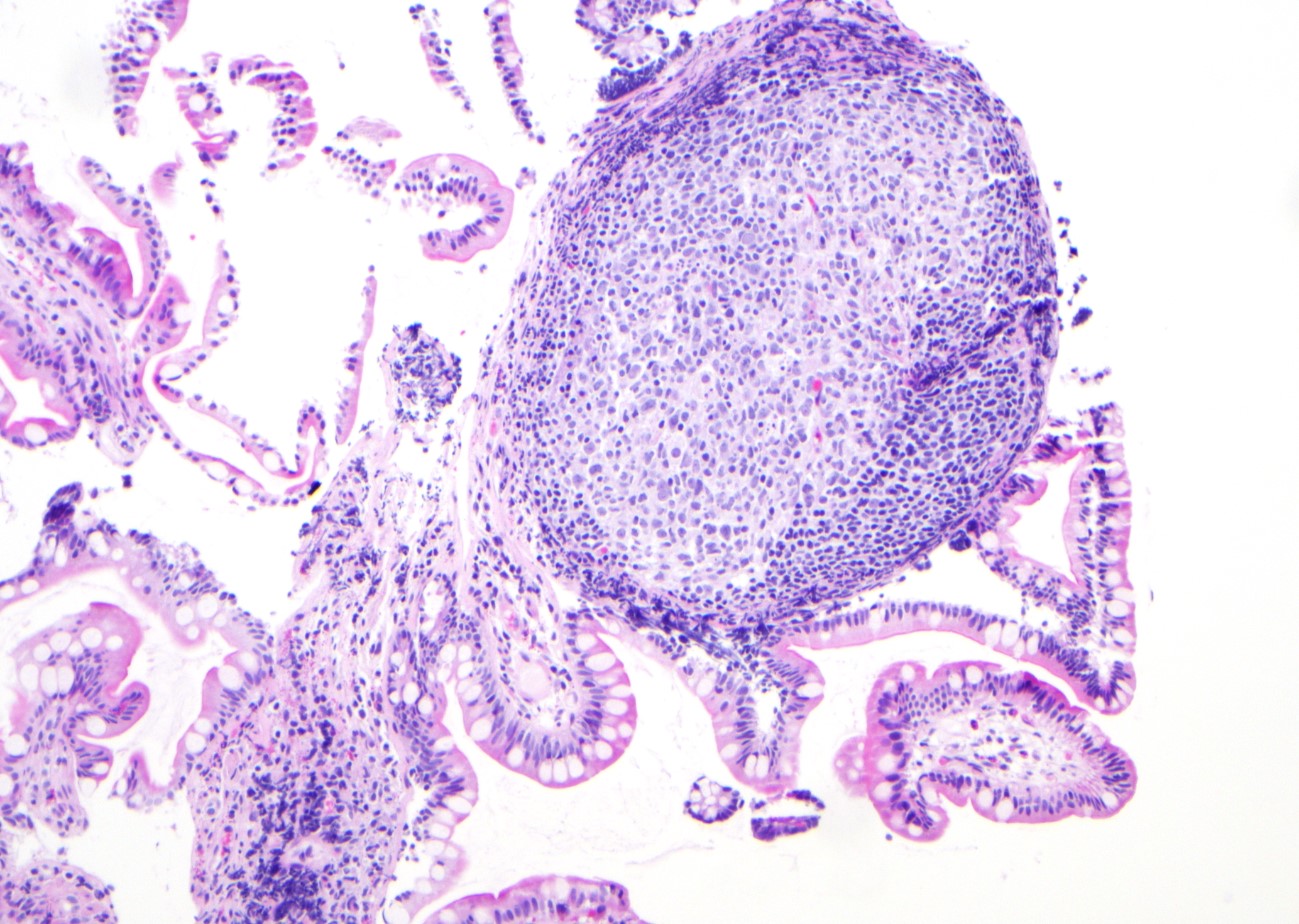
Contributed by Dr. Kaveh Naemi

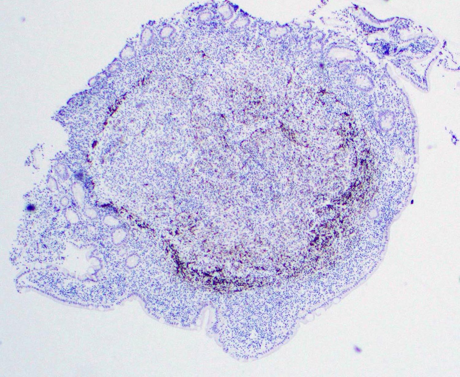
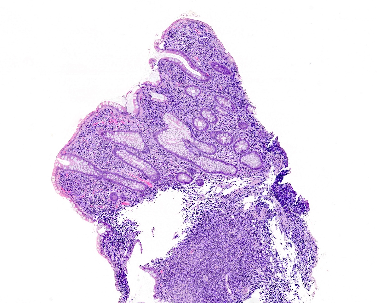
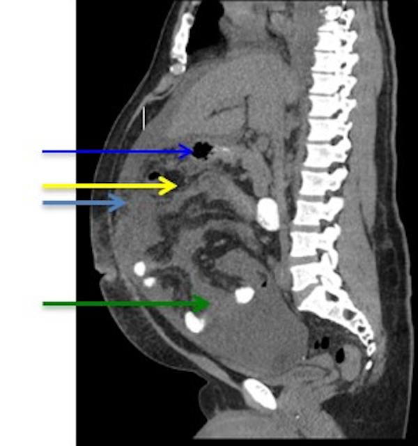
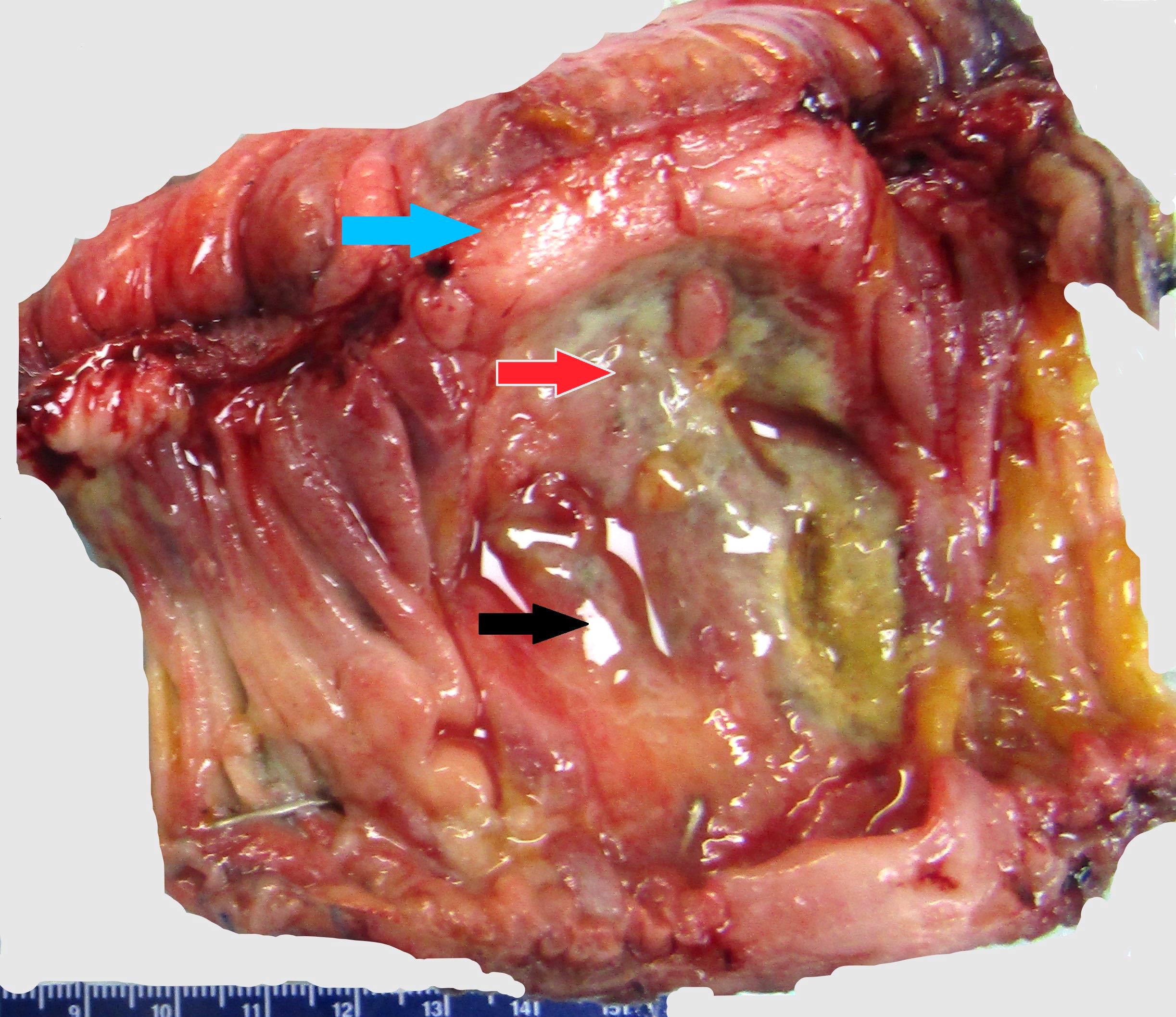
Ileocecal valve mass
Contributed by Saja Asakrah, M.D.
Axillary lymph node
CD20
PAX5

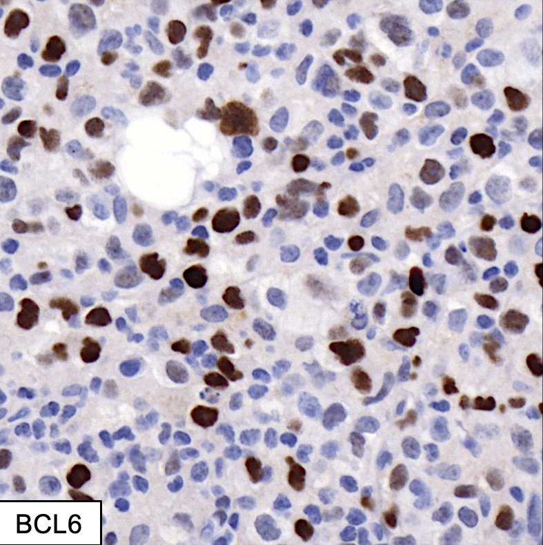
BCL6
CD10
Ki67 positivity
BCL2
EBER in situ hybridization

Contributed by Saja Asakrah, M.D.
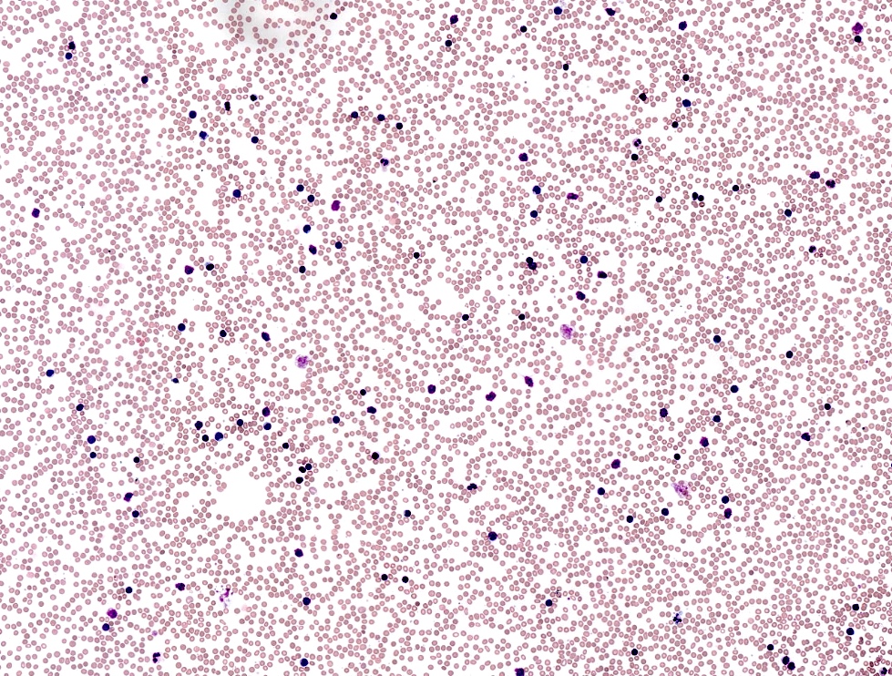
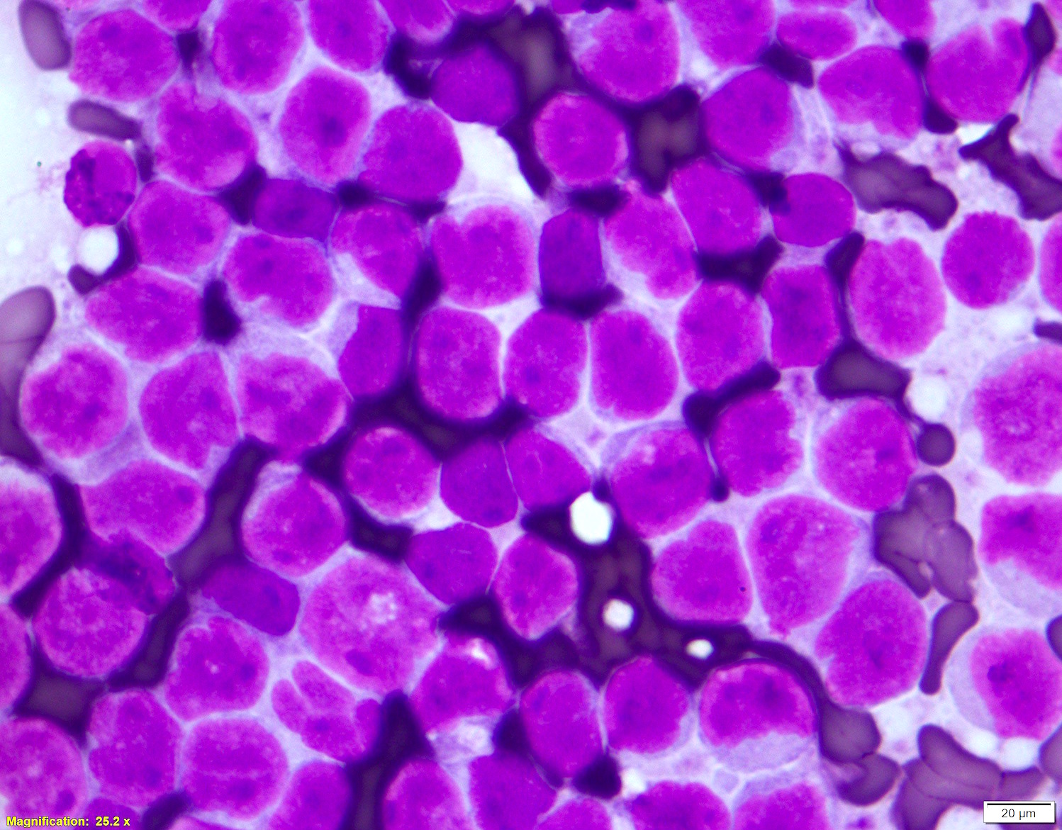
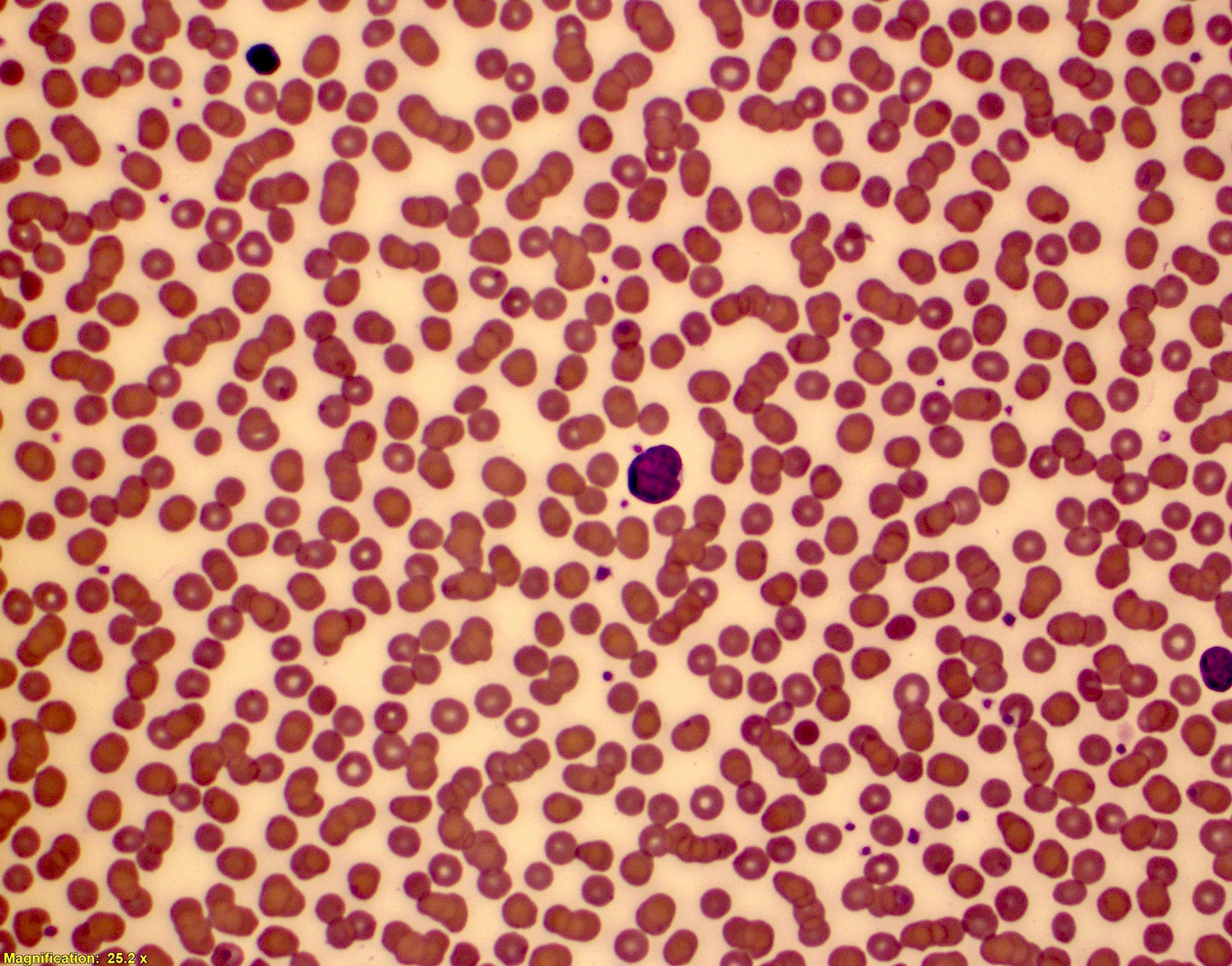
Blood smear, Wright-Giemsa
Contributed by Saja Asakrah, M.D.
B tube
Contributed by Saja Asakrah, M.D.

Tricolor / dual fusion FISH study
Break apart FISH study
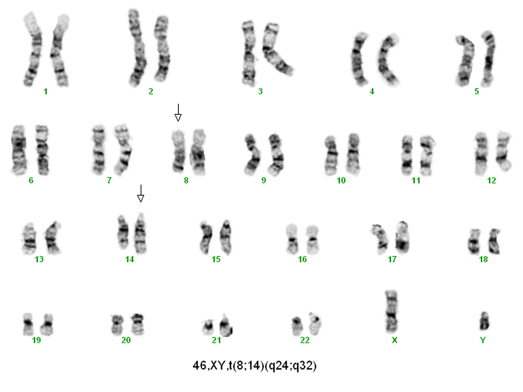
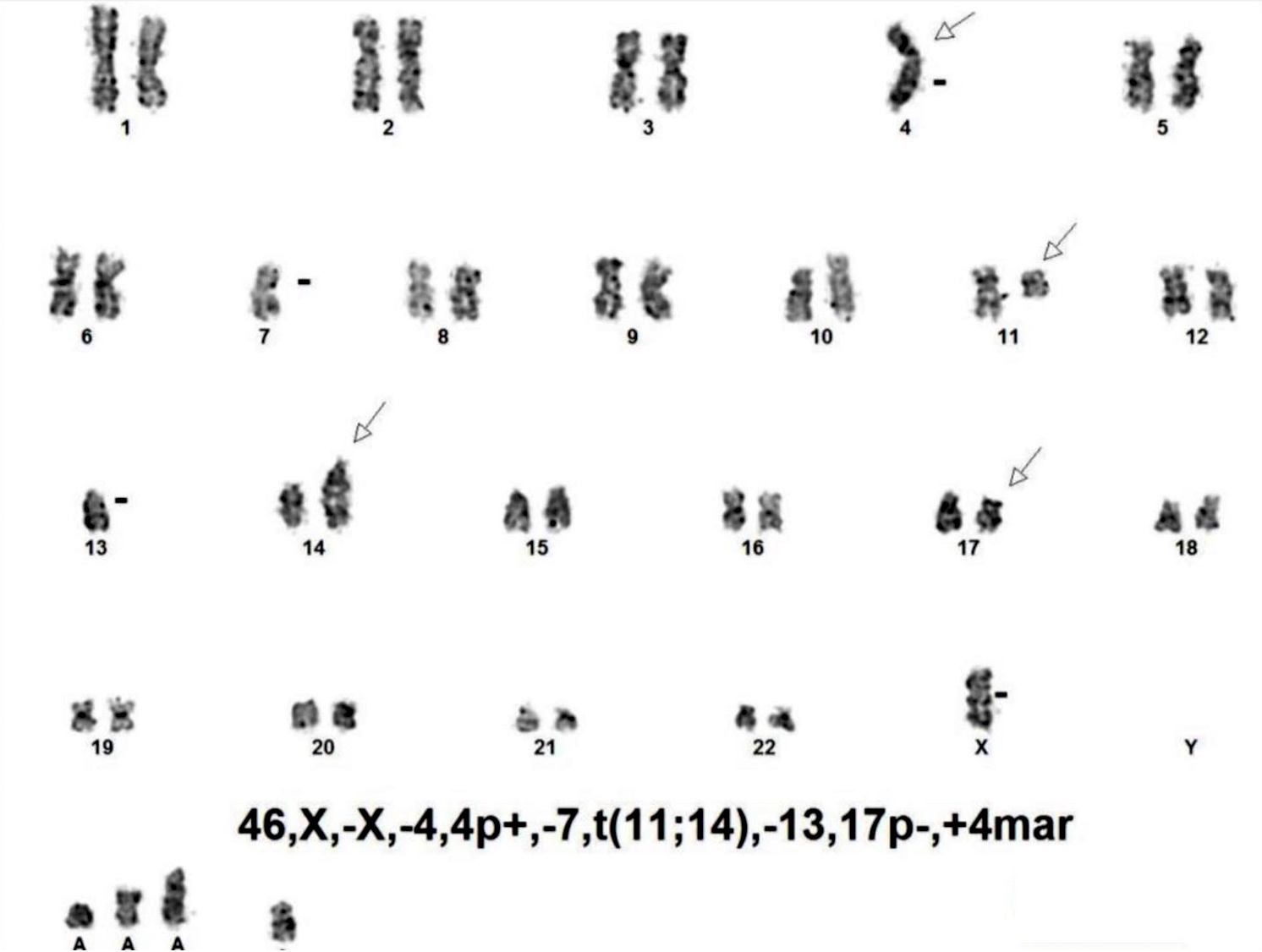
Karyotype
Images hosted on other servers:

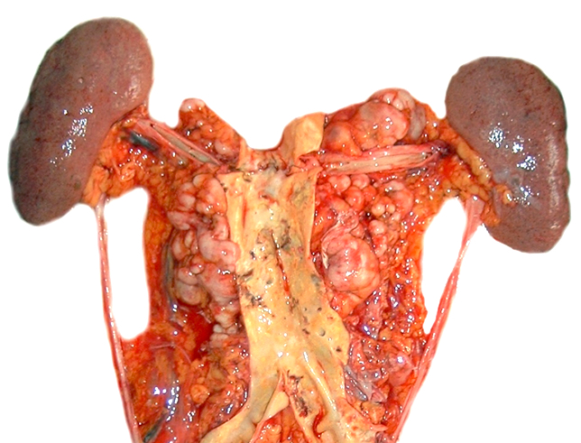
Mesenteric and retroperitoneal lymphadenopathy
AFIP images
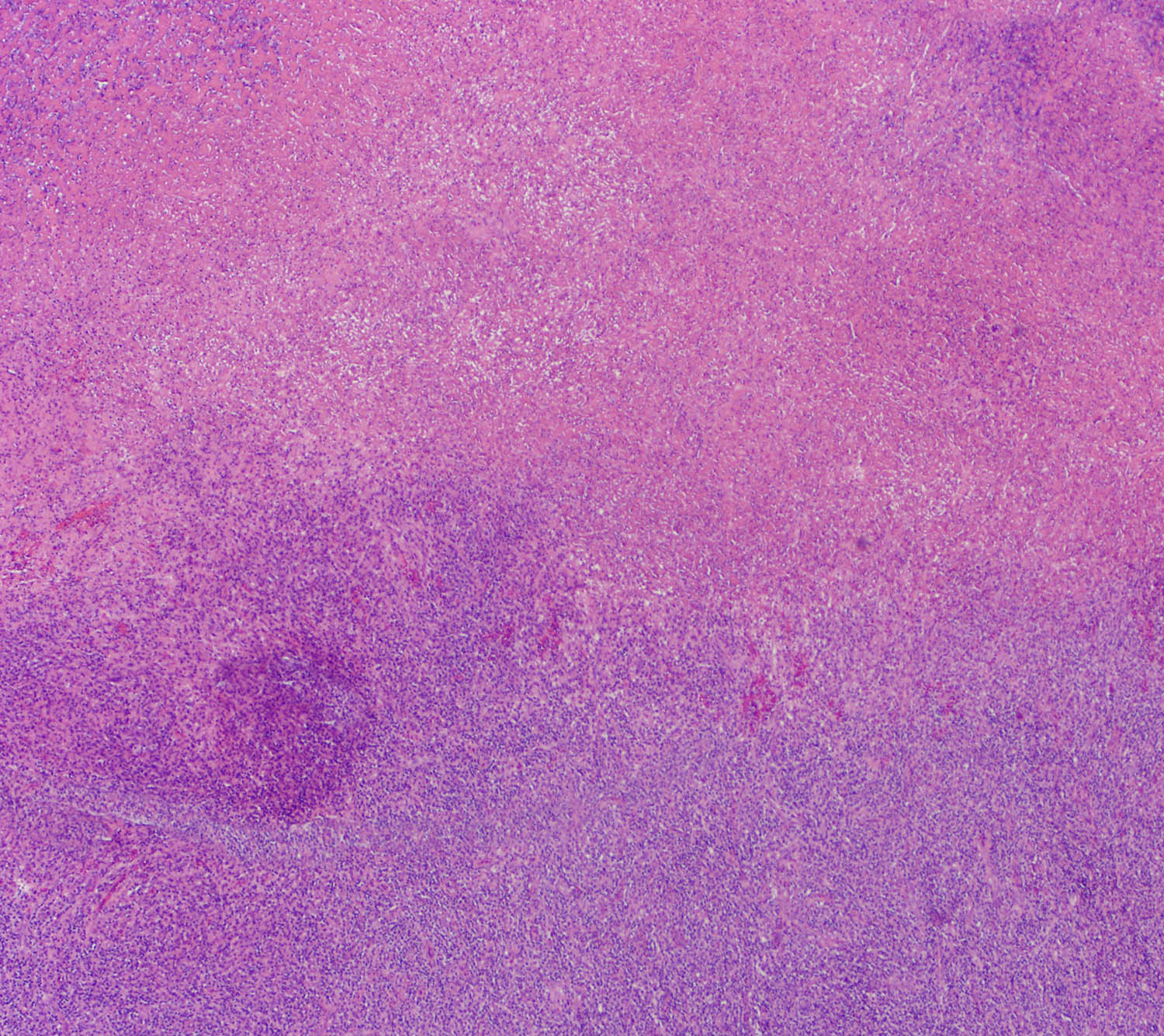
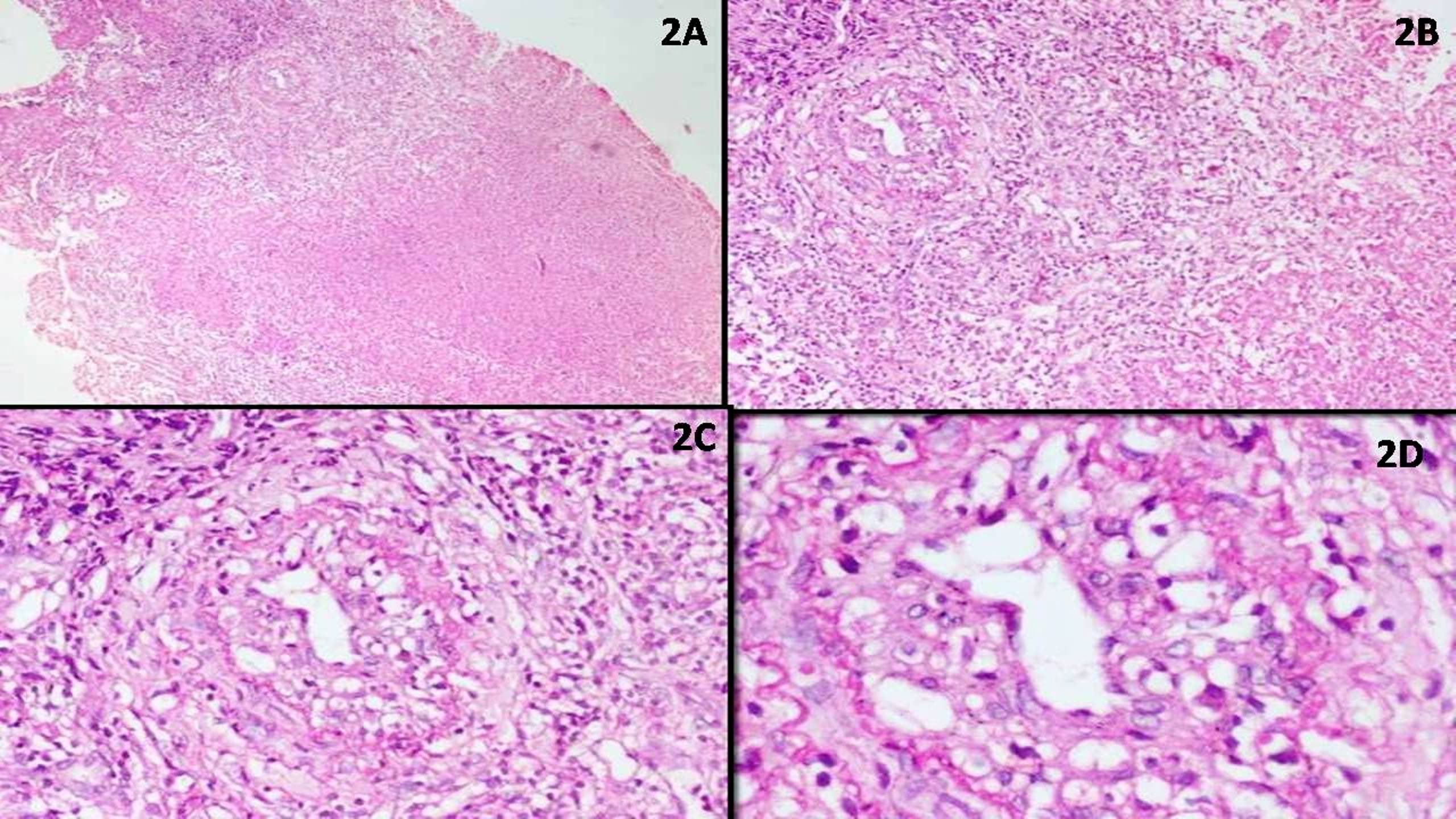
Diffuse fibrosis
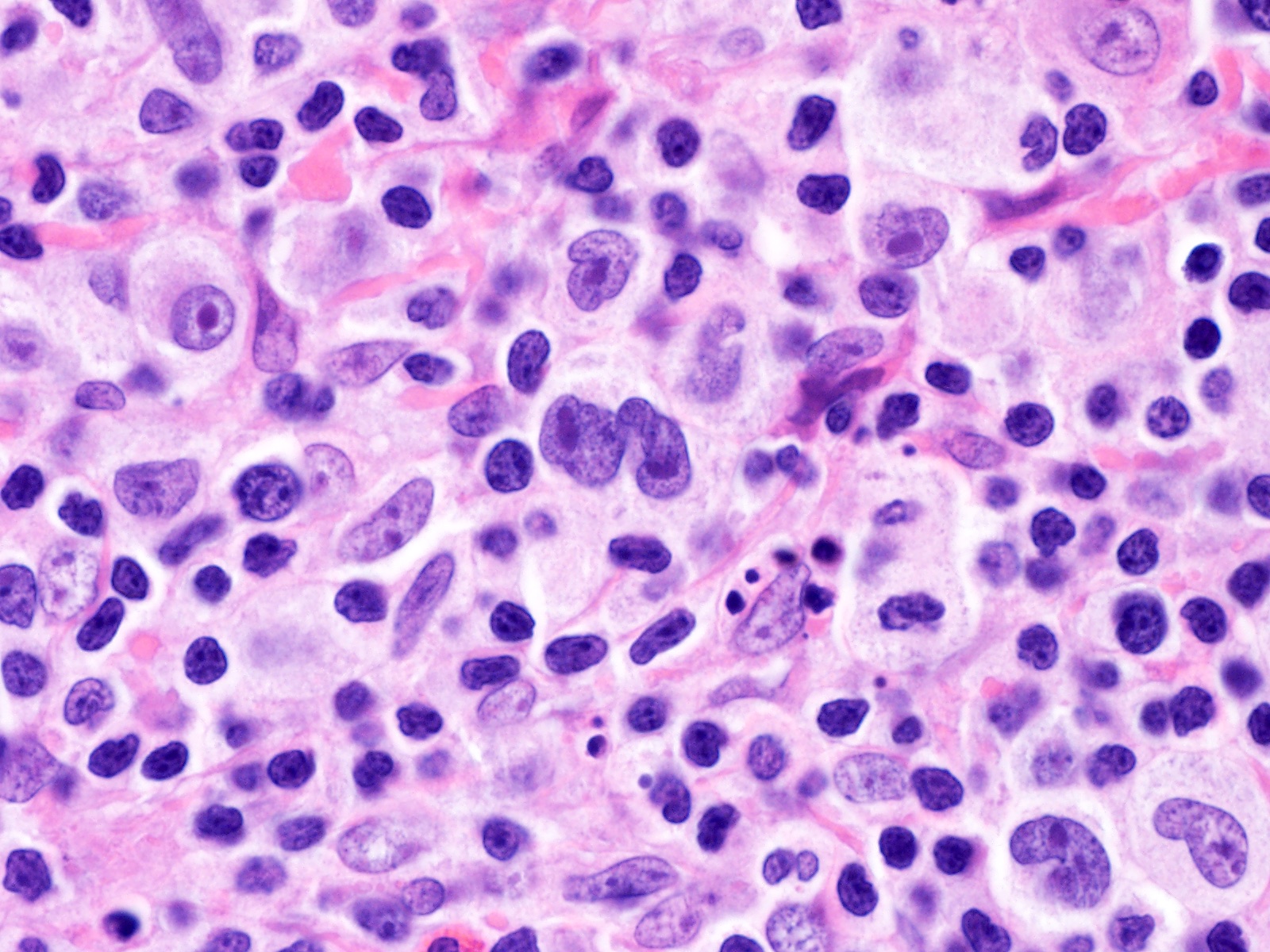
Numerous Hodgkin cells
Contributed by Laurence de Leval, M.D., Ph.D. and Carmen Bárcena, M.D.

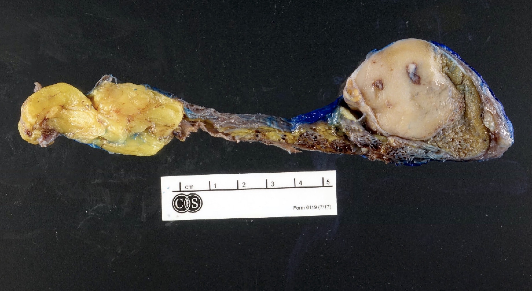
Macroscopy
Contributed by Laurence de Leval, M.D., Ph.D. and Carmen Bárcena, M.D.

Nodular pattern
CD20
HRS cells
CD20

PAX5

OCT2

CD30

CD15

MUM1
CD3

PD-1
EBER in situ hybridization
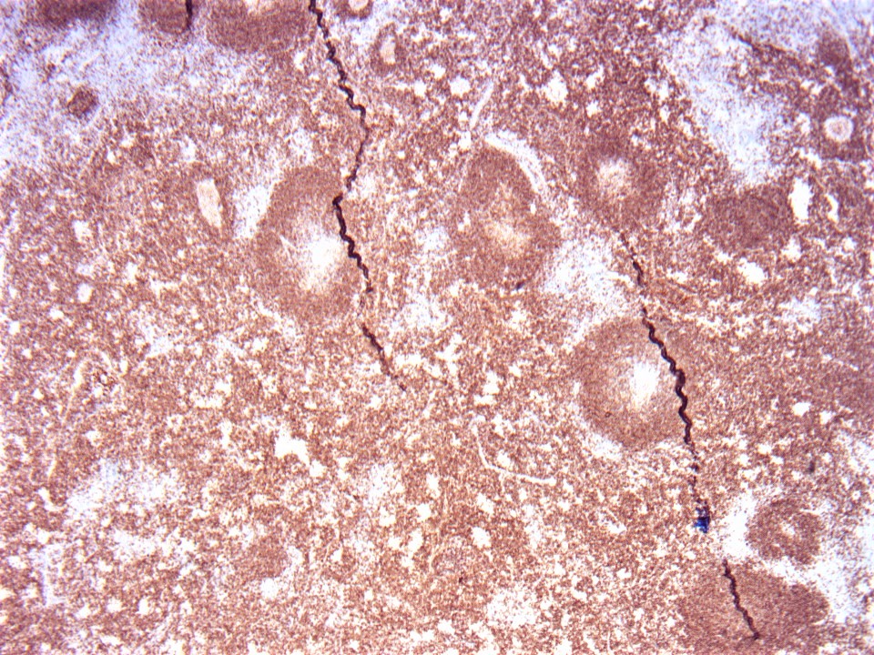
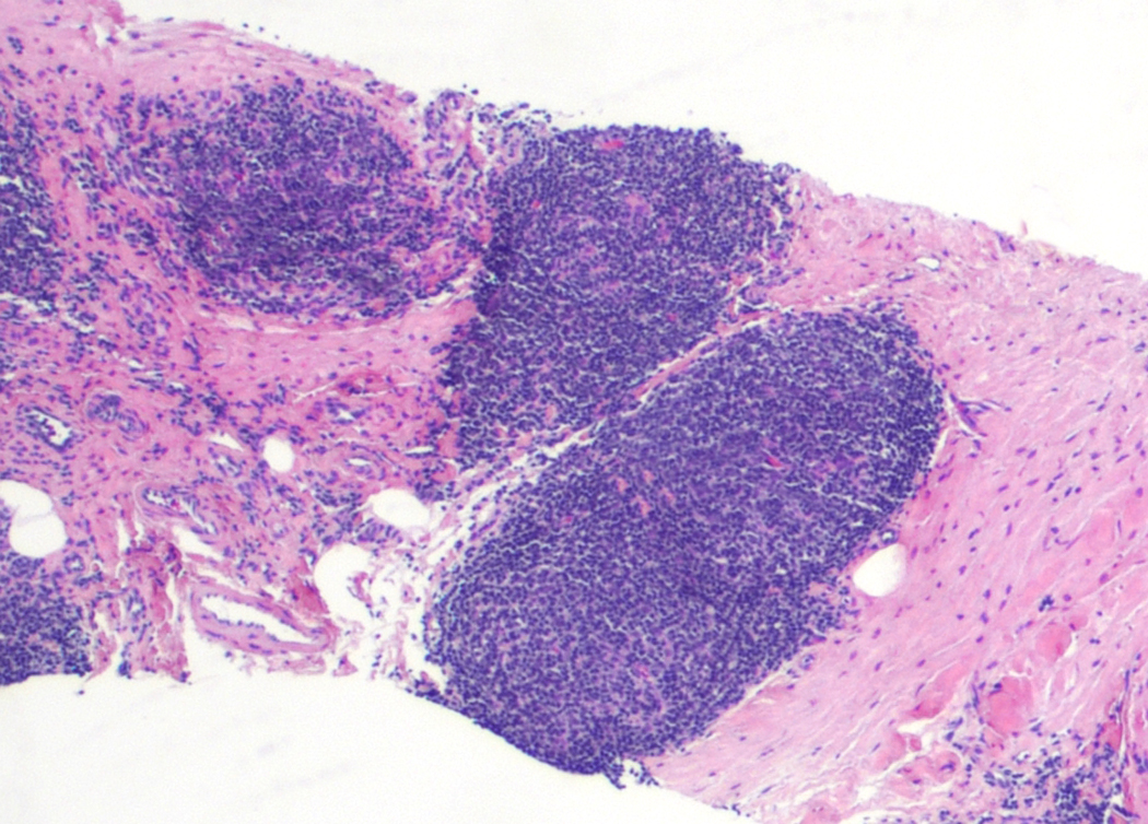
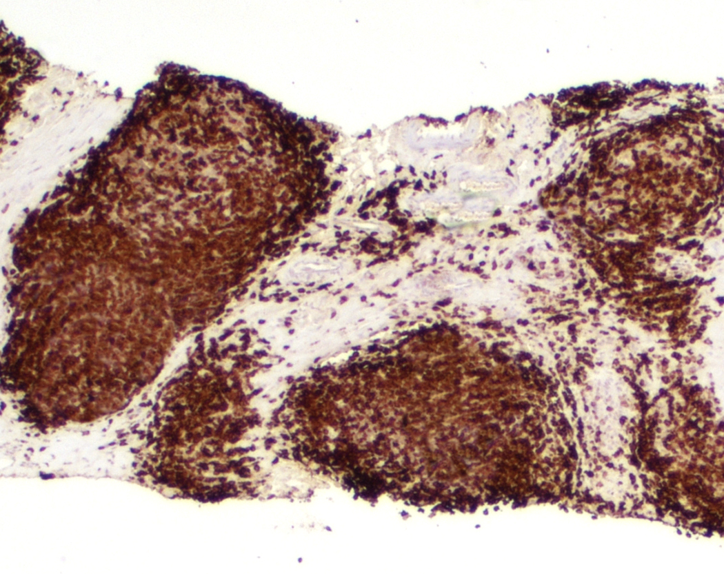
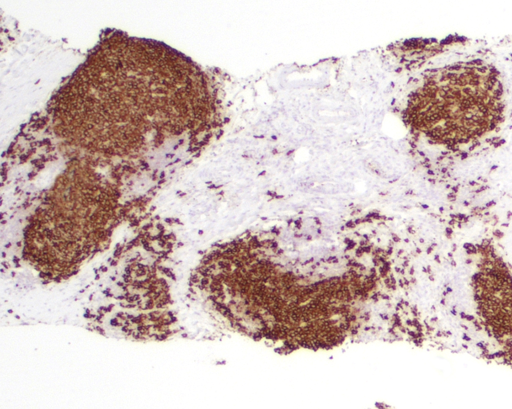
Contributed by Laurence de Leval, M.D., Ph.D. and Carmen Bárcena, M.D.
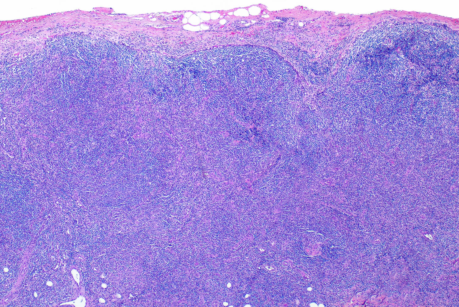
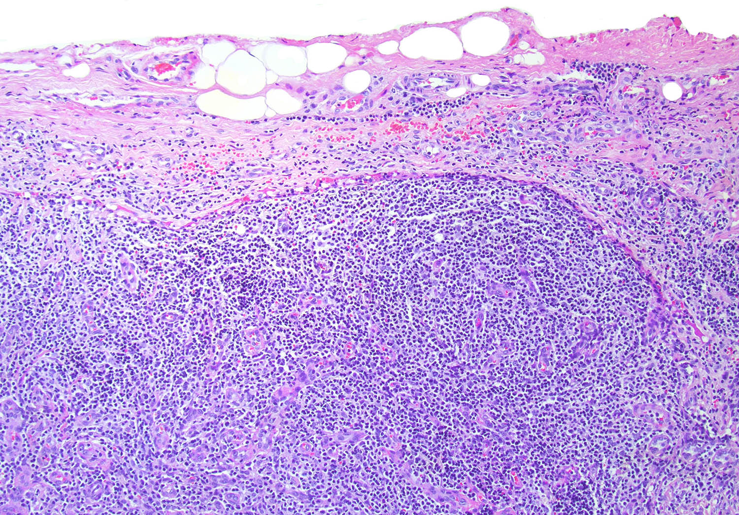
Interfollicular zones
Interfollicular expansion
Reed-Sternberg / Hodgkin cells
Reed-Sternberg / Hodgkin cells and microgranulomas
Polymorphous microenvironment
Focal necrosis
CD45
PAX5
MUM1
CD30
CD15
EBER in situ hybridization
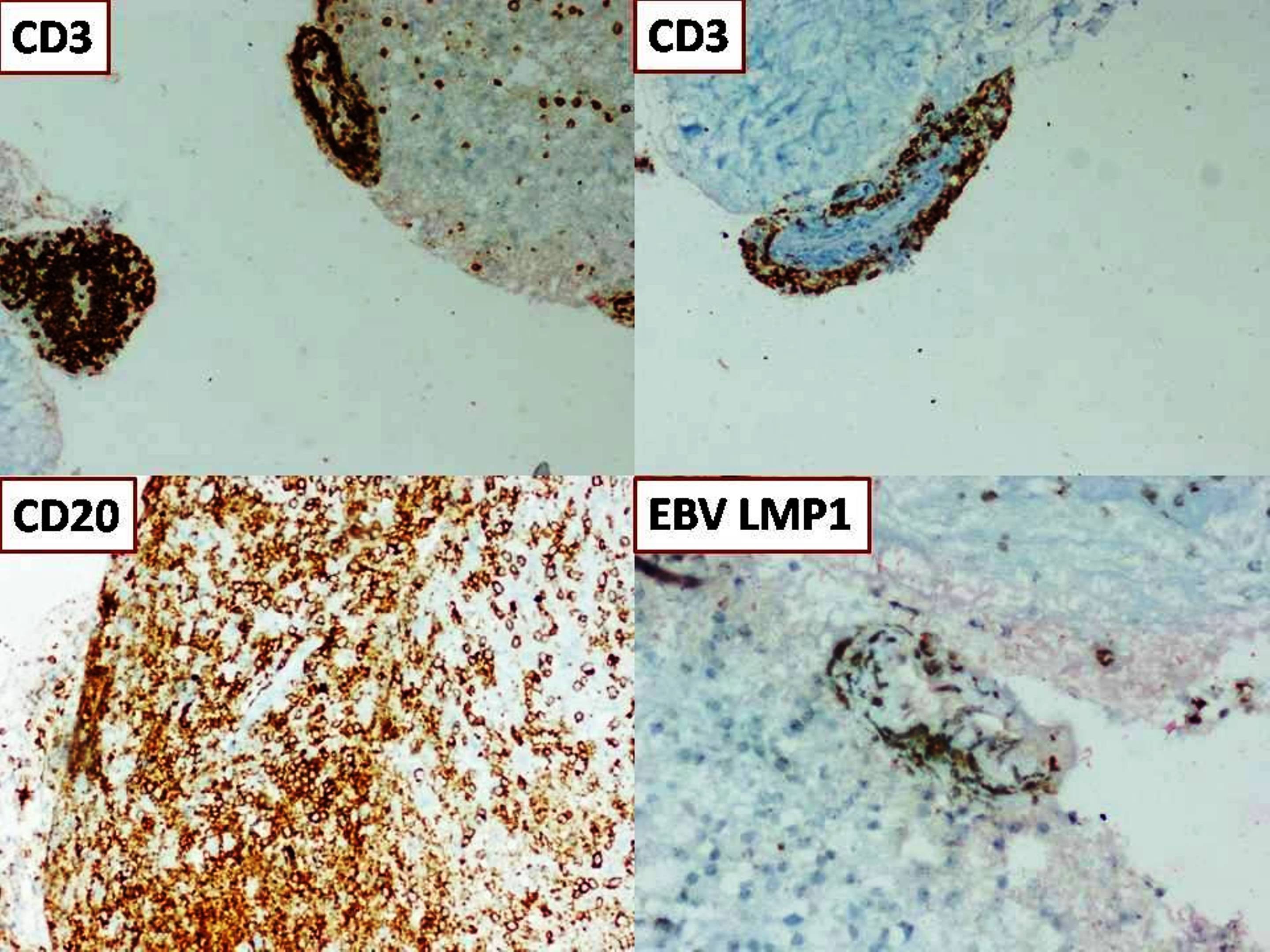
EBV LMP1
PDL1 SP263
Images hosted on other servers:

Lacunar cells


Bands of collagen

Reed-Sternberg cells with bilobed mirror image nuclei, prominent nucleoli and abundant amphophilic cytoplasm
Images hosted on other servers:

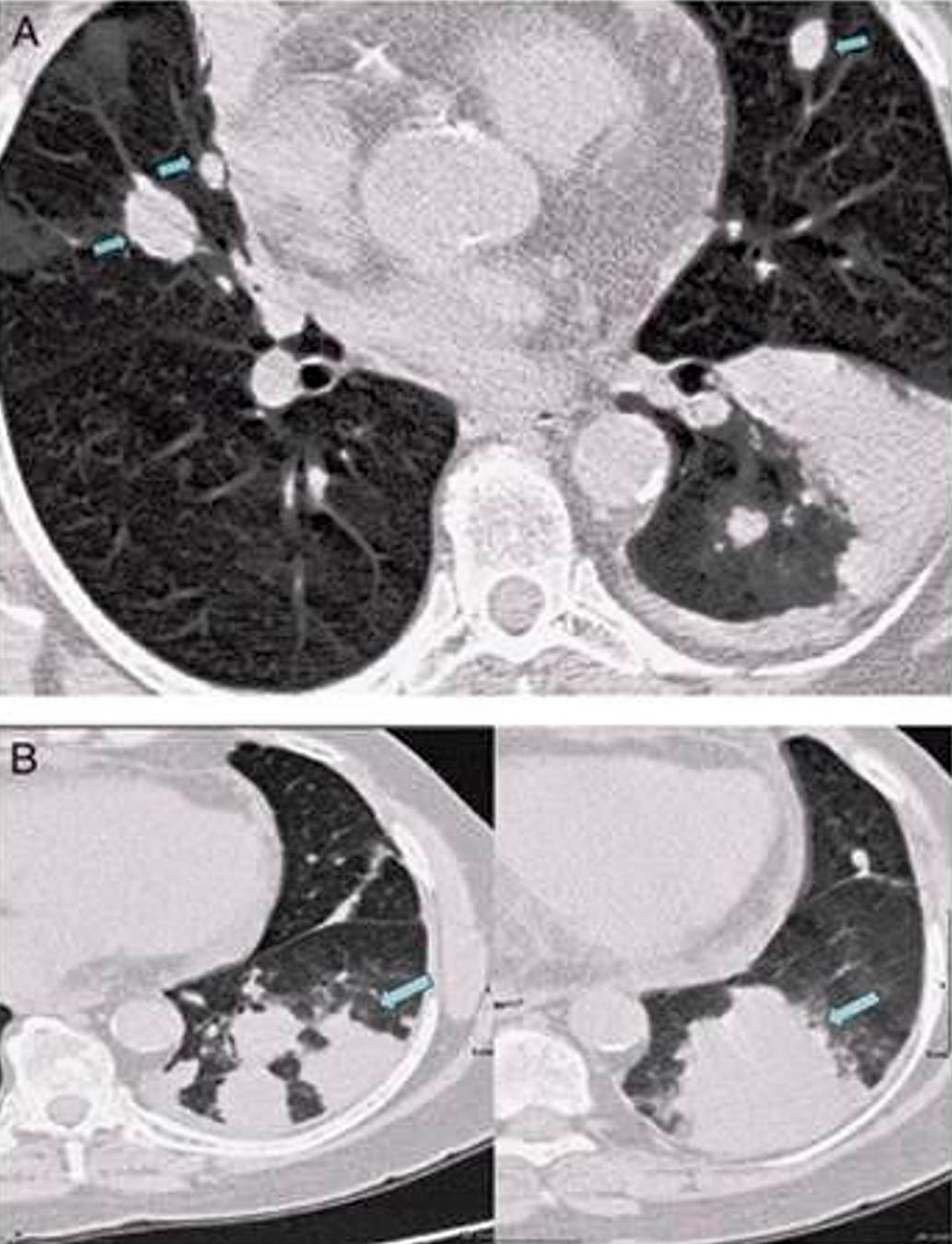
CT scan
Contributed by Béla Kajtár, M.D., Ph.D.
Paraaortic lymph nodes
Images hosted on other servers:

Splenic involvement
Contributed by Béla Kajtár, M.D., Ph.D.
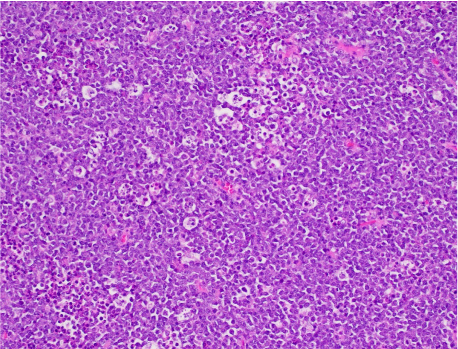
Proliferation centers
Diffuse lymphocytic infiltrate
Proliferation center
CD20
LEF1
Bone marrow involvement
Contributed by Béla Kajtár, M.D., Ph.D.
Touch prep SLL
Contributed by Béla Kajtár, M.D., Ph.D.
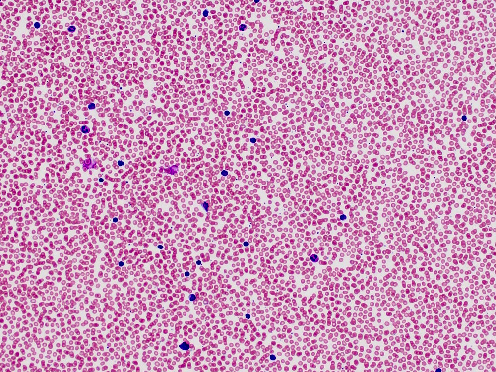
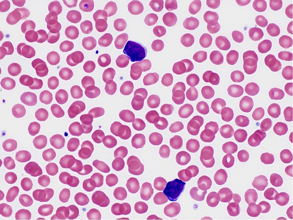
CLL in peripheral blood
Contributed by Béla Kajtár, M.D., Ph.D.
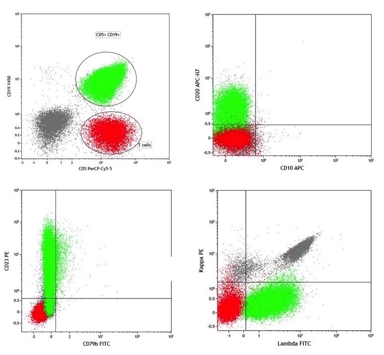
CLL, scatter plot
Contributed by Béla Kajtár, M.D., Ph.D.
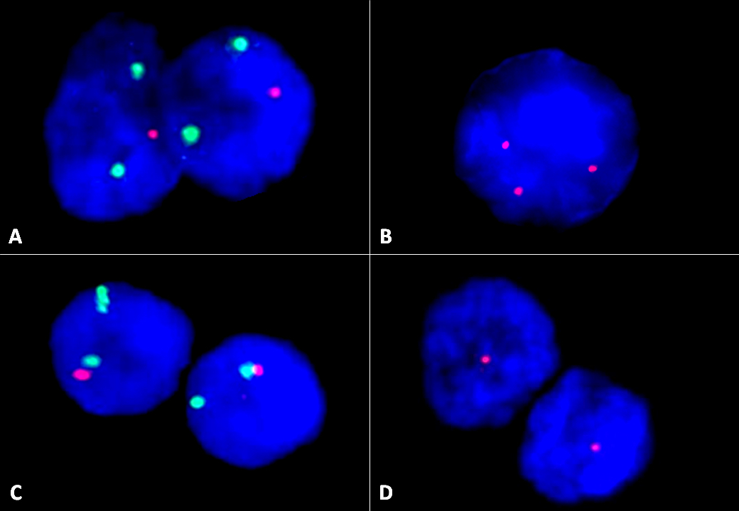
FISH images
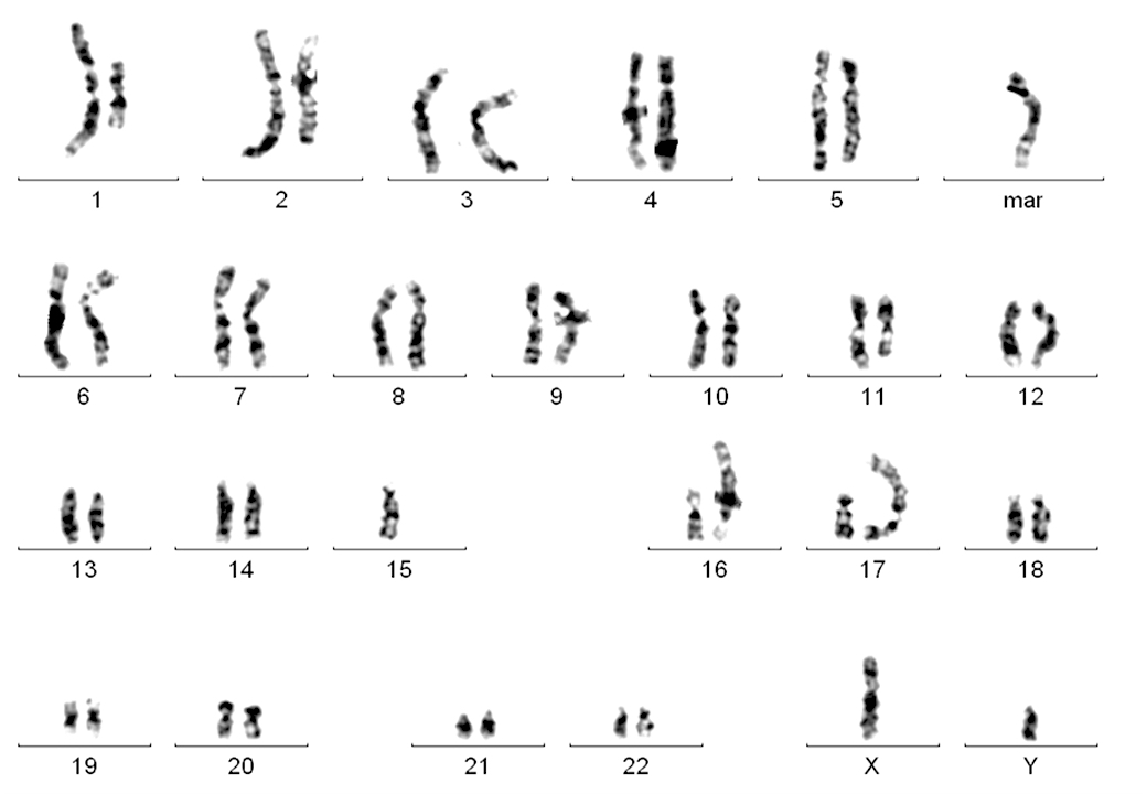
Karyogram
Images hosted on other servers:

Reed-Sternberg cell and microenvironment
Images hosted on other servers:

PET scan before and after therapy
Images hosted on other servers:

Fleshy lymph node

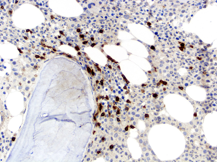
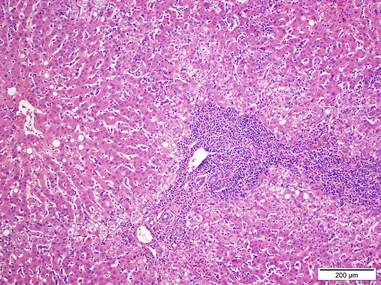
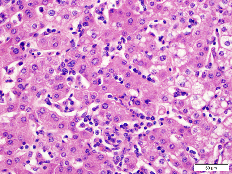
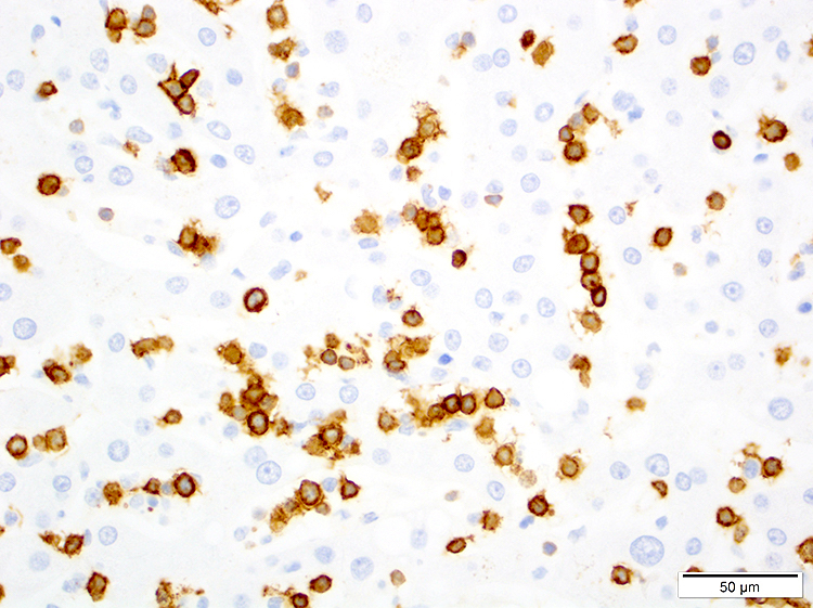
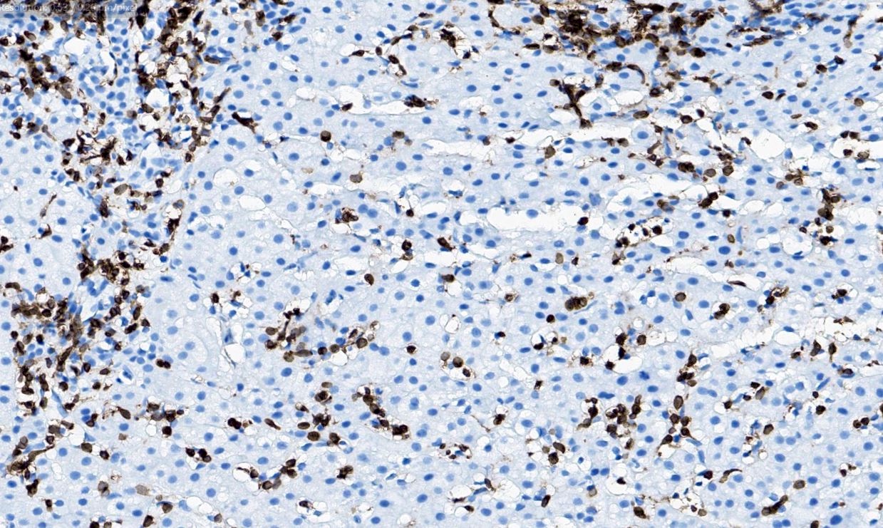
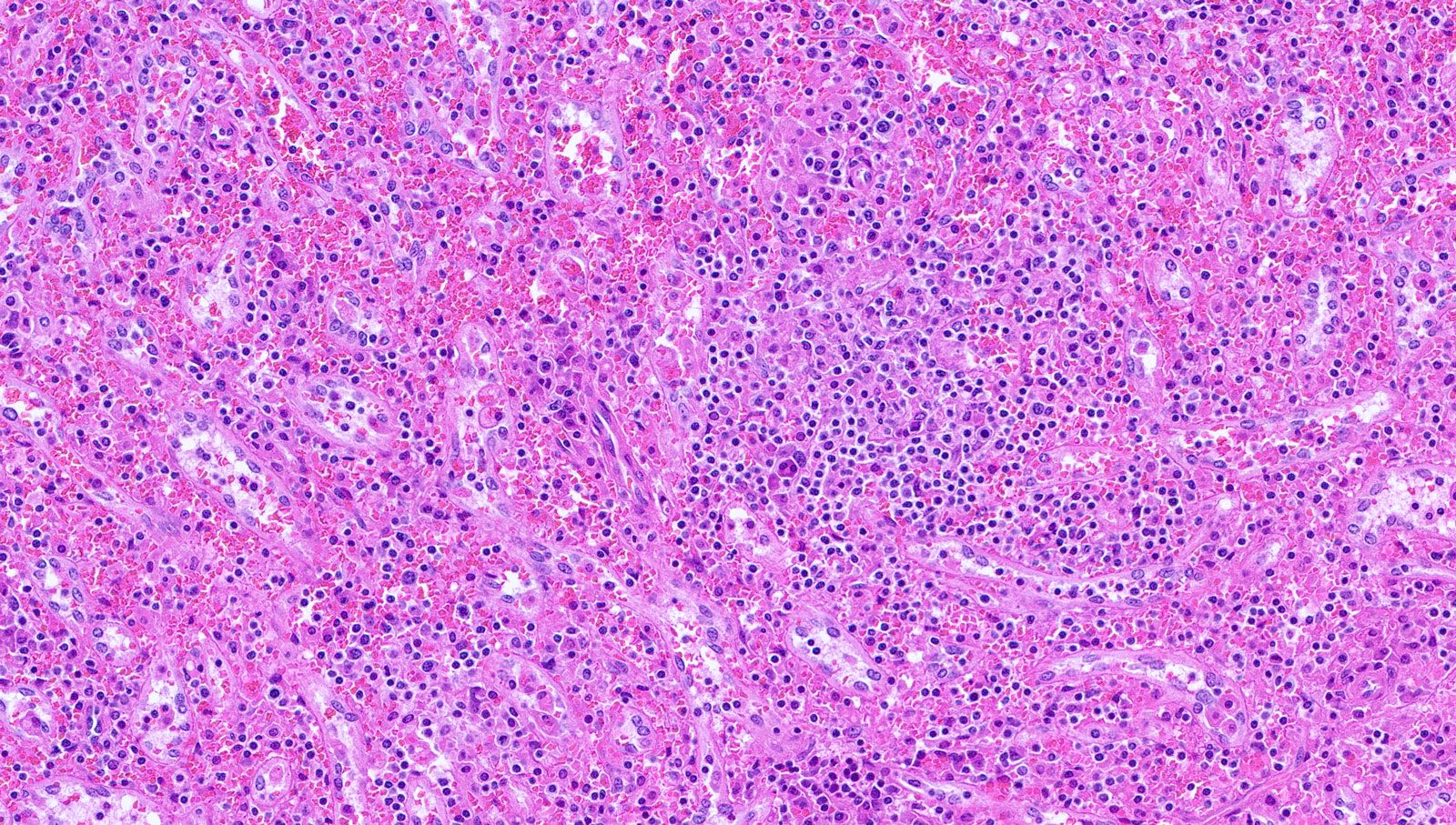
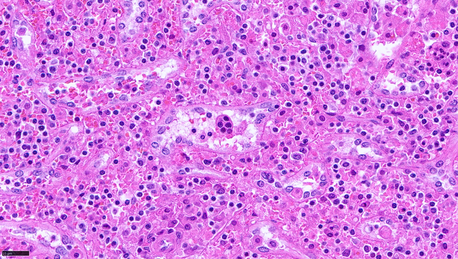
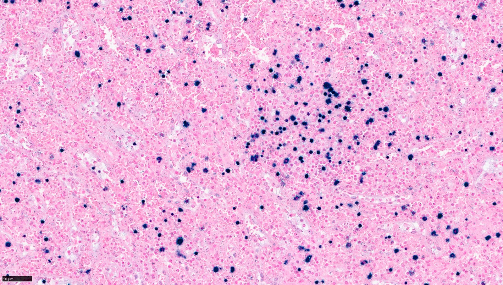
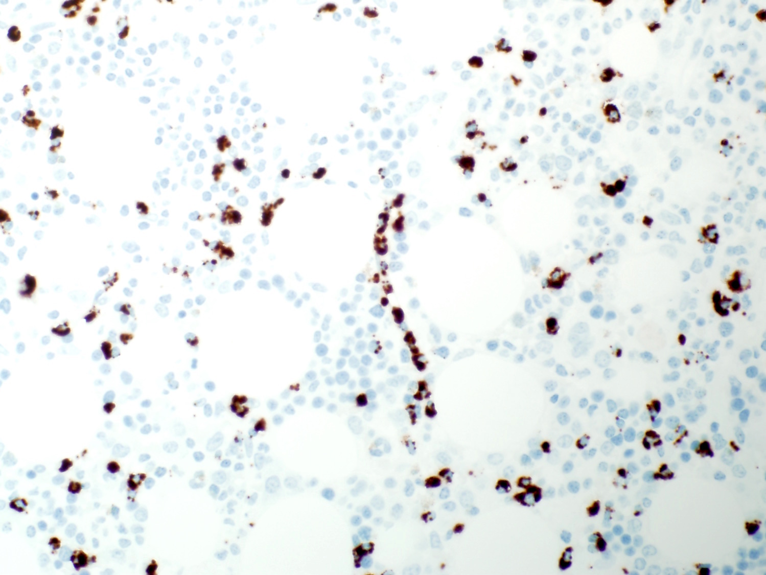
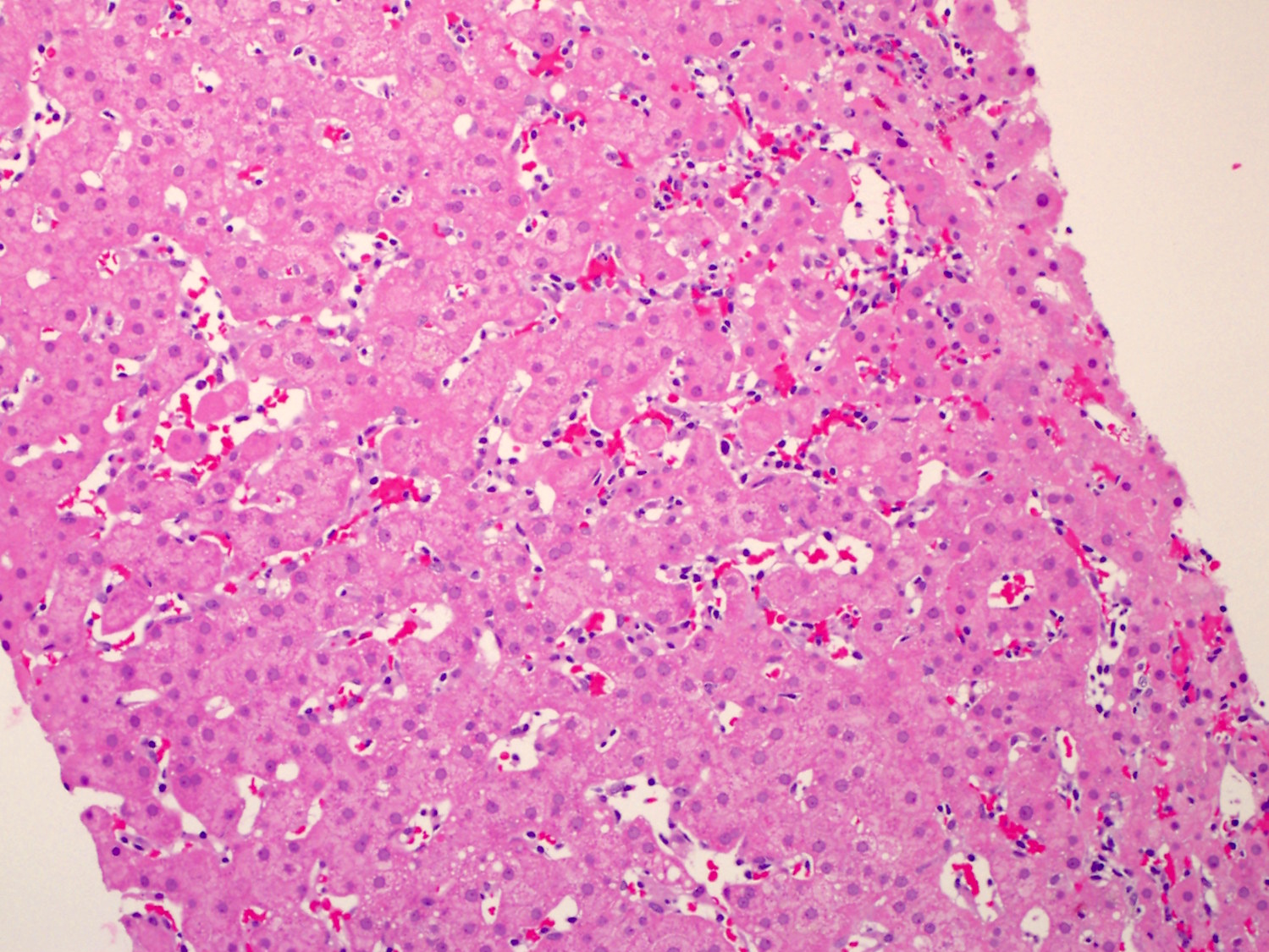
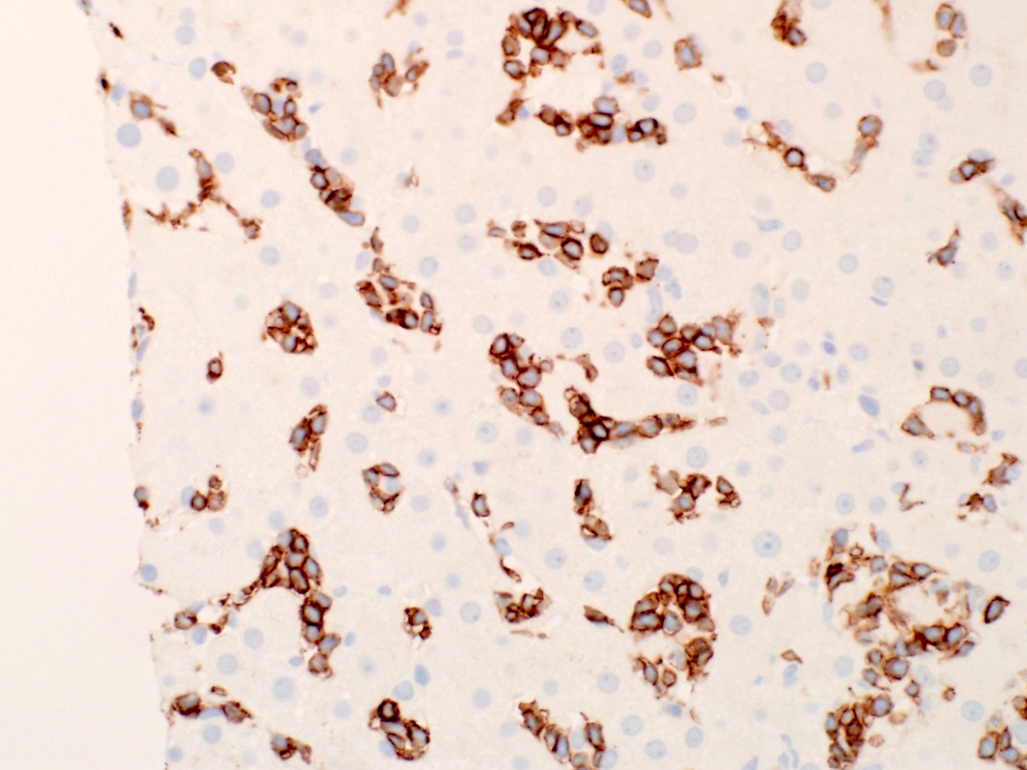
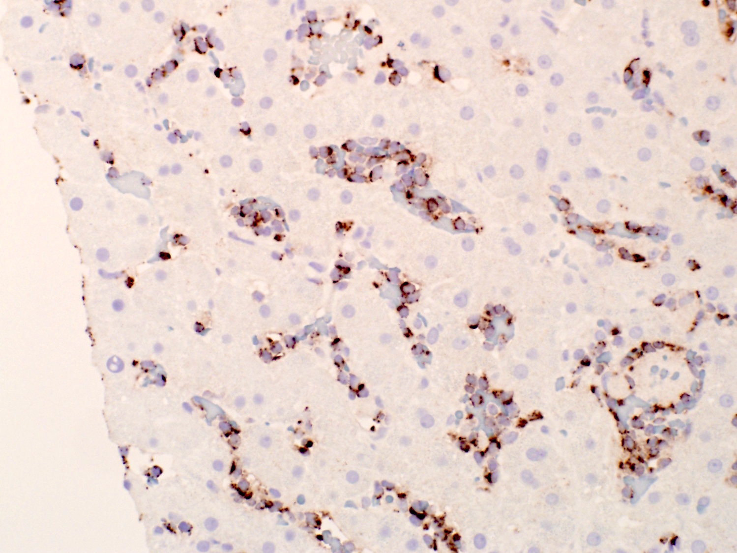
Liver with multifocal involvement
Contributed by Emily Mason, M.D., Ph.D.
Hodgkin and Reed-Sternberg cells
Nodular sclerosis CHL
Scattered Reed-Sternberg cells
Lacunar cell
Syncytial variant
Weak PAX5 expression
CD30 expression
Variable CD20 expression
EBV positivity
Syncytial variant, CD30 stain
Contributed by Emily Mason, M.D., Ph.D.

Reed-Sternberg cell
Images hosted on other servers:


Reed-Sternberg cell nucleus
Images hosted on other servers:

PDL1 / PDL2 gain, amplification
Images hosted on other servers:


Composite mantle cell lymphoma and CLL / SLL
Images hosted on other servers:

Diagnostic approach
Contributed by Genevieve M. Crane, M.D., Ph.D.
High grade B cell
lymphoma with
MYC and BCL2
translocations
BCL2
Myc
Images hosted on other servers:

H&E
Contributed by Genevieve M. Crane, M.D., Ph.D.
Lack of sIg
Images hosted on other servers:

Karyotype
Contributed by Claudia Mendez, M.D.
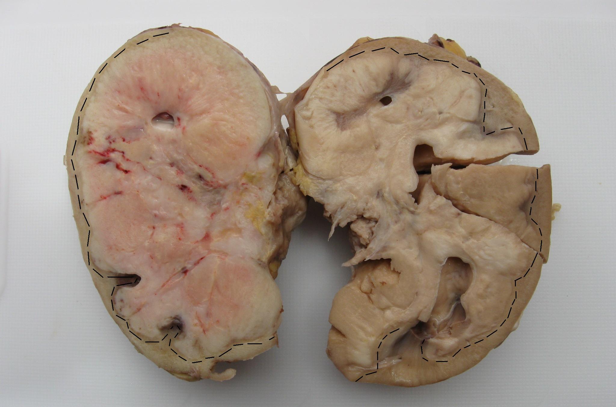
DLBCL involving the kidney
Contributed by Matthew M. Klairmont, M.D.
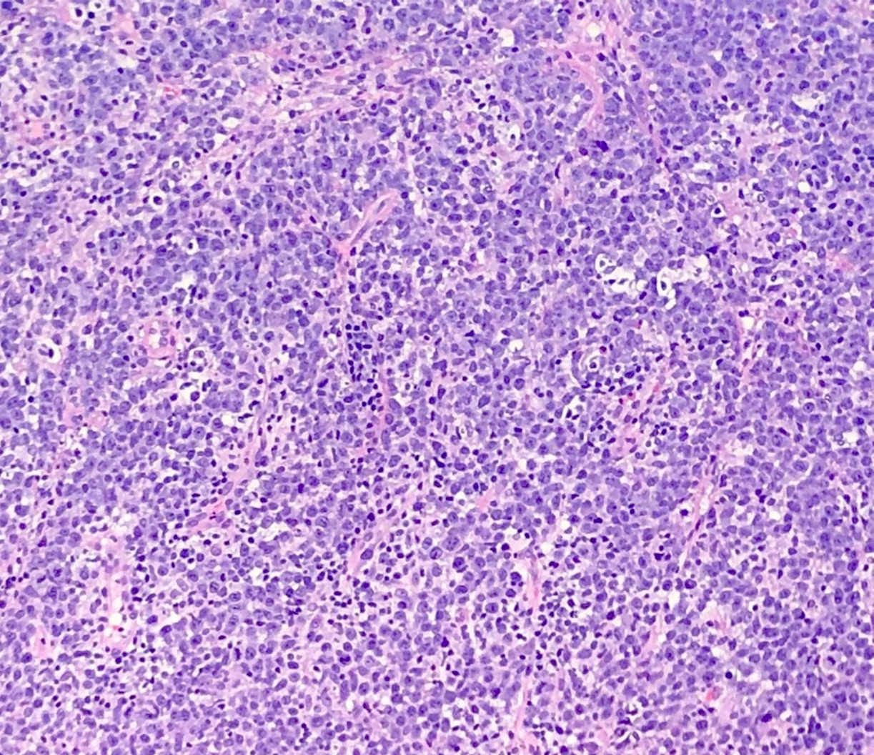
Diffuse growth pattern
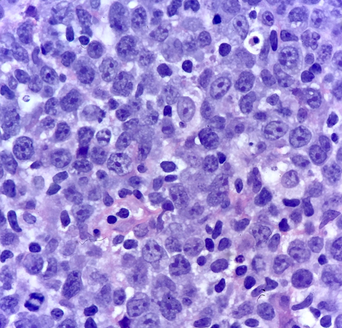
Cytologic features
Bone marrow involvement
Omental involvement
CD20 (bone marrow)
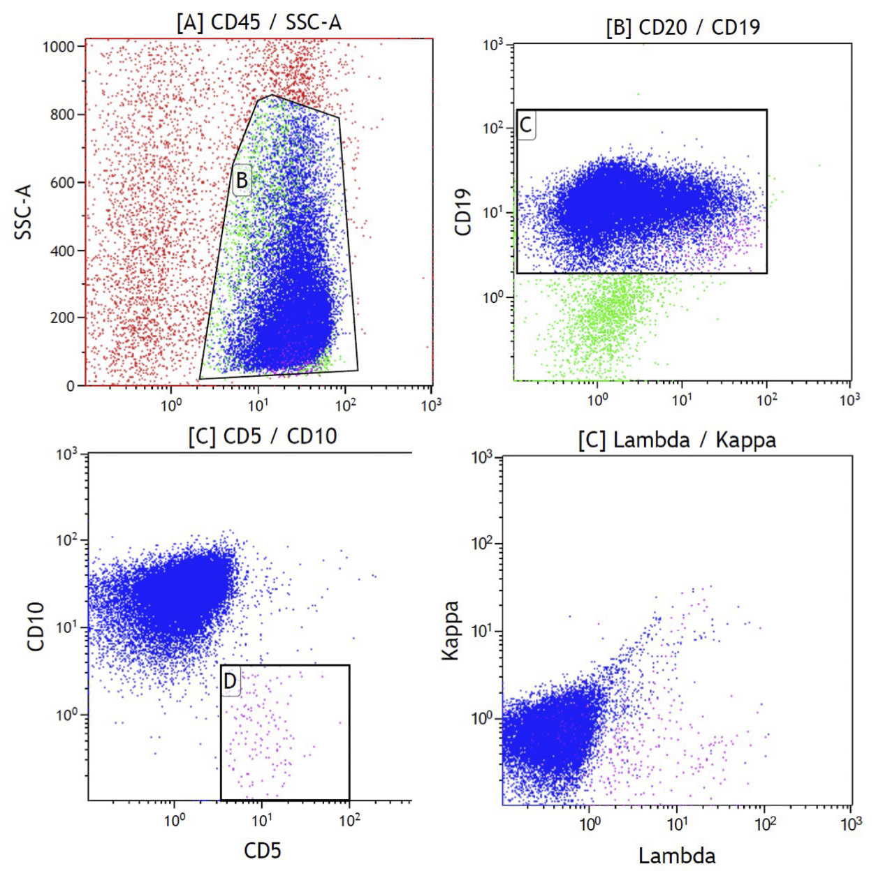
Contributed by Matthew M. Klairmont, M.D.
Scatter plots of splenic mass
Overview of histologic features
Images hosted on other servers:

Dysregulated NFkB signaling pathway
Images hosted on other servers:

CT pelvis and FDG PET
Contributed by Miguel Gonzalez-Mancera, M.D.
Well demarcated testicular mass
Contributed by Miguel Gonzalez-Mancera, M.D.
Diffuse infiltration
High mitotic activity
Centroblastic morphology
Immunoblastic morphology
Lymphoma infiltrating testicular parenchyma
CD20

CD10
BCL6
MUM1
Contributed by Sudhir Perincheri, M.B.B.S., Ph.D.
Polymorphic type
Reed-Sternberg-like cells
CD20
EBER

Monomorphic type
Necrosis
Images hosted on other servers:

Differentiating characteristics of EBV+ DLBCL and EBVMCU
Contributed by Sergio Pina-Oviedo, M.D.

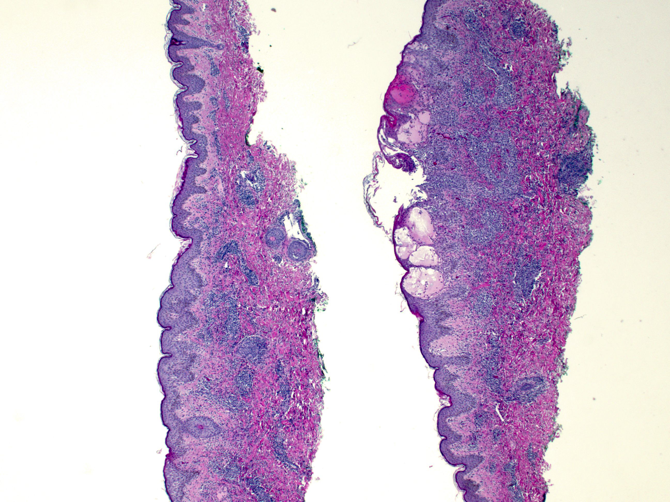
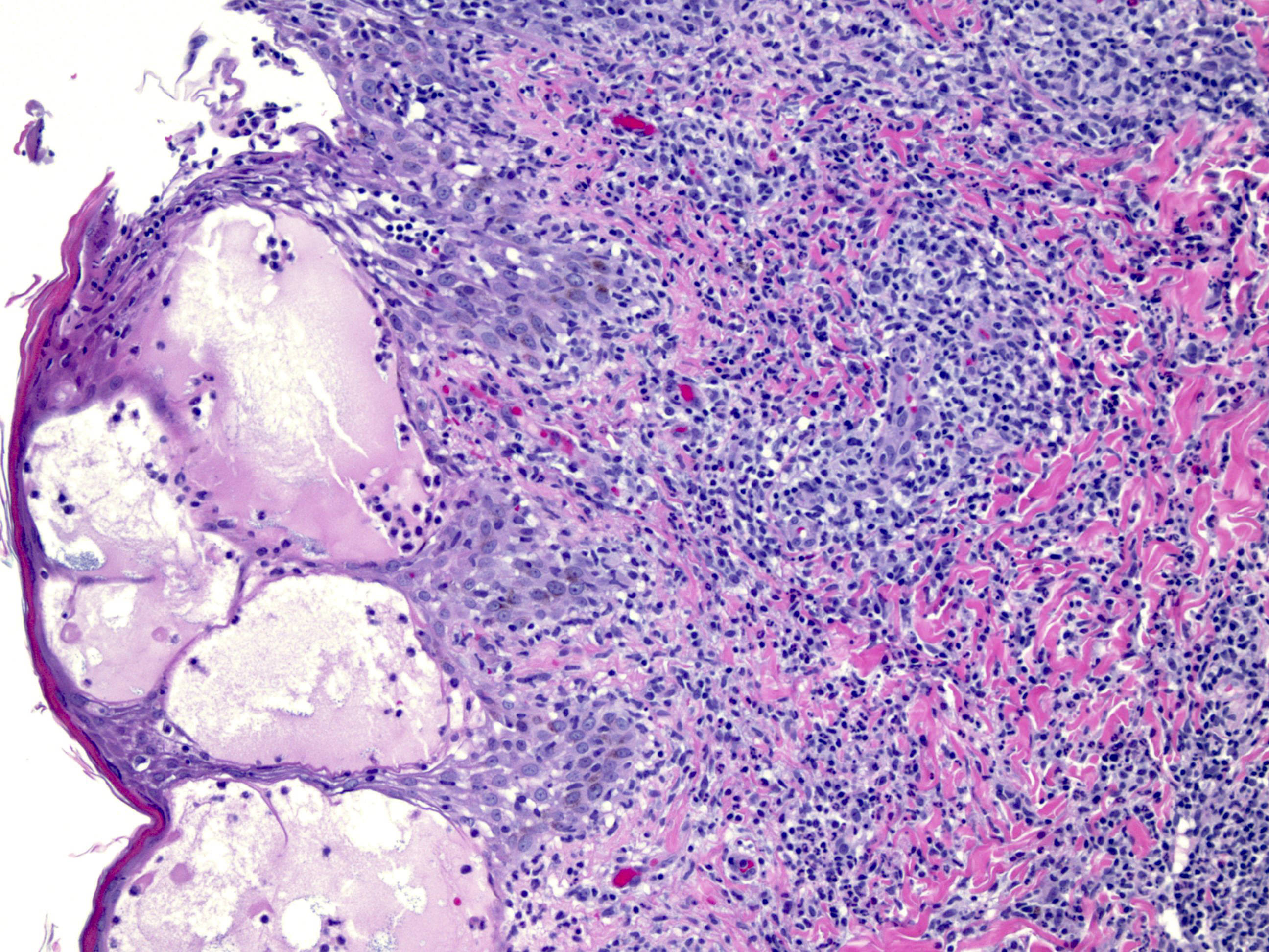
Surface ulceration and necrosis

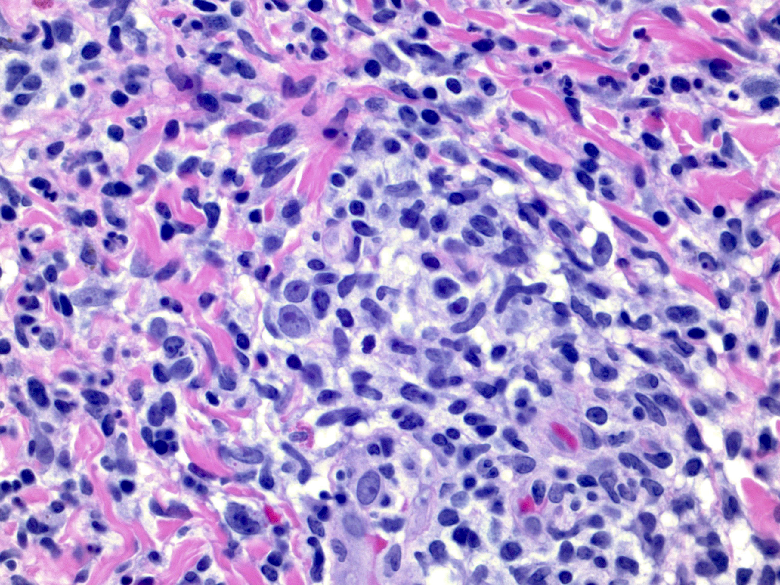
Atypical cells with Hodgkin / RS cell-like morphology

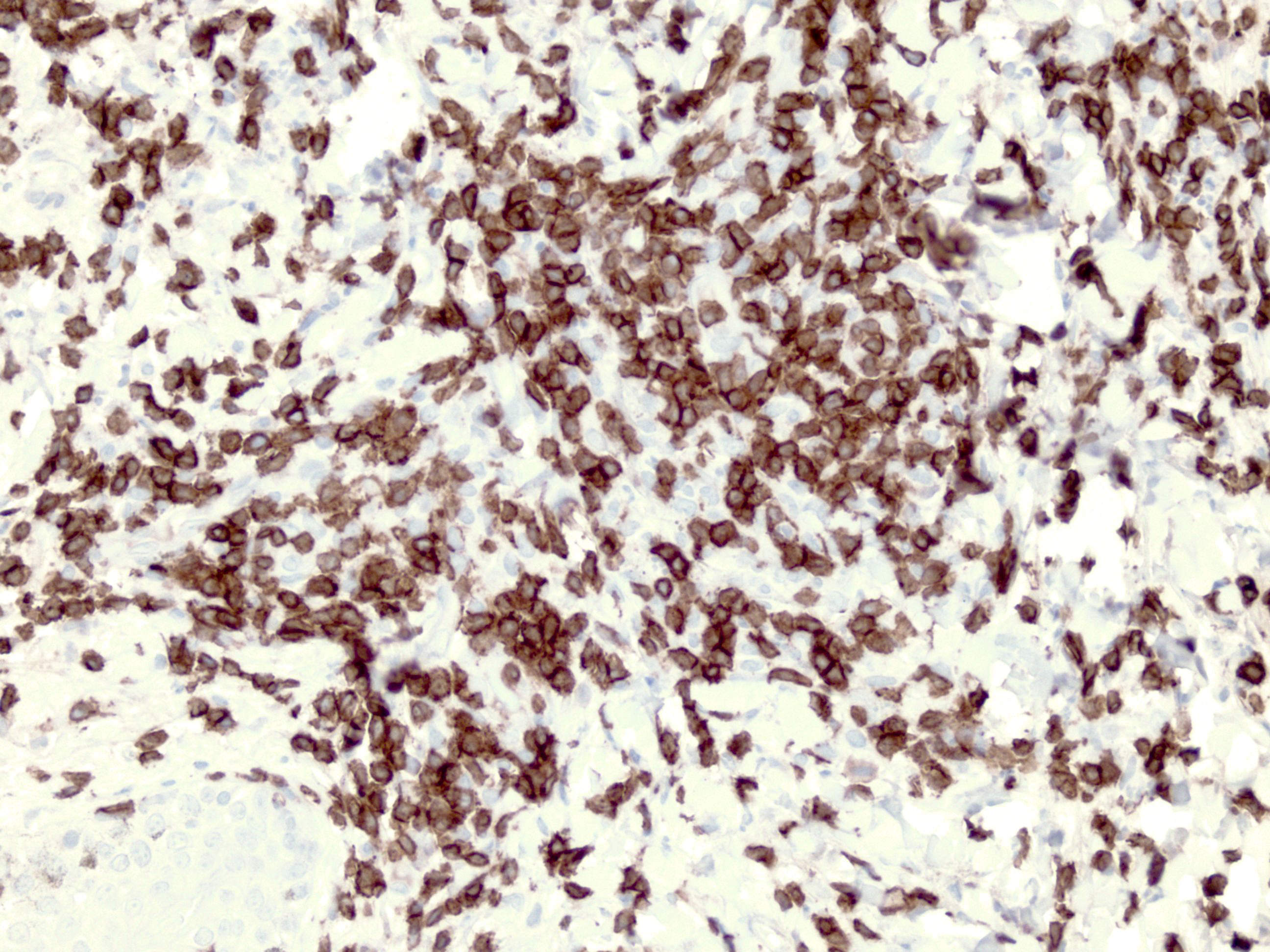
EBER ISH positive in atypical cells
CD20 positive B cells

CD3 positive T cells
Contributed by Carlos A. Murga-Zamalloa, M.D.

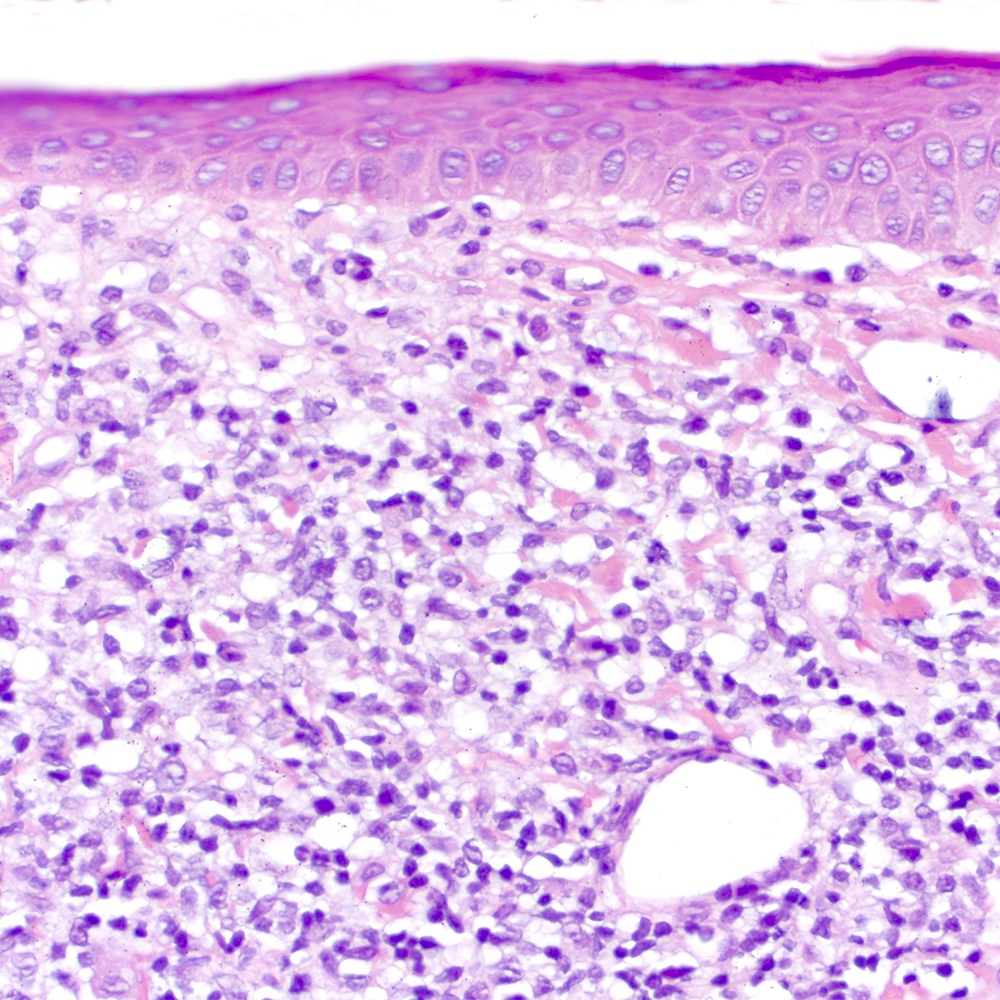
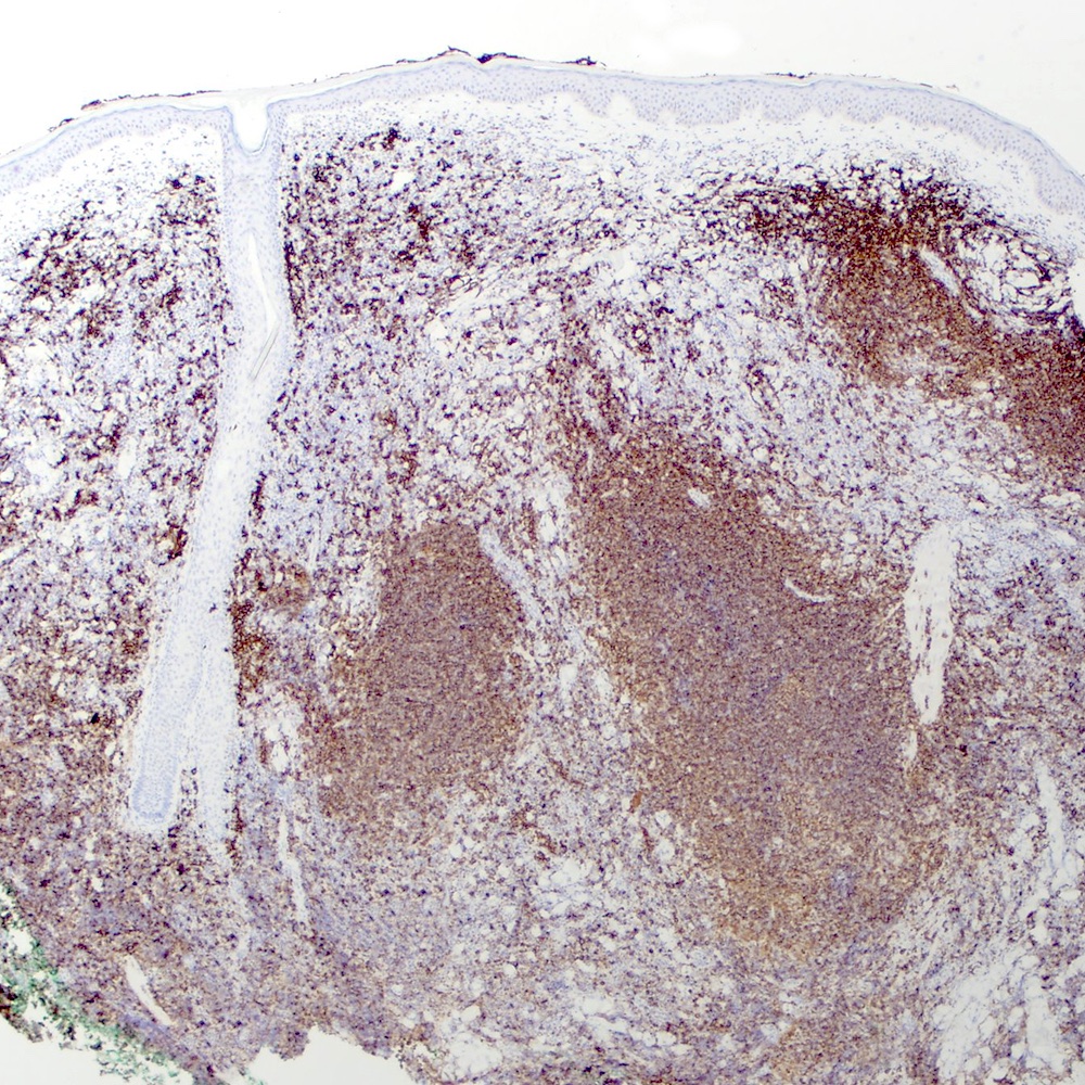
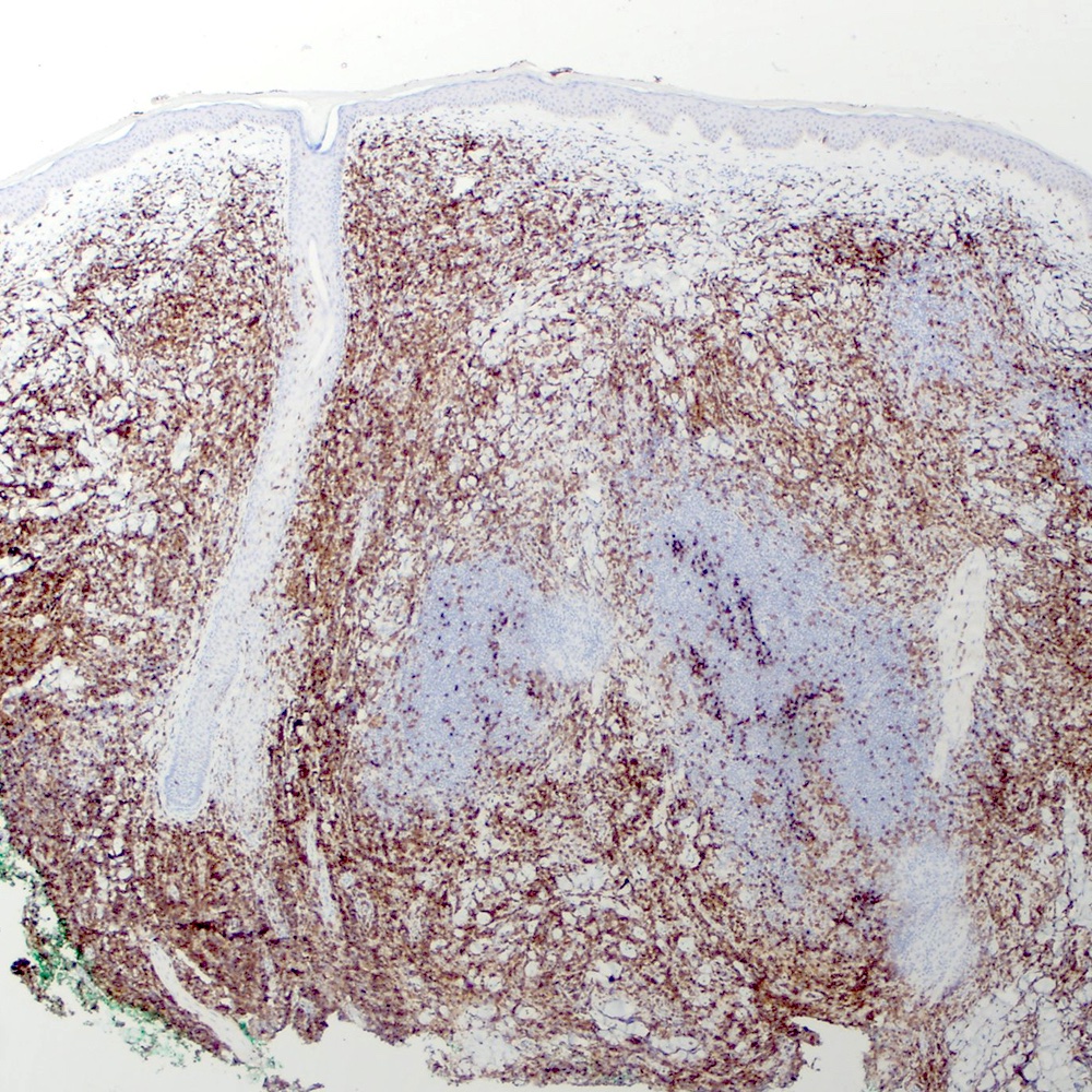
Diffuse atypical infiltrate

Morphological atypia

Pleomorphic infiltrate
Pleomorphism and marked cellular atypia

EBV positive lymphocytes (EBER in situ hybridization)
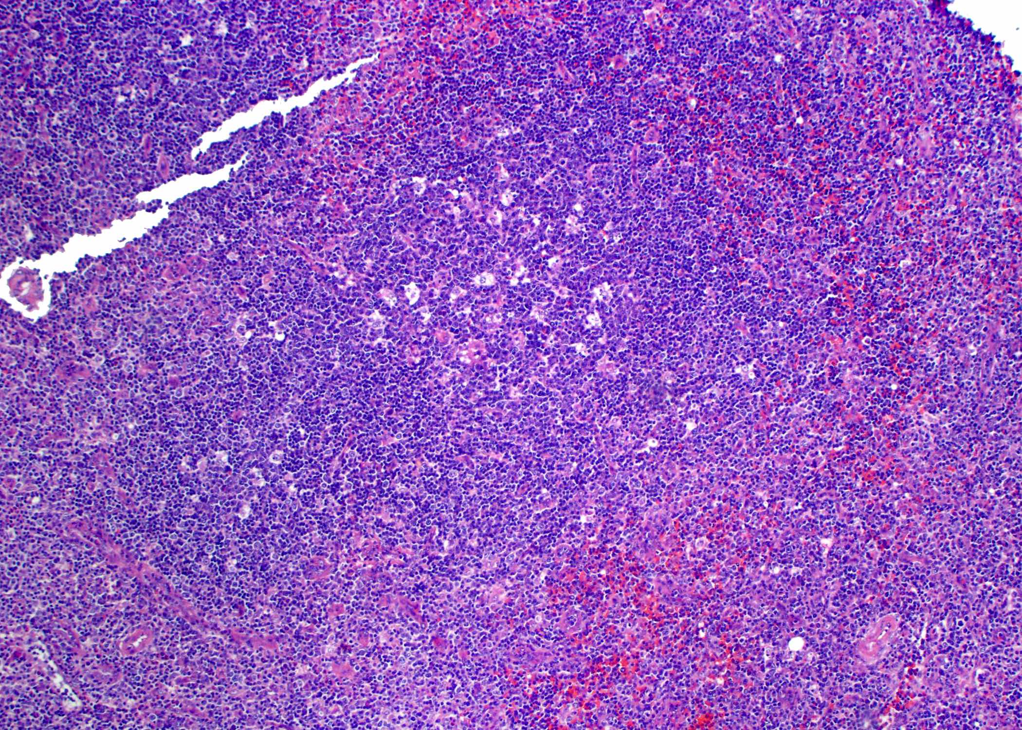
Contributed by Roberto N. Miranda, M.D.
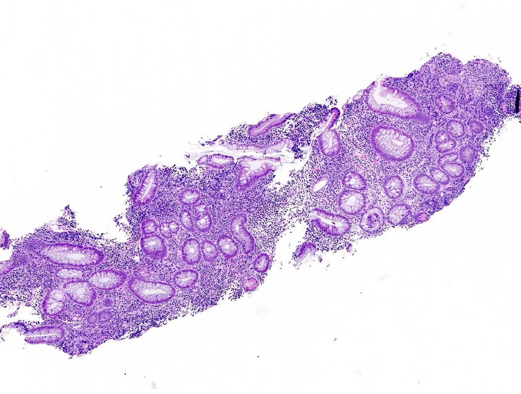
Ileocecal ulcer
Ileocecal infiltration
Angiocentric and angioinvasive lesion
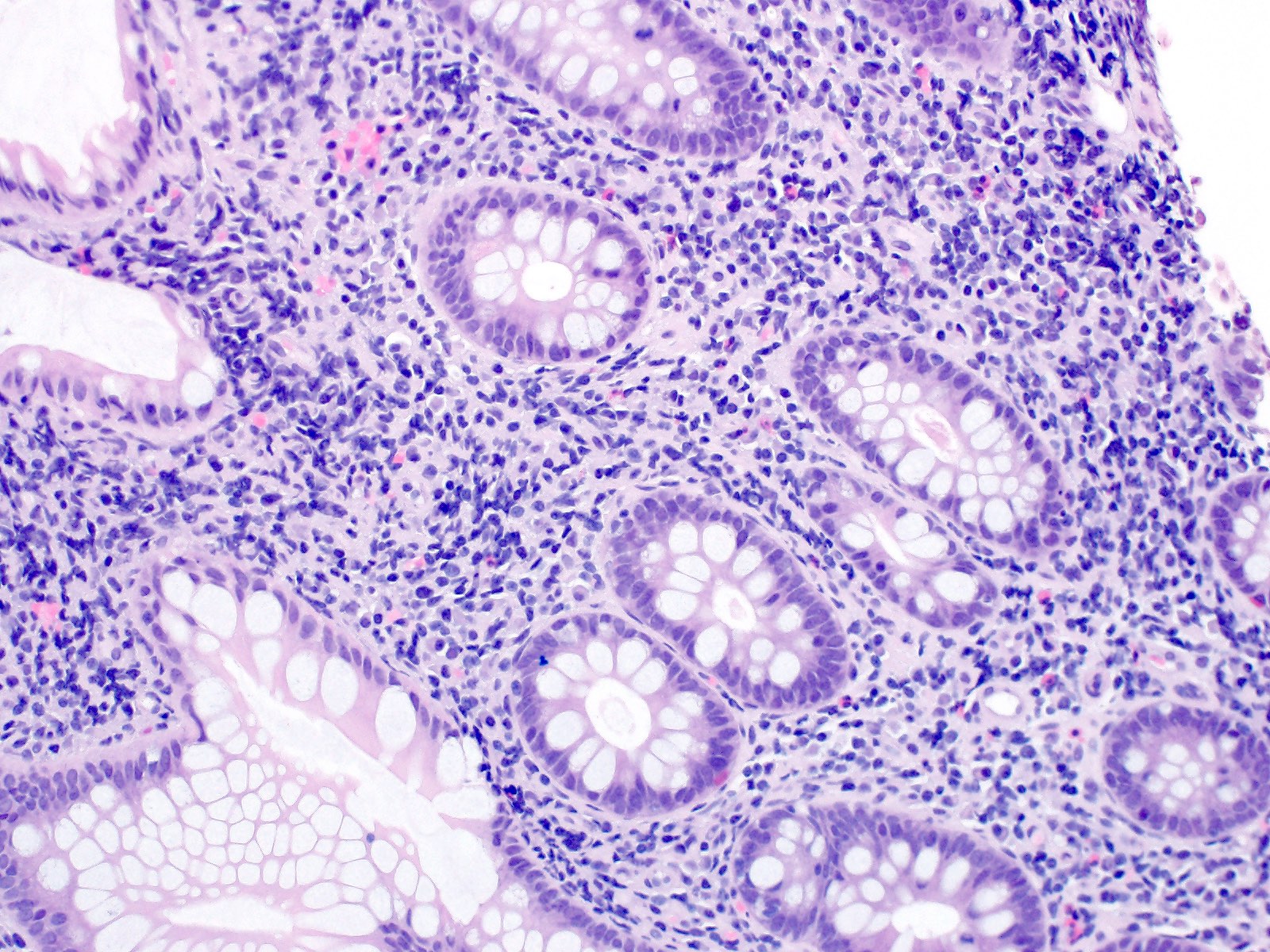
Mucosal and submucosal infiltration
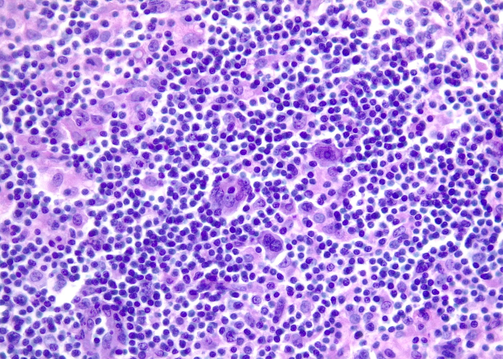
Cytologic atypia
Intraepithelial lymphocytes
Sinusoidal infiltration
Sinusoidal CD30 positivity
CD3 positivity
CD30 positivity
CD4 negativity
CD8 negativity

TIA1 positivity
Table 1: EBV viral gene expression patterns during different types of latency
| Genes | Latency III | Latency II | Latency I | Latency 0 |
| Epstein-Barr nuclear antigen 1 (EBNA1) | + | + | + | - |
| Epstein-Barr nuclear antigen 2 (EBNA2) | + | - | - | - |
| Epstein-Barr nuclear antigen 3 (EBNA3) | + | - | - | - |
| Epstein-Barr nuclear antigen (EBNA) LP | + | - | - | - |
| Latent membrane protein 1 (LMP1) | + | + | - | - |
| Latent membrane protein 2 (LMP2) | + | + | - | - |
| Epstein-Barr encoded RNAs (EBERs) | + | + | + | + |
| BHRF1 micro RNAs (miRNAs) | + | - | - | - |
| BamHI A rightward transcript (BART) micro RNAs (miRNAs) | + | + | + | + |
Table 2: Main Epstein-Barr virus serological profiles
| Anti-viral capsid antigen (VCA) IgG | Anti-viral capsid antigen (VCA) IgM | Anti-Epstein-Barr nuclear antigen (EBNA) IgG | Interpretation |
| - | - | - | Seronegative individual |
| Variable | + | - | Primary infection |
| + | - | + | Past infection |
| + | - | - | Past infection (adults) or primary infection (children) |
| + | + | + | Past infection or end of primary infection |
| - | - | + | Indeterminate |
Table 3: Viral latency type in EBV associated lymphoproliferative disorders and lymphomas
| Disease | Percentage of EBV related cases | Latency pattern | Viral proteins expressed | EBER expression pattern | ||||
| EBNA1 | EBNA2 | LMP1 | LMP2 | EBER | ||||
| B cell lymphoproliferative disorders | ||||||||
| Infectious mononucleosis | 100 | III | + | + | + | + | + | Many small and large cells are EBV+ (mainly B cells and rare positive T and NK cells); most of the EBV+ cells are present in the paracortical area |
| EBV positive mucocutaneous ulcer | 100 | I or II | + | - | + | + | + | EBV expression is variable with most cases showing scattered EBV+ cells |
| Lymphoproliferative disorders associated with immune deficiency and dysregulation | > 90 | III or II | + | + | + | + | + |
|
| T / NK lymphoproliferative disorders of childhood | ||||||||
| Hydroa vacciniforme lymphoproliferative disorder | 100 | II | + | - | + | + | + | EBV expression in around 50% of lesional cells |
| Severe mosquito bite allergy | 100 | II | + | - | - | - | + | EBV is positive in few of the lesional NK cells; a much higher density of EBV+ cells should raise suspicion of NK cell lymphoma |
| Chronic active EBV disease (CAEBVD) | 100 | II | + | - | + | + | + | EBV is uniformly expressed in many cytotoxic T cells in most cases |
| B cell lymphomas | ||||||||
| EBV positive diffuse large B cell lymphoma, NOS | 100 | II or III | + | + | + | + | + | Most of the large atypical lymphoma cells are EBV+, a cutoff of 80% has been proposed |
| Diffuse large B cell lymphoma with chronic inflammation | 100 | II or III | + | + | + | + | + | Most of the lymphoma cells are diffusely positive for EBER |
| Fibrin associated large B cell lymphoma | 100 | II or III | + | + | + | + | + | Most of the lymphoma cells are diffusely positive for EBER |
| Primary effusion lymphoma | Most of the lymphoma cells are positive for EBER in the EBV positive cases | |||||||
| HIV associated | 100 | I | + | - | - | - | + | |
| HIV unrelated | 70 - 90 | I | + | - | - | - | + | |
| Lymphomatoid granulomatosis | 100 | III | + | + | + | + | + | The large neoplastic B cells are EBV positive; the number of EBV+ cells determines the grade |
| Plasmablastic lymphoma | 60 - 75 | I | + | - | - | - | + | Most of the lymphoma cells are diffusely EBV positive |
| Burkitt lymphoma | Most lymphoma cells are positive for EBER in EBV positive cases | |||||||
| Endemic | > 95 | I | + | - | - | - | + | |
| Sporadic | 20 - 80 | I | + | - | - | - | + | |
| AIDS related DLBCL | The pattern of EBV expression coincides with the histological subtype of lymphoma occurring in the immunocompetent state | |||||||
| Immunoblastic | 70 - 100 | III | + | + | + | + | + | |
| Nonimmunoblastic | 10 - 30 | III | + | + | + | + | + | |
| CNS lymphomas | 80 - 100 | III | + | + | + | + | + | |
| Hodgkin lymphoma |
| |||||||
| EBV unrelated | 20 - 90 | II | + | - | + | + | + | |
| EBV associated | 100 | II | + | - | + | + | + | |
| EBV positive T and NK cell lymphomas | ||||||||
| Extranodal, NK / T cell lymphoma | 100 | I or II | + | - | Variable | Variable | + | Virtually all lymphoma cells are positive for EBER |
| Aggressive NK cell leukemia | 90 | II | + | - | + | + | + | Most lymphoma cells are EBV positive |
| Primary nodal EBV positive T / NK cell lymphoma | 100 | II | + | - | + | + | + | Most lymphoma cells are EBV positive |
| Systemic EBV positive T cell lymphoma of childhood | 100 | II | + | - | + | + | + | Most lymphoma cells are EBV positive |
Table 4: Differential diagnosis of EBV positive B cell lymphoproliferative disorders and lymphomas
| Features | Infectious mononucleosis | EBV+ mucocutaneous ulcer | EBV+ classic Hodgkin lymphoma | EBV+ diffuse large B cell lymphoma |
| Clinical | ||||
| Age | Young, elderly | Elderly | Young and elderly | Elderly |
| Lymphadenopathy | Present | Absent | Present, nodal or mediastinal | Present, high stage |
| LDH elevation | Present, mild to moderate | Absent | Present | Present |
| Extranodal disease | Absent | Present | Extremely rare as primary disease | Can be present, late stages |
| Clinical course | Self limited in majority of cases | Waxing and wanning | Progressive | Aggressive, poor outcome |
| Morphology | ||||
| Architecture | Paracortical | Ulcer | Effacement | Effacement |
| Circumscription | Absent | Present, lymphocytic rim at base | Absent | Absent, diffuse involvement |
| Large cells | Reed-Sternberg-like cells | Reed-Sternberg-like cells | Reed-Sternberg cells | Sheets of large neoplastic cells, some RS-like cells |
| EBV latency type | III | II / III | II | III / II |
| Immunohistochemistry | ||||
| CD45 | Positive in most cells | Positive in most cells | Negative in HRS cells | Positive in neoplastic cells |
| CD20 | Positive in large cells | Positive in large cells | Mostly negative in HRS cells, faint reactivity in HRS cells in ~20% of cases | Positive in large cells |
| PAX5 | Positive, strong | Positive, strong | Positive, weak | Positive, strong |
| BOB.1 | Positive, strong | Positive, strong | Negative, can be weak | Positive, strong |
| MUM1 | Positive | Positive | Positive | Positive |
| BCL6 | Negative | Negative | Negative | Can be positive |
| CD10 | Negative | Negative | Negative | Negative |
| CD30 | Positive in HRS-like cells, usually dim | Positive in HRS-like cells, usually dim | Positive in HRS cells, strong | Positive |
| CD15 | Positive in up to 50% of cases | Positive in up to 50% of cases | Positive, variable | Positive in up to 50% of cases |
| PDL1 | Negative | Negative | Positive in > 80% | Can be positive (40 - 60%, extranodal) |
| Diagnostic molecular testing | ||||
| B cell | Polyclonal | Clonal in 50% of the cases | Clonal | Monoclonal IGH gene rearrangements |
| T cell | Polyclonal | Oligoclonal and restricted TCR rearrangement patterns | Polyclonal, restricted pattern in elderly patients | Oligoclonal and restricted TCR rearrangement patterns |
| Genetic features | No immune evasion features | No immune evasion features | Immune evasion (host evasion) | Immune evasion (host evasion) |
Table 5: Differential diagnosis of EBV positive T and NK lymphomas
| Feature | Extranodal NK / T cell lymphoma | Aggressive NK cell leukemia | EBV positive nodal T and NK cell lymphoma | Systemic EBV+ T cell lymphoma of childhood |
| Clinical presentation | ||||
| Age | Adults | Young to middle aged adults | Older adults | Children, young adults |
| Site at presentation | Nasopharynx (70 - 80%), others (20 - 30%): skin, gastrointestinal (GI) | Bone marrow, spleen, peripheral blood, rarely lymph nodes (20%) | Lymph nodes, no nasal involvement by definition | Systemic proliferation: bone marrow, liver or spleen, CNS |
| Behavior | Localized disease, frequent dissemination | Fatal | Aggressive | Fulminant |
| Median survival | 26 - 76 months | Weeks | 4 months | Days to weeks |
| Hemophagocytic syndrome | Generally absent | Present | Uncommon | Always present |
| Morphology | ||||
| Cytology of neoplastic cells | Variable atypia, spectrum from small to large cells | Large granular atypical lymphocytes, distinct nucleoli and clear cytoplasm (smears) | Pleomorphic medium sized cells, with centroblastic, anaplastic or plasmacytoid features | Small to intermediate sized with subtle to absent atypia (most common) or large atypical cells |
| Necrosis | Common | Frequently present | Variable | Absent |
| Angiocentricity and angiodestruction | Present | Frequently present | Uncommon | Absent |
| Apoptosis | Present | Frequently present | Variable | Absent |
| Ancillary testing | ||||
| CD2 | Positive | Positive | Positive | Positive |
| CD3 | Often negative; subset is positive | Negative | Positive | Positive |
| CD3ε | Positive | Positive | Positive | Positive |
| CD4 | Negative | Negative | Negative | Usually negative |
| CD8 | Positive | Usually negative | Positive (> 80%) | Usually positive |
| CD56 | Positive | Positive | Mostly negative (positive < 20%) | Negative |
| Cytotoxic granules | Positive | Positive | Positive | Positive |
| EBER | All neoplastic cells | All neoplastic cells, a subset is negative (< 15%) | All neoplastic cells | Majority of neoplastic cells |
Table 6: Differential diagnosis of EBV+ B cell lymphoproliferations with Hodgkin-like features
| Disease | Clinical features | Morphology | Immunophenotype | Lineage, clonality and molecular features |
| EBV+ mucocutaneous ulcer |
|
|
|
|
| EBV+ diffuse large B cell lymphoma, NOS |
|
|
|
|
| EBV+ classic Hodgkin lymphoma |
|
|
|
|
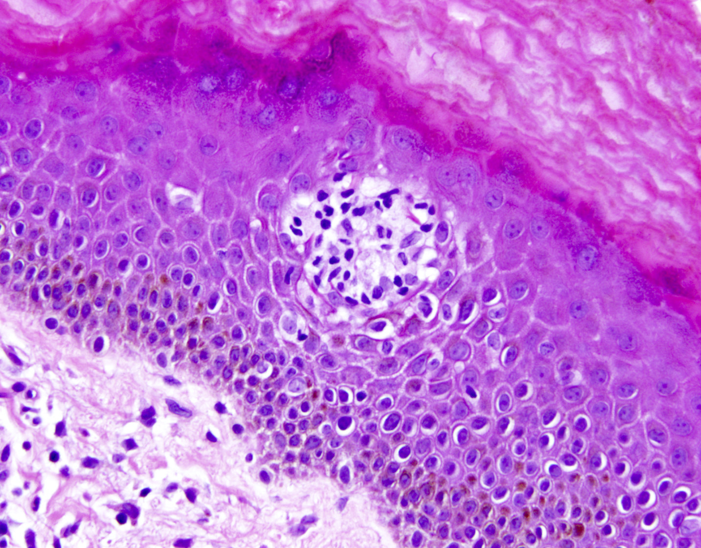
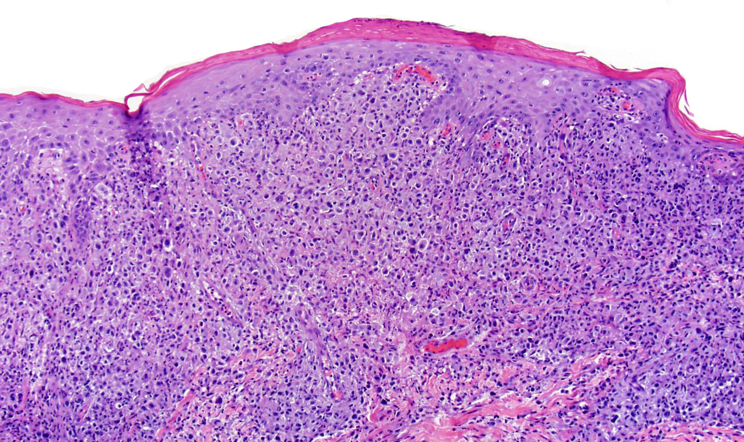
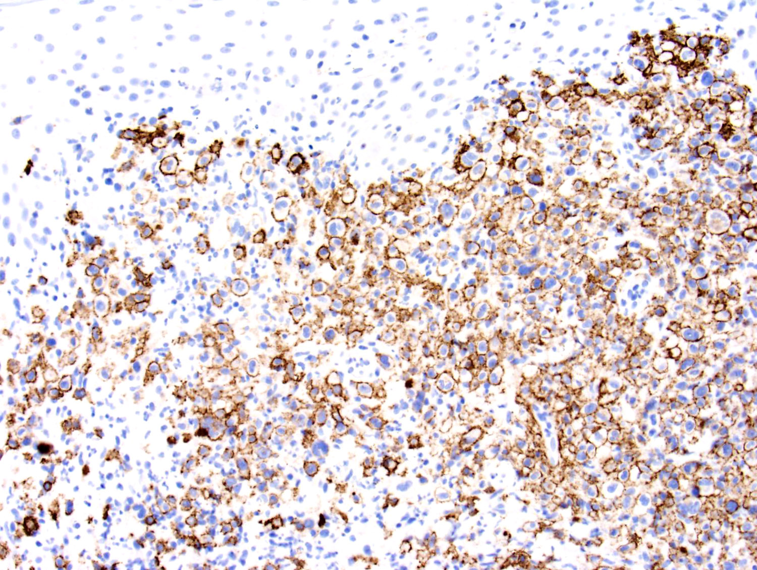
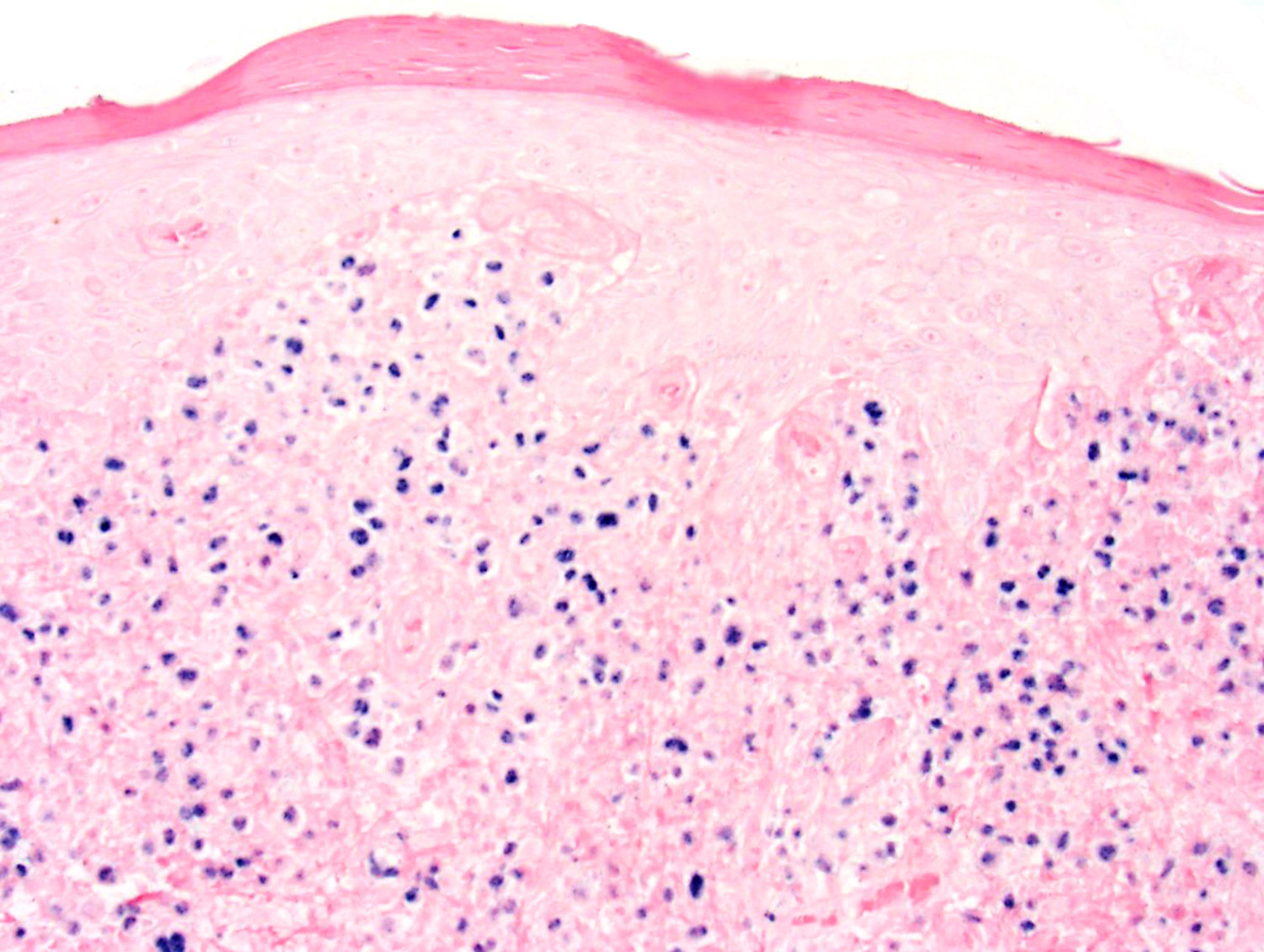
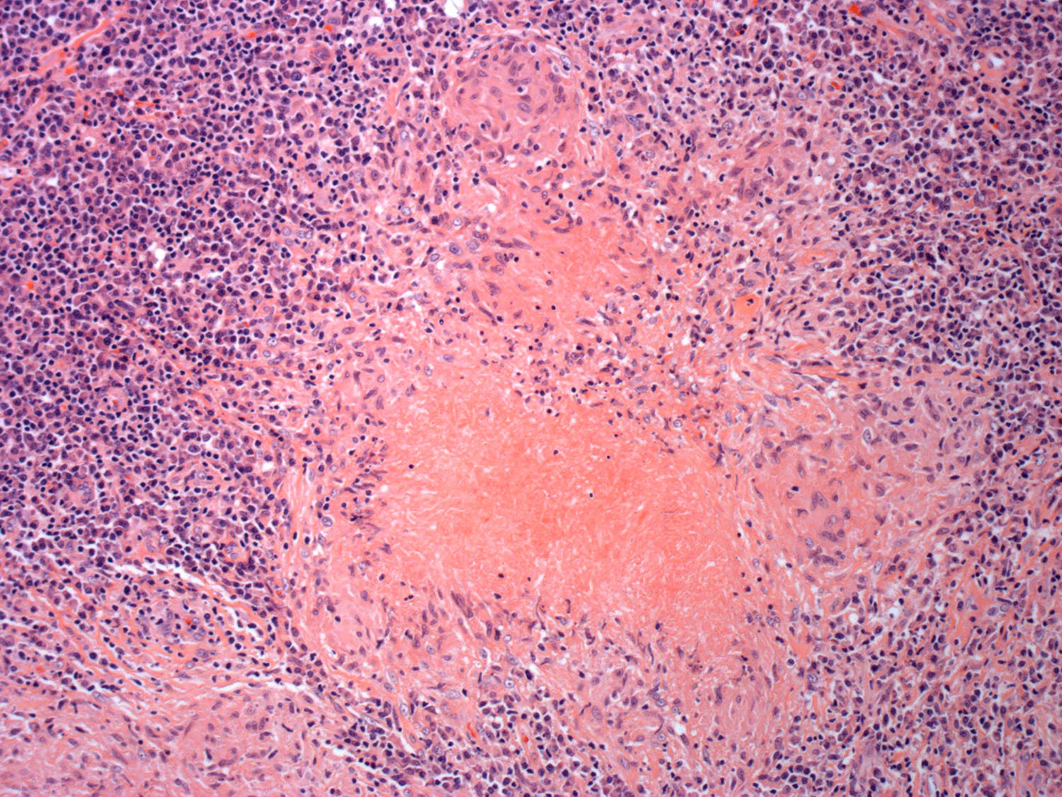
Contributed by Francisco Bravo, M.D.

Hydroa
vacciniforme
lymphoproliferative
disorder
Contributed by Roberto N. Miranda, M.D. and Roman Segura-Rivera, M.D.

Infectious mononucleosis in an excised lymph node
Interfollicular expansion in infectious mononucleosis
Infectious mononucleosis

Infectious mononucleosis


Follicular hyperplasia arising in immune deficiency / dysregulation
Germinal center reaction
Hydroa vacciniforme

Hemophagocytic
lymphohistiocytosis
EBV positive plasmablastic lymphoma
Extranodal NK / T cell lymphoma showing angiocentricity

Extranodal NK / T cell lymphoma


Extranodal NK / T cell lymphoma in the intestine
Contributed by Roberto N. Miranda, M.D. and Roman Segura-Rivera, M.D.

Downey cells
Contributed by Auris Huen, M.D.
Skin lesion
Contributed by Roberto N. Miranda, M.D. and Carlos A. Torres-Cabala, M.D.
Nasal lesion of ENKTCL

Diffuse infiltrate
Cytological atypia
EBER positivity
Angioinvasion in ENKTCL
Atypical infiltrate in skin
CD3 positivity
CD7 positivity

CD56 positivity
Perforin positivity
Hemophagocytosis
ENKTCL NT in bone marrow
Bone marrow CD3
Bone marrow EBER
Skin involvement by PTCL, NOS
Cytological atypia in PTCL, NOS
T cell LGL leukemia
CD3 in T cell LGL
CD57 in T cell LGL
TIA1 in T cell LGL
Skin in EBV+ DLBCL
CD20 in DLBCL
EBER in DLBCL
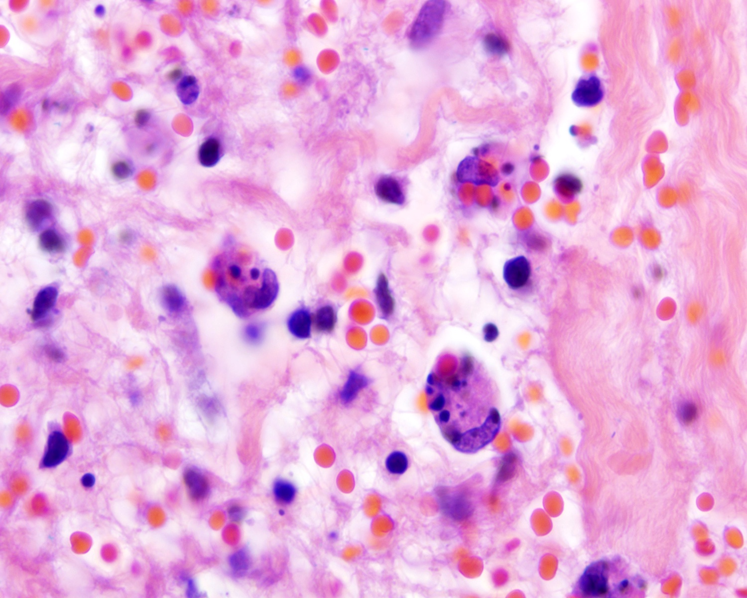
Necrosis in tuberculosis
Necrosis in histoplasmosis
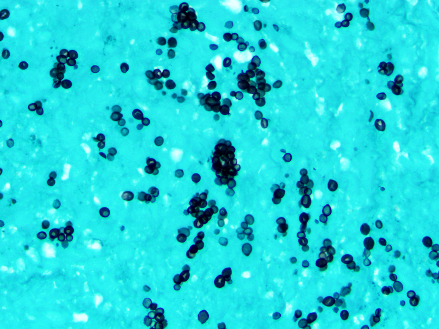
Histoplasmosis GMS stain
Contributed by Jordan M. Hall, M.D., Hyunkyu Shin, M.D., Dr. Christian Schürch, M.D., Ph.D., Falko Fend, M.D. and Claudia Wickenhauser, M.D., Ph.D. (Case #529)

Knee synovium
Knee joint space fibrin

PAX5
CD30
Ki67
EBER ISH
H&E staining
H&E staining
CD138
MUM1
CD3

CD20

CD79a
ALK
MYC
HHV8

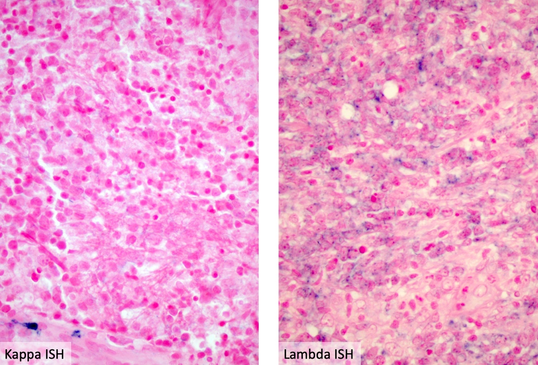
Kappa
Lambda
Cyclin D1
CD56
EBER
BCMA
Contributed by Abdallah Flaifel, M.D.
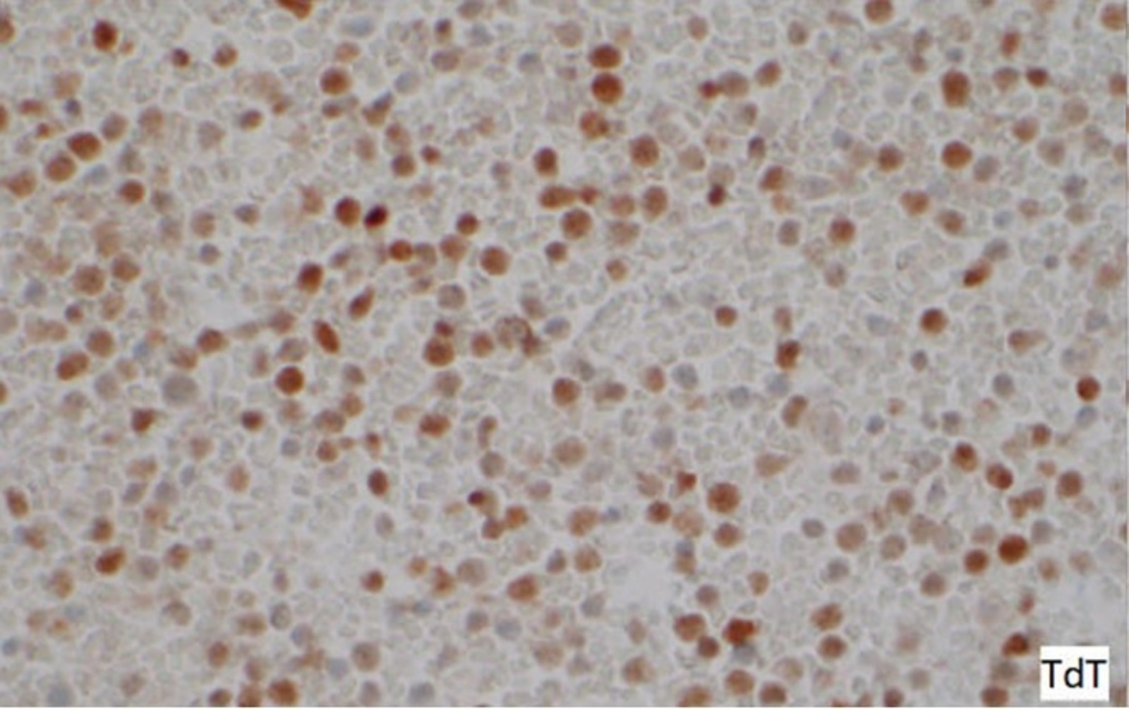
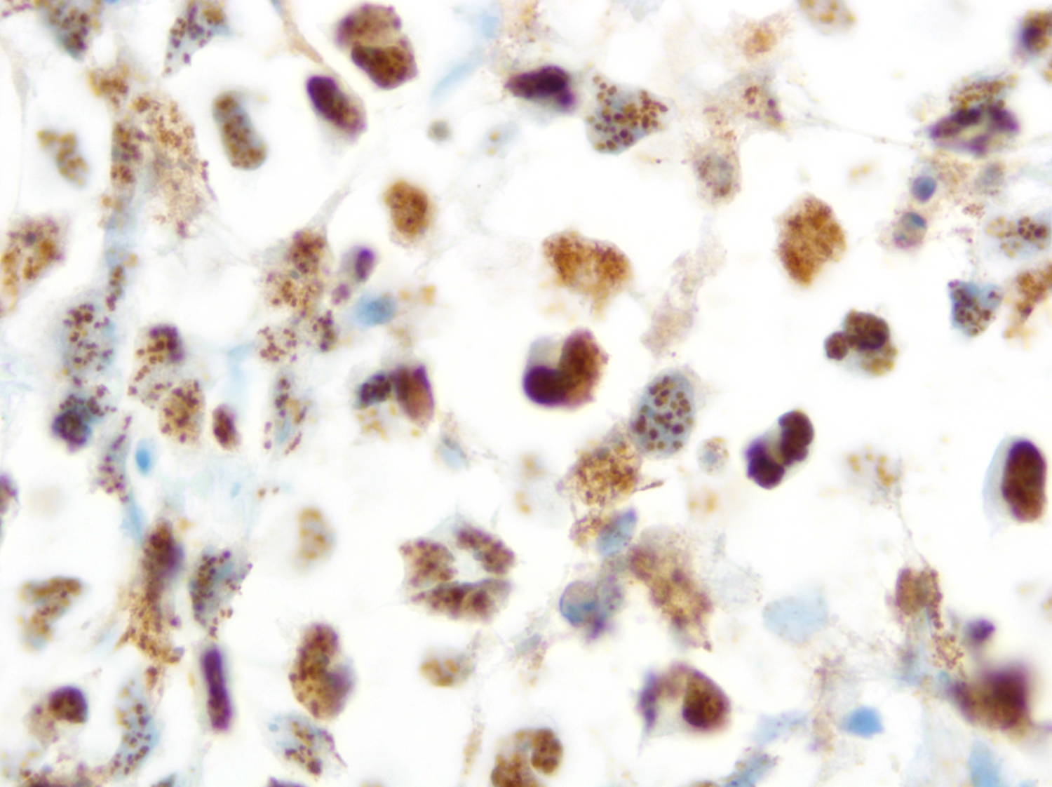
Cell block TdT
Images hosted on other servers:

Schematic of a flow cytometer
Contributed by Abdallah Flaifel, M.D.
Pericardial fluid cytospin
Cell block TdT
Contributed by Nicholas Ward, M.D.
B ALL flow cytometry
B ALL flow cytometry
Contributed by Carlos A. Murga-Zamalloa, M.D.
Cell block preparation
Cell block preparation (CD20)
Cell block preparation (MUM1)
Cell block preparation (HHV8)
Cytospin preparation (Wright-Giemsa)
| Nodal Follicular Lymphoma | Duodenal Follicular Lymphoma |
| Grade 1 - 2 or 3 | Grade 1 - 2 |
| Stage III or IV | Stage I or II |
| BCL2, CD10, BCL6: Positive | BCL2, CD10, BCL6: Positive |
| AID: Positive | AID: Negative |
| CD21 stain: Dense stain in the center of germinal center of follicles |
CD21 stain: Accentuated staining at the periphery of germinal center of follicles |
| BCL2 and BCL6 rearrangements: + | BCL2 and BCL6 rearrangements: + |
| CREBBP mutations present | CREBBP mutations present |
| KMT2D mutations present | Lower KMT2D mutations present |
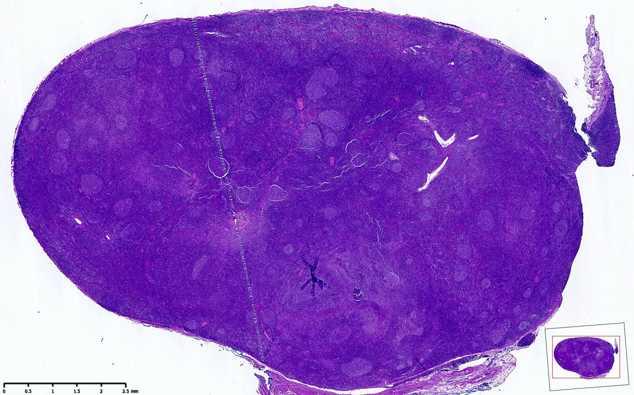
Contributed by Mahsa Khanlari, M.D.
Small intestinal polyp

CD10 and CD20 Immunohistochemistries
BCL2 IHC
CD21 IHC
Contributed by @ahmsab_MD on Twitter
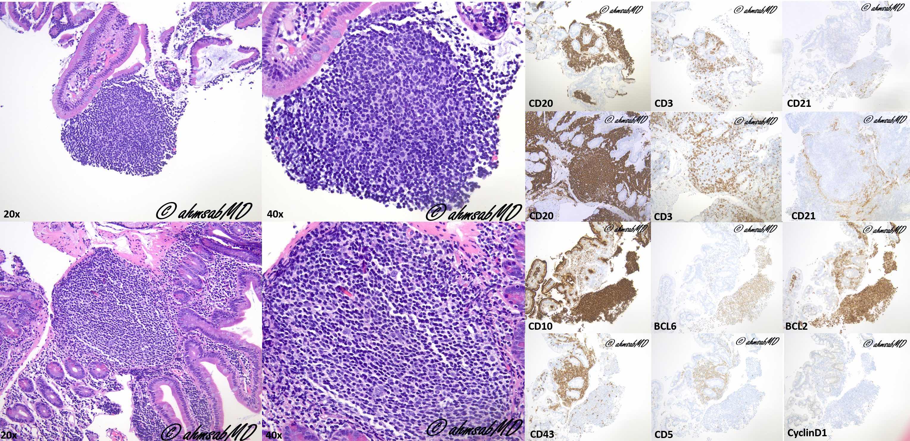
Duodenal type follicular lymphoma
Images hosted on other servers:

Development of FL-like B cells
Contributed by Elaine S. Jaffe, M.D.
H&E overview
BCL2 overview
CD10 overview

BCL2 neoplastic follicle

H&E neoplastic follicle
CD10 neoplastic follicle
H&E reactive follicle
BCL2 reactive follicle
CD10 reactive follicle
Contributed by Nicholas Ward, M.D. and Roberto N. Miranda, M.D.
Mixed histology pattern
Atypical lymphoid aggregates
CD3
CD20
PD-1
ICOS
CD10
IgD
Neoplastic lymphocytes
CD3
CD20
CD21
PD-1
Images hosted on other servers:

T cells expressing CD4 without surface CD3 (n, o, p, q)

Significant population of CD3- / dim CD4+ T cells (b, c)
Images hosted on other servers:

FISH for the detection of t(5;9)(q33;q22)

5' and 3' RACE identifies ITK-SYK fusion
| World Health Organization grading of follicular lymphoma | |||
| Grade | Definition | Pattern | Immunohistochemistry and cytogenetics |
| 1 | 0 - 5 centroblasts/high power field | Follicular or diffuse |
IHC: CD10: + (95 - 100%) BCL2: + (85 - 90%) FISH: BCL2 translocation: + (80 - 90%) BCL6 rearrangement: + (3%) Ki67: < 20%* |
| 2 | 6 - 15 centroblasts/high power field | Follicular or diffuse | |
| 3A | > 15 centroblasts/high power field Centrocytes present | Follicular If diffuse component: Reported as diffuse large B cell lymphoma and follicular lymphoma (% of each component is reported); correlate with clinical features and overall grade in cases with small areas of diffuse pattern |
IHC: CD10: + (80 - 95%) BCL2: + (50 - 75%) FISH: BCL2 translocation: + (60 - 70%) BCL6 rearrangement: + (30 - 40%) Ki67: > 20% |
| 3B | > 15 centroblasts/high power field Lack centrocytes | Follicular If diffuse component: Reported as diffuse large B cell lymphoma and follicular lymphoma (% of each component is reported) |
IHC: CD10: + (40 - 85%) BCL2: + (45 - 75%) FISH: BCL2 translocation: + (15 - 30%) BCL6 rearrangement: + (40 - 50%) CD10-IRF4/MUM1+: common Ki67: > 50% |
Notes:
- IHC: immunohistochemistry
- FISH: fluorescence in situ hybridization
- High power field of 0.159 mm2 (40× objective)
- Follicular: > 75% (proportion follicular %)
- Diffuse: 0% (proportion follicular %)
- * ~20% of low grade follicular lymphomas have a high proliferation (Ki67) rate (Am J Surg Pathol 2005;29:1490)
- References: Arch Pathol Lab Med 2018;142:1330, Haematologica 2018;103:1182, Swerdlow: WHO Classification of Tumours of Haematopoietic and Lymphoid Tissues, 4th Edition, 2017
Contributed by Jennifer Chapman, M.D.

Follicular lymphoma

Back to back follicles
Mantle zone
Low grade morphology
CD20
CD3
Germinal center markers (CD10 and BCL6)
BCL2
Follicular dendritic cell meshworks
High grade morphology
Paratrabecular pattern of involvement in bone marrow
Contributed by Mahsa Khanlari, M.D.
Fine needle aspirate smear
Contributed by Jennifer Chapman, M.D.
Peripheral blood smear involvement
Contributed by Mahsa Khanlari, M.D.
Flow cytometric immunophenotyping
Flow cytometric immunophenotyping, monotypic population
Contributed by Mahsa Khanlari, M.D.

IGH-BCL2 dual color FISH
Contributed by Annapurna Saksena, M.B.B.S, M.D. and Elaine S. Jaffe, M.D.
Diffuse growth pattern
Atypical large lymphoid cells
Multicentric Castleman disease
Multicentric Castleman disease, HHV8
HHV8 LANA positive atypical cells
HHV8 LANA nuclear staining
EBER ISH negative atypical cells
Contributed by Julie Teruya-Feldstein, M.D.
Follicles of varying sizes
Hyalinization in some follicles
Plasmablasts
EBER1
Plasmablasts positive for MUM1
Plasmablasts negative for CD30
Plasmablasts negative for BCL6
Plasmablasts negative for CD10
Plasmablasts negative for CD79a
Contributed by Buthaina Al-Maashari, M.D. and Dietrich Werner, M.D.
Bone marrow core biopsy

BRAF IHC
Images hosted on other servers:


Lymph node FNA
Contributed by Buthaina Al-Maashari, M.D. and Dietrich Werner, M.D.
Circulating hairy cell
Images hosted on other servers:

Typical leukemic cell in hairy cell leukemia
Contributed by Buthaina Al-Maashari, M.D. and Dietrich Werner, M.D.
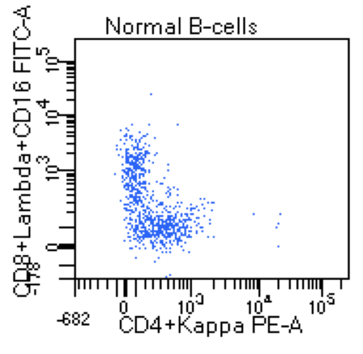
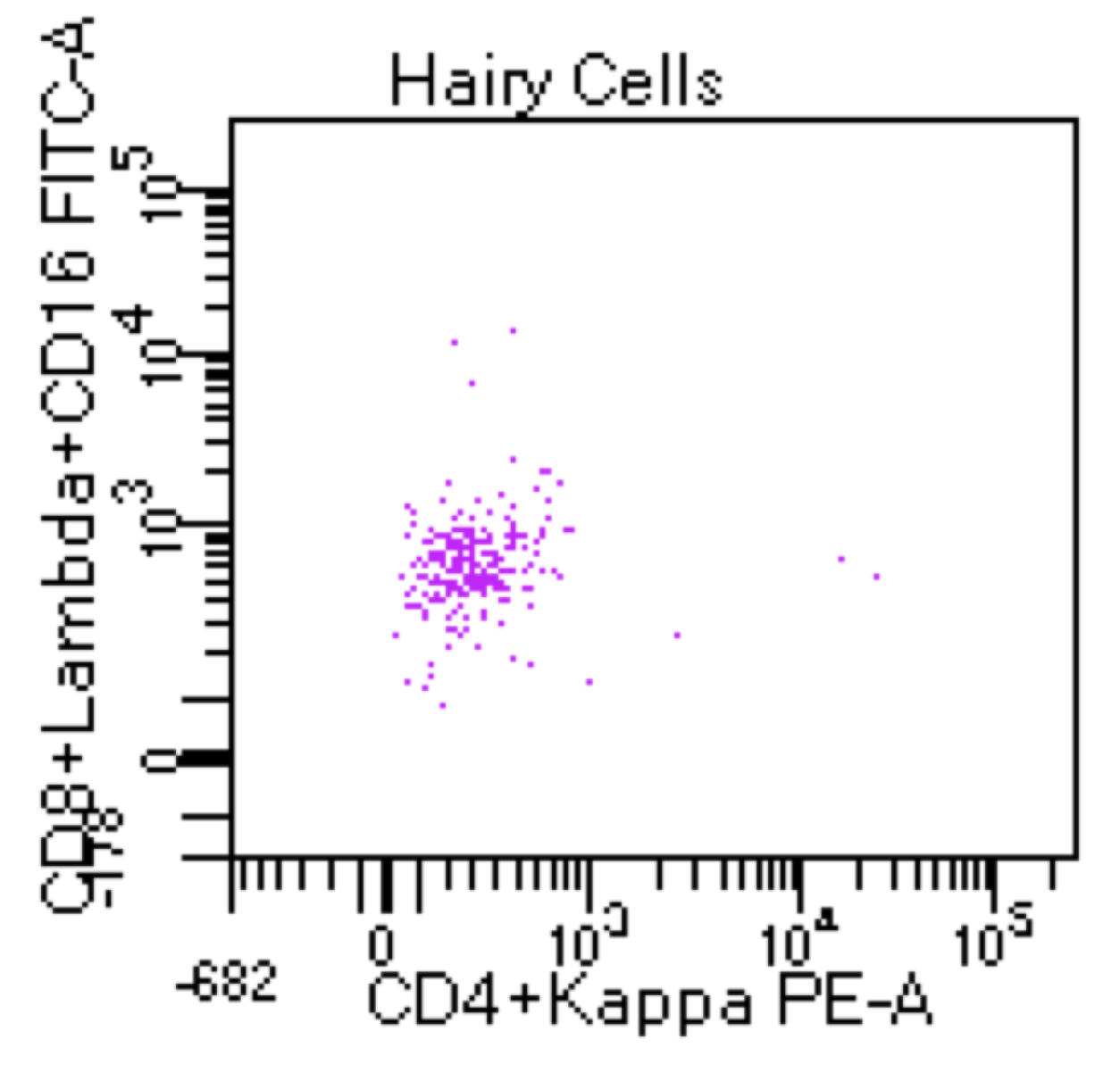
Peripheral blood analysis of a 67 year old man with cytopenias

Peripheral blood analysis of a 67 year old man with cytopenias

Contributed by Pallavi Khattar, M.D. and Case #437
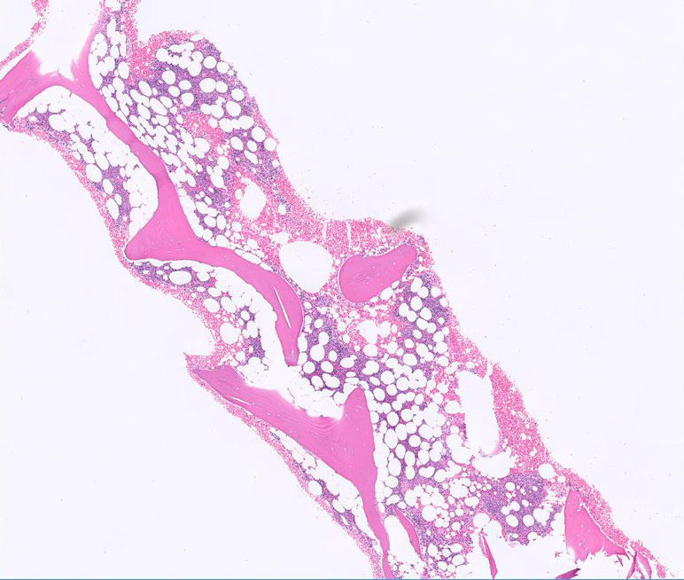
Bone marrow biopsy
CD20 immunostain
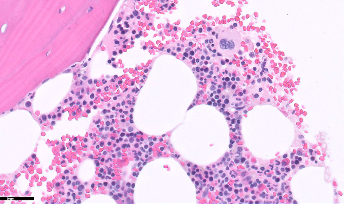
Bone marrow aspirate
Bone marrow aspirate
Bone marrow biopsy
Bone marrow aspirate
Bone marrow aspirate
CD20 immunostain
CD3 immunostain

DBA-44 immunostain
Contributed by Roberto N. Miranda, M.D.

Bone marrow infiltration
Bone marrow with dyspoietic changes
CD3 positive
CD4 negative

CD8 negative
TCR βF1 negative
TCRγδ positive
Liver infiltration
HSTCL: CD3 positive
HSTCL: CD4 negative

CD8 positive
TIA1 positive
Small size morphology
Intermediate size morphology
Blastic morphology
Contributed by Roberto N. Miranda, M.D.
Positivity for CD2, TCRγδ and CD56
Images hosted on other servers:

Diagnostic approach
Contributed by Jon C. Aster, M.D., Ph.D. and Genevieve M. Crane, M.D., Ph.D.
Oropharyngeal mass
CD10
MYC
Ki67
HGBL, NOS
High Ki67
Contributed by Hillary Rose Elwood, M.D.
Shave biopsy shows epidermal spongiosis, vesiculation and necrosis

Lymphocytic infiltrate is CD3+, TIA+, subset weak CD4+, subset CD30+ and negative for CD8 and CD56
Contributed by Hillary Rose Elwood, M.D.

EBER in situ hybridization
Contributed by Kyle Bradley, M.D.
Mantle zone and interfollicular involvement
Early lymph node involvement by mantle cell lymphoma
Follicular lymphoma with concurrent in situ mantle cell neoplasia
Contributed by Jinjun Cheng, M.D., Ph.D.
Large granular lymphocytosis
Contributed by Anamarija M. Perry, M.D.
Colon with indolent T-LPD
Infiltration of lamina propria
Atypical infiltrate of indolent
T-LPD

CD8 IHC stain
CD4 IHC stain
TIA1 IHC stain

Contributed by Carlos A. Murga-Zamalloa, M.D.
Epitheliotropism of atypical infiltrates
Infiltrates adjacent to ulcer site
Submucosal extension of infiltrates
CD3 positive infiltrates
Aberrant CD5 loss
Contributed by Kathryn Gibbons, M.D.
ILBCL involving skin and subcutaneous tissue
Large lymphoma cells in small vessels
ILBCL involving the brain parenchyma
Vessel with involvement by ILBCL
Large lymphoma cells in the vessels
CD20
BCL6
MUM1
Contributed by Jayalakshmi Balakrishna, M.D.
Diffuse pattern
Expansile follicles
Diffuse effacement
with medium
sized cells

Medium sized atypical lymphoid cells
Round to irregular nuclear contours and conspicuous nucleoli
Follicular pattern
CD20
BCL2
BCL6
MUM1
CD21
Contributed by Itziar Salaverria, Ph.D.

FISH break apart probe for IRF4
Contributed by Anna Shestakova, M.D., Ph.D.
Diffuse lymph node infiltration by lymphoma cells
Interspersed tingible body macrophages
Atypical lymphoma cells
CD20 stain
CD10 stain
BCL6 stain

Ki67 stain
Contributed by Nicholas Joseph Dcunha, M.B.B.S., M.D. and Elanthenral Sigamani, M.B.B.S., M.D.
Mass in lung (CT scan)
Contributed by Nicholas Joseph Dcunha, M.B.B.S., M.D. and Elanthenral Sigamani, M.B.B.S., M.D.
Lymphocytic vasculitis with angioinvasion (lung)
CD20 in tumor cells and CD3

CD30 and EBV LMP1 IHC
Lymphocytic vasculitis with angioinvasion (skin)
CD20 and CD30 in tumor cells; CD3 and CD4 in reactive T cells
Lymphocytic vasculitis with angioinvasion (CNS)
CD20 and EBV LMP1 in tumor cells; CD3 in reactive T cells
Contributed by Ling Zhang, M.D.

Increased lymphoid cells
Round eccentrically located nuclei
Condensed chromatin, inconspicuous nucleoli
Small mature lymphoid cells

Intermingled plasma cells
Lymphoid cells negative for CD3
Diffuse CD20 positivity
Focal plasma cell positivity (CD138)
Immunoglobulin M positivity
Contributed by Ling Zhang, M.D. and Caroline An, M.D.
Kappa clonal population of B cells
CD38+ / CD138+ plasma cells
Lambda restriction
Plasma cells express CD45
Presence of CD19
Presence of CD20 expression
B cells expressing both CD19 and CD20
B cells are lambda monoclonal
B cells show variable expression for CD38
Contributed by Ling Zhang, M.D.

ISH clonal kappa light chain expression
Negative lambda light chain expression
Contributed by Roberto N. Miranda, M.D.
Salivary gland lymphoepithelial lesion
Cytokeratin and lymphoepithelial lesion
CD20 positivity in salivary gland lymphoepithelial lesion
Gastric gland lymphoepithelial lesion
Thyroid gland MALT lymphoma
PCMZL
PCMZL subepidermal lesion
PCMZL - CD20 positive
PCMZL - CD3 negative
Lung BALT lymphoma
Lymphoepithelial lesion of the stomach
Stomach MALT lymphoma
Lambda light chain restriction
Kappa negativity
Breast MALT lymphoma
CD10 negativity in MALT lymphoma
BCL6 negativity in MALT lymphoma
Contributed by Chi Young Ok, M.D.
Monotonous immature cells
PAX5 immunohisto-
chemical stain
CD5 immunohisto-
chemical stain
Proliferation index, Ki67

Follicular dendritic cell meshwork
Large cells with variable sizes
Cyclin D1 immunohisto-
chemical stain
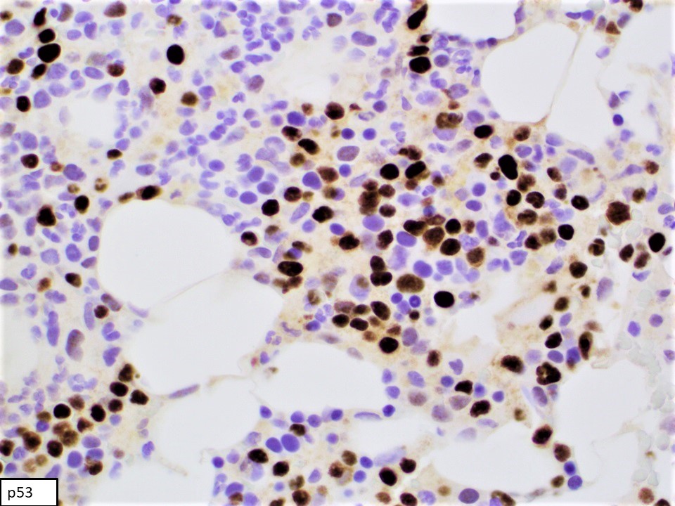
p53 pattern of staining
Diffuse involvement of bone marrow
Contributed by Patricia Tsang, M.D., M.B.A. (Case #499)

Abdominal wall mass
PAX5
CD5
Ki67
Cyclin D1
SOX11
Contributed by Chi Young Ok, M.D.
Immature monotonous cells in cytology
Contributed by Chi Young Ok, M.D.
Lymphoma cells in peripheral blood
Contributed by Chi Young Ok, M.D.
Complex karyotype

Deletion of TP53 gene
Contributed by Chi Young Ok, M.D.
Monotonous population of small lymphoid cells
Follicular localization of lymphoid cells
Spleen involvement
Cyclin D1 expression

Mantle zone pattern
Pink histiocytes

Cyclin D1 and SOX11 in mantle zone pattern

Ki67 in mantle zone pattern
Bone marrow involvement
Follicular dendritic cell meshwork
Contributed by Chi Young Ok, M.D.
Small to intermediate centrocytes in cytology
Contributed by Chi Young Ok, M.D.
Atypical mature cell in peripheral blood
Contributed by Chi Young Ok, M.D.
Abnormal FISH result

Abnormal karyotype result
Contributed by Nicholas Nowacki, M.D.
Lymphoid infiltrate

Small lymphoma cells
Marrow aspirate

CD3 highlights T cells
CD5 positive
CD20 positive

Cyclin D1 positive
SOX11 negative
Contributed by Nicholas Nowacki, M.D.
Peripheral blood
Contributed by Nicholas Nowacki, M.D.
Aberrant CD5 coexpression

CD20 moderate expression
Surface light chain restriction
Contributed by Anamarija M. Perry, M.D.
Lymph node with nodal marginal zone lymphoma (NMZL)
Atypical infiltrate
Lymph node with pediatric NMZL
CD20 stain
CD10 stain
BCL2 stain
Ki67 stain
PAX5 stain in pediatric NMZL
IgD stain in pediatric NMZL
Contributed by Xiaoling Guo, M.D., Ph.D. and Yanhua Wang, M.D.
CT abdomen and pelvis
Contributed by Xiaoling Guo, M.D., Ph.D. and Yanhua Wang, M.D.
Ulcerated mass

Cross section of the mass
Contributed by Xiaoling Guo, M.D., Ph.D. and Yanhua Wang, M.D.

Uninvolved mucosa
Mucosa overlying the mass
Monomorphic transmural lymphocytic infiltrate

CD3
CD7

CD8
Granzyme B
TIA1
CD20
Ki67
Images hosted on other servers:

Characteristic immunophenotype


Immunophenotypic comparison
Contributed by Catalina Amador, M.D.
Effacement of nodal architecture

Medium to large sized lymphocytes
Clear cell morphology

CD21 and CD23: absent expansion of follicular dendritic cell meshworks
CD20 negative for B cell markers
Positive membranous expression of CD4
ICOS membranous positivity
PD-1 membranous positivity
BCL6 nuclear positivity
Contributed by Elaine S. Jaffe, M.D., Jayalakshmi Balakrishna, M.D. and Lauren B. Smith, M.D.
Vaguely nodular architecture
Rare large neoplastic cells
LP cells or popcorn cells
Neoplastic cells positive for CD20
T cell rich variant with rare small B cells in the background
Neoplastic cells positive for CD20
OCT2 stain is helpful in highlighting the neoplastic cells
Neoplastic cells positive for PAX5
Neoplastic cells positive for BCL6
IgD expressing LP cells

T cell rosettes around LP cells
CD4 T cells predominate in the background
T cells express PD-1

PD-1 positive T cells form rosettes around LP cells
Expanded follicular dendritic cell meshwork
Effaced lymph node architecture

Lymphocyte predominant cell (LP cell / popcorn cell)
LP cells express CD20
Contributed by Elaine S. Jaffe, M.D., Jayalakshmi Balakrishna, M.D. and Lauren B. Smith, M.D.
Vaguely nodular architecture
Rare large neoplastic cells
LP cells or popcorn cells
Neoplastic cells positive for CD20
T cell rich variant with rare small B cells in the background
Neoplastic cells positive for CD20
OCT2 stain is helpful in highlighting the neoplastic cells
Neoplastic cells positive for PAX5
Neoplastic cells positive for BCL6
IgD expressing LP cells
T cell rosettes around LP cells
CD4 T cells predominate in the background
T cells express PD-1
PD-1 positive T cells form rosettes around LP cells
Expanded follicular dendritic cell meshwork
Effaced lymph node architecture
Lymphocyte predominant cell (LP cell / popcorn cell)
LP cells express CD20
Contributed by Cade Arries, M.D.
Bone marrow
Bone marrow, CD30

Bone marrow, CD15
Bone marrow, EBER
Lymph node
Lymph node

Lymph node, CD15

Lymph node, CD30
Lymph node, PAX5
Lymph node, EBER
Contributed by Daniel Cassidy, M.D. and Jennifer Chapman, M.D.
Architectural distortion
Polymorphic lymphoid infiltrate
Full spectrum of maturation
Composition of lymphoid infiltrate
Polymorphic infiltrate
Variably sized B cells
PAX5 variable cell size
Variably sized T cells
Scattered immunoblasts
EBER positivity
Contributed by Kathryn Gibbons, M.D.

Lymph node with pediatric nodal marginal zone lymphoma

Atypical infiltrate
CD20 stain

PAX5 stain
CD43 stain

IgD stain
Pediatric type follicular lymphoma versus follicular lymphoma usual type
| Pediatric type FL | Usual type FL | |
| Age | Young | Old age (sixth decade) |
| Stage | Low (I - II) | High (III - IV) in majority of cases |
| Location | Head and neck | Variable |
| Extranodal location | Absent | Present, variable |
| Histology | Grade 3 | Grade 1 - 3 |
| BCL2 (IHC) | Negative / dim | Usually positive |
| CD10 | Positive (~100%) | Positive (usually) |
| Ki67 | High | Low (except for high grade) |
| t(14;18) IGH-BCL2 | Absent | Present, up to 90% |
| Monotypic B cells by flow cytometry | Frequent | Frequent |
| Monoclonal IgH rearrangements | Frequent | Frequent |
| BCL6 or MYC rearrangements | Absent | Variably present |
| Genetic | 1p36 loss | Complex, variable |
| Mutations | TNFRSF14, MAP2K1, IRF8 (K66R) | CREBBP, EZH2, KMT2D |
| Prognosis | Favorable | Variable |
Contributed by L. Jeffrey Medeiros, M.D. and Mahsa Khanlari, M.D.
Lymph node in 23 year old man
IHC for CD20
IHC for CD10

IHC for BCL6
IHC for BCL2
IHC for IgD; attenuated and focally absent mantle zone
Images hosted on other servers:

A, B: monomorphic morphology
characteristic of GATA3 subtype
C, D: polymorphous morphology
characteristic of TBX21 subtype
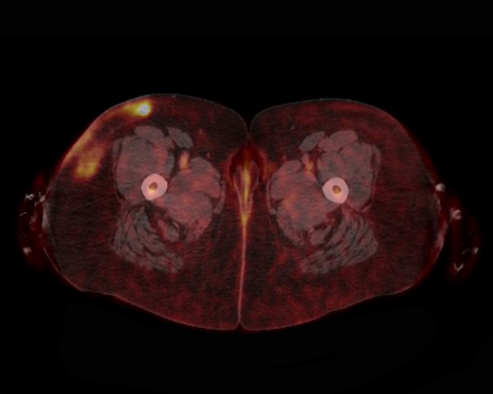
Contributed by Matthew M. Klairmont, M.D.
PTCL, NOS

PTCL, NOS involving the skin
Images hosted on other servers:


Imprints of PTCL, NOS (lymphoepithelioid / Lennert variant)
Contributed by Julio Poveda, M.D.

Intermediate to large sized cells
CD138
CD20
MUM1
Images hosted on other servers:

Landscape using WES
Images hosted on other servers:

Immunodeficiency
associated LPDs
in WHO-HAEM4R
and WHO-HAEM5

3 part nomenclature
for immunodeficiency
associated LPDs
Contributed by Miguel Gonzalez-Mancera, M.D.

Heterogeneous infiltrate

Heterogeneous infiltrate

Gastrointestinal biopsy

CD3
CD20

CD68
MUM1
Kappa light chain

Lambda light chain
EBV EBER
LMP1
Images hosted on other servers:

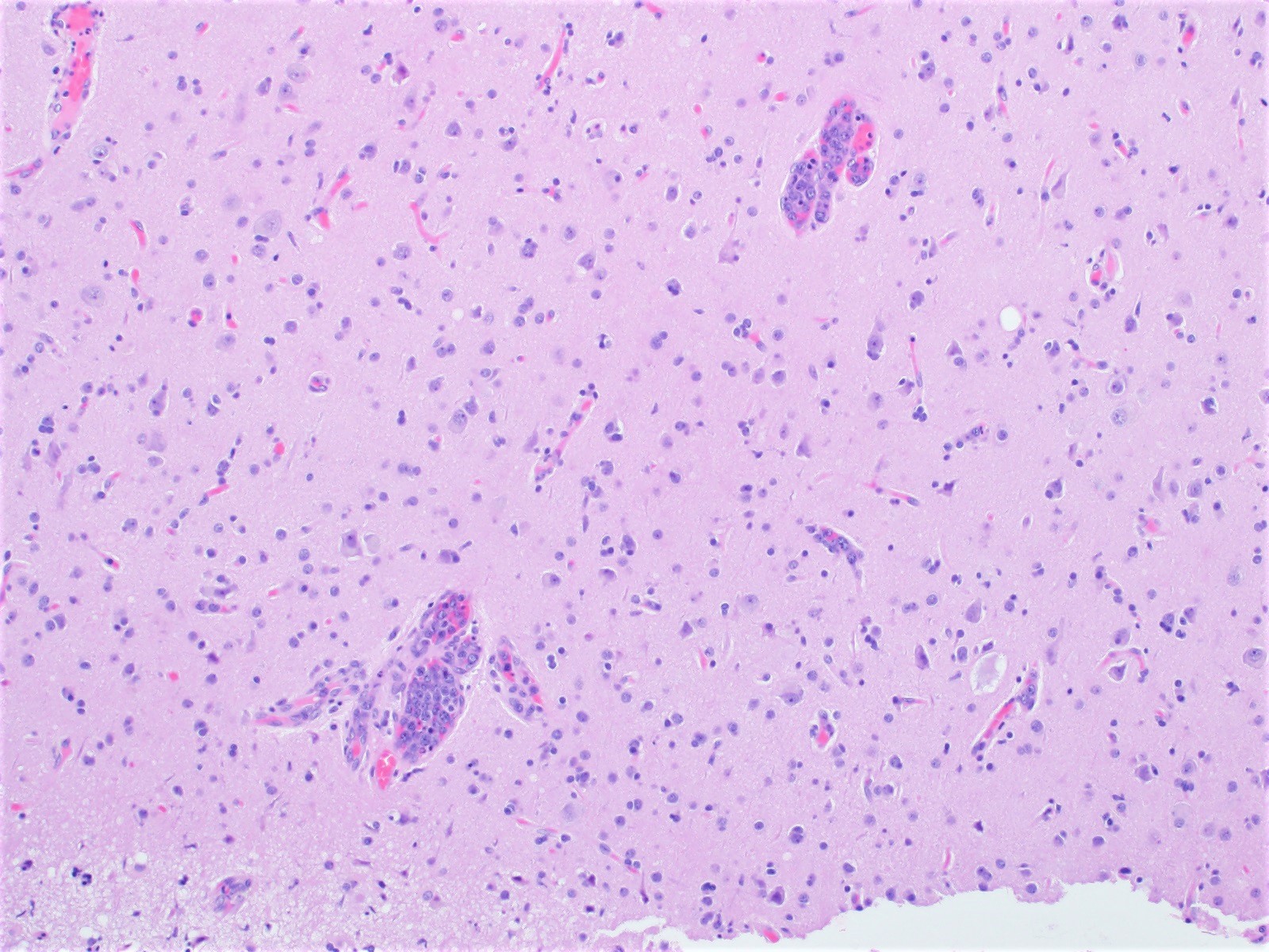
Brain MRI with contrast enhancing lesion

Solid and enhancing nodules on MRI
Images hosted on other servers:
Cerebral lymphoma
spreading across
the corpus callosum
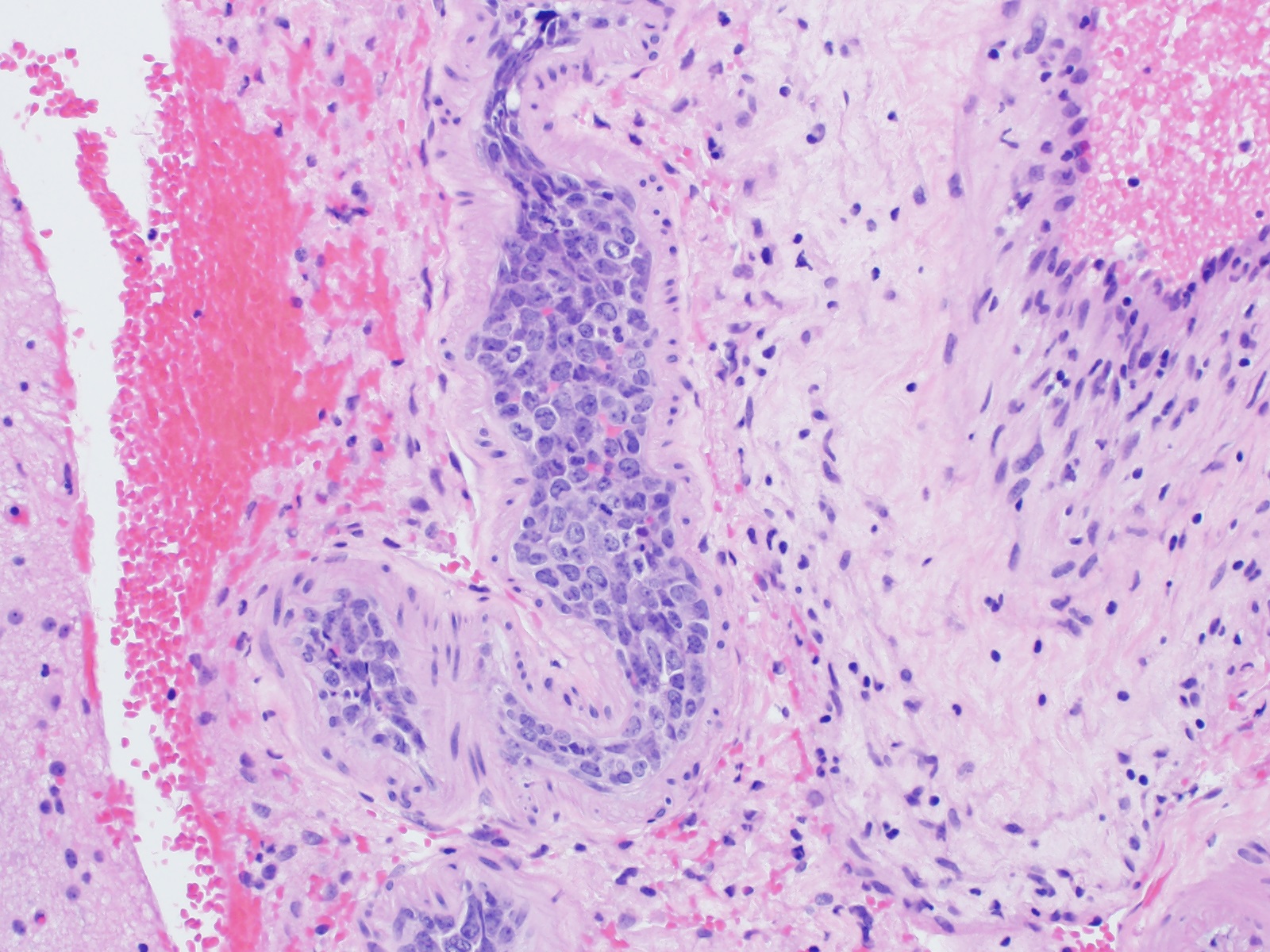
Contributed by Elizabeth Courville, M.D. and Lena Young, D.O.
Perivascular infiltrate of atypical lymphoid cells
Diffuse infiltrate
of intermediate
to large
mononuclear cells
Cytology of mononuclear cells
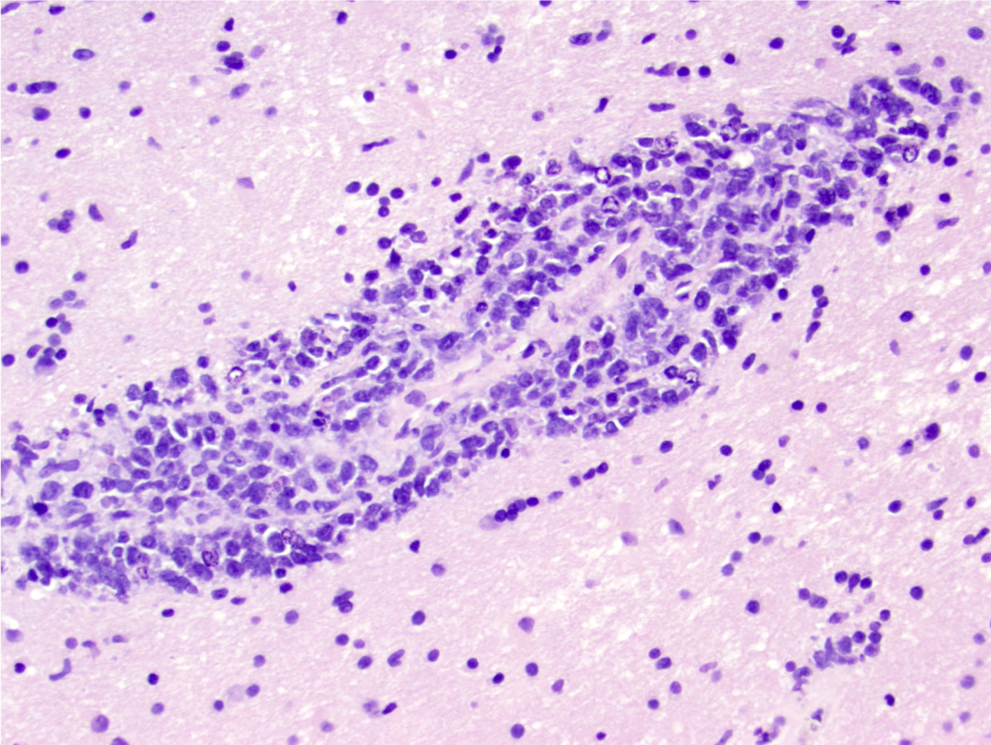
Perivascular cuffing by neoplastic lymphocytes
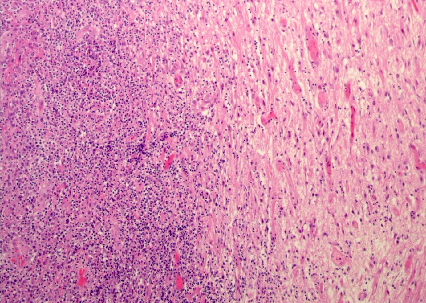
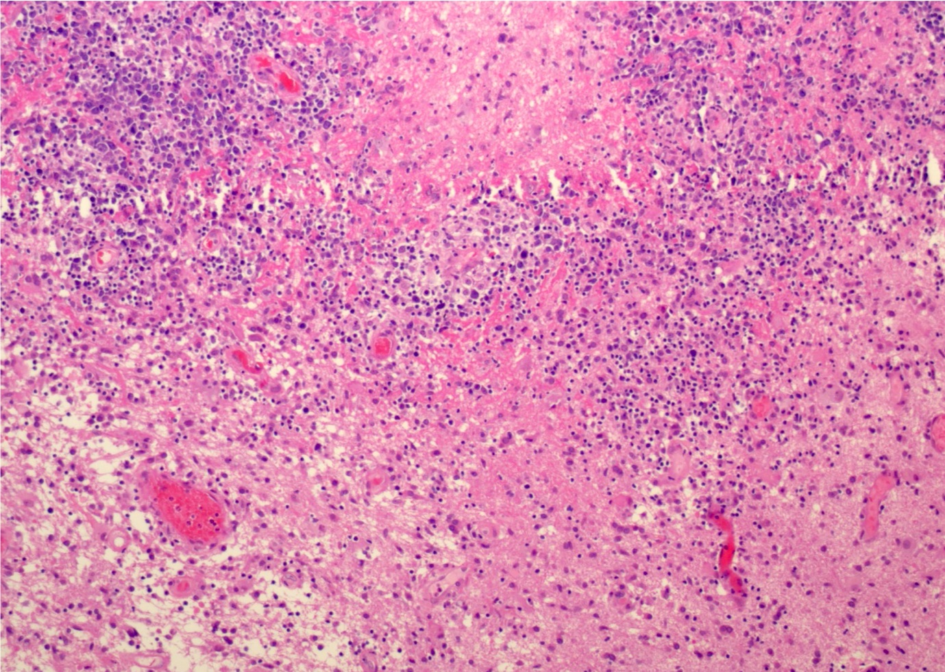
Infiltrate of neoplastic intermediate to large lymphocytes with background inflammation and necrosis
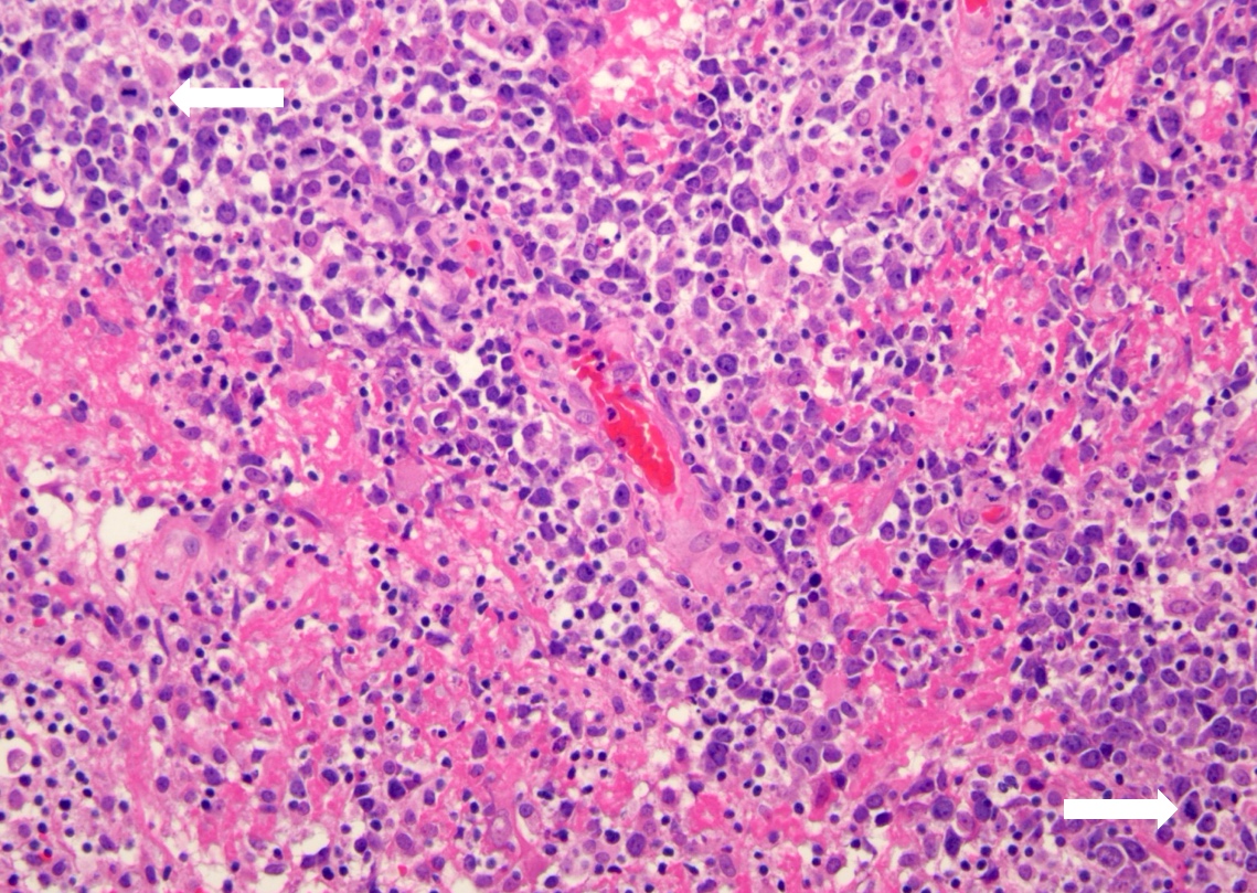
Pleomorphic lymphocytes infiltrating brain

Immunohisto-
chemistry for B and T cells
ISH shows lambda restriction
Contributed by Jennifer Chapman, M.D.
Dense dermal infiltrate
Predominantly small to medium sized lymphocytes
Scattered large cells
Most cells show strong CD3 expression
Most cells show strong CD4 expression
Numerous cells show strong PD1 expression
Subset of abnormal cells show strong ICOS expression
Small CD8+ T cells
Contributed by Roberto N. Miranda, M.D.
Skin epidermotropism and dermal infiltrate
Epidermotropism with intraepidermal cluster
Lymphoma at the dermoepidermal junction
Large pleomorphic cells
CD3 positivity
CD4 negativity of epidermotropic cells
CD7 positivity of epidermotropic cells
CD8 positivity of epidermotropic cells
TIA1 positivity of lymphoma cells
Proliferation marker Ki67
Pagetoid reticulosis
Microabscess of Pautrier in pagetoid reticulosis
CD3 in pagetoid reticulosis

CD8 in pagetoid reticulosis
βF1 pagetoid reticulosis
Folliculotropism in CD8 epidermotropic TCL
Epidermotropism in CD8 epidermotropic TCL
Epidermotropic small lymphoma cells
Liver involvement by CD8 cytotoxic TCL
CD3 positive lymphoma cells
CD8 positive lymphoma cells
Contributed by Roberto N. Miranda, M.D. and Alexandra Hristov, M.D.

Dermal infiltrate with grenz zone
Morphology of infiltrates
Characteristic CD68 staining
Cytotoxic immunophenotype
Contributed by Roberto N. Miranda, M.D.
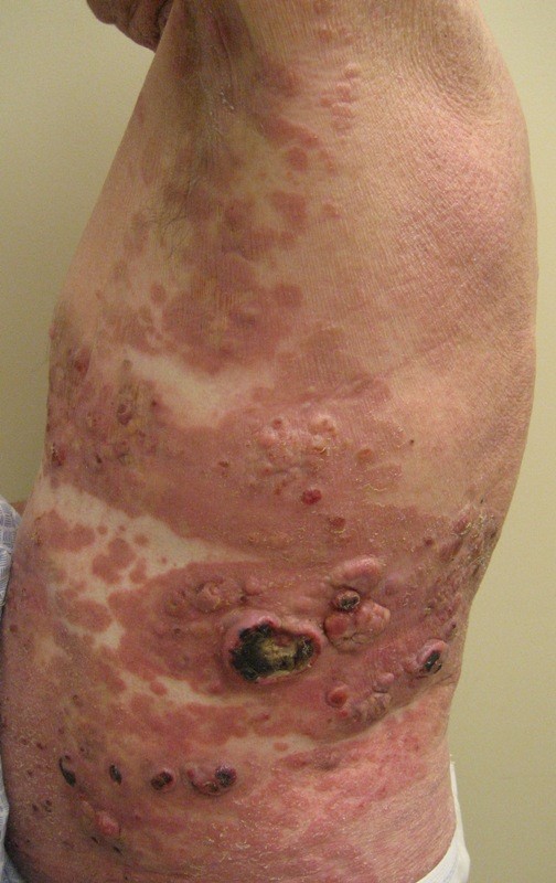
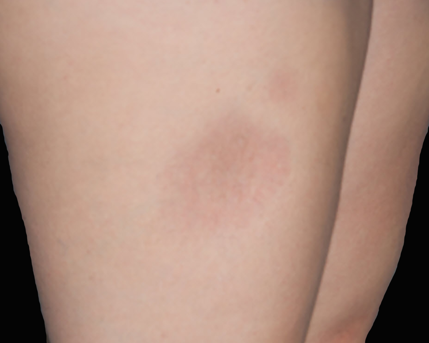
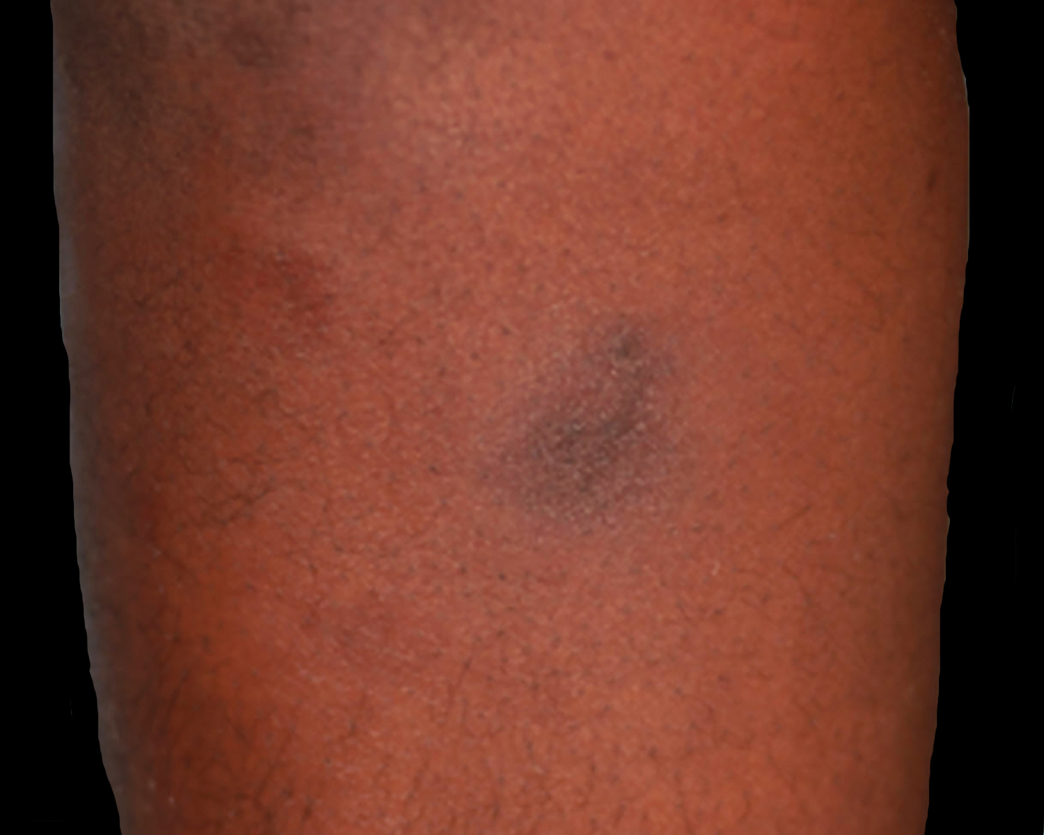
Ulcerated tumors
Erythematous plaques
Contributed by Roberto N. Miranda, M.D.

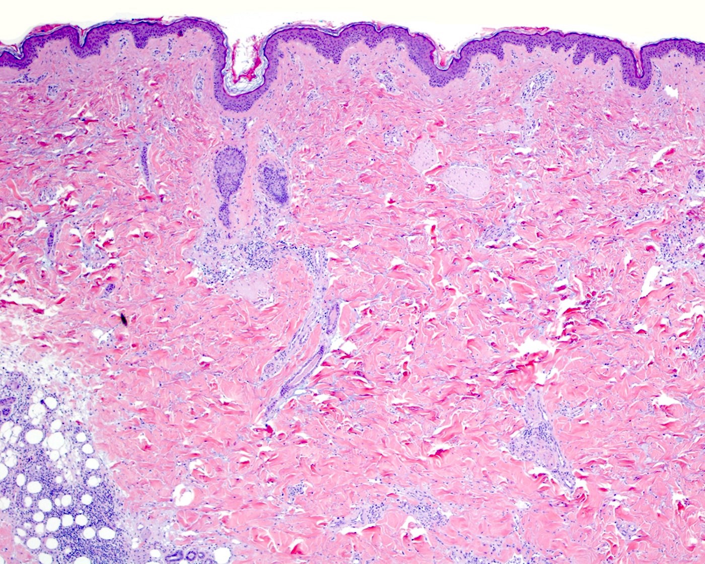
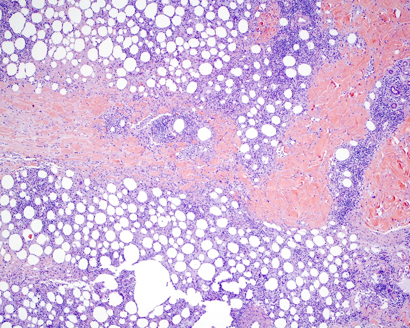
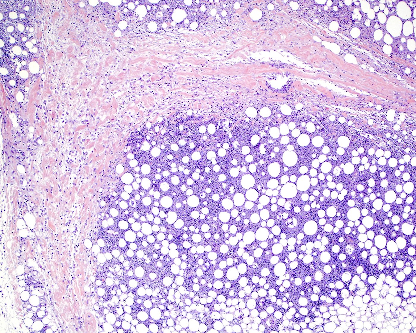
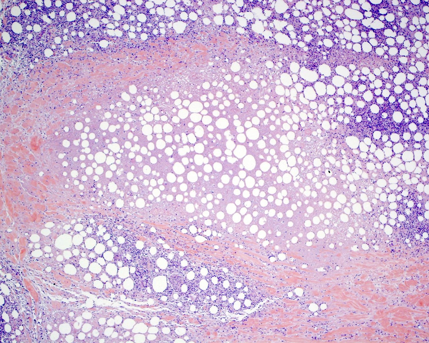
Diffuse infiltrate
Epidermotropic infiltrate
Fat rimming
Cytologic atypia
TCR alpha beta negativity
Pagetoid pattern
Hemophagocytosis
CD3 positivity
CD4 loss
CD4 negativity (CD4 loss)
CD7 negativity (CD7 loss)
CD8 positivity
TIA1 positivity
TCR delta positivity
TCR beta chain negativity
Contributed by Roberto N. Miranda, M.D.
TCR gamma delta positivity
TCR gamma delta
positivity and
TCR alpha beta
chain negativity
Contributed by Mario L. Marques-Piubelli, M.D. and Roberto N. Miranda, M.D. (Case #519)
Extracavitary presentation of PEL
EBER
LANA-1
LANA-1 in an extracavitary presentation
Images hosted on other servers:

70 year old man with HHV8+, EBV+ pleural effusions
Contributed by Mario L. Marques-Piubelli, M.D. and Roberto N. Miranda, M.D. (Case #519)
Diff-Quik
Wright-Giemsa
PEL Thinprep
Clinical features:
| Nodal follicular lymphoma | Testicular follicular lymphoma | |
| Age (median) | Adults and elder (sixth decade) | Children and young adults |
| Gender (M:F) | Men and women | Men only |
| Affected sites | Lymph nodes with extranodal spread | Testicle and adnexa |
| Symptoms | Generalized lymphadenopathy | Painless mass |
| Stage (Ann Arbor) | High (III - IV) in most cases | IE |
Pathologic features:
| Nodal follicular lymphoma | Testicular follicular lymphoma | |
| Gross appearance | Discrete mass or complete effacement | Discrete mass or diffuse involvement |
| Histologic grade | Grades 1 - 3 | Grade 3 |
Immunophenotype:
| Nodal follicular lymphoma | Testicular follicular lymphoma | |
| CD10 | Variable | Variable |
| BCL2 | Usually positive | Negative |
Molecular features:
| Nodal follicular lymphoma | Testicular follicular lymphoma | |
| IGH-BCL2 | Present, up to 90% | Negative |
Contributed by Roberto N. Miranda, M.D.
Follicular lymphoma of testis
Seminiferous tubuli and neoplastic follicle
Large centroblasts of follicular lymphoma

BCL6 IHC
CD21 IHC
DLBCL of testis
Contributed by Jennifer Chapman, M.D.

Intermediate to large sized cytologically atypical lymphoid cells
Diffuse distribution of intermediate to large sized lymphoma cells
CD19
CD20
CD23
CD30

Endobronchial biopsy
Endobronchial biopsy
Contributed by Mahsa Khanlari, M.D.
Papanicolaou
Contributed by Richard K. Wood, M.D. and Dietrich Werner, M.D.
Bone marrow involvement
CD20 positivity in bone marrow
CD79a positivity in bone marrow
Cyclin D1 negativity in bone marrow
Contributed by Allam Shawwa, M.D.
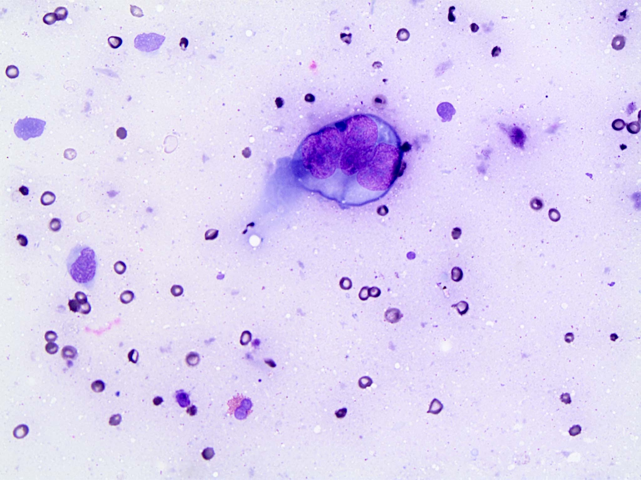
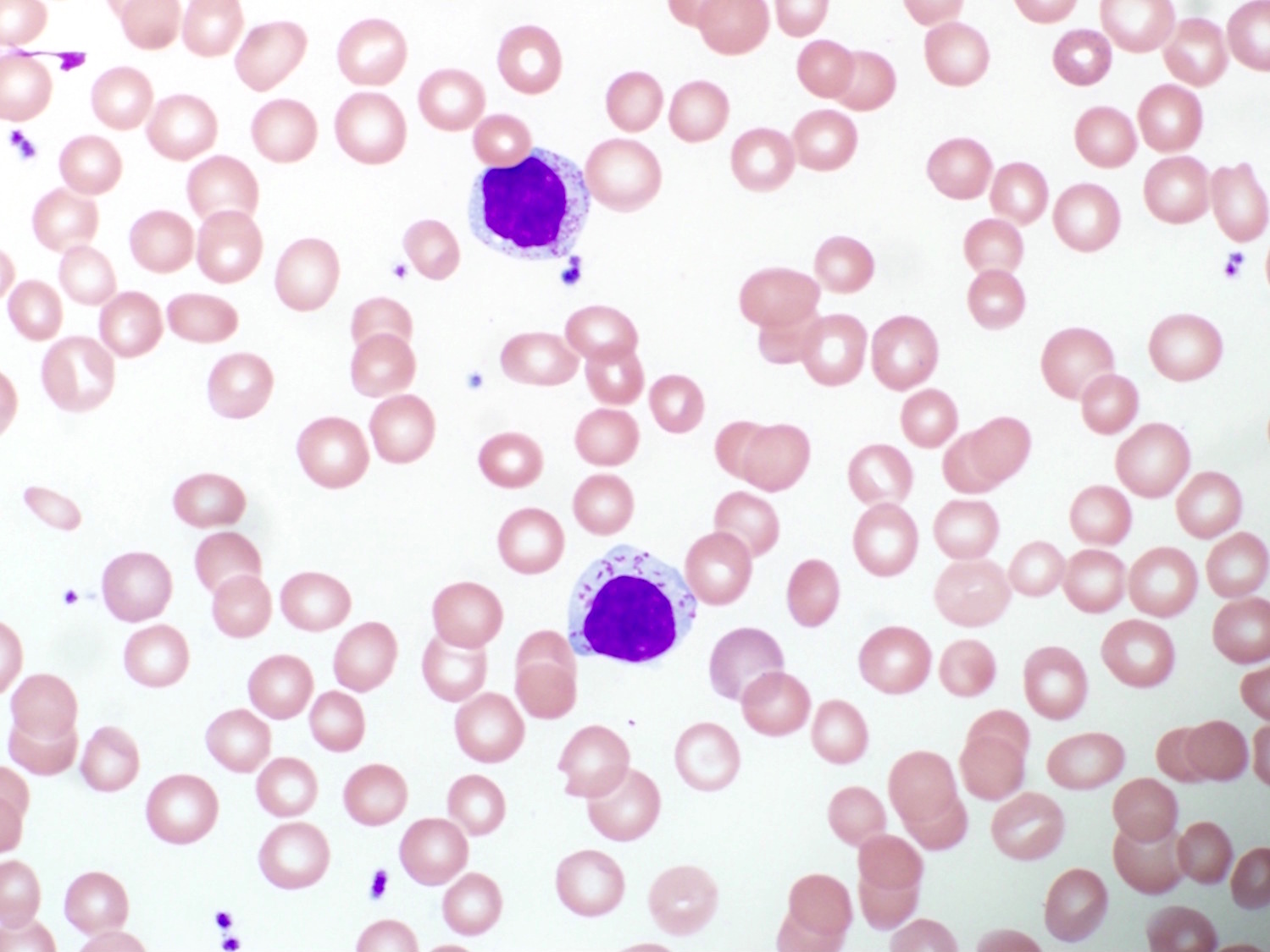
Prolymphocytes in peripheral blood

Prolymphocytes in peripheral blood

Contributed by Shahbaz Khan, M.D. and Sepideh Nikki Asadbeigi, M.D.
Diffuse large B cell lymphoma type
Extranodal
Contributed by Roberto N. Miranda, M.D.
Prominent exfoliative lesion
Prominent erythroderma
Contributed by Roberto N. Miranda, M.D.
Central nervous system (CNS) involvement
Contributed by Roberto N. Miranda, M.D.
Bone marrow involvement
CD3 positivity
Epidermotropic and lichenoid lymphoid infiltrate
Epidermotropic infiltrate
Cerebriform cytology
Lymph node involvement
Cytological atypia
Contributed by Roberto N. Miranda, M.D.
Lymph node touch print
Contributed by Roberto N. Miranda, M.D.
Cerebriform lymphocytes in peripheral blood smear
Contributed by Roberto N. Miranda, M.D.
Flow cytometry in SS
Images hosted on other servers:

Diffusely enlarged spleen
Images hosted on other servers:

Neoplastic villous lymphocytes

Body regions

Lymph node regions
TNMB classification of MF / SS
Lymph node grading
TNM classification of non-MF / SS
Contributed by Henry K. Wong, M.D., Ph.D.
Mycosis fungoides, patch stage
Mycosis fungoides, plaque stage
Mycosis fungoides, tumor stage
Images hosted on other servers:

Sézary cells, peripheral smear
Diagnosis and staging of cutaneous lymphoma
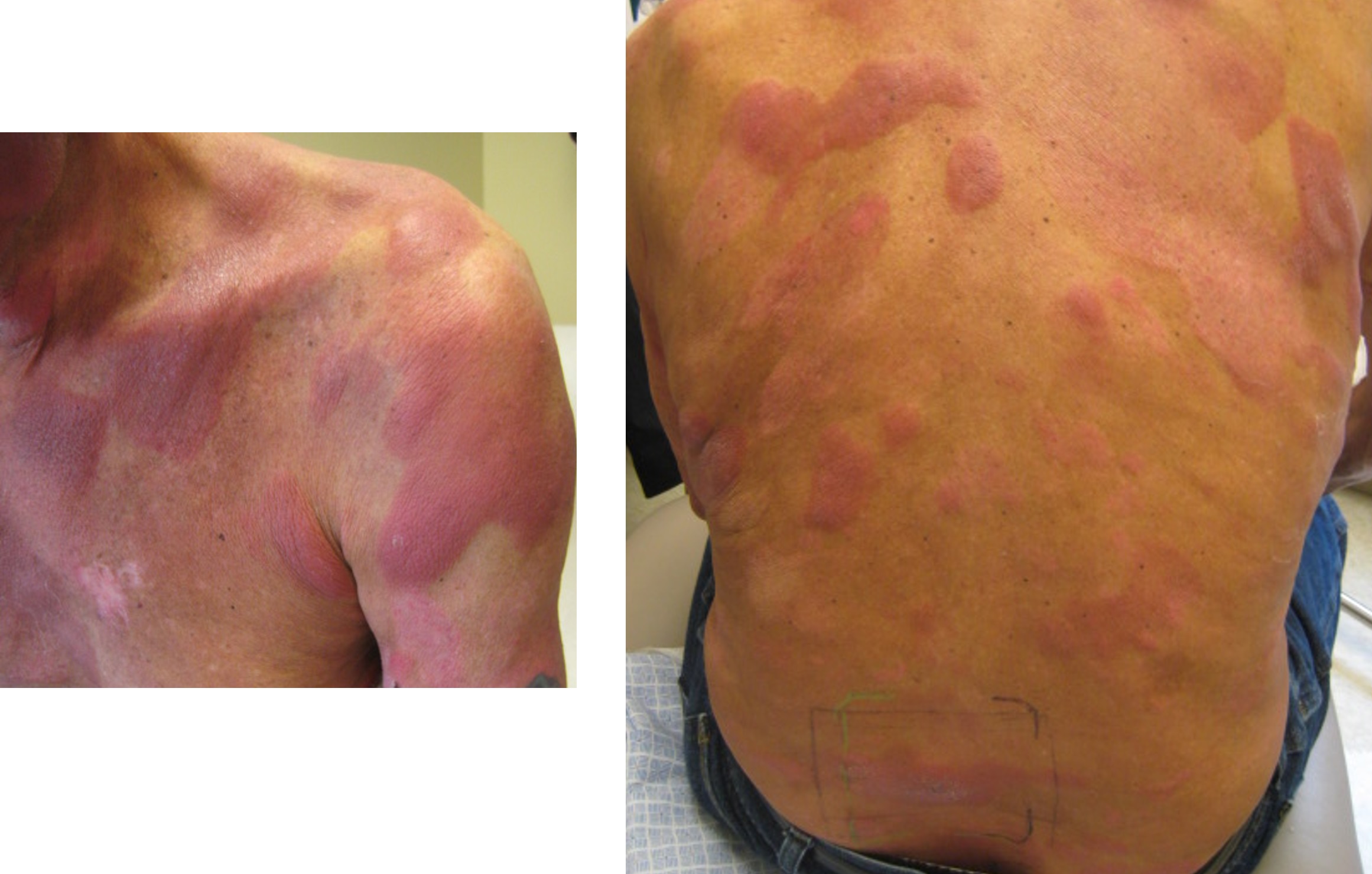
Contributed by Roberto N. Miranda, M.D.
Subcutaneous uptake
Contributed by Roberto N. Miranda, M.D.
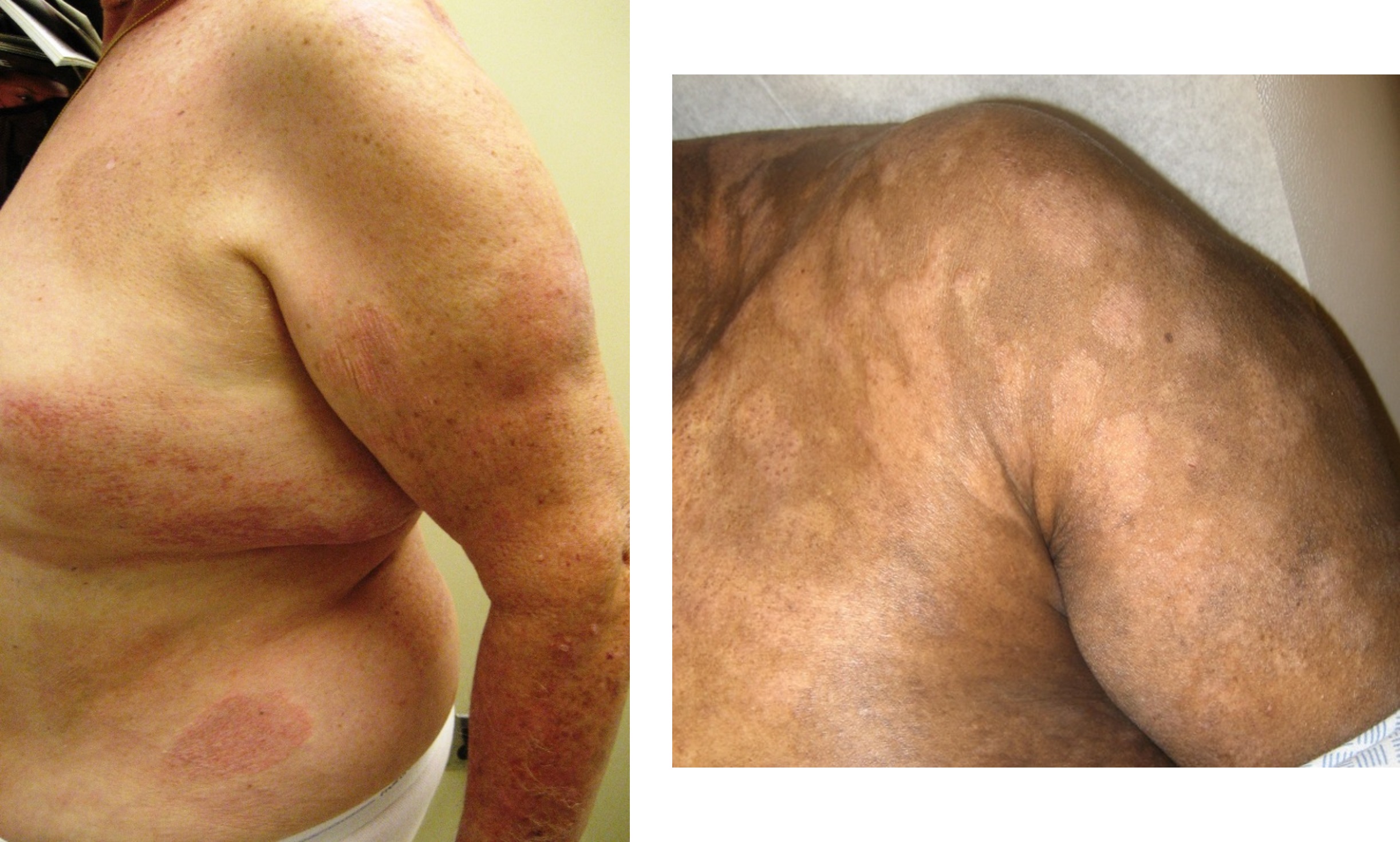
Subcutaneous lesions
Subcutaneous plaque
Contributed by Roberto N. Miranda, M.D.
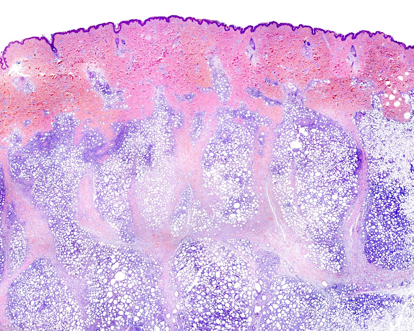
Deep dermis and subcutaneous involvement
Epidermis and reticular dermis sparing
Lobular distribution
Acellular hyaline change
Adipocyte rimming
CD8 positivity
CD4 negativity
TIA1 positivity
βF1 (TCRα / β) positivity
TIM-3 positivity
Contributed by Yoon Kyung Jeon, M.D.
Lymphocytic infiltrates in the liver
Lymph node, atypical infiltrates

Lymph node, atypical infiltrates
Lymphocytic infiltrates in the liver (CD3)
Images hosted on other servers:

PCR for TCR gamma gene
Contributed by Elaine Jaffe, M.D. and João Víctor Alves de Castro, M.D.

Proposed interrelatedness of EBV positive T / NK LPDs
Images hosted on other servers:

Muscle involvement and coronary artery dilation
Contributed by Elaine Jaffe, M.D. and João Víctor Alves de Castro, M.D.

Spleen, red and white pulp
Lymphoid aggregate in the red pulp
Hemophagocytosis
EBER positive T cells in spleen
Ulcerative lesion
Perivascular and periadnexal involvement
Follicular and paracortical hyperplasia
Paracortical hyperplasia

Dense CD3 positive infiltrate
TCR delta positive infiltrate
EBER positive T cells in lymph node
CD3 in the red pulp

CD8 positive T cells
Images hosted on other servers:

NK cells with abnormal phenotype in the bone marrow
Contributed by Konrad Chan, M.D.
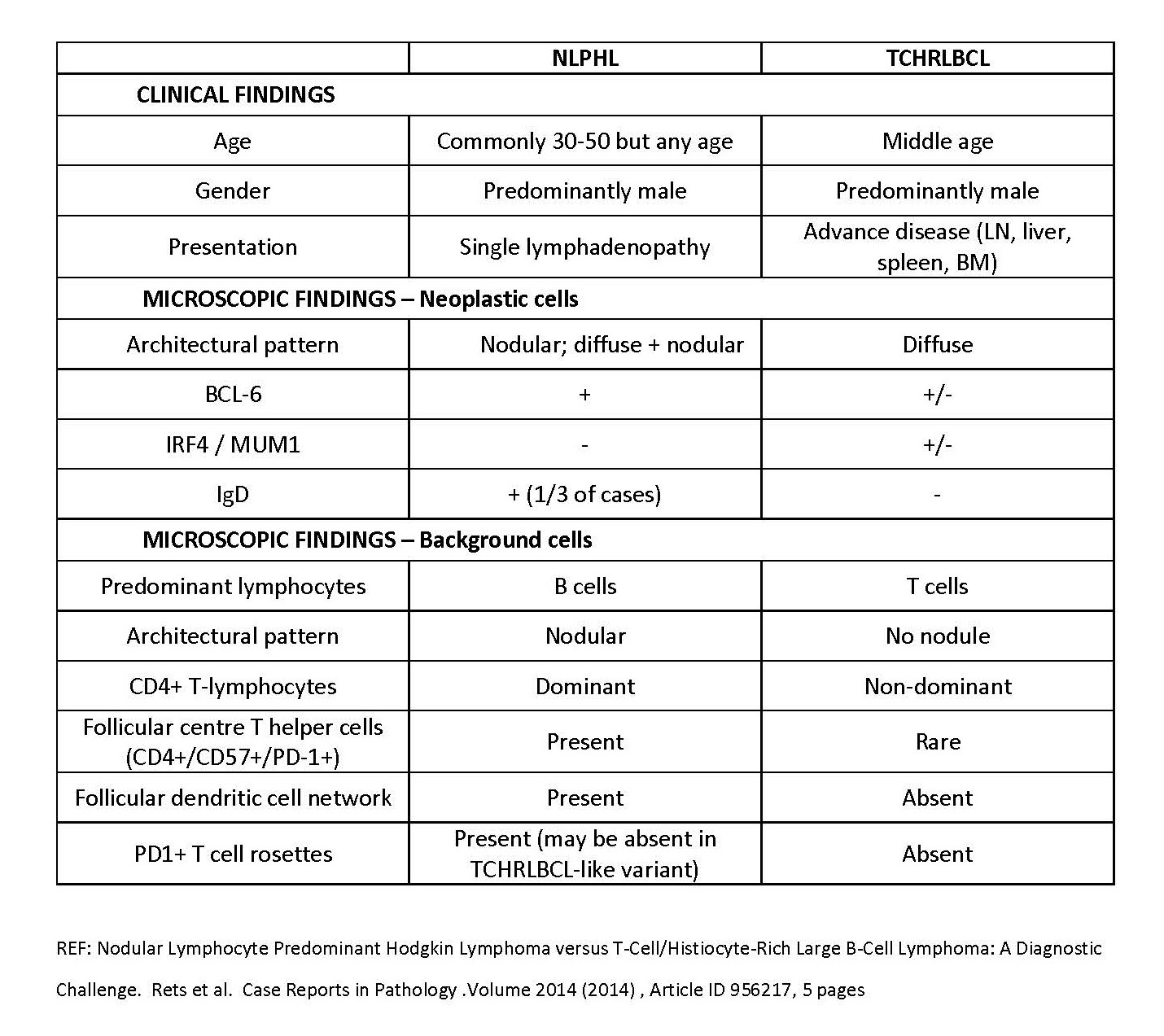
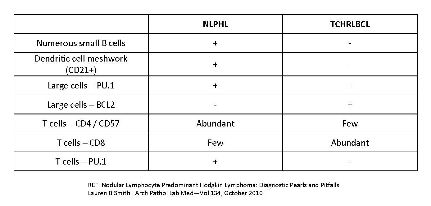
NLPHL versus THRLBCL
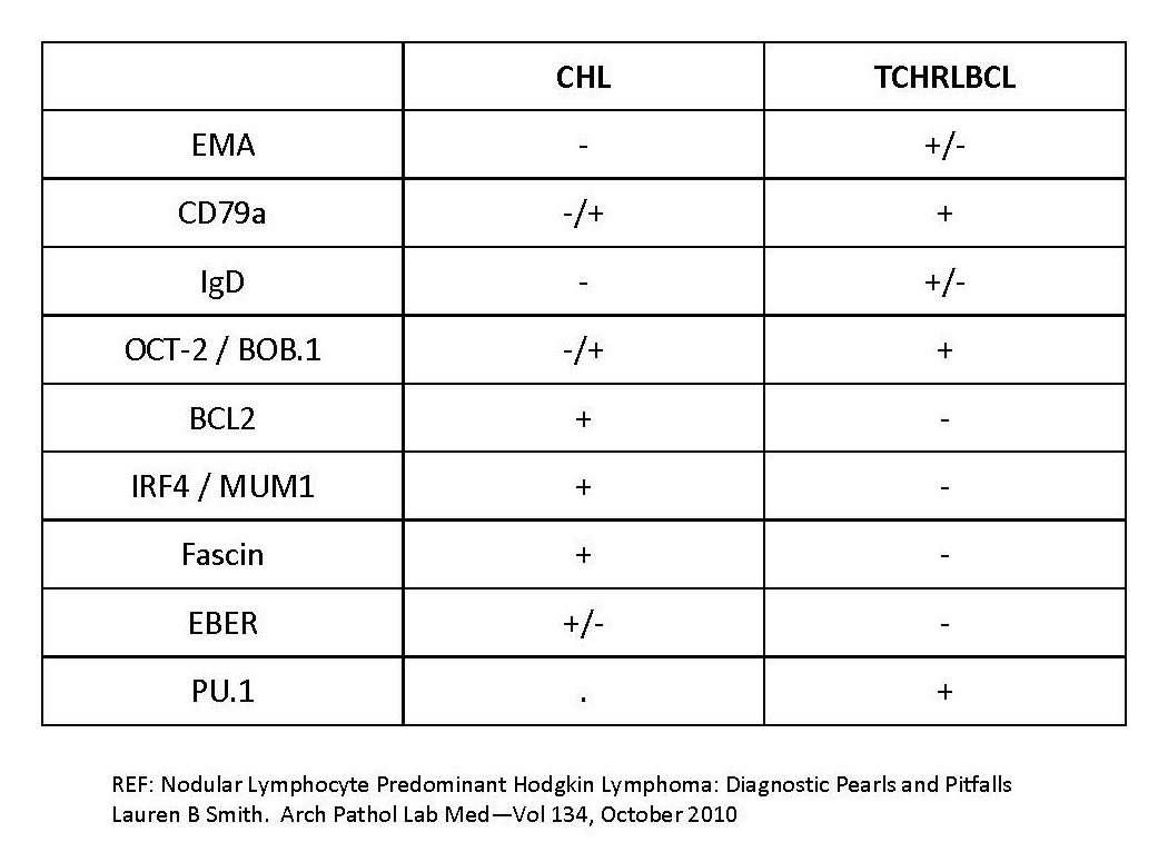
CHL versus THRLBCL
Contributed by Aaron Auerbach, M.D., M.P.H., Asmaa Gaber Abdou, M.D. and Nancy Youssef Asaad, M.D.
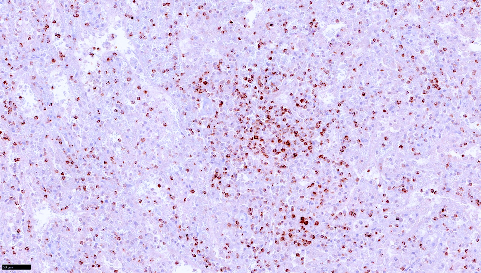
Scattered large atypical cells
Large neoplastic cells
CD20 in a biopsy

CD20 in an excision
CD3+ small T cells
CD68+ histiocytes
Contributed by Contributed by Mingyi Chen, M.D., Ph.D. (Case #317)

CD3

CD20

PAX5

CD30
NLPHL versus THRLBCL
Contributed by Min Shi, M.D., Ph.D. and Dragan Jevremovic, M.D., Ph.D.
T LGLL bone marrow involvement

Intrasinusoidal CD3+ T LGLL
Intrasinusoidal CD8+ T LGLL
Intrasinusoidal granzyme B+ T LGLL

Intrasinusoidal TIA1+ T LGLL
T LGLL liver involvement
CD8+ T LGLL in liver

CD5- T LGLL in liver
TIA1+ T LGLL in liver
Contributed by Min Shi, M.D., Ph.D. and Dragan Jevremovic, M.D., Ph.D.
T LGLL in peripheral blood
Contributed by Min Shi, M.D., Ph.D. and Dragan Jevremovic, M.D., Ph.D.
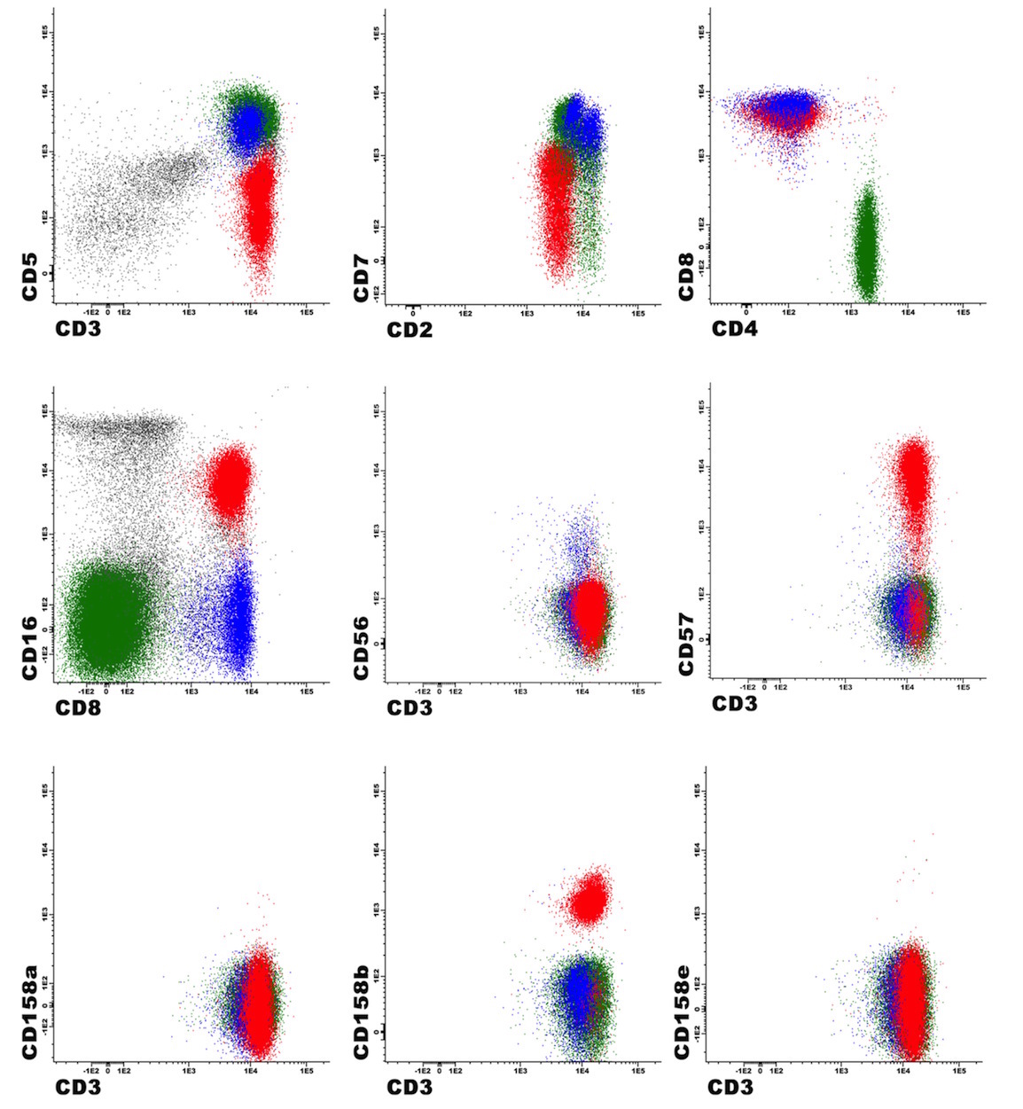
T LGLL flow cytometry
Contributed by Min Shi, M.D., Ph.D. and Dragan Jevremovic, M.D., Ph.D.
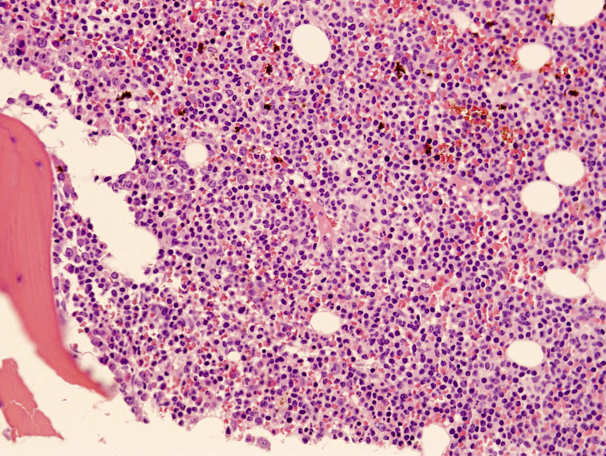
Diffuse bone marrow involvement
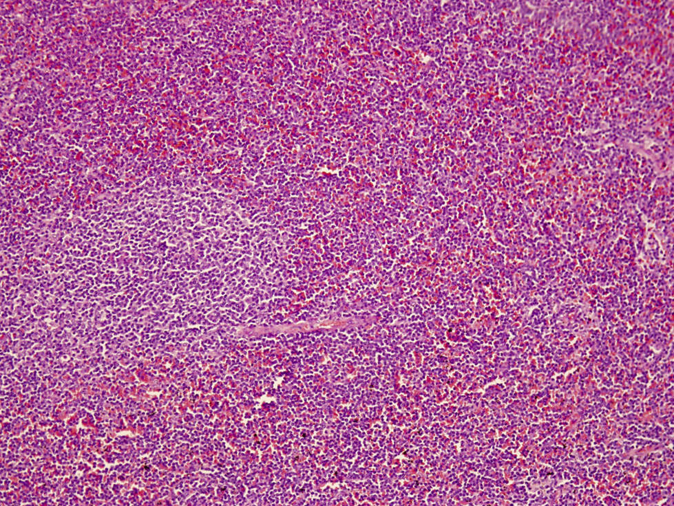
Splenic red pulp infiltrate
Monotonous, paracortical lymphoid infiltrate
Intermediate size, atypical lymphocyte
CD3 positivity in bone marrow
Monotonous T cell infiltrate
TCL1A positivity
Uniform strong TCL1A staining
Contributed by Min Shi, M.D., Ph.D. and Dragan Jevremovic, M.D., Ph.D.
Cerebriform variant
Prolymphocytes in peripheral blood
Small cell variant
Contributed by Min Shi, M.D., Ph.D. and Dragan Jevremovic, M.D., Ph.D.

T PLL flow cytometry
Contributed by Min Shi, M.D., Ph.D. and Dragan Jevremovic, M.D., Ph.D.
FISH for TCL1A separation
Table 1
| FL with Castleman-like changes |
| FL with plasmacytic differentiation with or without IgG4 positive plasma cells |
| FL with marginal zone differentiation, typically involving MALT sites |
| FL negative for CD10, positive for MUM1 with BCL6 abnormalities |
| EBV positive FL |
| Floral variant of FL |
Contributed by Jennifer Chapman, M.D.
Castleman-like changes

Castleman-like changes
Floral variant

Floral variant

Follicular lymphoma negative for CD10 and expressing MUM1
Images hosted on other servers:

Gene mutations in JAK / STAT pathway
Contributed by Patricia Tsang, M.D., M.B.A.
Cold agglutinin disease (CAD)
CAD lymphoid aggregate
CAD (CD20)
CAD (CD3)
Table 1: T / NK cell entities - comparison of WHO (2016), WHO (2022) and ICC (2022)
| WHO HAEM4R | WHO HAEM5 | ICC |
| Precursor T cell neoplasms | ||
| T lymphoblastic leukemia / lymphoma | T lymphoblastic leukemia / lymphoma, NOS | T lymphoblastic leukemia / lymphoma |
Early T cell precursor lymphoblastic leukemia / lymphoma | Early T cell precursor lymphoblastic leukemia | Early T cell precursor acute lymphoblastic leukemia, NOS |
| Early T cell precursor acute lymphoblastic leukemia, BCL11B activated | ||
| NK lymphoblastic leukemia / lymphoma* | [Entity removed] | NK cell acute lymphoblastic leukemia* |
| Tumor-like lesions with T cell predominance | ||
| [Not included] | Kikuchi-Fujimoto disease | [Not included] |
| [Not included] | Indolent T lymphoblastic proliferation | [Not included] |
| [Not included] | Autoimmune lymphoproliferative syndrome | [Not included] |
| Mature T / NK cell leukemias | ||
| T prolymphocytic leukemia | T prolymphocytic leukemia | T cell prolymphocytic leukemia |
| T cell large granular lymphocytic leukemia | T large granular lymphocytic leukemia | T cell large granular lymphocytic leukemia |
| Chronic lymphoproliferative disorder of NK cells* | NK large granular lymphocytic leukemia | Chronic lymphoproliferative disorder of NK cells* |
| Adult T cell leukemia / lymphoma | Adult T cell leukemia / lymphoma | Adult T cell leukemia / lymphoma |
| Sézary syndrome | Sézary syndrome | Sézary syndrome |
| Aggressive NK cell leukemia | Aggressive NK cell leukemia | Aggressive NK cell leukemia |
| Primary cutaneous T cell lymphomas | ||
| Primary cutaneous CD4 positive small or medium T cell LPD* | Primary cutaneous CD4 positive small or medium T cell LPD | Primary cutaneous small or medium CD4 positive T cell LPD |
| Primary cutaneous acral CD8 positive T cell lymphoma* | Primary cutaneous acral CD8 positive lymphoproliferative disorder | Primary cutaneous acral CD8 positive lymphoproliferative disorder |
| Mycosis fungoides | Mycosis fungoides | Mycosis fungoides |
| Primary cutaneous CD30 positive T cell LPD: lymphomatoid papulosis | Primary cutaneous CD30 positive T cell LPD: lymphomatoid papulosis | Primary cutaneous CD30 positive T cell LPD: lymphomatoid papulosis |
| Primary cutaneous CD30 positive T cell LPD: primary cutaneous anaplastic large cell lymphoma | Primary cutaneous CD30 positive T cell LPD: primary cutaneous anaplastic large cell lymphoma | Primary cutaneous CD30 positive T cell LPD: primary cutaneous anaplastic large cell lymphoma |
| Subcutaneous panniculitis-like T cell lymphoma | Subcutaneous panniculitis-like T cell lymphoma | Subcutaneous panniculitis-like T cell lymphoma |
| Primary cutaneous gamma / delta T cell lymphoma | Primary cutaneous gamma / delta T cell lymphoma | Primary cutaneous gamma / delta T cell lymphoma |
| Primary cutaneous CD8 positive aggressive epidermotropic cytotoxic T cell lymphoma* | Primary cutaneous CD8 positive aggressive epidermotropic cytotoxic T cell lymphoma | Primary cutaneous CD8 positive aggressive epidermotropic cytotoxic T cell lymphoma |
| [Not included] | Primary cutaneous peripheral T cell lymphoma, NOS | [Not included] |
| Intestinal T cell and NK cell lymphoid proliferations and lymphomas | ||
| Indolent T cell lymphoproliferative disorder of the gastrointestinal tract* | Indolent T cell lymphoma of the gastrointestinal tract | Indolent clonal T cell LPD of the gastrointestinal tract |
| [Not included] | Indolent NK cell LPD of the gastrointestinal tract | Indolent NK cell LPD of the gastrointestinal tract |
| Enteropathy associated T cell lymphoma | Enteropathy associated T cell lymphoma | Enteropathy associated T cell lymphoma |
| Type II refractory celiac disease | ||
| Monomorphic epitheliotropic intestinal T cell lymphoma | Monomorphic epitheliotropic intestinal T cell lymphoma | Monomorphic epitheliotropic intestinal T cell lymphoma |
| Intestinal T cell lymphoma, NOS | Intestinal T cell lymphoma, NOS | Intestinal T cell lymphoma, NOS |
| Hepatosplenic T cell lymphoma | ||
| Hepatosplenic T cell lymphoma | Hepatosplenic T cell lymphoma | Hepatosplenic T cell lymphoma |
| Anaplastic large cell lymphoma | ||
| Anaplastic large cell lymphoma, ALK positive | ALK positive anaplastic large cell lymphoma | Anaplastic large cell lymphoma, ALK positive |
| Anaplastic large cell lymphoma, ALK negative | ALK negative anaplastic large cell lymphoma | Anaplastic large cell lymphoma, ALK negative |
| Breast implant associated anaplastic large cell lymphoma* | Breast implant associated anaplastic large cell lymphoma | Breast implant associated anaplastic large cell lymphoma |
| Nodal T follicular helper (TFH) cell lymphoma | ||
| Angioimmunoblastic T cell lymphoma | Nodal TFH cell lymphoma, angioimmunoblastic type | Follicular helper T cell lymphoma, angioimmunoblastic type |
| Follicular T cell lymphoma* | Nodal TFH cell lymphoma, follicular type | Follicular helper T cell lymphoma, follicular type |
| Nodal peripheral T cell lymphoma (PTCL) with TFH phenotype* | Nodal TFH cell lymphoma, NOS | Follicular helper T cell lymphoma, NOS |
| Other peripheral T cell lymphomas | ||
| Peripheral T cell lymphoma, NOS | Peripheral T cell lymphoma, NOS | Peripheral T cell lymphoma, NOS |
| EBV positive NK / T cell lymphomas | ||
| [Not included] [variant of PTCL, NOS] | EBV positive nodal T and NK cell lymphoma | Primary nodal Epstein-Barr virus positive T / NK cell lymphoma* |
| Extranodal NK / T cell lymphoma, nasal type | Extranodal NK / T cell lymphoma | Extranodal NK / T cell lymphoma, nasal type |
| EBV positive T and NK cell lymphoid proliferations and lymphomas of childhood | ||
| Severe mosquito bite allergy | Severe mosquito bite allergy | Severe mosquito bite allergy |
| Hydroa vacciniforme-like lymphoproliferative disorder | Hydroa vacciniforme lymphoproliferative disorder, classic or systemic | Hydroa vacciniforme lymphoproliferative disorder, classic or systemic |
| Chronic active EBV infection of T and NK cell type, systemic form | Systemic chronic active EBV disease | Chronic active EBV disease (T and NK cell phenotype) |
| Systemic EBV positive T cell lymphoma of childhood | Systemic EBV positive T cell lymphoma of childhood | Systemic EBV positive T cell lymphoma of childhood |
Contributed by Tom Deng, M.D.
Angiocentric destruction
Coagulative necrosis
CD3
CD56
EBER

Auerbach: 2022
Beck: 2021

Cerroni: 2020
Crane: 2024
Crane: 2021
Dorfman: 2023
Duffield: 2020
Foucar: 2023
Foucar: 2019
Gru: 2023
Hsi: 2017
Hudnall: 2019
IARC: 2017
Jaffe: 2016
King: 2023
Linden: 2015
Medeiros: 2017
Medeiros: 2021
Medeiros: 2023
Molina: 2019
Naeim: 2018
Subtil: 2019
Vasef: 2019
Wang: 2018
Wang: 2020
Find related Pathology books: hematopathology, dermatopathology, molecular, immunology / transplant, lab medicine
