


Images hosted on other servers:

hATTR clinical epidemiologic characteristic
Images hosted on other servers:

99mTc-DPD scan of cardiac ATTR
Contributed by Chunyu Cai, M.D., Ph.D.
Early amyloid neuropathy

Nerve edema
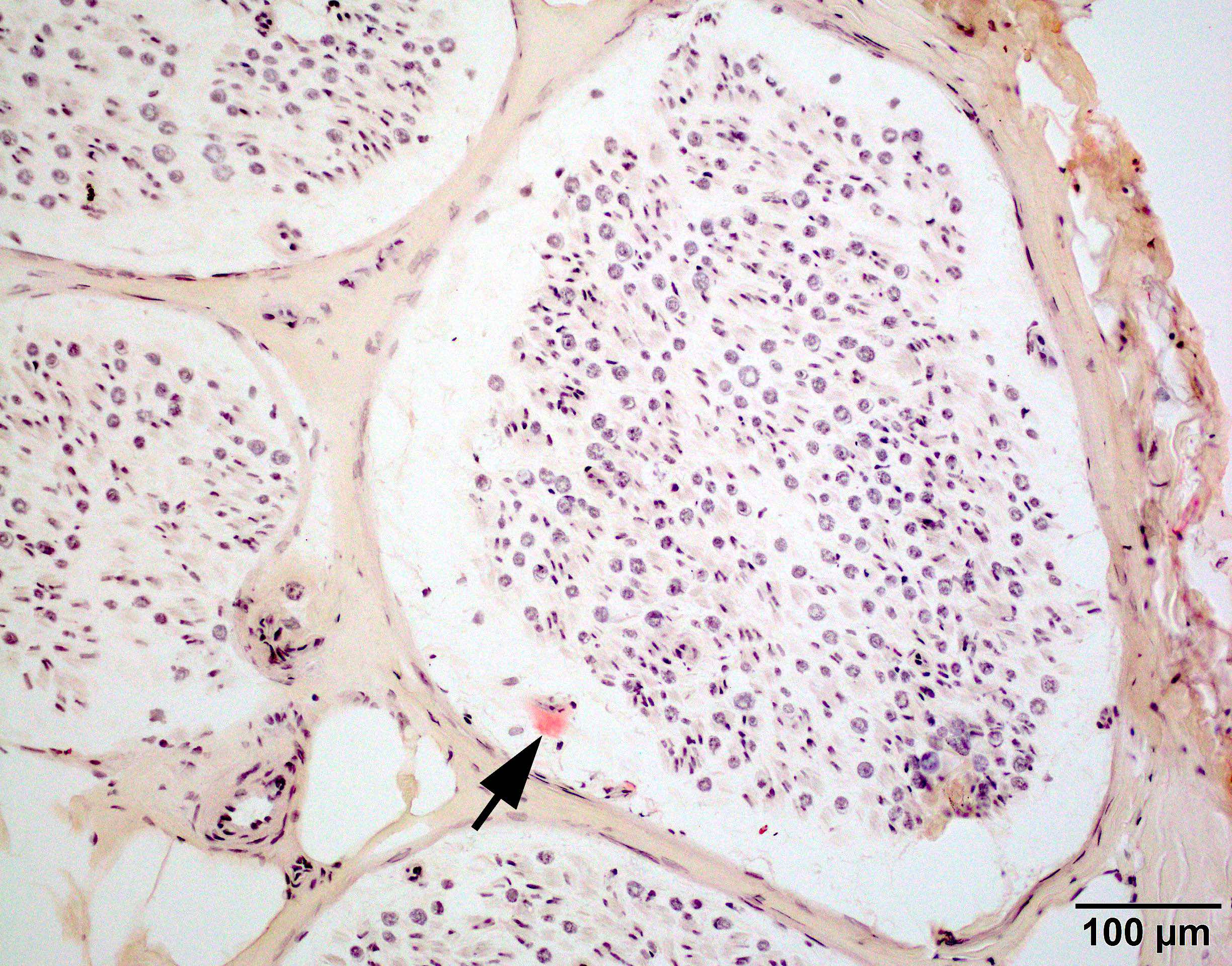
Congo red amyloid deposit
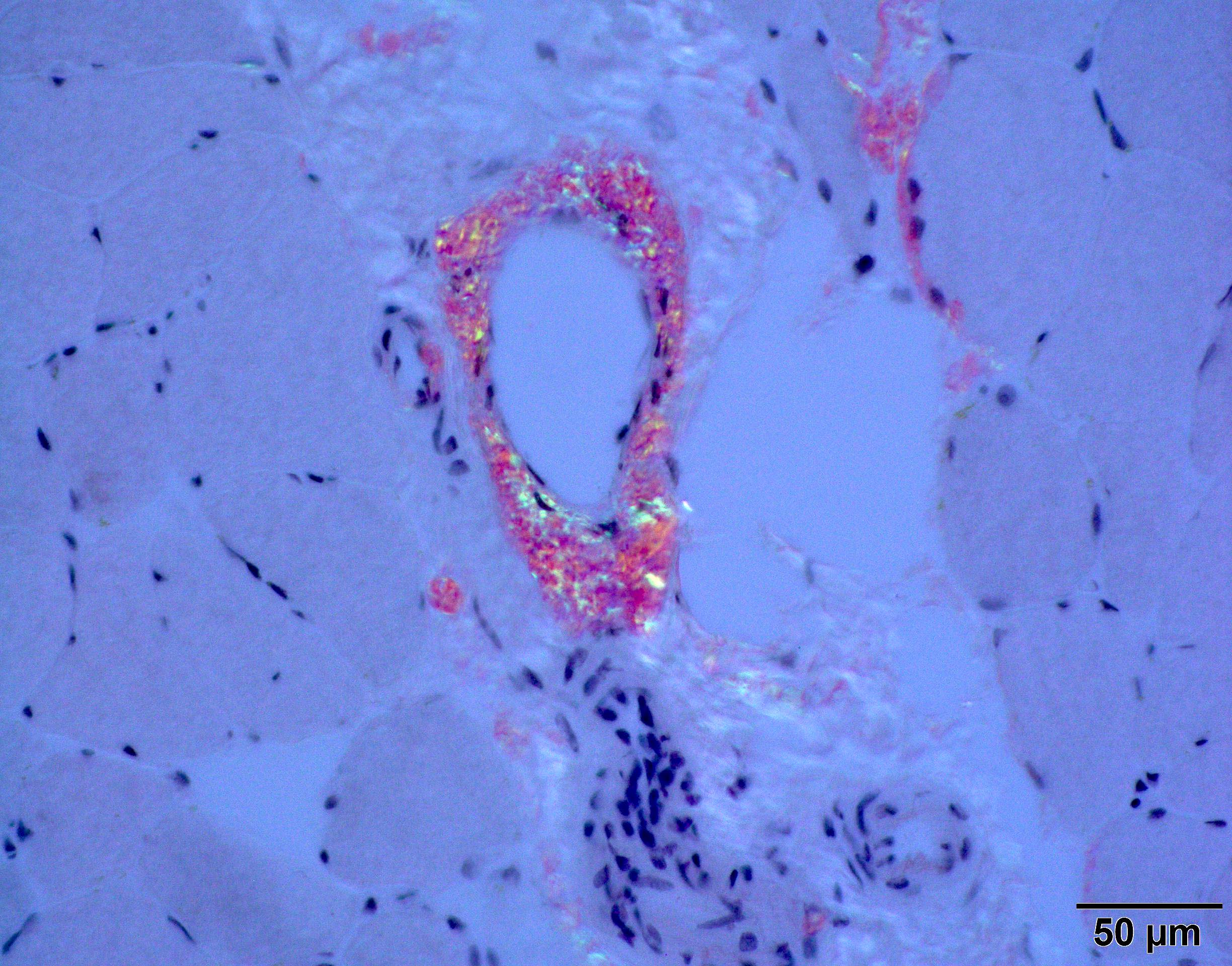
Congo red polarized
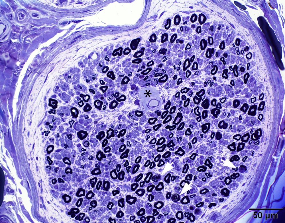
Loss of small myelinated axons
Late amyloid neuropathy
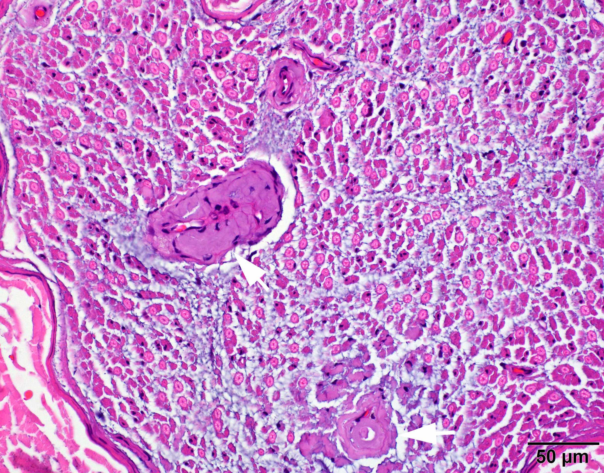
Nerve edema
Congo red amyloid deposit
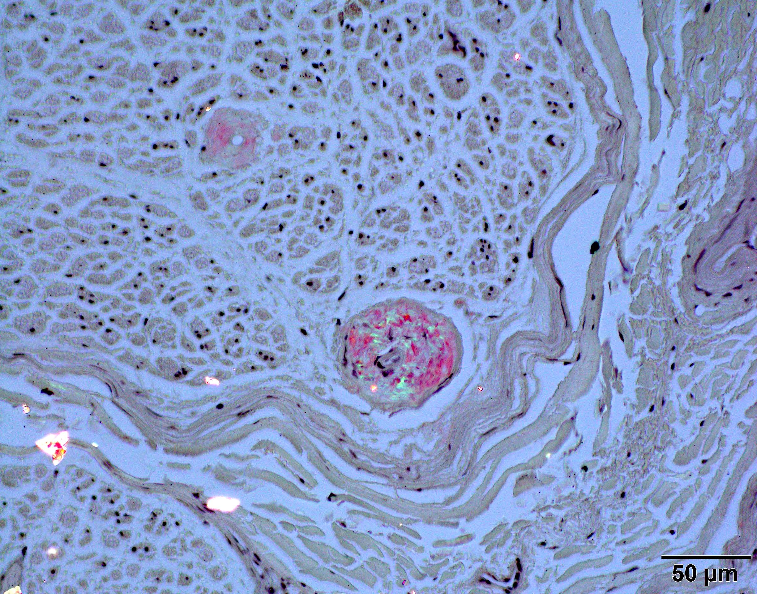
Congo red polarized
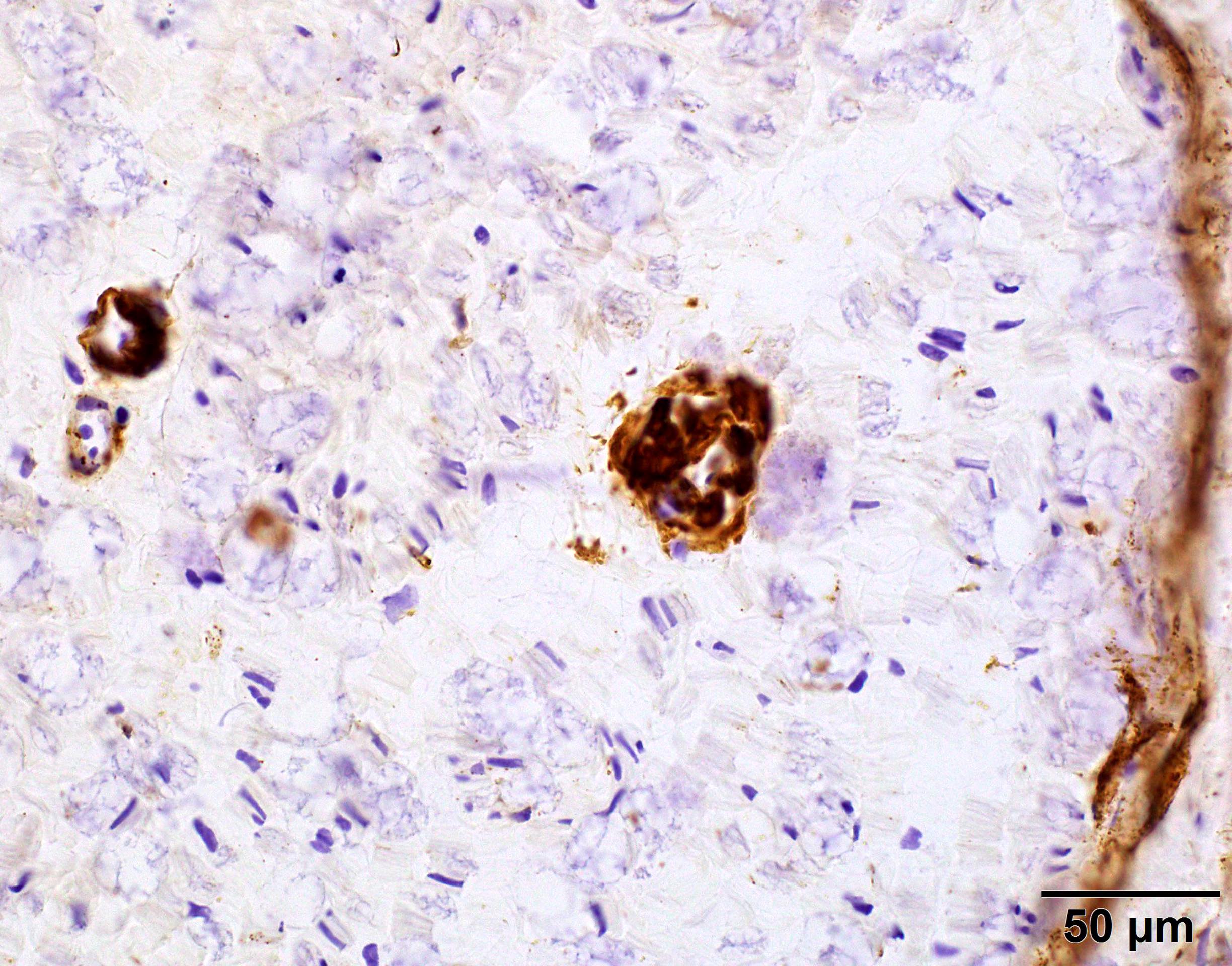
C5b9 amyloid deposit
Small and large axon loss
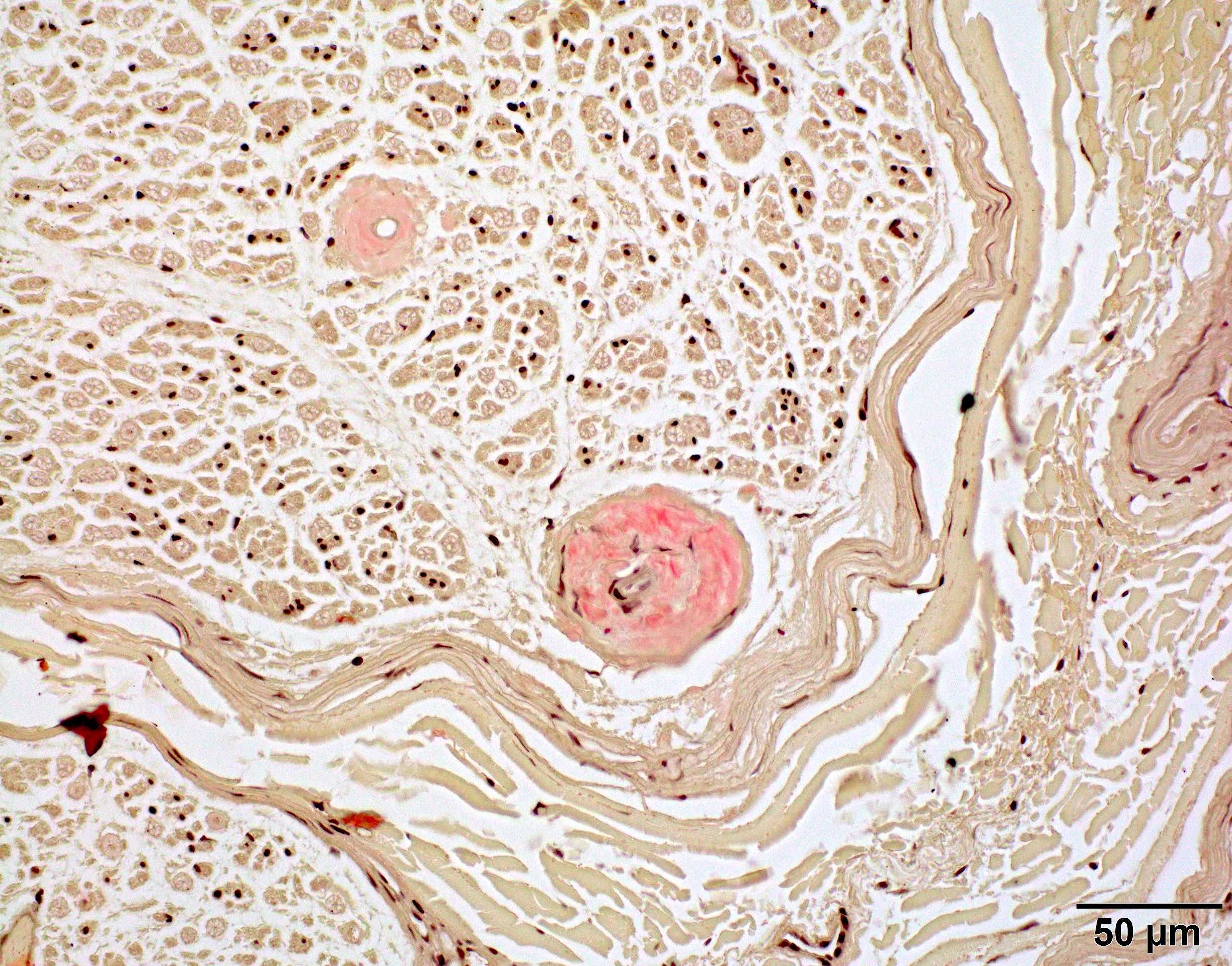
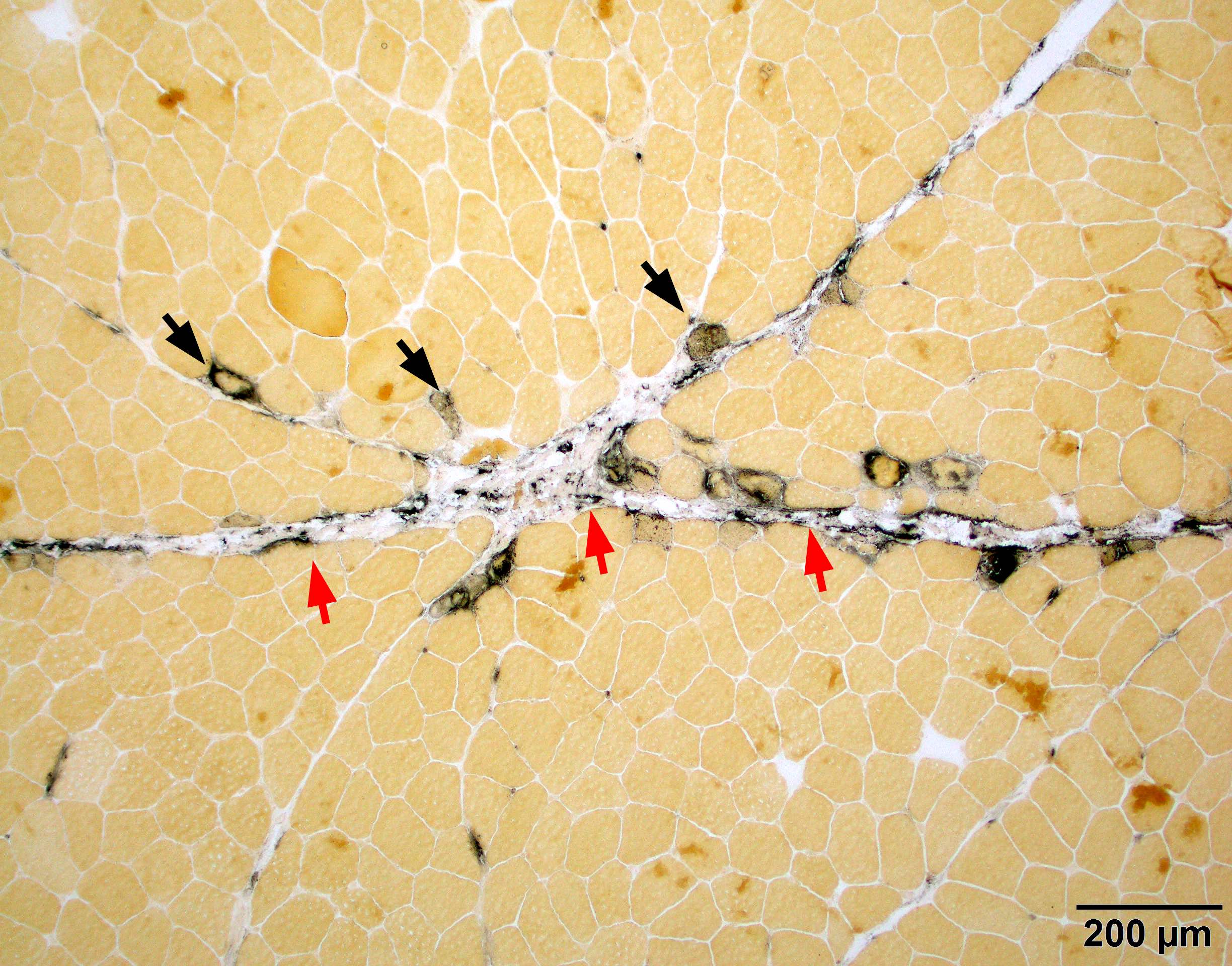
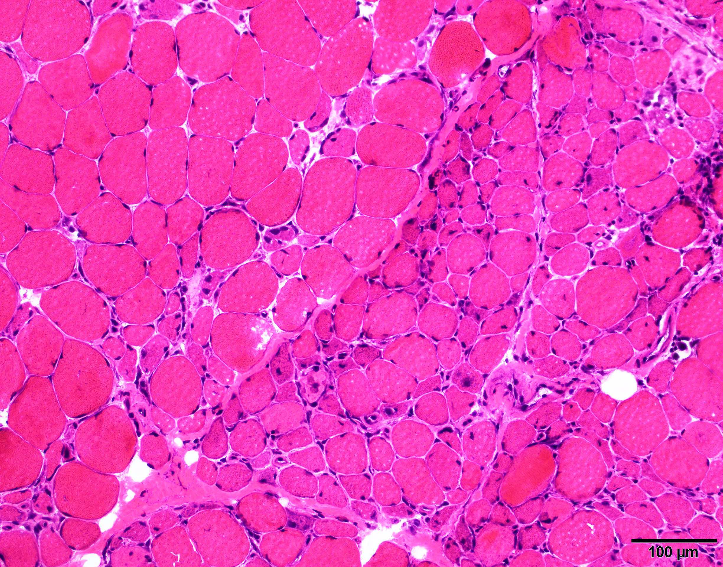
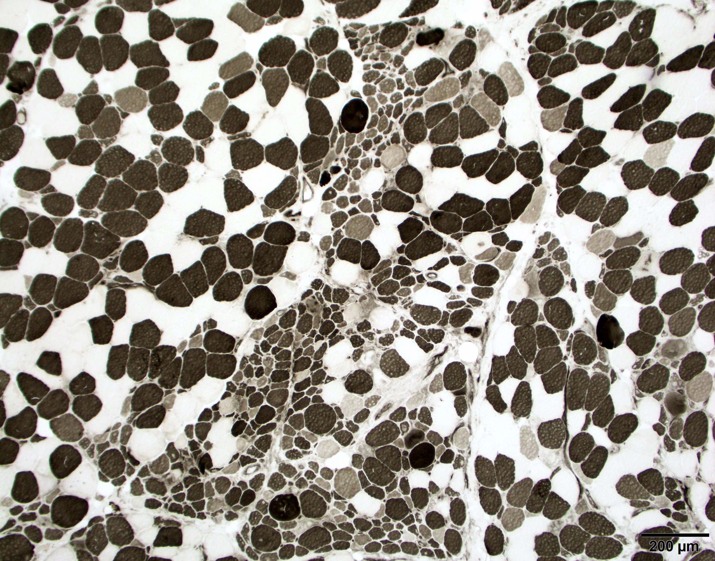
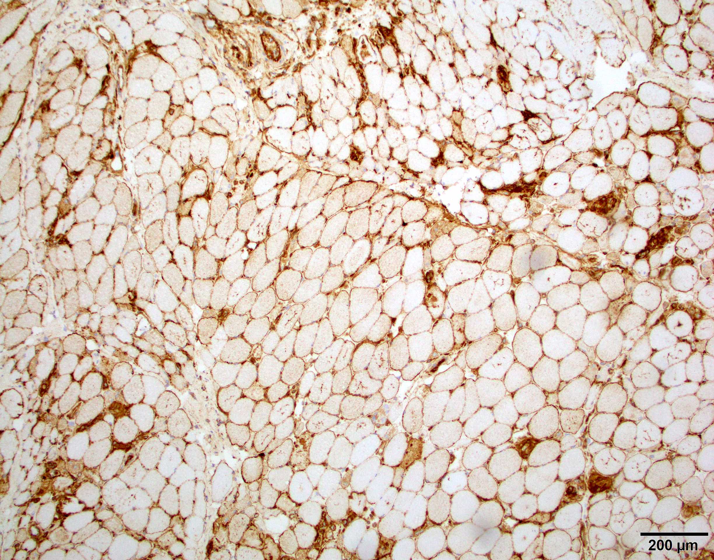
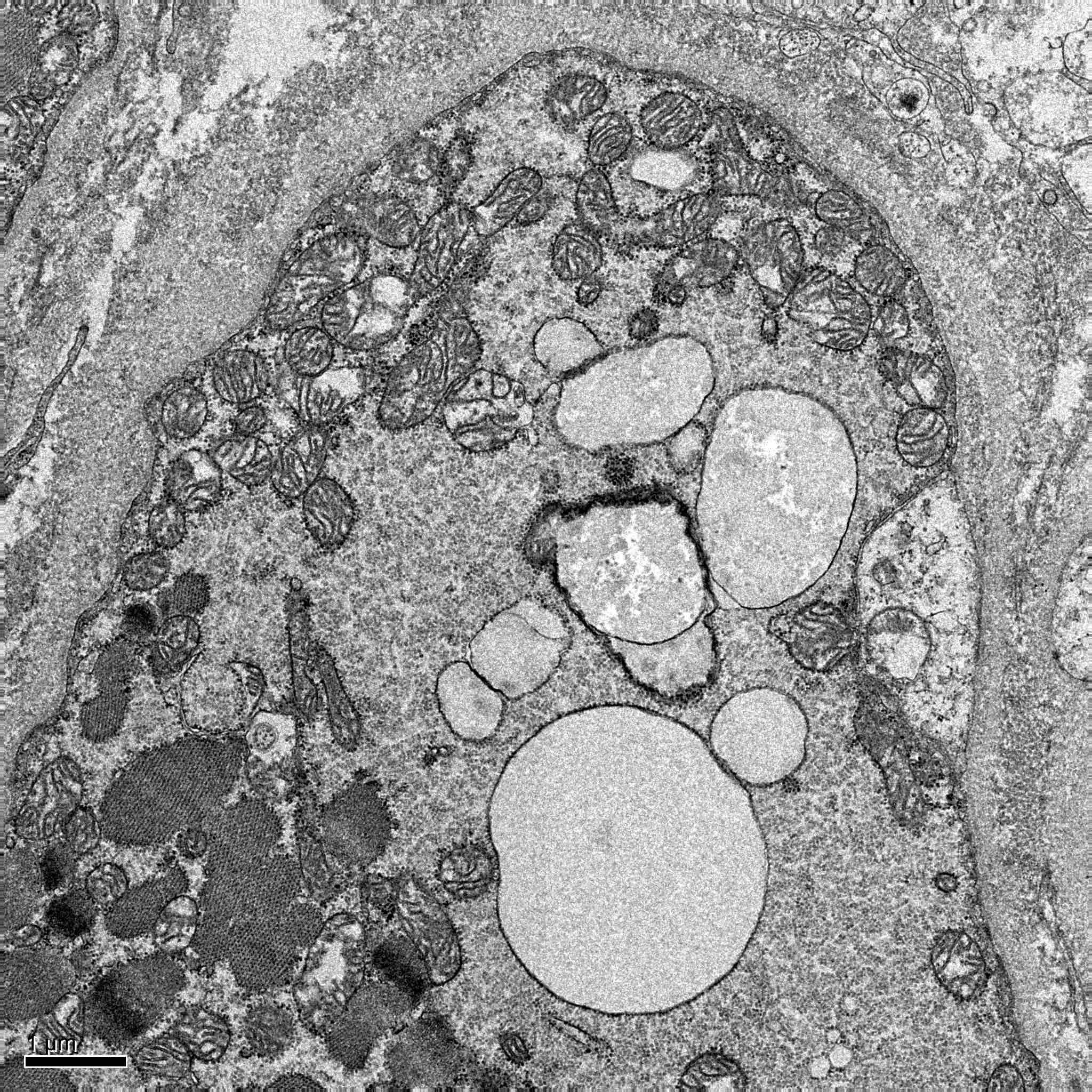
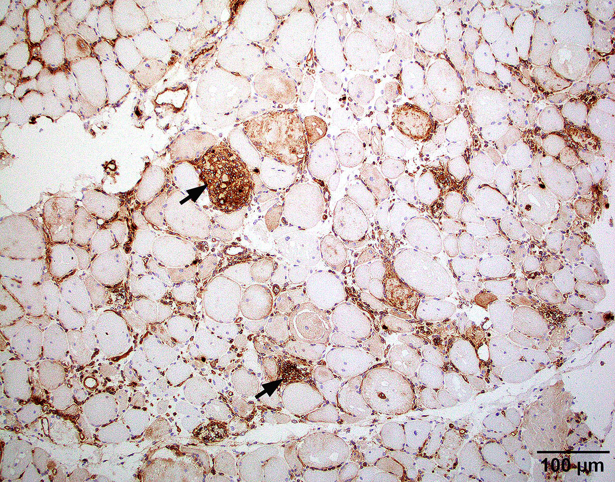
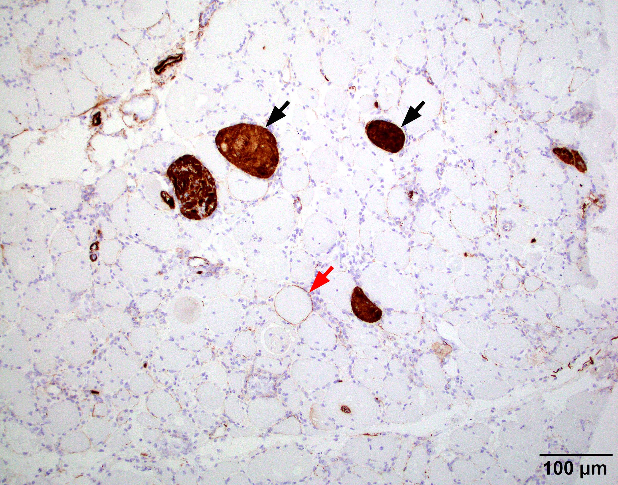
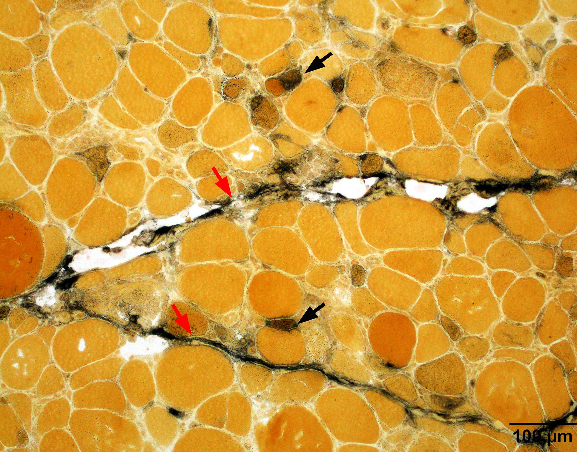
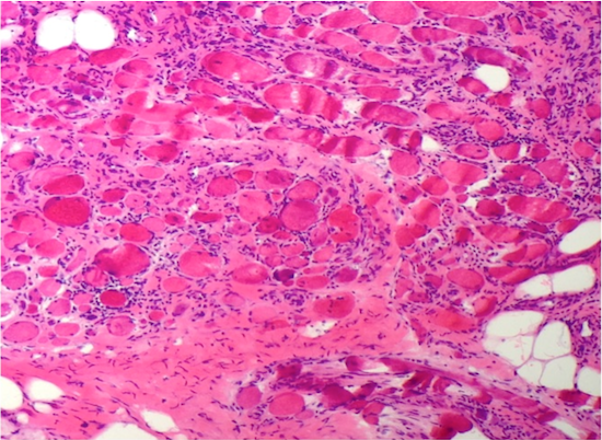
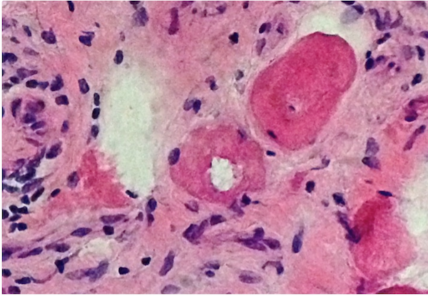
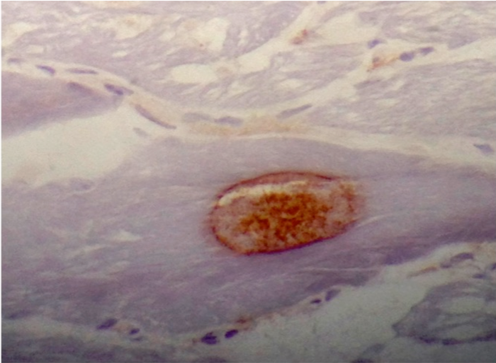
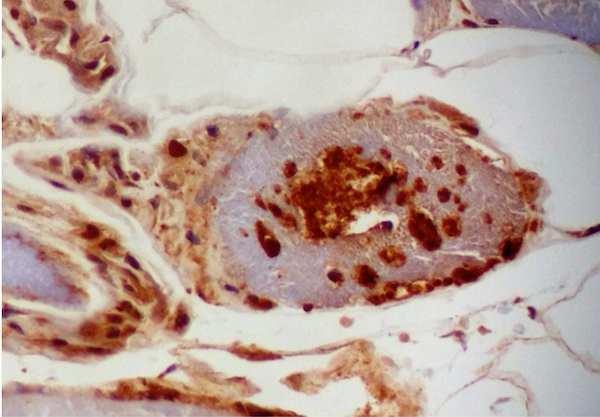
Hereditary transthyretin amyloid neuropathy
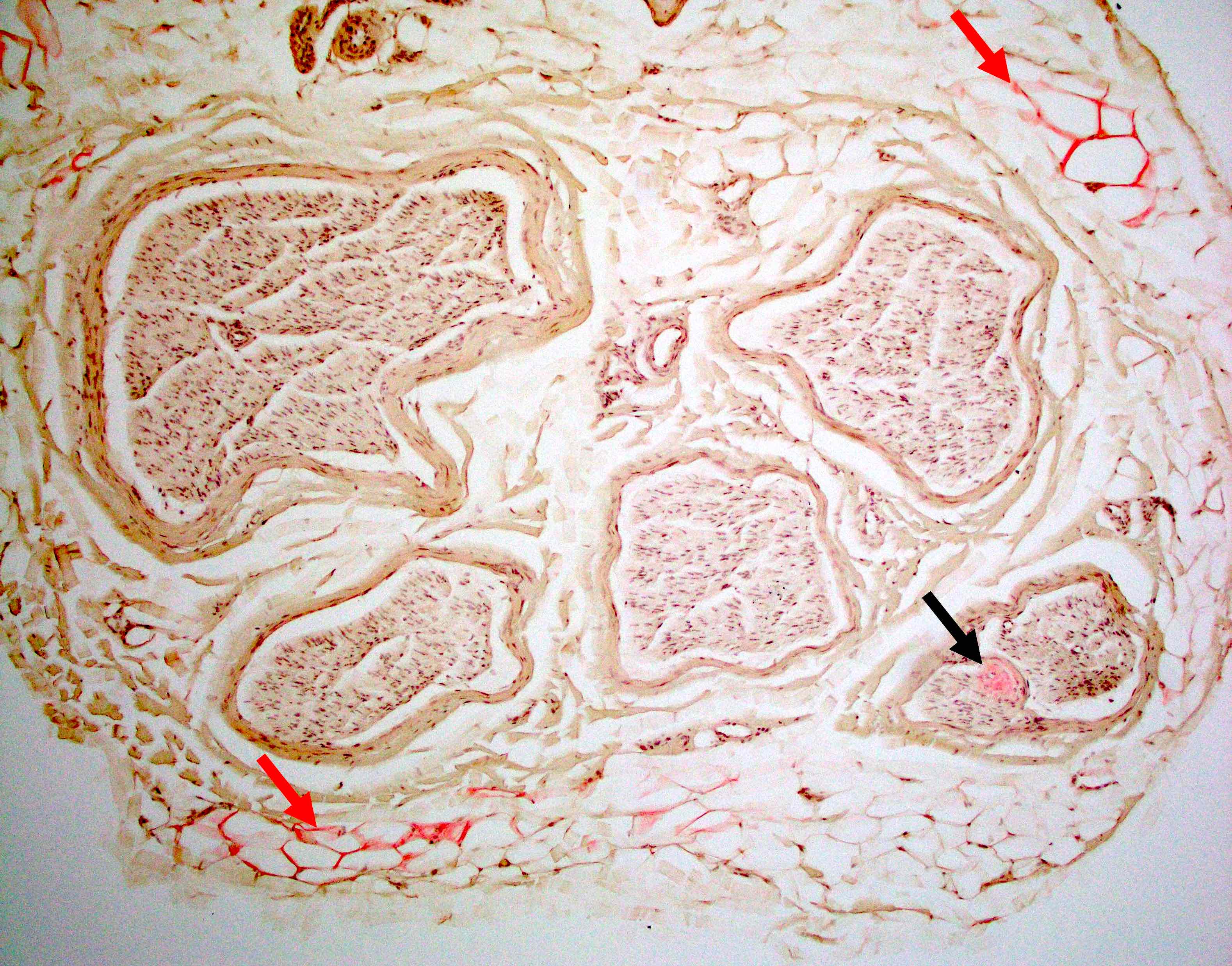
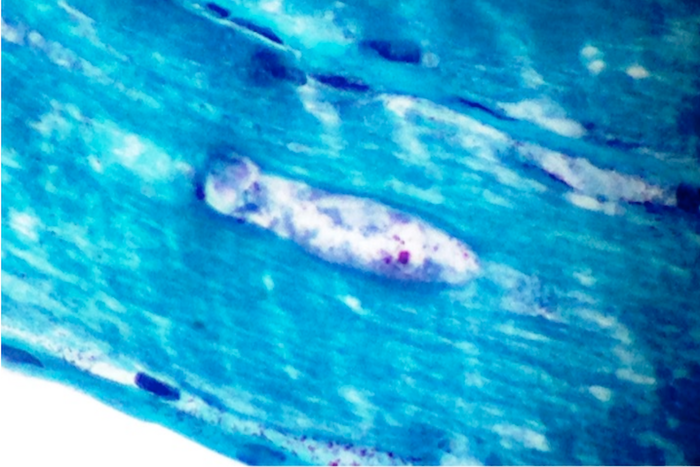
Nerve Congo red
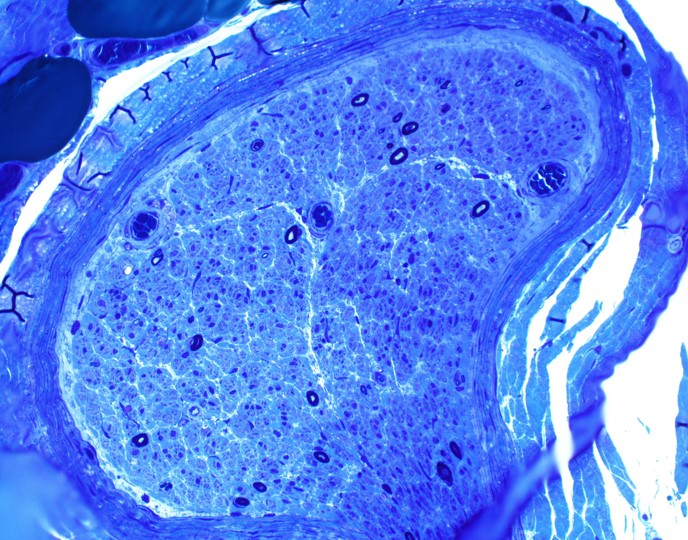
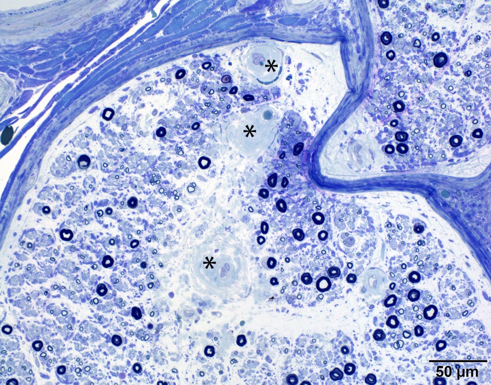
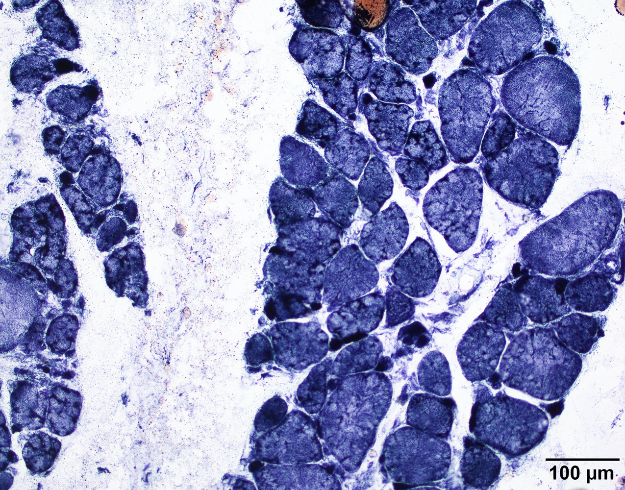
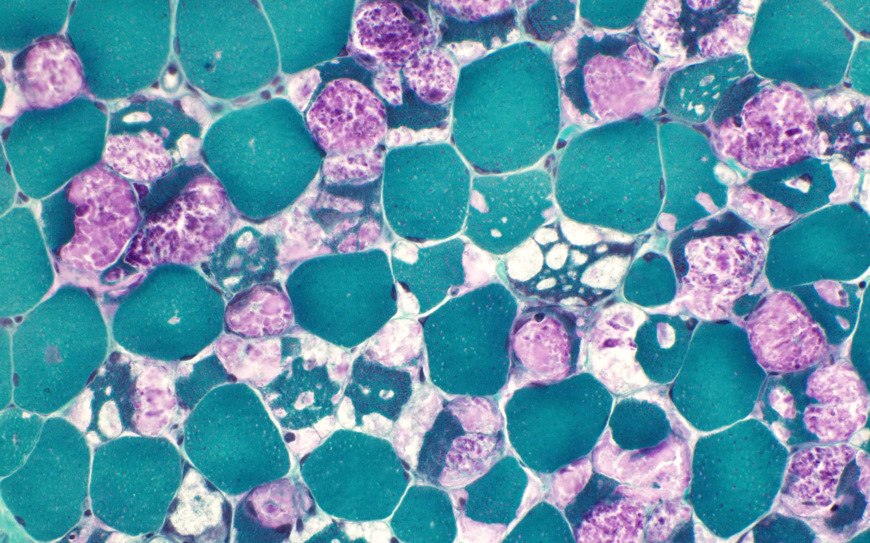
Nerve plastic section
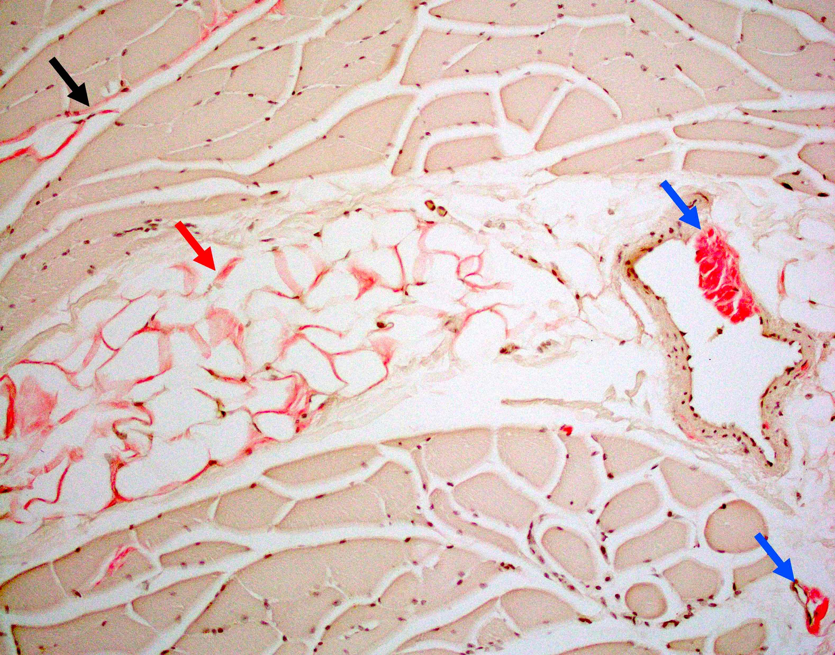
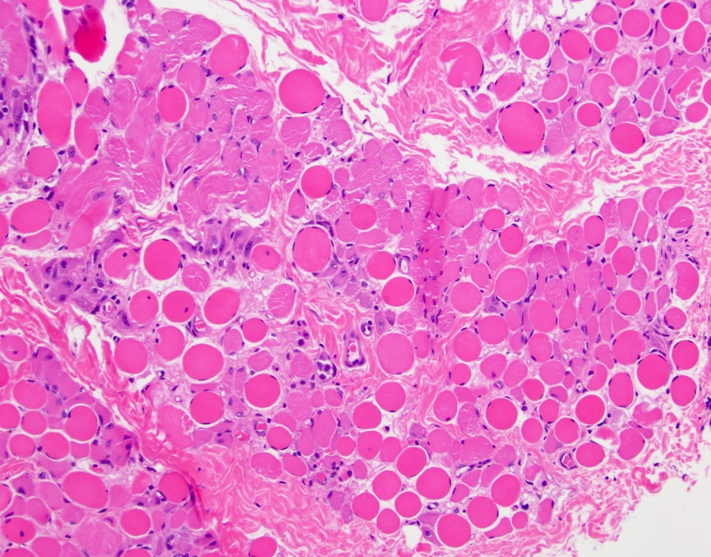
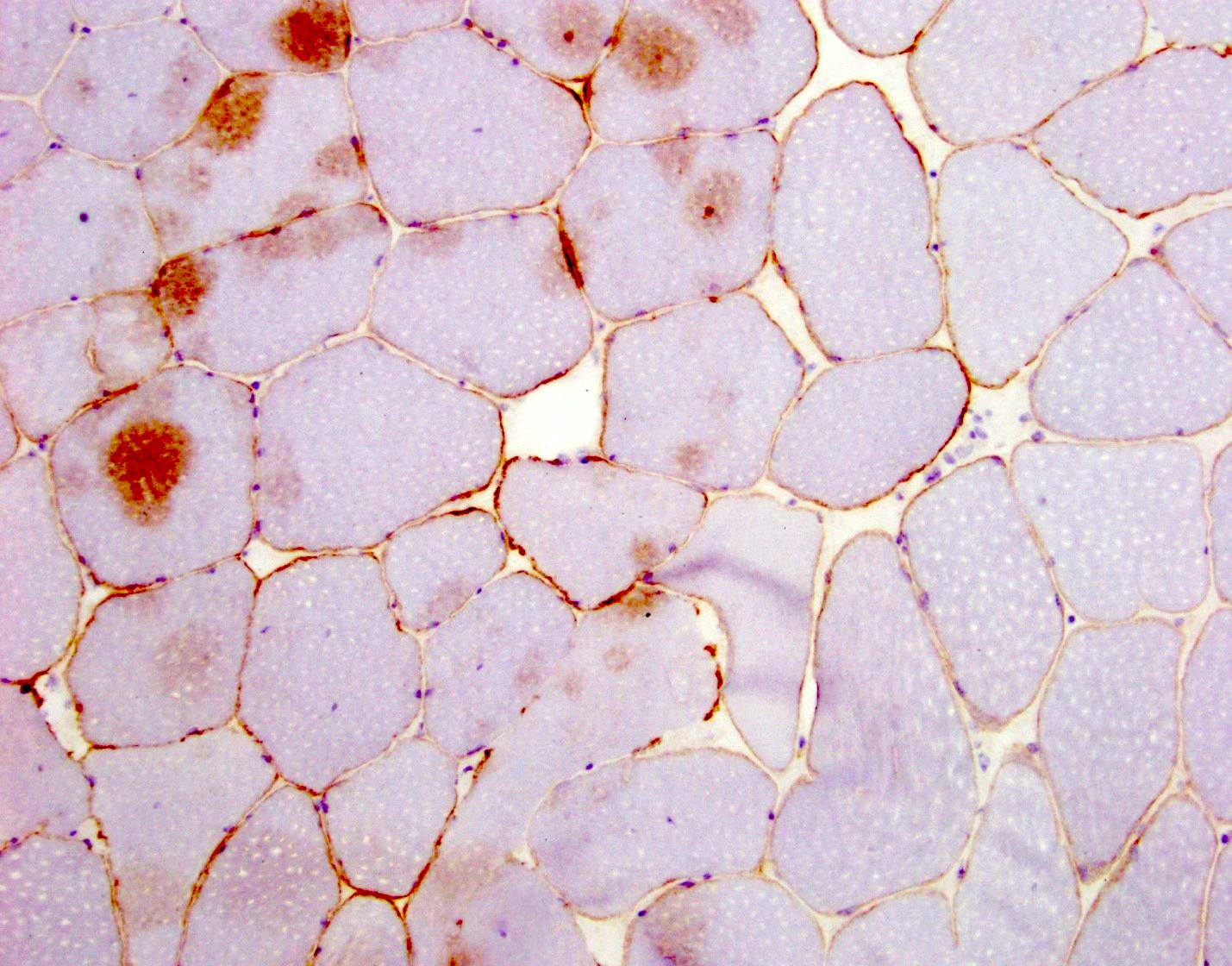
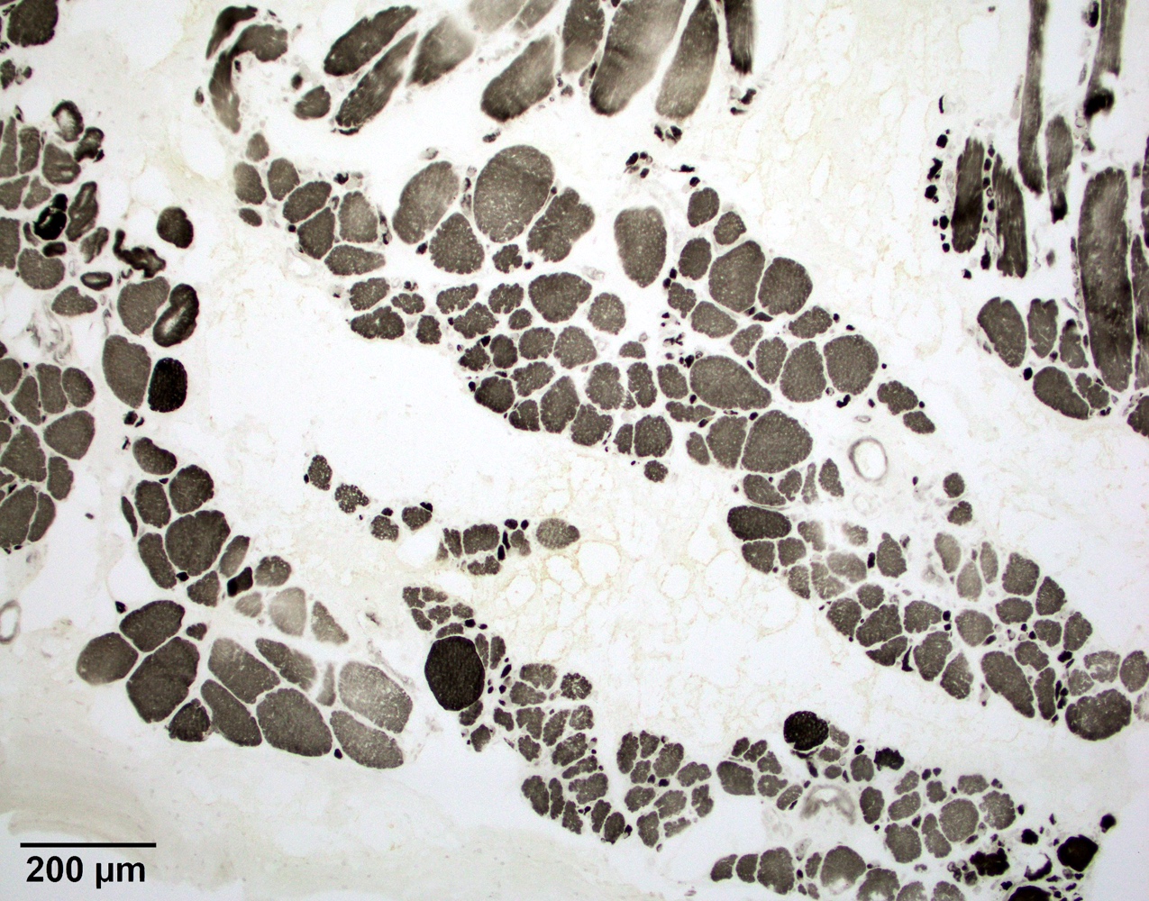
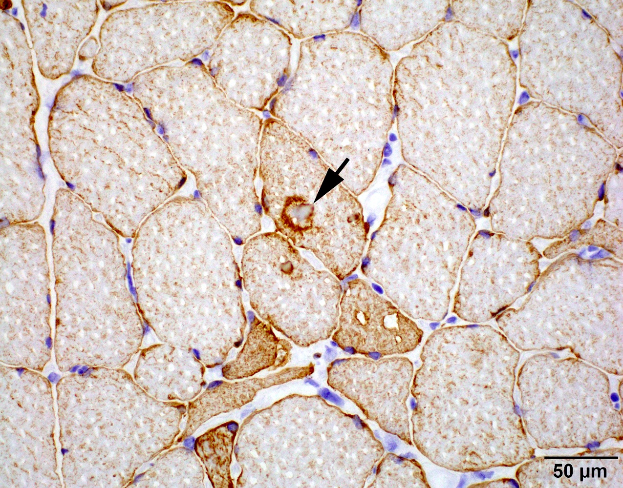
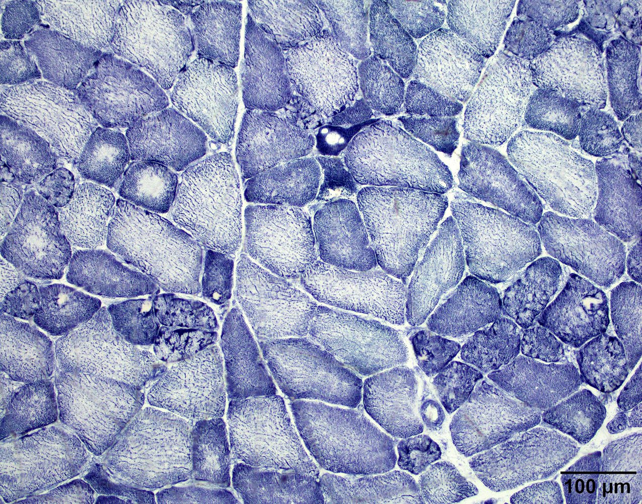
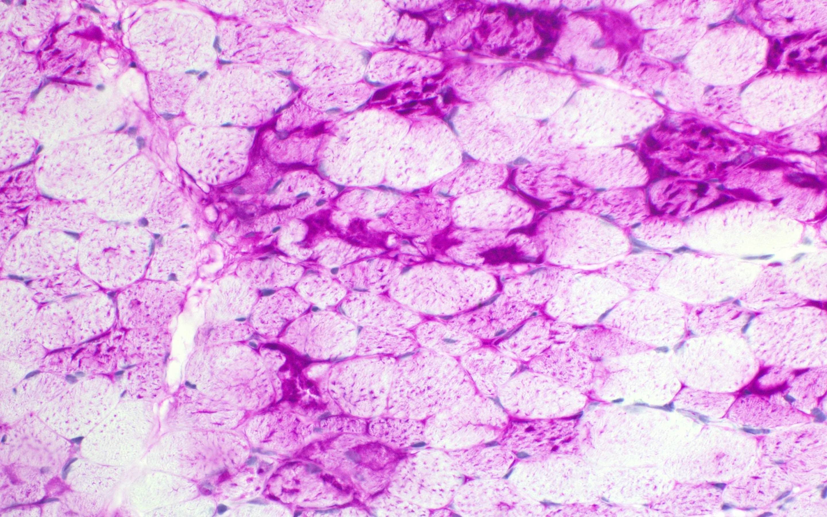
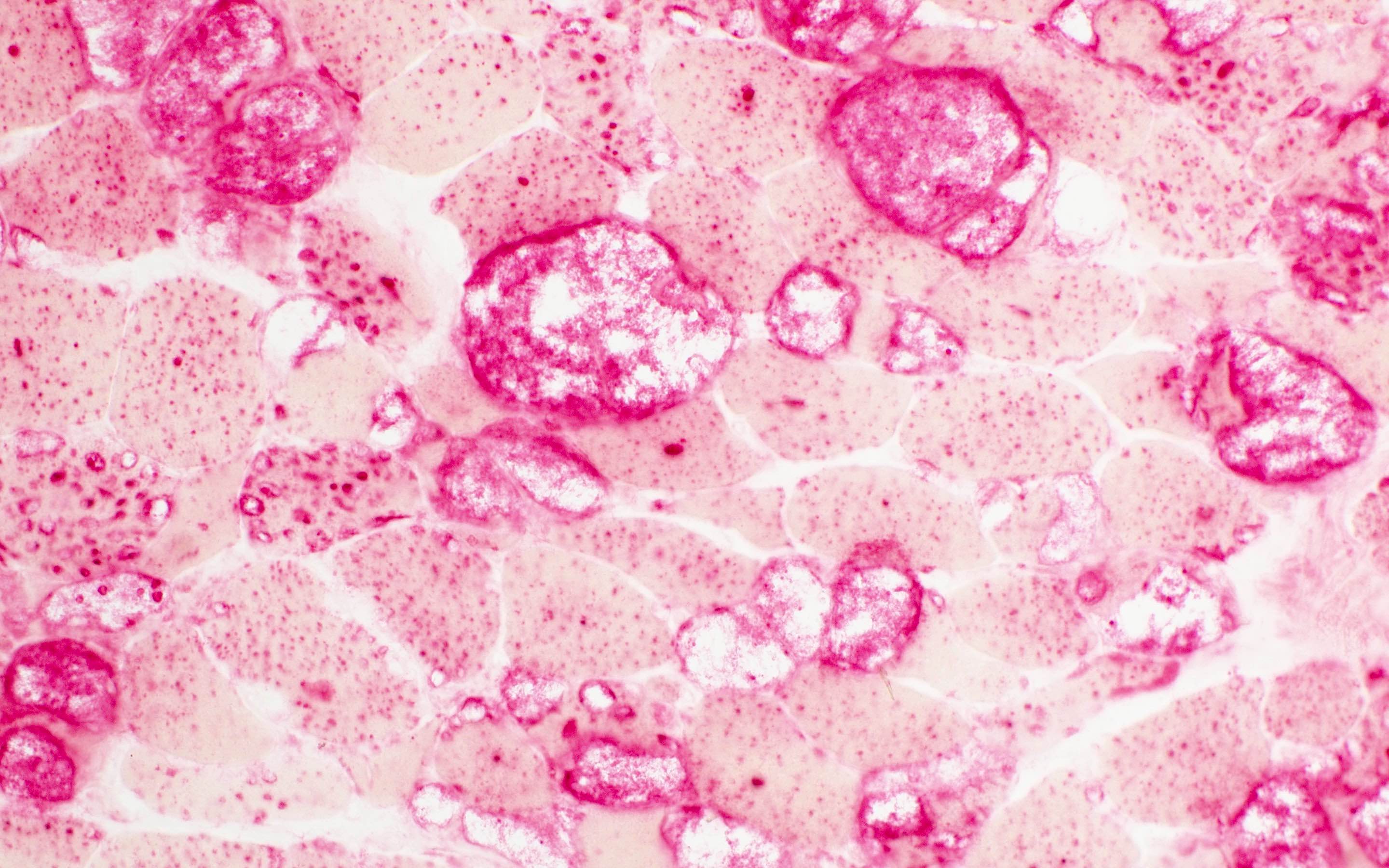
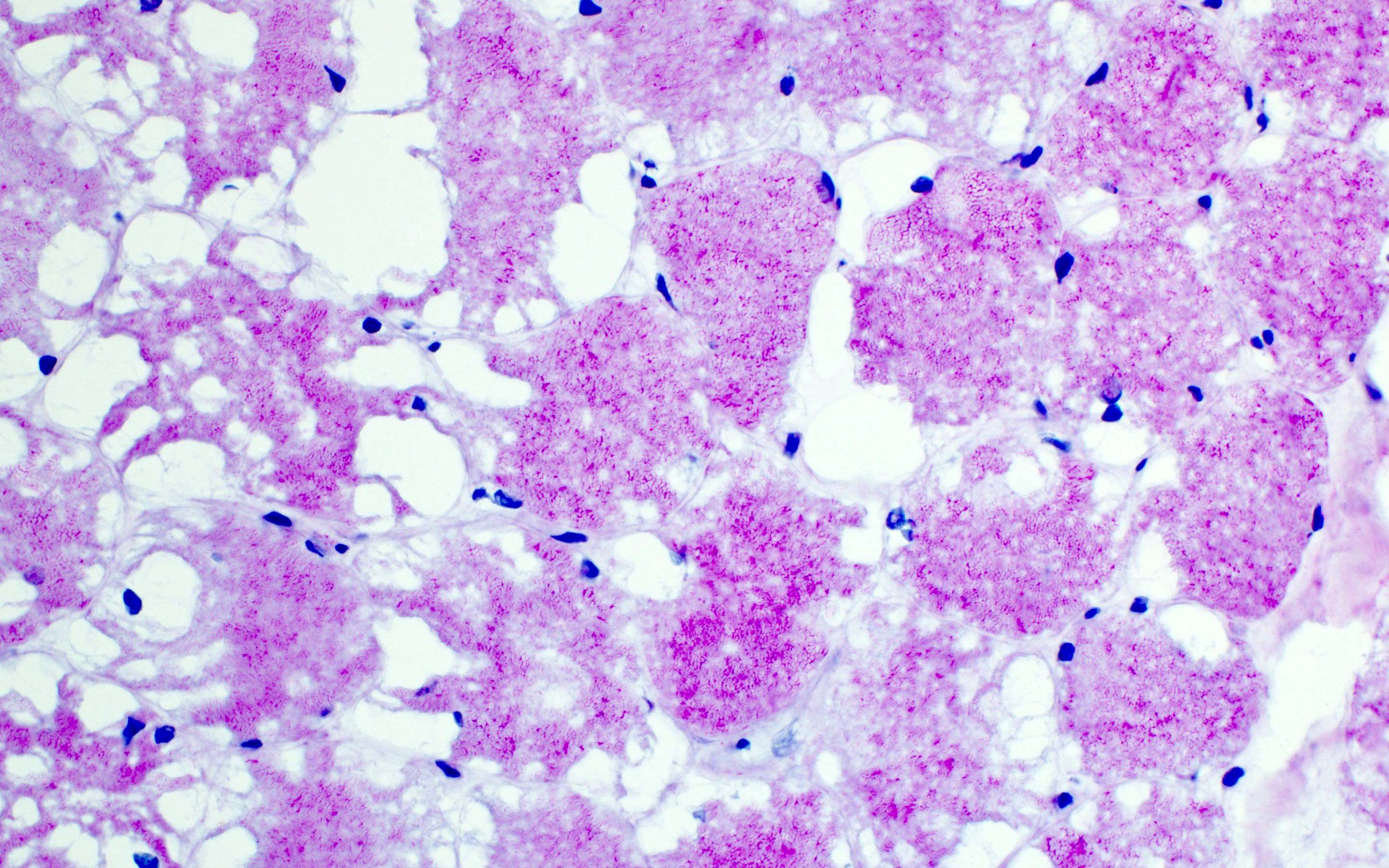
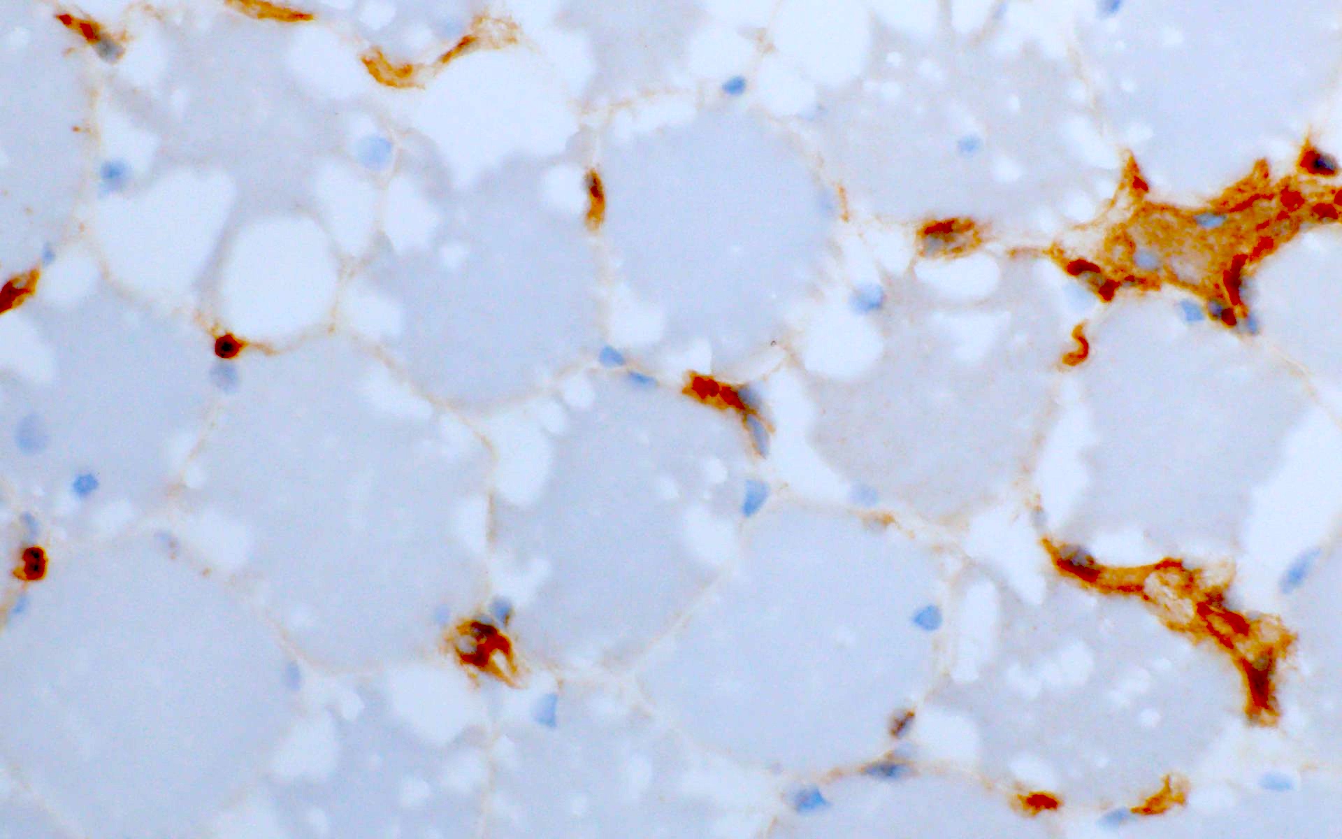
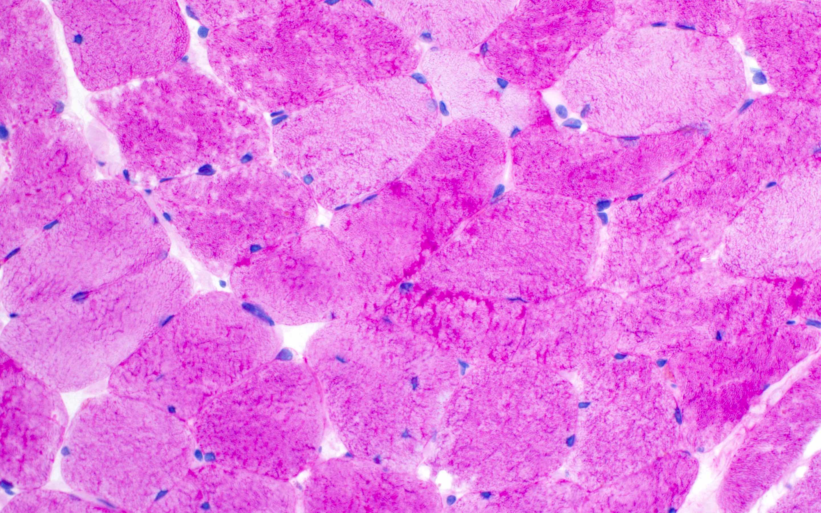
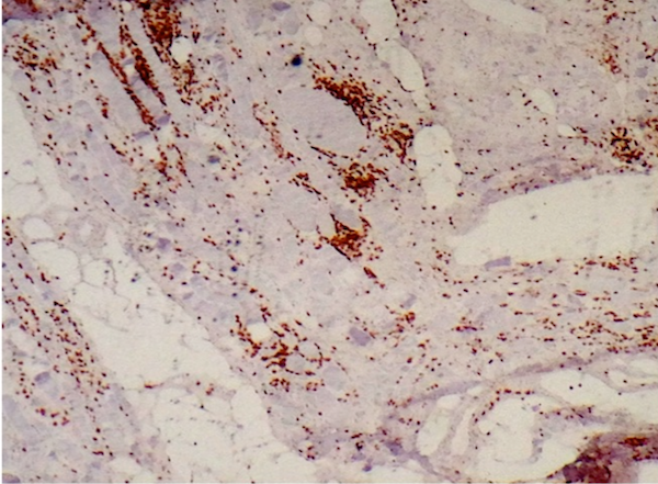
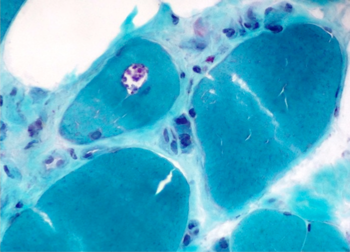
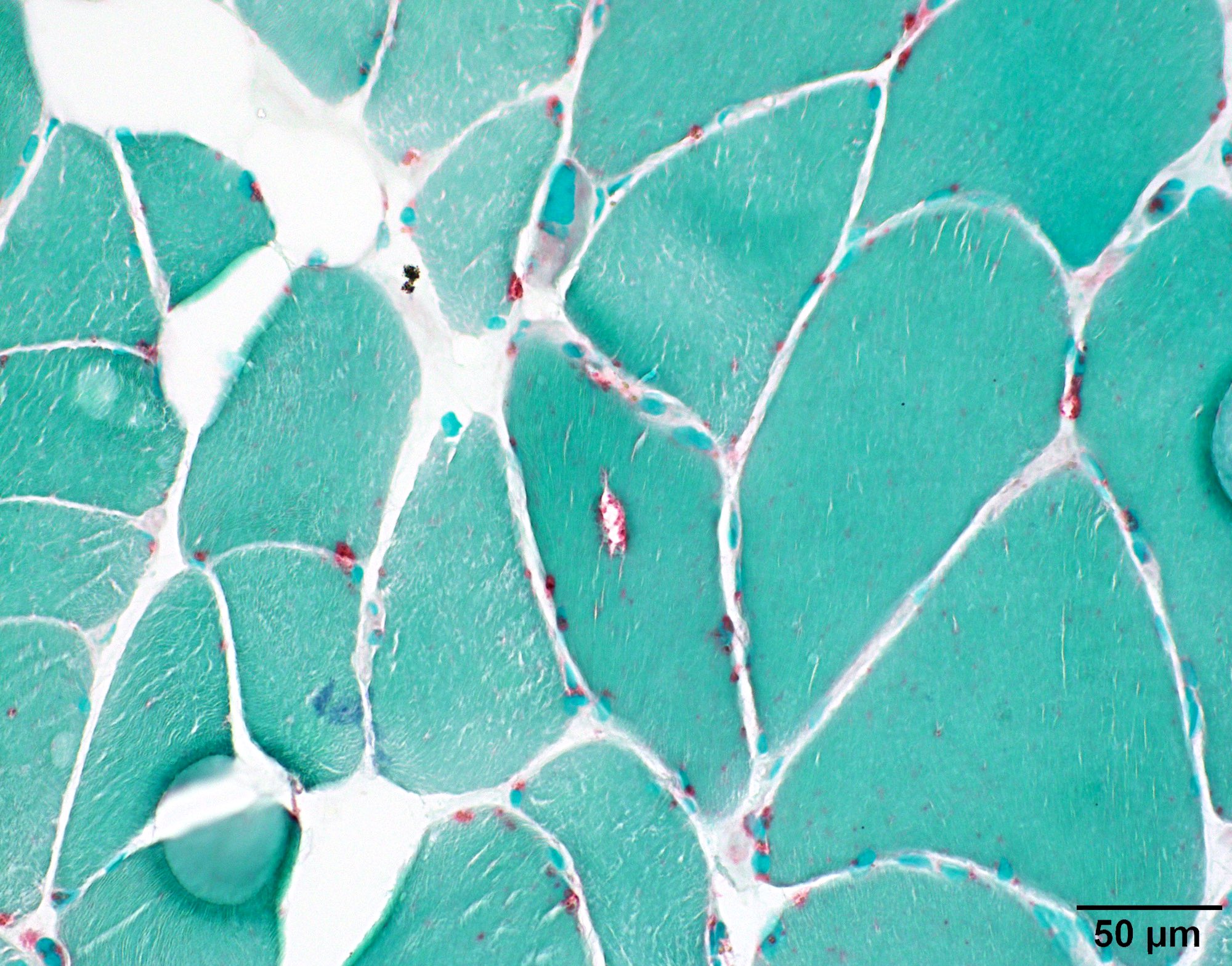
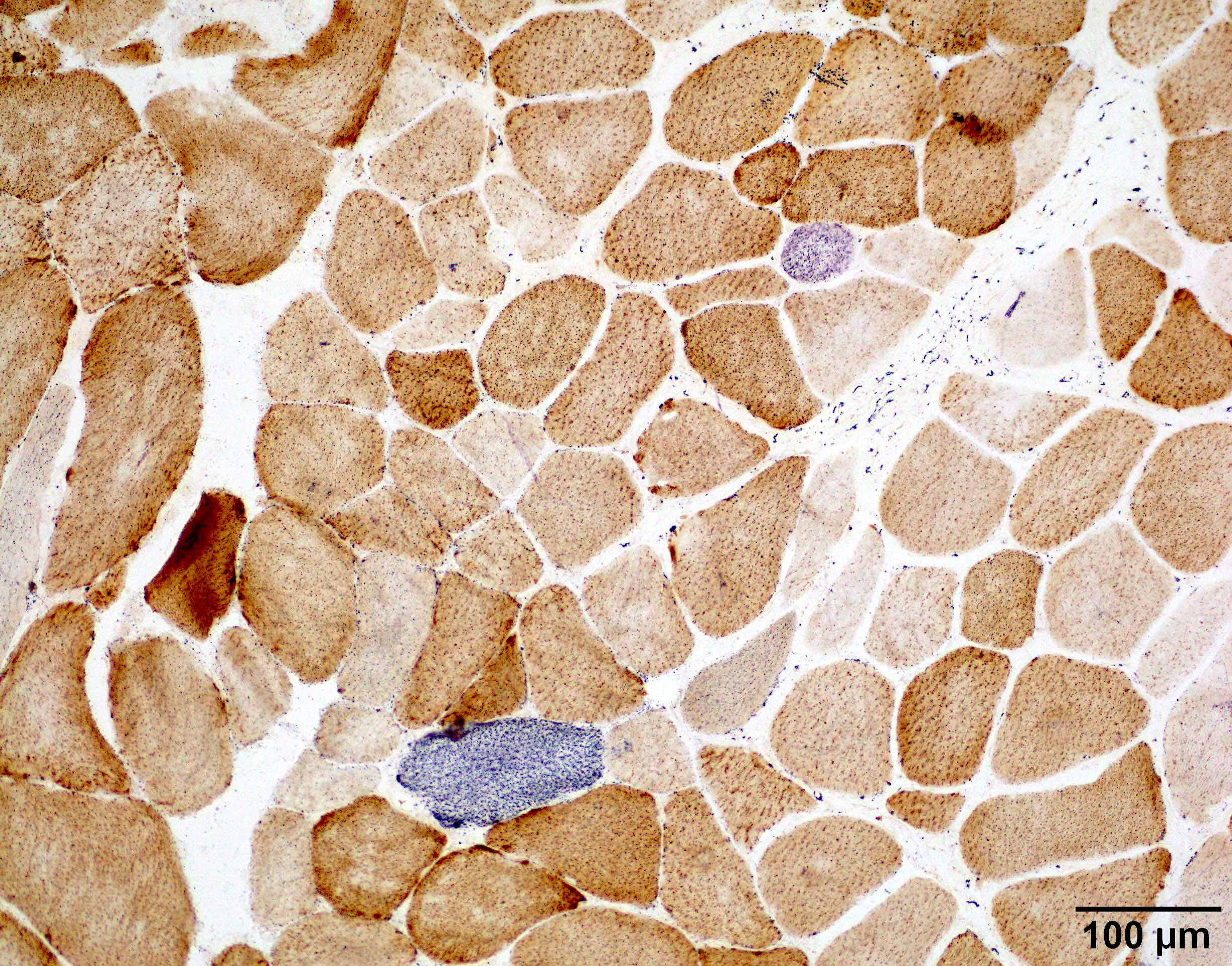
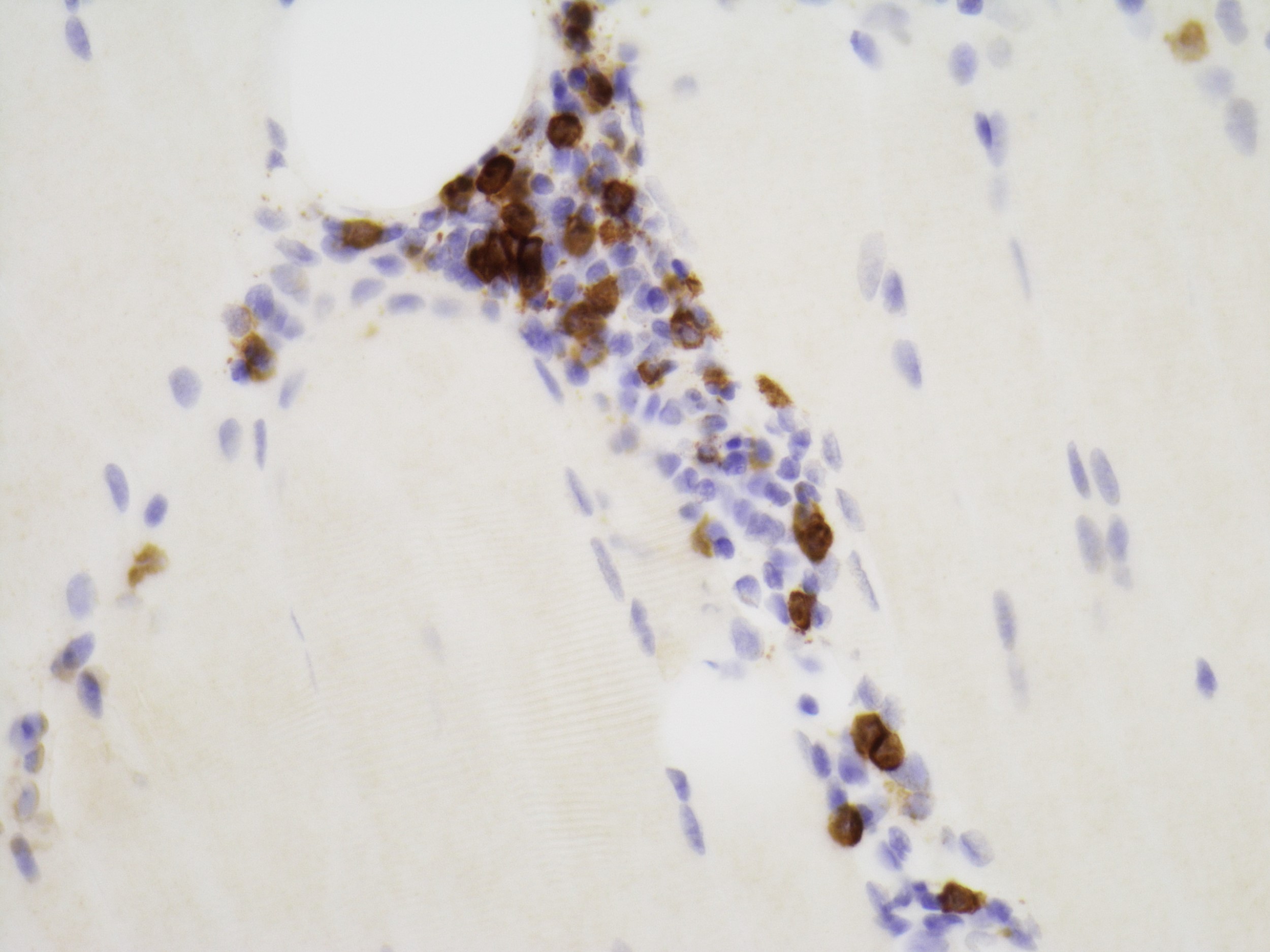
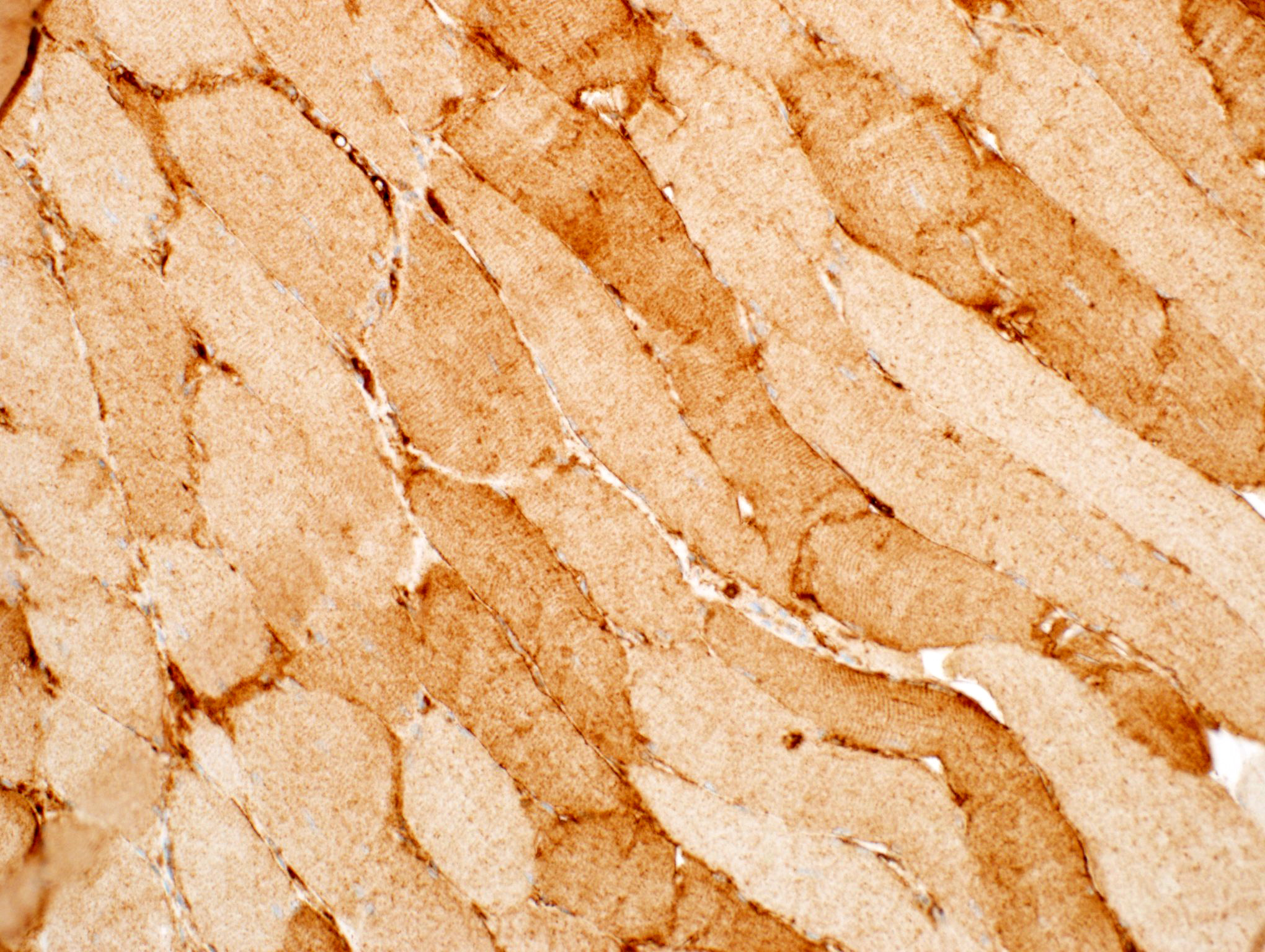
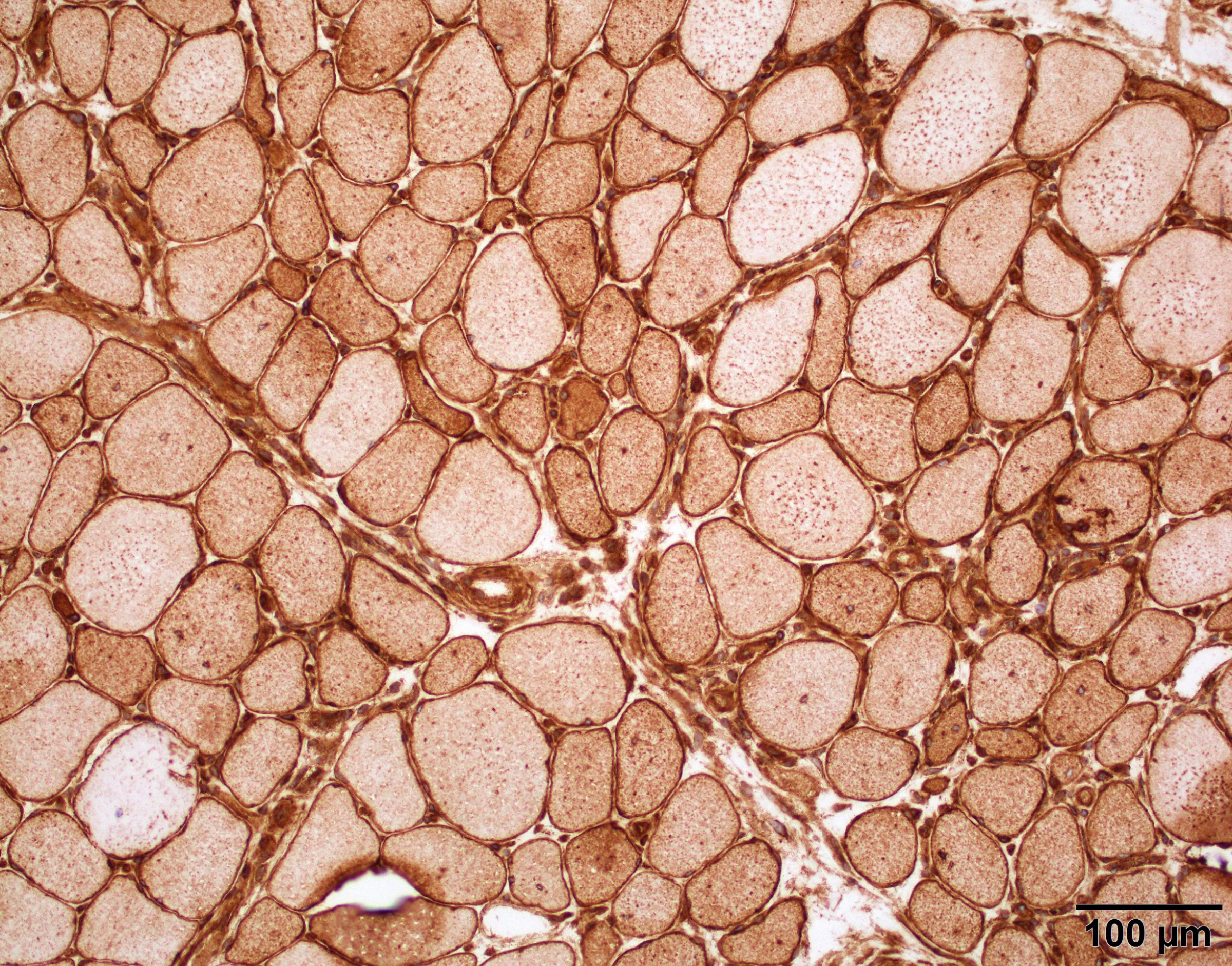
Muscle Congo red
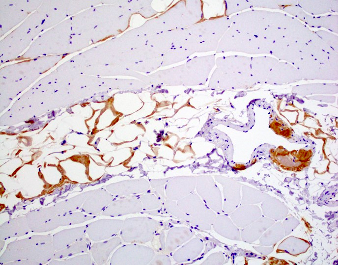
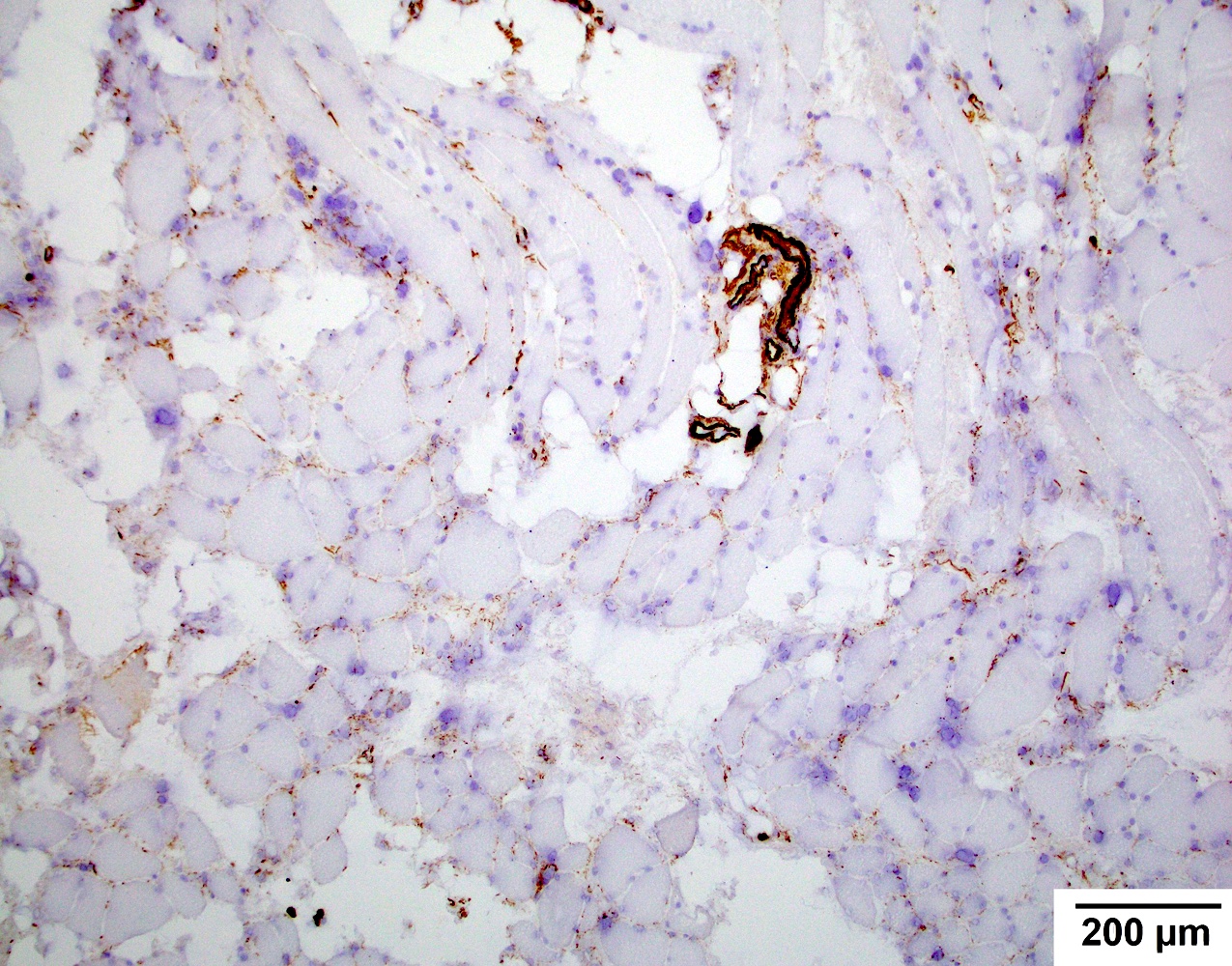
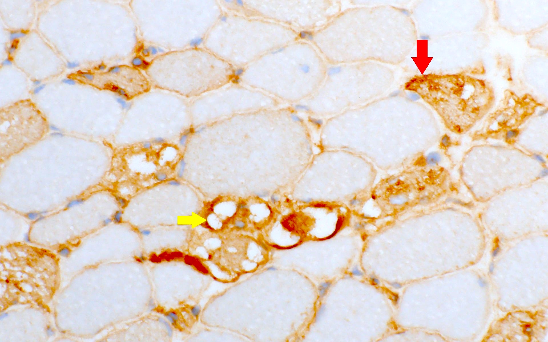
Muscle TTR
AL amyloid neuropathy
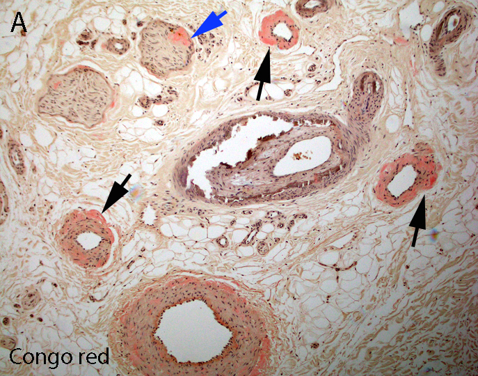
Congo red
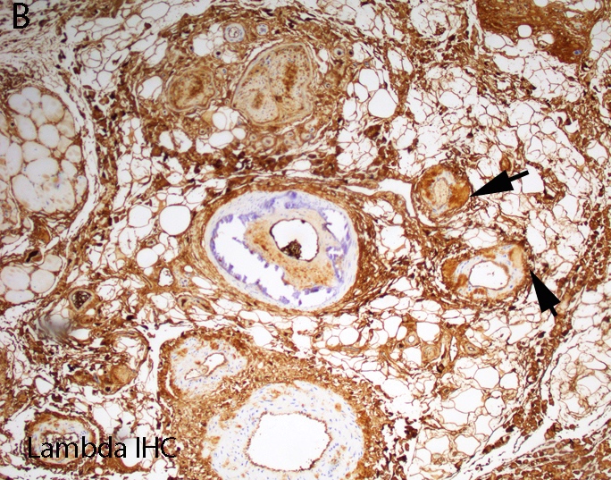
Lambda IHC
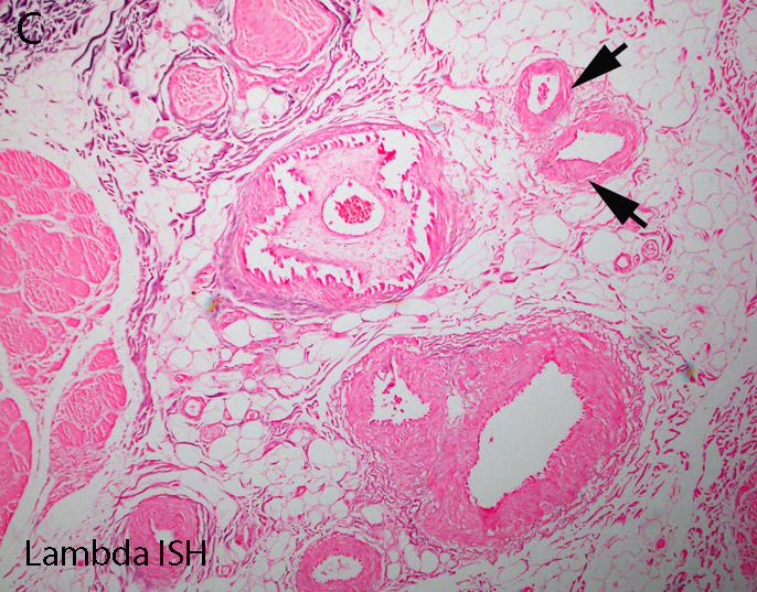
Lambda in situ hybridization
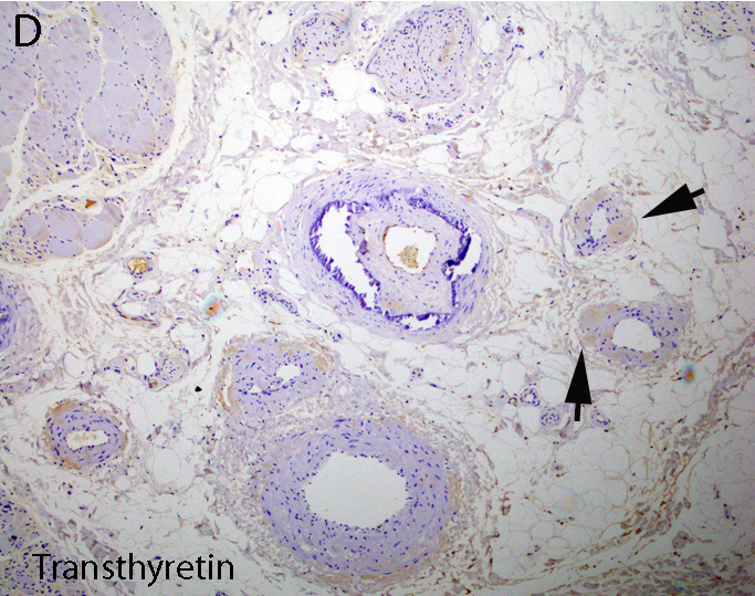
Transthyretin IHC
Contributed by Chunyu Cai, M.D., Ph.D.
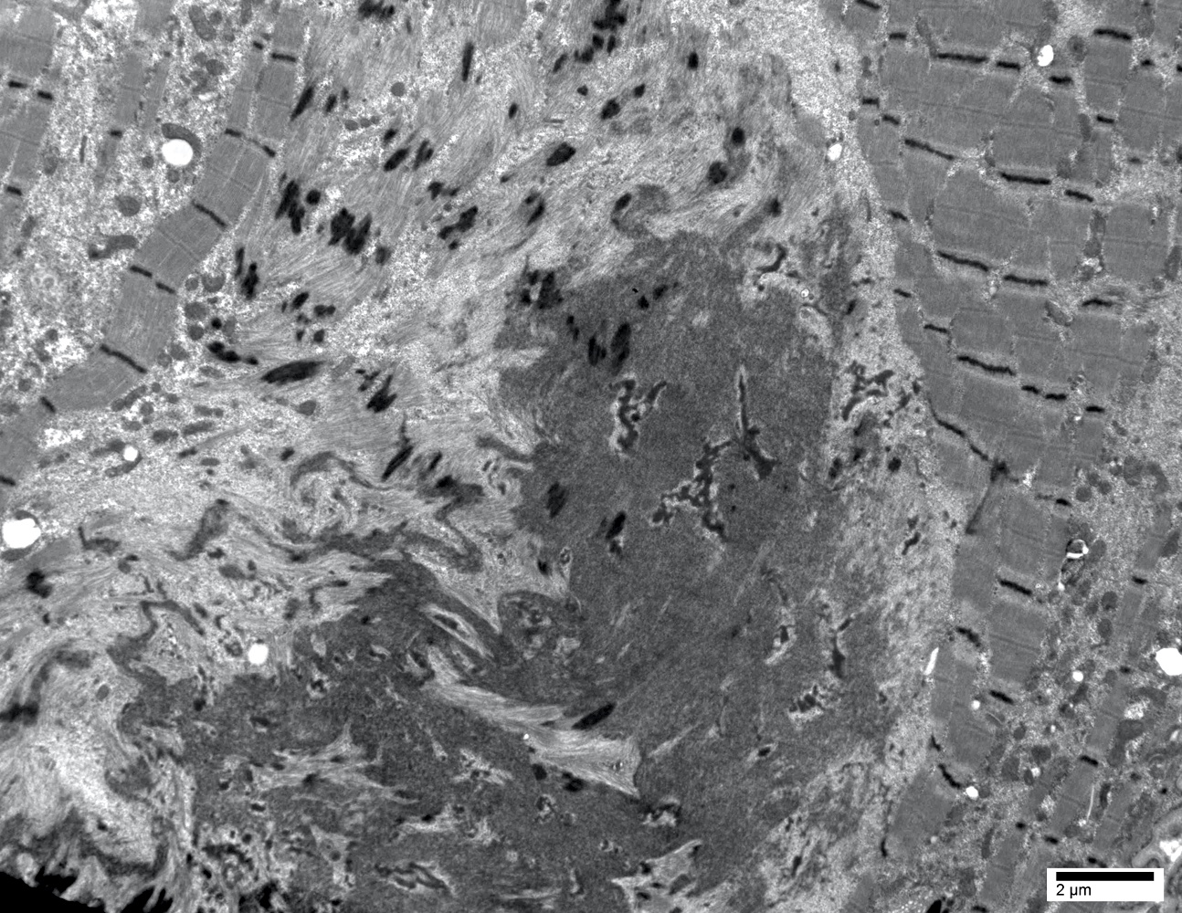
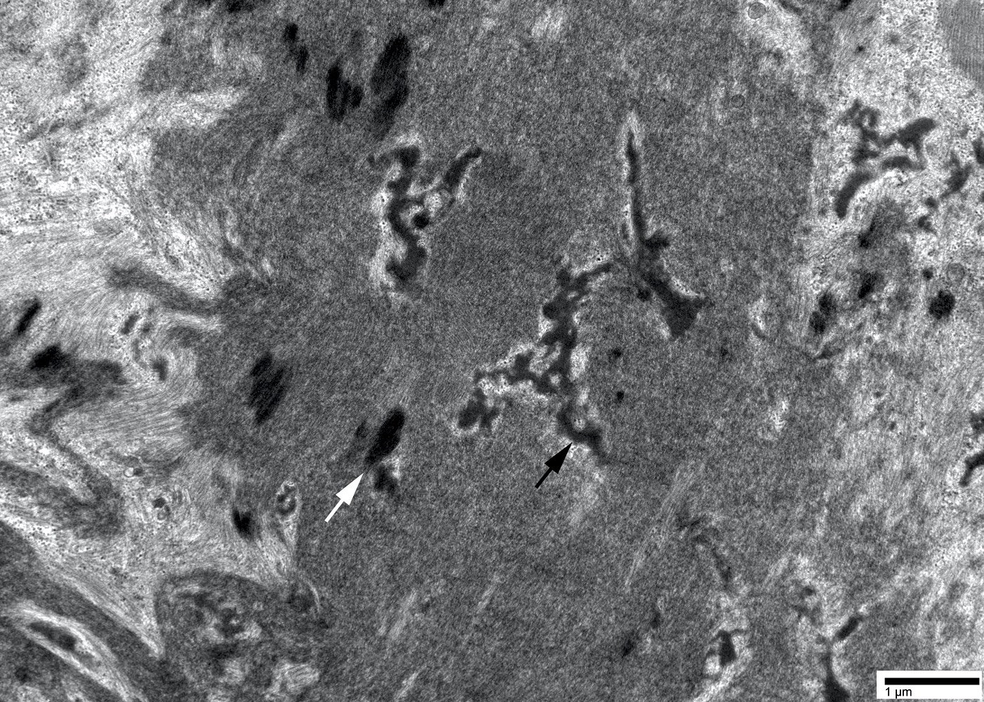
Amyloid deposit
The principles for pathology confirmation of amyloidosis and significance of subtyping
Contributed by Chunyu "Hunter" Cai, M.D., Ph.D.
Jo1 myositis

Jo1 myositis MCH1
Jo1 myositis C5b9
Jo1 myositis myopathy alkaline phosphatase
PL7 myositis
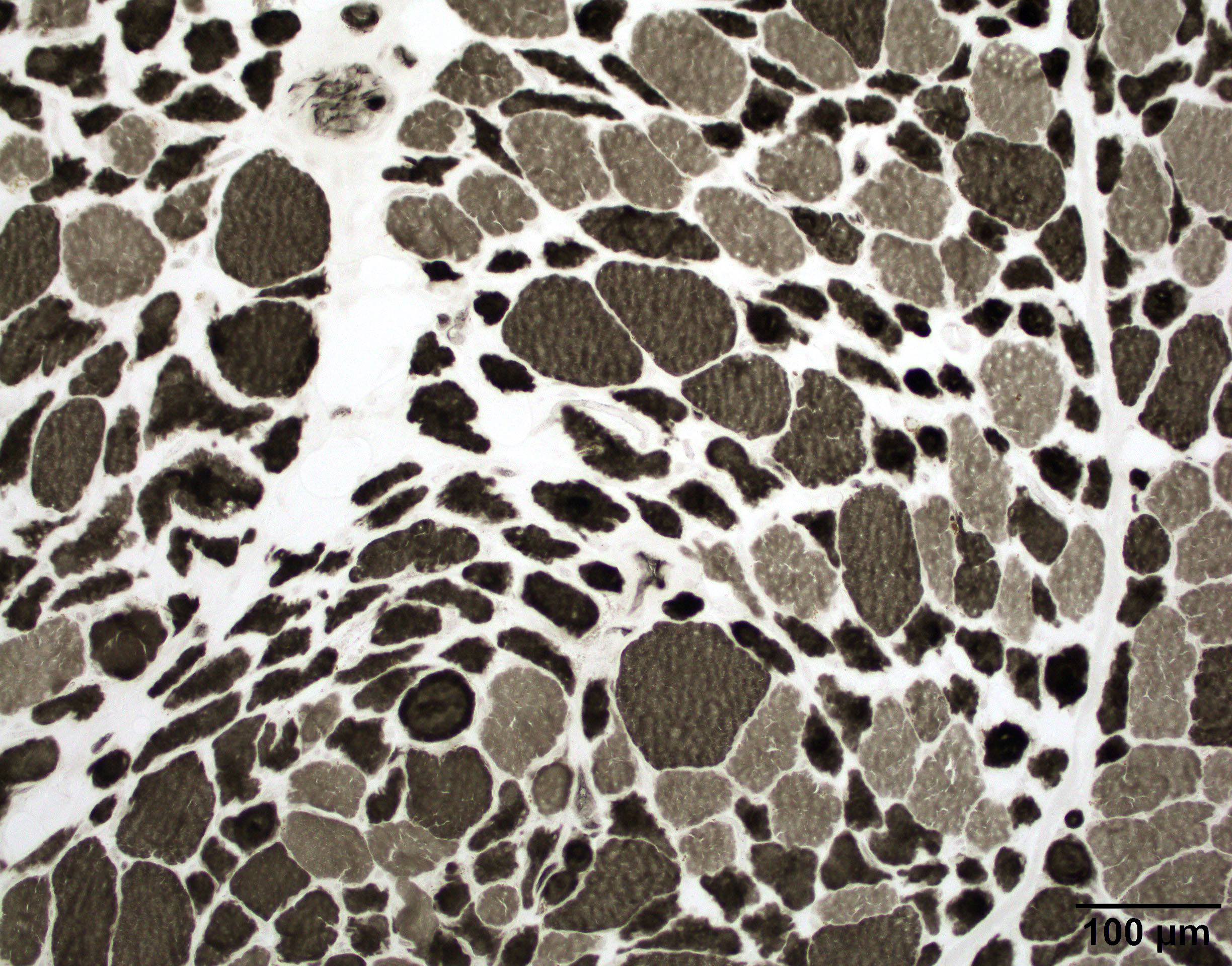
PL7 myositis ATPase pH 4.3

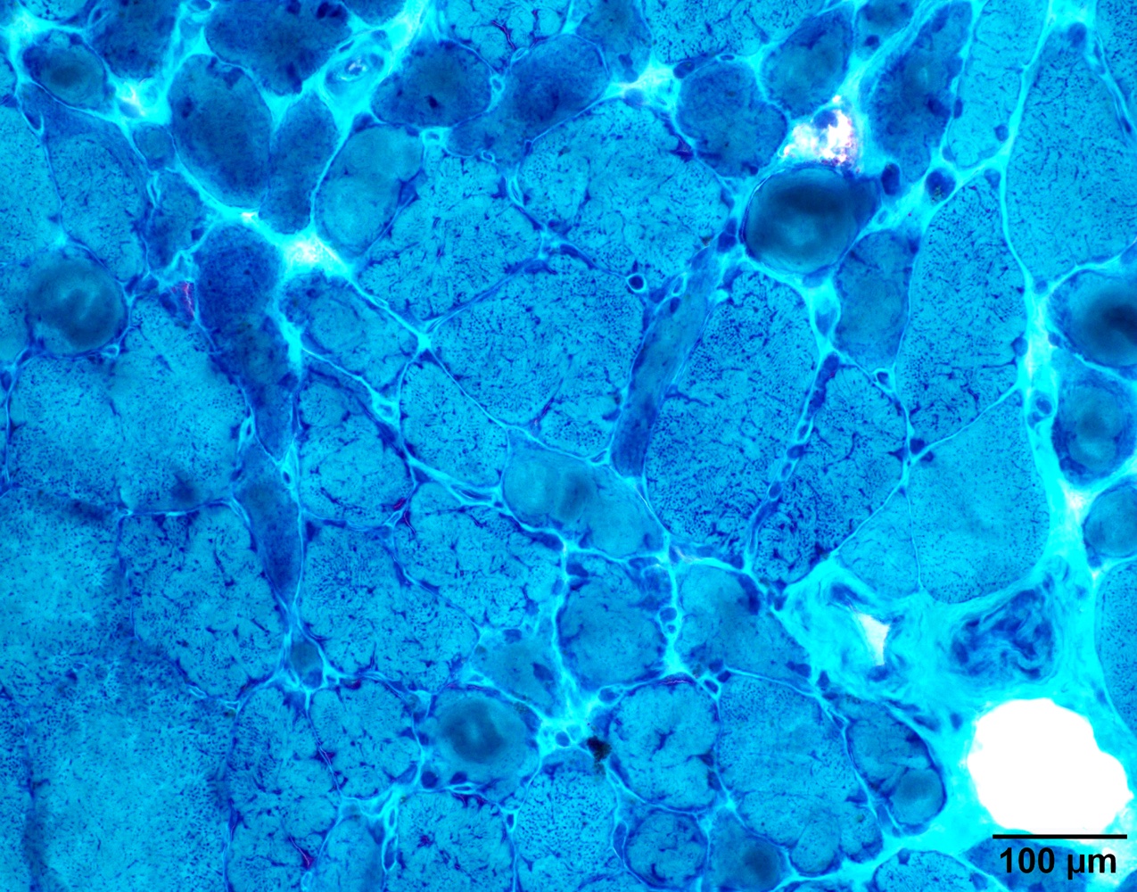
PL7 myositis alkaline phosphatase
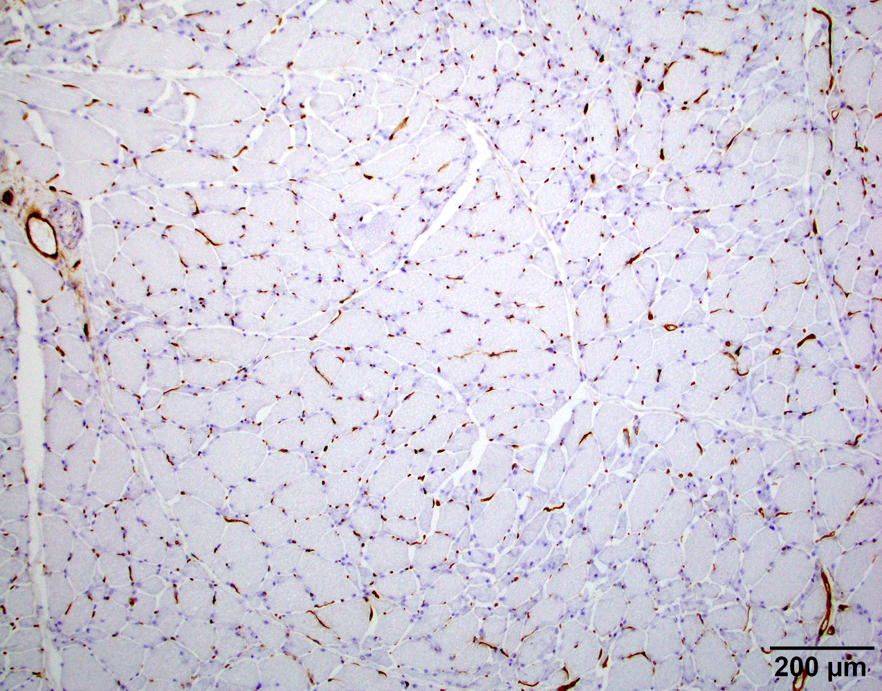
PL7 myositis MCH1

PL7 myositis C5b9
Contributed by Dennis Burns, M.D. and Chunyu "Hunter" Cai, M.D., Ph.D.
Intranuclear actin filament aggregates
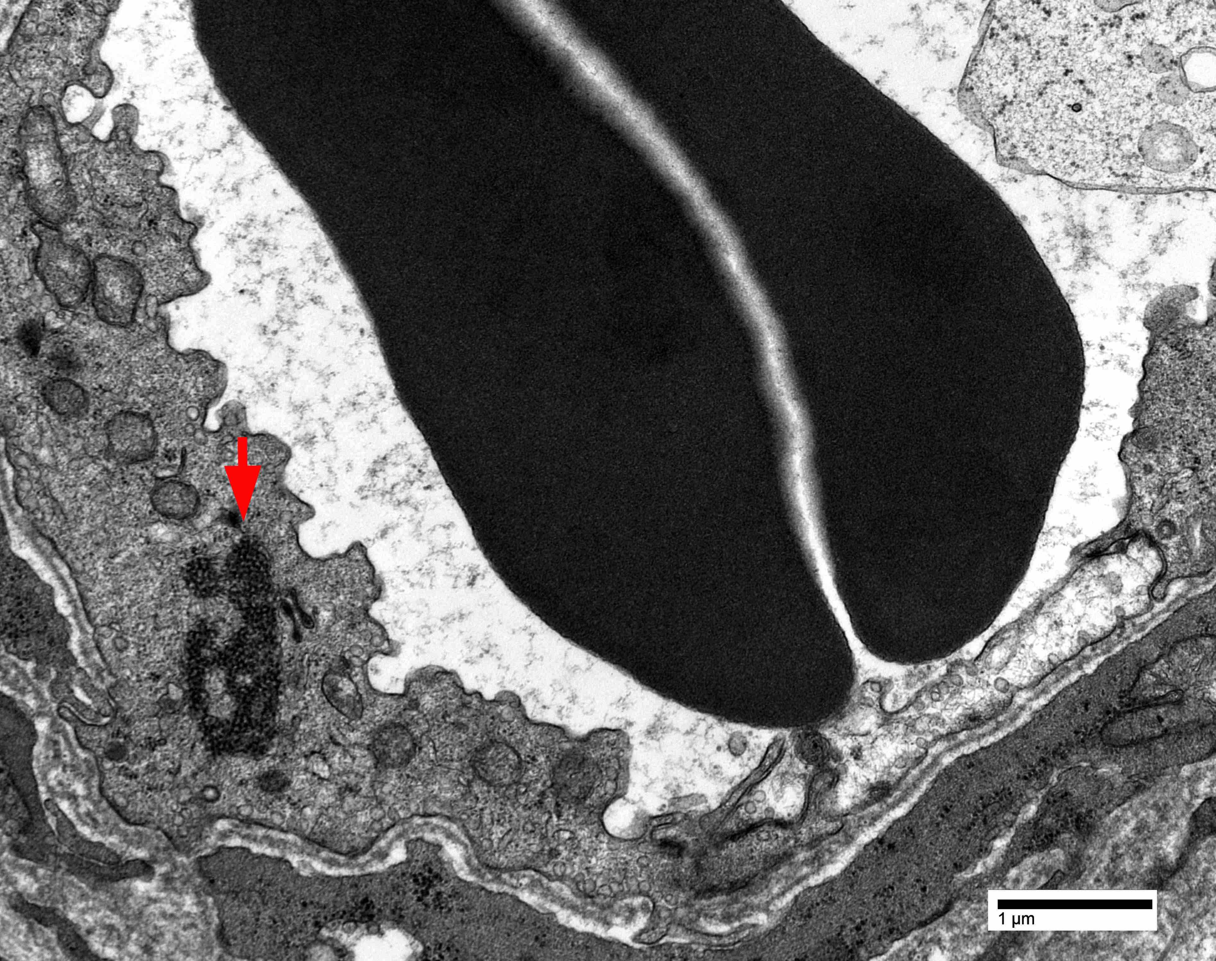
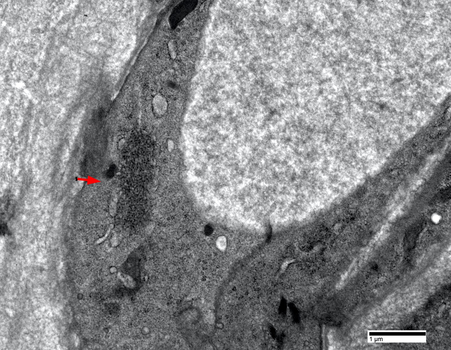
Endothelial tubuloreticular inclusion
Contributed by Jesse L. Kresak, M.D.
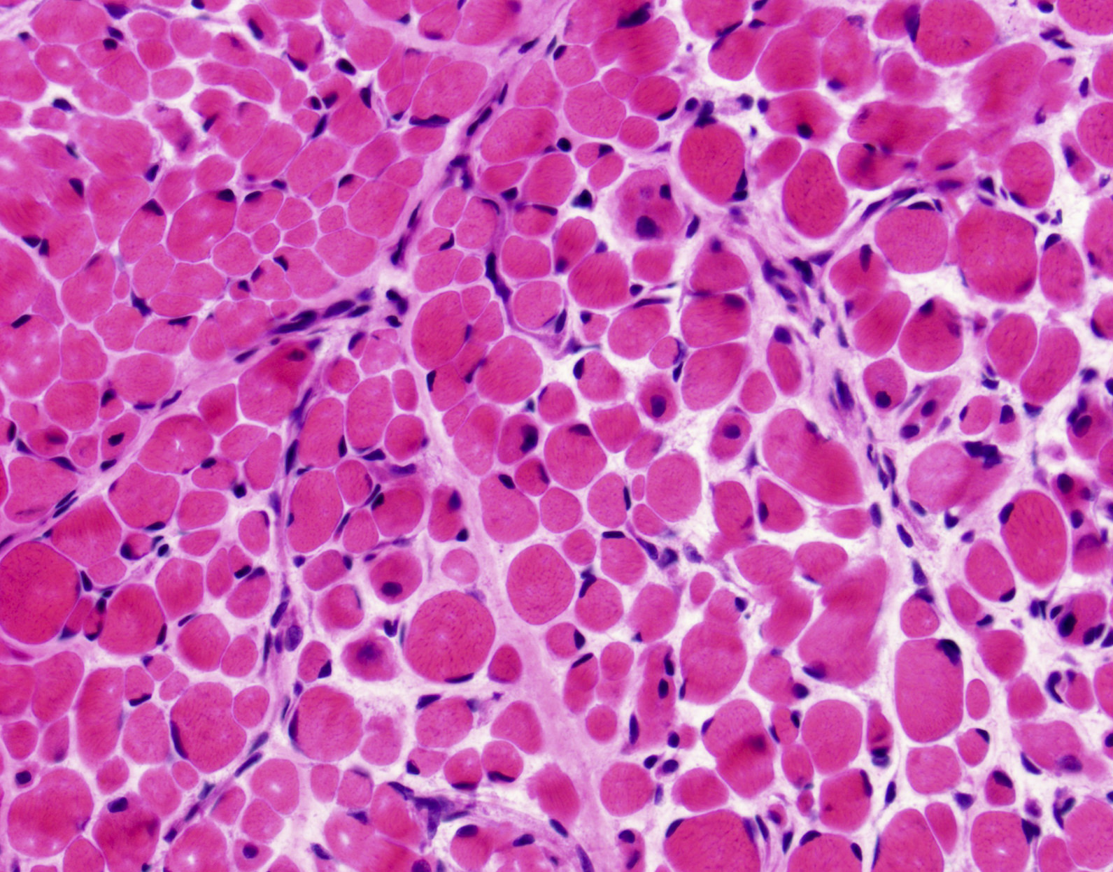
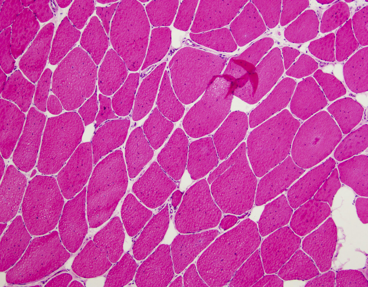
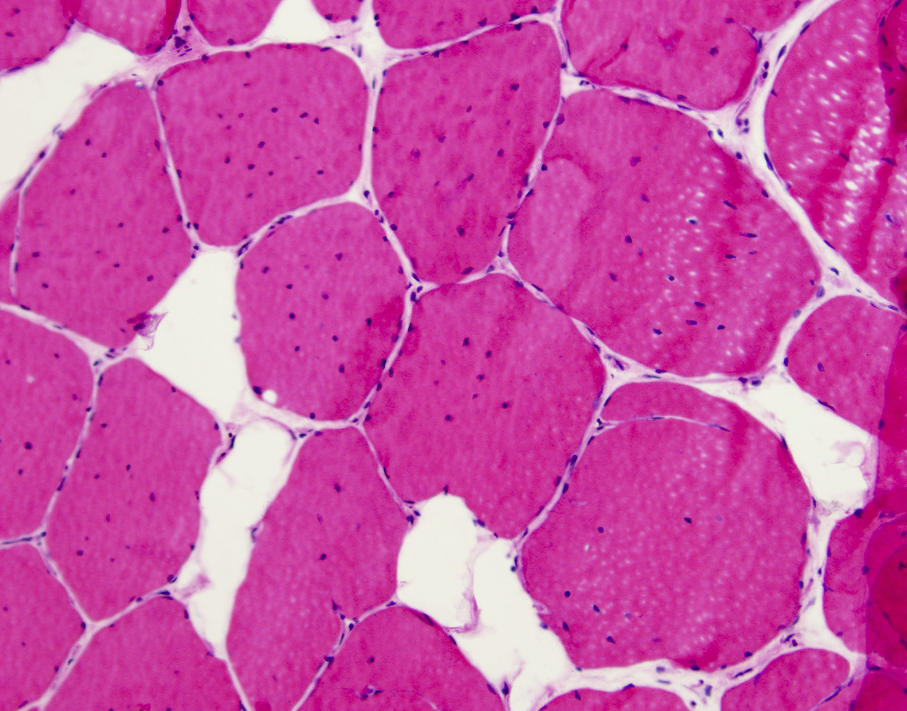
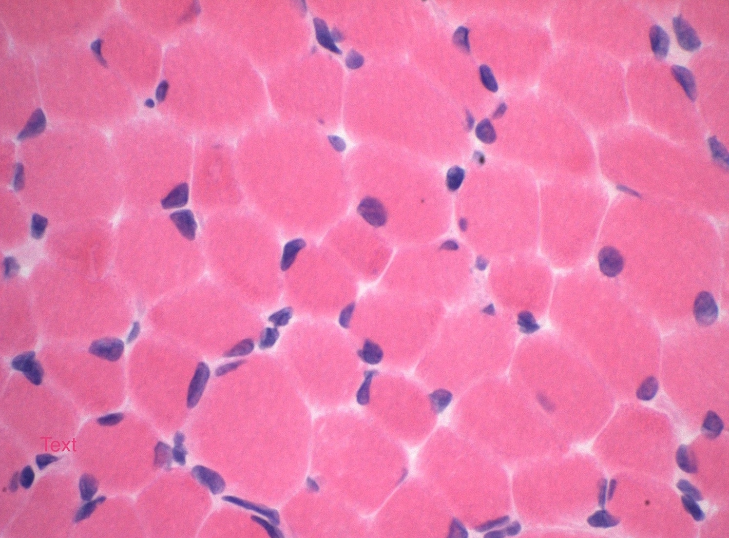
Myopathic changes
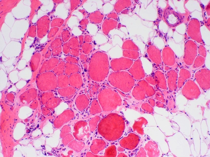
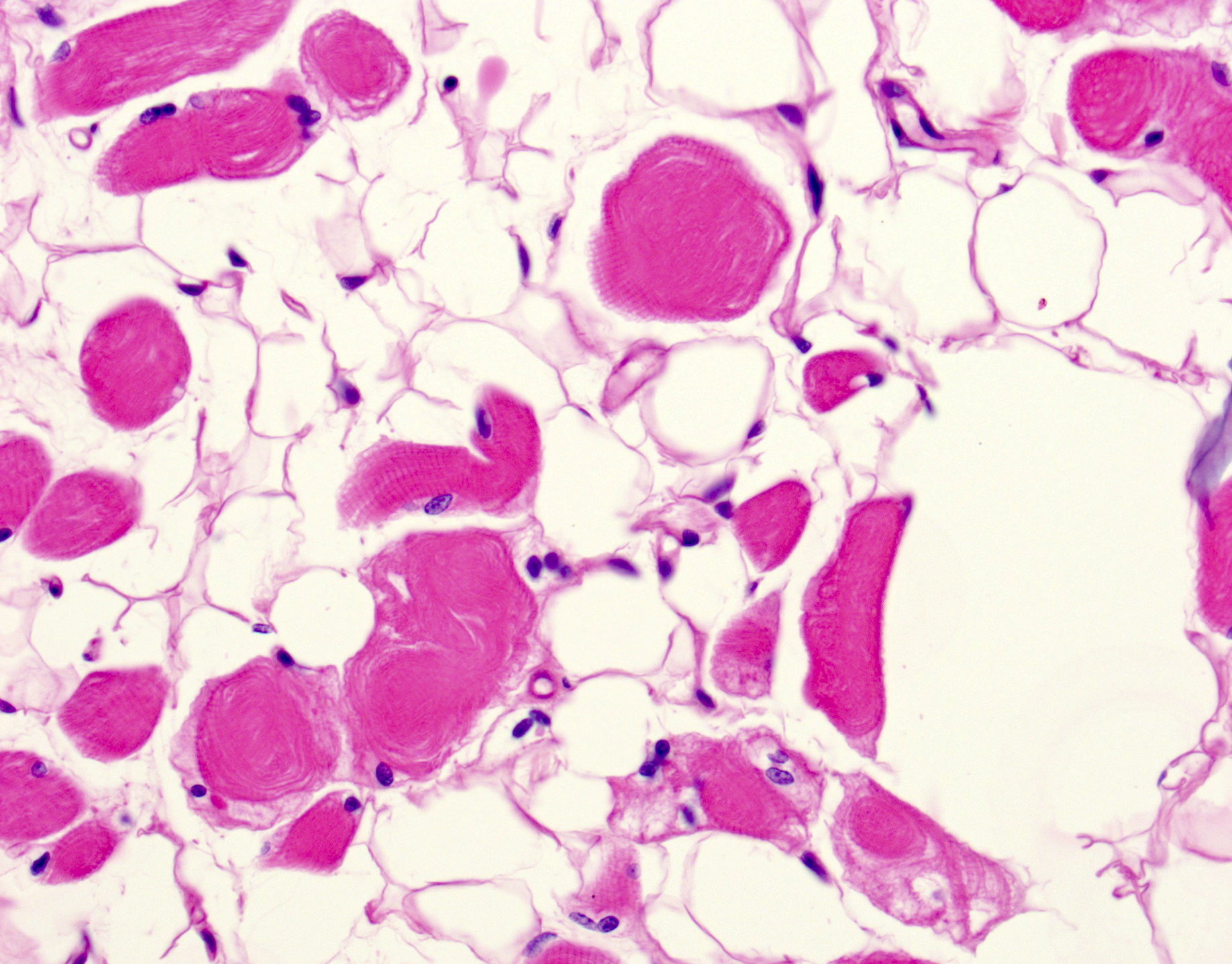
Fatty replacement

End stage muscle
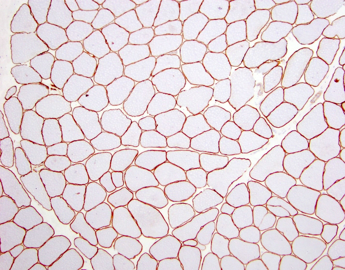
Dystrophin IHC control
Dystrophin IHC in Duchenne muscular dystrophy
Dystrophin IHC in Becker muscular dystrophy
Contributed by Jesse L. Kresak, M.D.
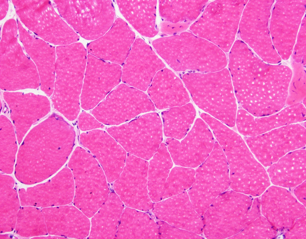
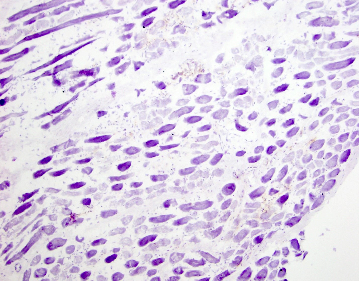
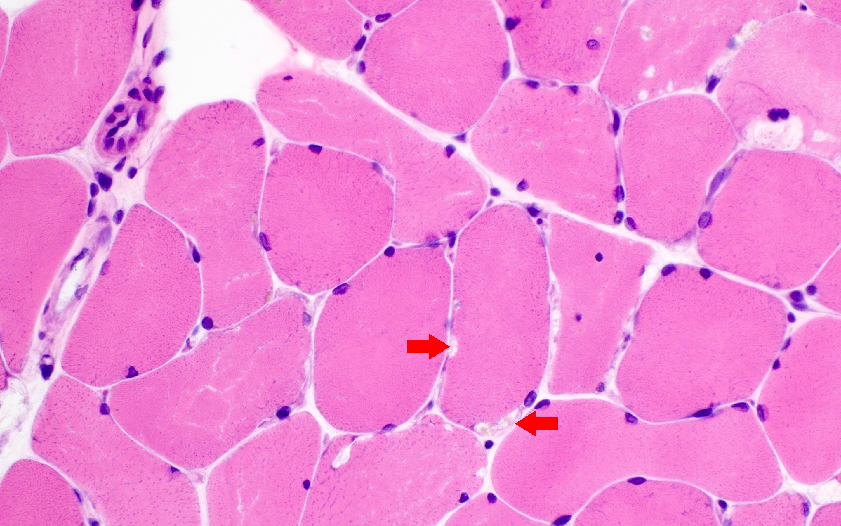
H&E

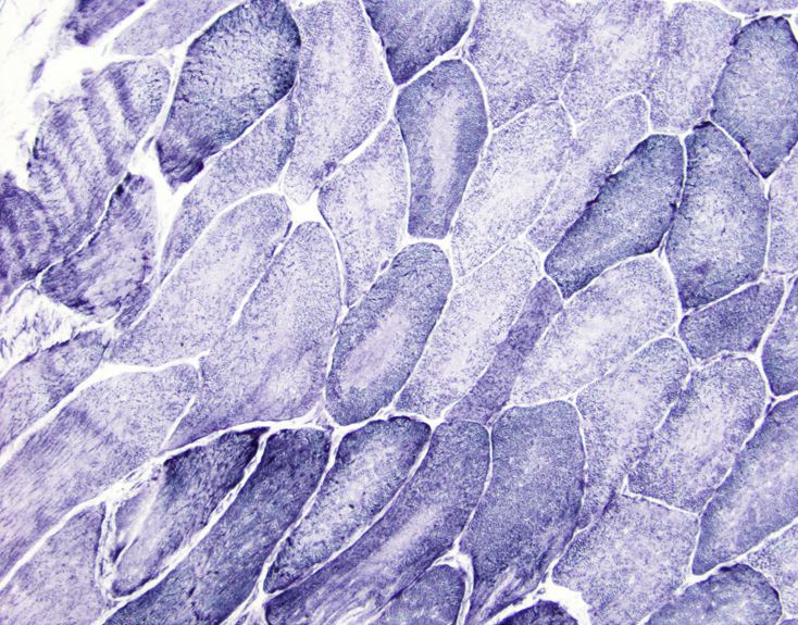
NADH and NADH1
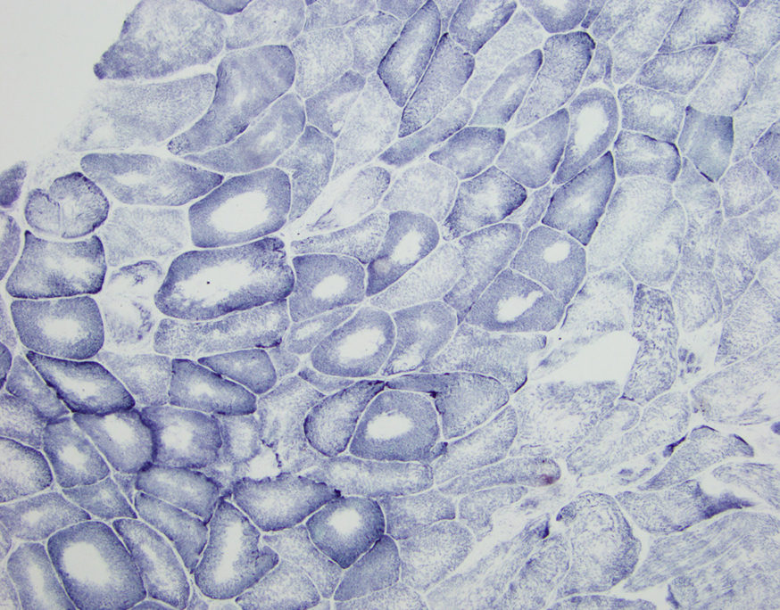
SDH
Images hosted on other servers:
Selective muscle involvement in a 59 year-old man
Images hosted on other servers:
Elongated face and inverted V shaped mouth
Contributed by Marie Rivera-Zengotita, M.D.
H&E
NADH
Images hosted on other servers:
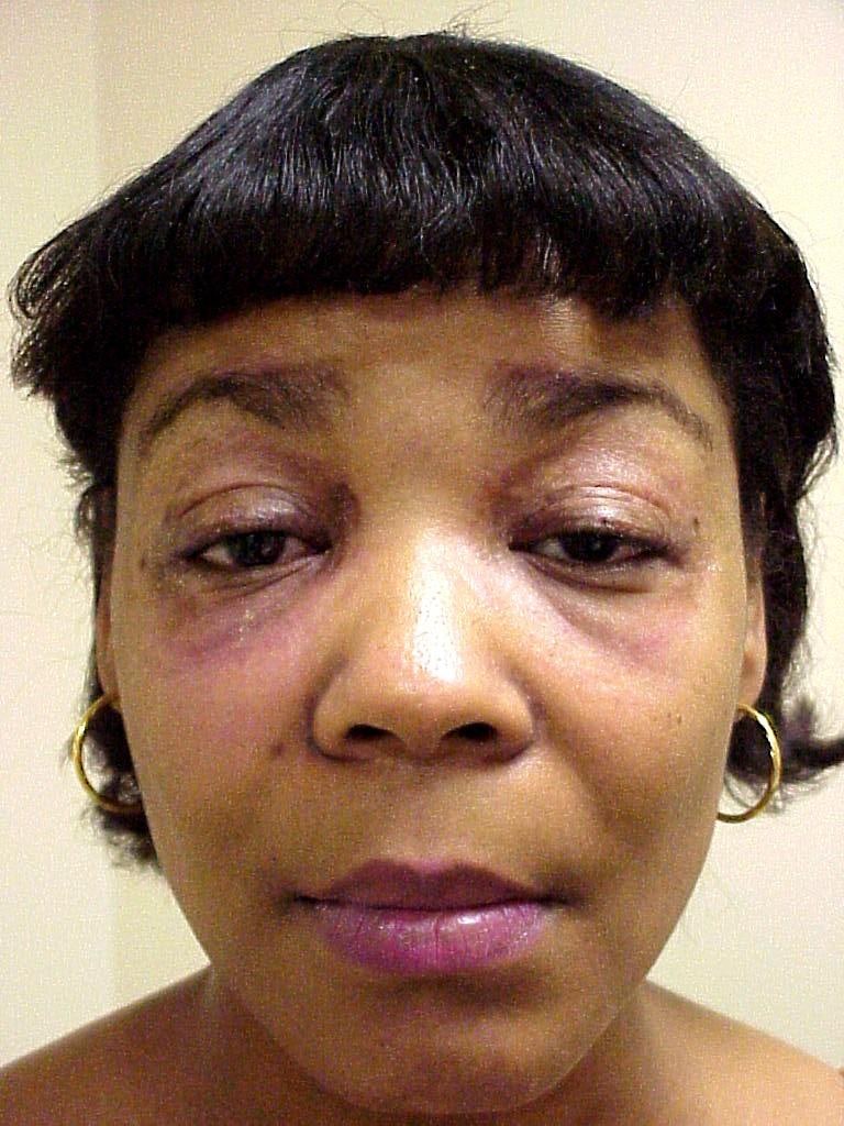
Heliotrope rash: the "classic" violaceous rash over the eyes and the malar region of the face
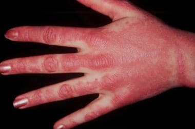
Gottron's papules: erythematous papules on the dorsum of MCP or interphalangeal joints; biopsy shows acanthosis and hyperkeratosis with vacuolar change and a scattered perivascular inflammatory infiltrate

Calcinosis: subcutaneous cases occur in long term, intractable cases, usually of juvenile type
Contributed by Meggen Walsh, D.O., M.S., P.A. and Jesse L. Kresak, M.D.

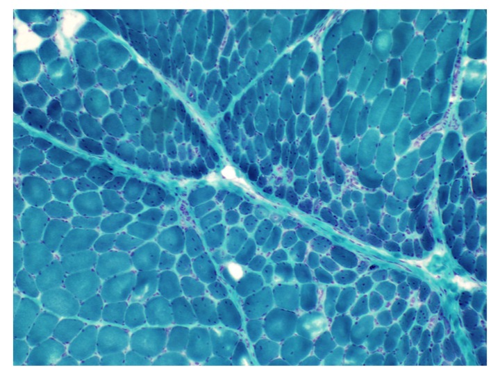
Perifascicular atrophy
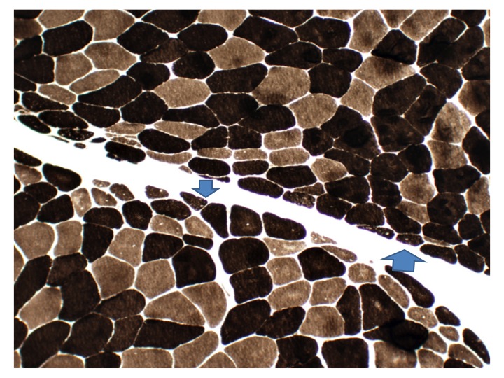
Perifascicular atrophy stain
GMS


Myosin I/II immunostain


Images hosted on other servers:

Weakness of lips and shoulder muscles
Contributed by Chunyu Cai, M.D., Ph.D.
Case 1: 70 year old man with genetically confirmed FSHD1 (8 D4Z4 repeats on a 4qA haplotype)

Chronic
Fiber size variation

Fiber type grouping
Lobulated internal architecture

Lobulated internal architecture
MHC1
Lobulated fiber on EM

Misoriented sarcomere on EM
Case 2: 63 year old woman with genetically confirmed FSHD1 (2 D4Z4 repeats on a 4qA haplotype)

Chronic myopathy with inflammation
Type 1 predominance
Lobulated internal architecture NADH

MHC1
Terminal complement complex
Case 3: 66 year old man with heterozygous pathogenic mutation of SMCHD1 and a FSHD phenotype, consistent with FSHD2

FSHD2 muscle biopsy
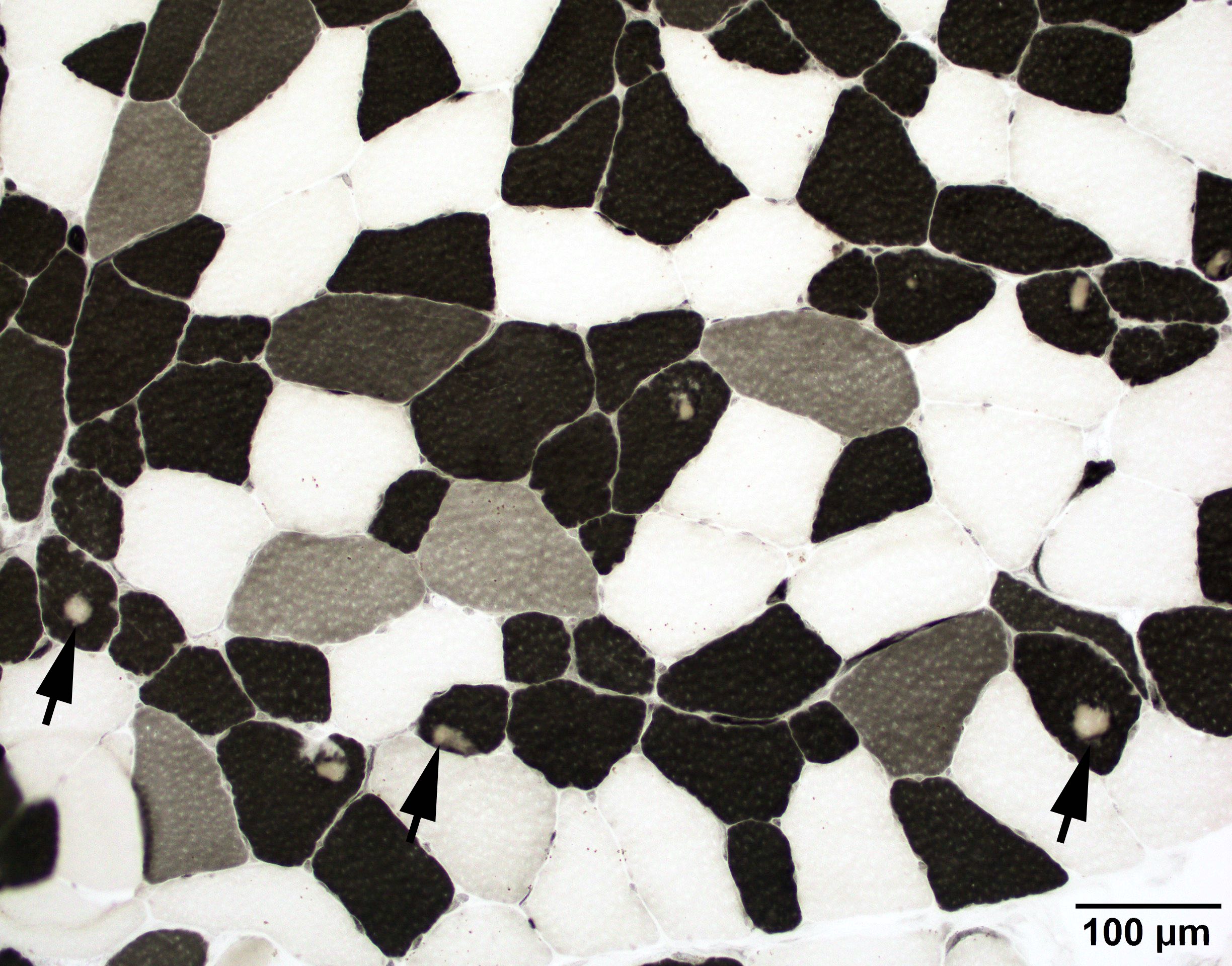
FSHD2 ATPase 4.3

Hyaline bodies GT
Hyaline bodies, desmin
Lobulated internal architecture, NADH
EM hyaline body
Images hosted on other servers:

Schematic of D4Z4
Facioscapulohumeral muscular dystrophy (Year of the Zebra)
FSHD patient's diagnostic journey
FSHD genetics
Contributed by Truong Phan Xuan Nguyen, M.D.

Metabolic glycolytic pathway
| GSD 0 | GYS1 (skeletal muscle) or GYS2 (liver) | Glycogen synthase 1 | Autosomal recessive | Glycogen synthase 1 deficiency |
| GSD II | GAA | Acid maltase | Autosomal recessive | Acid maltase deficiency; Pompe disease |
| GSD III | AGL | Debrancher enzyme | Autosomal recessive | Debrancher enzyme deficiency; Cori-Forbes disease |
| GSD IV | GBE1 | Branching enzyme | Autosomal recessive | Branching enzyme deficiency; Andersen disease |
| GSD V | PYGM | Glycogen phosphorylase | Autosomal recessive | Glycogen phosphorylase deficiency; McArdle disease |
| GSD VII | PFKM | Phosphofructokinase | Autosomal recessive | Phosphofructokinase deficiency; Tarui disease |
| GSD IXd | PHKA1 | Phosphorylase kinase | X linked inheritance | Phosphorylase kinase deficiency |
| Phosphoglycerate kinase deficiency | PGK1 | Phosphoglycerate kinase | X linked inheritance | N/A |
| GSD X | PGAM2 | Phosphoglycerate mutase | Autosomal recessive | Phosphoglycerate mutase deficiency |
| GSD XI | LDHA | Lactate dehydrogenase | Autosomal recessive | Lactate dehydrogenase deficiency |
| GSD XII | ALDOA | Aldolase A | Autosomal recessive | Aldolase A deficiency |
| GSD XIII | ENO3 | β enolase | Autosomal recessive | β enolase deficiency |
| GSD XIV | PGM1 | Phosphoglucomutase | Autosomal recessive | Phosphoglucomutase deficiency |
| GSD XV | GYG1 | Glygogenin 1 | Autosomal recessive | Glygogenin 1 deficiency |
Contributed by Ichizo Nishino, M.D., Ph.D.
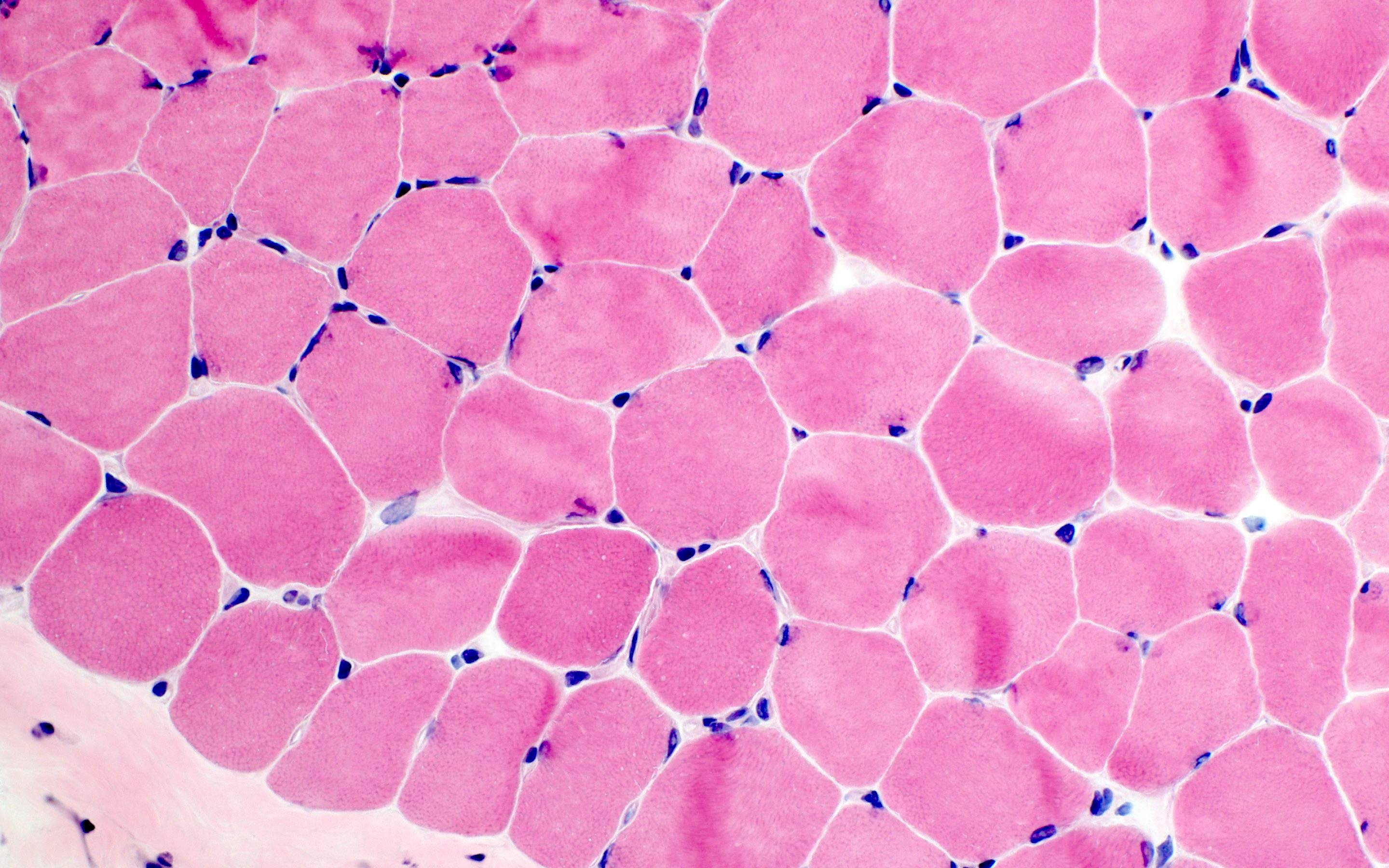
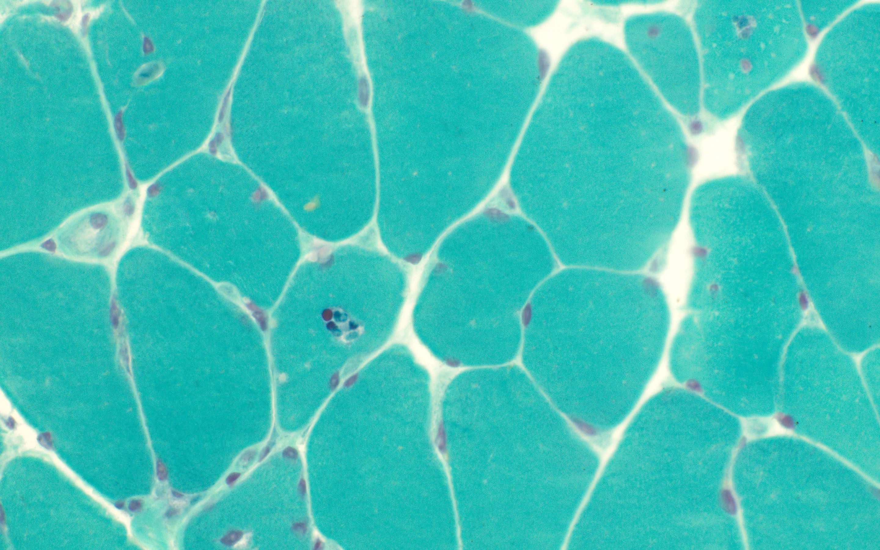
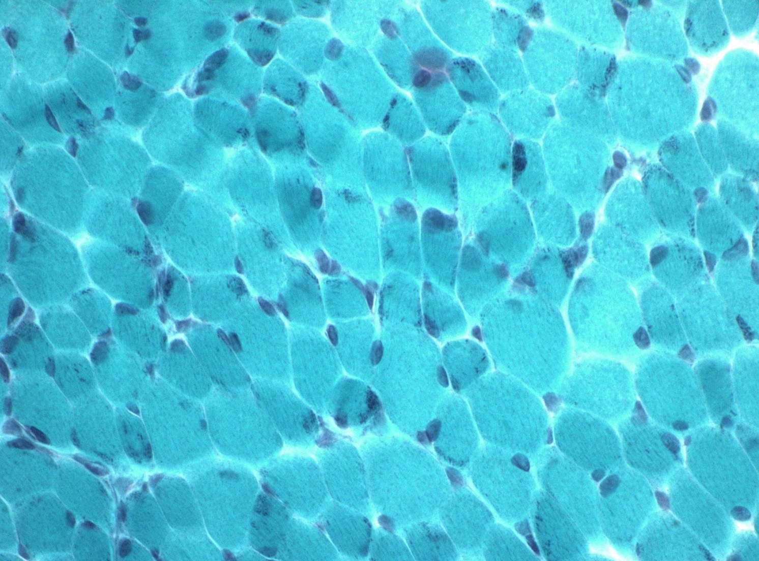
GSD 0: no specific changes
GSD 0: glycogen depletion
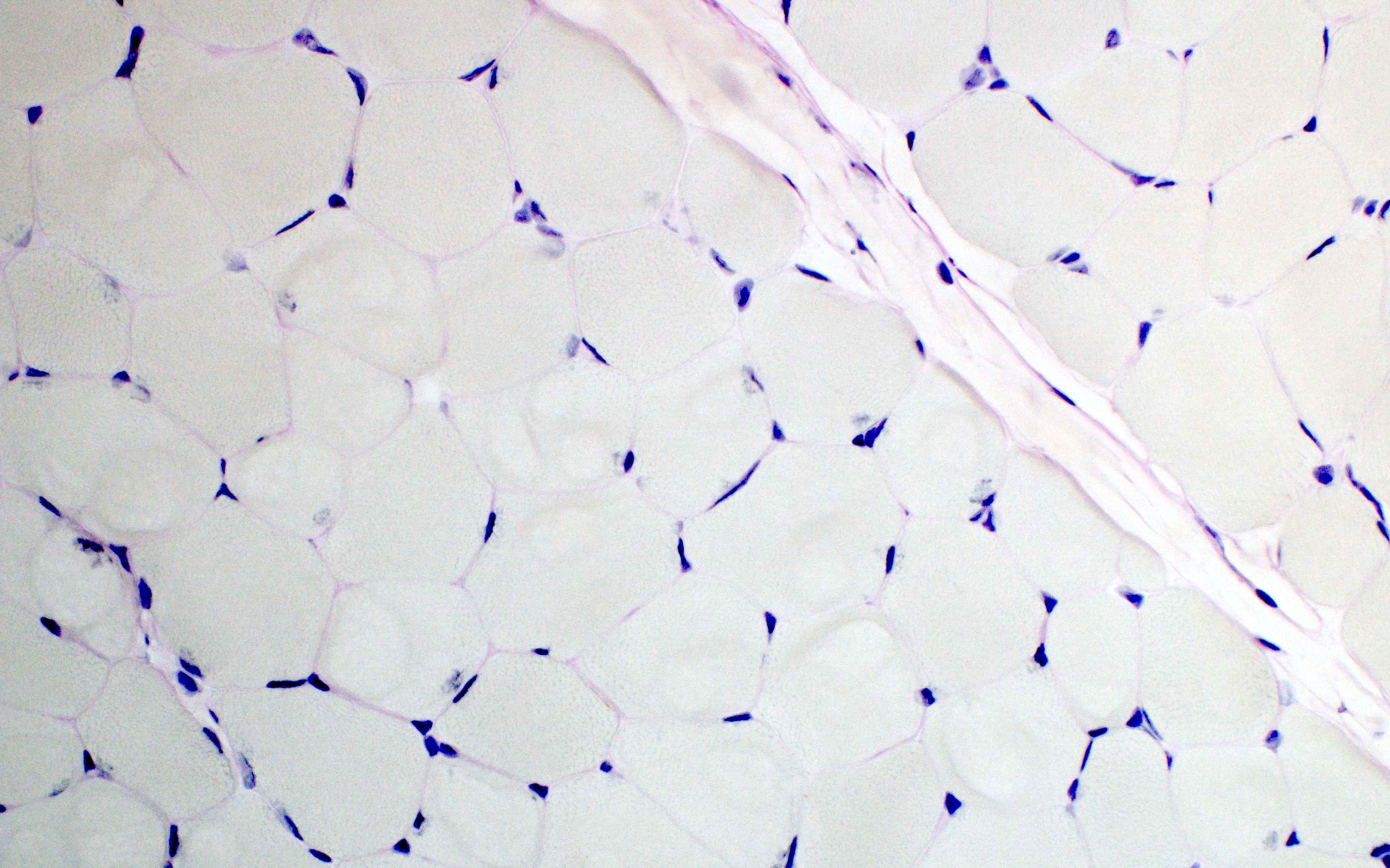
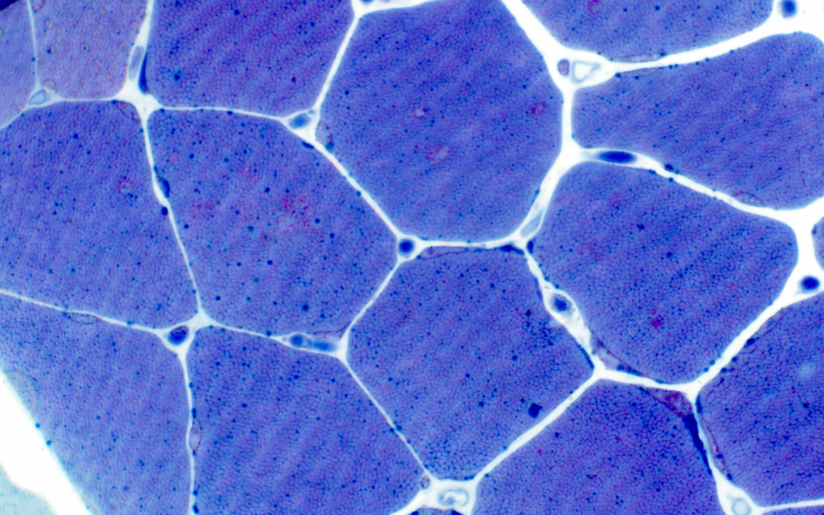
GSD II childhood: marked sarcoplasmic vacuoles
GSD II childhood: PAS

GSD II childhood: epon PAS

GSD II childhood: NADH TR

GSD II childhood:
predominant
involved
type 1 fibers
GSD II childhood:
high acid
phosphatase
activity
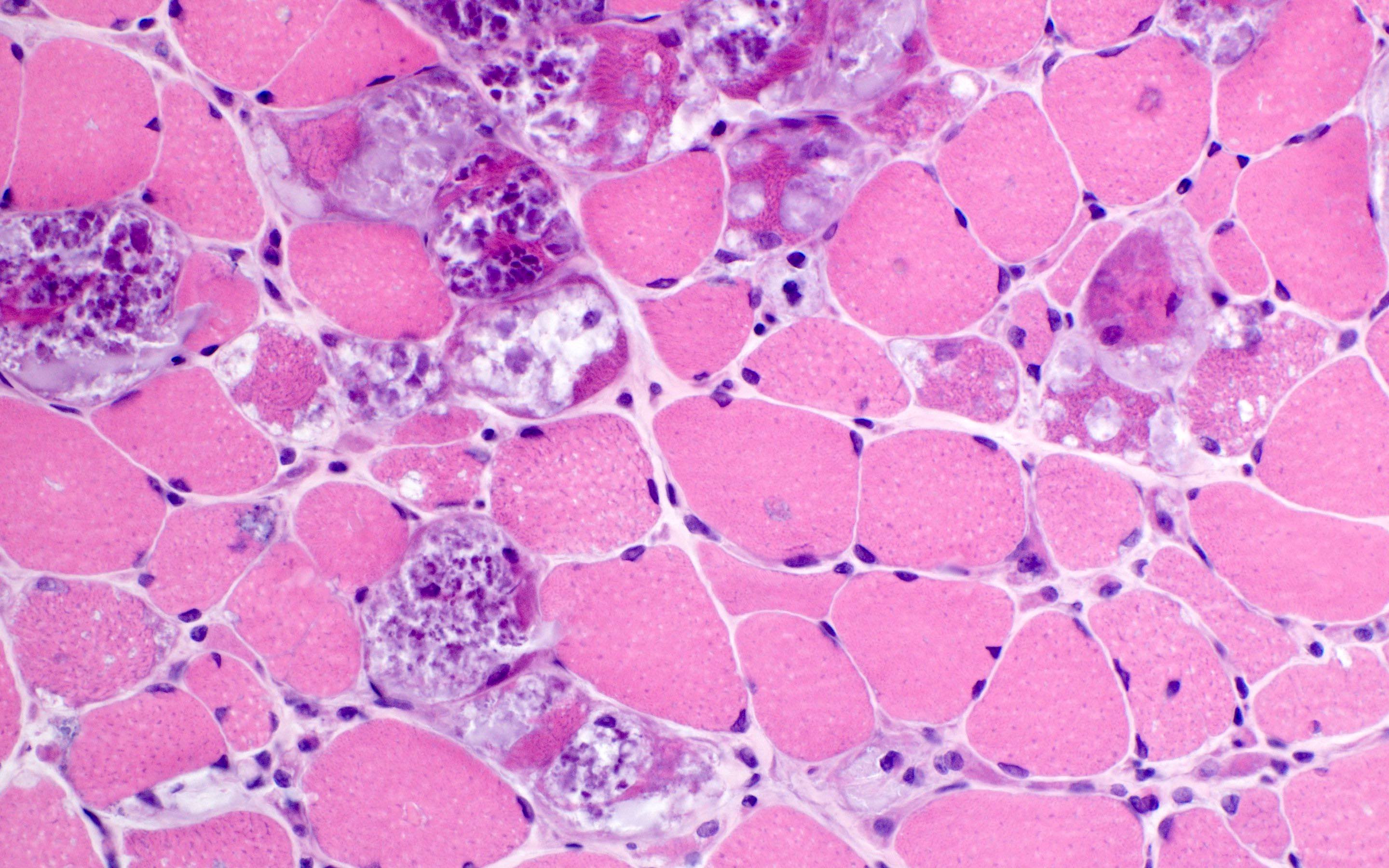
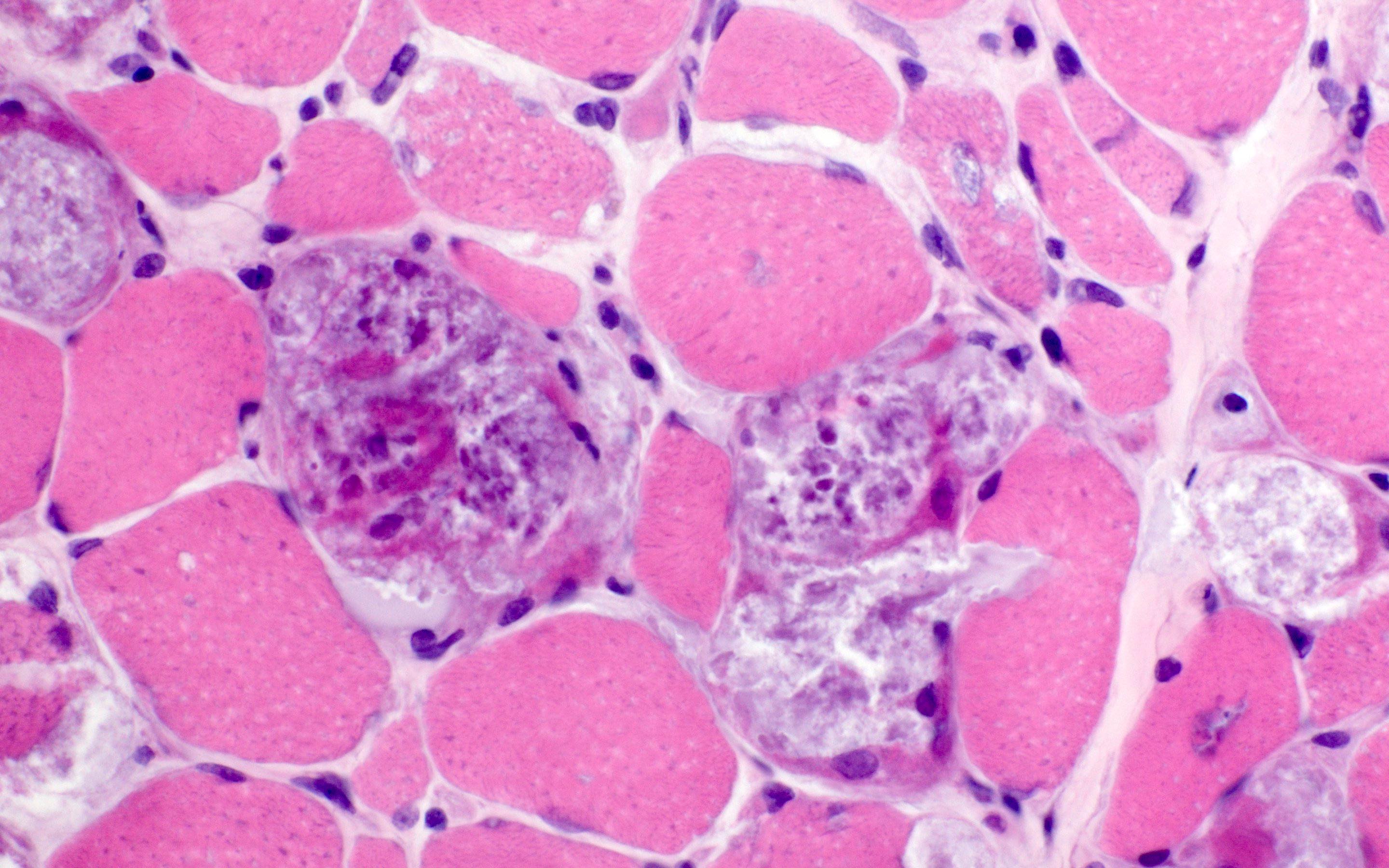
GSD II childhood: modified Gomori trichrome
GSD II childhood:
MHC I

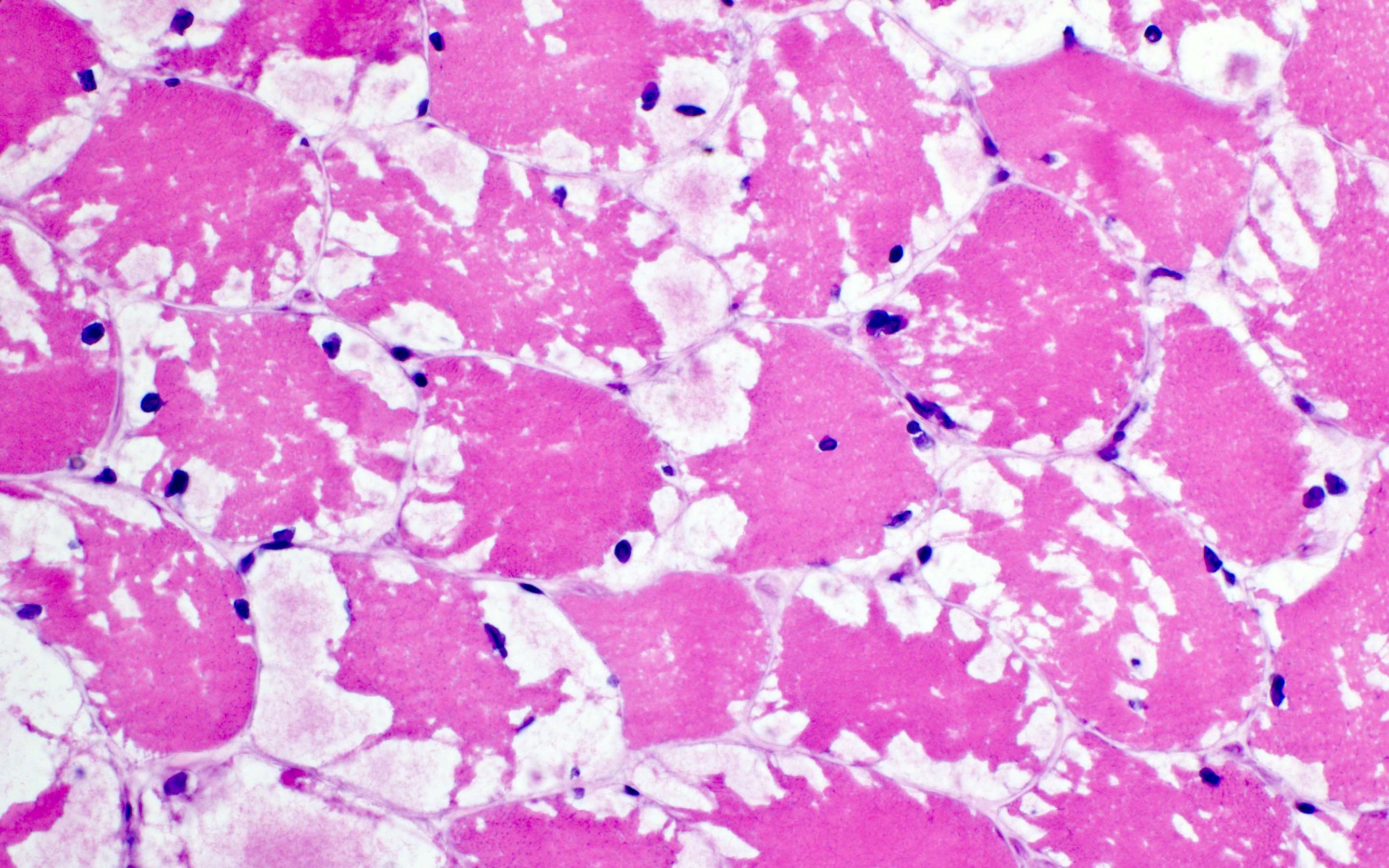
GSD II adult: small vacuoles
GSD II adult: modified Gomori trichrome
GSD II adult: epon PAS
GSD II adult: increased acid phosphatase activity
GSD III: nonmembrane bound vacuoles
GSD III: PAS positive
GSD III: MHC I

GSD IV: fibrosis
fibers and rounded
opalescent
inclusions

GSD IV: PAS
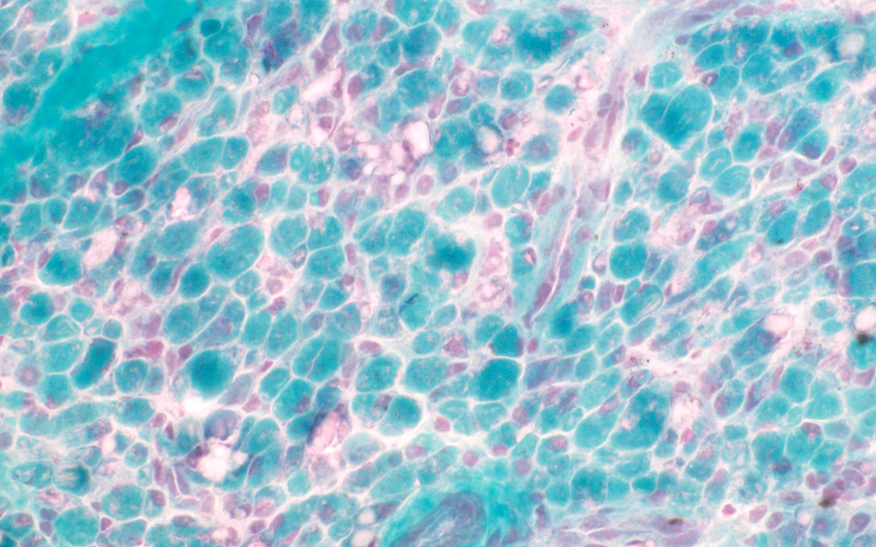
GSD IV: modified Gomori trichrome
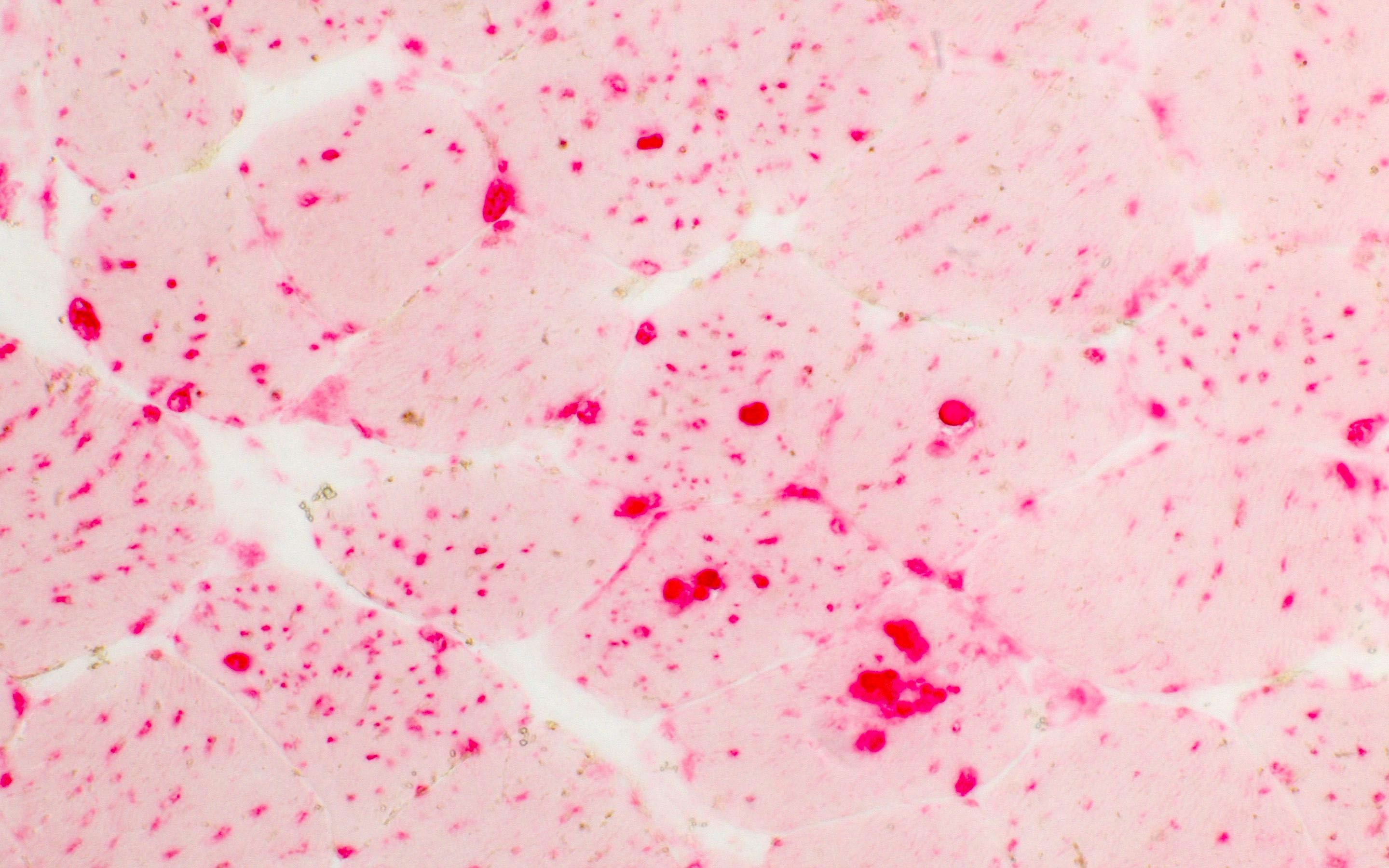
GSD V: small sarcolemmal vacuoles
GSD V: PAS
GSD V: negative PHS
GSD VII: small sarcolemmal vacuoles

GSD VII: PAS
GSD VII:
phosphofructokinase
is negative
Contributed by Ichizo Nishino, M.D., Ph.D.

GSD II childhood: membrane bound vacuoles

GSD II adult: membrane bound vacuoles

GSD III: nonmembrane bound glycogen
GSD IXd: sarcoplasmic glycogen deposits
Contributed by Chunyu "Hunter" Cai, M.D., Ph.D.

HMGCR myopathy
HMGCR myopathy - MHC1
HMGCR myopathy - C5b9
HMGCR myopathy - alkaline phosphatase
Contributed by Meggen Walsh, D.O., M.S., P.A.
Myopathic features
Ubiquitin
CD3
Gomori trichrome
Contributed by Jesse L. Kresak, M.D
Scattered internal nuclei
Increased internal nuclei
Markedly increased internal nuclei
Fatty replacement and ring fibers
Images hosted on other servers:

DM2 muscle biopsy: FISH and MBNL1 immunofluorescence
Contributed by Wesley M. Hiser, M.D.
H&E

Gomori trichrome
Images hosted on other servers:

Nemaline rods
Contributed by Meggen Walsh, D.O., M.S., P.A.
Skeletal muscle: multiple nuclear "clumps" or nuclear "bags"; also few atrophic angulated myofibers (H&E)
Table 1: Genetic diseases with shared features of progressive ptosis and dysphagia
| Gene | Site | Heritance | Genetic defect | Clinical | Reference |
| PABPN1 | 14q11.2 | AD | GCN repeats | Late onset OPMD | Acta Neuropathol 2022;144:1157 |
| HNRNPA2B1 | 7p15.2 | AD | Frameshift | Early onset OPMD | Nat Commun 2022;13:2306 |
| LRP12 | 8q22.3 | AD | CGG repeats | Oculopharyngeal distal myopathy (OPDM) type 1 | JAMA Neurol 2021;78:853 |
| GIPC1 | 19p13.12 | AD | CGG repeats | OPDM type 2 | Am J Hum Genet 2020;106:793 |
| NOTCH2NLC | 1q21.2 | AD | CGG repeats | OPDM type 3 | Nat Genet 2019;51:1222 |
| RILPL1 | 12q27.31 | AD | CGG repeats | OPDM type 4 | Am J Hum Genet 2022;109:533 |
| NUTM2B::AS1 | 10q22.3 | AD | CGG repeats | Oculopharyngeal myopathy with leukodystrophy (OPML) | Nat Genet 2019;51:1222 |
Contributed by Chunyu Cai, M.D., Ph.D.

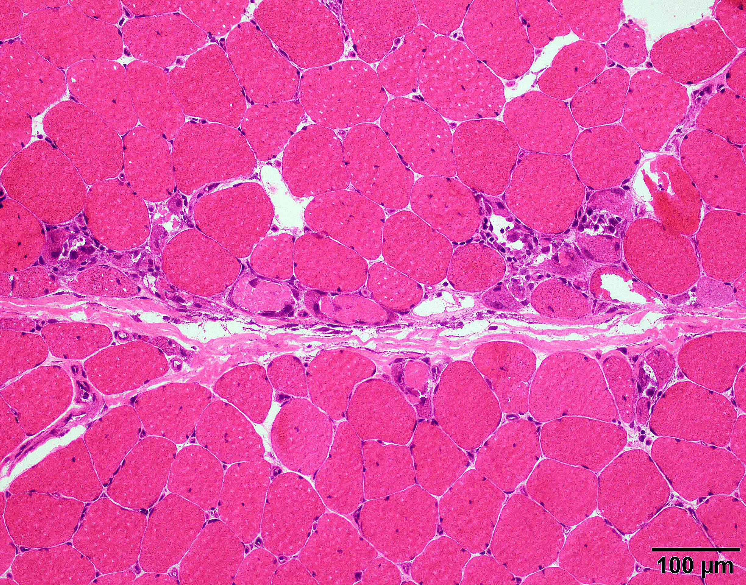
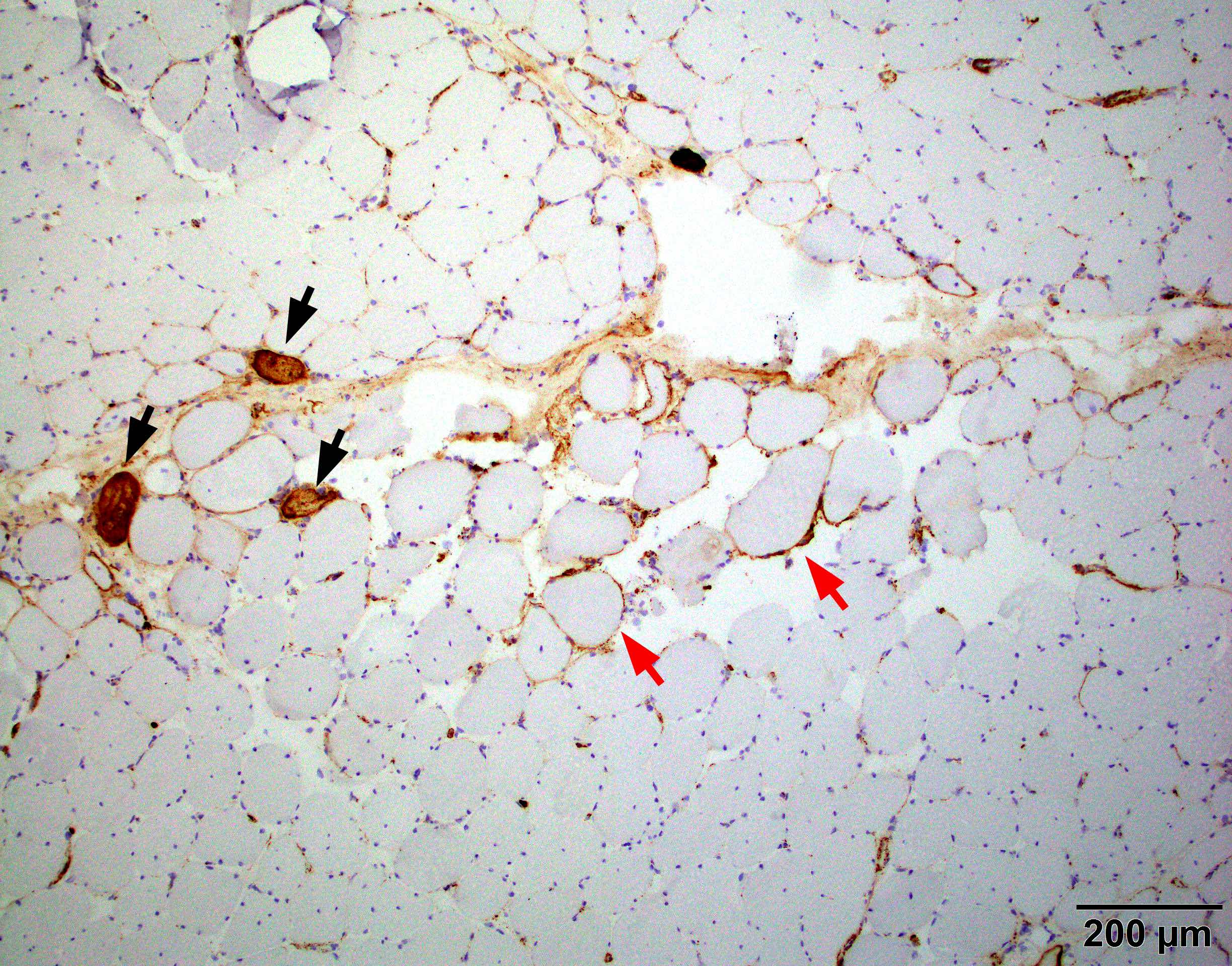
Myopathy with rimmed vacuoles

Gomori trichrome
Acid phosphatase
COX SDH double stain

MHC1
Contributed by Chunyu Cai, M.D., Ph.D.
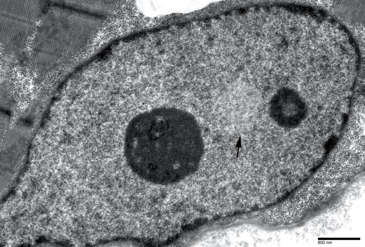
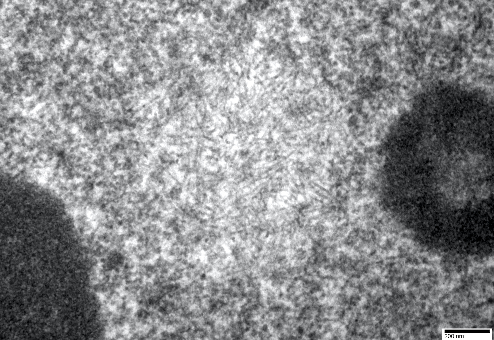
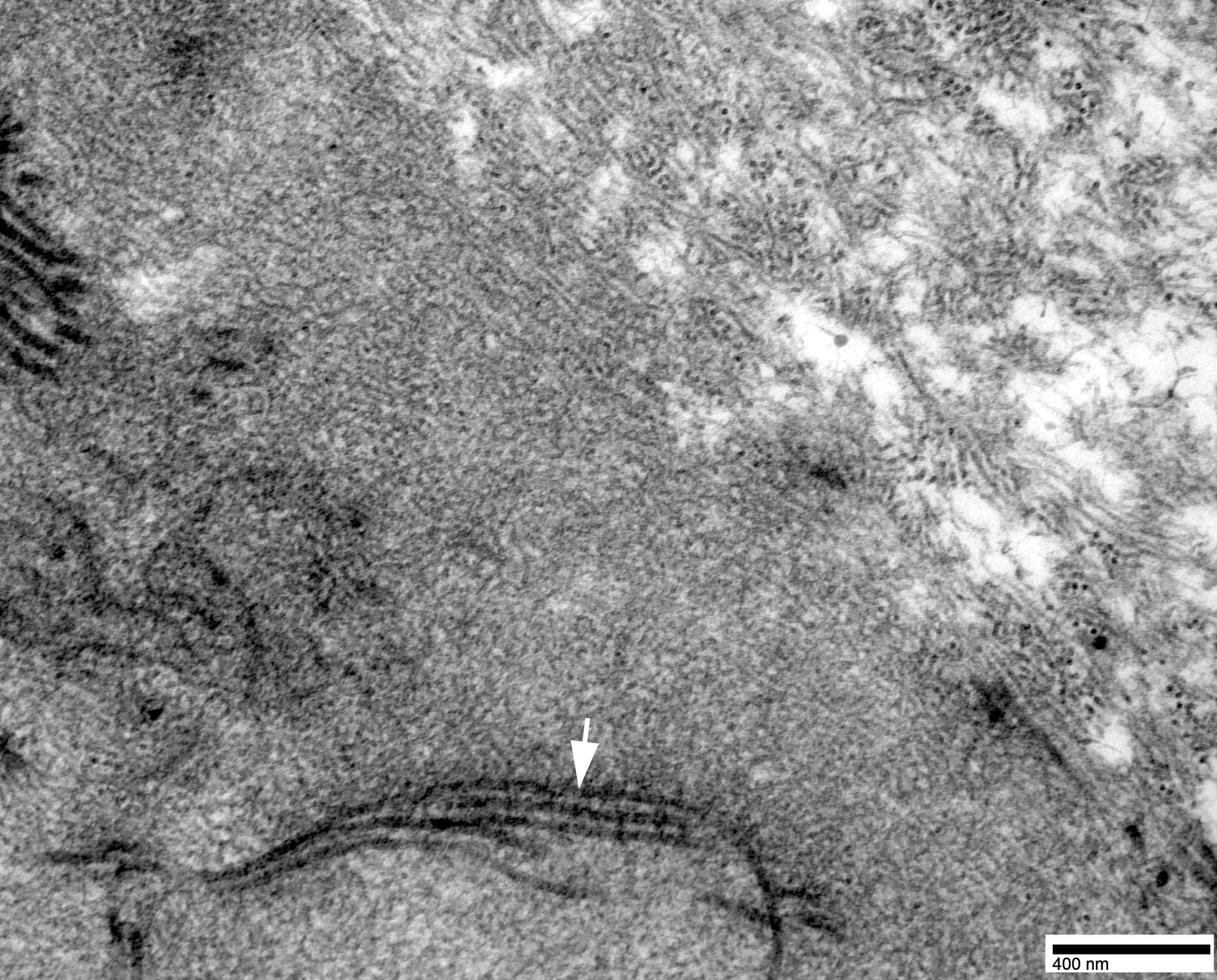
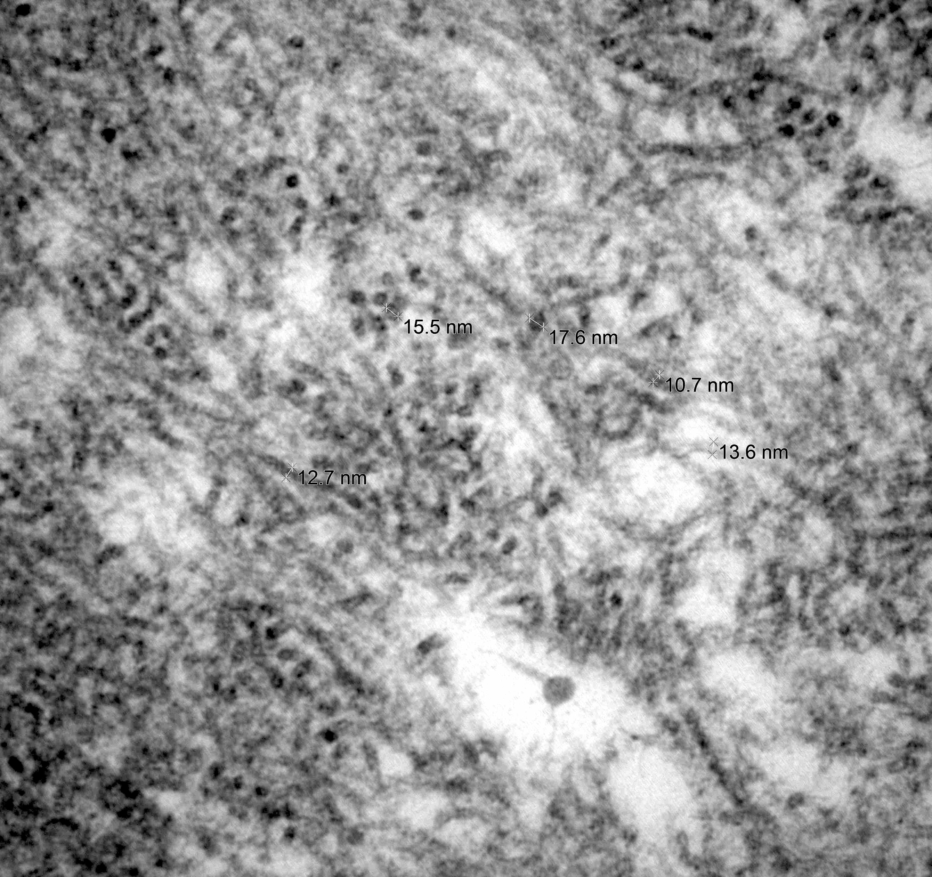
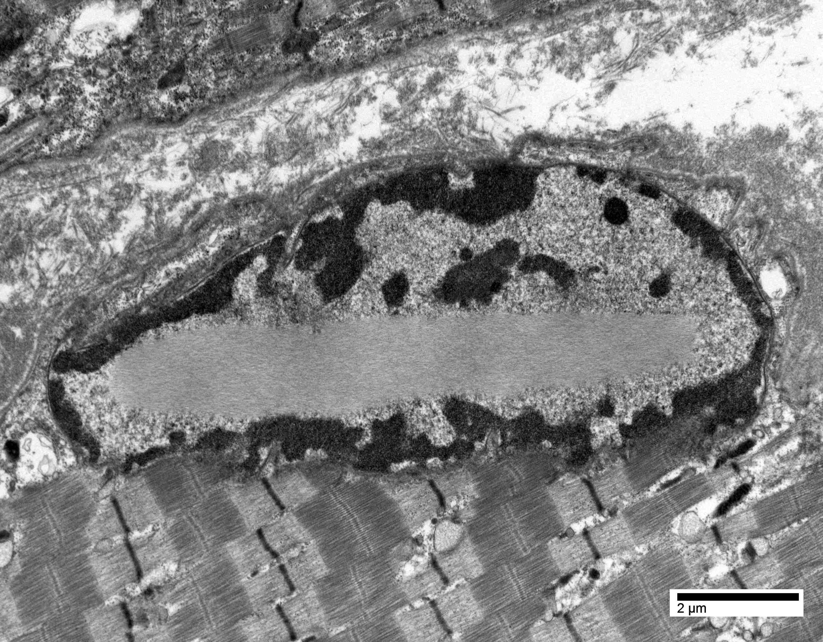
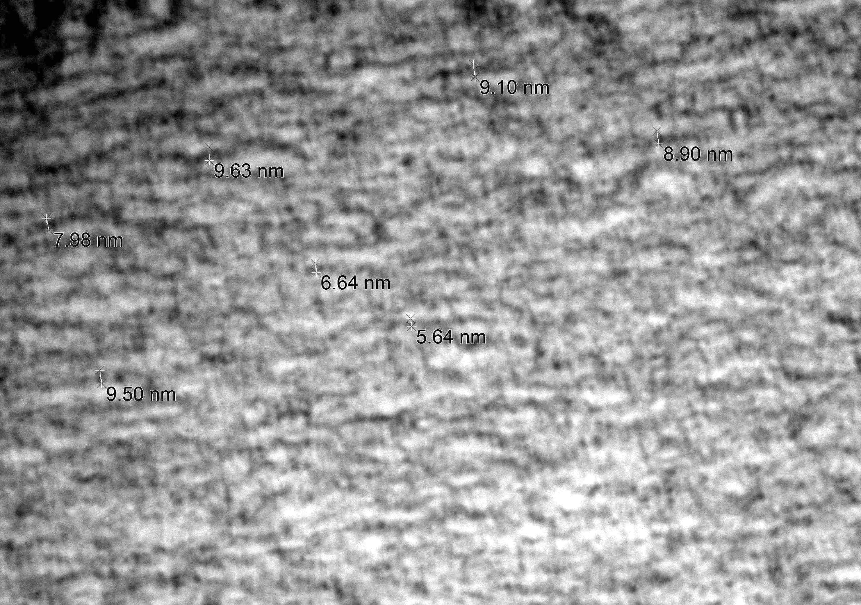
Intranuclear tubulofilamentous inclusion

Filaments reveal tubular nature
Contributed by Carrie A. Mohila, M.D., Ph.D., Matthew Cykowski, M.D. and Chunyu Cai, M.D., Ph.D.
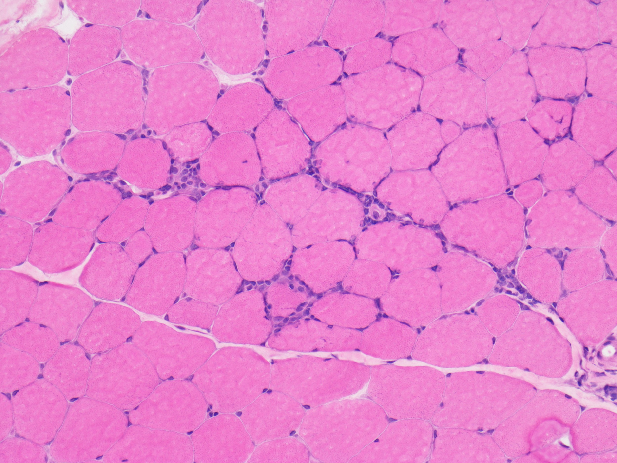
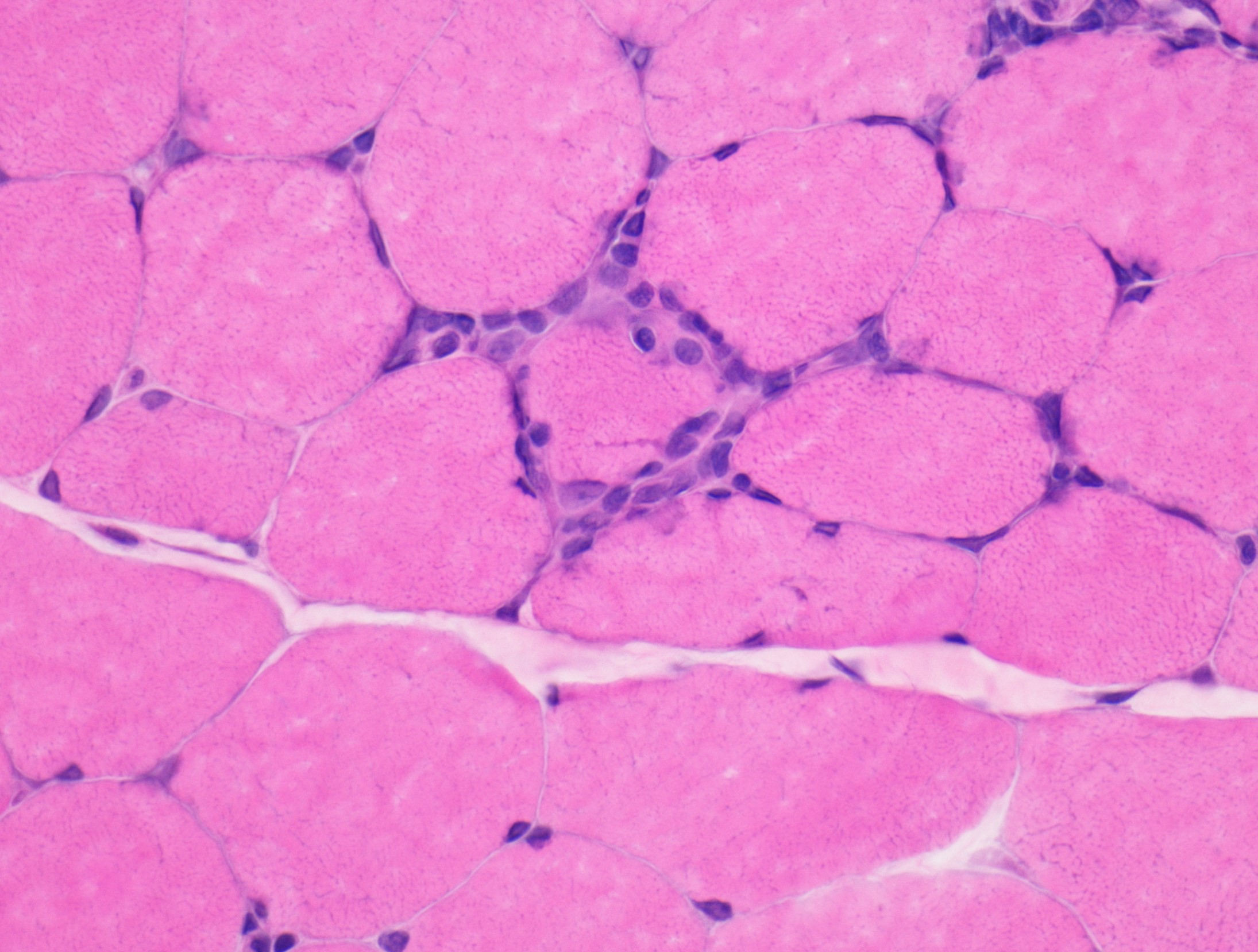
Endomysial lymphocytic inflammation
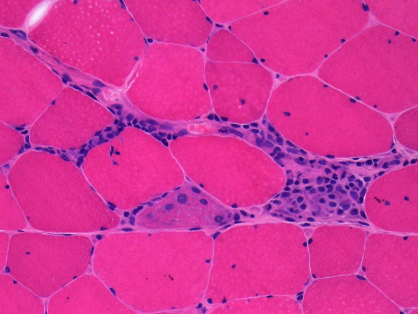
Endomysial lymphocytic infiltrate
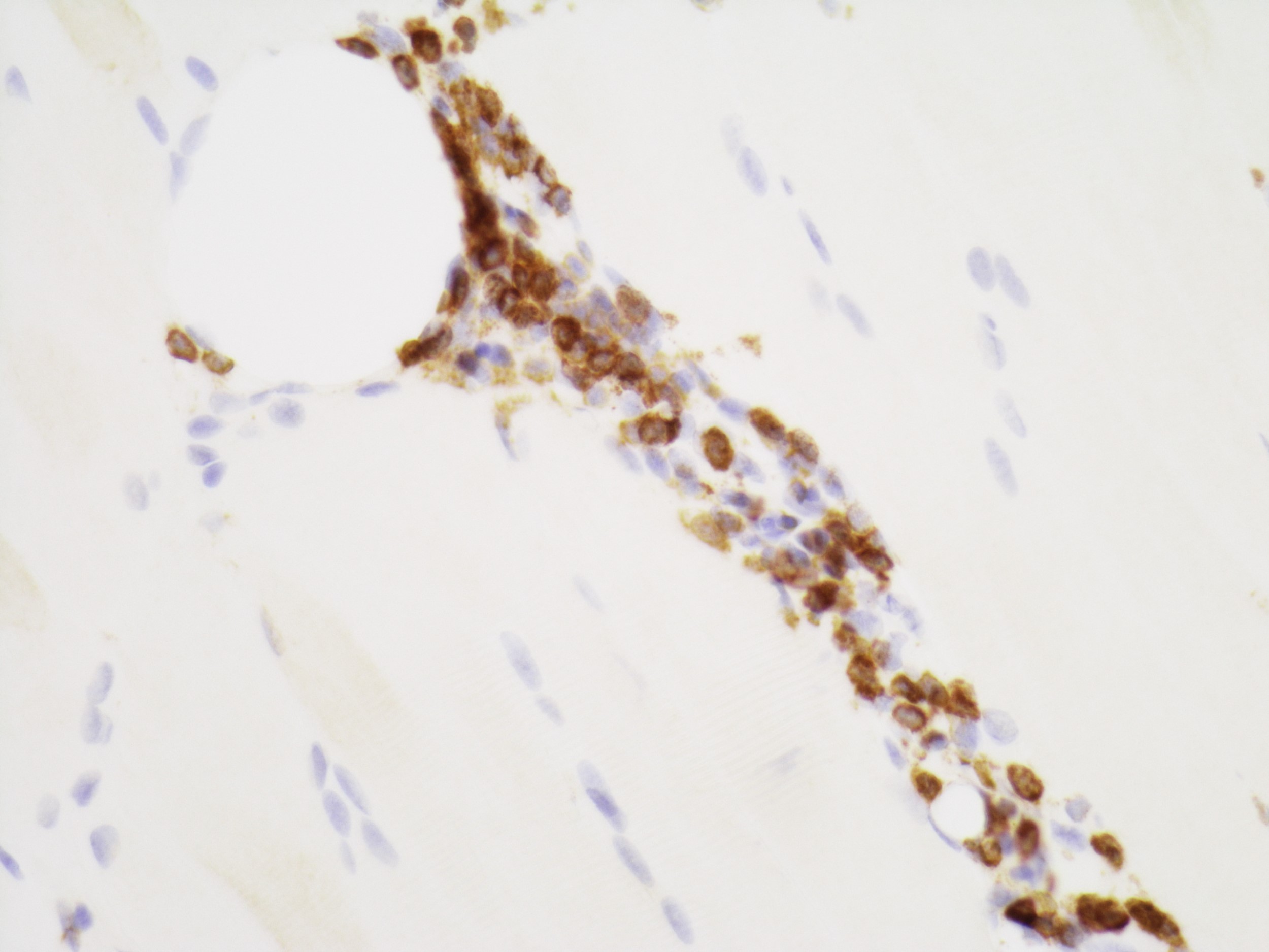
CD3+ lymphocytic infiltrate
Predominantly CD8+ lymphocytic infiltrate
MHC1
Polymyositis MHC1

Amato: 2015

Bilbao: 2014
Cooper: 2015

Dubowitz: 2020
Gray: 2018
Husain: 2021

Zhou: 2019
Find related Pathology books: muscle and peripheral nerve nontumor, neuropathology, pediatric
