
Contributed by James Guggenheimer, D.D.S.
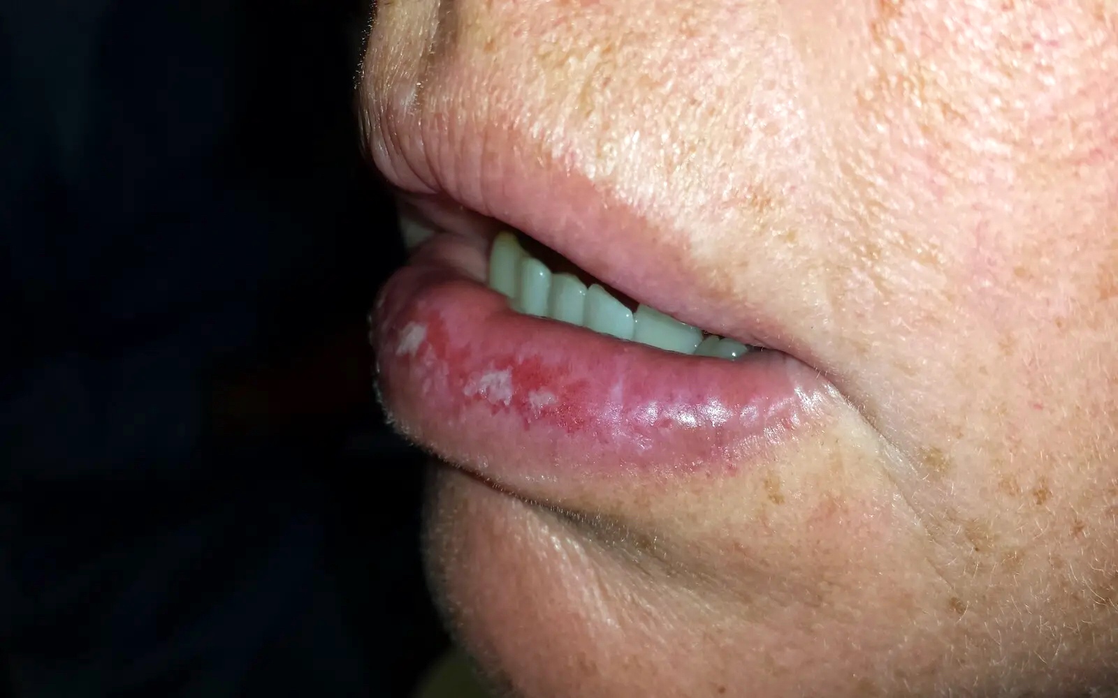
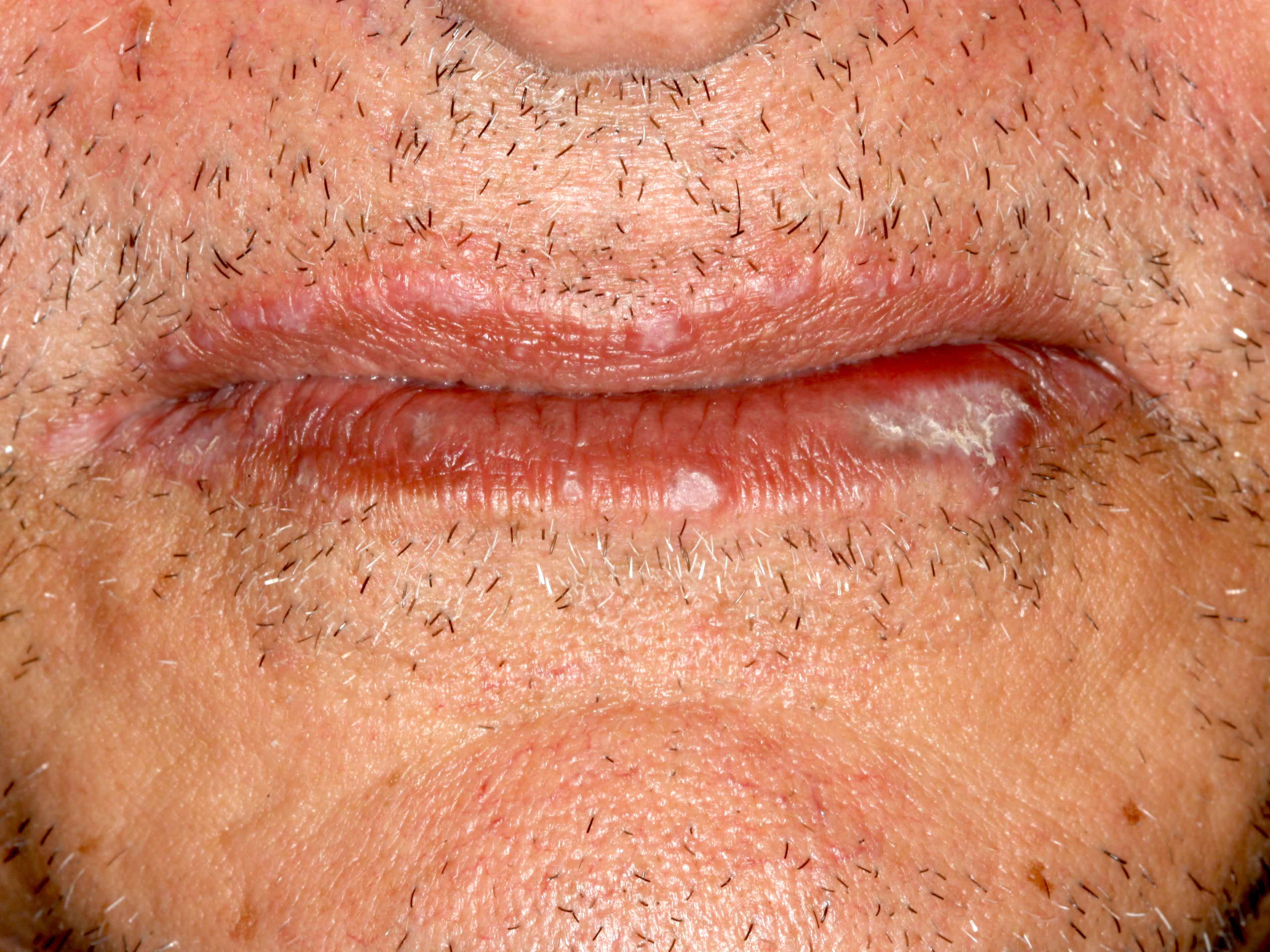
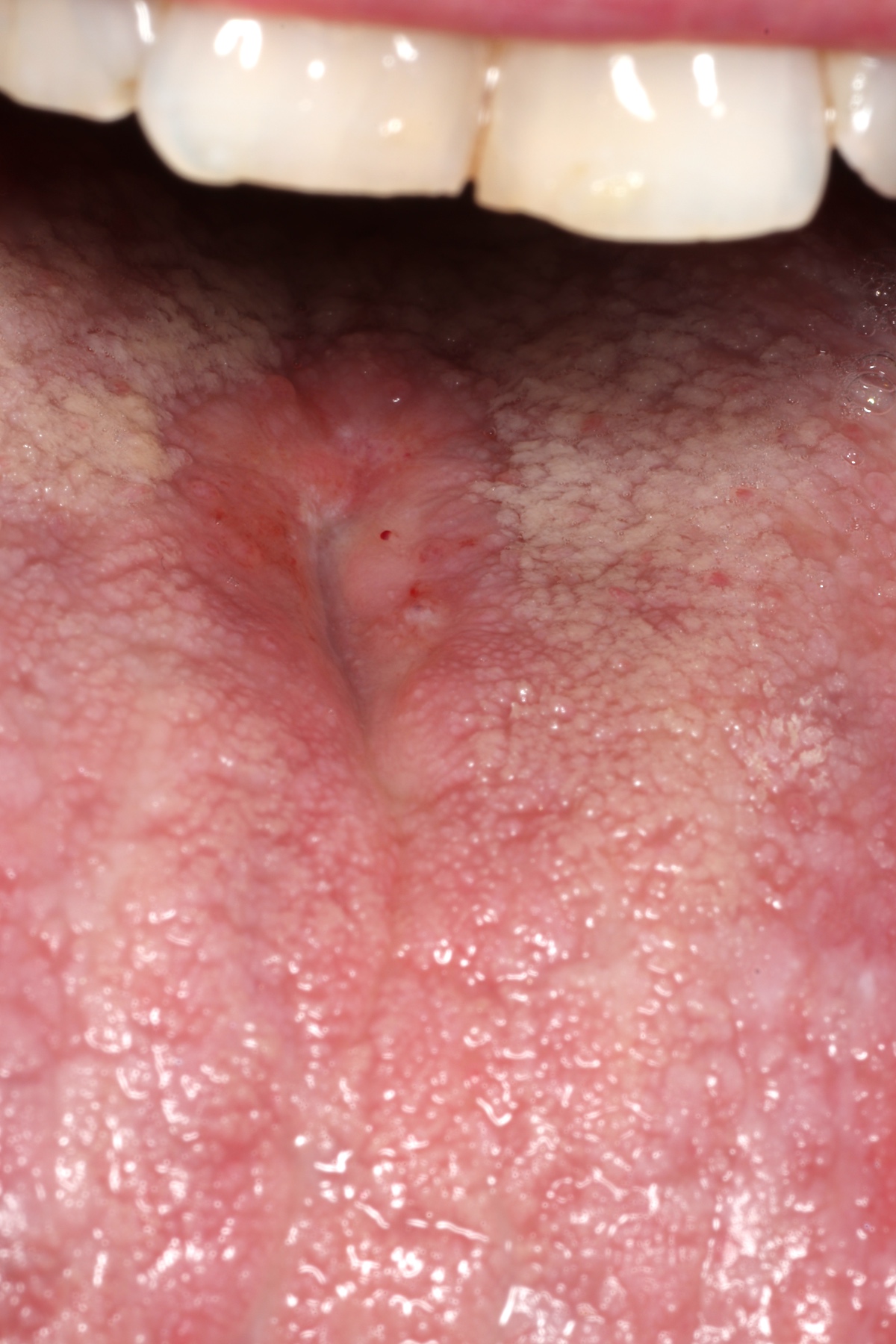
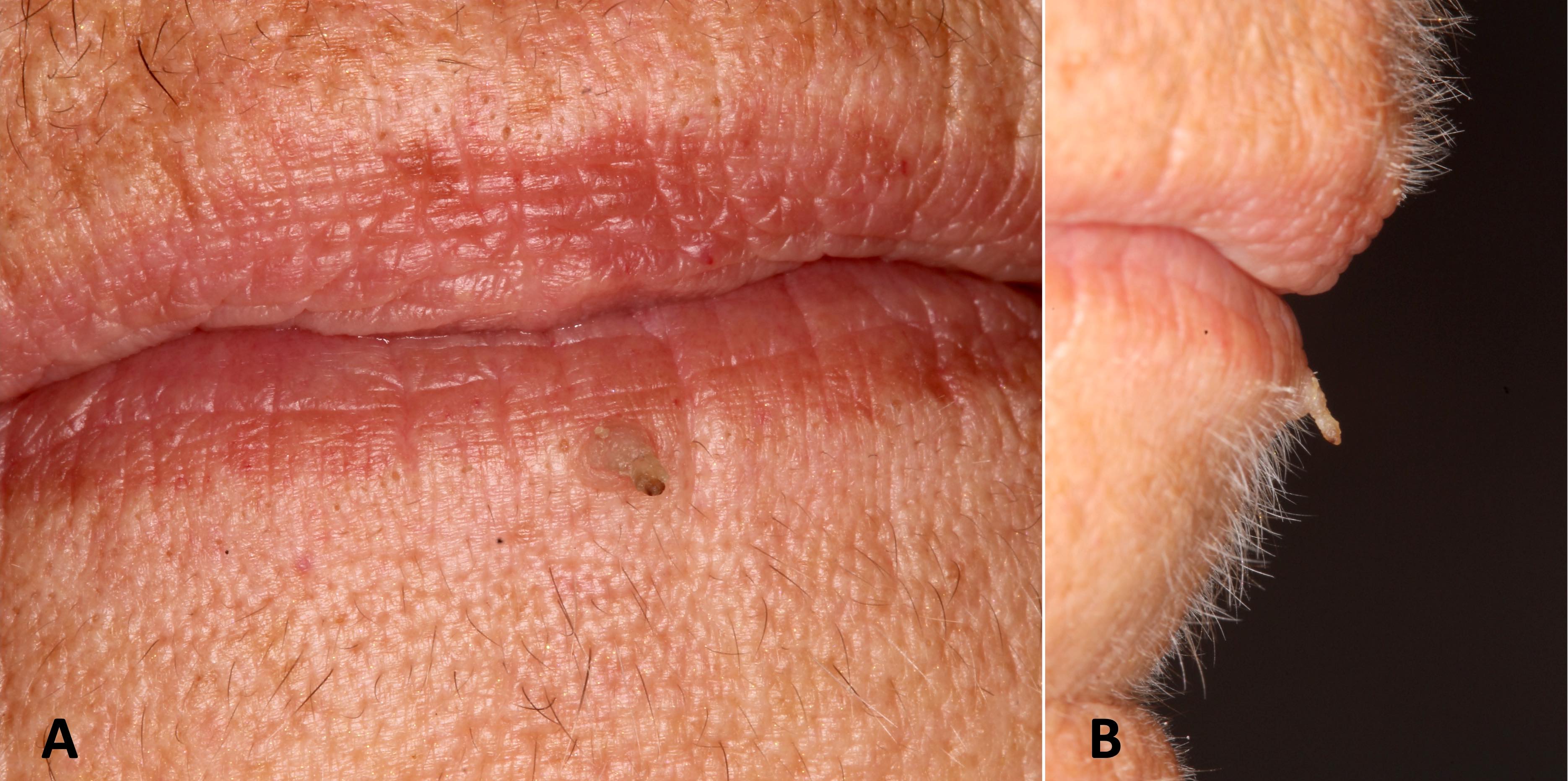
Blurring of lip vermilion
Contributed by Joshua Seth Goldfaden, D.D.S.
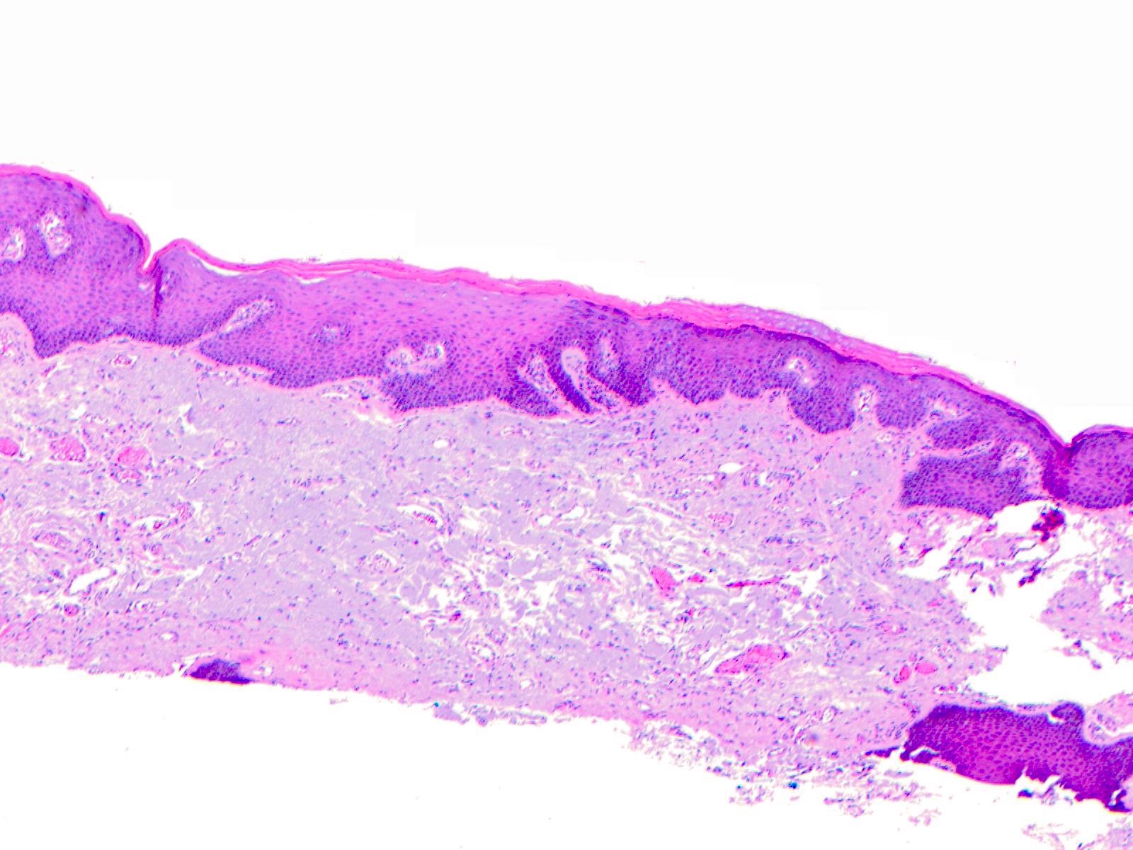
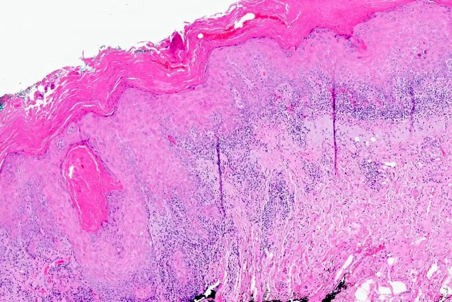
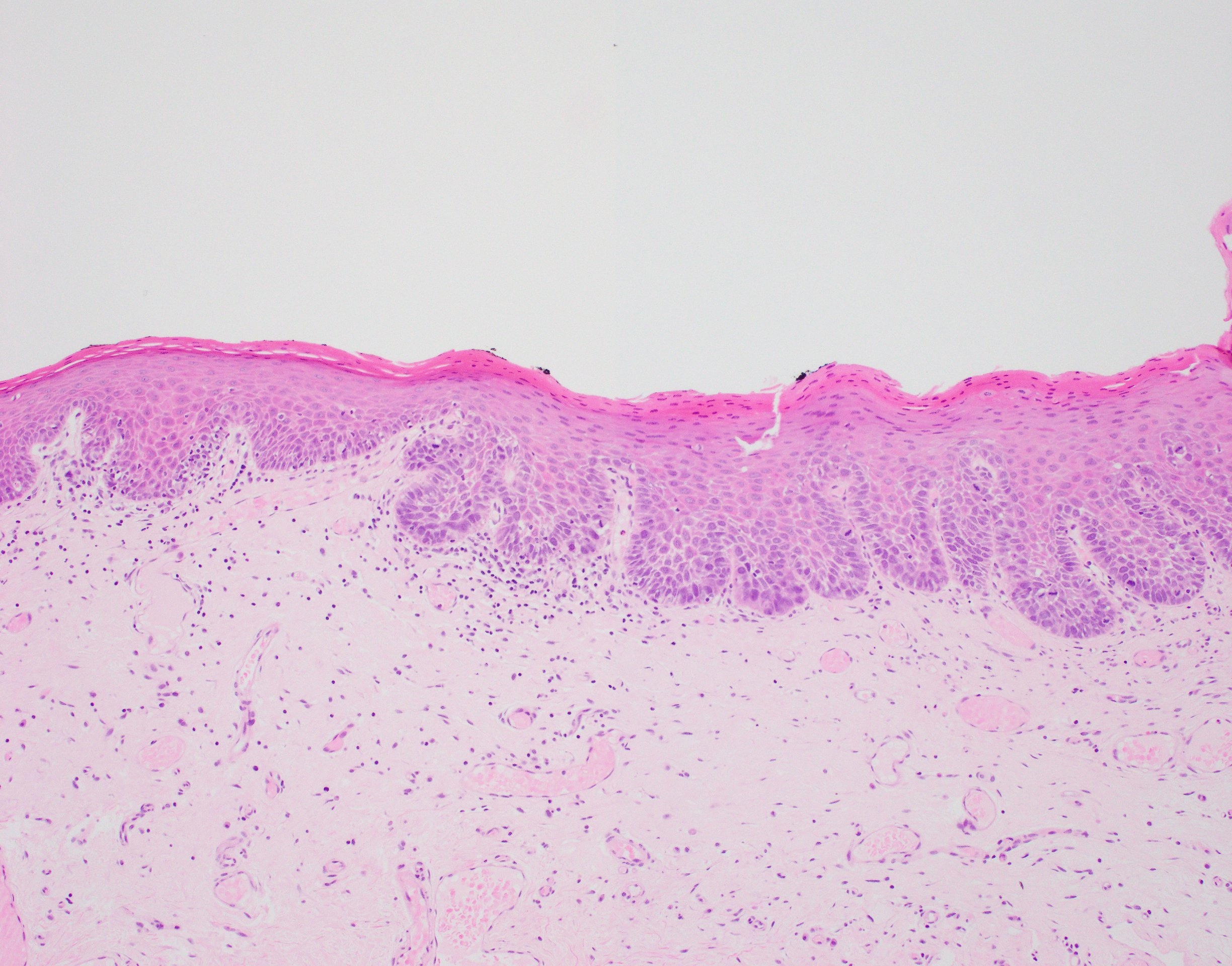
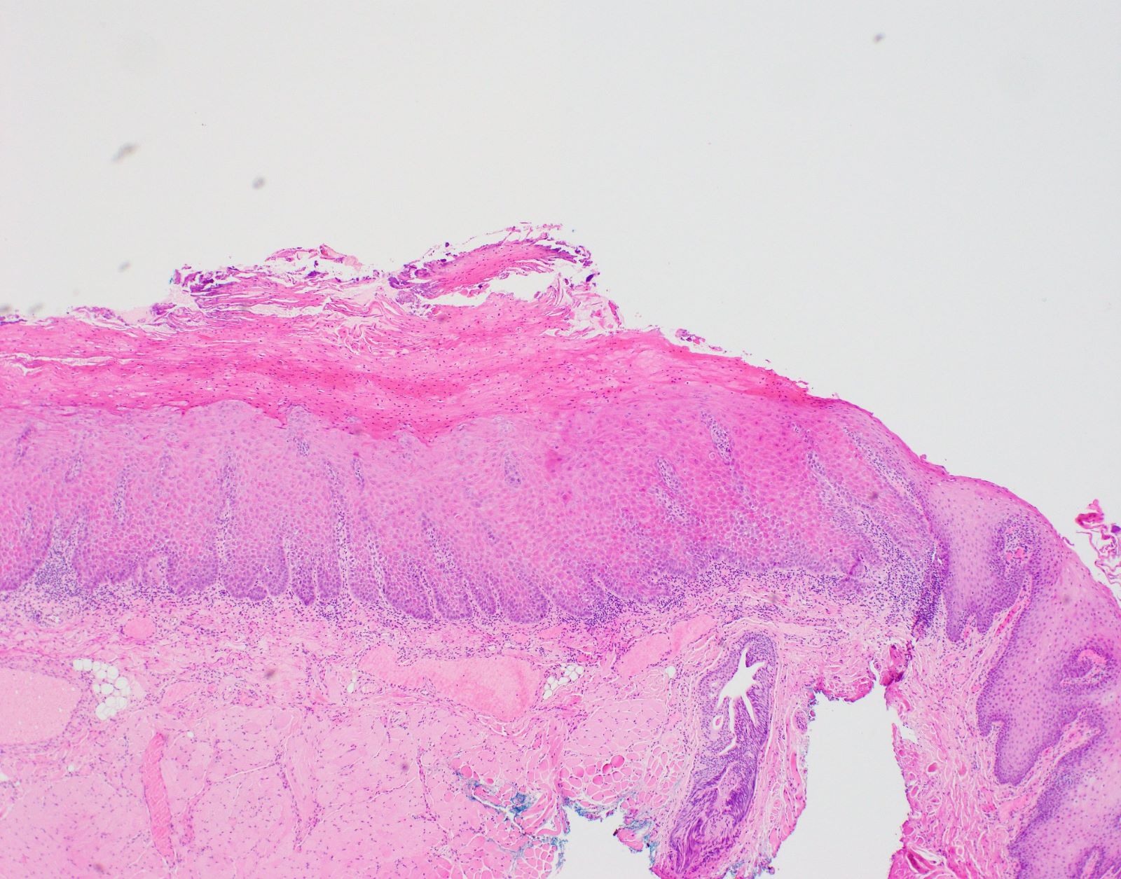
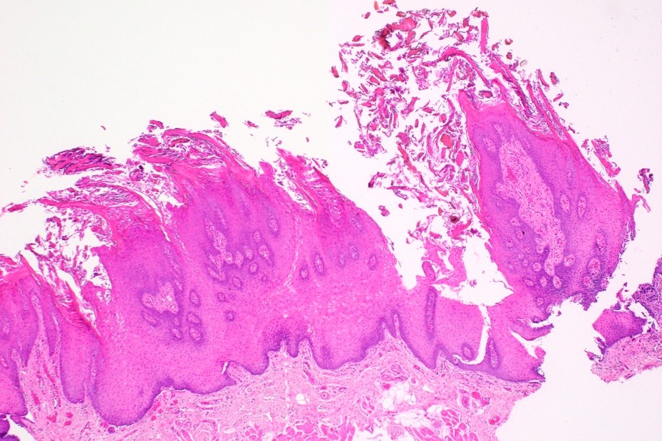
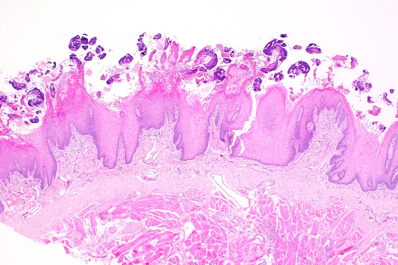
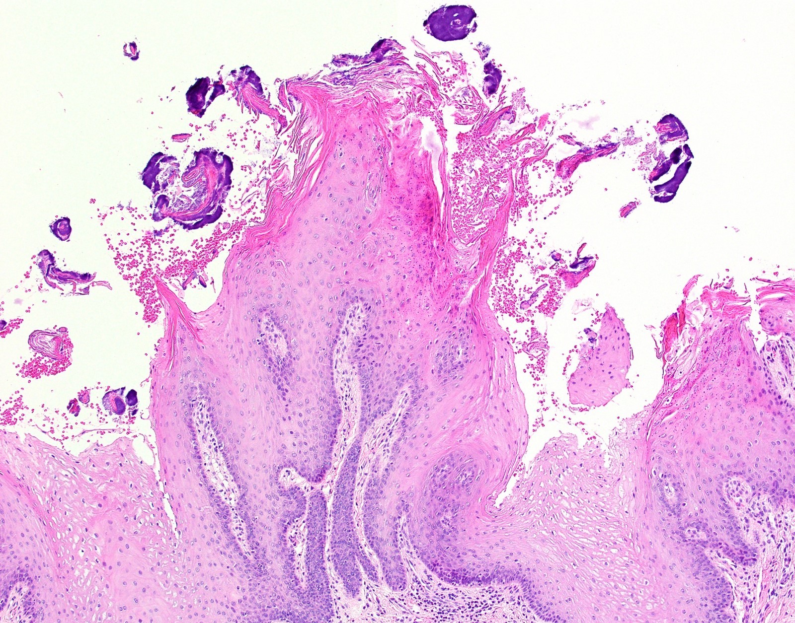
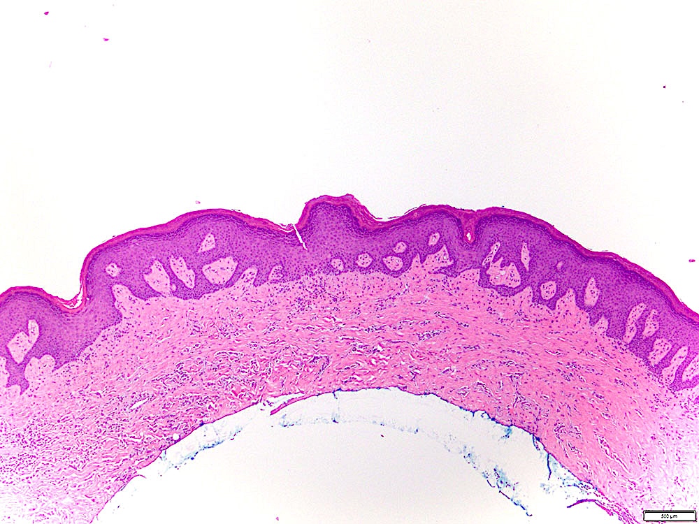
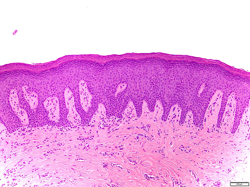
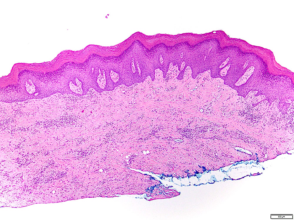
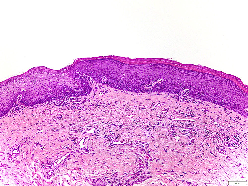
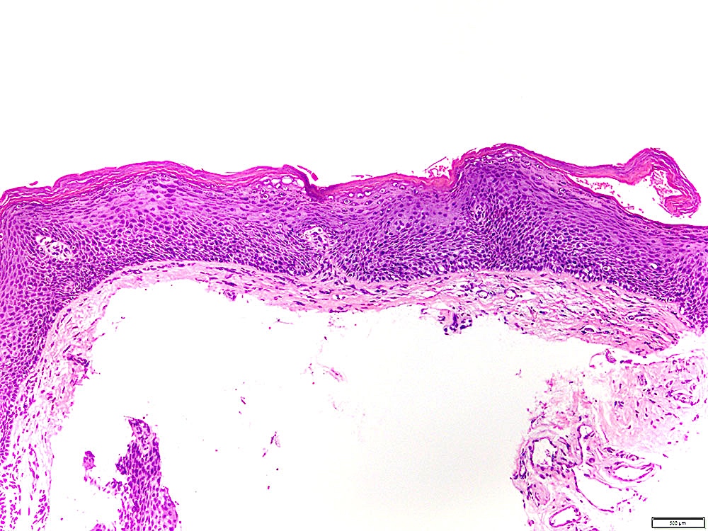
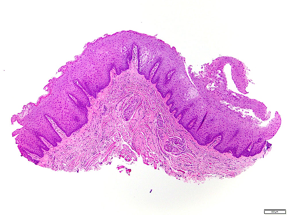
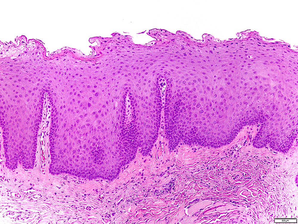
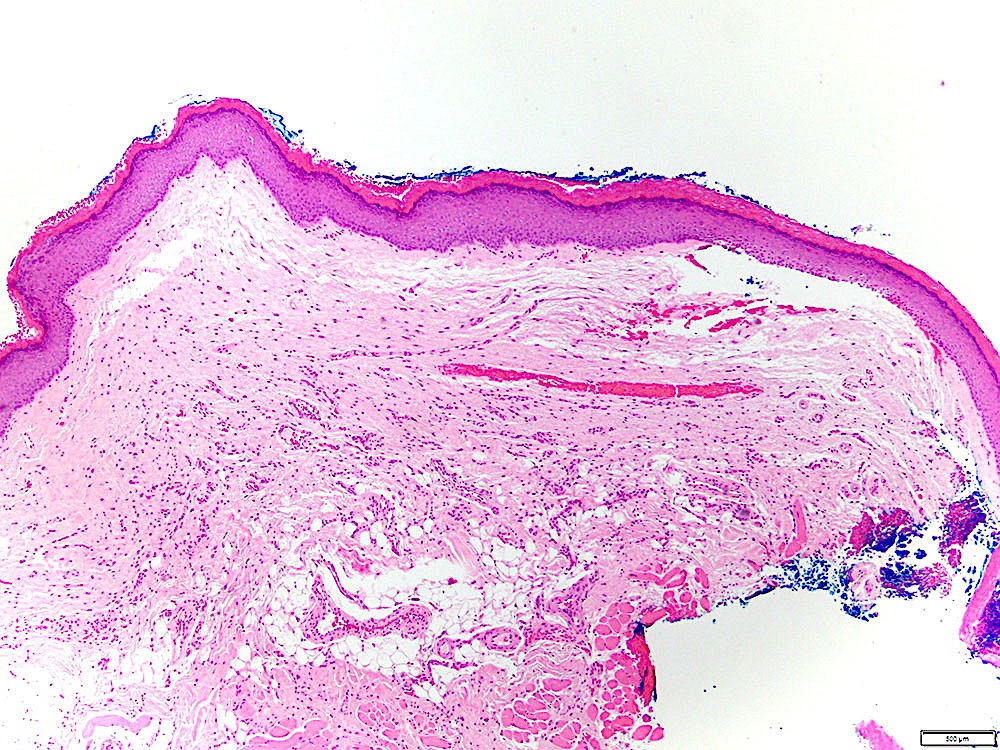
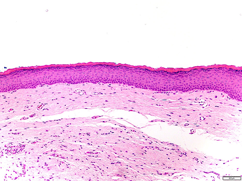
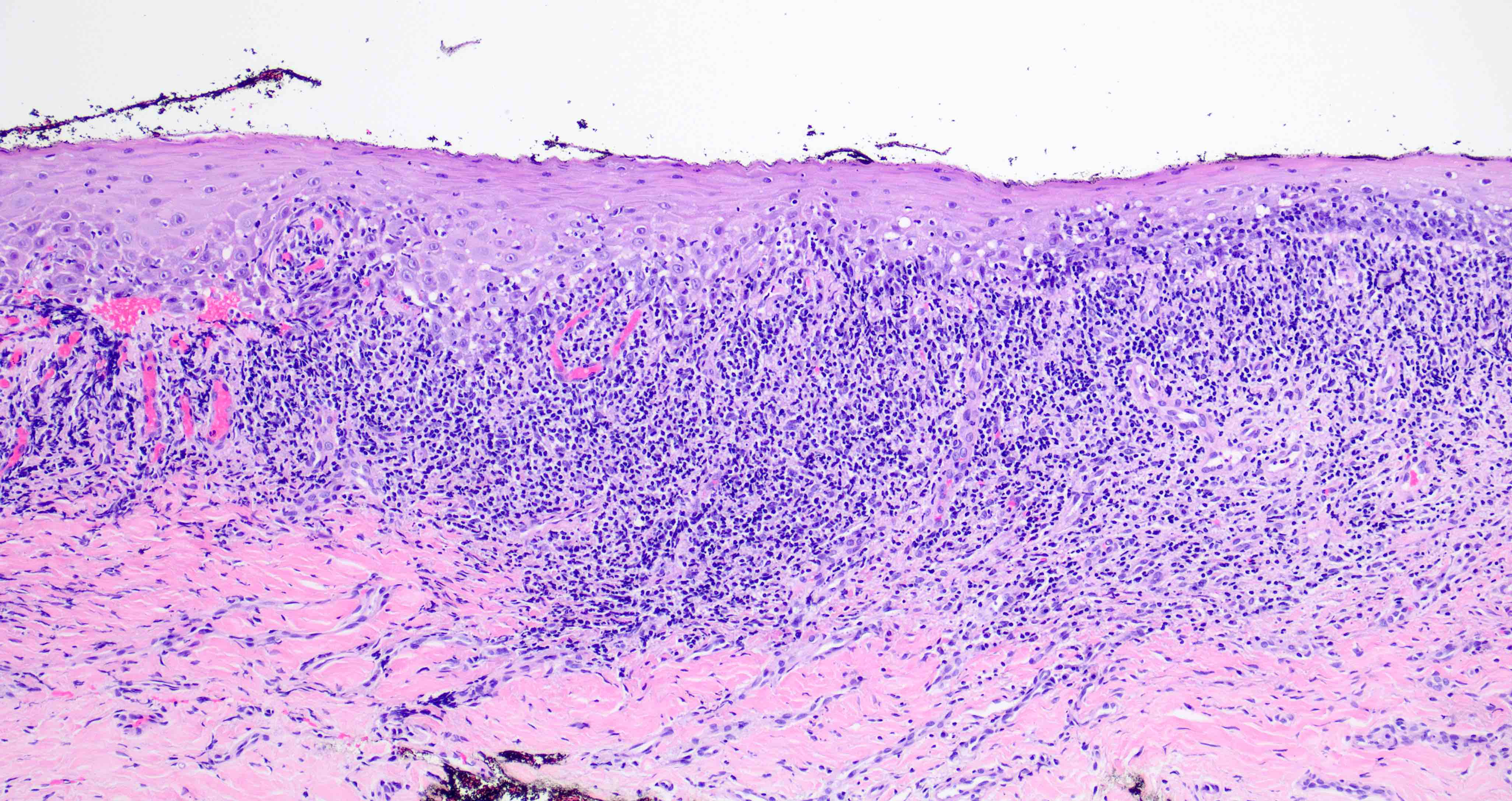
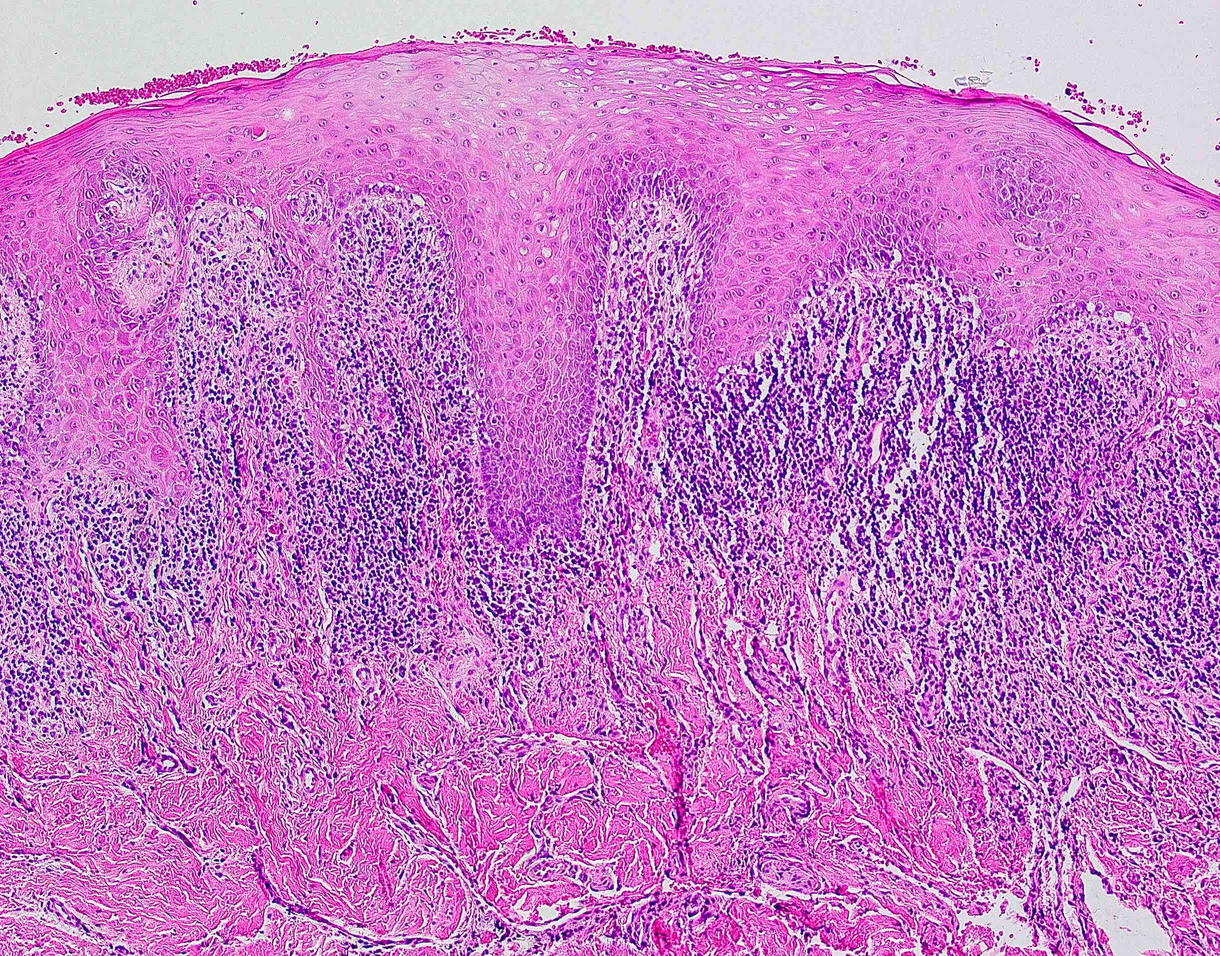
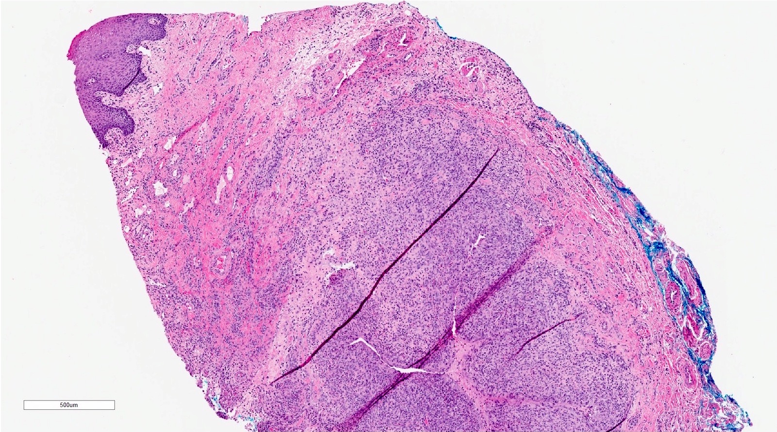
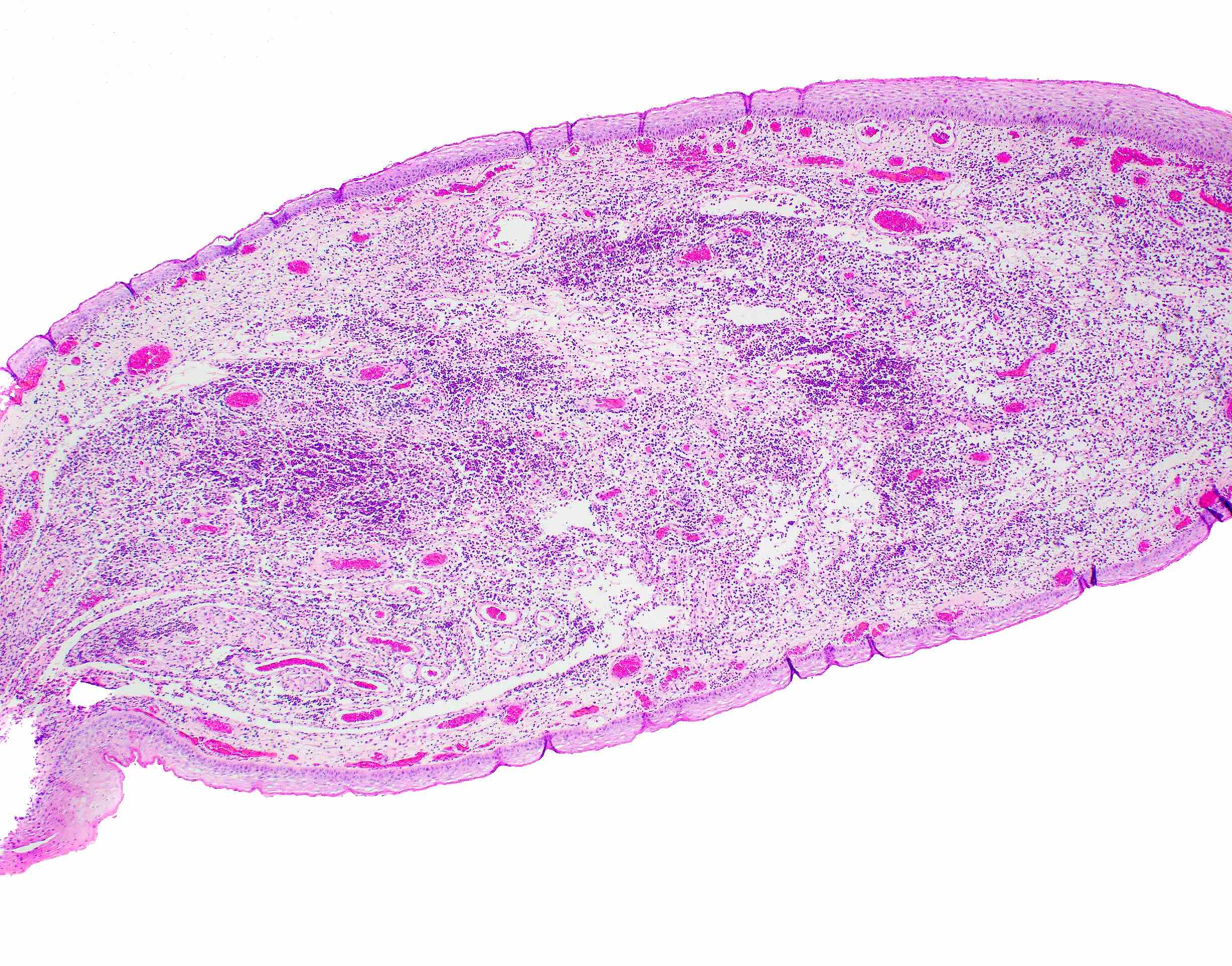
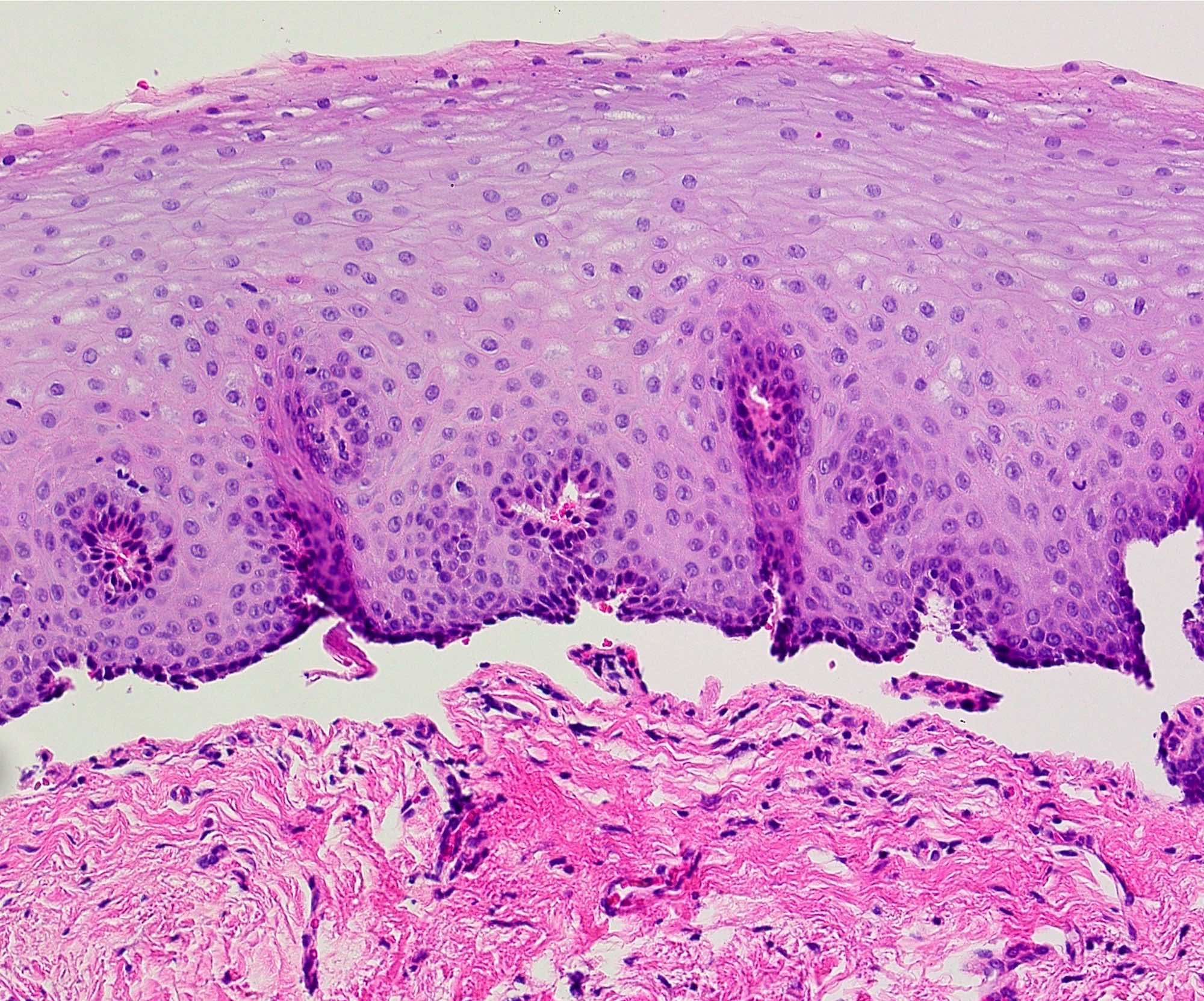
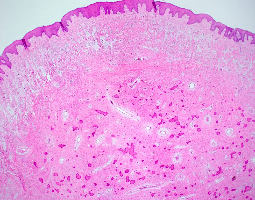
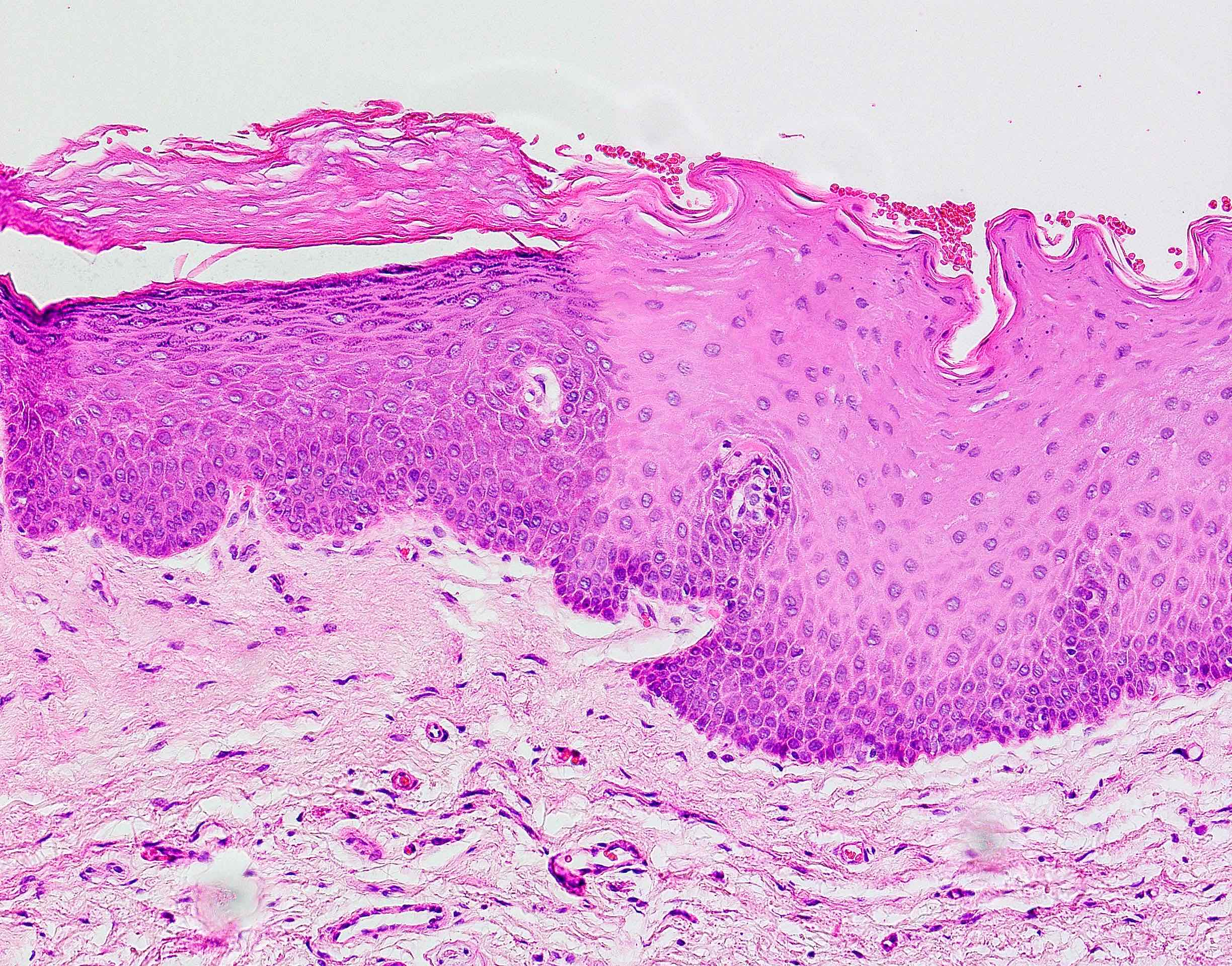
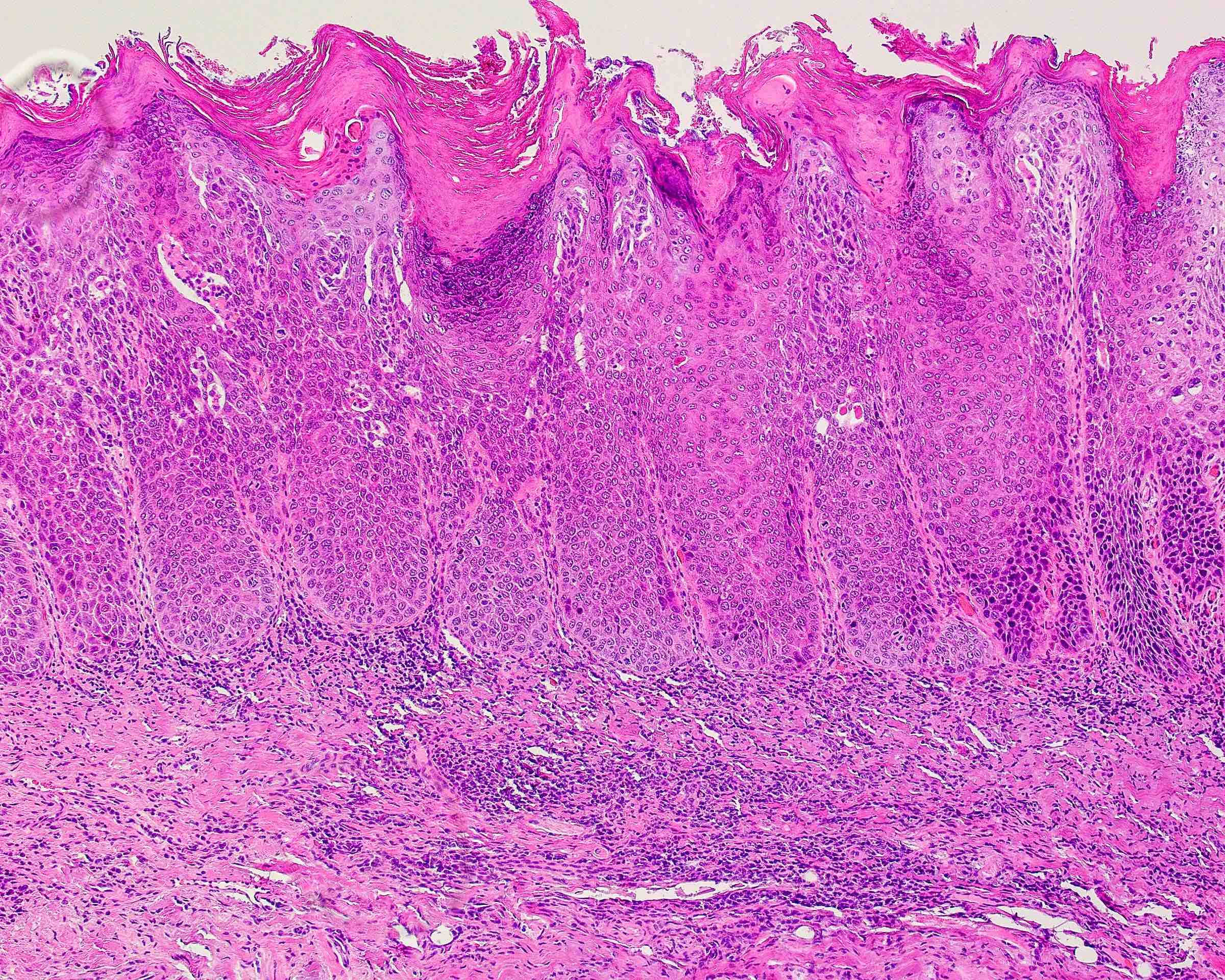
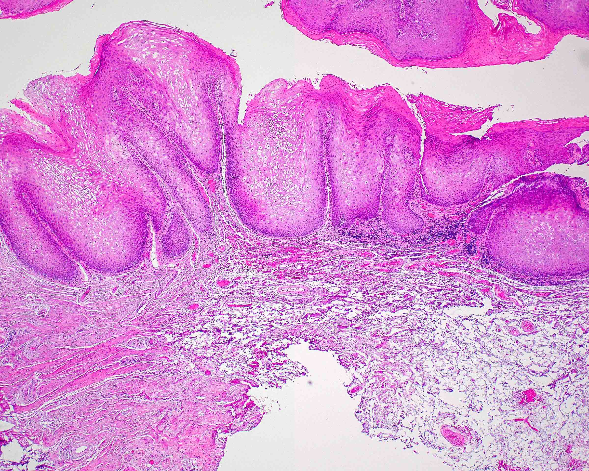
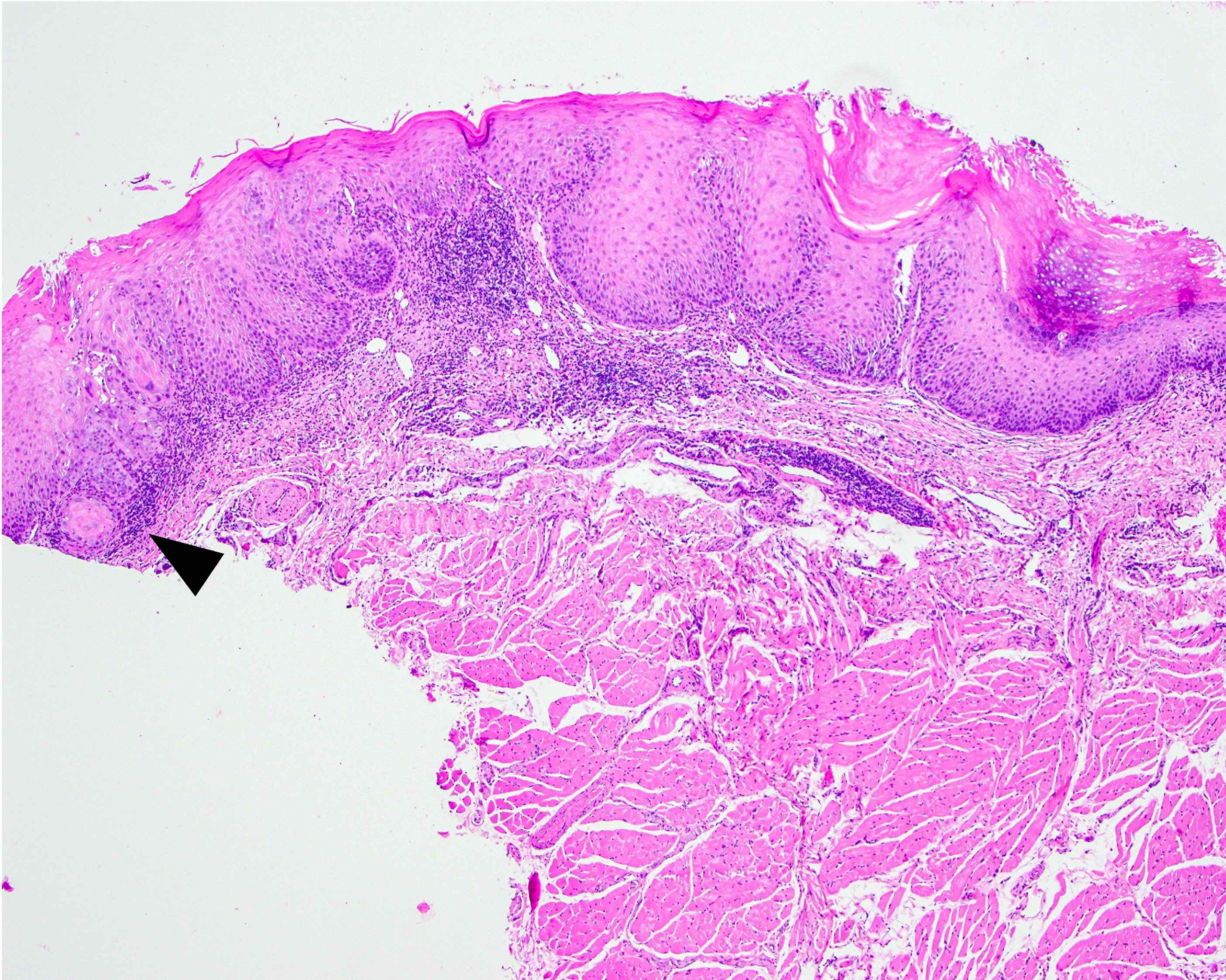
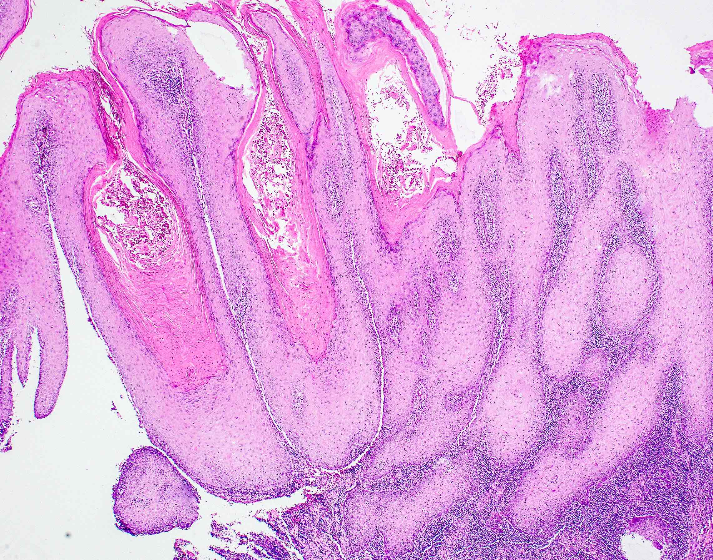
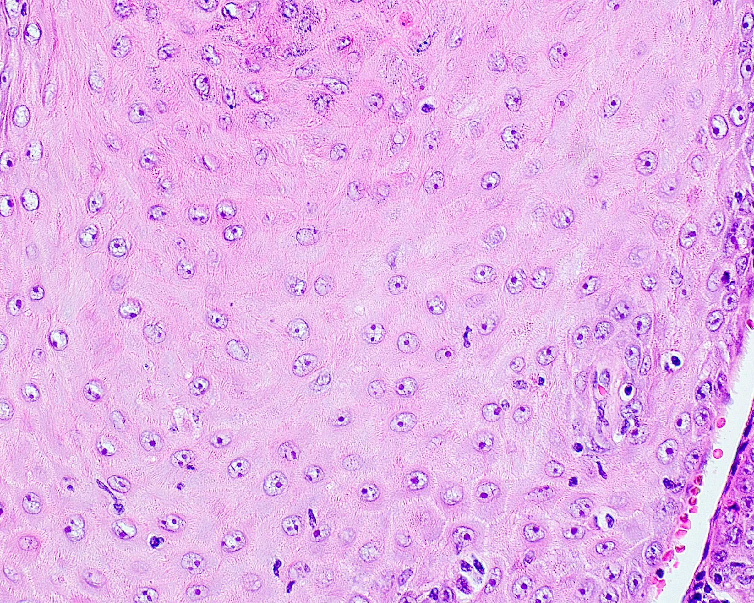
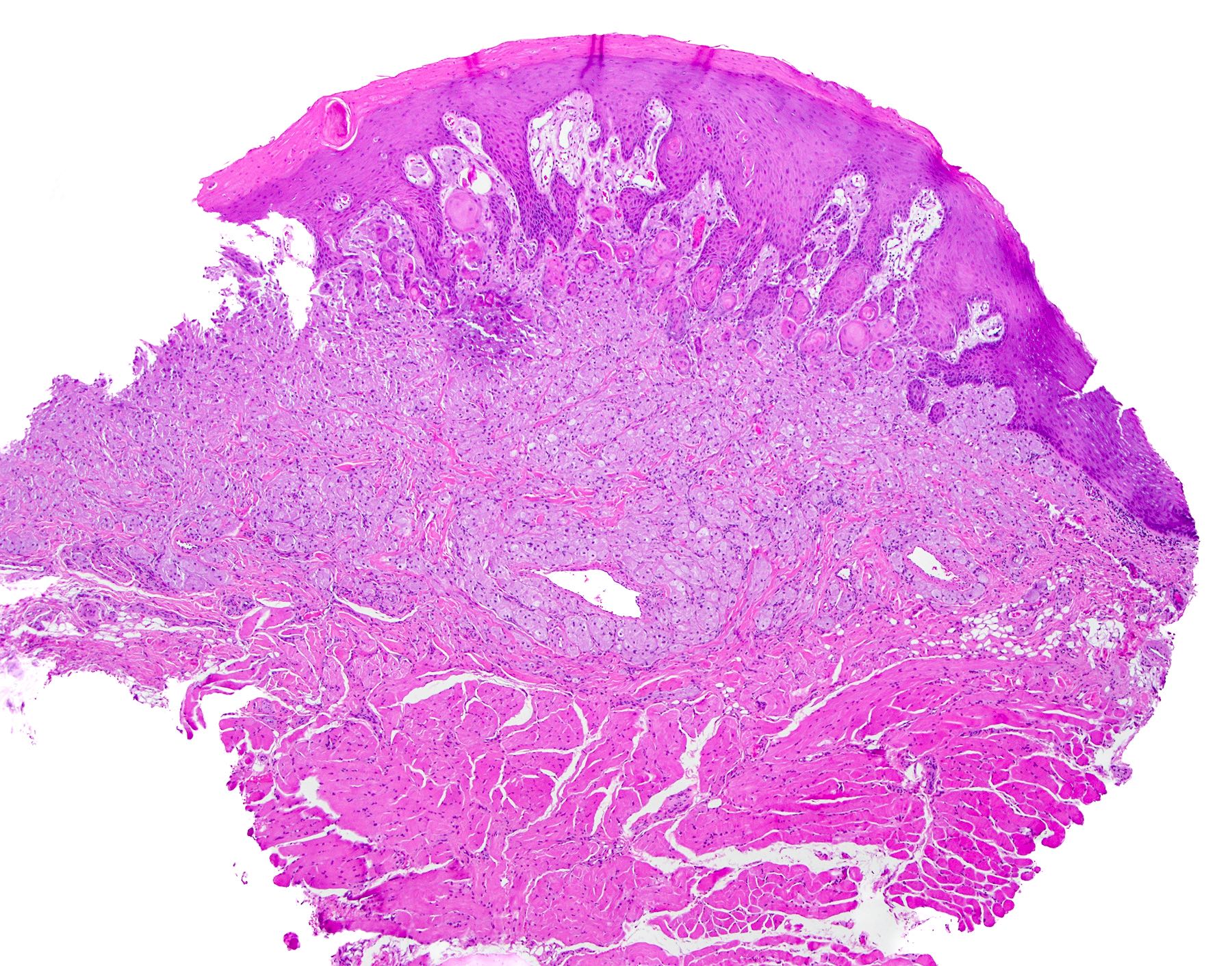
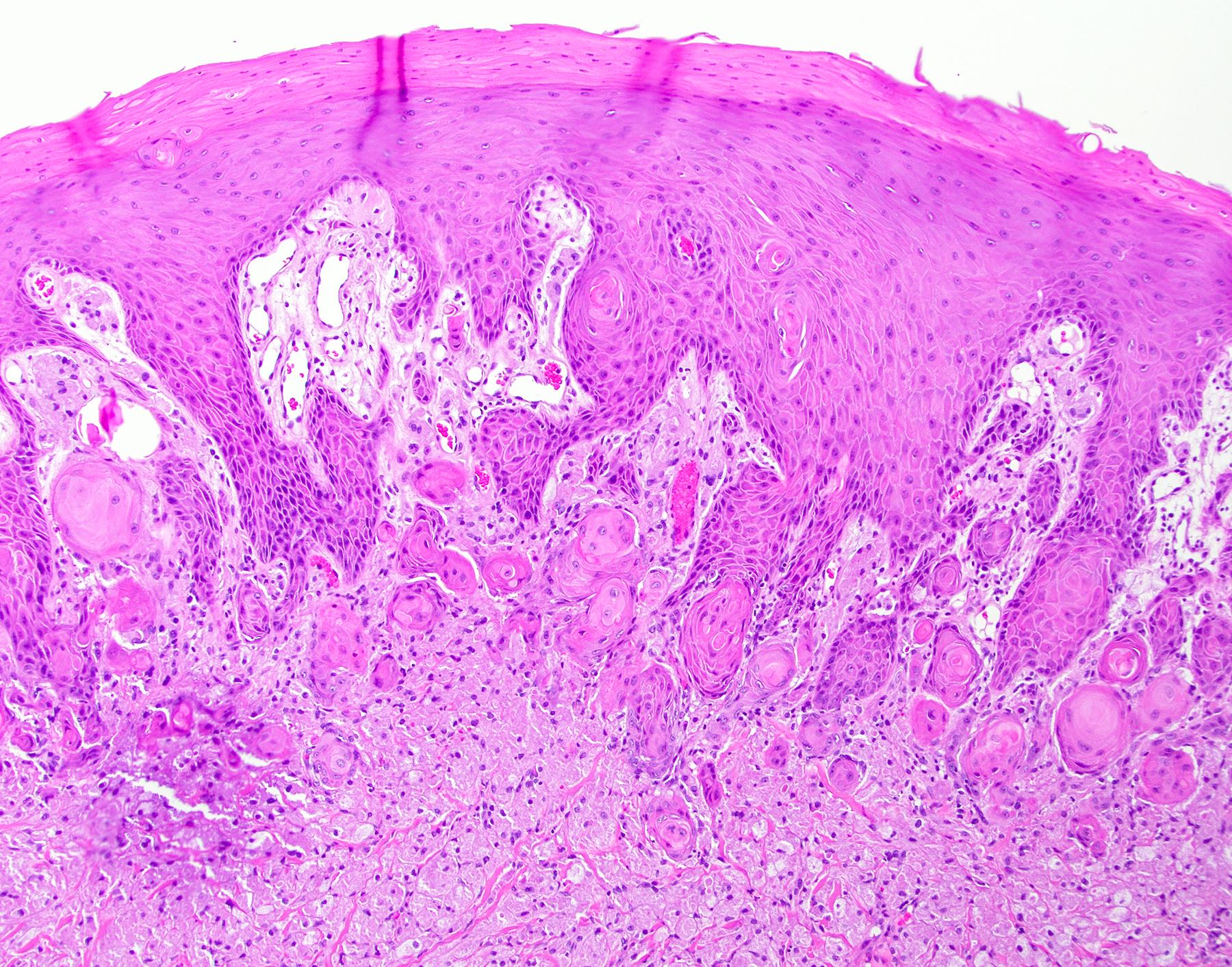
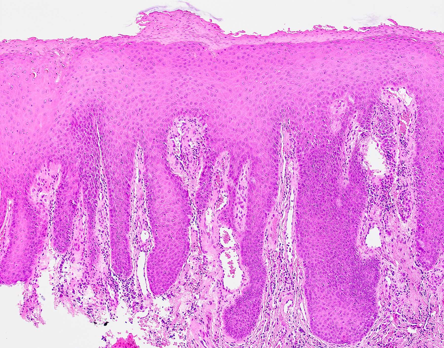
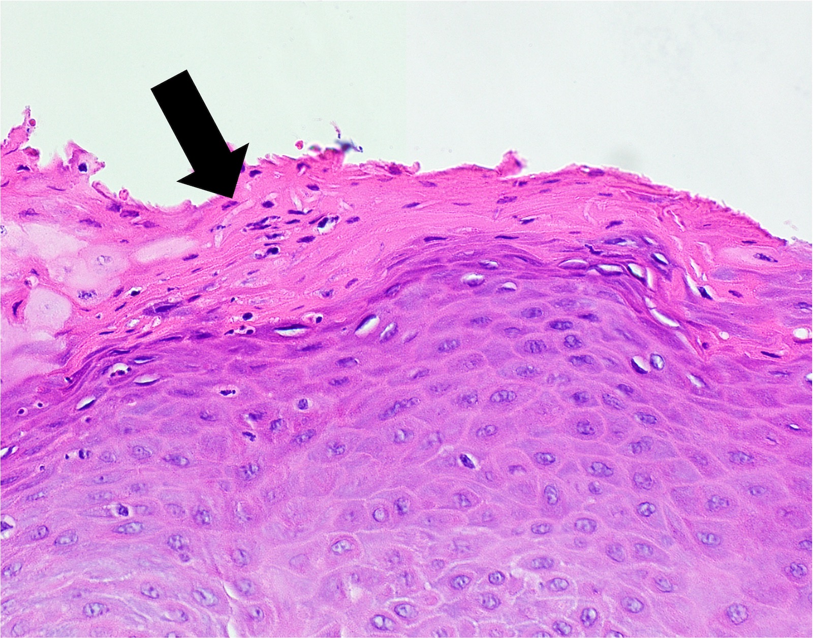
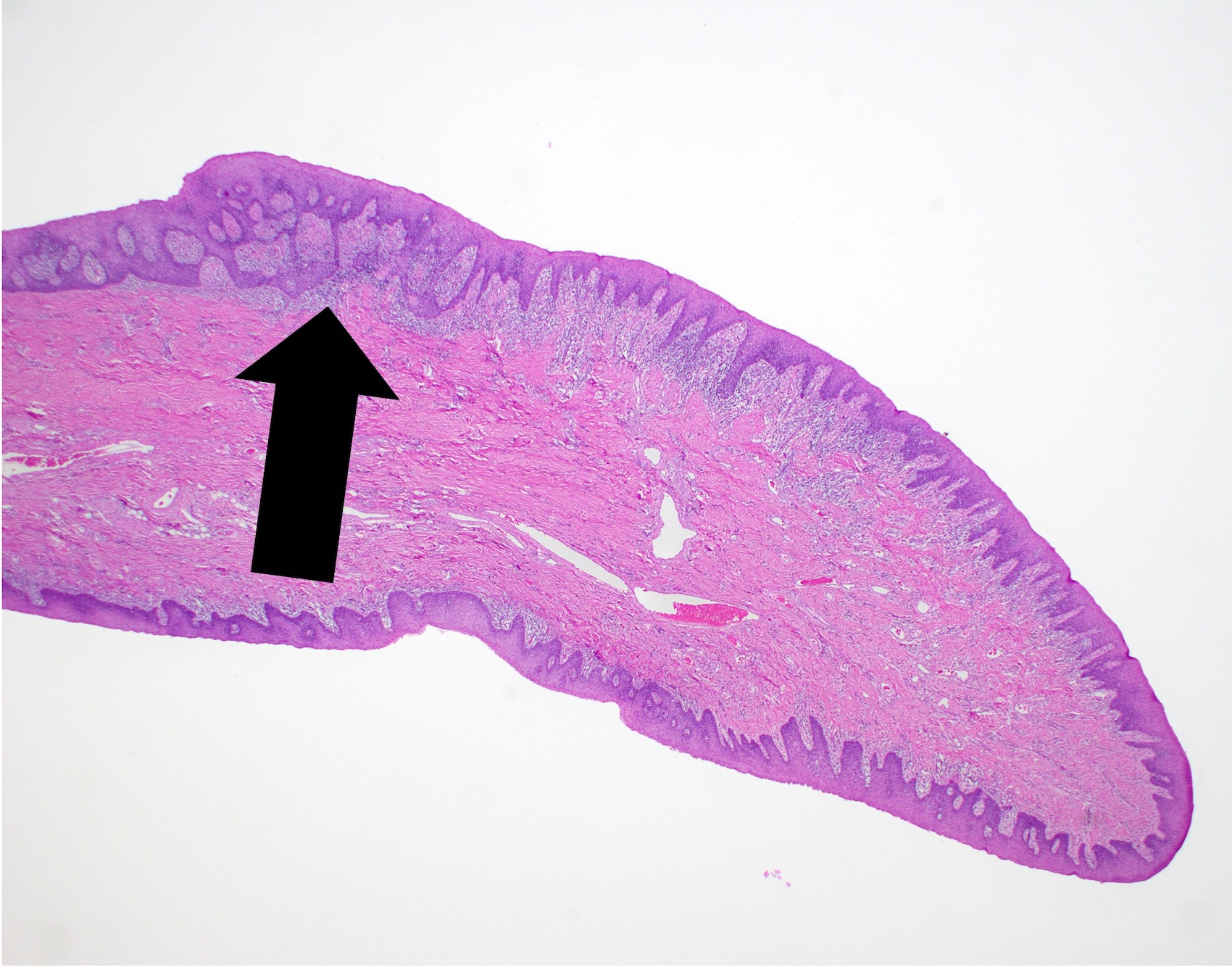
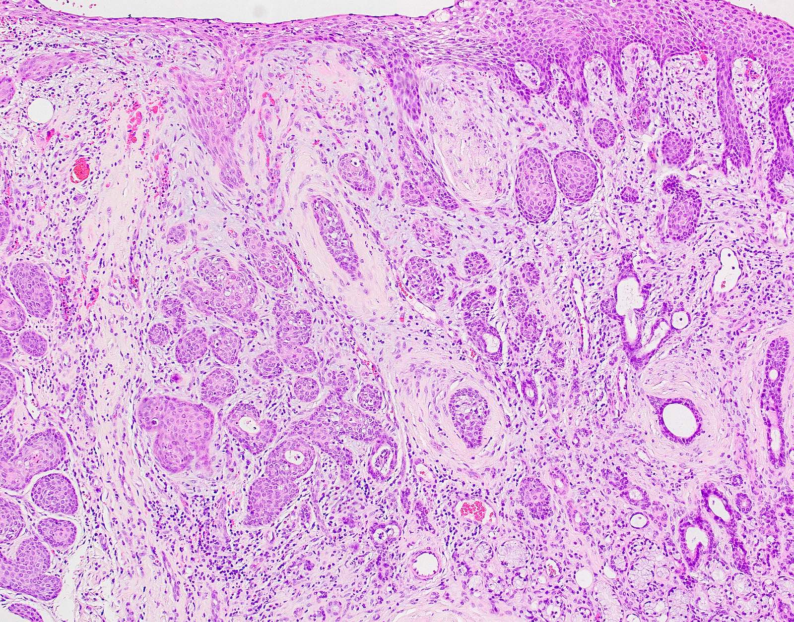
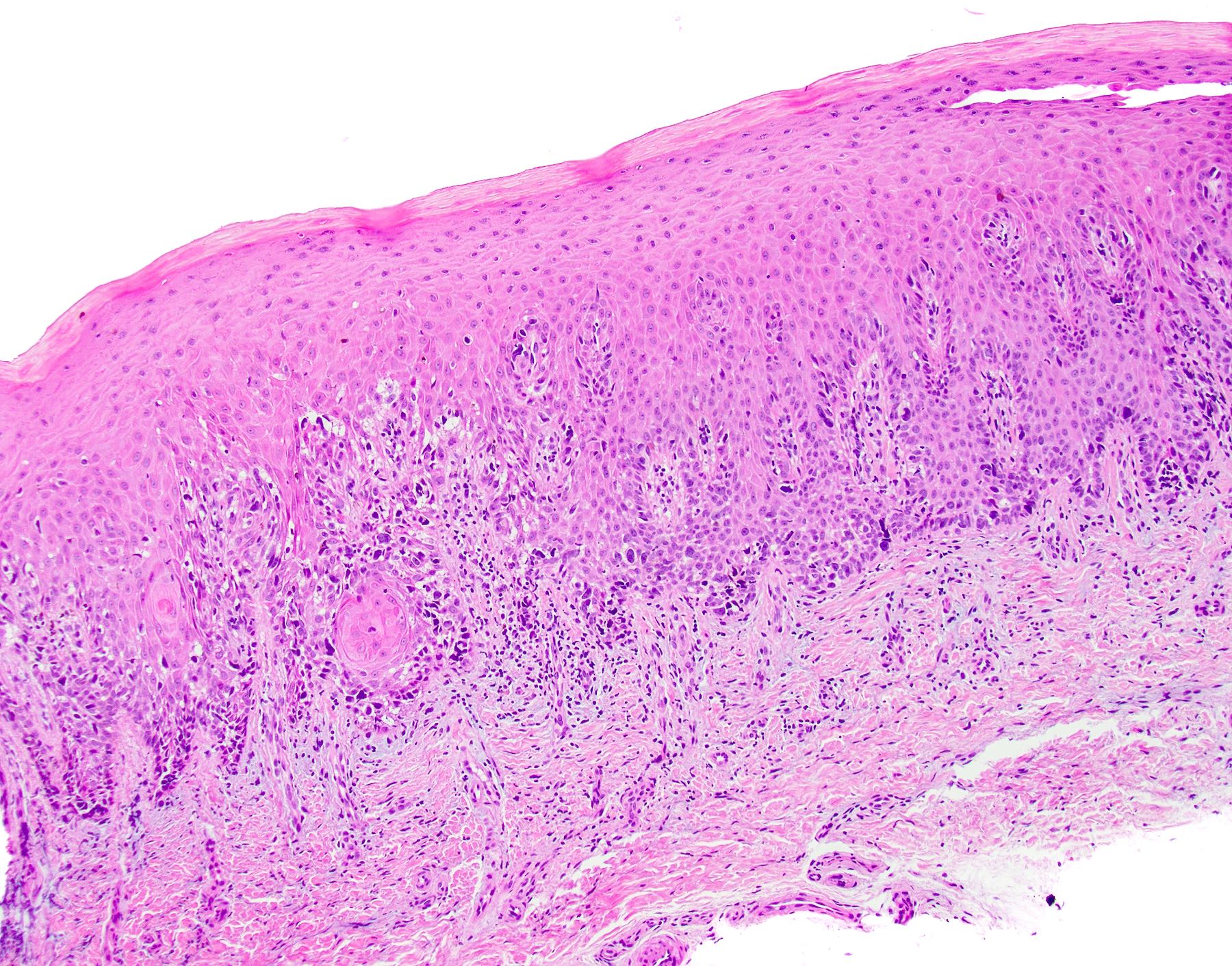
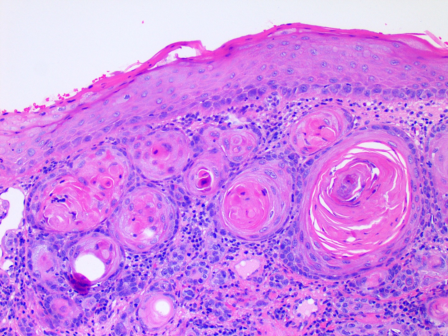
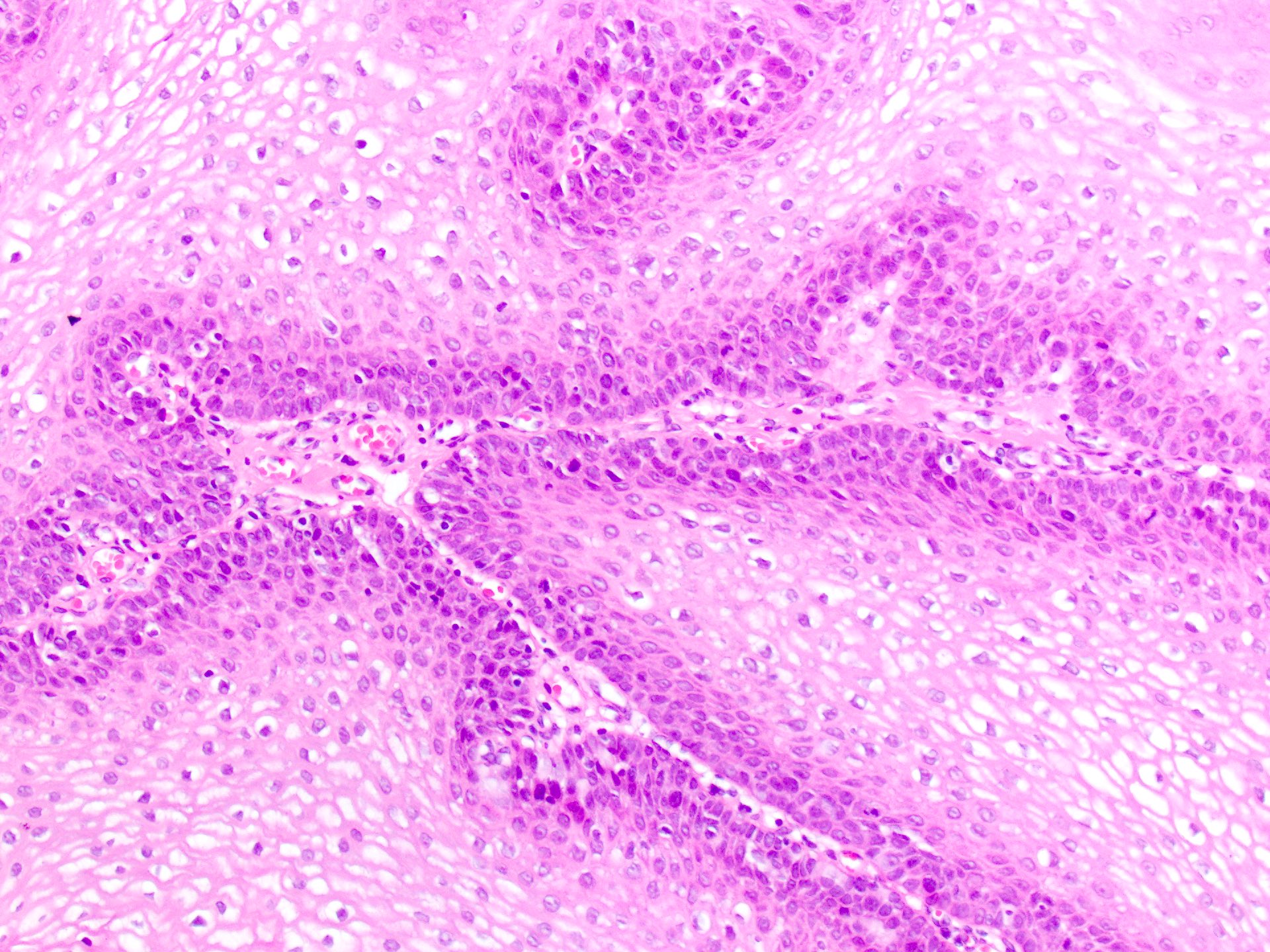
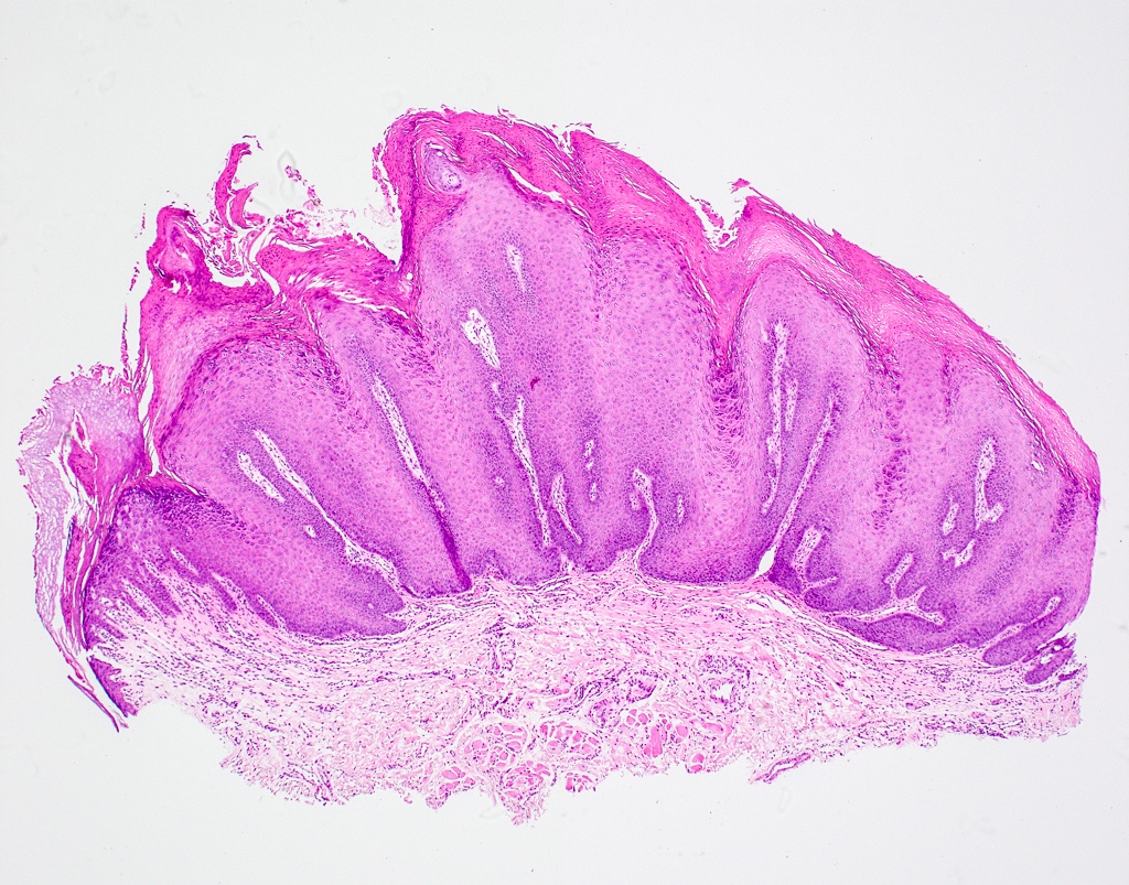
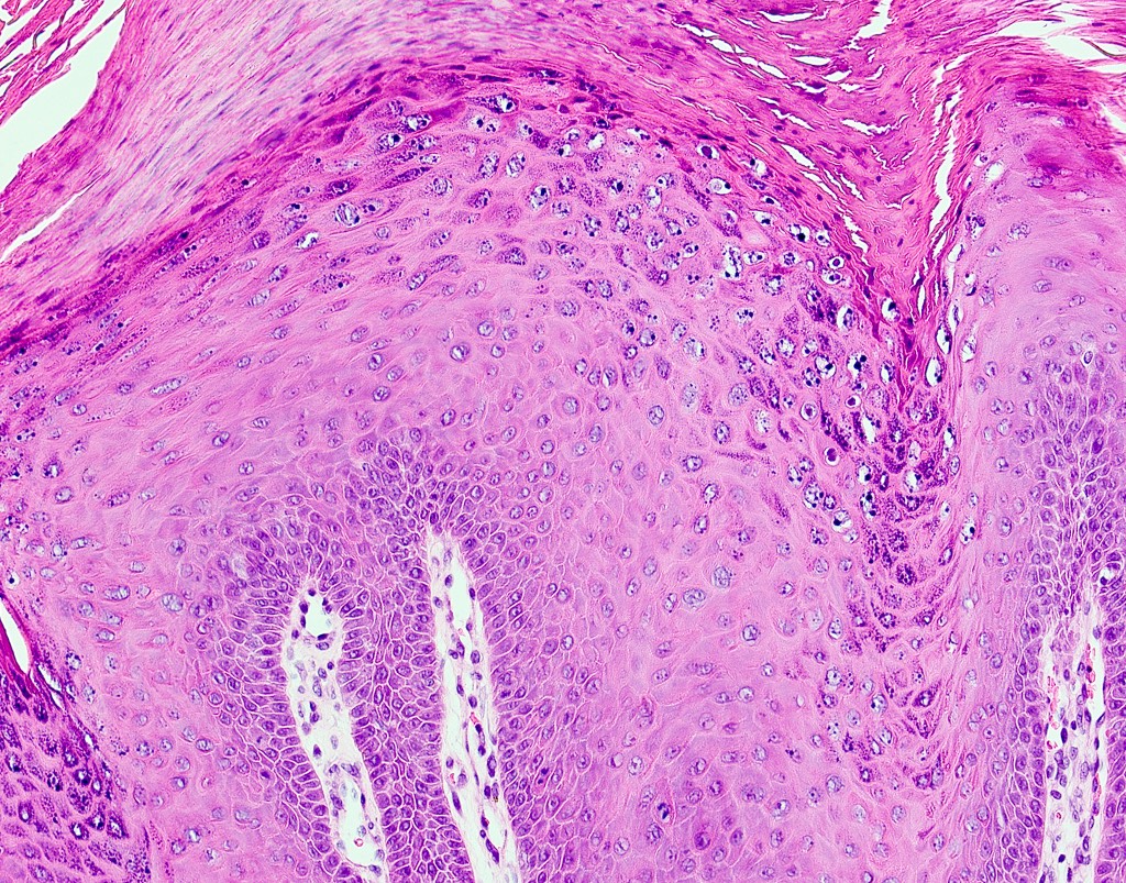
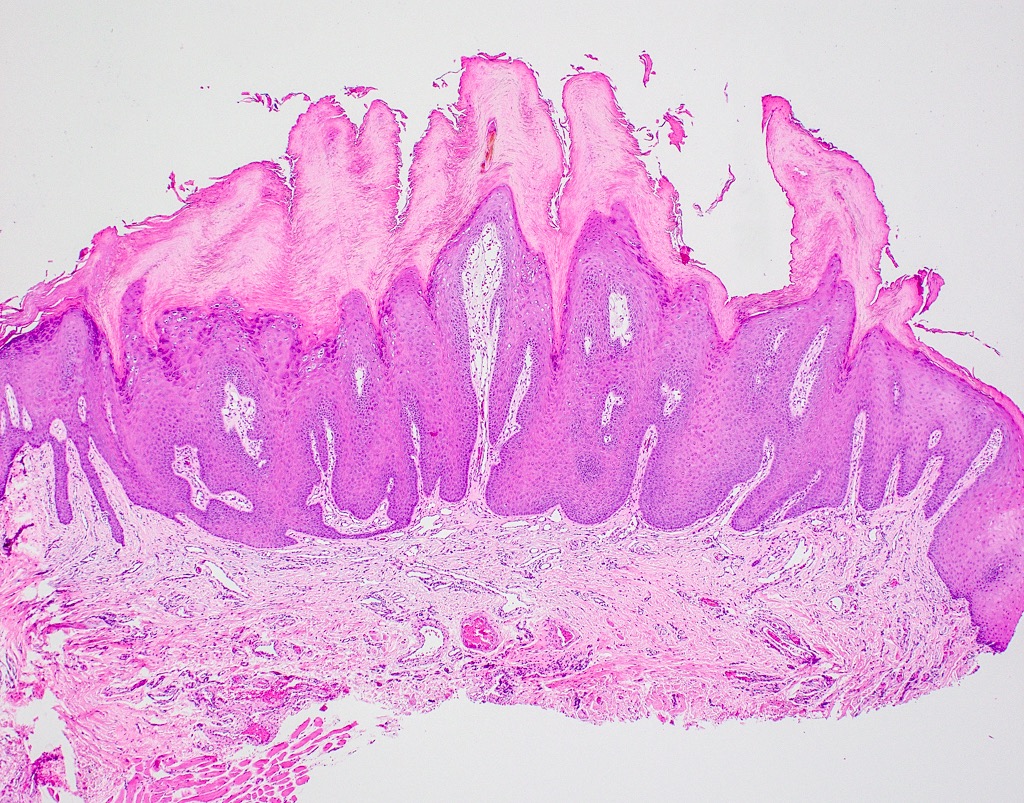
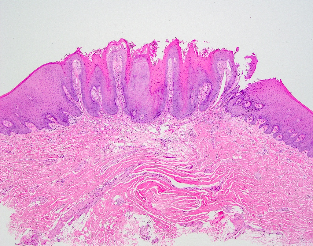
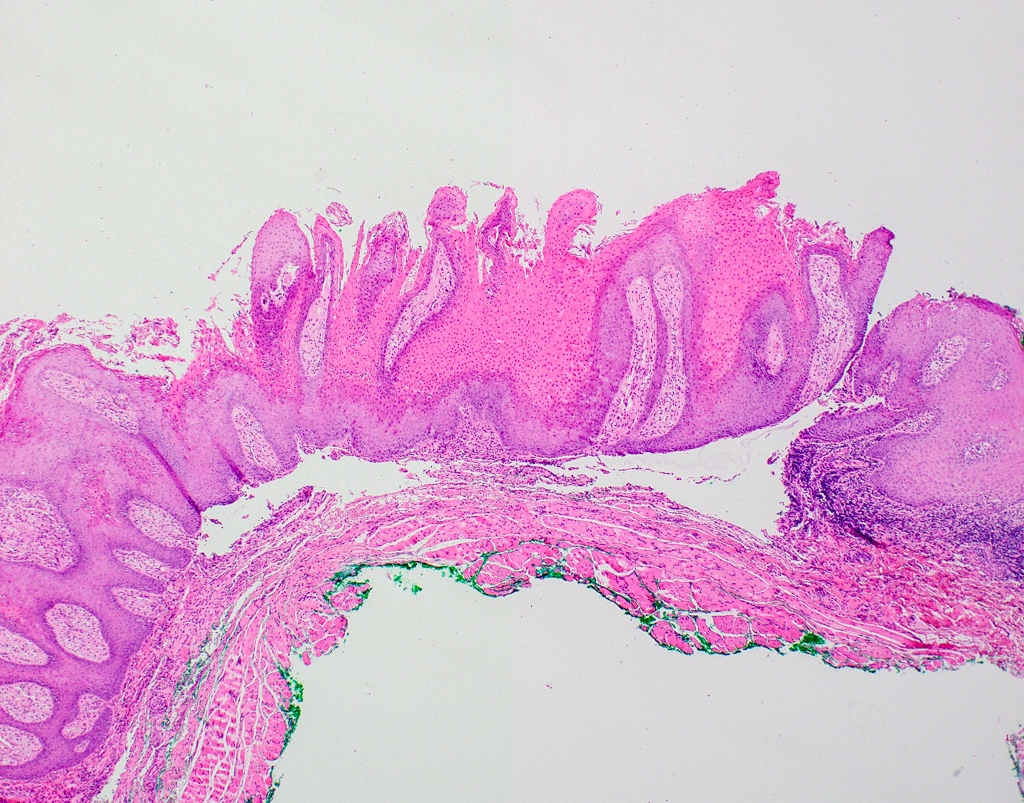
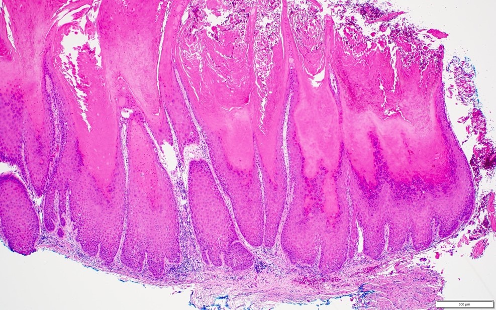
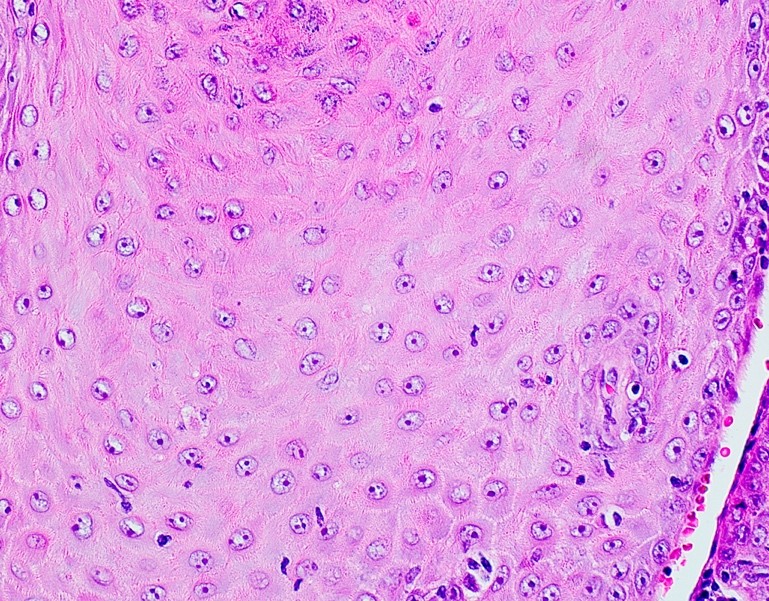
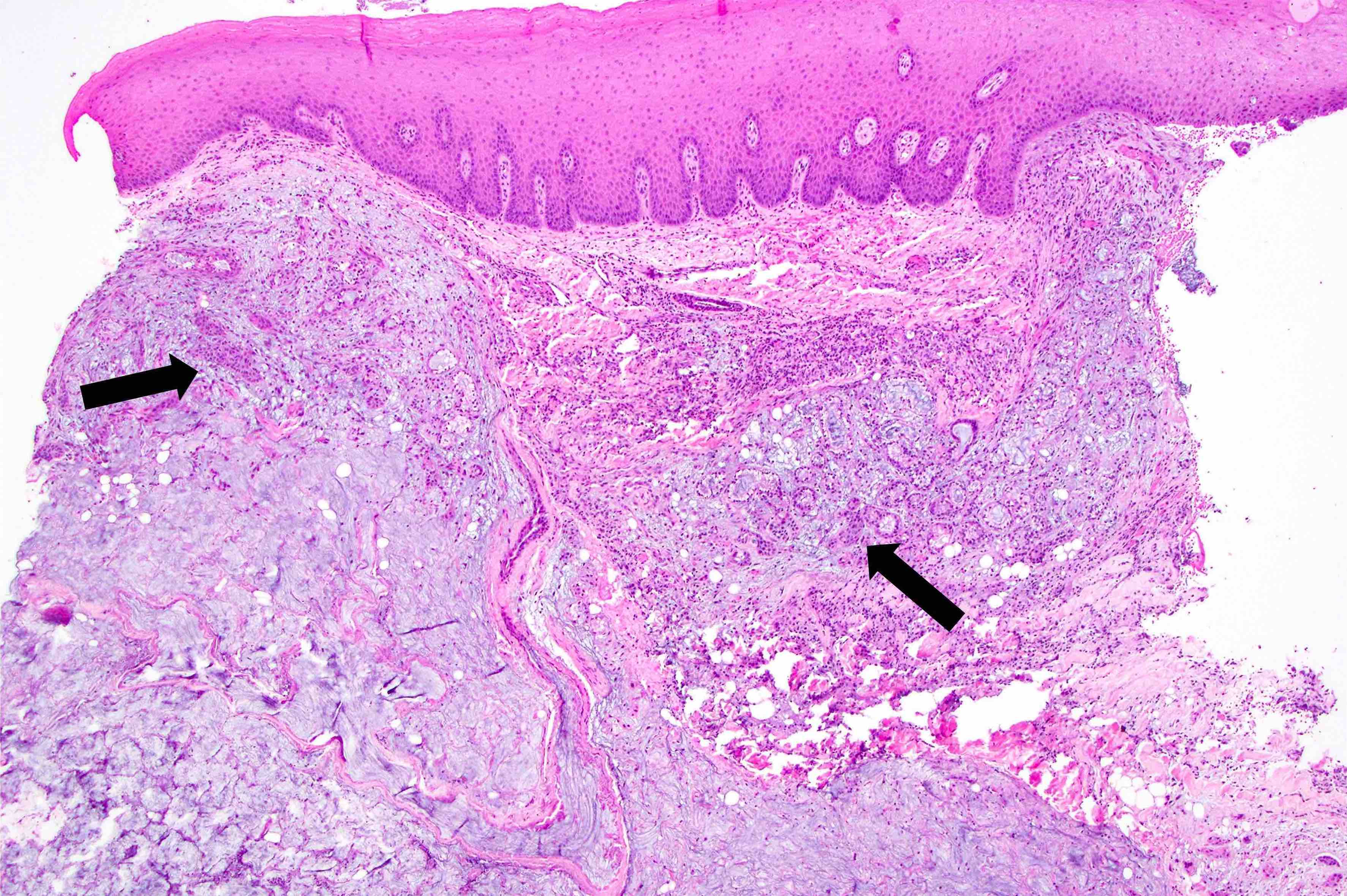
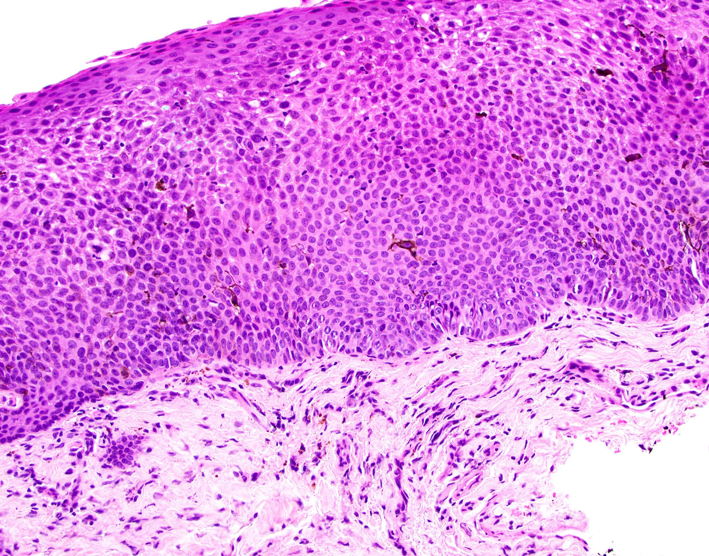
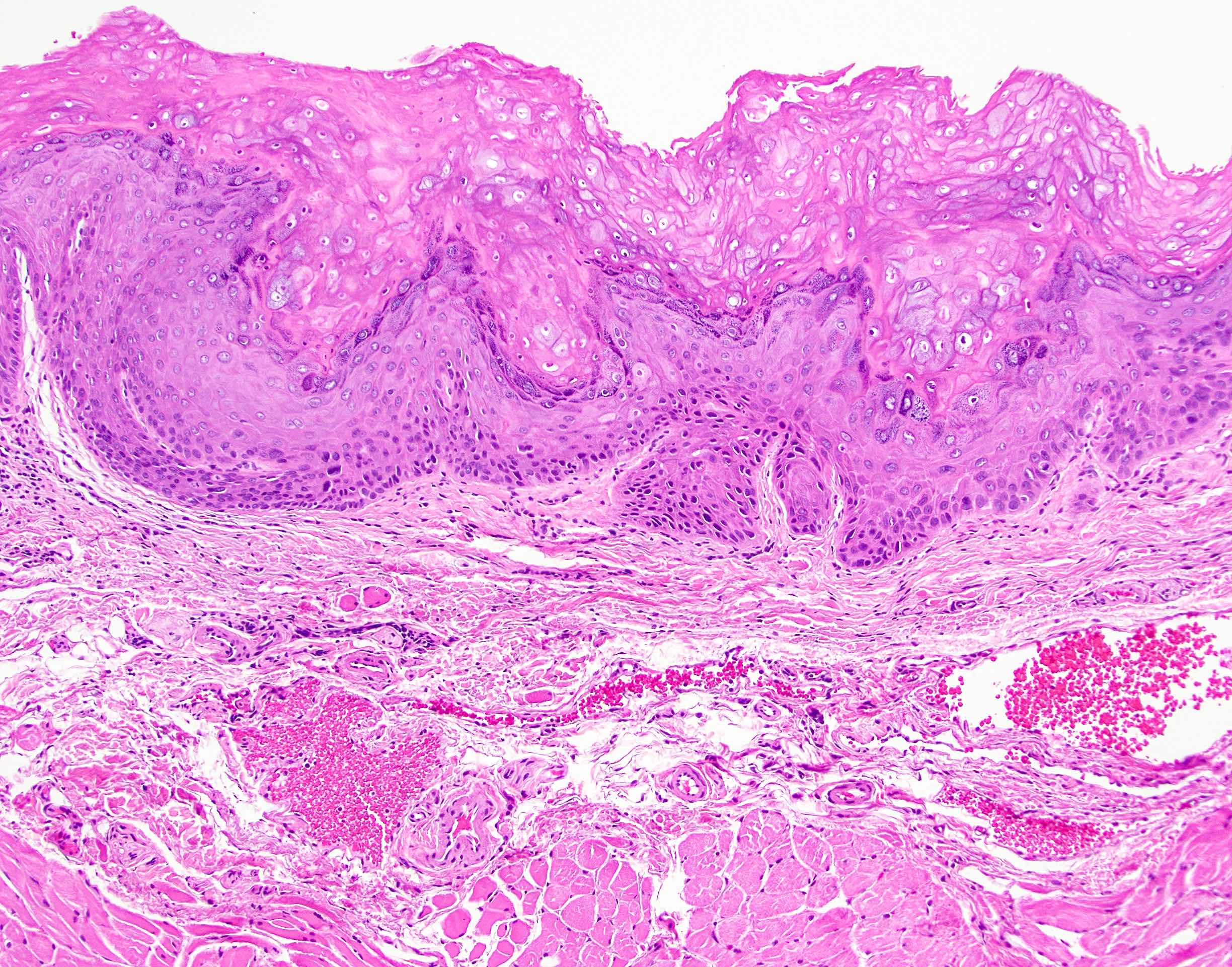
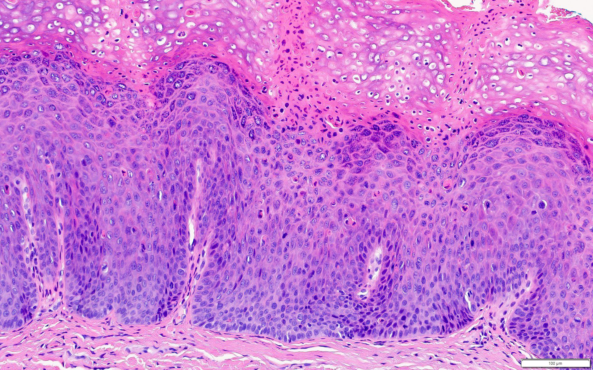
Hyperkeratotic, dysplastic epithelium
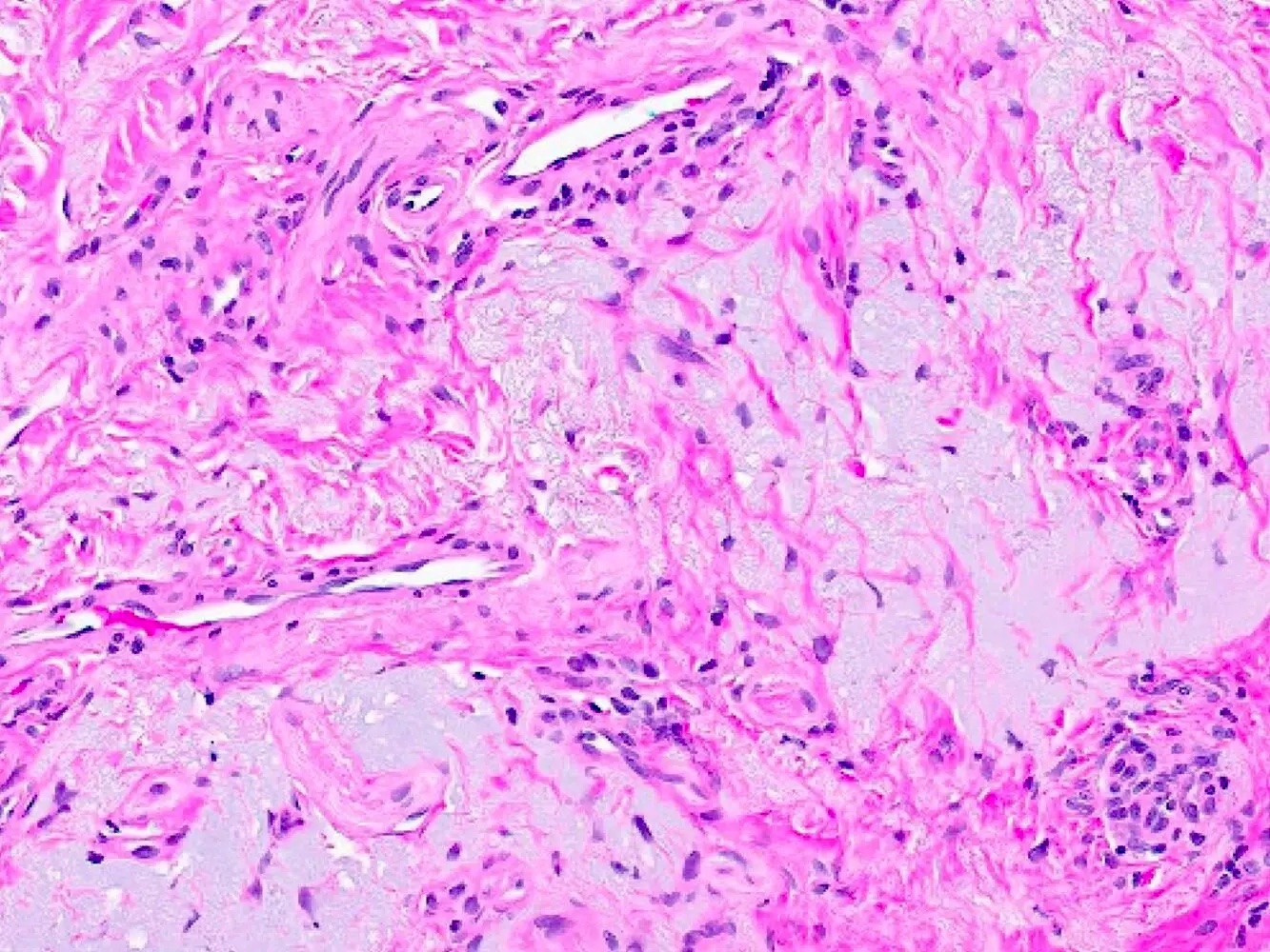
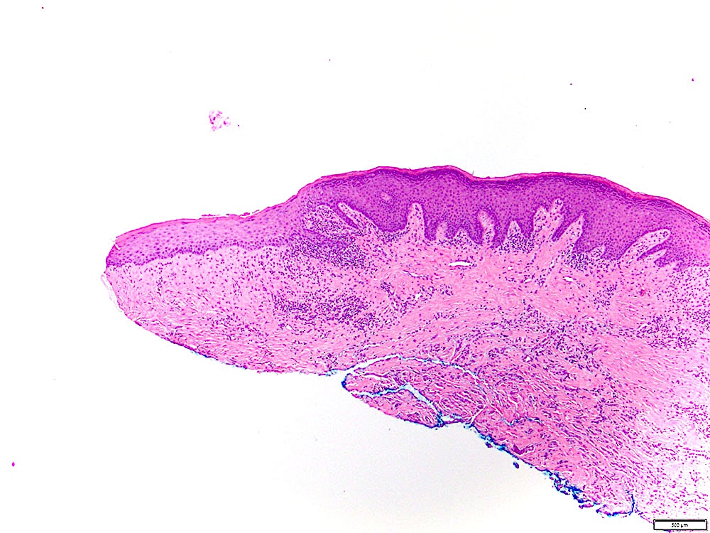
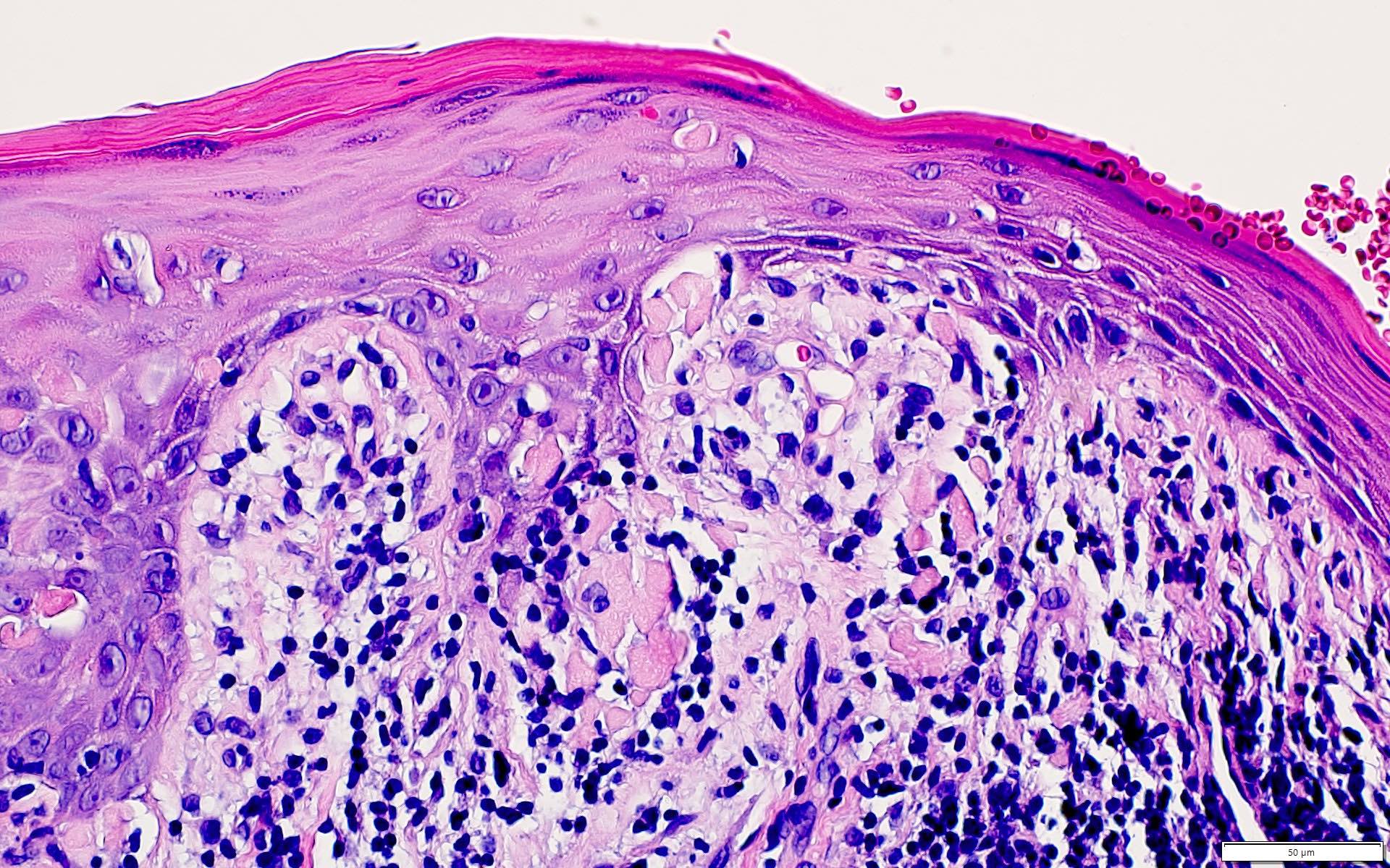
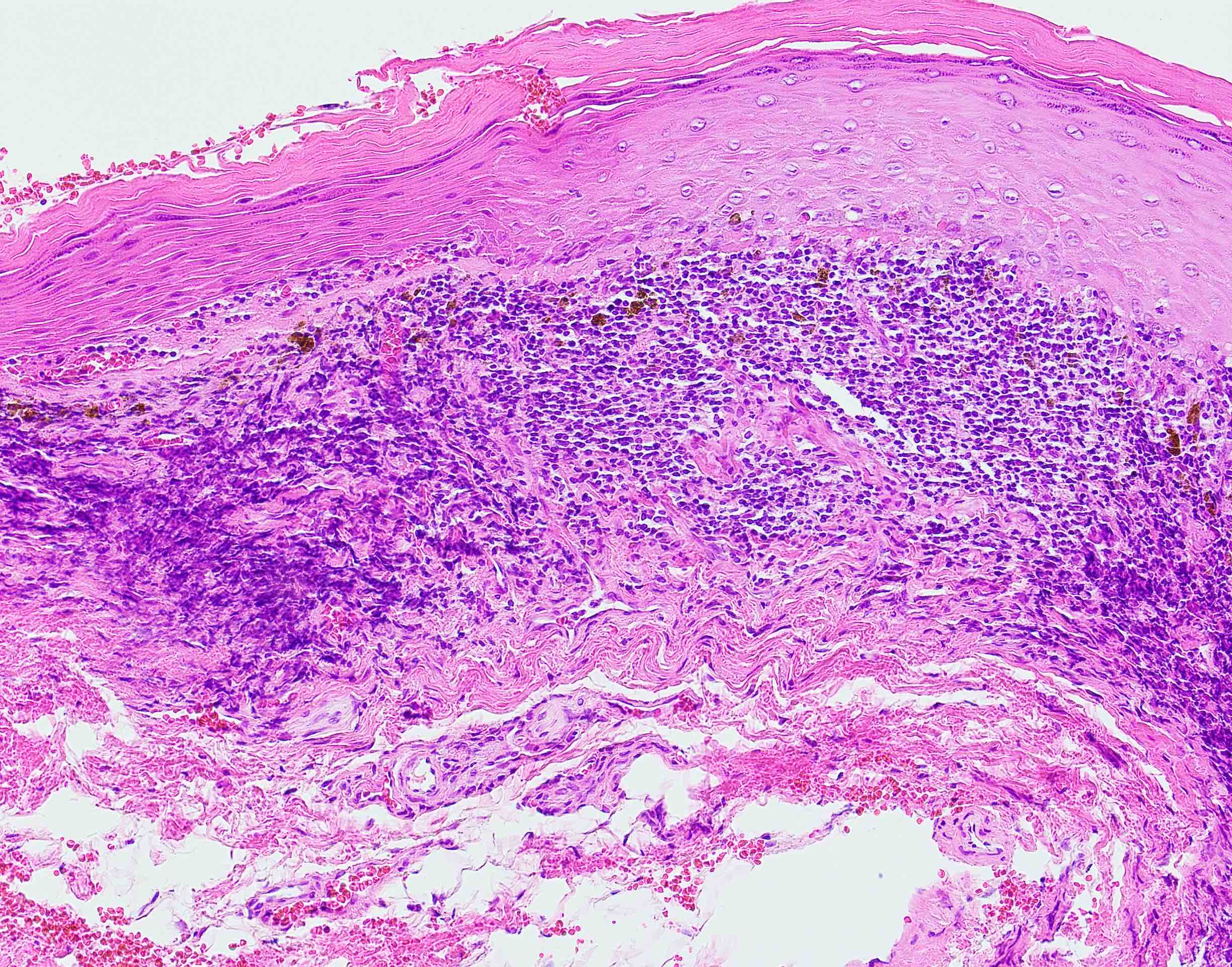
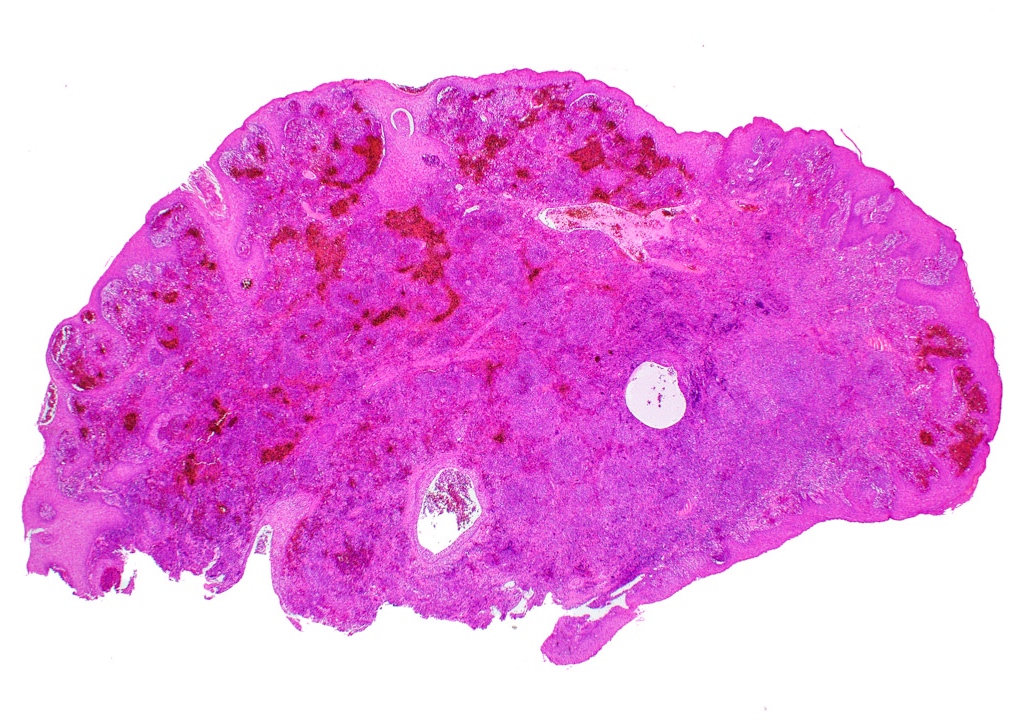
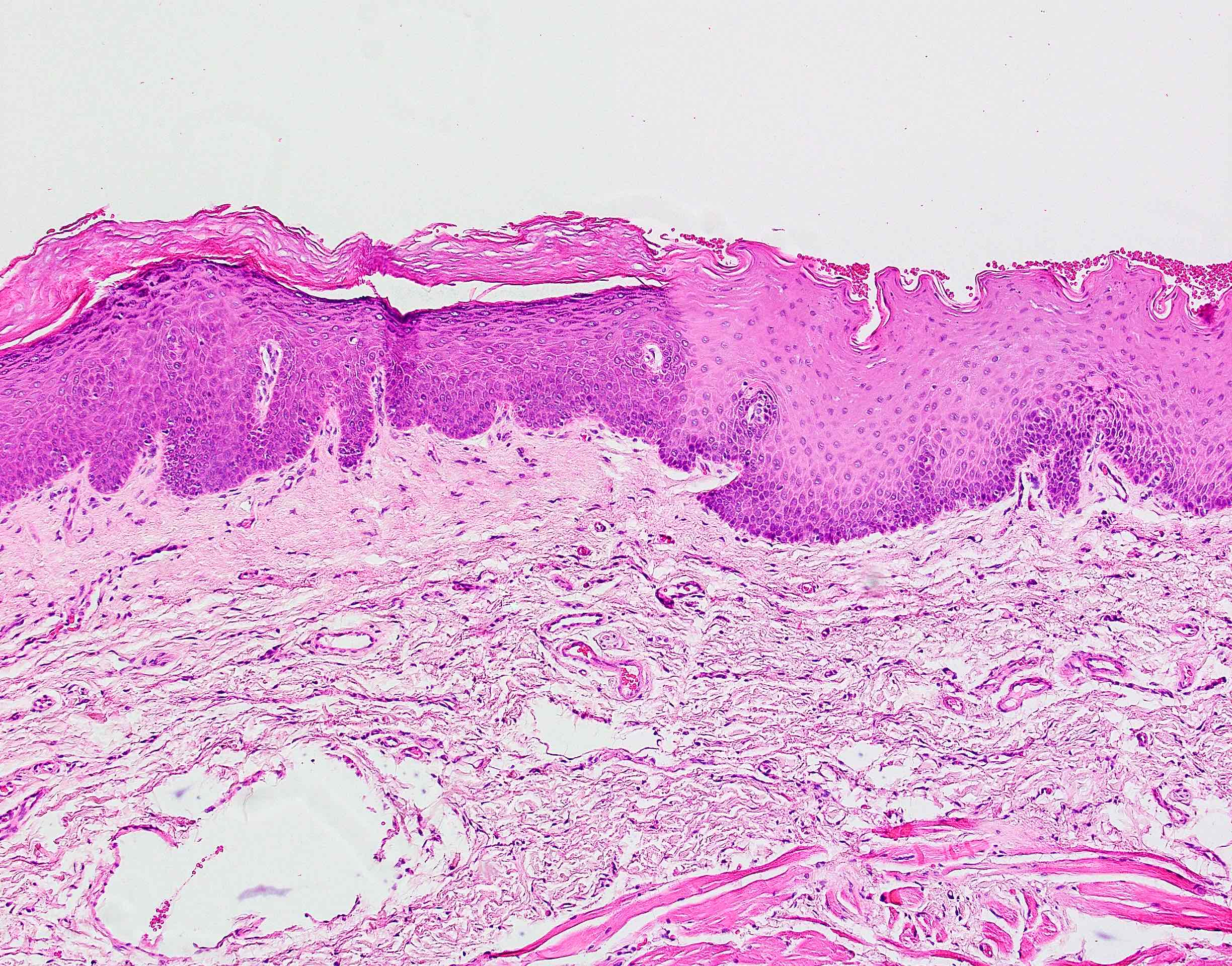
Prominent solar elastosis and telangiectasia
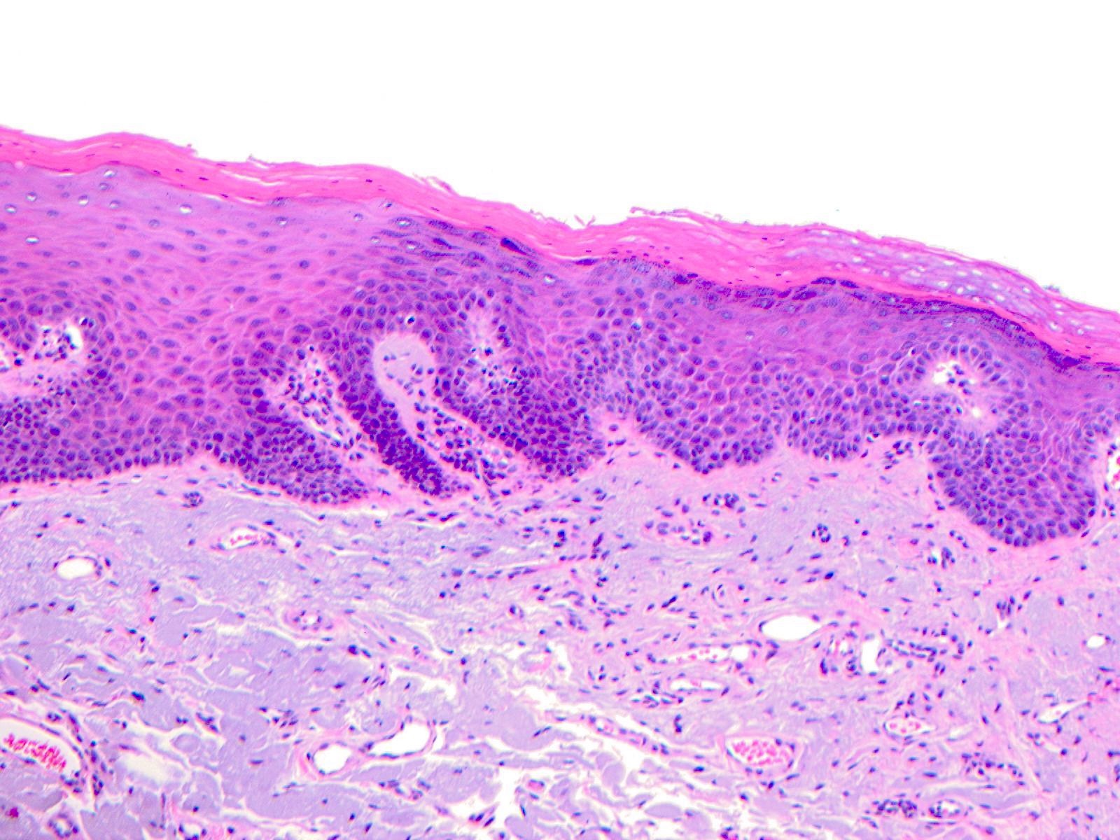
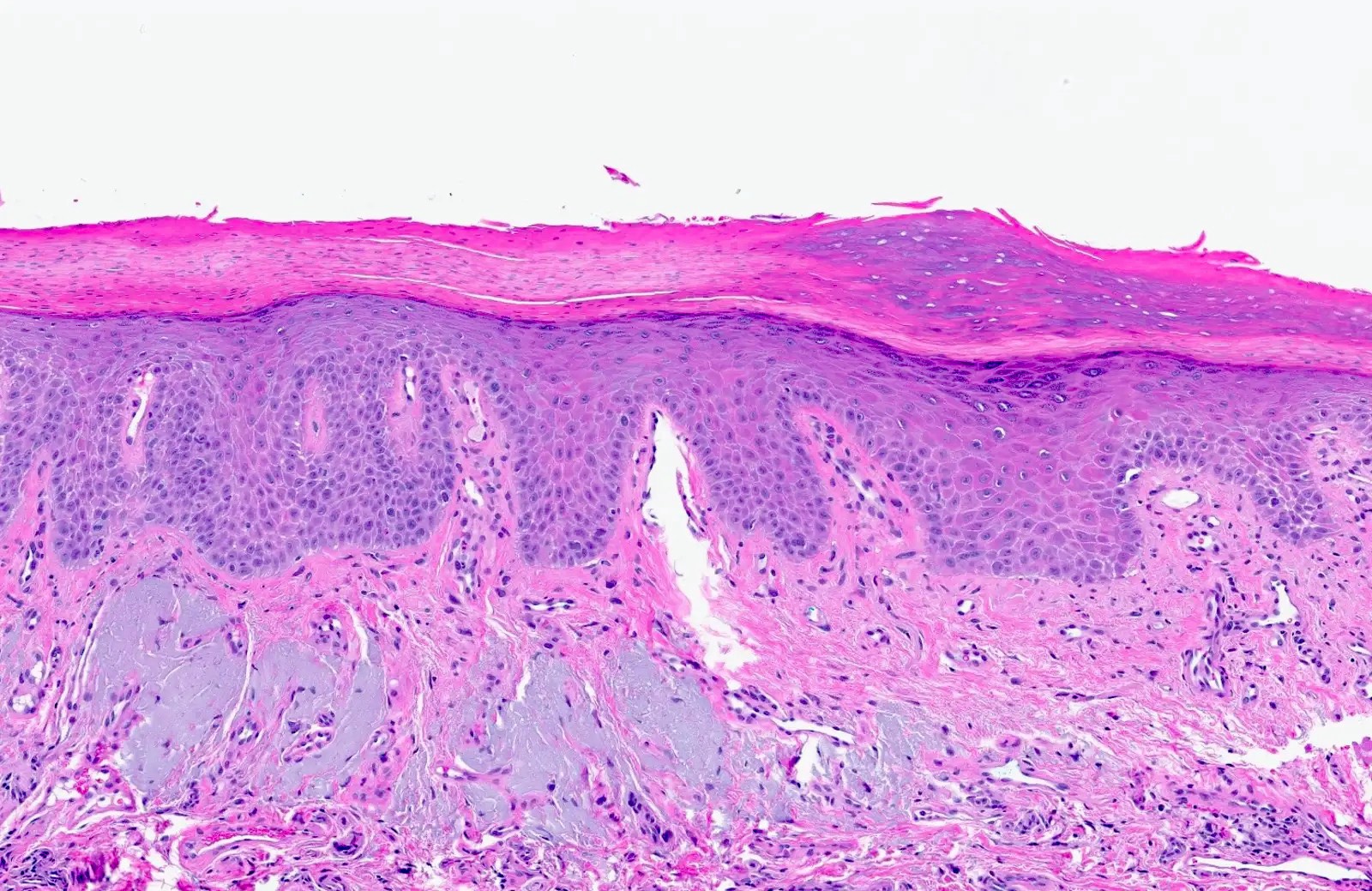
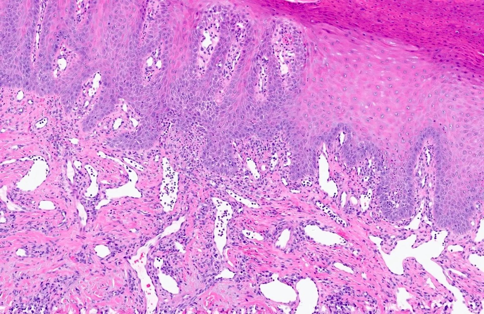
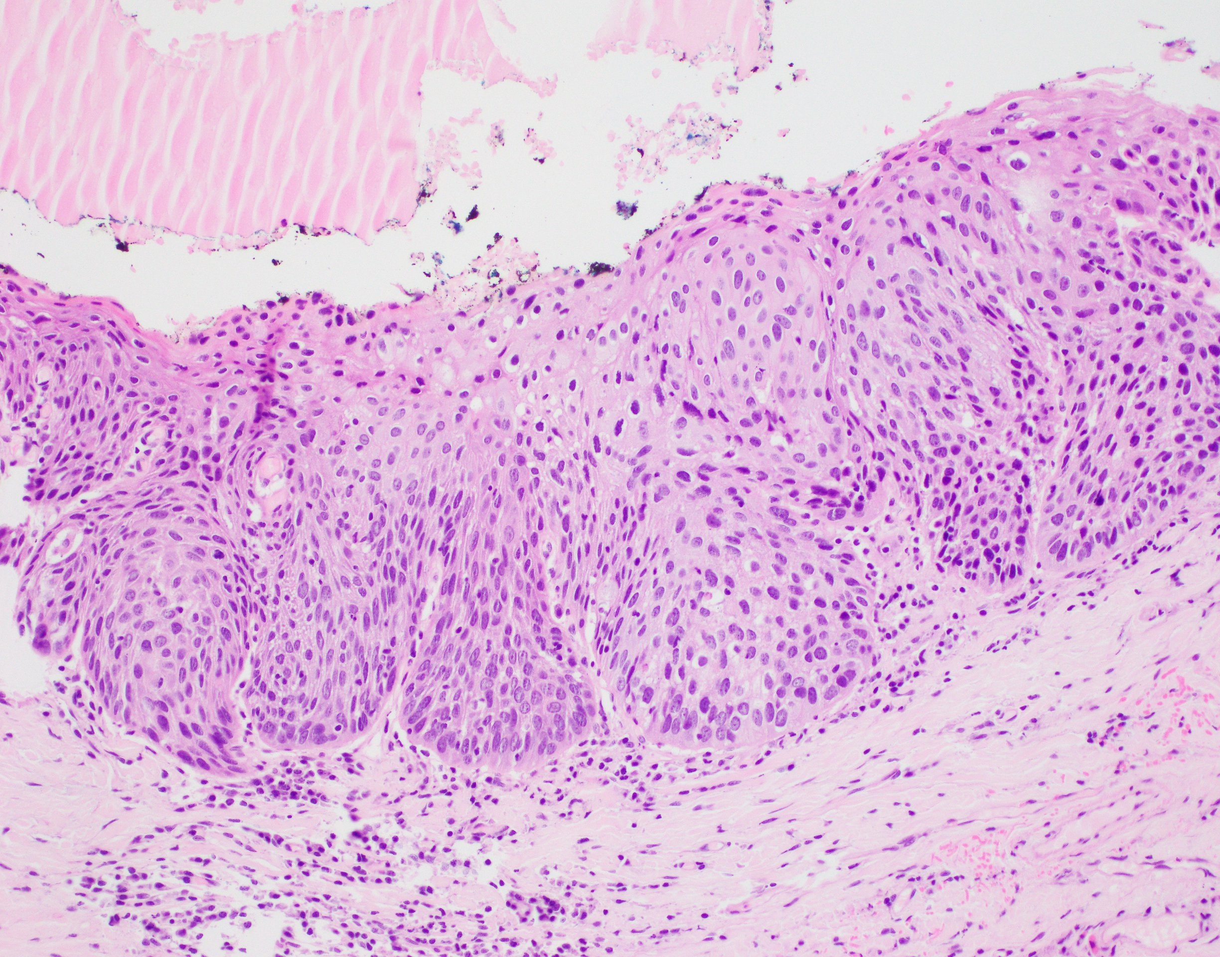
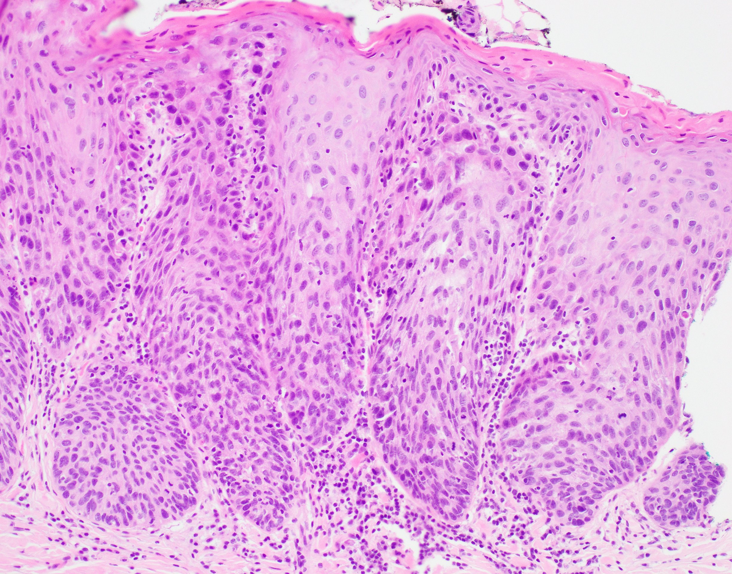
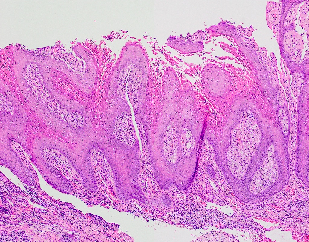
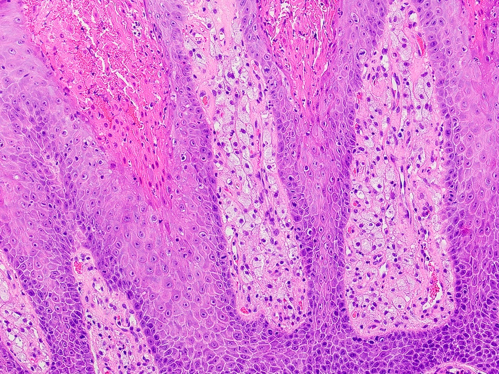
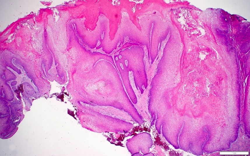
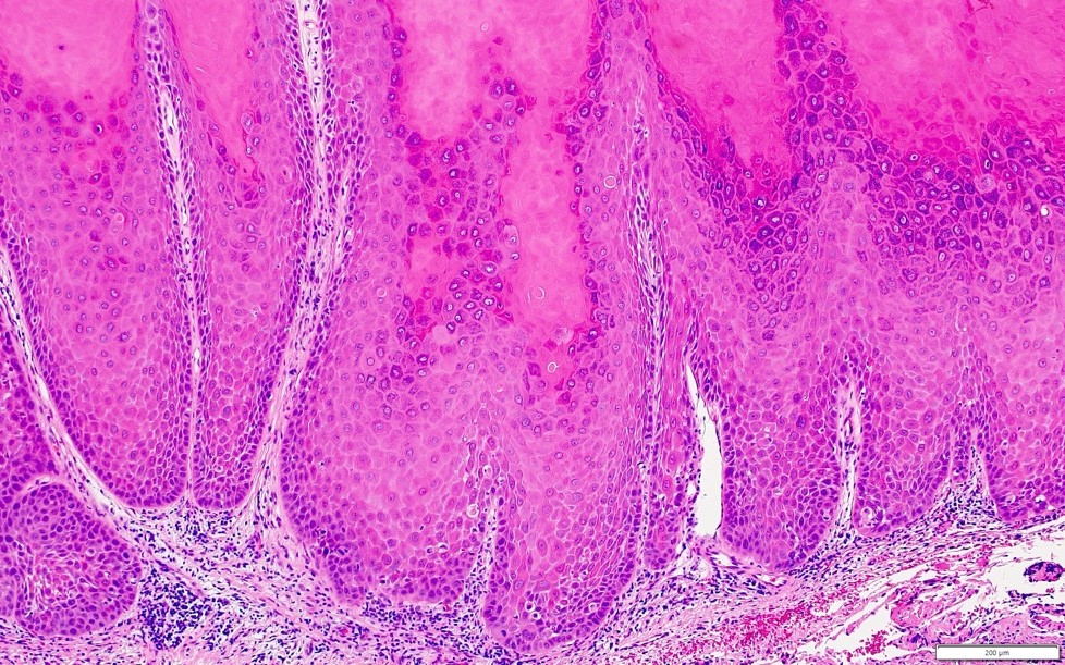
Epithelium with dysplastic changes
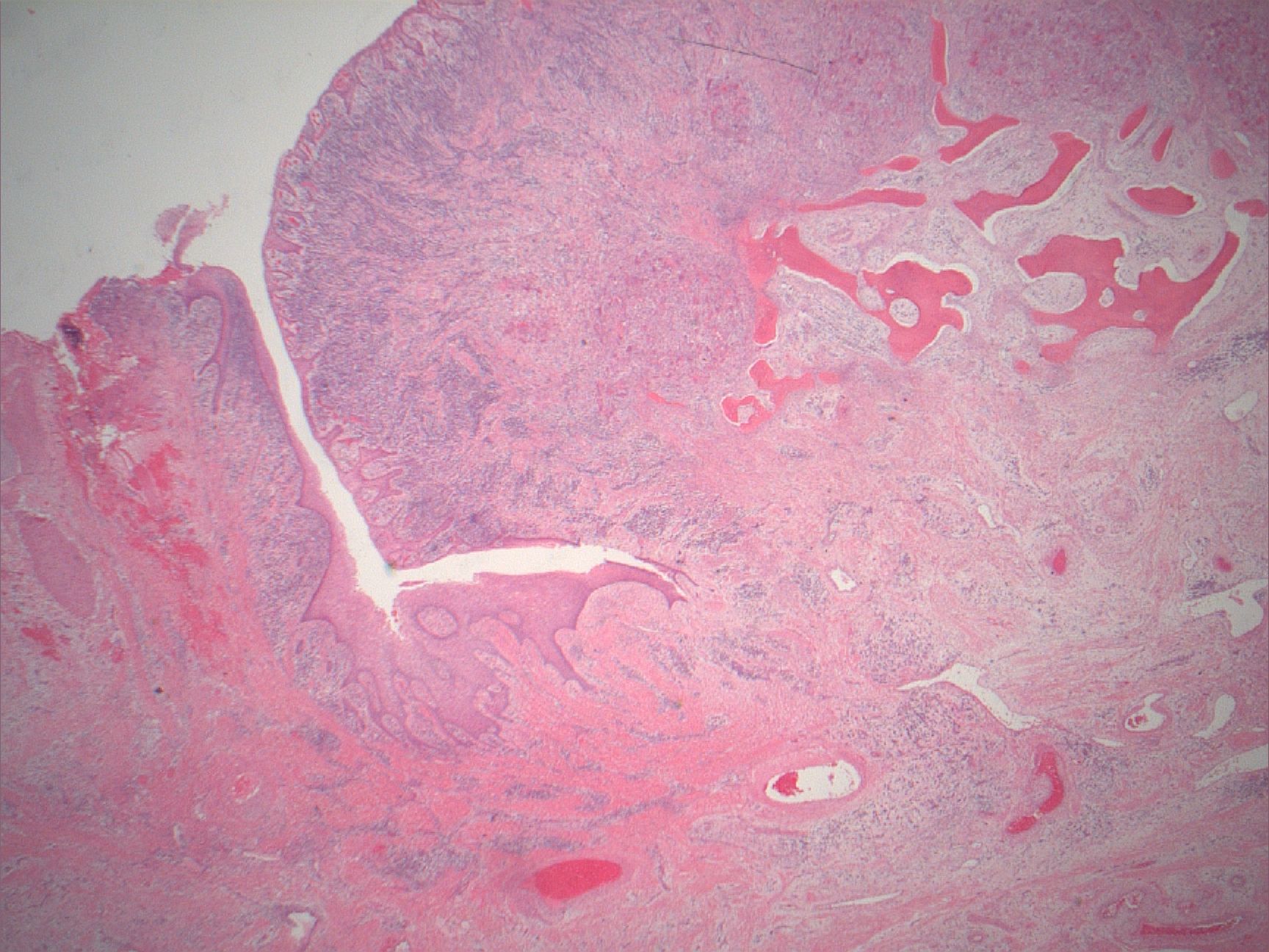
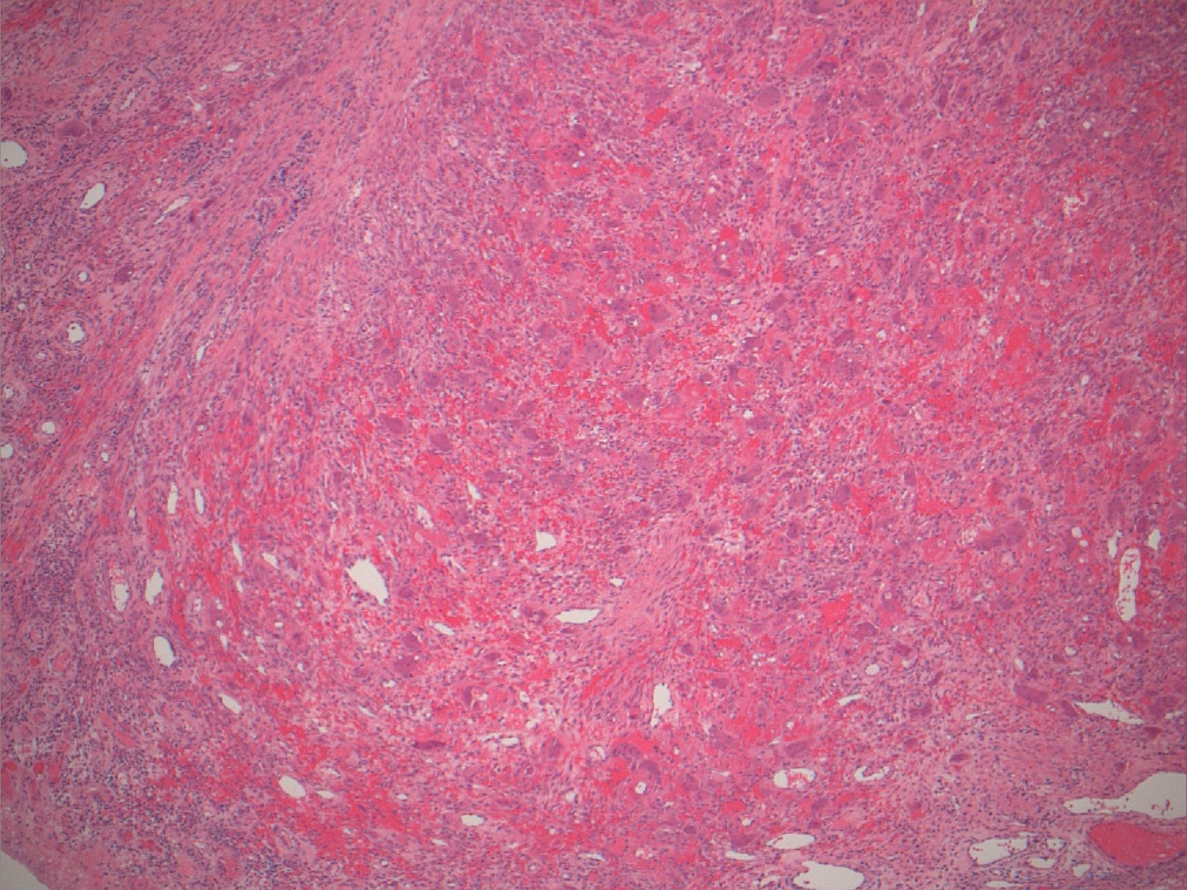
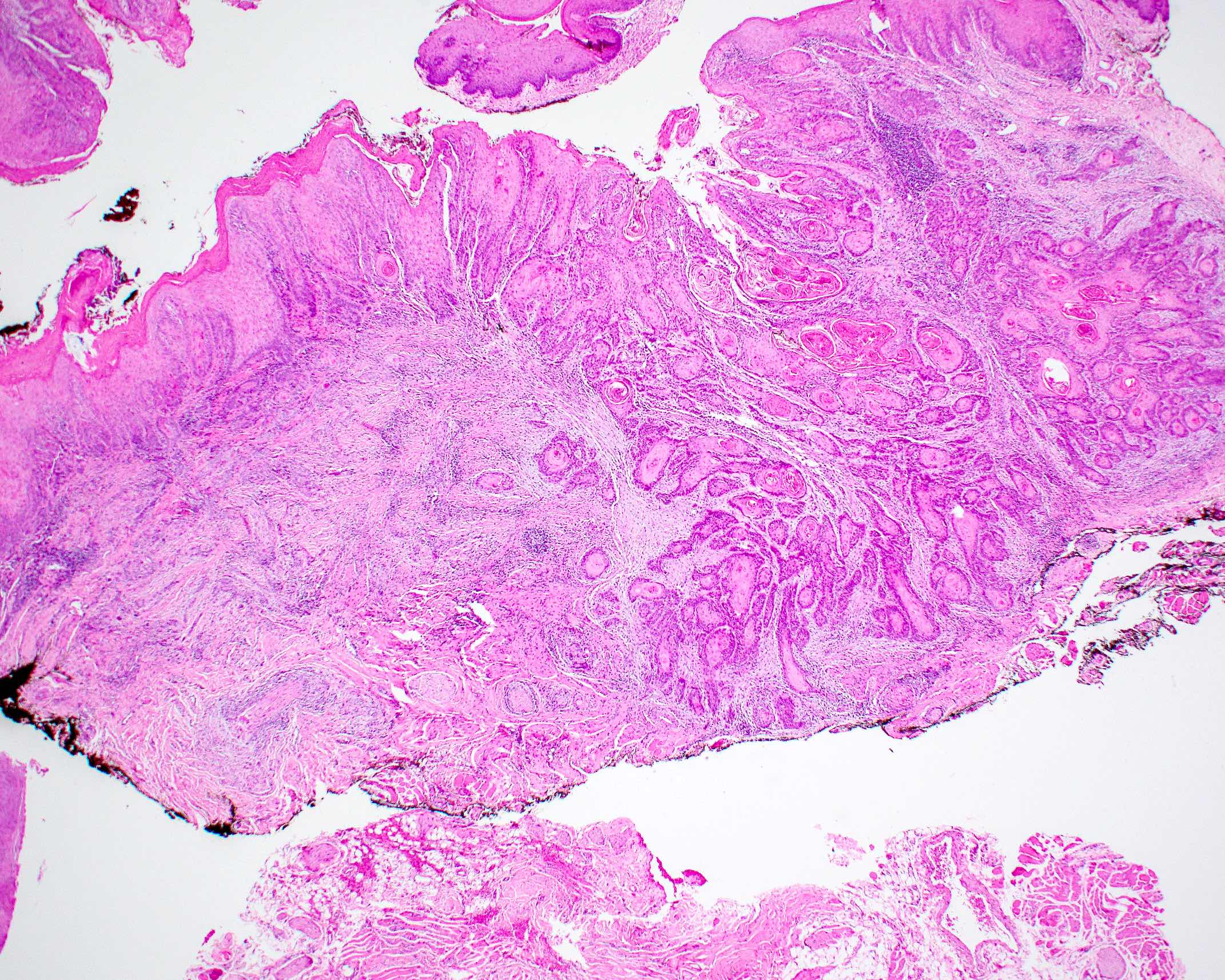
Arising squamous cell carcinoma
Keratosis and solar elastosis
Epithelial dysplasia and dilated vessels
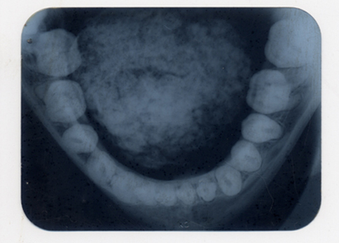
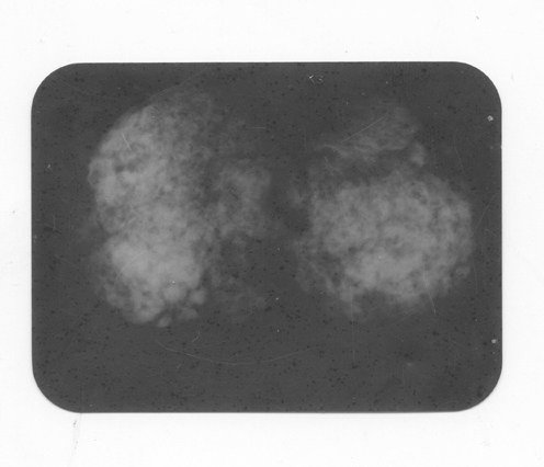
Contributed by Sarah H. Glass, D.D.S. and Duane Schafer, D.D.S., M.S.
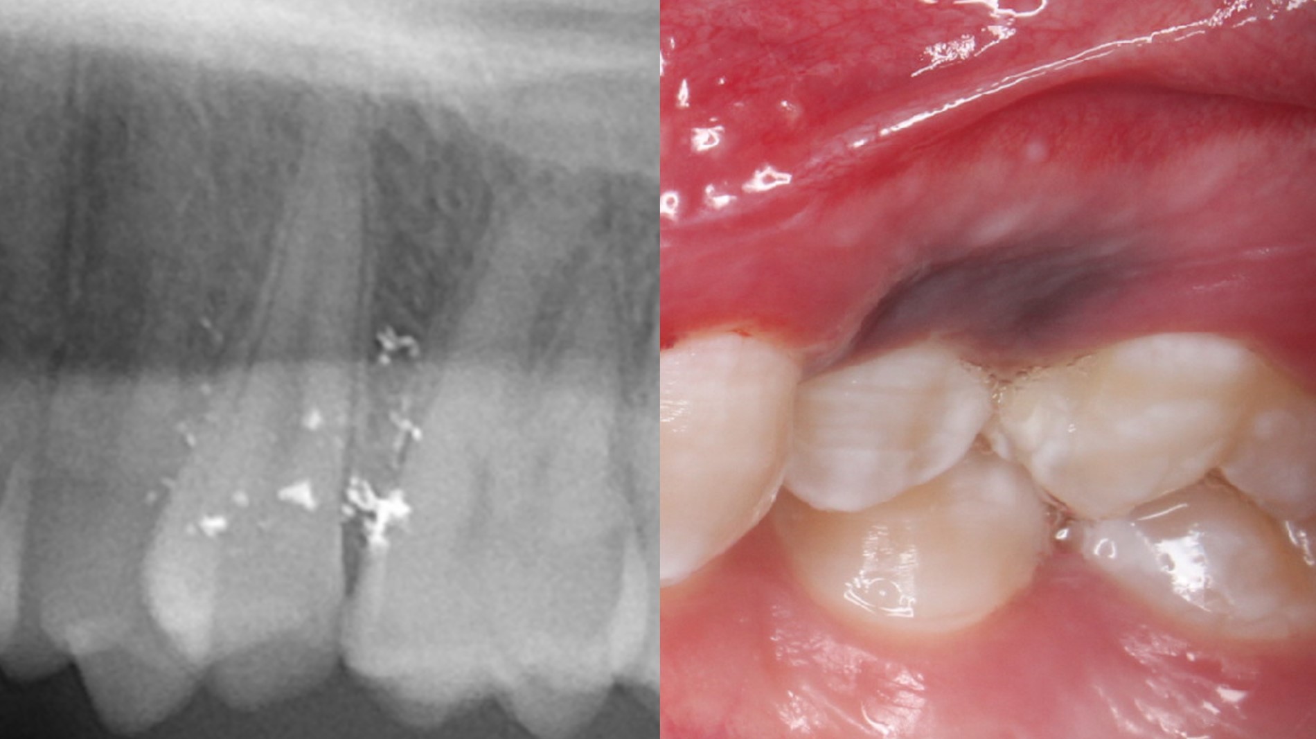
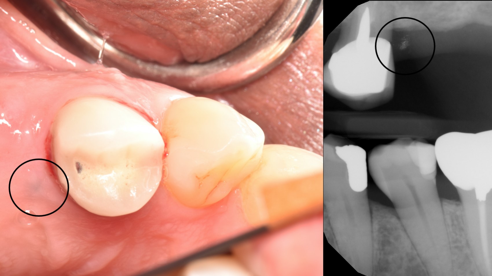
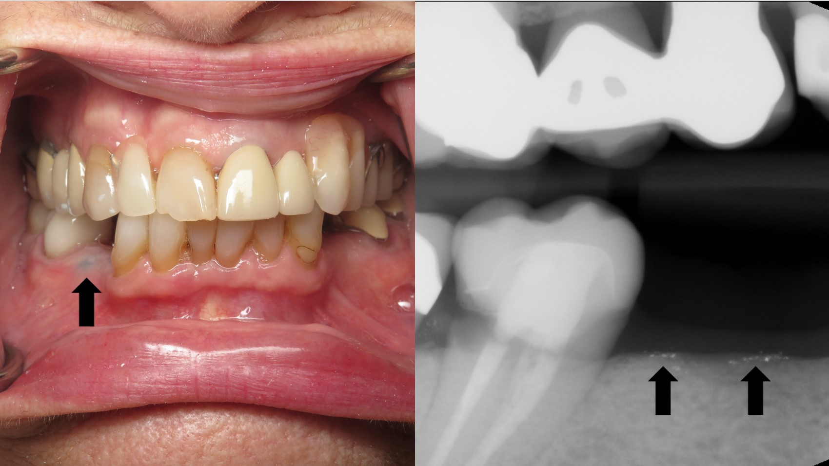
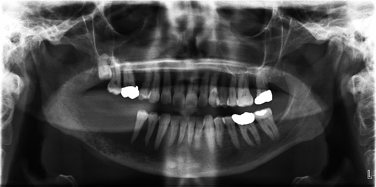
Radiopaque amalgam fragments
Contributed by Sarah H. Glass, D.D.S. and Duane Schafer, D.D.S., M.S.
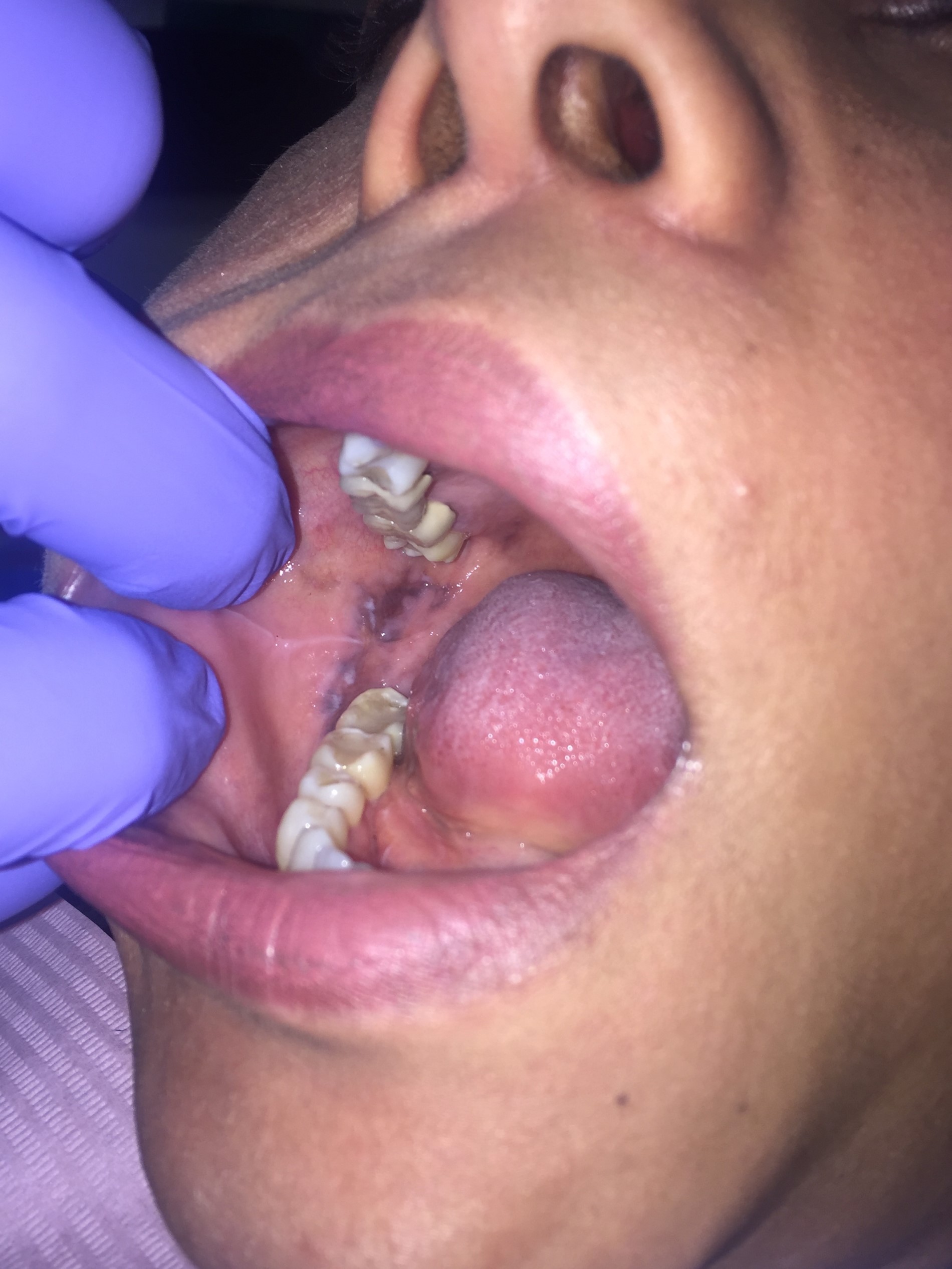
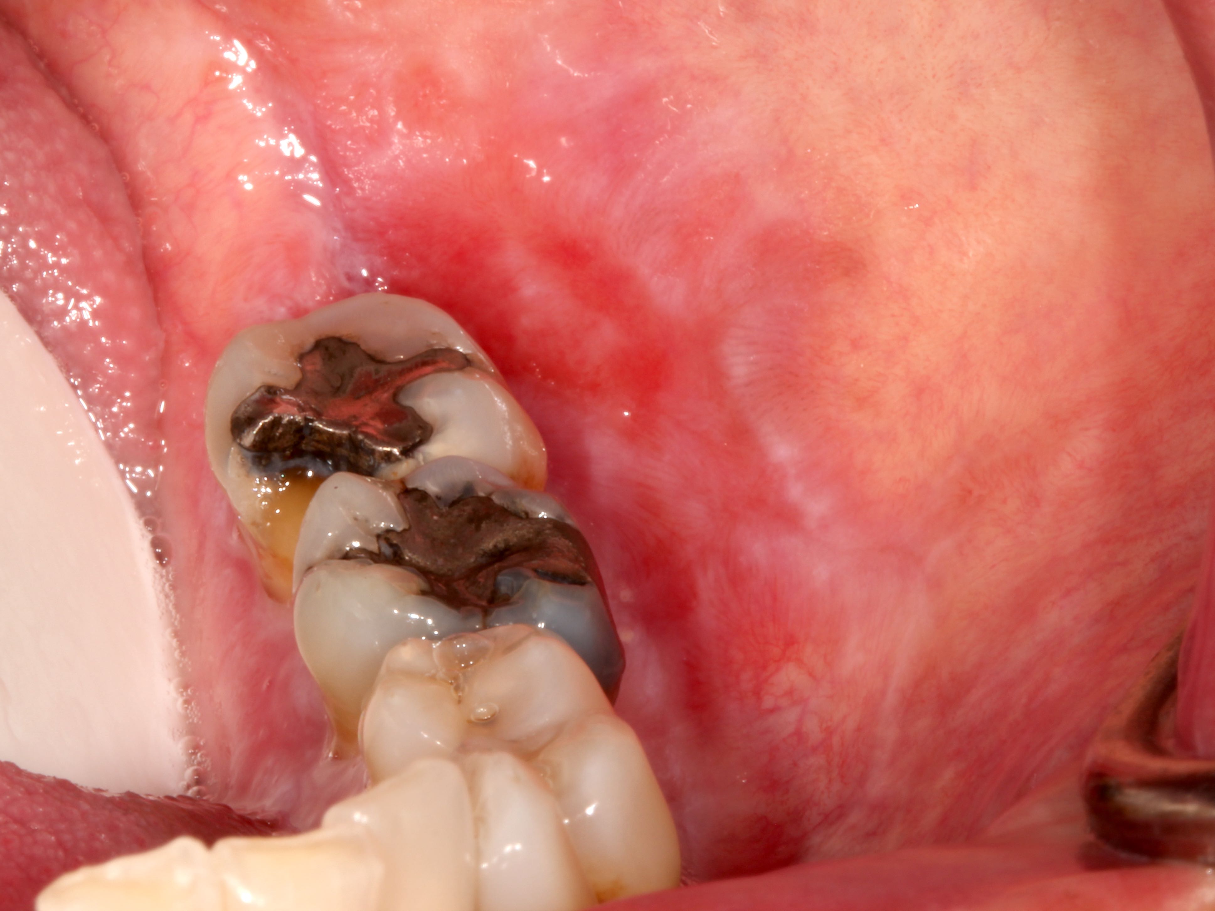
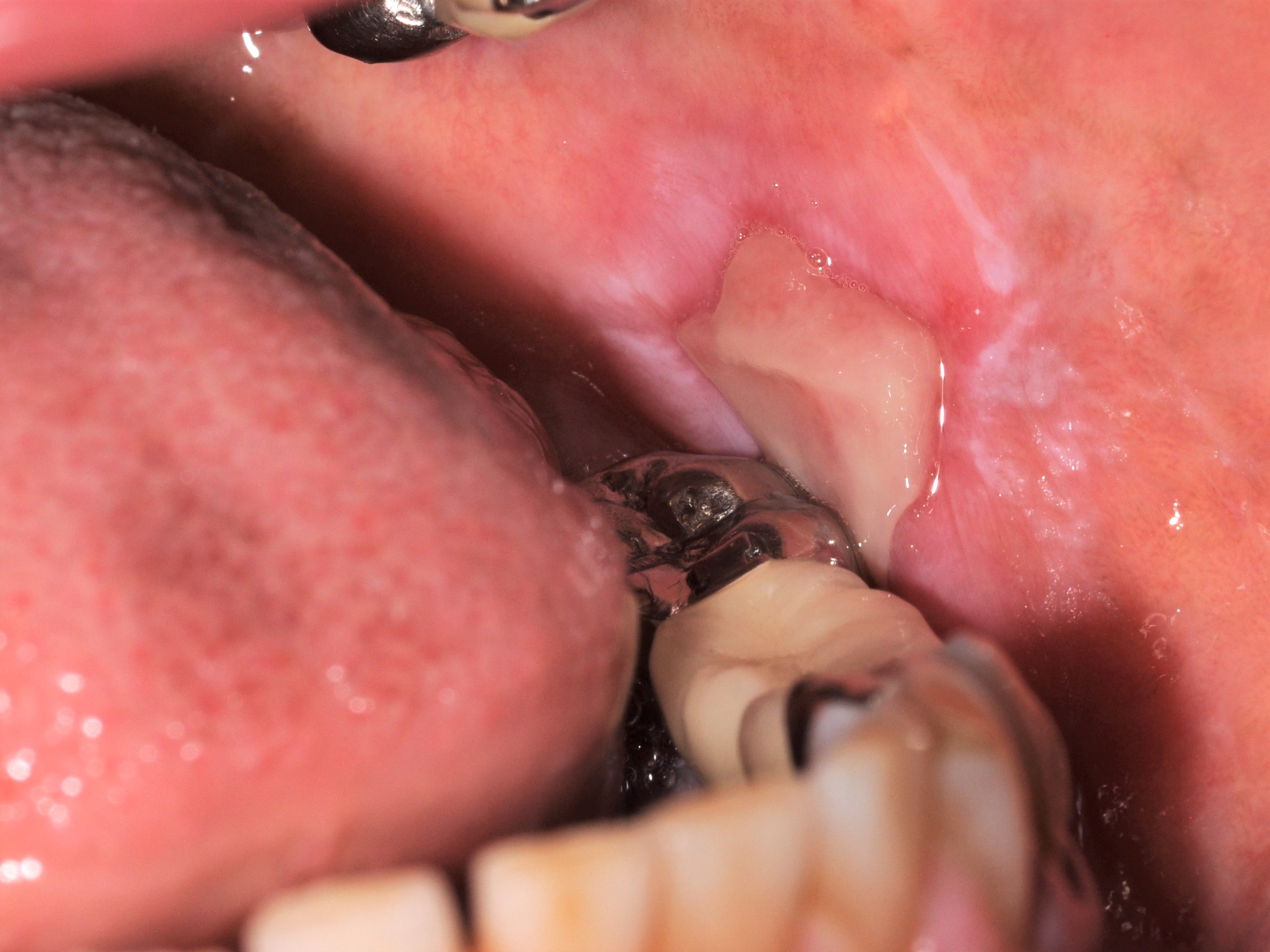
Buccal mucosa amalgam tattoo

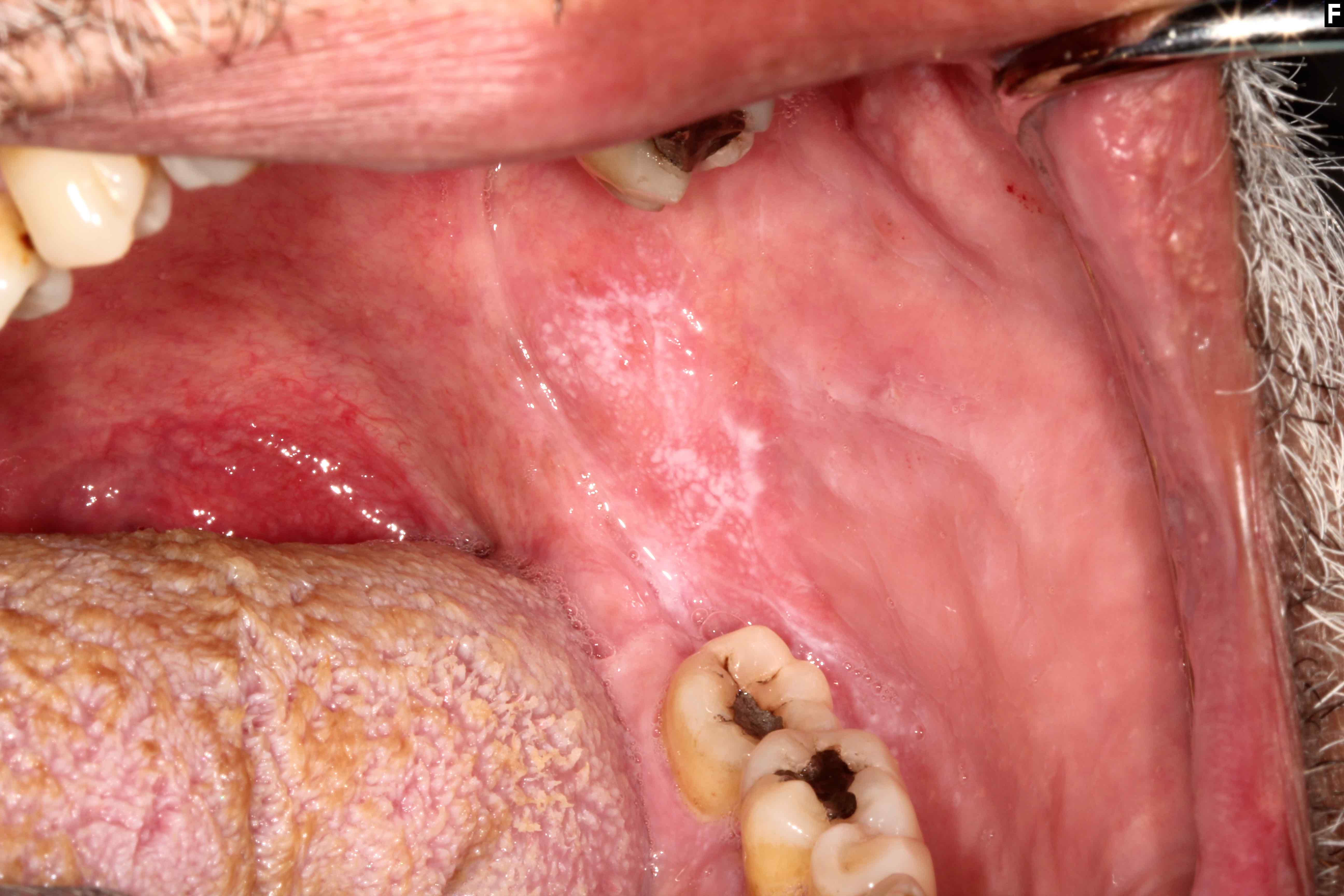
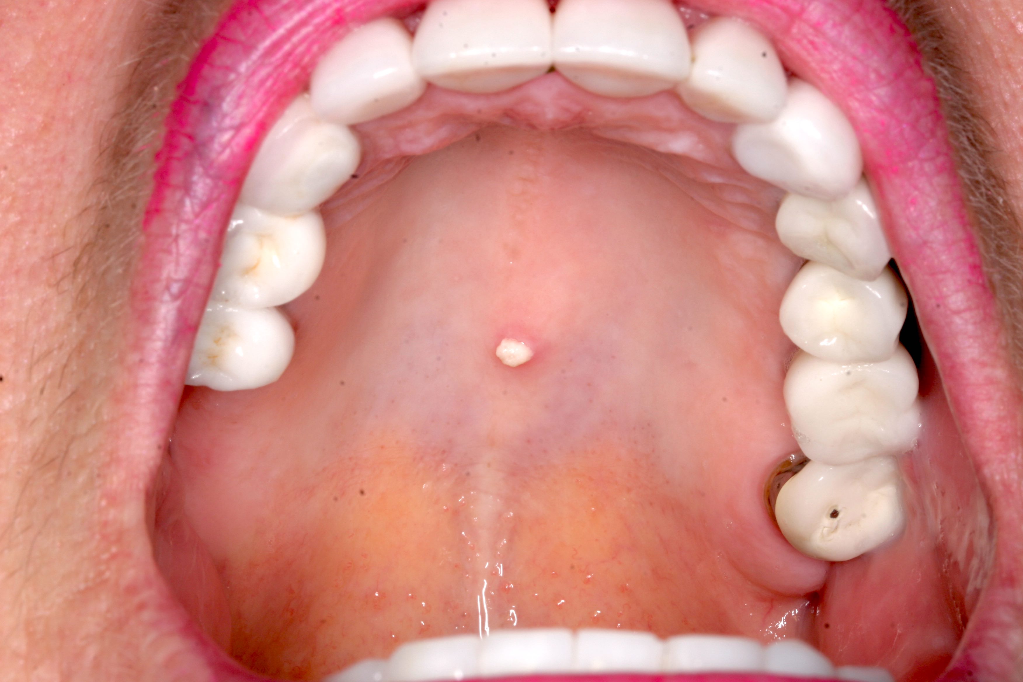
Large maxillary amalgam tattoo
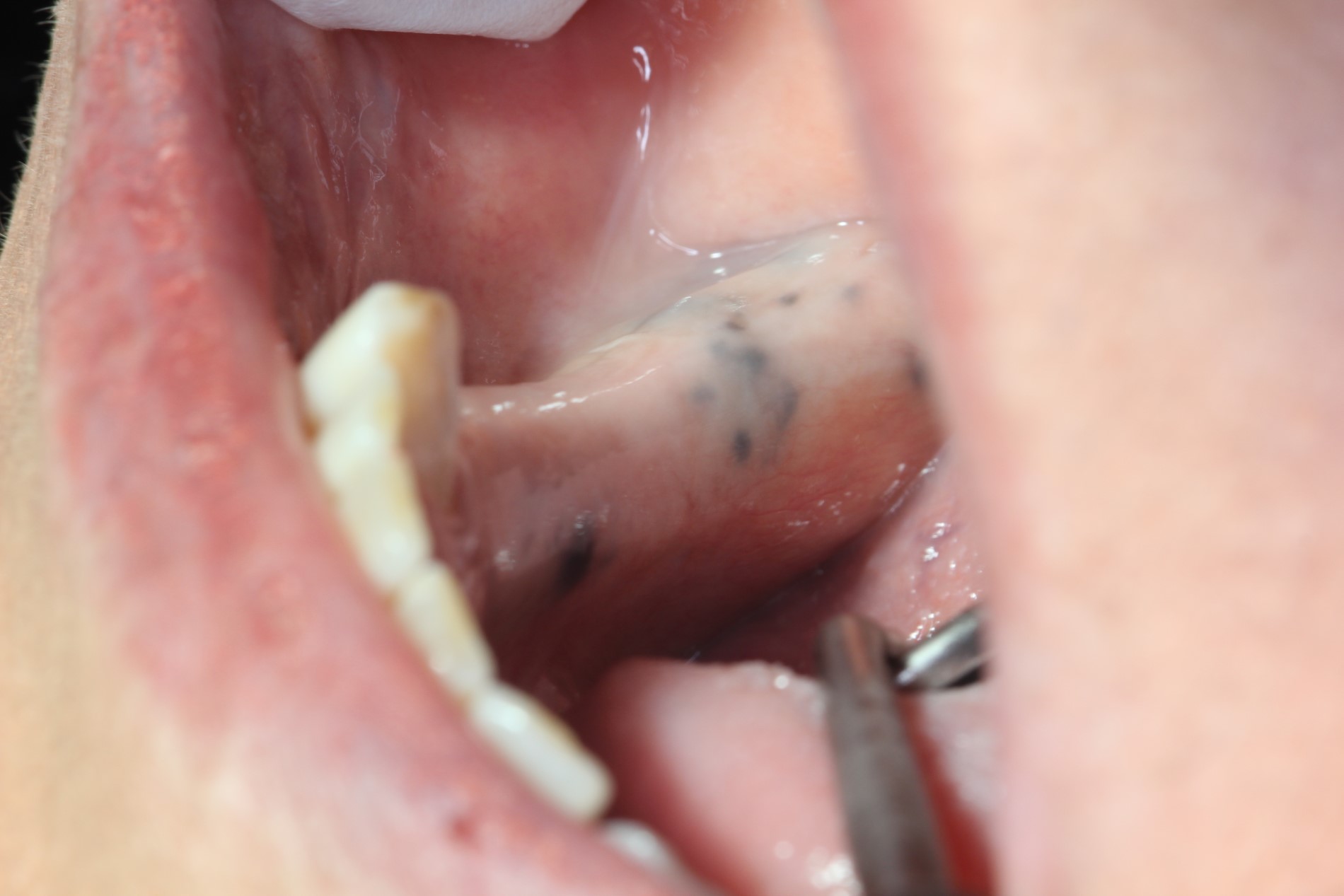
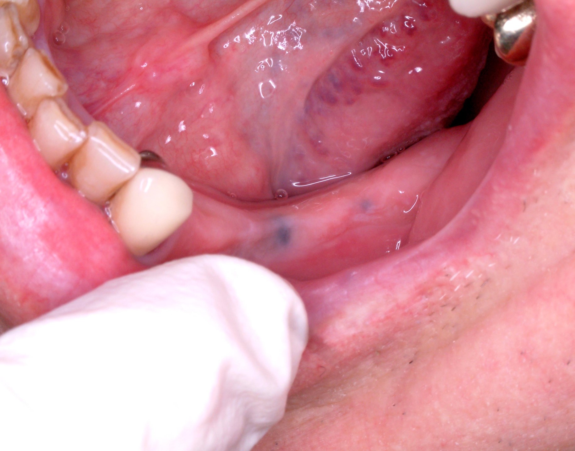
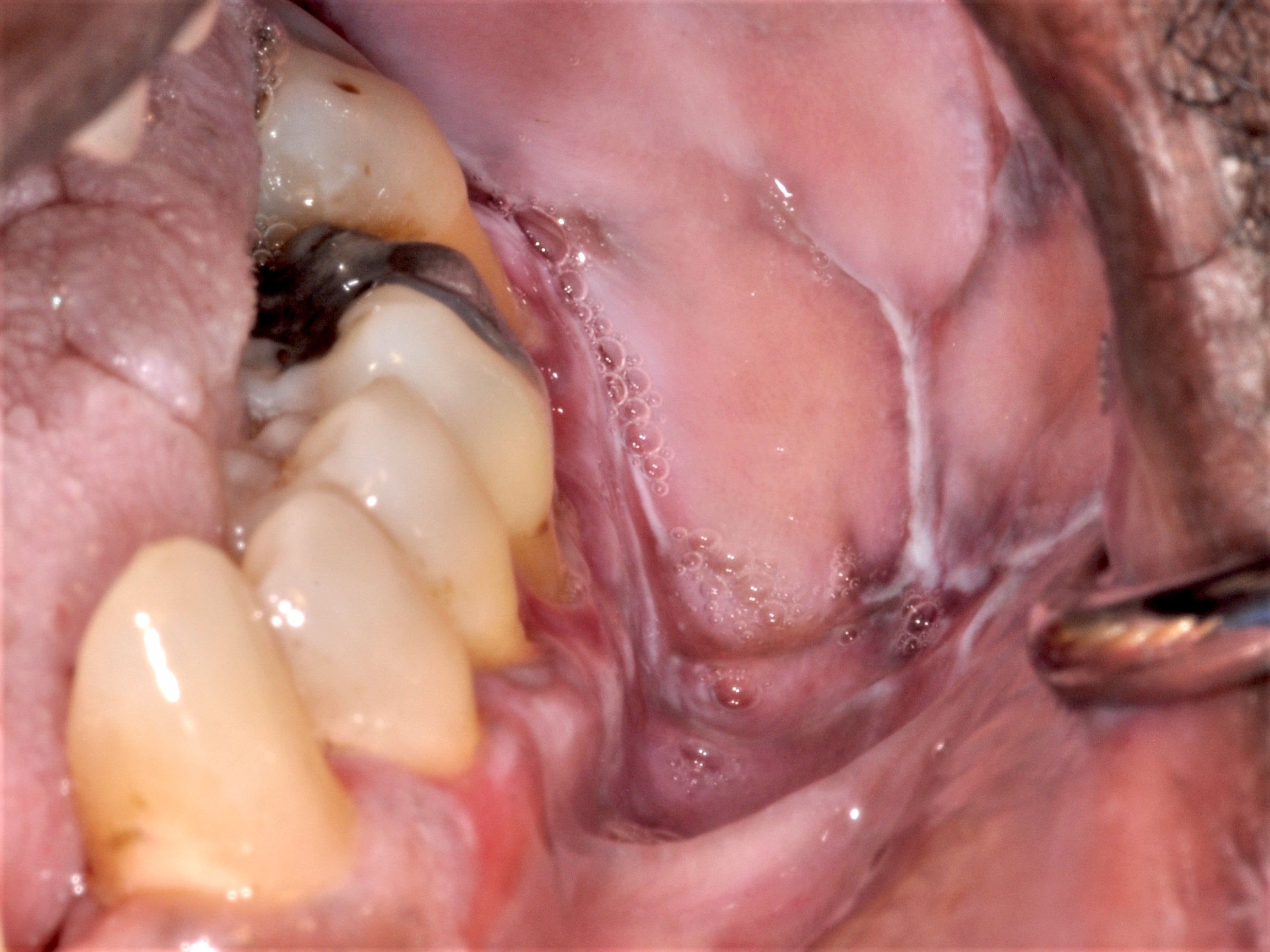
Alveolar ridge amalgam tattoos
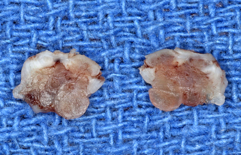
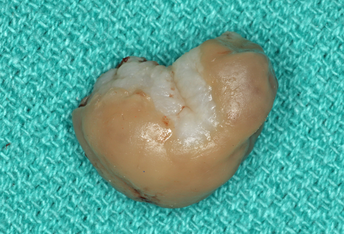
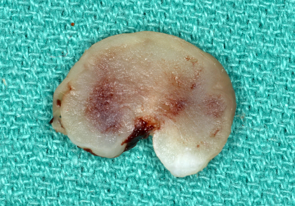
Contributed by Sarah H. Glass, D.D.S.
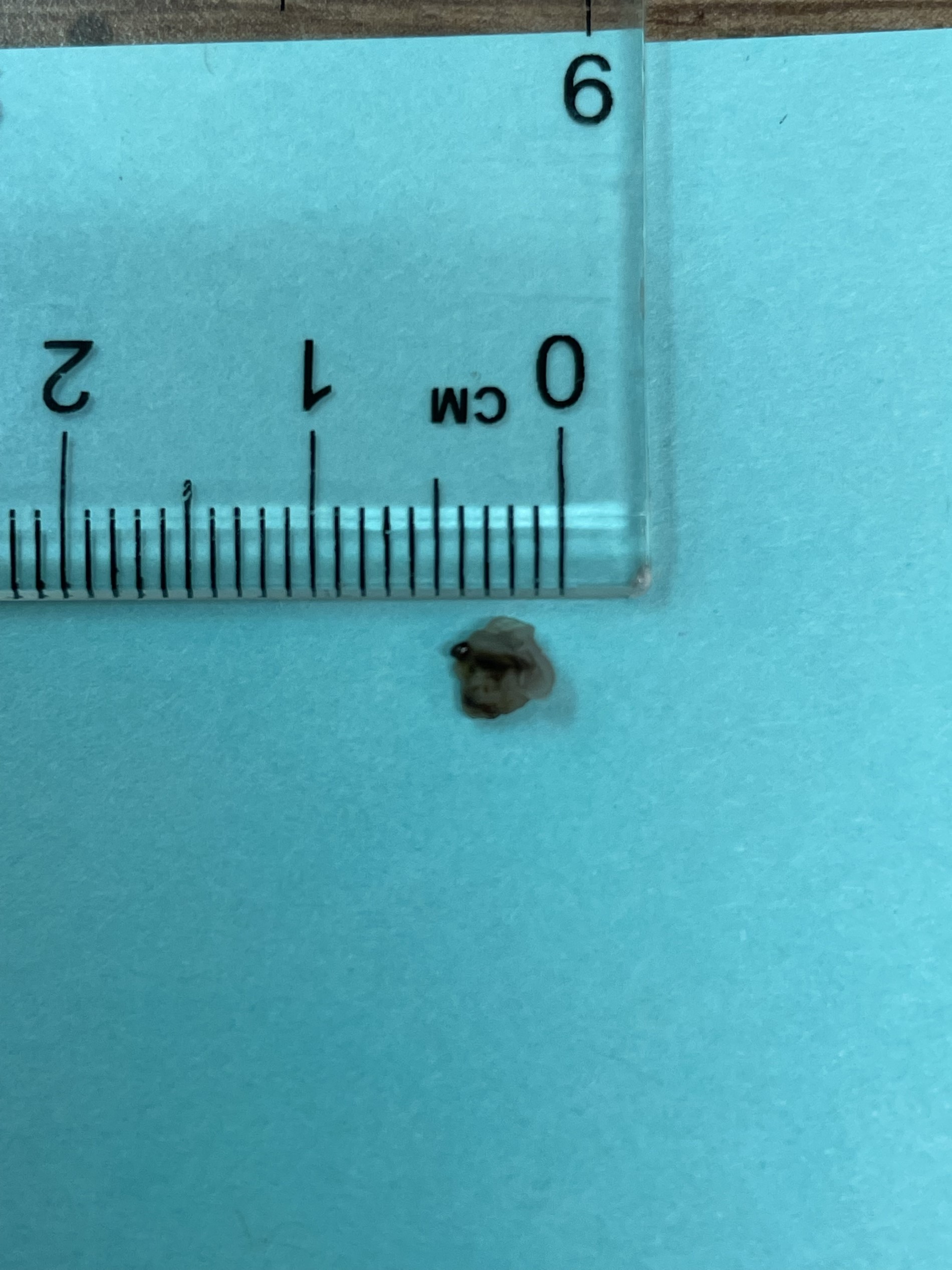
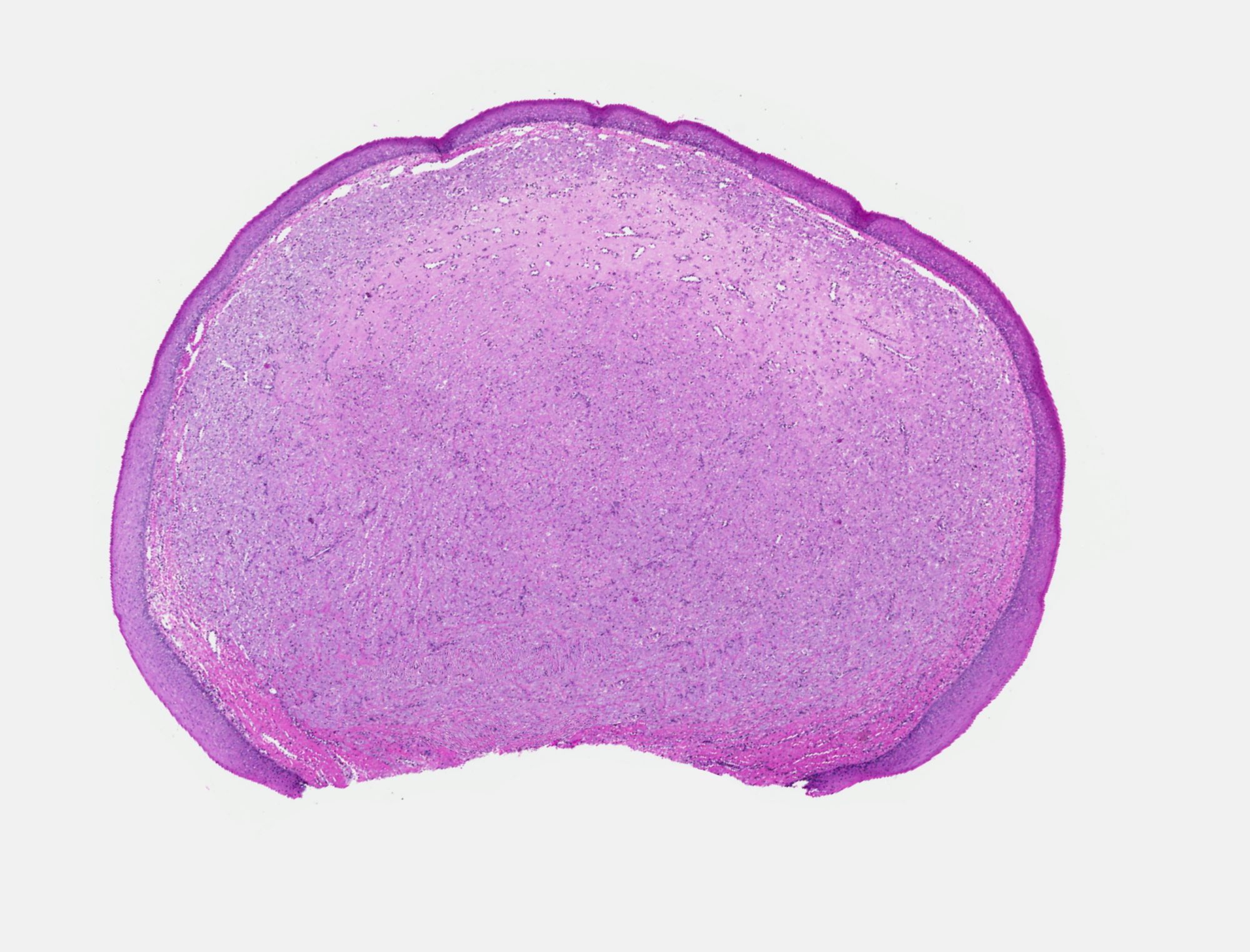
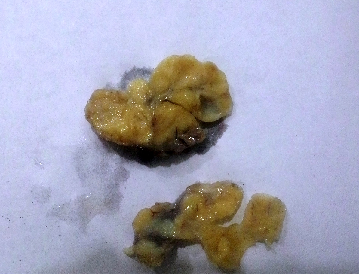
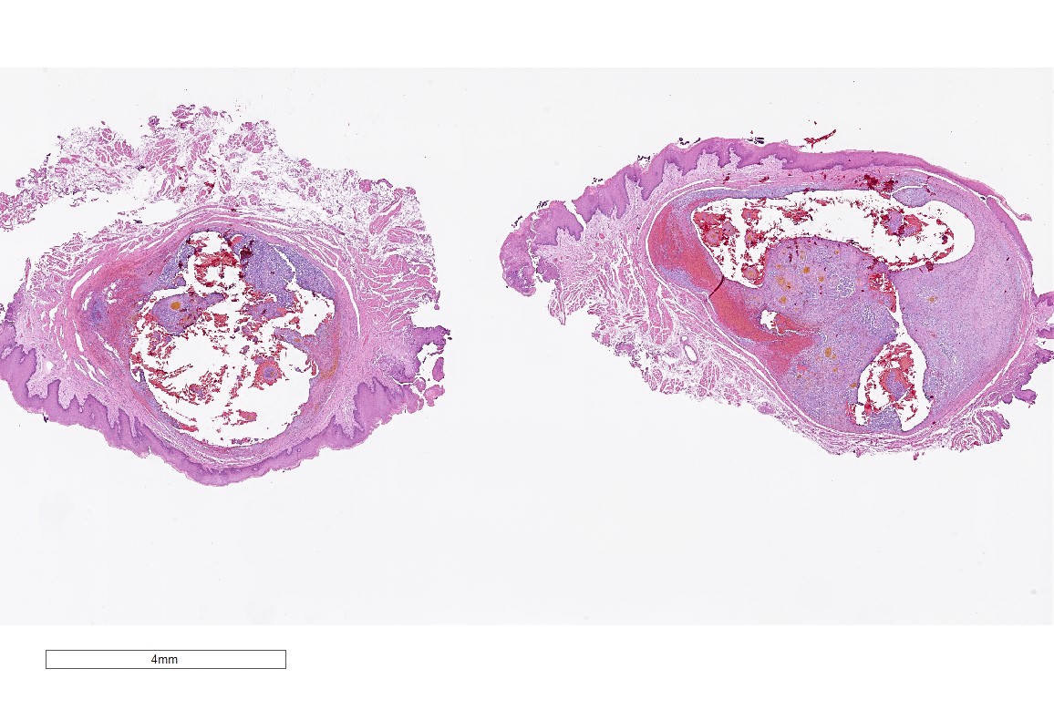
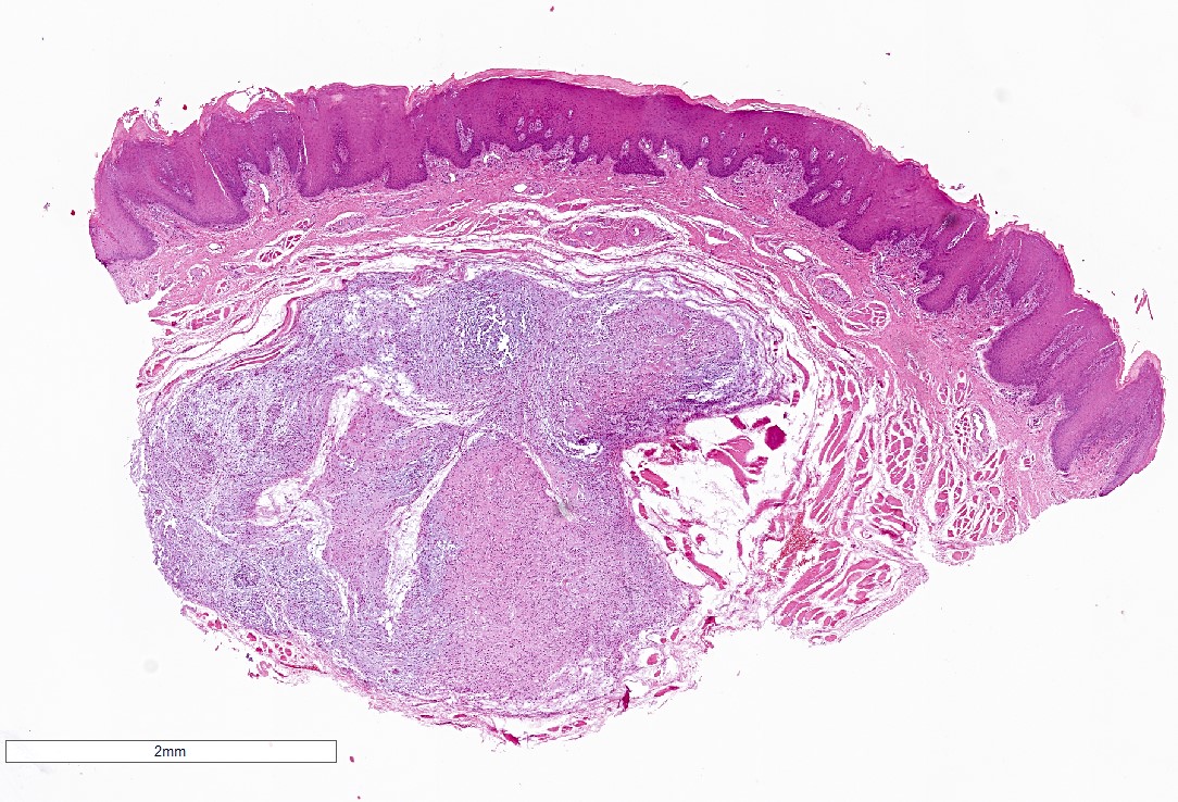
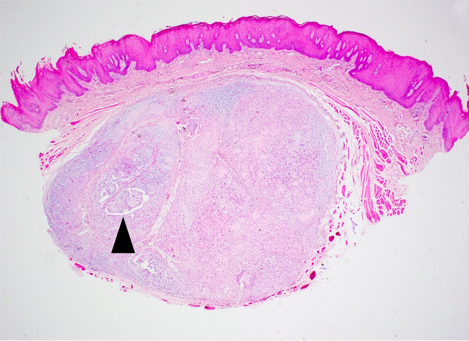
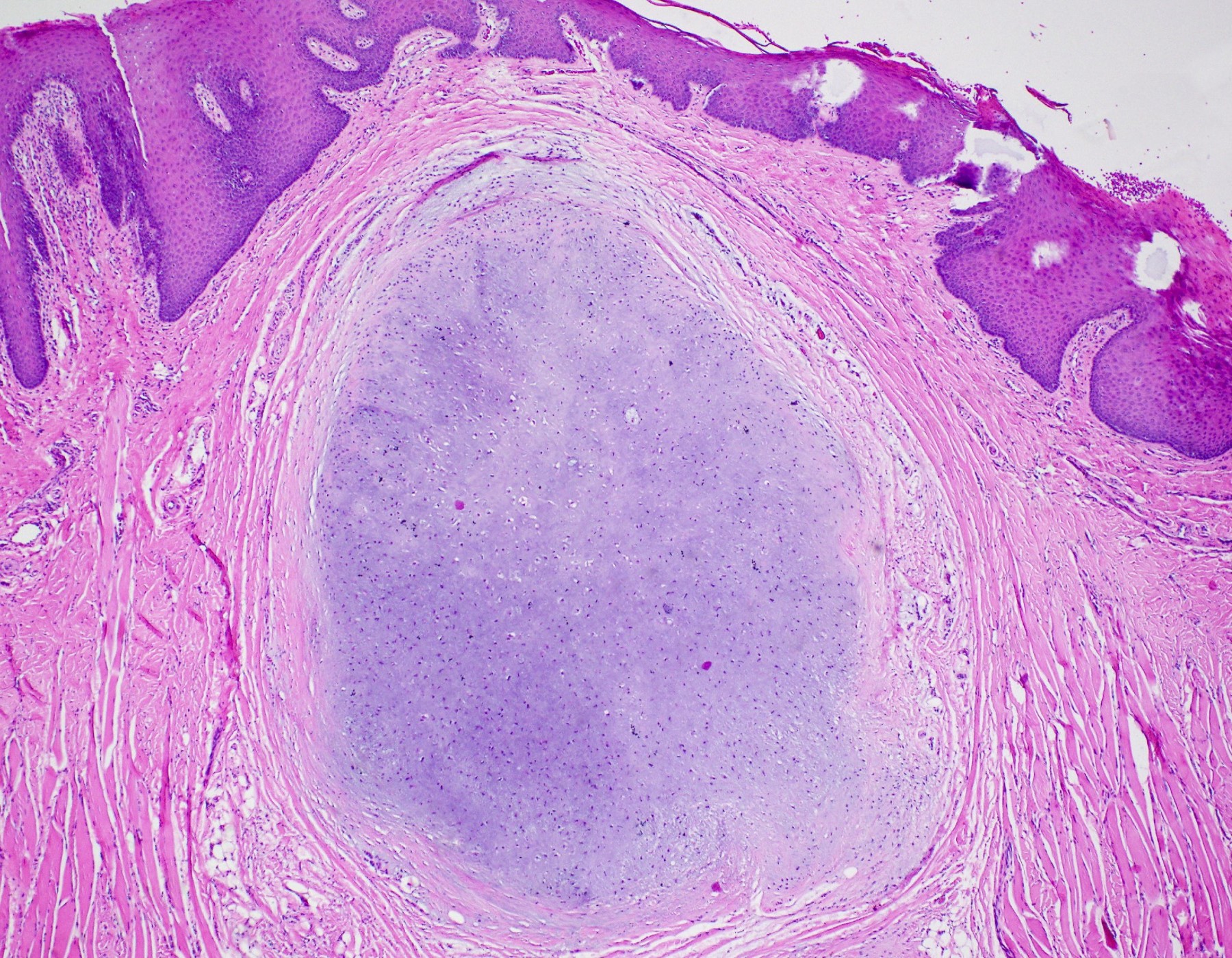
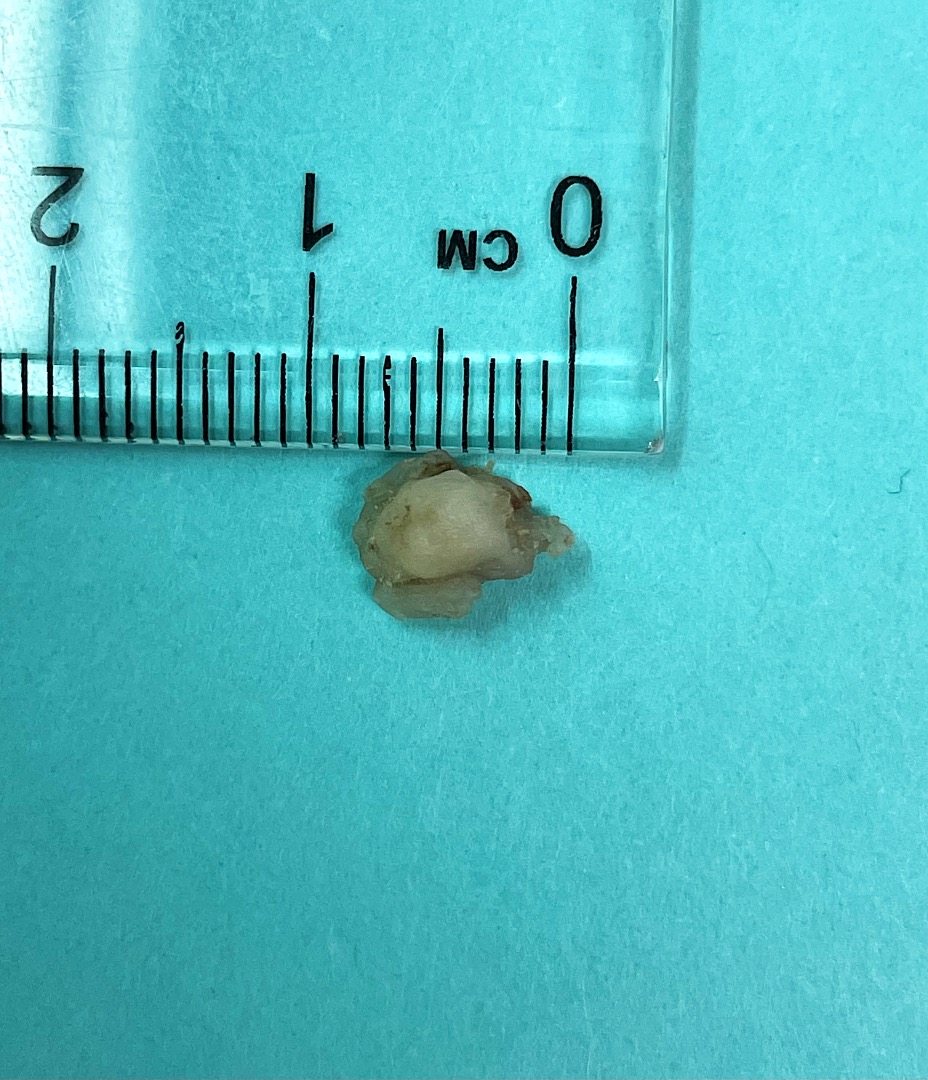
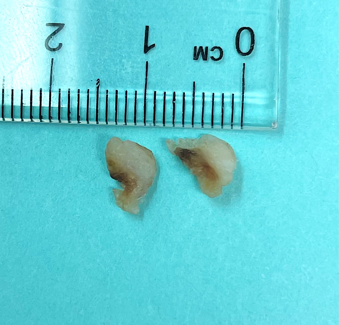
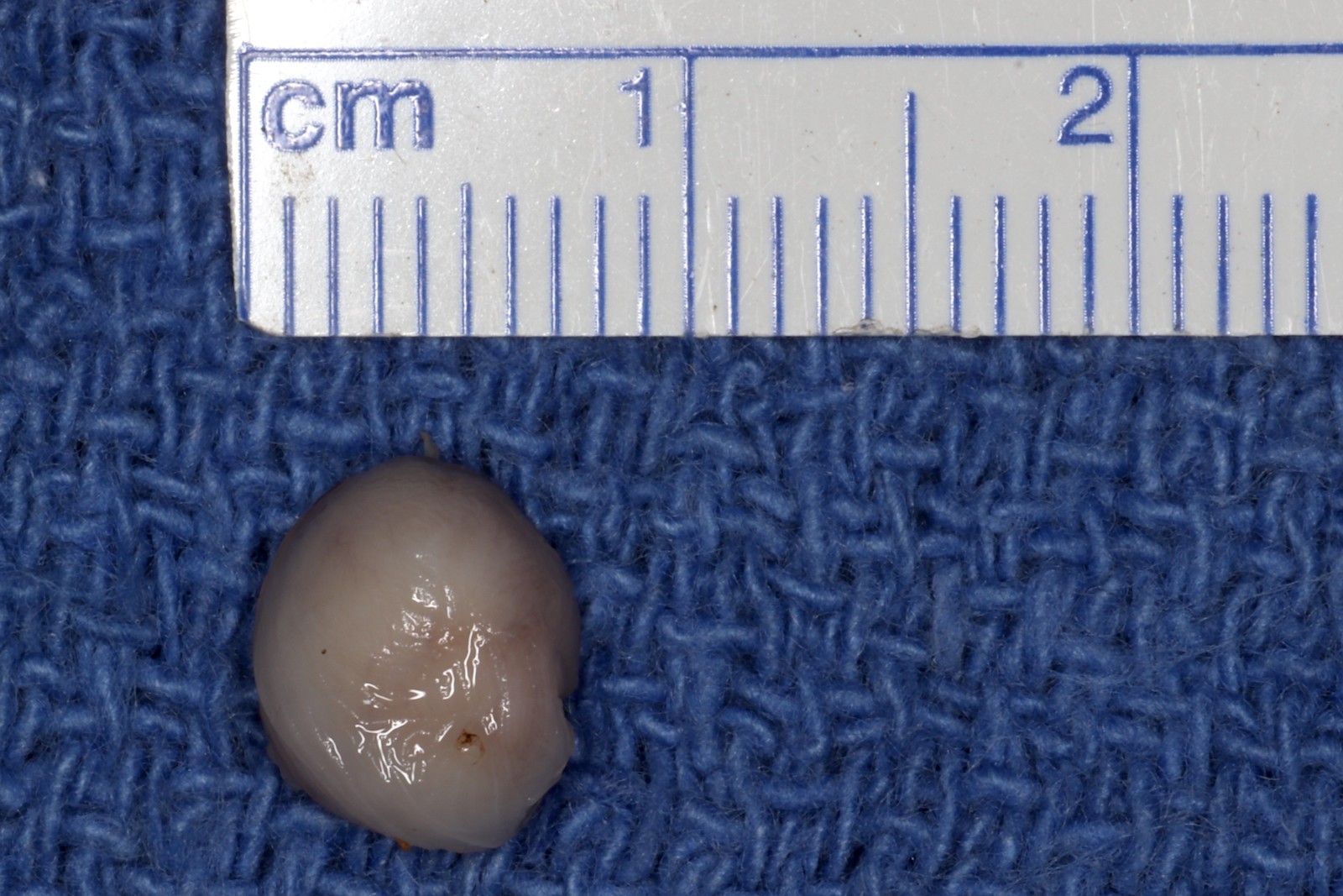
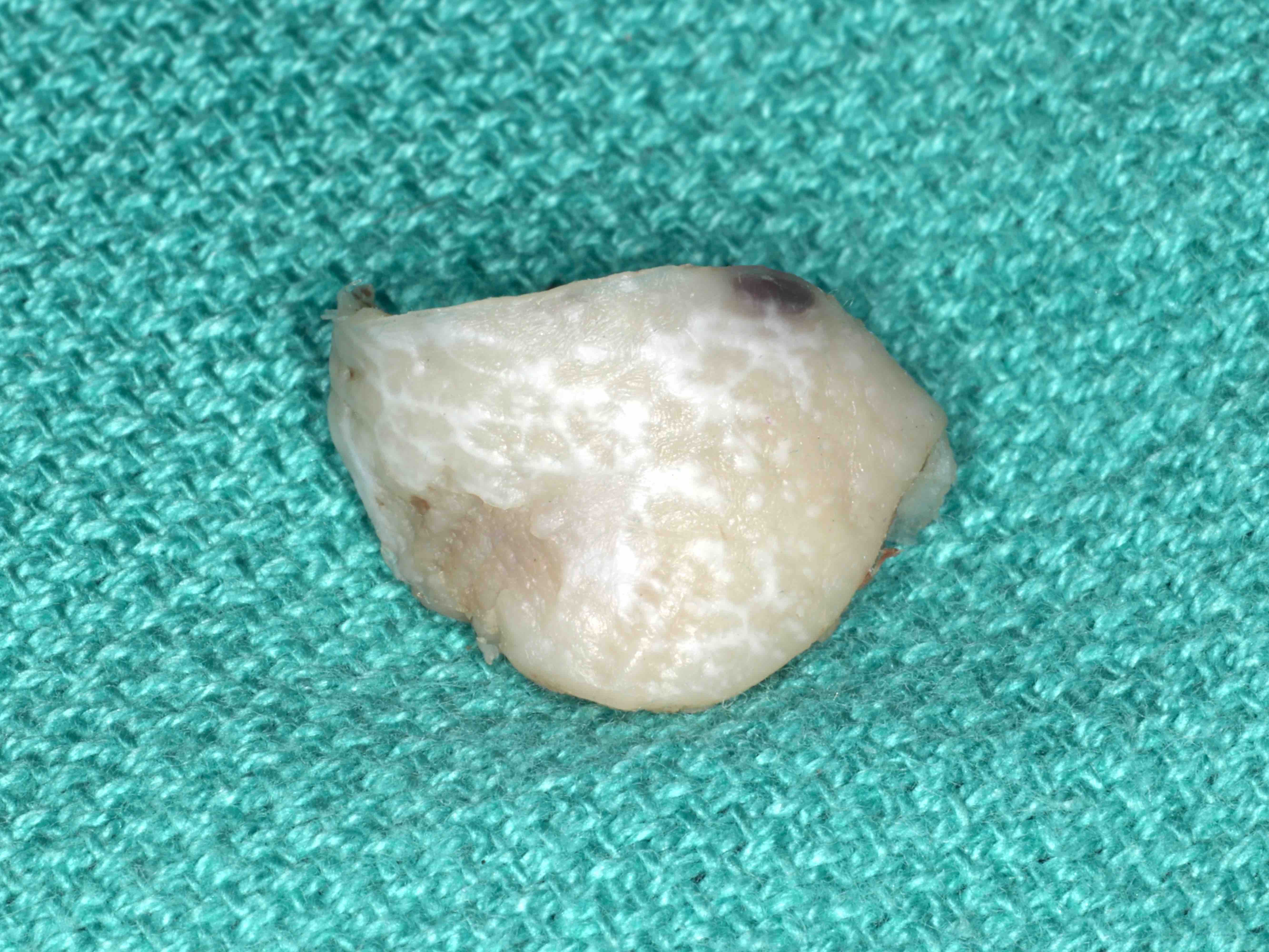
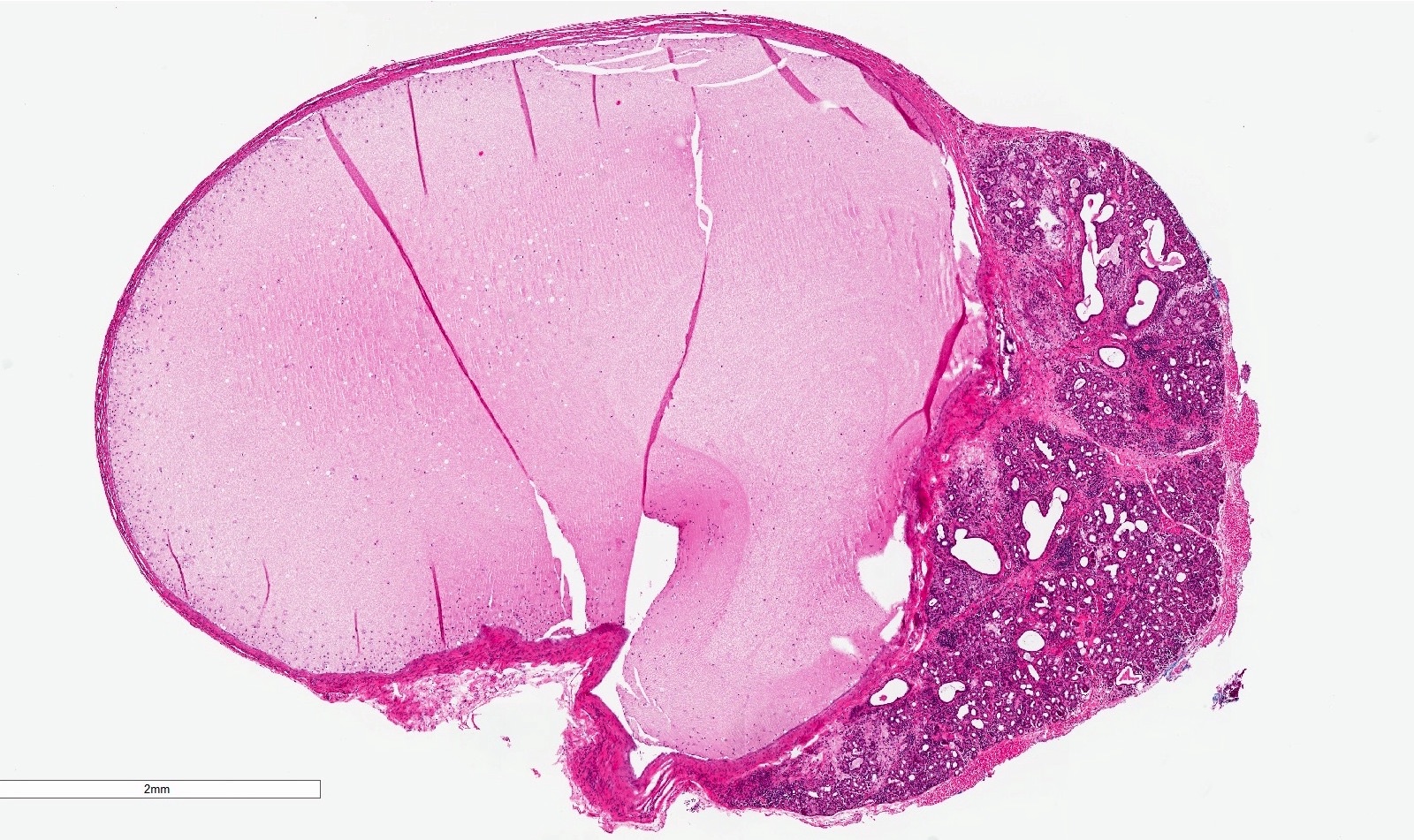
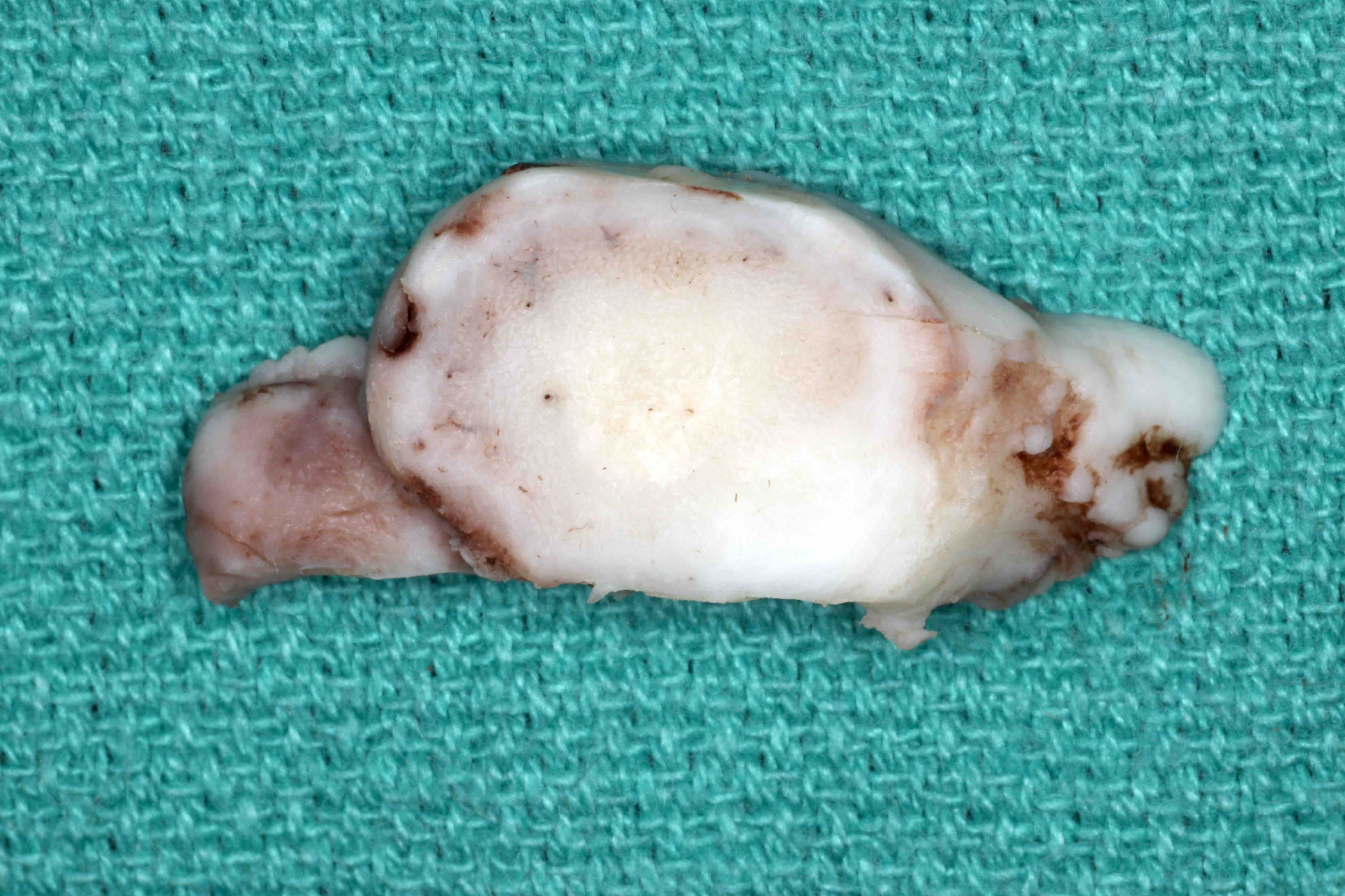
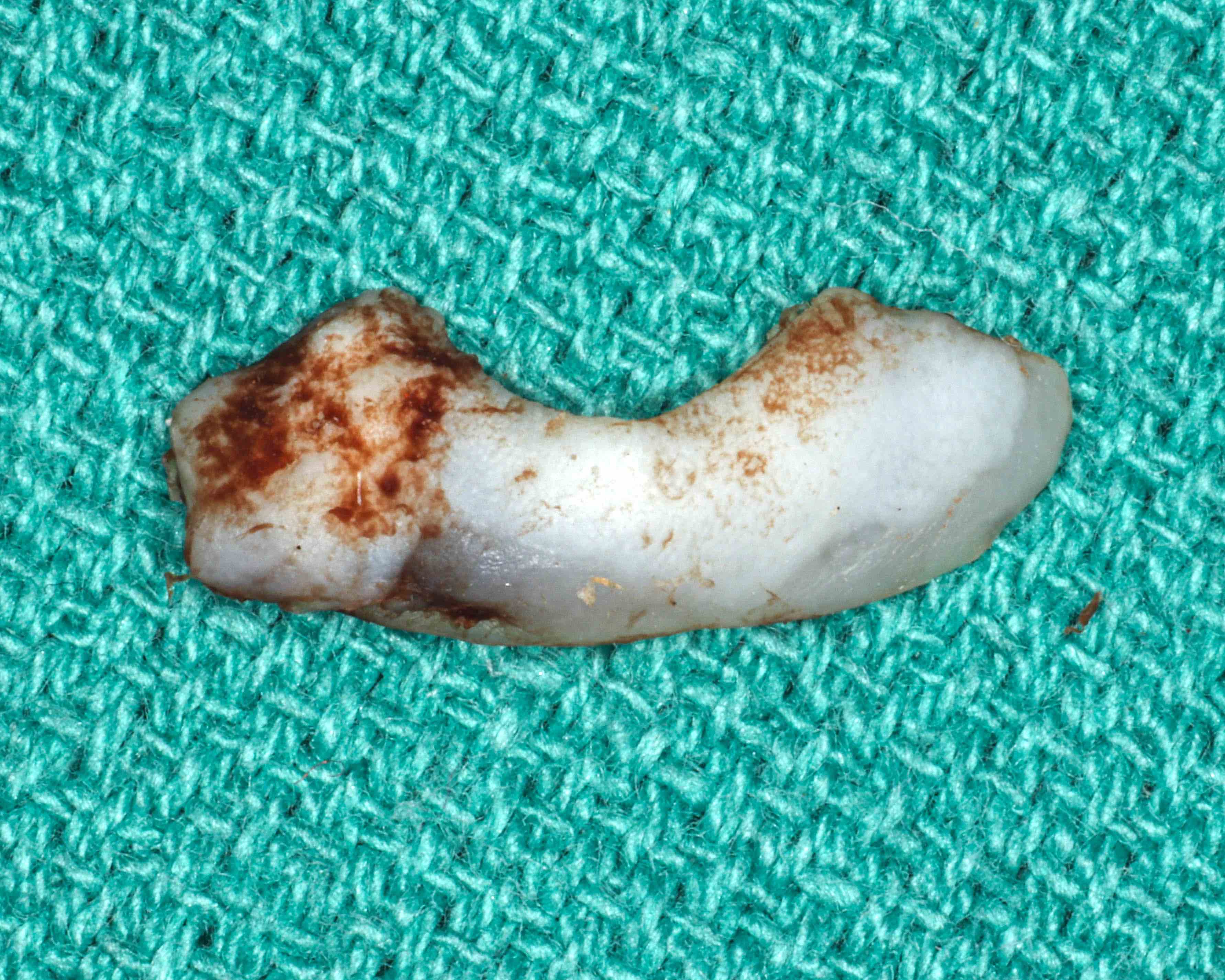
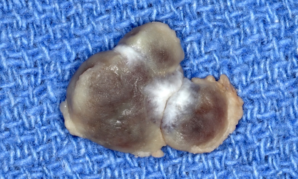
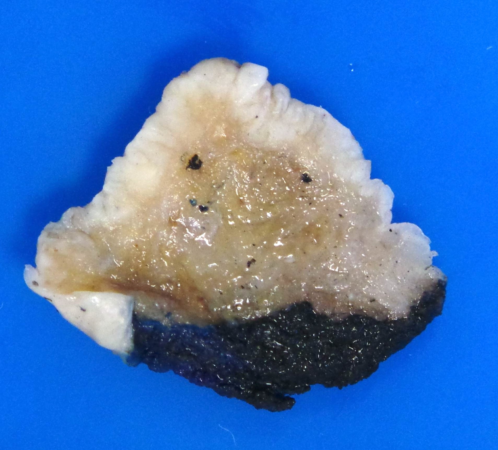
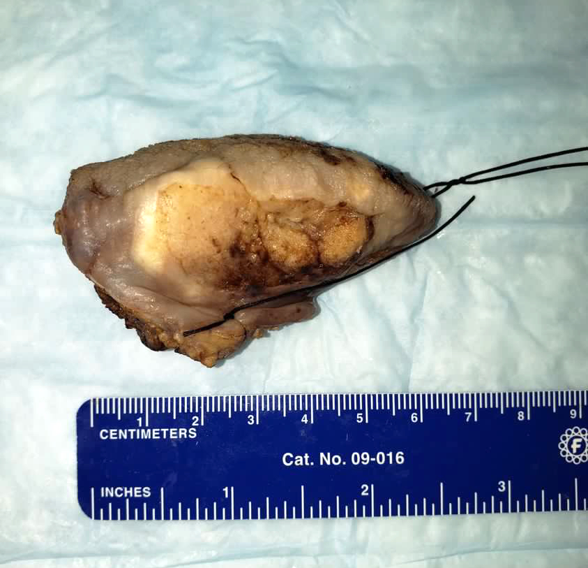
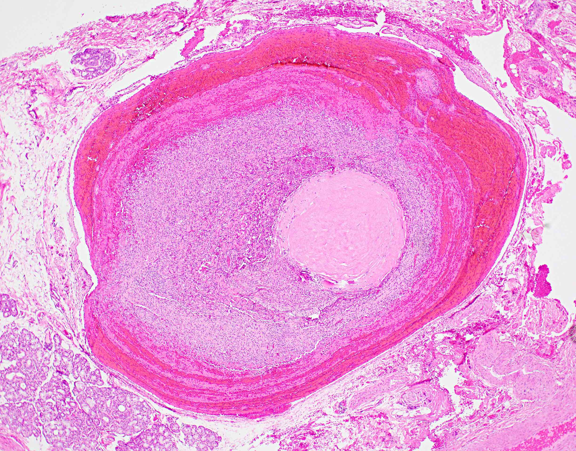
Excisional biopsy specimen
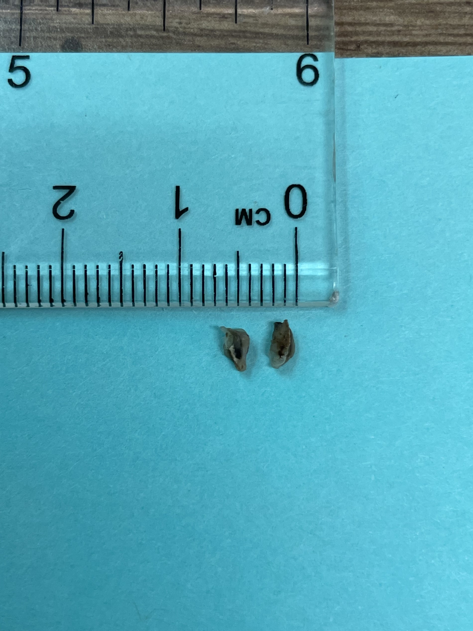
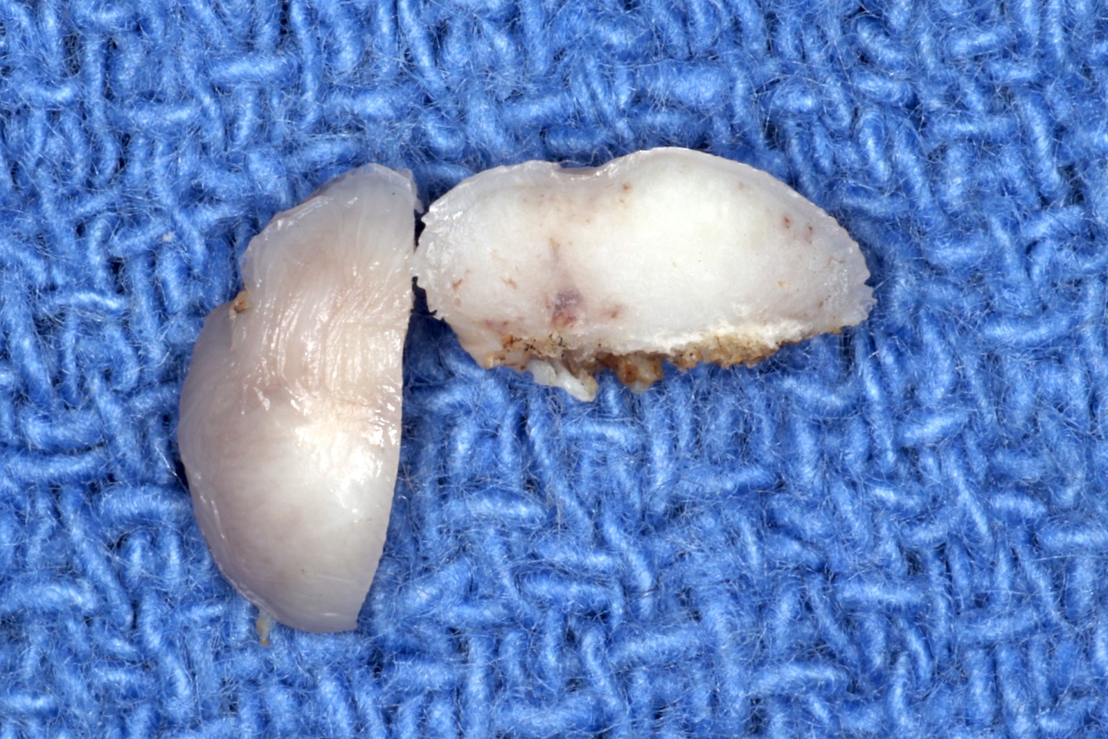
Cross section
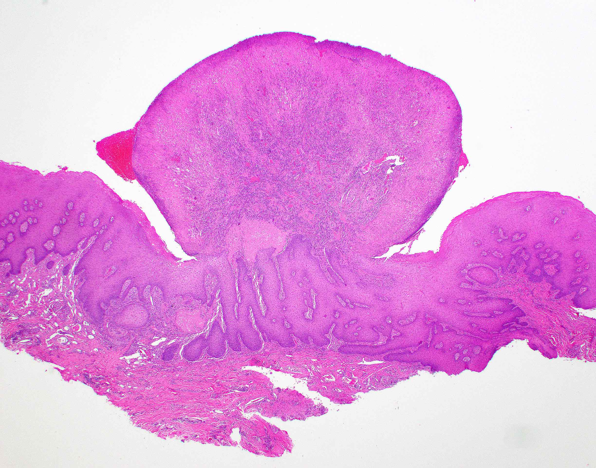
Contributed by Sarah H. Glass, D.D.S.
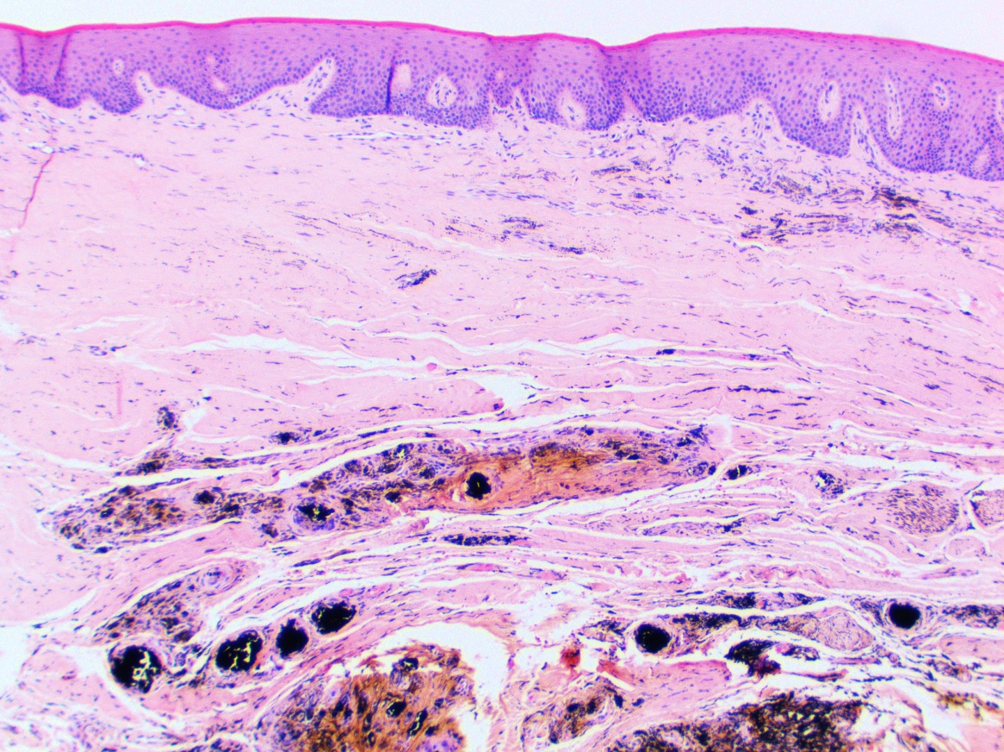
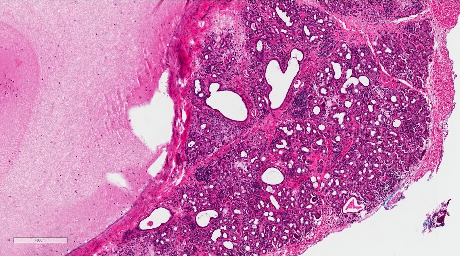
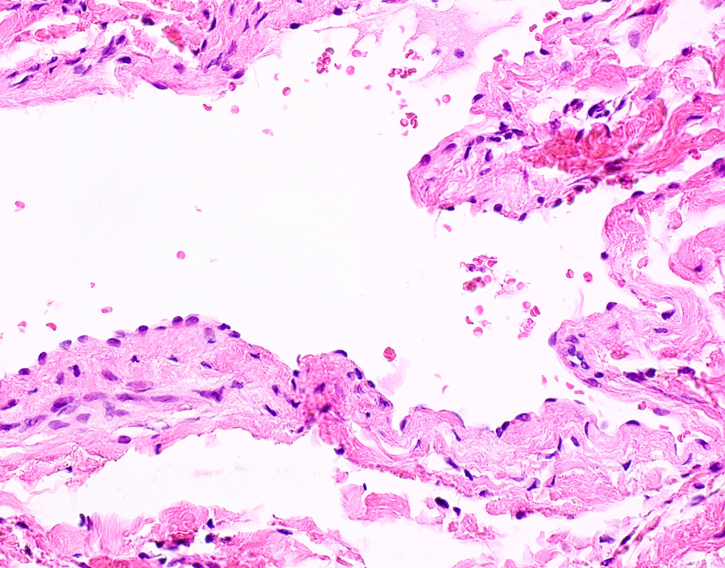
Extensive amalgam deposition
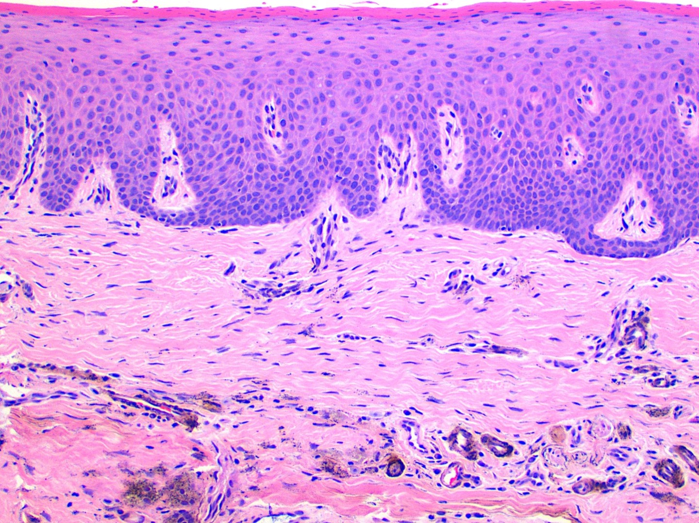
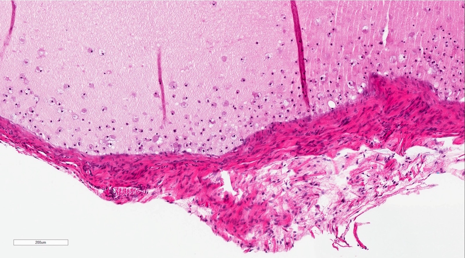
Scattered amalgam granules
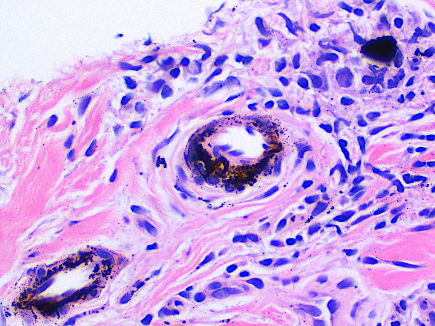
Around blood vessels
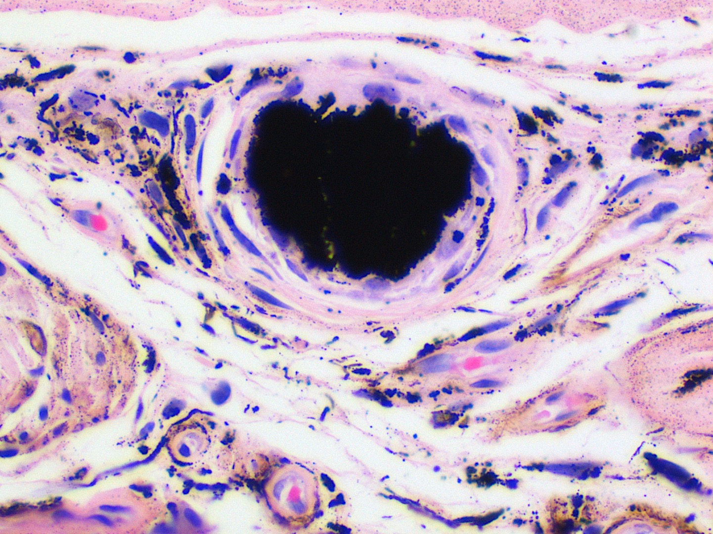
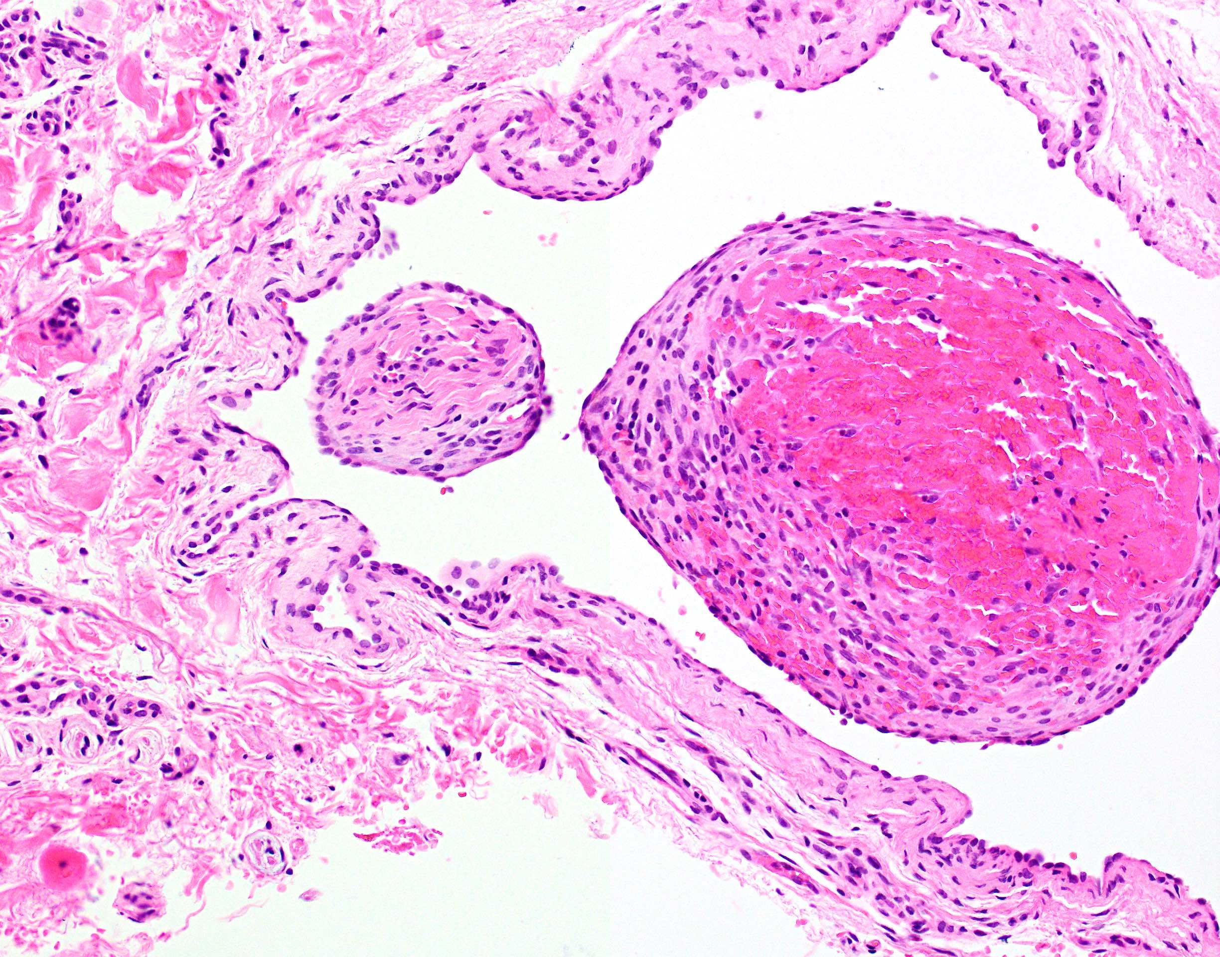
Large amalgam fragment
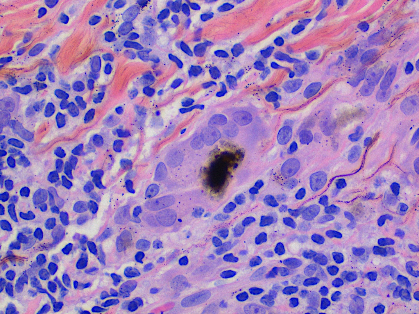
Giant cell engulfment
Contributed by Katherine Hulme, M.B.Ch.B.
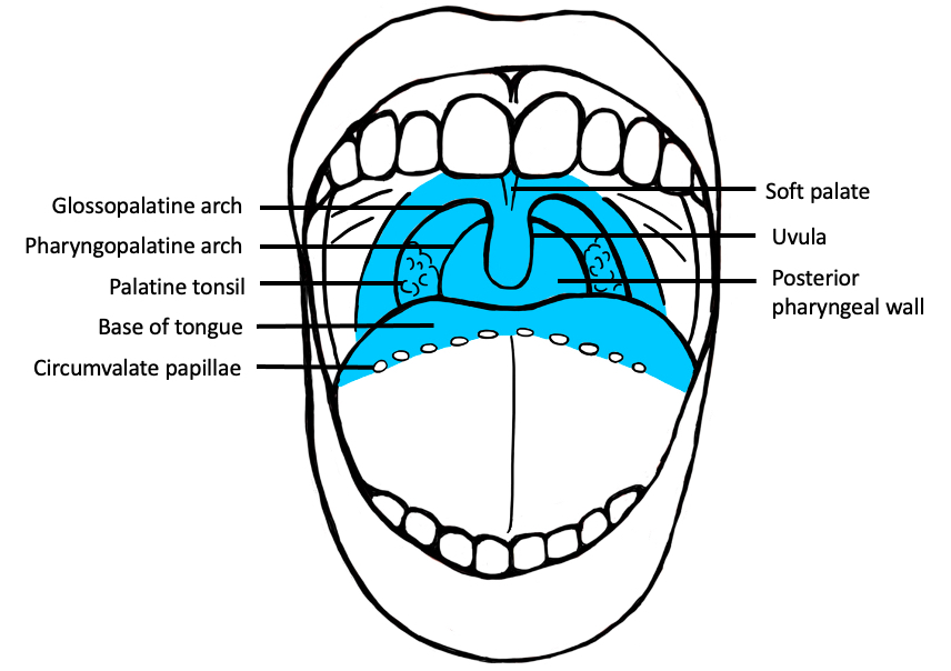
Oropharynx (frontal view)
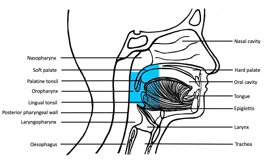
Oropharynx (lateral view)
Images hosted on other servers:


Aphthous ulcers
26 year old man:

Minor ulcer

Major ulcer

Laser therapy


After one week of laser treatment
Images hosted on other servers:

Osseous choristoma


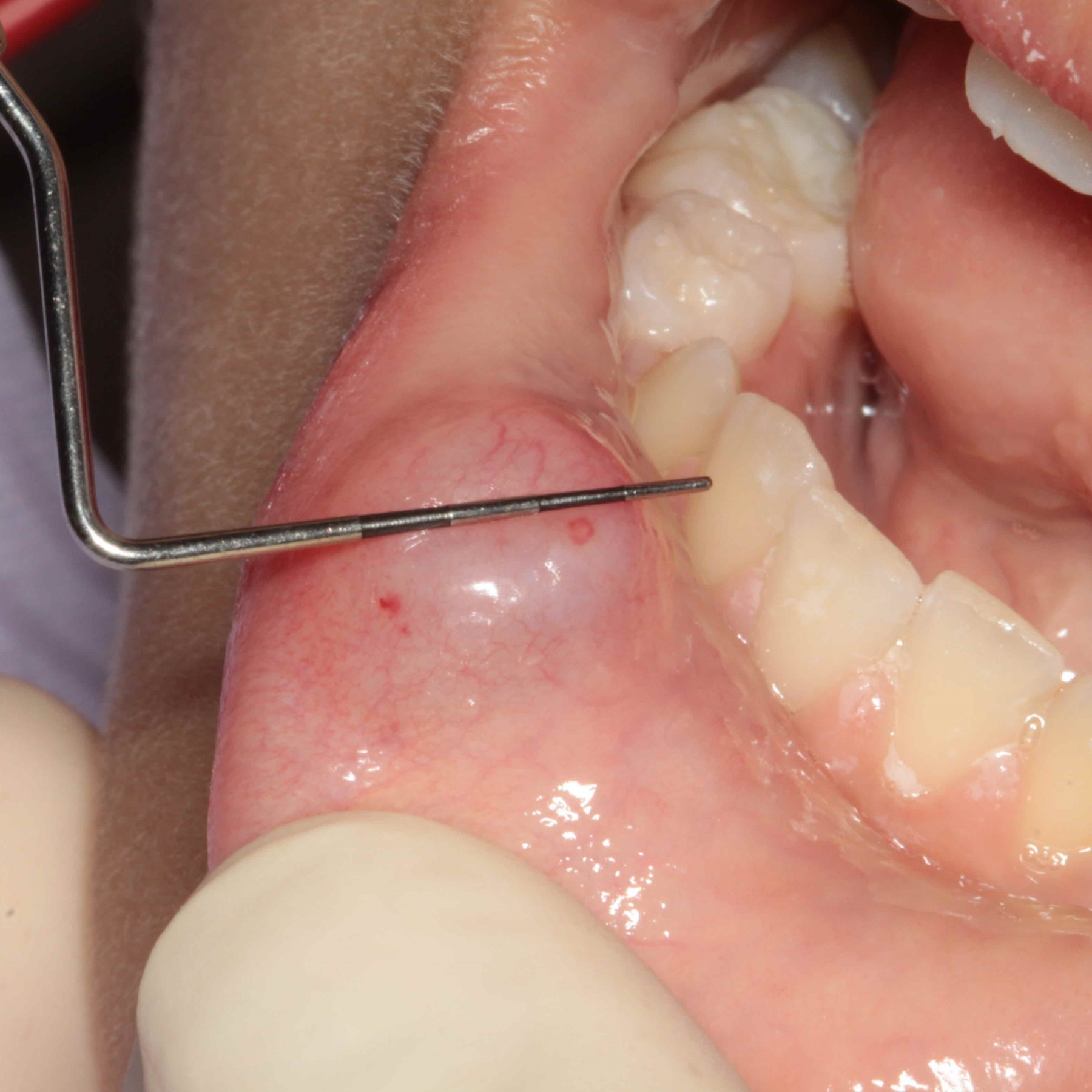
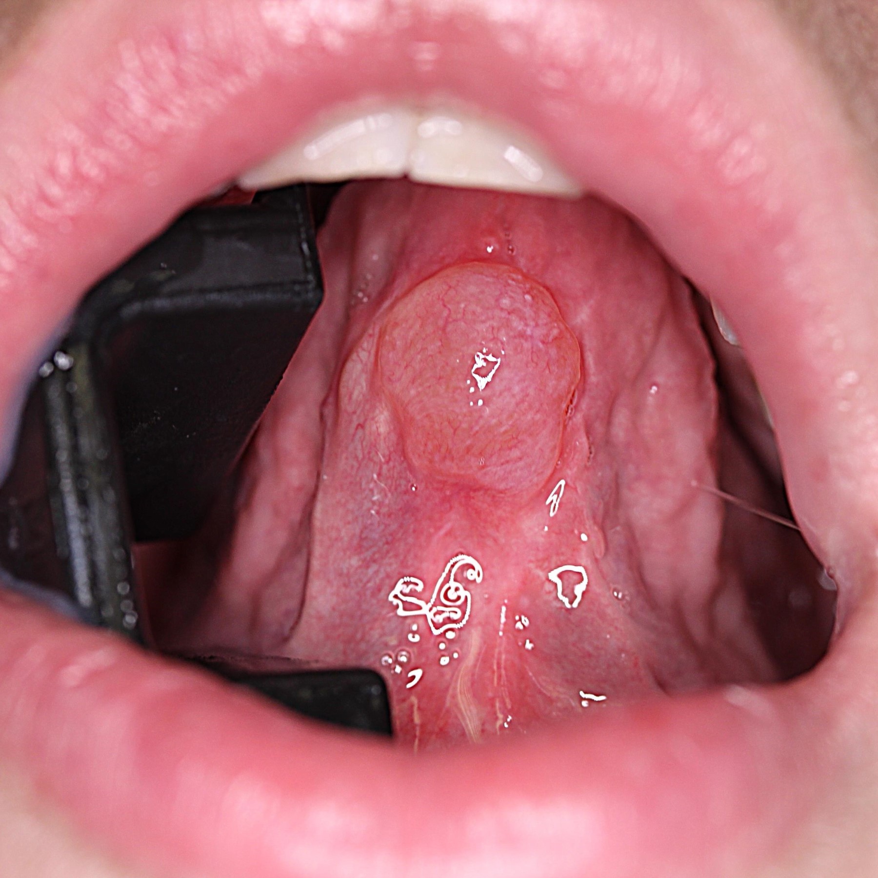
Contributed by Dhiraj B. Nikumbh, M.B.B.S., M.D.
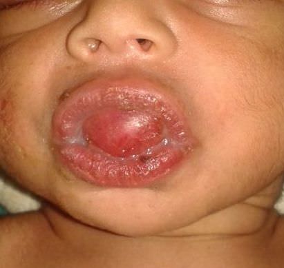
2 day old girl with
difficulty eating and
breathing due to
upper jaw lesion
Images hosted on other servers:

Lesion at 3 weeks

Lesion at 16 months

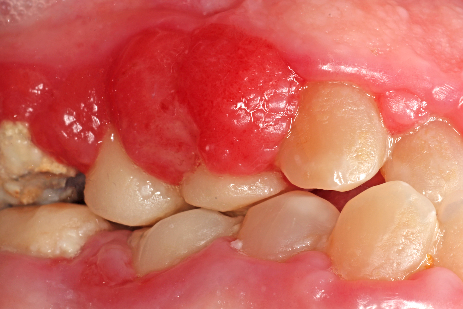
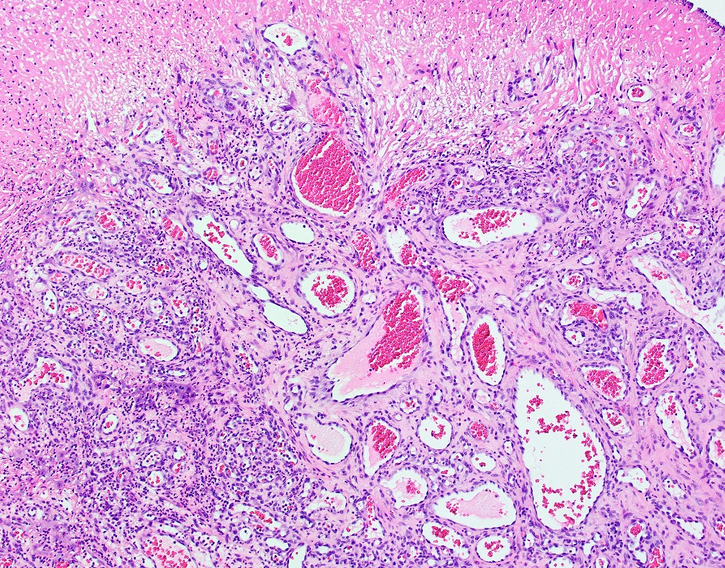
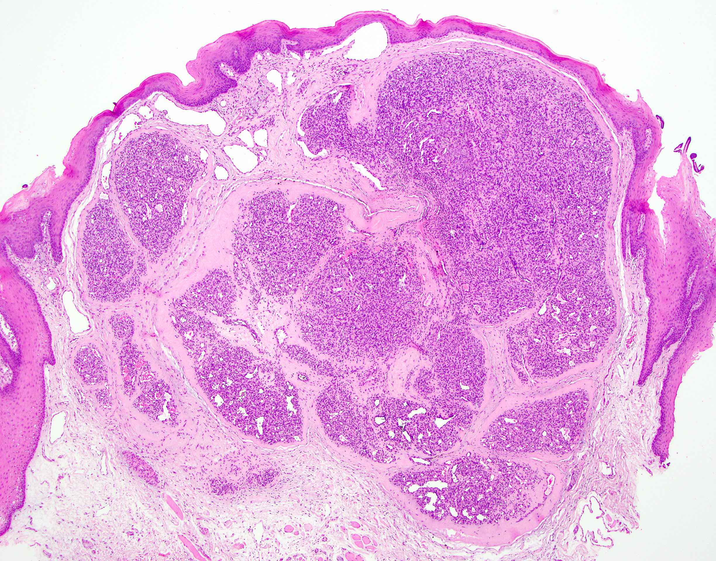
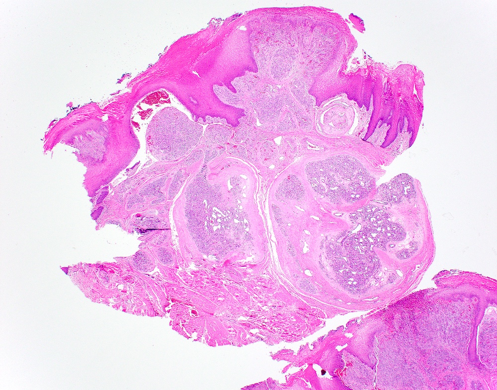
Tumor arising from anterior maxillary alveolus
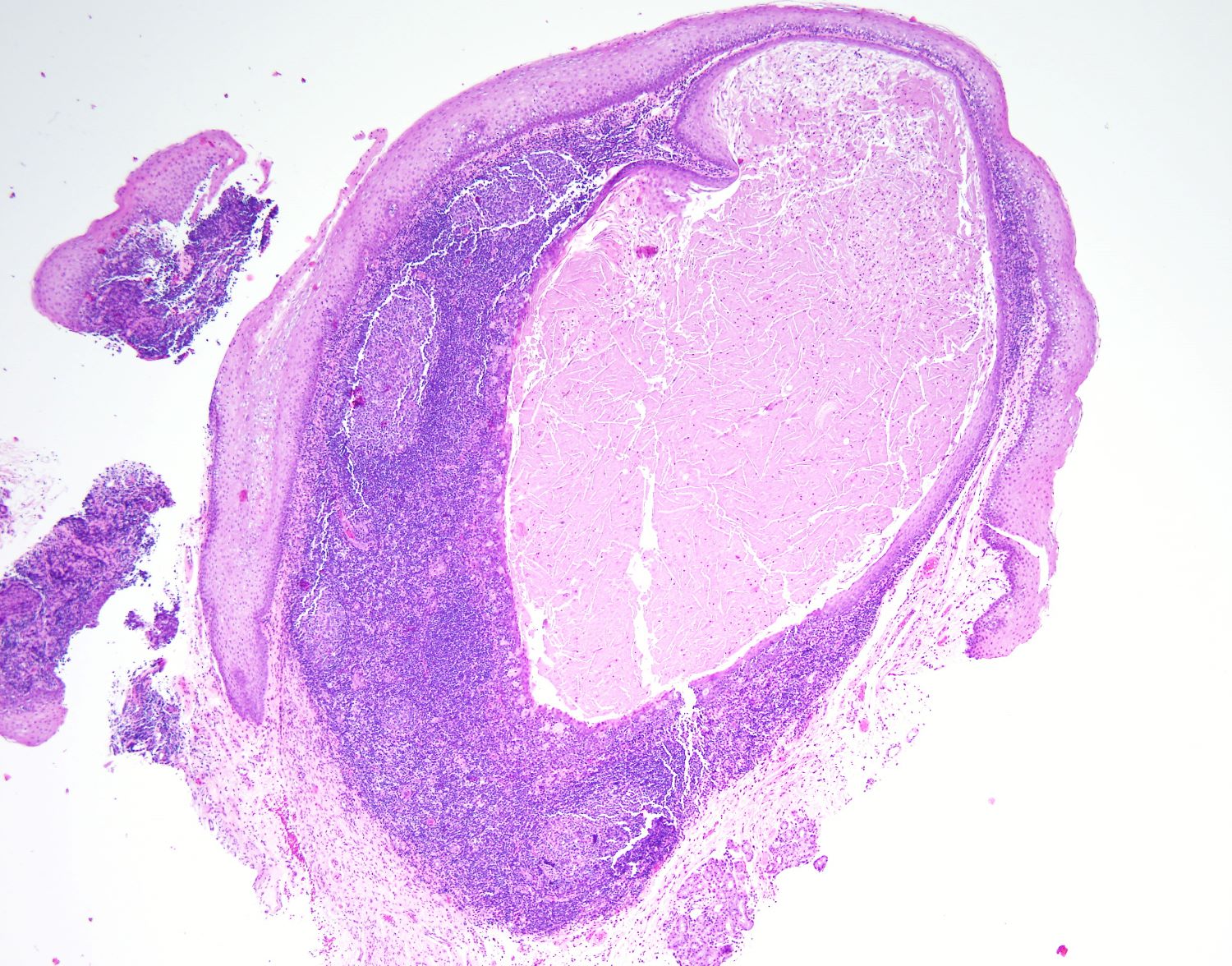
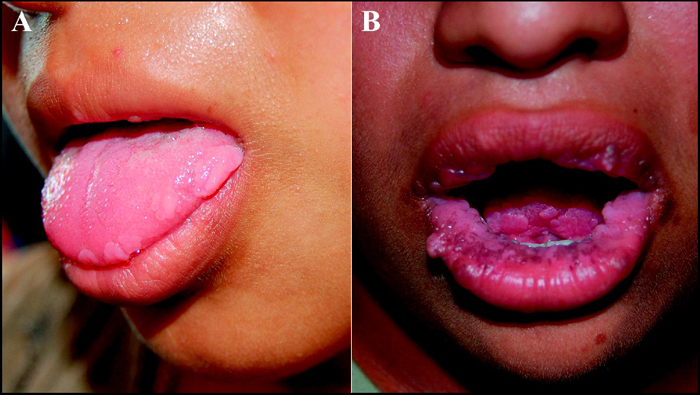
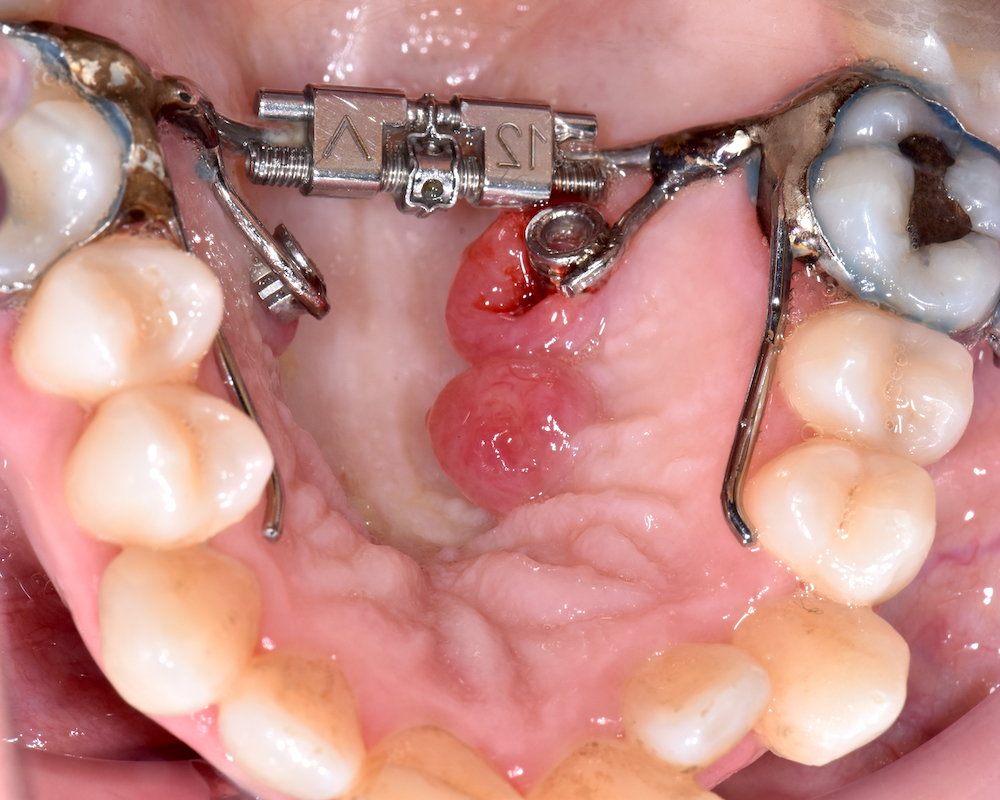
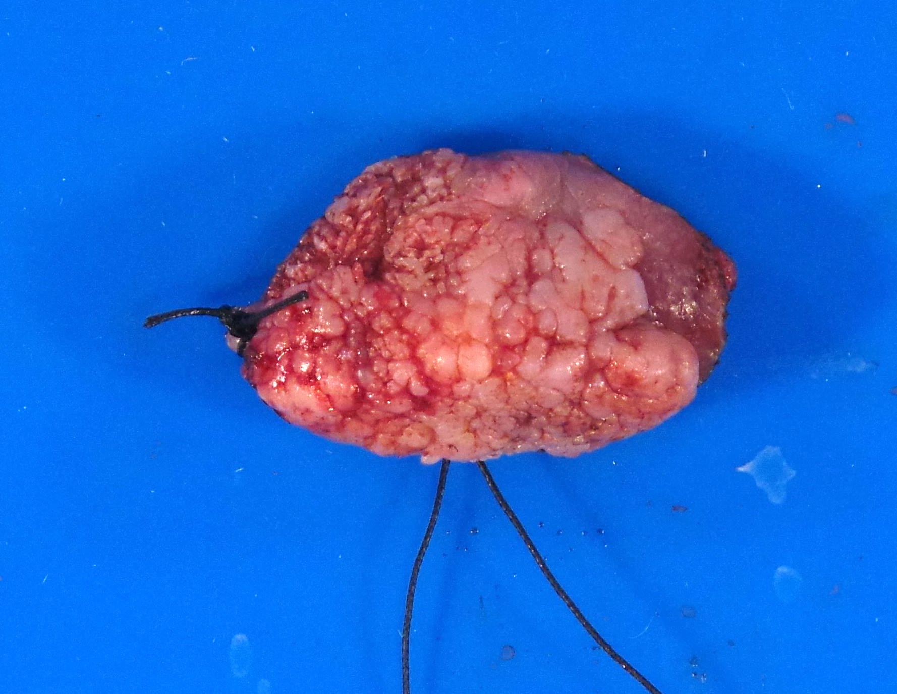
Contributed by Dhiraj B. Nikumbh, M.B.B.S., M.D.
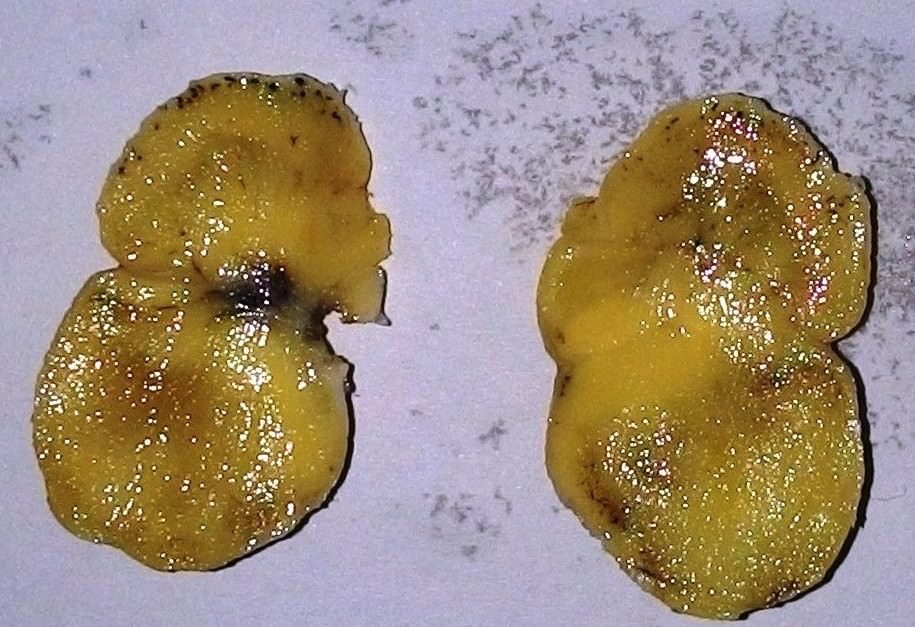
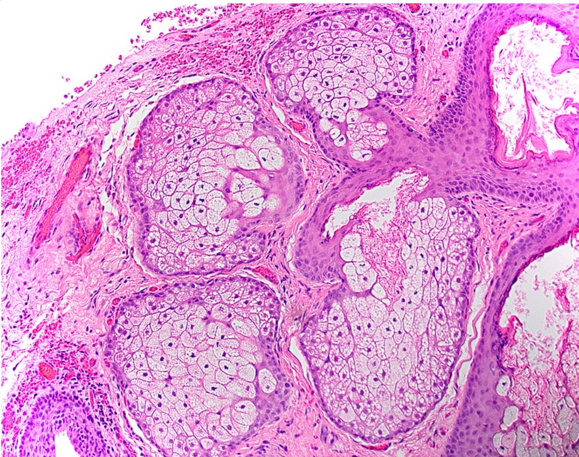
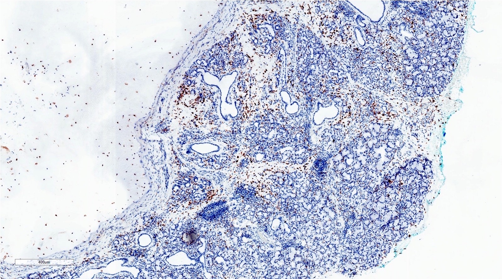
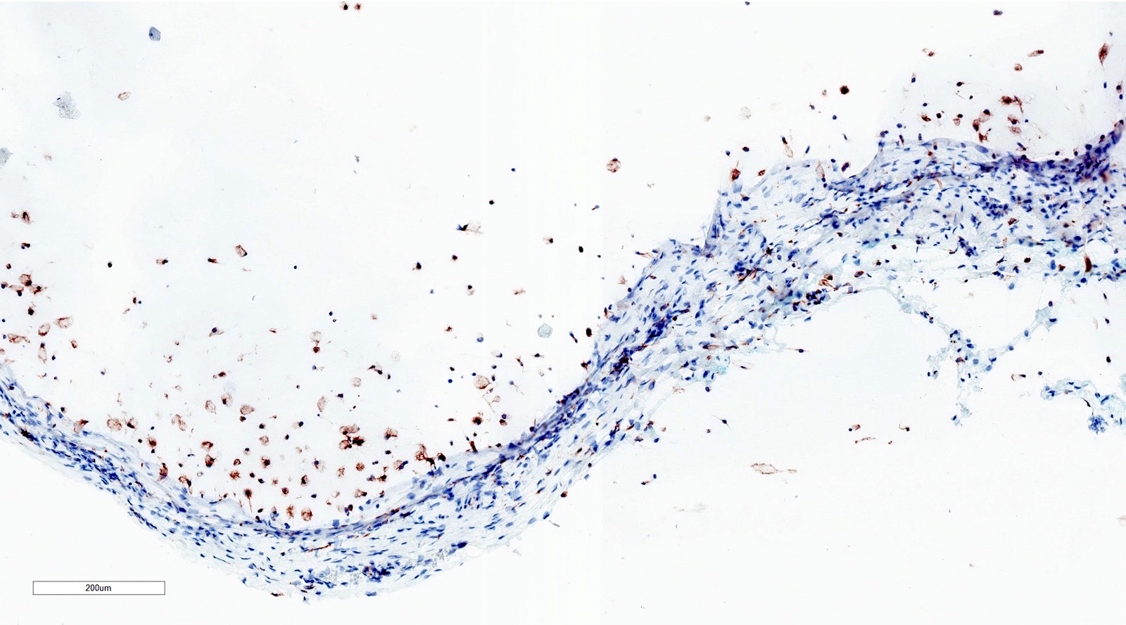
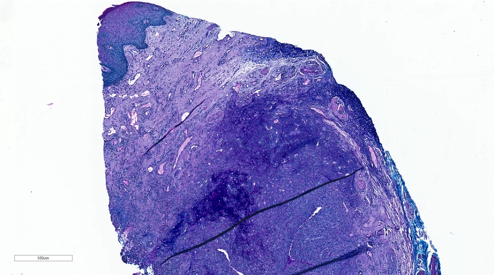
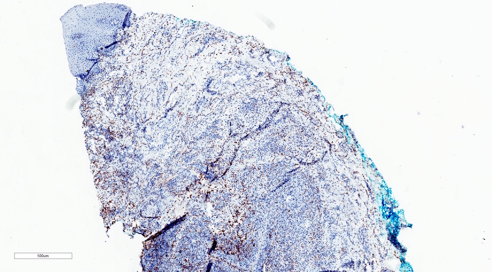
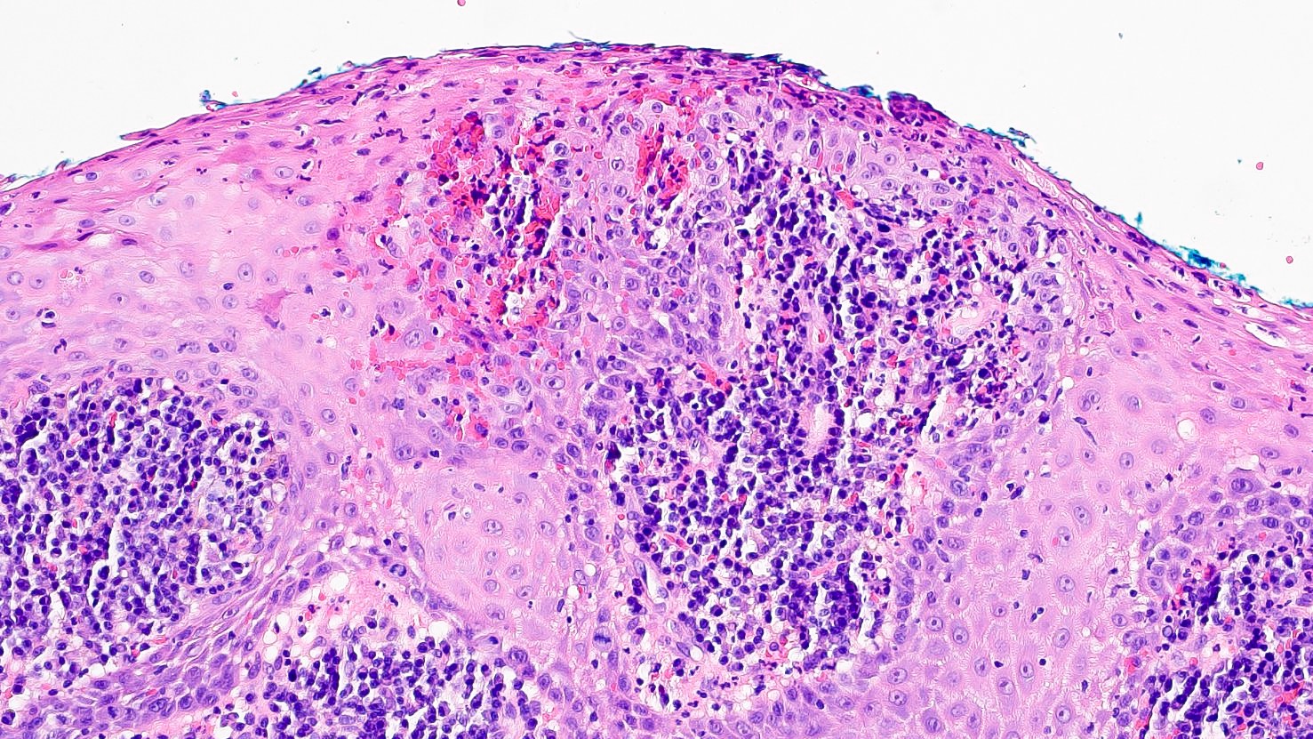
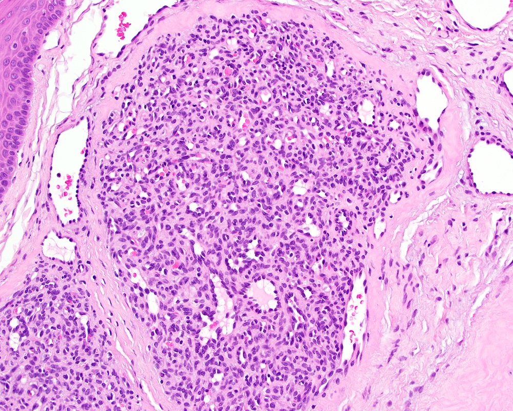
Congenital granular cell tumor
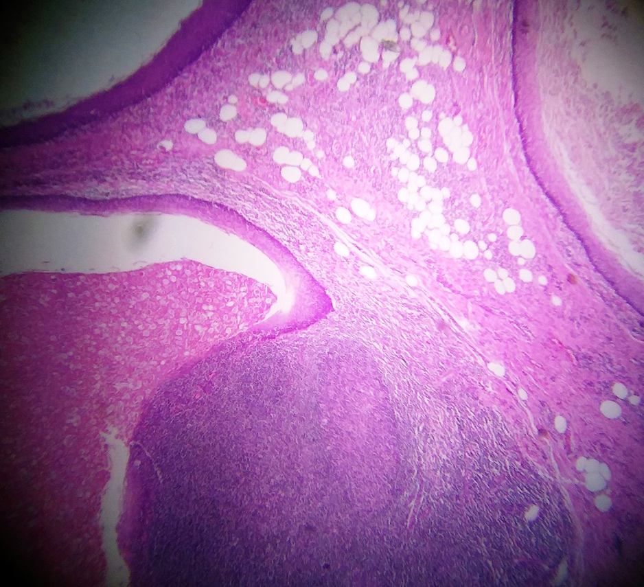
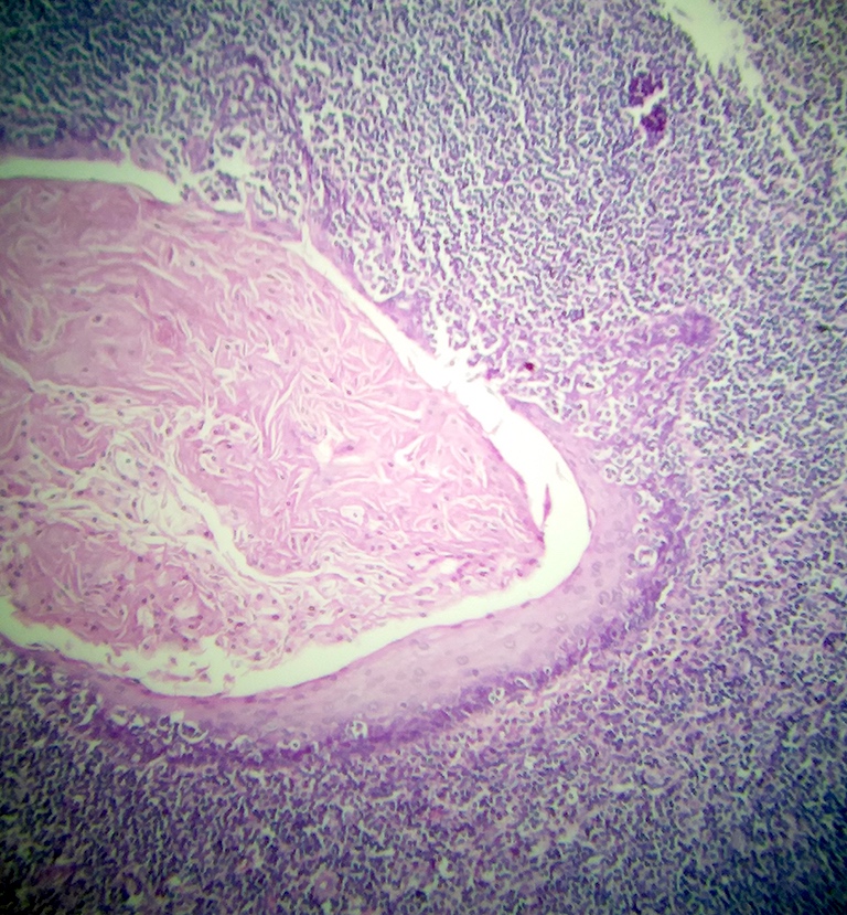
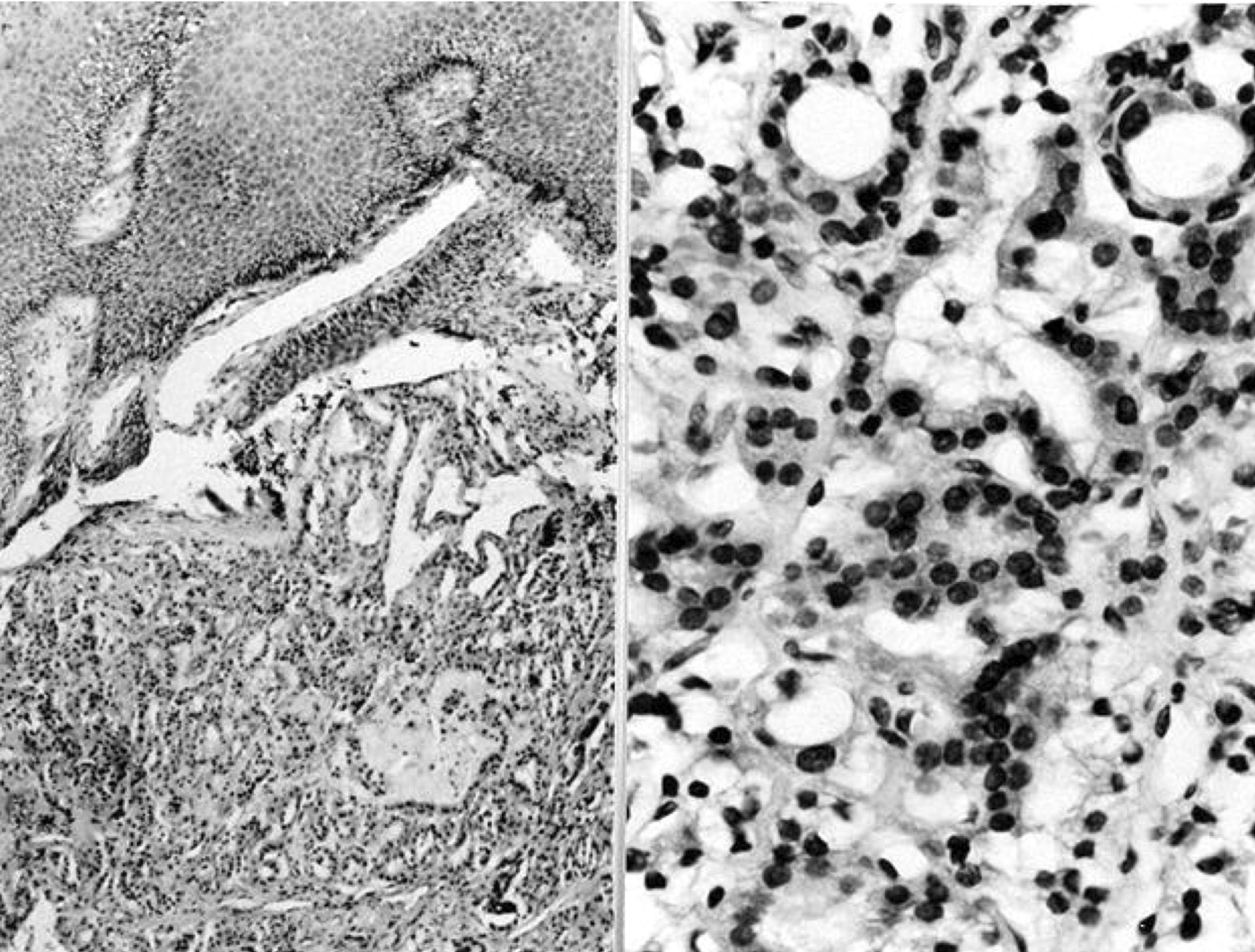
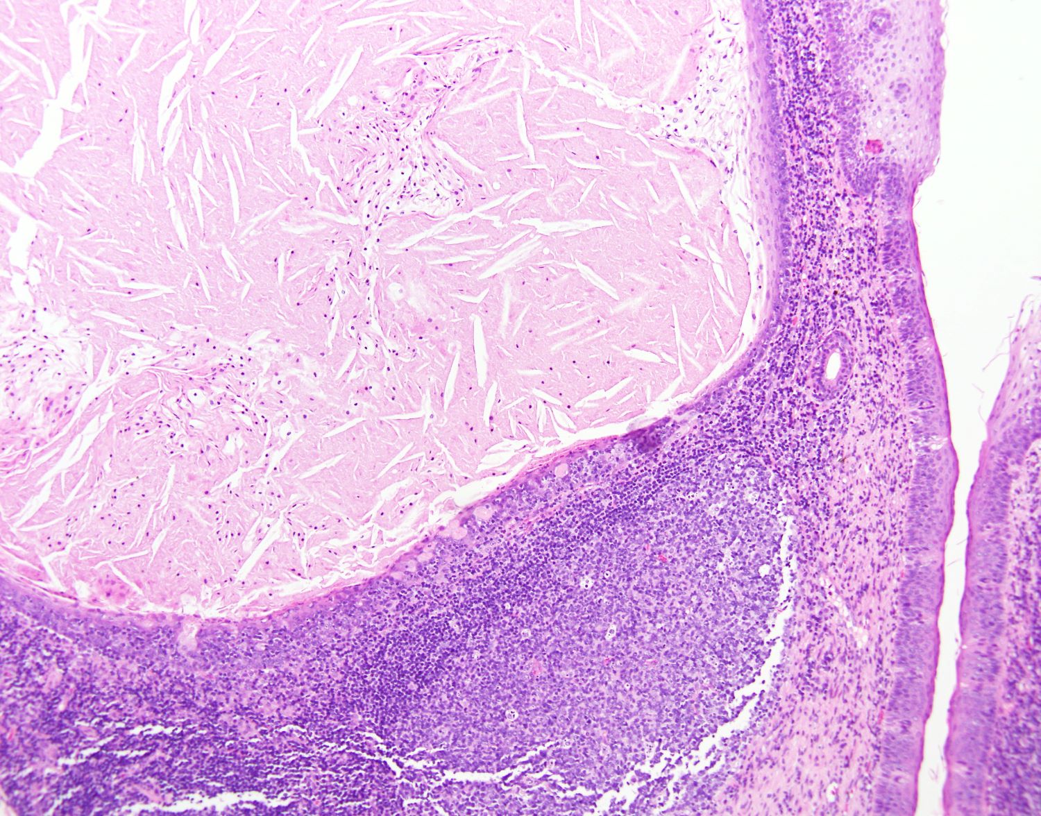
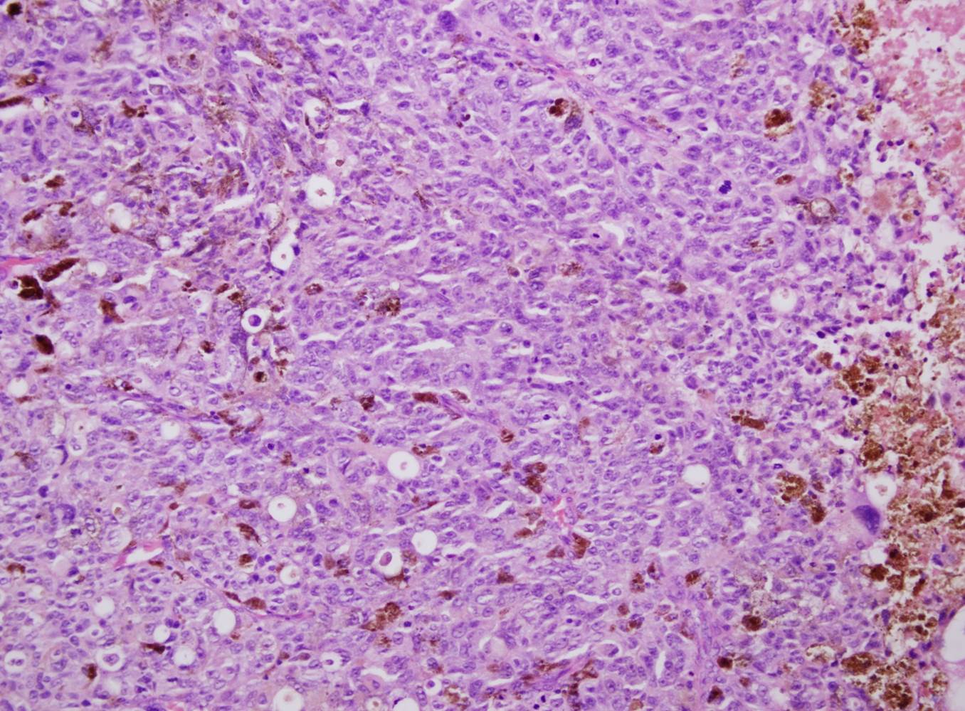
Contributed by Dhiraj B. Nikumbh, M.B.B.S., M.D. and Kelly Magliocca, D.D.S., M.P.H. (Case #524)

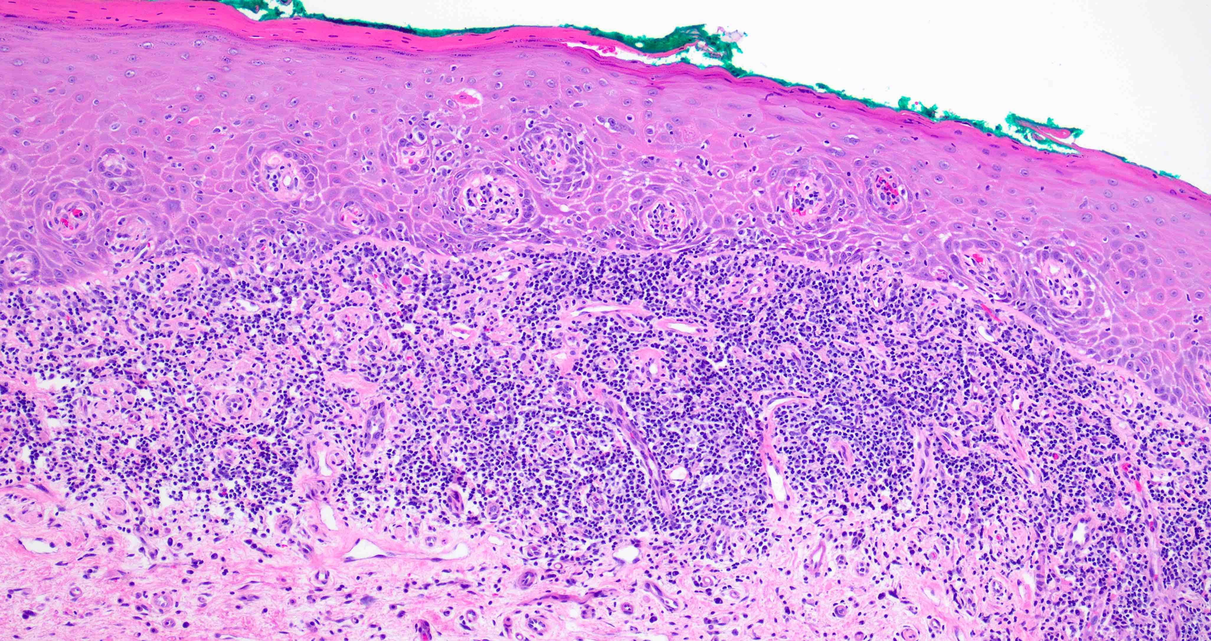
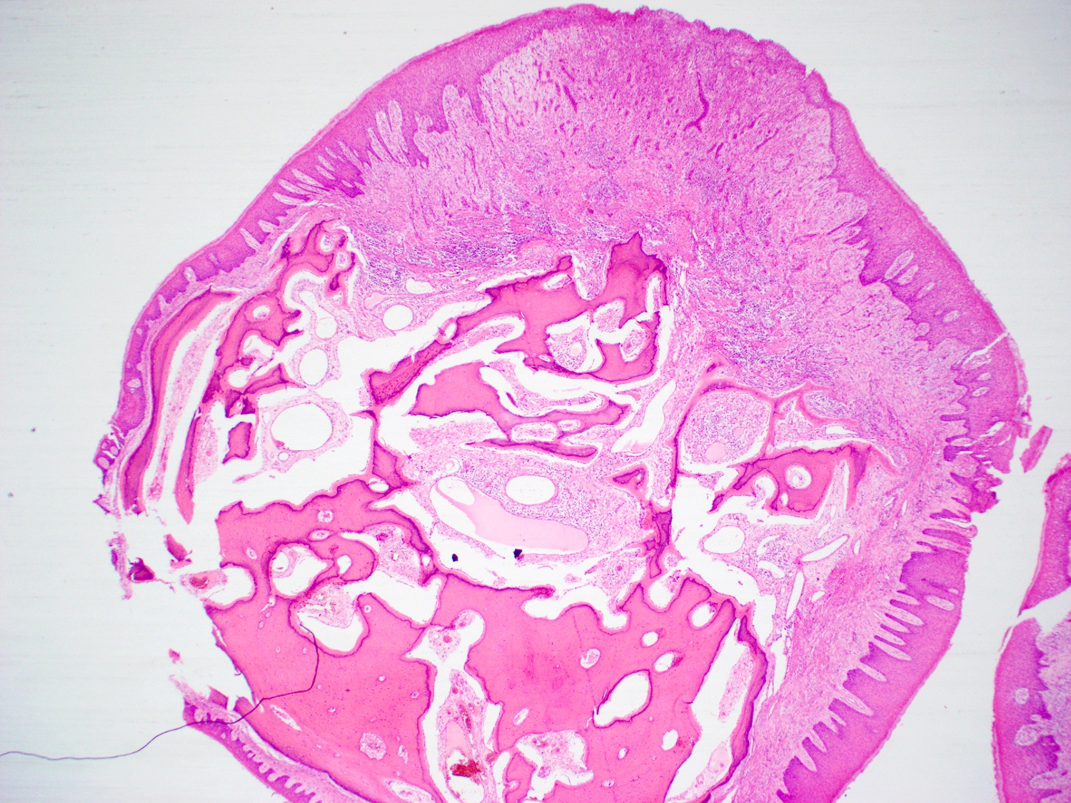
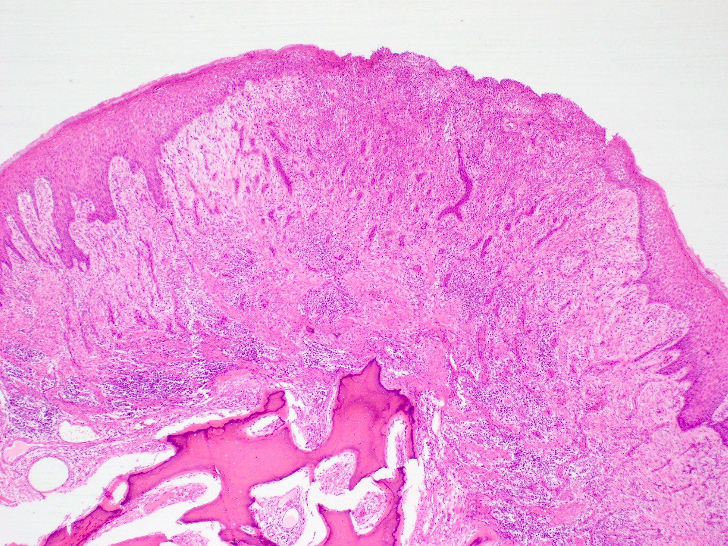
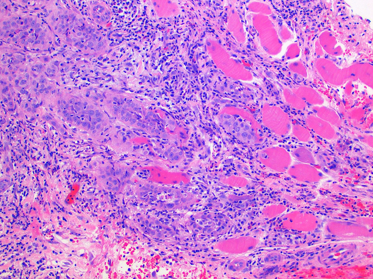
Stratified squamous epithelium
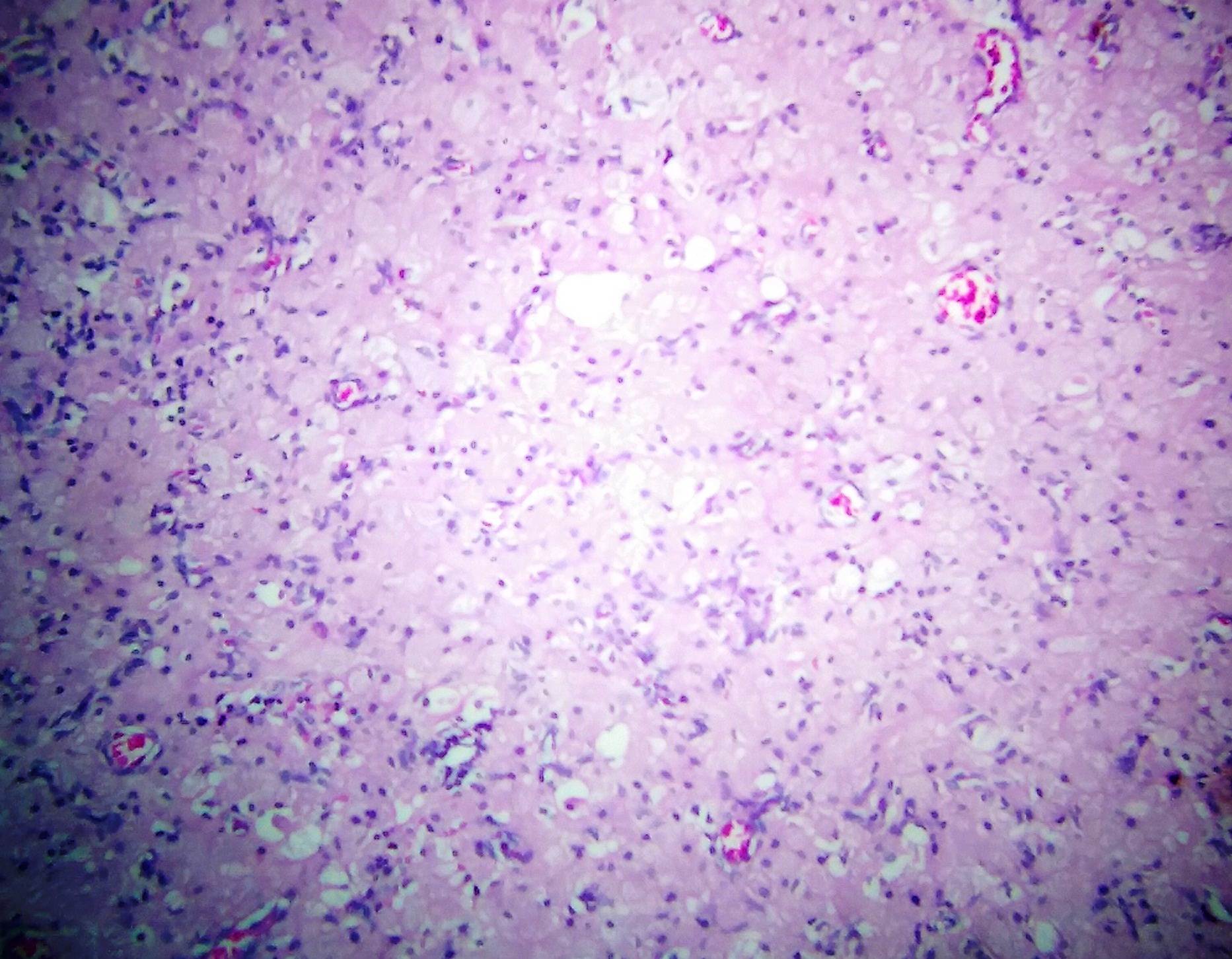
Abundant granular eosinophilic cytoplasm
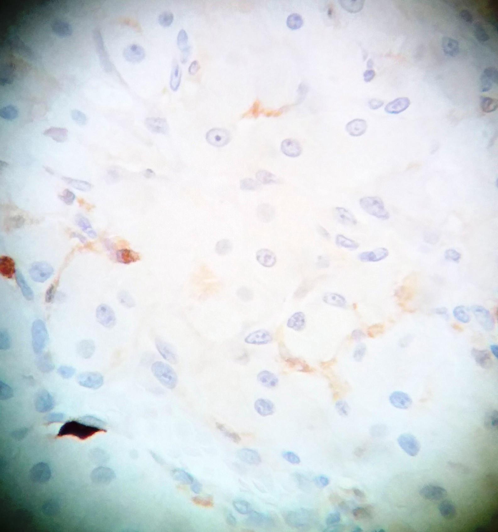
S100
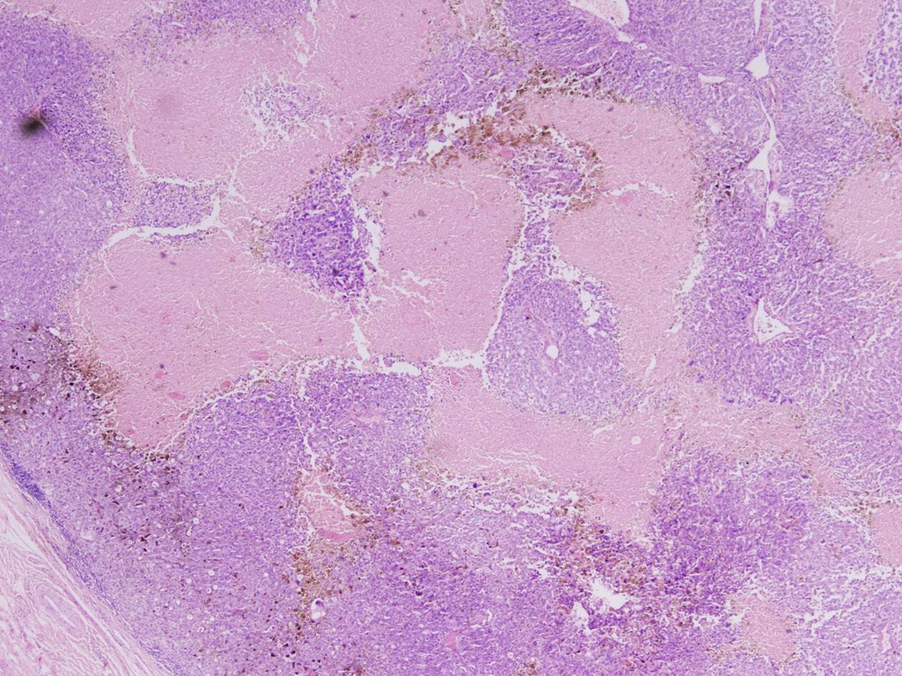
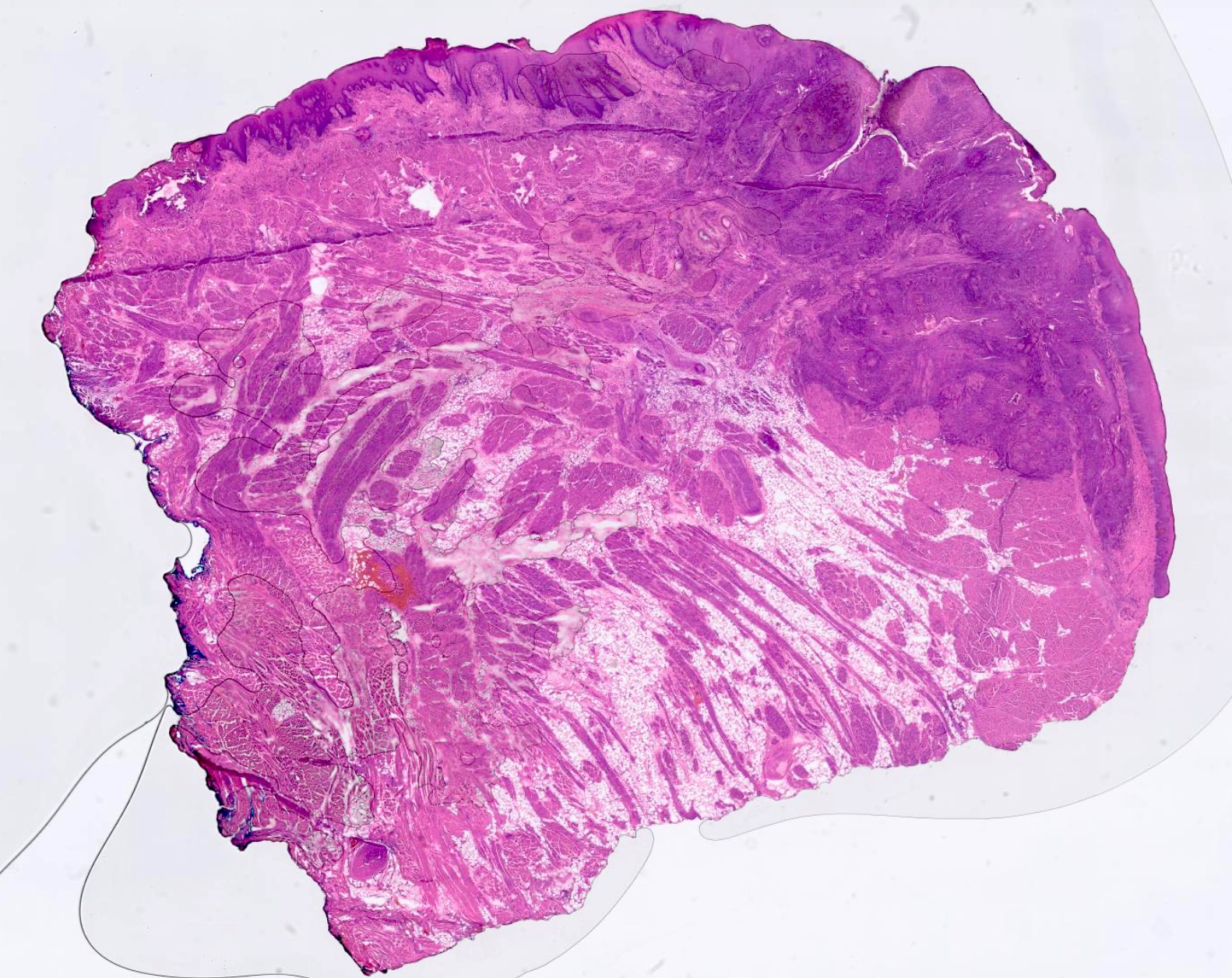
Scanning magnification
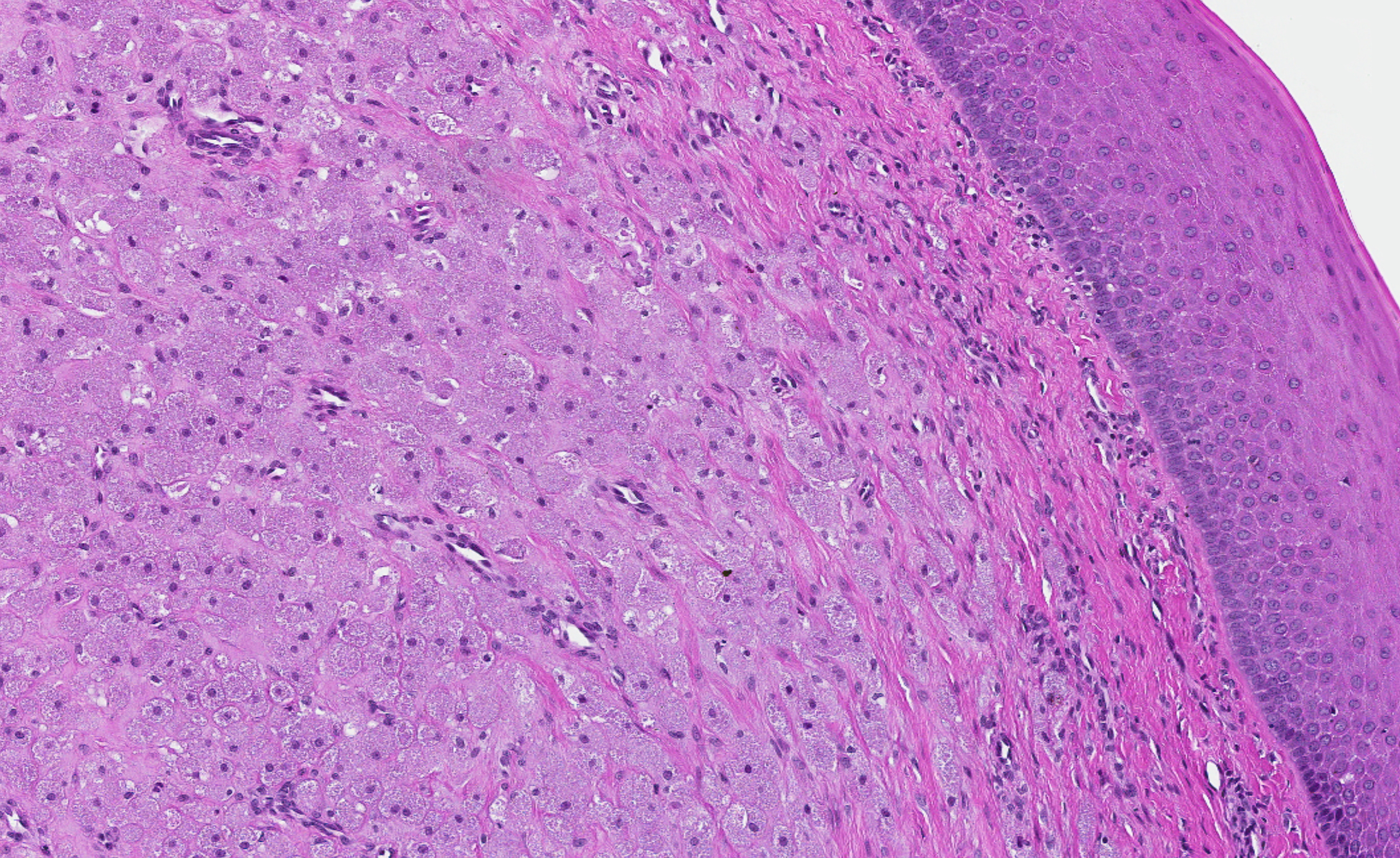
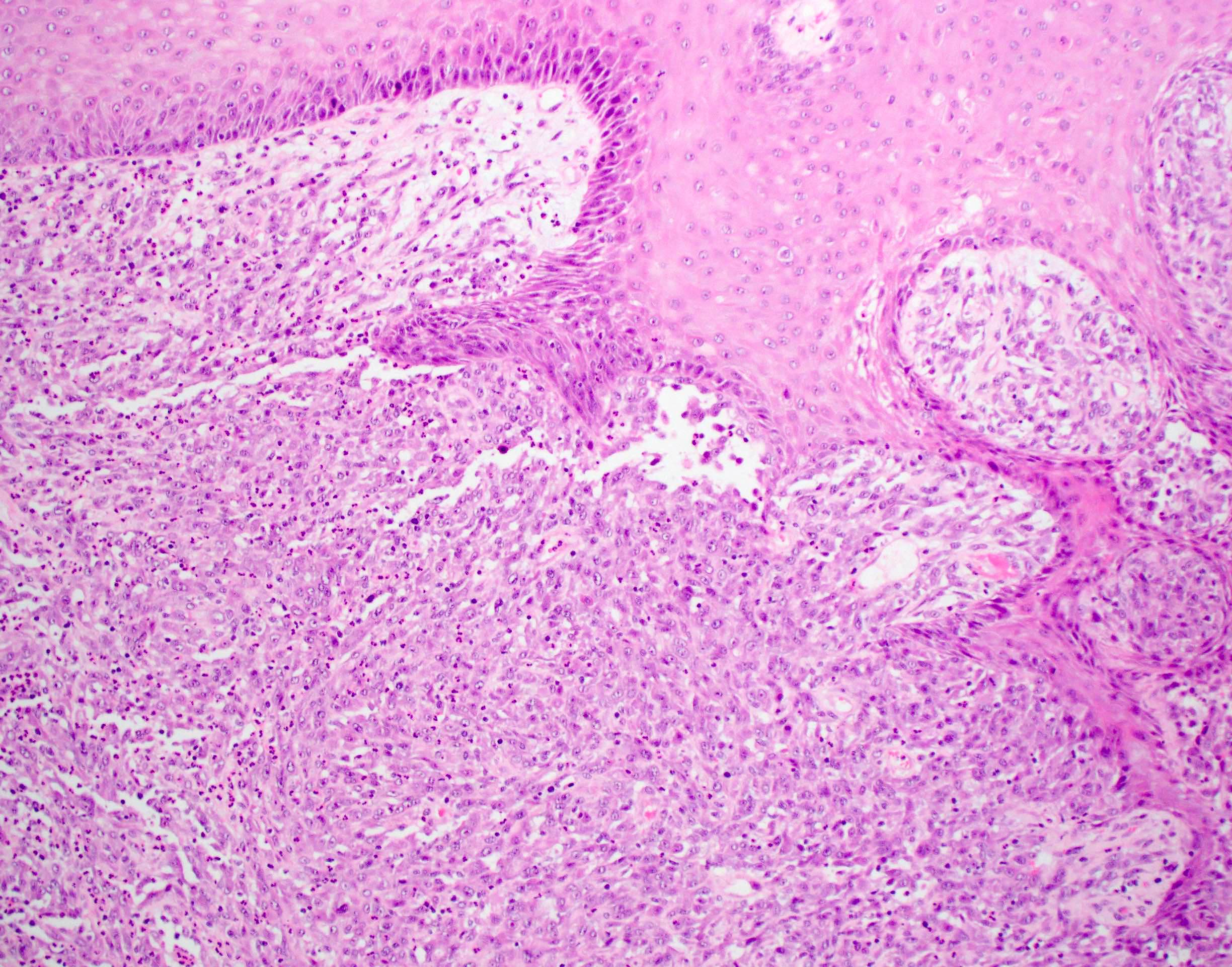
Stroma and overlying surface epithelium
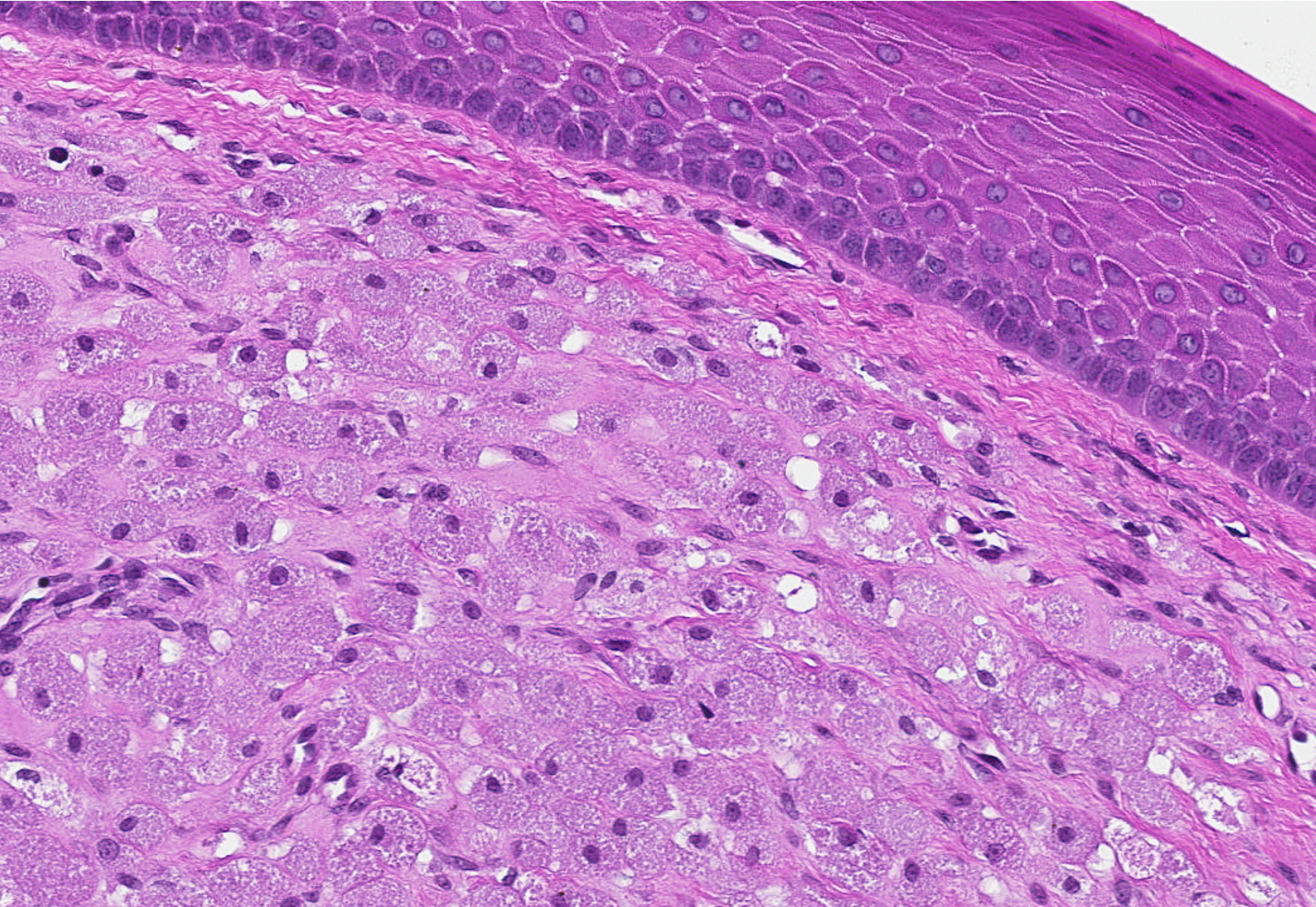
Sheet-like growth

Granular stromal cells
Images hosted on other servers:

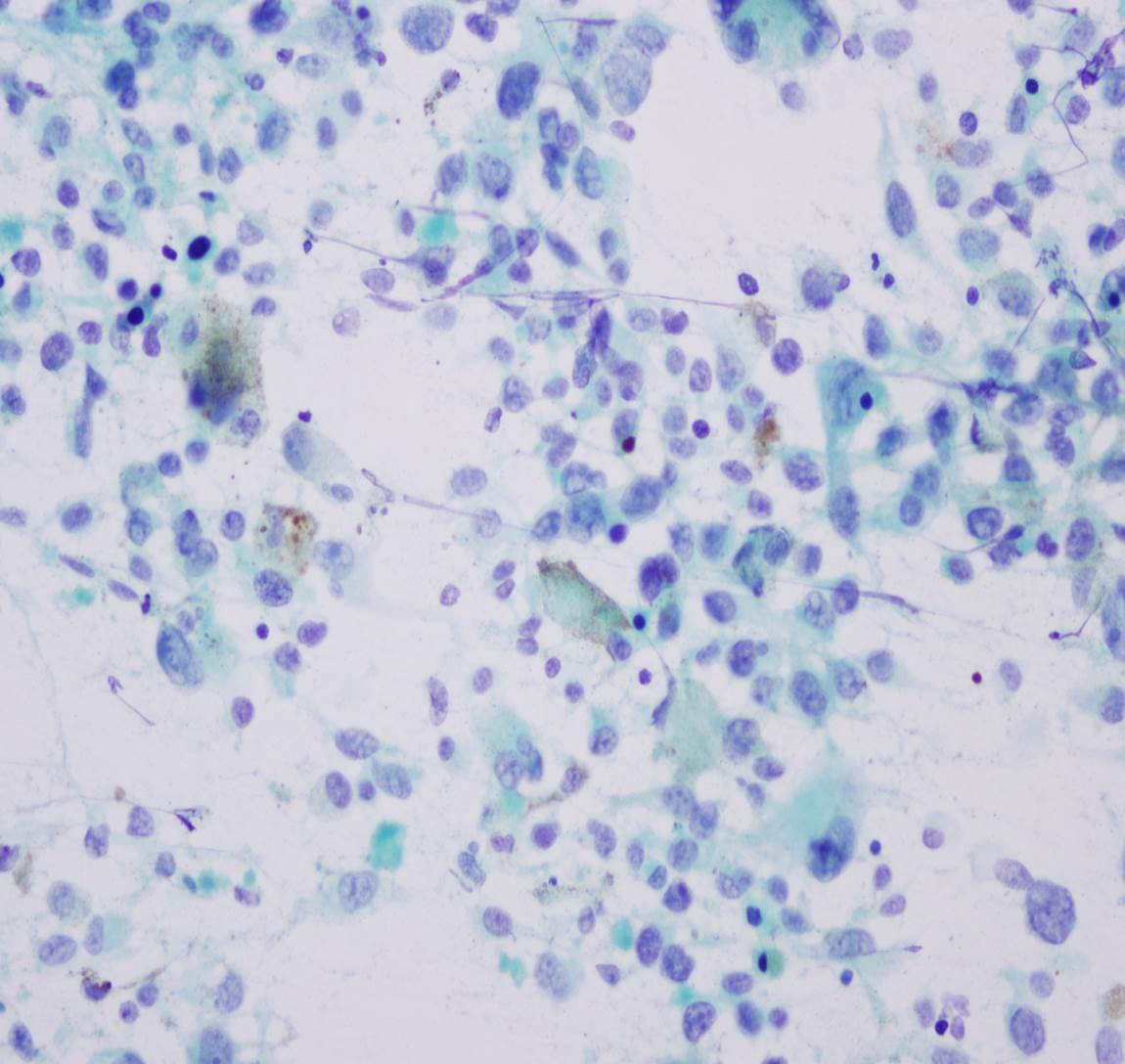
Polygonal cells
Images hosted on other servers:

Submental swelling
Contributed by Dhiraj B. Nikumbh, M.D.
Multiple tonsillar epidermoid cyst
Images hosted on other servers:

Sublingual dermoid cyst

Sublingual epidermoid cyst
Contributed by Dhiraj B. Nikumbh, M.D.

Multiple tonsillar epidermoid cyst
Images hosted on other servers:

Cyst filled with keratin

Sebaceous gland and hair follicles
Images hosted on other servers:

Anterior floor
of the mouth
and base of tongue

Foregut duplication cyst
Images hosted on other servers:

Foregut duplication cyst
Images hosted on other servers:


Forgut duplication cyst of anterior tongue
Contributed by Katherine Hulme, M.B.Ch.B.
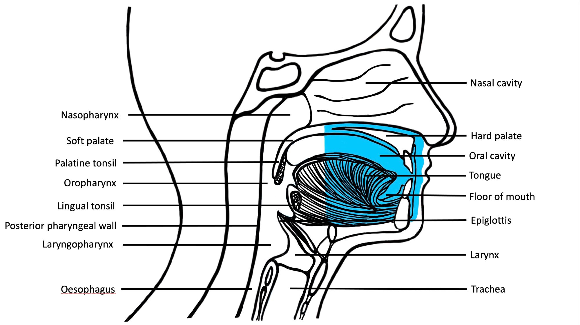
Oral cavity (sagittal view)
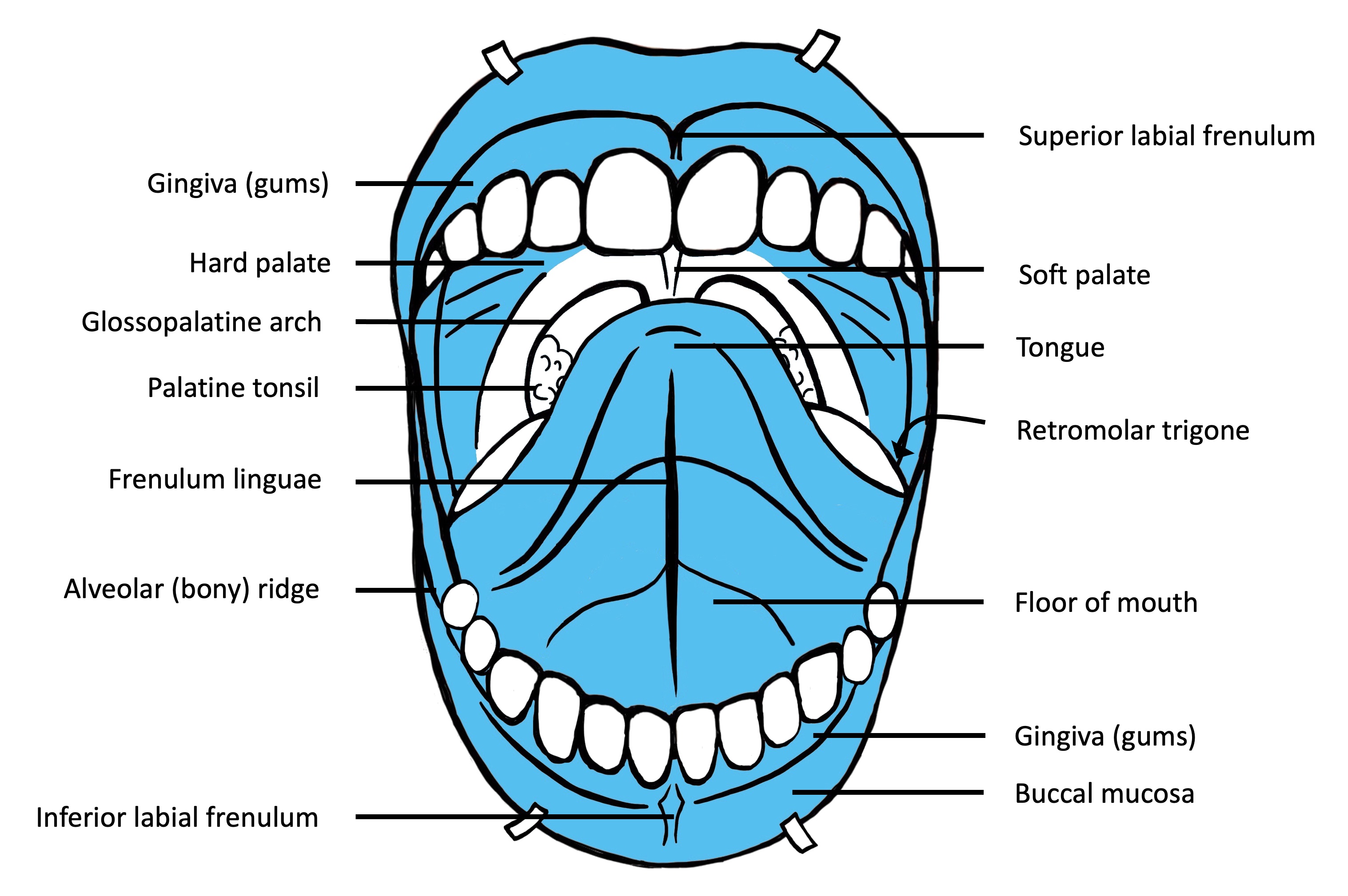
Oral cavity (frontal view)
Contributed by Molly Housley Smith, D.M.D.
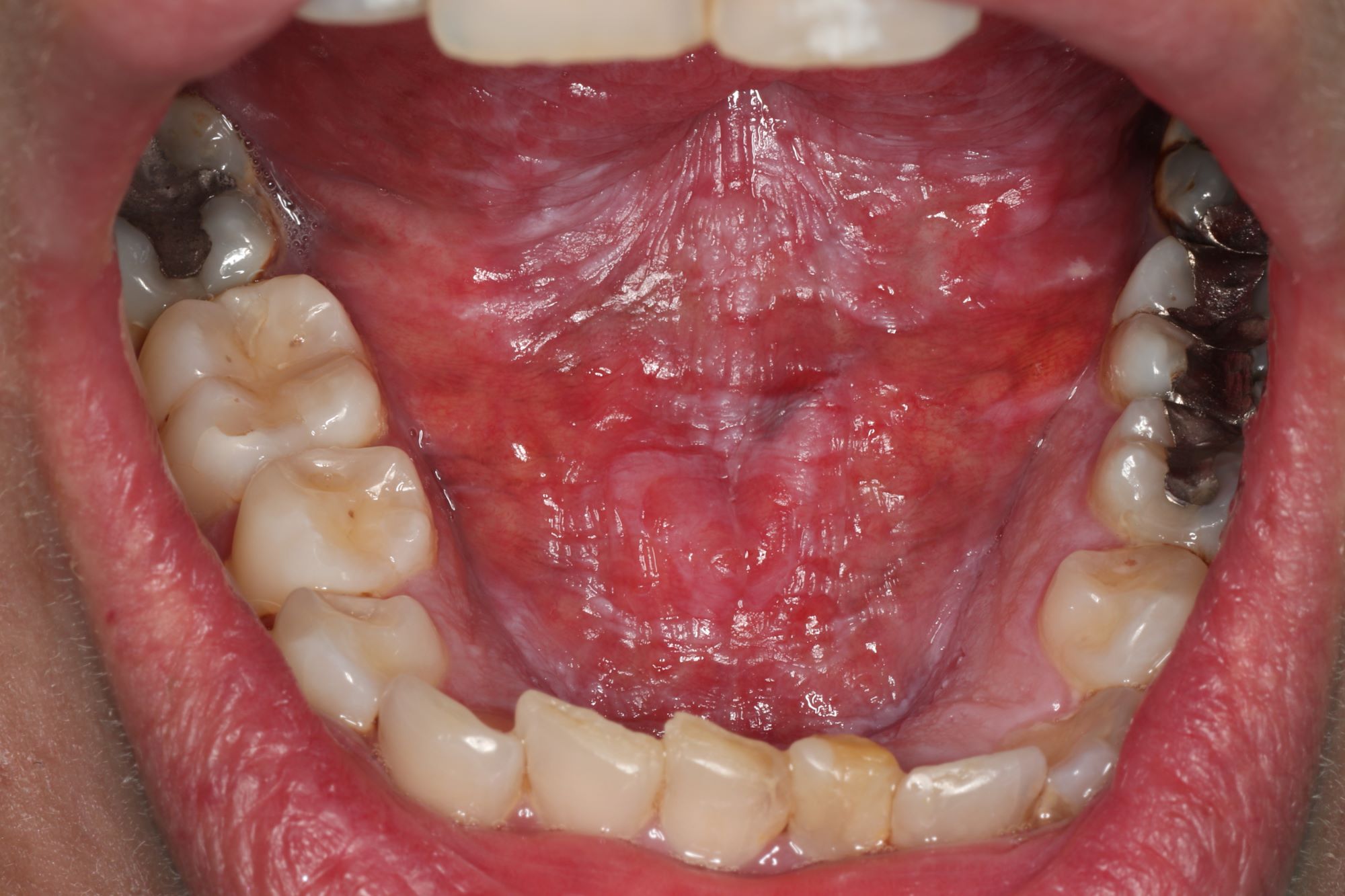
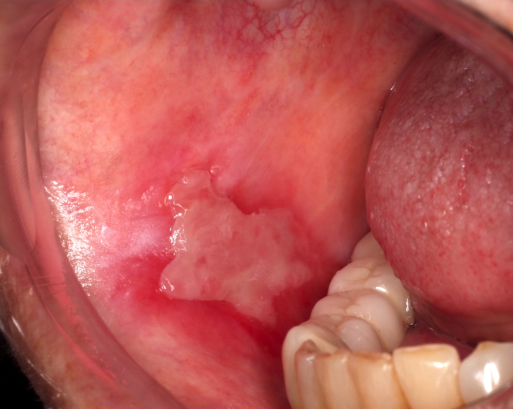
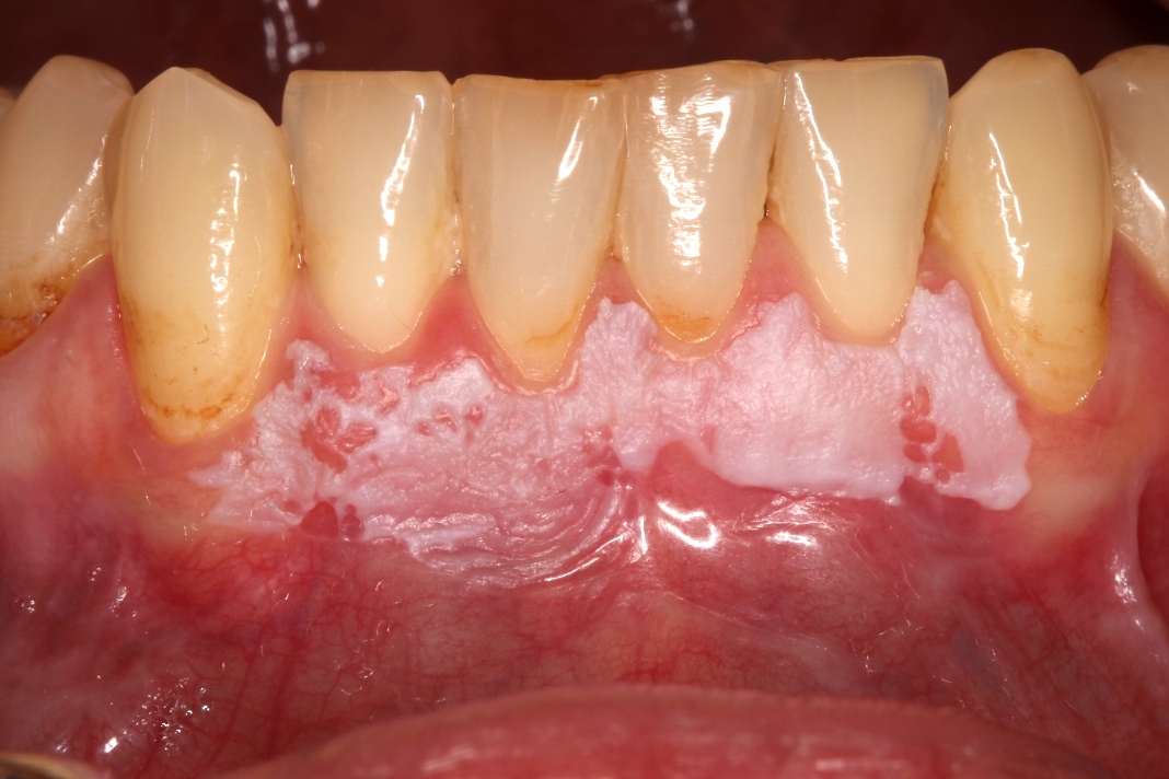
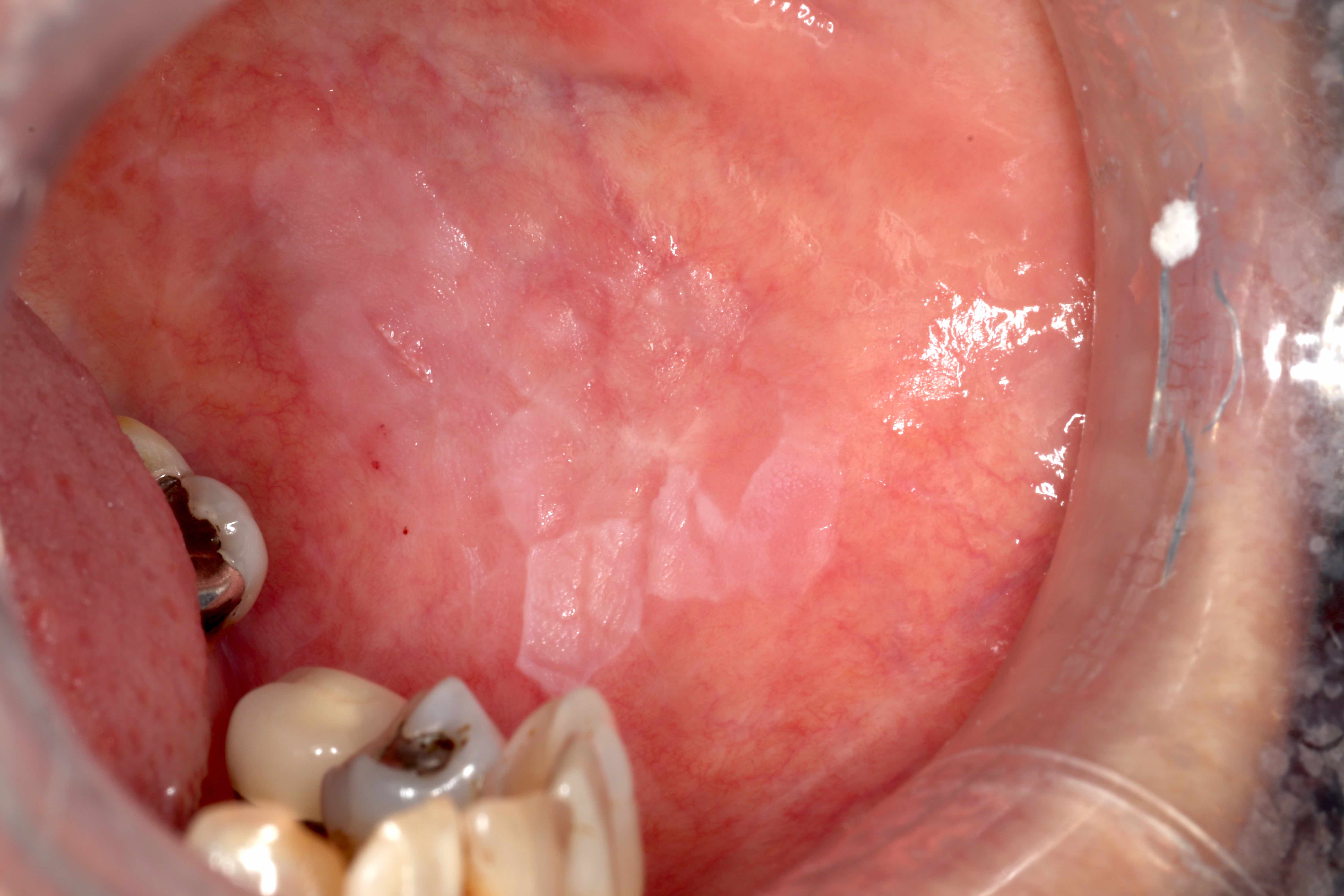
Floor of mouth leukoplakia
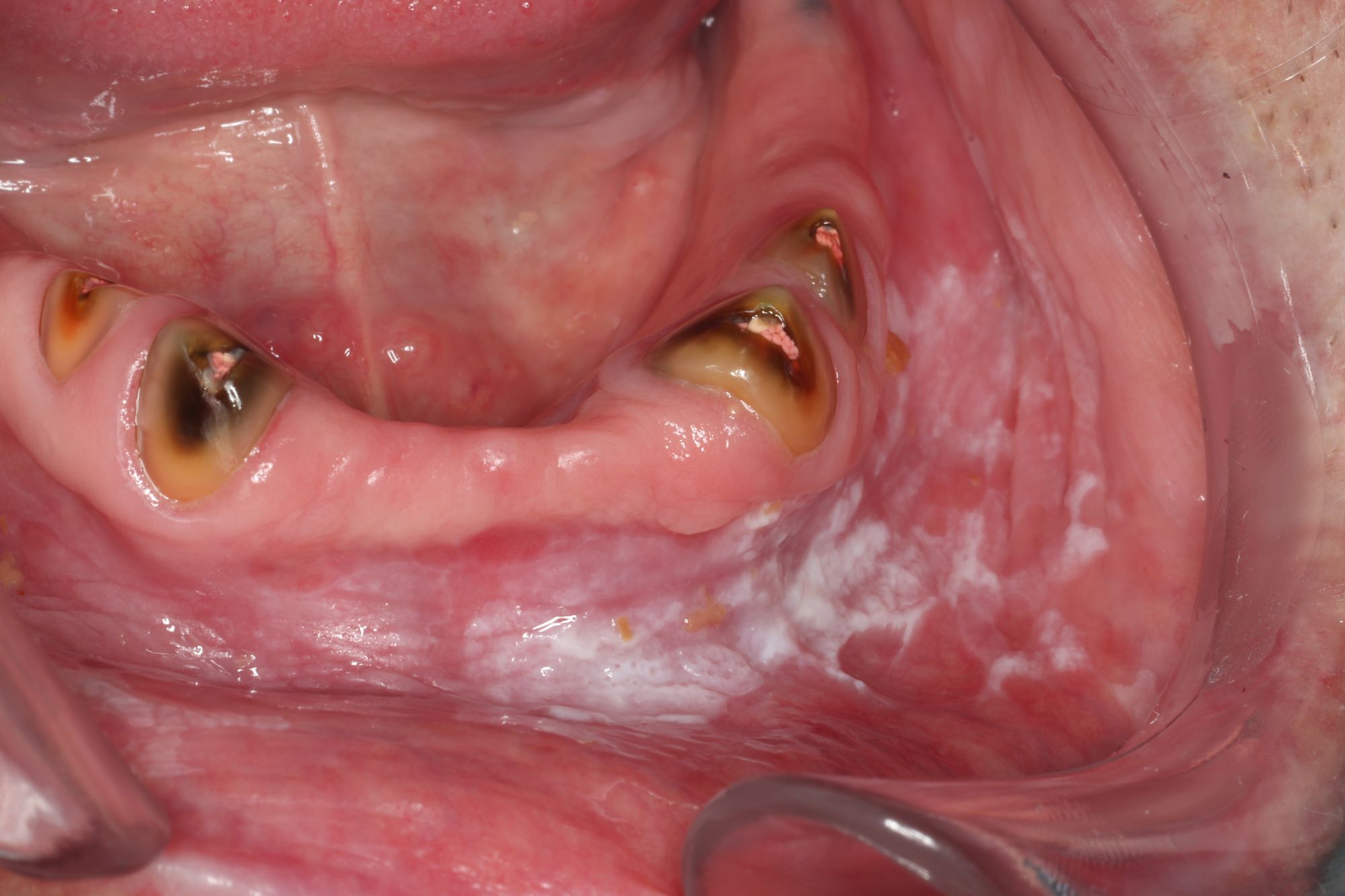
Buccal and labial leukoplakia
Images hosted on other servers:

Leukoplakia of buccal mucosa
Contributed by Veronica Cheung, M.Ch.D.
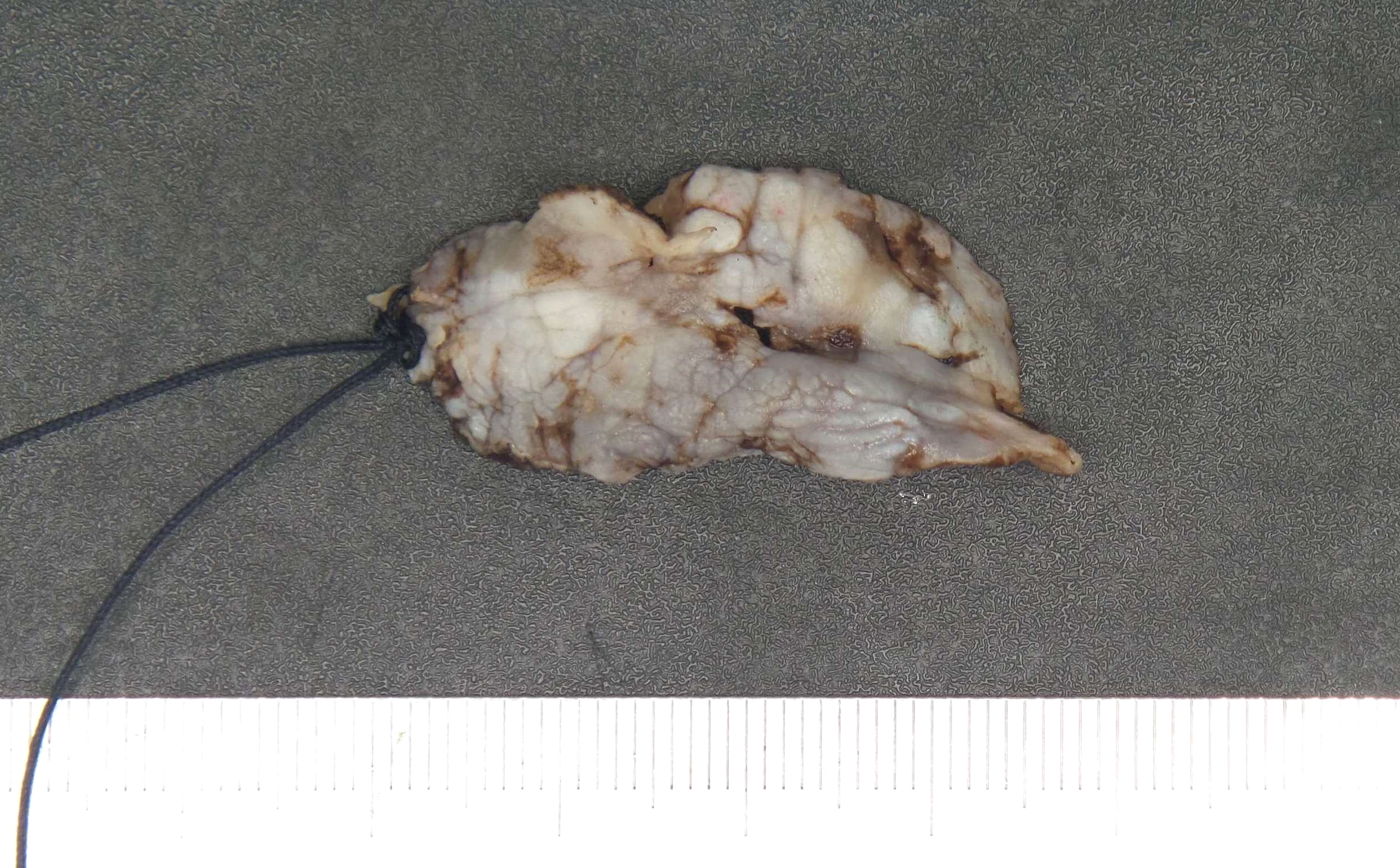
Laser resection oral mucosa
Contributed by Veronica Cheung, M.Ch.D.
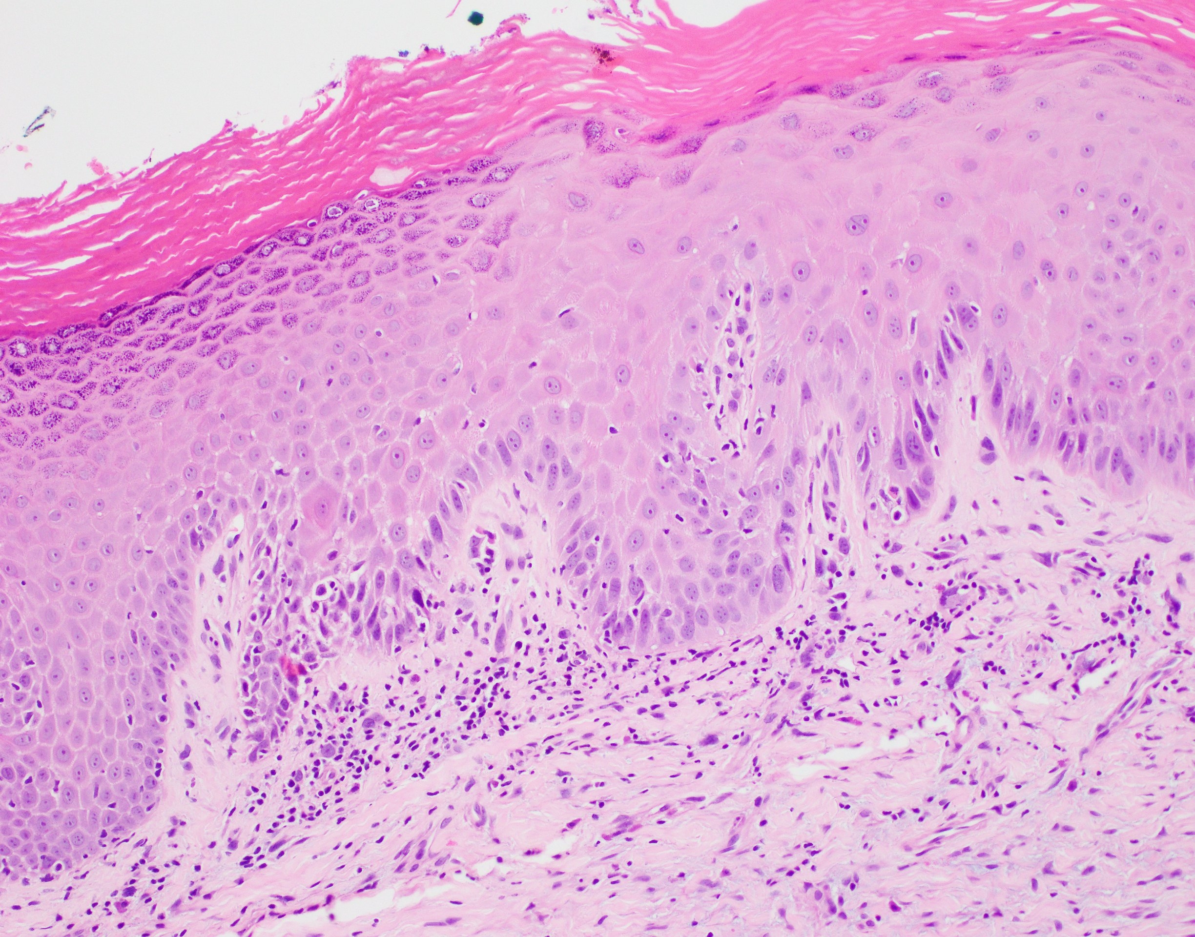
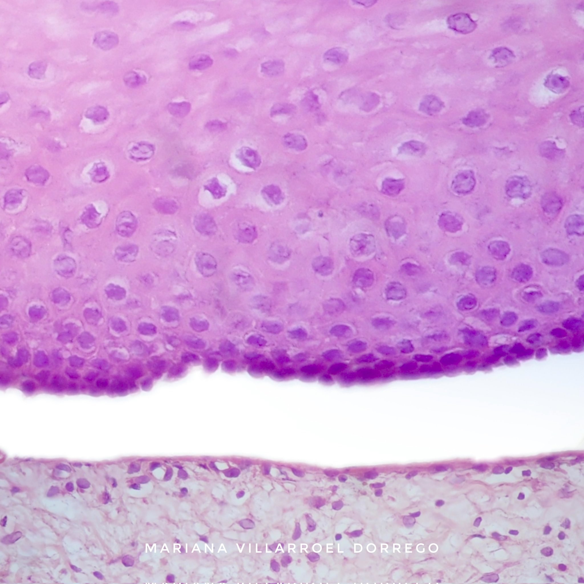
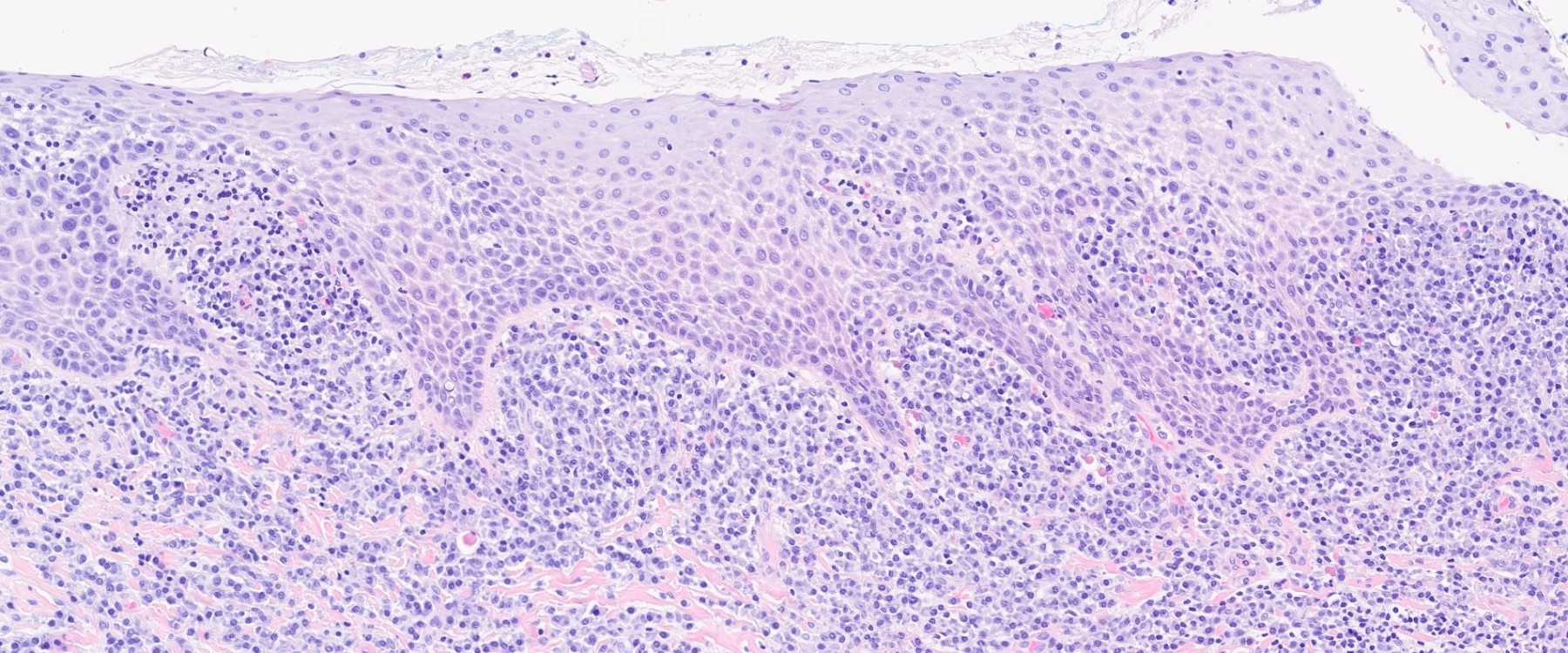
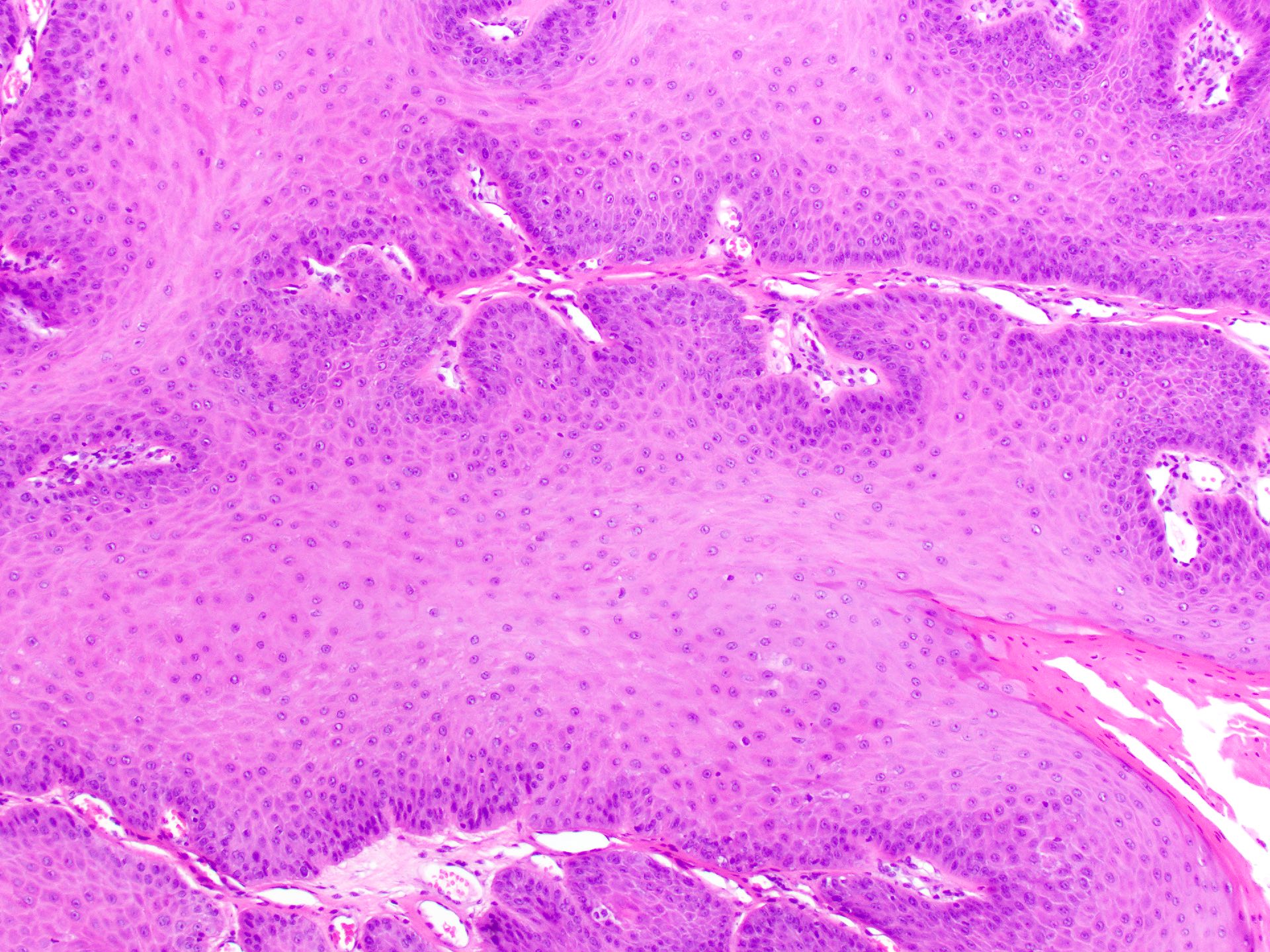
Basal cytologic and architectural change
Drop shaped rete
Dysplasia involving > two - thirds of mucosa
Severe cytologic, architectural atypia
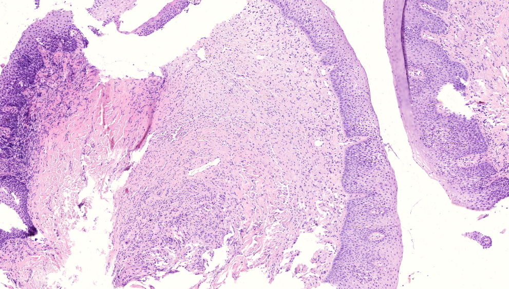
Transition from normal to dysplastic mucosa

Floor of mouth dysplasia with skip lesion
Images hosted on other servers:

Cystic appearing mass

T1 weighted image, MRI

T2 weighted image, MRI
Images hosted on other servers:

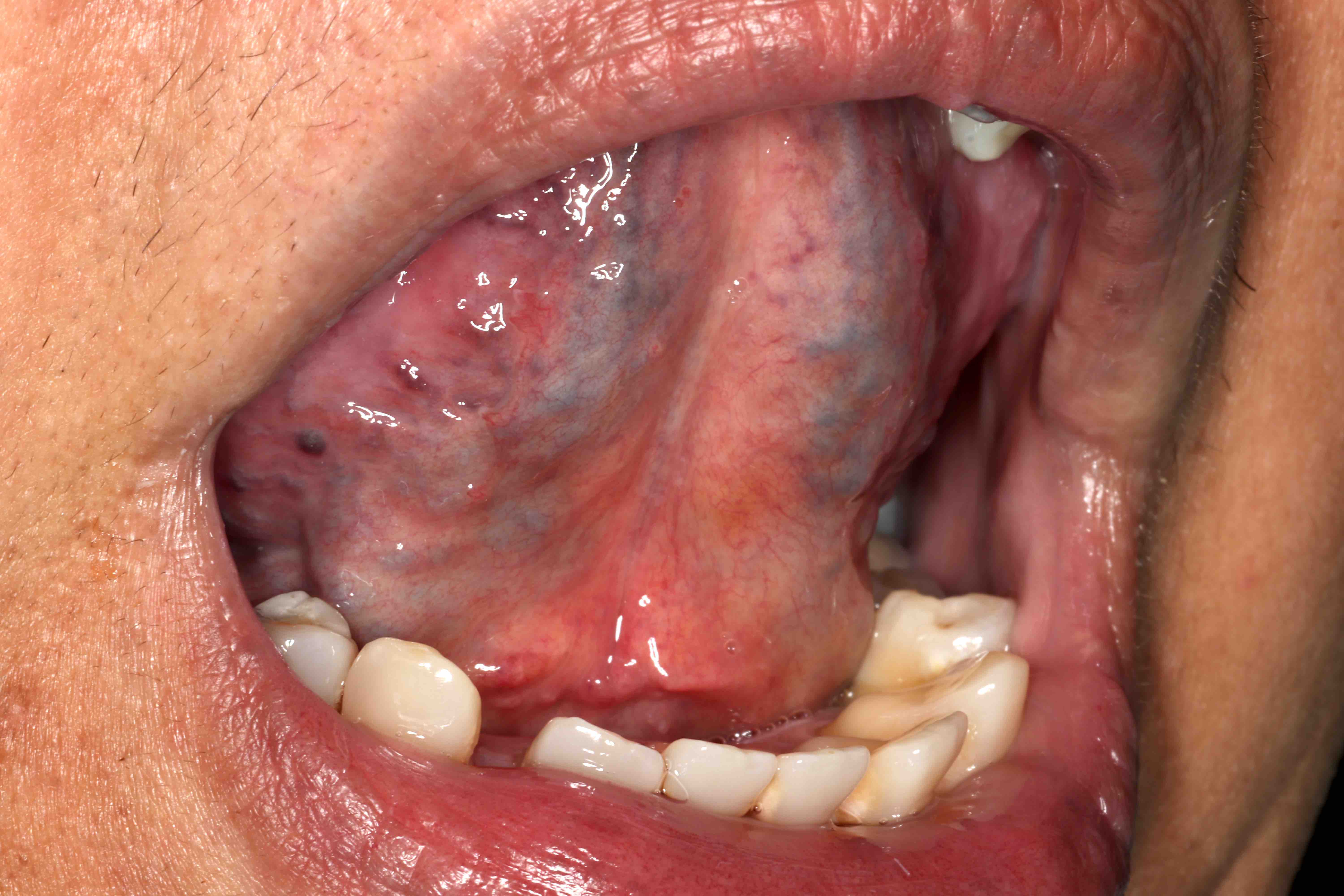
Submucosal nodule
Images hosted on other servers:

Well circumscribed, tan, hemorrhagic mass
Contributed by Molly Housley Smith, D.M.D.
Prominent hemorrhagic areas
Entrapment within muscle
Focal papillary growth pattern
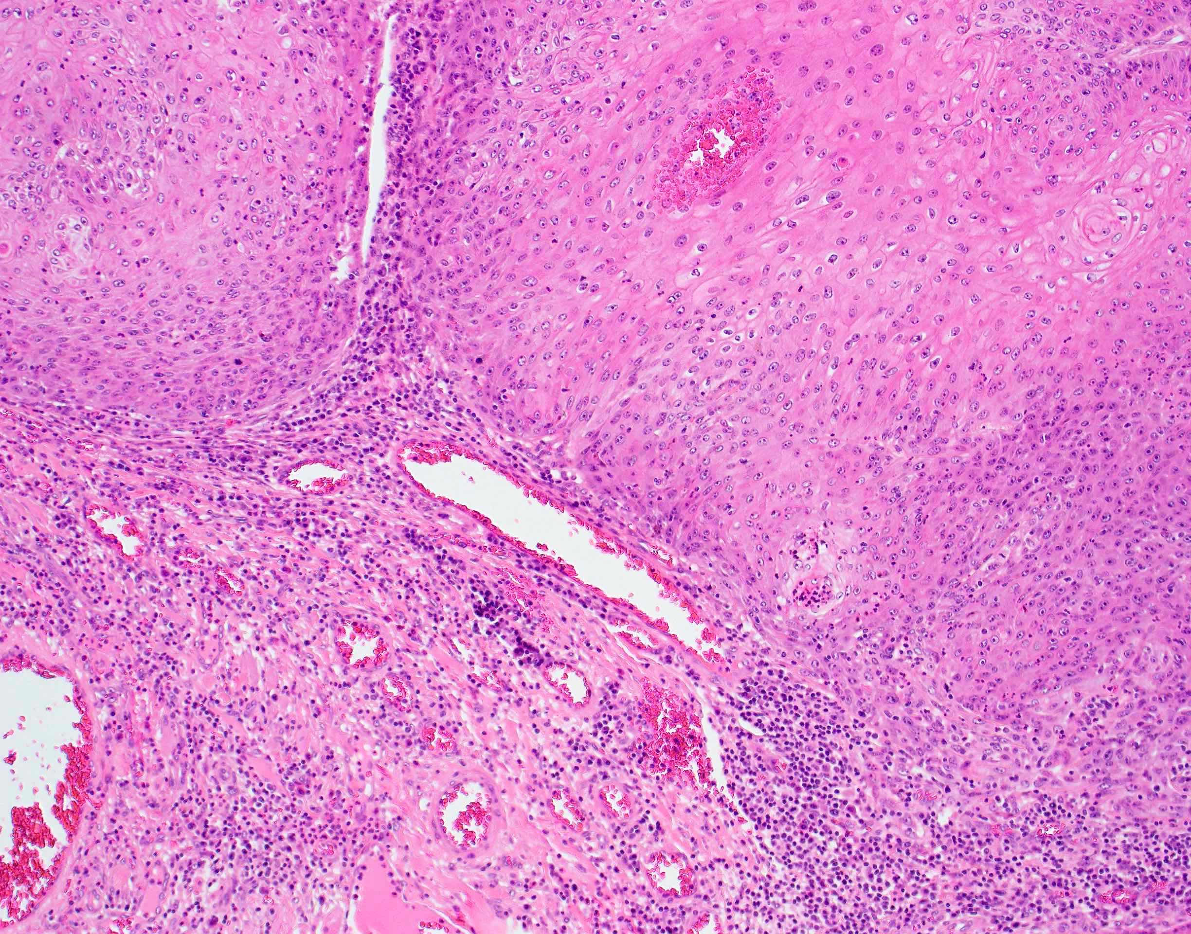
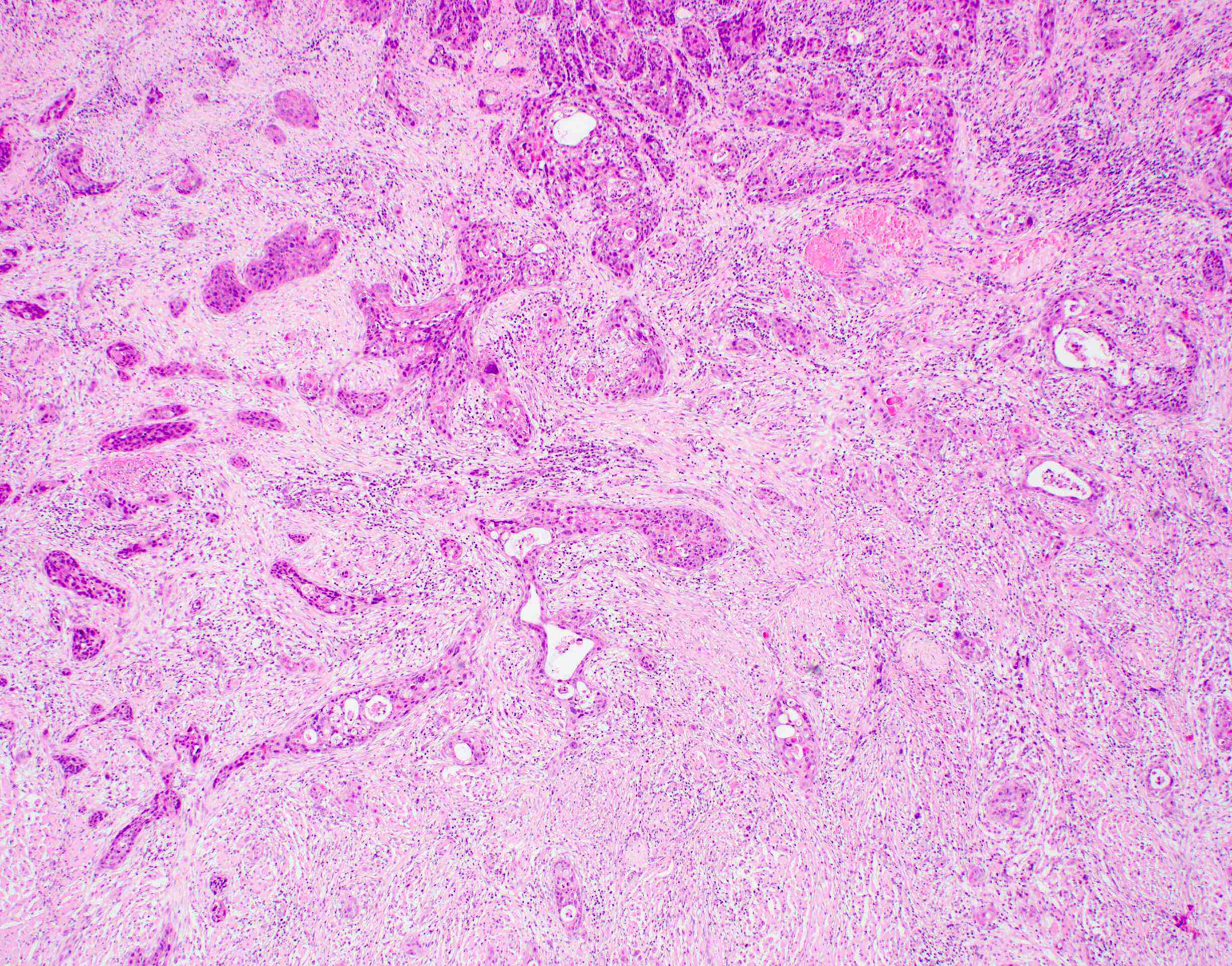
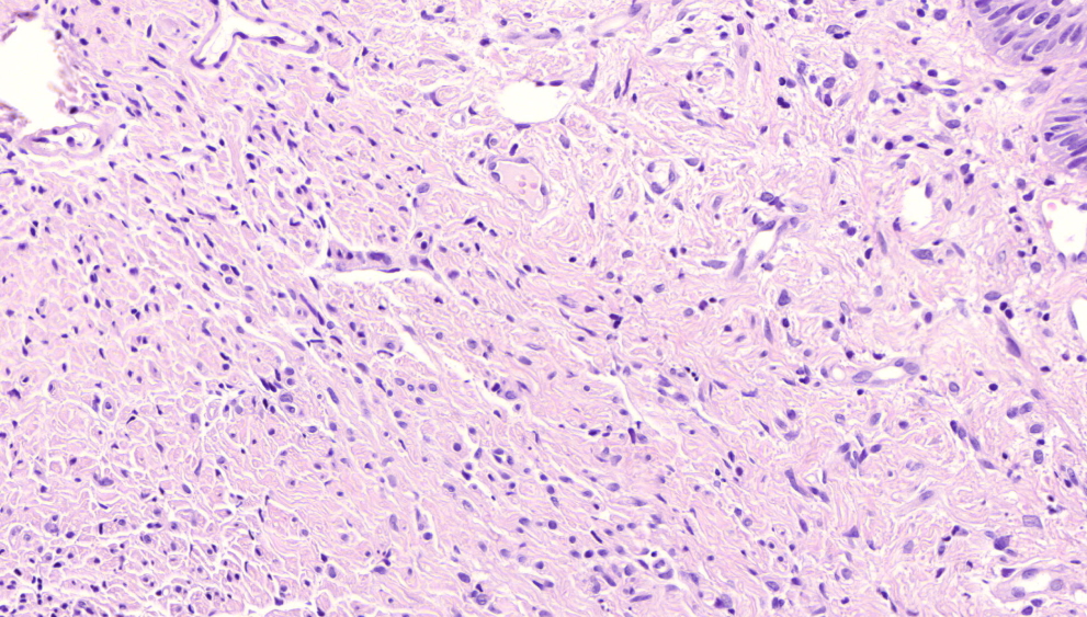
Chondromyxoid stroma
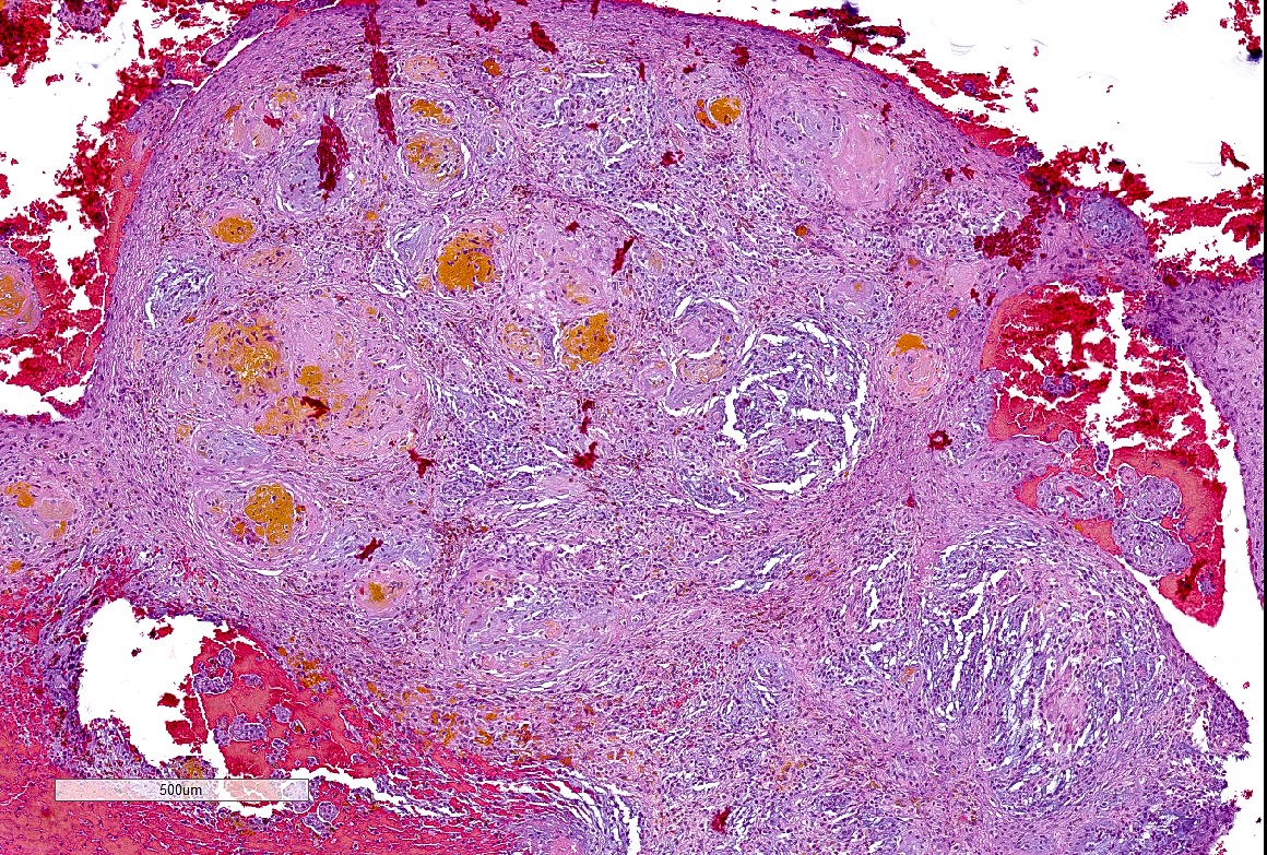
Hemosiderin breakdown products

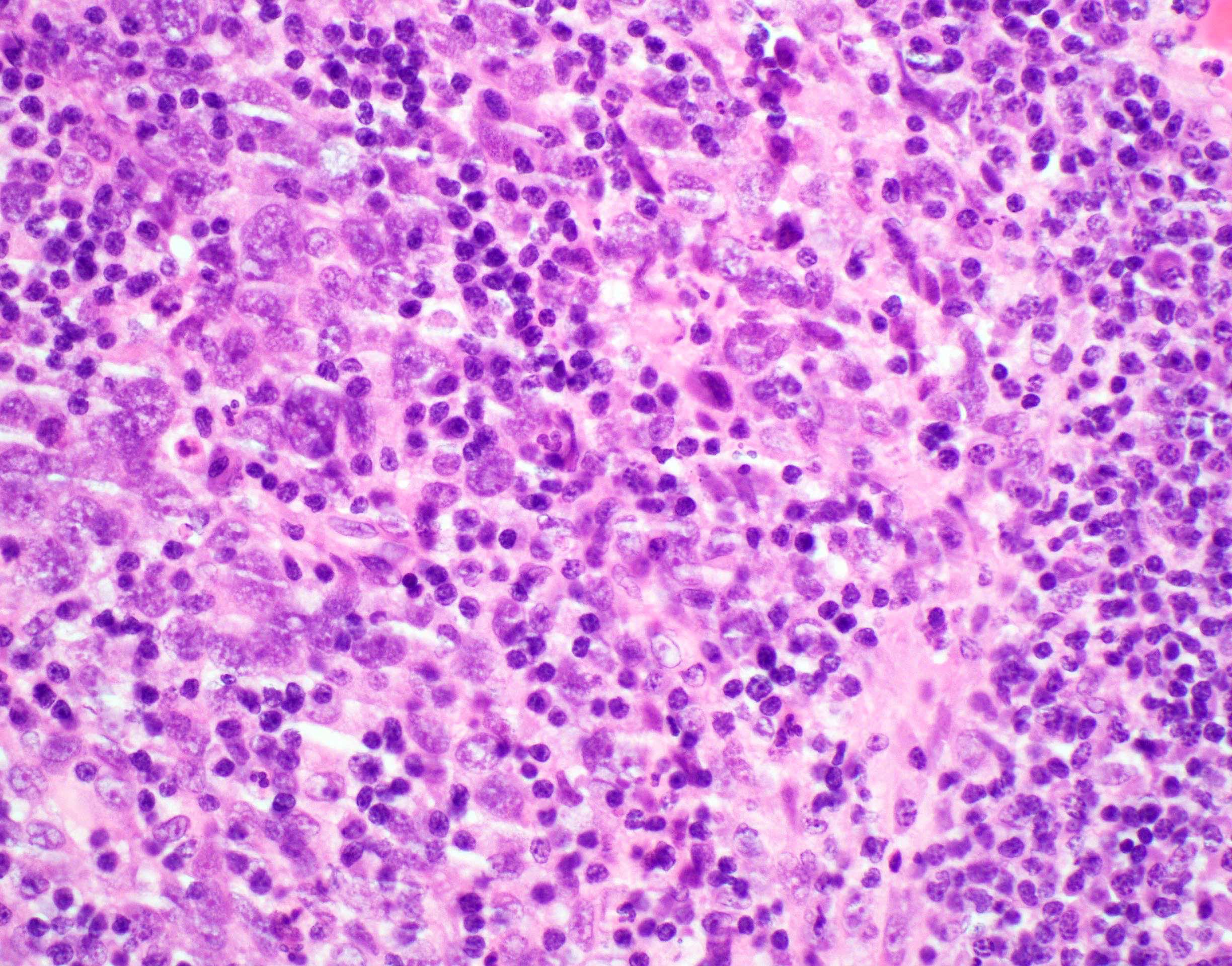
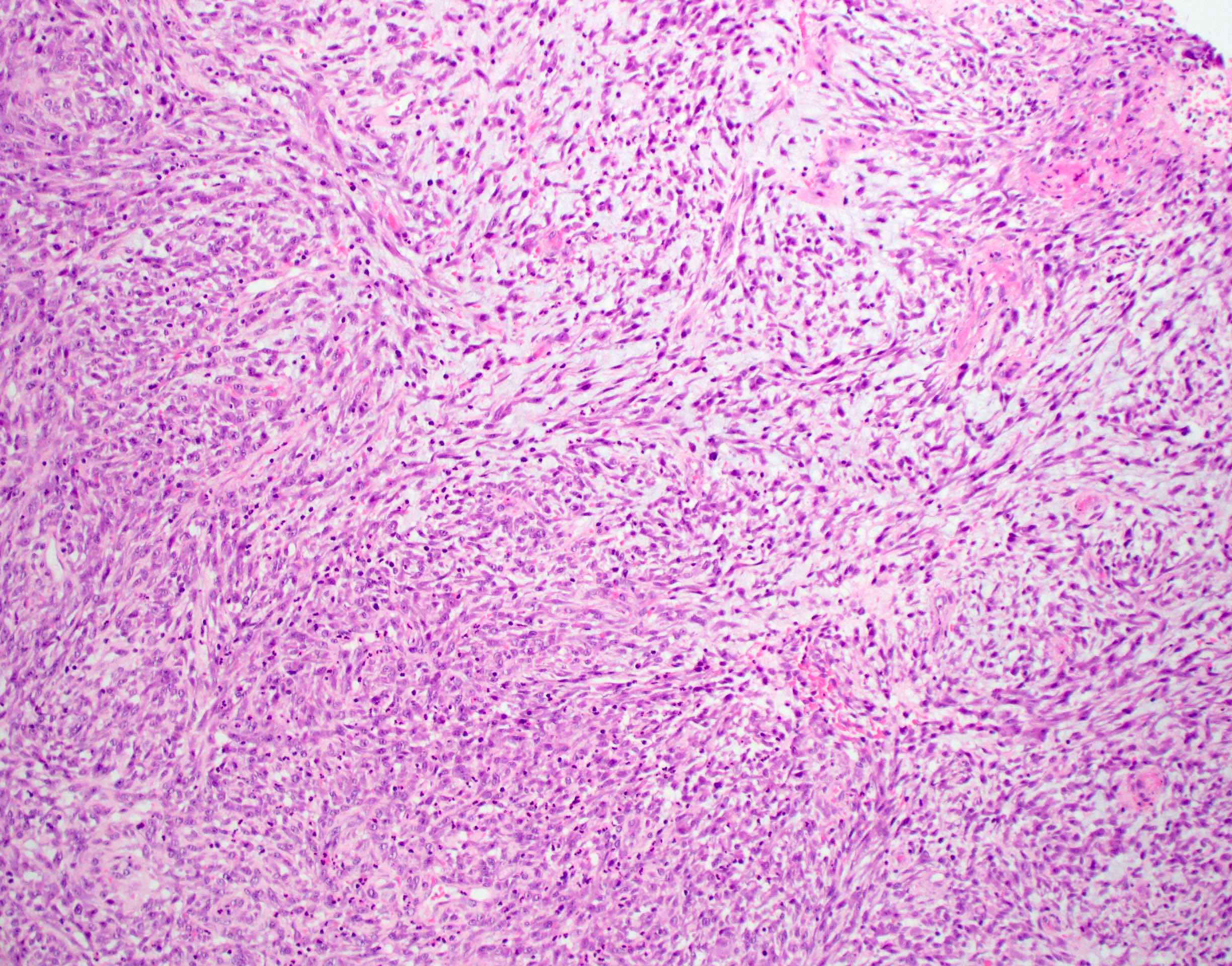
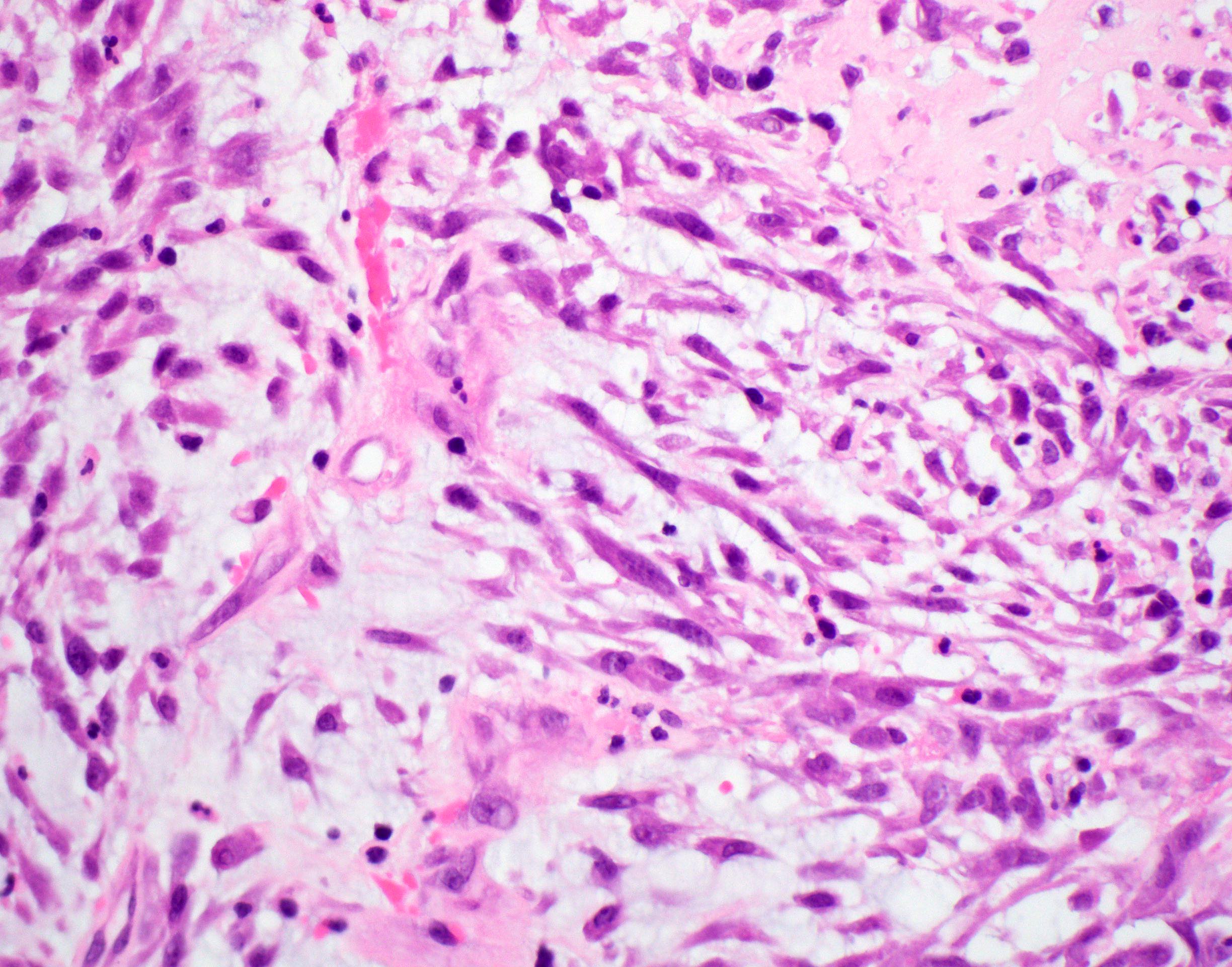
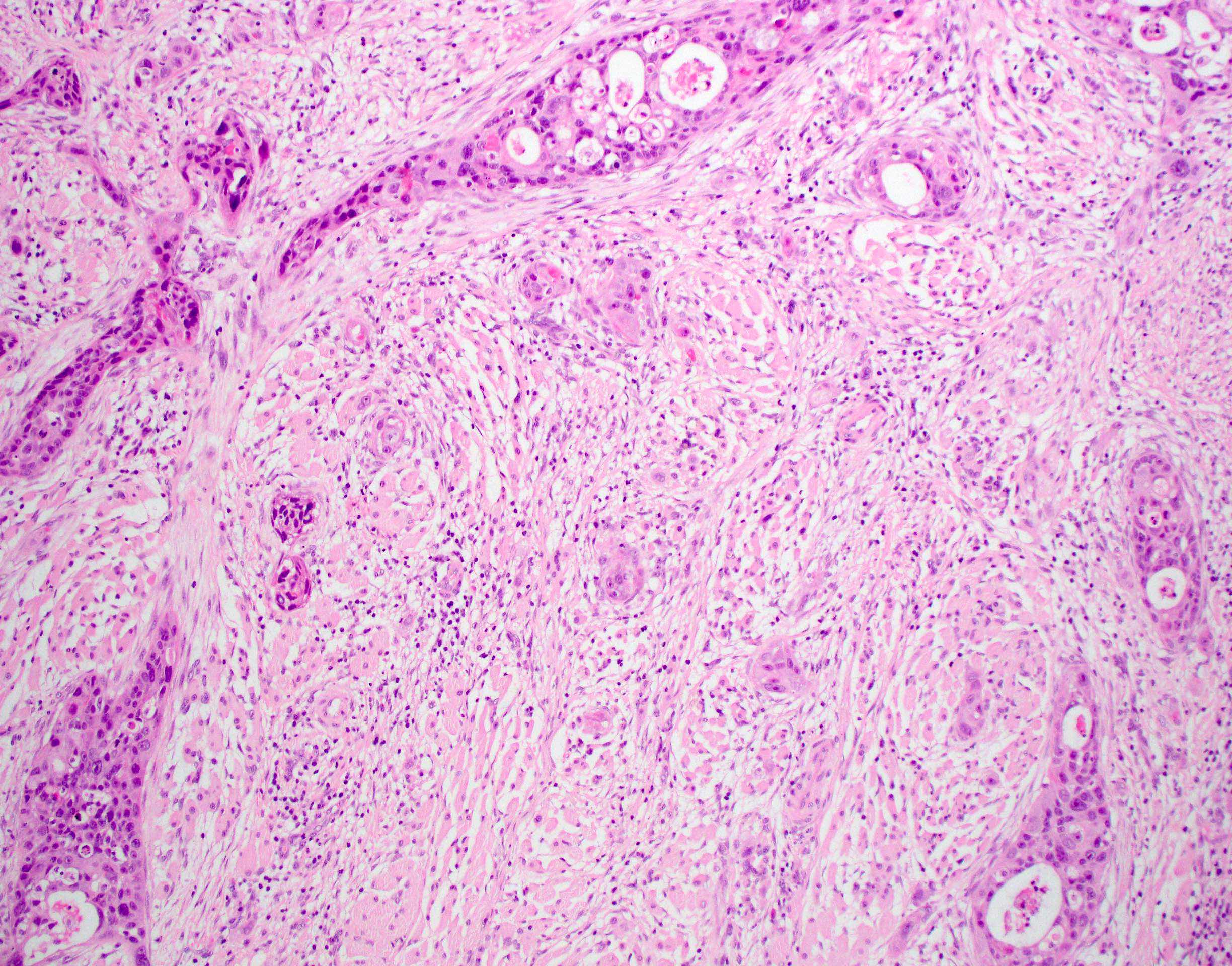
Sheets of ovoid cells

Reticular pattern
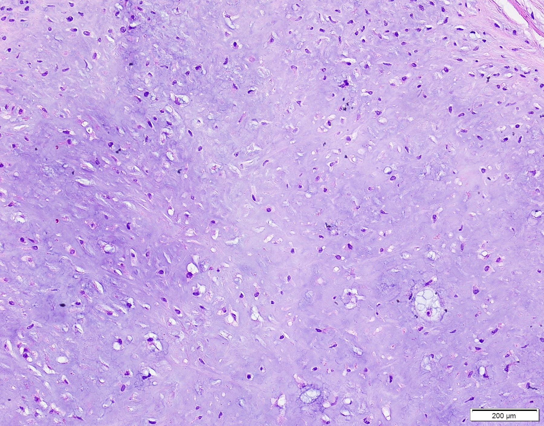
Chondromyxoid stroma
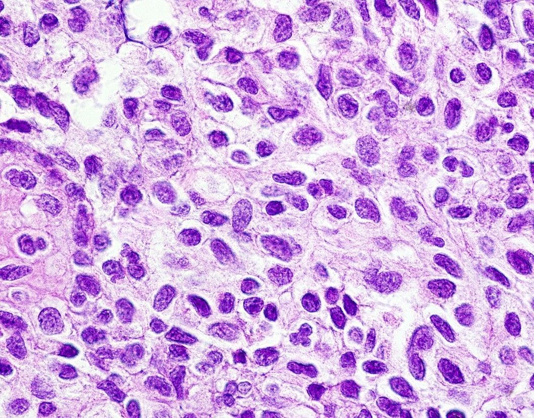
Ovoid nuclei
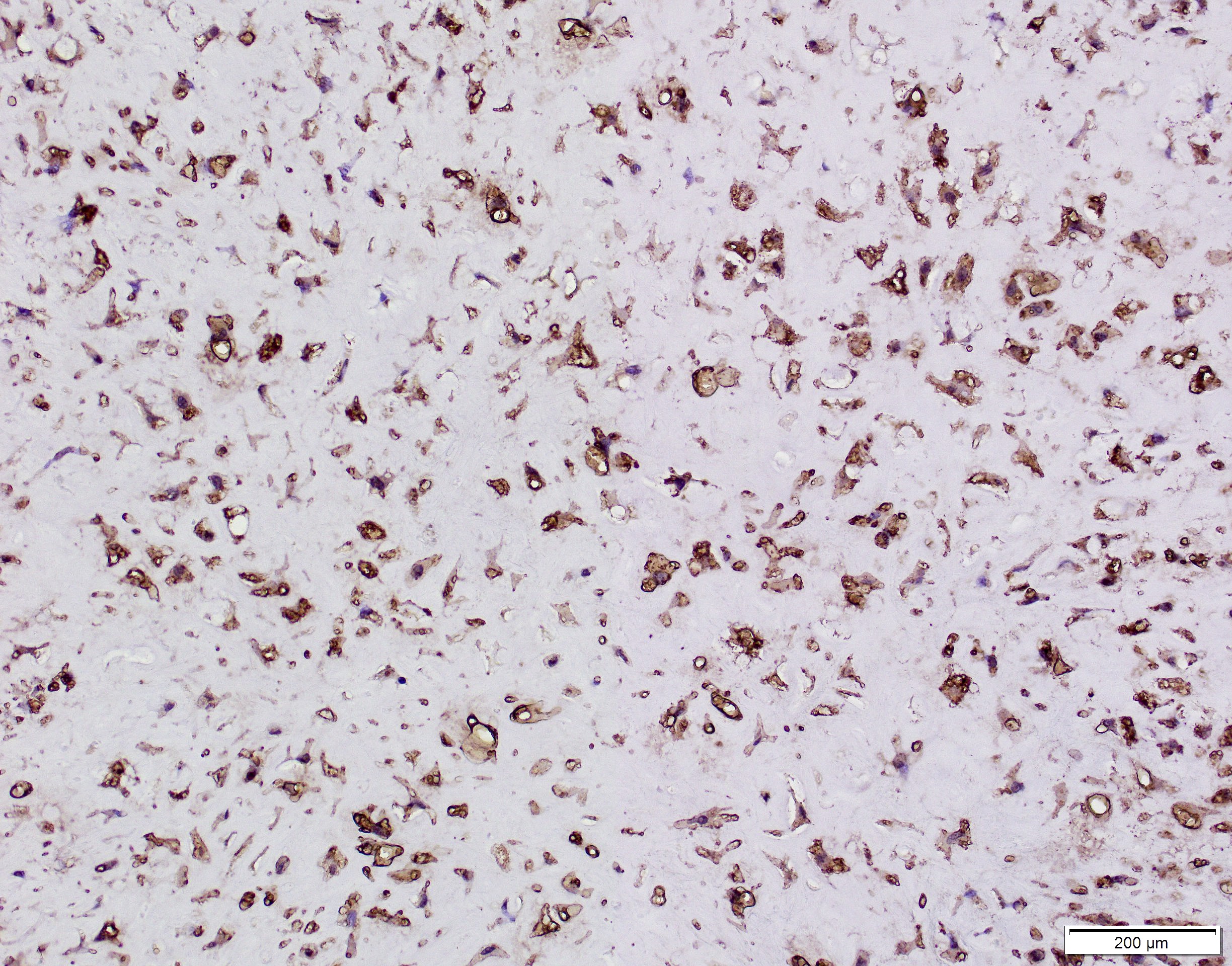
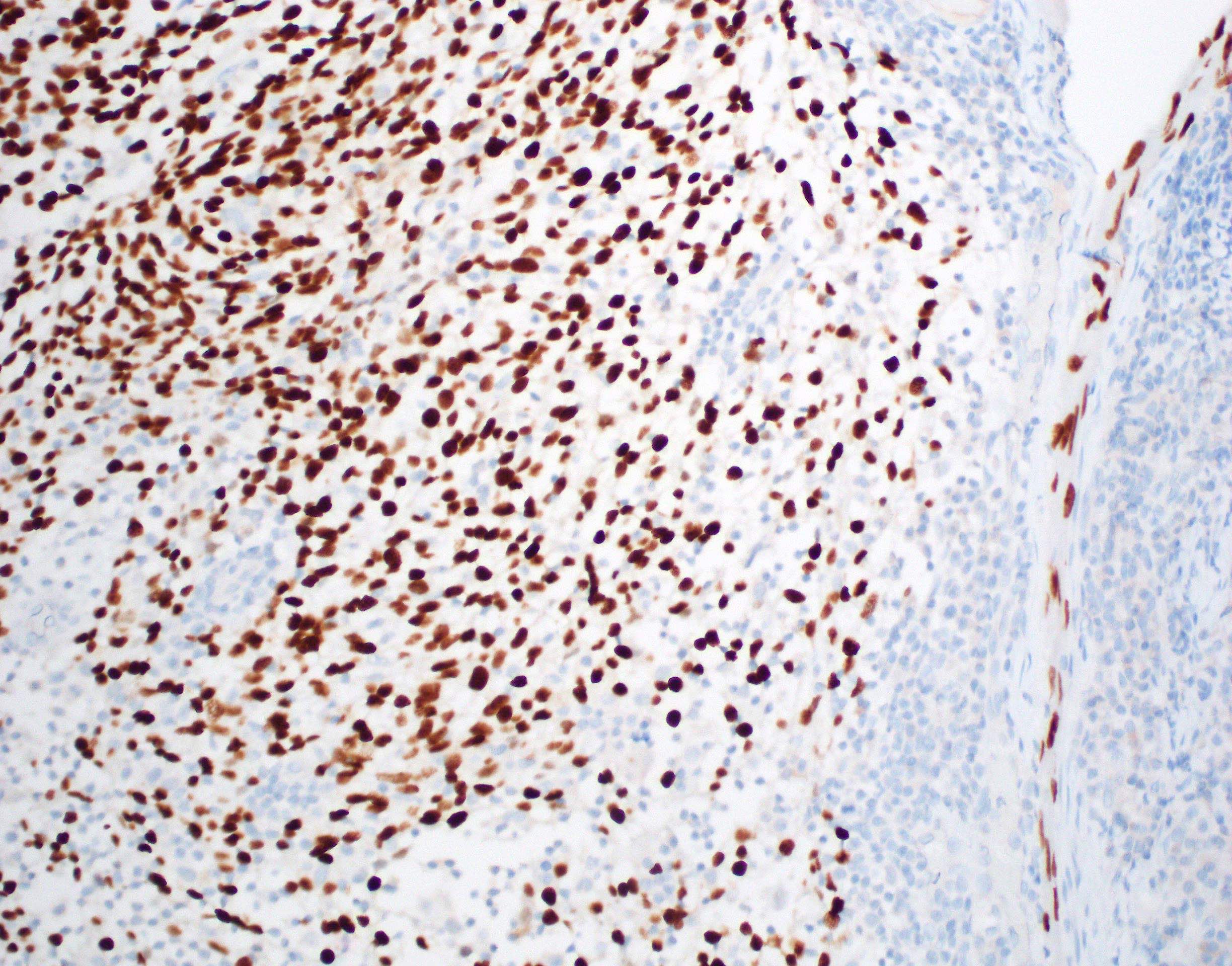
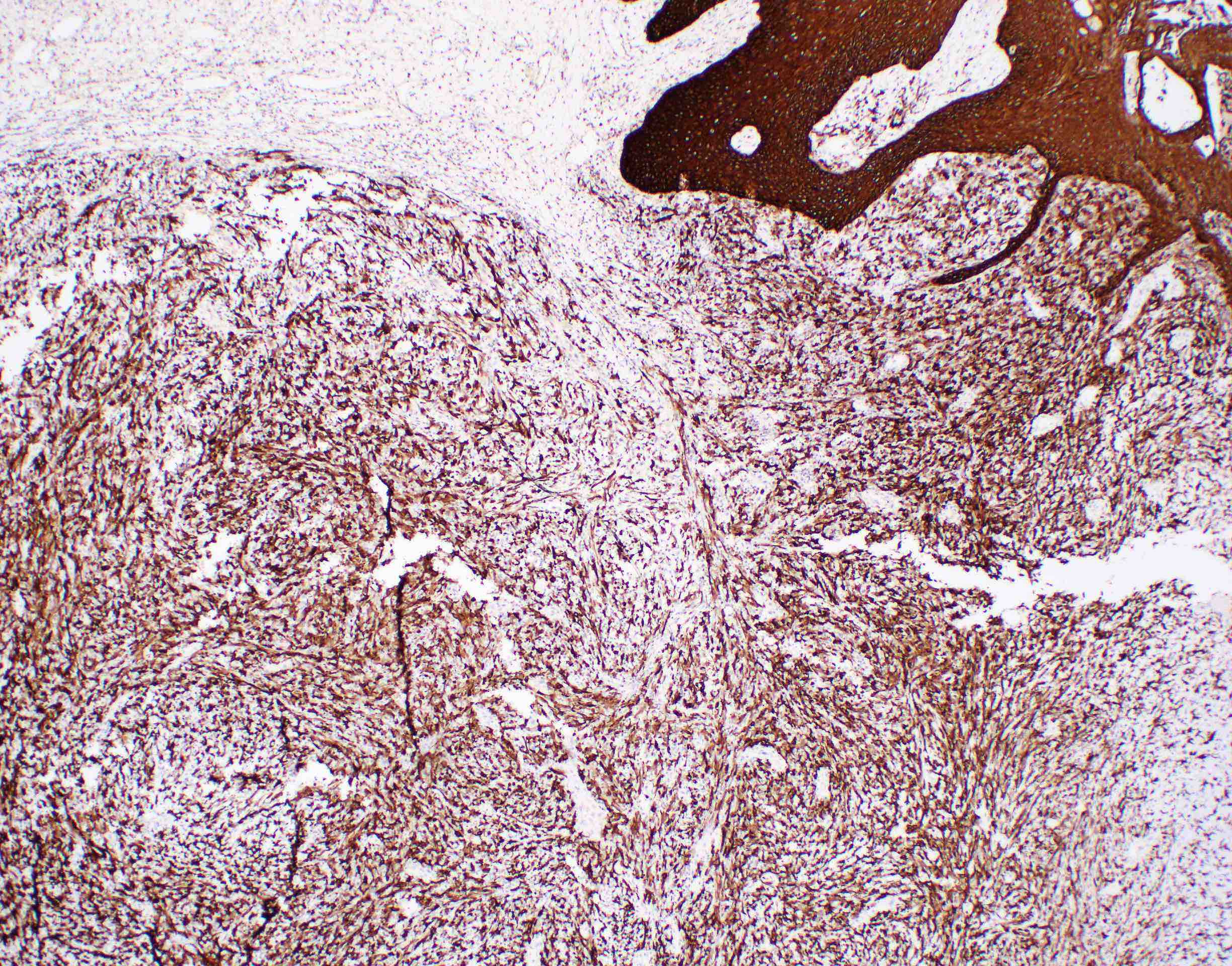
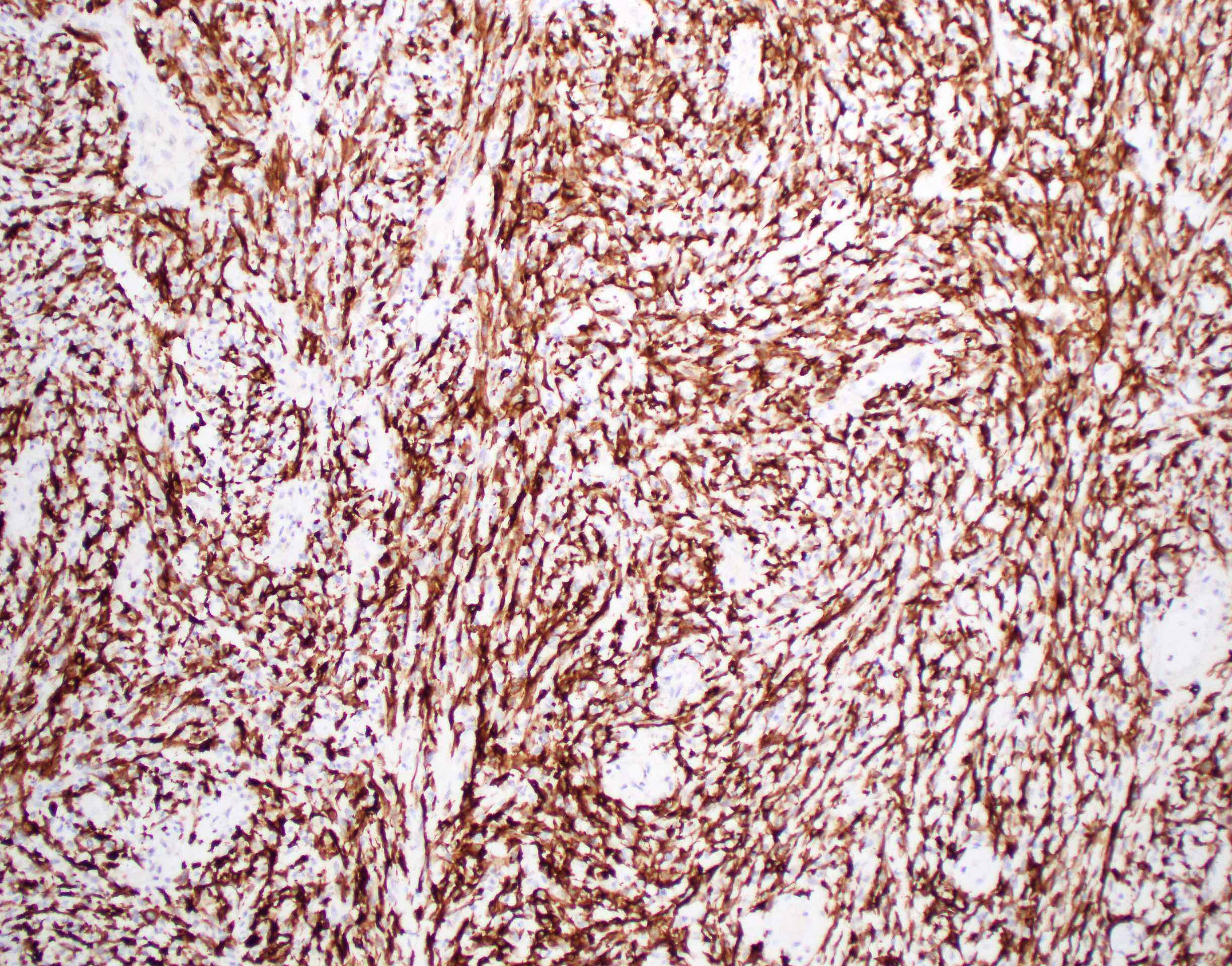
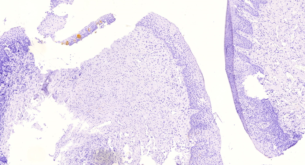
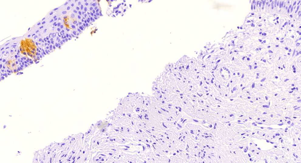
GFAP
Images hosted on other servers:

FISH rearrangement of RREB1 and MRTFB
Images hosted on other servers:

55 year old woman

60 year old man

26 year old woman

37 year old woman
Images hosted on other servers:
Keratinocytes (fig d)
Images hosted on other servers:


21 year old woman:
irregular lip uclerations and buccal mucosal

23 year old woman:
palatal ulcerations


40 year old man:
hemorrhagic bullae and target lesions
Contributed by Molly Housley Smith, D.M.D.
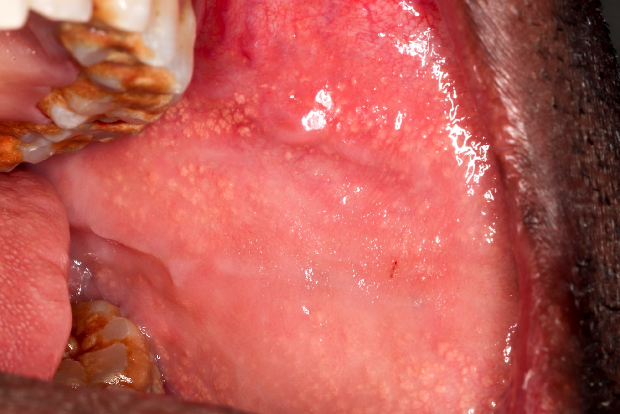
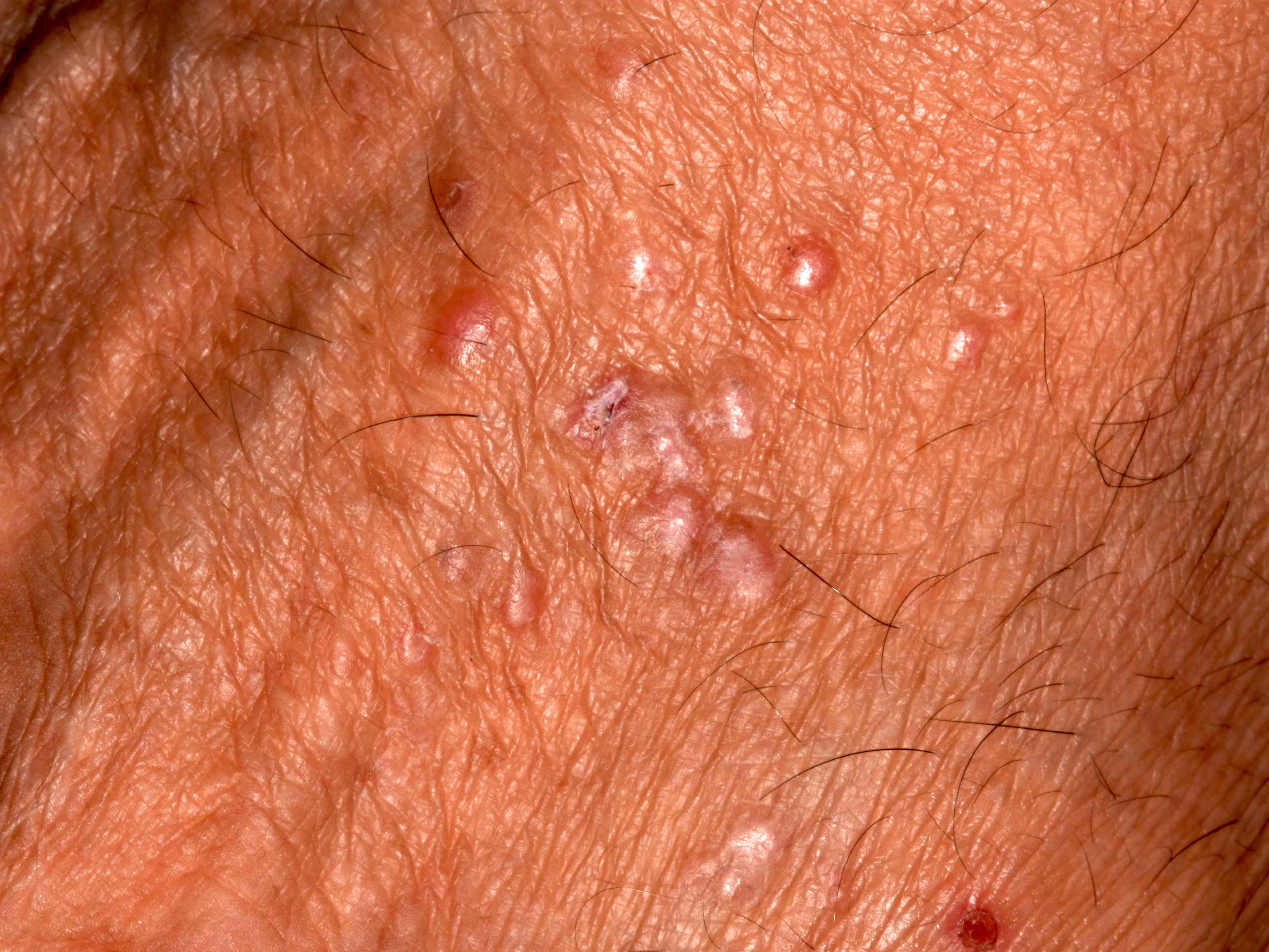
Yellow, floral papules
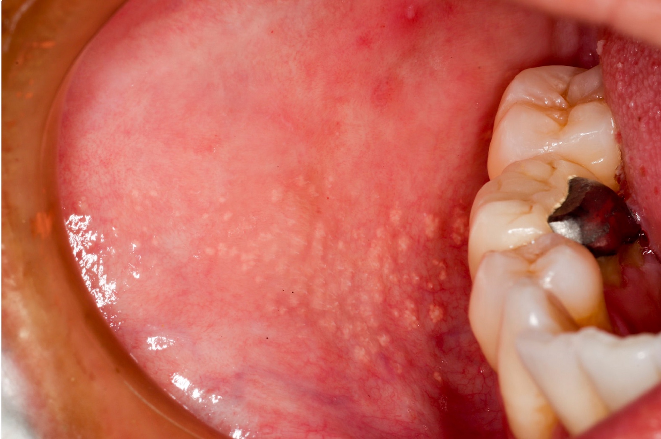
1 - 3 mm yellow-white papules
Image hosted on other servers:

Yellow-white papules
Contributed by Molly Housley Smith, D.M.D.
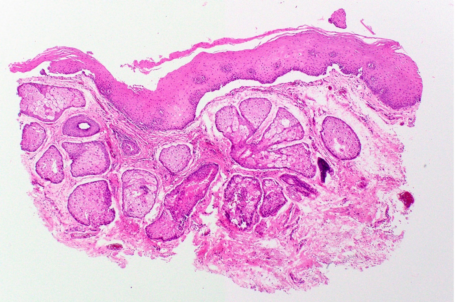
Oral mucosa containing sebaceous lobules
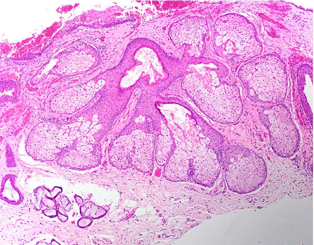
Sebaceous lobules adjacent to mucous glands
Sebaceous lobules
Images hosted on other servers:


Before and after therapy


Before and after therapy

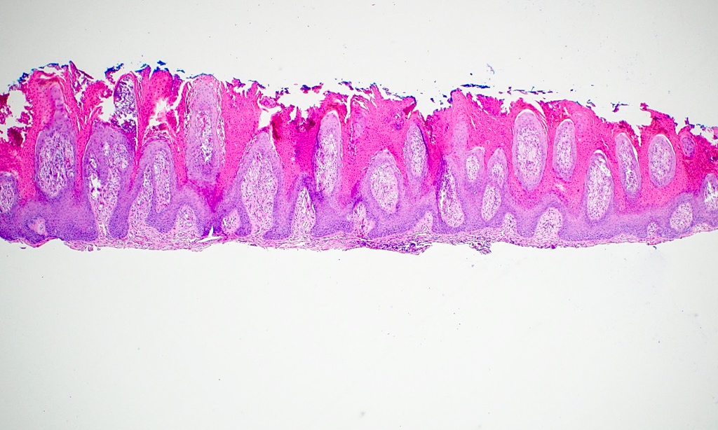
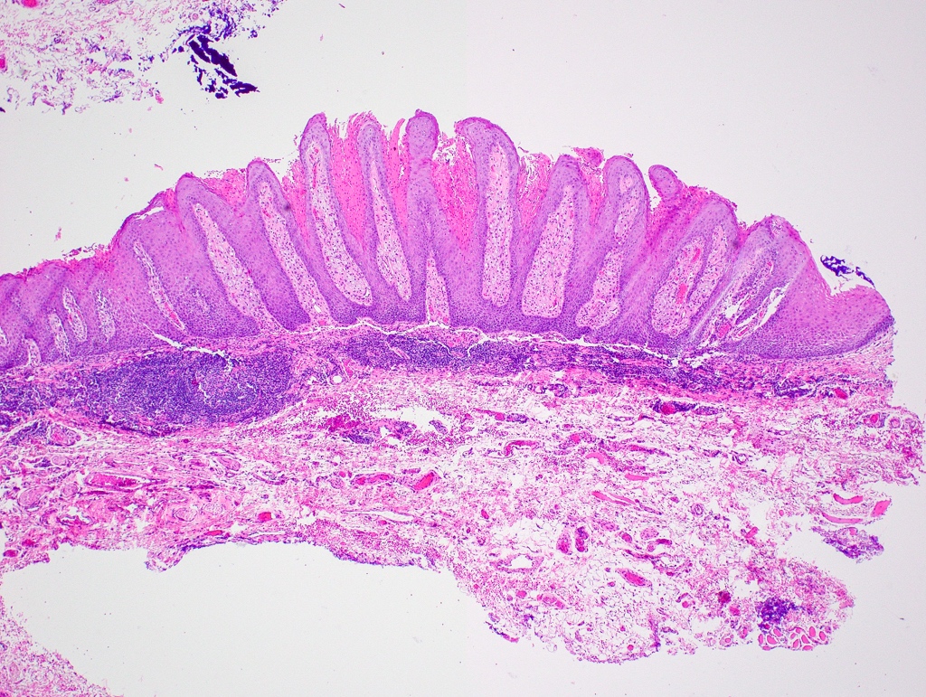
Geographic tongue
Images hosted on other servers:

Psoriform epidermal hypertrophy
Contributed by Sarah H. Glass, D.D.S.
Bosselated gingival mass
Small tongue mass
Contributed by Sarah H. Glass, D.D.S.
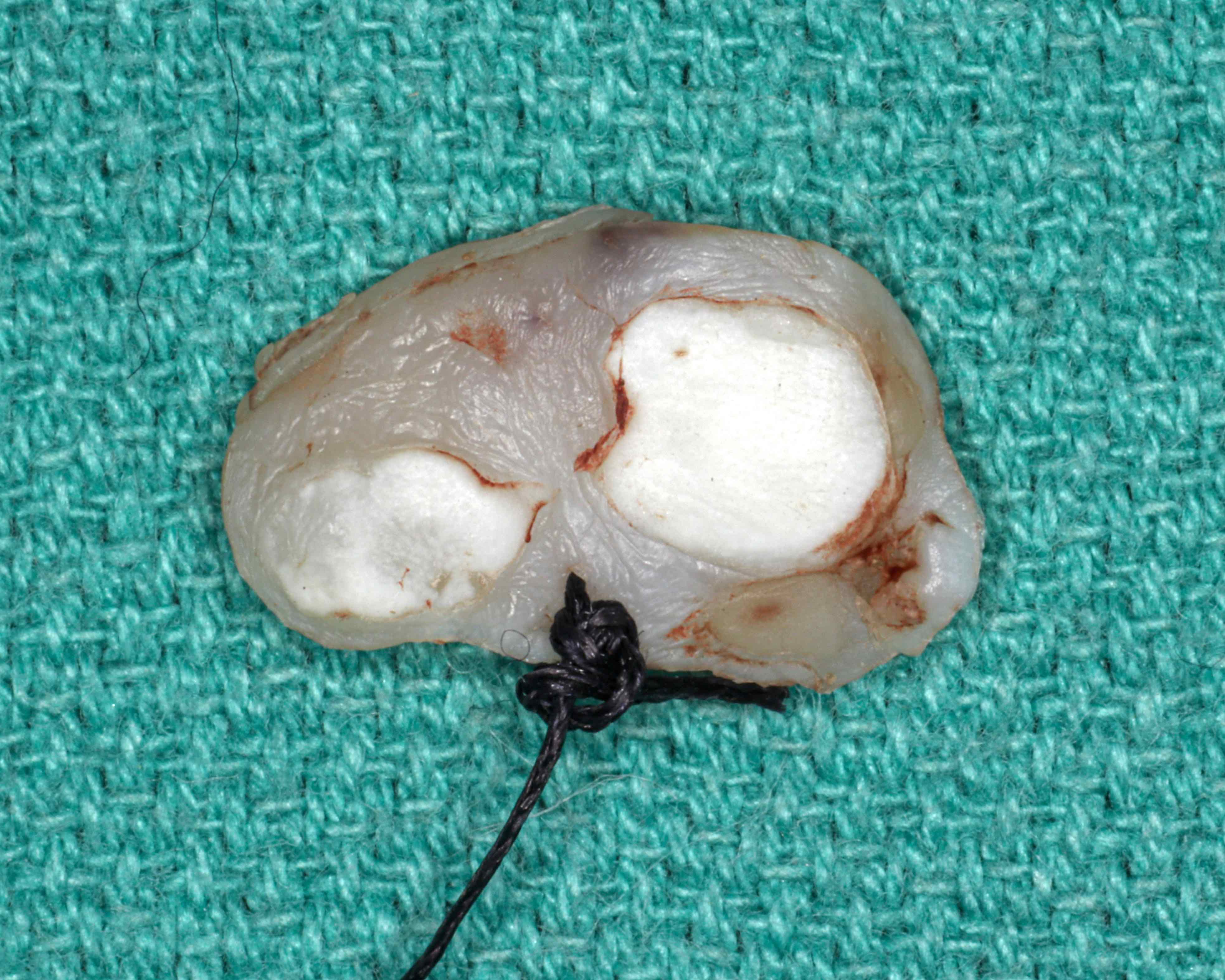
Dome shaped mass
White cut surface
Contributed by Sarah H. Glass, D.D.S.
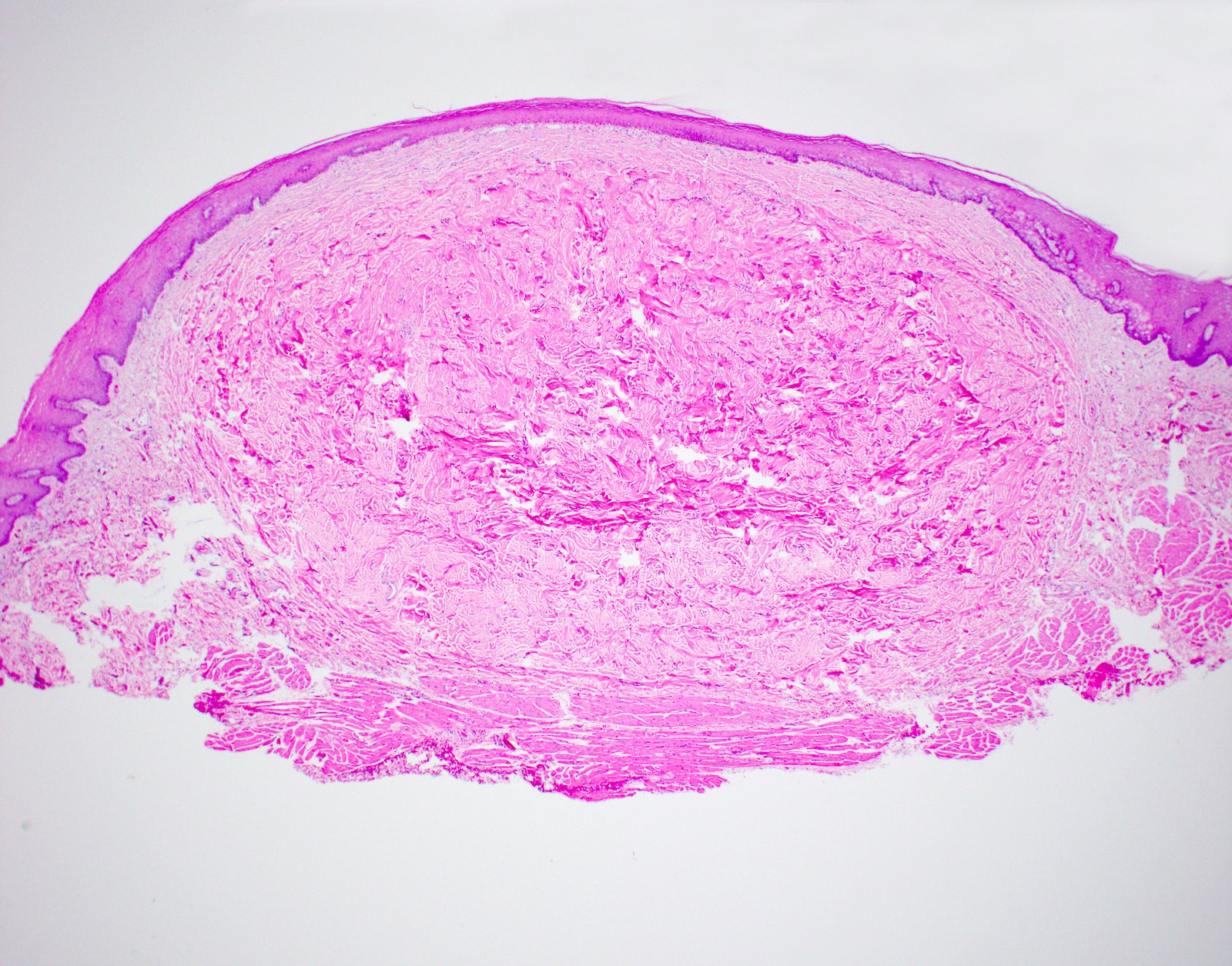
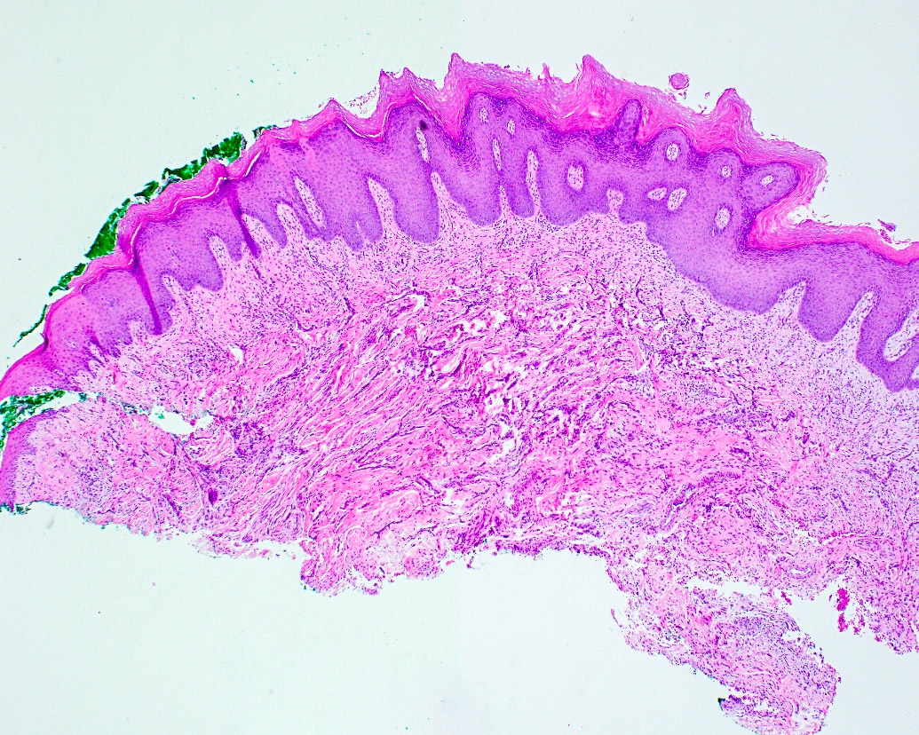
Nodular fibrous proliferation
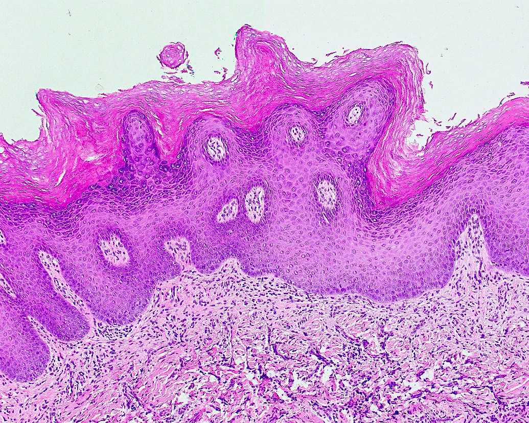
Elongated rete ridges
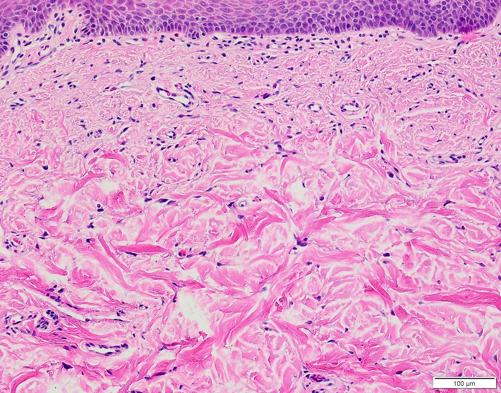
Stellate fibroblasts
Multinucleated fibroblasts
Images hosted on other servers:
9 year old girl:

Preoperative

1 year follow up
11 year old boy:

Preoperative

Postoperative
Images hosted on other servers:


Multiple granulomas


Lip and gingival biopsies

Granulomatous lesion and infiltrates
Contributed by Molly Housley Smith, D.M.D.
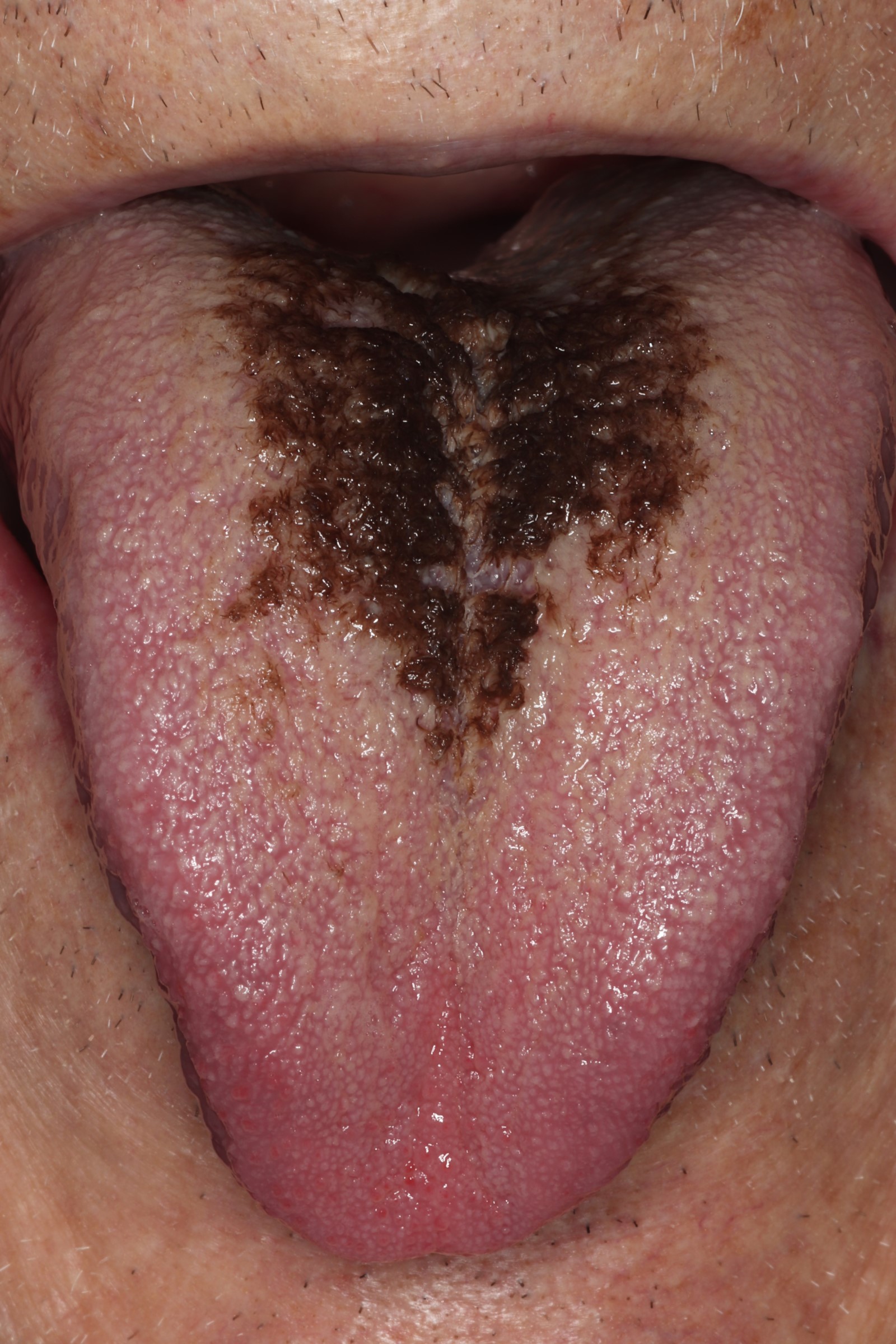
Black hairy tongue
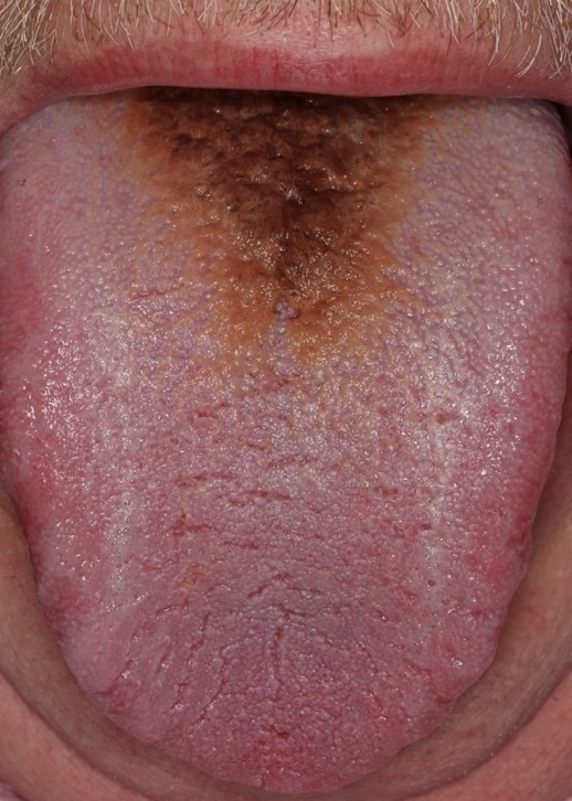
Brown hairy tongue
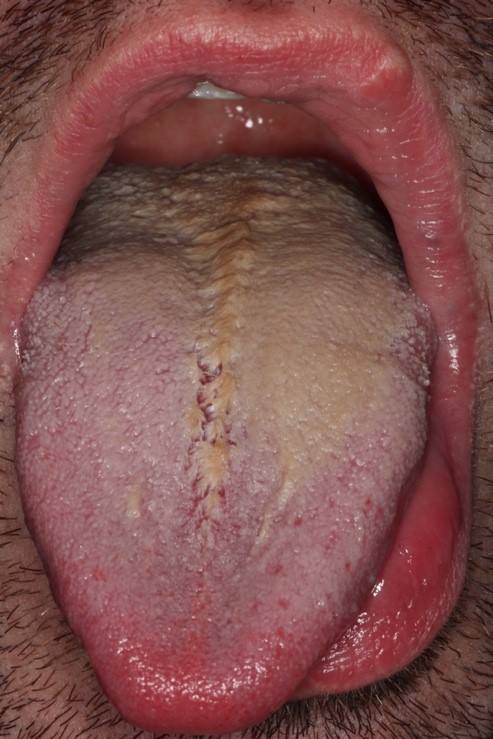
Yellow hairy tongue
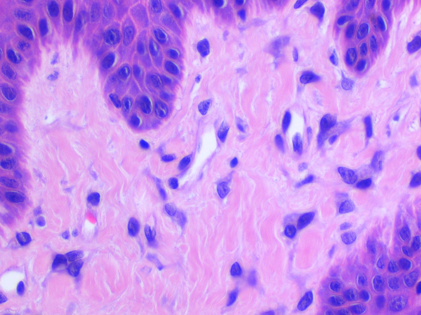
Contributed by Molly Housley Smith, D.M.D.
Hyperparakeratosis
Superficial bacterial colonies
Elongated filiform papilla
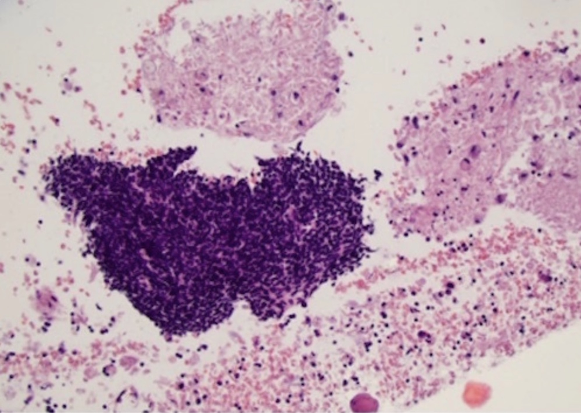
Contributed by Zahra Maleki, M.D.
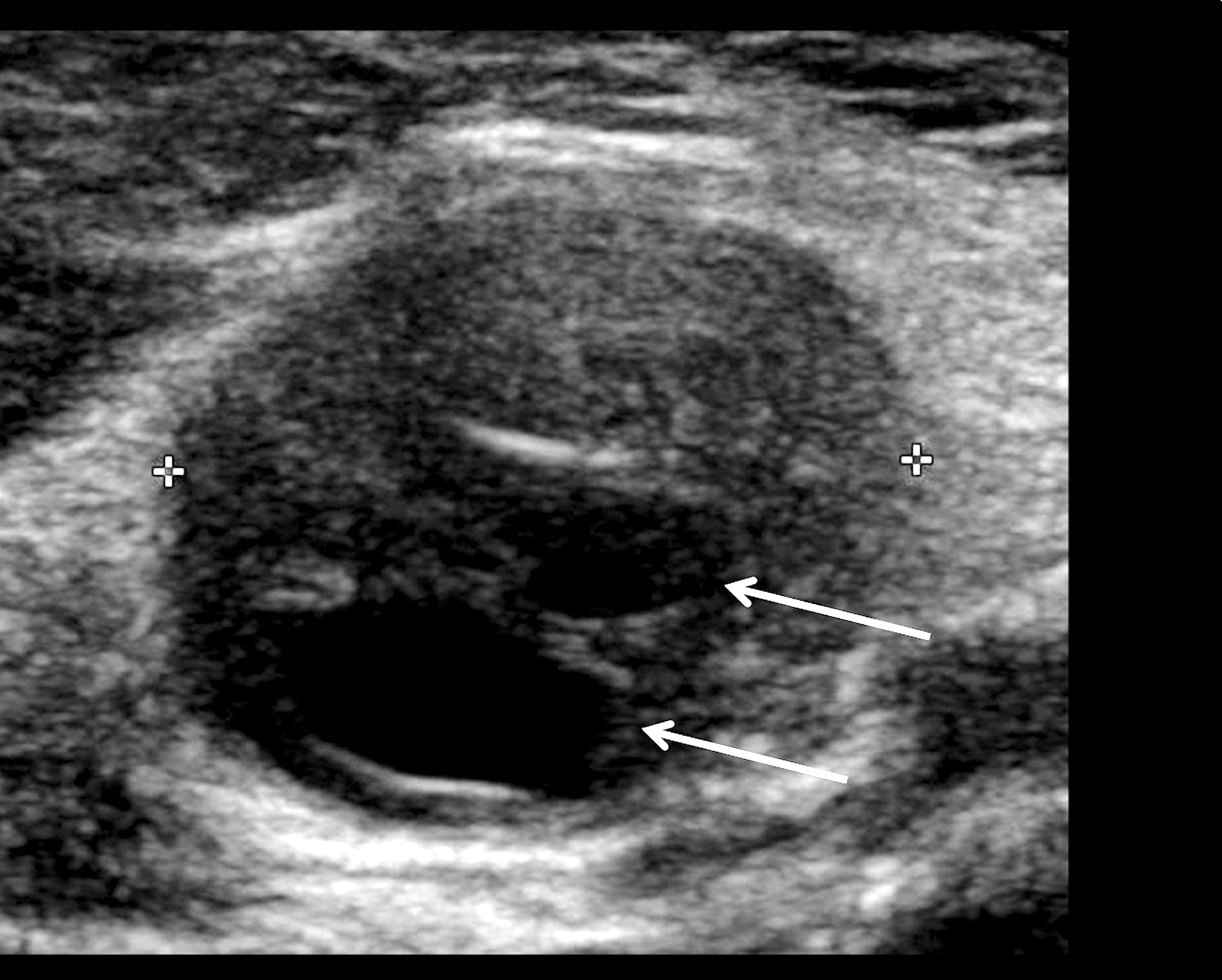
Ultrasound
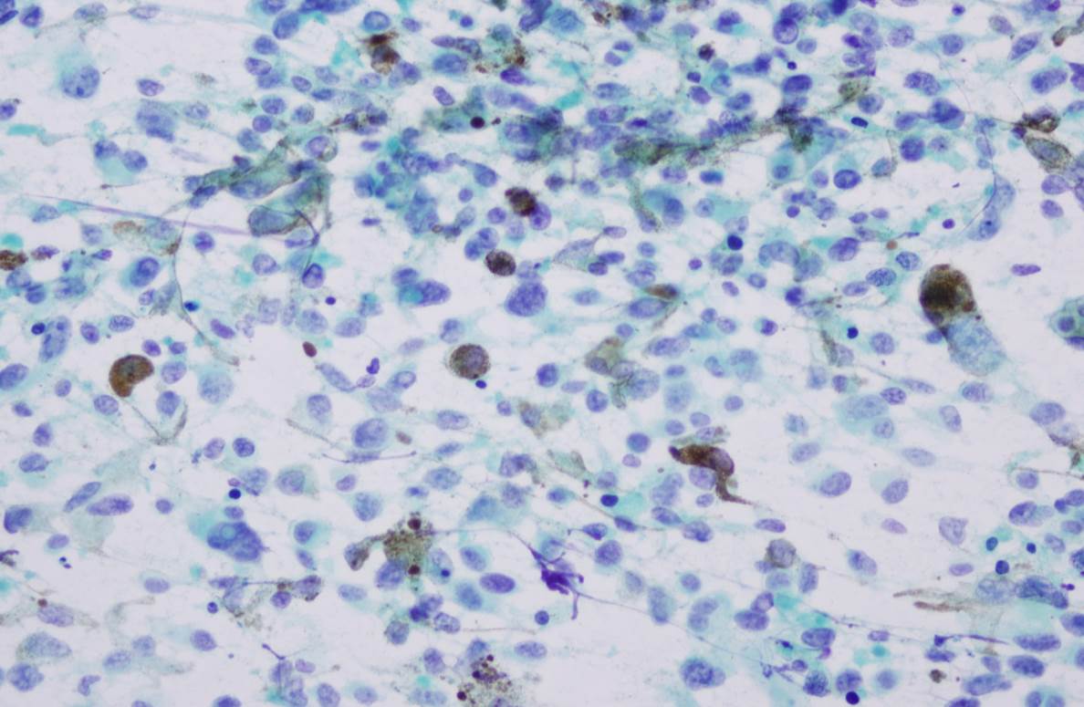
Contributed by Zahra Maleki, M.D. and @AnaPath10 on Twitter
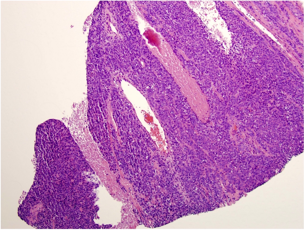
Infiltrating tumor
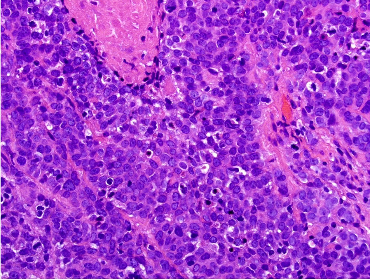
Basaloid cells
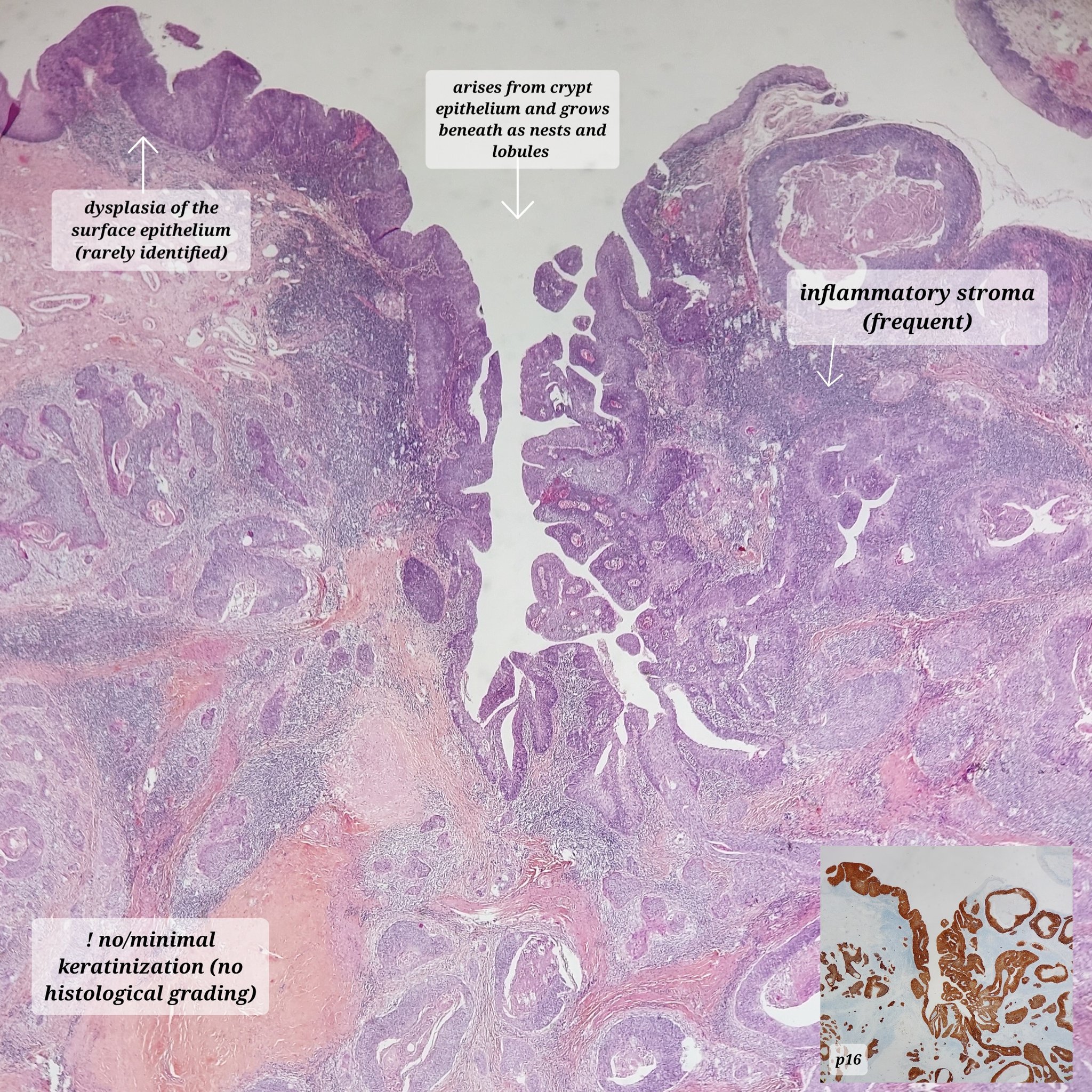
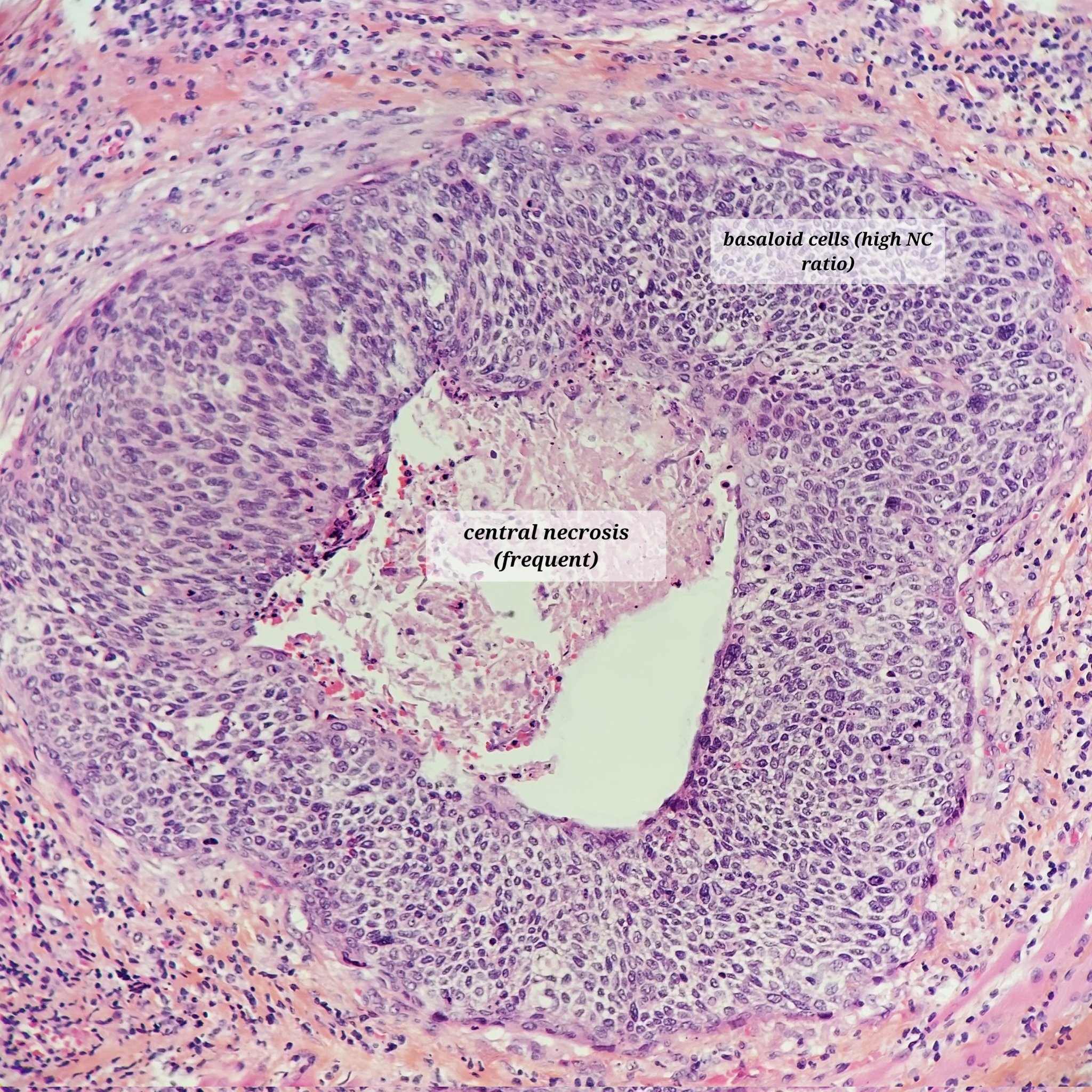
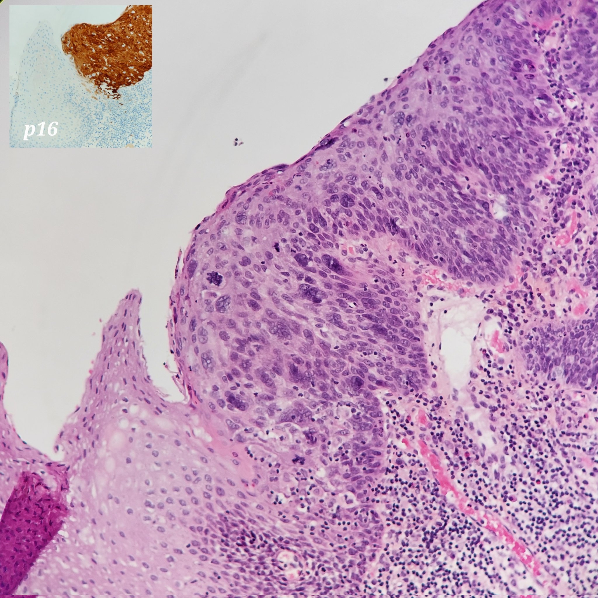
HPV positive oropharynx squamous cell carcinoma
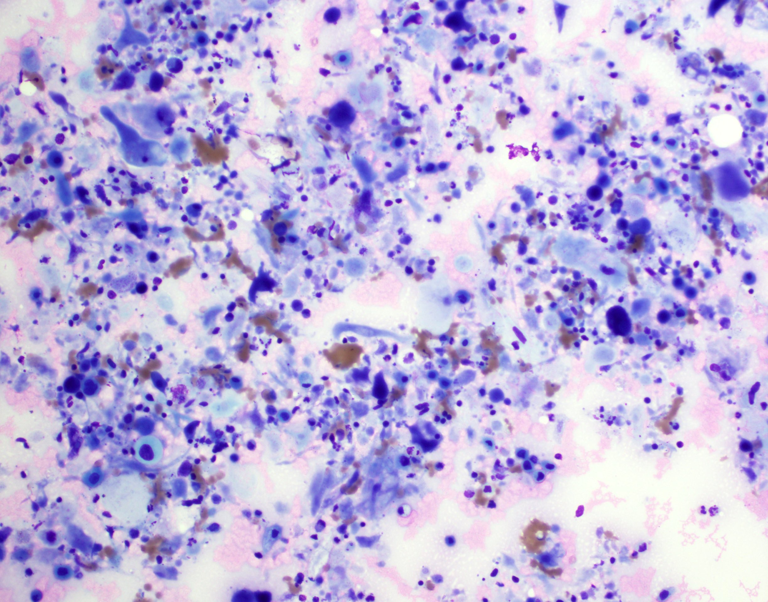
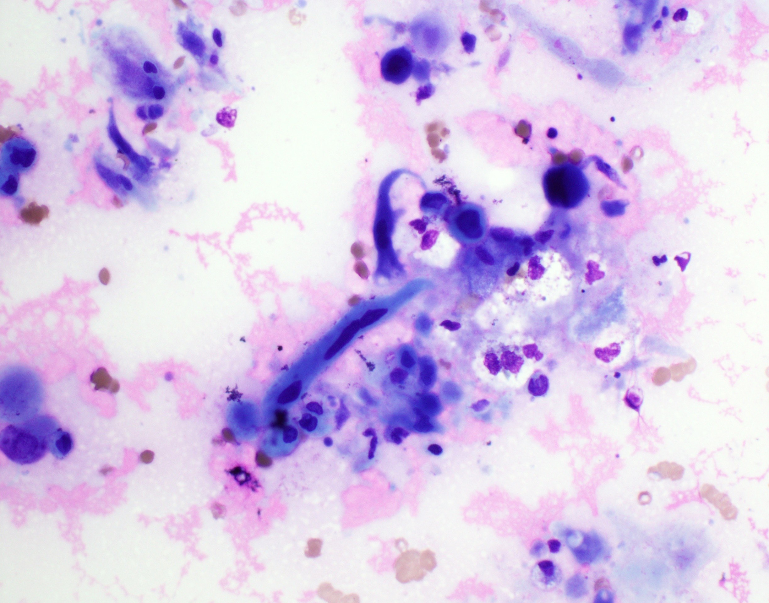
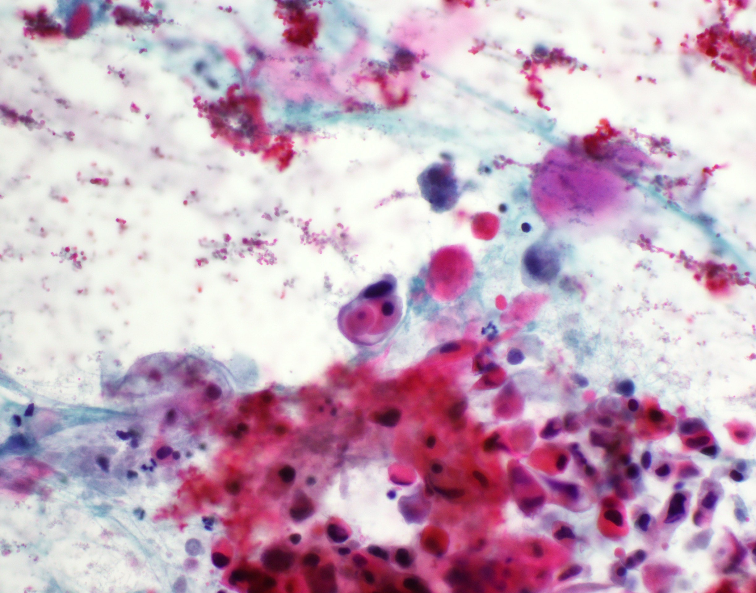
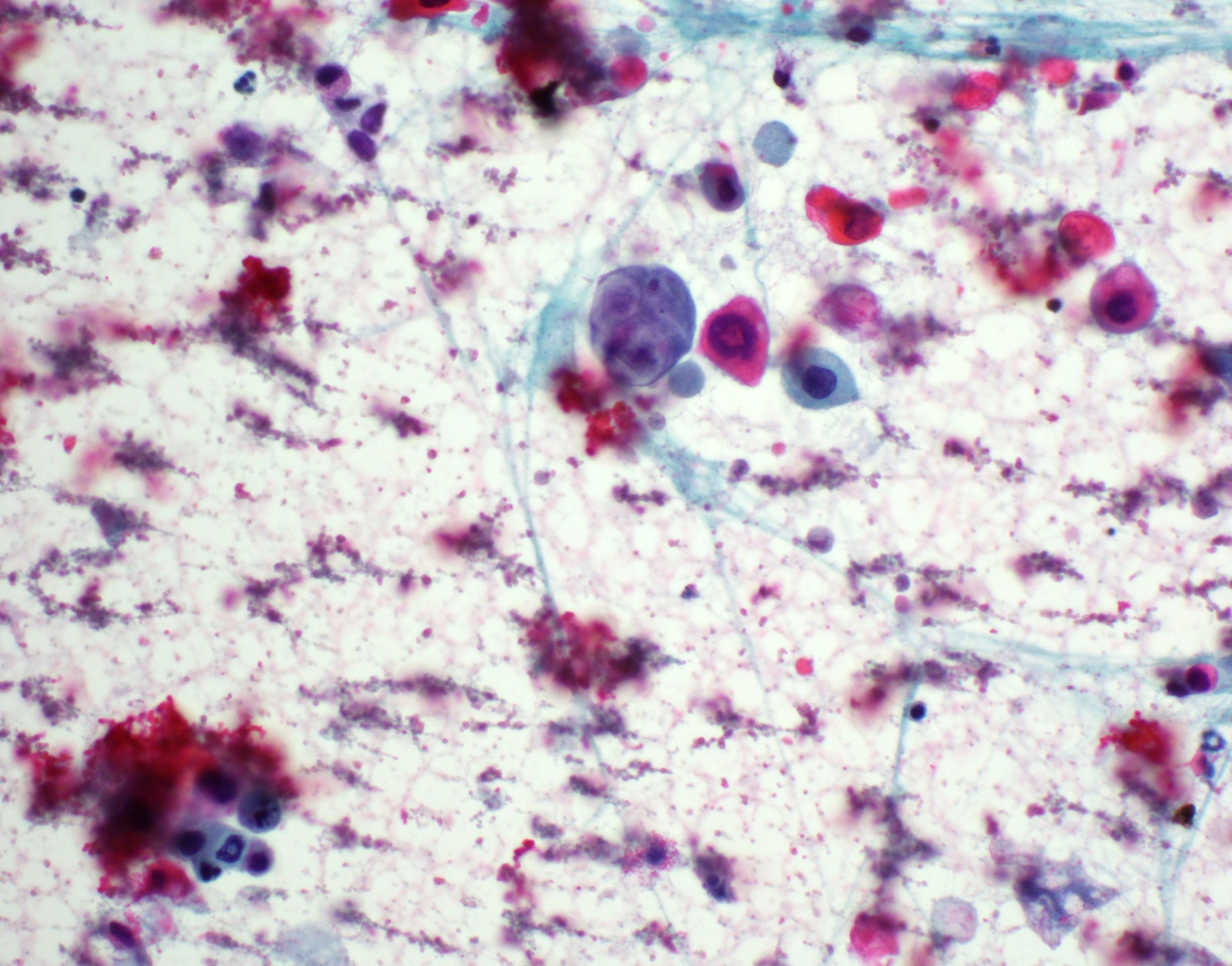
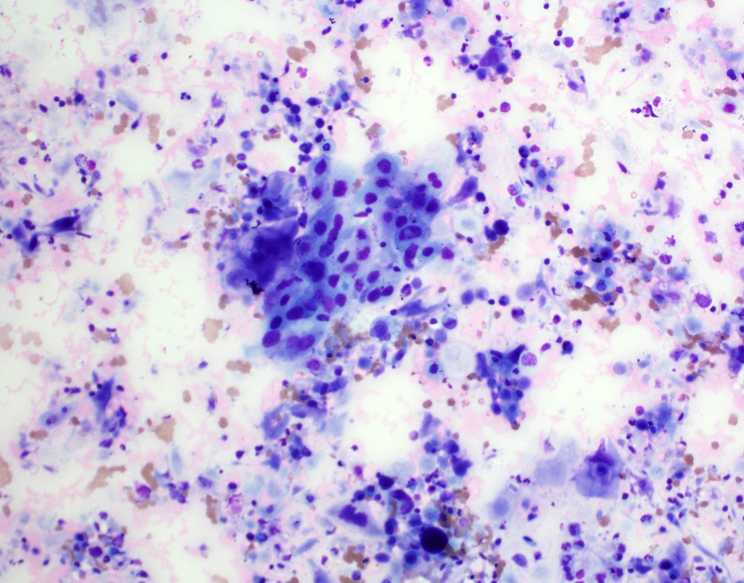
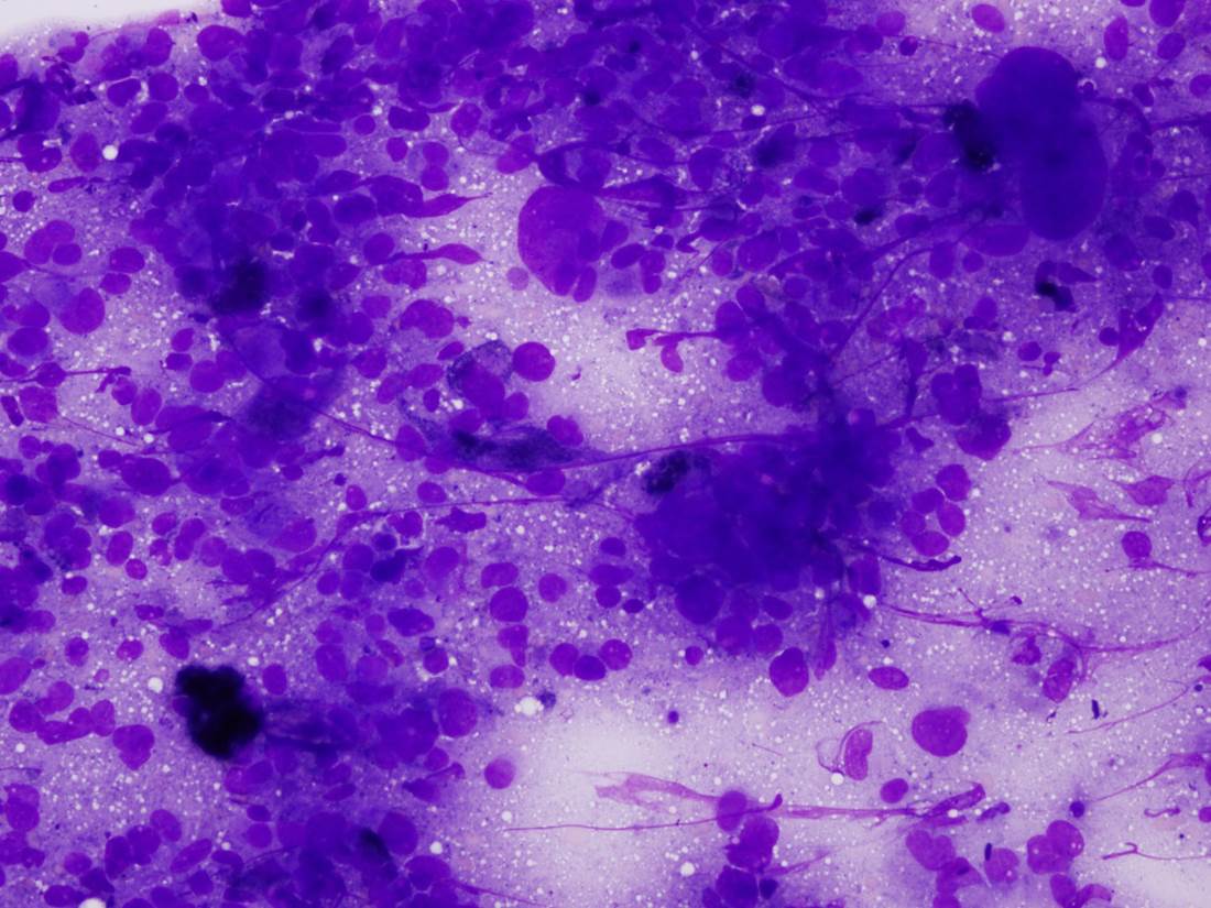
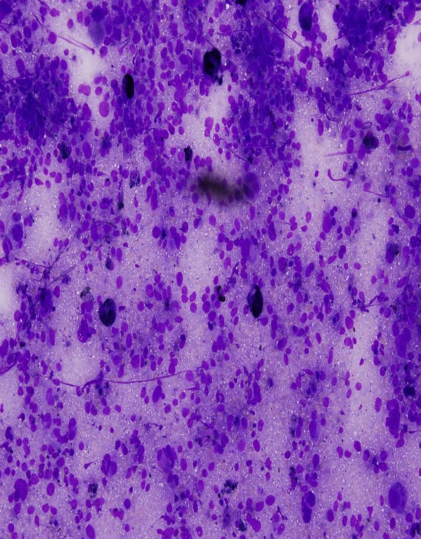
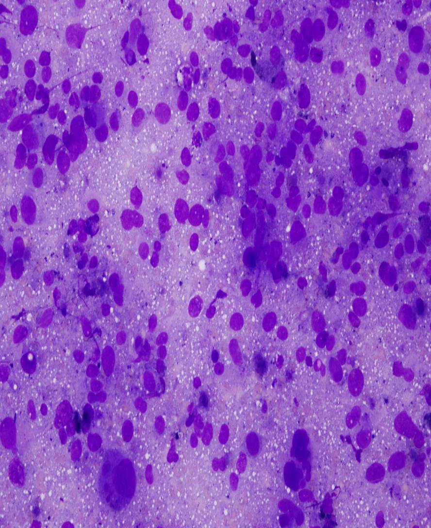
Contributed by Zahra Maleki, M.D.
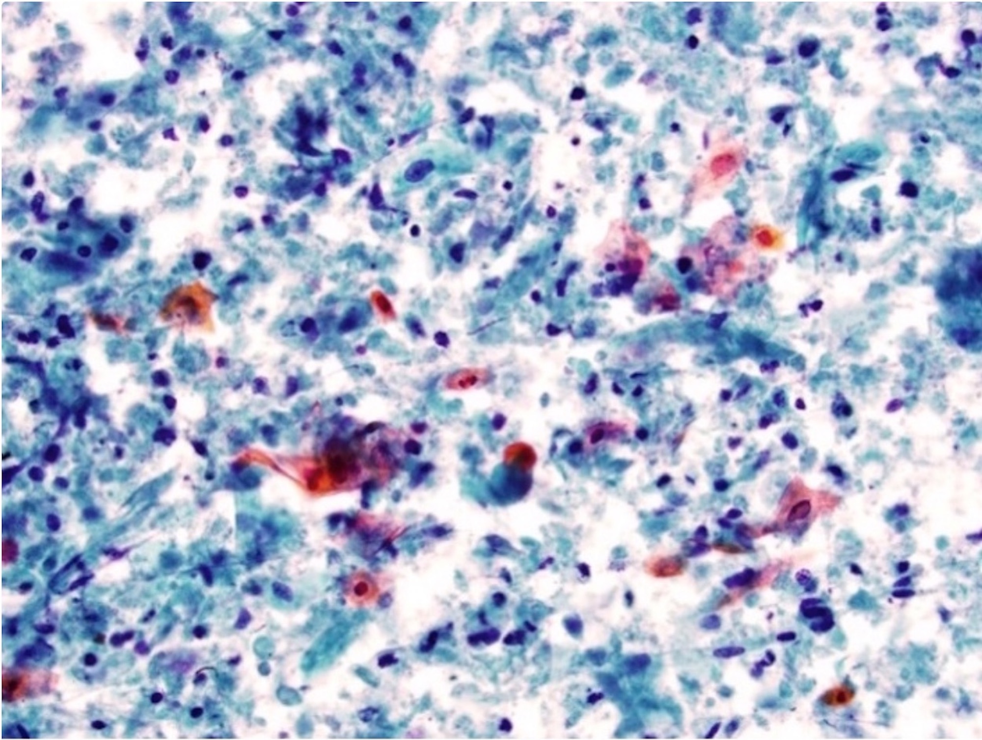
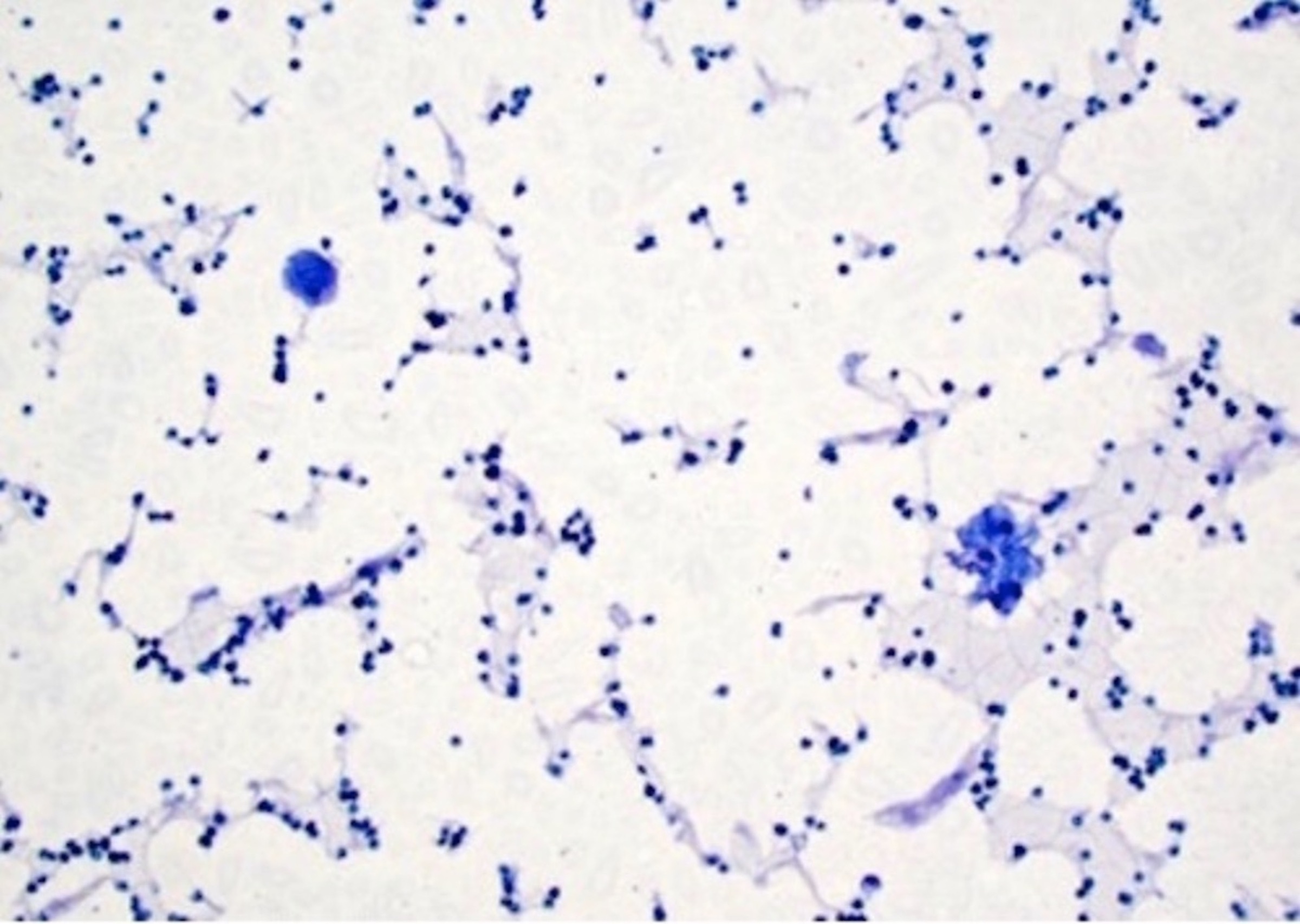
Atypical squamous cells
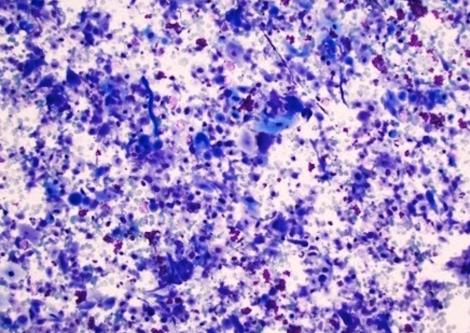
Pleomorphism
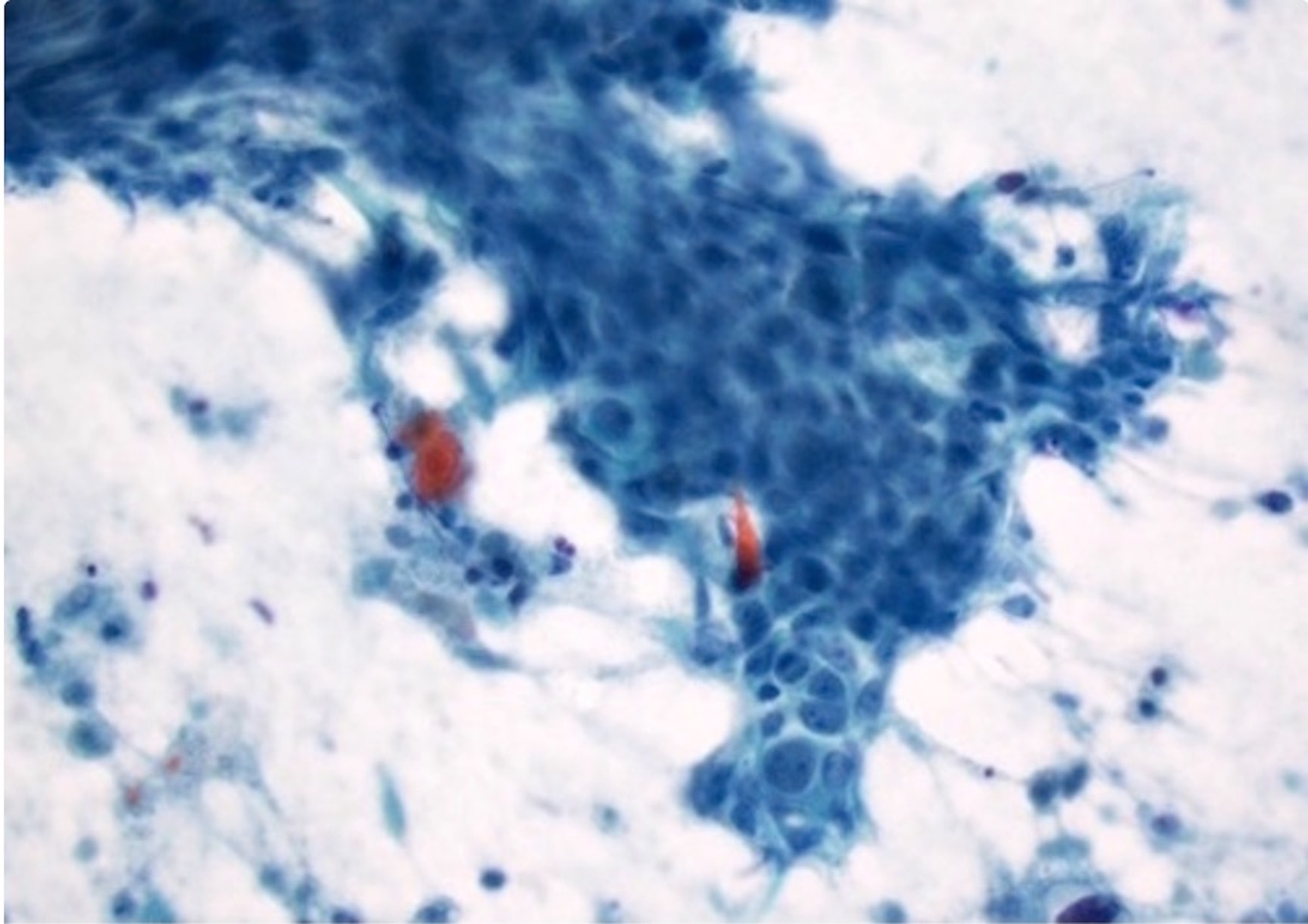
Focal keratinization
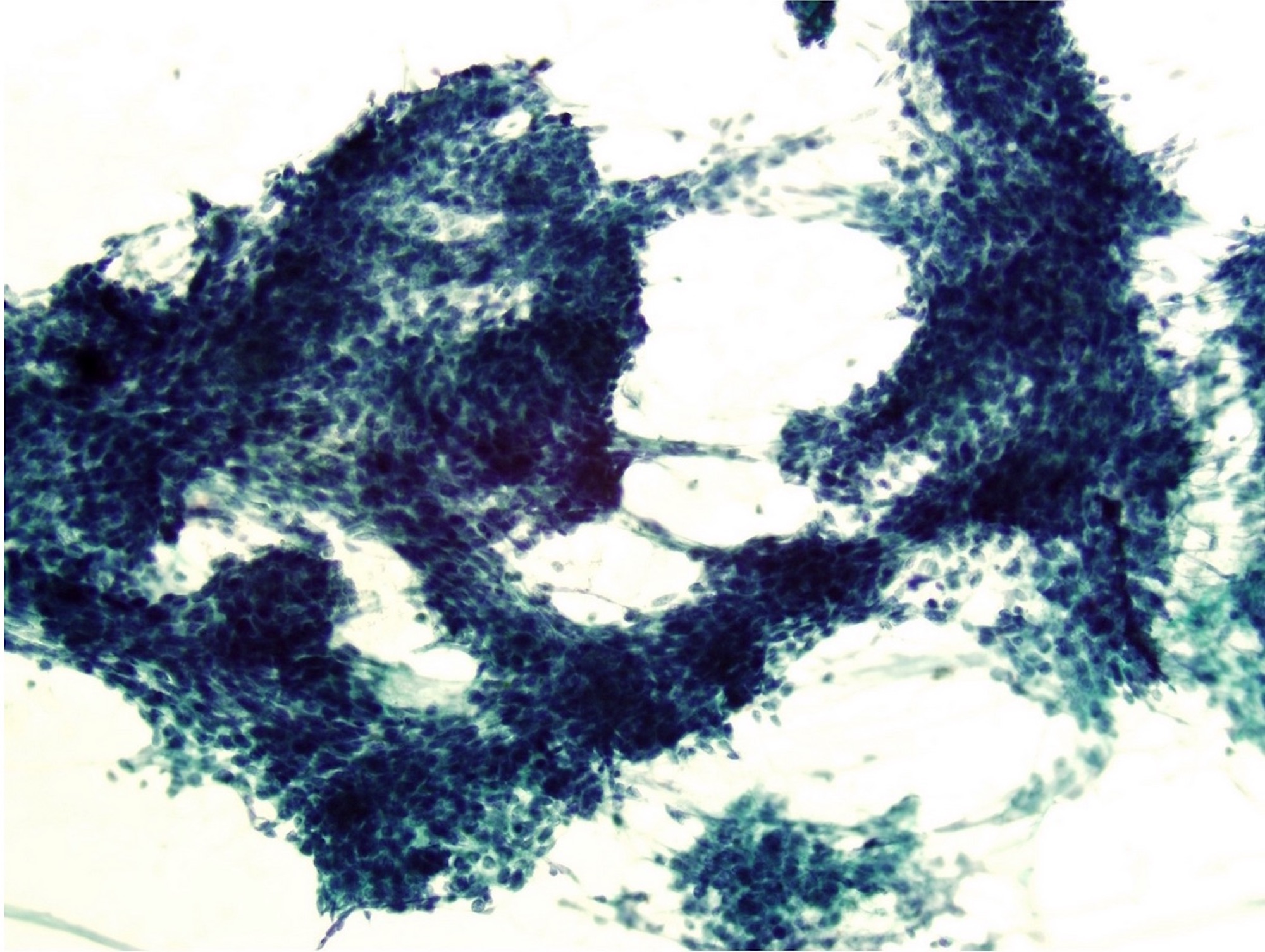
Cohesive fragments
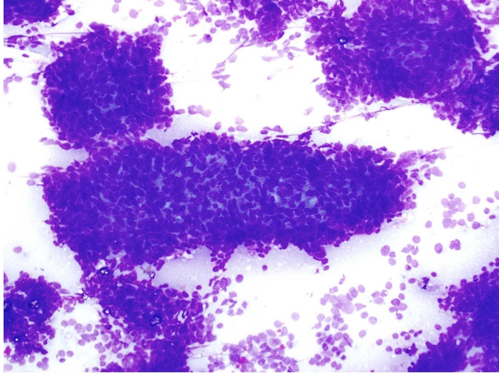
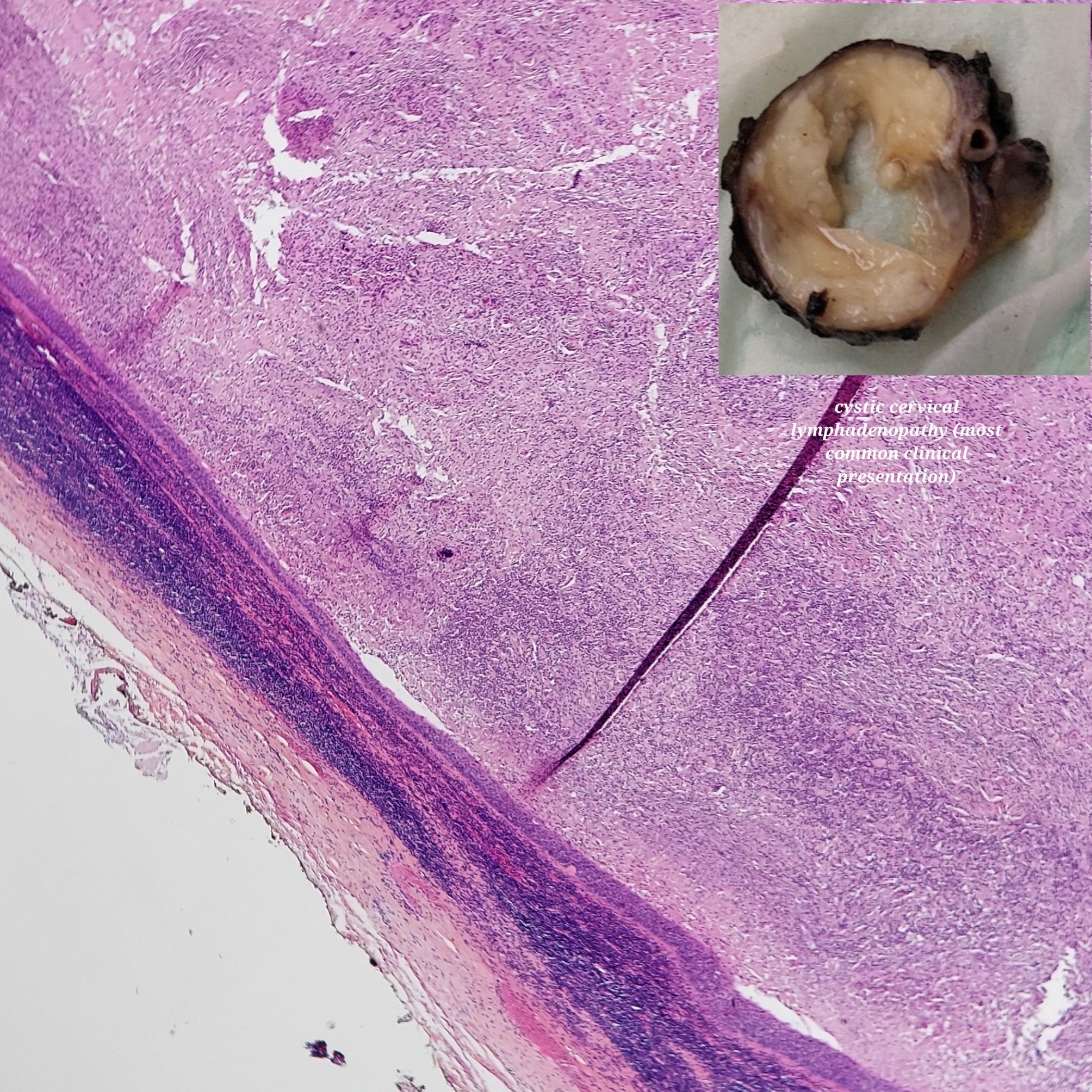
Basaloid features
Cyst aspiration
Cell block
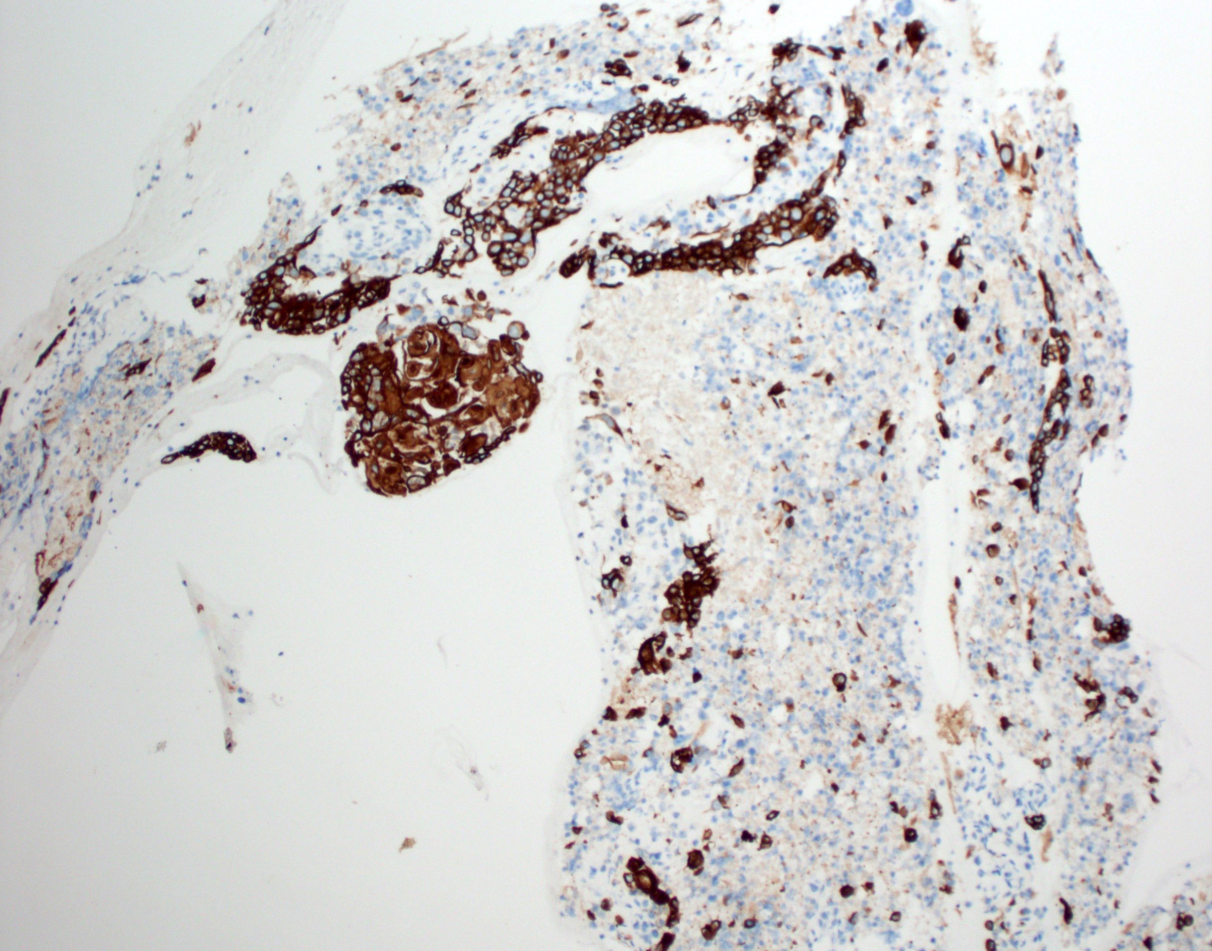
p63
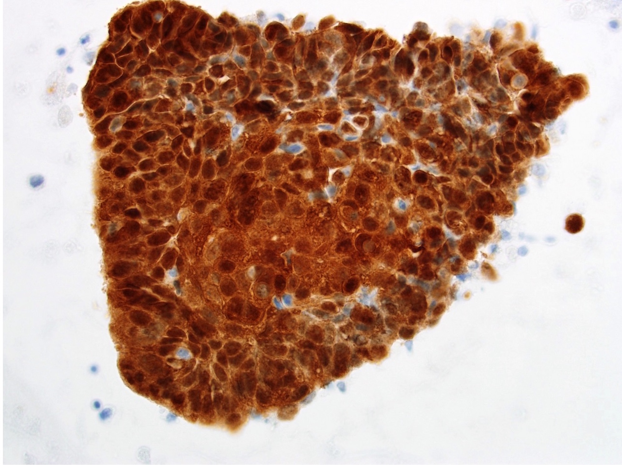
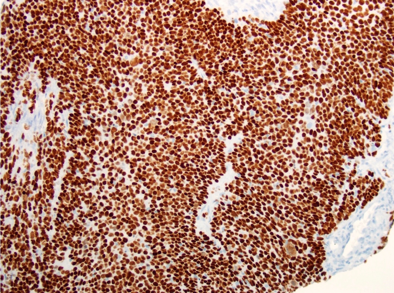
p16
Contributed by Zahra Maleki, M.D.
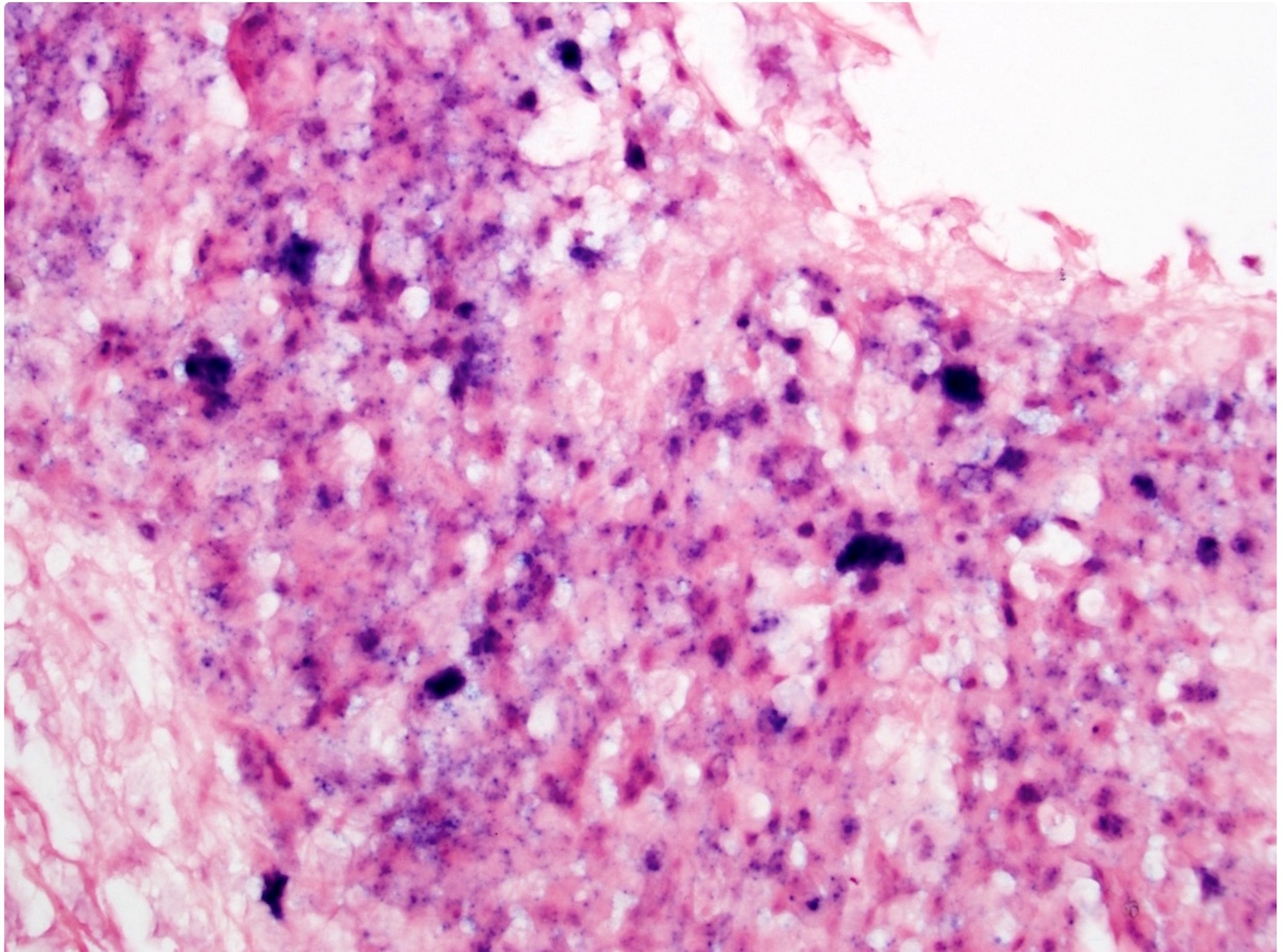
High risk HPV DNA ISH
HPV16 DNA ISH
HPV RNA ISH
Contributed by Katherine Hulme, M.B.Ch.B.
Oropharynx (frontal view)
Oropharynx (lateral view)
Images hosted on other servers:

Preoperative soft palate tumor
Contributed by Ruta Gupta, M.B.B.S., M.D.

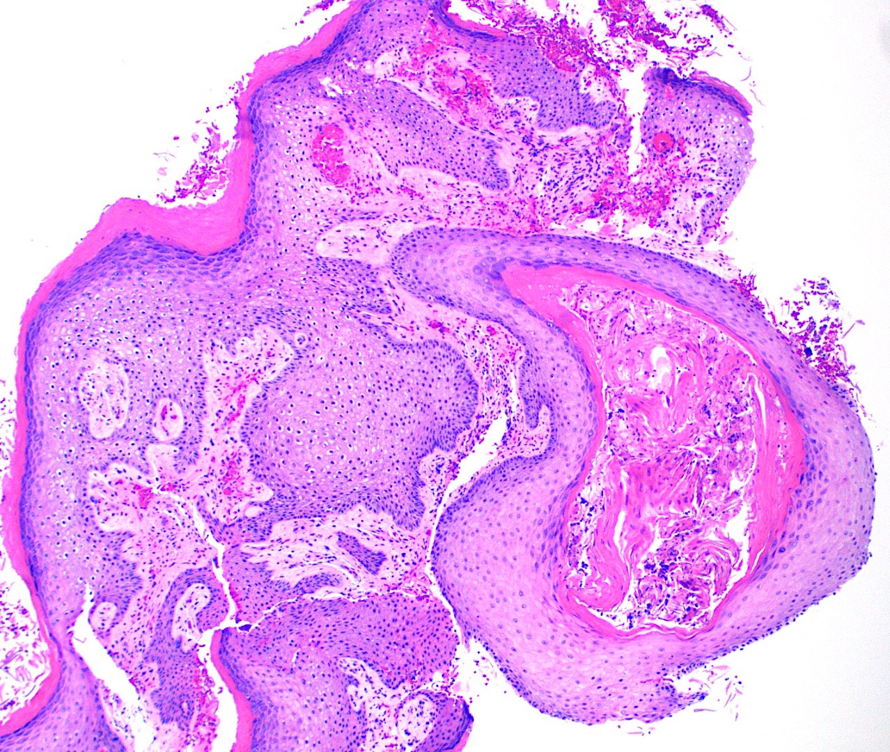
Verrucous mucosal lesion
Ulceroproliferative
mucosal lesion
Contributed by Ruta Gupta, M.B.B.S., M.D.

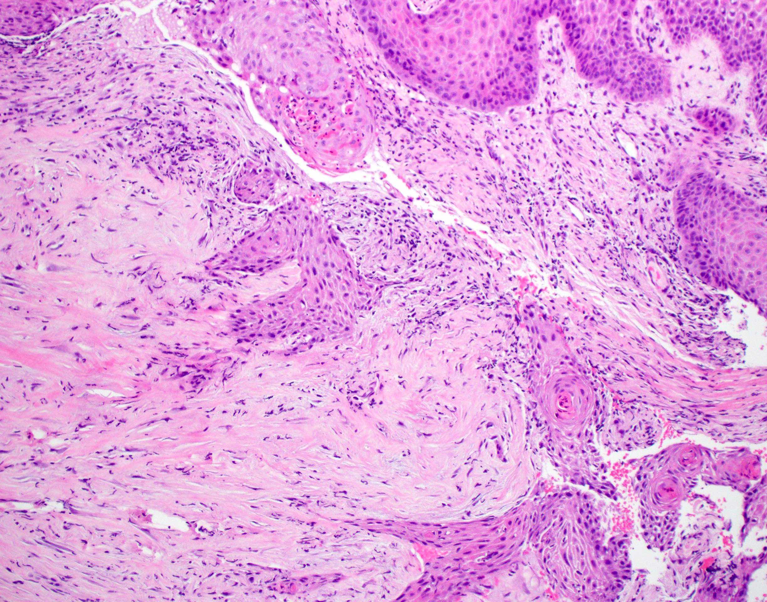
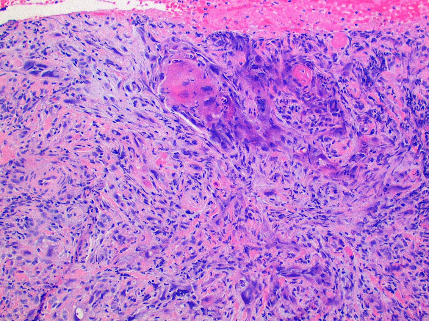
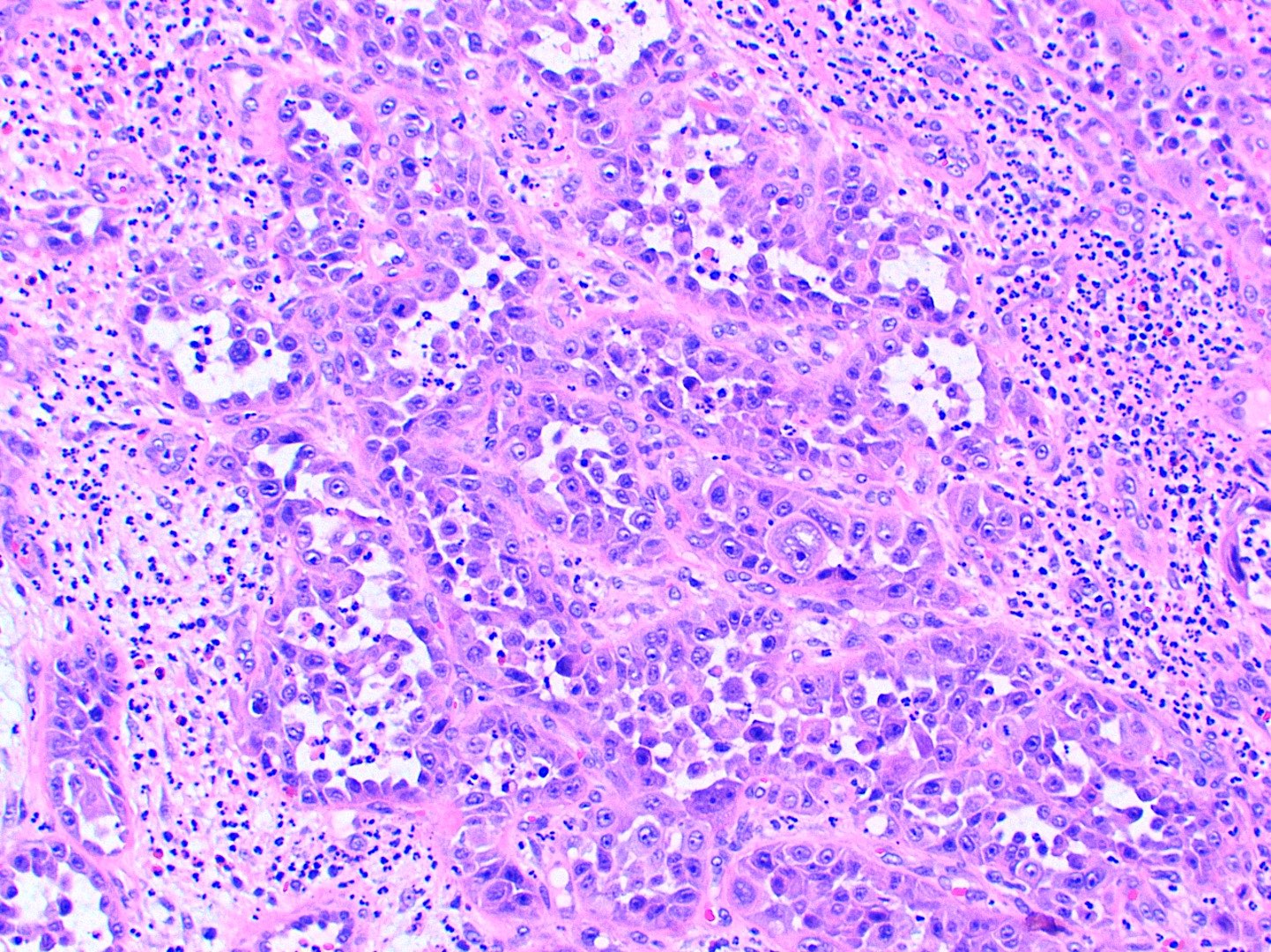
Well differentiated SCC
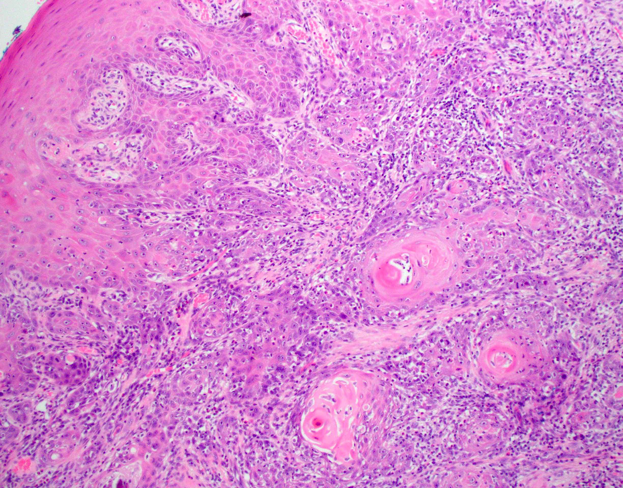
Moderately differentiated SCC
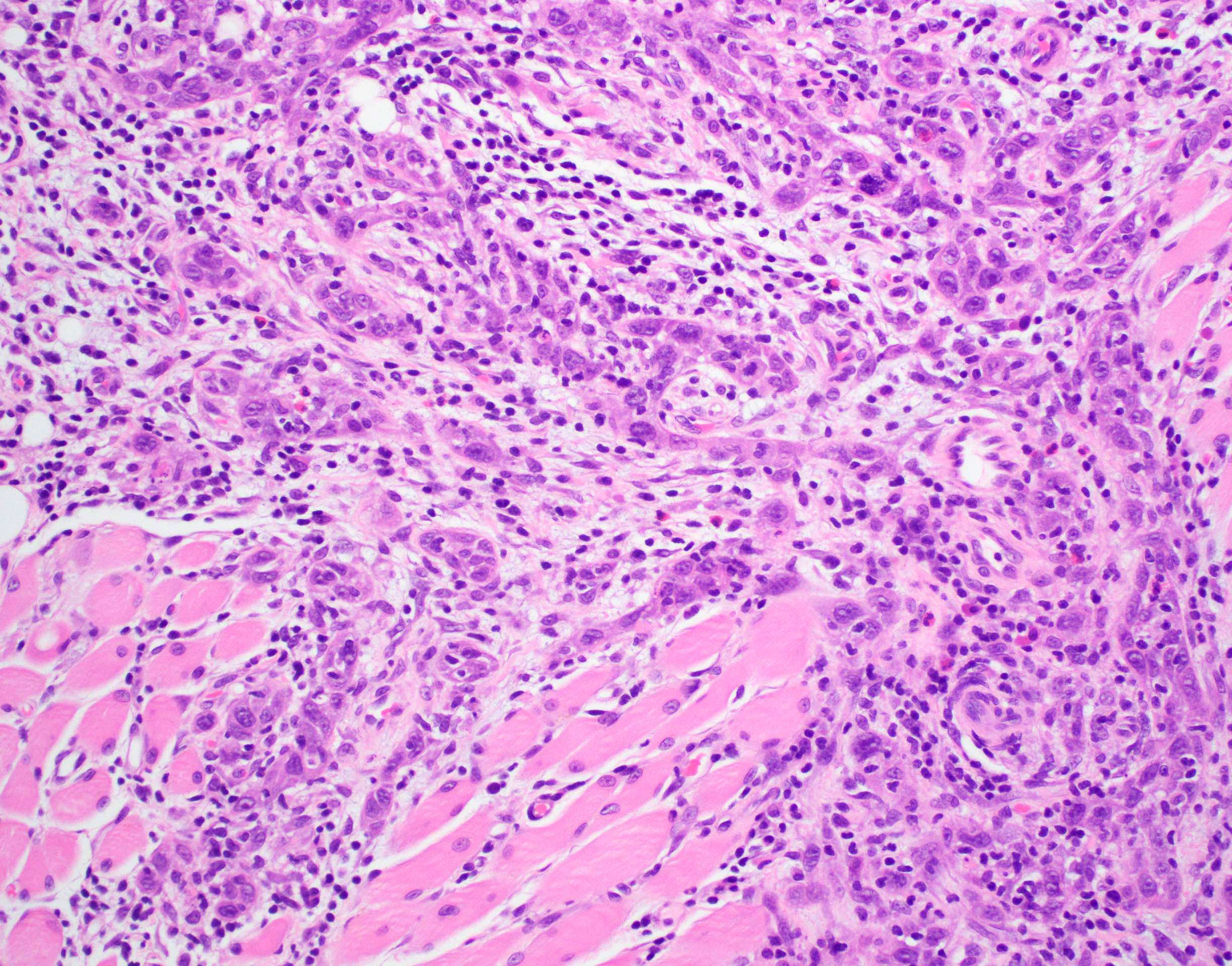
Poorly differentiated SCC
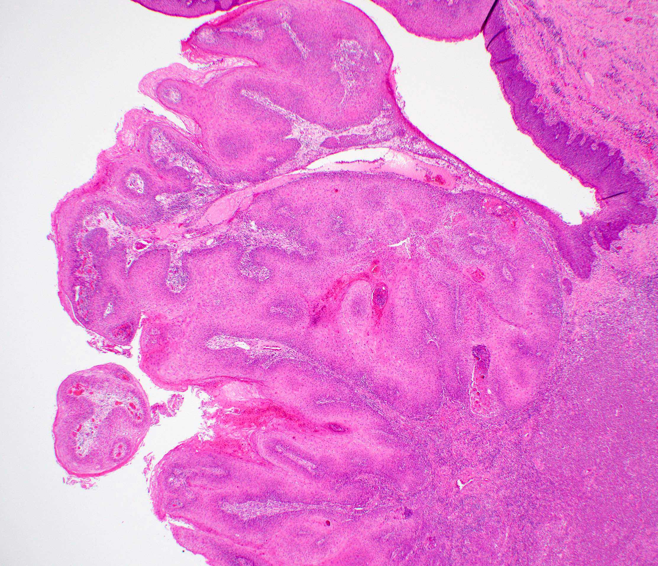
Verrucous SCC


Papillary SCC

Lymphoepithelial carcinoma
p40 in lymphoepithelial carcinoma
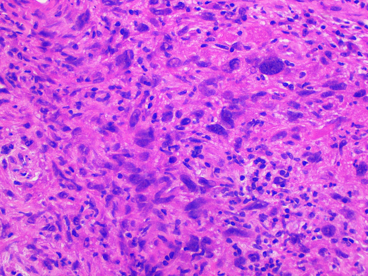
Spindle cell / sarcomatoid carcinoma
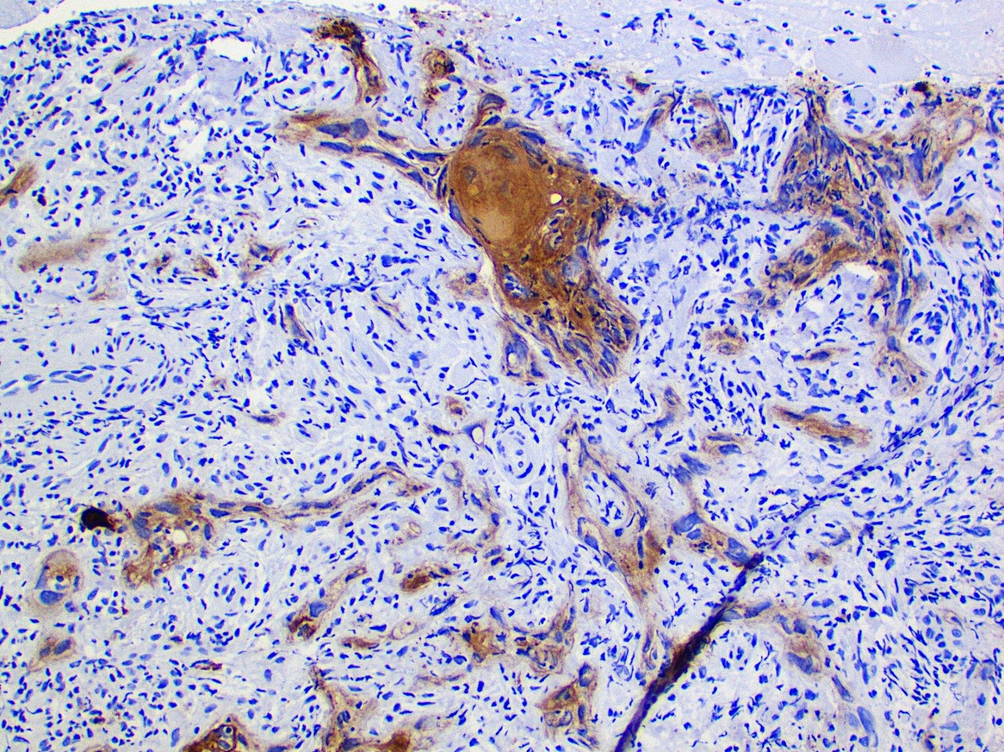
CK5/6 in spindle cell SCC

p40 in spindle
cell / sarcomatoid
carcinoma
Adenosquamous carcinoma
Mucicarmine in adenosquamous carcinoma
Contributed by Ruta Gupta, M.B.B.S., M.D.
Keratin and dyskeratotic cells
Clusters of keratin and dyskeratotic cells

Clusters of well differentiated squamous cells
CK5/6 in cell block
Images hosted on other servers:

Multiple small nodules on palate
Images hosted on other servers:

Inflamed bulbous projection
Contributed by Brandon Stapleton, D.M.D., Molly Housley Smith, D.M.D. and Jimmy Vellis, D.D.S., M.S.
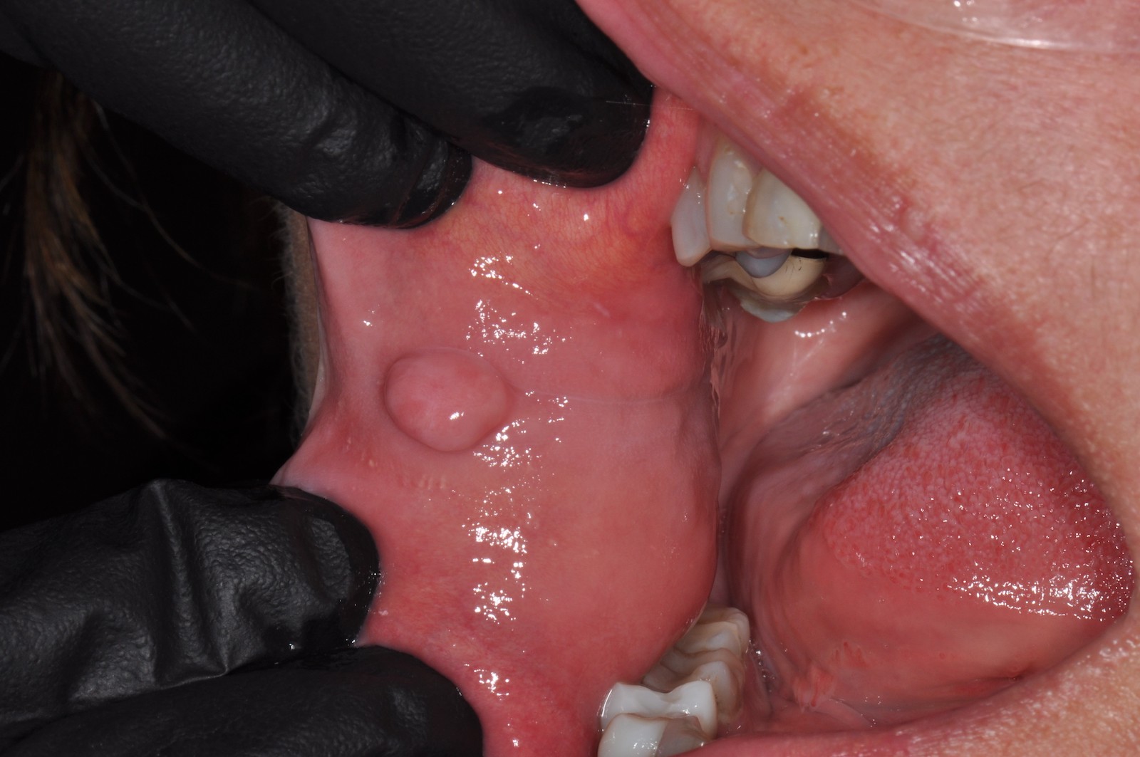
Fibroma of buccal mucosa
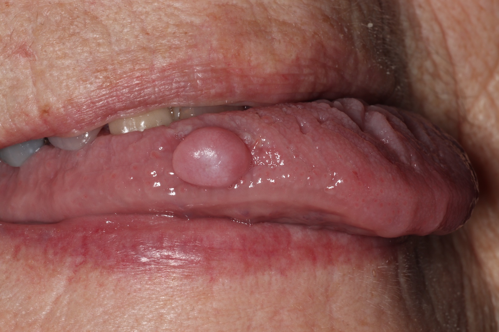
Fibroma of the tongue

Ulcerated irritation fibroma
Contributed by Molly Housley Smith, D.M.D.
Tan to gray nodule
Cut surface
Contributed by Molly Housley Smith, D.M.D.
Nodular mass

Hyperkeratotic surface
Scattered chronic inflammatory cells
Contributed by Ivan J. Stojanov, D.M.D., M.M.Sc.
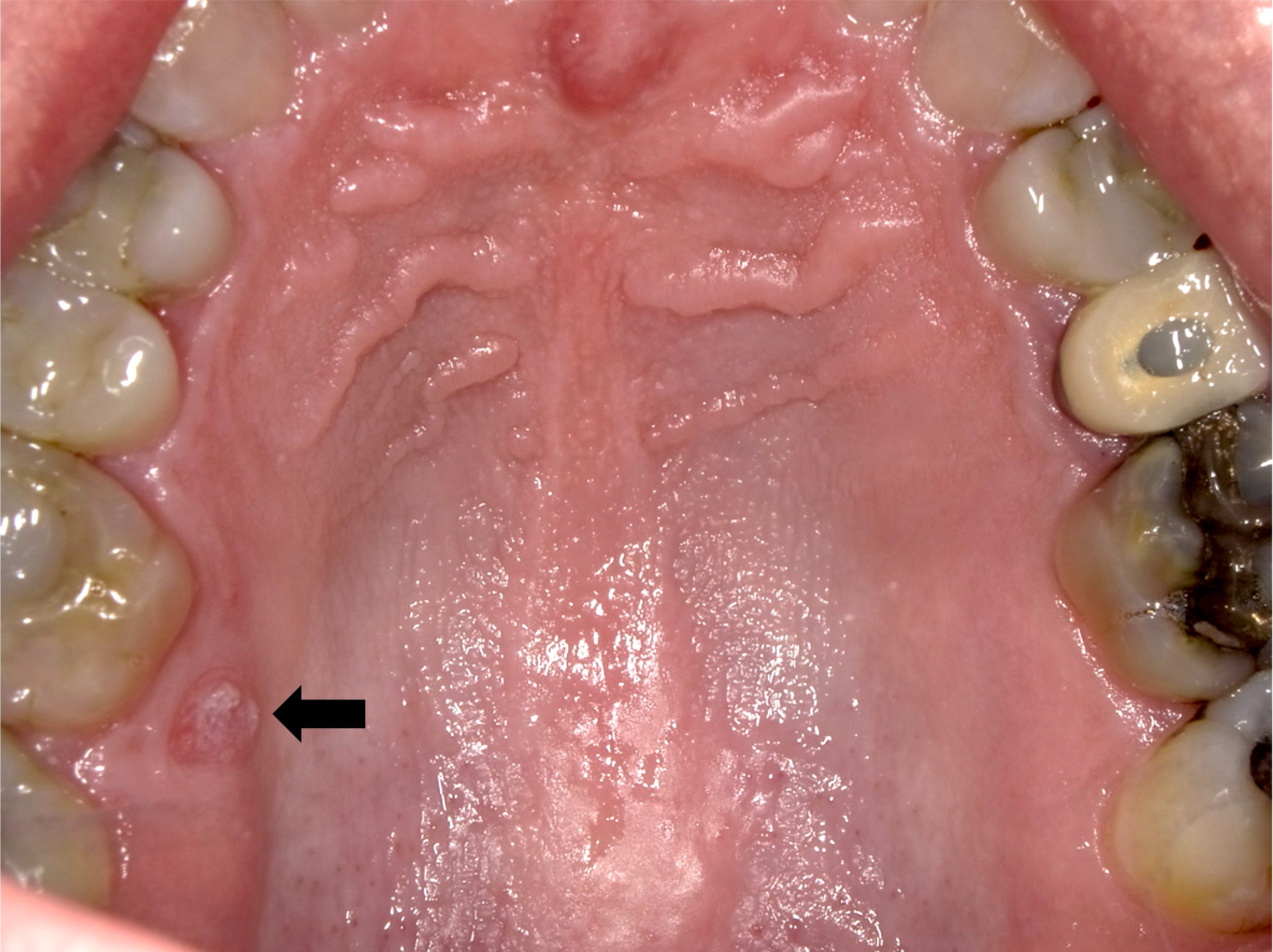
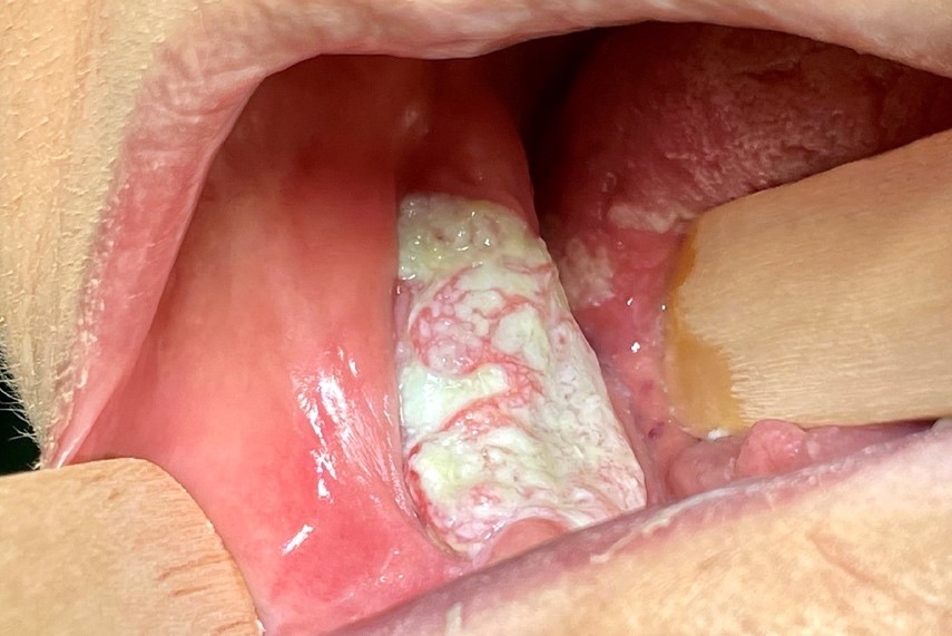
Left posterior maxillary leukoplakia
Right posterior maxillary leukoplakia
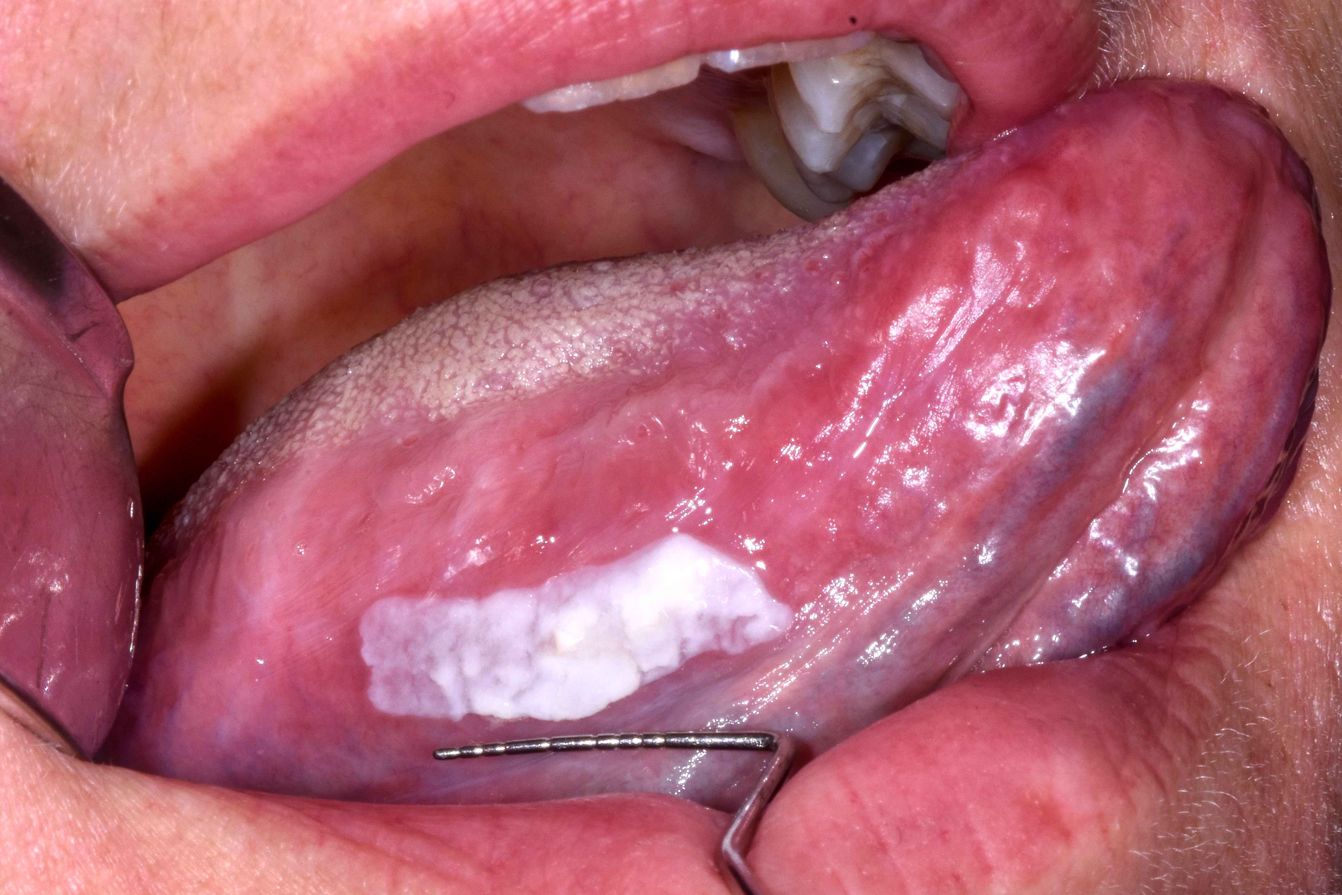
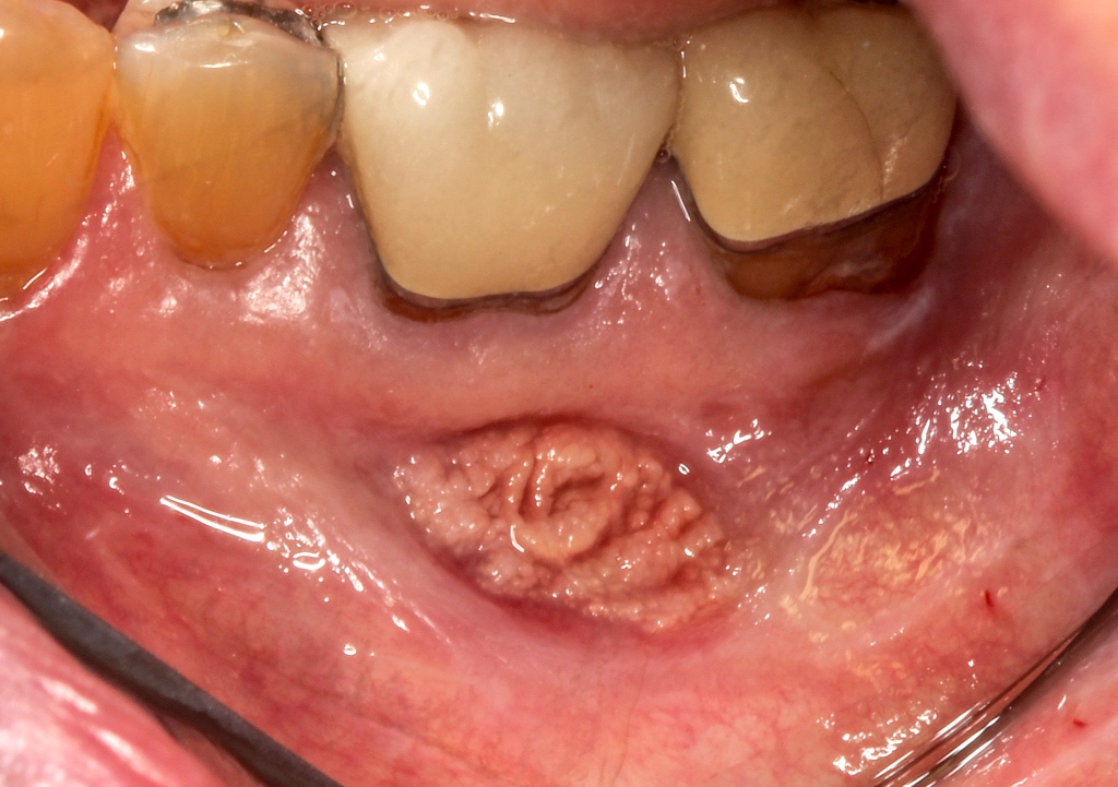
Left ventral tongue leukoplakia
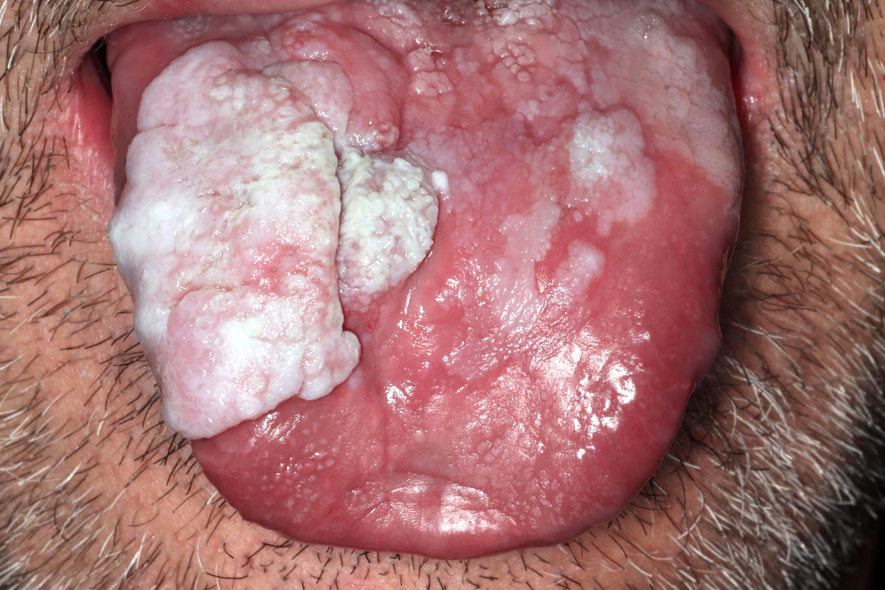
Extensive leukoplakia of tongue
Contributed by Ivan J. Stojanov, D.M.D., M.M.Sc.
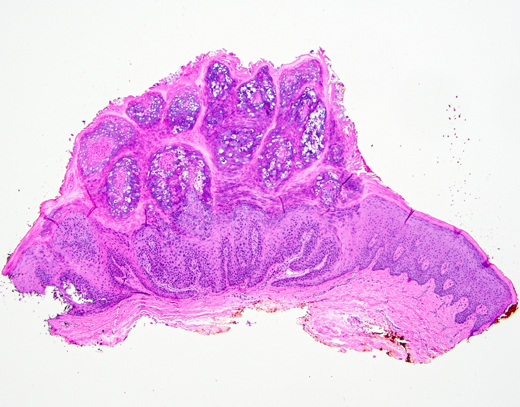
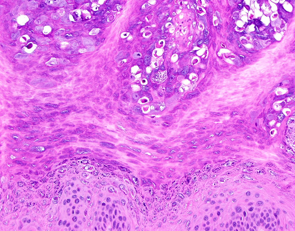
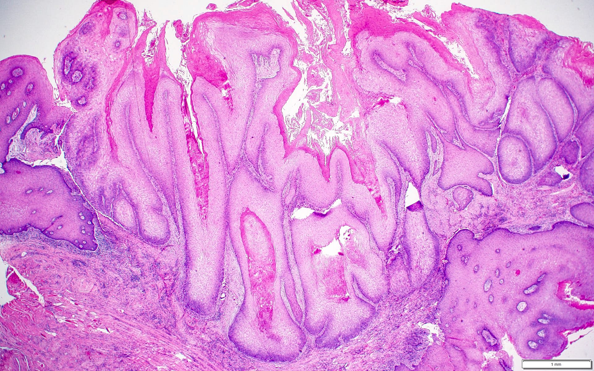
Undulating / verrucoid surface architecture
Absence of epithelial atypia
Sharply demarcated hyperkeratosis
Verrucous hyperplasia
Leukoplakia periphery
Moderate dysplasia
Leukoplakia with no dysplasia
Parakeratosis and epithelial hyperplasia
Hyperkeratosis and epithelial atrophy
Minimal cytologic dysplastic features
Images hosted on other servers:

CGH leukoplakia versus SCC

Genes implicated in malignant transformation
Contributed by Mark Mintline, D.D.S., Sahar Mirfarsi, D.D.S. and Molly Housley Smith, D.M.D.

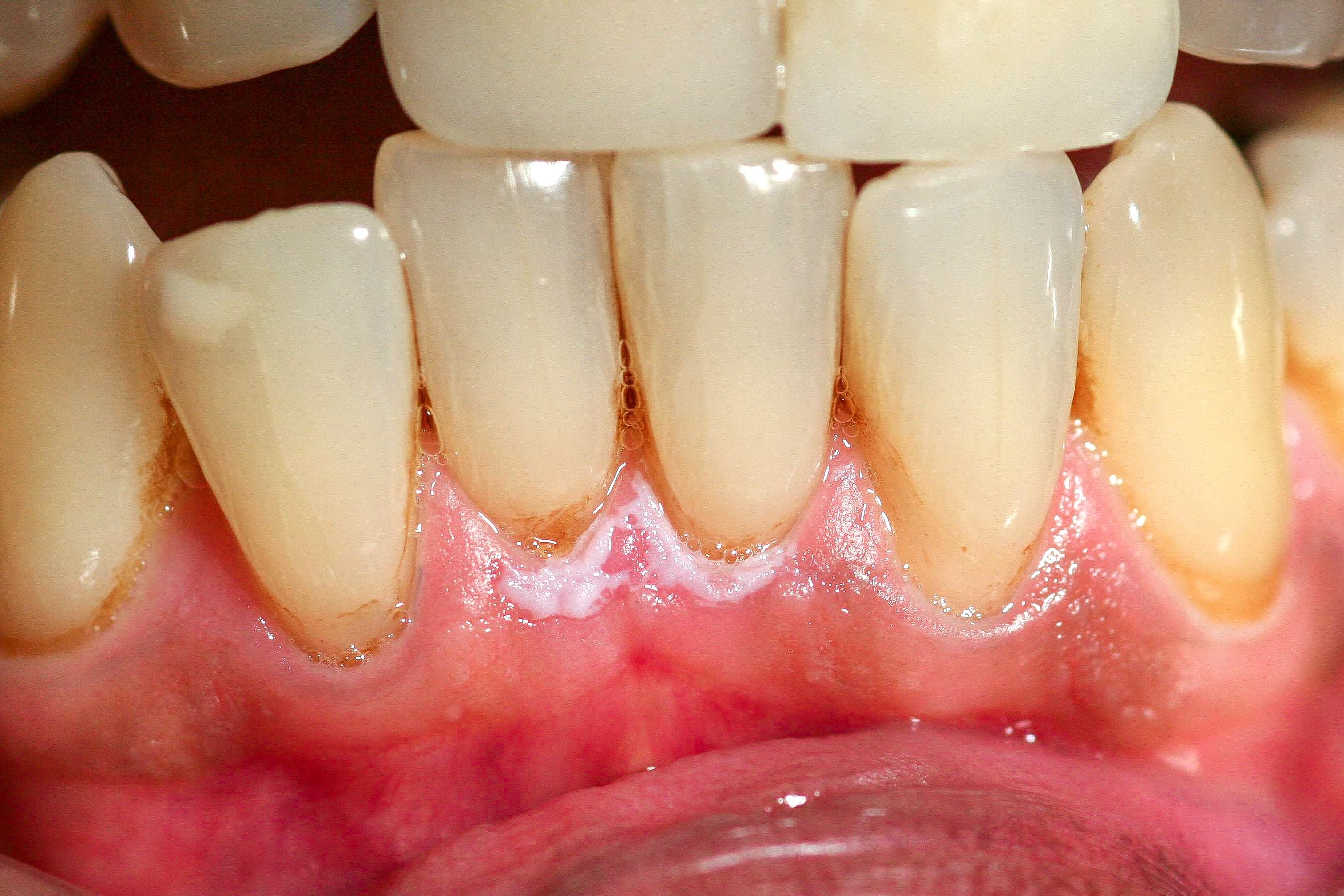
Reticular oral lichen planus
Erosive lichen planus
Desquamative gingivitis
Ulceration
Cutaneous plaques
Papular lichen planus
Reactive pigmentation
Cutaneous papules
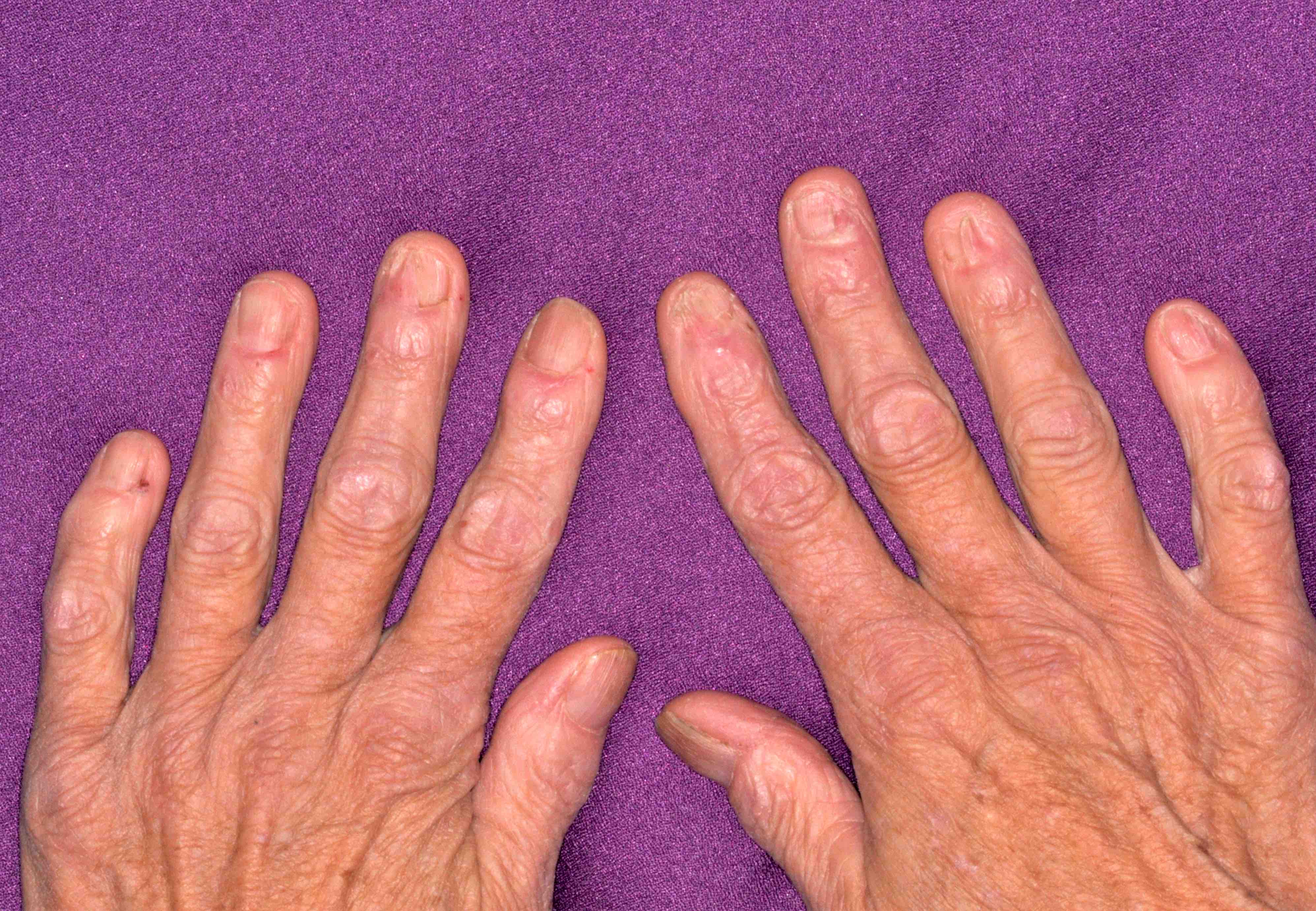
Nail changes
Contributed by Molly Housley Smith, D.M.D.
White striations
Contributed by Mark Mintline, D.D.S. and Molly Housley Smith, D.M.D.
Interface mucositis
Hydropic degeneration
Sawtooth rete ridges
Colloid bodies
Melanin pigmentation
Images hosted on other servers:

CT and laryngoscopy

SPECT / CT
CT scan:

Distinct margins

Lingual thyroid

Well defined mass

Aberrant thyroid tissue


48-year-old white female with obstructive lingual thyroid
MRI:

Axial MRI: lingual thyroid

38 year old female
Radionuclide scan:

Scintigraphy with Tc-99m

Tc99m pertechnetate planar image

99mTcO4 radionuclide thyroid scan


Thyroid scintigraphy
Angiography:

External carotid artery angiogram

Ectopic thyroid

Carotid arteriography
Images hosted on other servers:
Tongue mass:


Endoscopy:

Flexible and rigid

Swelling
Lingual thyroid in surgery:

Exposed tumor

Intraoperative photograph and surgical specimen
Images hosted on other servers:


Excised specimen
Contributed by Andrey Bychkov, M.D., Ph.D. and AFIP


Enlarged thyroid follicles
Resembles malignancy
Images hosted on other servers:

With skeletal muscle


Thyroid follicles

Papillary thyroid
carcinoma in
lingual thyroid
Images hosted on other servers:

Follicular cells
Ectopic thyroid in a 6 year old child
Images hosted on other servers:


Lingual tonsil anatomy
Contributed by Molly Housley Smith, D.M.D.
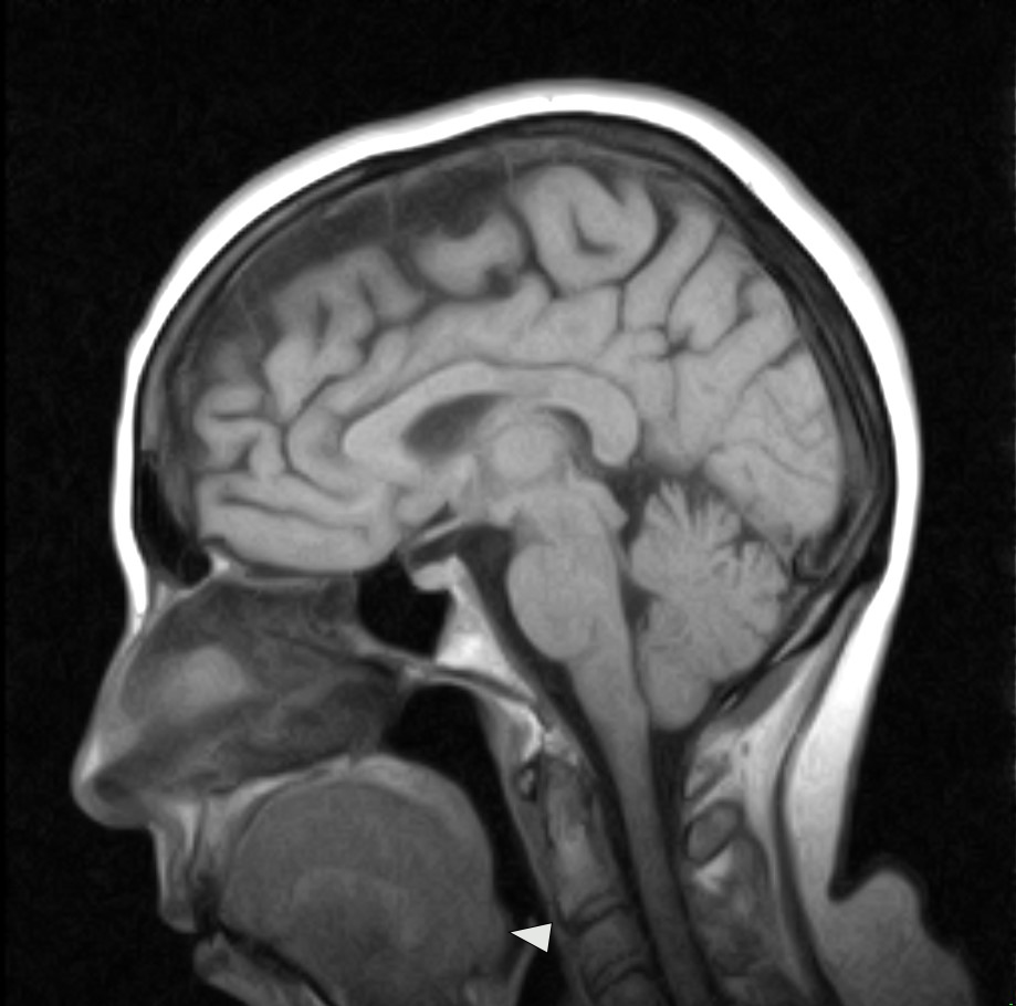
Sagittal T1 weighted MRI
Contributed by Molly Housley Smith, D.M.D.
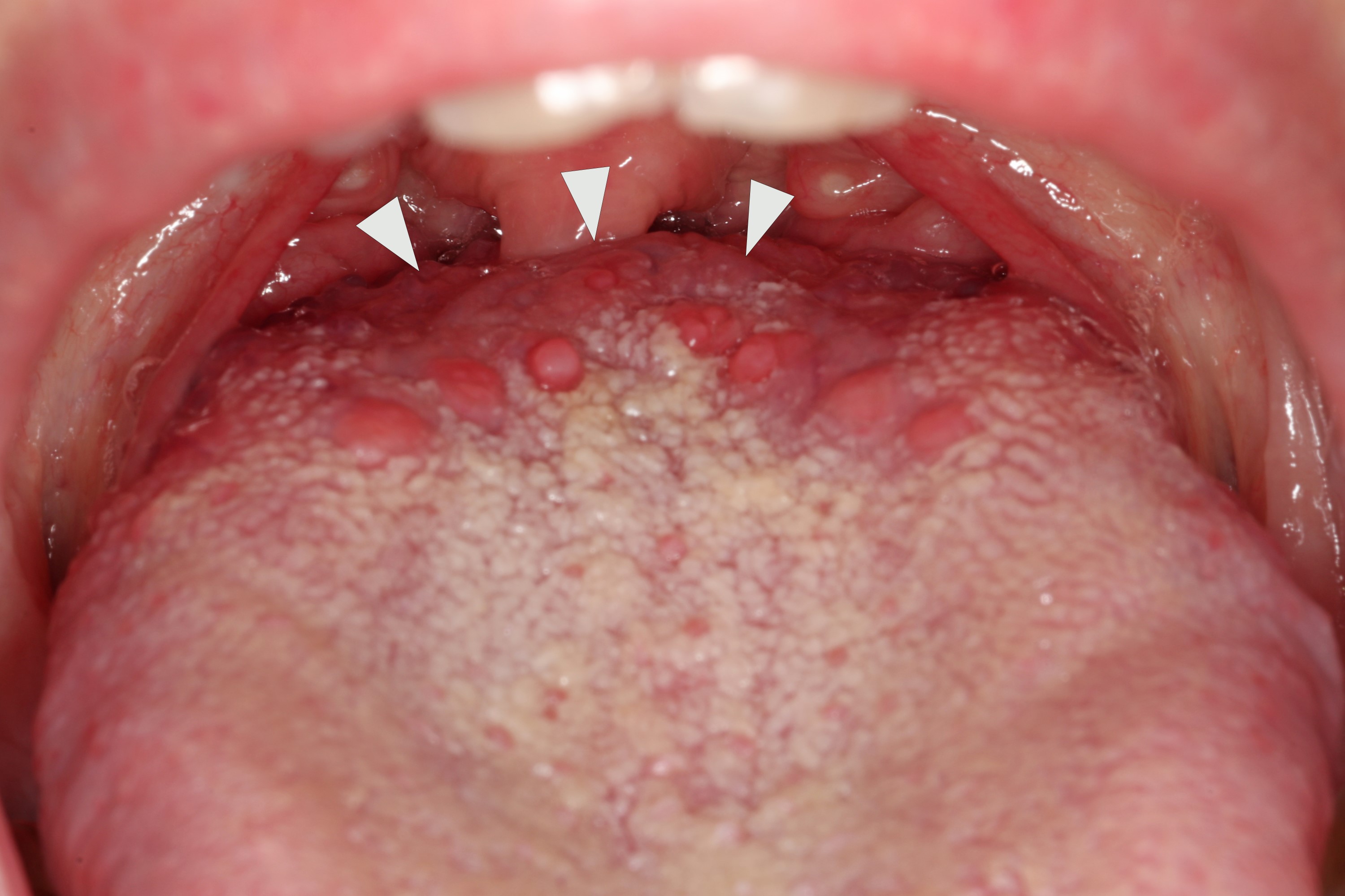
Posterior dorsal tongue
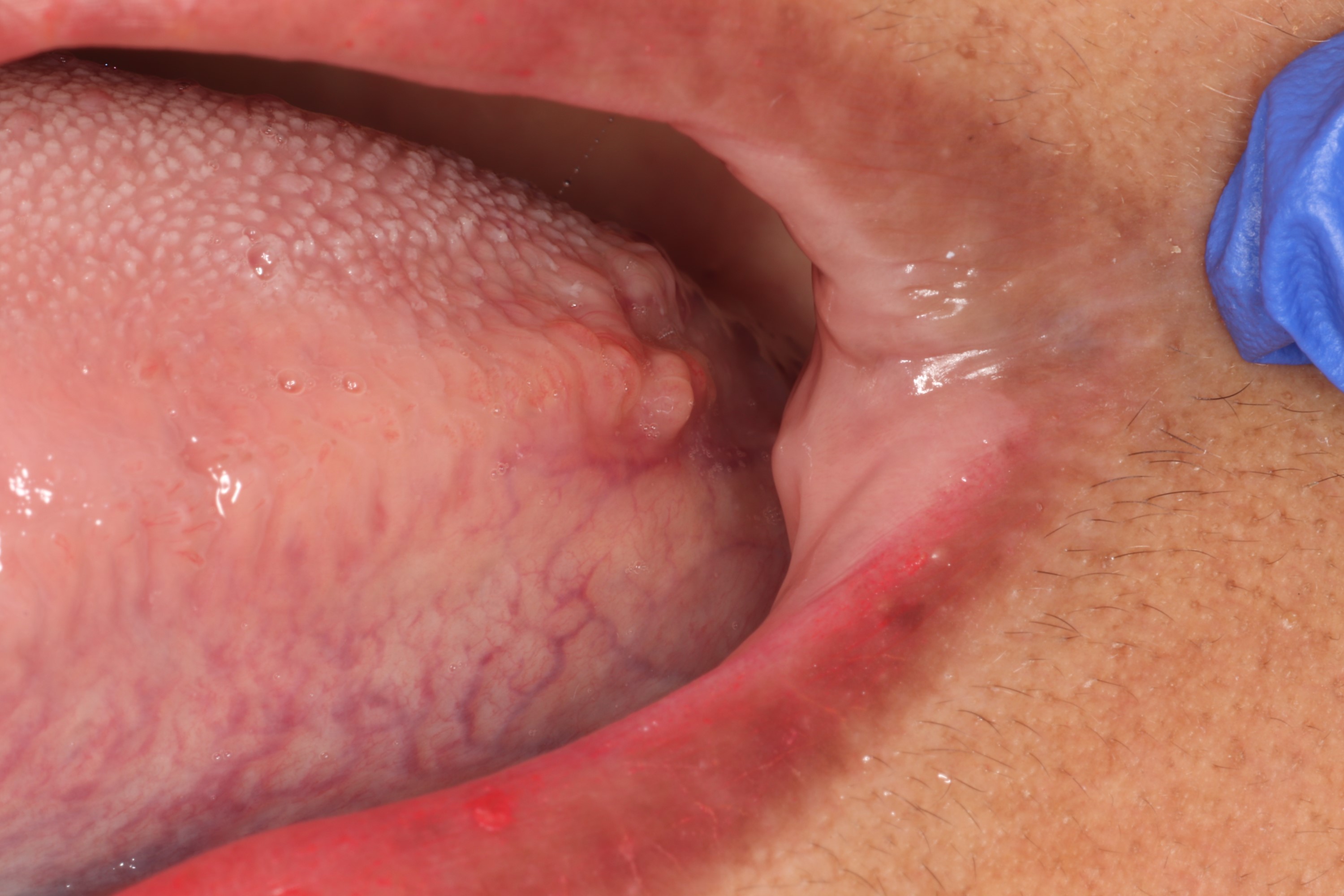
Posterior lateral tongue
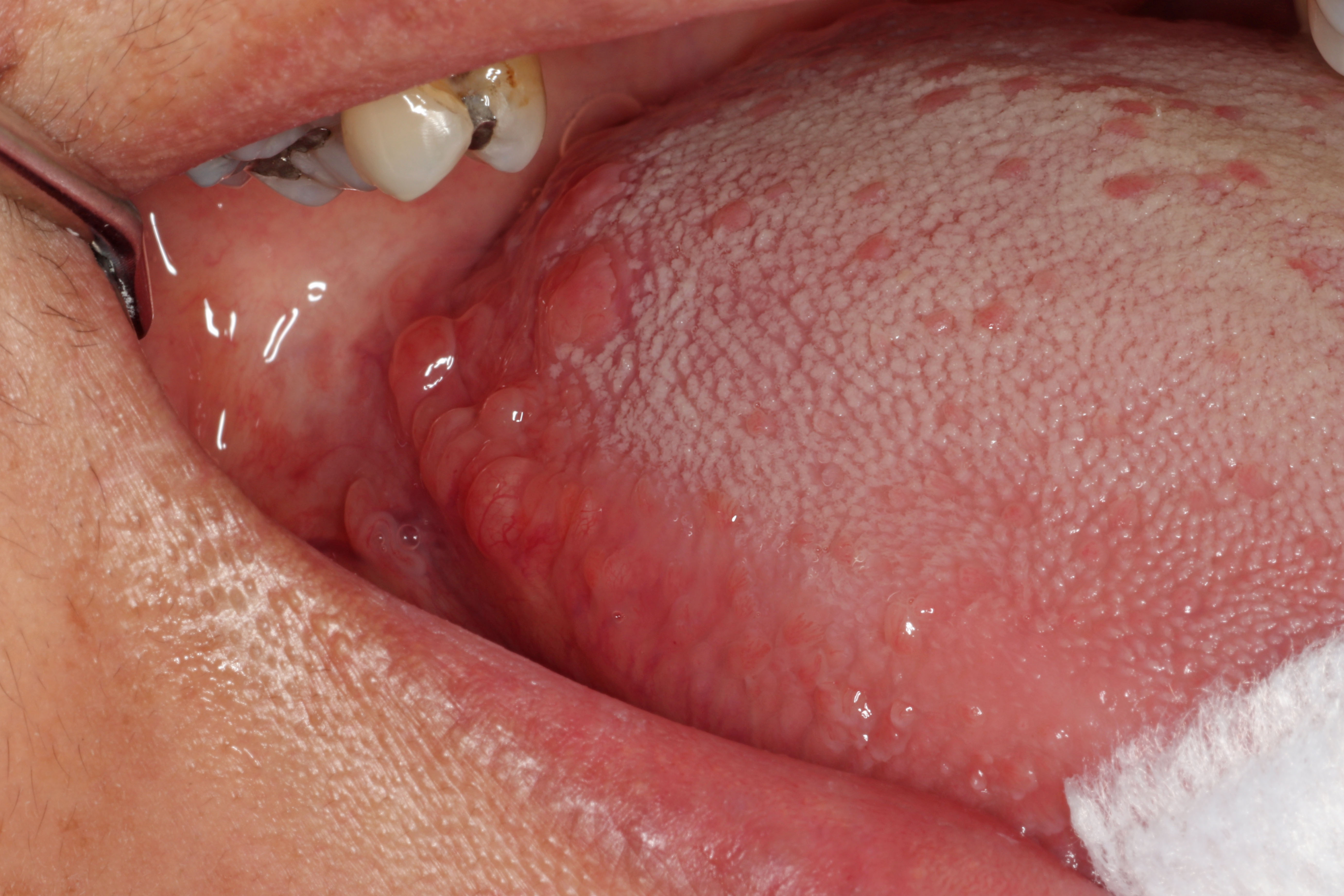
Numerous enlarged papules
Images hosted on other servers:

Inferior view

Superior view
Contributed by Molly Housley Smith, D.M.D.

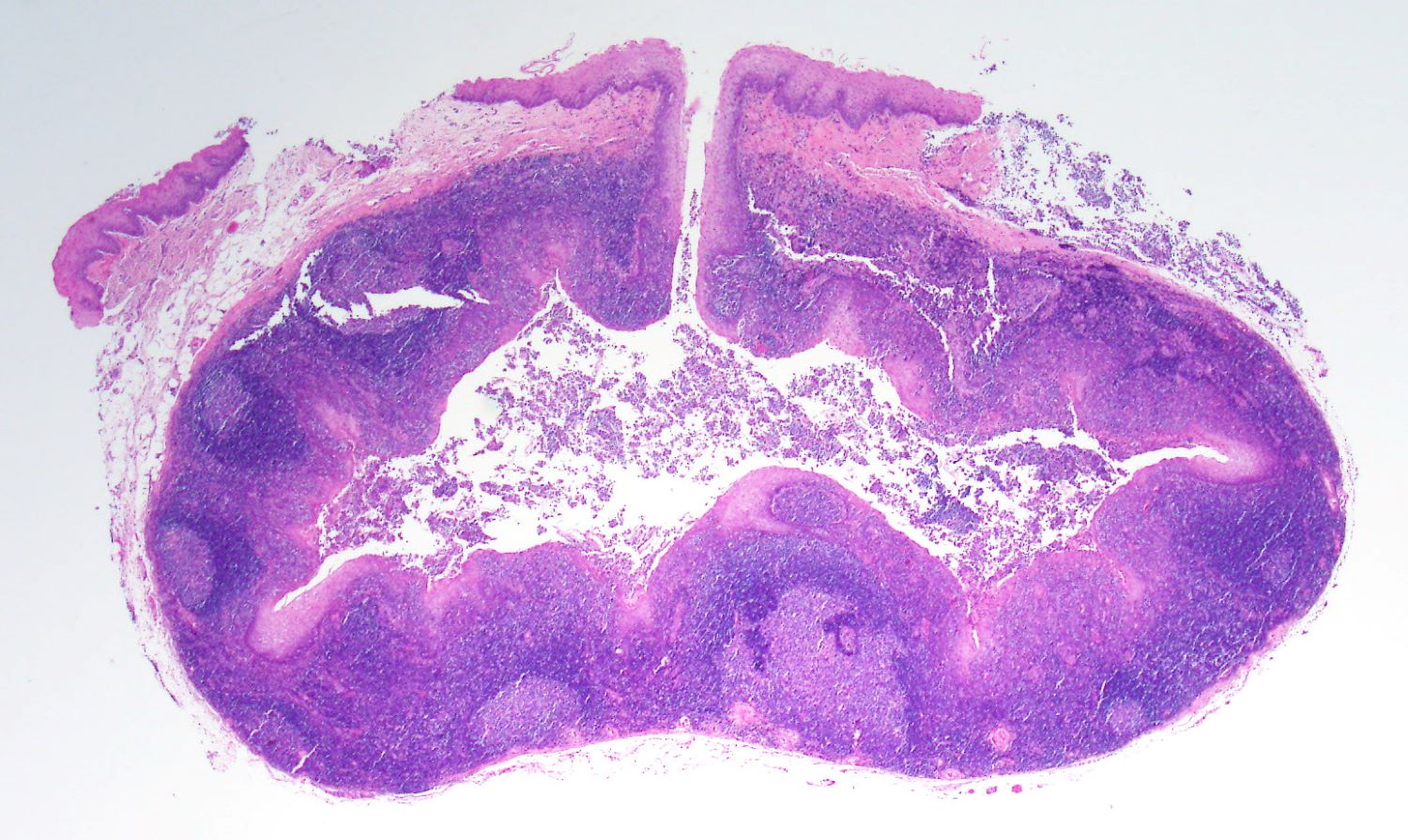
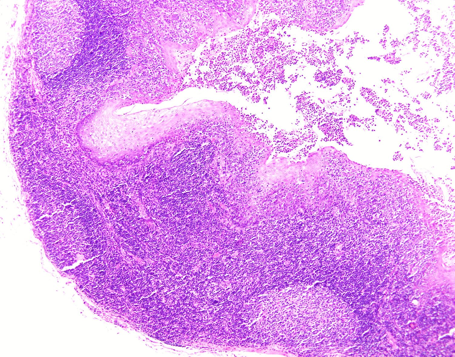
Germinal center formation

Epithelial crypt formation

Diffuse lymphocytic tissue
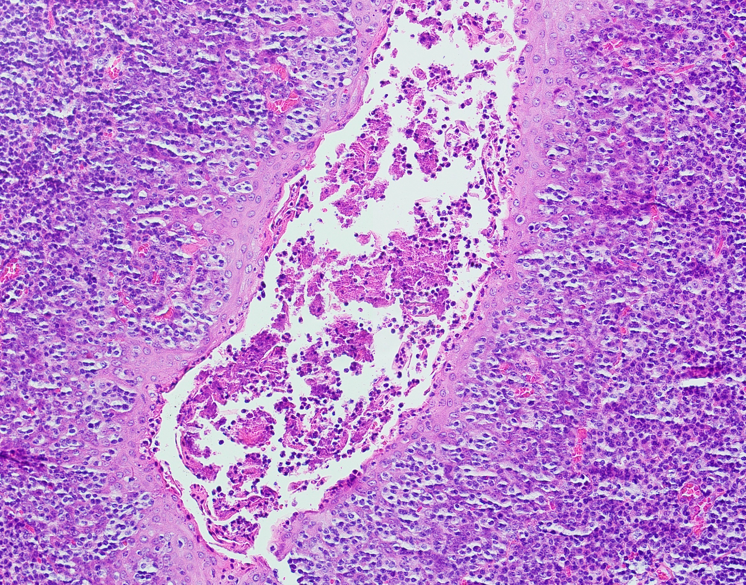
Reticular epithelium
Histology of the lingual tonsil
Lingual tonsillectomy for obstructive sleep apnea
Images hosted on other servers:

Lymphoepithelial cyst in tonsil

Lymphoepithelial cyst, floor of mouth
Images hosted on other servers:

Lymphoepithelial cyst, floor of mouth
Contributed by John Kalmar, D.M.D., Ph.D.
Floor of mouth cyst
Squamous epithelial lining
Lateral tongue cyst
Germinal center in stroma
Images hosted on other servers:


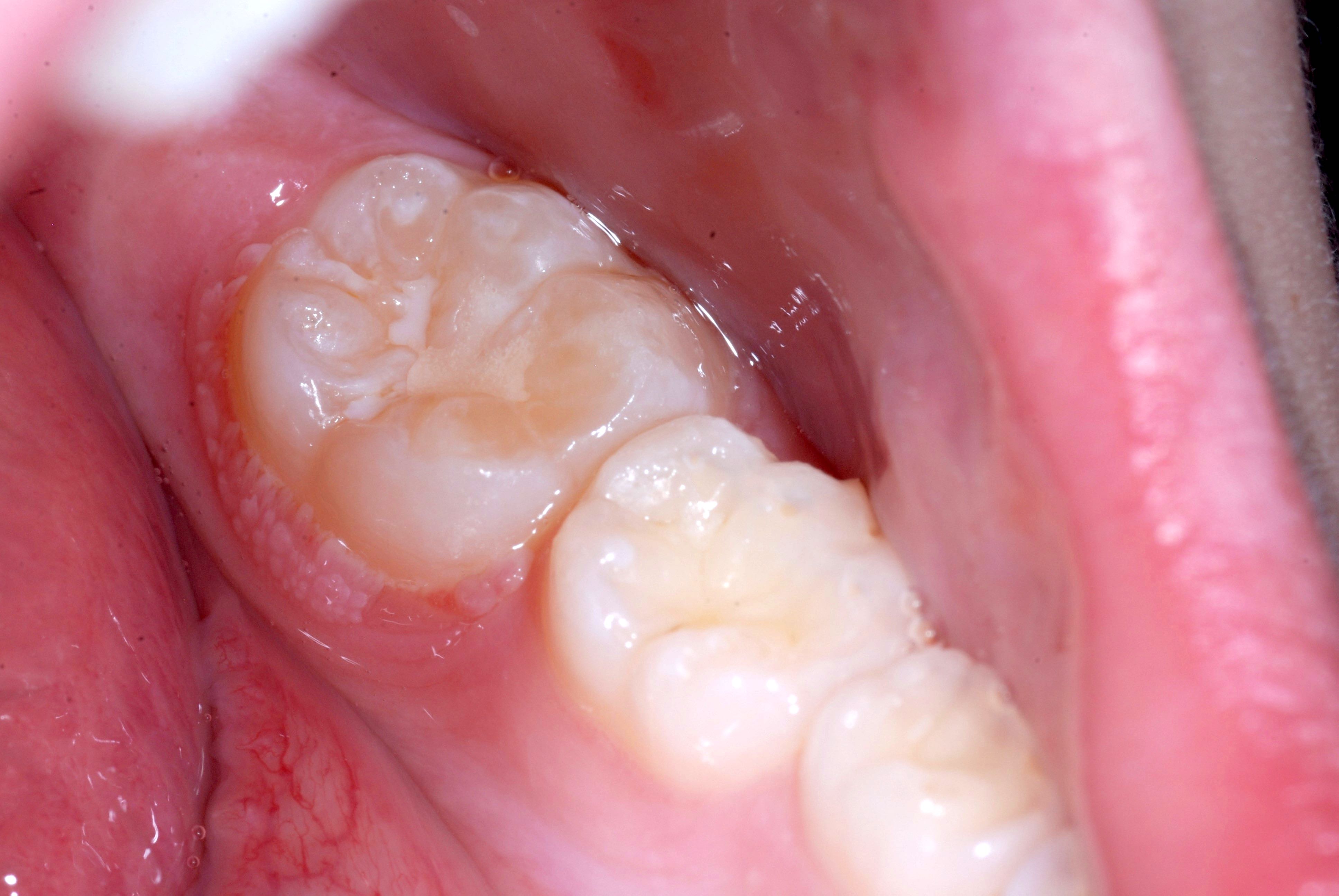
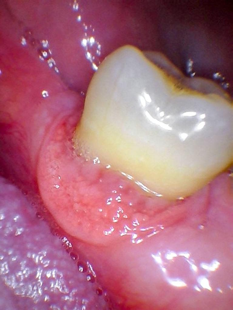
Median rhomboid glossitis

Diabetic patient
Images hosted on other servers:


Diffuse swelling of lip
Images hosted on other servers:


Nonnecrotizing granulomas

Giganto-epithelioid granuloma
Contributed by Nathan Lee, D.M.D.
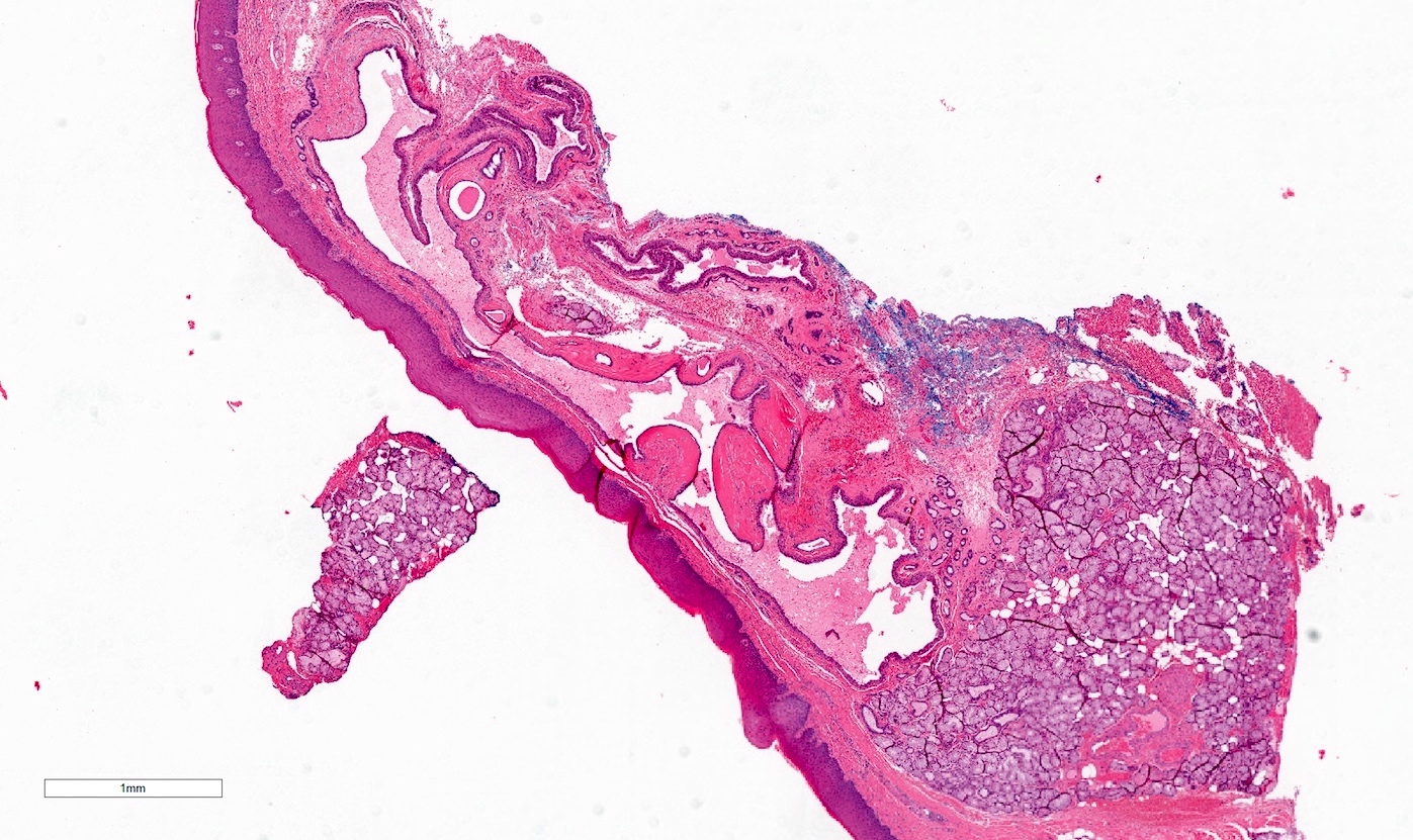
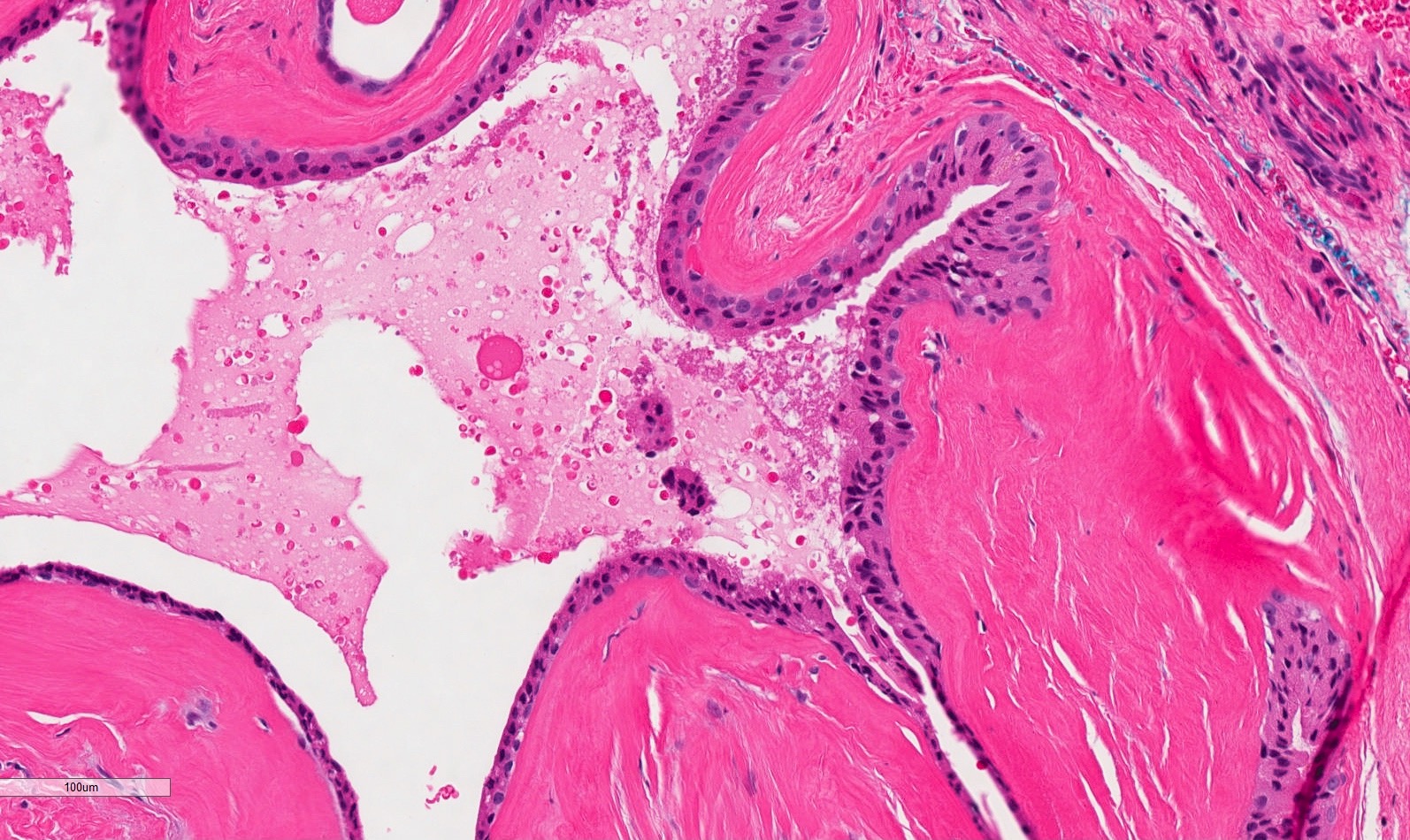
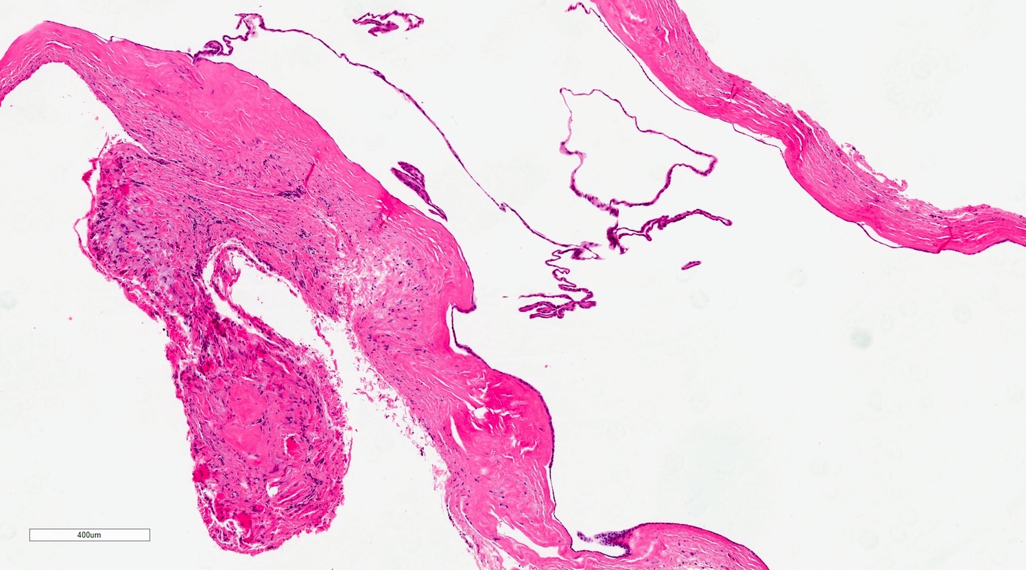
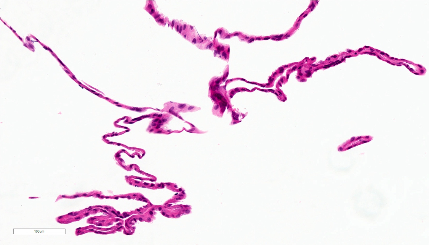
Oral cavity mucocele
Images hosted on other servers:

Intact lesions
Contributed by Vancouver General Hospital
Early mucocele removed intact
Late mucocele removed ruptured
Mucocele mimicking mucoepidermoid carcinoma
Early mucocele removed intact
Late mucocele removed ruptured
Images hosted on other servers:

Slightly elevated

Multiple lesions
Lesions on tongue and lip
Images hosted on other servers:

Parakeratosis

Typical nuclear changes
Images hosted on other servers:

Lateral cephalograph

Raised left ala of nose

Extraoral swelling

Obliteration of labial sulcus

Endoscopic view
Images hosted on other servers:

Surgical specimen
Images hosted on other servers:

Pseudostratified cylindrical epithelium

Cyst lining

Stratified squamous epithelium
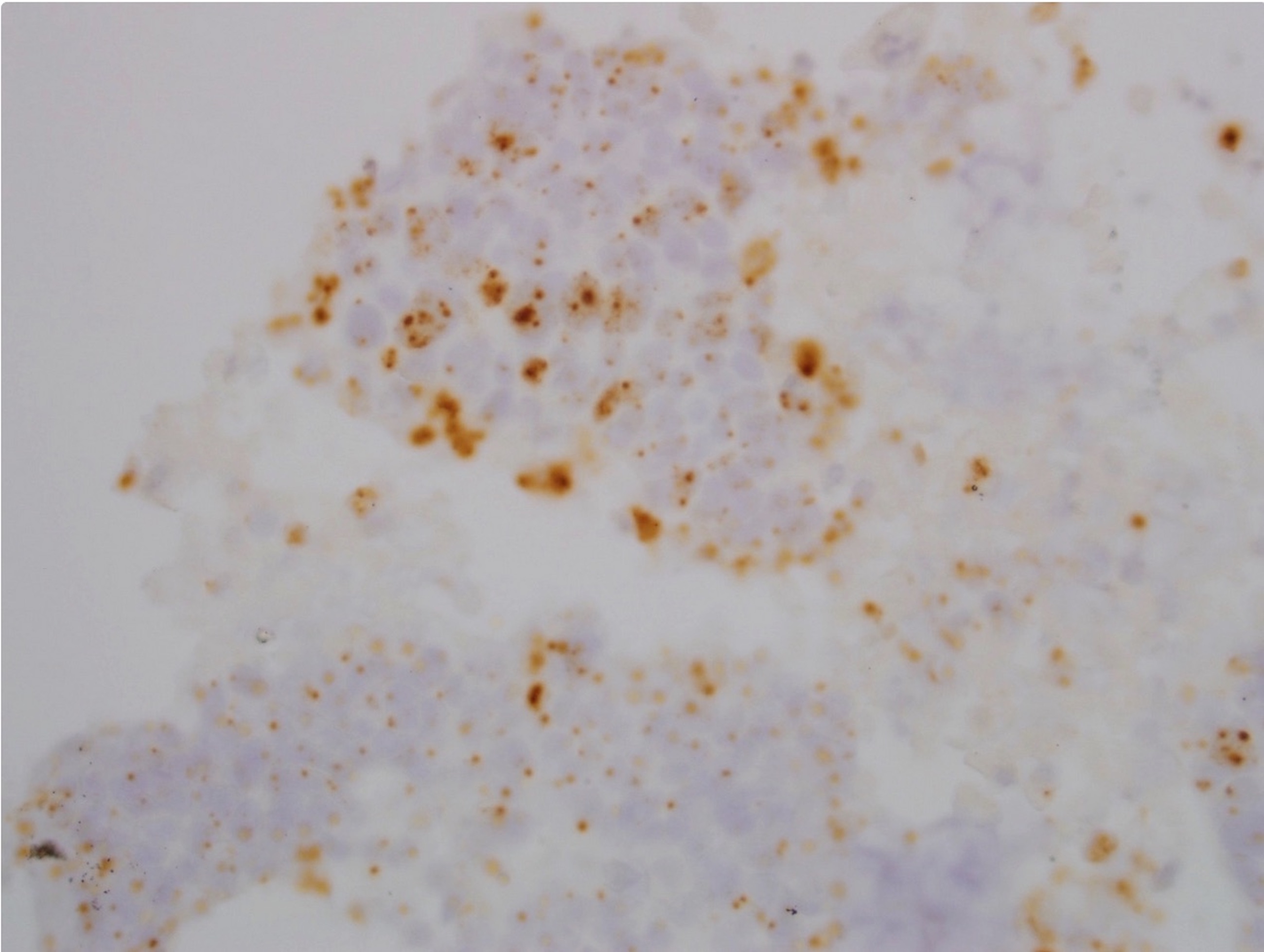
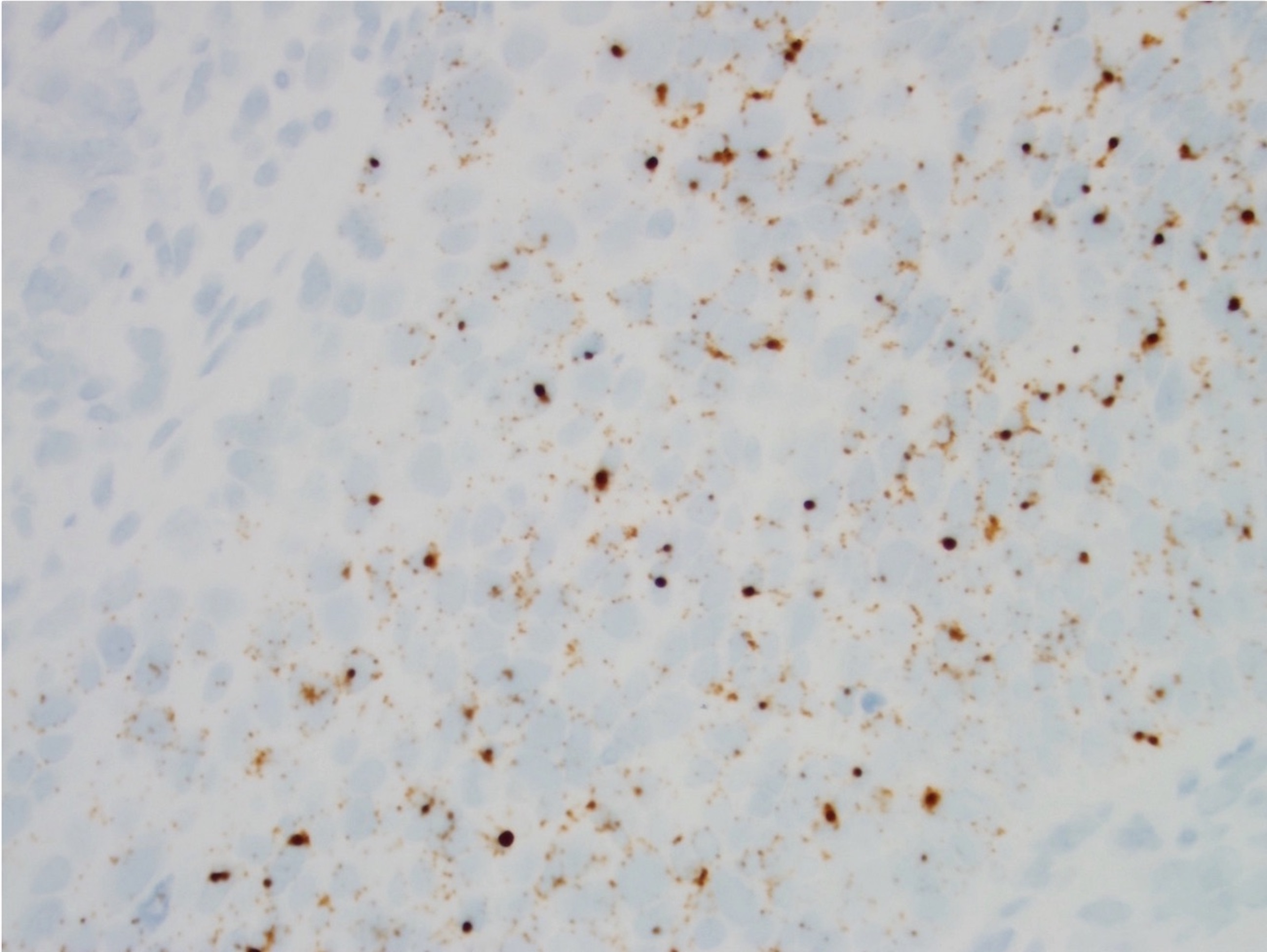
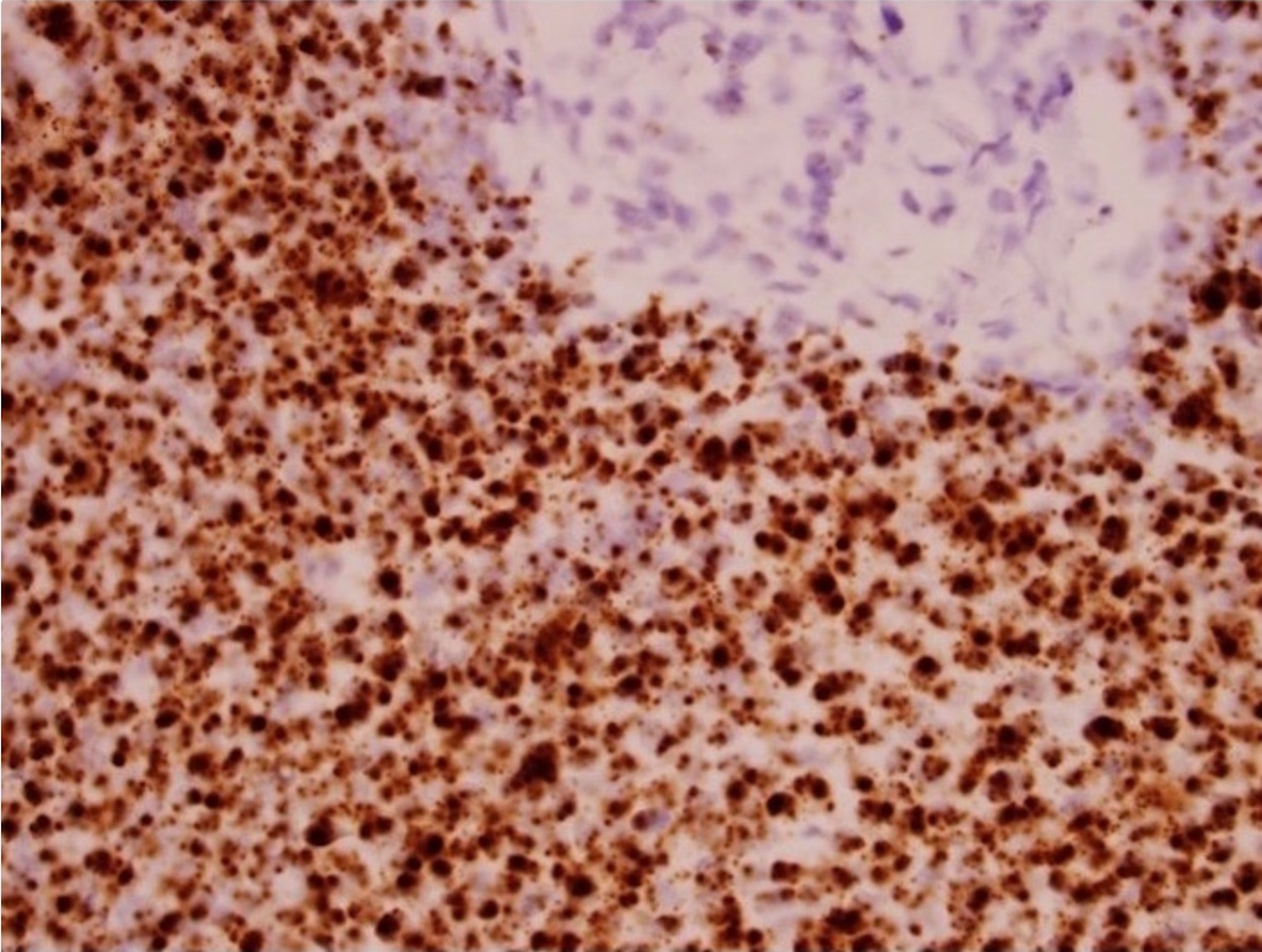
Case #419
Nests and nodules
of tumor cells with
melanin pigments
Tumor cells with
extensive necrosis
and melanin
pigments
PAP
HMB45 (cell block)


Giemsa
Images hosted on other servers:

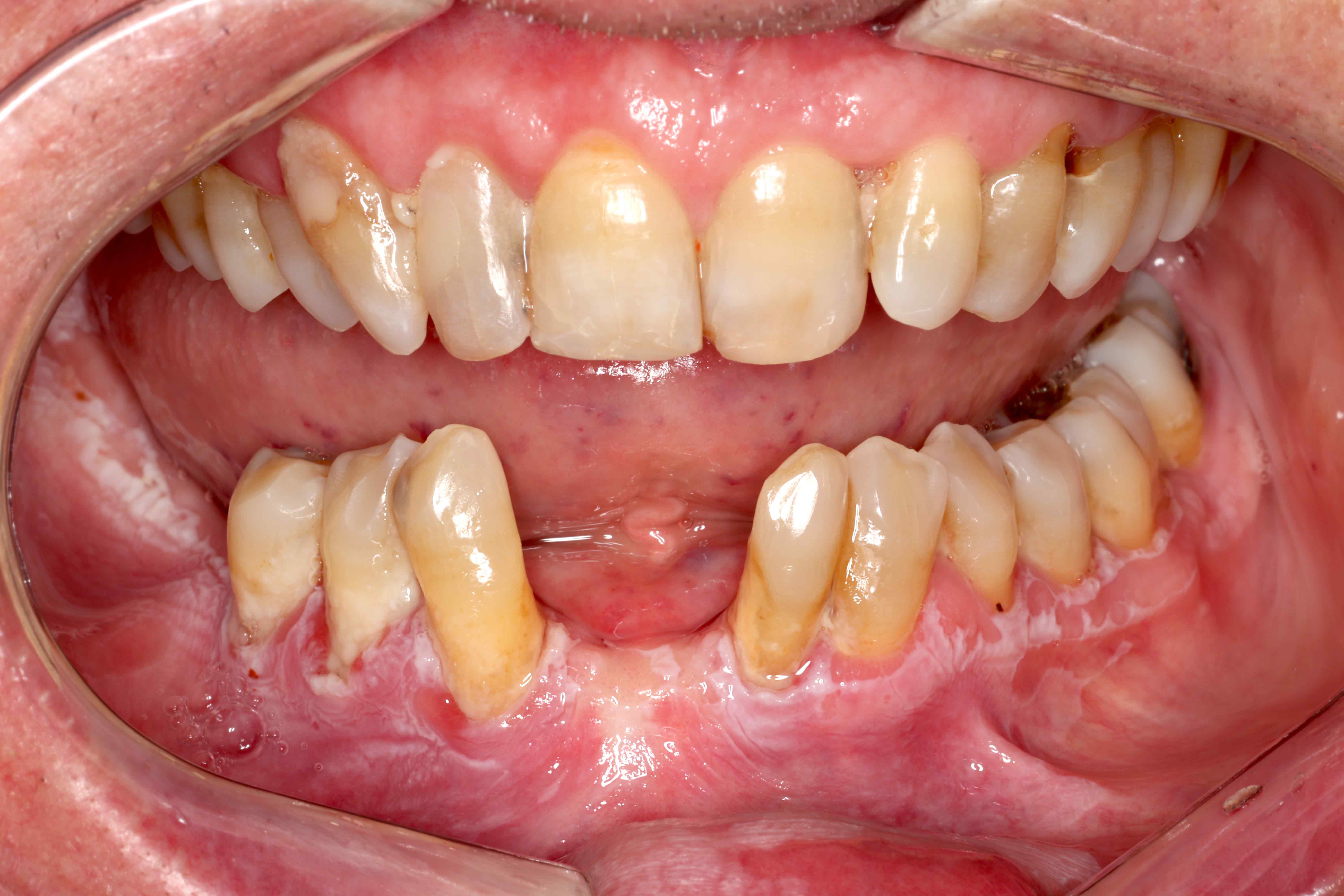
Blanching and fibrosis

With verrucous leukoplakia


38 year old man:
preoperative, after suturing graft and 1 year followup


10 year old boy:
difficulty in mouth opening


12 year old girl:
shrunken uvula, pretreatment and posttreatment
Images hosted on other servers:

Dense fibrosis

Advanced
Contributed by Molly Housley Smith, D.M.D.
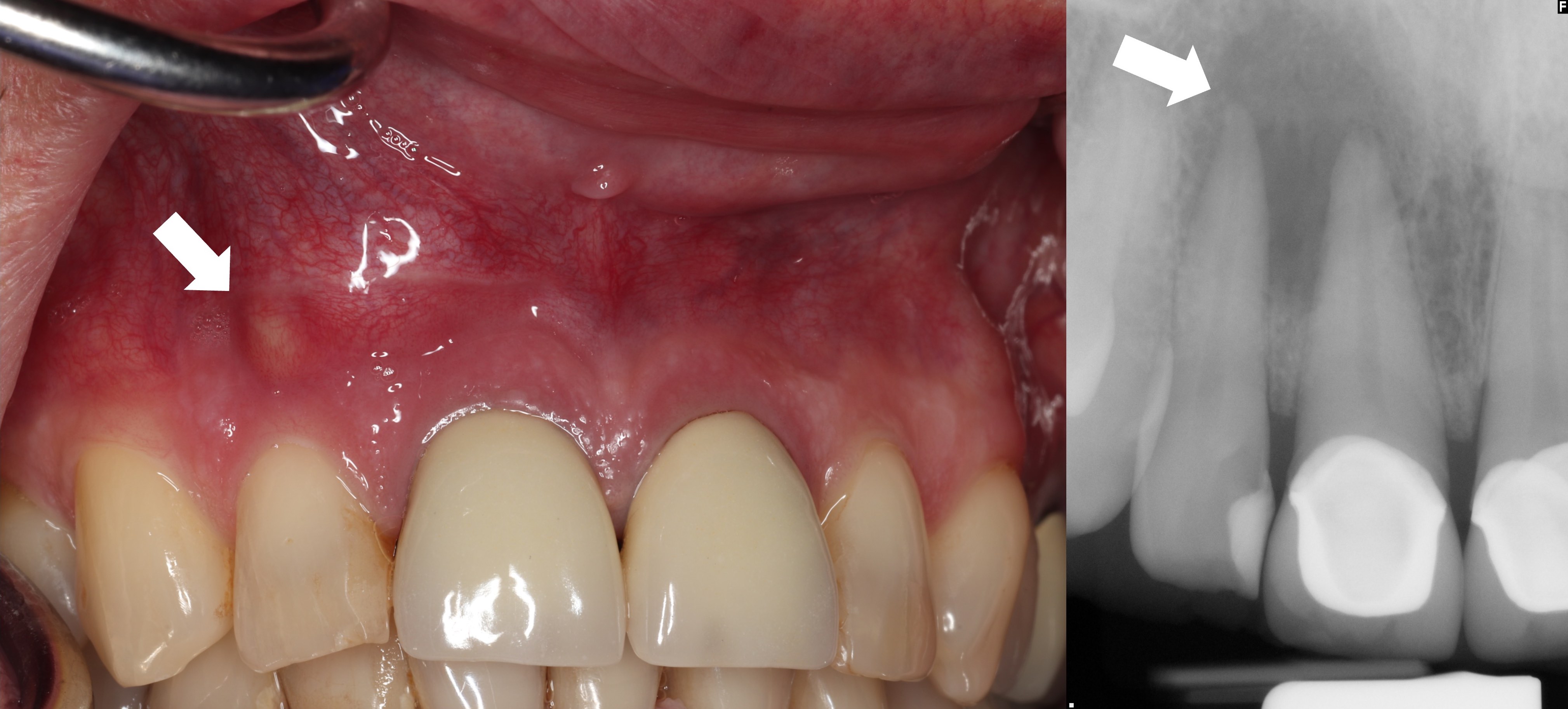
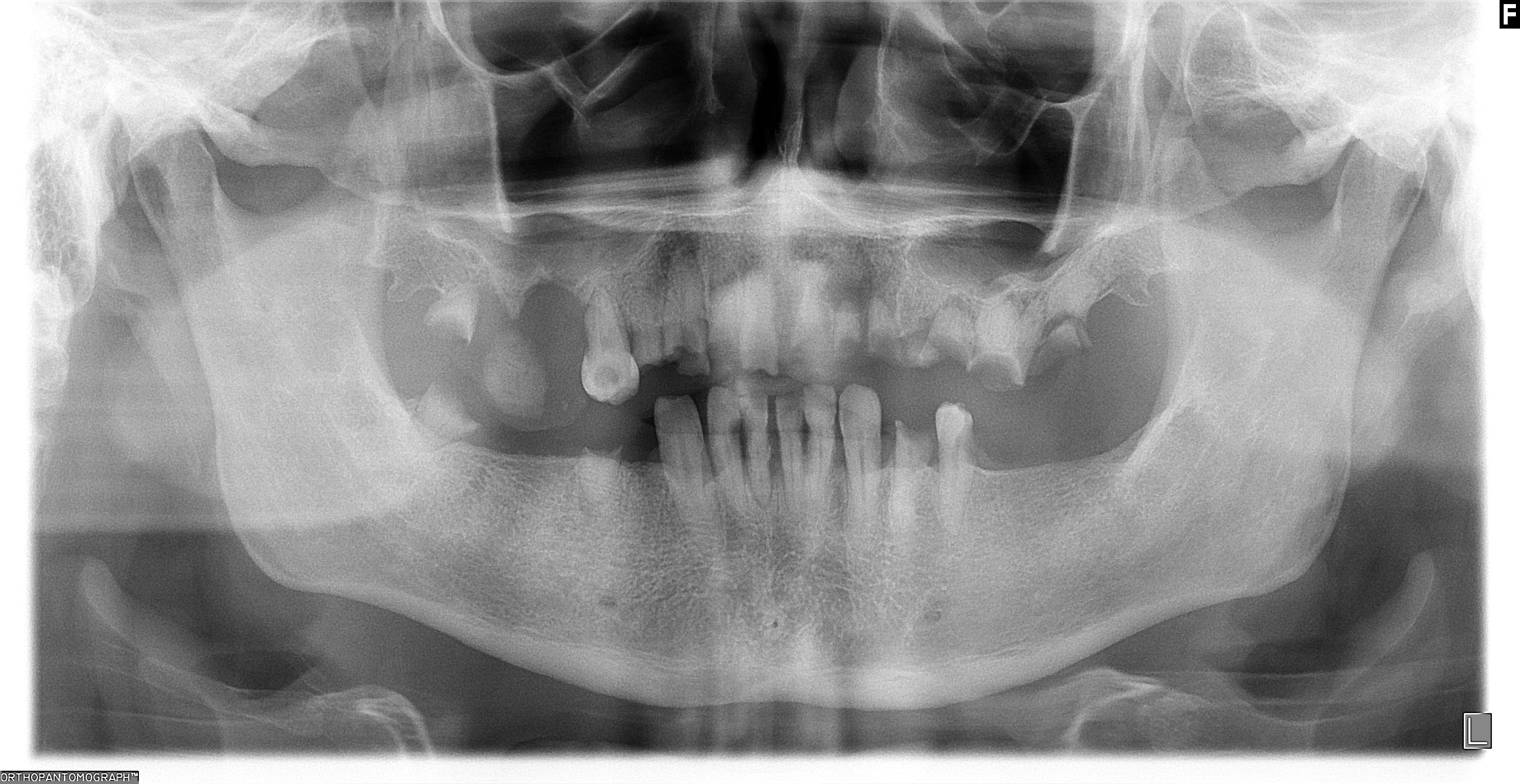
Periapical radiolucency
Contributed by Molly Housley Smith, D.M.D.
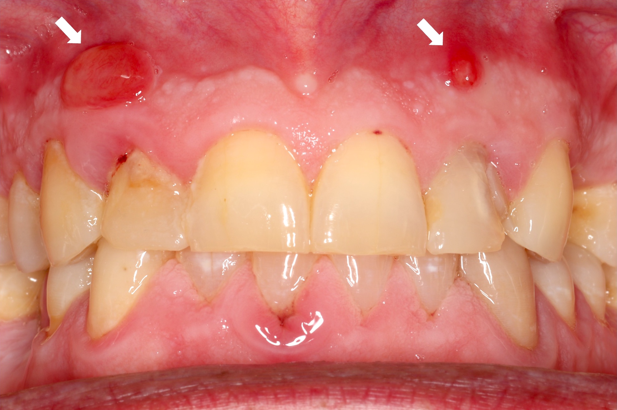
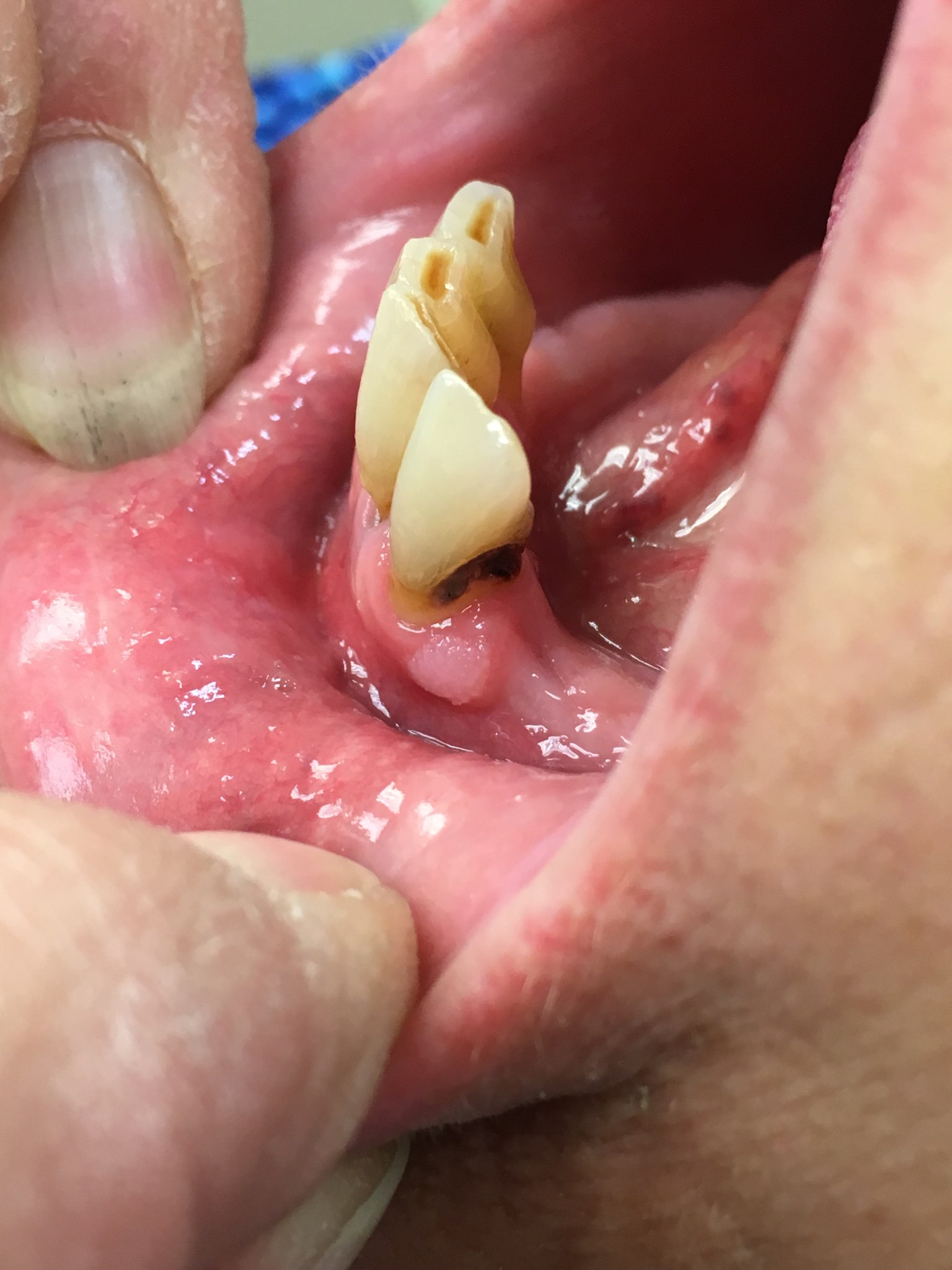
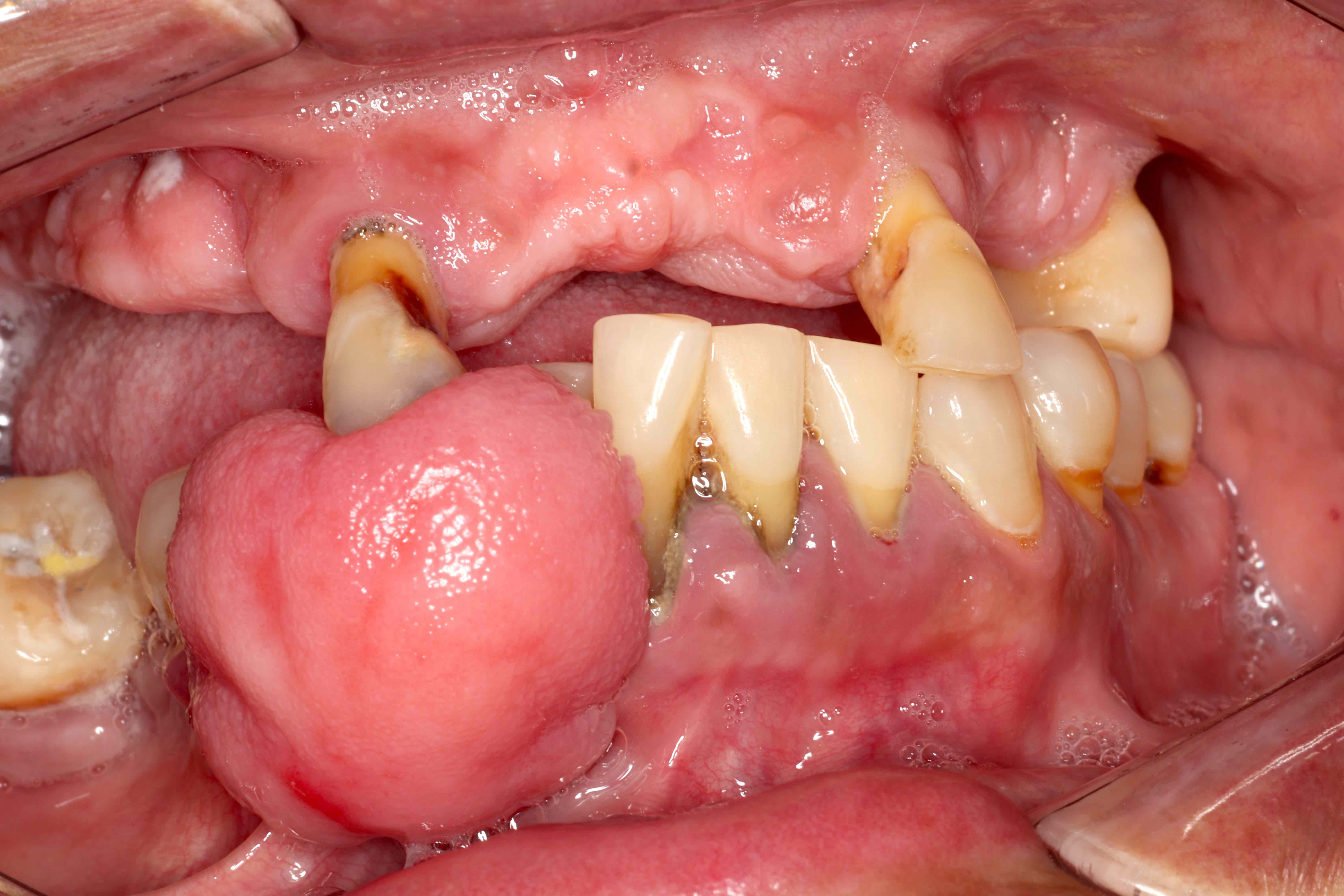
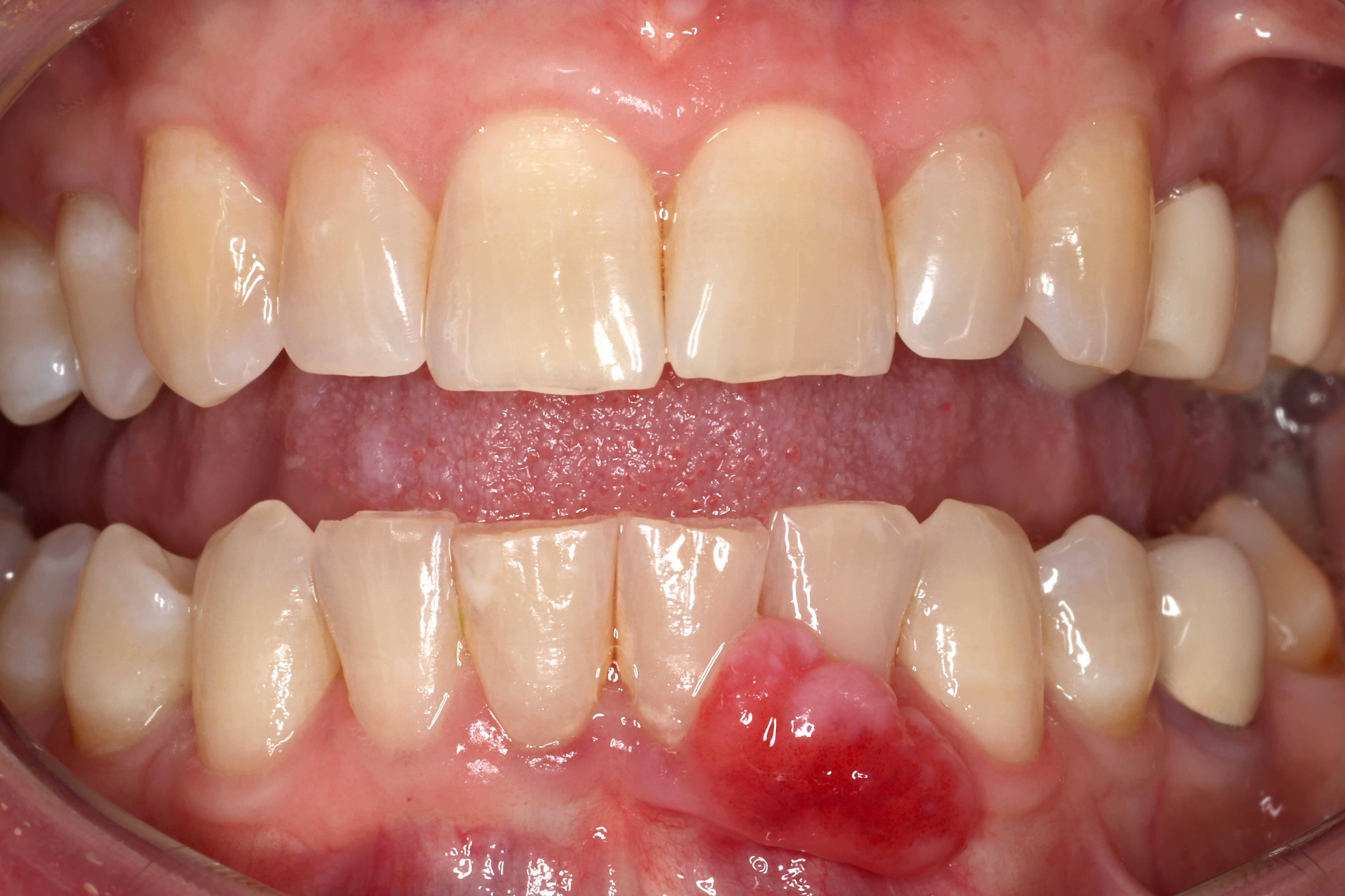
Fluctuant gingival nodules
Yellow gingival nodule
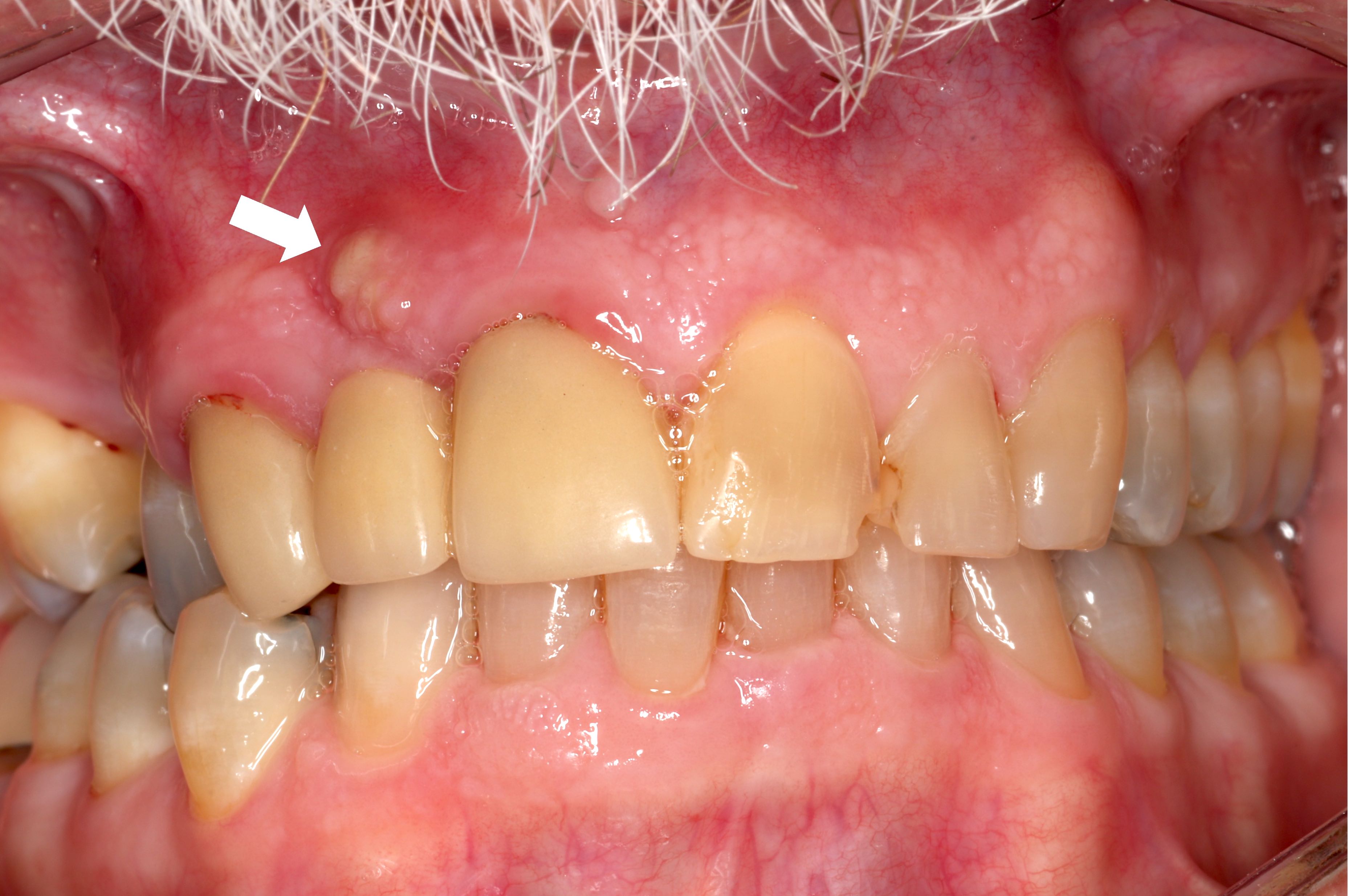
Contributed by Molly Housley Smith, D.M.D.
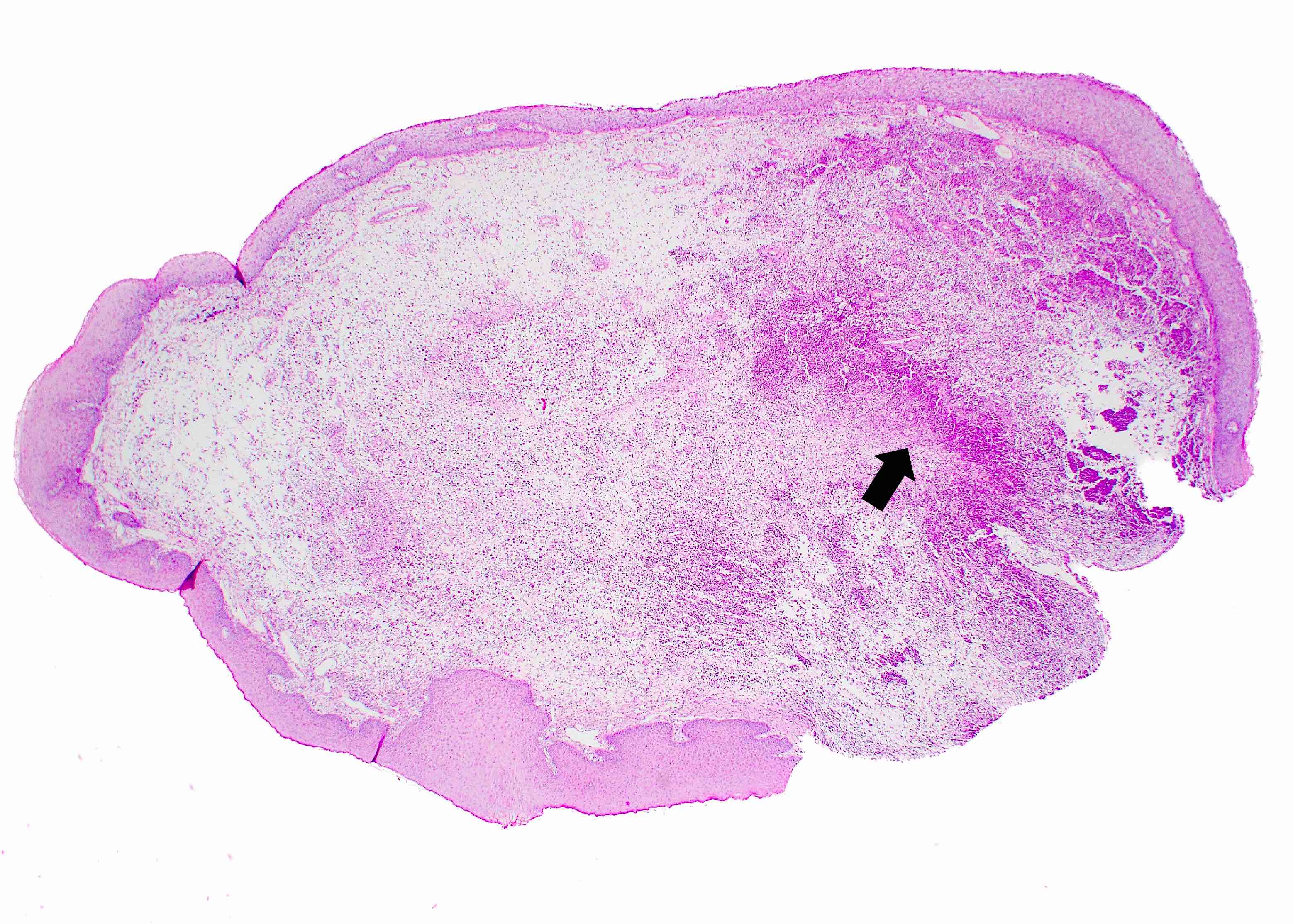
Exophytic nodule
Loose, inflamed mesenchymal stroma

Dense collections of neutrophils
Etiology and treatment of a dental fistulous tract / parulis
Tracing a dental fistulous tract with gutta percha
Images hosted on other servers:

Epidermal / dermal junction

Table 2 MMPDA Index
Contributed by Molly Housley Smith, D.M.D. and Sonal Tuli, M.D.
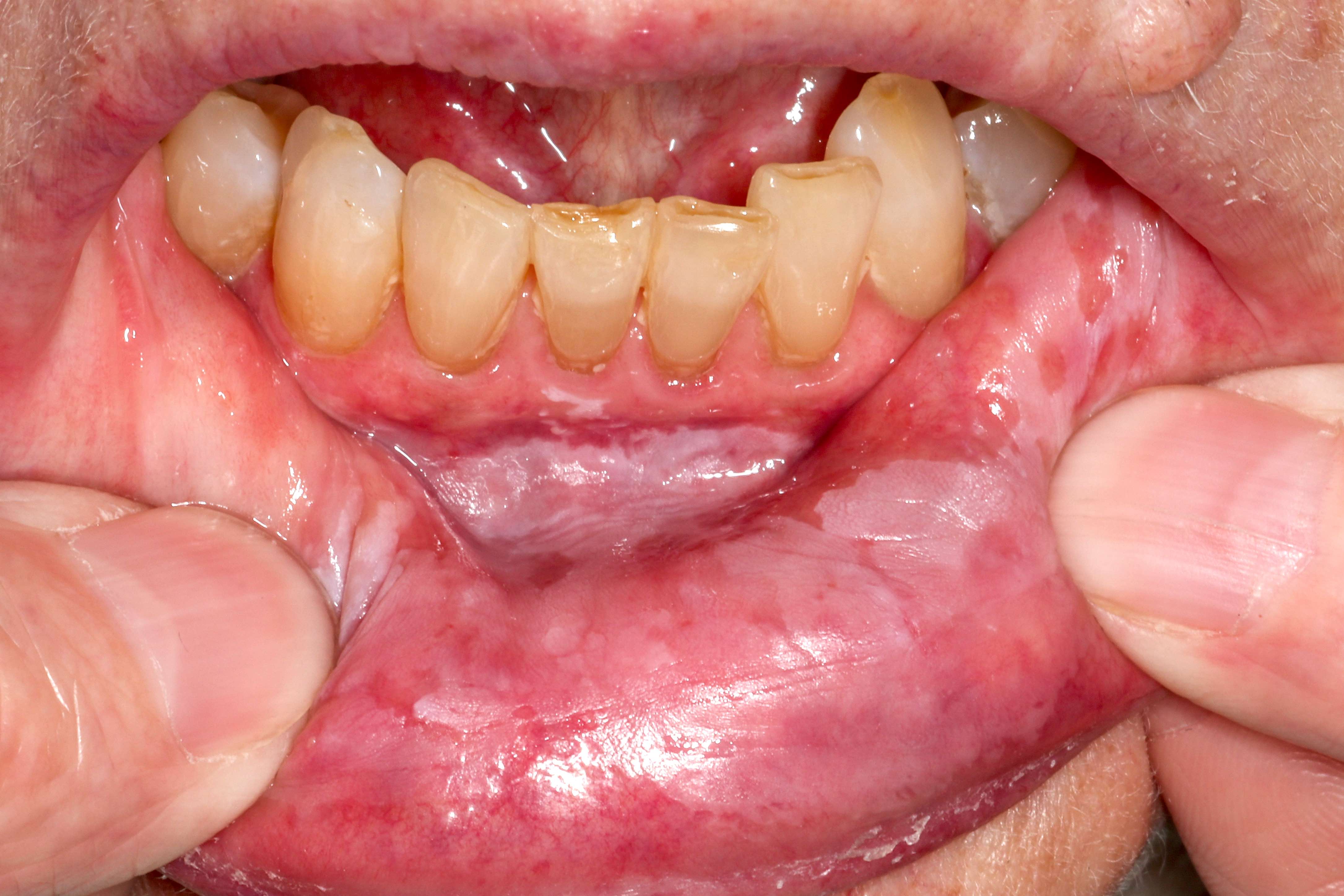
Diffuse gingival involvement
Localized gingival ulceration
Bullae development
Buccal mucosa involvement
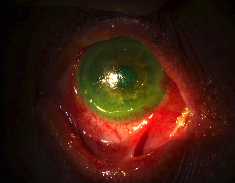
Symblepharon
Contributed by Molly Housley Smith, D.M.D. and @reportesVilla on Twitter
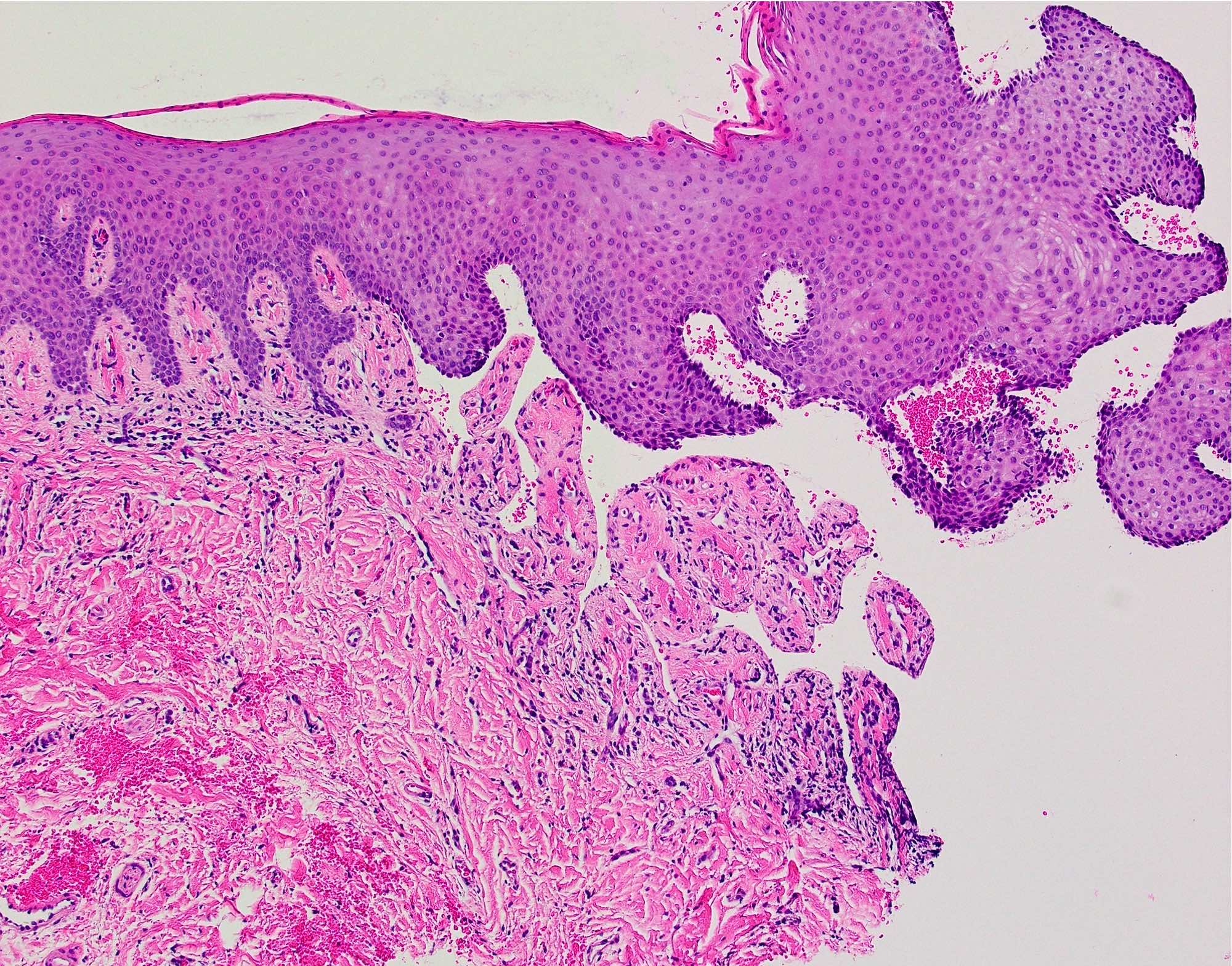
Subepithelial separation
Subepithelial cleft
contains fibrin,
erythrocytes and
inflammatory cells

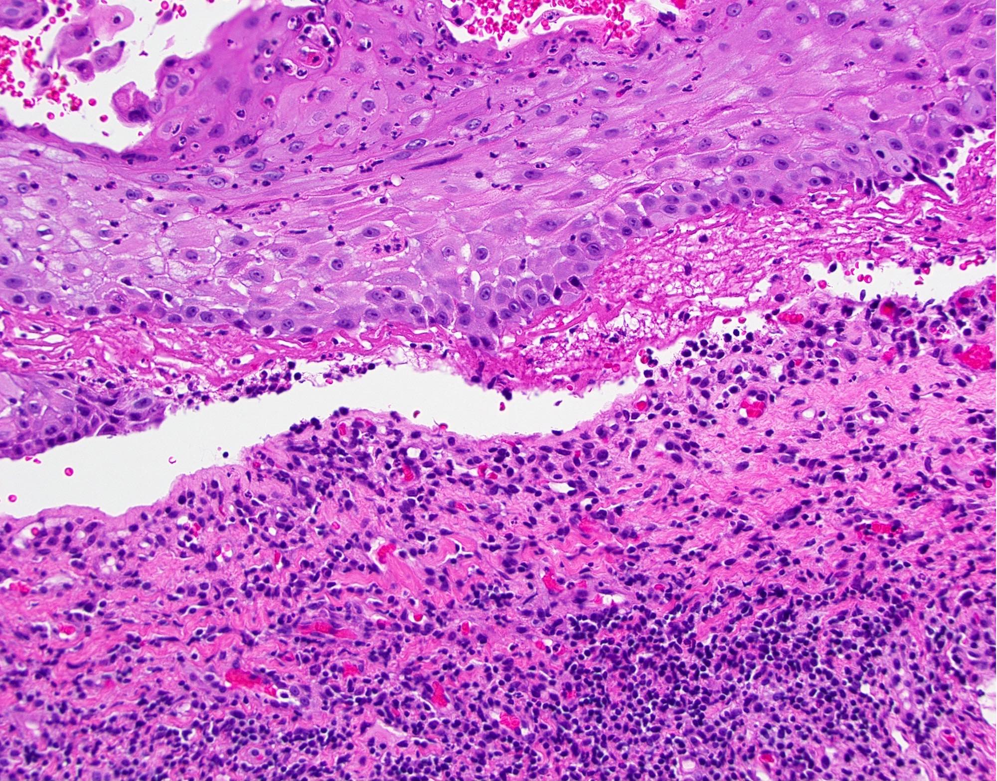
Subepithelial separation
Minimal inflammation
Pemphigoid
Clinical, histology, laboratory findings with discussion on pathophysiology
Images hosted on other servers:
36 year old woman:

Intra oral lesion

After 15 days
52 year old woman:

Erosive lesions

Erythematous gingivae
Images hosted on other servers:


36 year old woman

52 year old woman
Images hosted on other servers:

Resembes pyogenic granuloma


22 year old woman:
preoperative, excision and postoperative


53 year old man:
pre and postoperative


57 year old woman:
pre and postoperative


81 year old woman:
pre and postoperative


Central giant cell granuloma:
swelling, extended to midline and excisional biopsy
Images hosted on other servers:

22 year old woman
Contributed by Tim Bracey, M.B.Ch.B., Ph.D.
28 year old woman with rapidly growing lesion
Images hosted on other servers:

22 year old woman

81 year old woman


53 year old man

Central giant cell
granuloma: irregular
giant cells
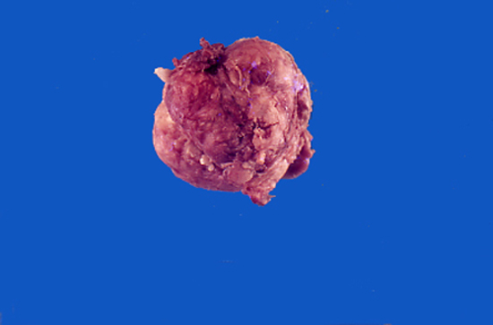
Contributed by Molly Housley Smith, D.M.D.
Visible ossification
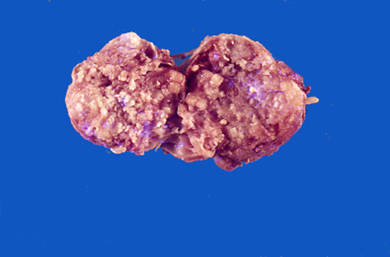
Contributed by Molly Housley Smith, D.M.D.
Nodular mass
Multilobular red growth
Images hosted on other servers:

Typical clinical appearance

17 year old boy

21 year old man

25 year old man

26 year old man

31 year old man
Contributed by Molly Housley Smith, D.M.D.
Partially calcified mass
Contributed by Timothy Fielder, M.B.B.S., B.Med.Sci., Ruta Gupta, M.D. and Molly Housley Smith, D.M.D.
Well demarcated mucosal lesion
Ulceration
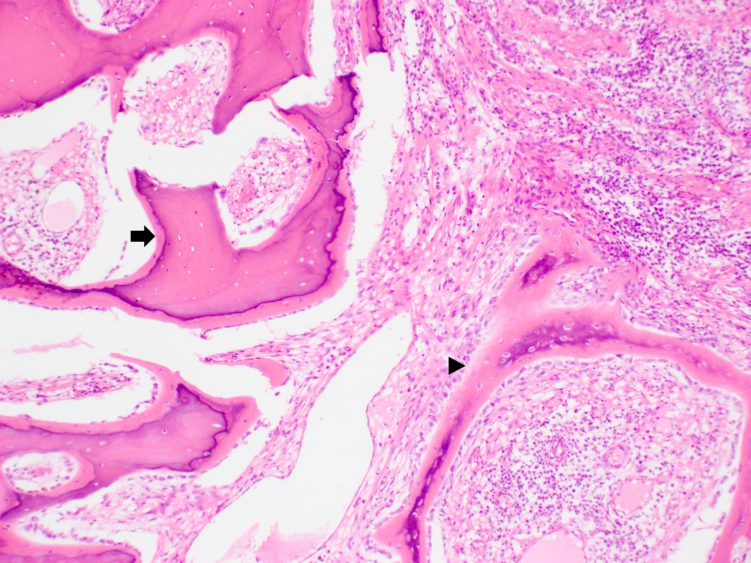
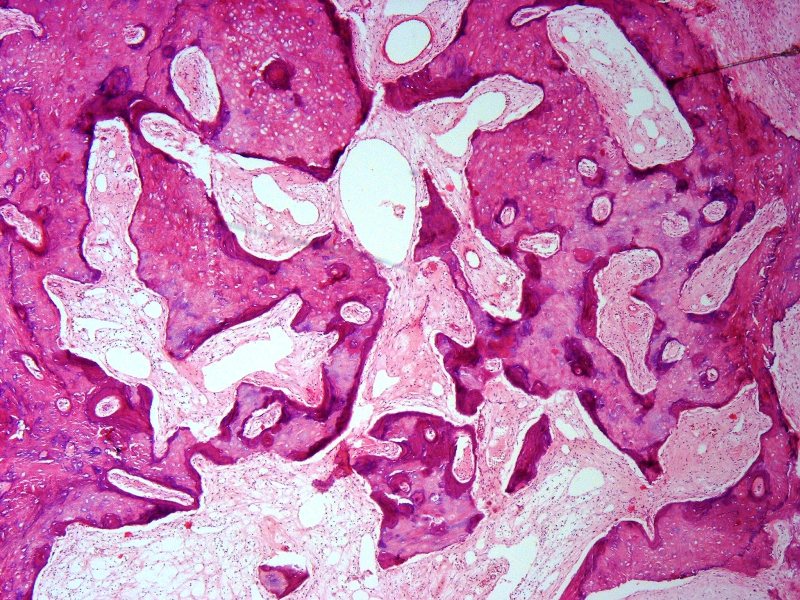
Metaplastic ossification
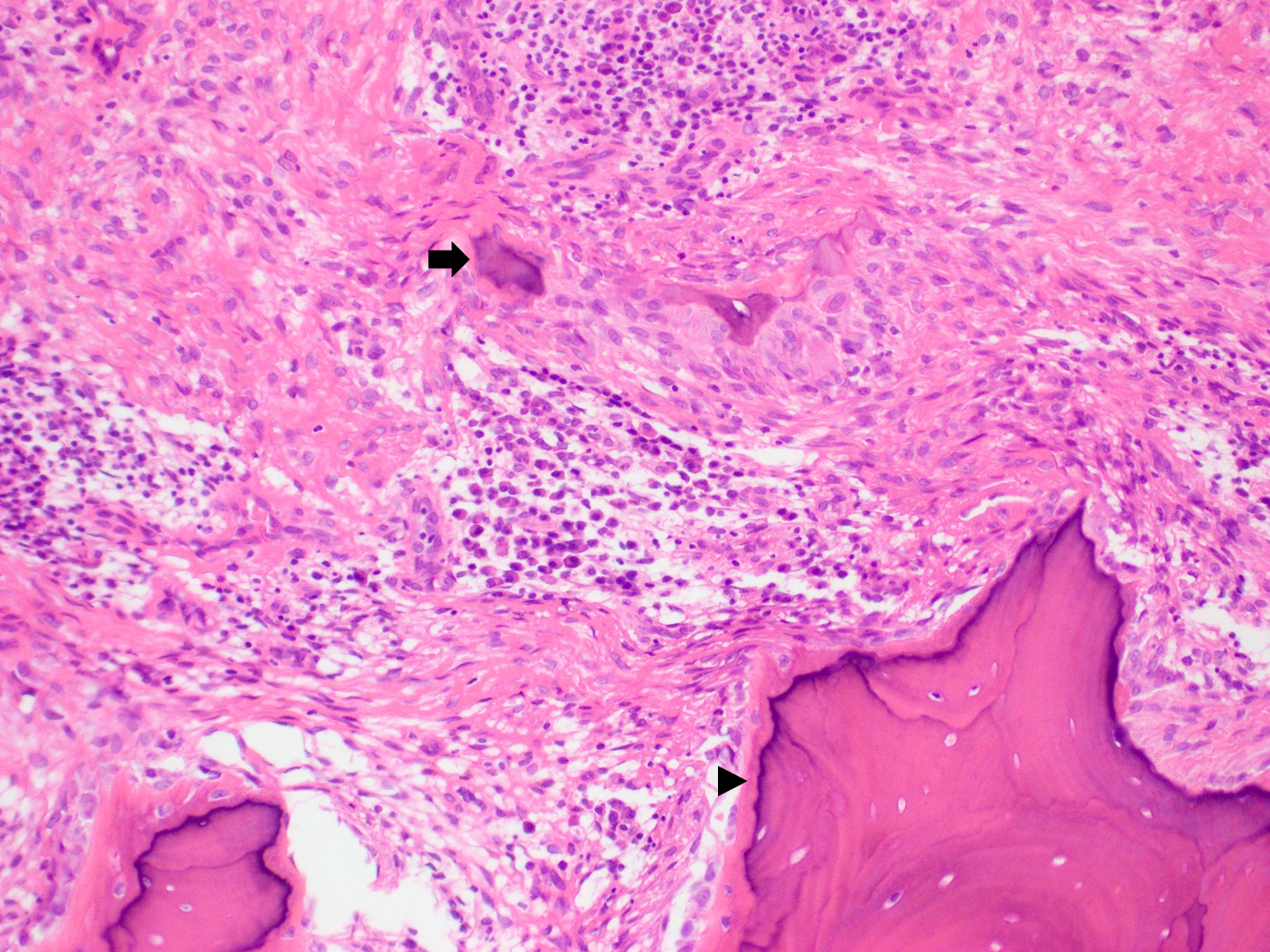
Ossification and calcification
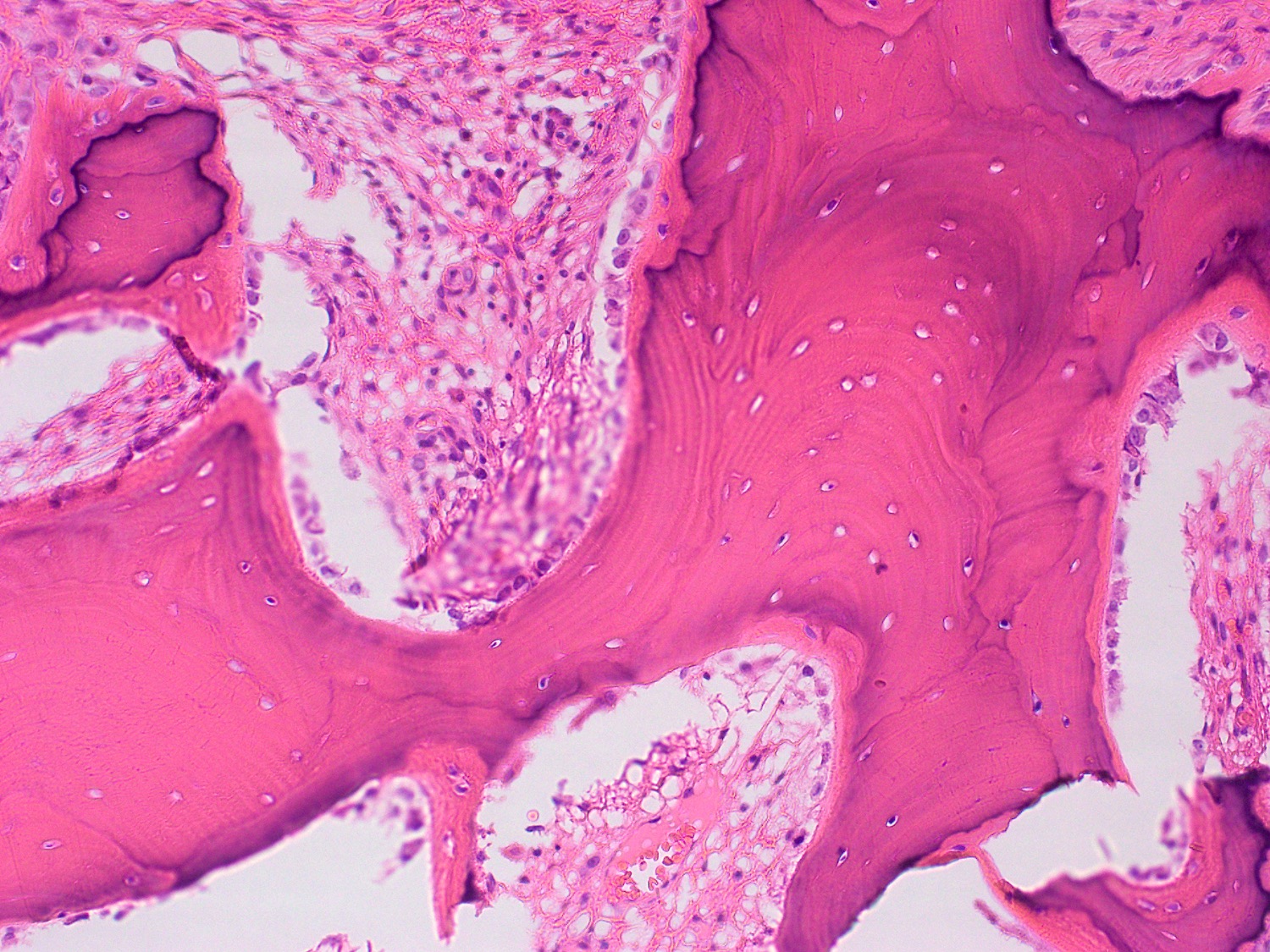
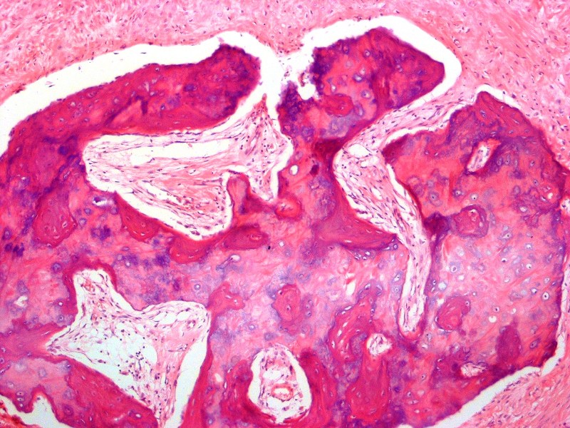
Lamellar bone
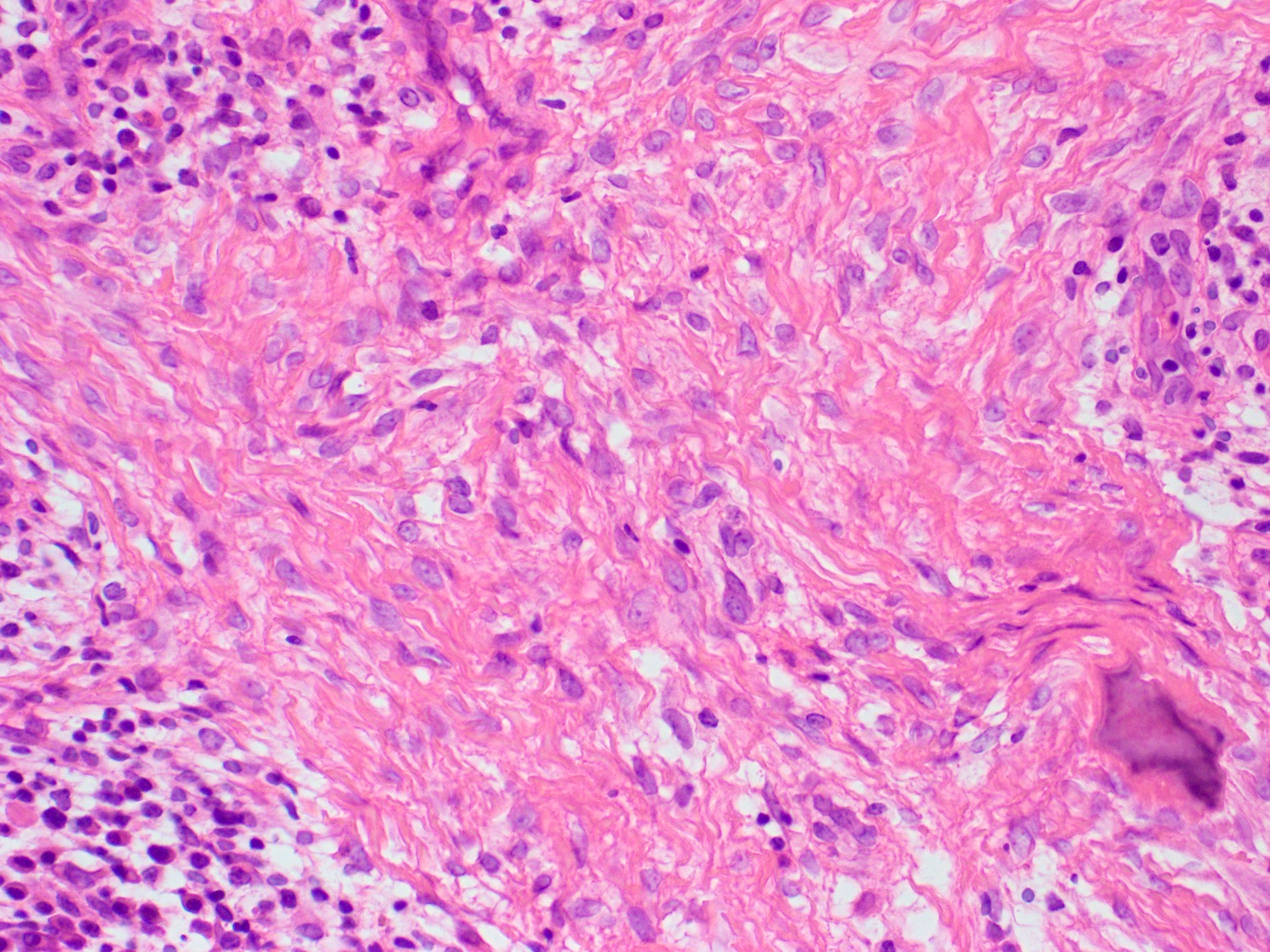
Collagenous matrix
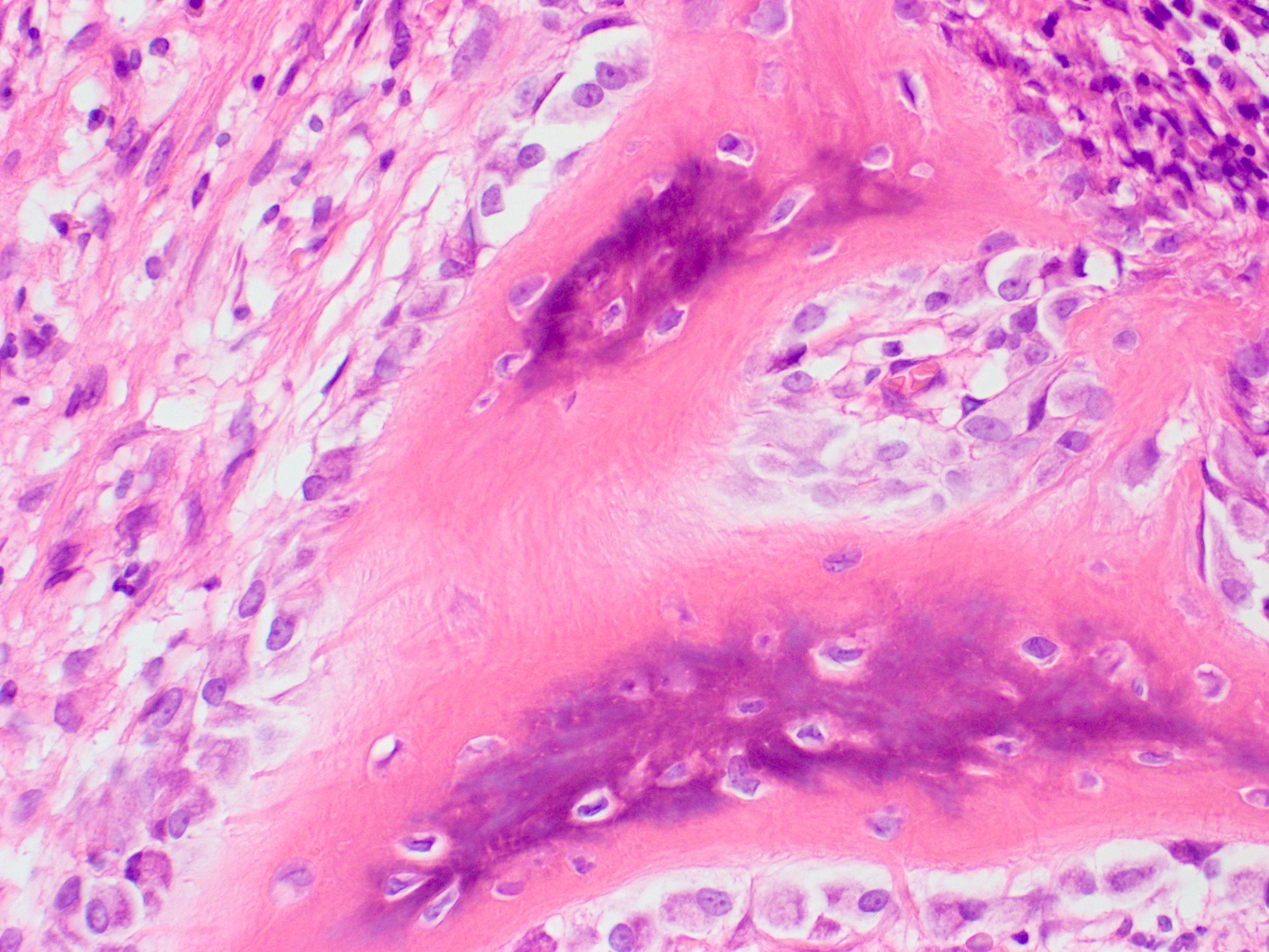
Woven bone
Peripheral cemento-ossifying fibroma

Cementum-like bone
Images hosted on other servers:

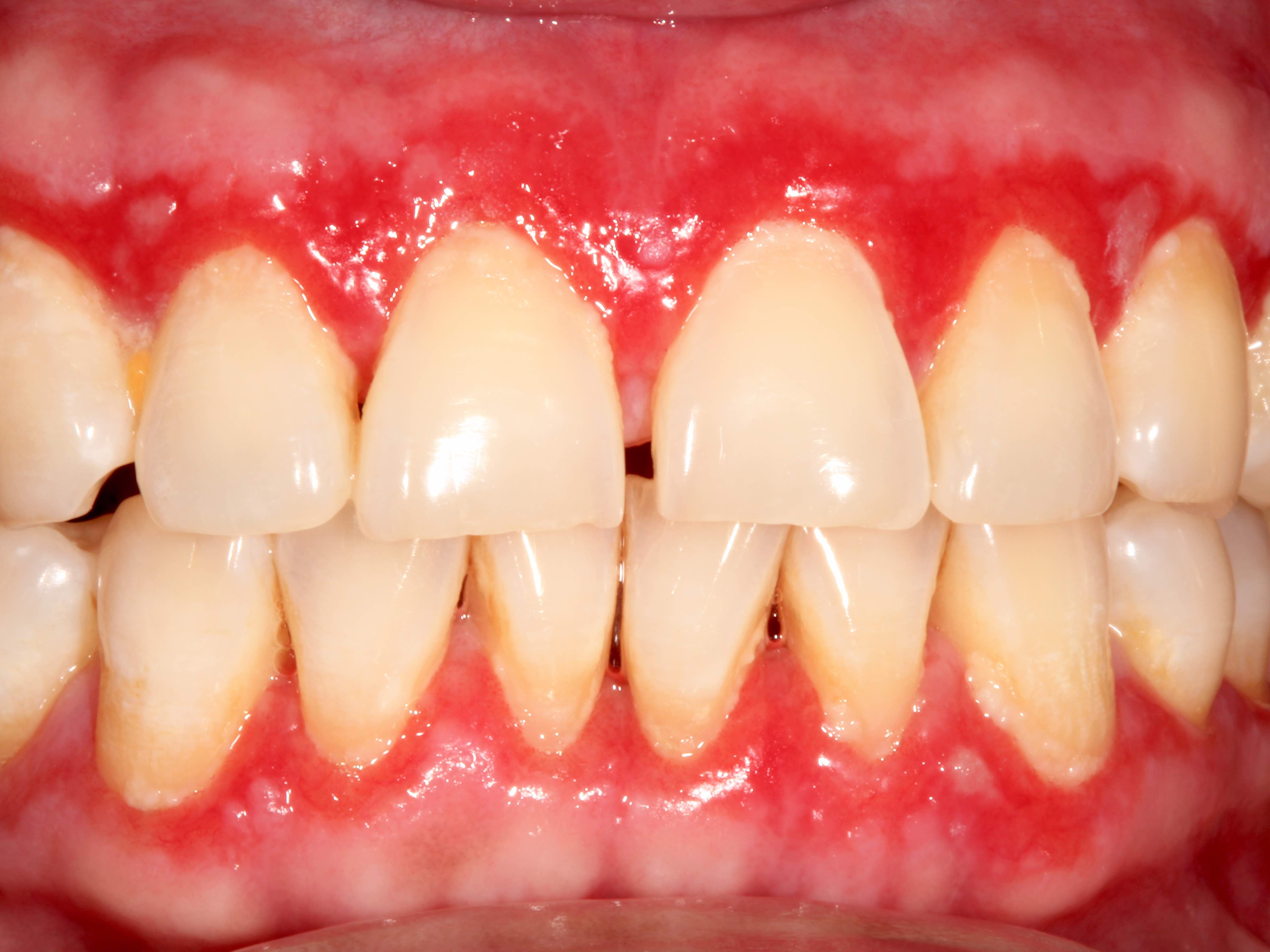
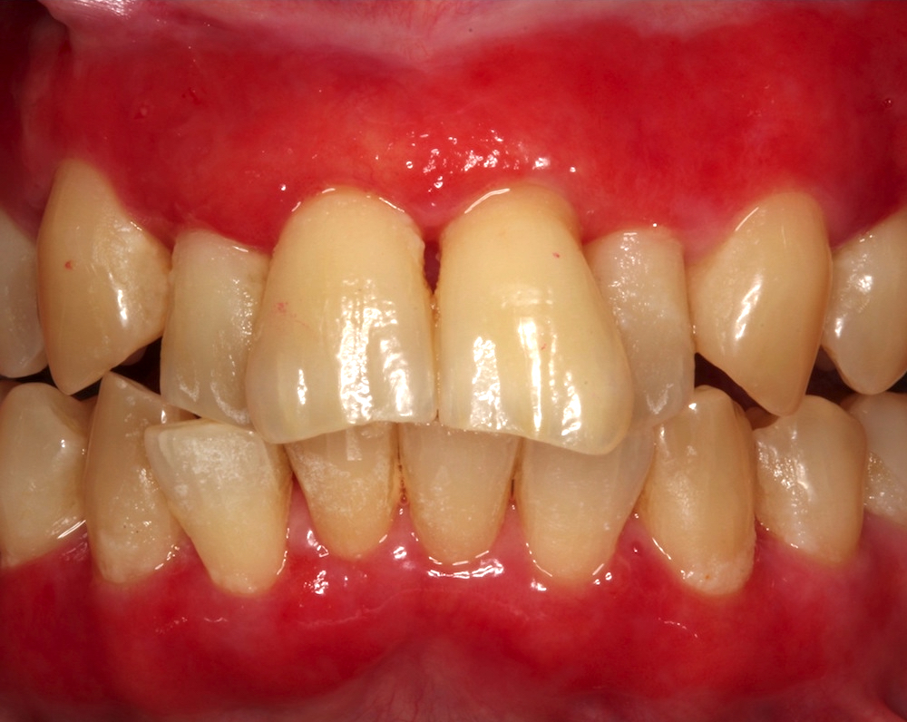
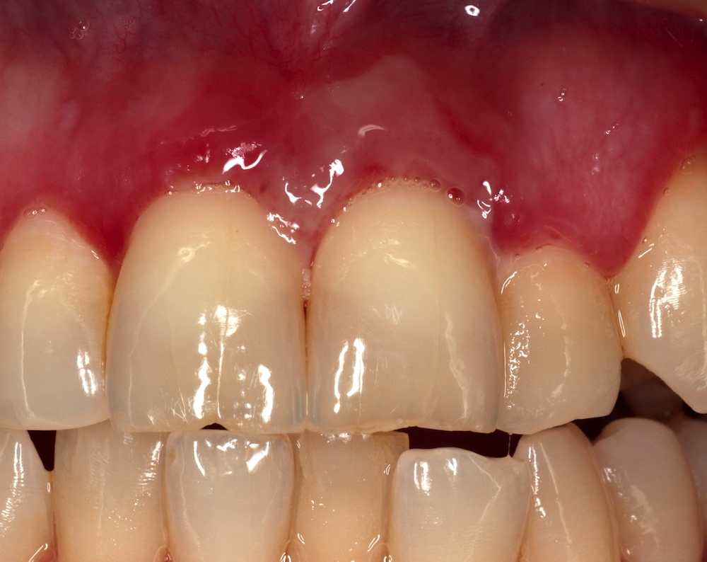
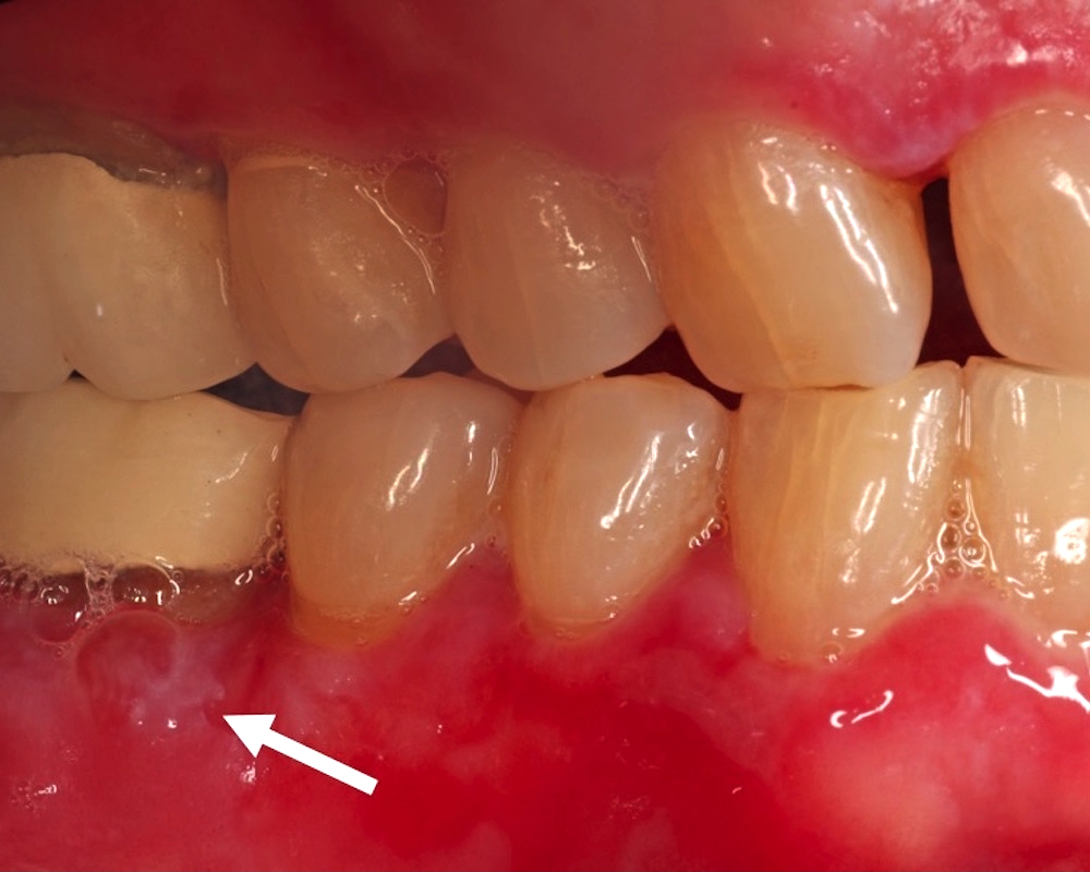
Diffuse erythema and edema of the gingiva
Contributed by S. Rajendran, M.B.B.Ch., M.Sc., Ph.D.
Diffuse plasma cell infiltrate

CD138

MUM1

CD56
CD20
Kappa

Lambda

Contributed by Molly Housley Smith, D.M.D., Michael Piepgrass, D.M.D.,
John McGehee, D.M.D., Michael Menis, D.D.S. and Trent Clifton, D.M.D., M.D.
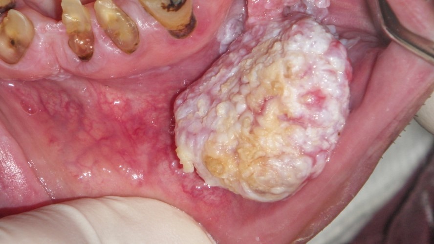
Patchy mandibular leukoplakia
Verrucoid surface texture
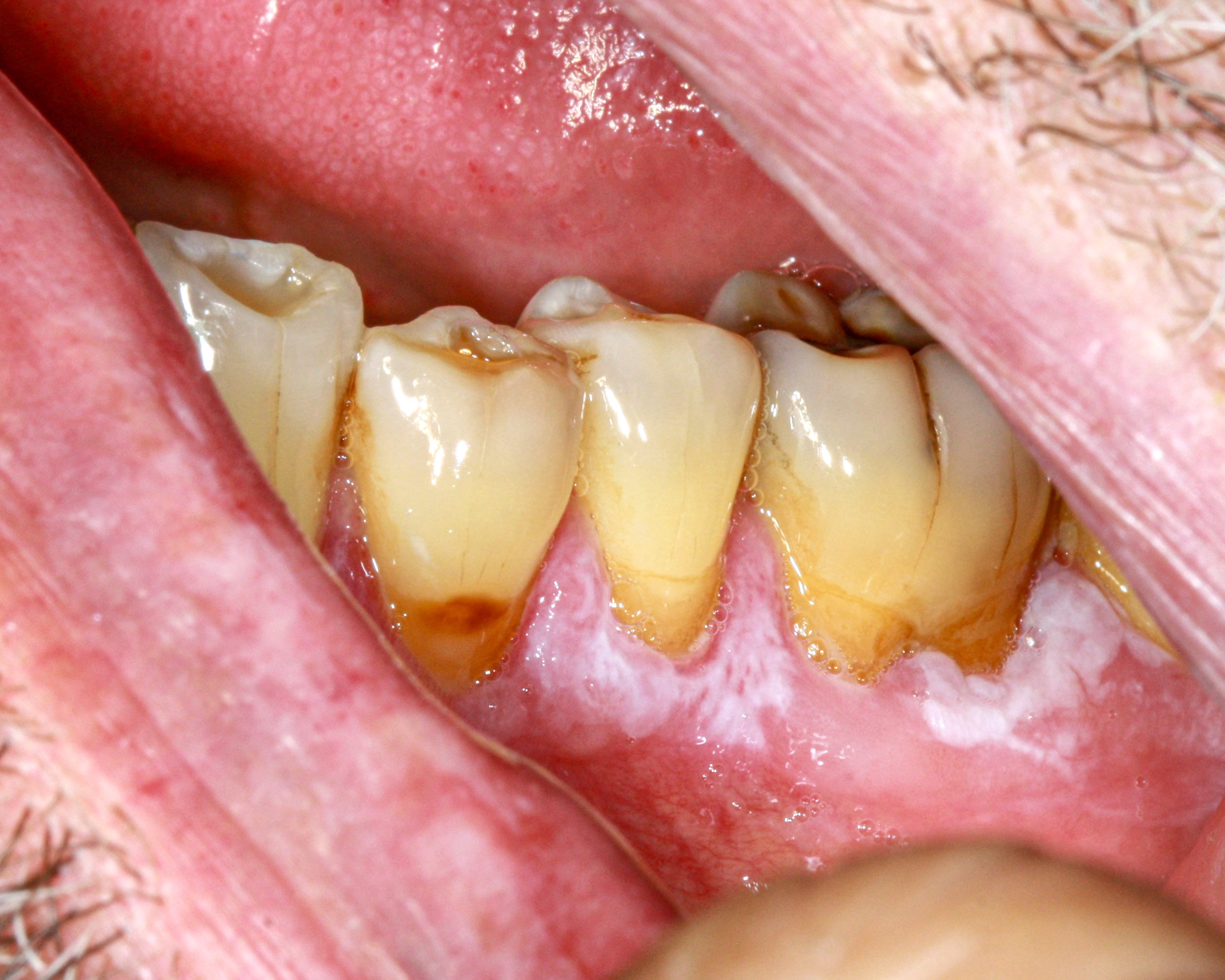
Ring around the collar
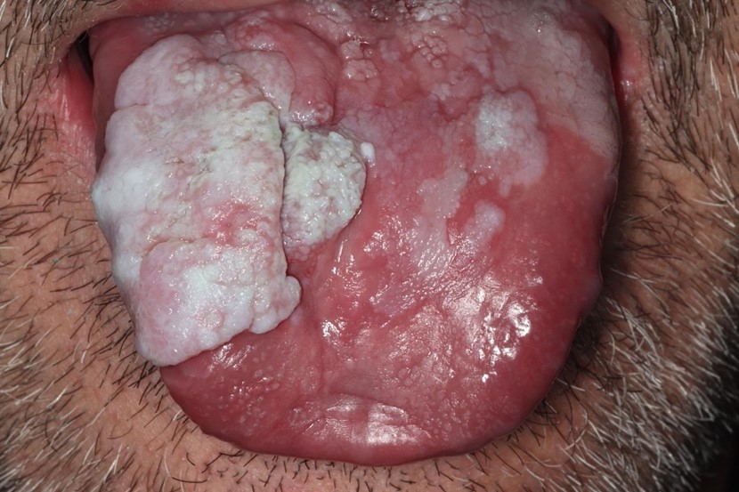
Diffuse leukoplakia
Buccal mucosa
Tongue lesion
Nonmigrating white lesions
Carcinomatous transformation
Contributed by Molly Housley Smith, D.M.D.
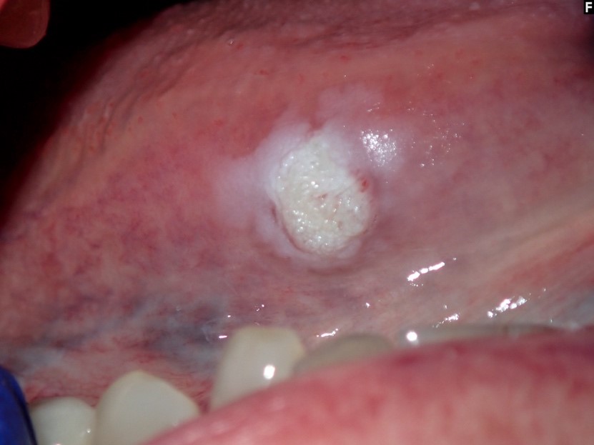
Ring around the collar
Multifocal lesions
Contributed by Molly Housley Smith, D.M.D.
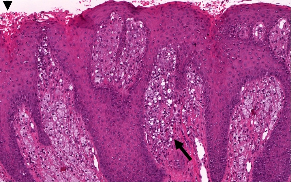
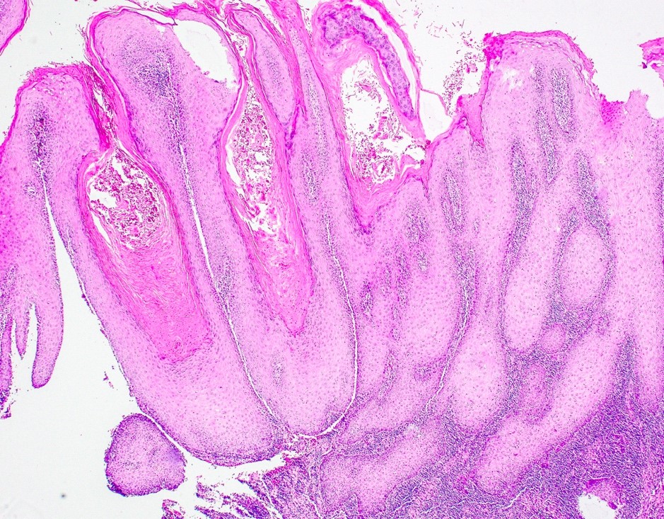
Corrugated hyperorthokeratosis

Skip pattern
Prominent granular cell layer
Skip pattern

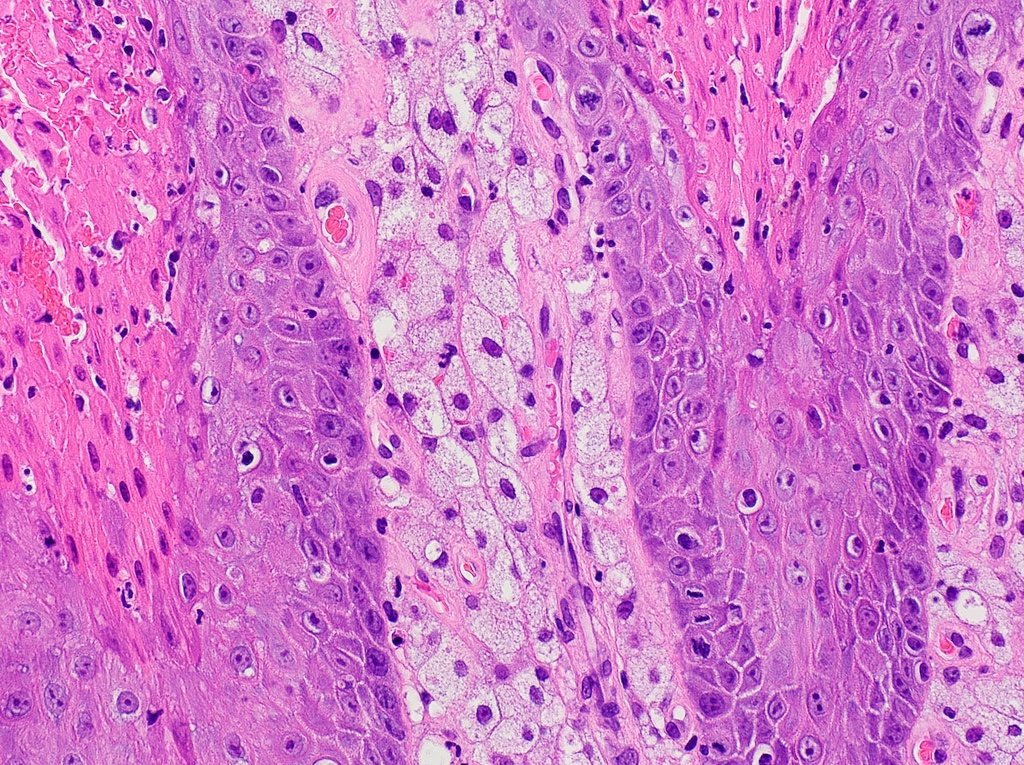
Cytologic dysplasia
Dysplastic and elongated epithelium
Bulky hyperkeratotic epithelial proliferation
Suspicion for invasion
Verrucous carcinoma transformation
Deceptively benign
Squamous cell carcinoma transformation
Contributed by Molly Housley Smith, D.M.D.
Median rhomboid glossitis
Images hosted on other servers:

Granular cell tumor
Contributed by Molly Housley Smith, D.M.D.
PEH in granular cell tumor
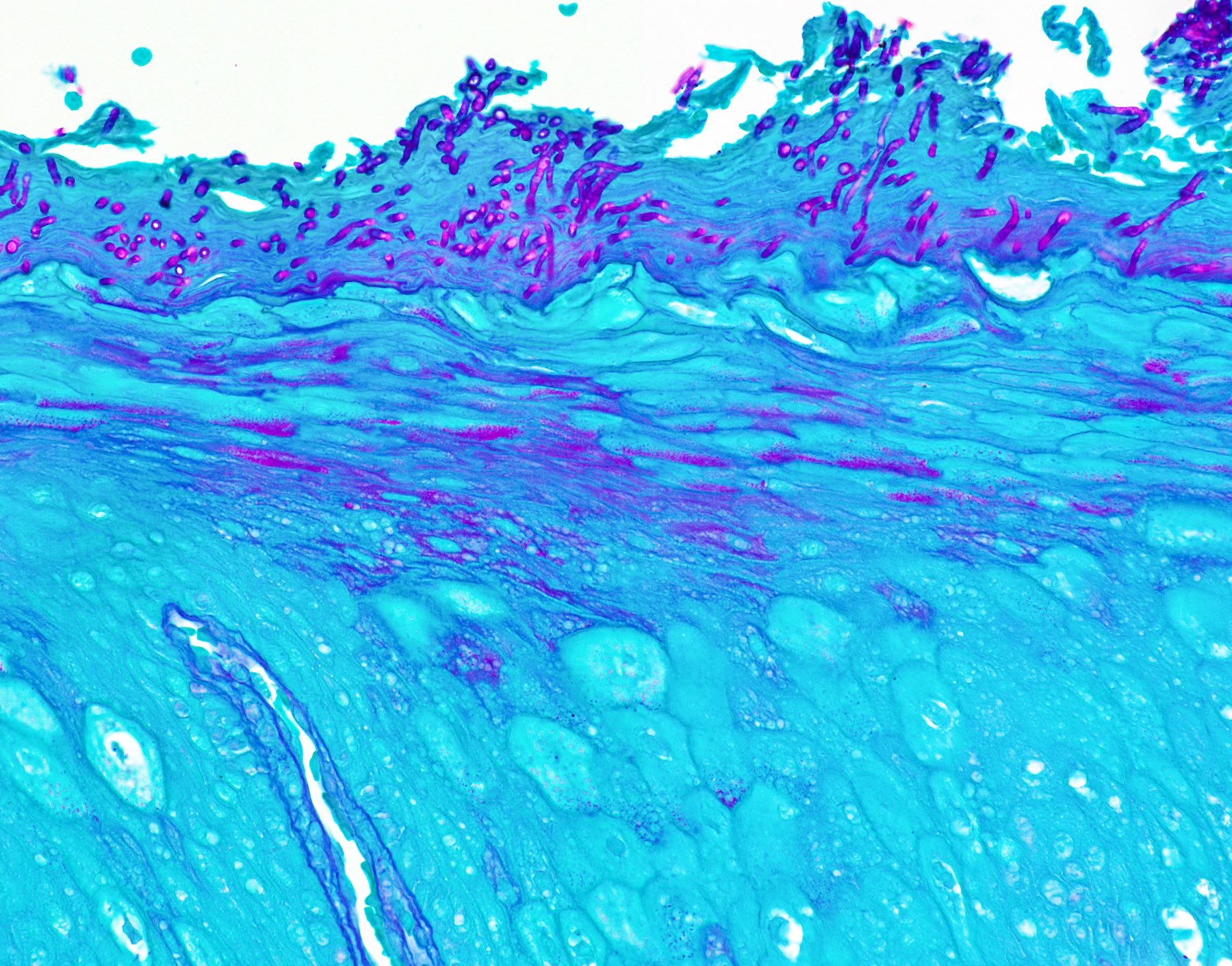
PEH in chronic hyperplastic candidiasis
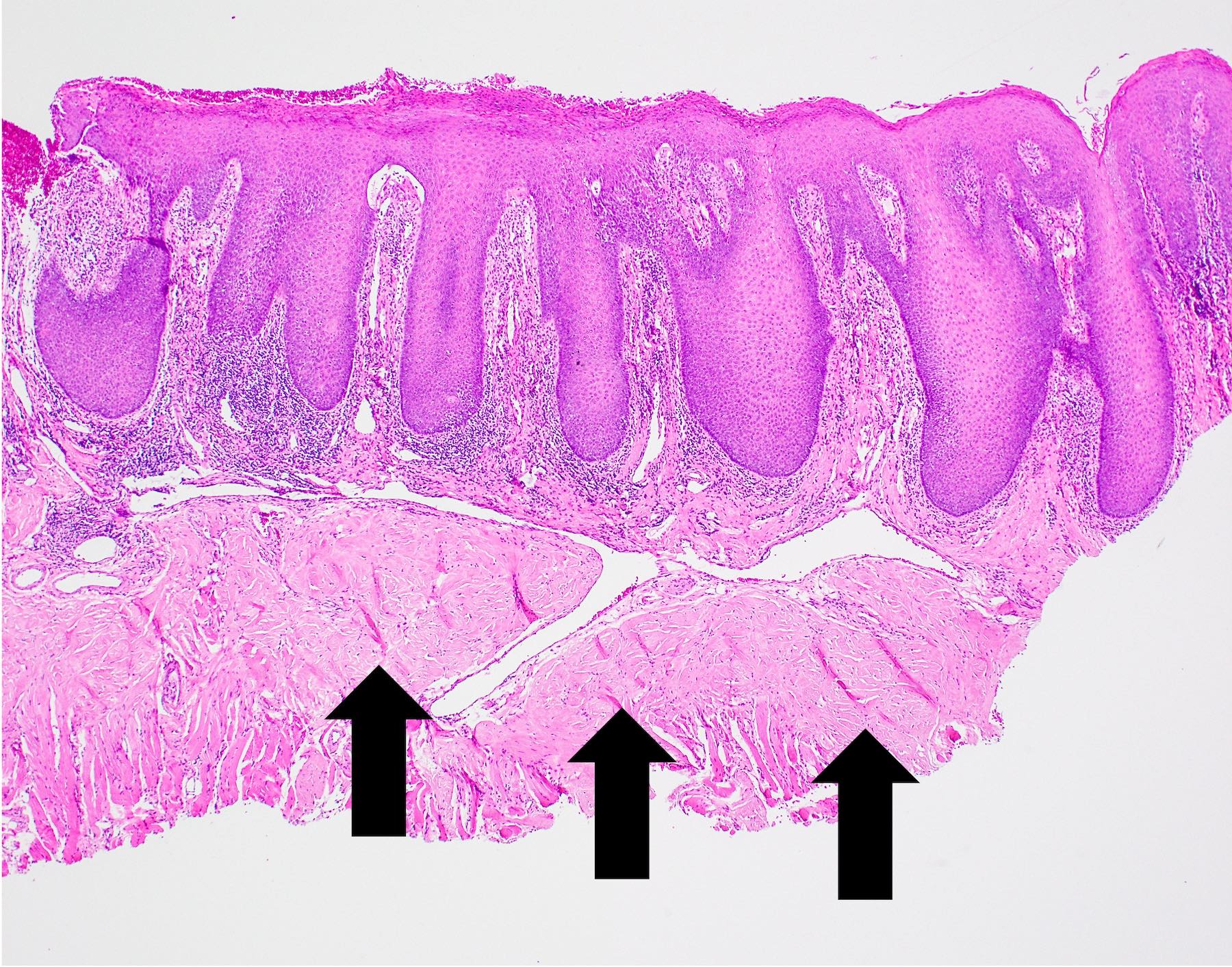
PEH in median rhomboid glossitis
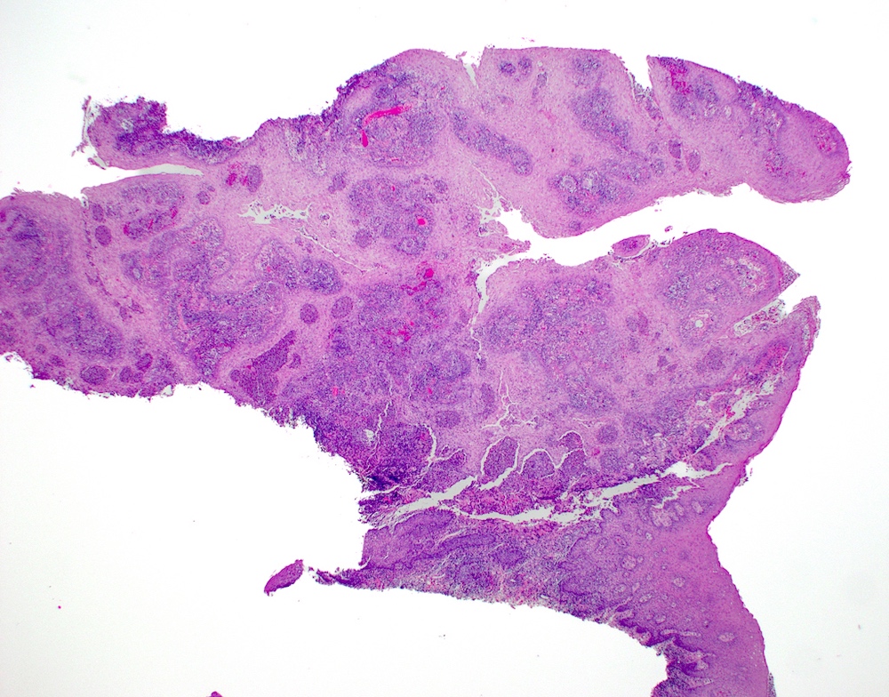
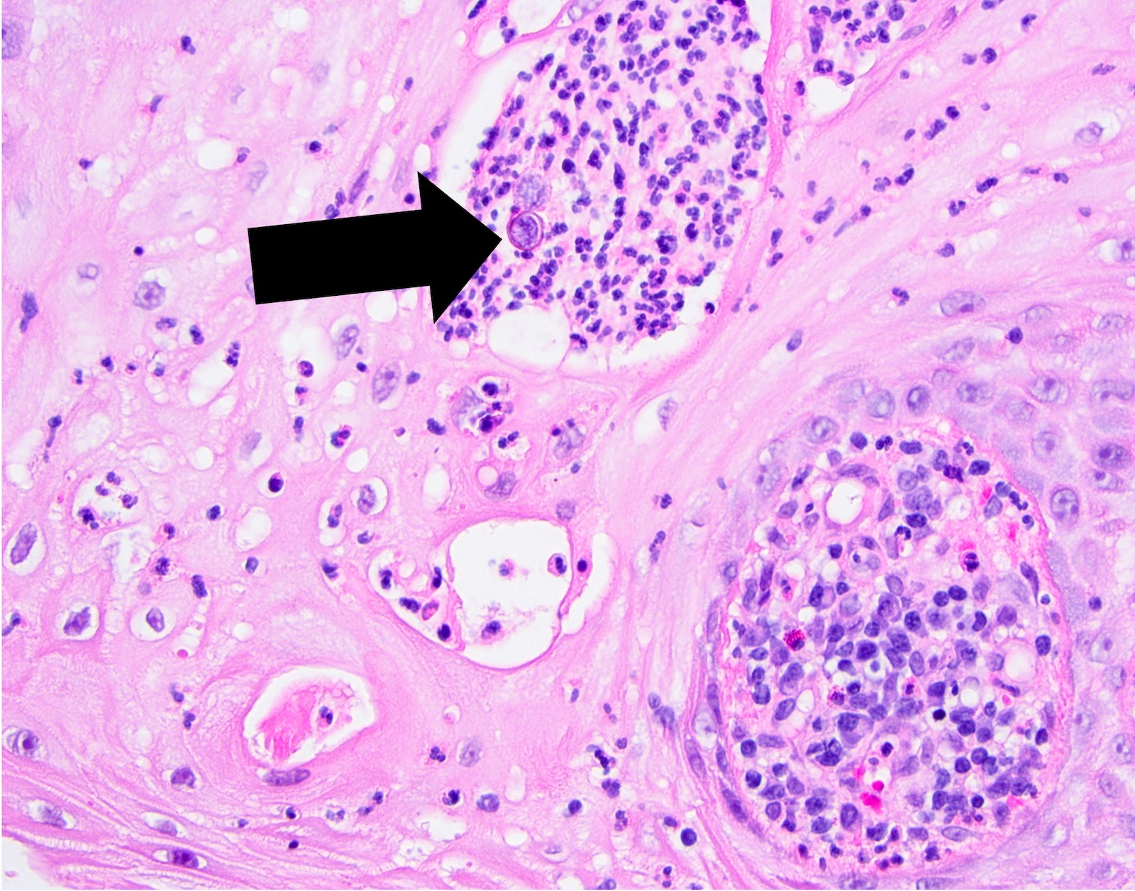
PEH in blastomycosis
PEH in epulis fissuratum
PEH in necrotizing sialometaplasia
PEH in melanoma in situ
PEH in granulomatosis with polyangiitis
PEH in excision of skin lesion
Definition, causes and histologic appearance of PEH
PEH in granular cell tumor
Contributed by Brittany Camenisch, D.M.D., Nehal Almehmadi, B.D.S., Sana Naheed, B.D.S., Evan Lynch, M.D., Ph.D.,
Justin Kolasa, M.D., D.M.D., Molly Housley Smith, D.M.D. and Susanna Goggin, D.M.D.

Gingival pyogenic granuloma
Reactive pyogenic granuloma

Pedunculated vascular nodule

Red ulcerated pyogenic granuloma
Nodular red pebbly masses

Epulis granulomatosum

Ulcerated gingival pyogenic granuloma
Contributed by Molly Housley Smith, D.M.D.
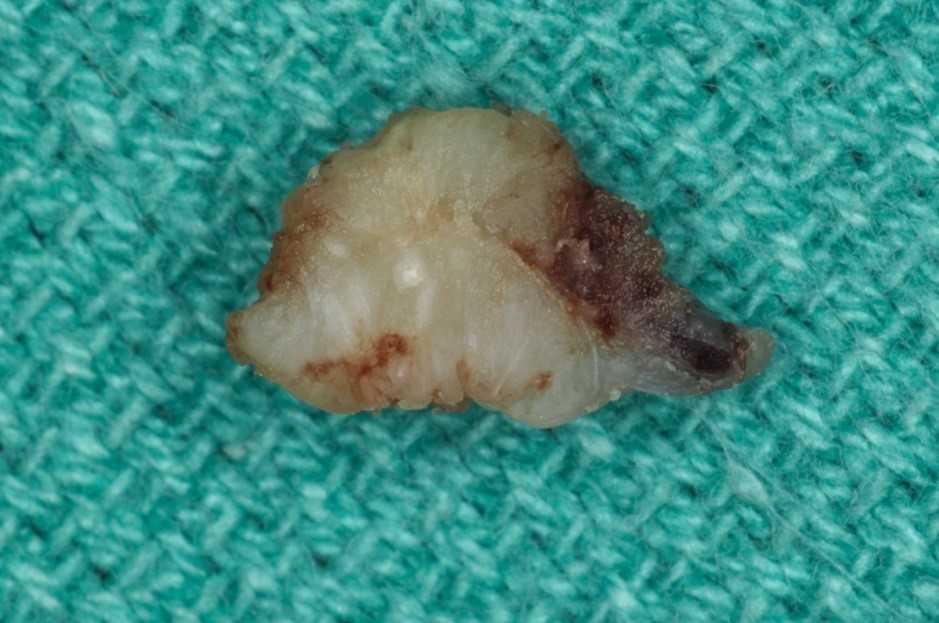
Lobulated and hyperkeratotic mass

Lobulated and hyperkeratotic mass, sectioned

Deep inflammatory infiltrate
Ulcerated pyogenic granuloma
Ulcerated pyogenic granuloma, sectioned
Contributed by Molly Housley Smith, D.M.D.
Pedunculated mass

Prominent vasculature
Prominent blood vessels

Acute inflammation
Lobular arrangement
Lobular and exophytic architecture
Capillary proliferation
Histopathology of pyogenic granuloma
Clinical and etiologic features
Images hosted on other servers:


Nonhomogenous parotid lesion
Images hosted on other servers:

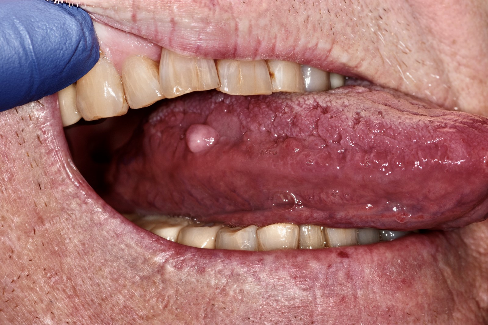
Labial mucosal bluish nodule

Parotid salivary gland swelling

Smooth firm labial nodule
Images hosted on other servers:

Smooth firm labial nodule
Contributed by Vancouver General Hospital

True cyst removed intact

Squamous metaplasia, ruptured cyst
Contributed by Sarah Glass, D.D.S.
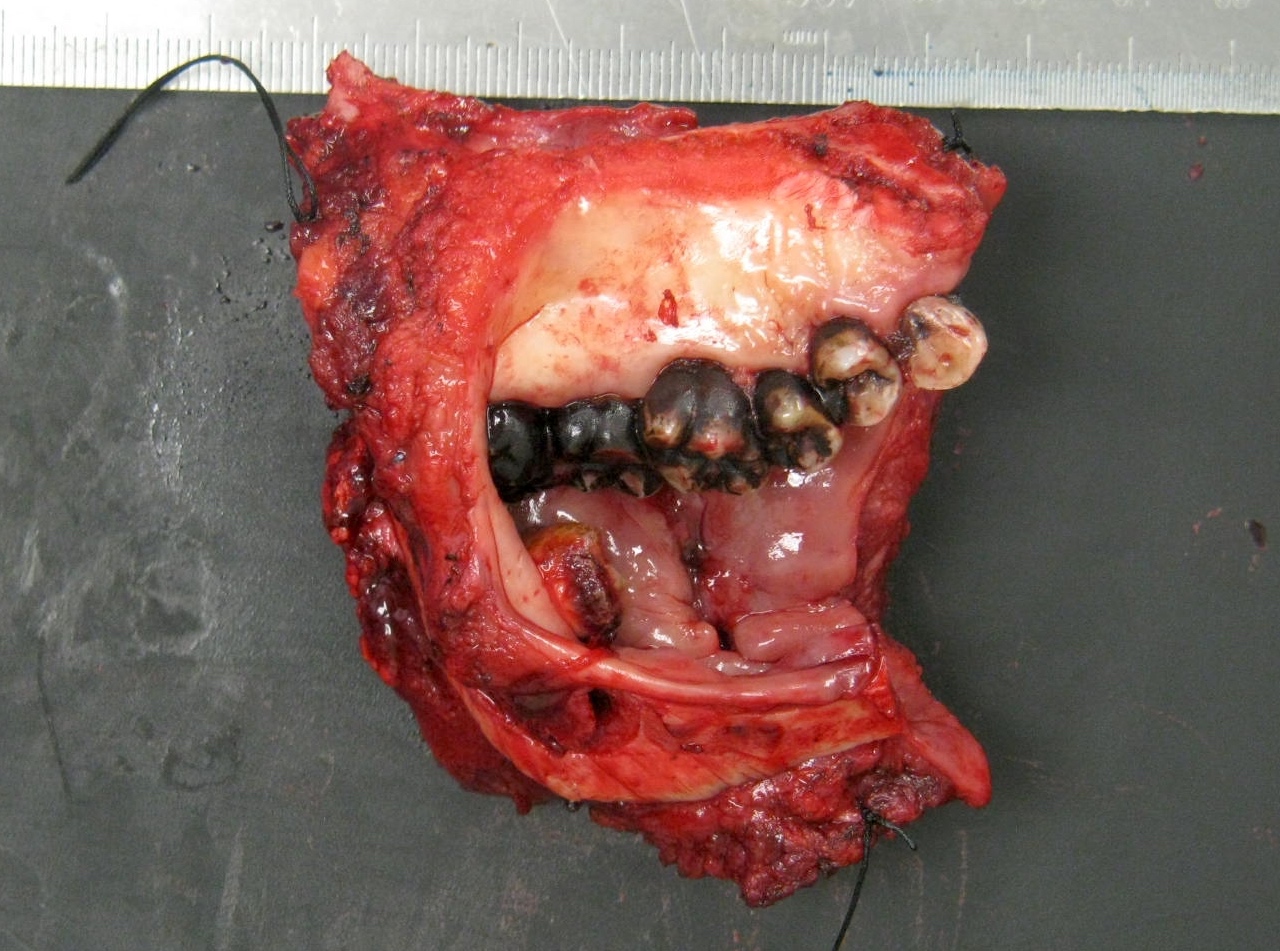
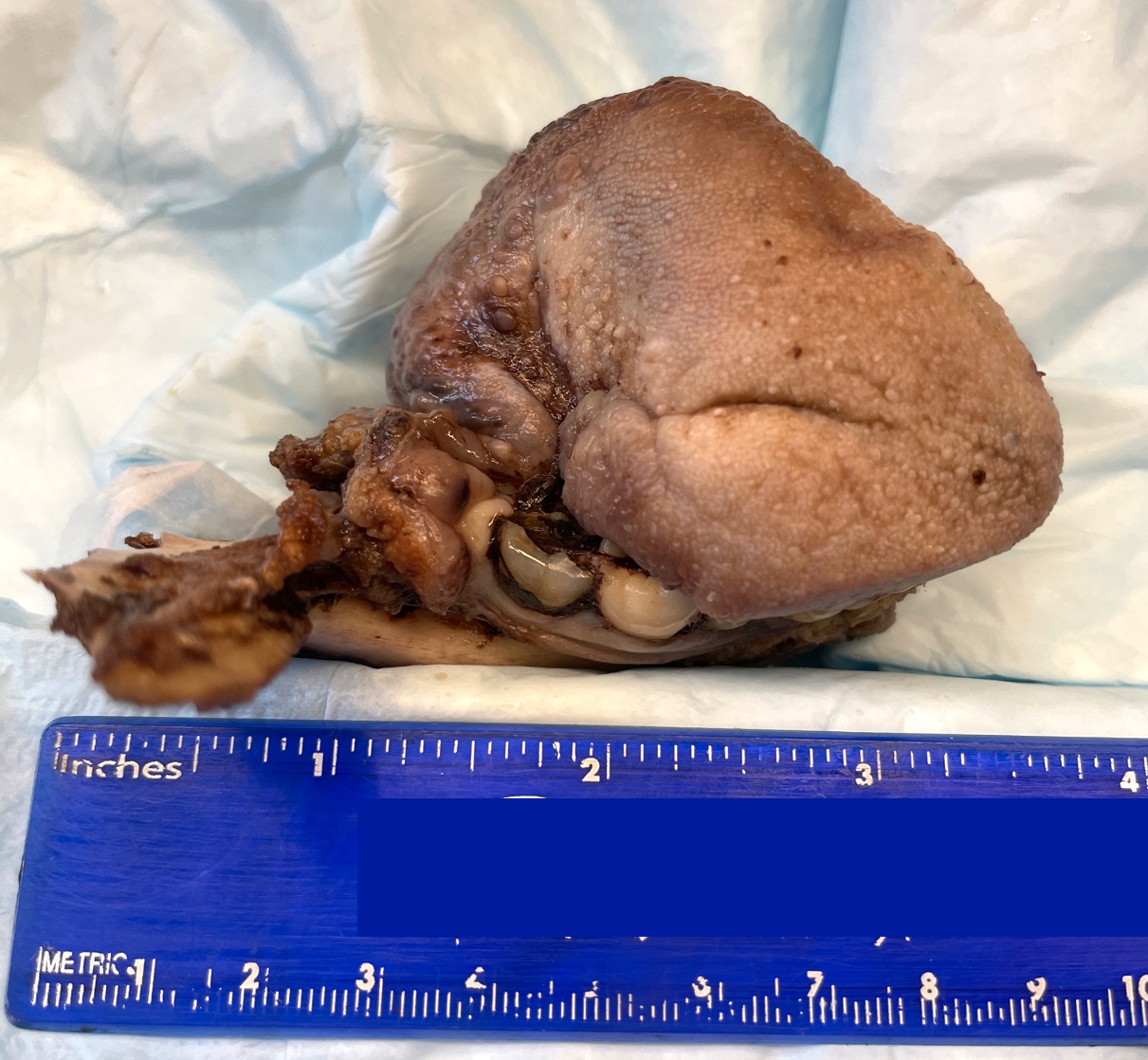
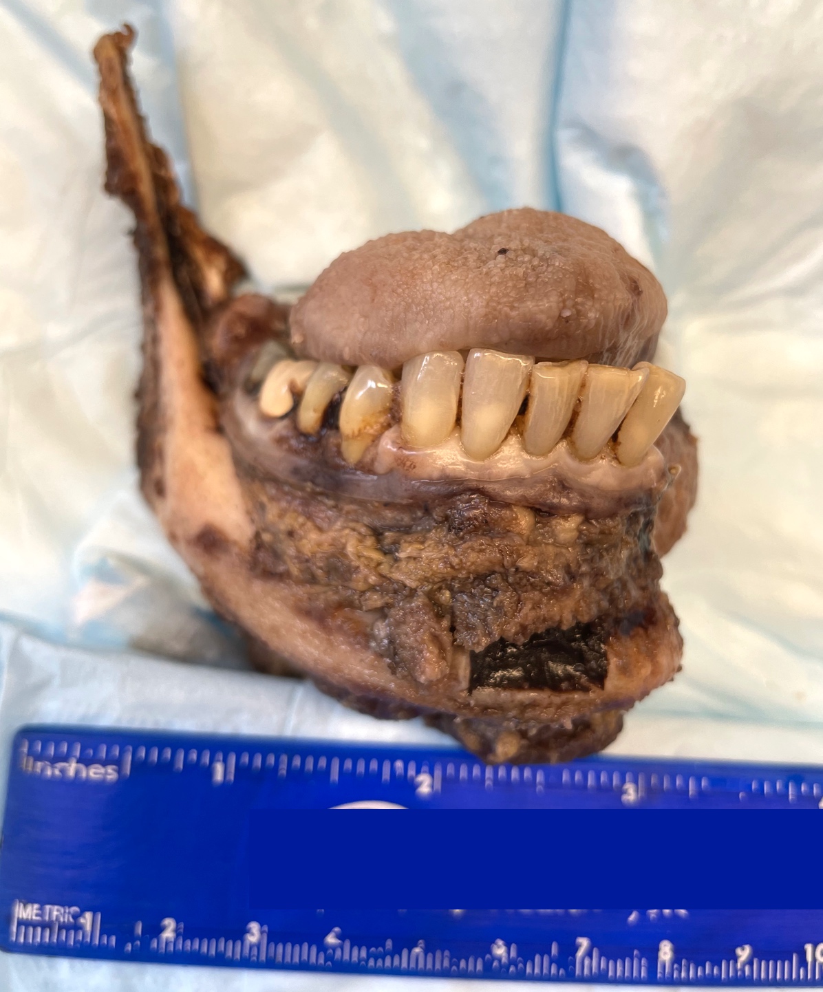
Mandible invasion
Contributed by Sarah Glass, D.D.S.
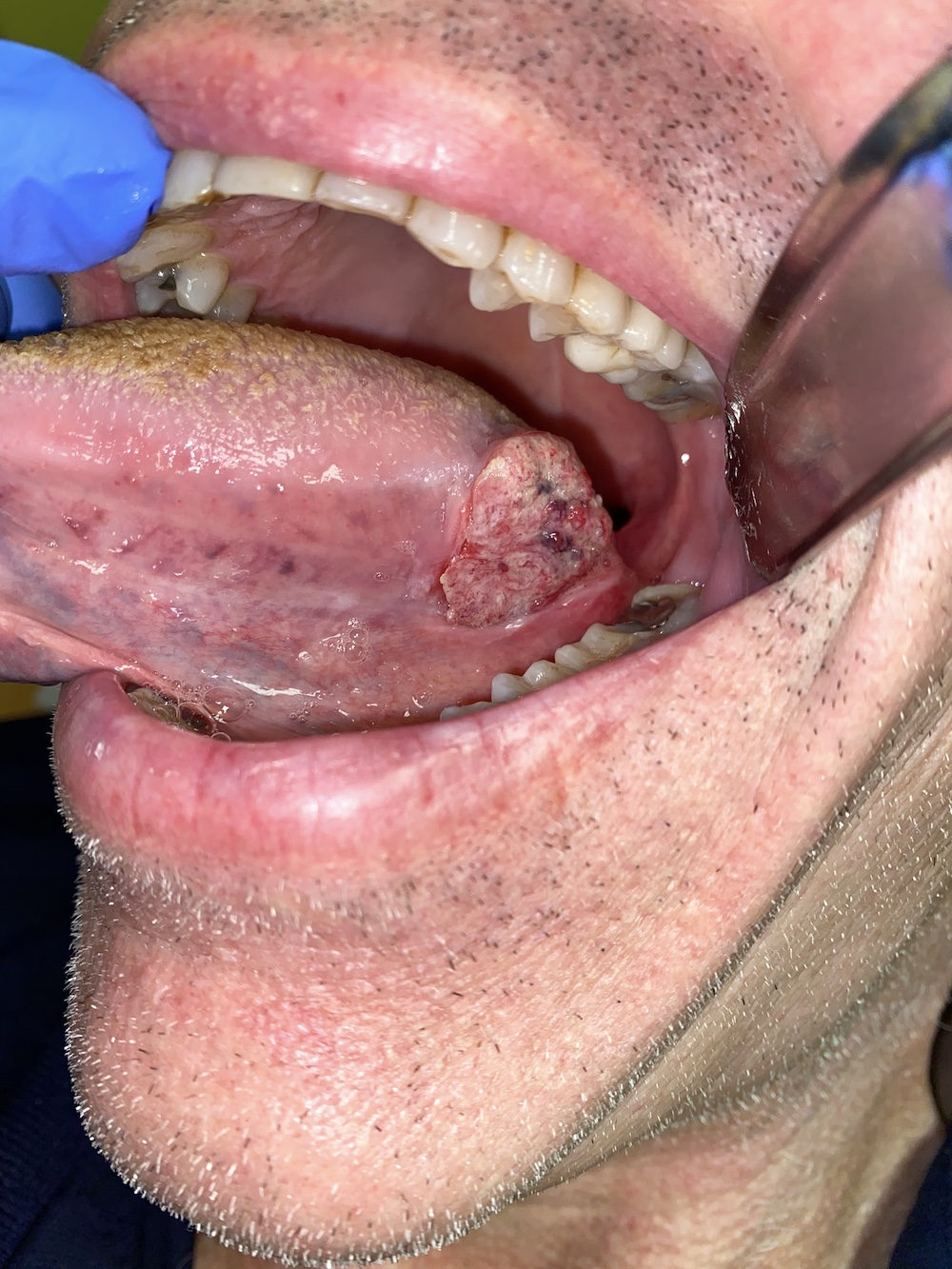
Exophytic mass lateral tongue
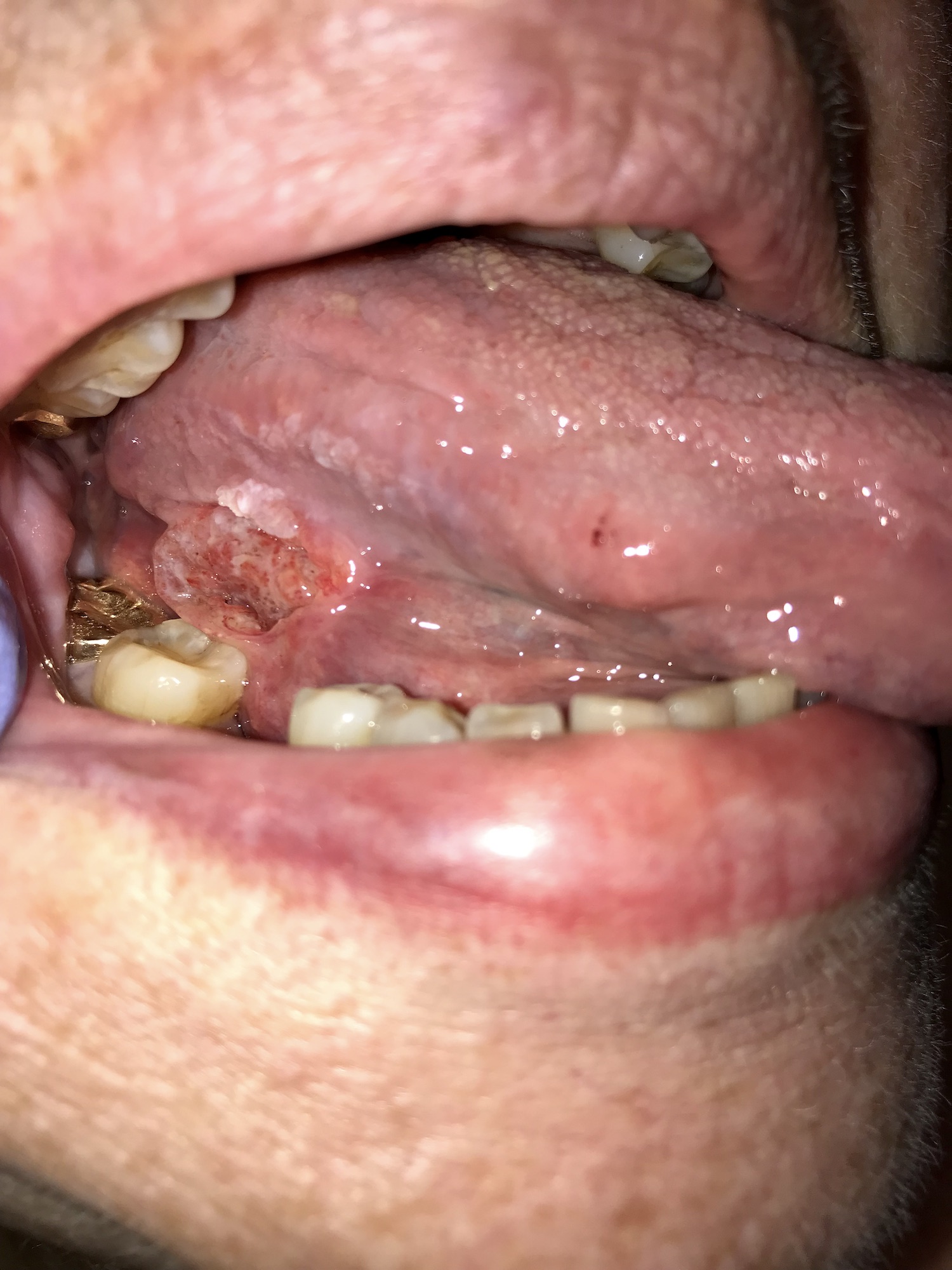
Ulceration ventrolateral tongue
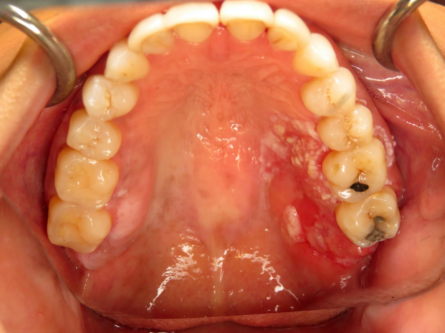
Red-white maxillary gingiva
Contributed by Kathleen Higgins, D.D.S., M.S., Stephen Roth, D.D.S. and Kathleen Schultz, D.M.D.
Glossectomy specimen
Glossectomy specimen cross section

Resection
lateral view
Resection superior view
Resection anterior view
Glossectomy specimen
Contributed by Kathleen Higgins, D.D.S., M.S., Duane Schafer, D.D.S., M.S. and Sarah Glass, D.D.S.
Tumor with margins

Basement membrane breach
Keratin pearl
Muscle invasion

Individual cell keratinization
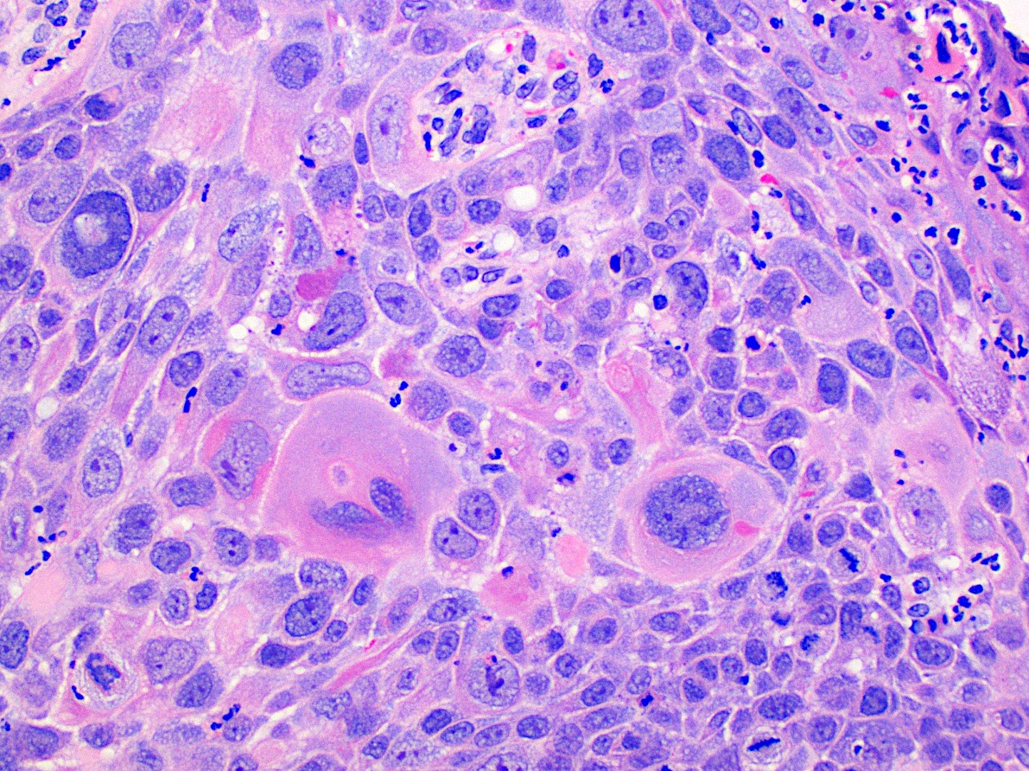
Impressive pleomorphism

Surrounding nerve
Scattered malignant cells

Basaloid variant

Adenosquamous variant
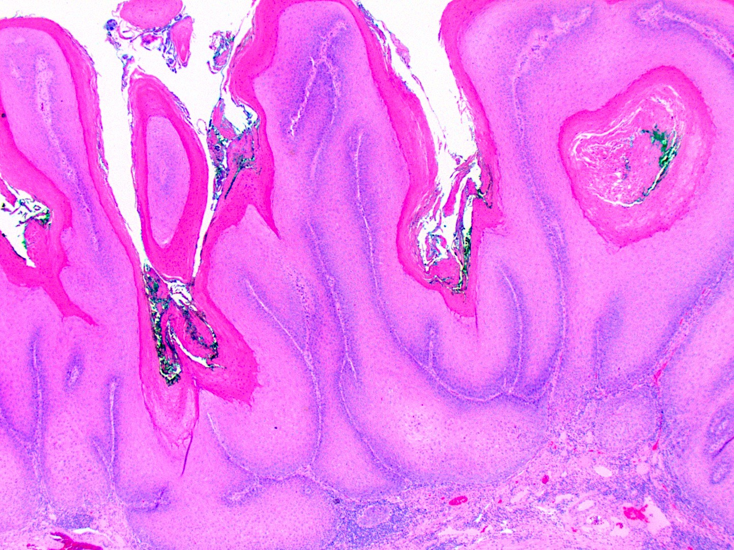
Verrucous carcinoma
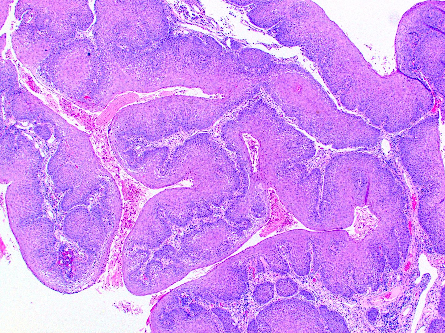
Papillary variant
Acantholytic variant
Spindle variant
Cuniculatum variant
Cytokeratin positive
Contributed by Ivan J. Stojanov, D.M.D., M.M.Sc.
Squamous papilloma of gingiva
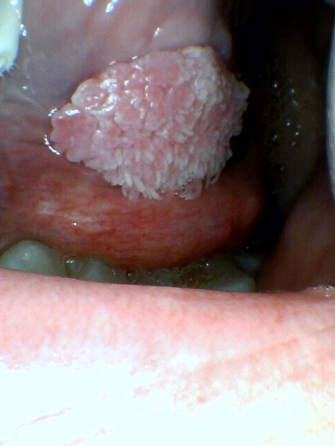
Squamous papilloma of tongue
Squamous papilloma of soft palatal mucosa
Images hosted on other servers:


Hard palate tumor
Contributed by Ivan J. Stojanov, D.M.D., M.M.Sc.
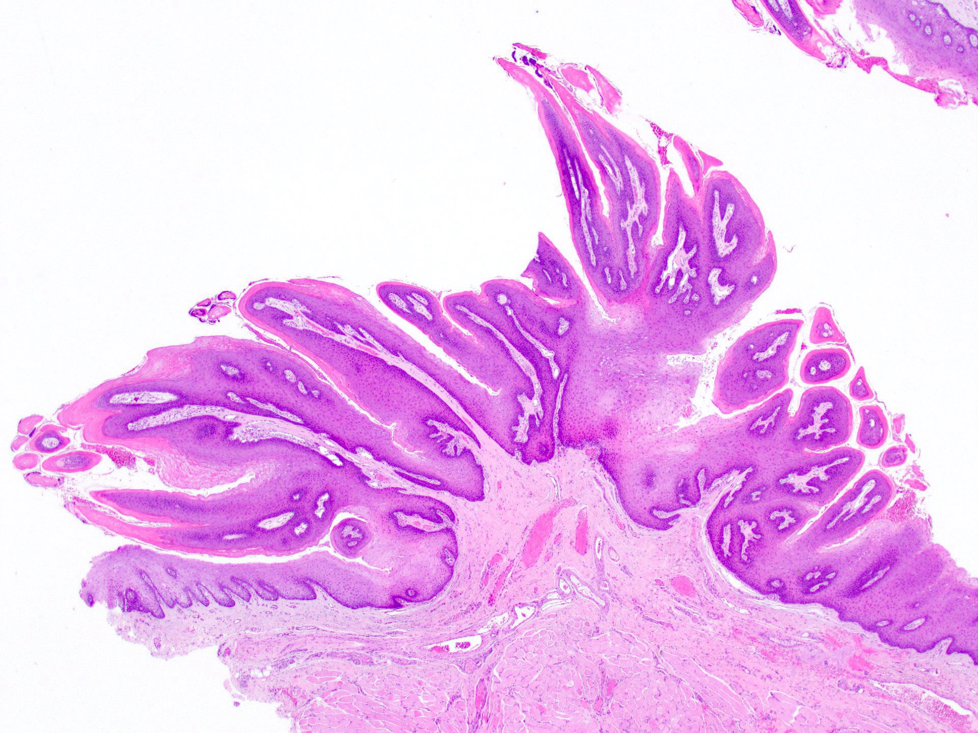
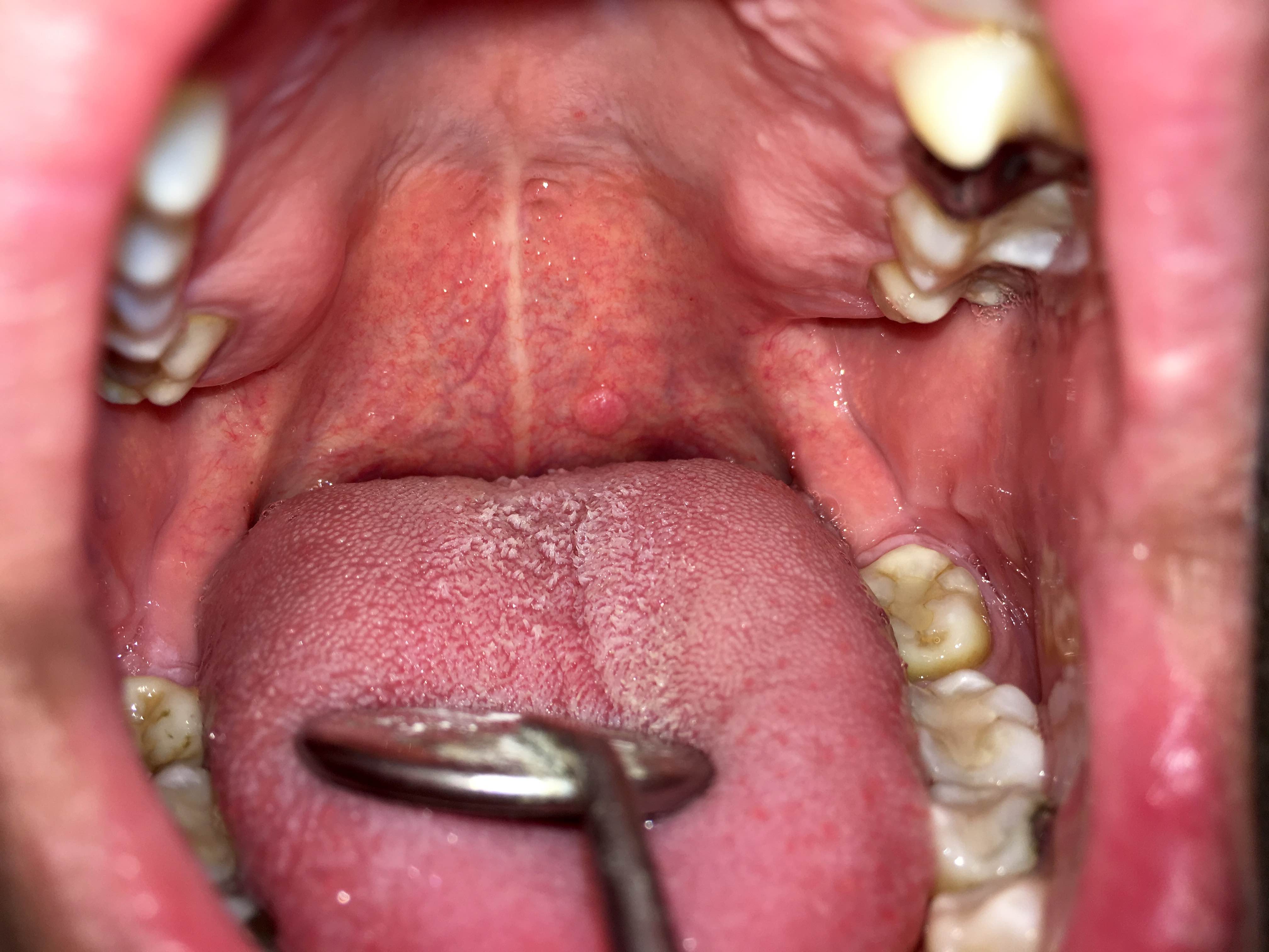
Pedunculated
Sessile
Irritated
Fibrovascular cores
Tangentially sectioned
Tangentially sectioned
Hyalinization
Irritated
Mitotic activity
With acanthosis


Contributed by Vasiliki Tzelepi, M.D.

Lateral border of tongue

Taste buds

S100

EMA
CK8 / 18
Images hosted on other servers:
70 year old woman:

Painful ulcer of tongue

Ill fitting prosthesis

Healing after removal of prosthesis
6 month old boy:

Ulcerative swelling
Images hosted on other servers:

Mixed cellular infiltrate
Contributed by Molly Housley Smith, D.M.D.
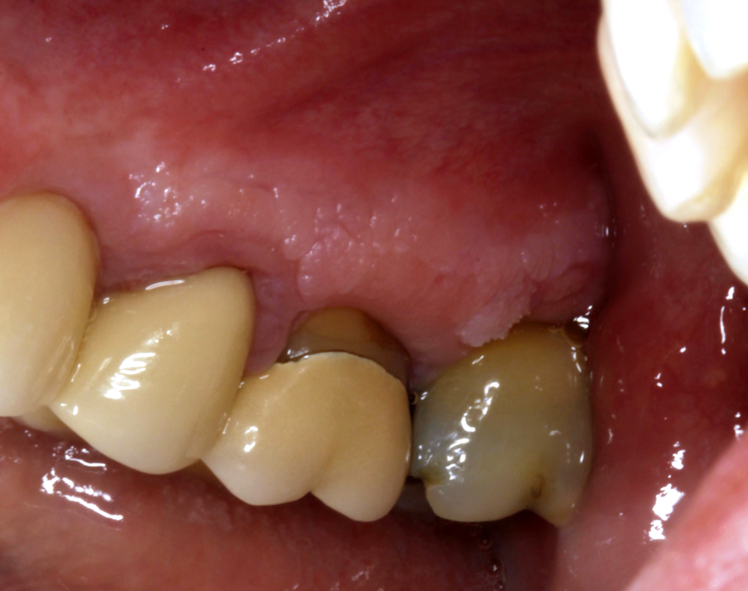
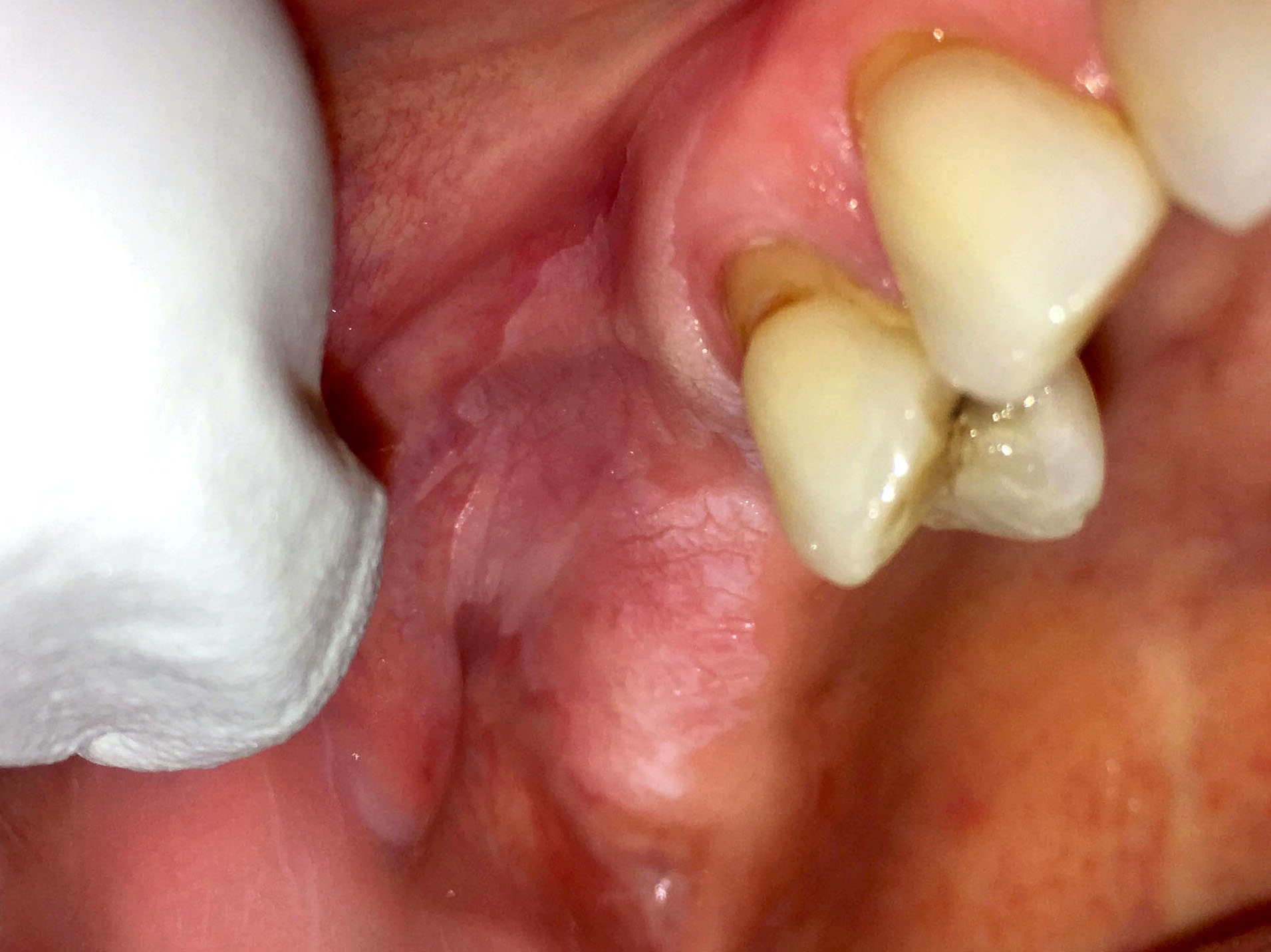
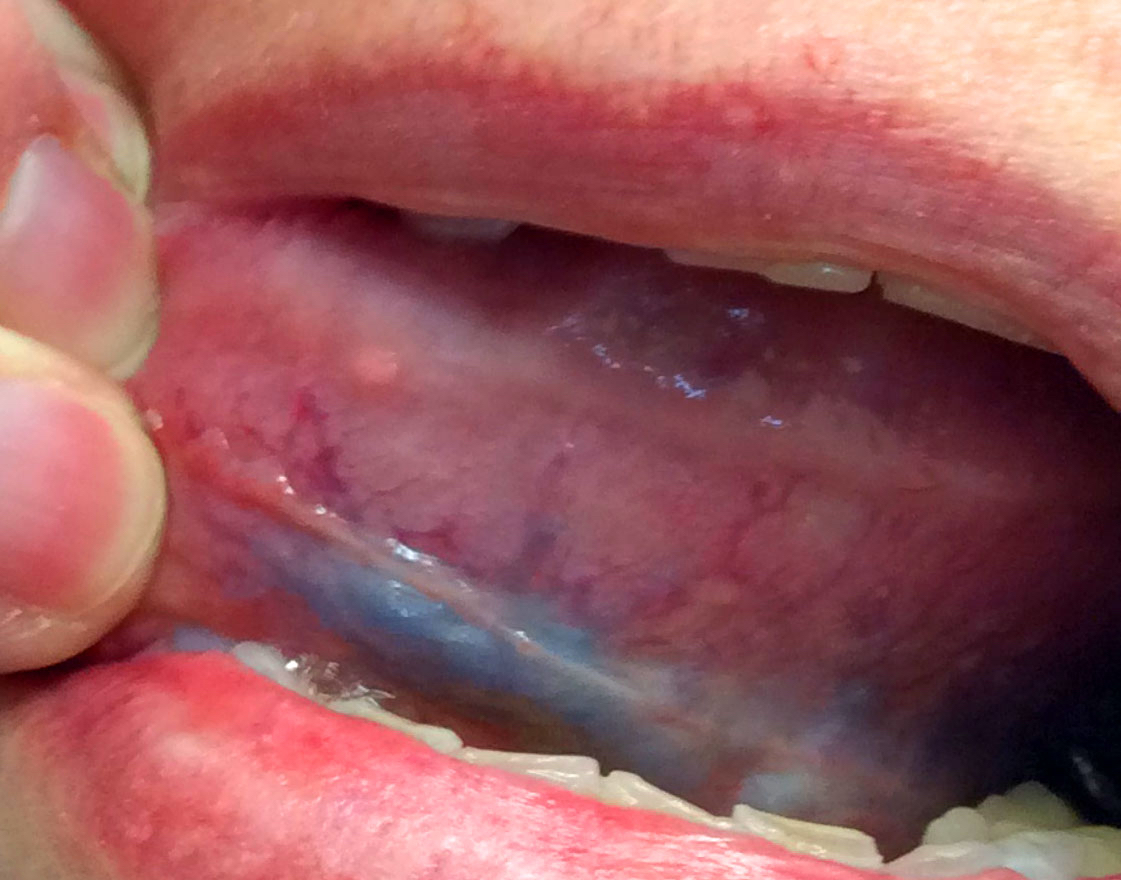
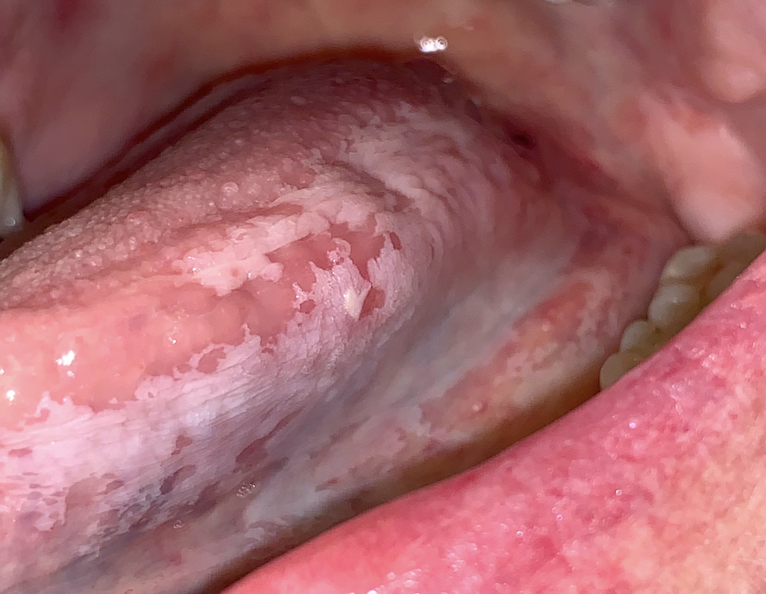
Sublingual varicosities
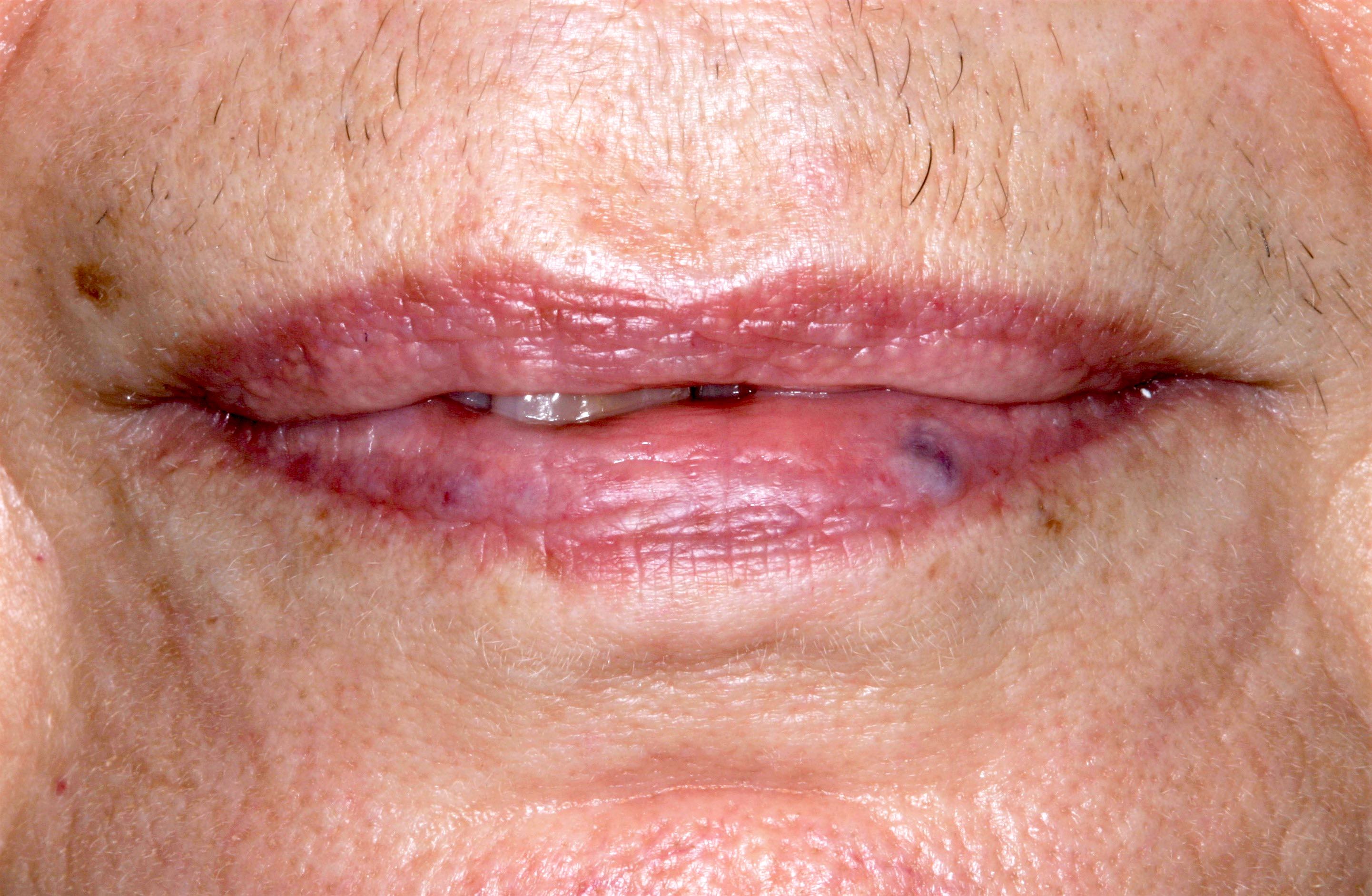
Varicosity on lower lip
Thrombosed varicosity of labial mucosa
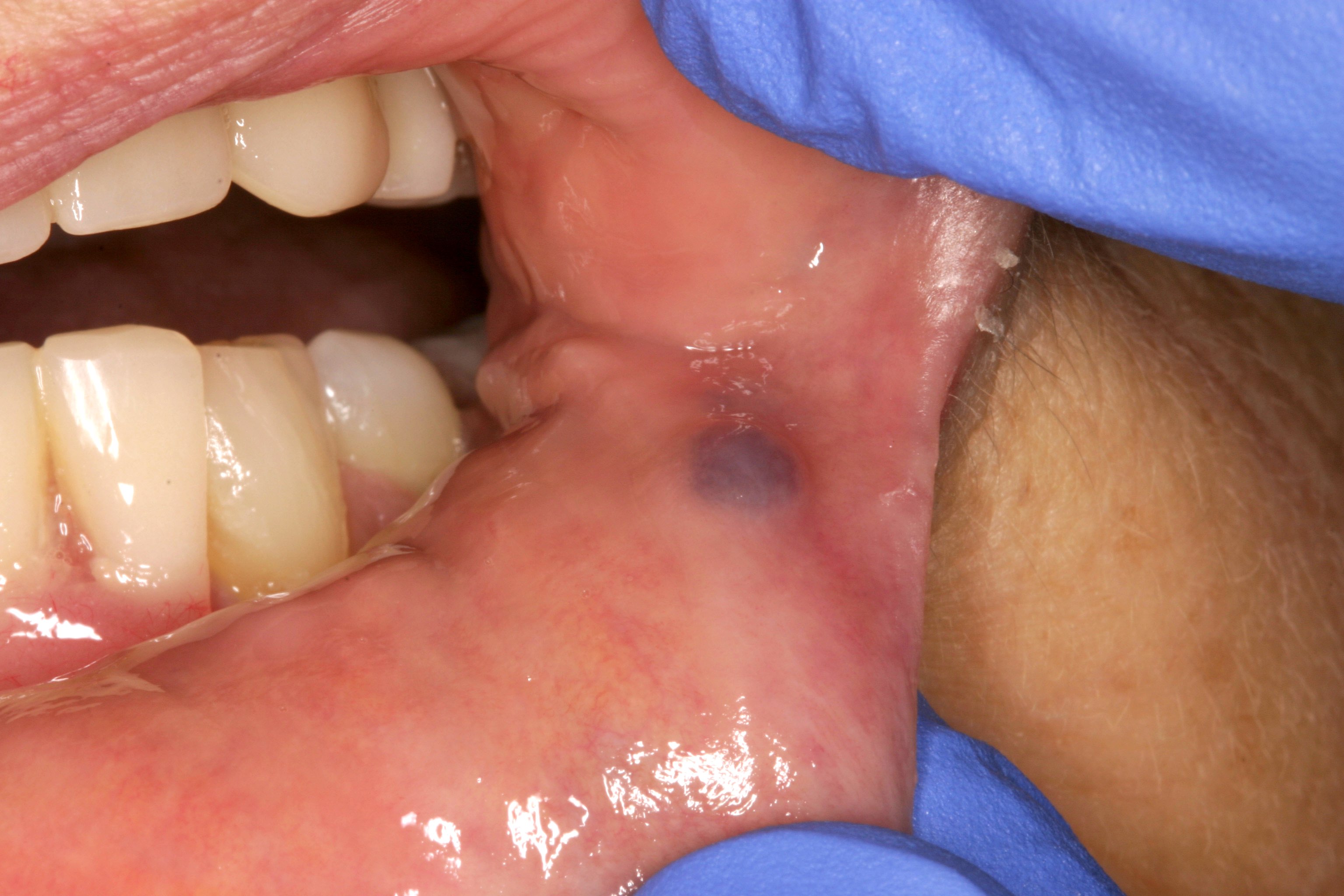
Contributed by Molly Housley Smith, D.M.D.
Oral varicosity
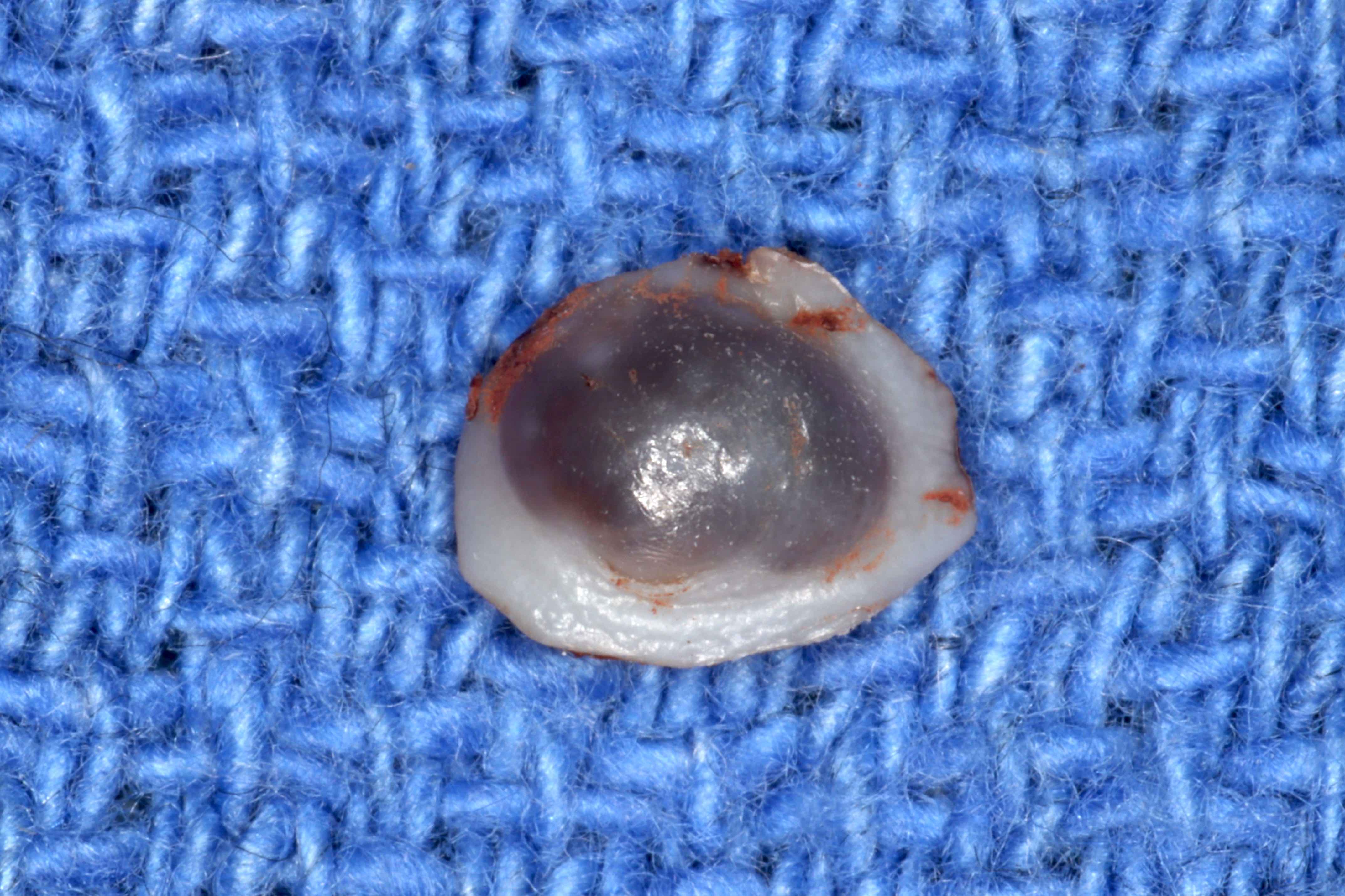
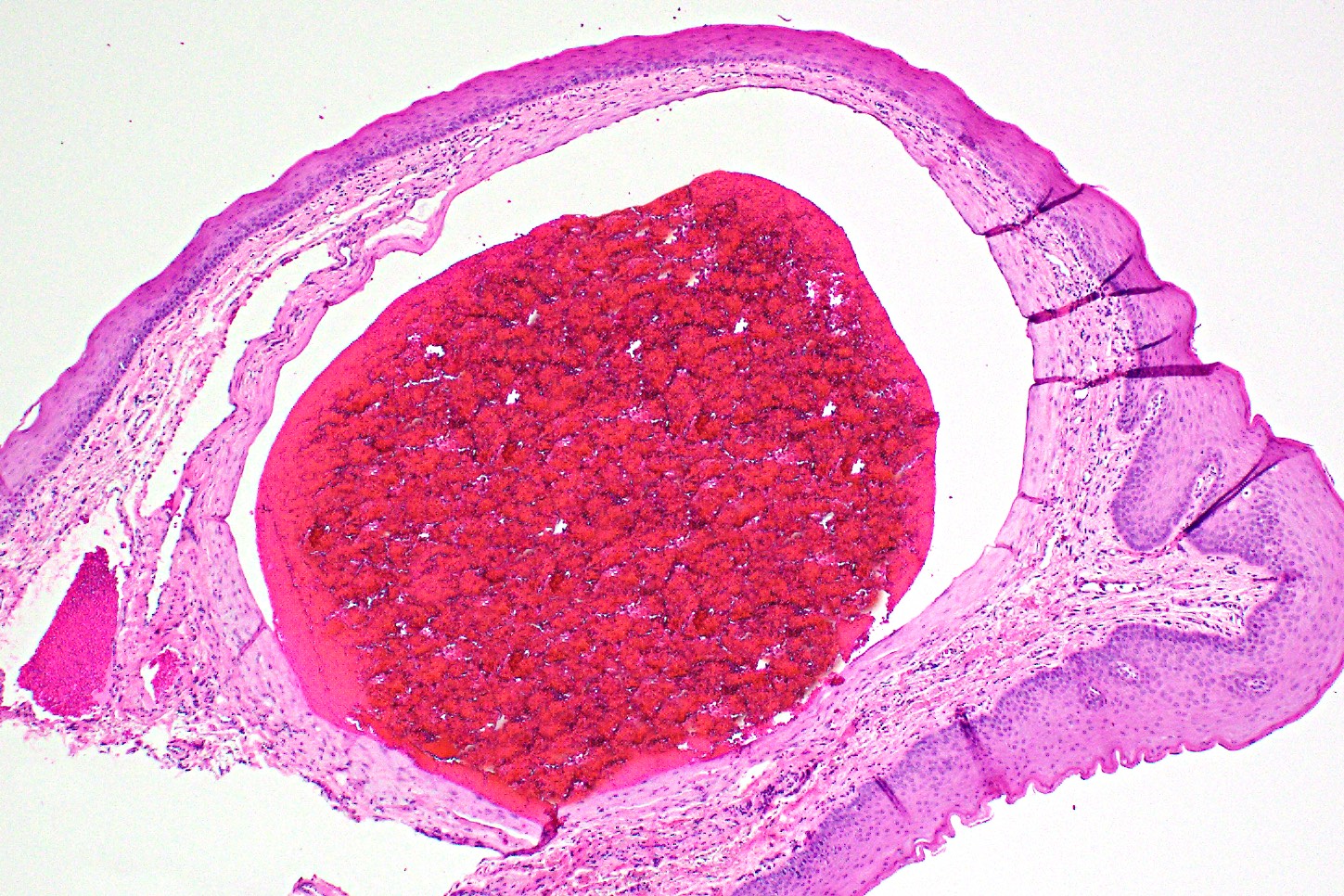
Contributed by Molly Housley Smith, D.M.D.
Oral varicosity
Compressed oral varicosity
Oral varicosity with organizing thrombus
Phlebolith formation
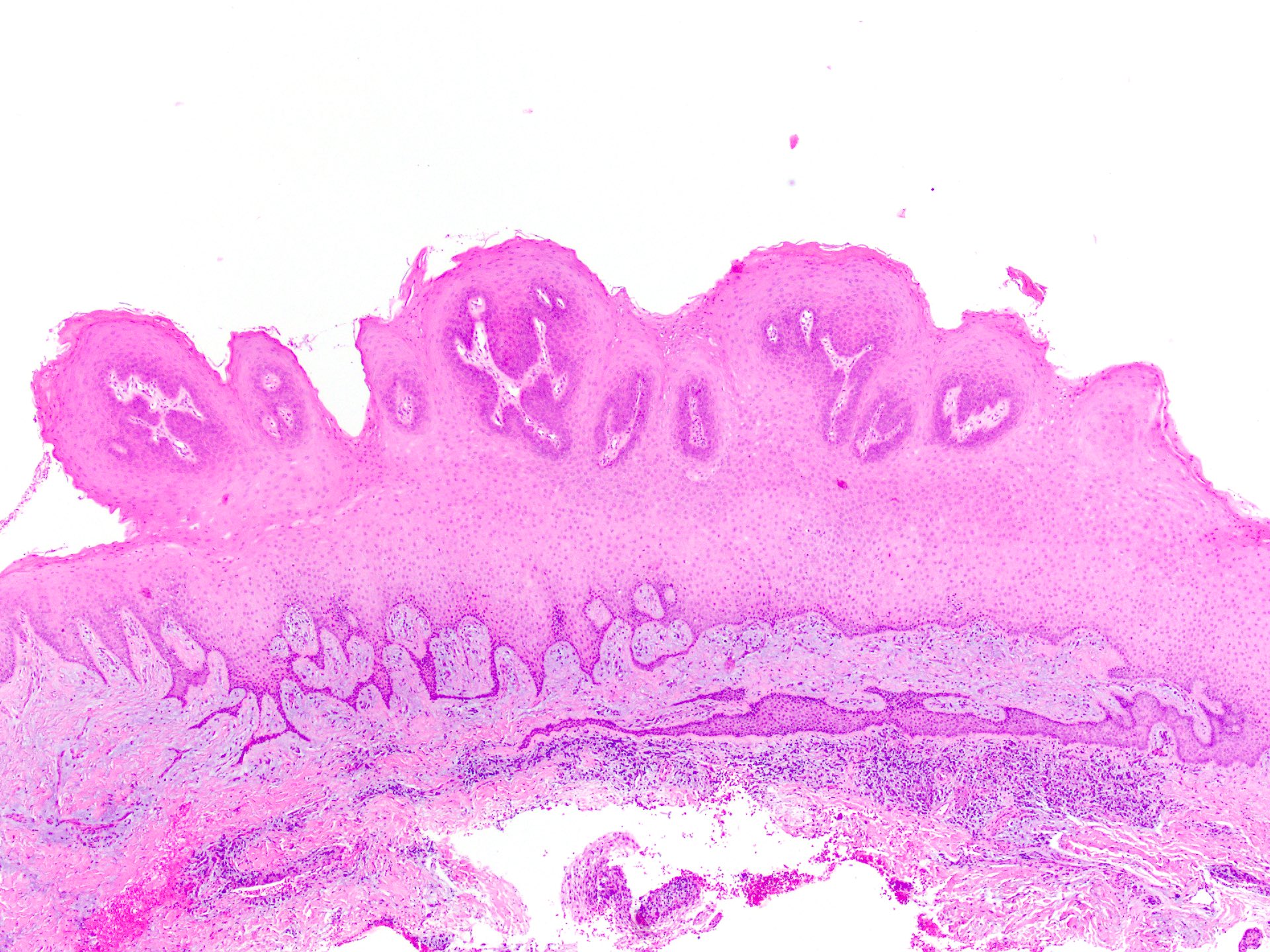
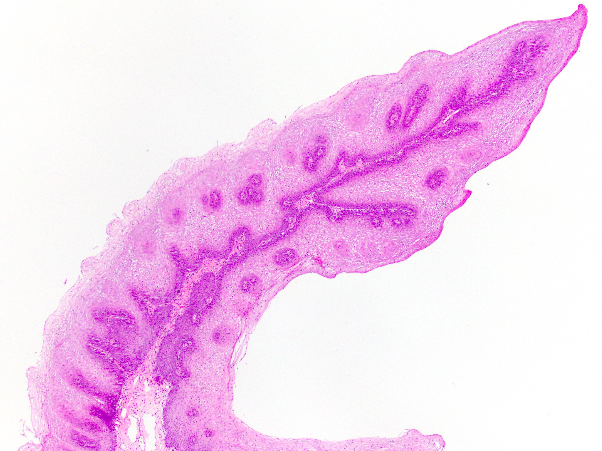
Contributed by Molly Housley Smith, D.M.D. and Michael Piepgrass, D.M.D., M.S.
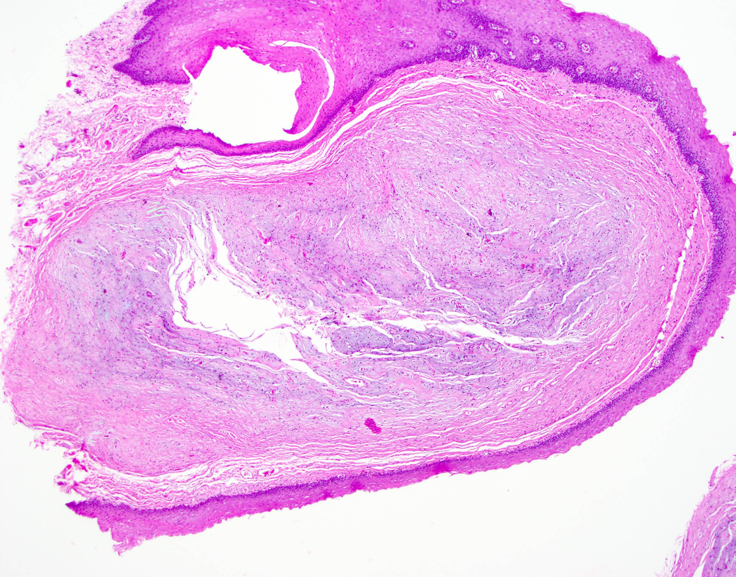
Well circumscribed papillary lesion
Prominent keratin horn
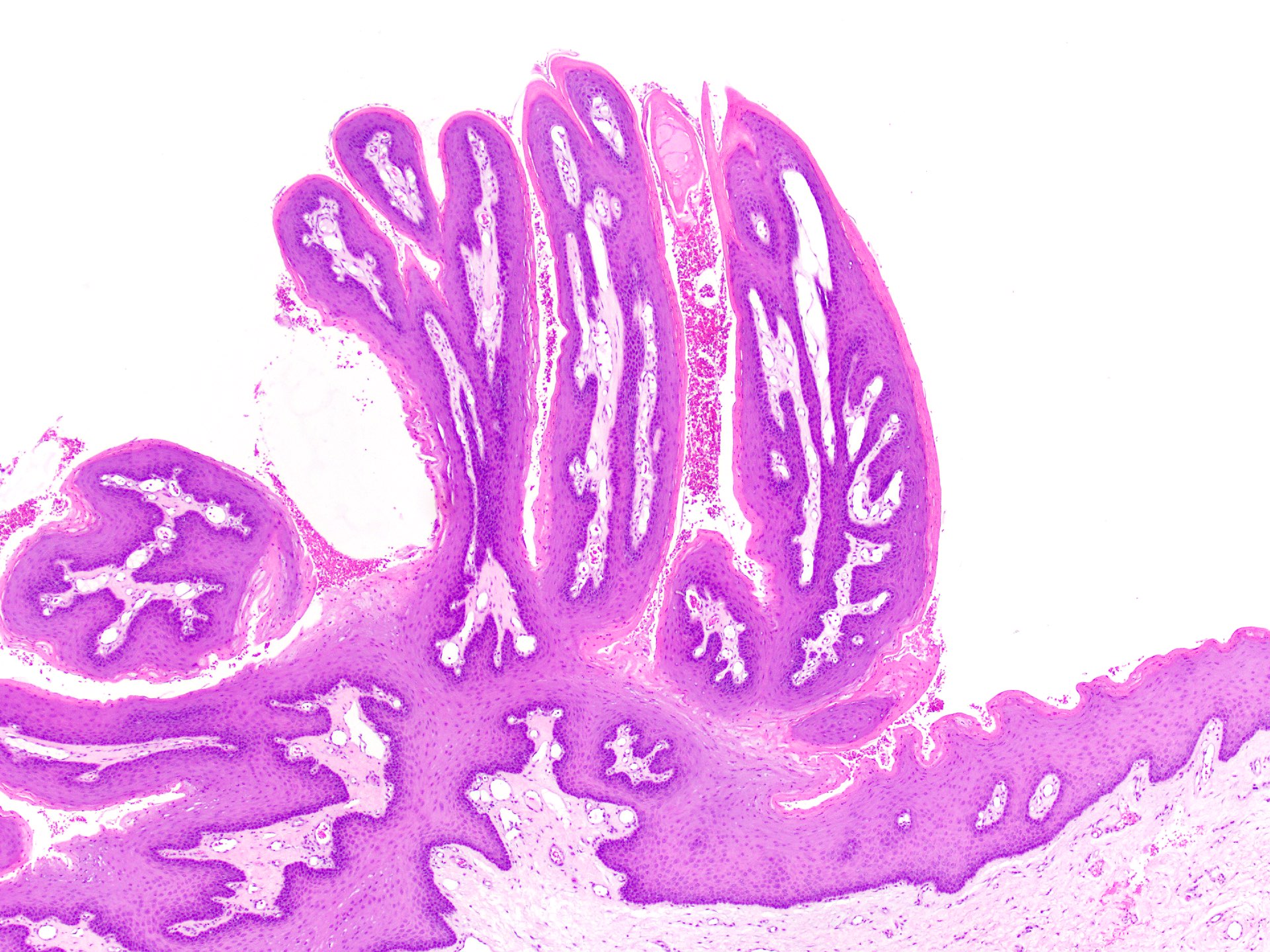
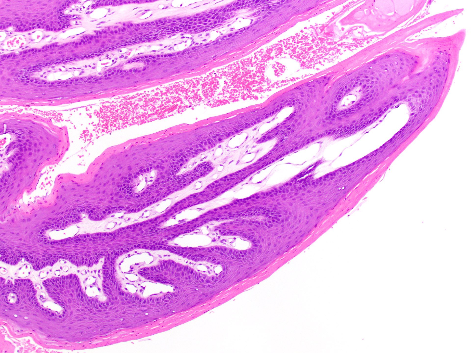
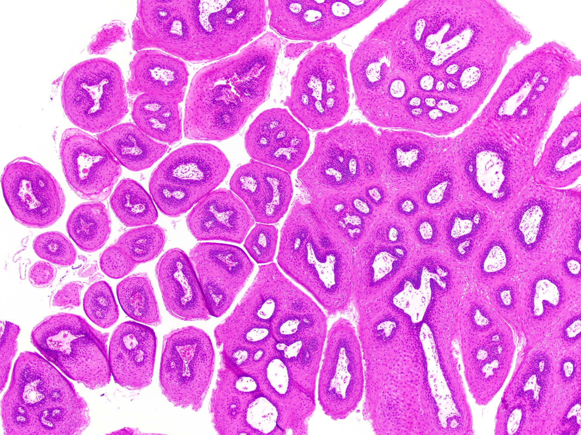
Contributed by Molly Housley Smith, D.M.D.
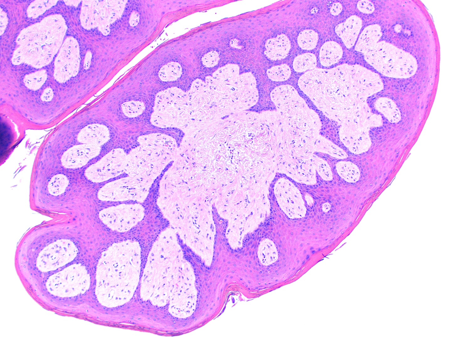
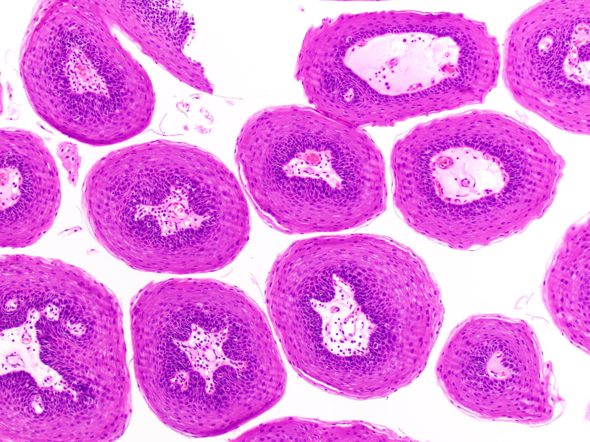
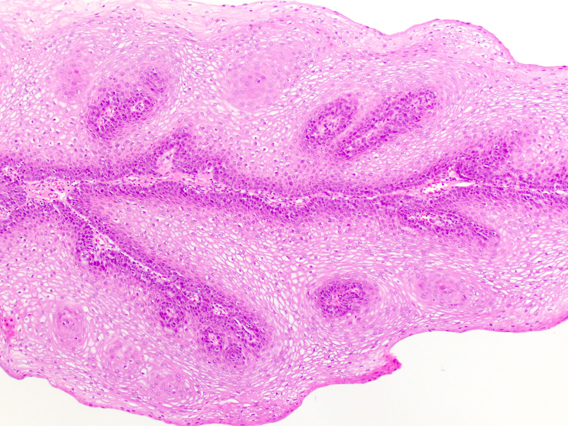
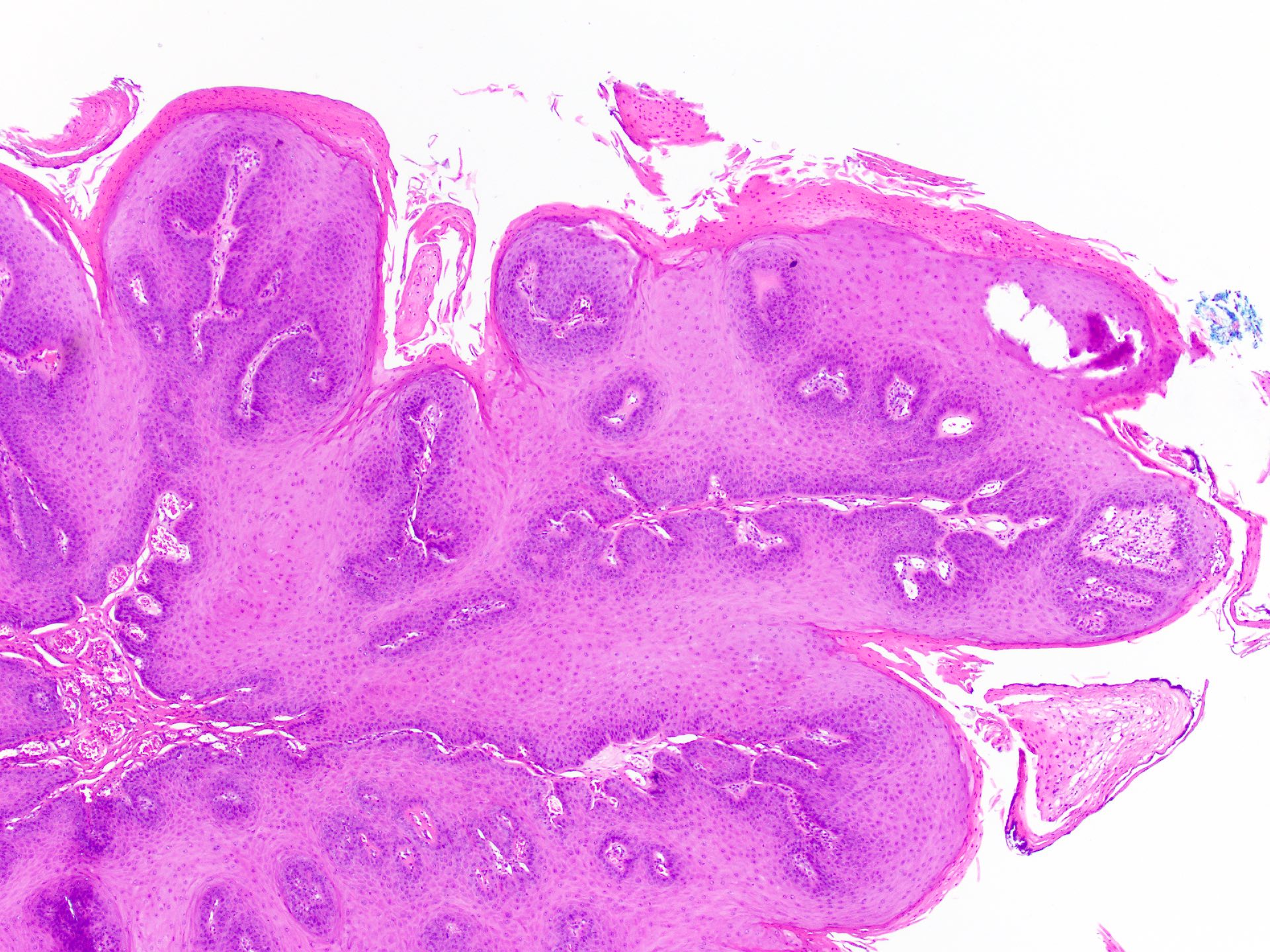
Prominent cupping of rete pegs
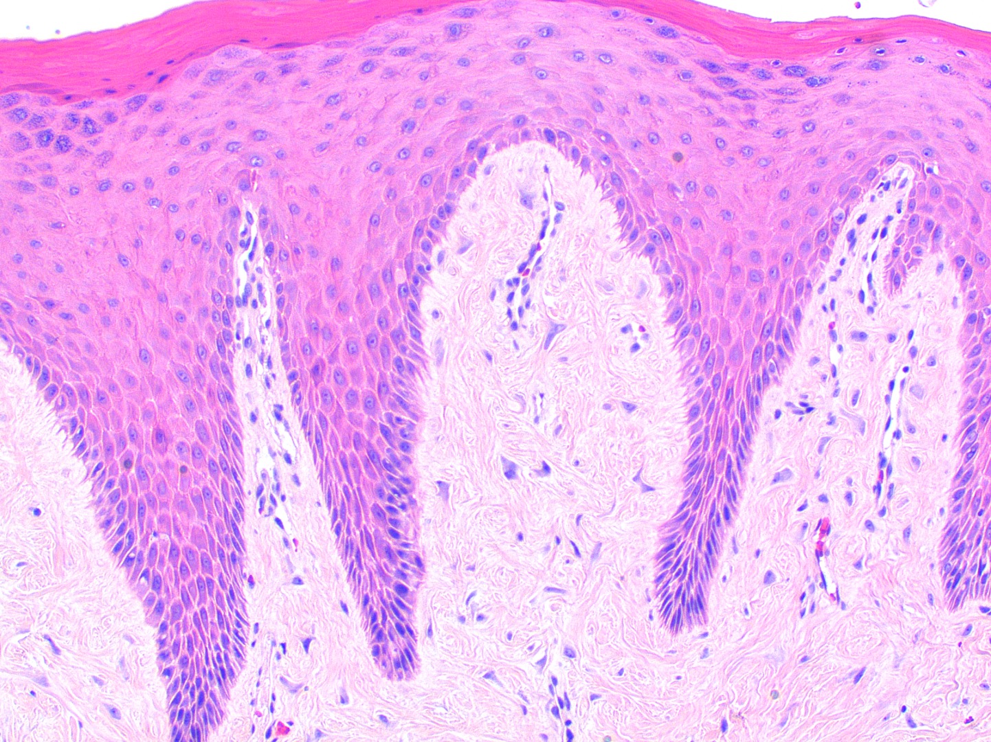
Hypergranulosis and koilocytosis
Hyperorthokeratosis
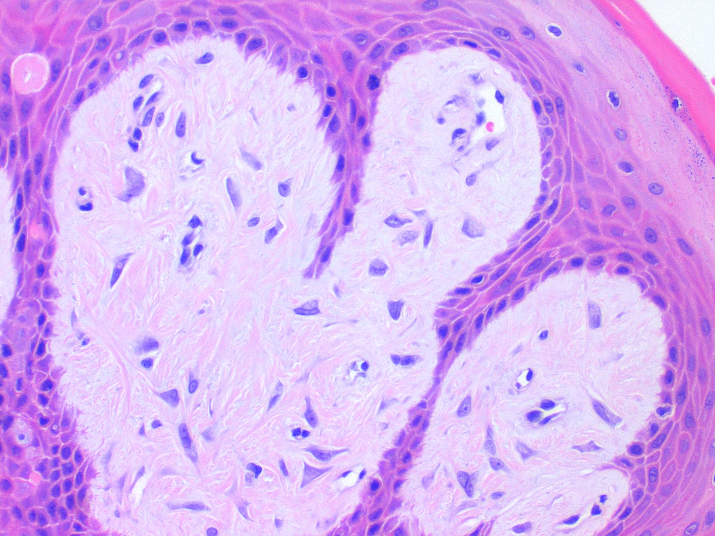
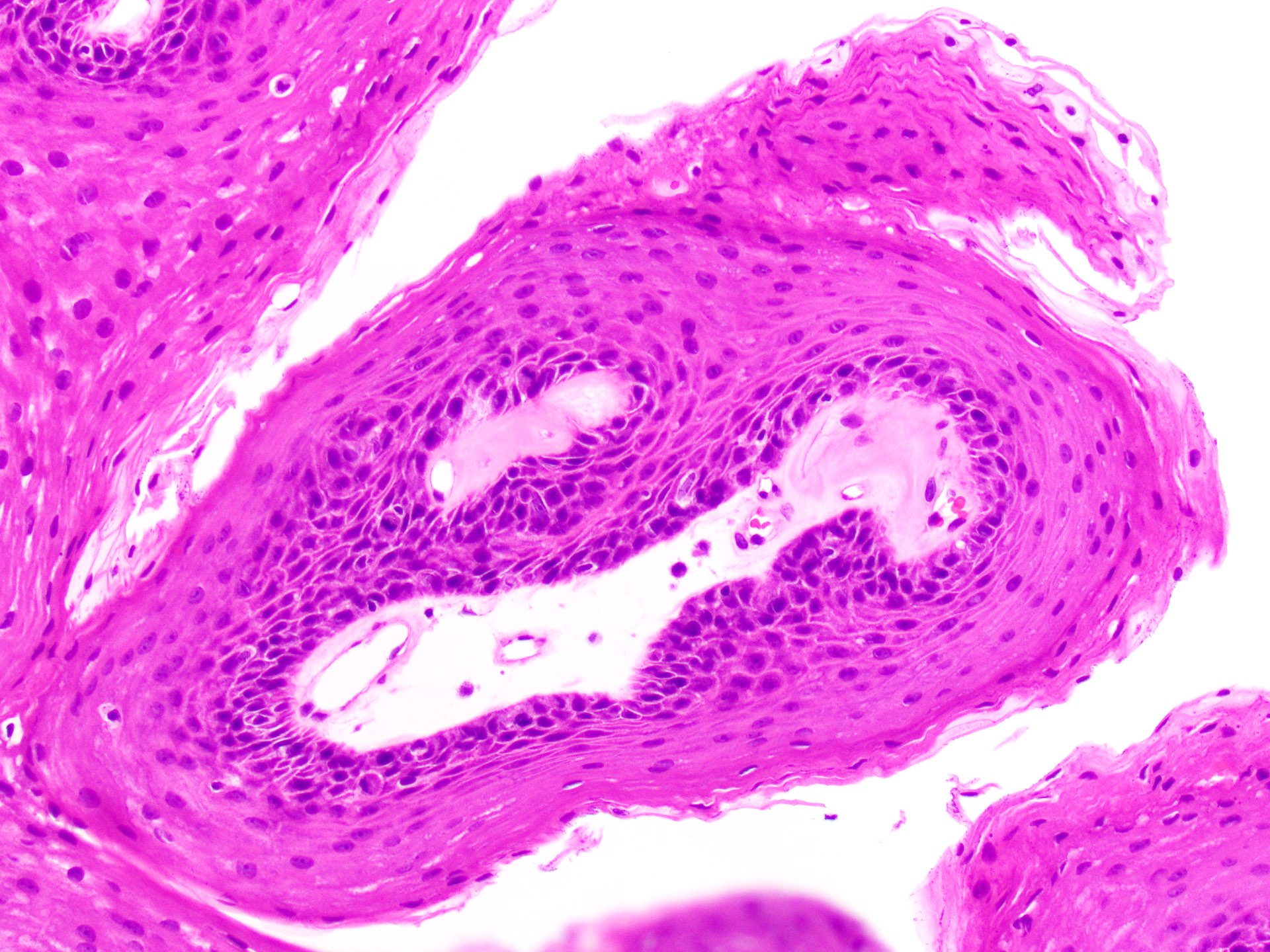
Hypergranulosis
Eosinophilic viral inclusions
Verruca vulgaris versus seborrheic keratosis pathology
Contributed by Louis Beto, D.D.S., M.D., Hal Levine, D.M.D., M.D., Mark Mintline, D.D.S. and Mark Sutor, D.D.S.
Yellow granular mass of oral vestibule
Well circumscribed solitary lesion
Yellow-red pebbly growth
Pink gingival mass
Contributed by Molly Housley Smith, D.M.D. and Kelly Magliocca, D.D.S., M.P.H.
Prominent verrucoid architecture
Inflammatory infiltrate
Well circumscribed mass
Keratin filled clefts
Leukocytic exocytosis
Foamy macrophages
Lipid laden macrophages

Slightly papillary silhouette
Surface parakeratosis and histiocytes

Histiocytes
VX of skin, histology
Clinical description of VX
Contributed by Molly Housley Smith, D.M.D., Thamer Musbah, M.D., Thomas C. Ocheltree, M.D. and Steven Anderson, M.D.
Verrucous carcinoma within PVL
Exophytic white mass
Verrucoid mass of alveolus
Exophytic verrucoid labial mass
Contributed by Molly Housley Smith, D.M.D.
Exophytic and downward growth
Contributed by Molly Housley Smith, D.M.D.
Hyperplastic, bulbous rete ridges
Church spire keratin peaks
Broad, plunging rete ridges
Keratin plugging

Cytologically bland
Verrucous carcinoma
Cutaneous verrucous carcinoma
Contributed by Molly Housley Smith, D.M.D.
Necrotizing sialometaplasia
Oral melanoacanthoma

Squamous papilloma
Oral epithelial dysplasia
Early proliferative verrucous leukoplakia
HPV associated dysplasia

Squamous cell carcinoma
Verrucous carcinoma

Carcinoma cuniculatum

Congenital granular cell epulis

Granular cell tumor
Ectomesenchymal chondromyxoid tumor

Hamartomatous polyp
Franchi: 2020
Gnepp: 2021
IARC: 2024
Neville: 2023
Slootweg: 2016
Stelow: 2020
Thompson: 2022
Wenig: 2017
Wenig: 2024
Find related Pathology books: head & neck/endocrine
