
Images hosted on other servers:

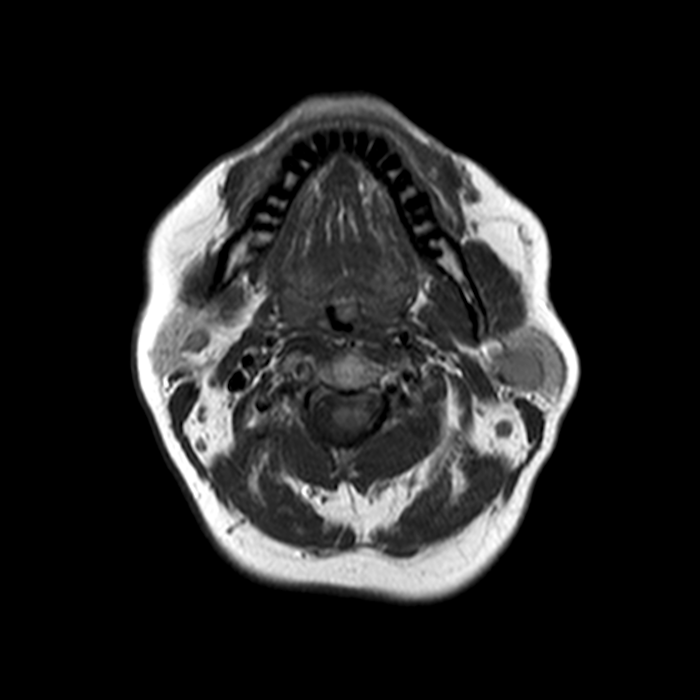
MRI: cystic lesion of the upper lip

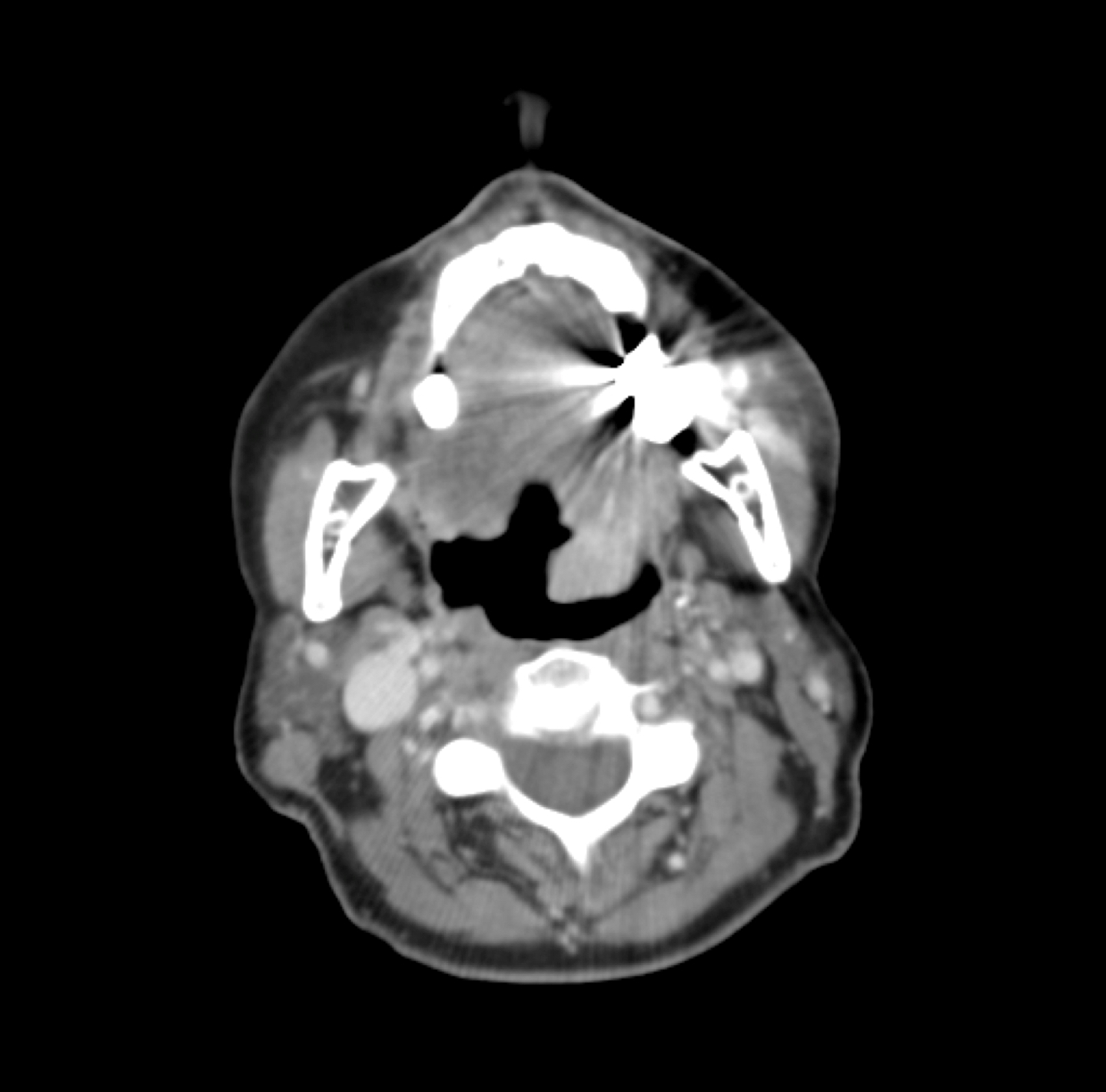
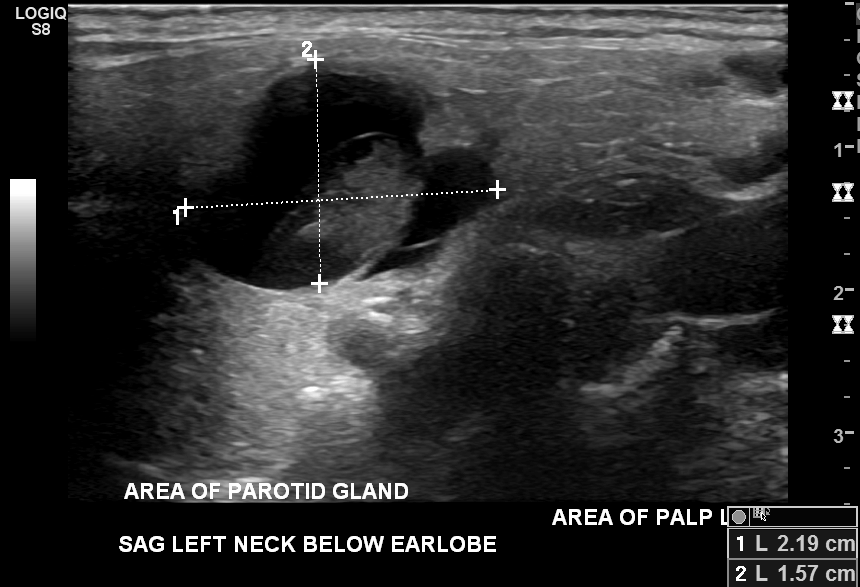
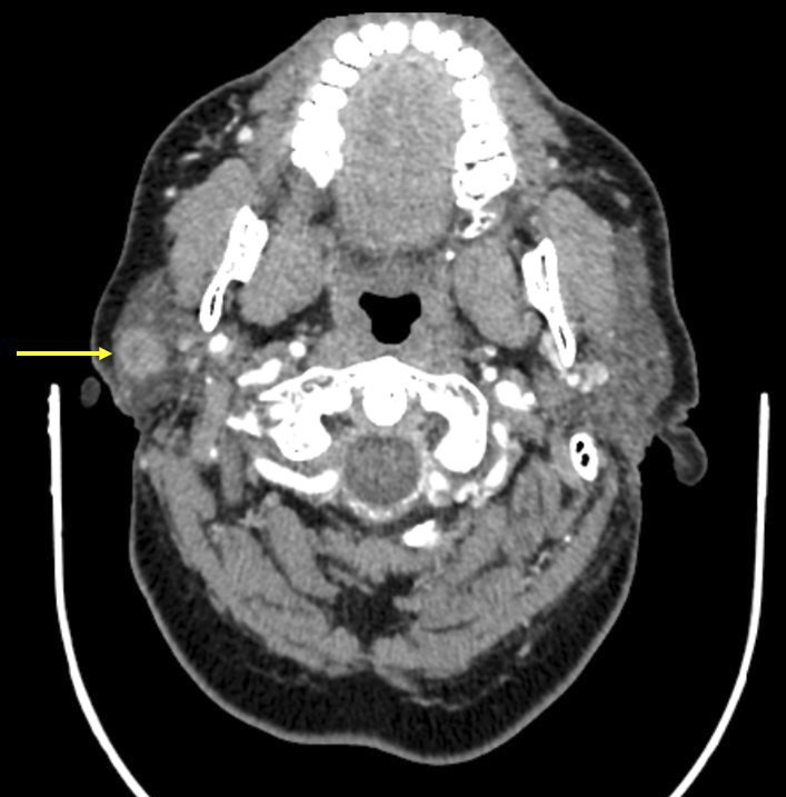
CT and US: right parotid nodule
Images hosted on other servers:

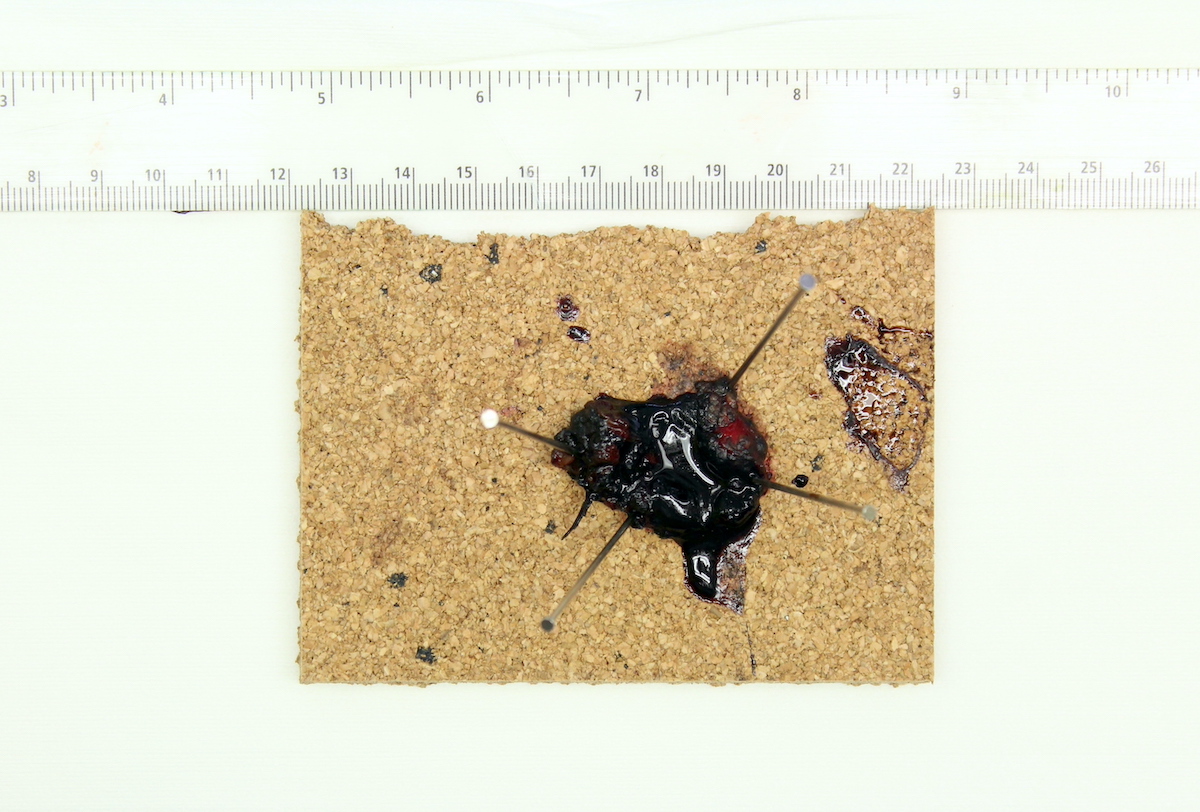
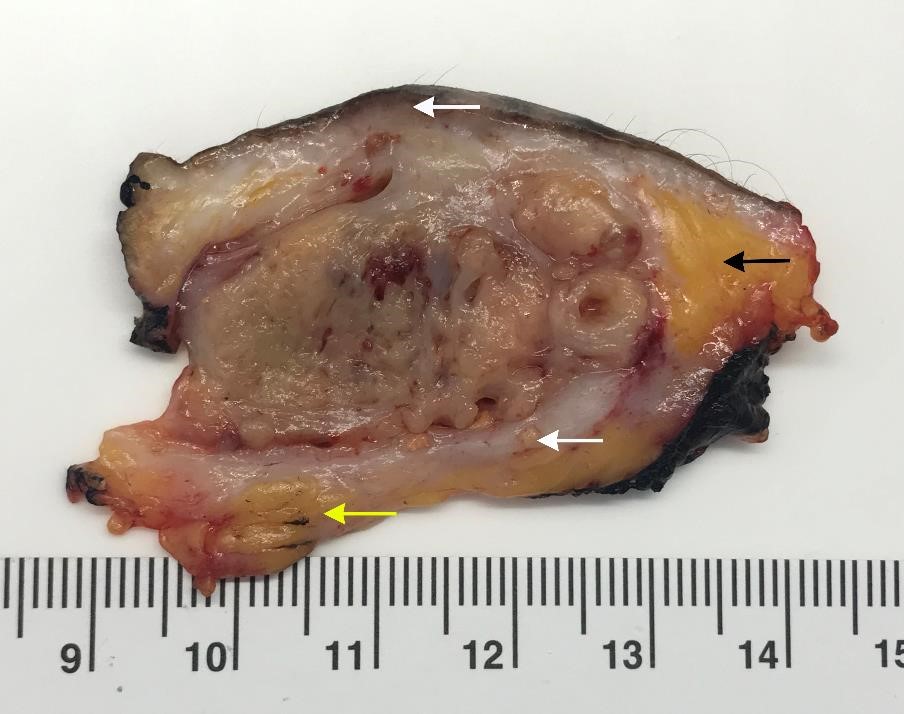
1.2 cm upper lip nodule
Images hosted on other servers:

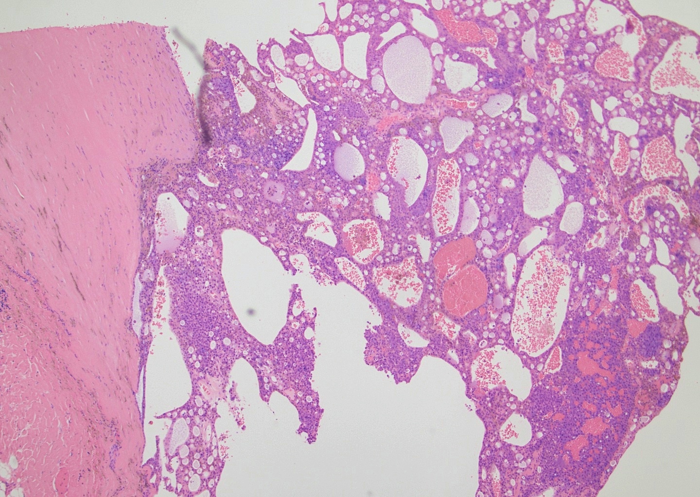
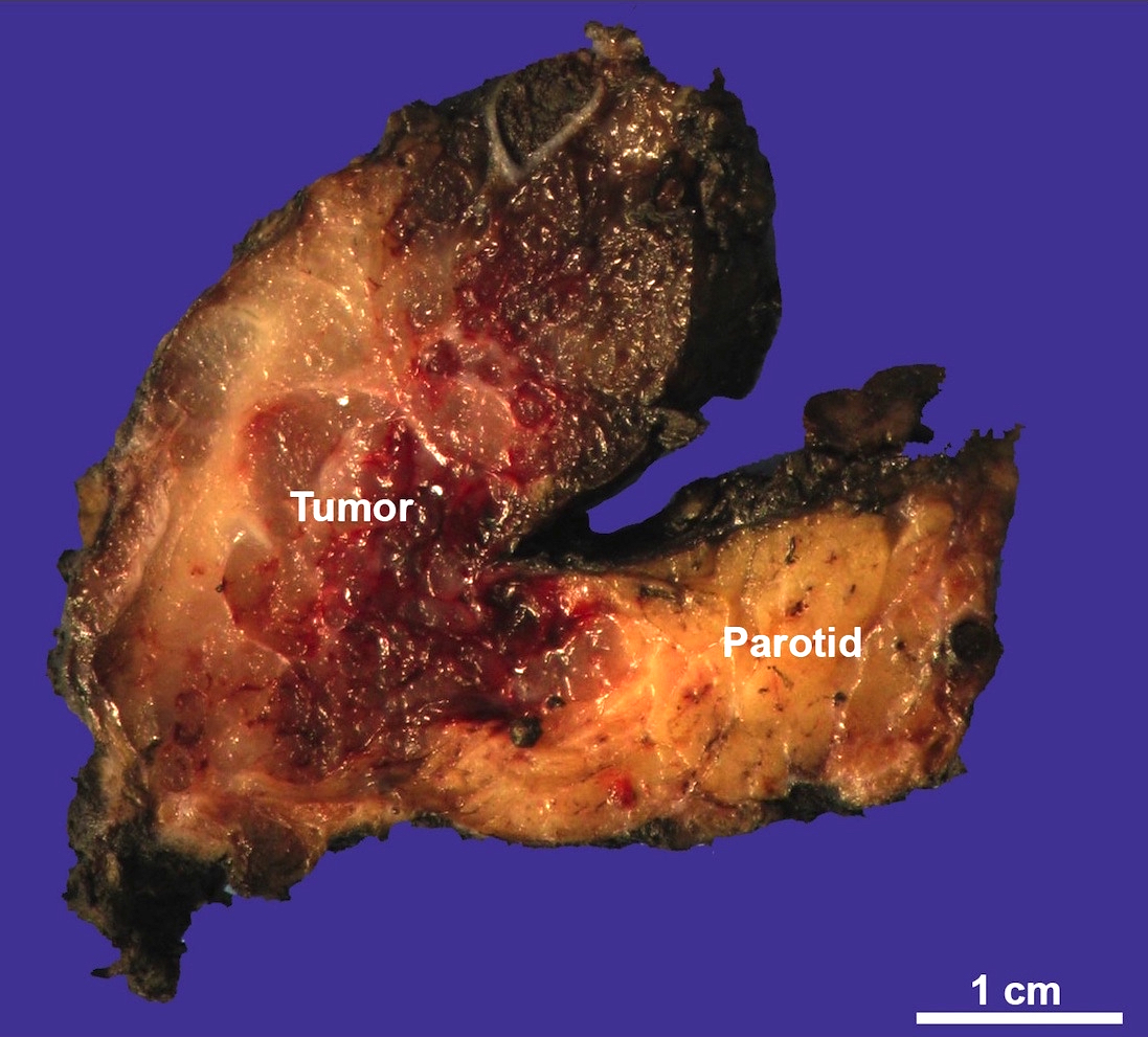
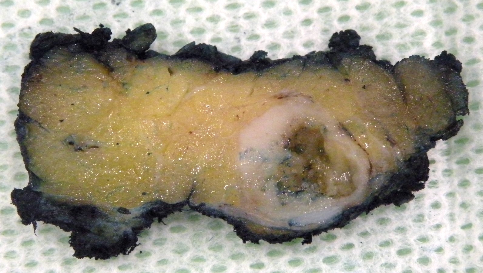
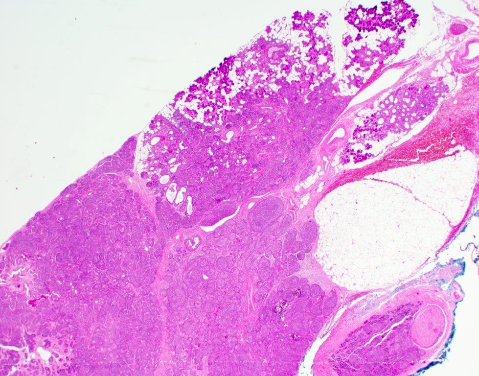
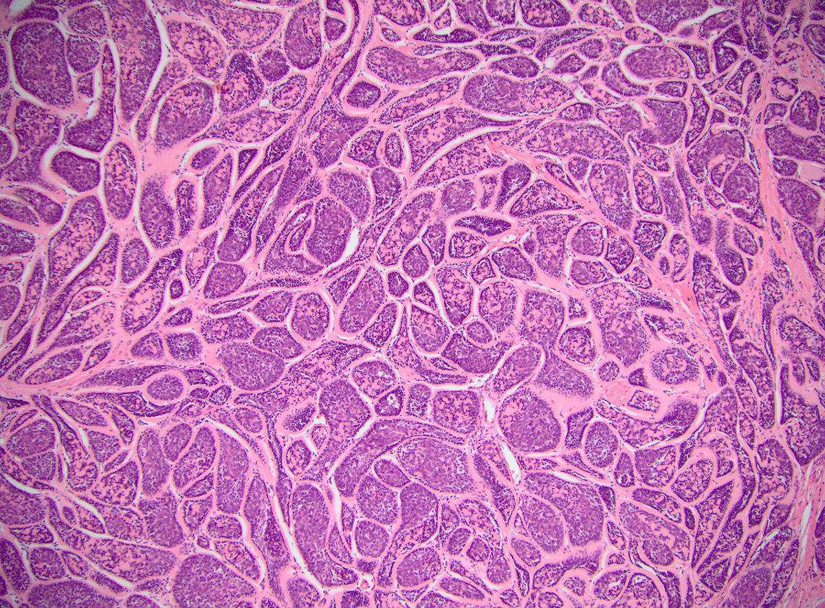
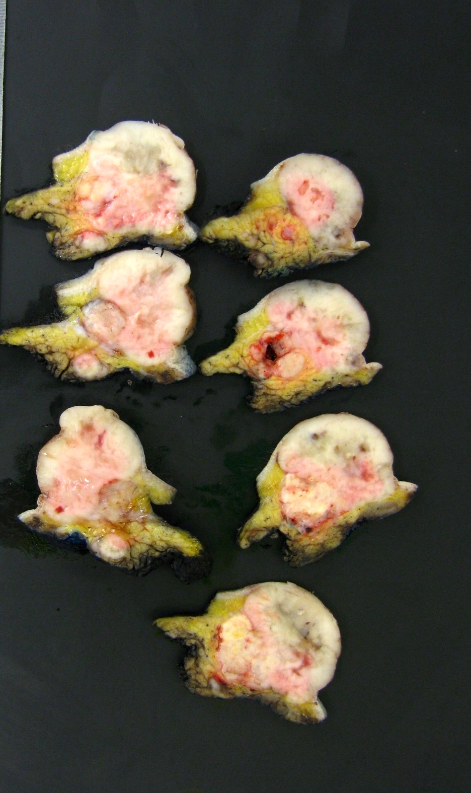
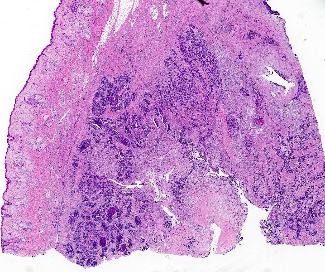
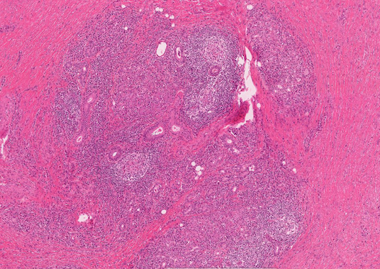
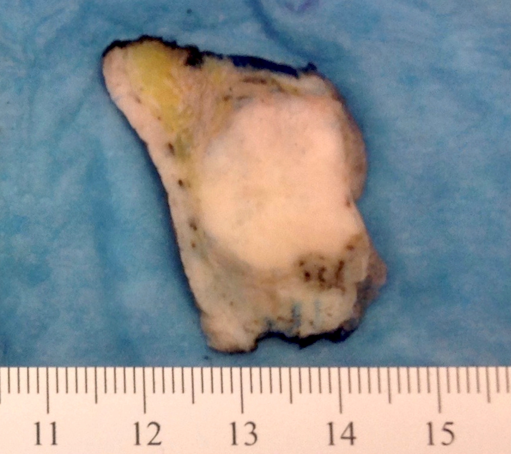
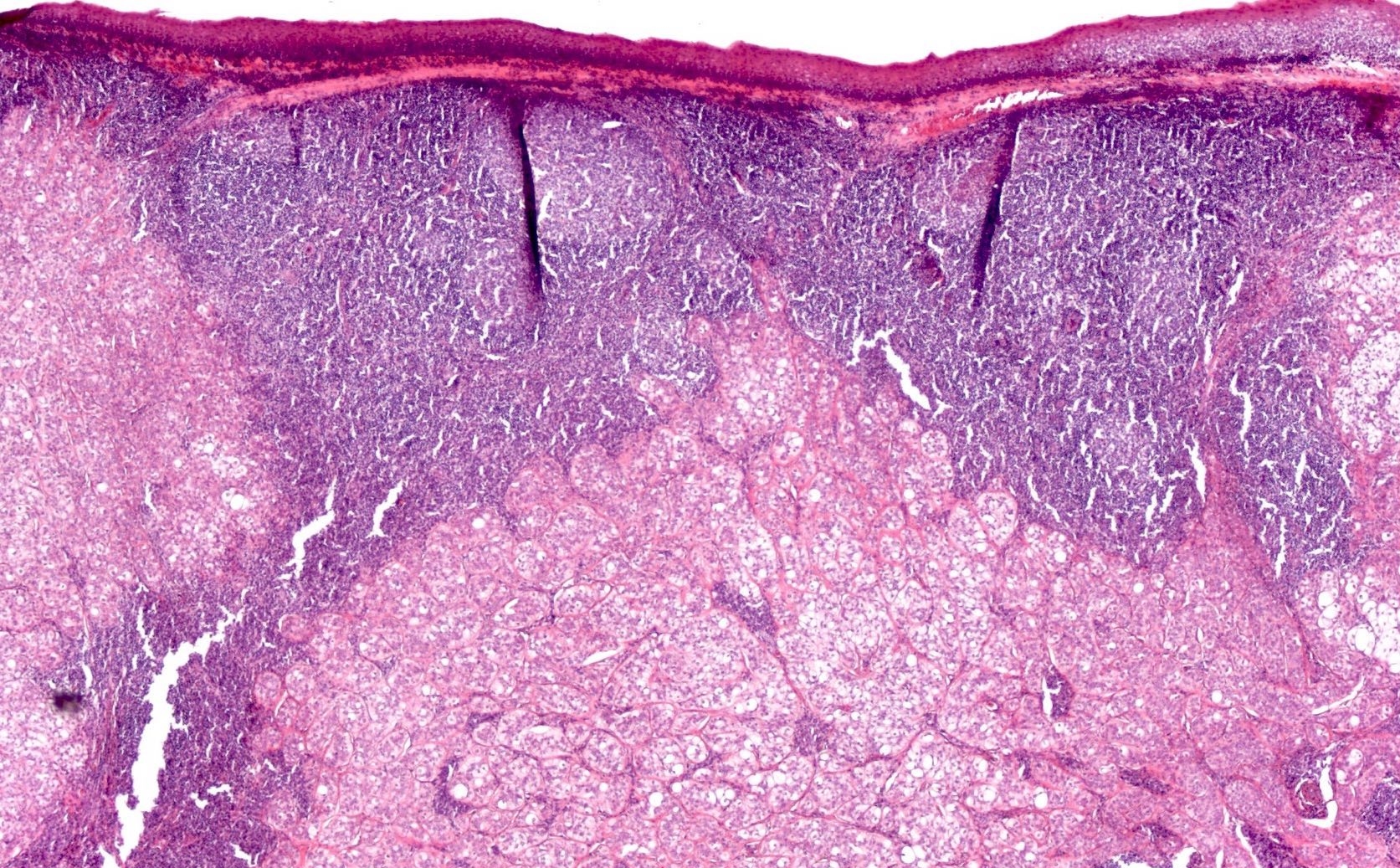
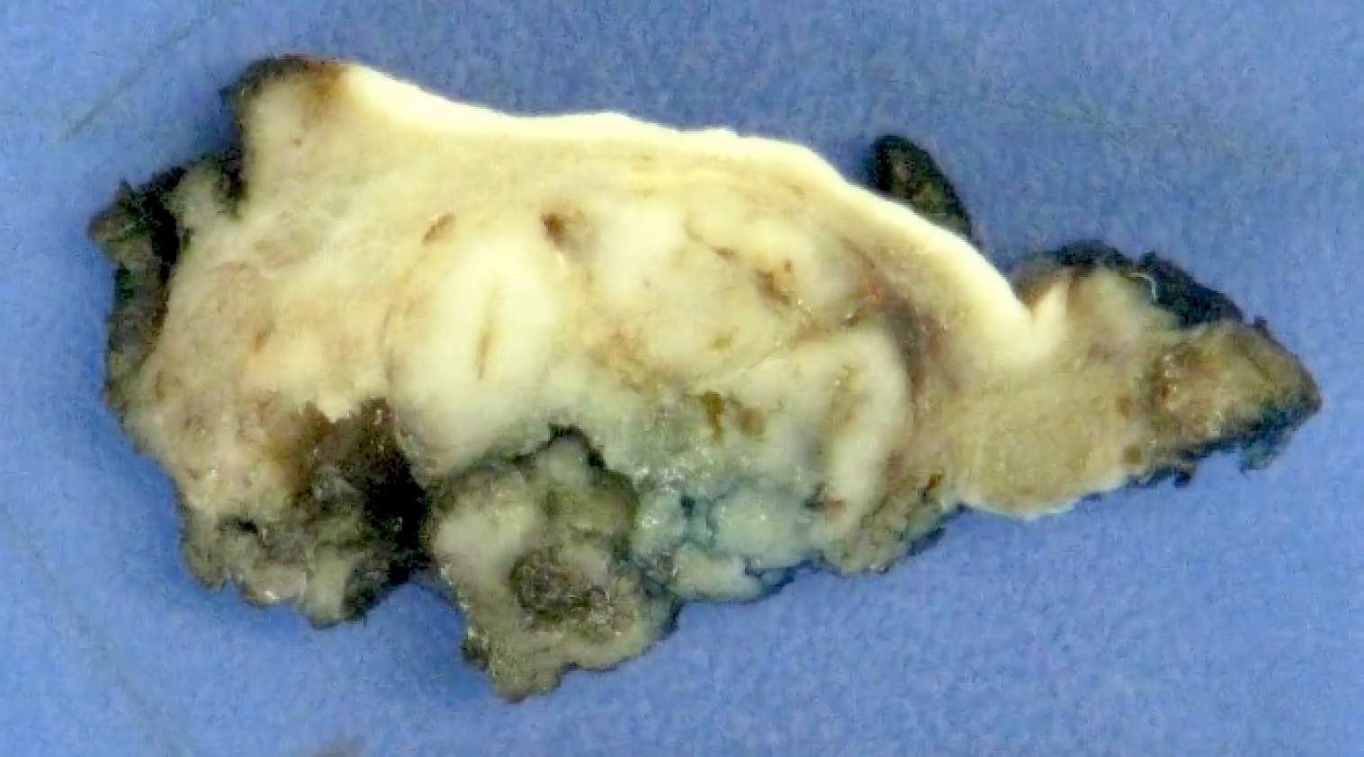
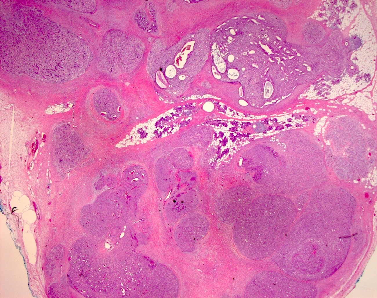
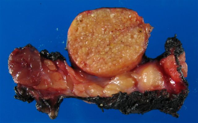
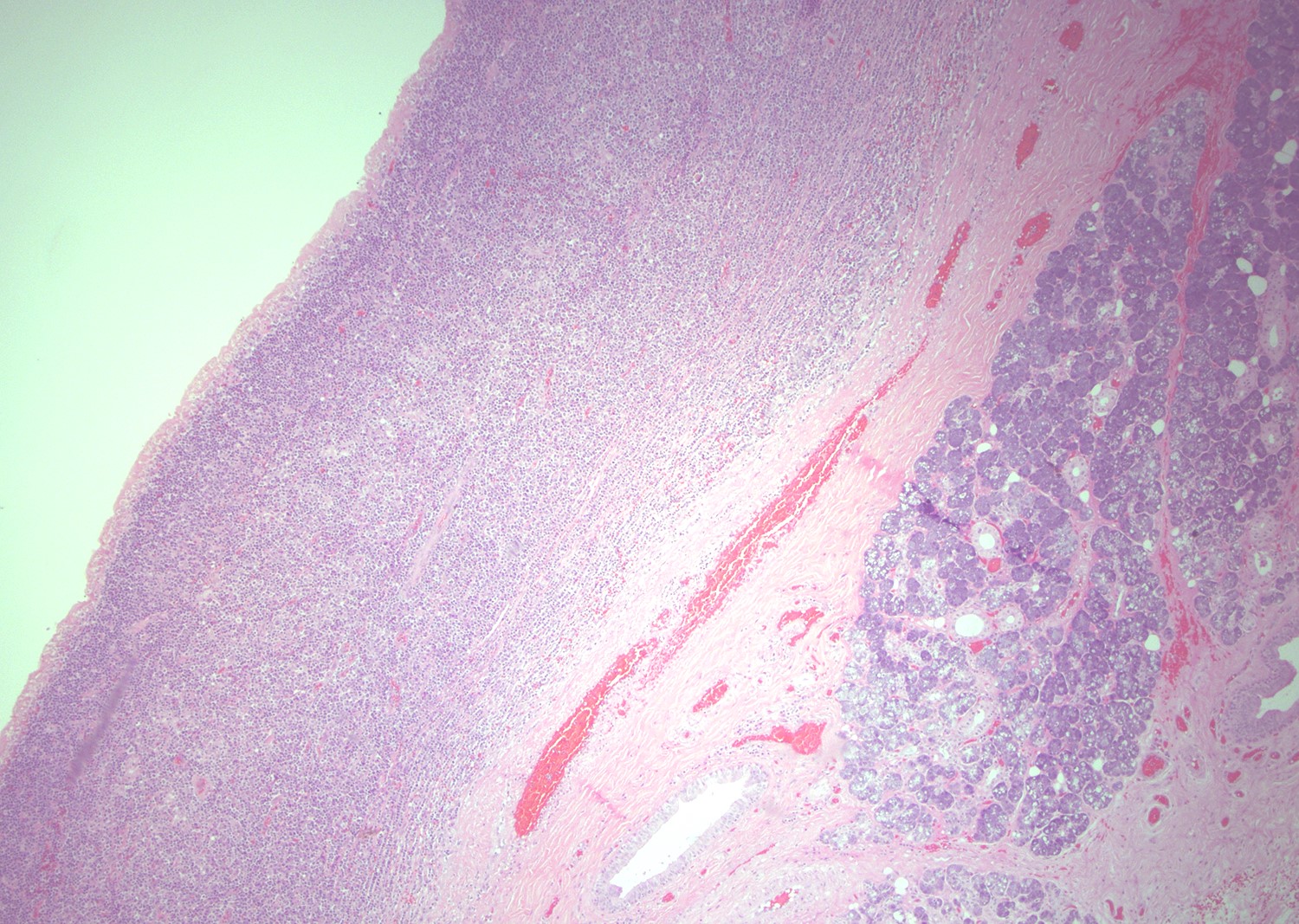
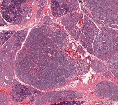
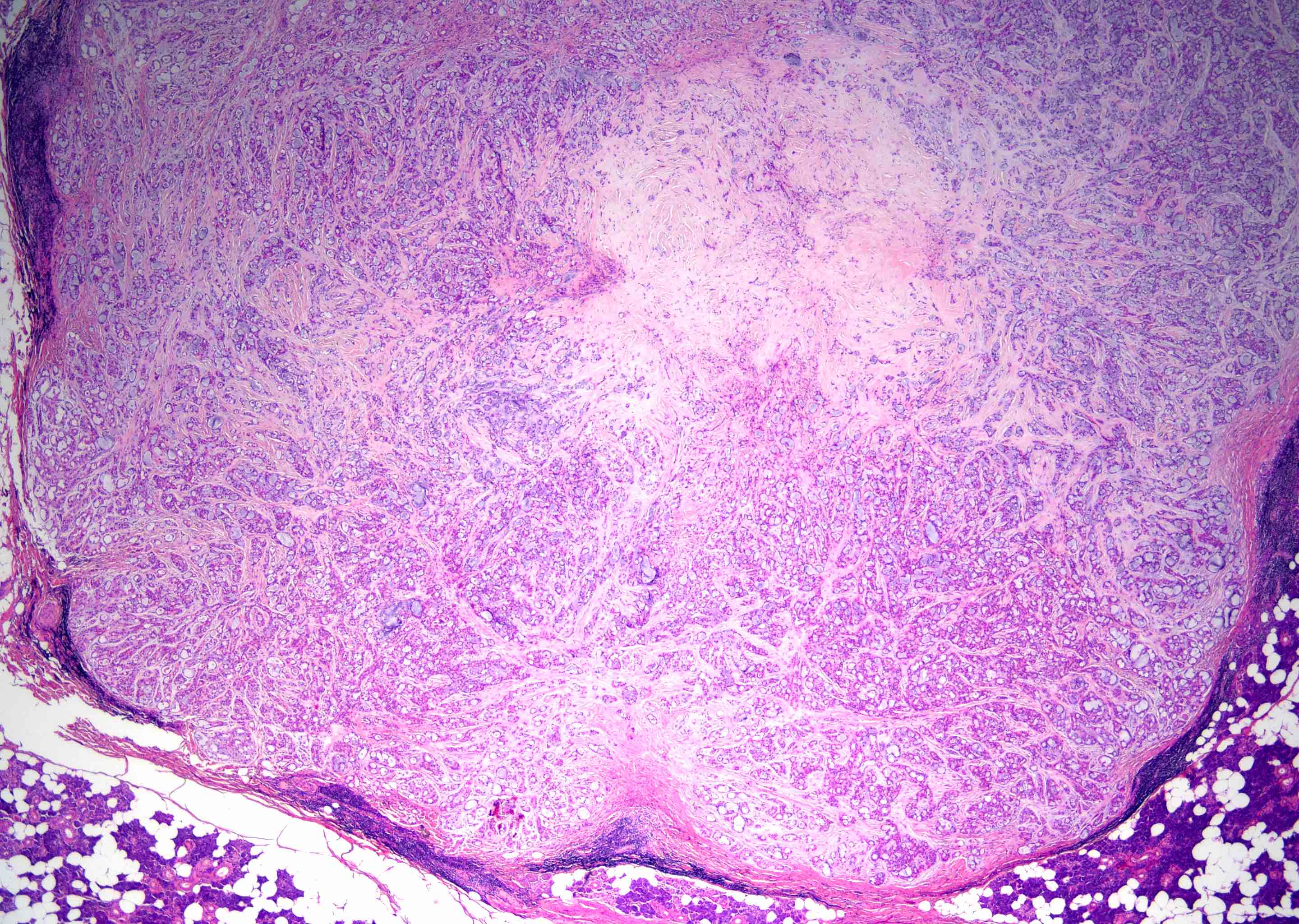
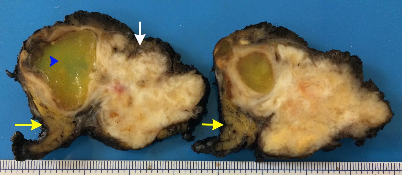
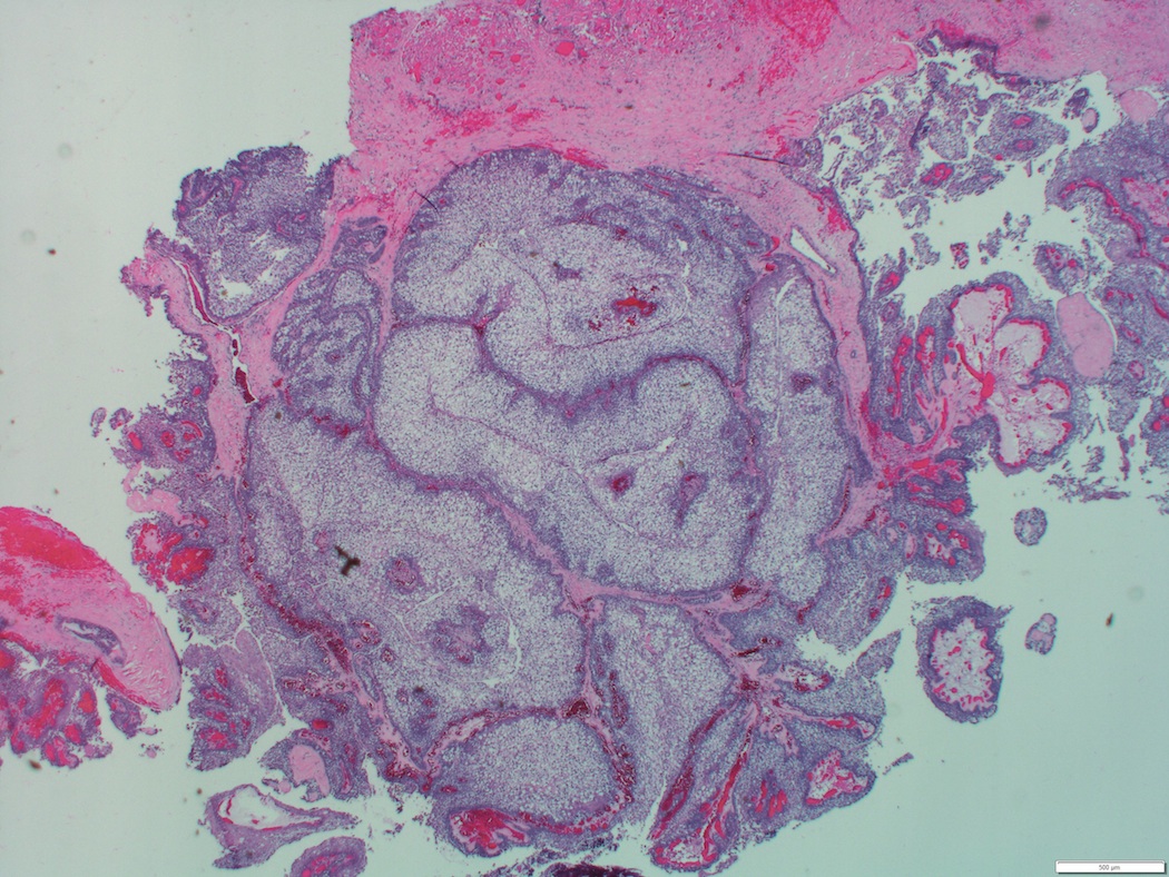
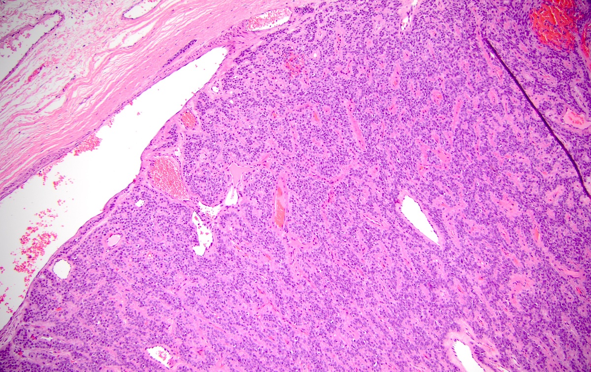
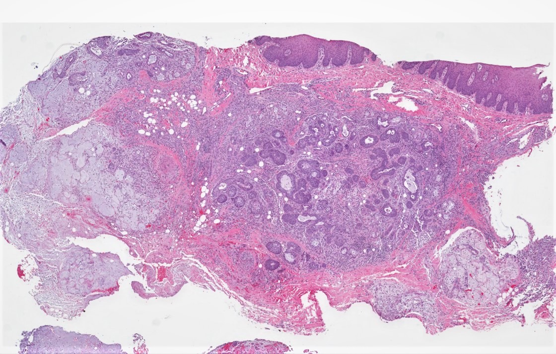
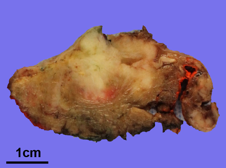
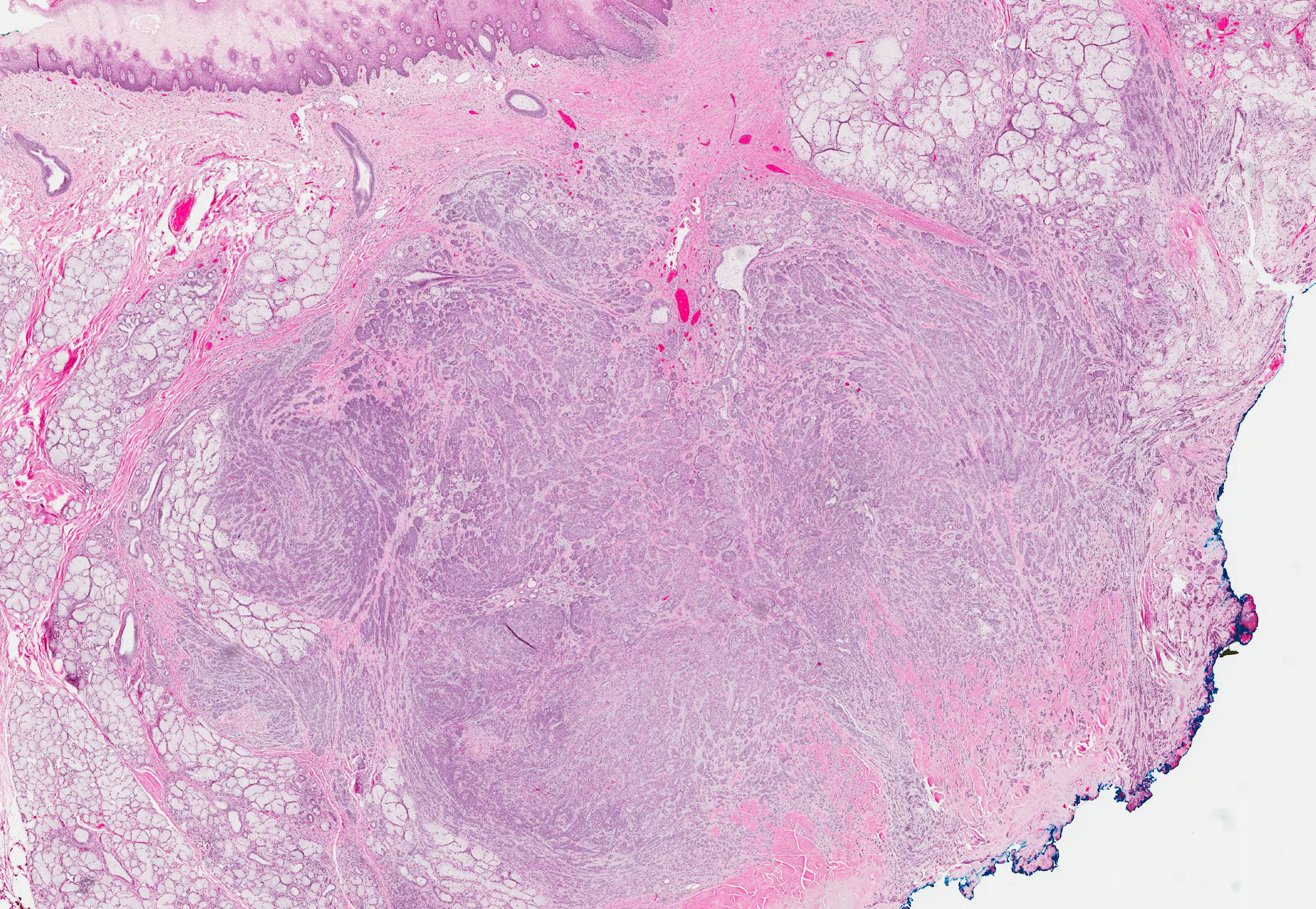
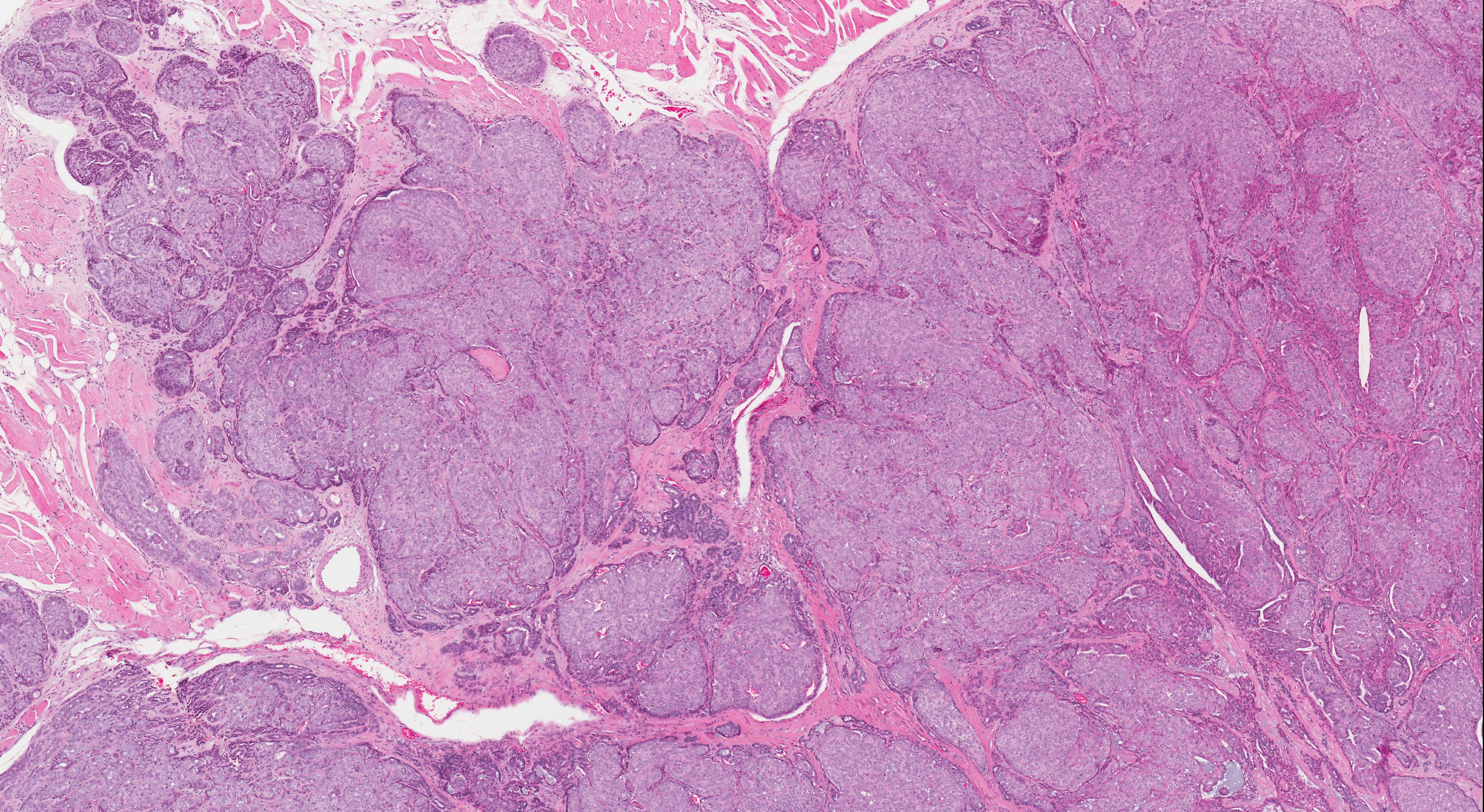
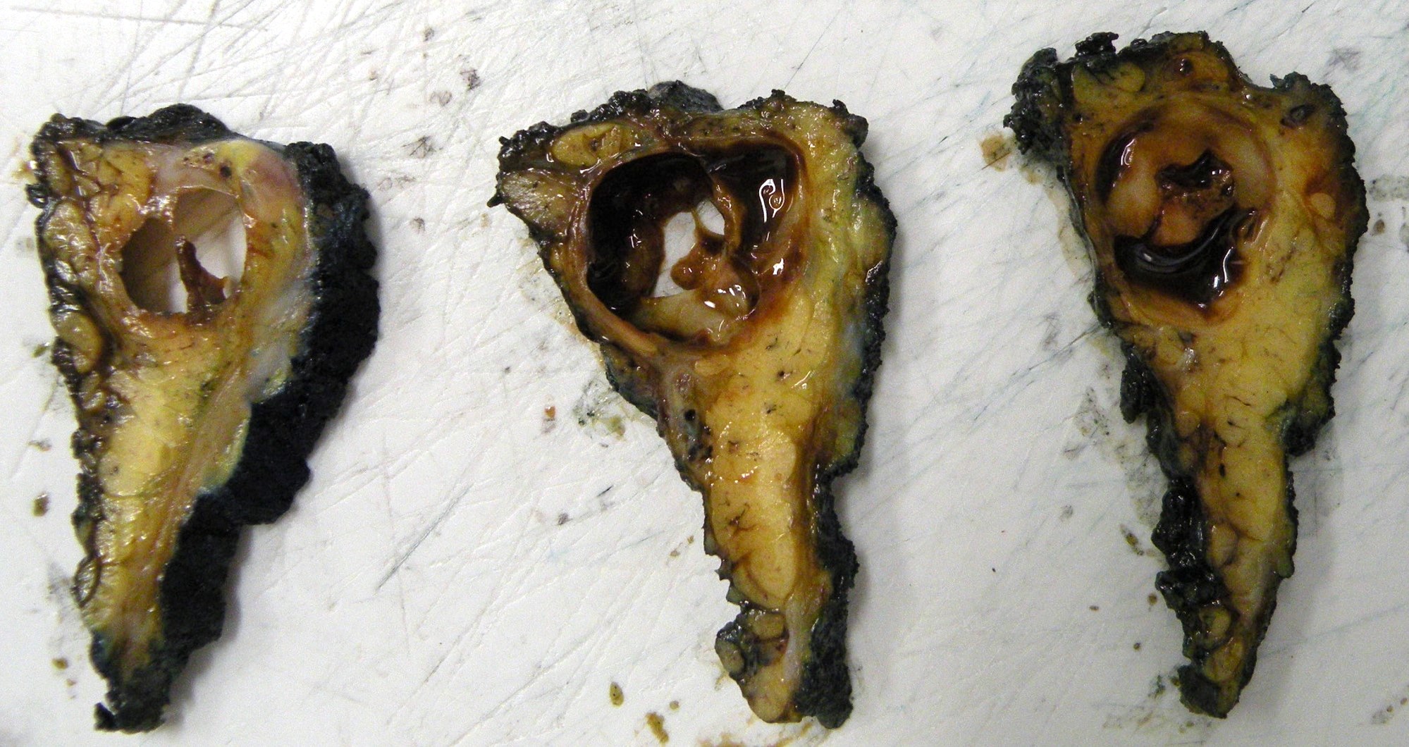
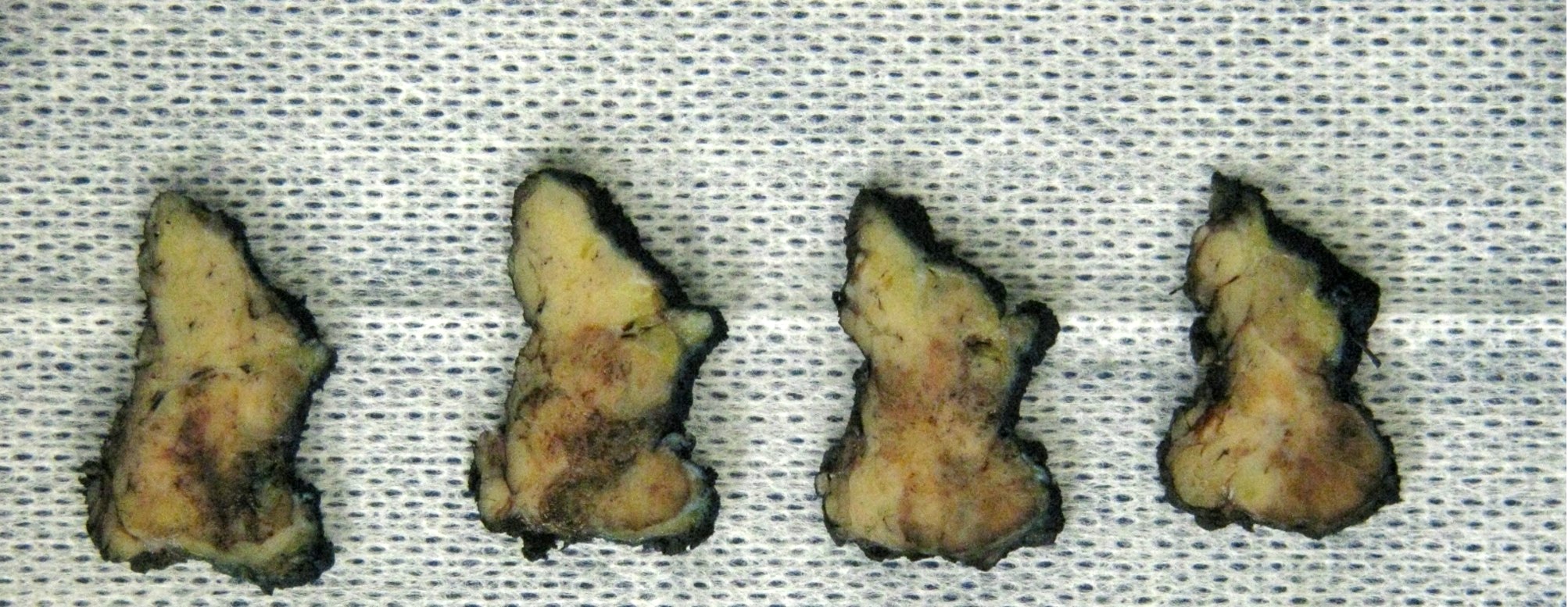
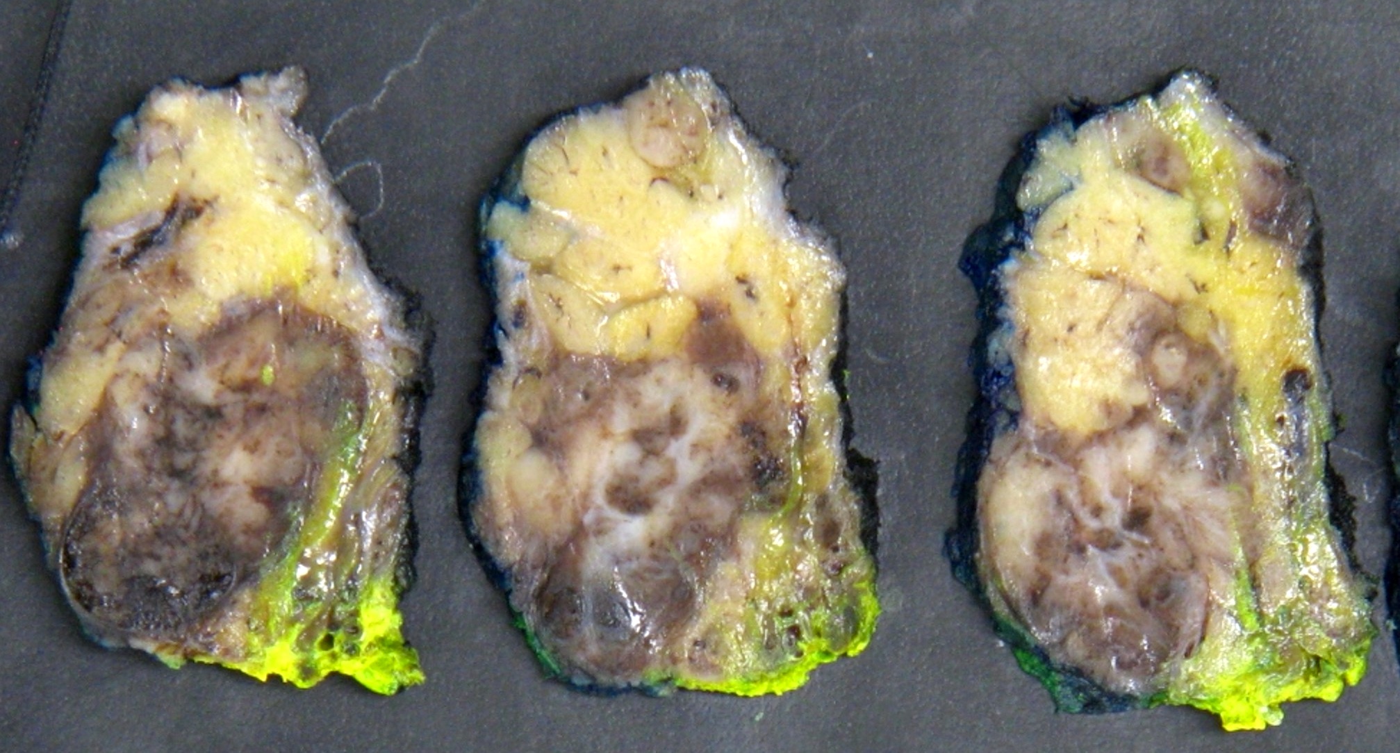
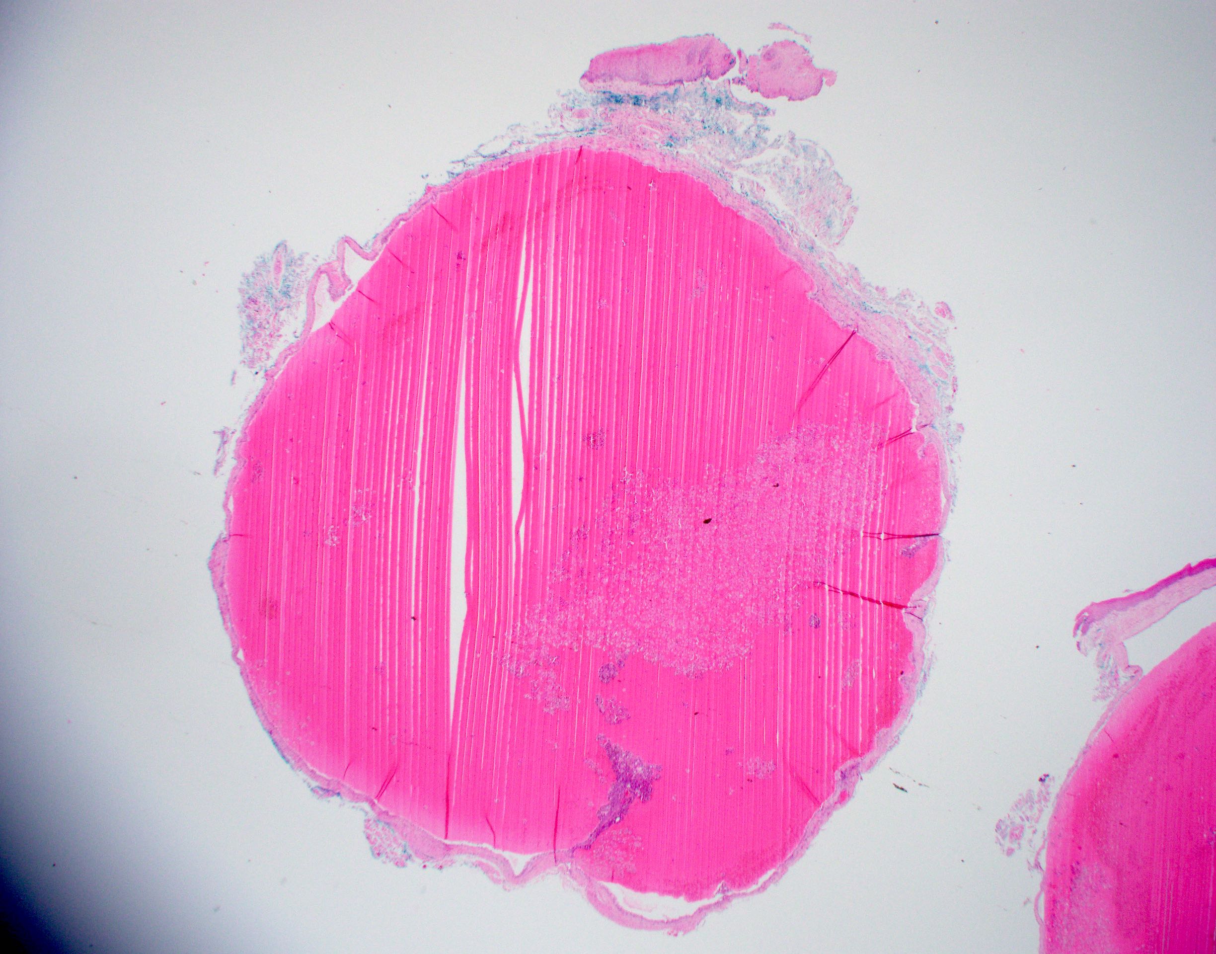
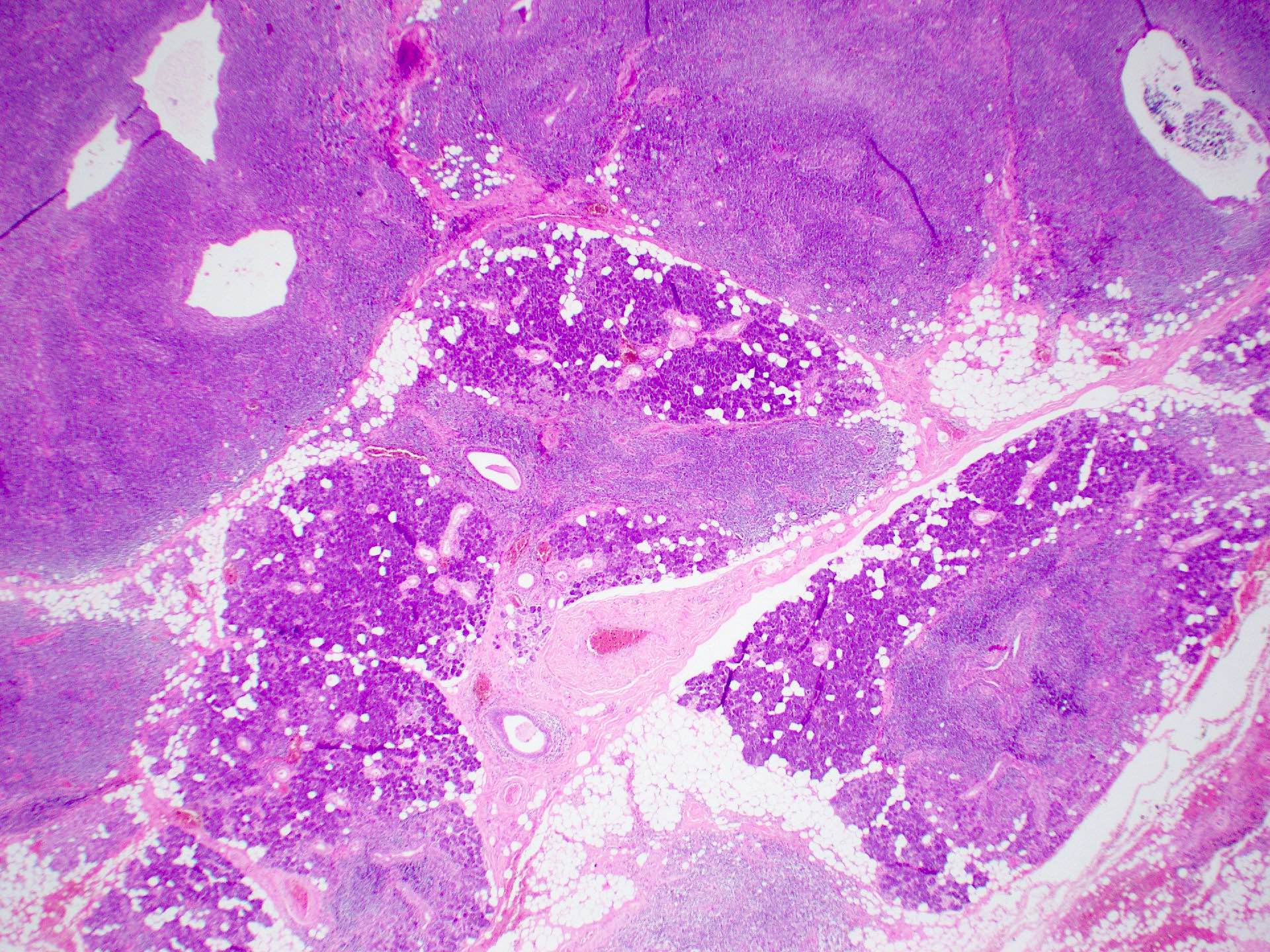
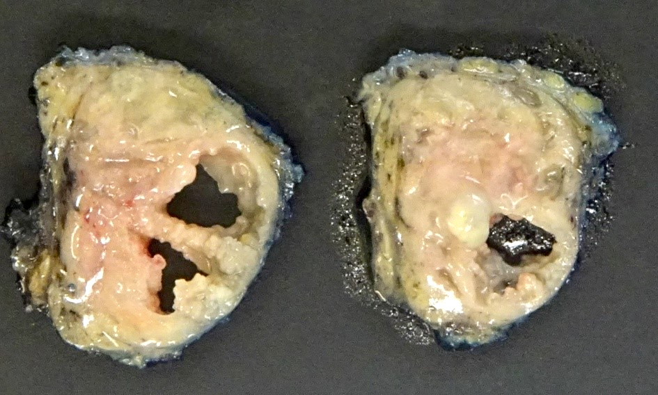
Well circumscribed multilobulated mass
Contributed by Rema A. Rao, M.D. and Arash H. Lahouti, M.D.
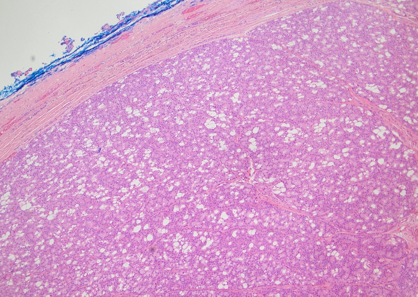
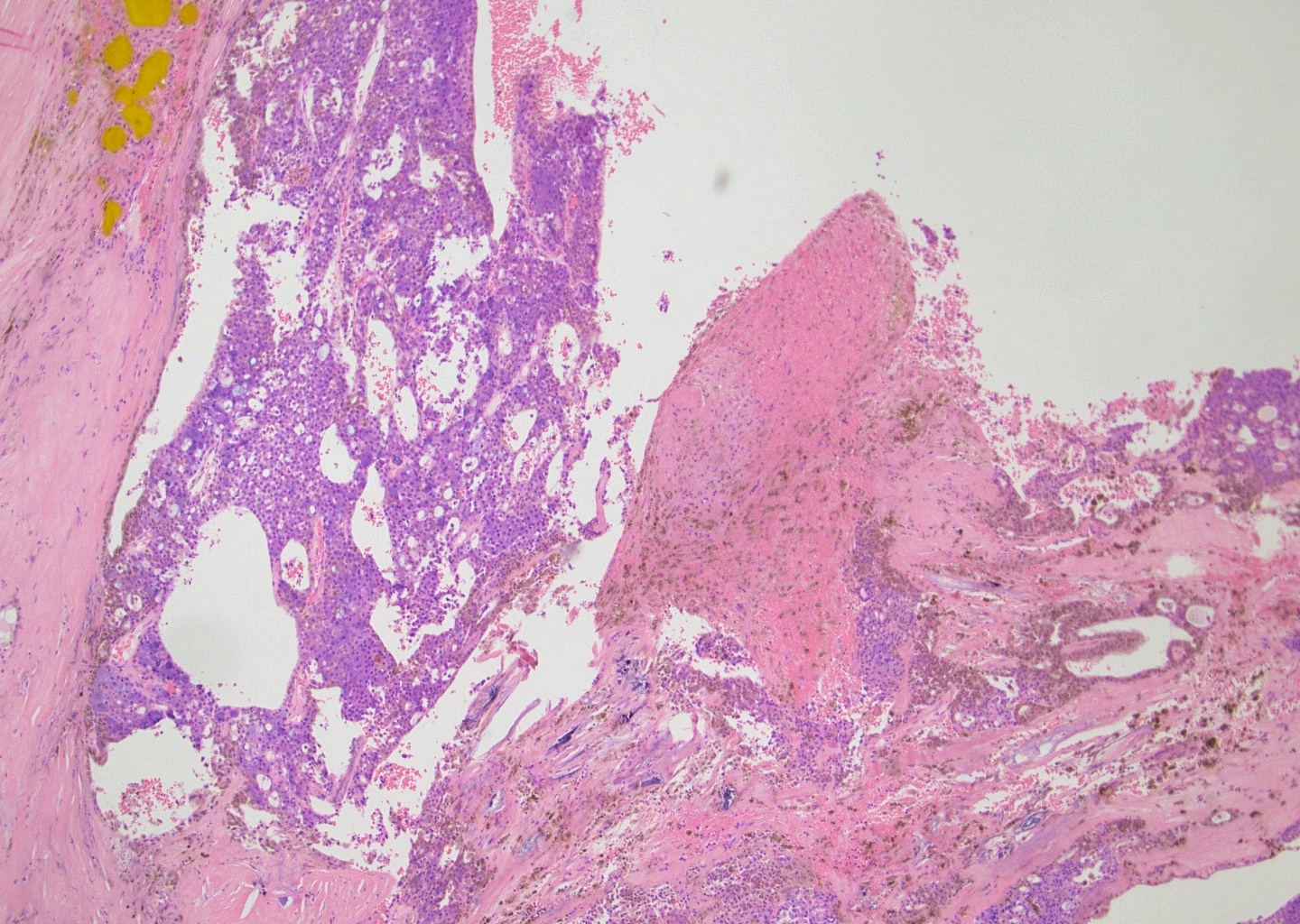
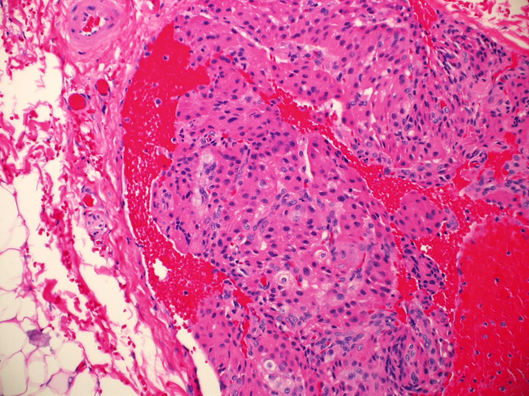
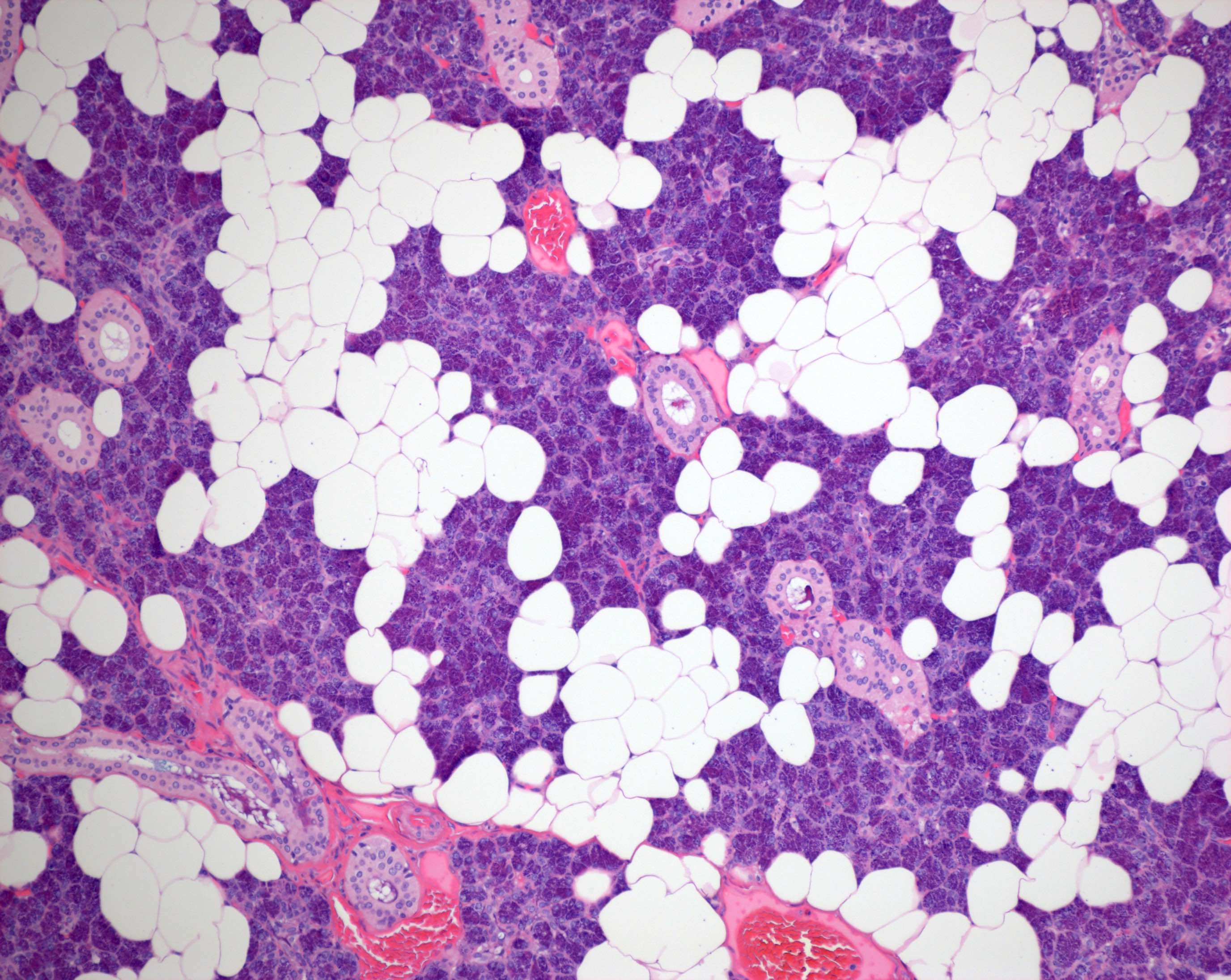
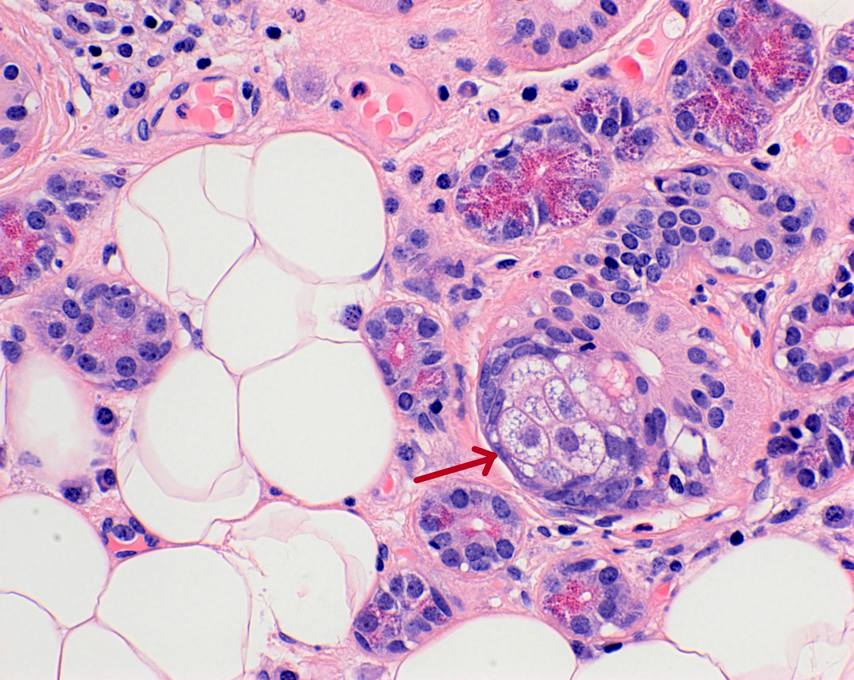
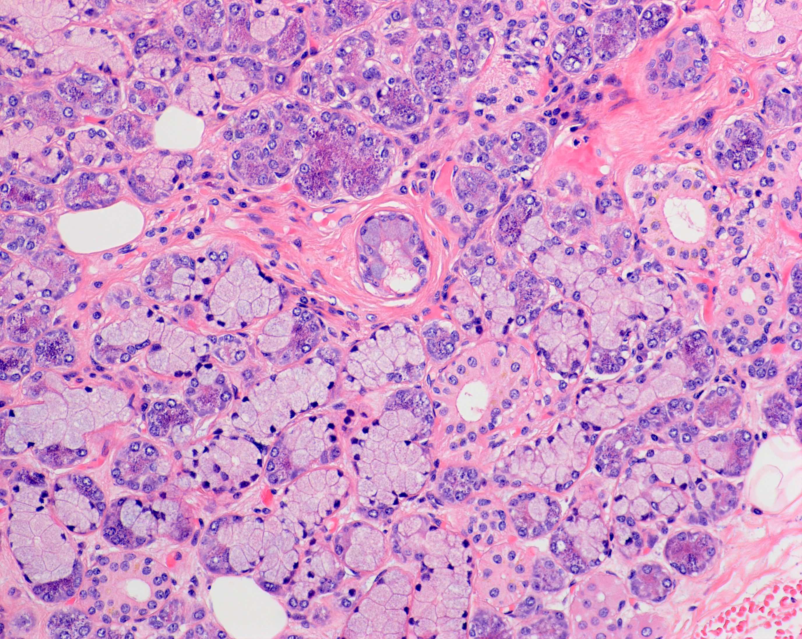
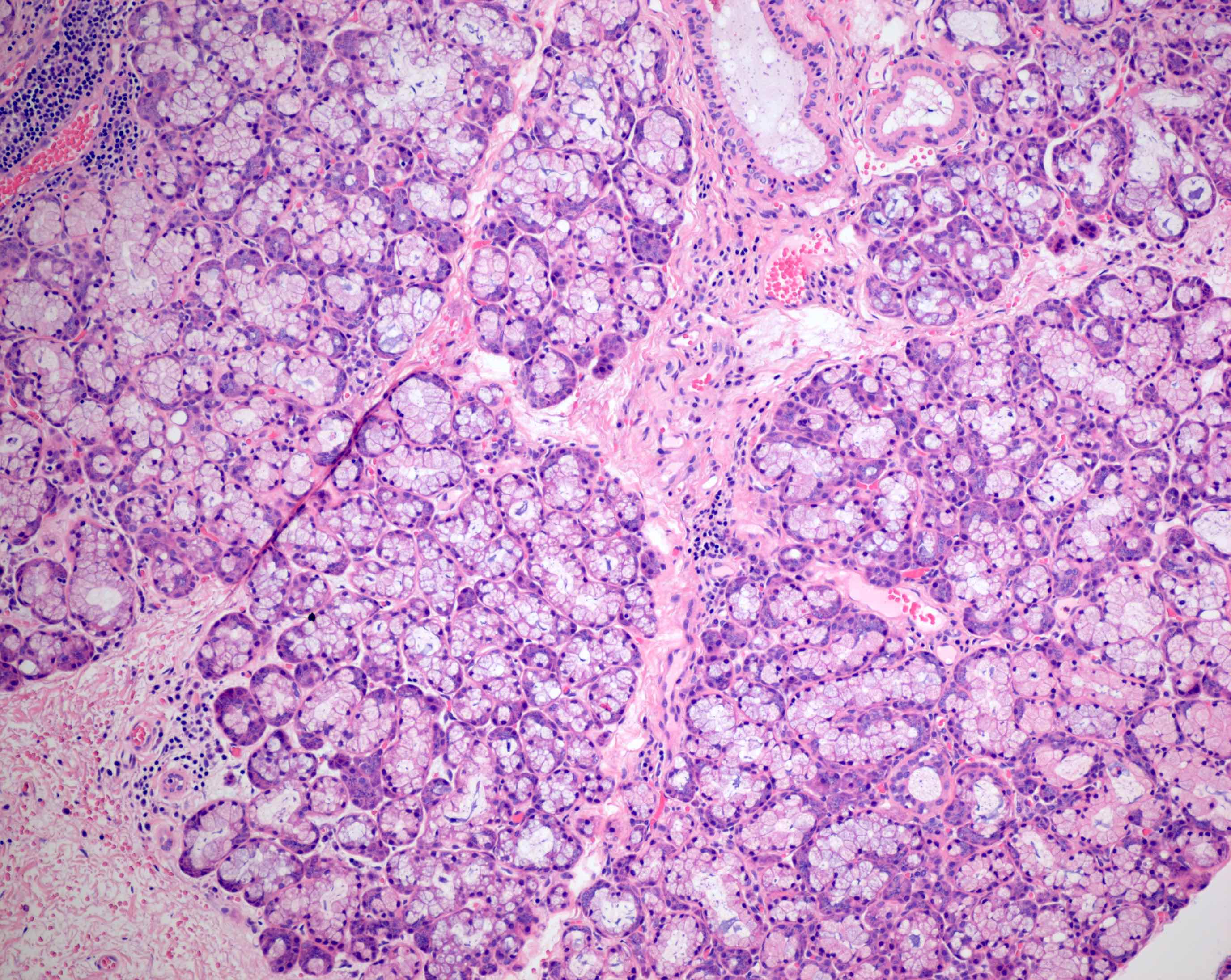
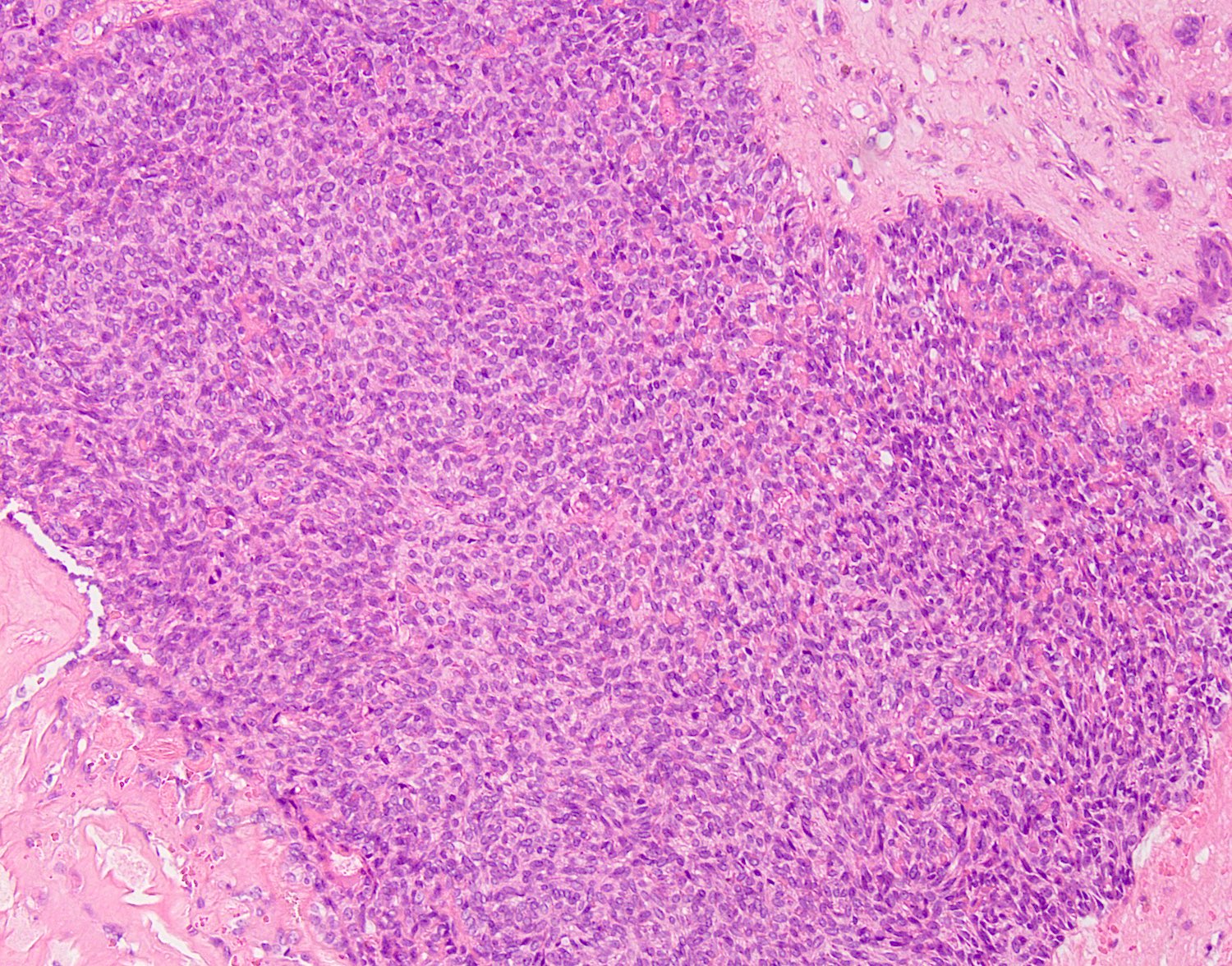
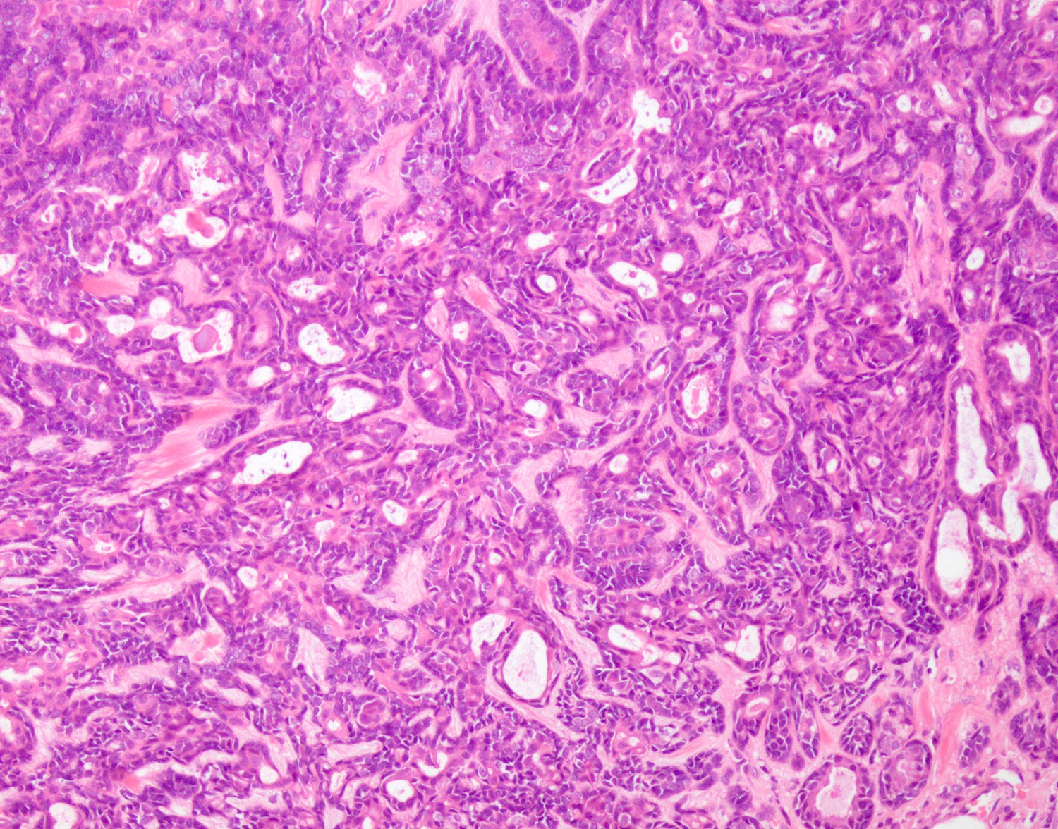
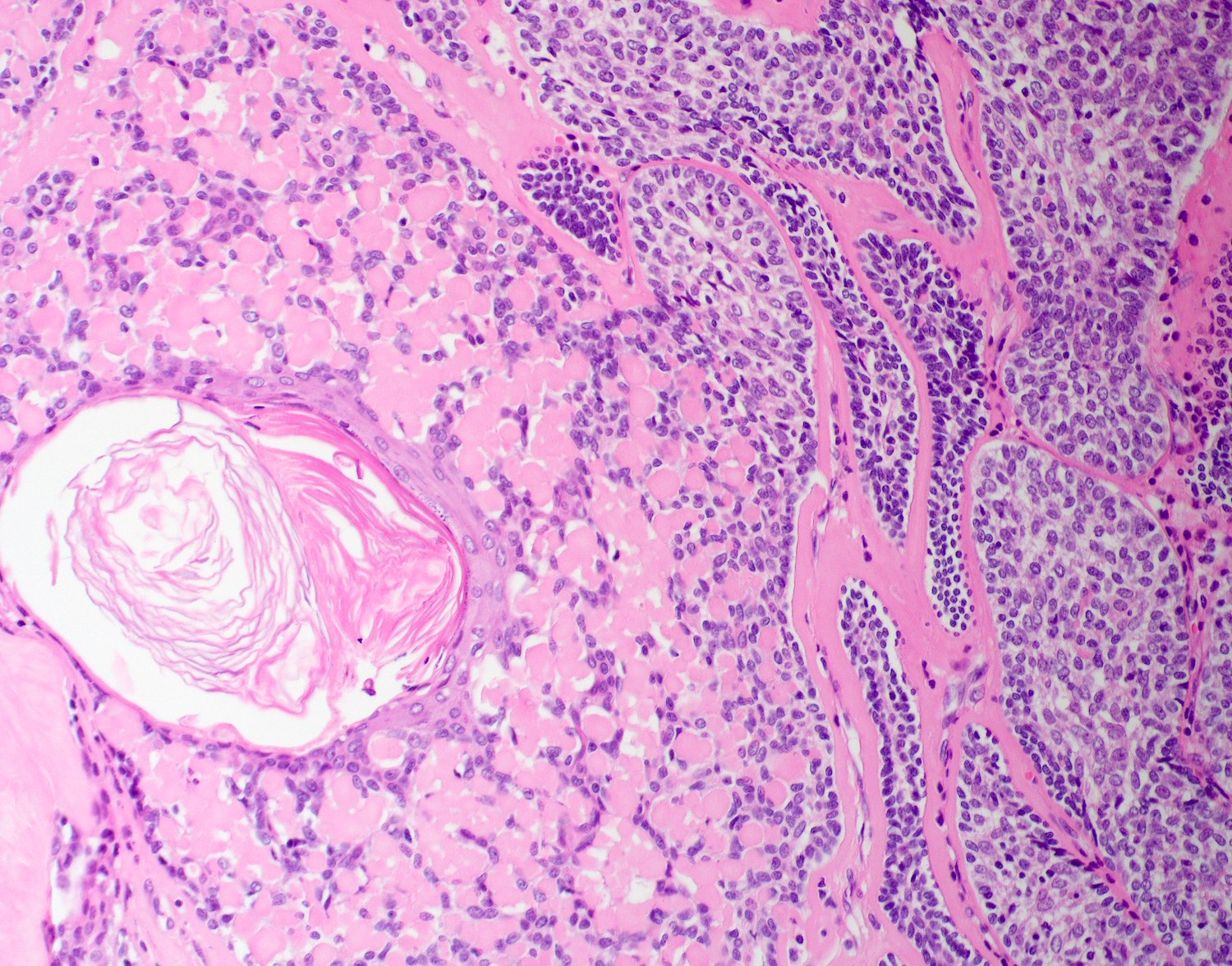
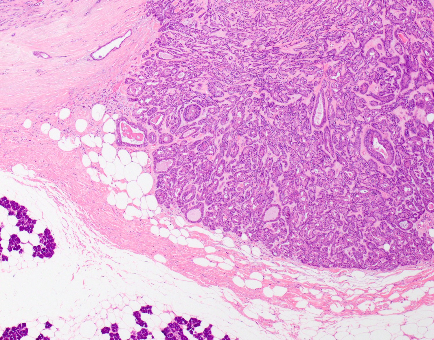
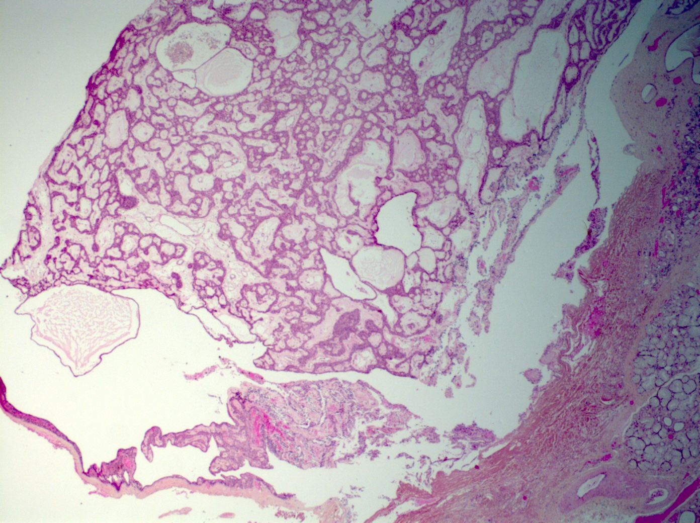
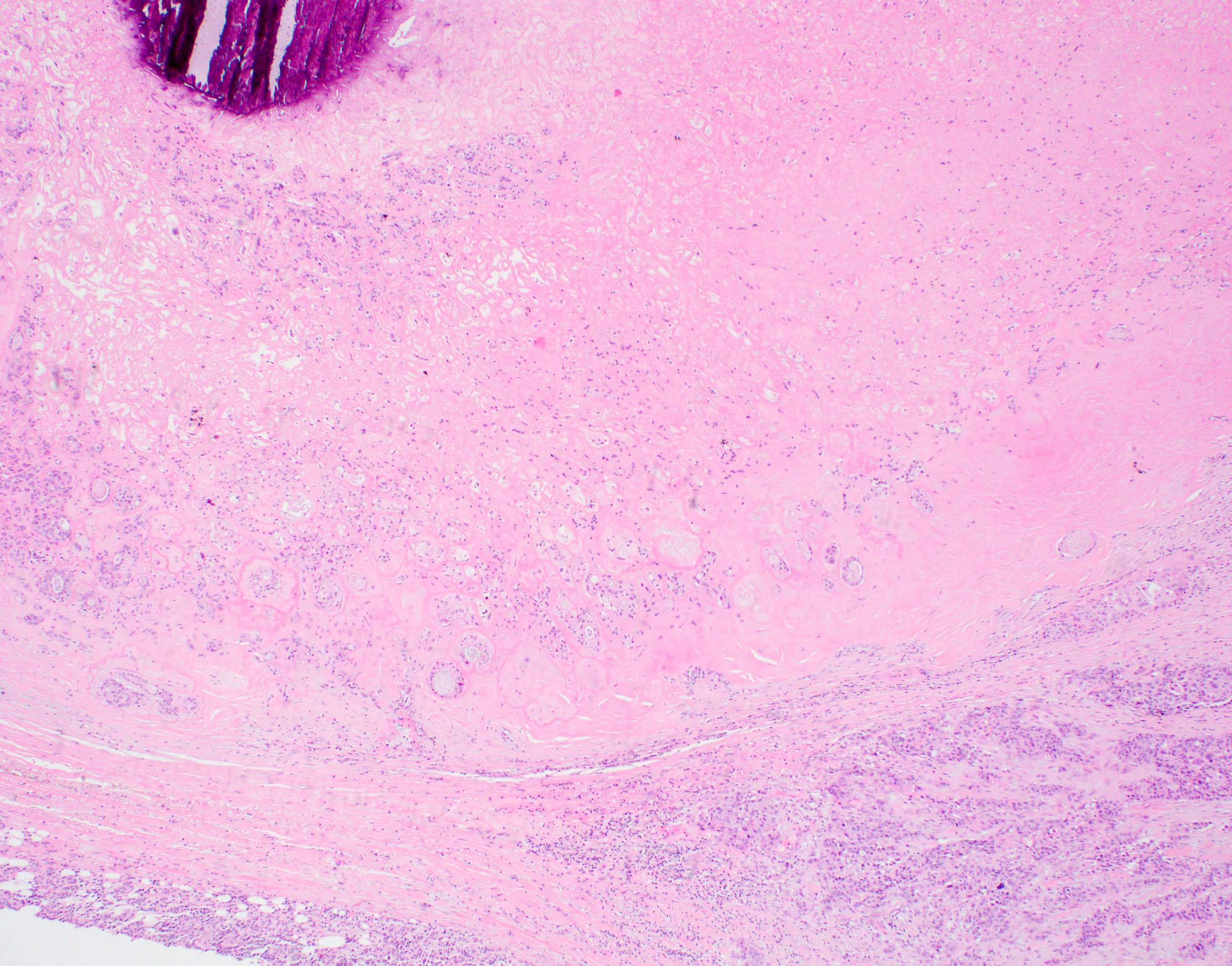
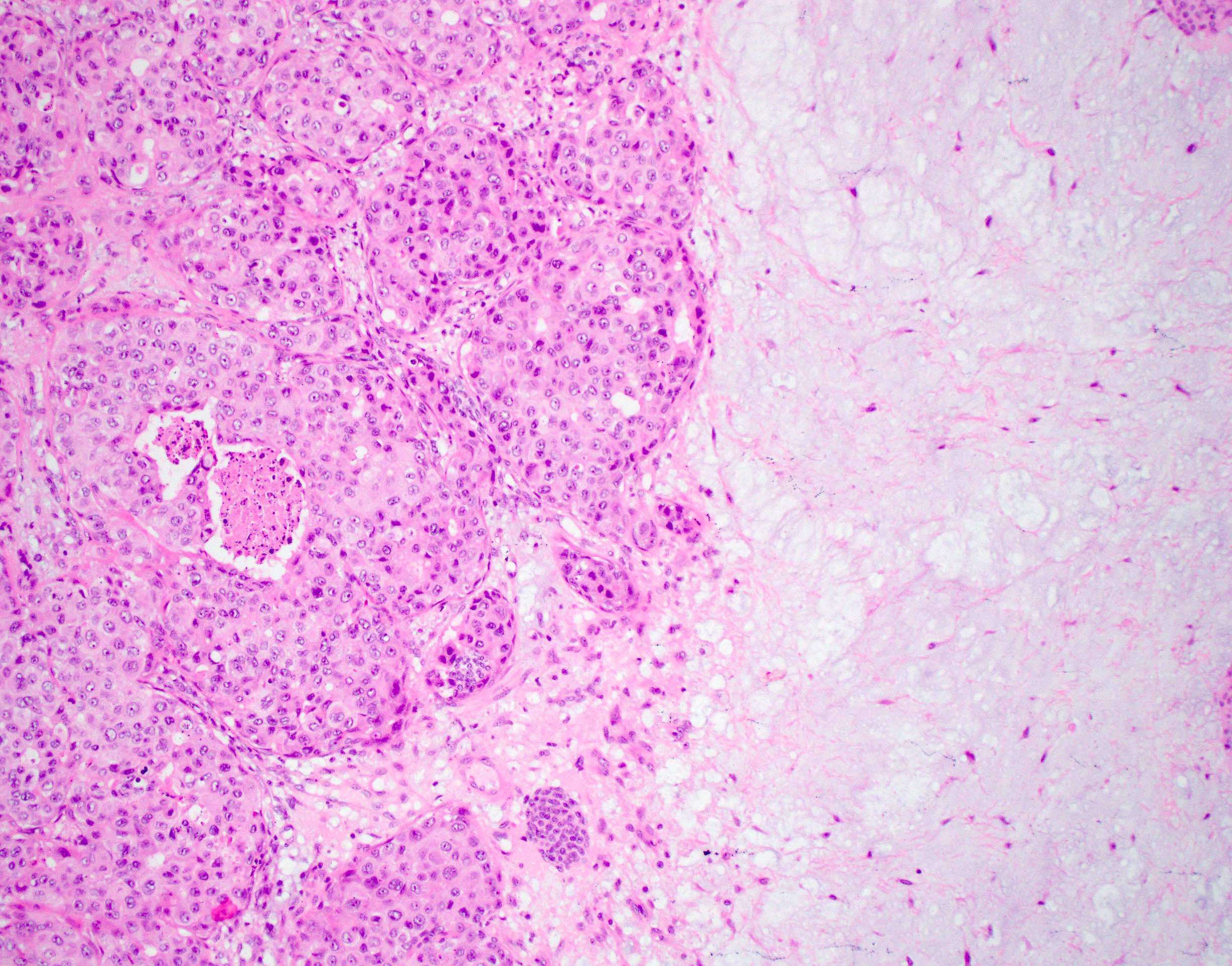
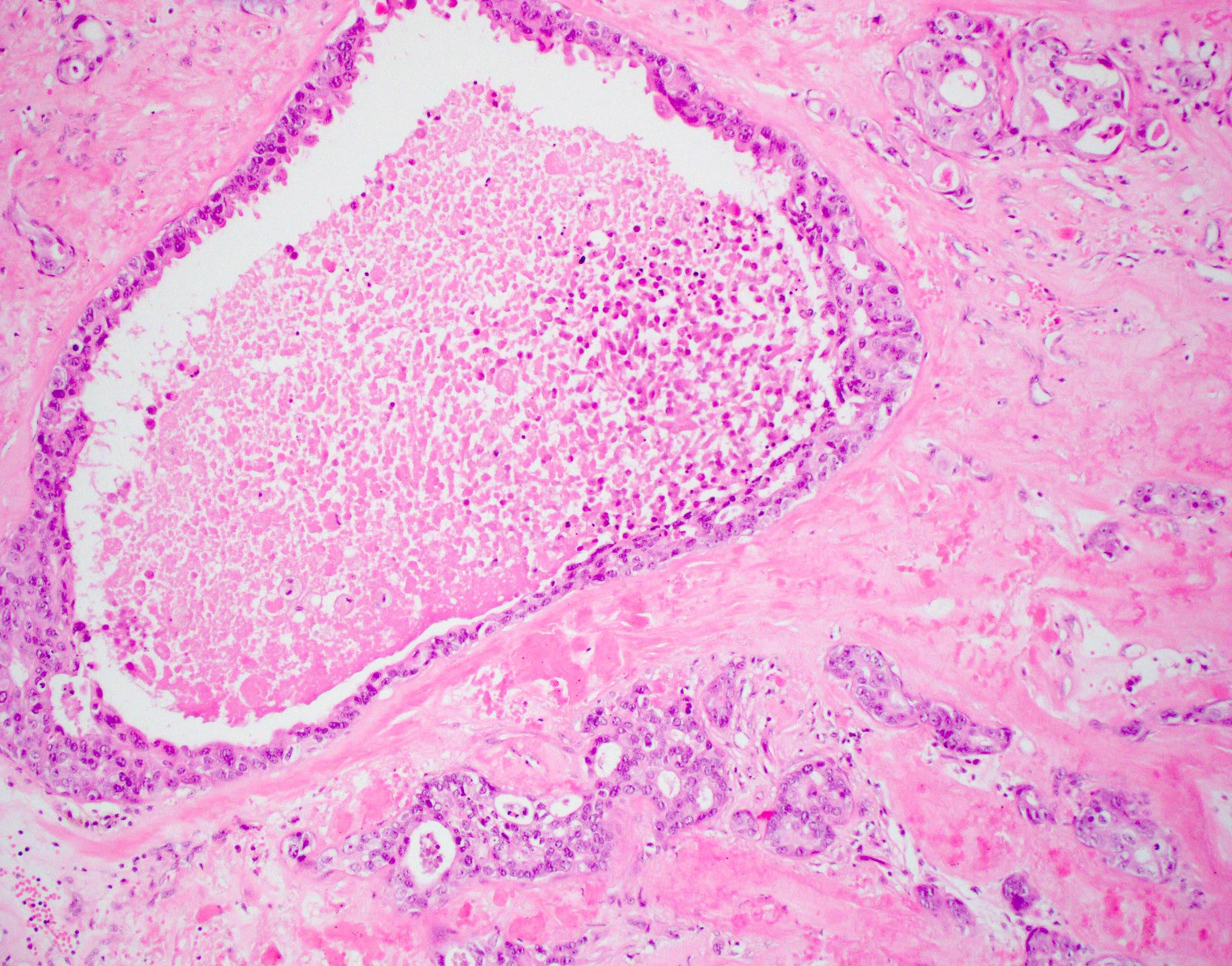
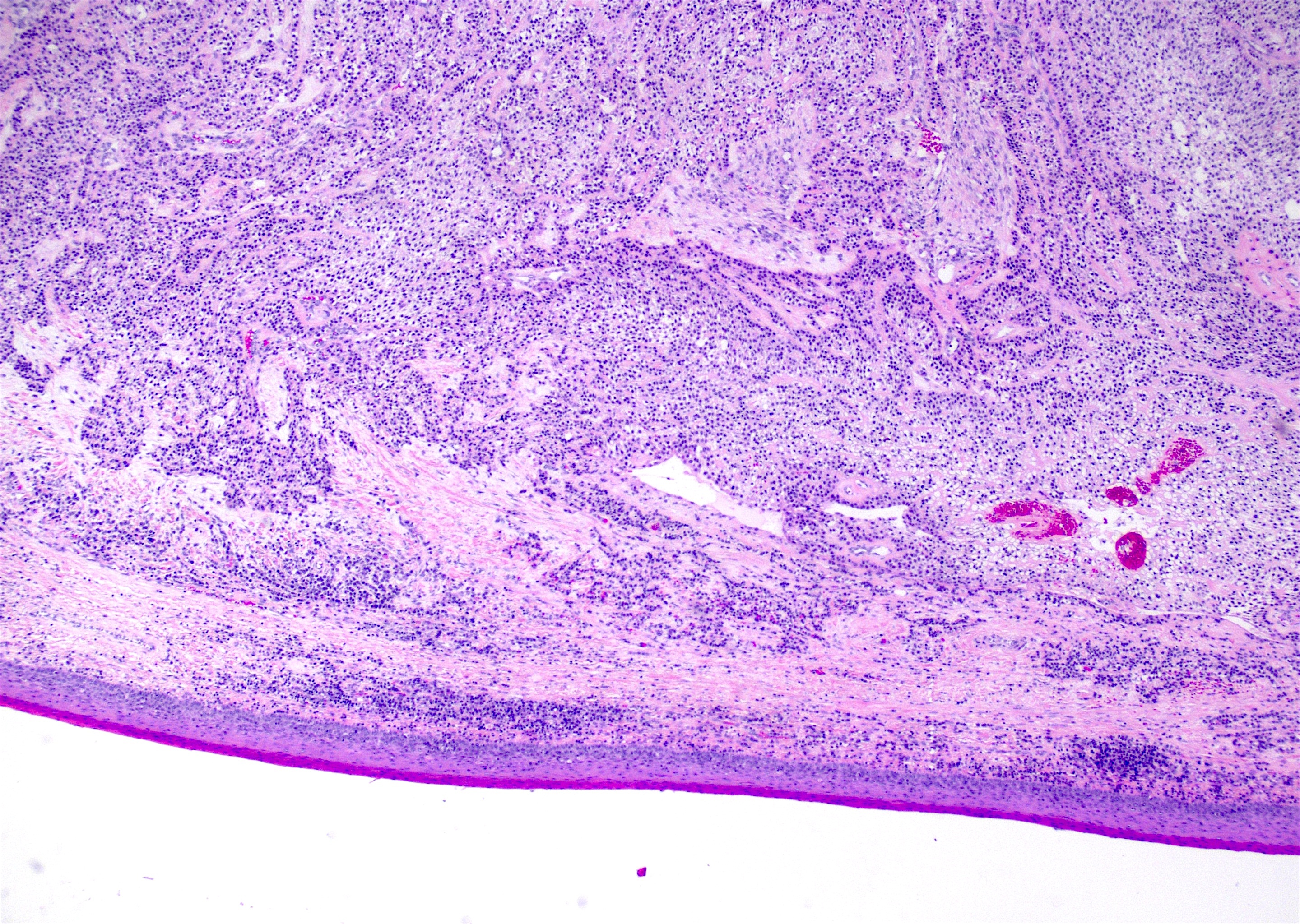
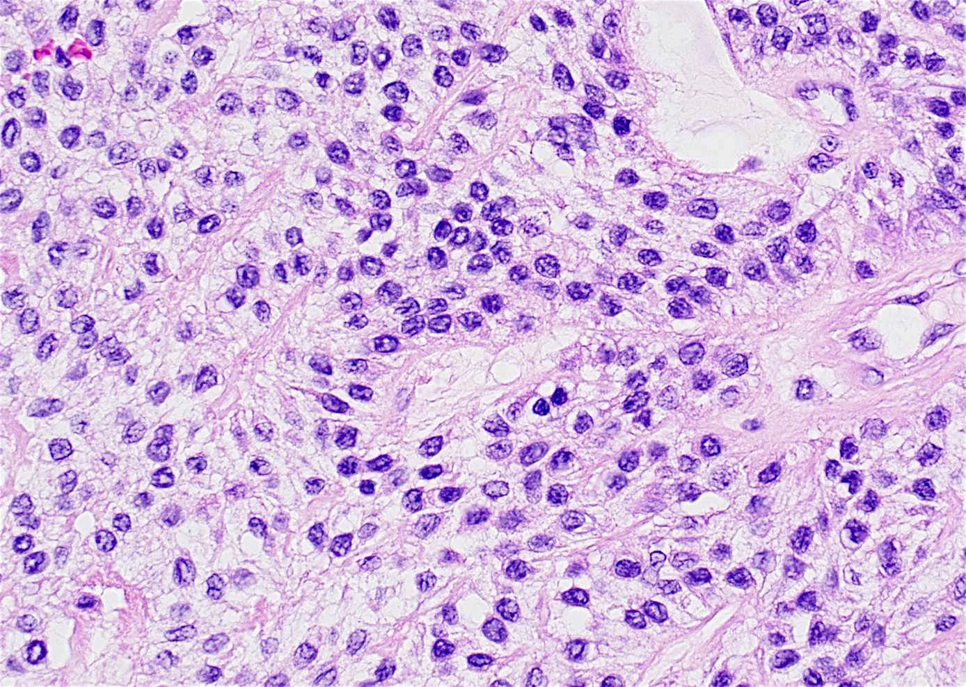
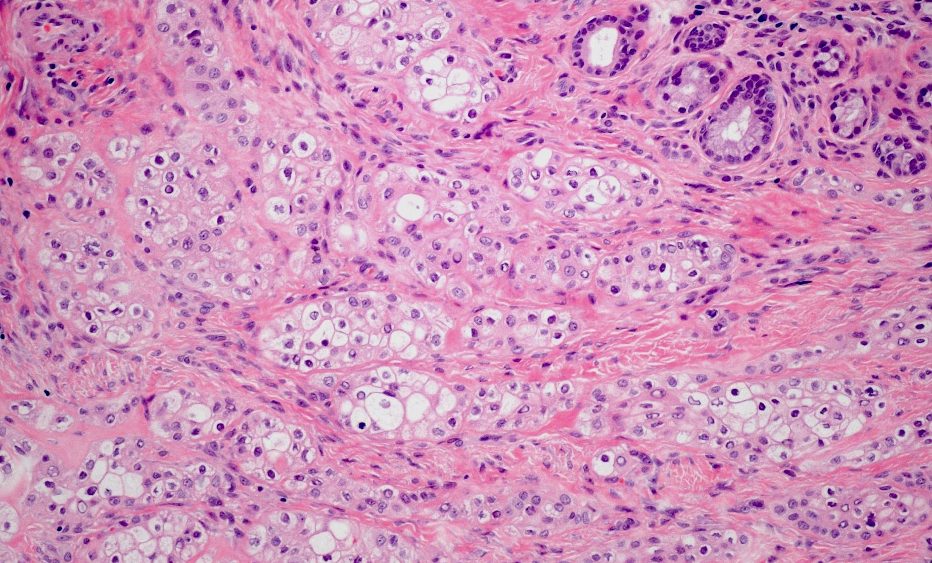
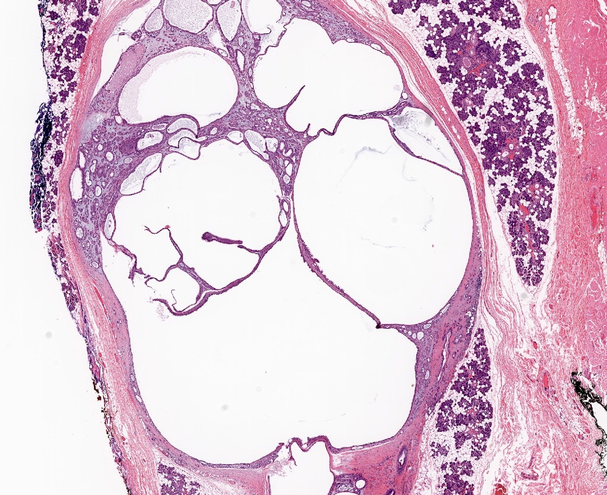
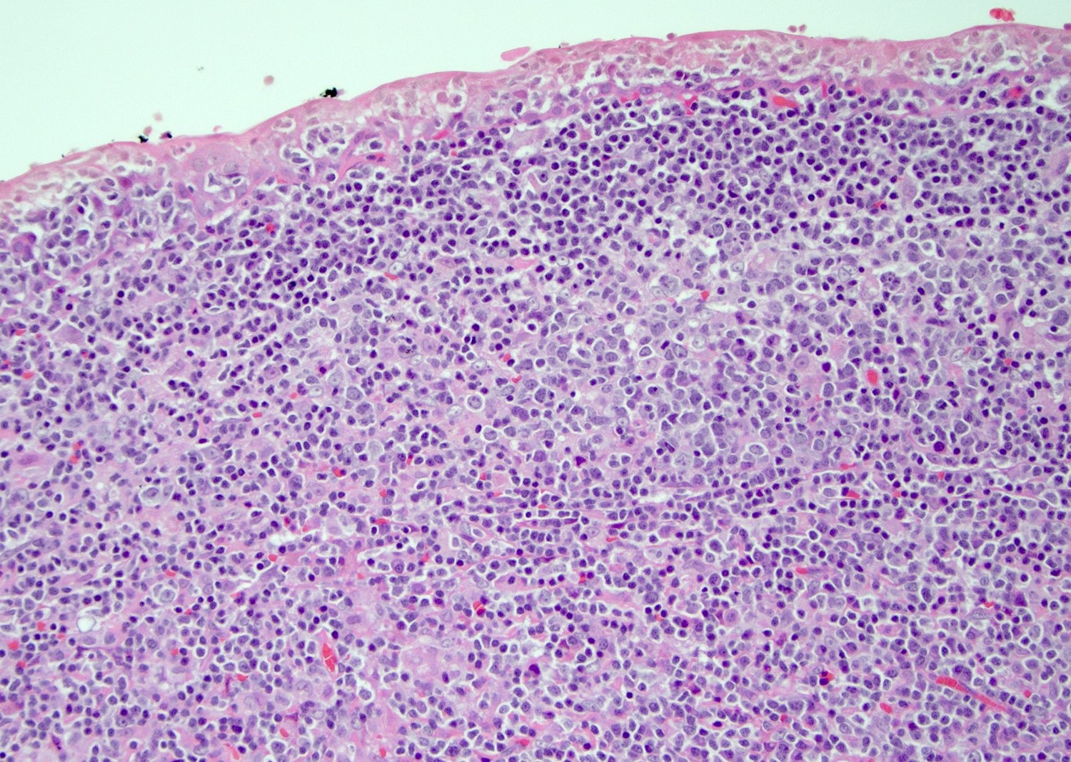
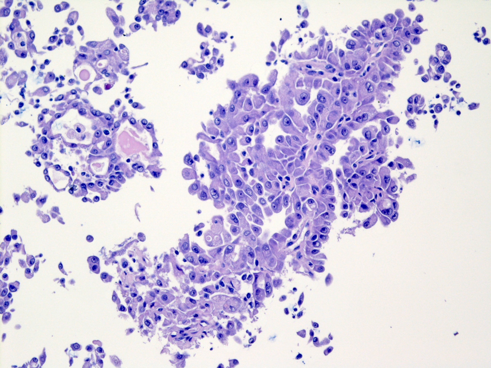
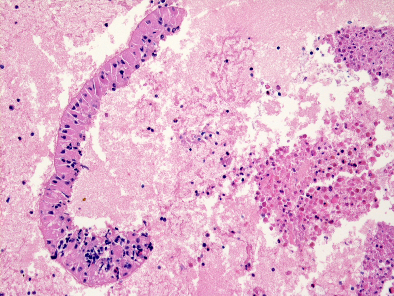
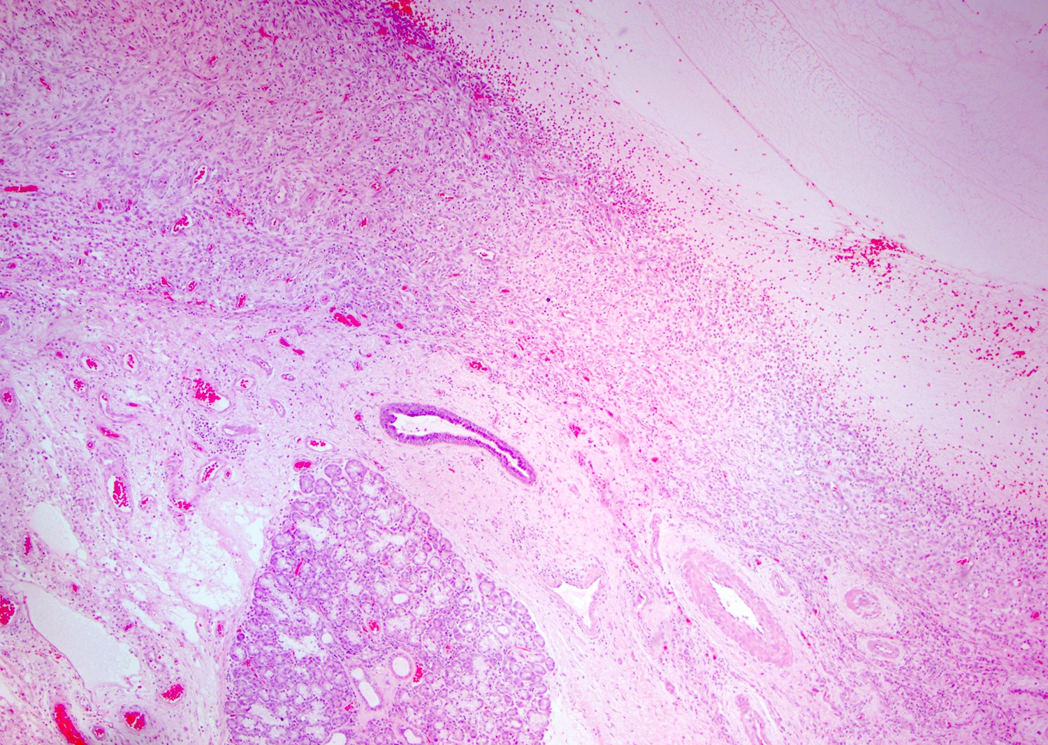
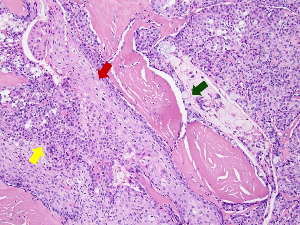
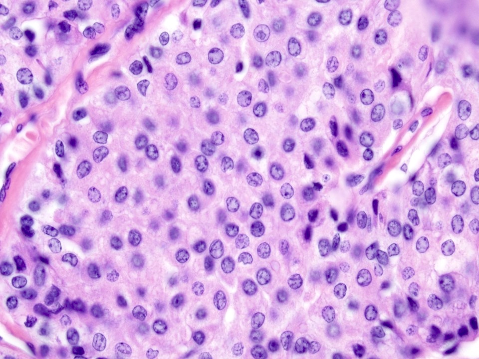
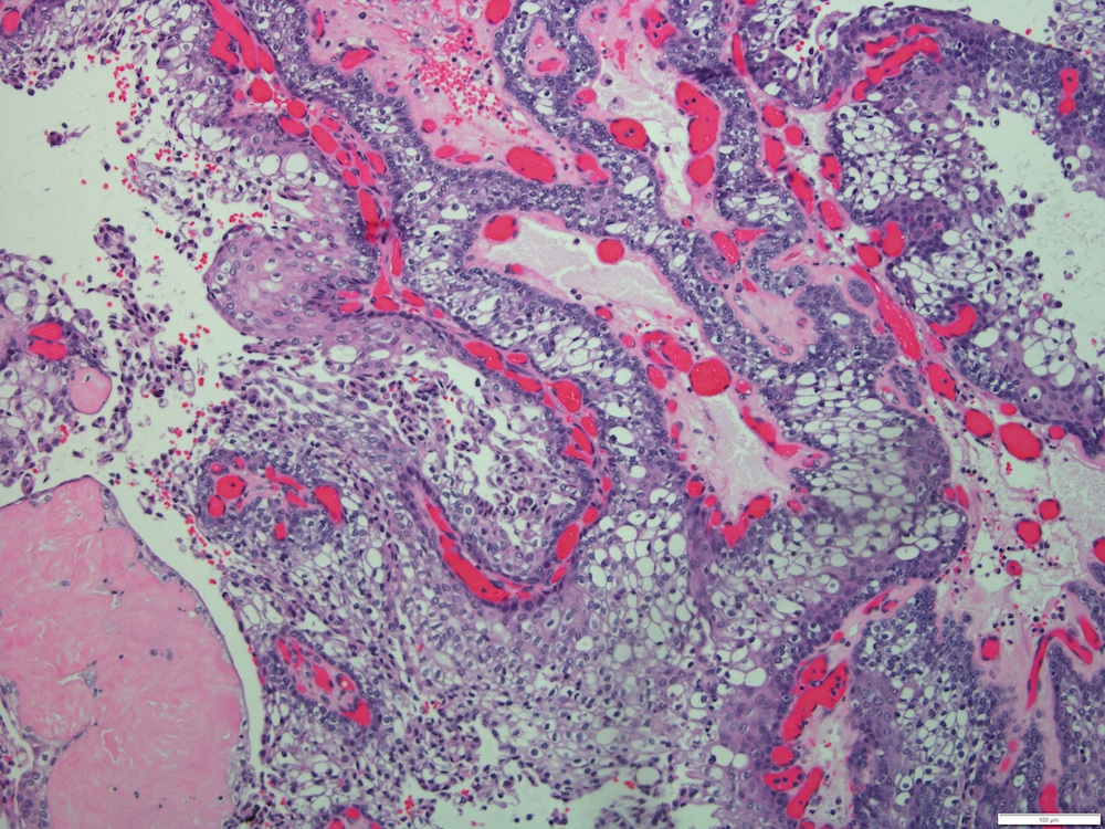
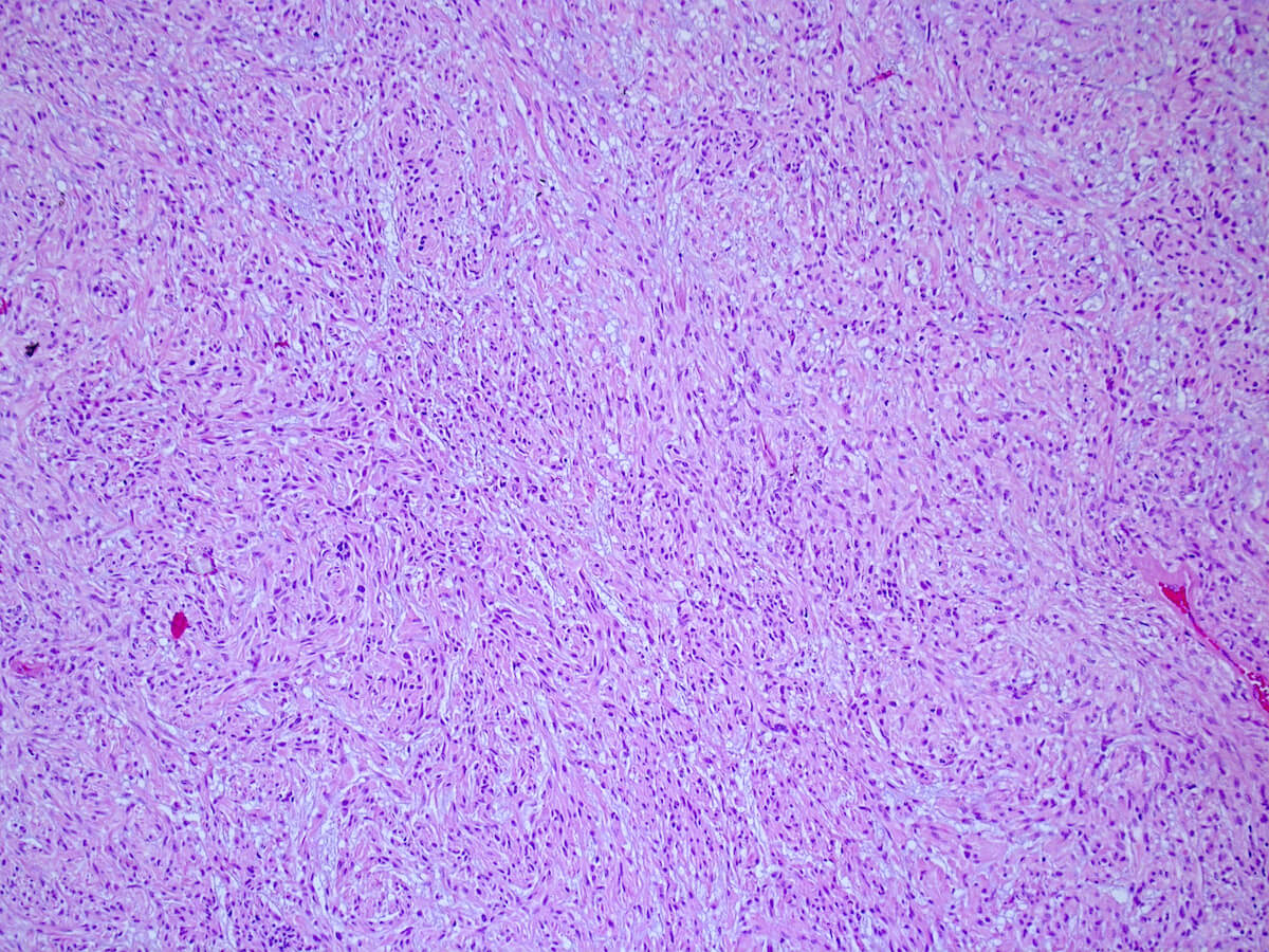
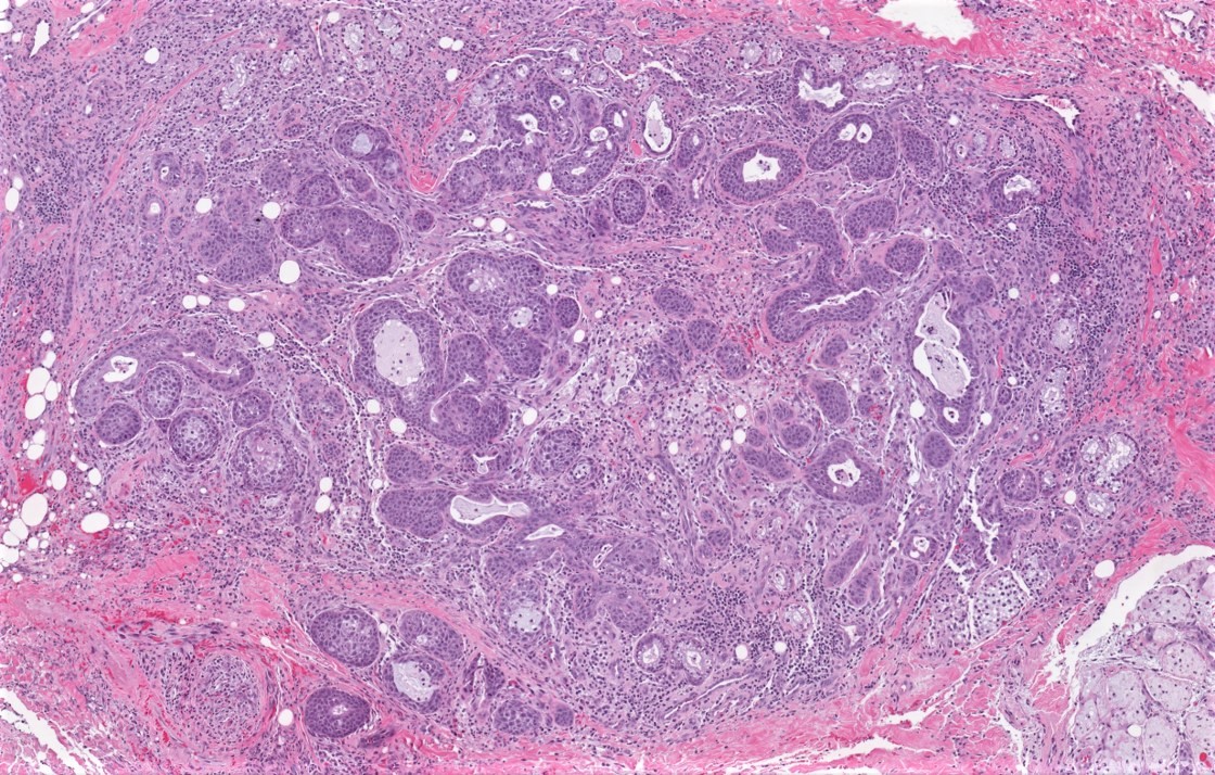
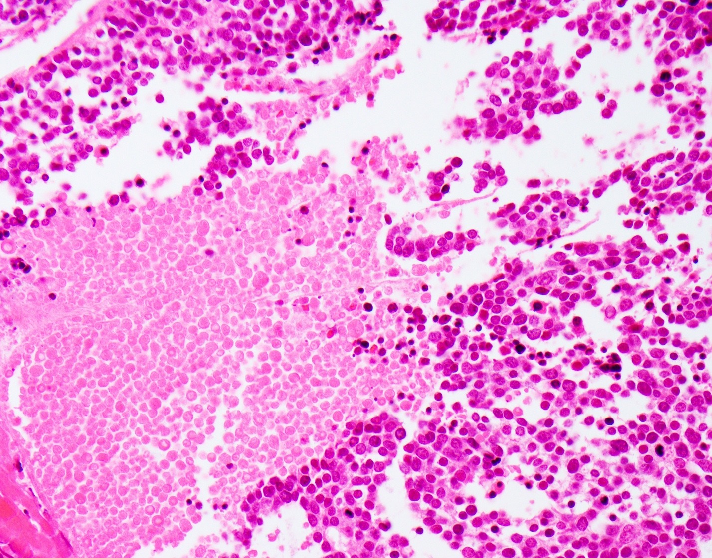
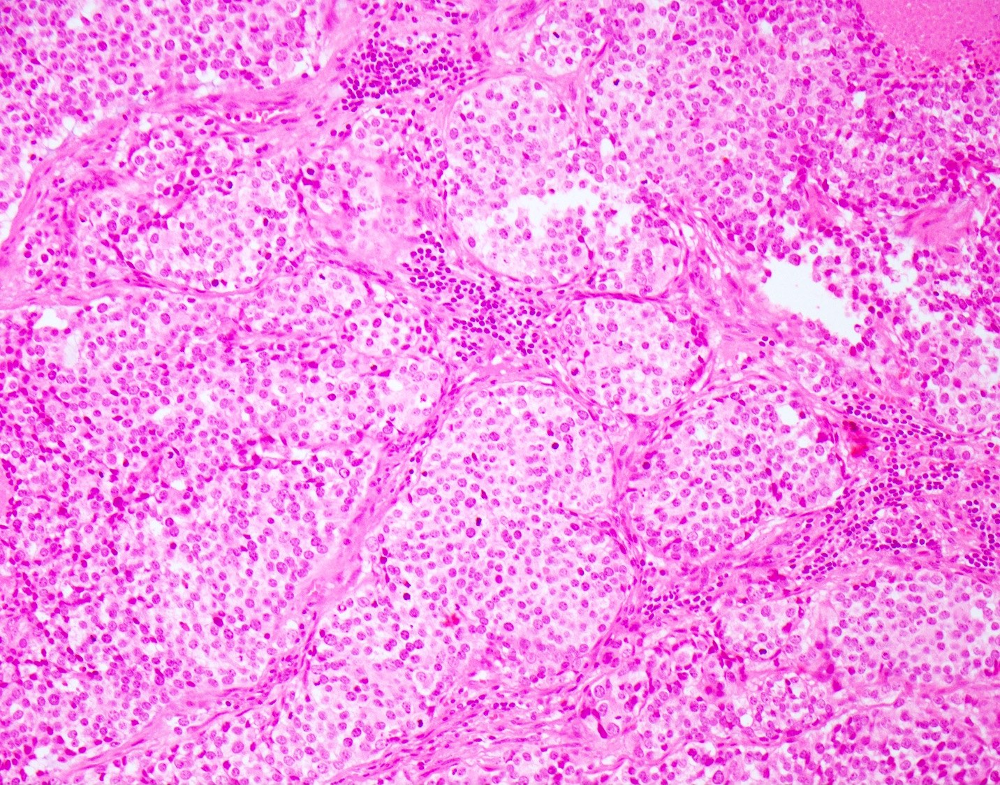
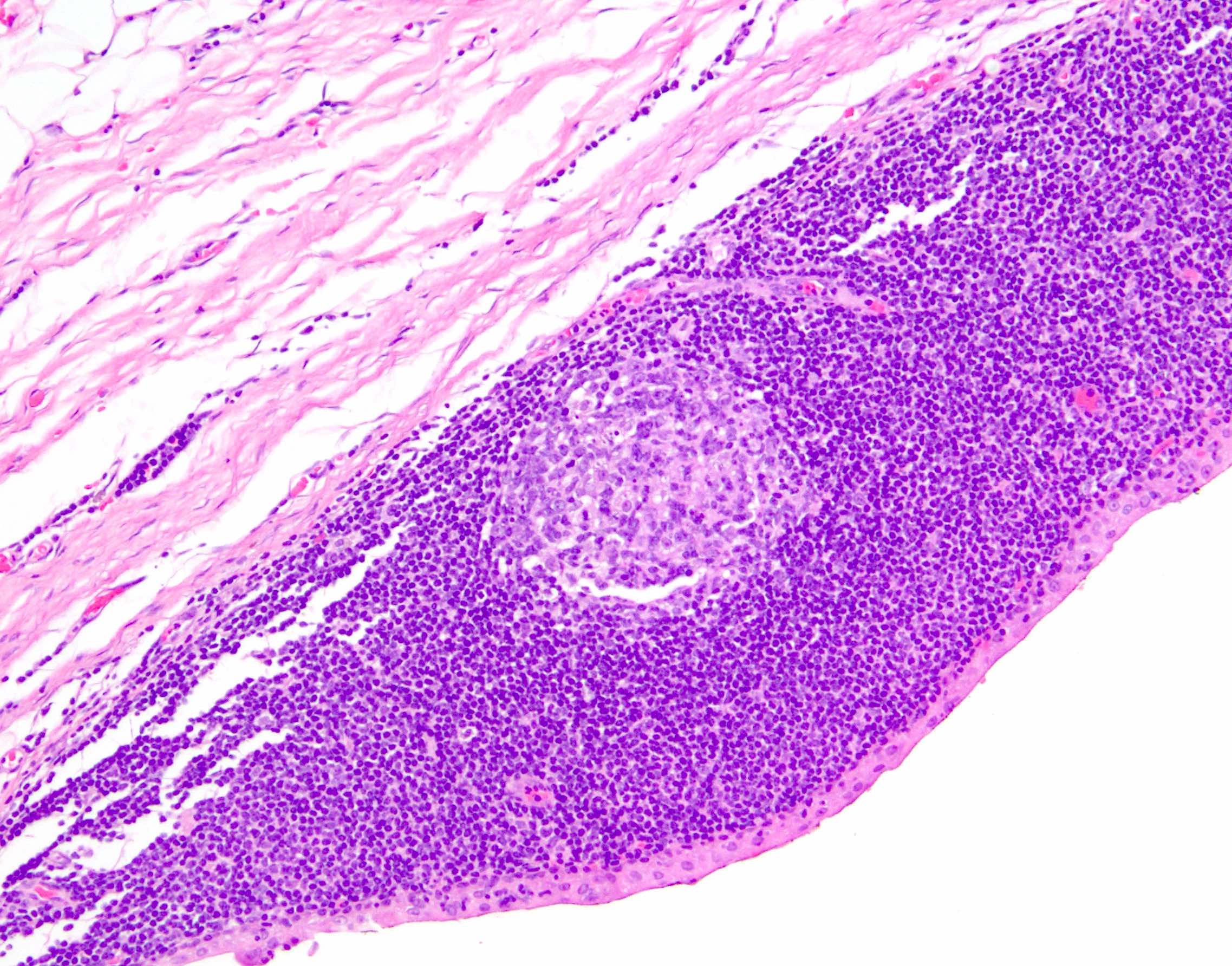
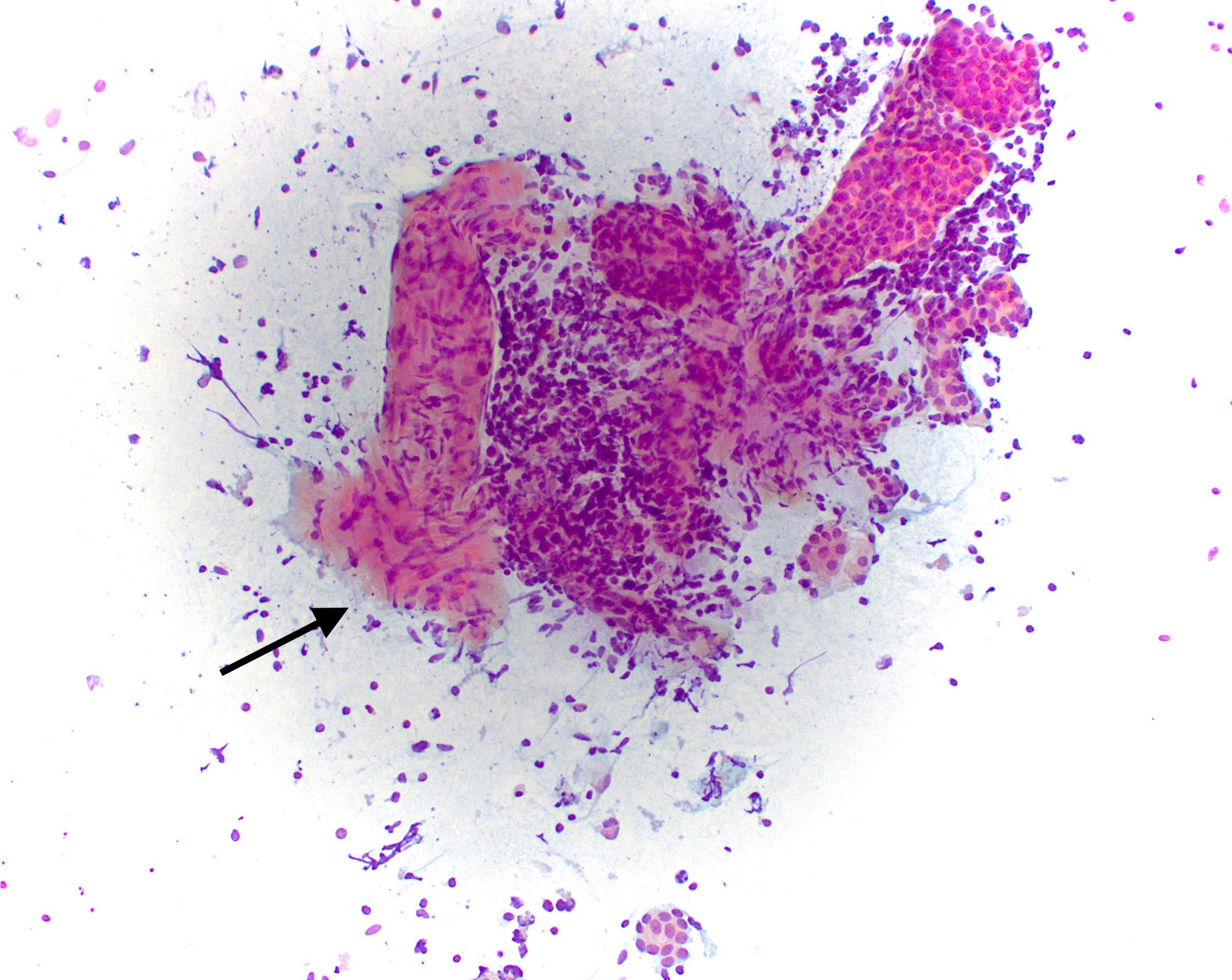
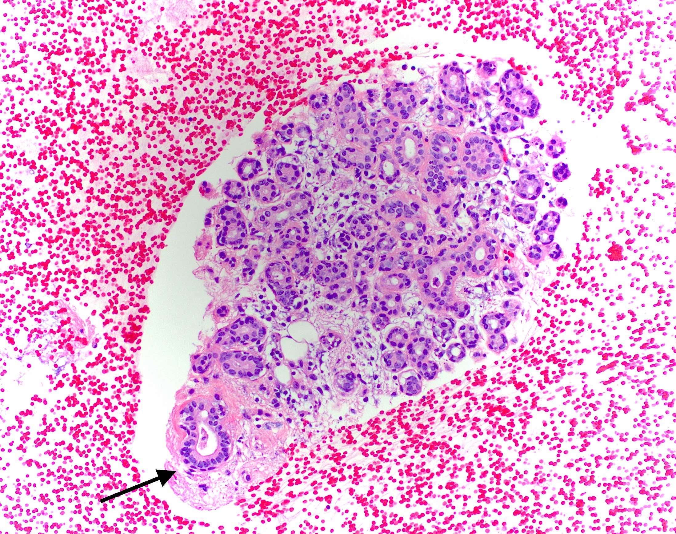
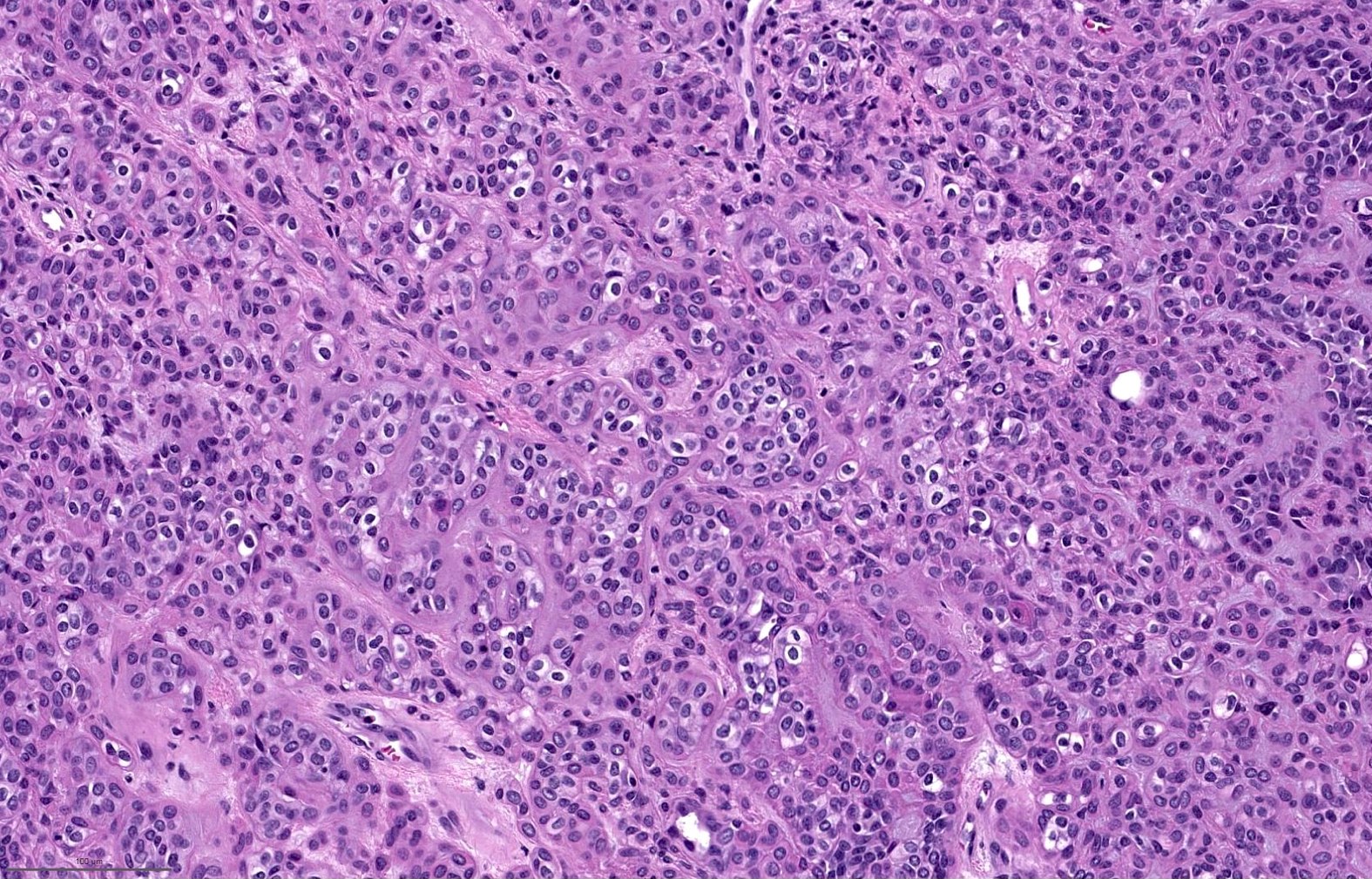
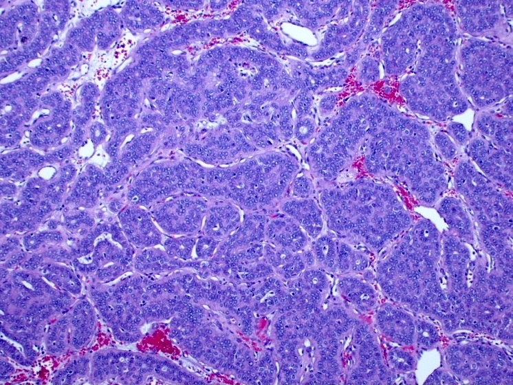
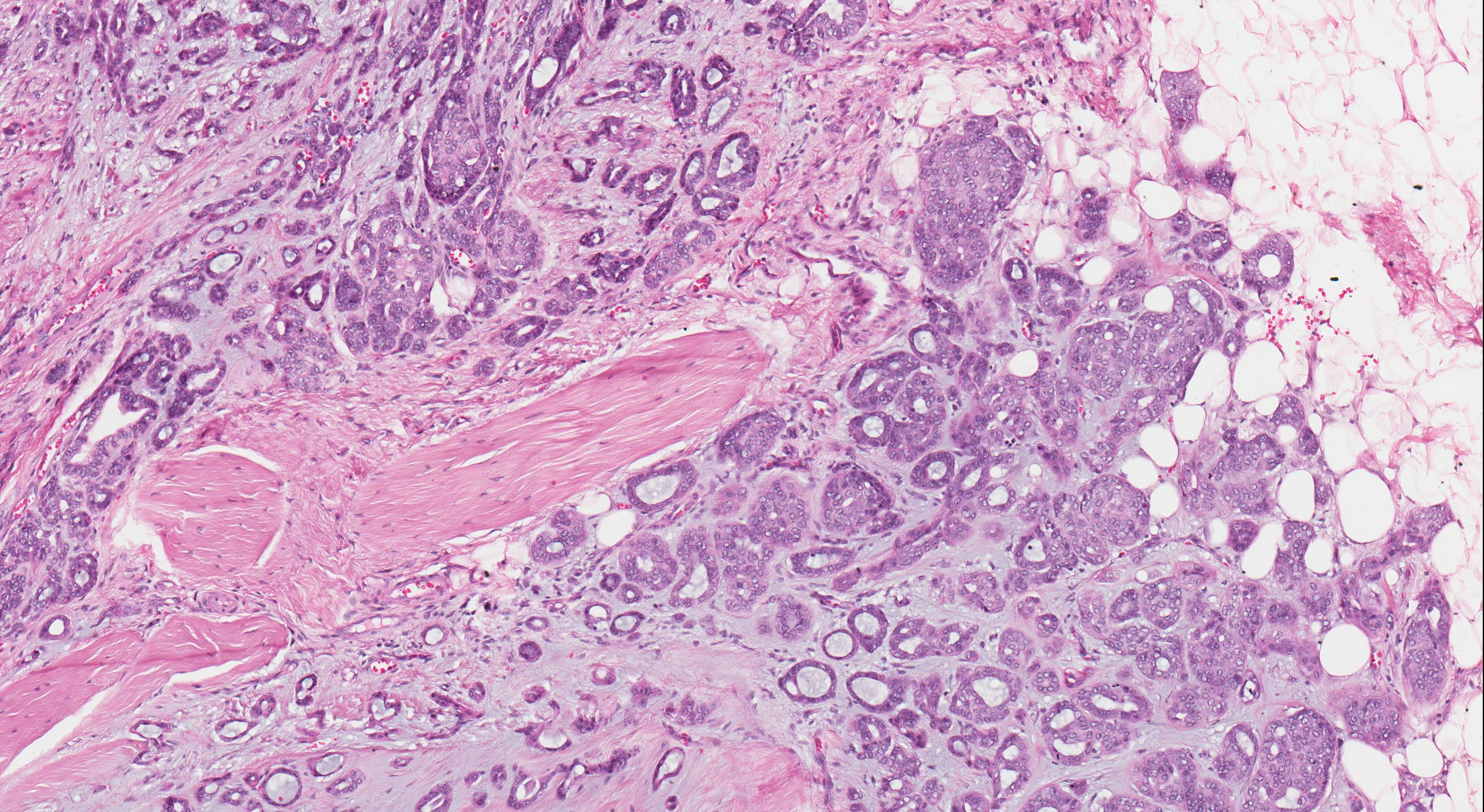
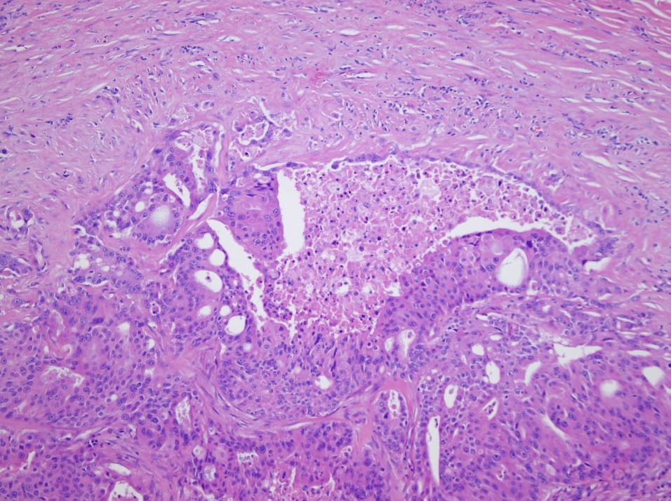
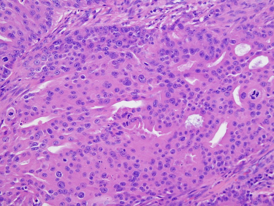
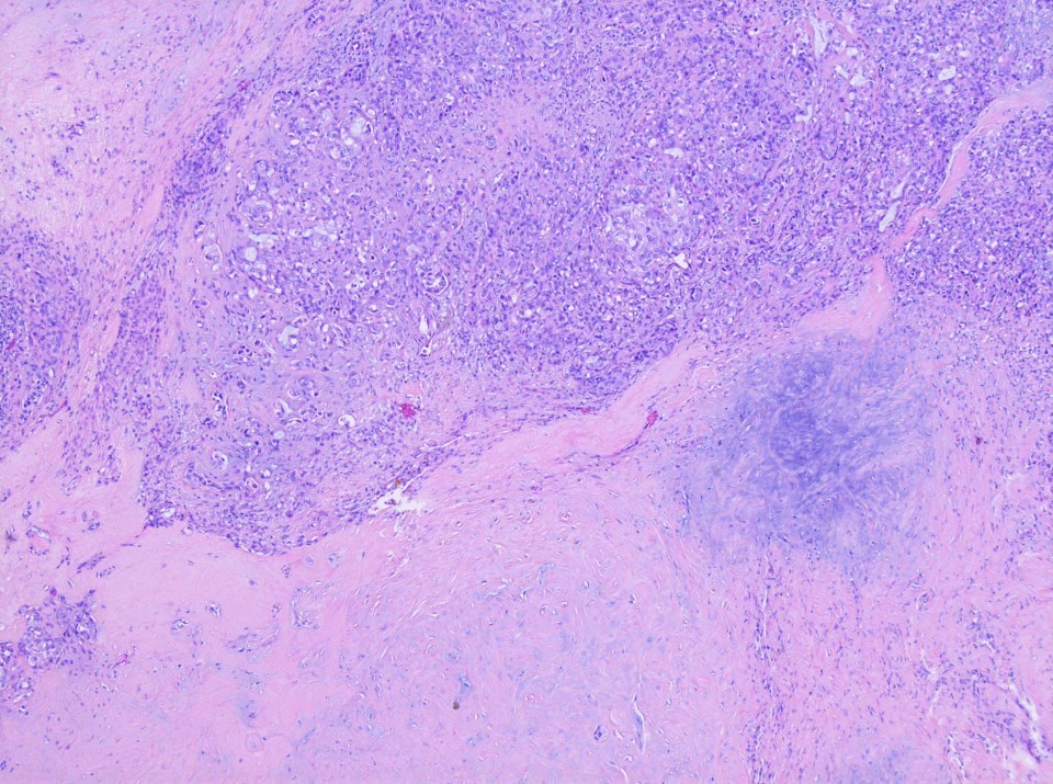
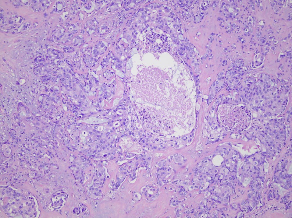
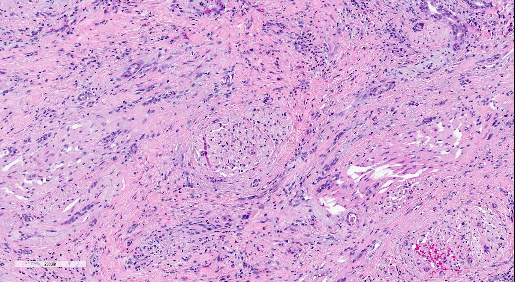
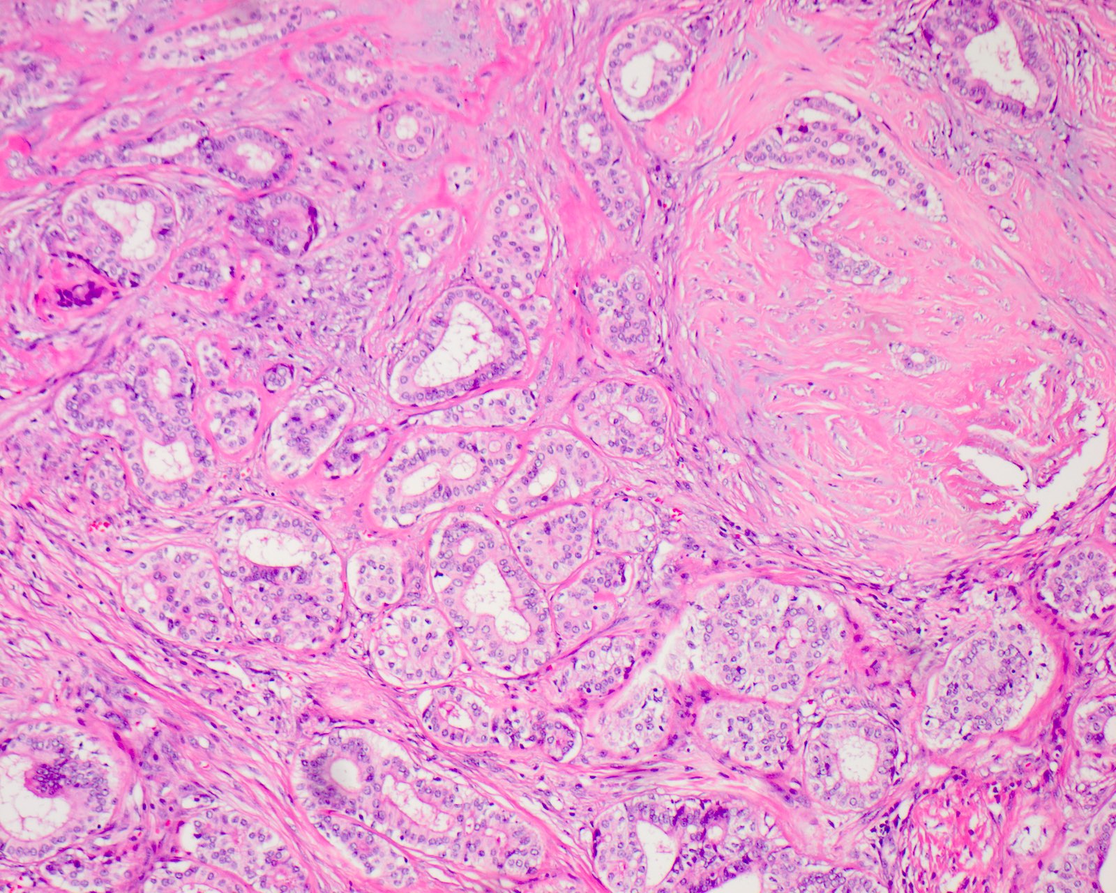
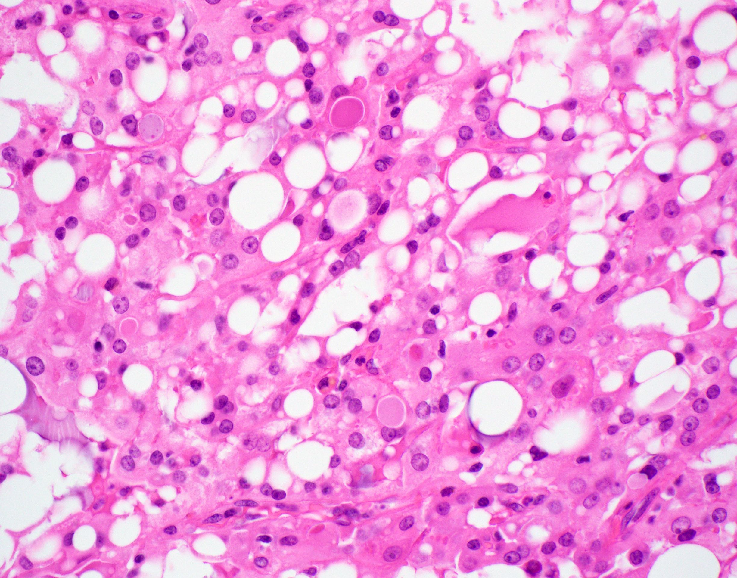
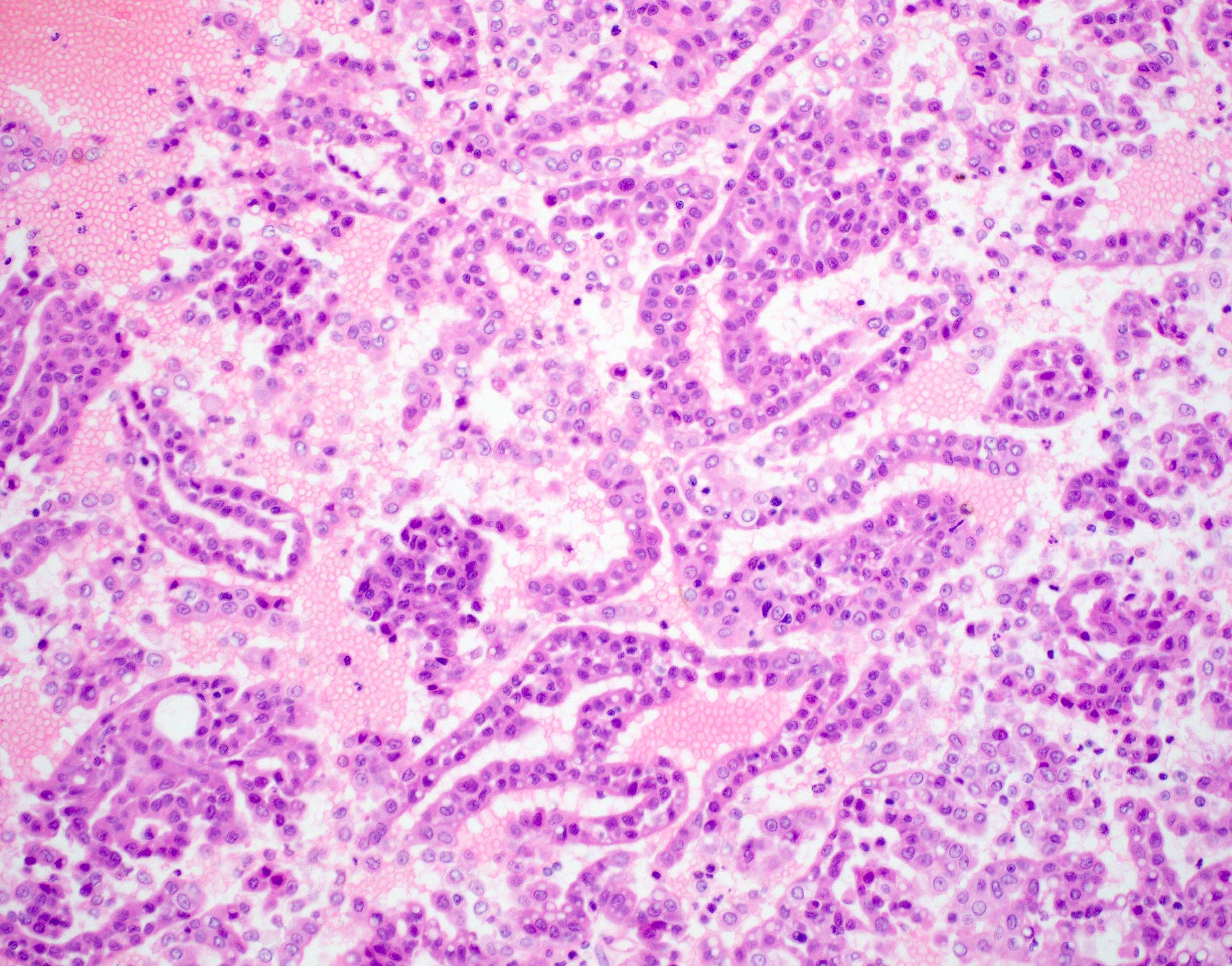
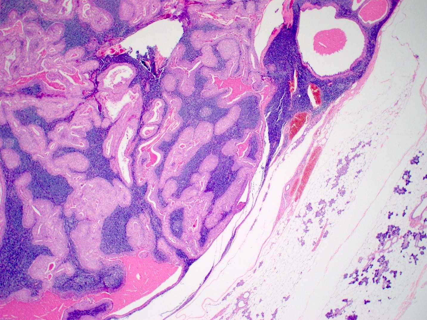
Well circumscribed tumor
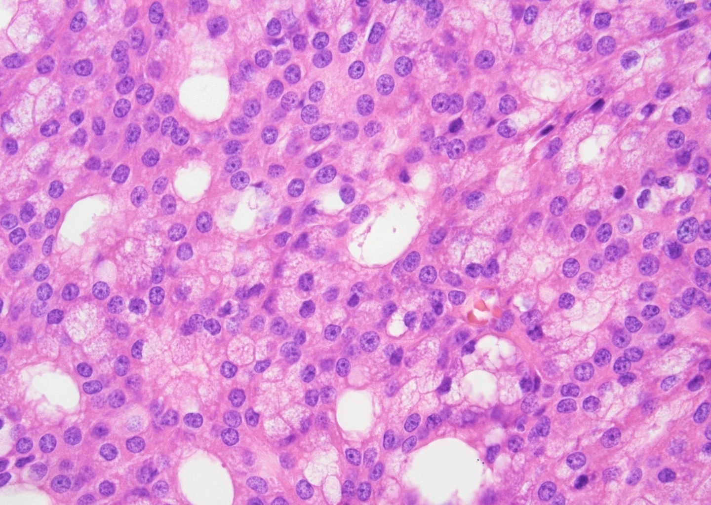
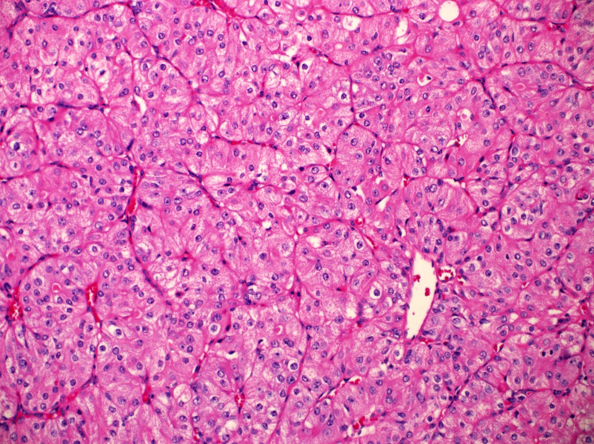
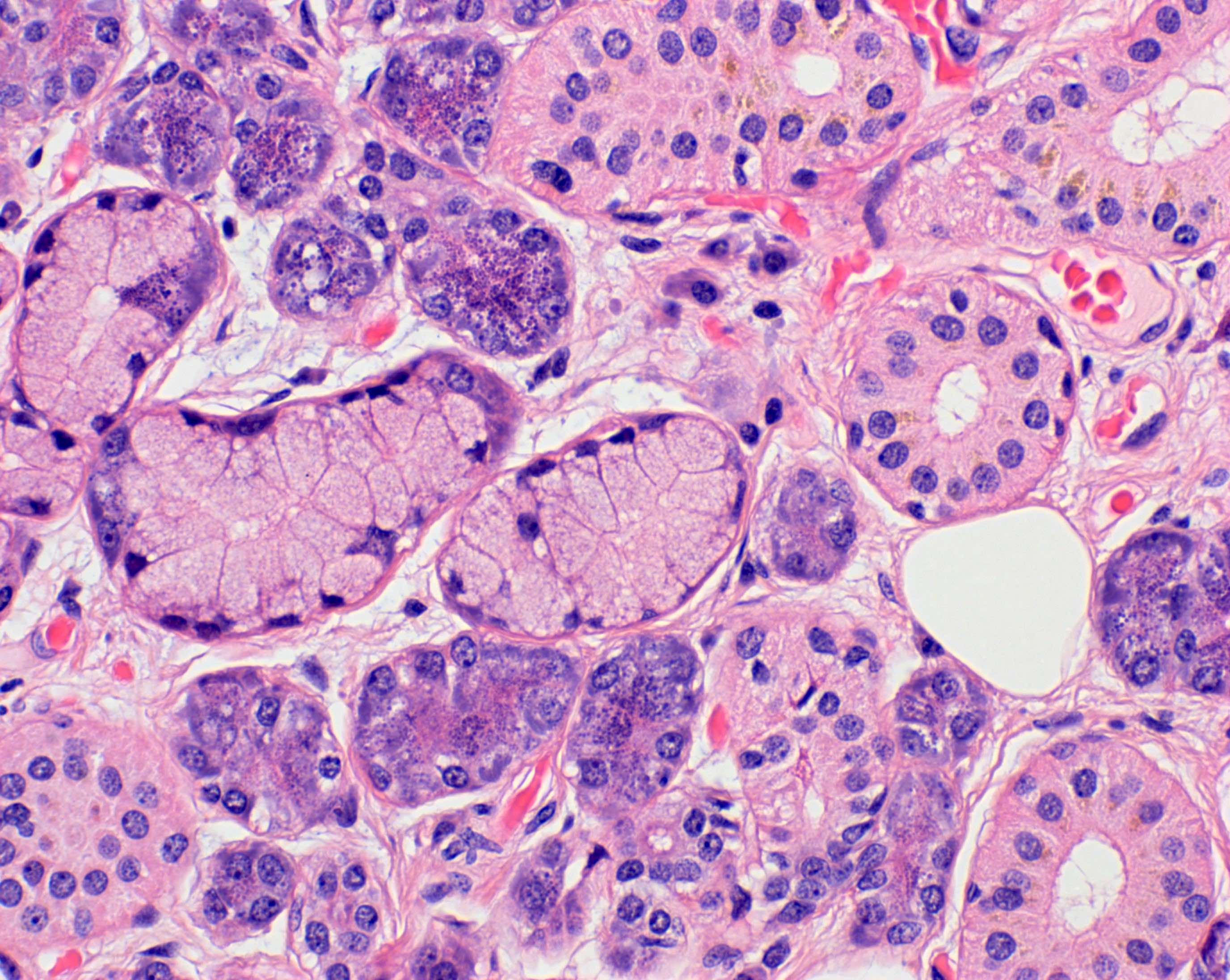
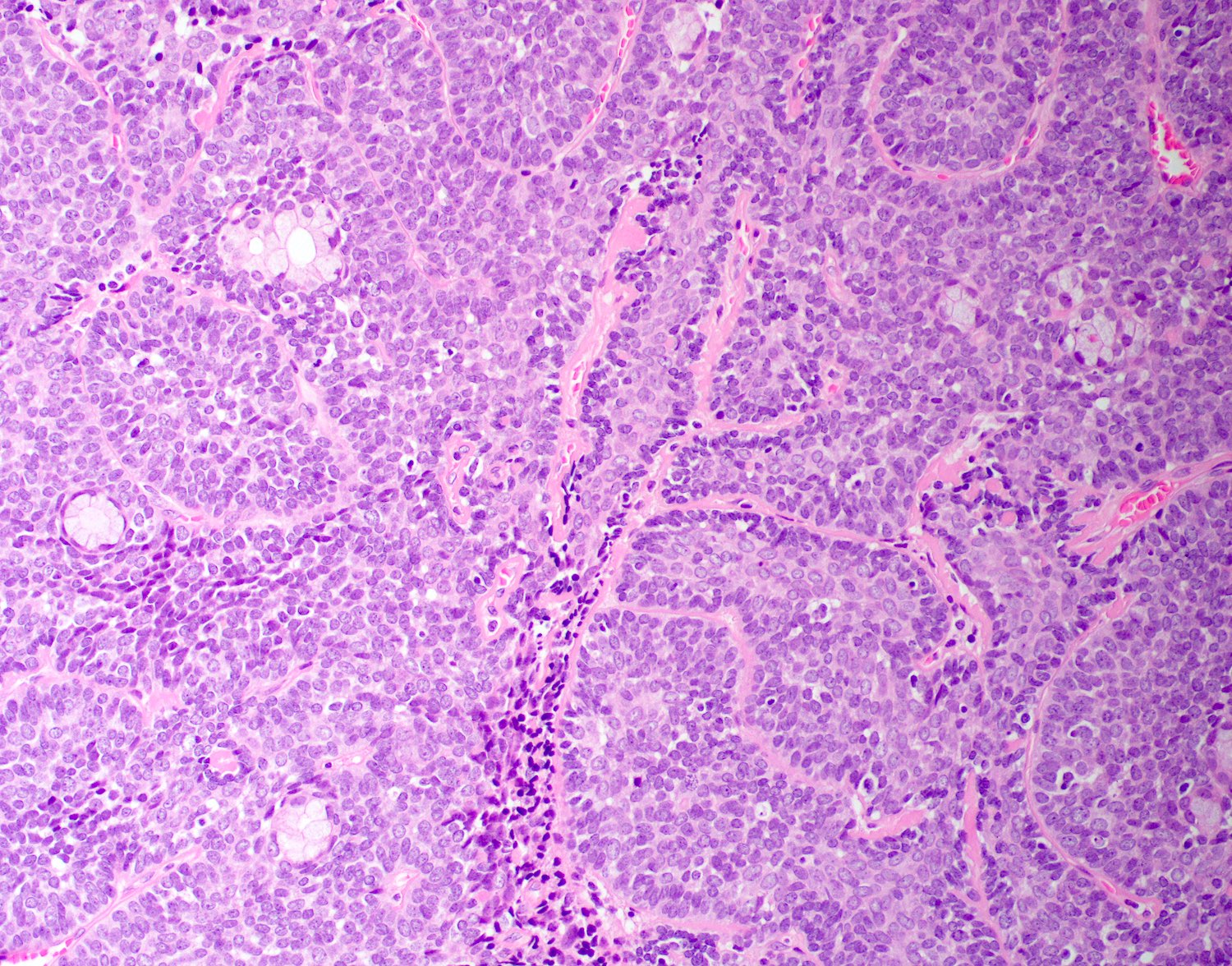
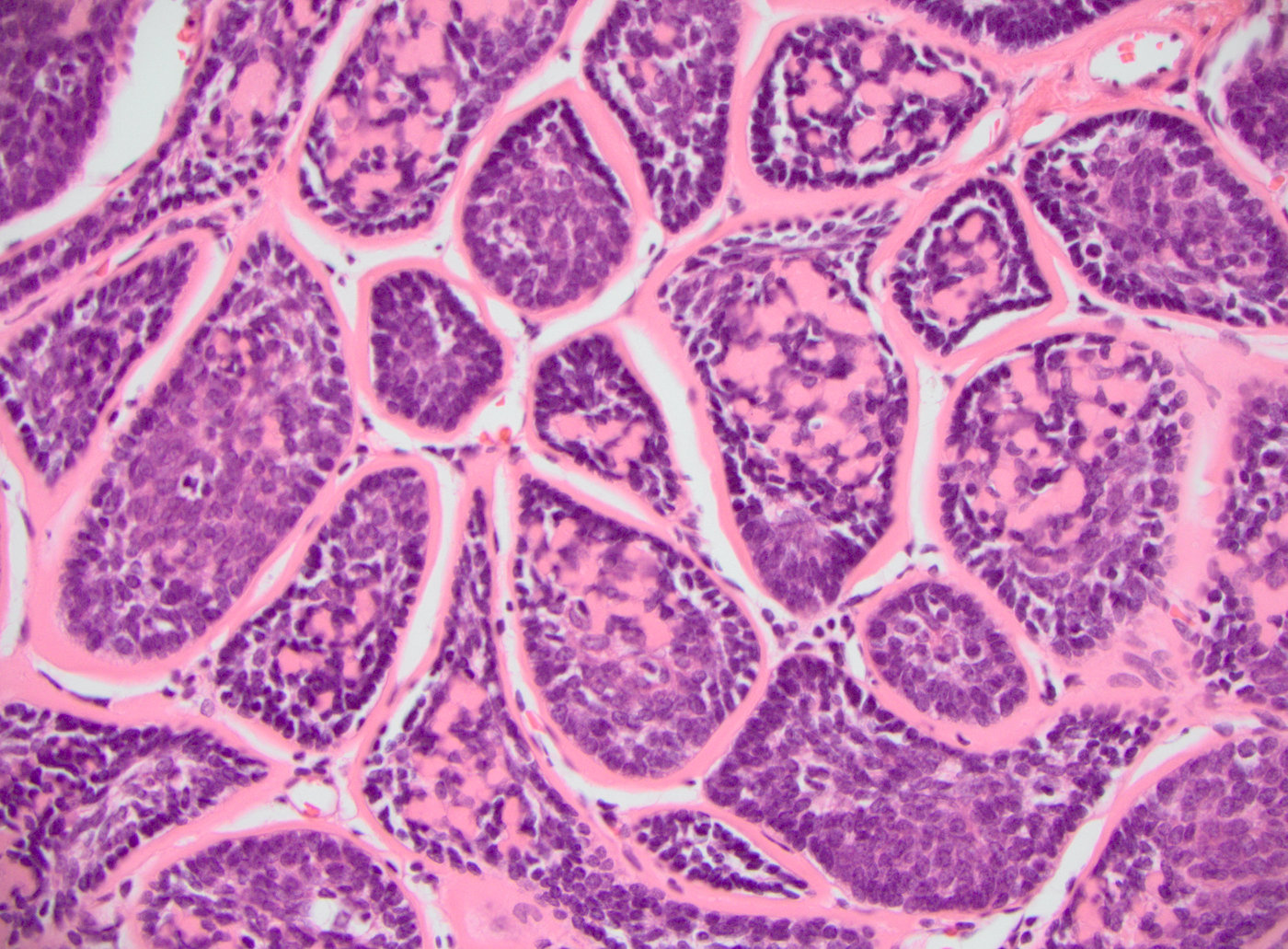
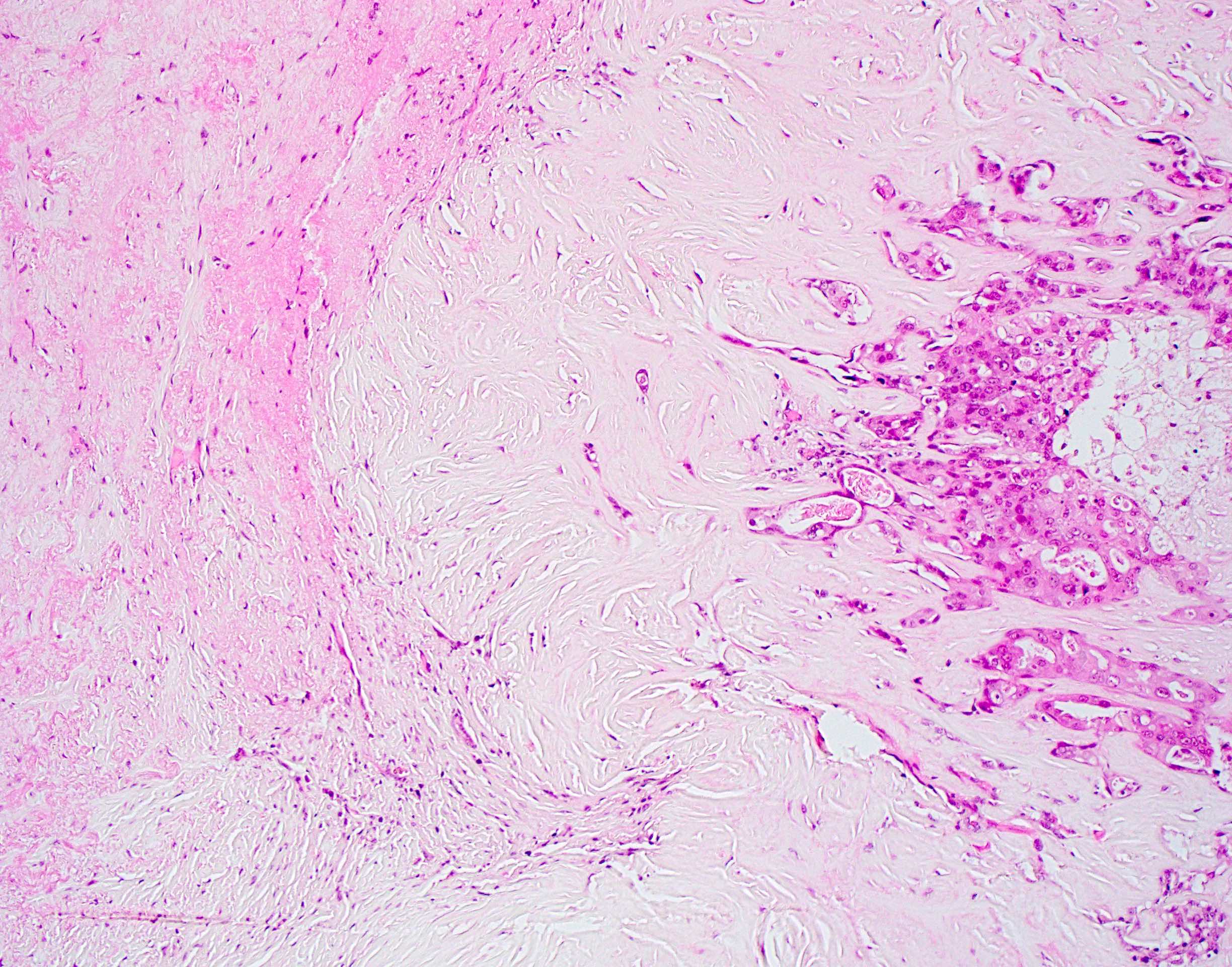
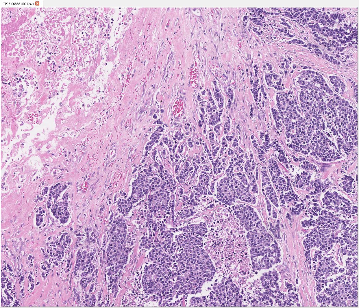
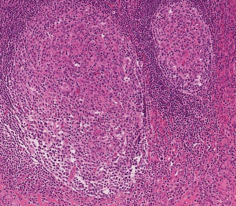
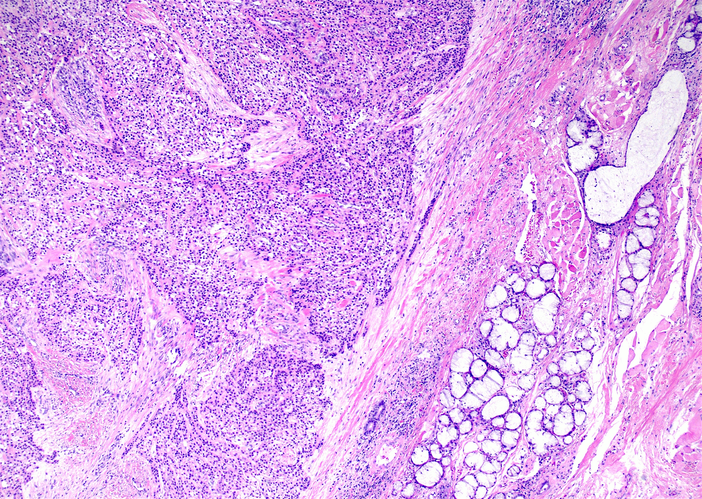
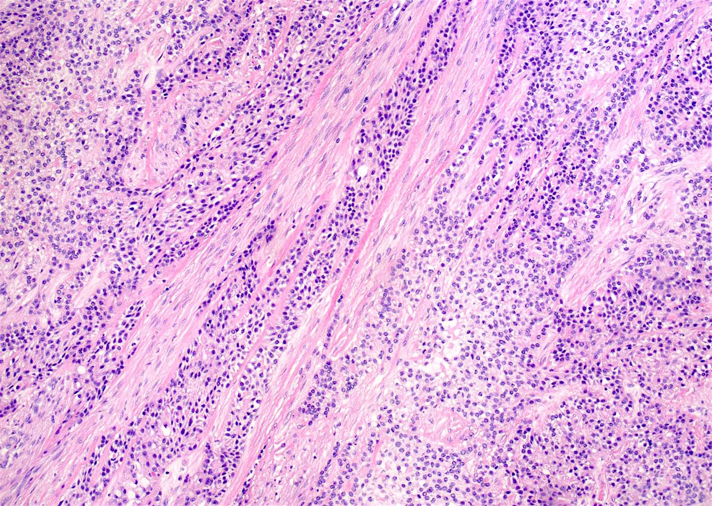
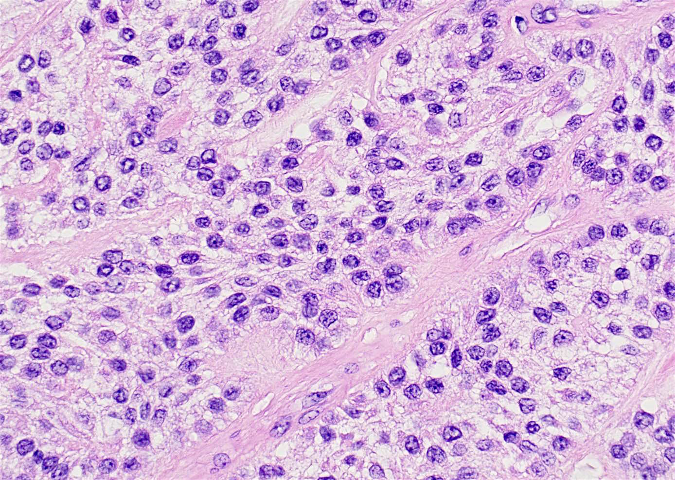
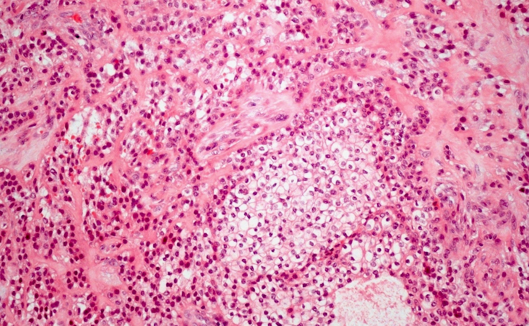
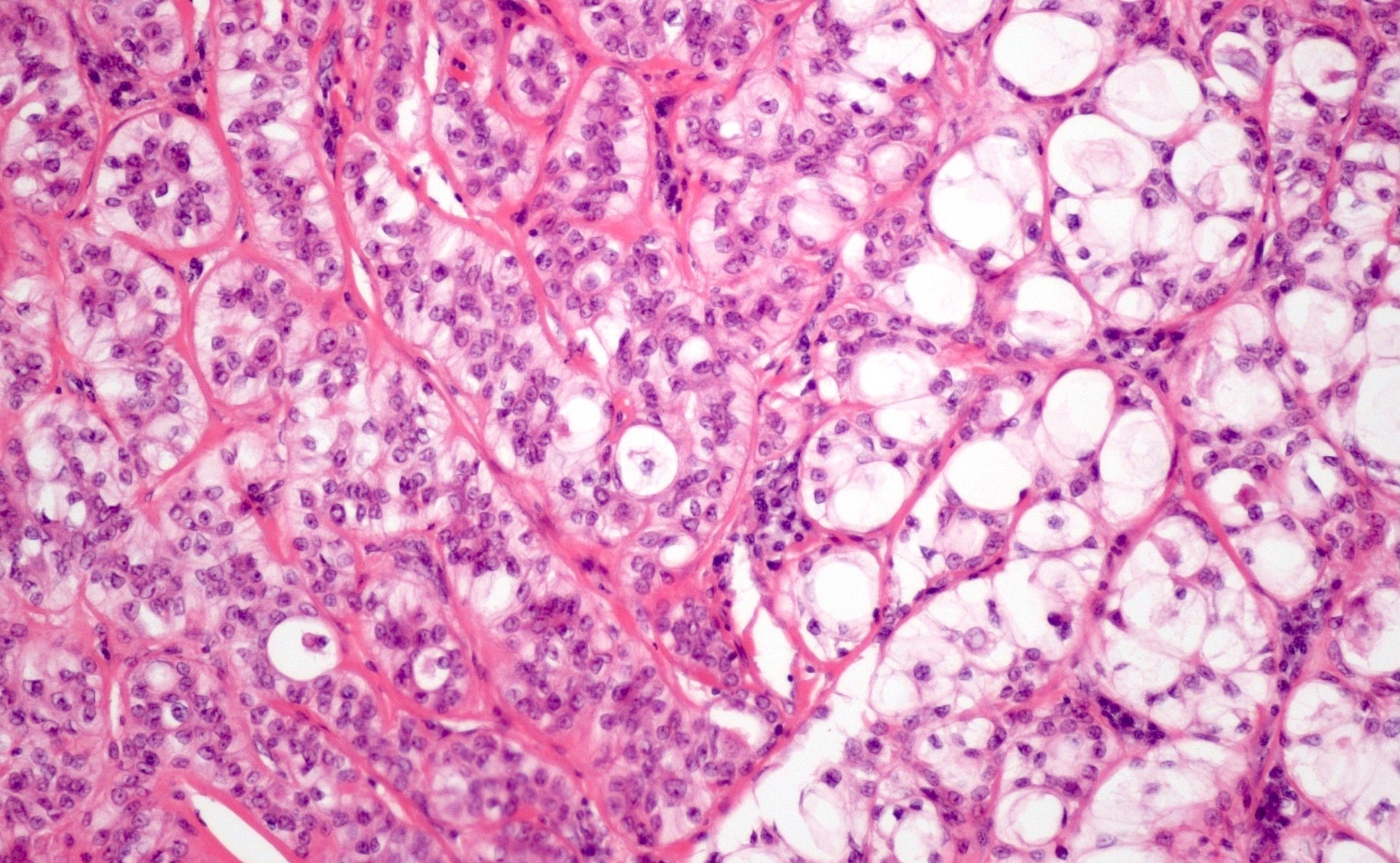
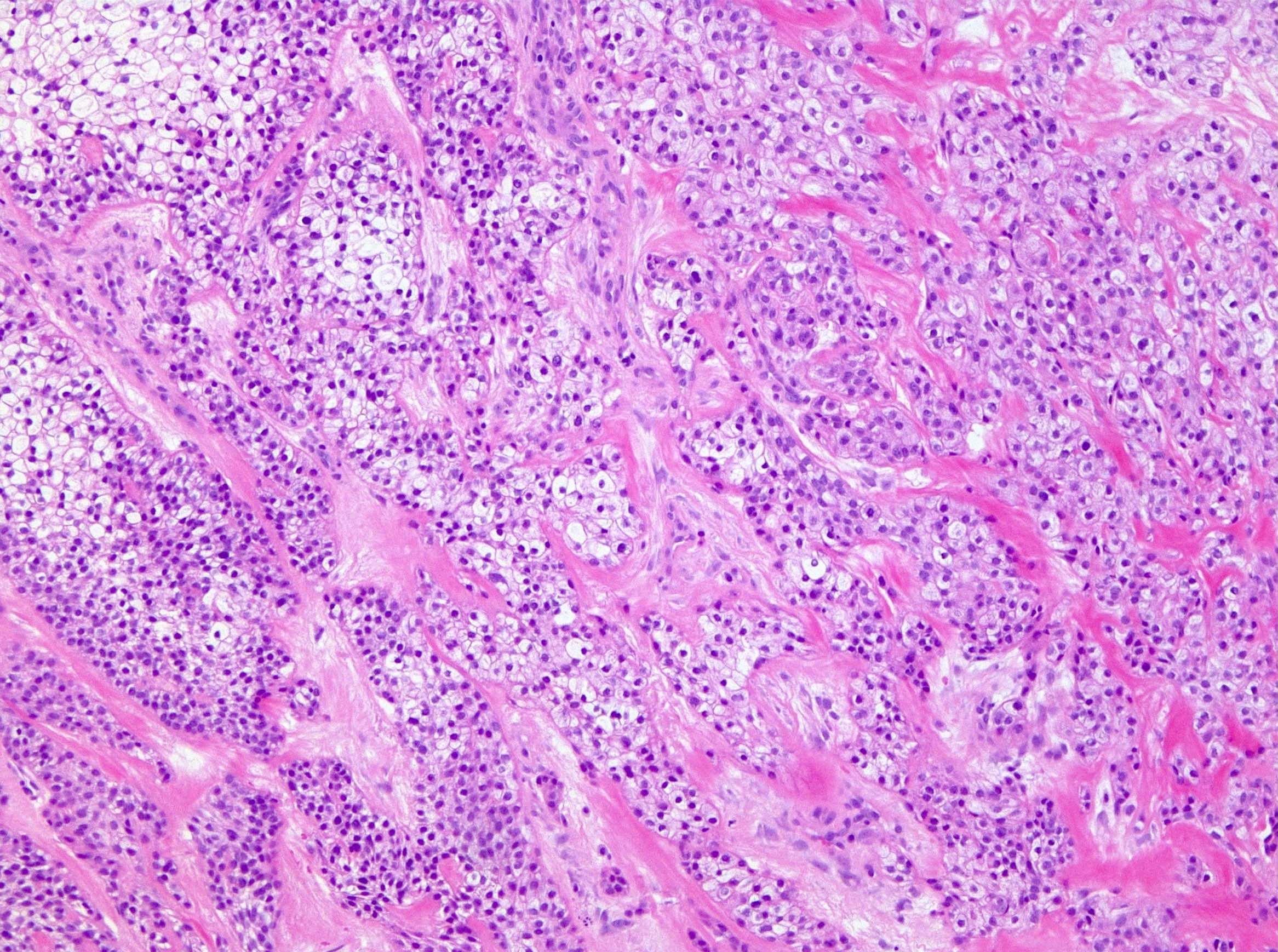
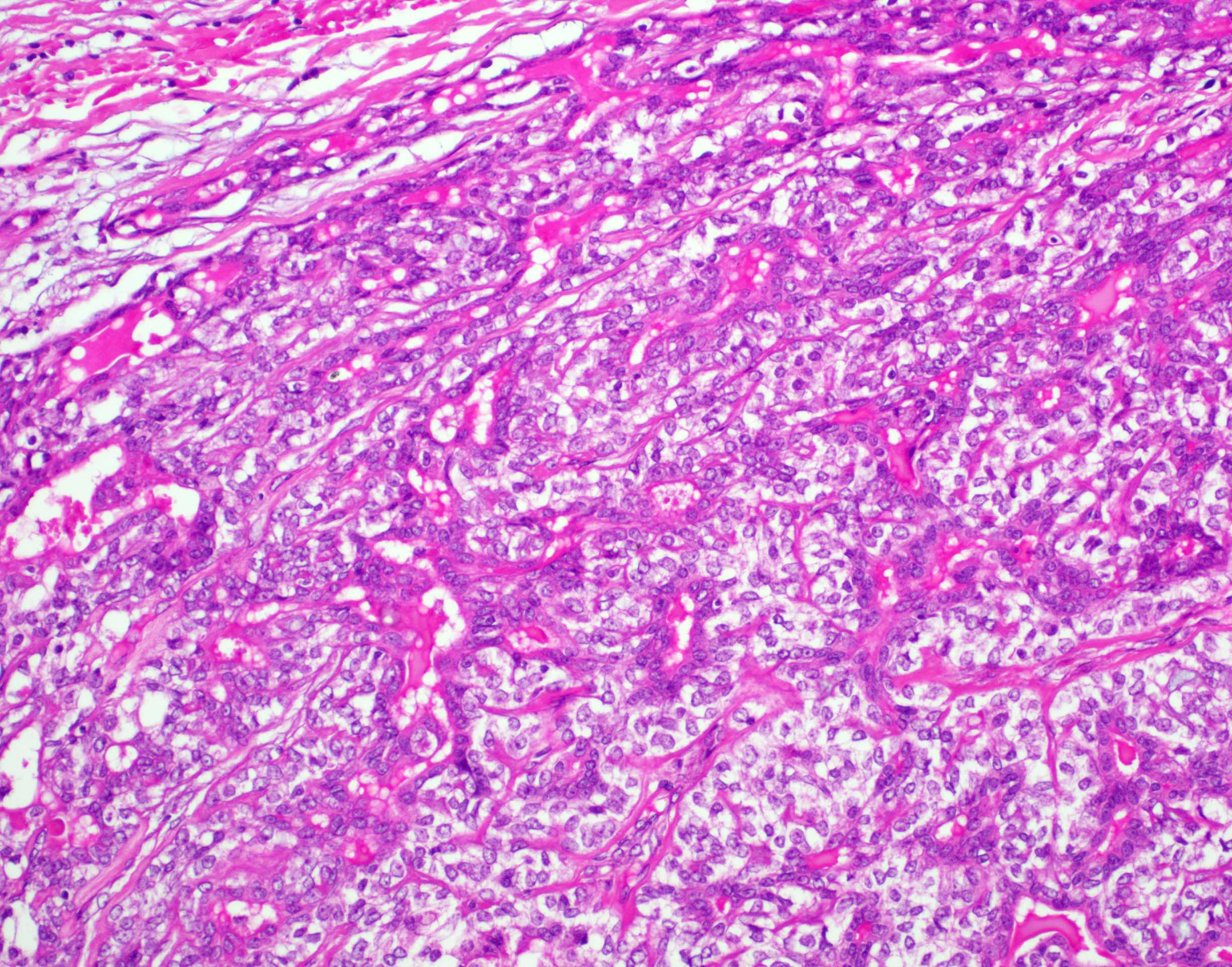
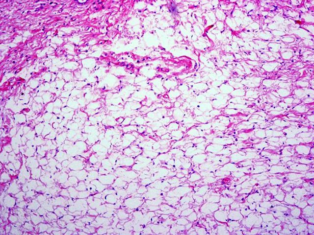
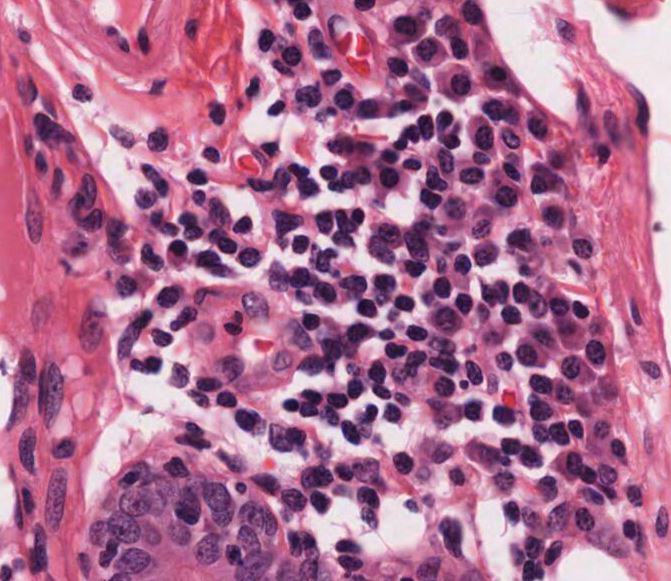
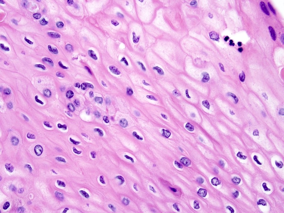
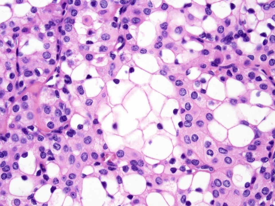
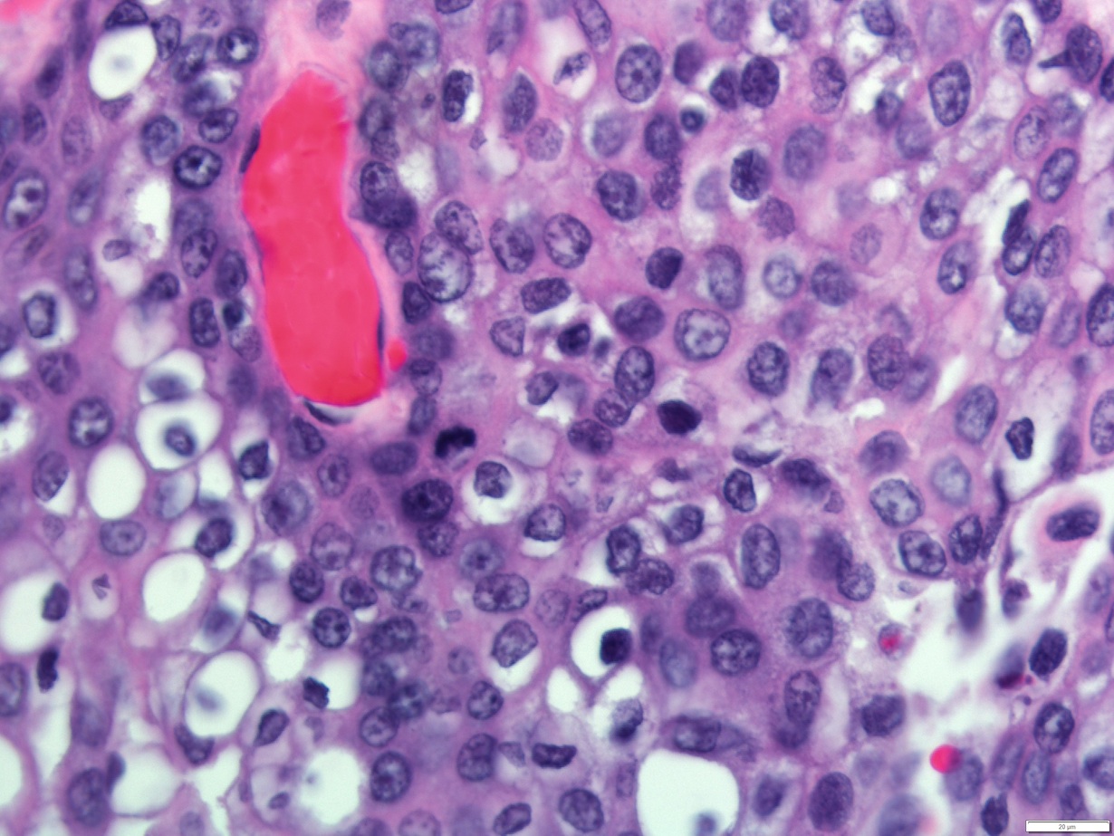
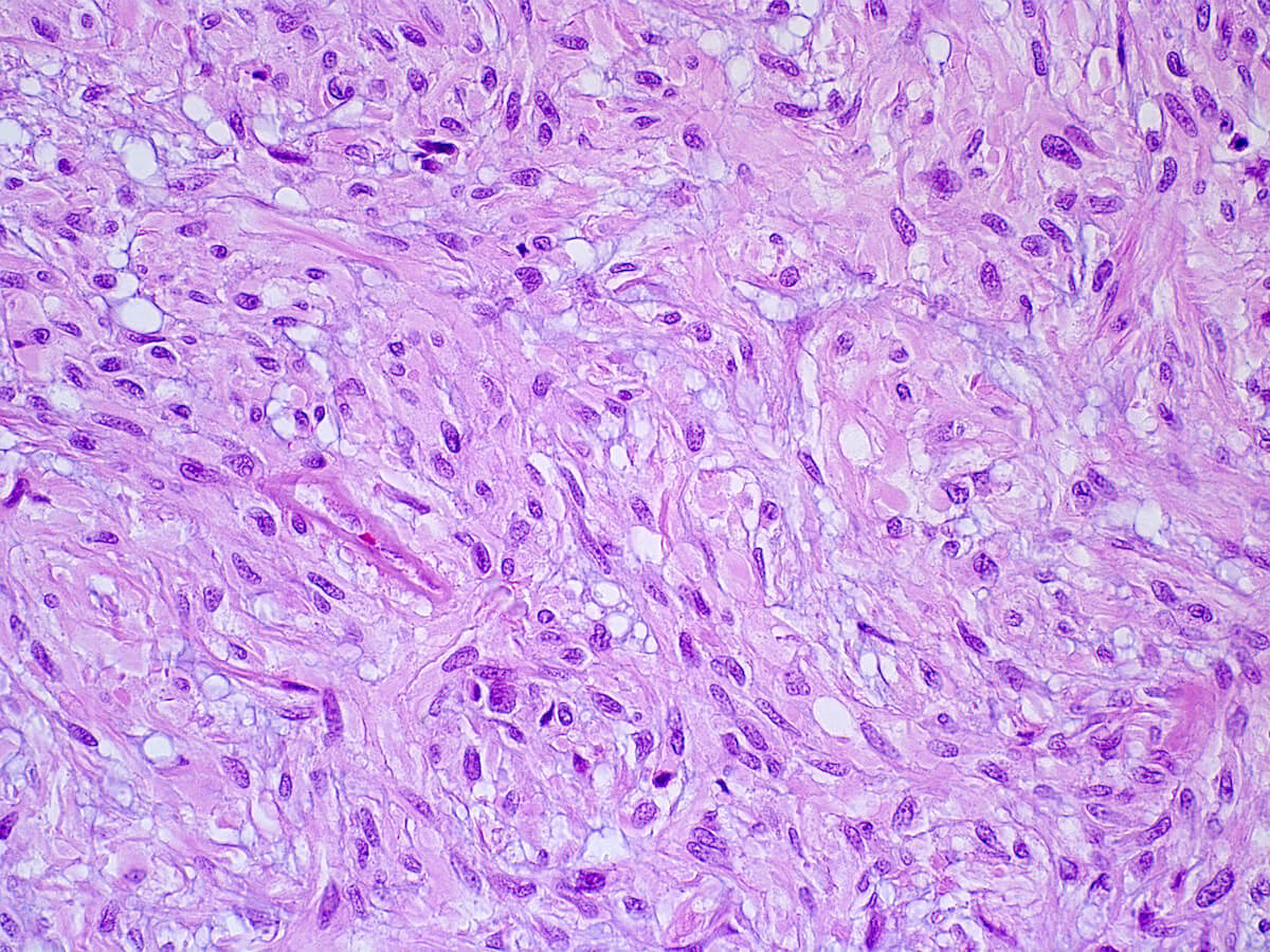
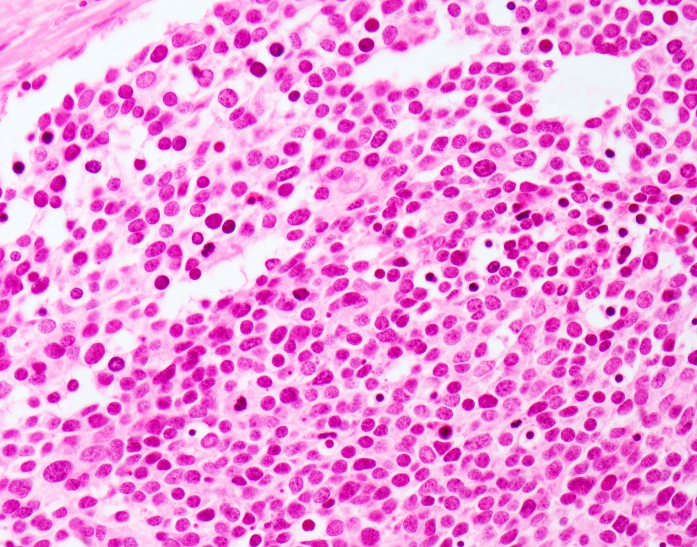
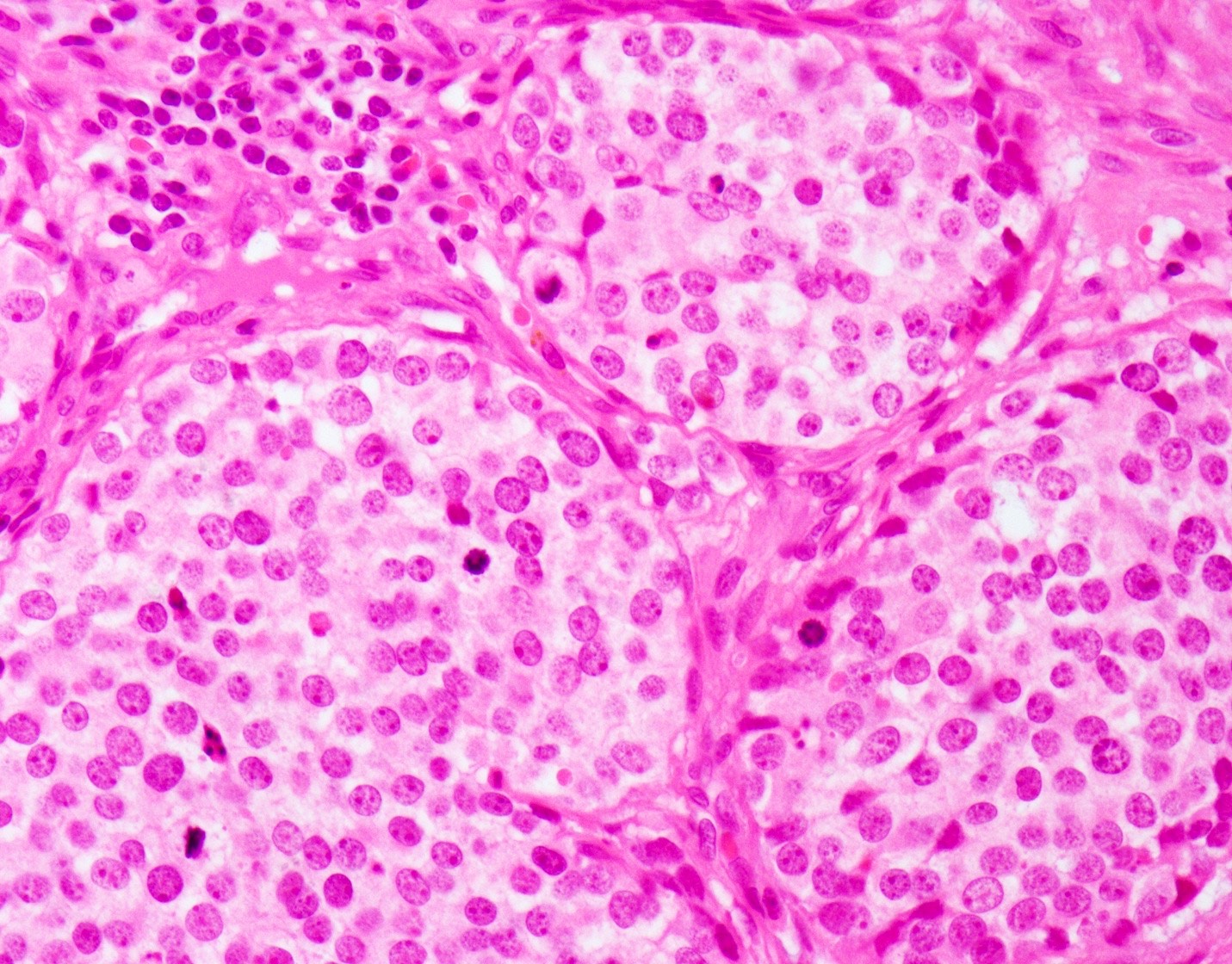
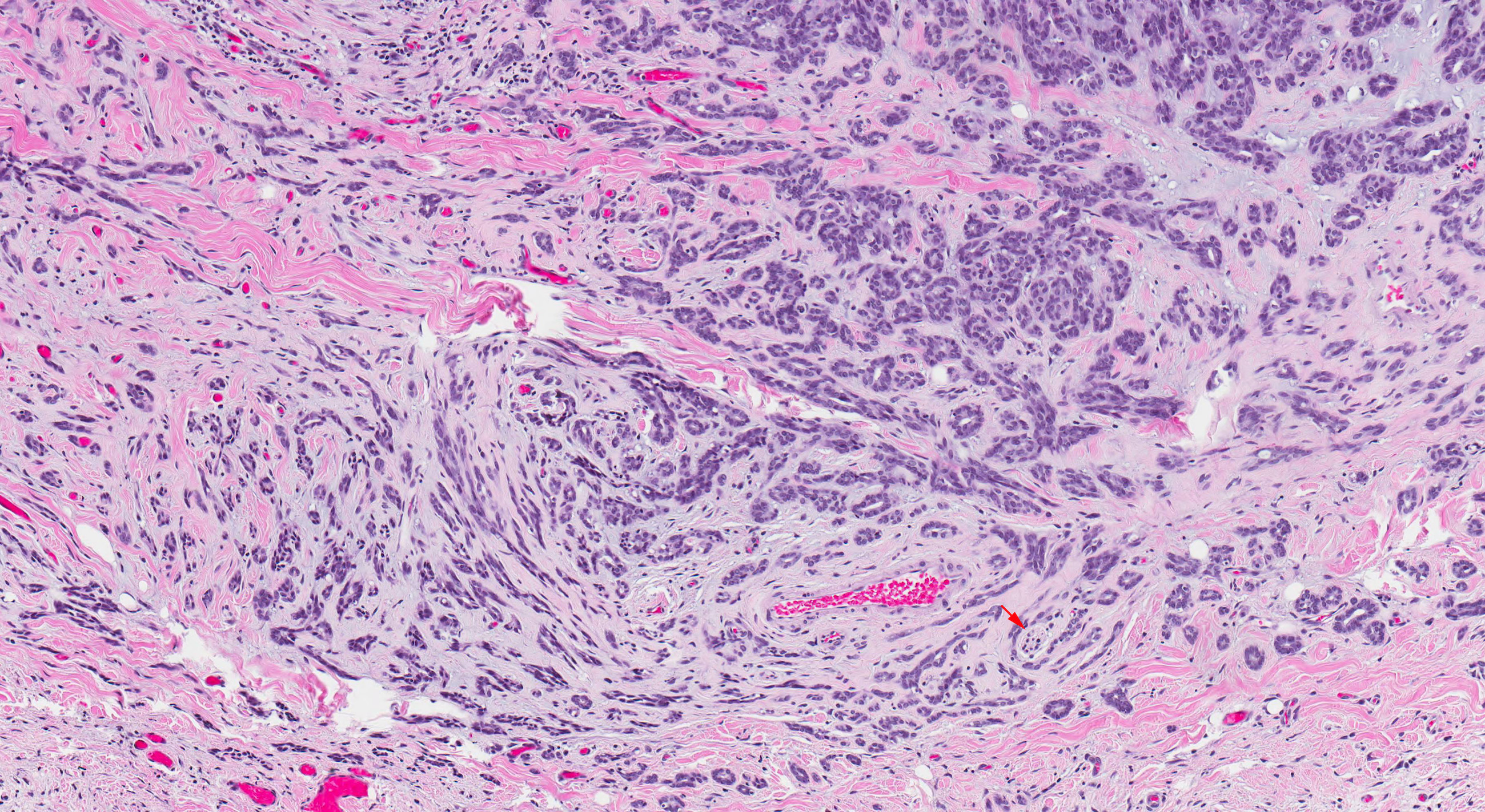
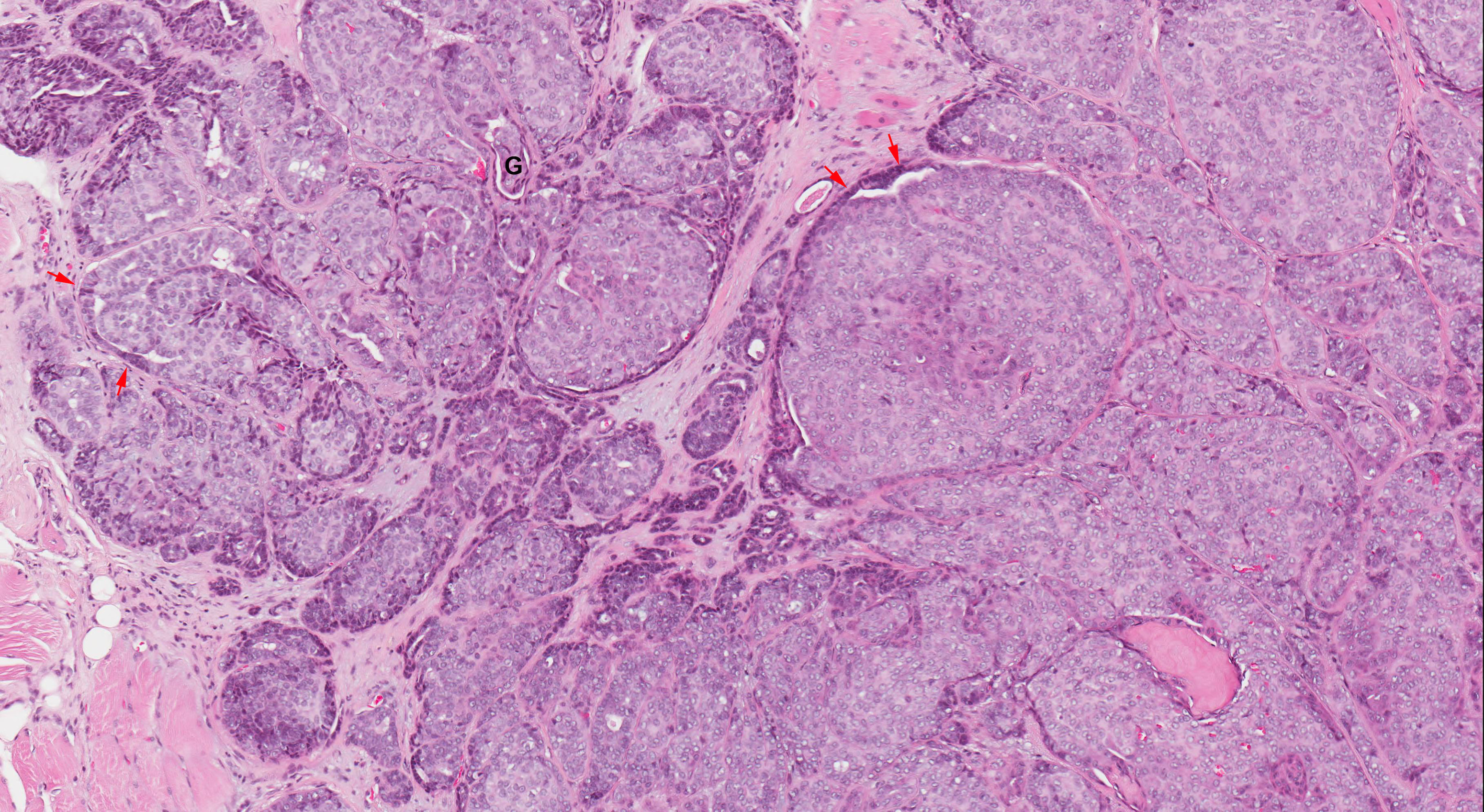
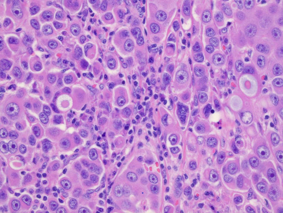
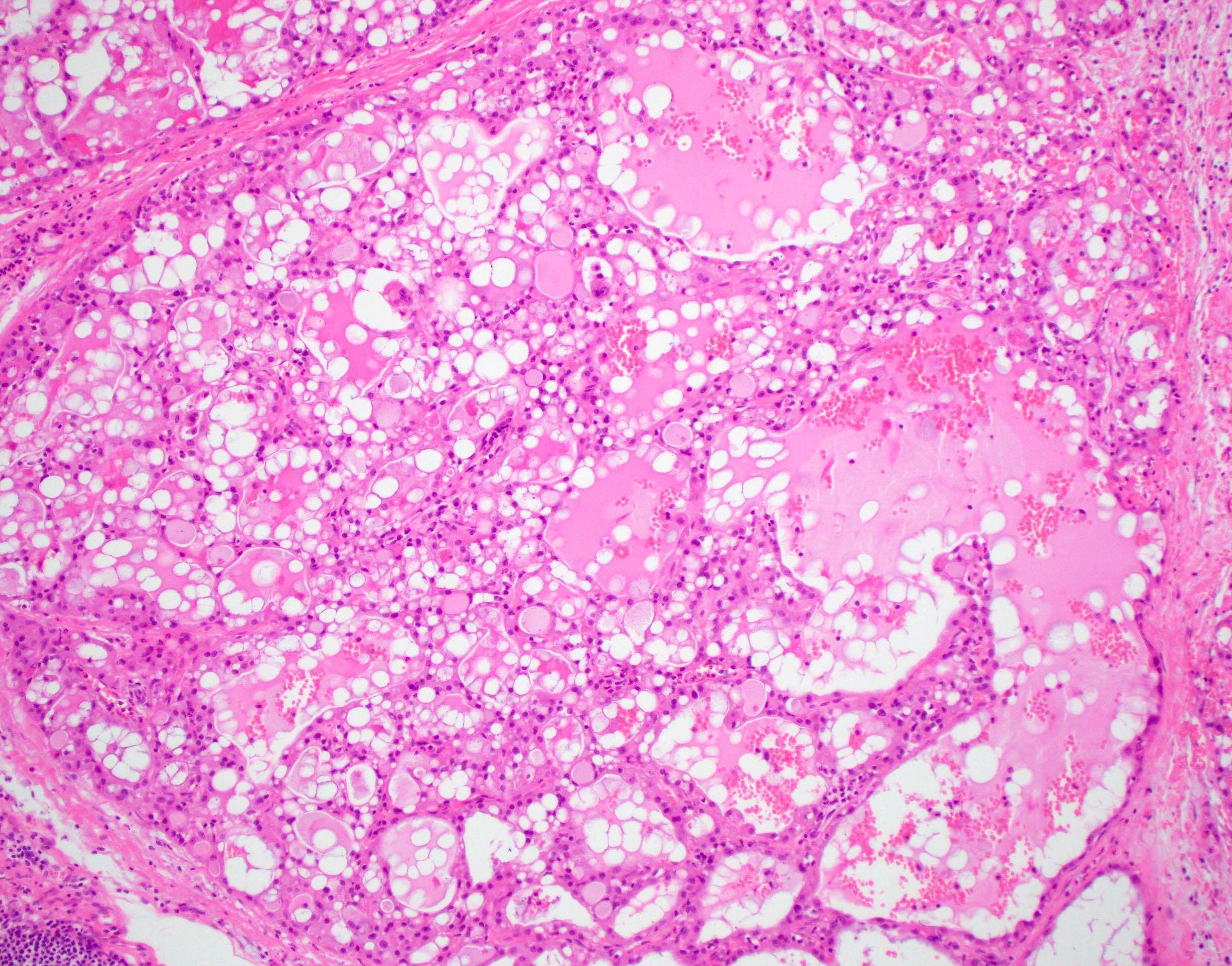
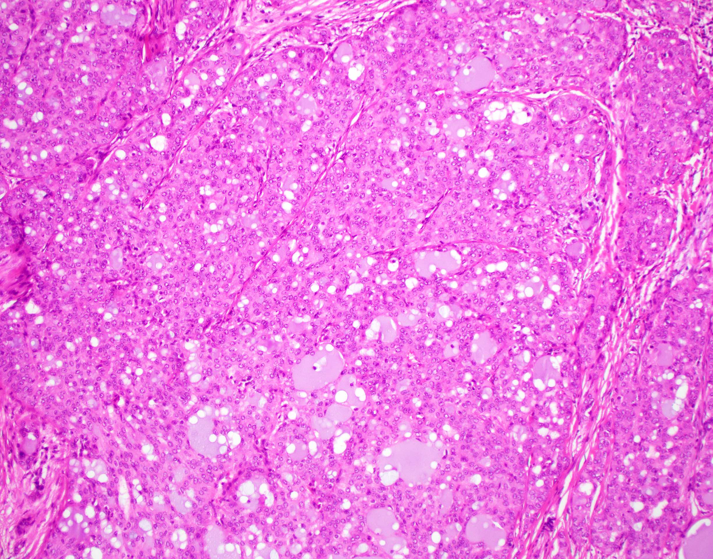
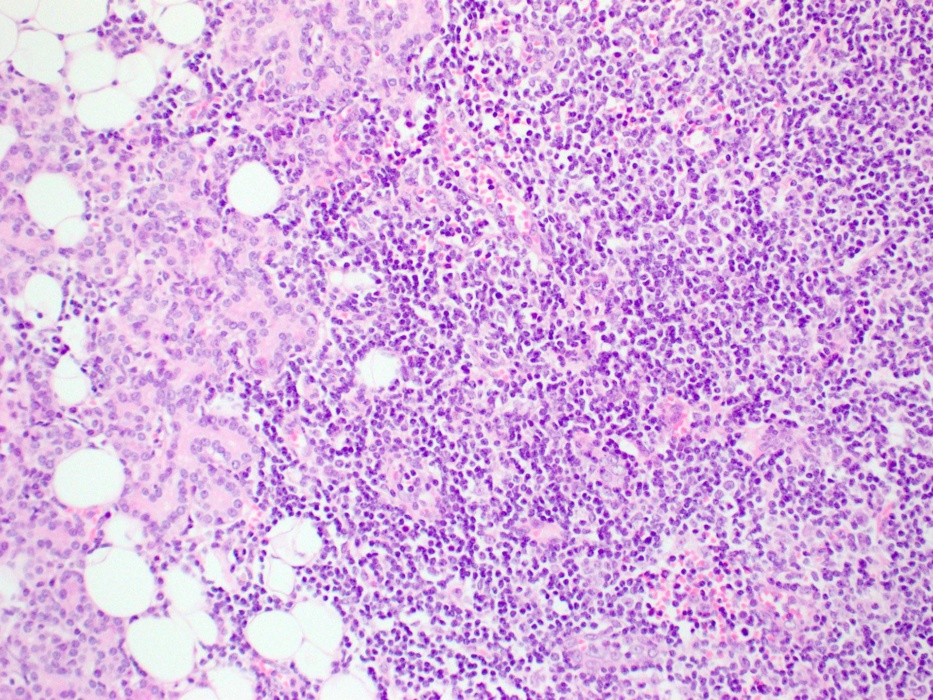
Finely vacuolated cytoplasm

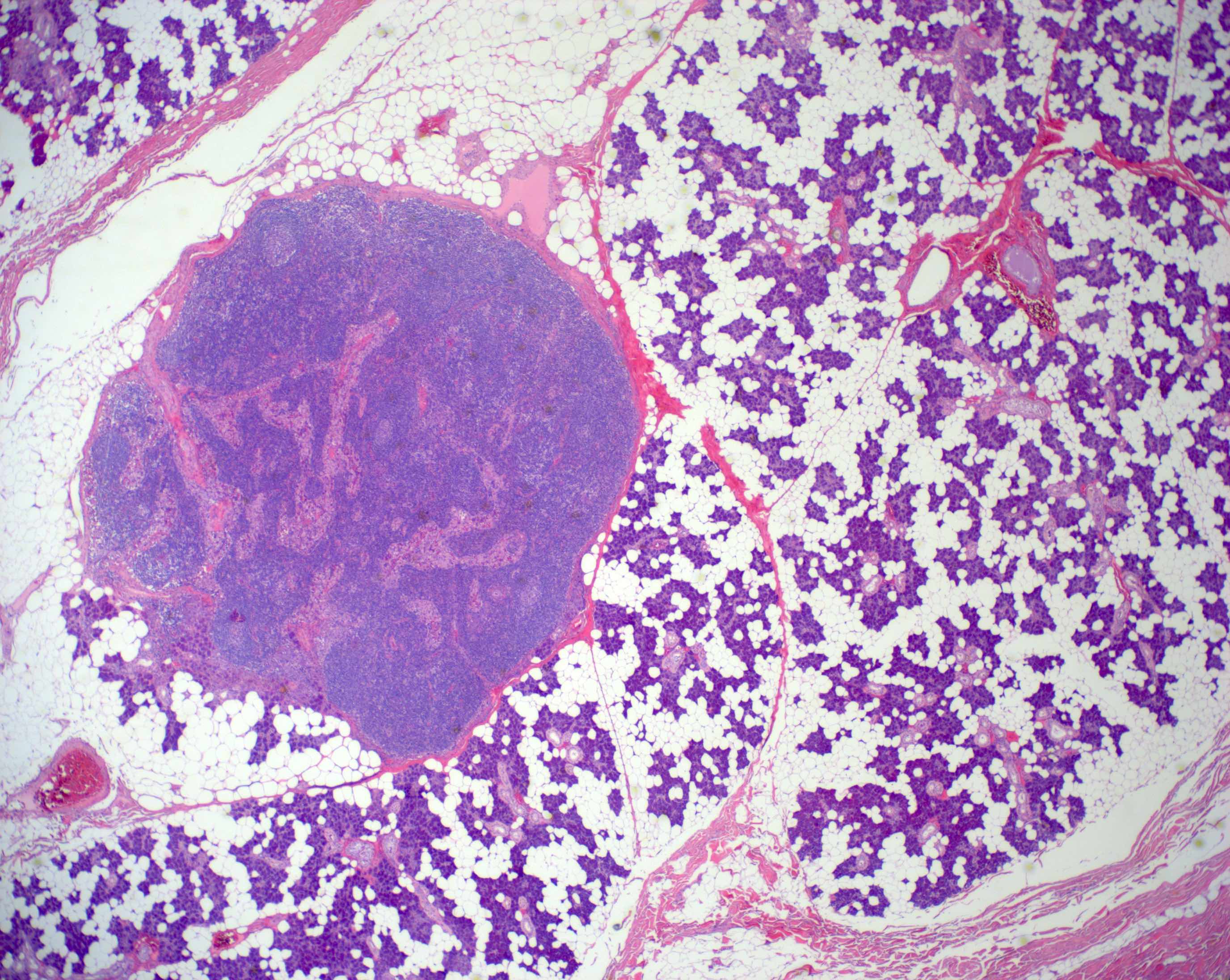
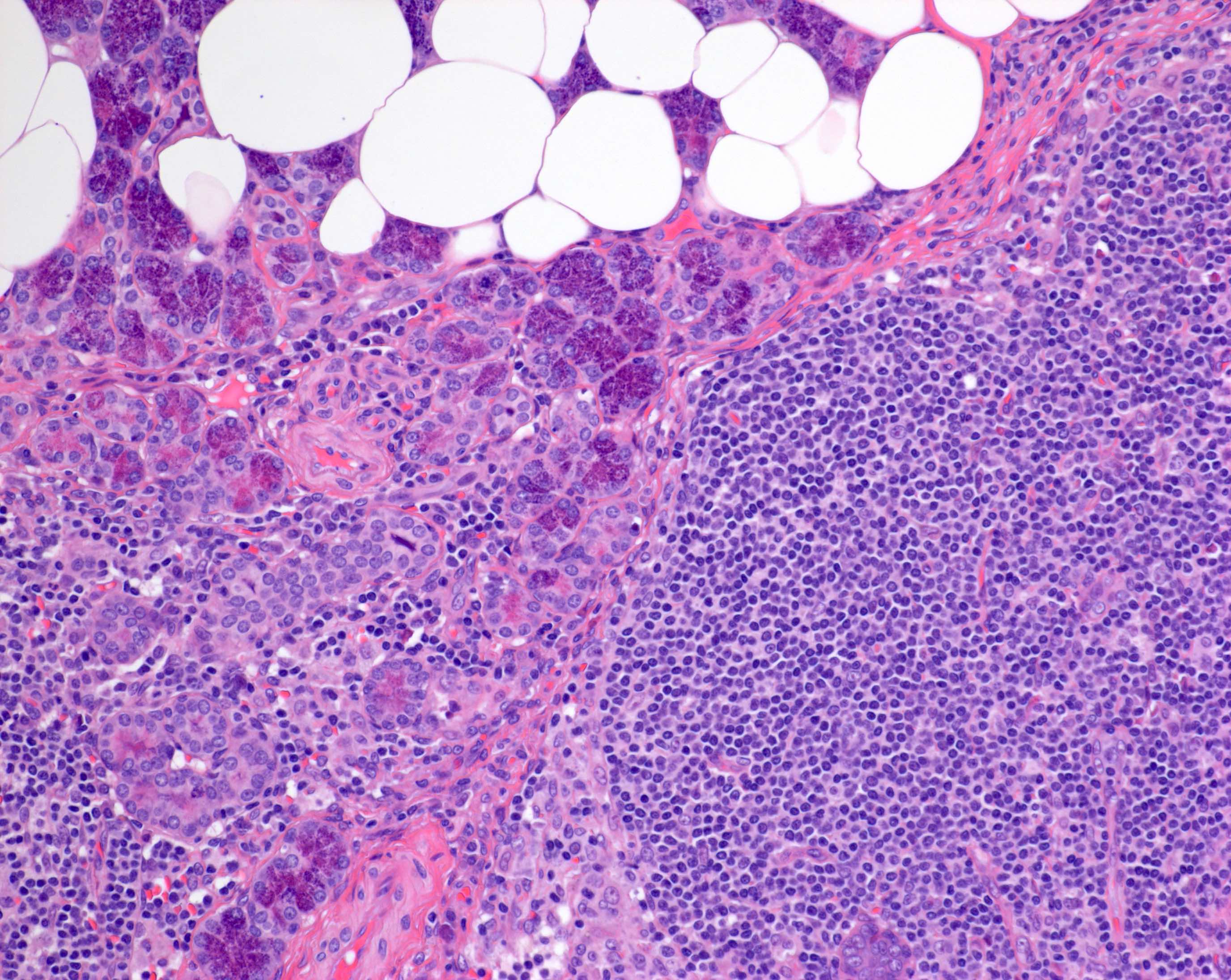
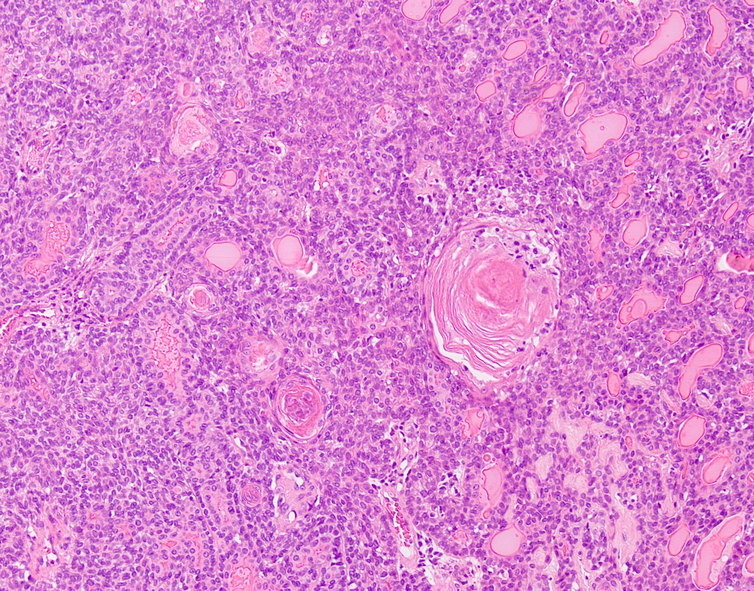
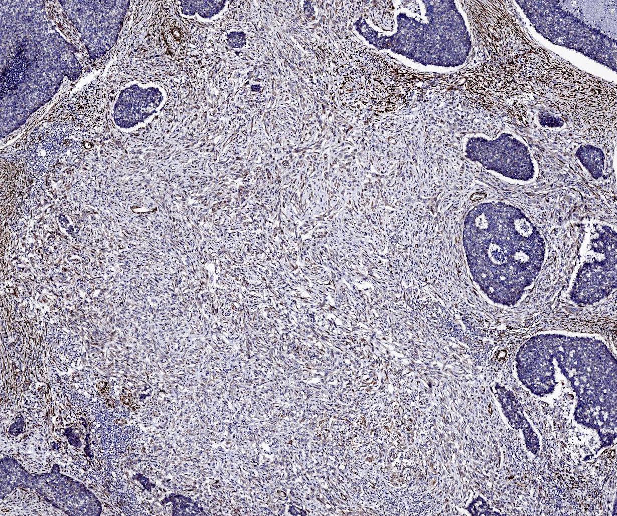
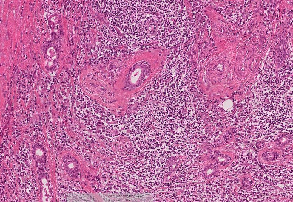
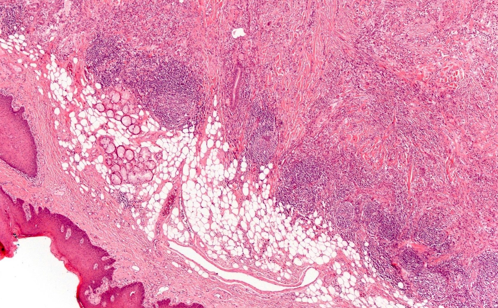
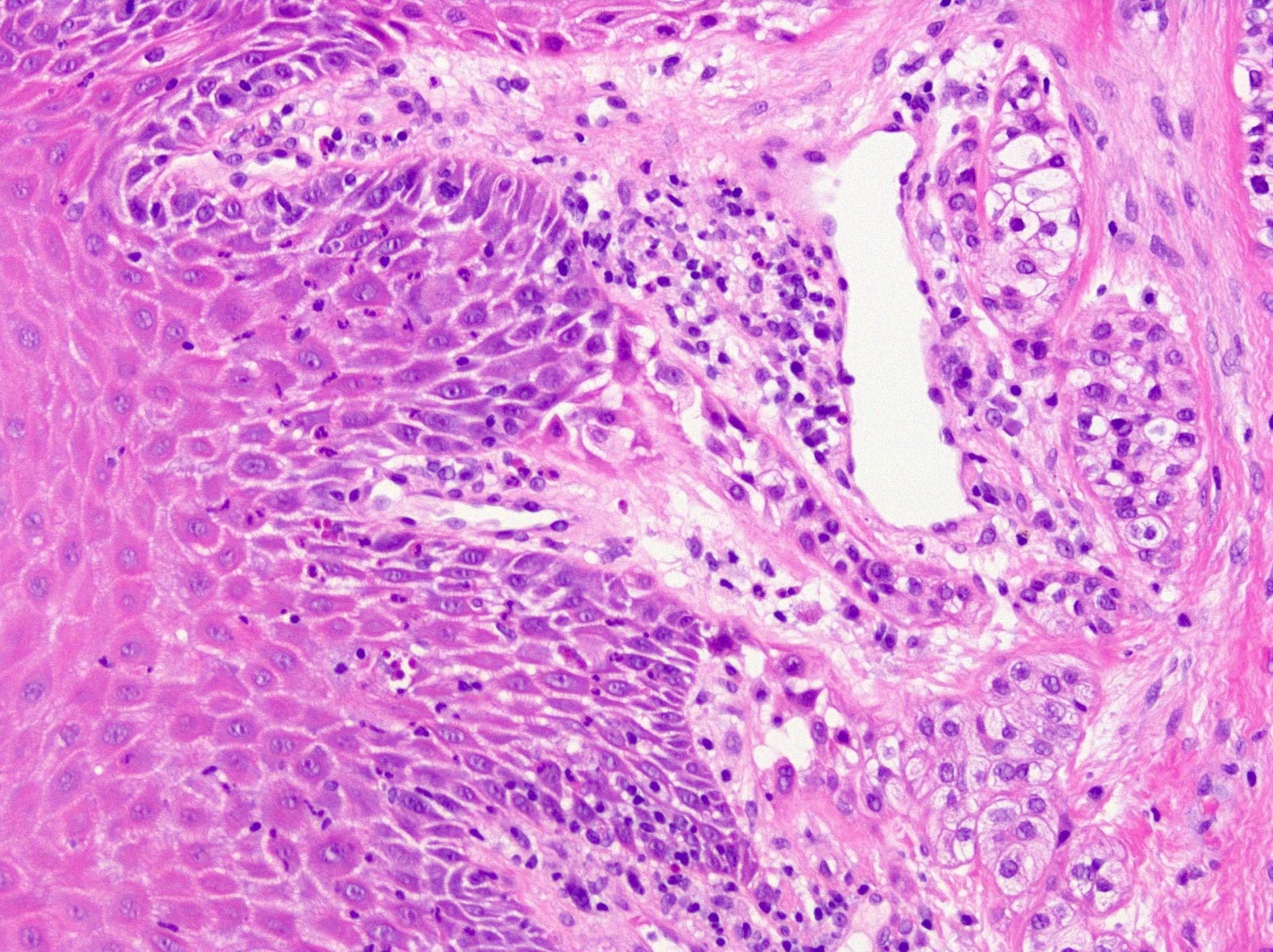
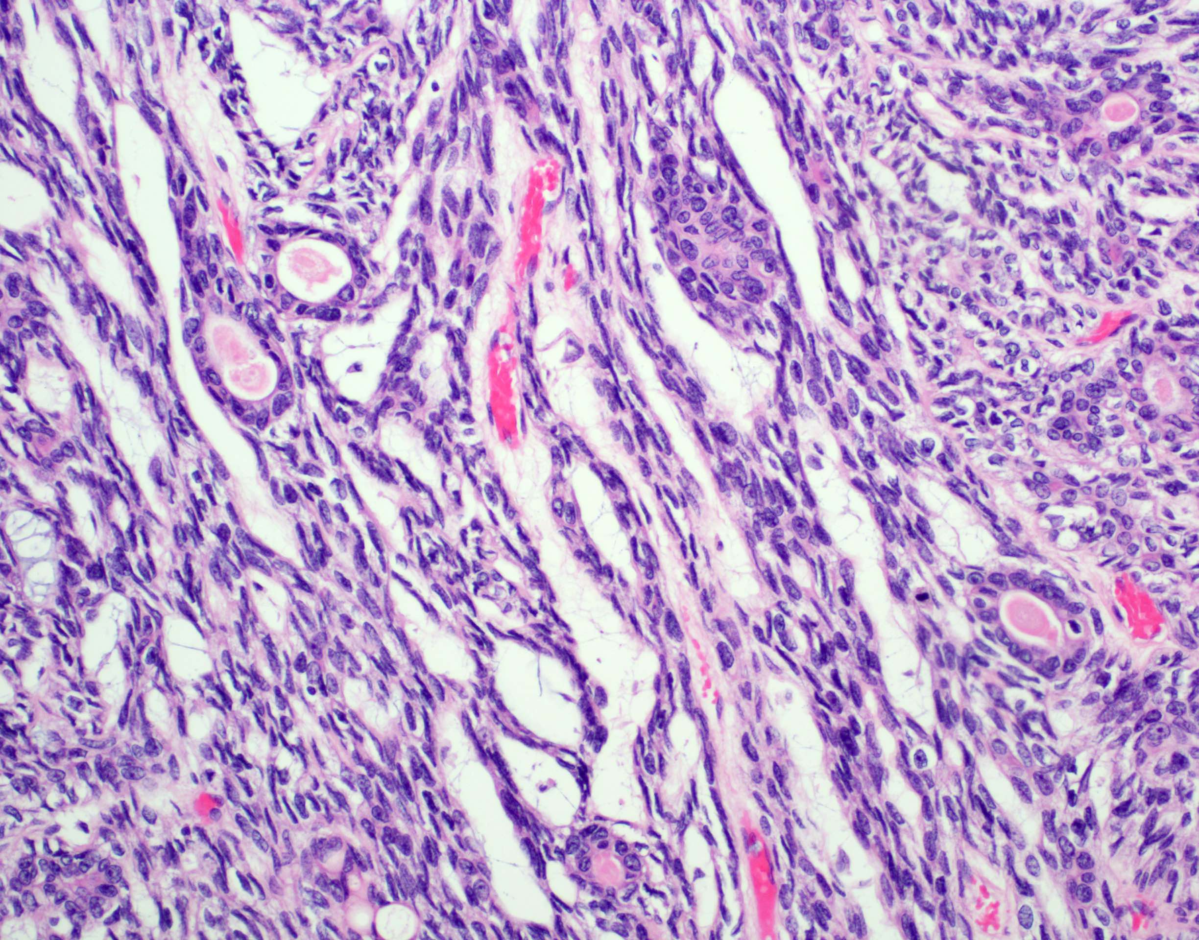
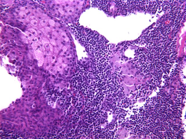
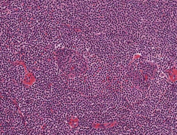
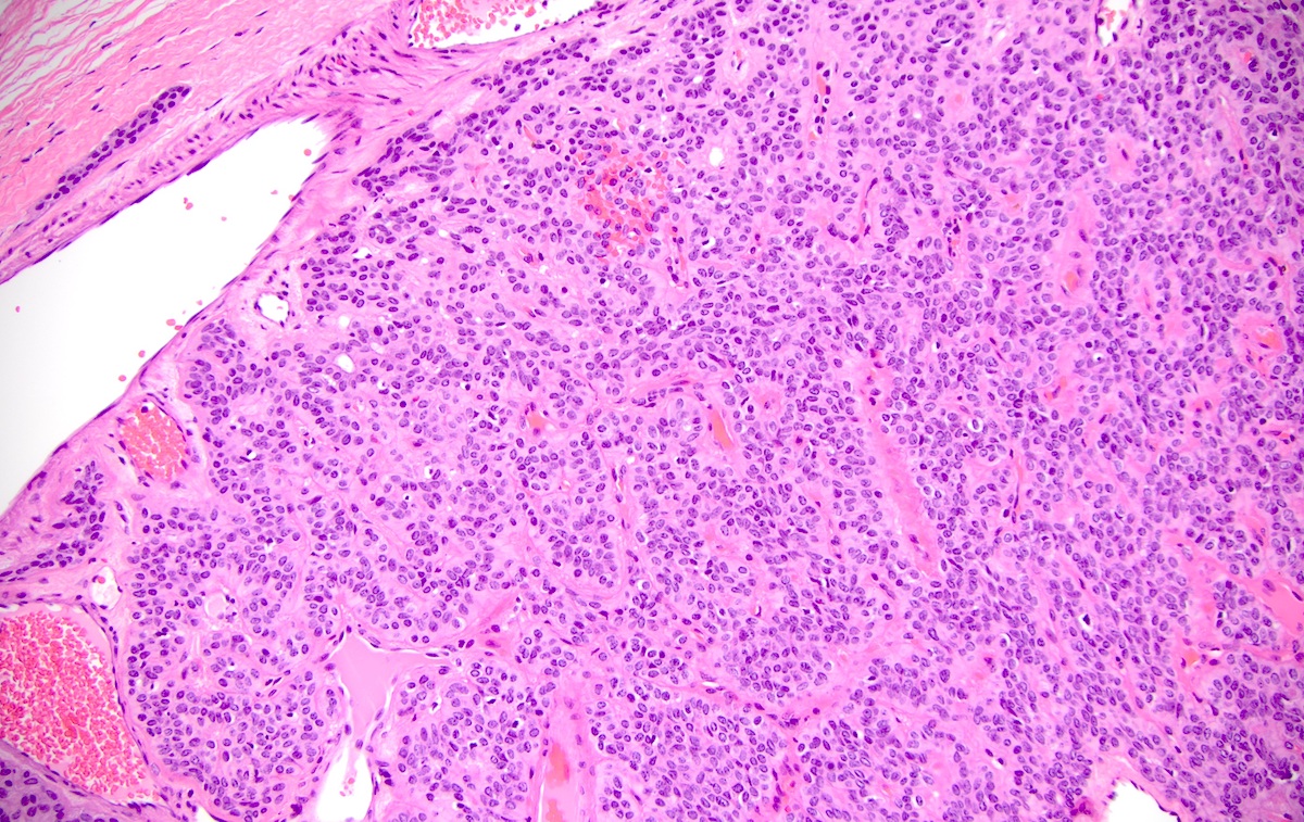
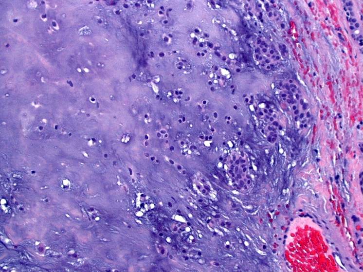
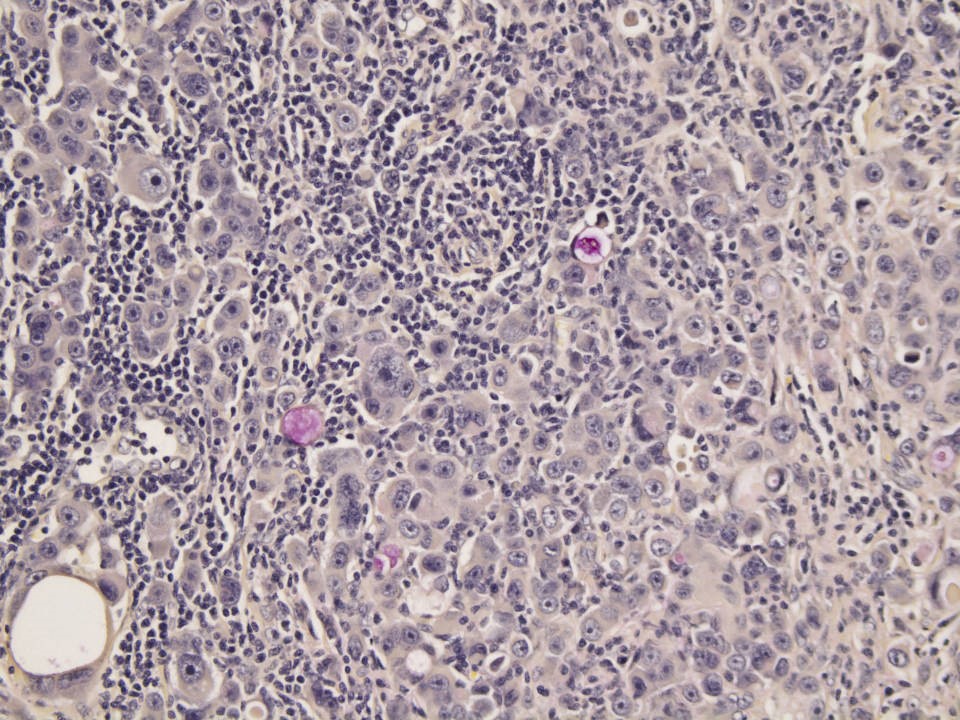
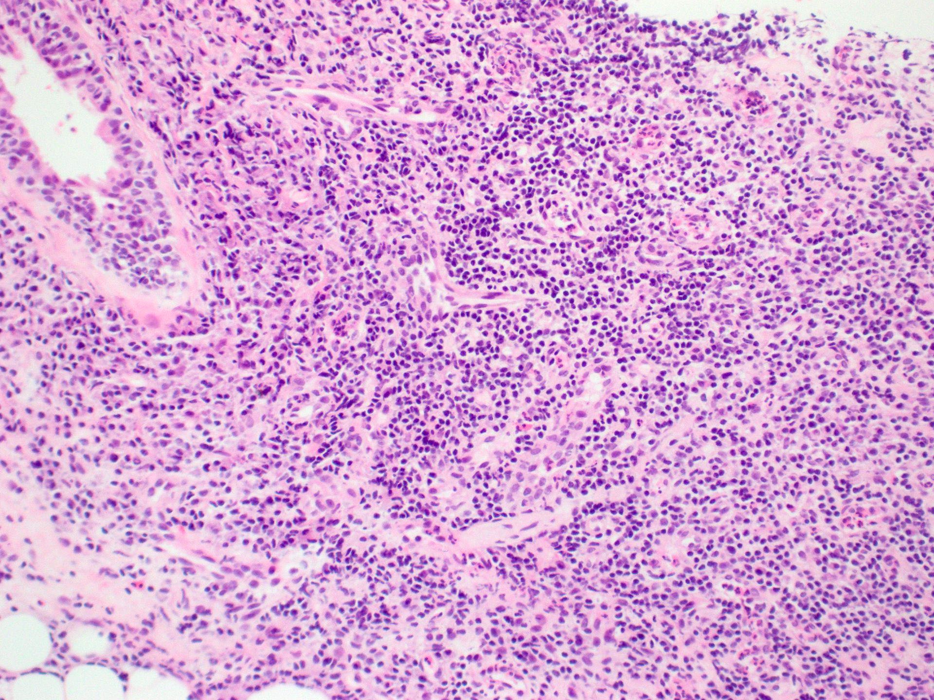
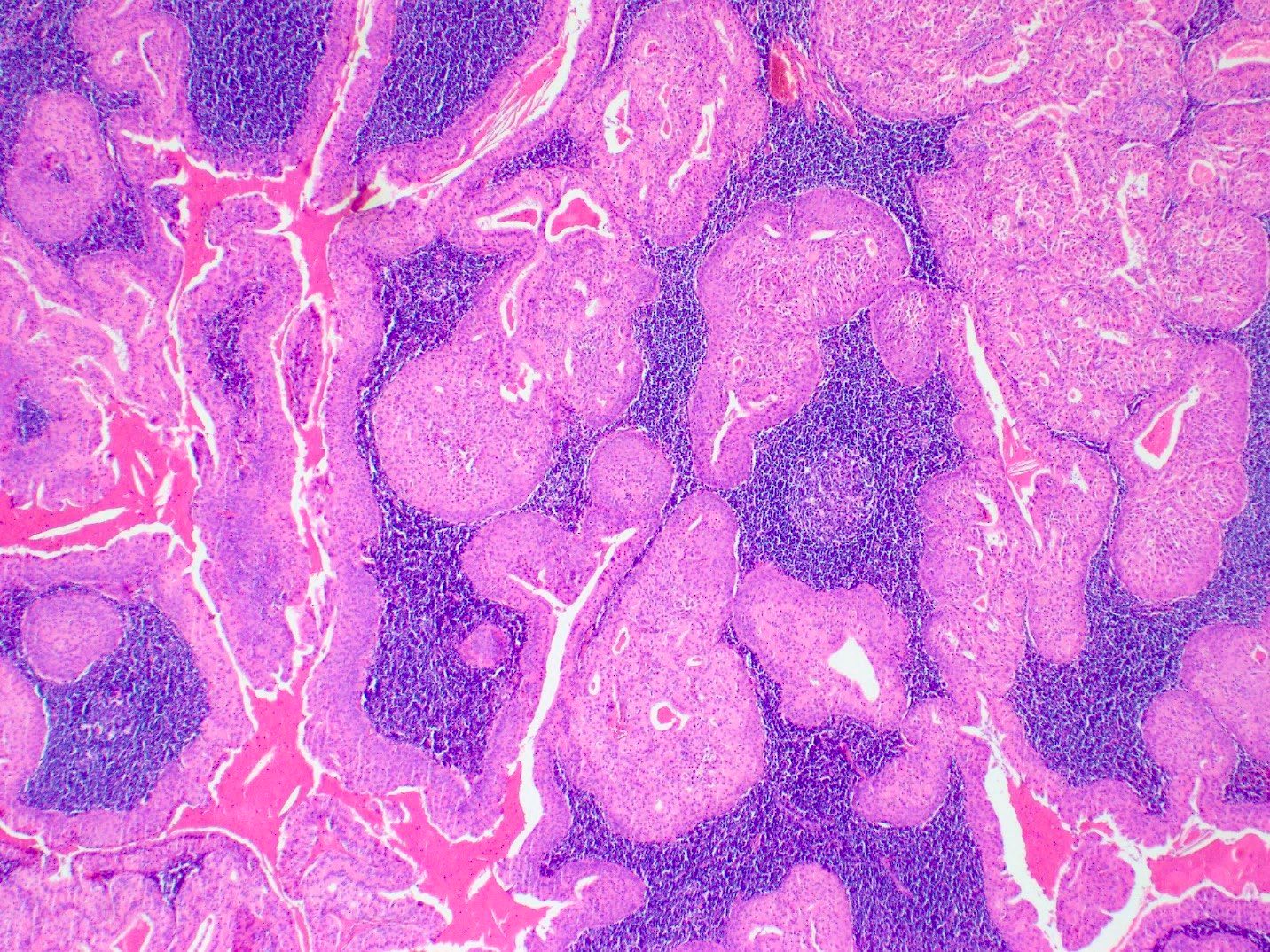
Tumor associated lymphoid proliferation
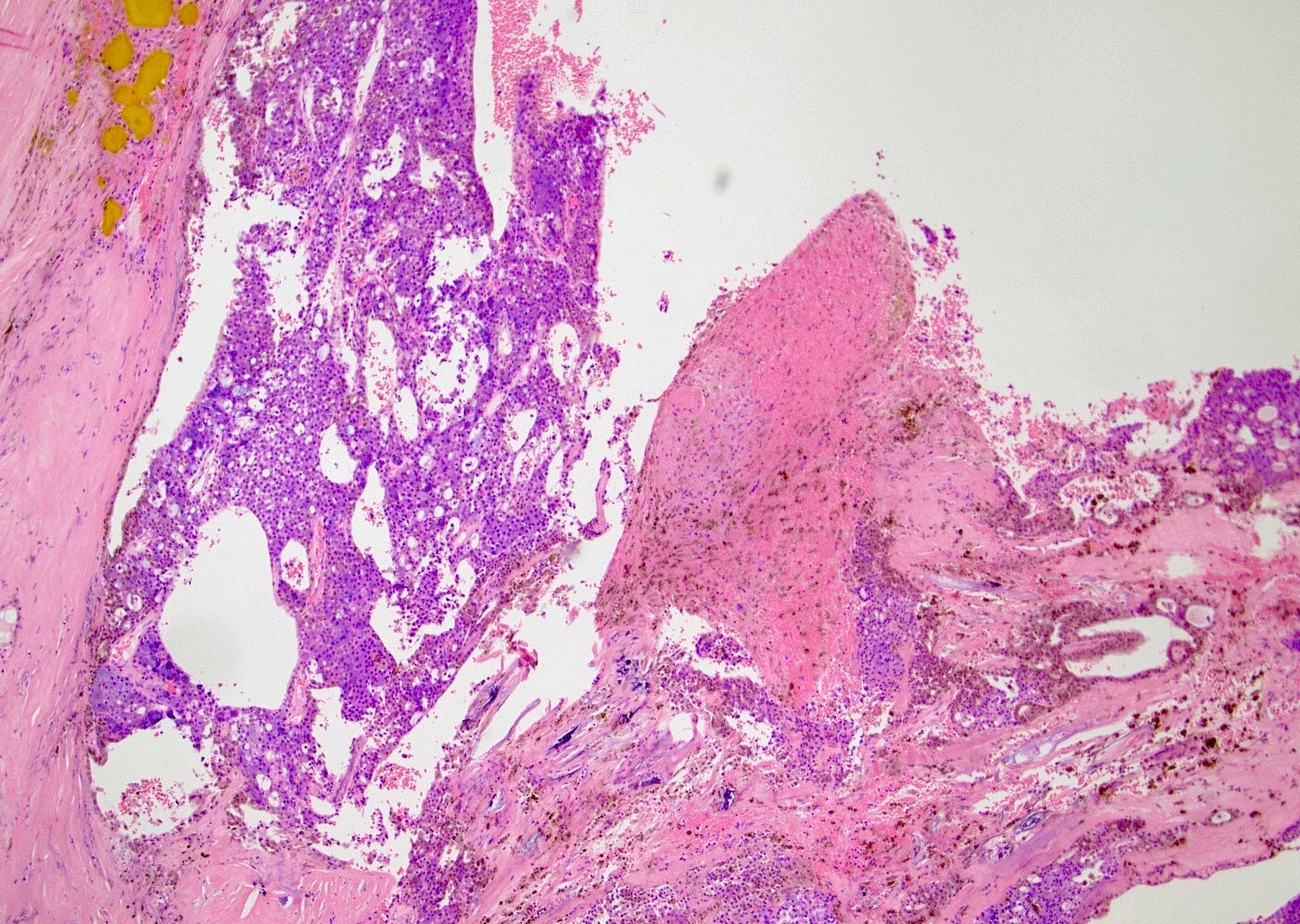

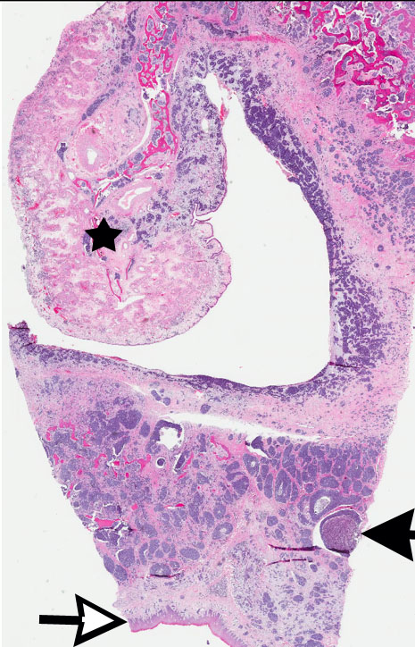
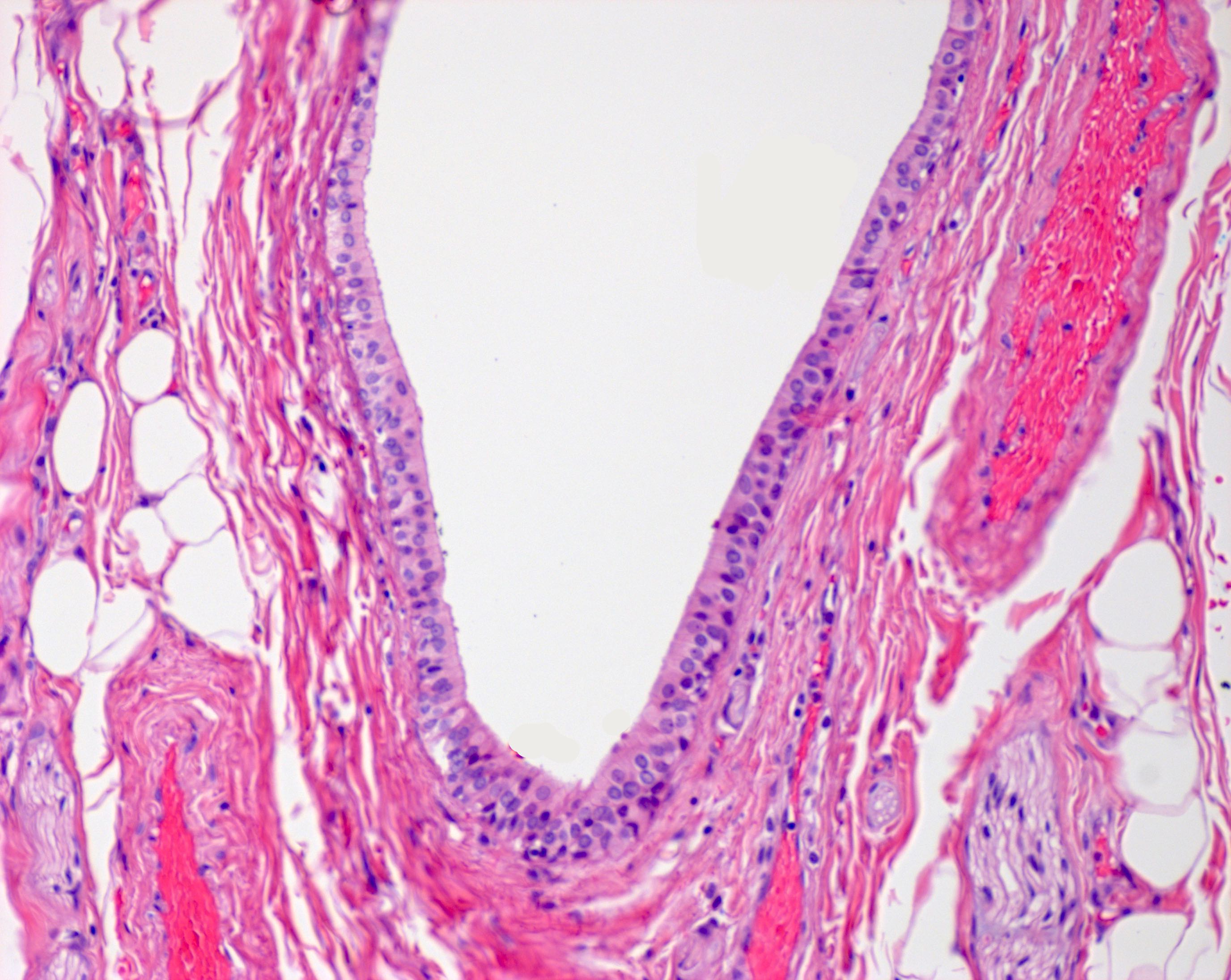
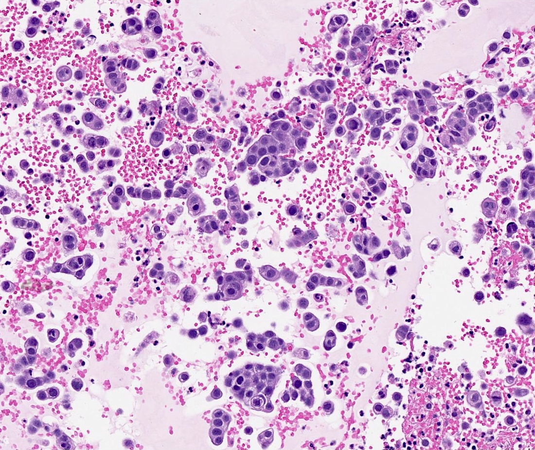
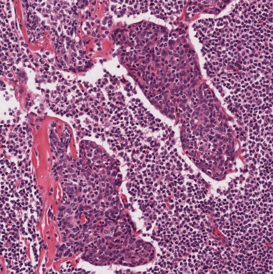
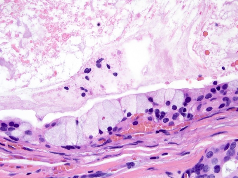
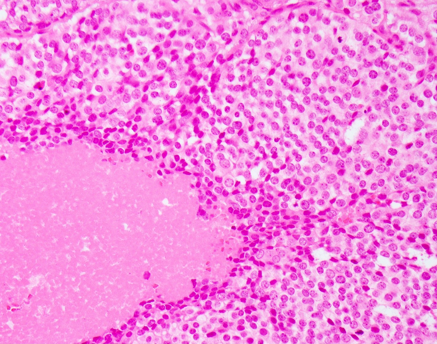
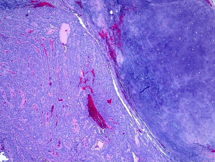
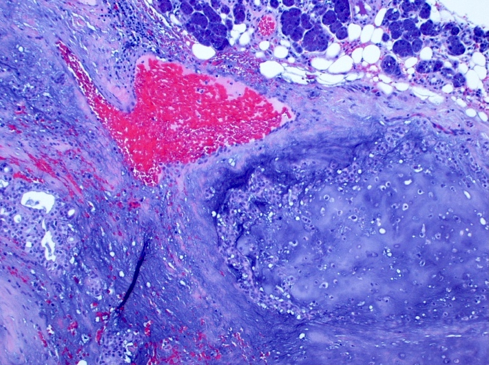
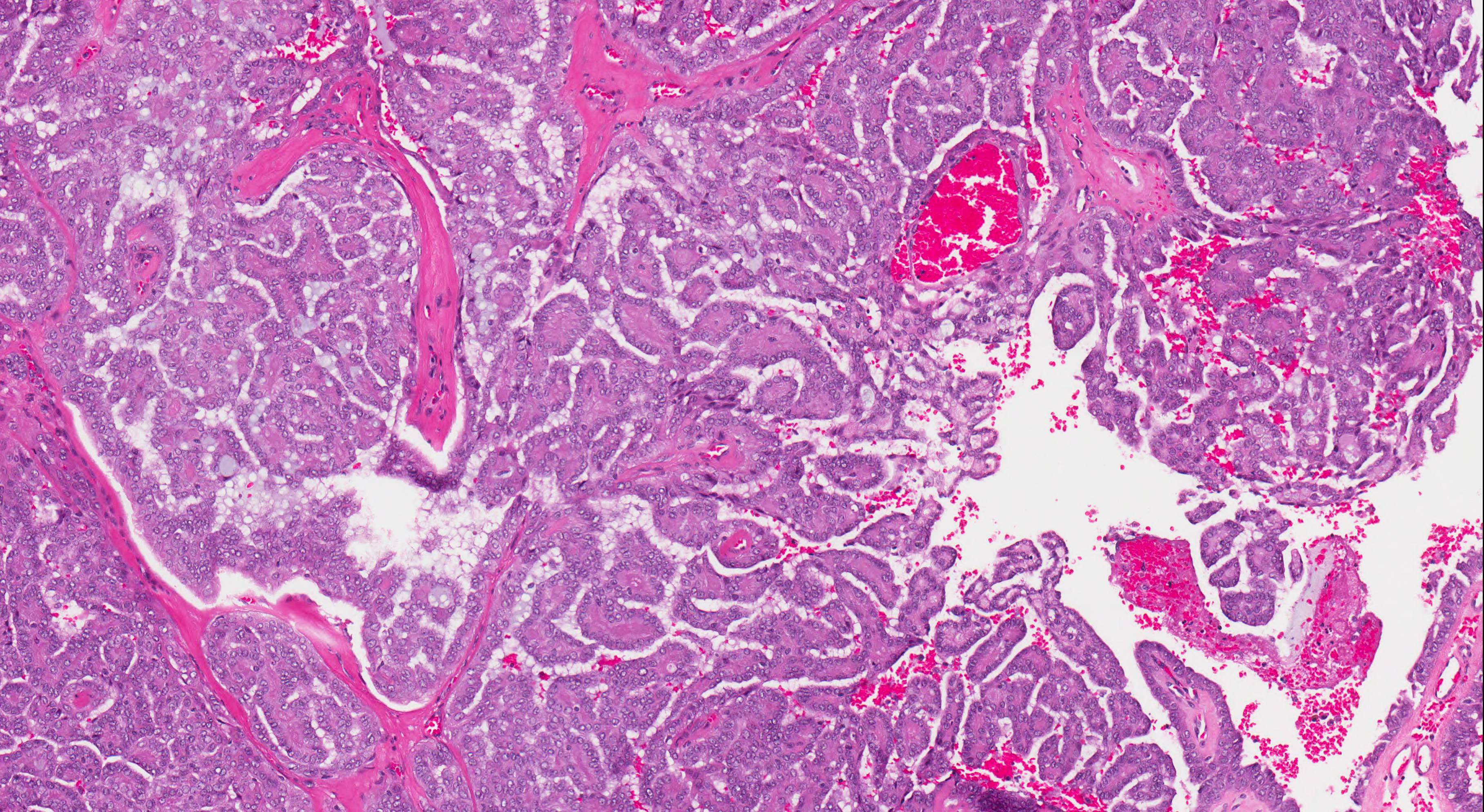
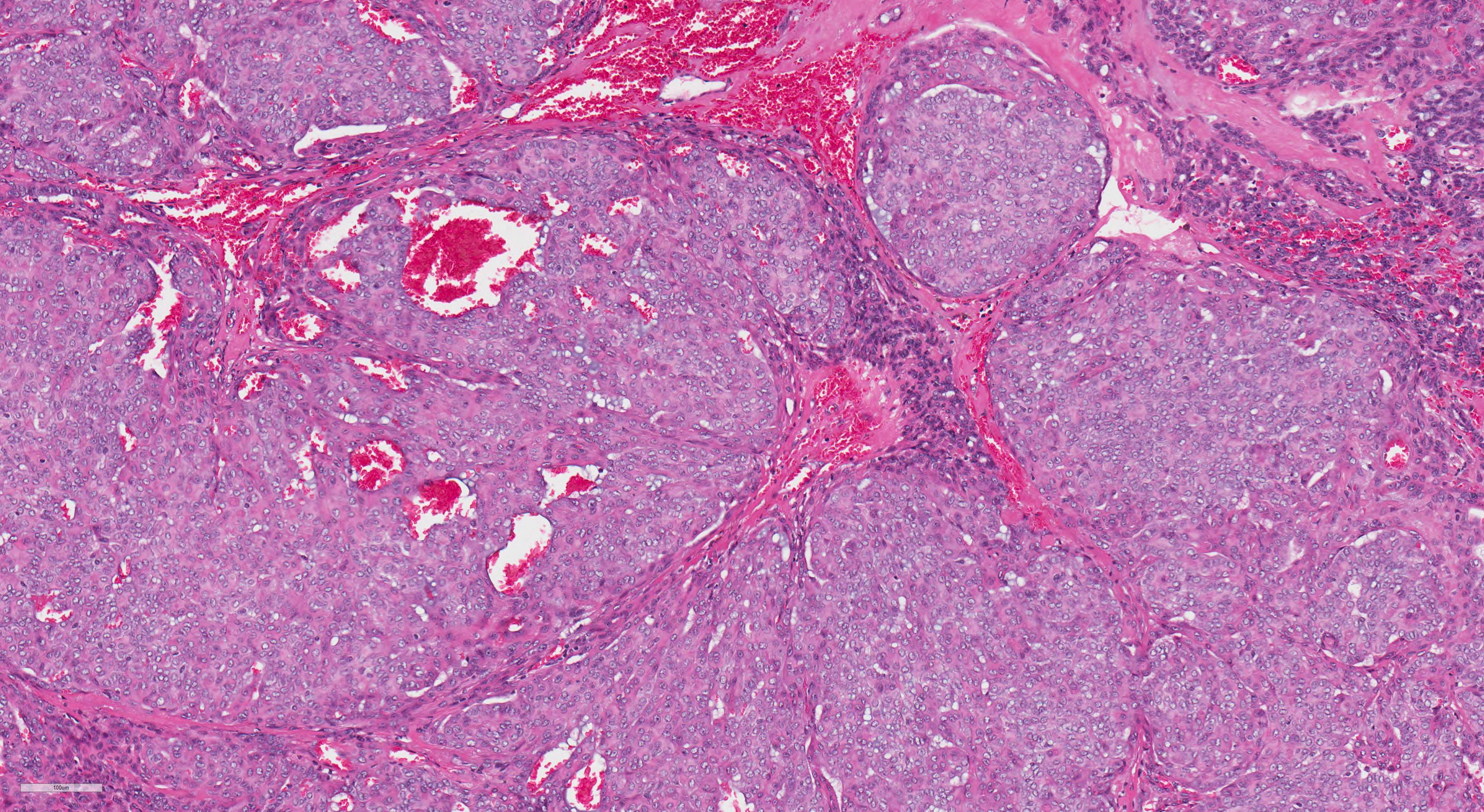
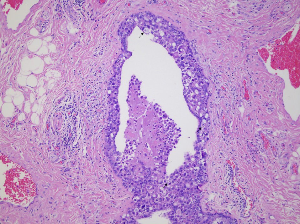
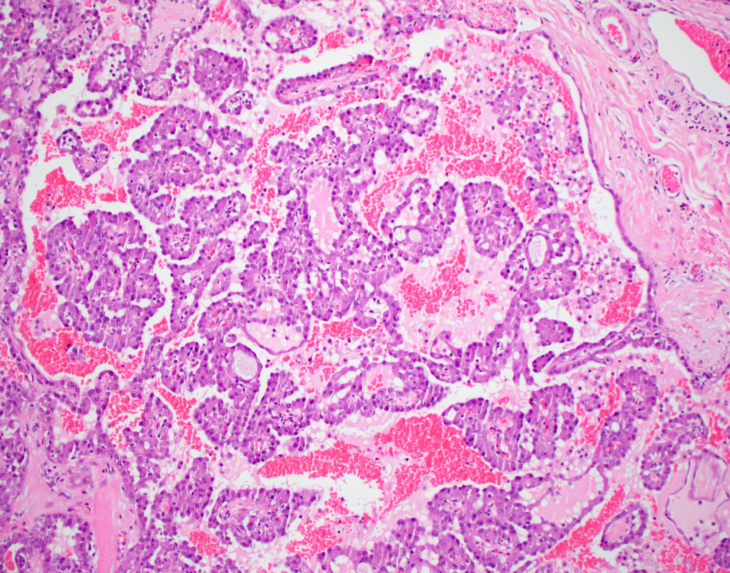
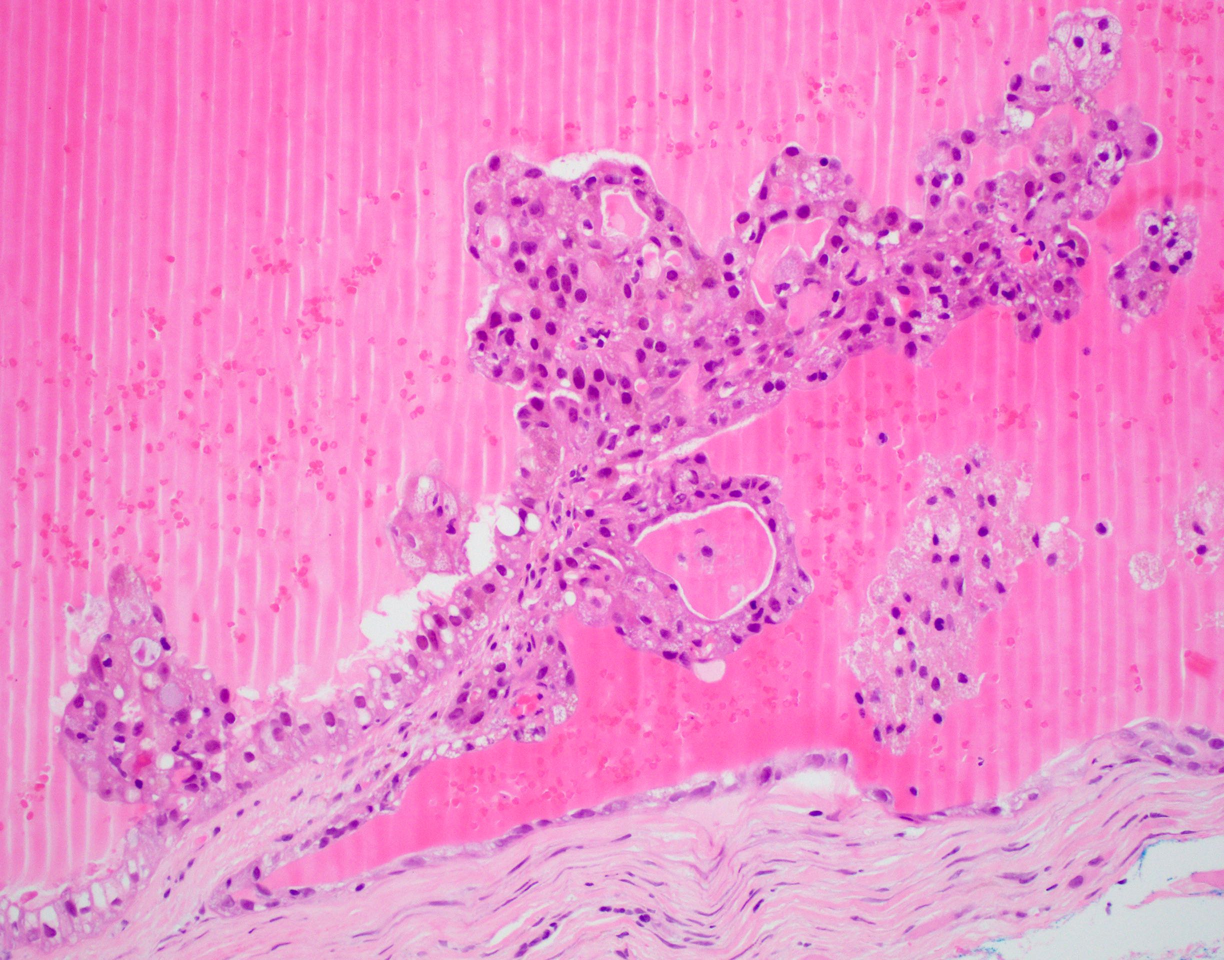
Cystic pattern and hemorrhage
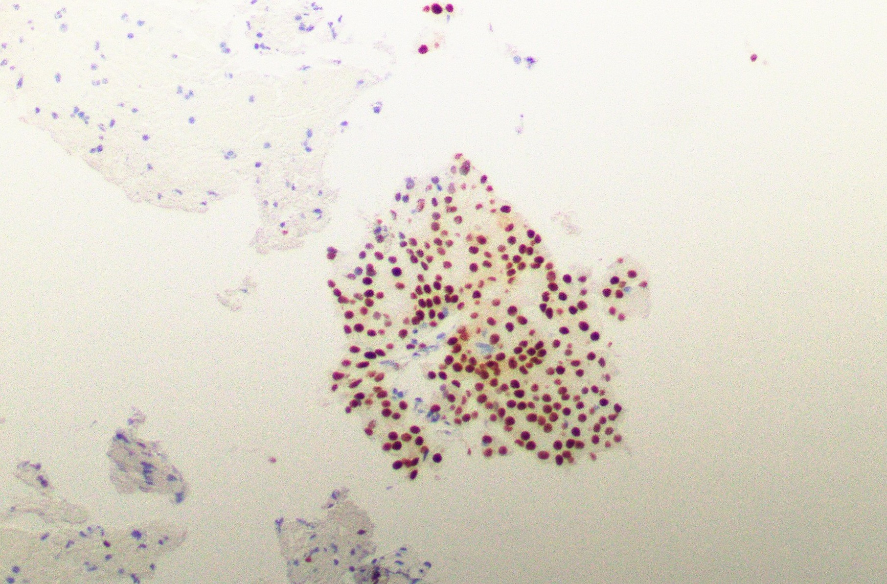
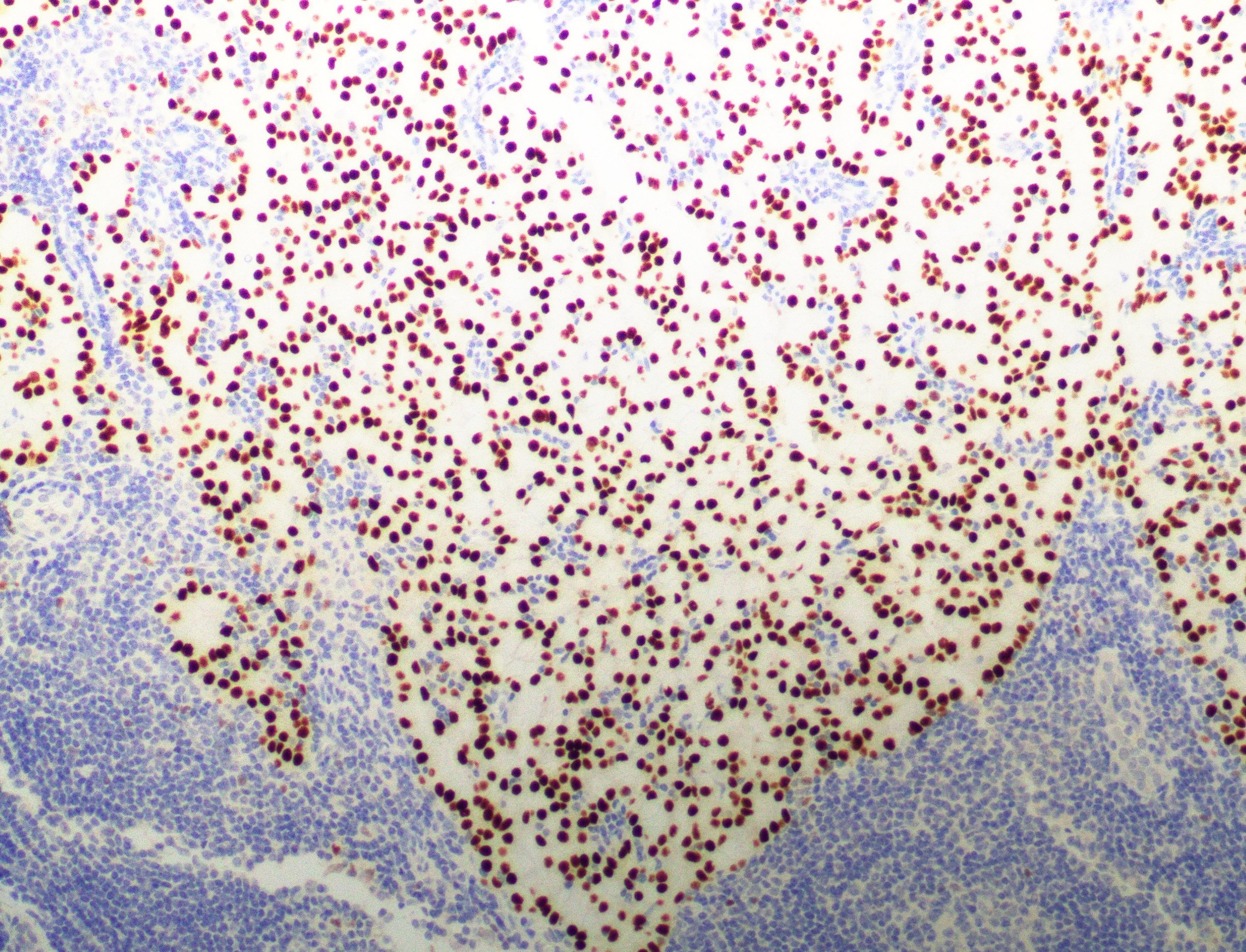
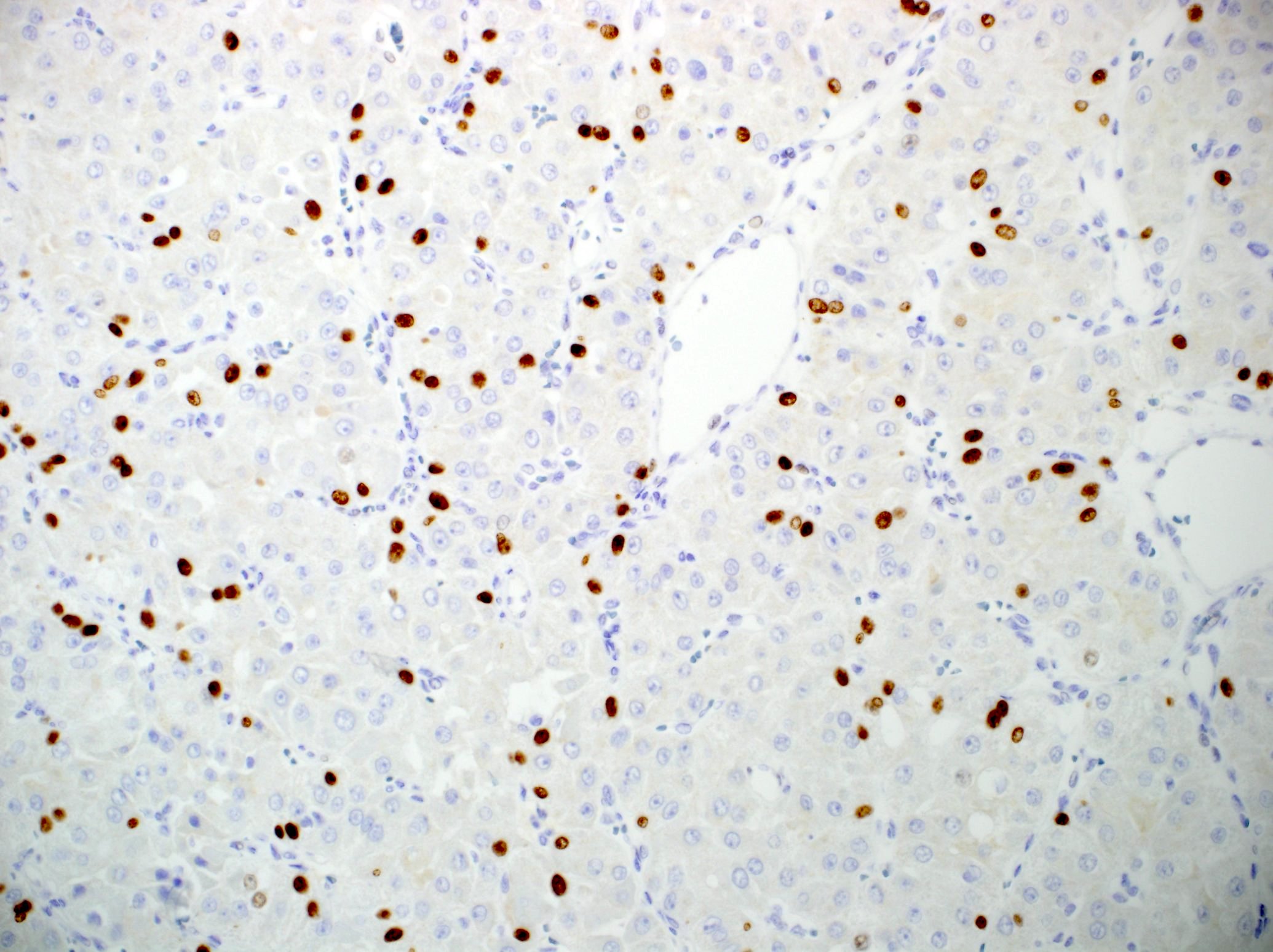
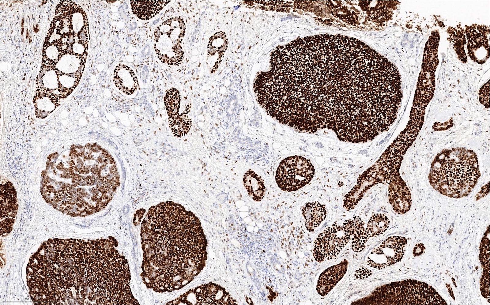
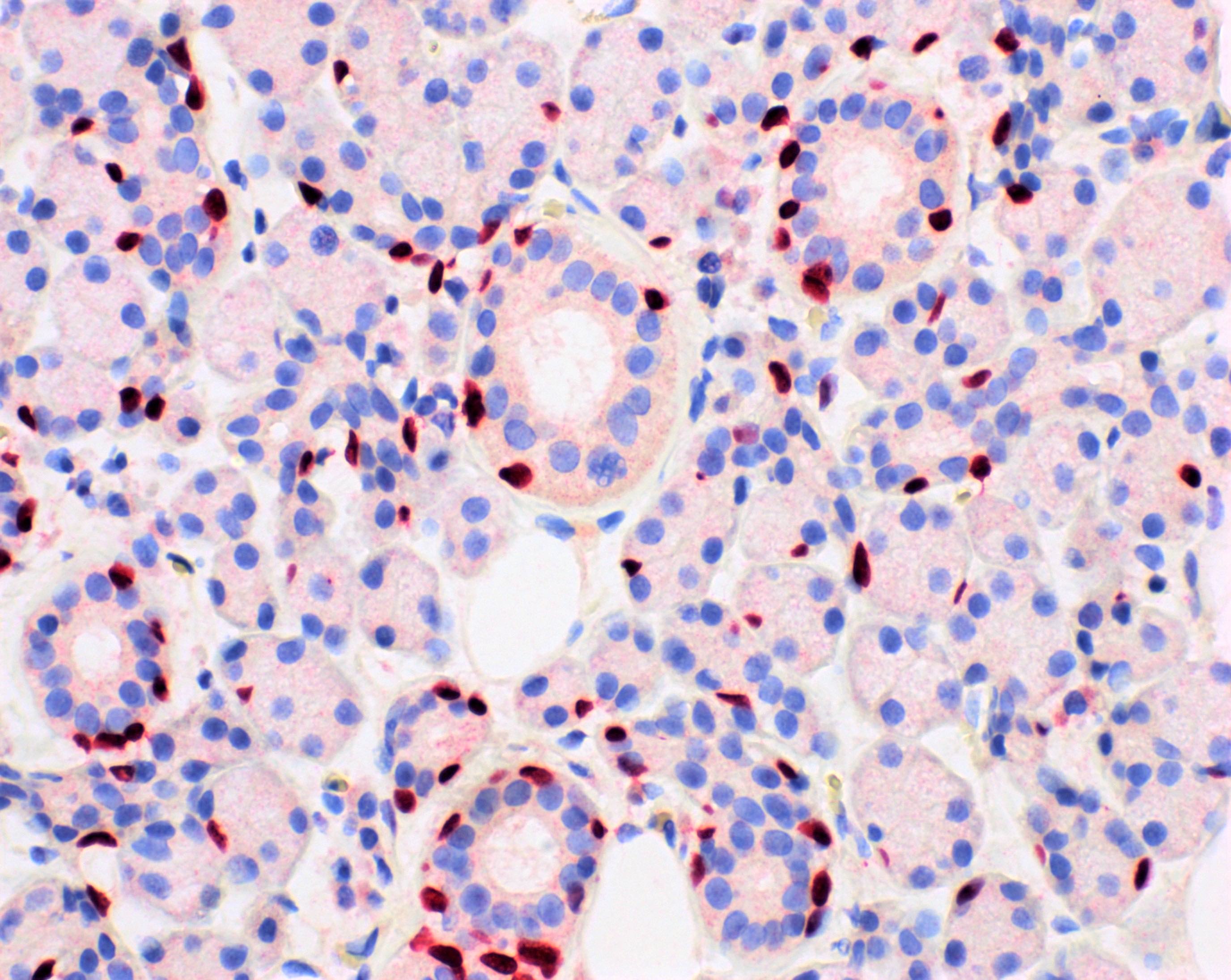
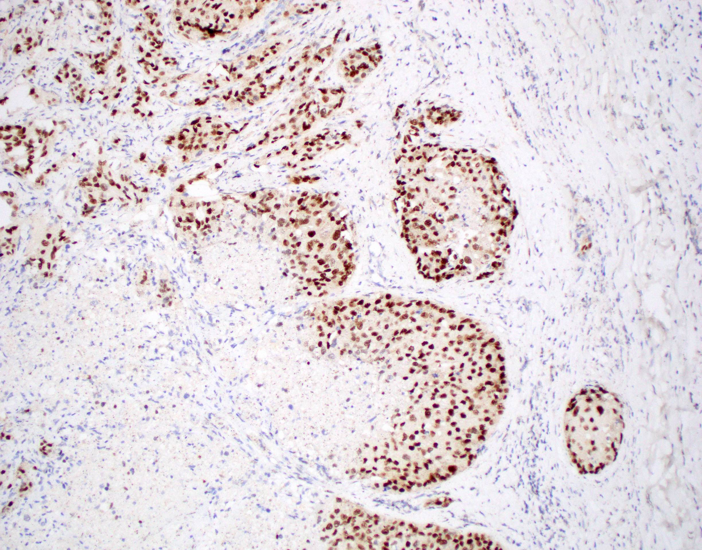
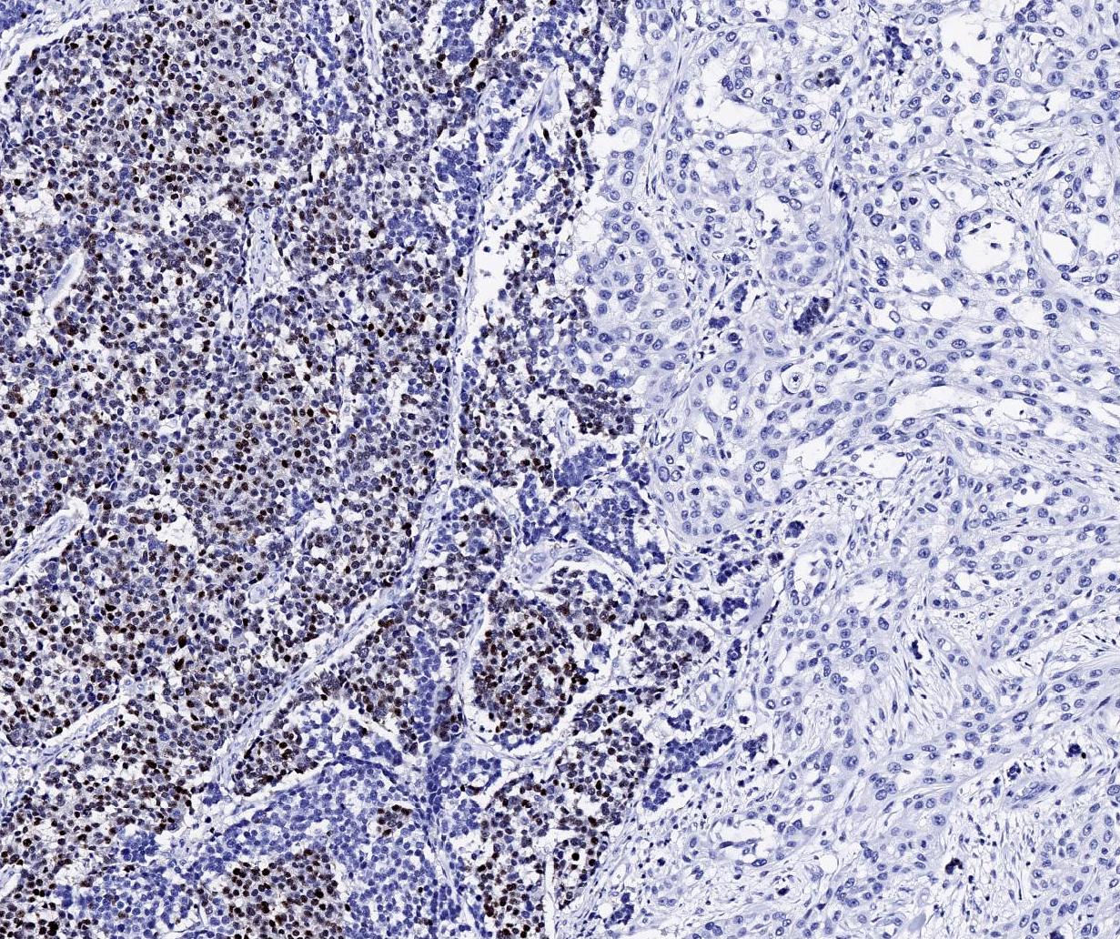
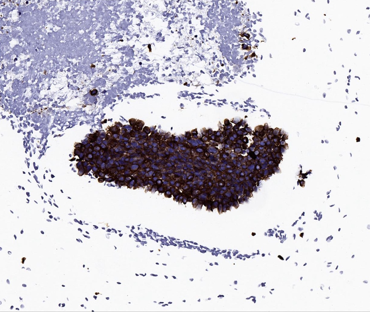
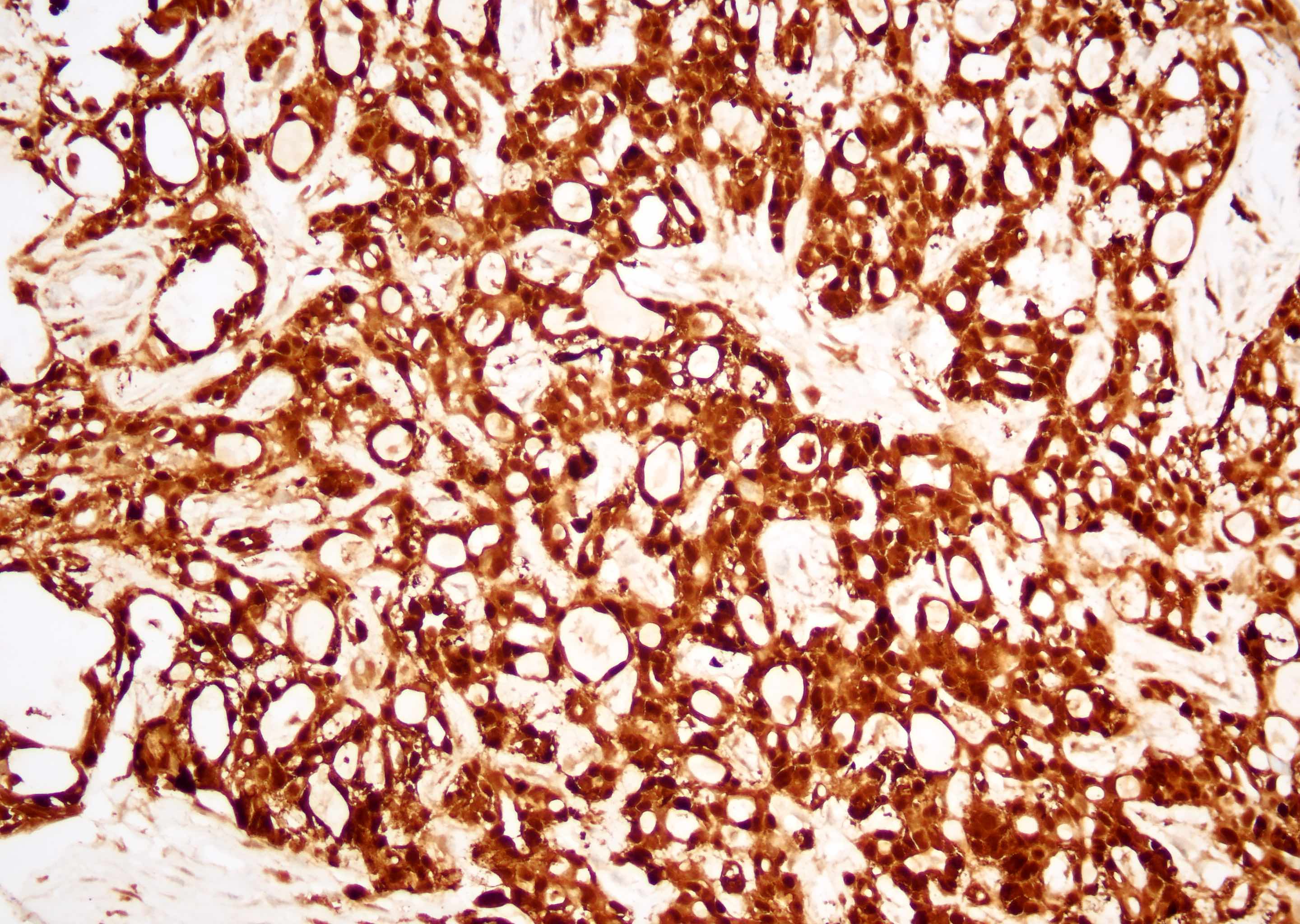
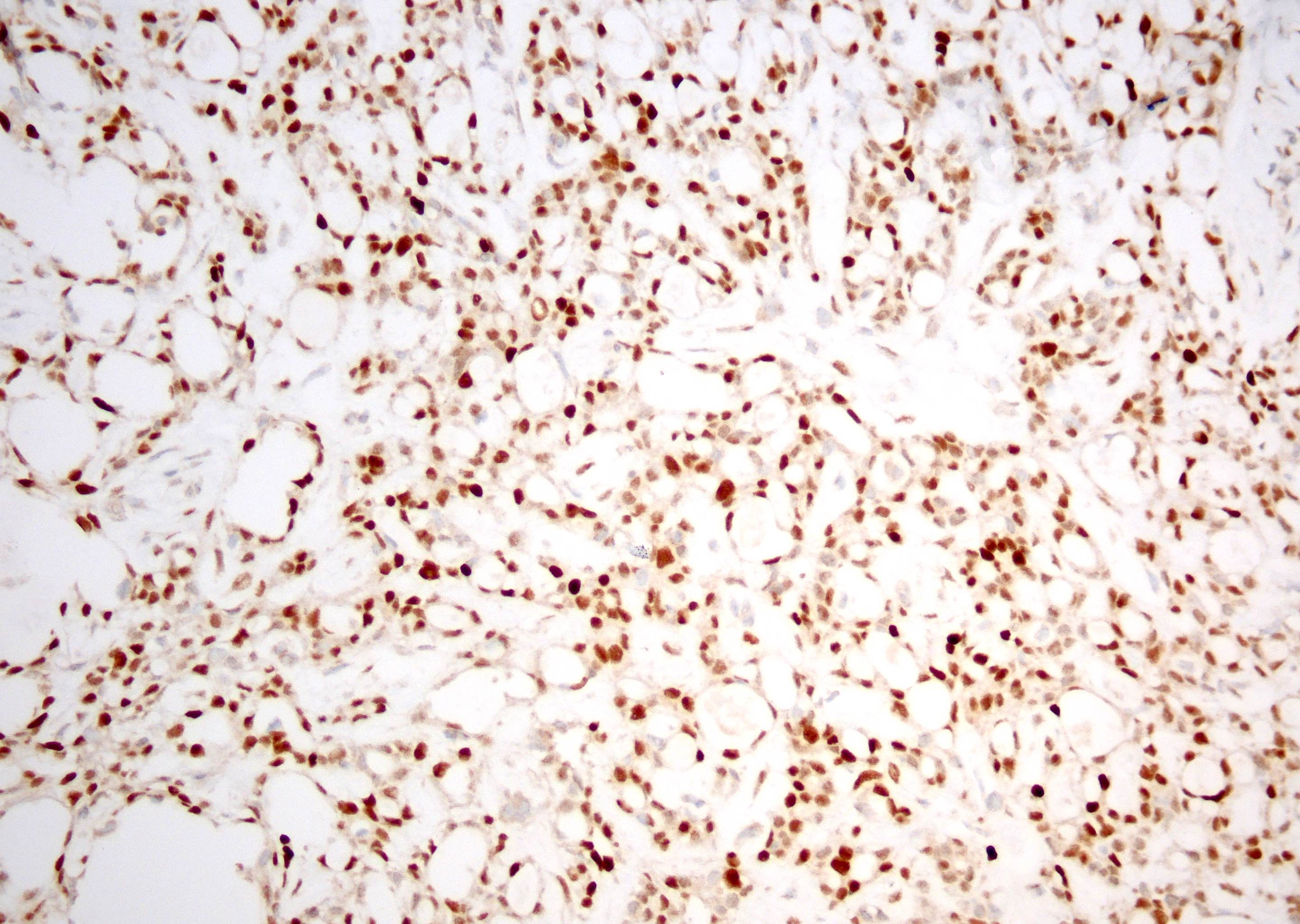
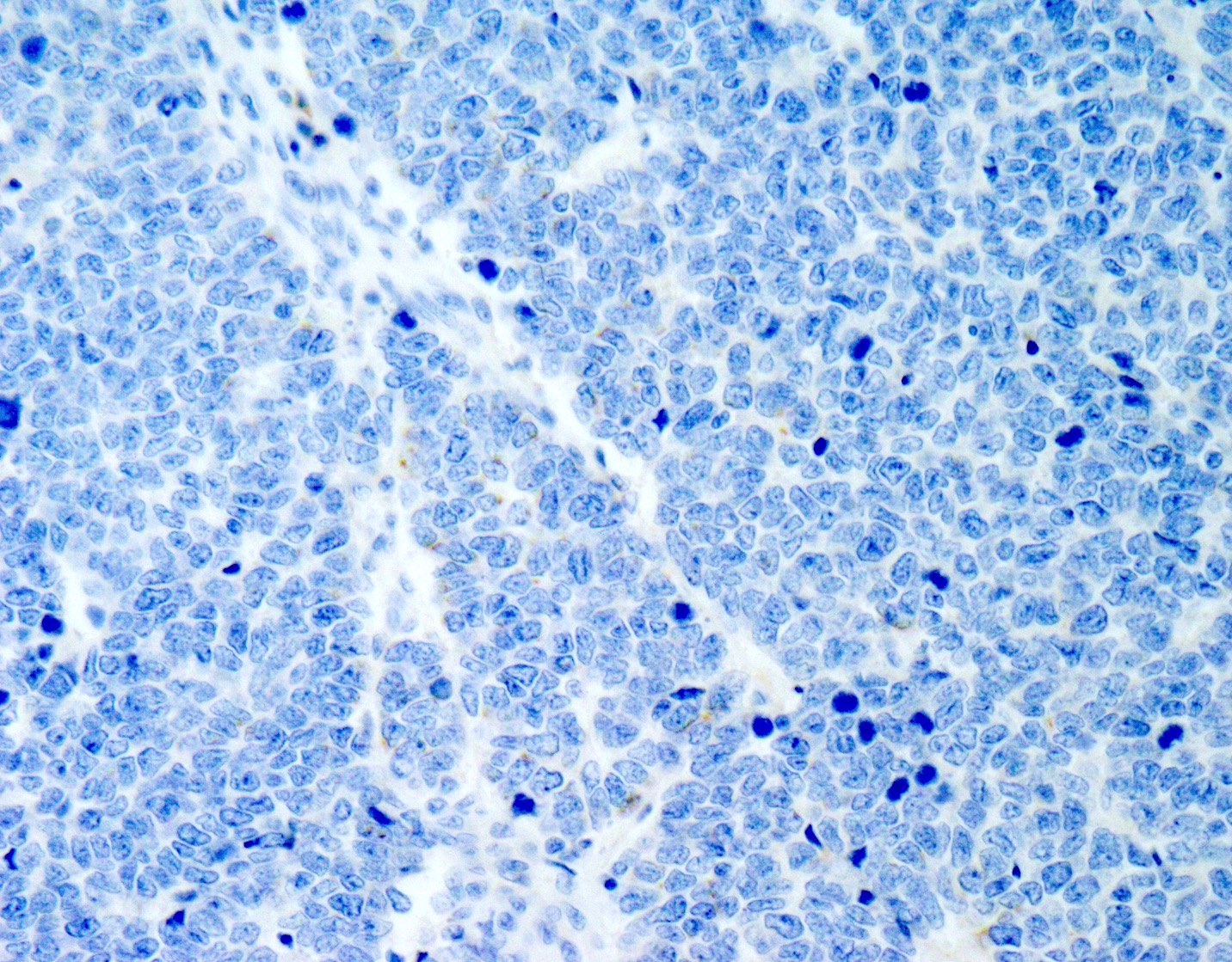
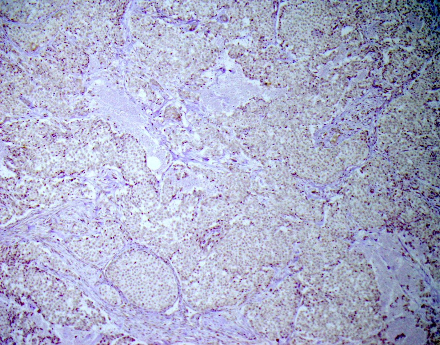
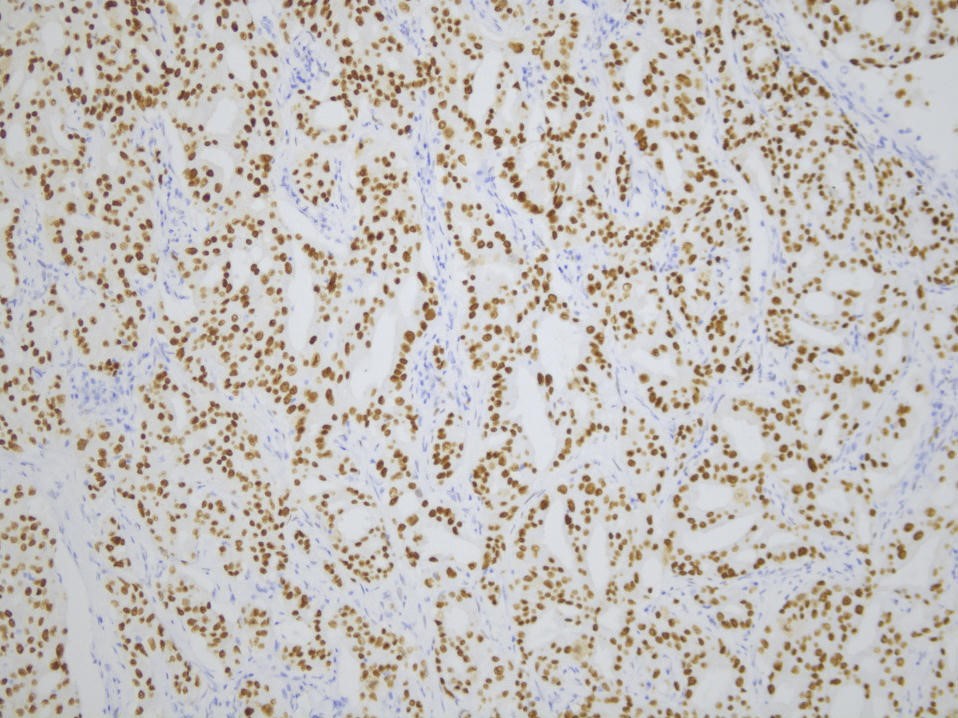
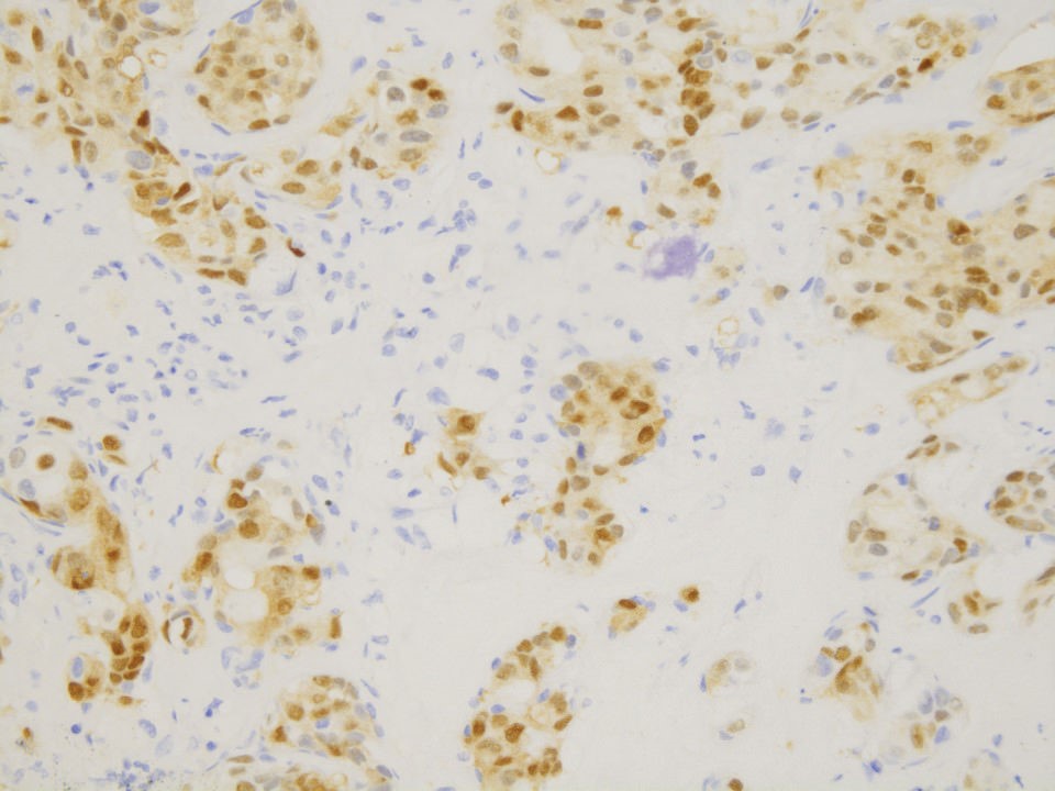
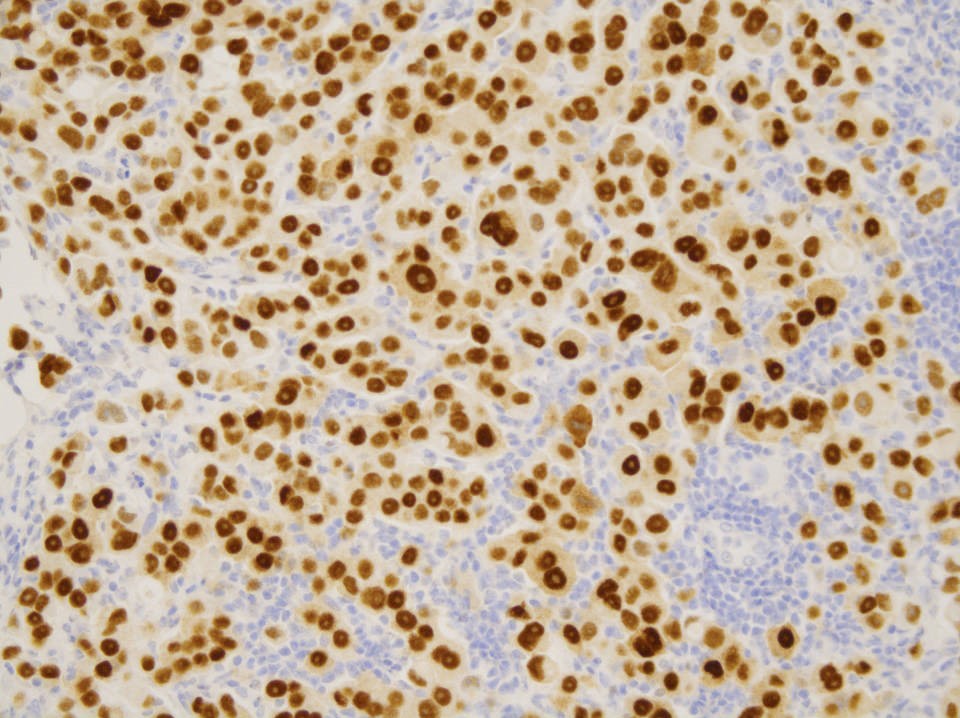
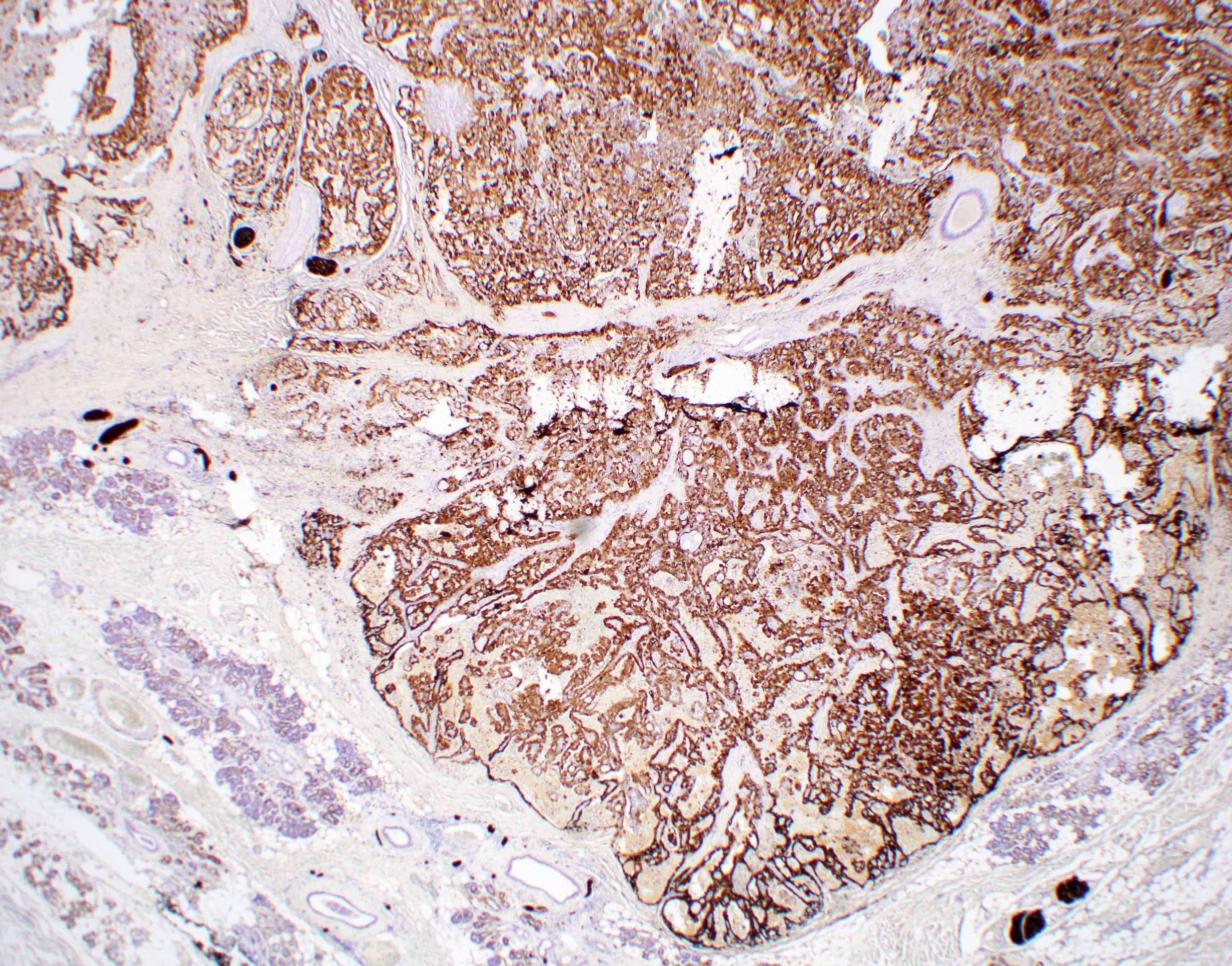
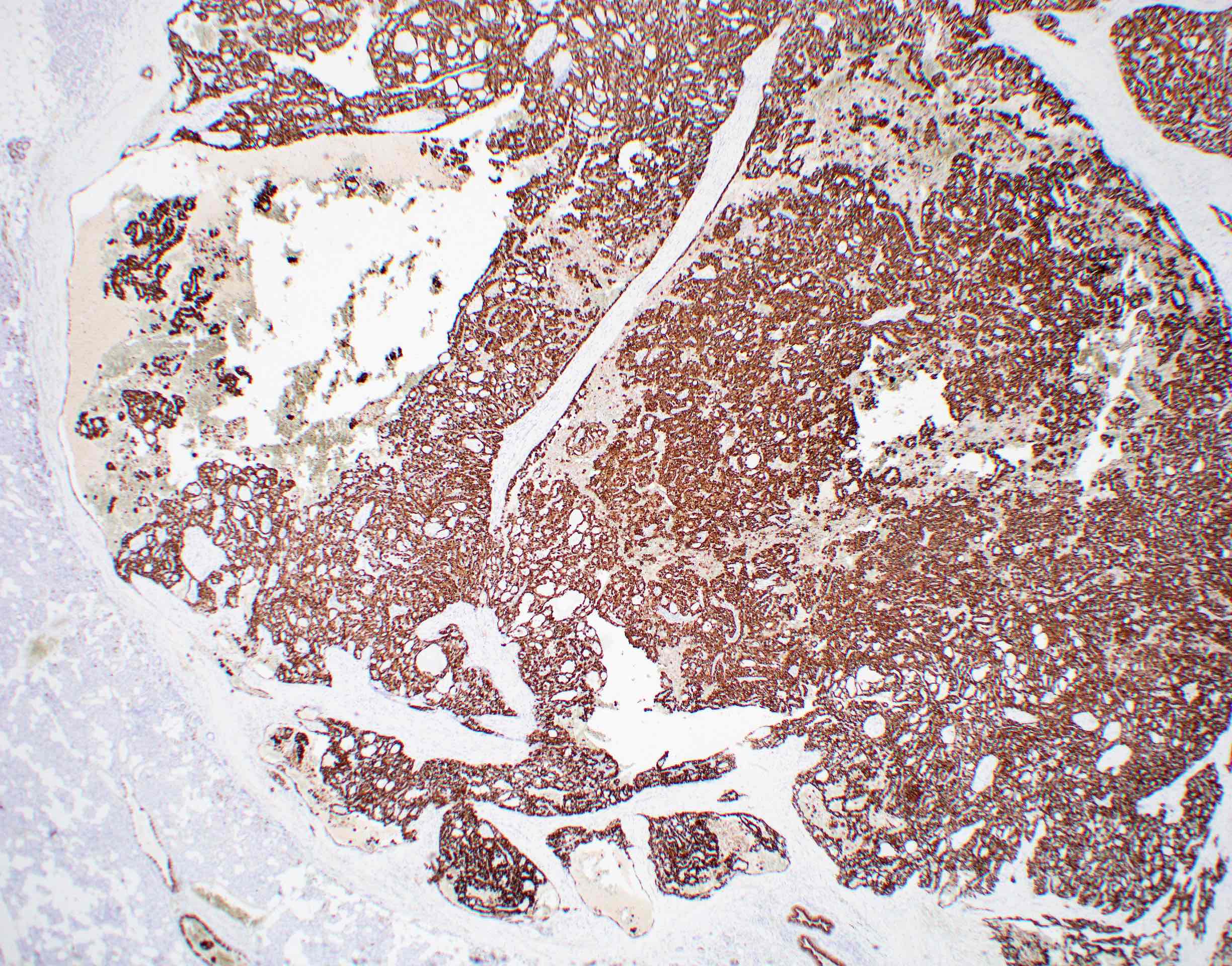
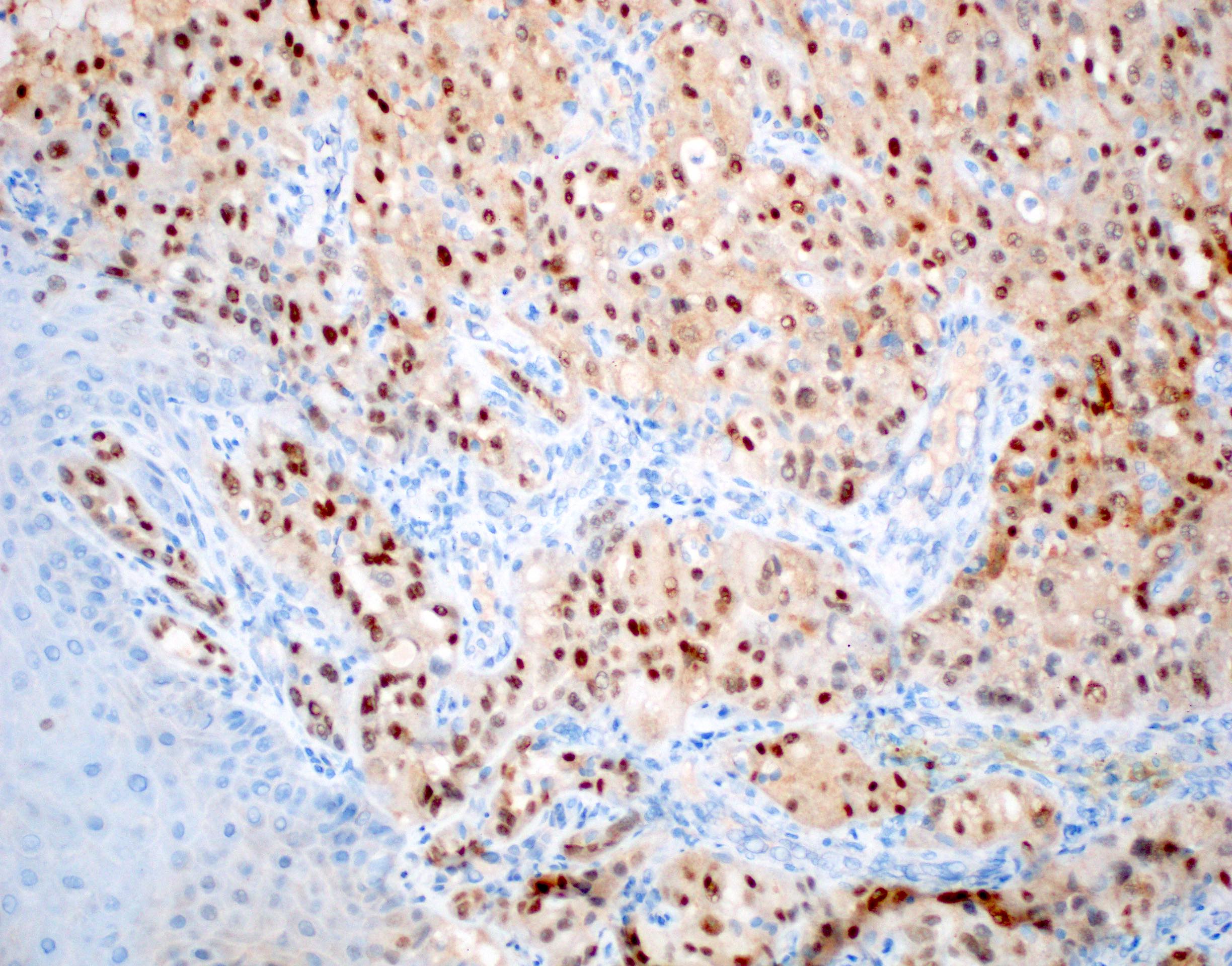
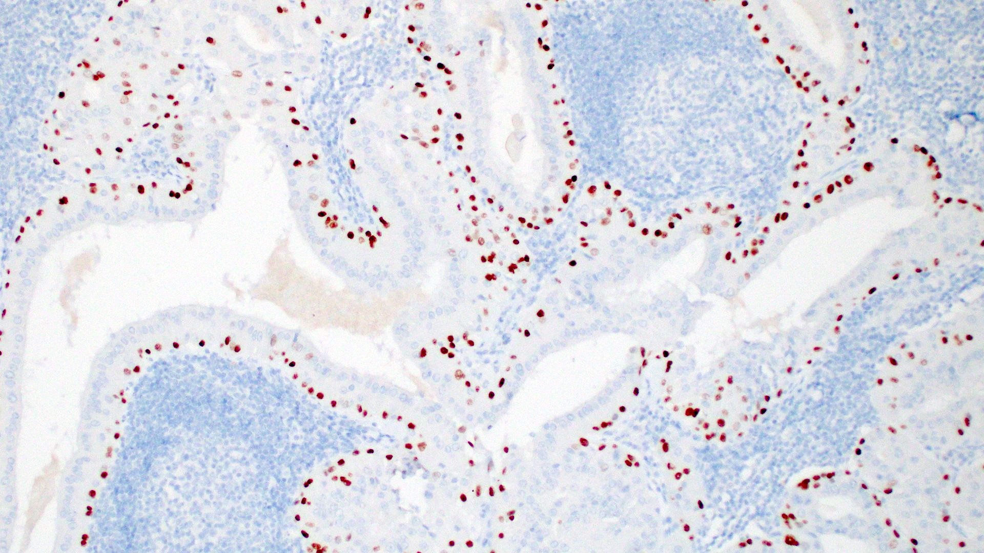
NR4A3 nuclear stain
Contributed by Rema A. Rao, M.D. and Arash H. Lahouti, M.D.

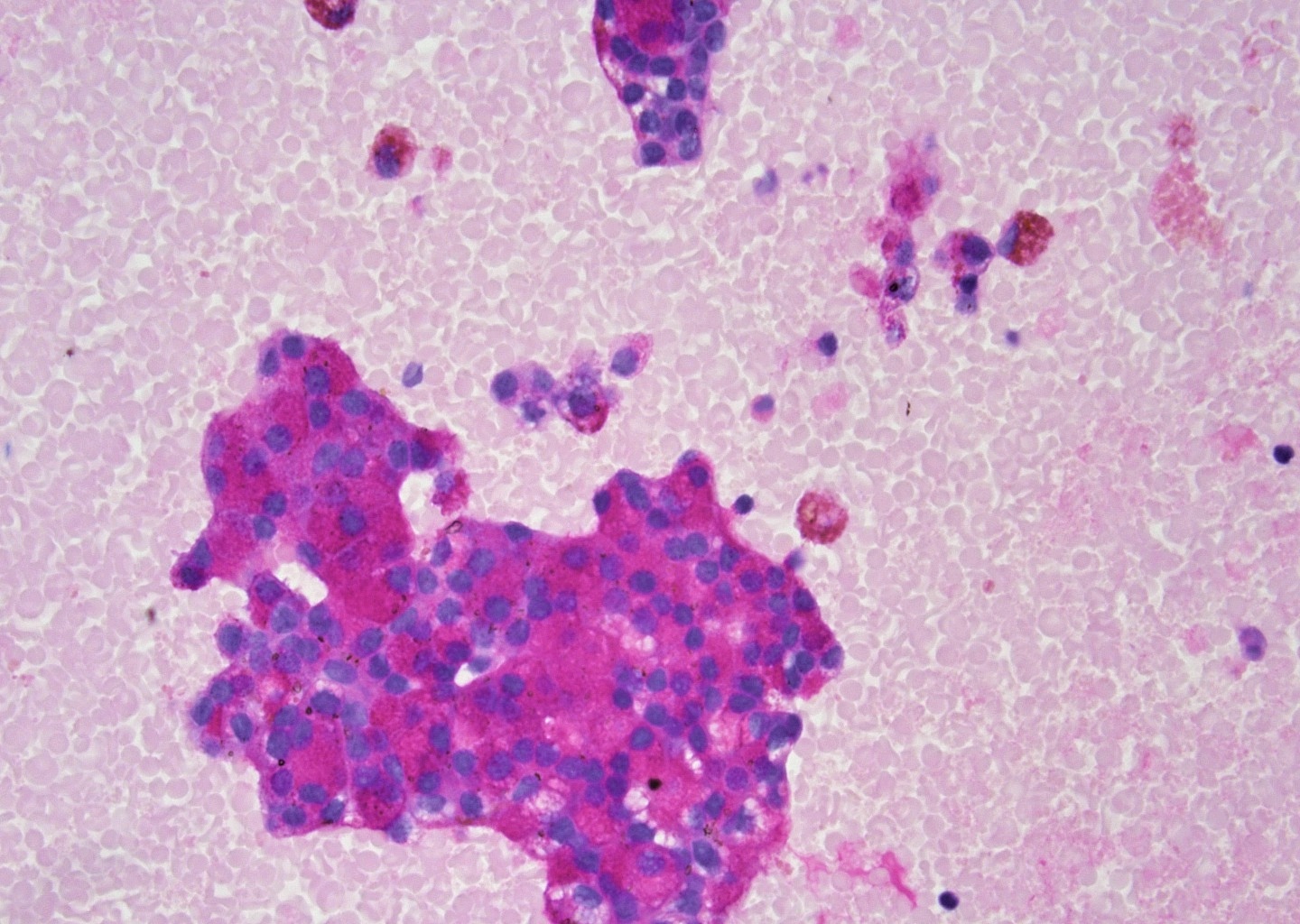
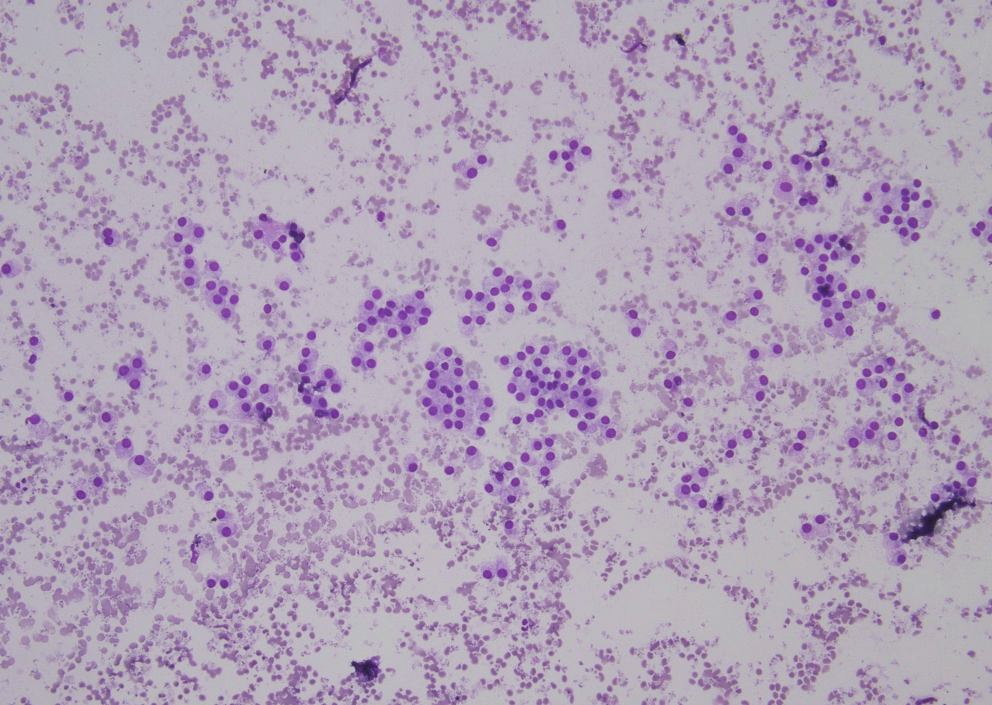
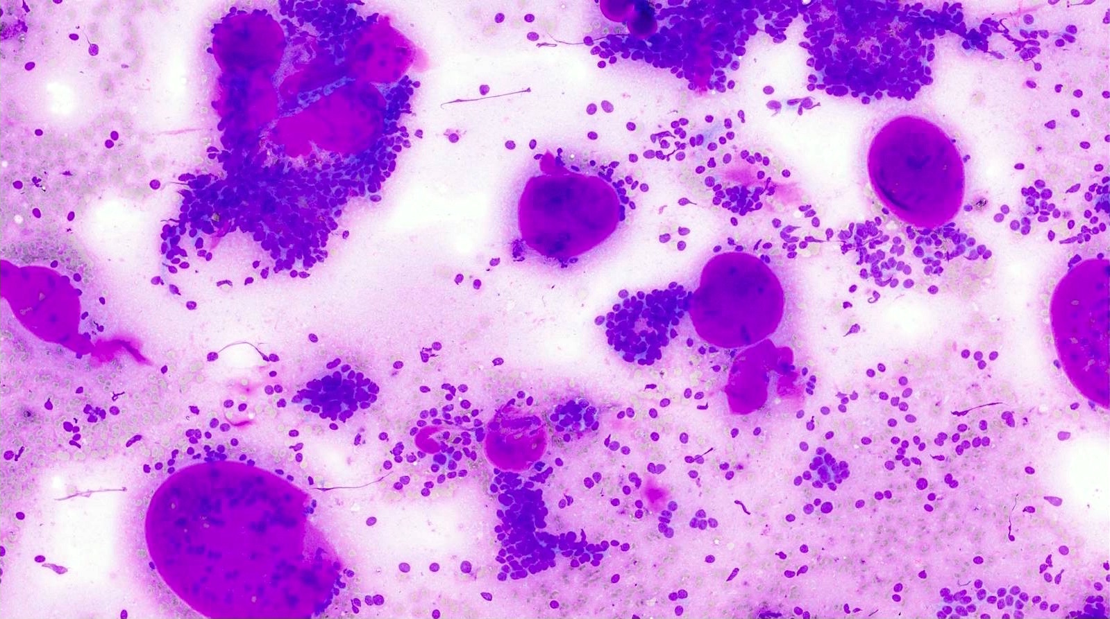
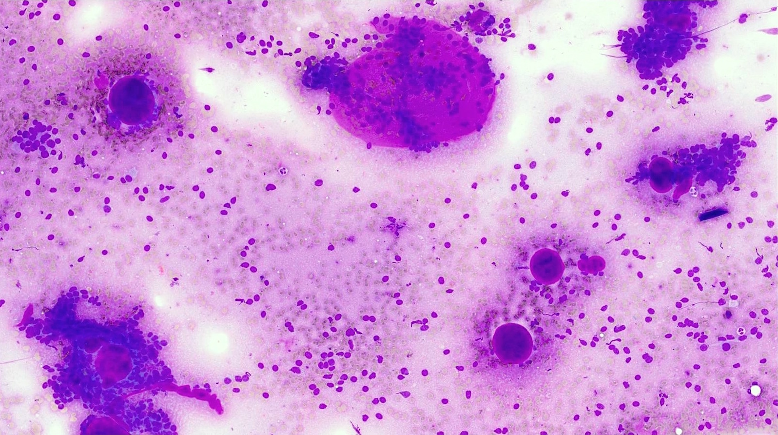
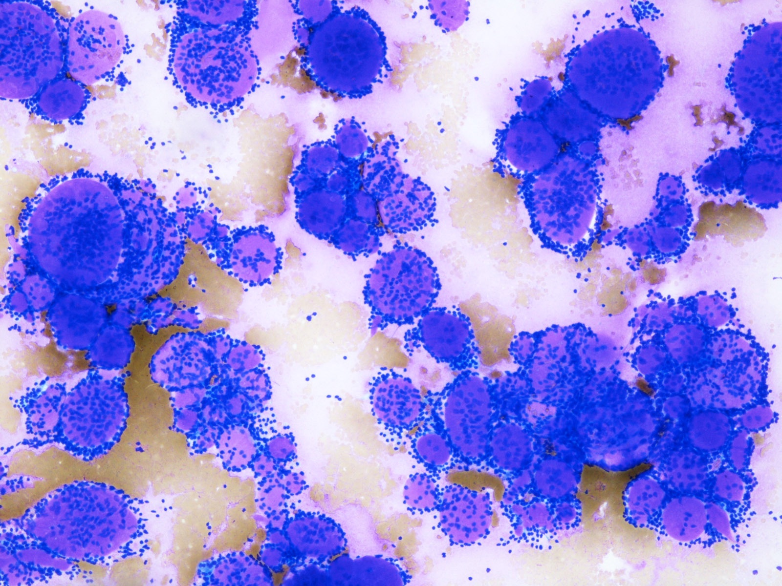
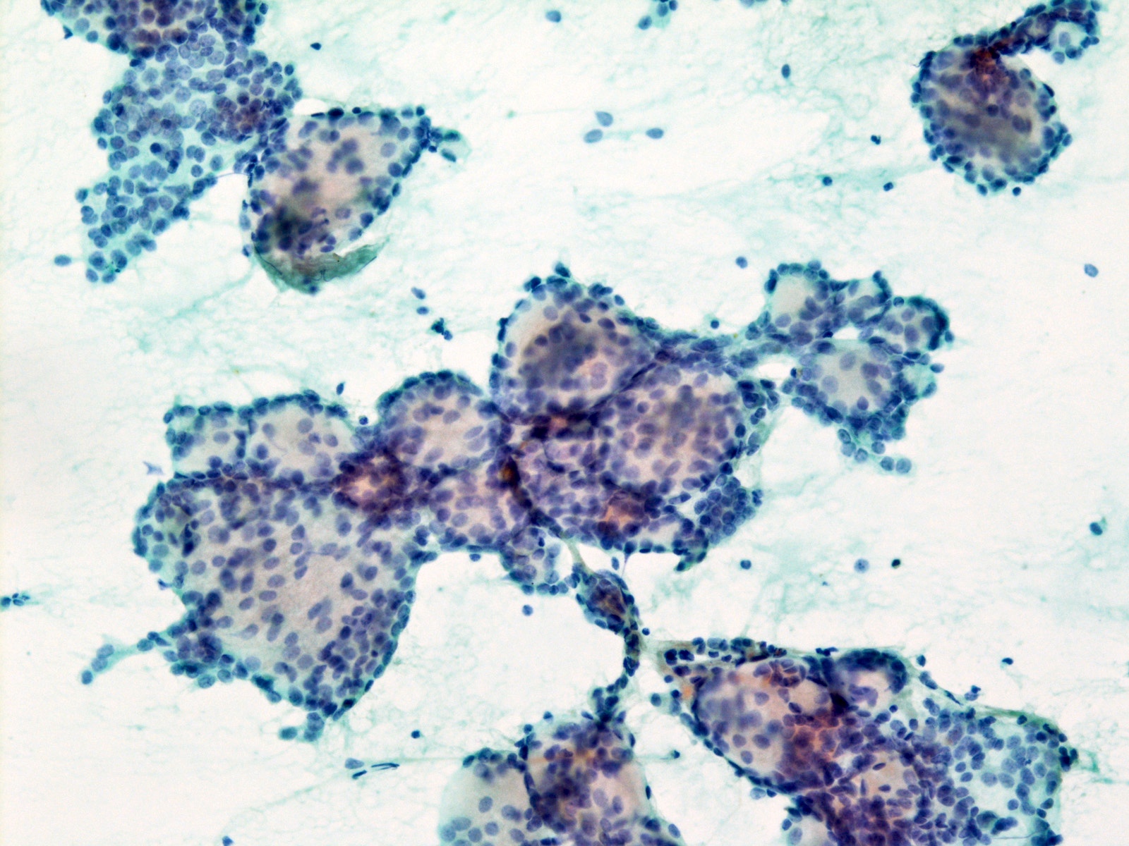
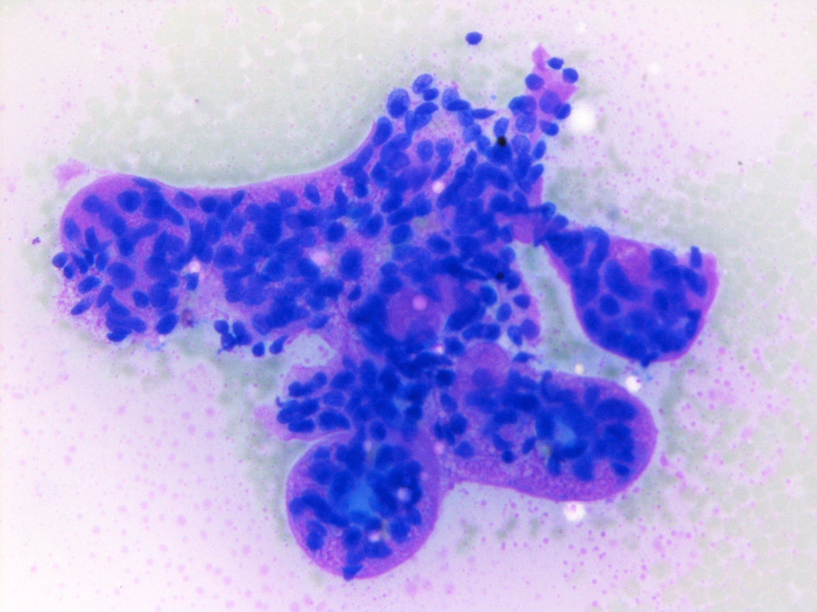
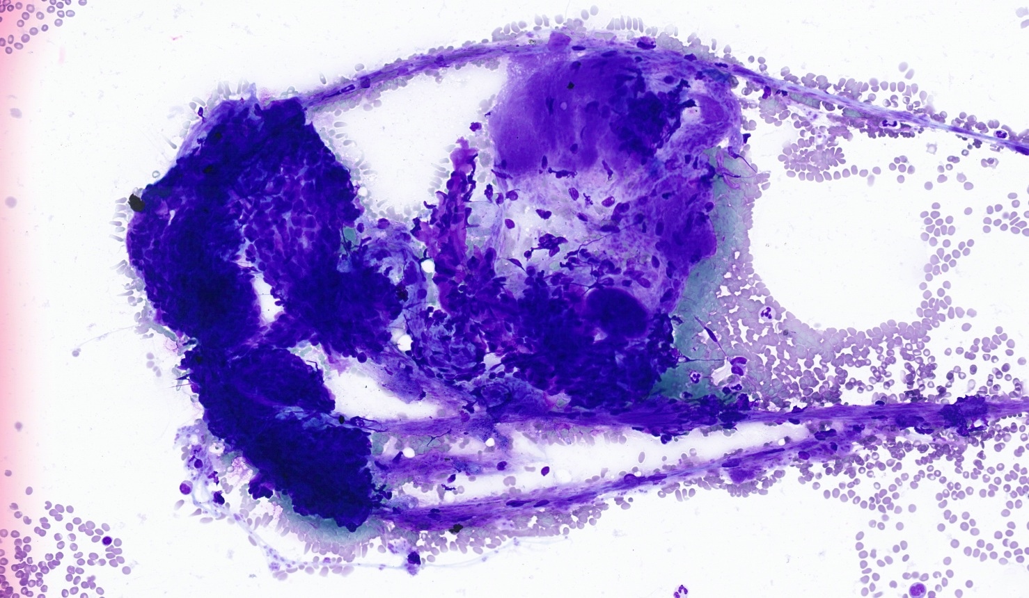
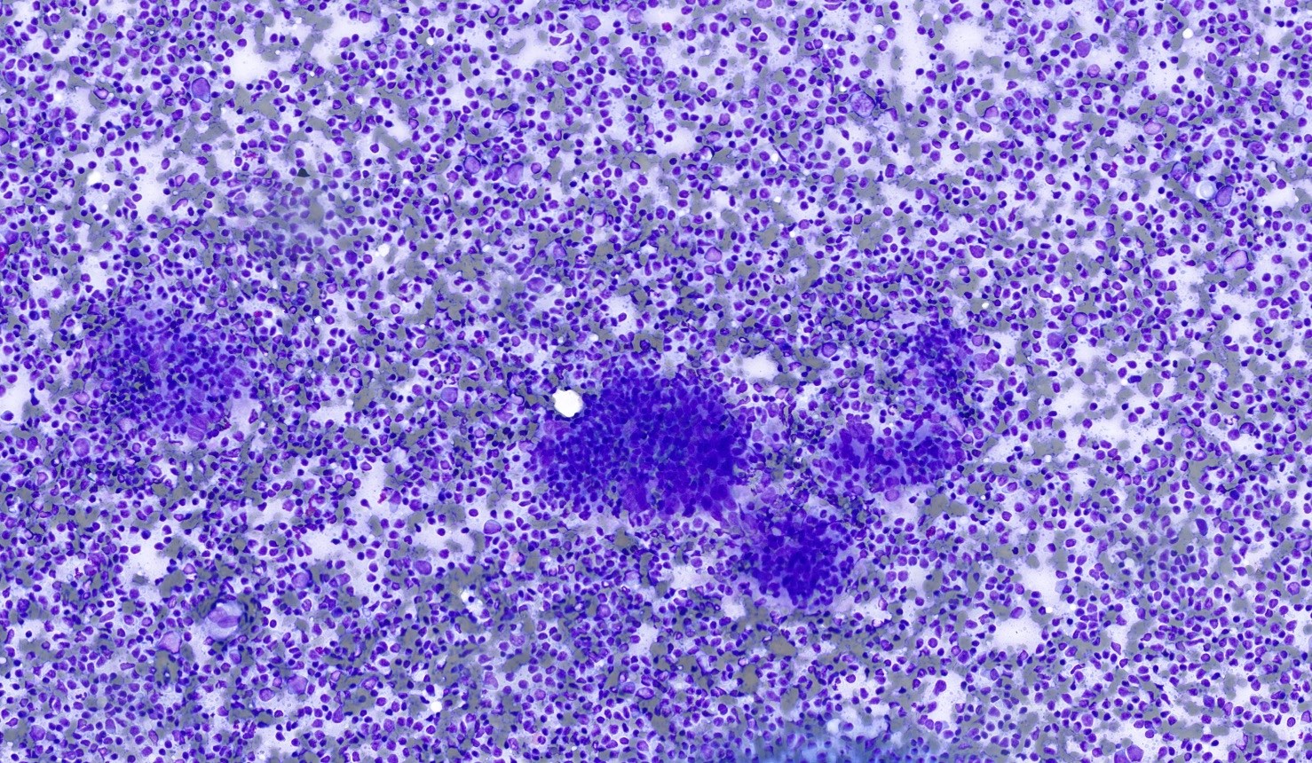
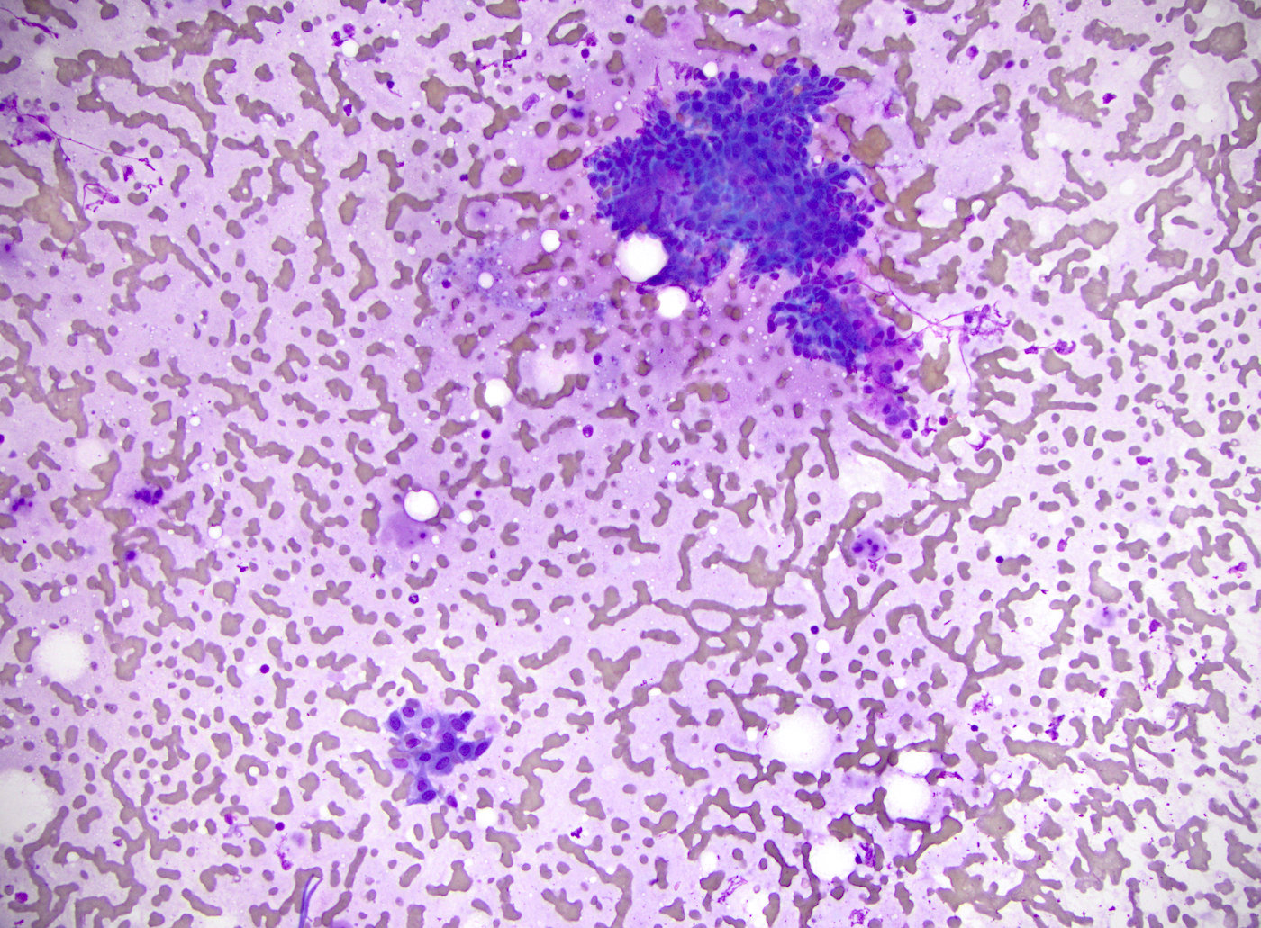
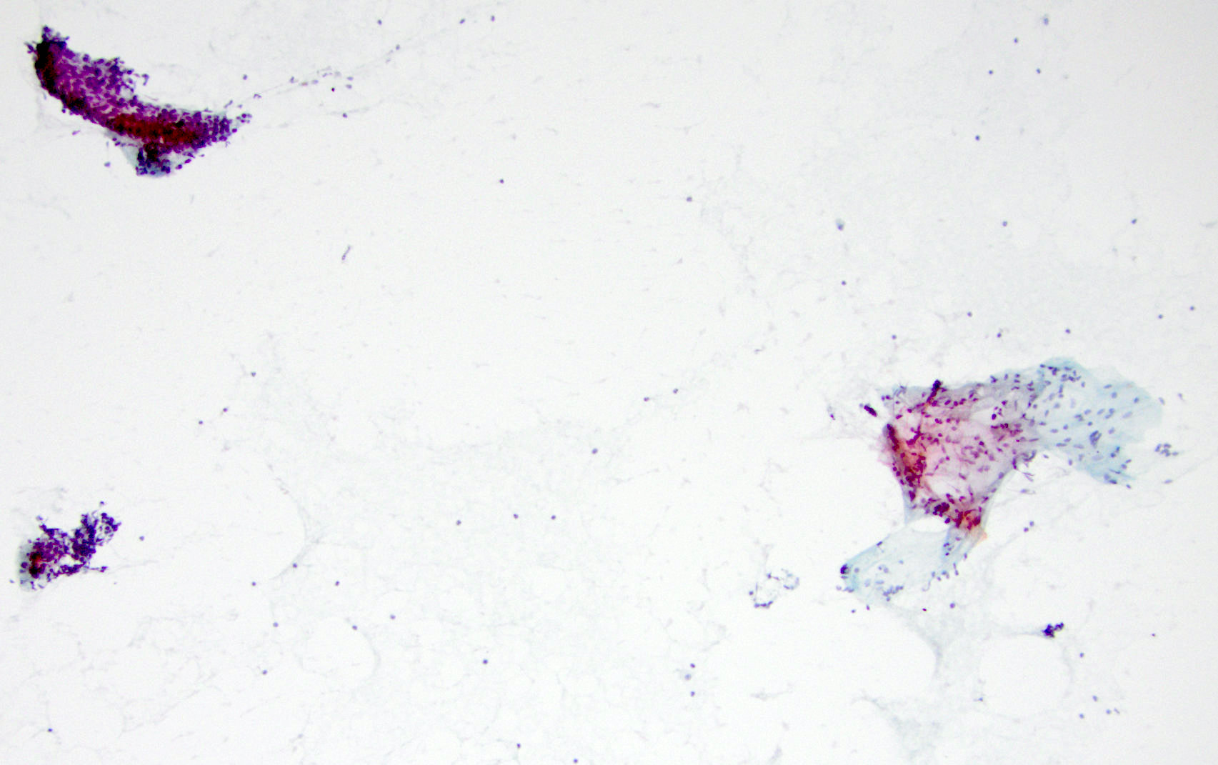
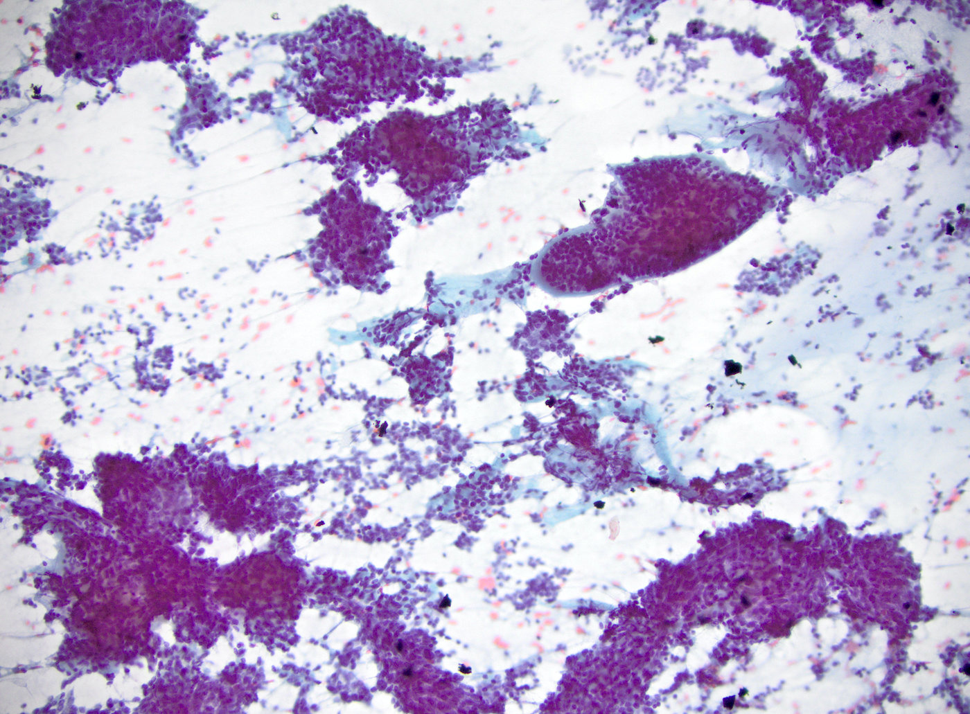
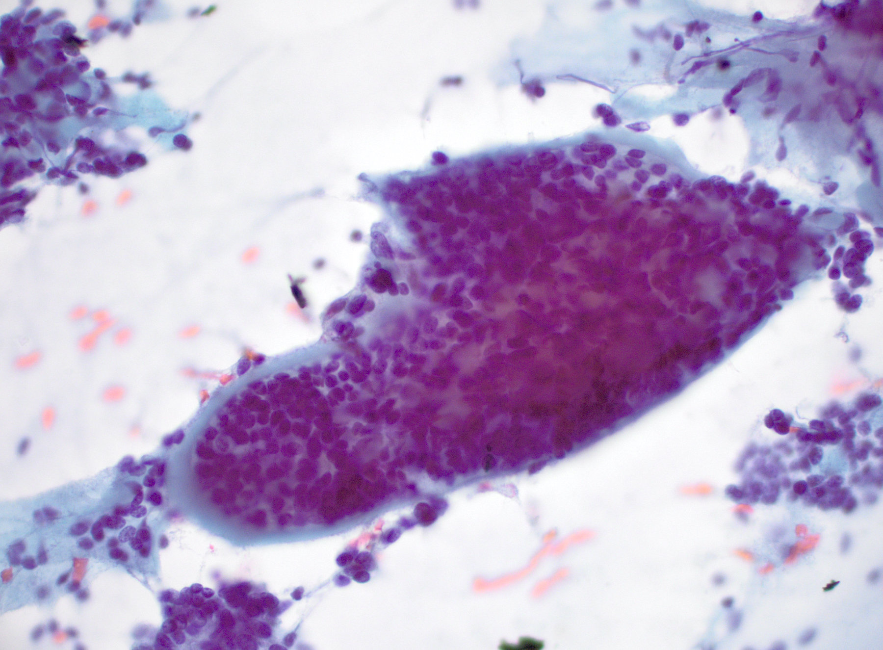
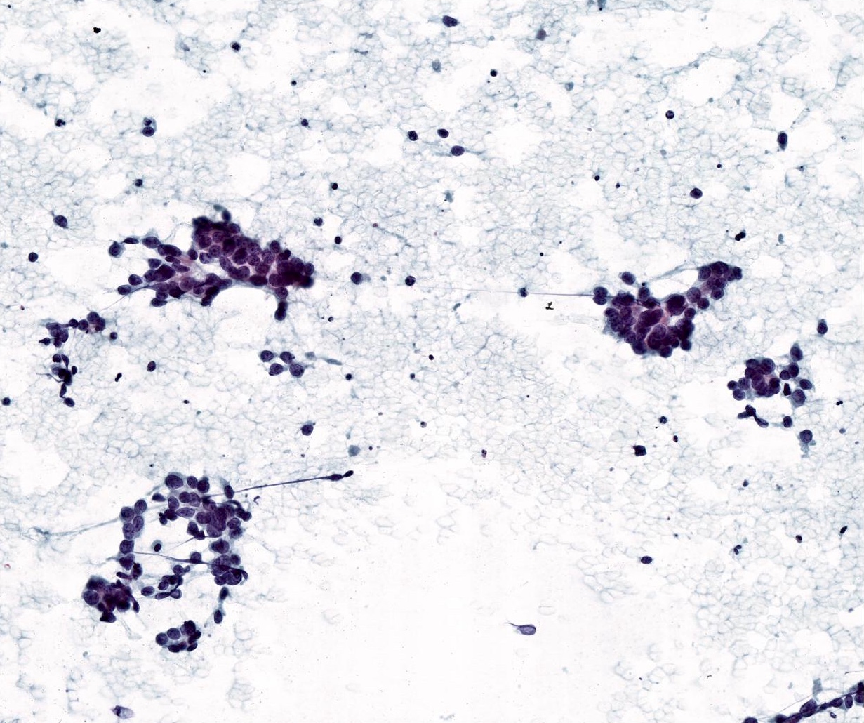
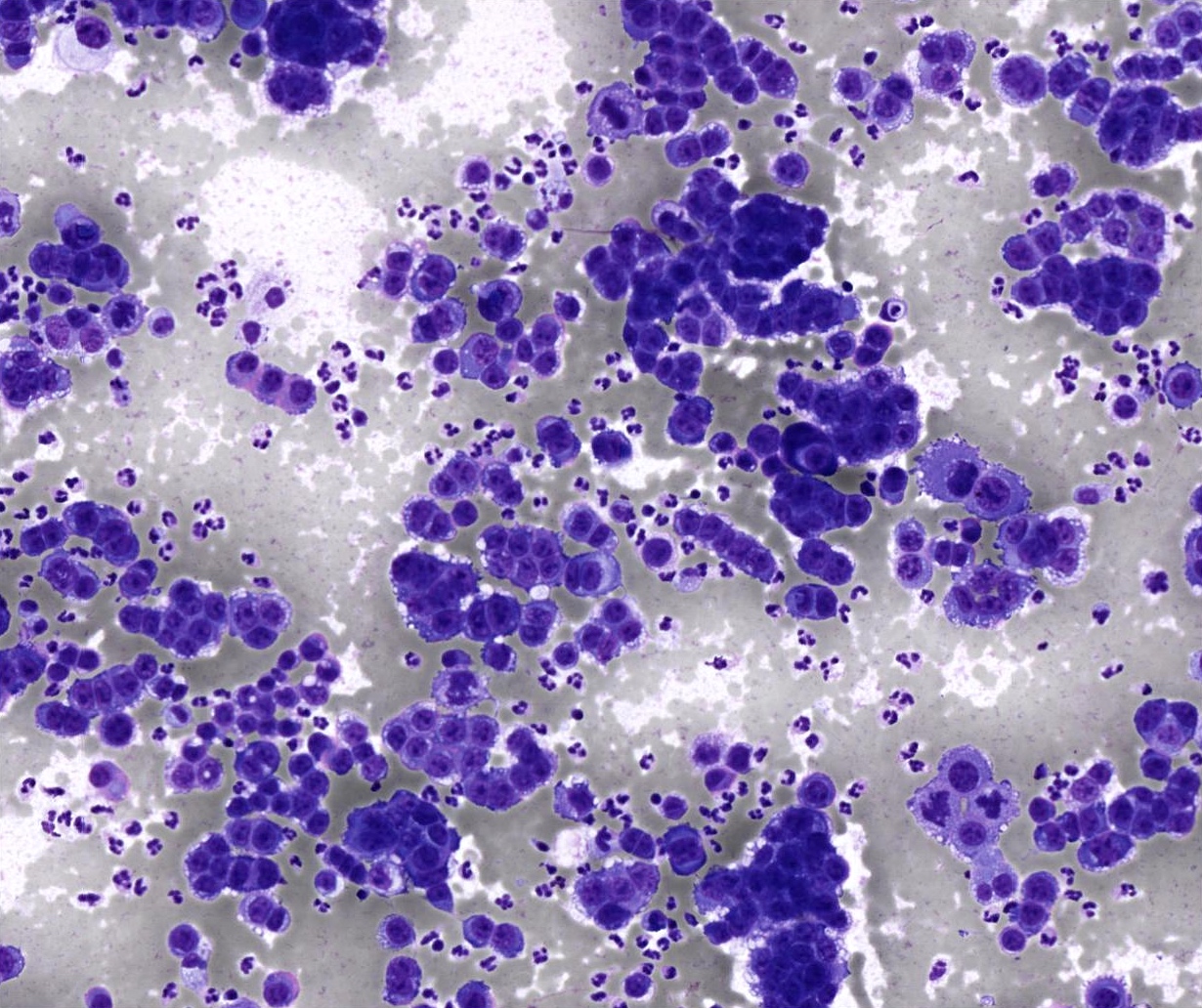
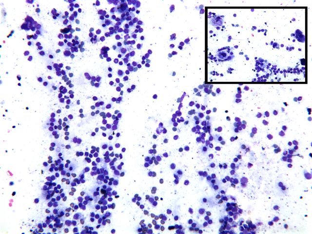
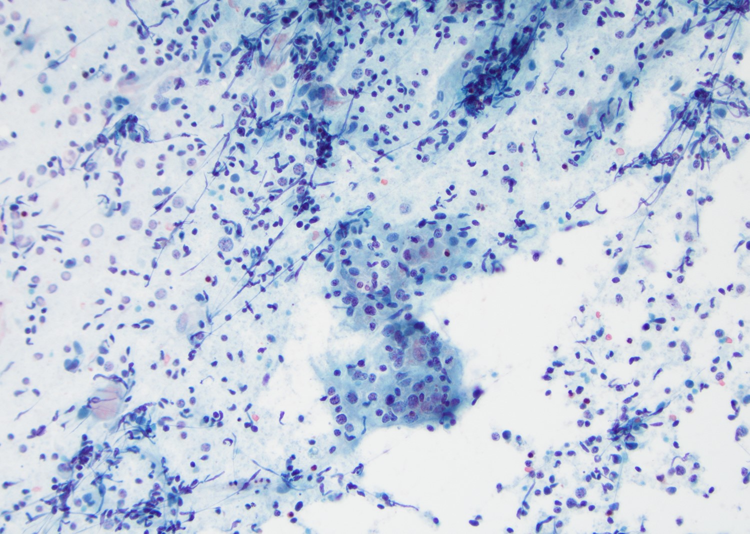
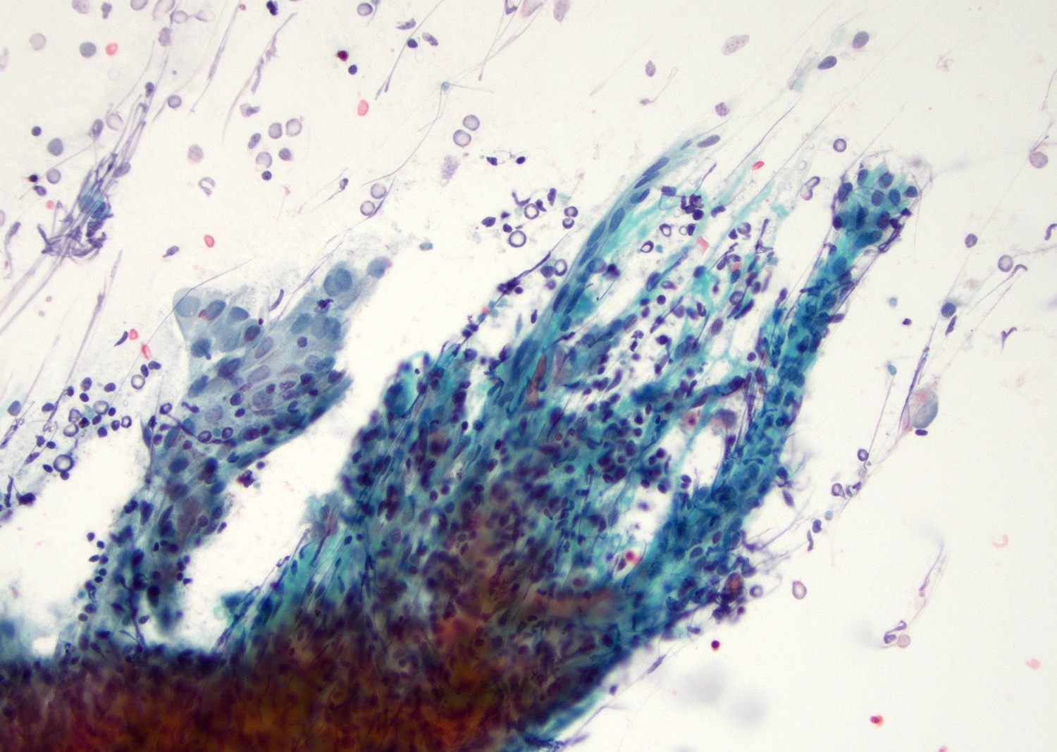
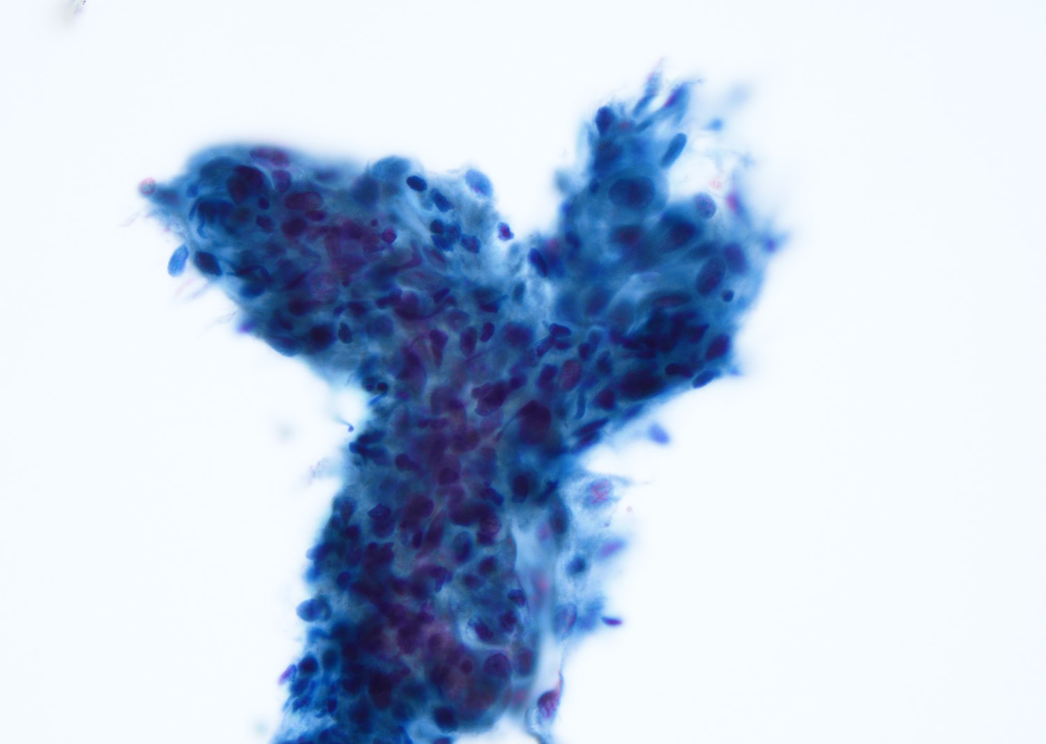
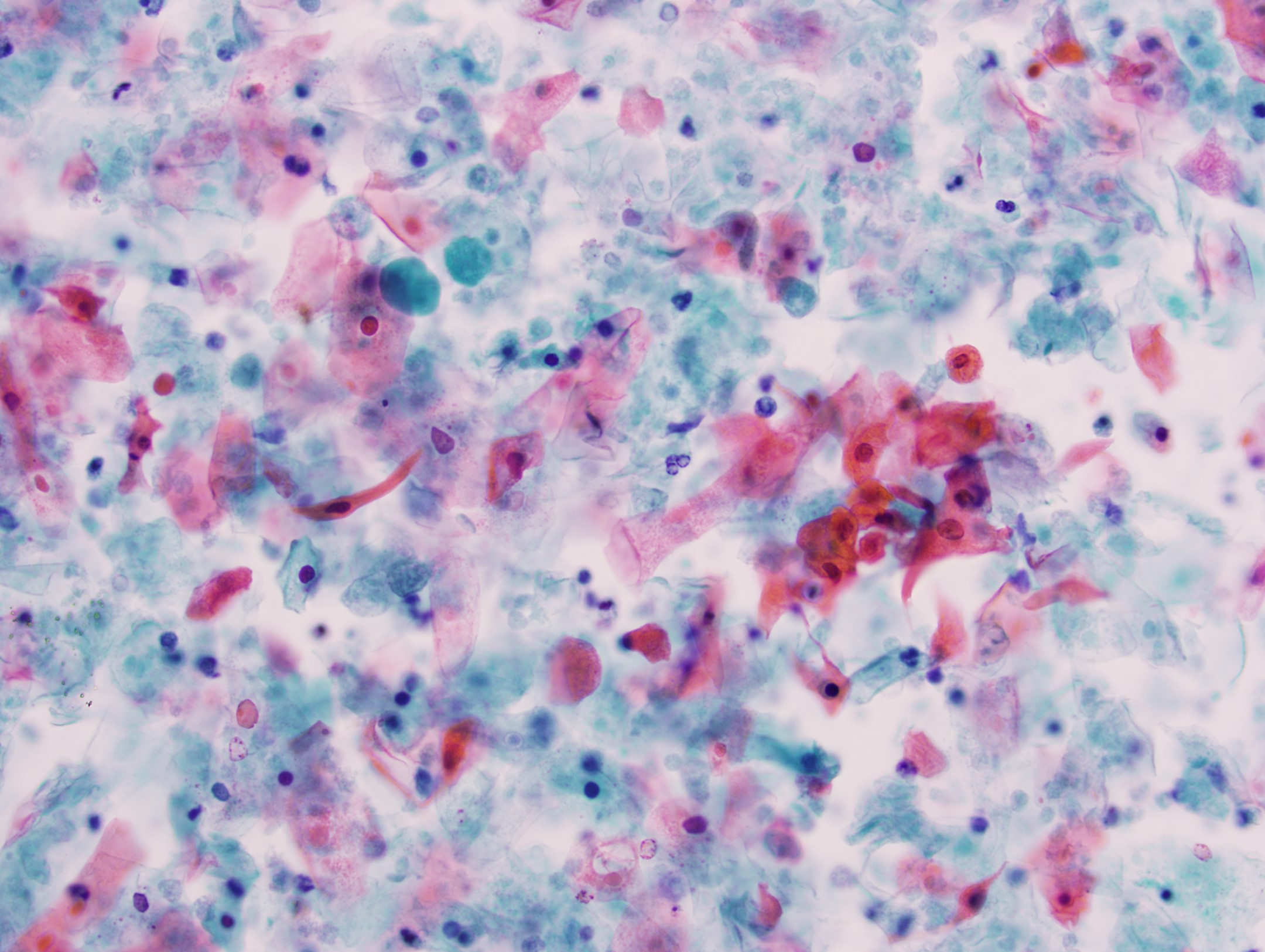
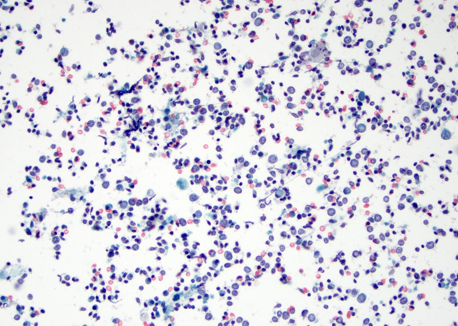
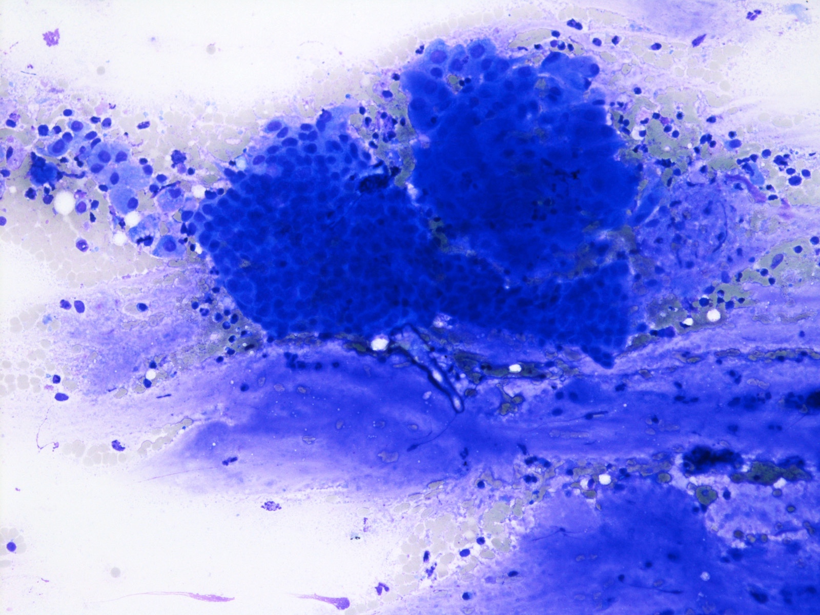
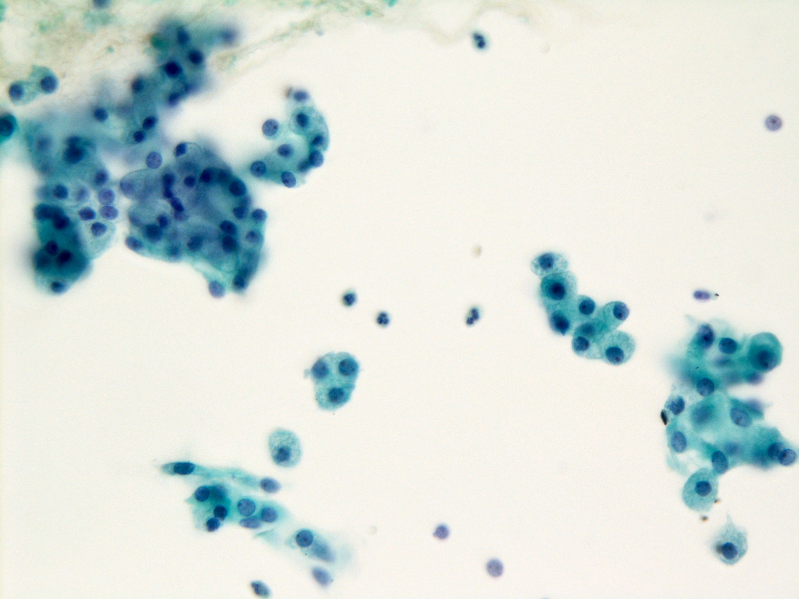
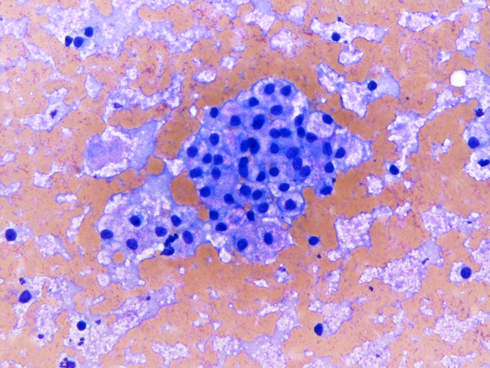
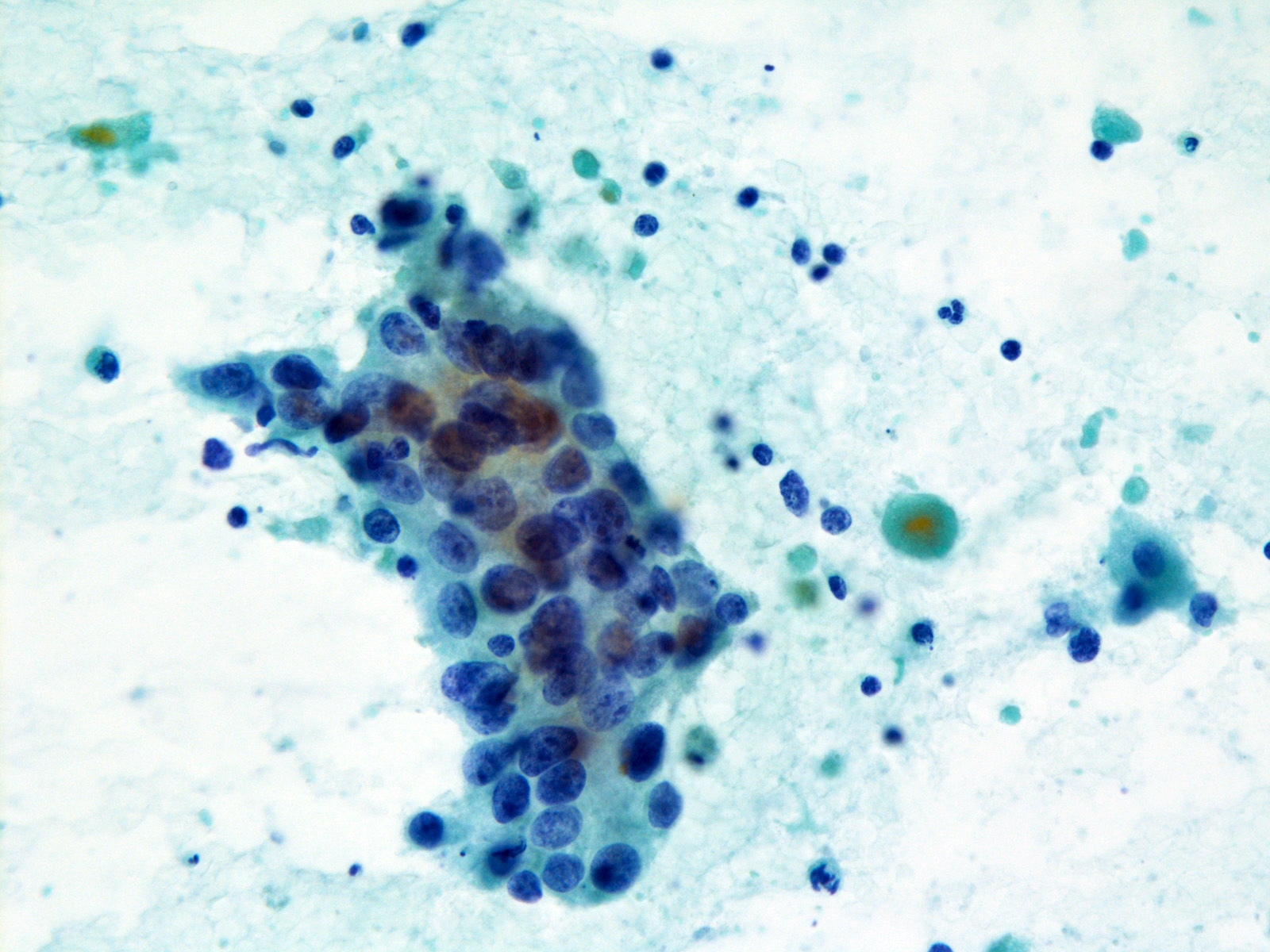
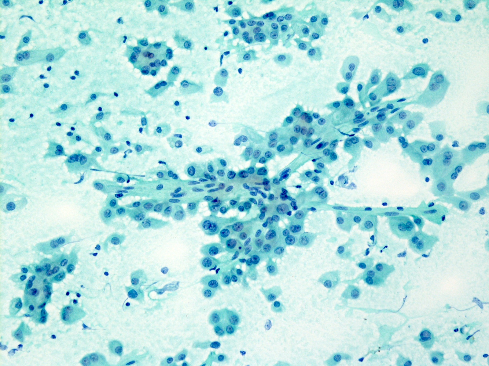
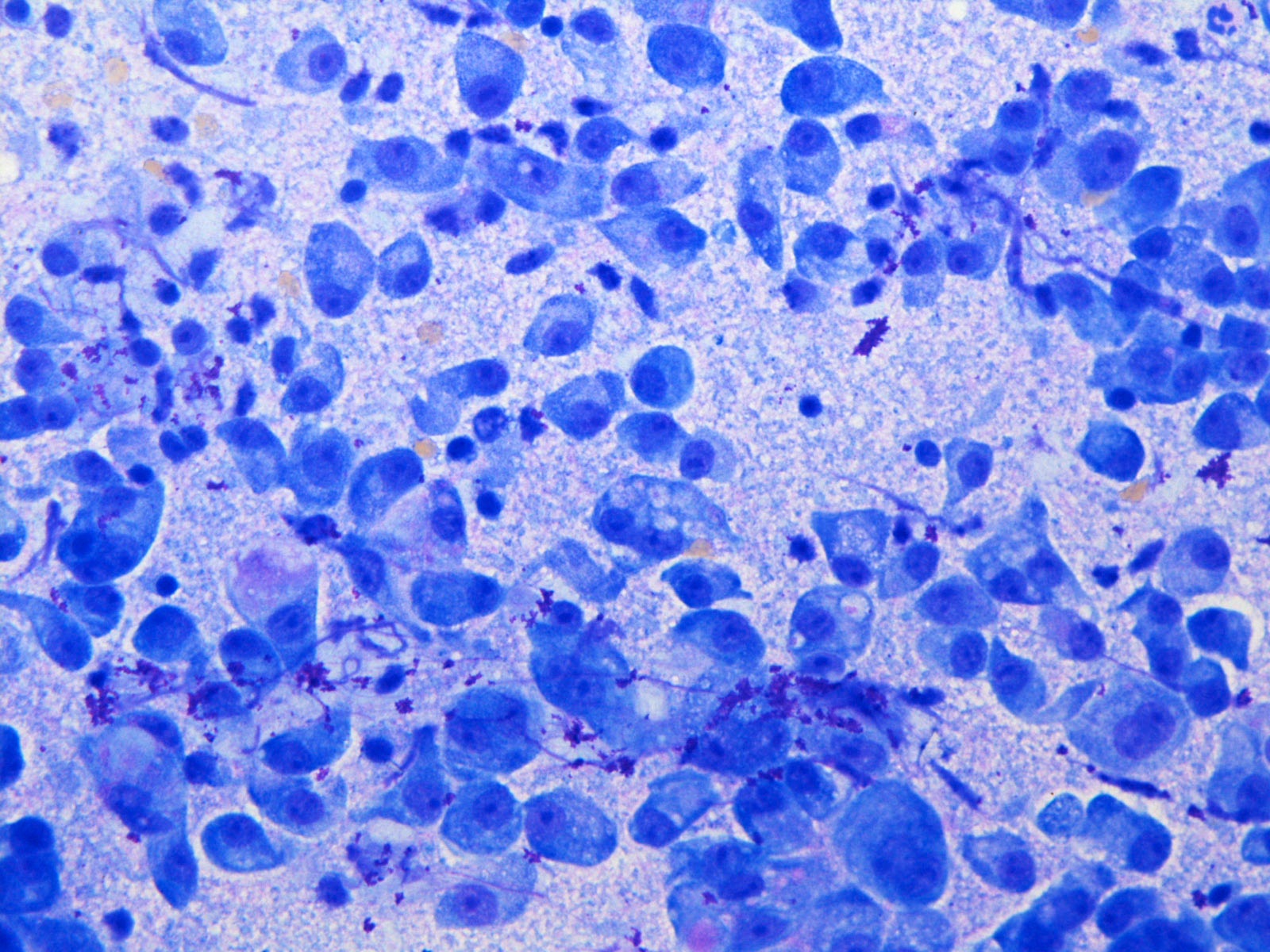
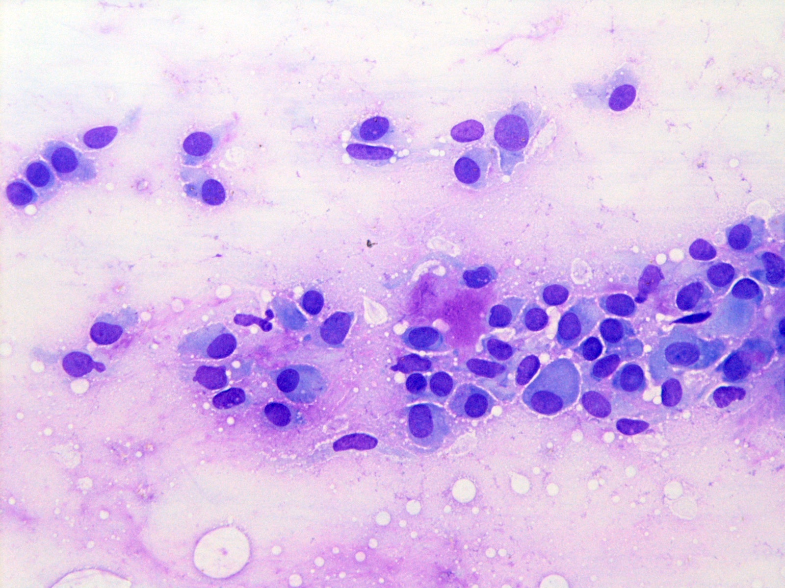
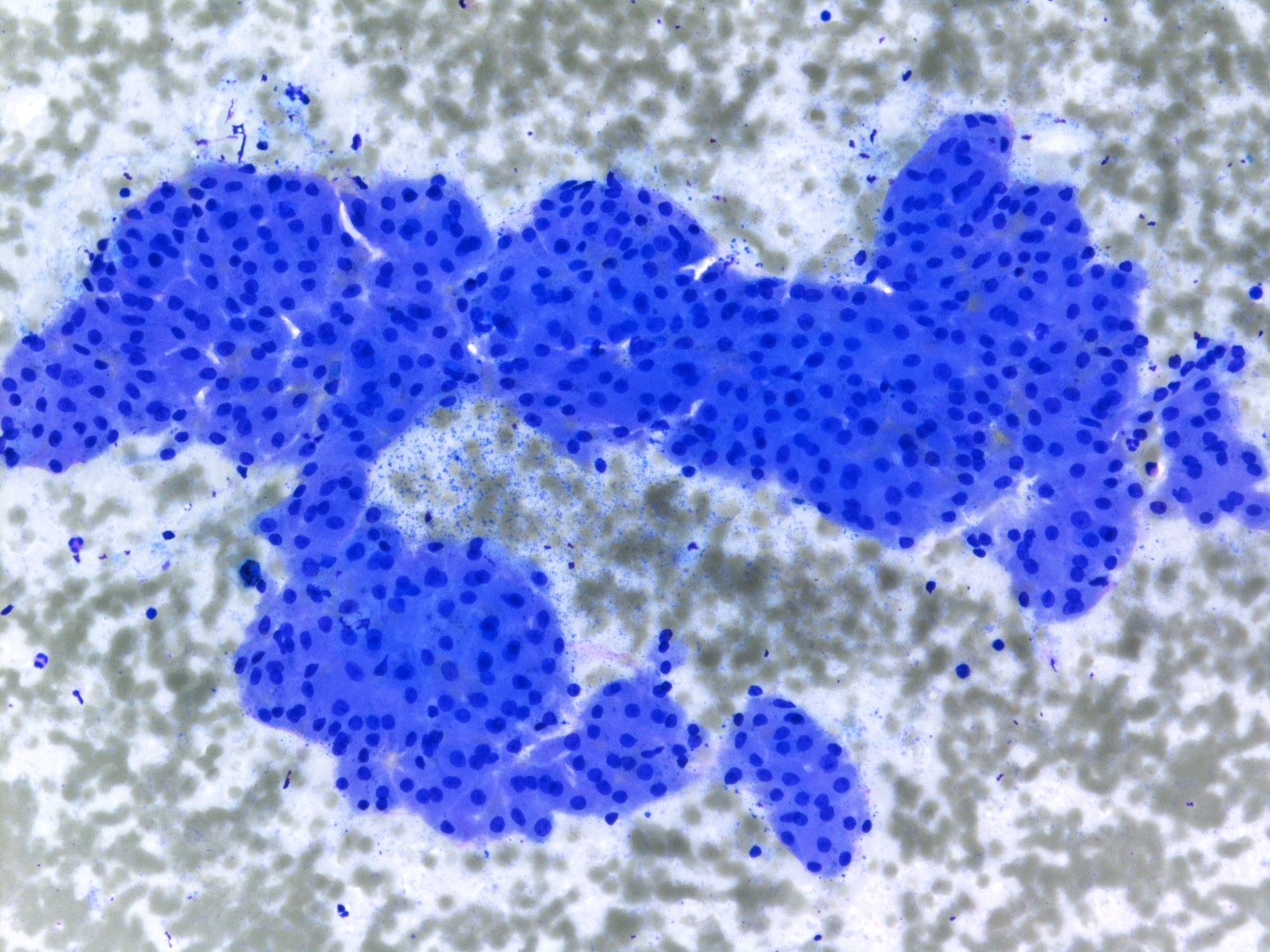
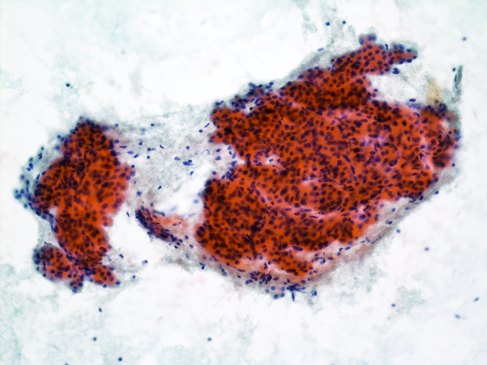
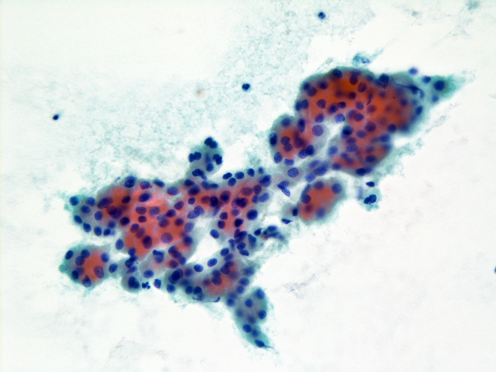
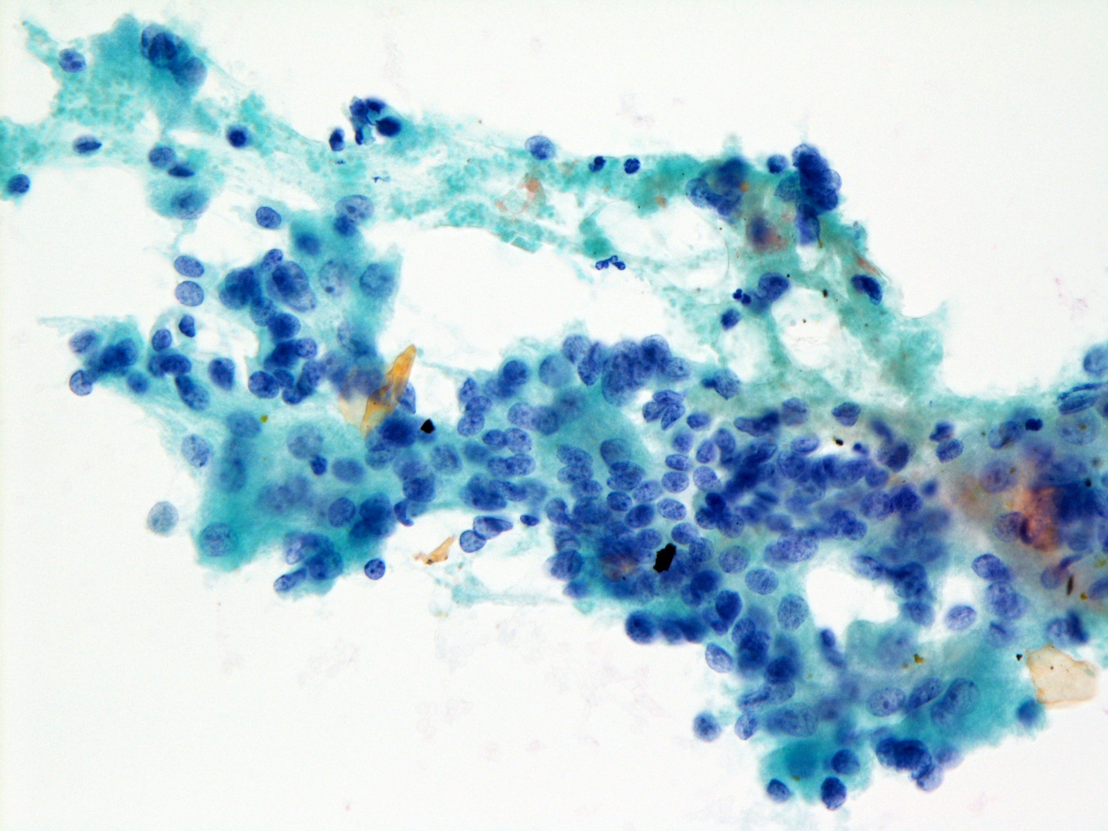
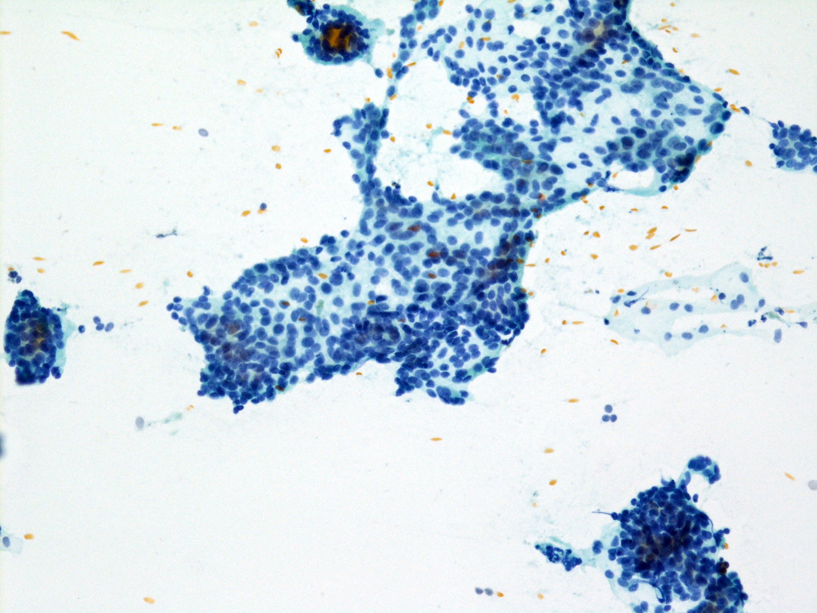
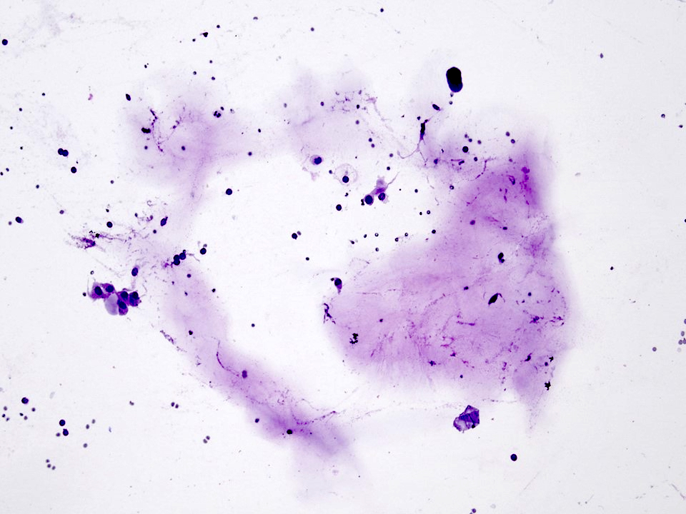
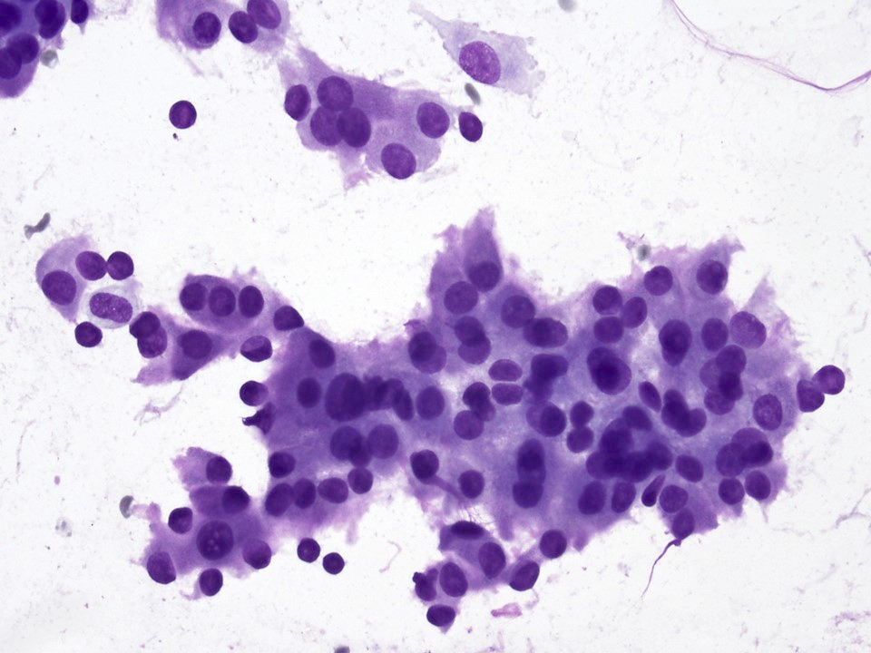
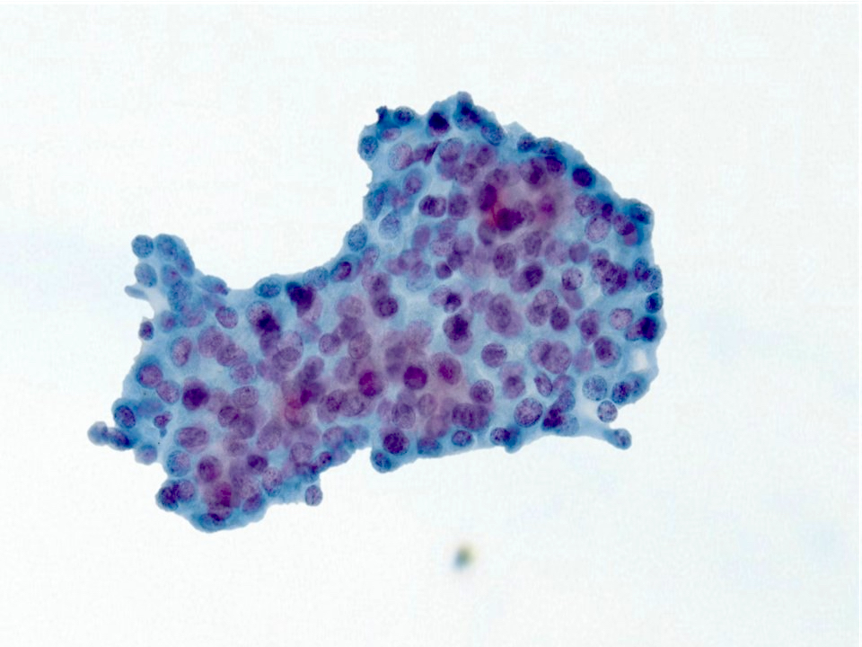
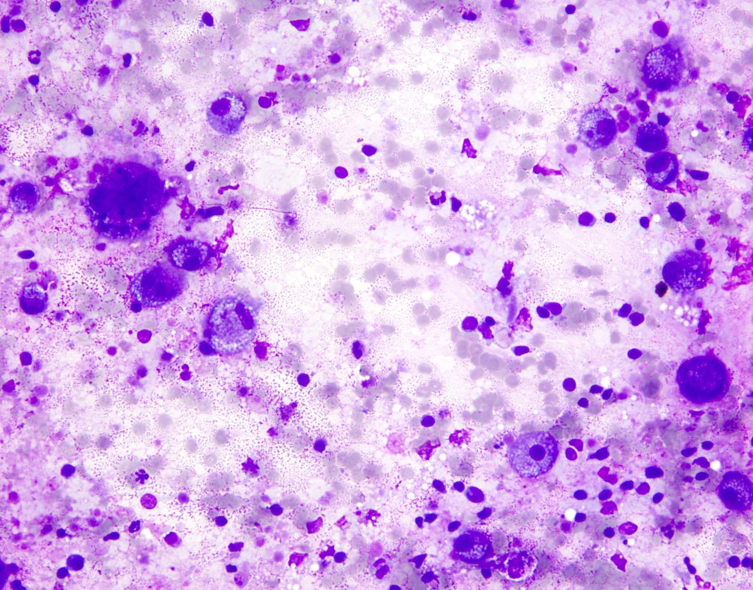
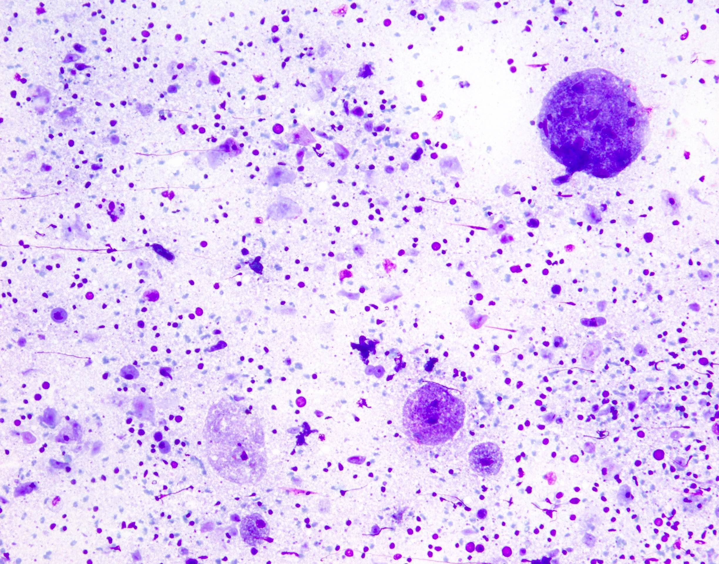
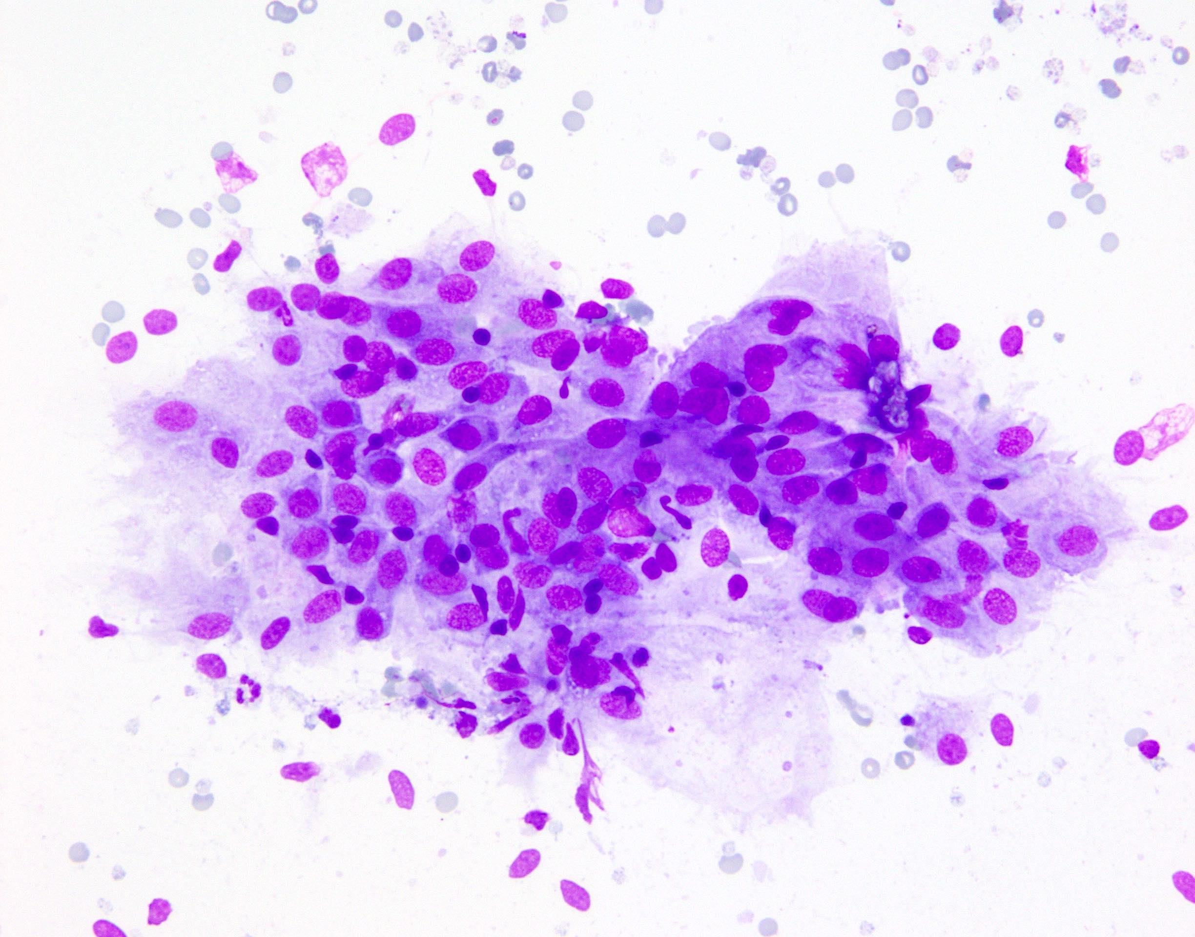
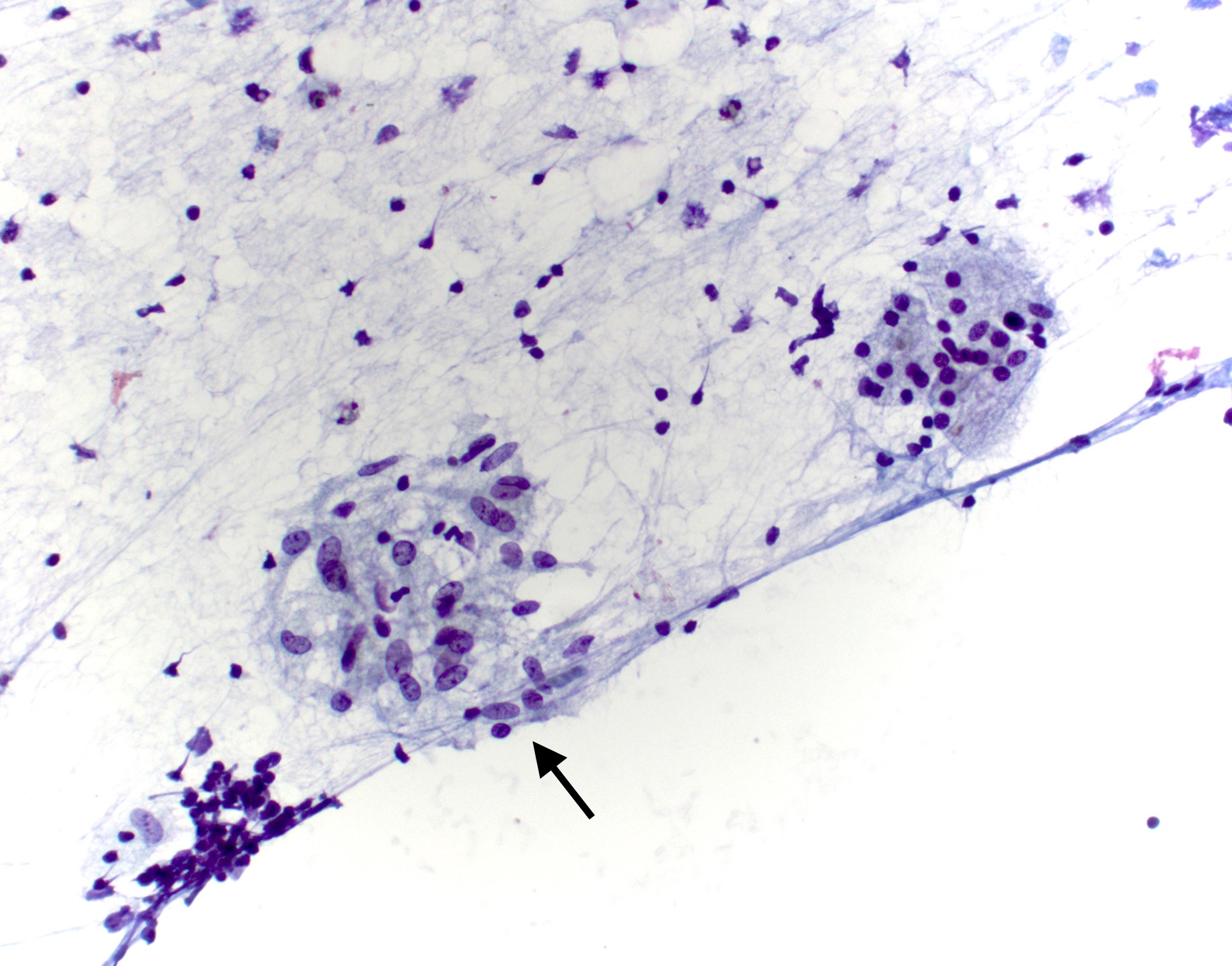
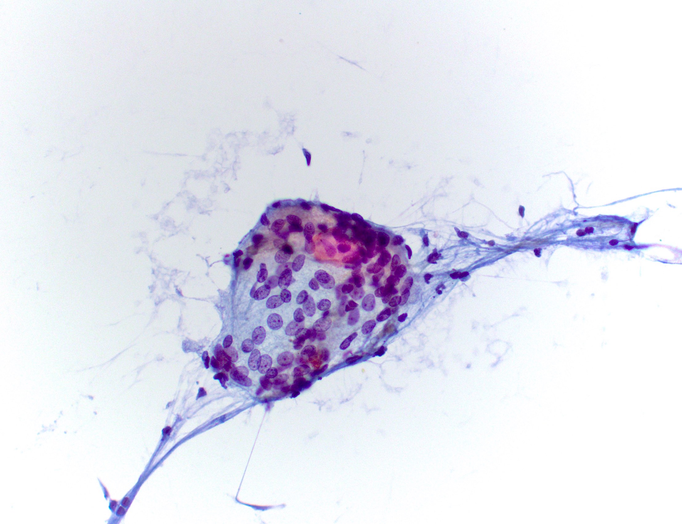
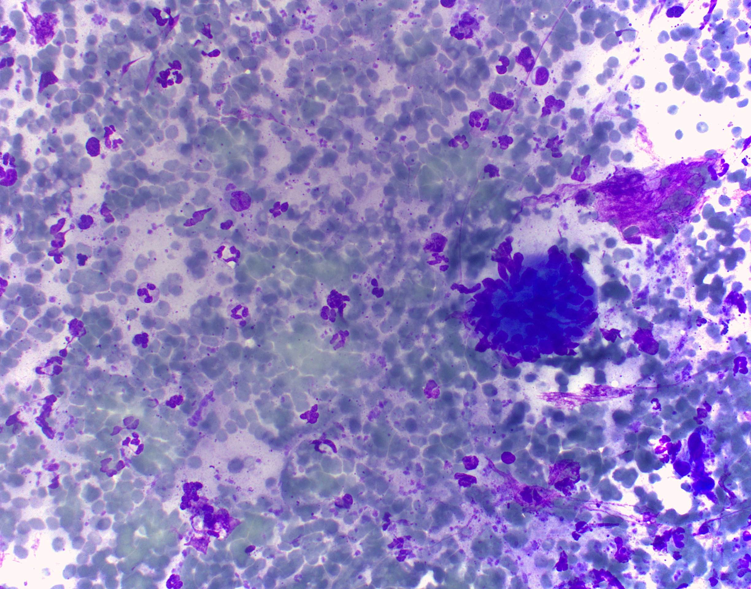
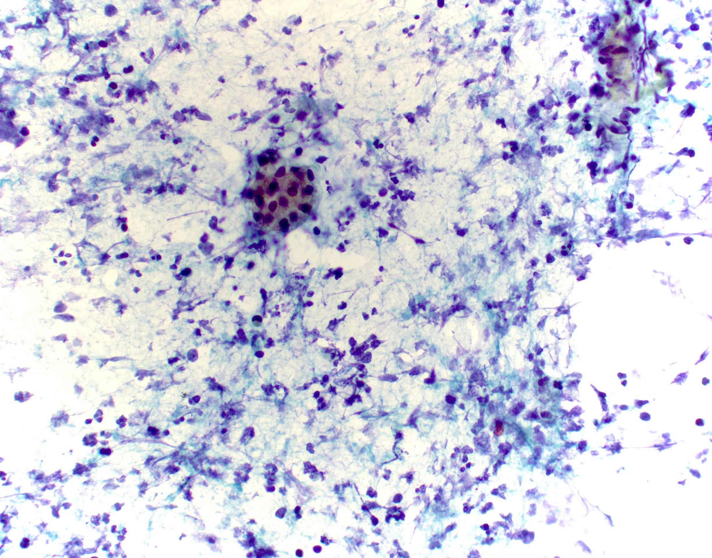
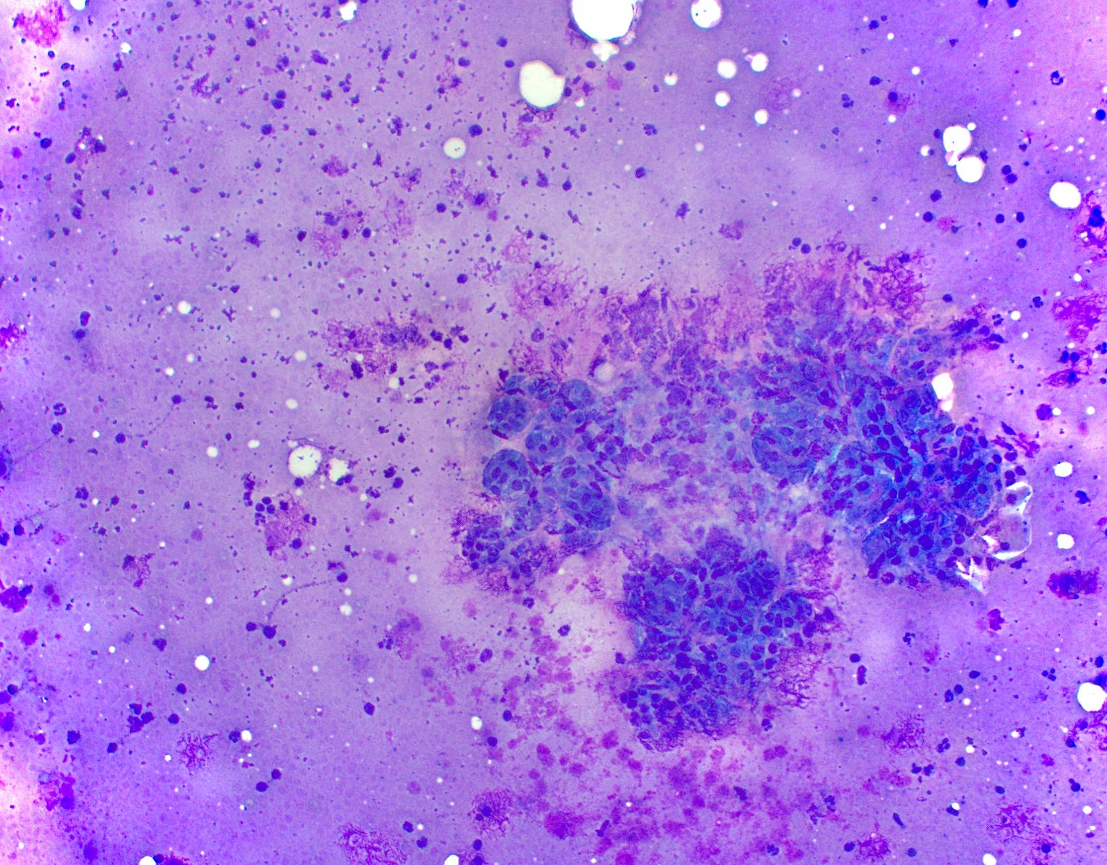
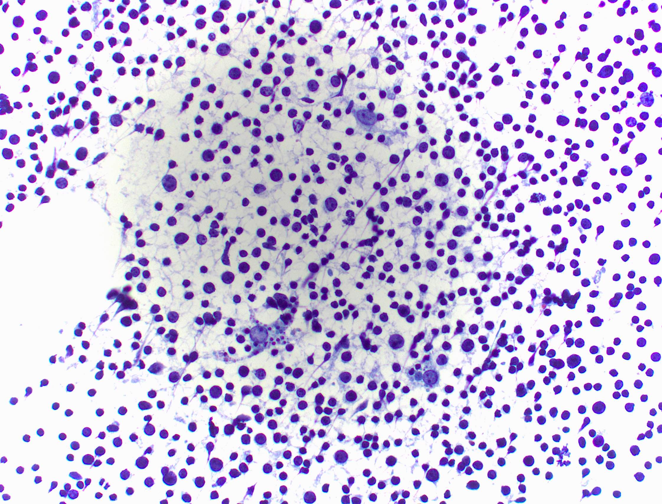
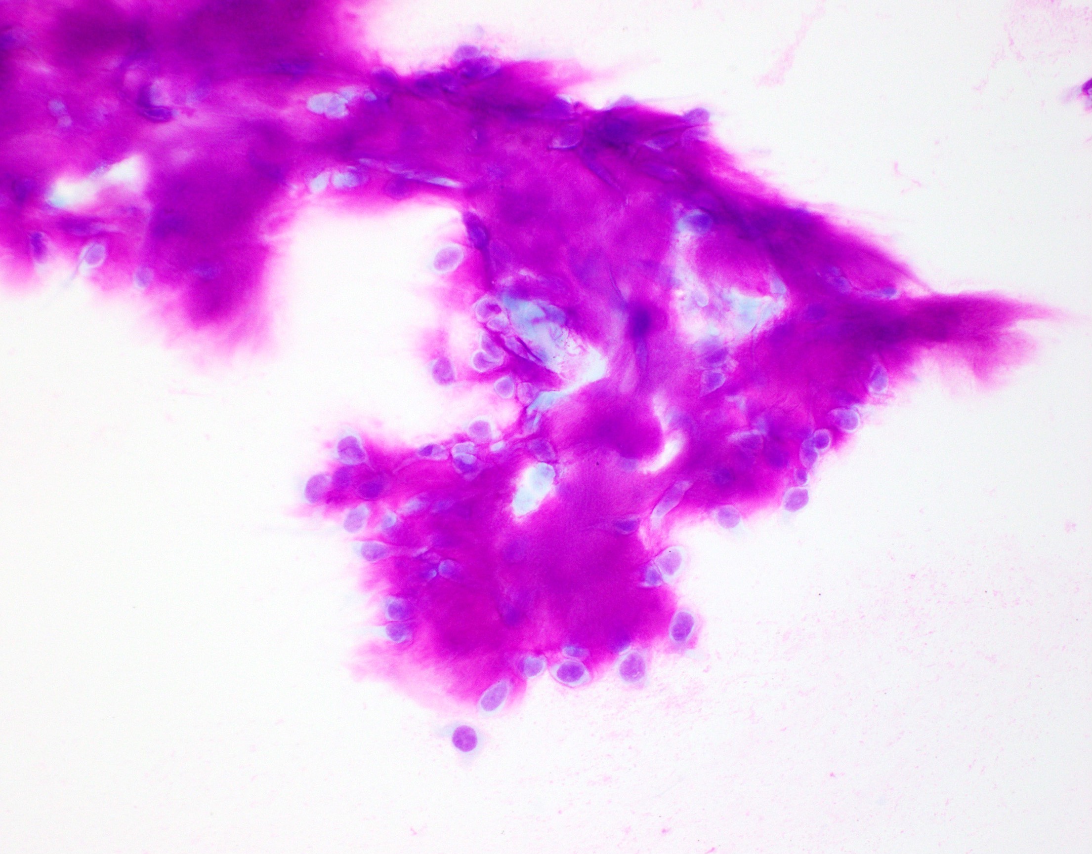
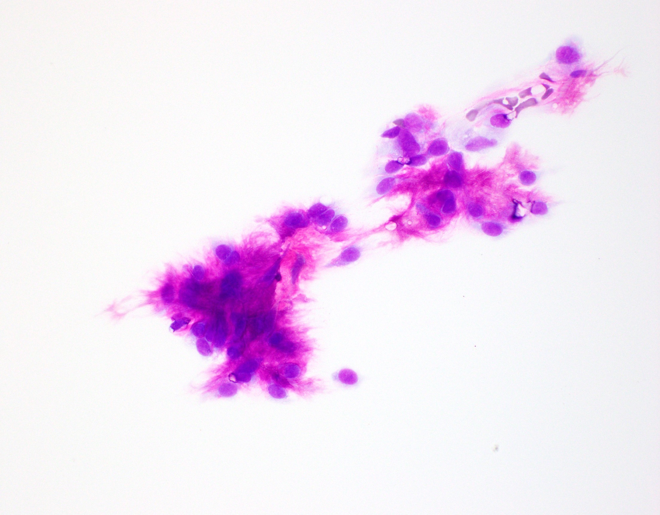
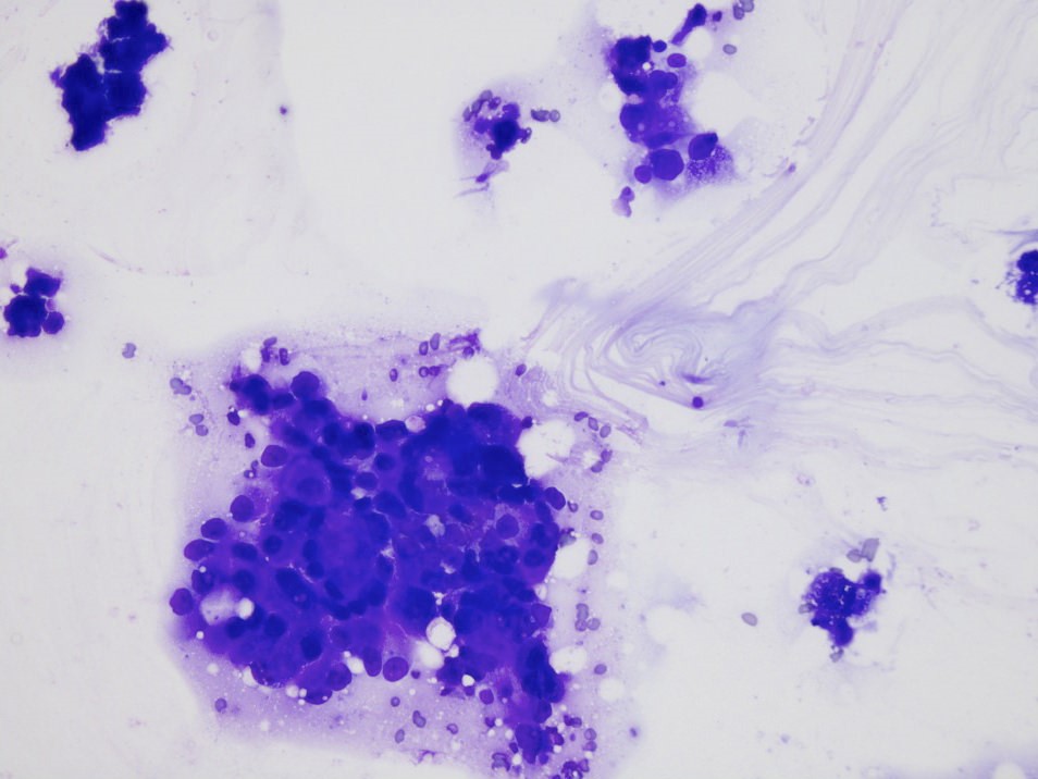
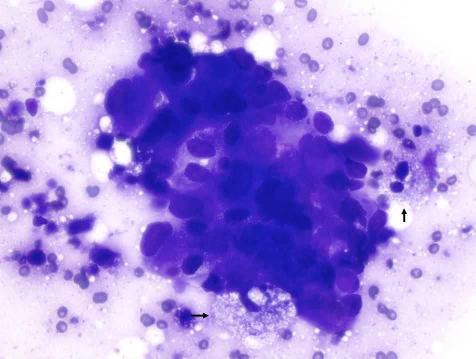
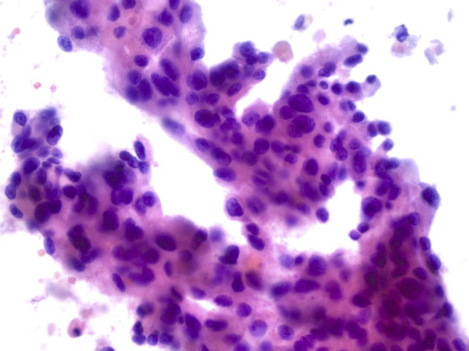
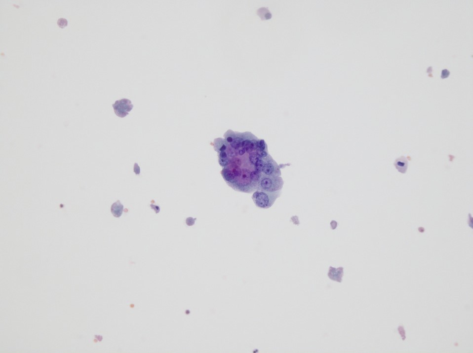
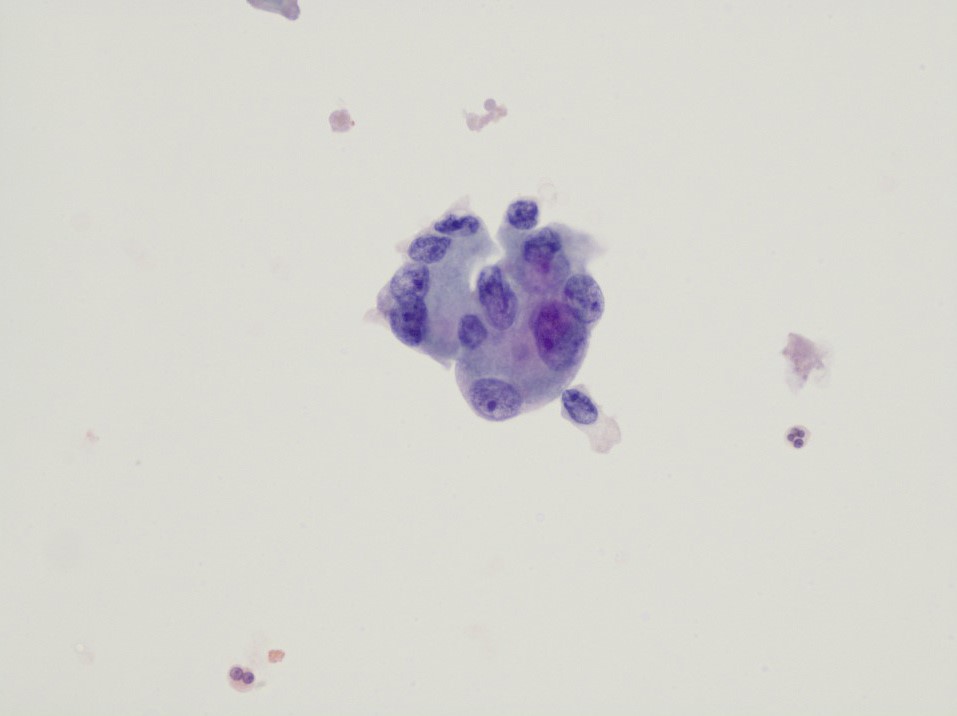
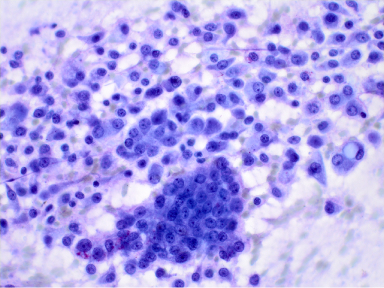
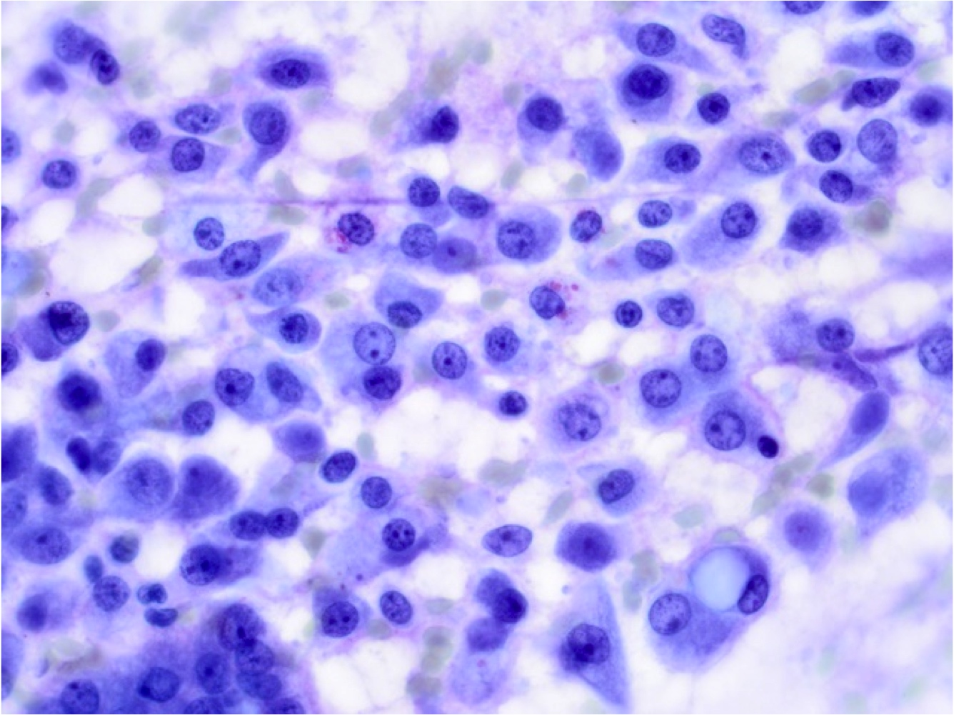
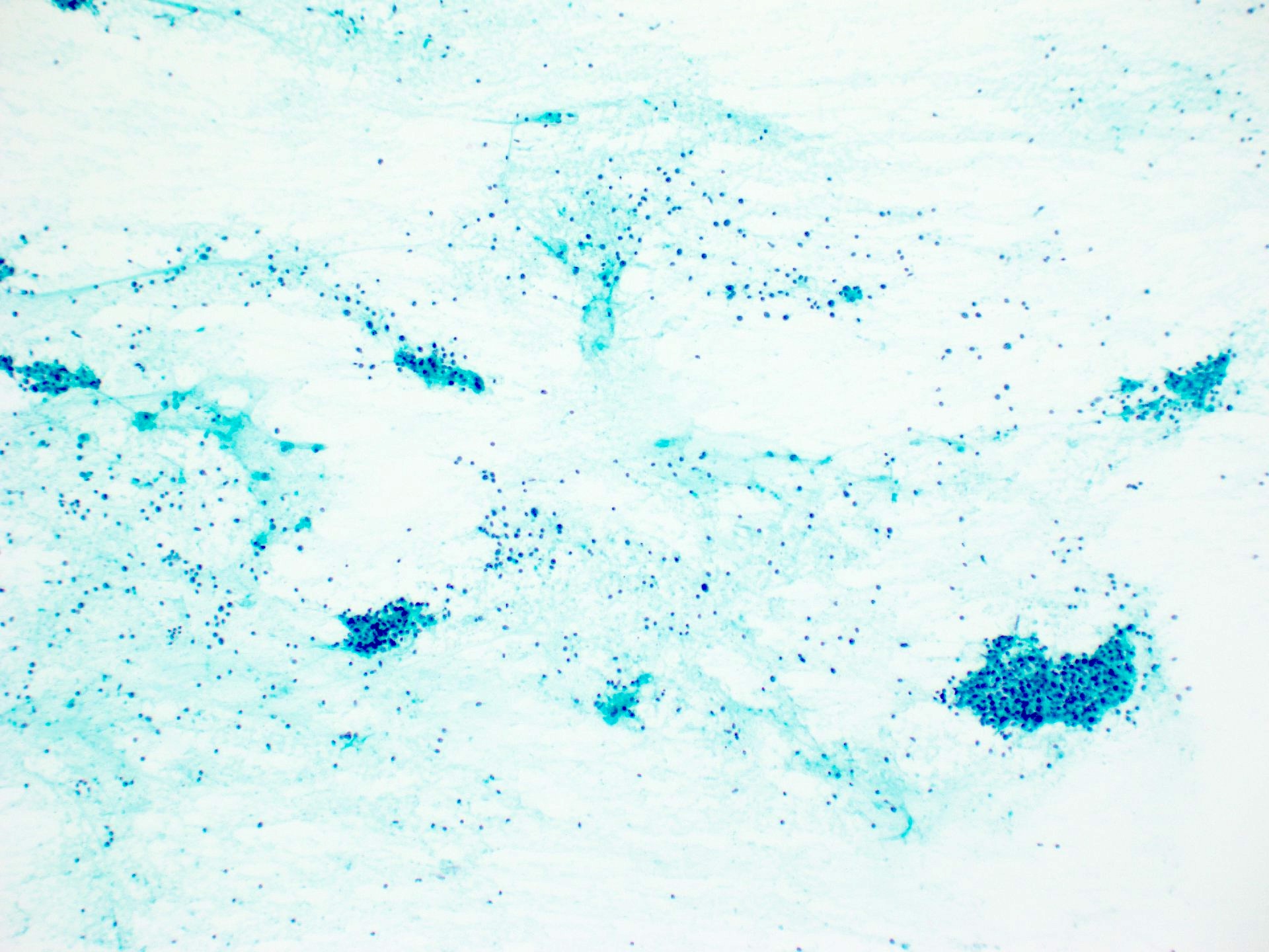
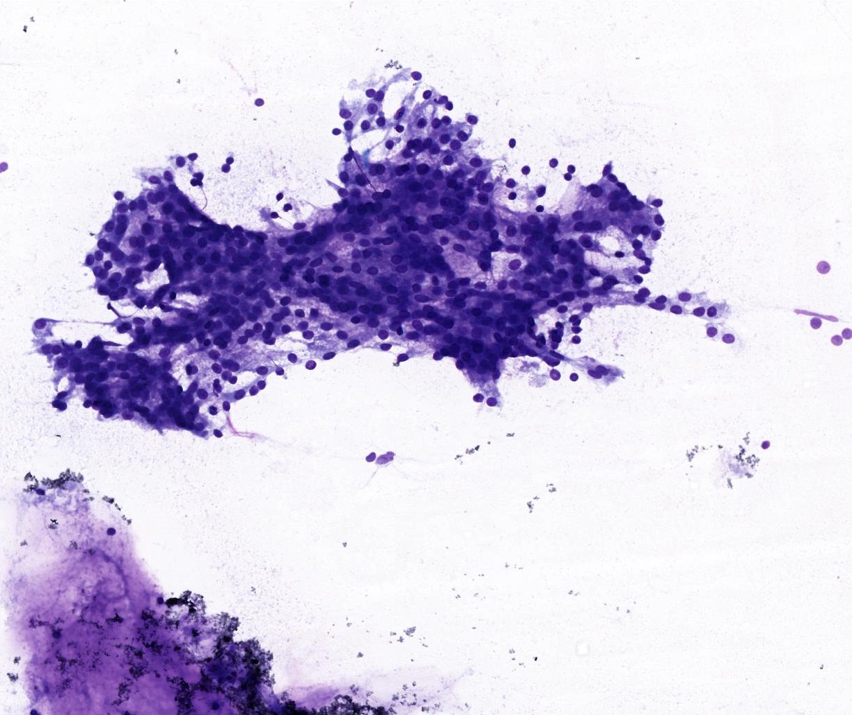
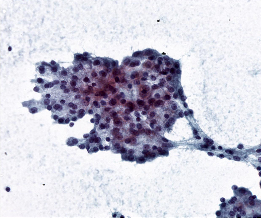
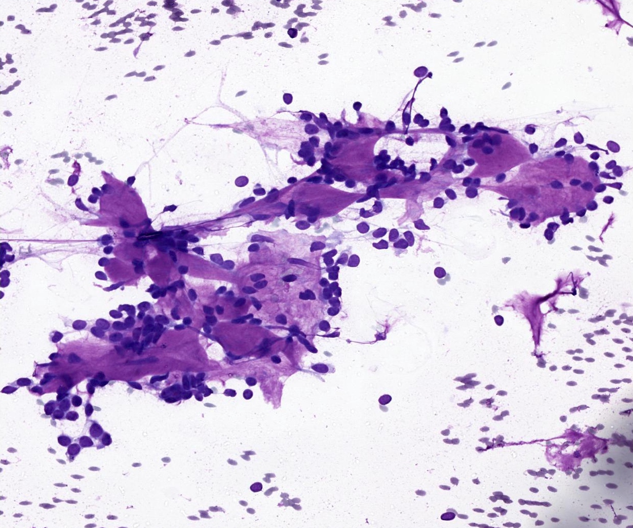
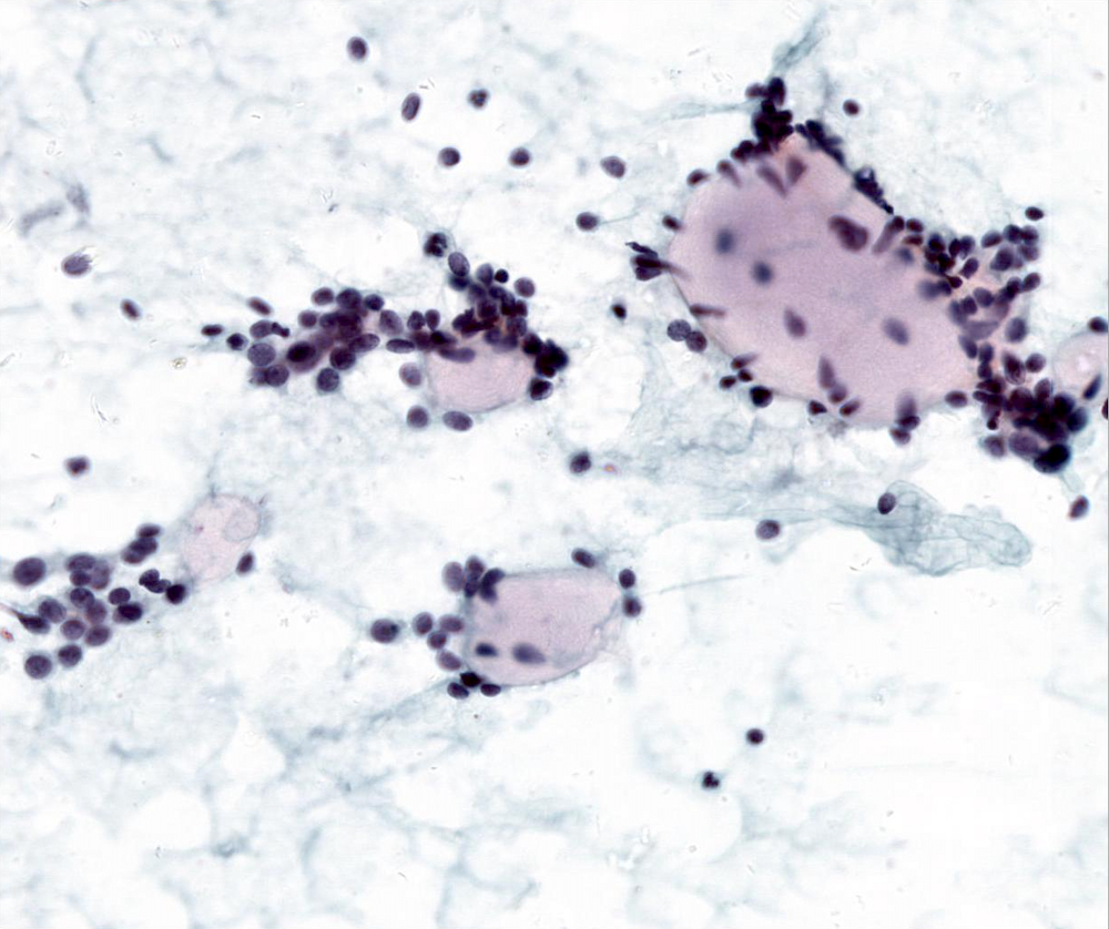
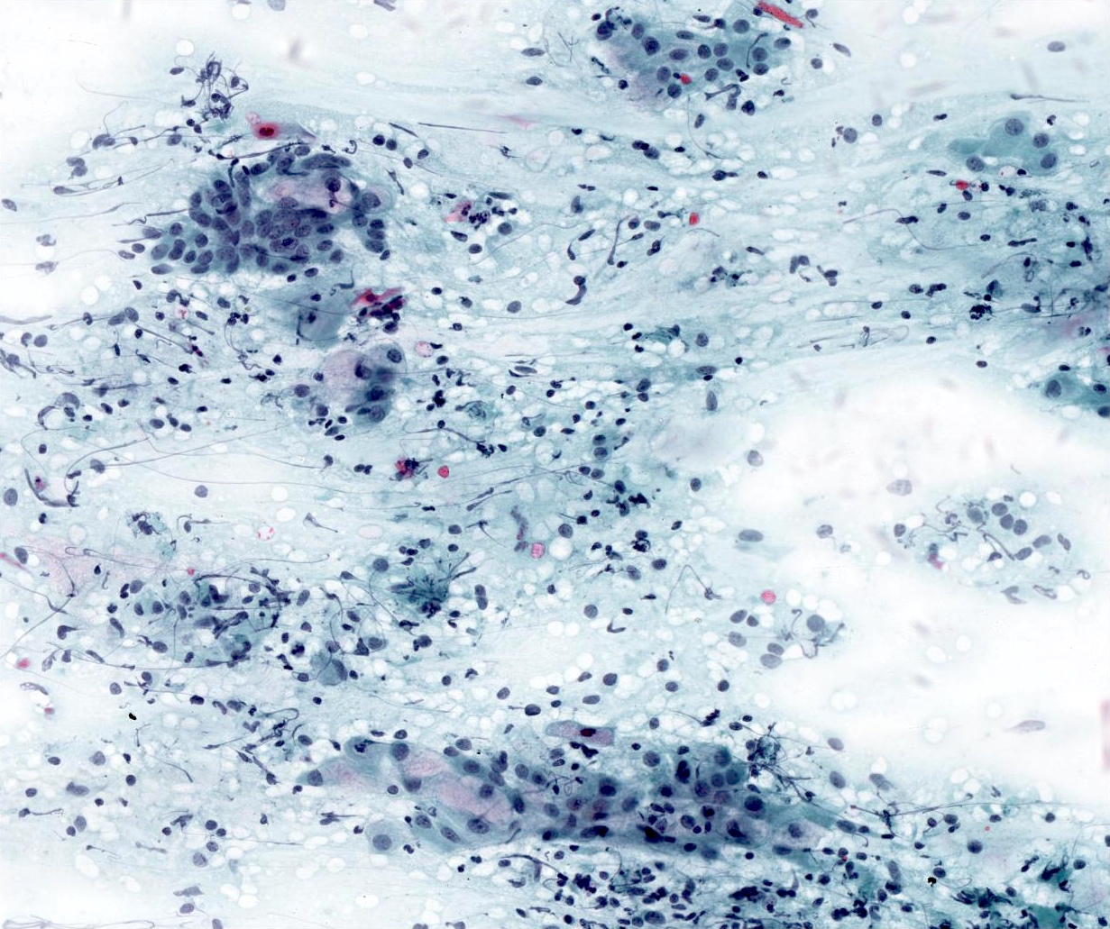
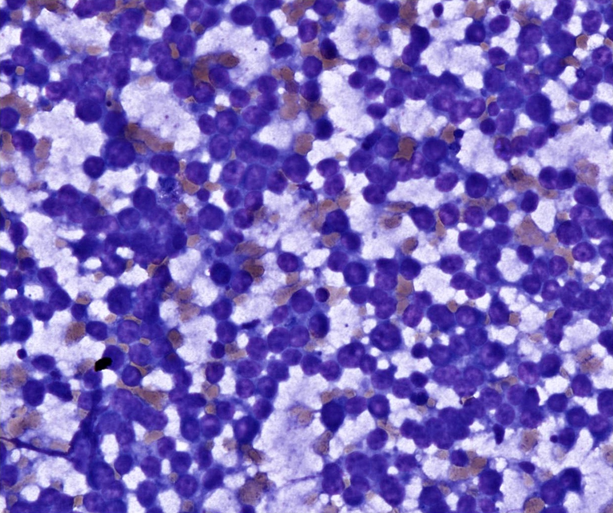
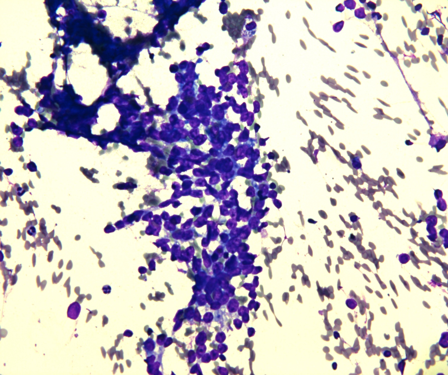
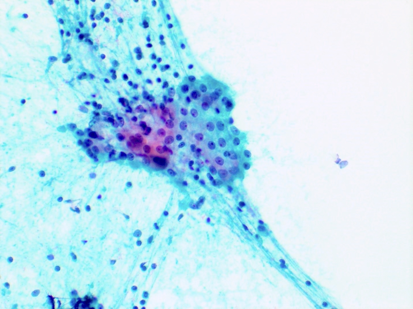
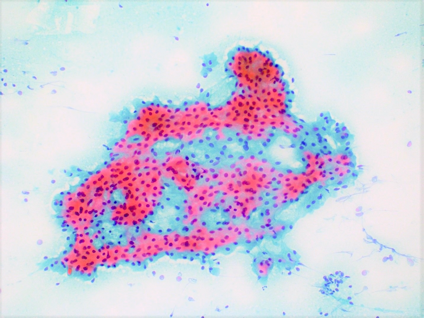
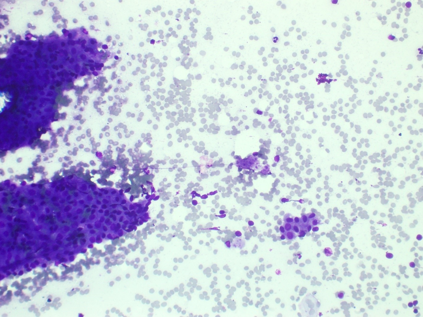
Loosely cohesive groups
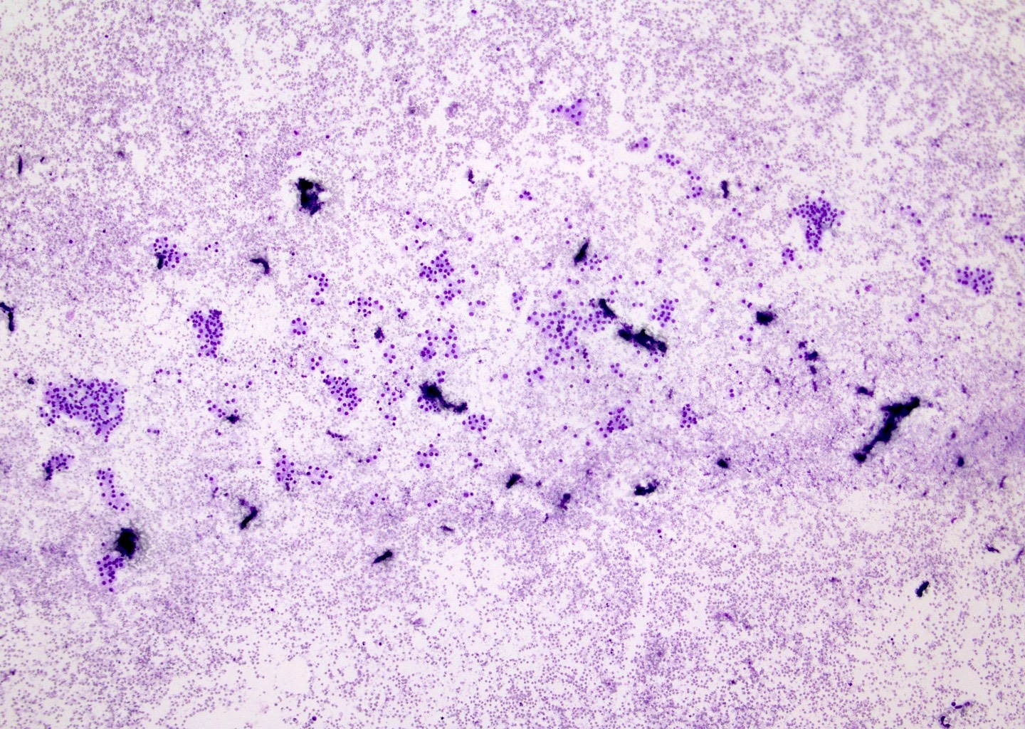
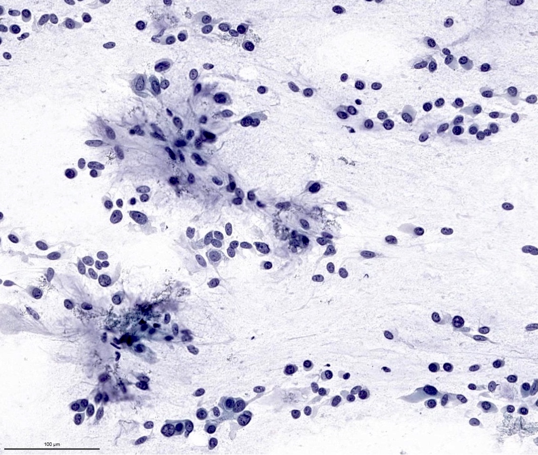
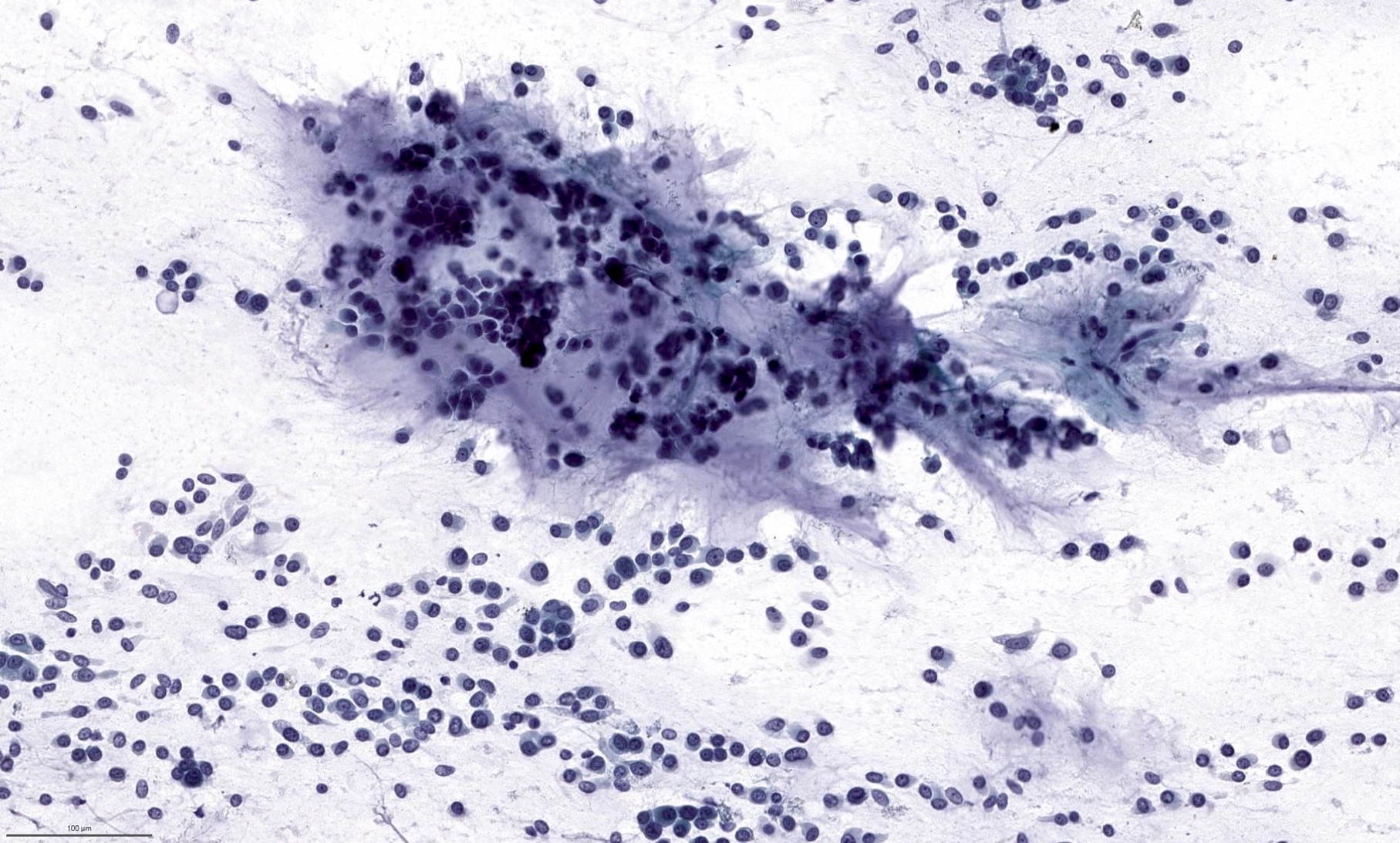
Cellular smears
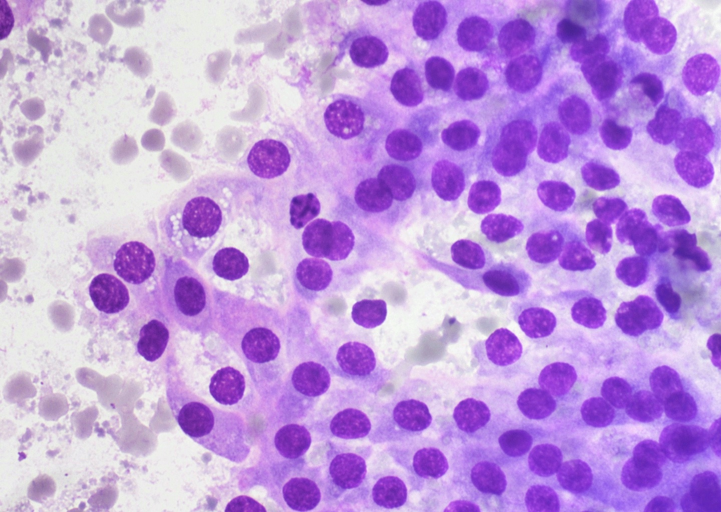
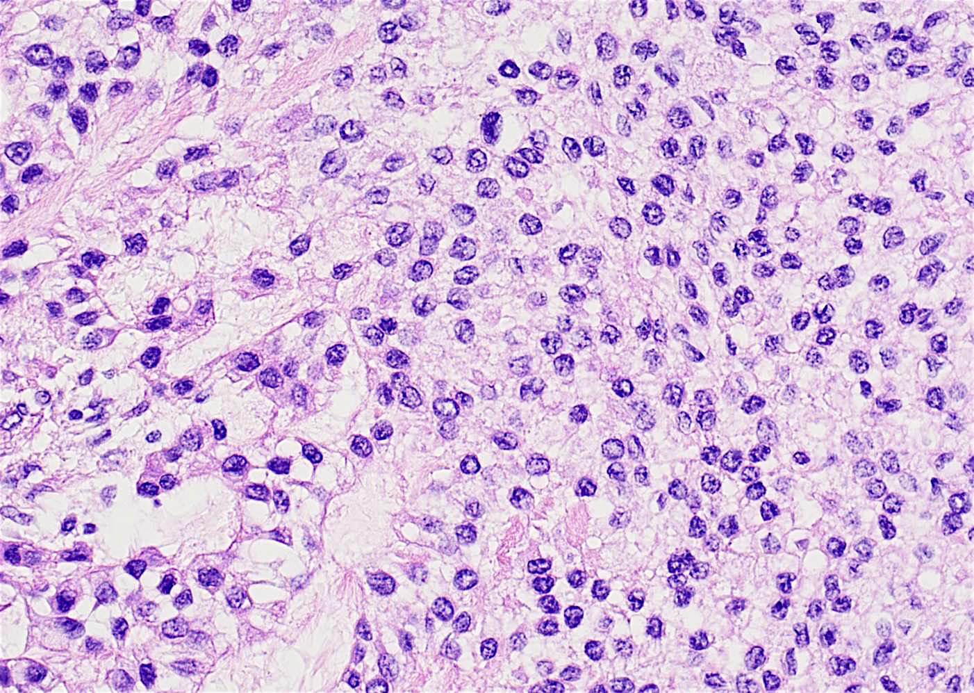
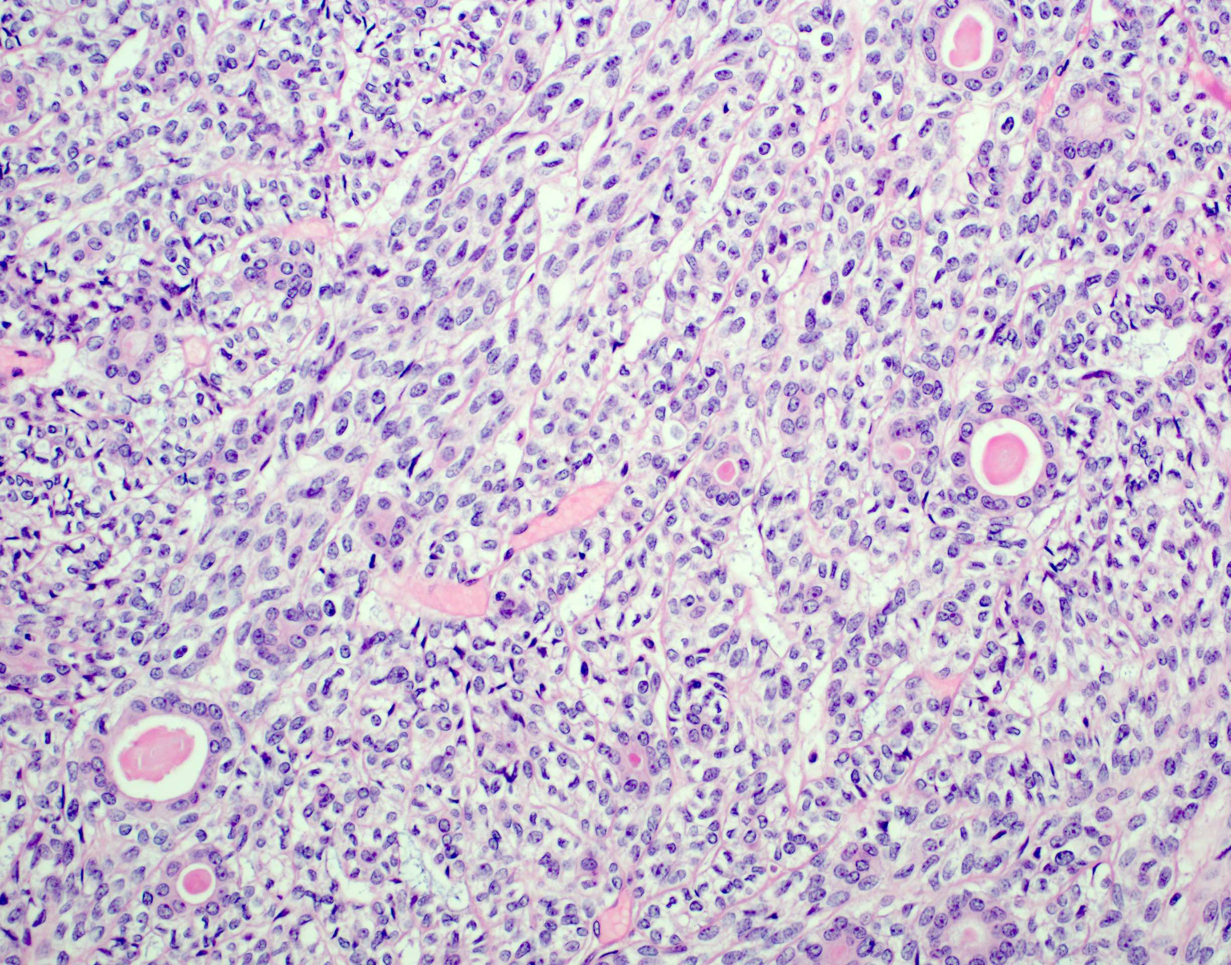
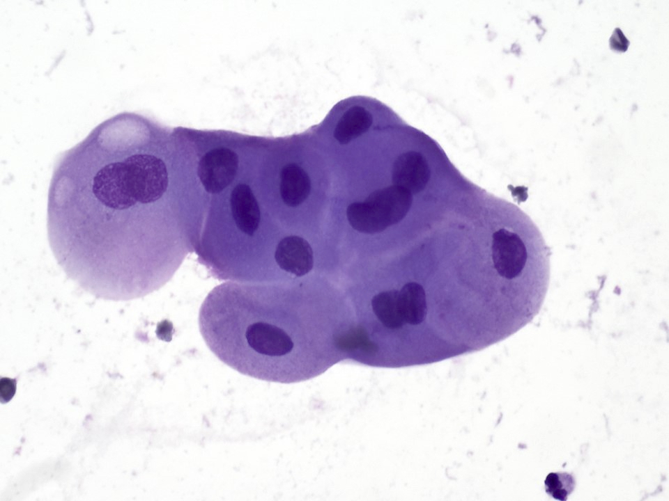
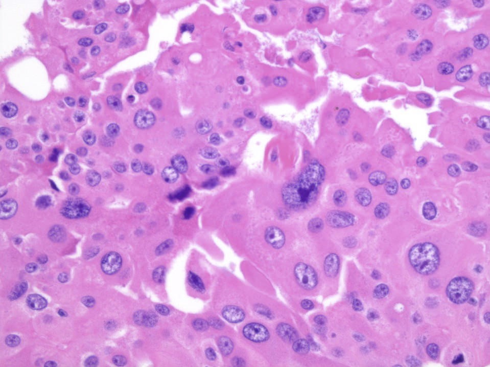
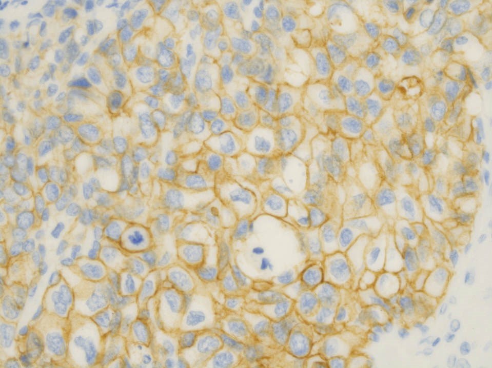
Finely vacuolated to granular cytoplasm

Round eccentric nuclei
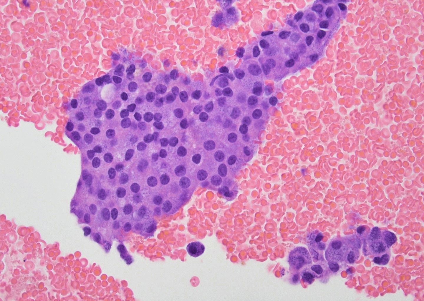
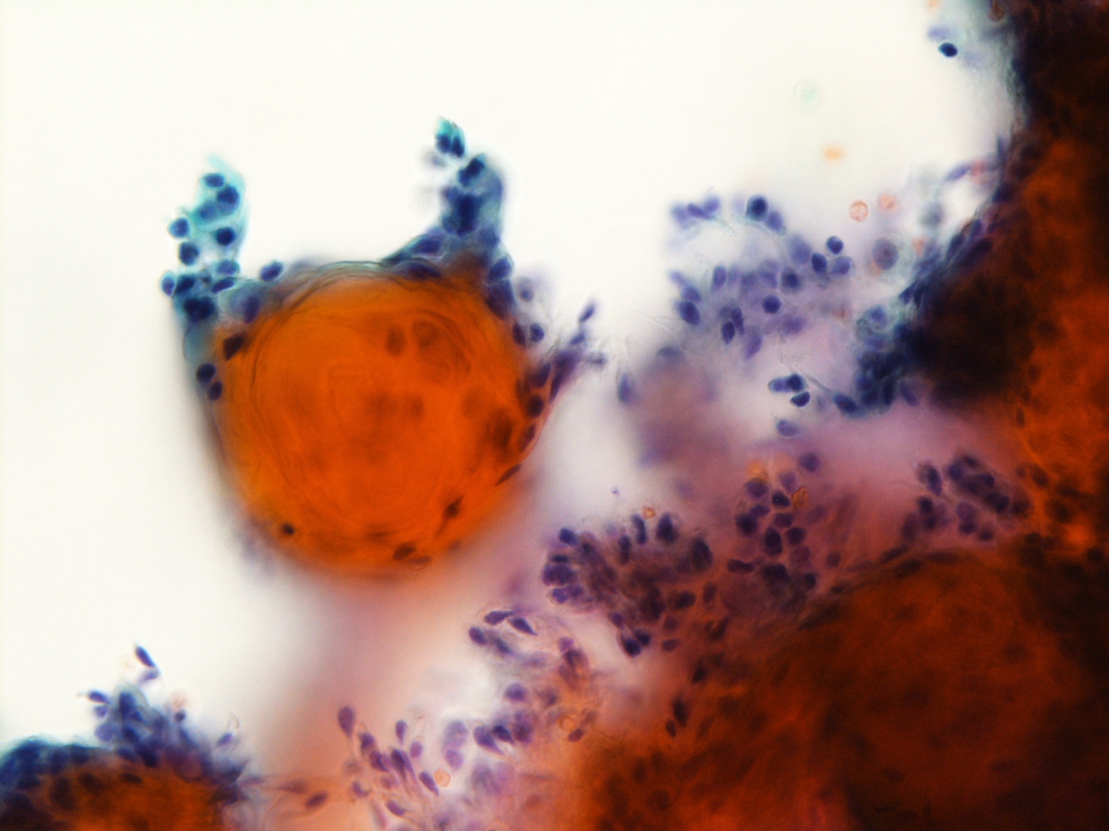
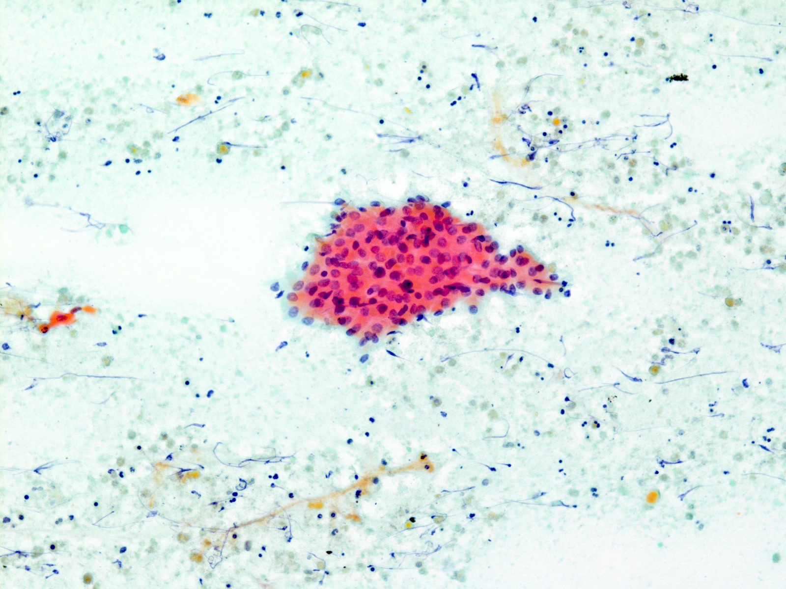
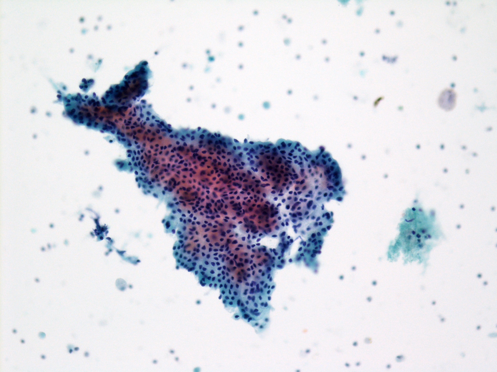
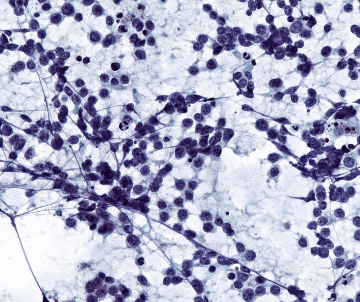
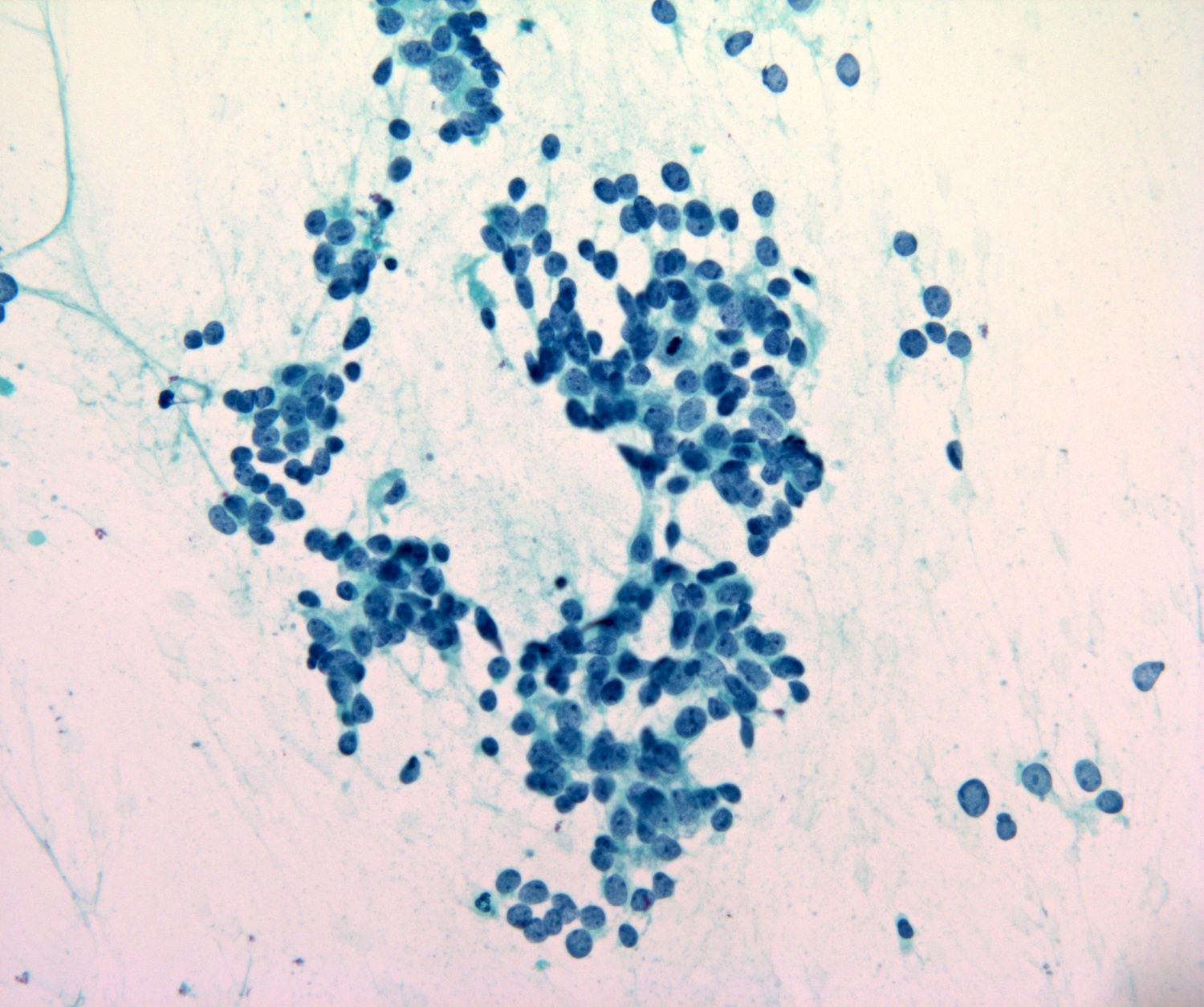
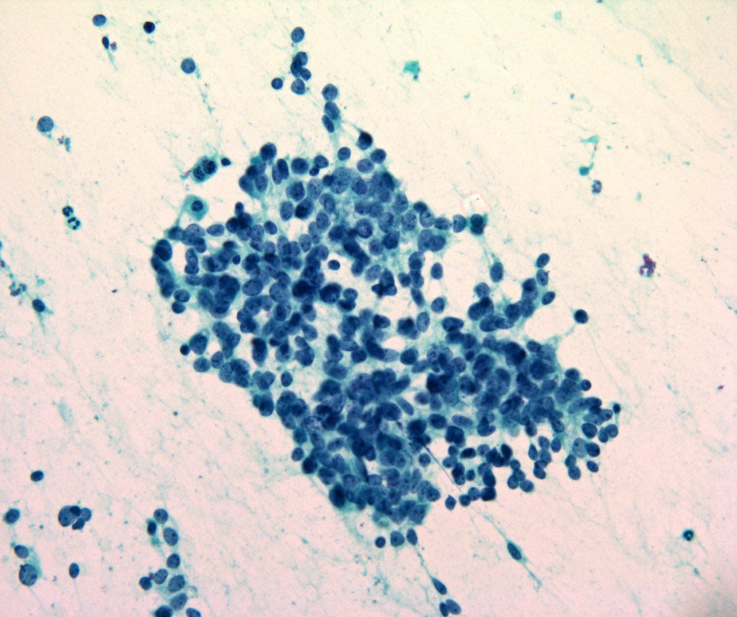
Loosely cohesive clusters
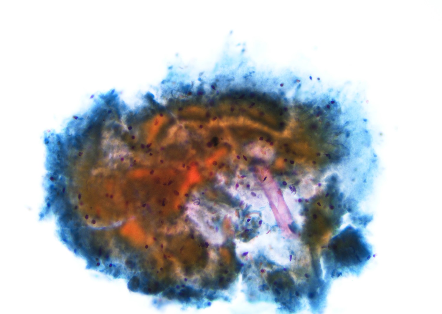
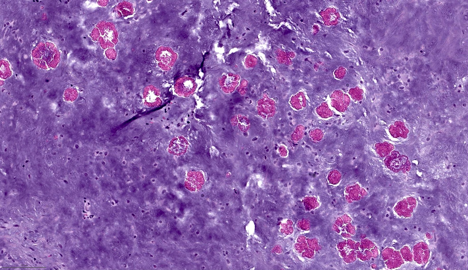
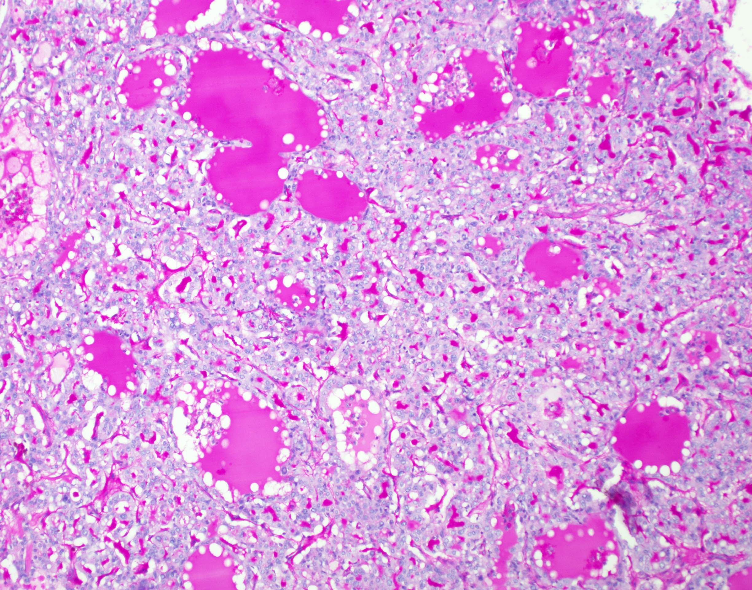
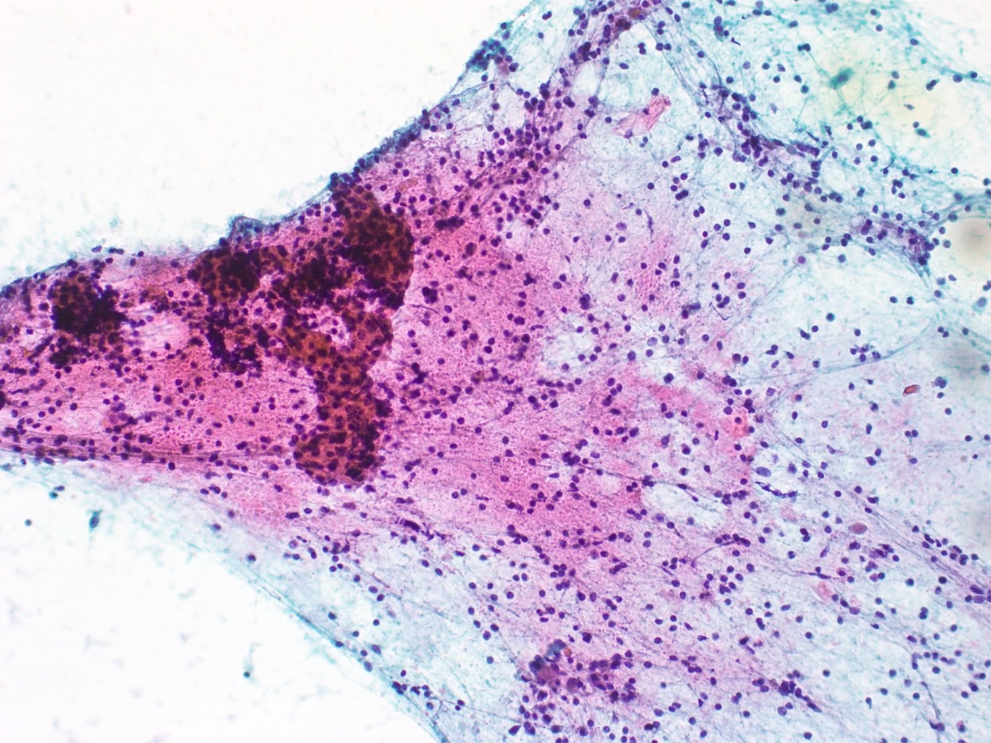
PASD
Images hosted on other servers:

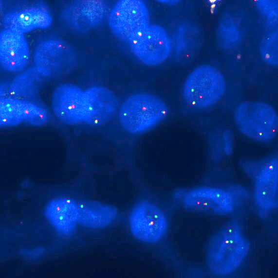
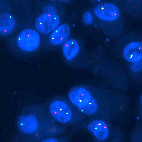
FISH, NR4A3 break apart probe
Images hosted on other servers:
MRI: cystic lesion of the upper lip
CT and US: right parotid nodule
Images hosted on other servers:
1.2 cm upper lip nodule
Images hosted on other servers:
Well circumscribed multilobulated mass
Contributed by Rema A. Rao, M.D. and Arash H. Lahouti, M.D.
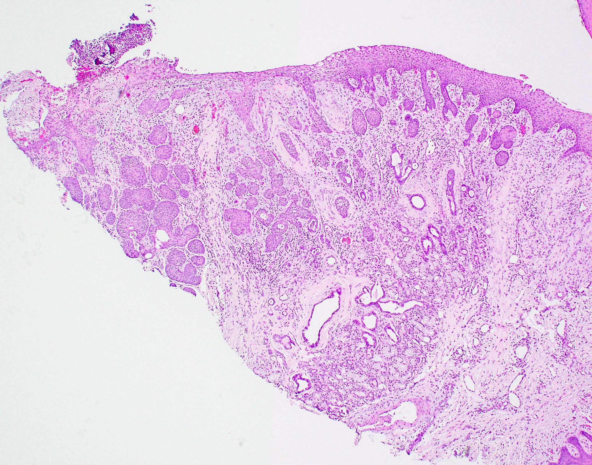
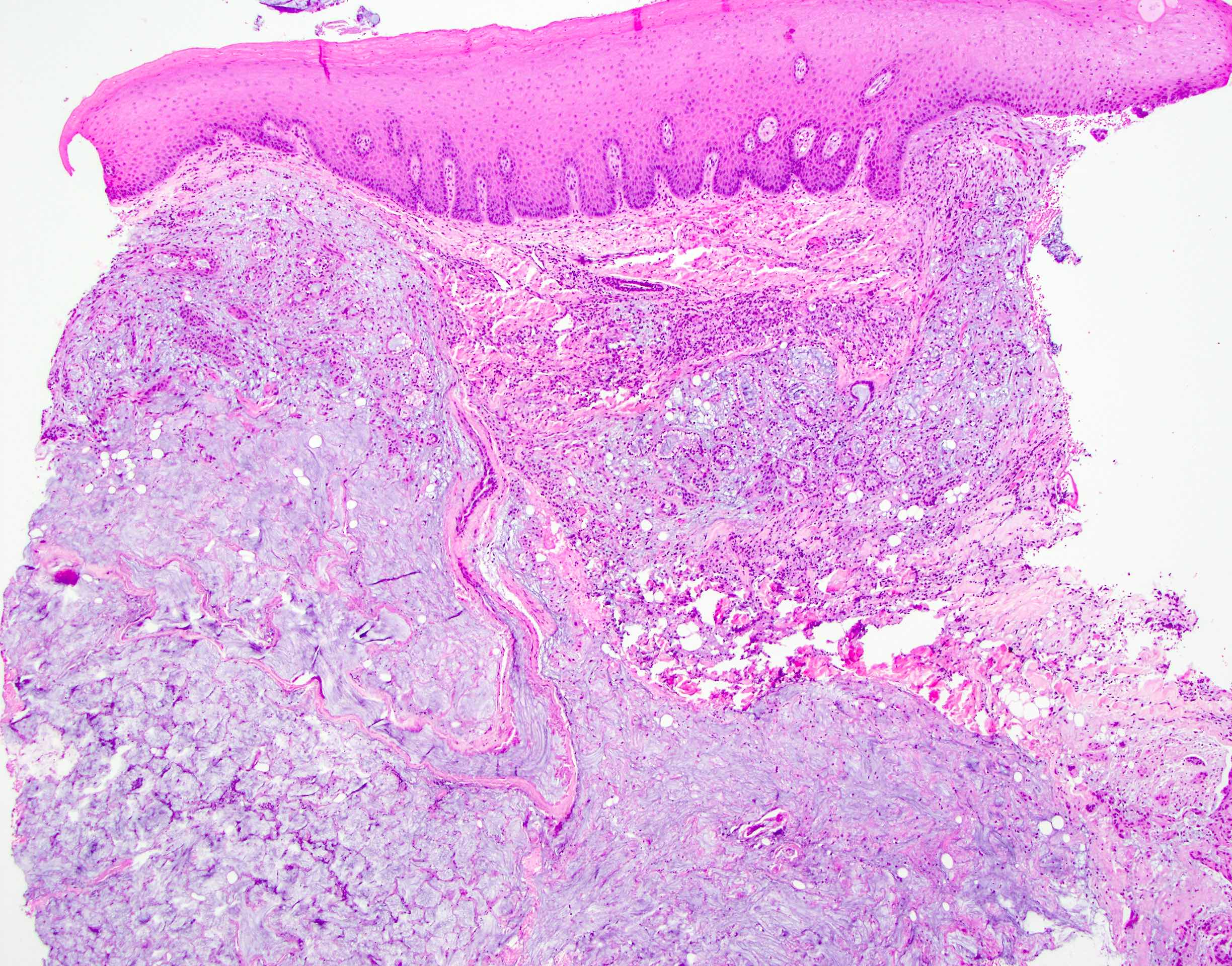
Well circumscribed tumor
Finely vacuolated cytoplasm
Tumor associated lymphoid proliferation
Cystic pattern and hemorrhage
NR4A3 nuclear stain
Contributed by Rema A. Rao, M.D. and Arash H. Lahouti, M.D.
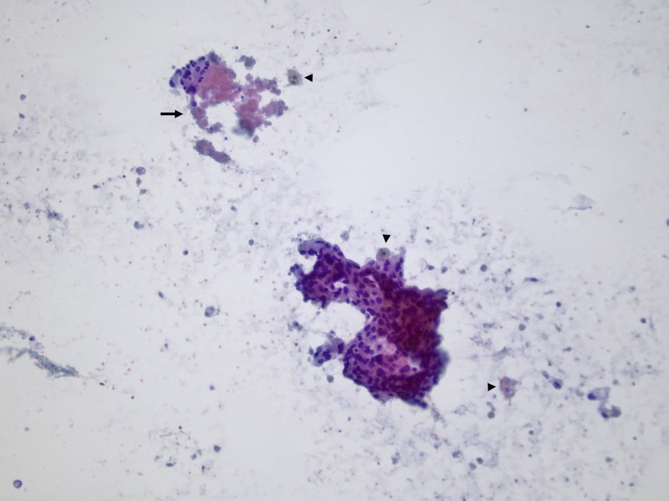
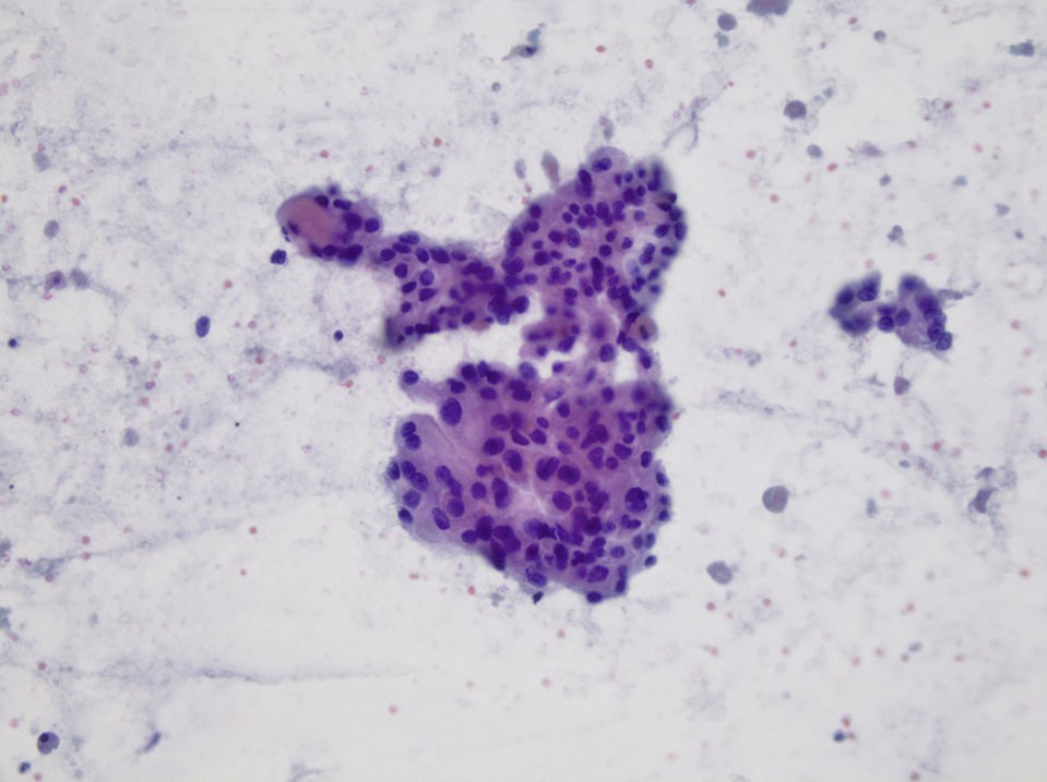
Loosely cohesive groups
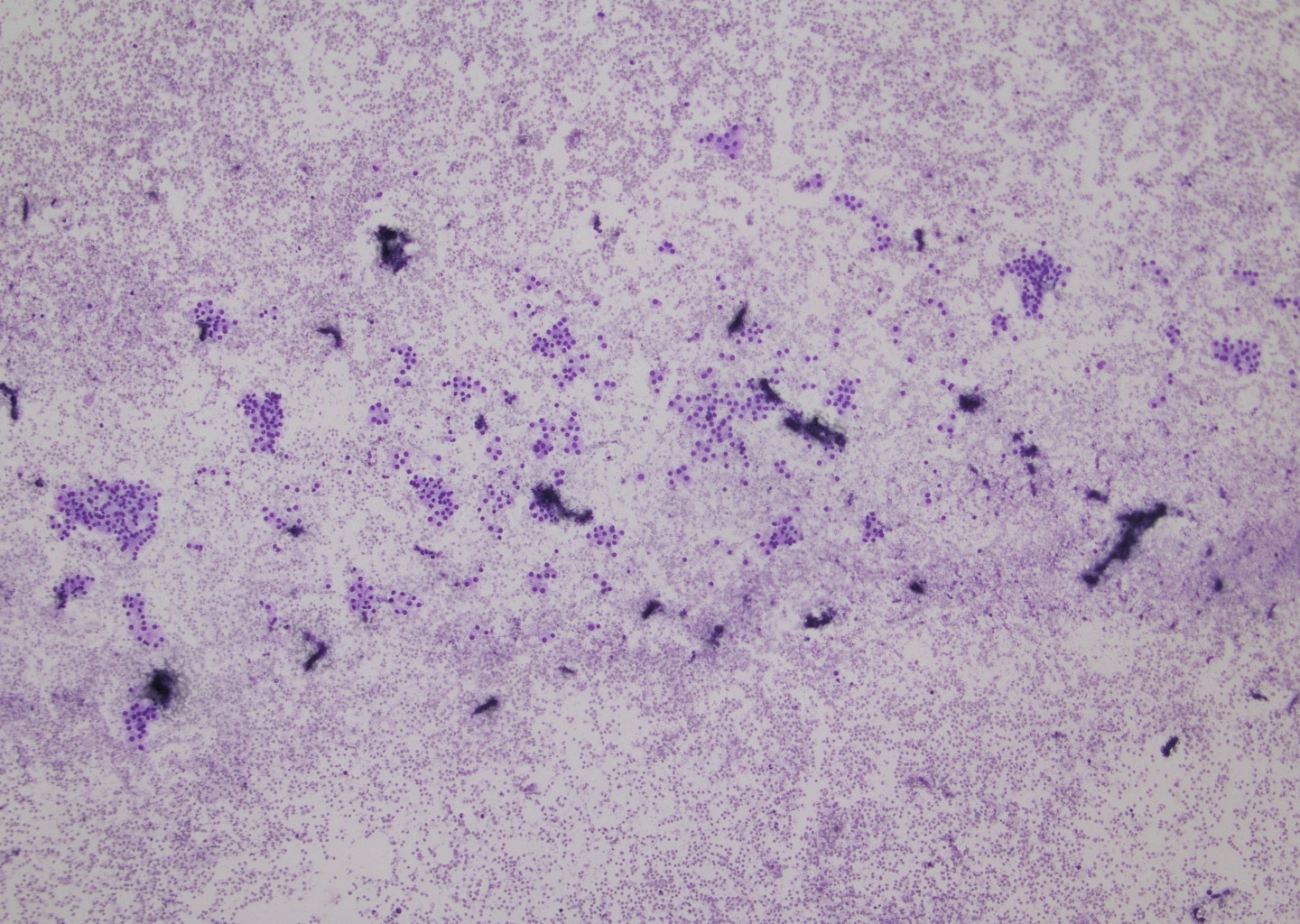
Cellular smears
Finely vacuolated to granular cytoplasm
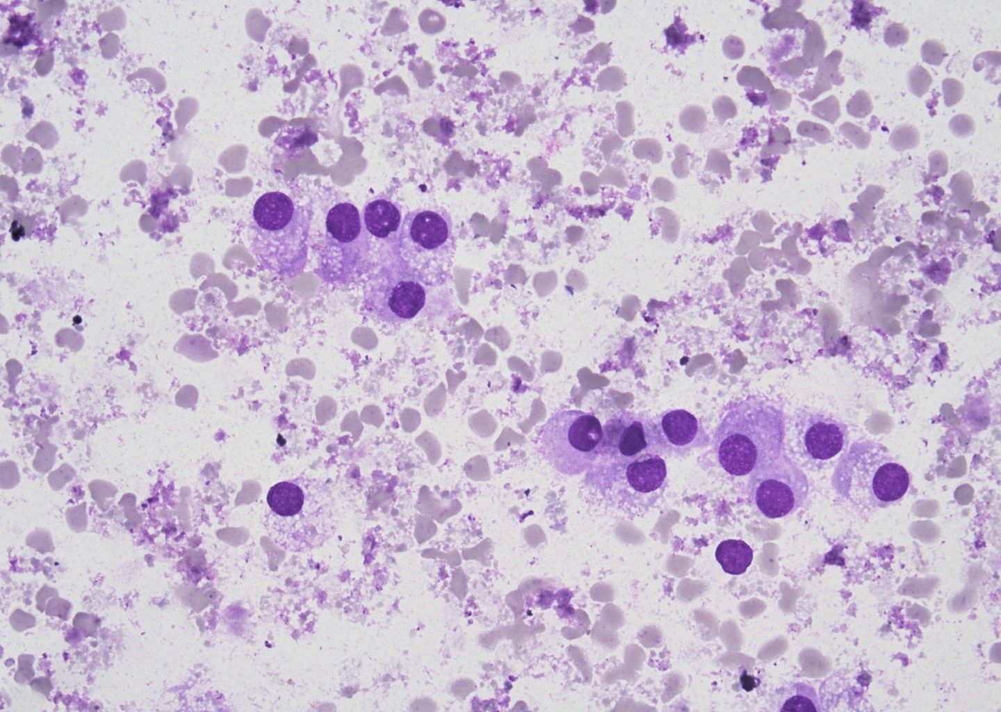
Round eccentric nuclei
Loosely cohesive clusters
PASD
Images hosted on other servers:

FISH, NR4A3 break apart probe
Contributed by Marino Leon, M.D.
Oncocytic carcinoma, H&E and p63
Images hosted on other servers:

Histologic impact on survival
Images hosted on other servers:

MRI parotid mass

Lung metastases
Images hosted on other servers:

Vocal cord mass
Contributed by Bin Xu, M.D., Ph.D. and Kelly Magliocca, D.D.S., M.P.H.
Parotid mass
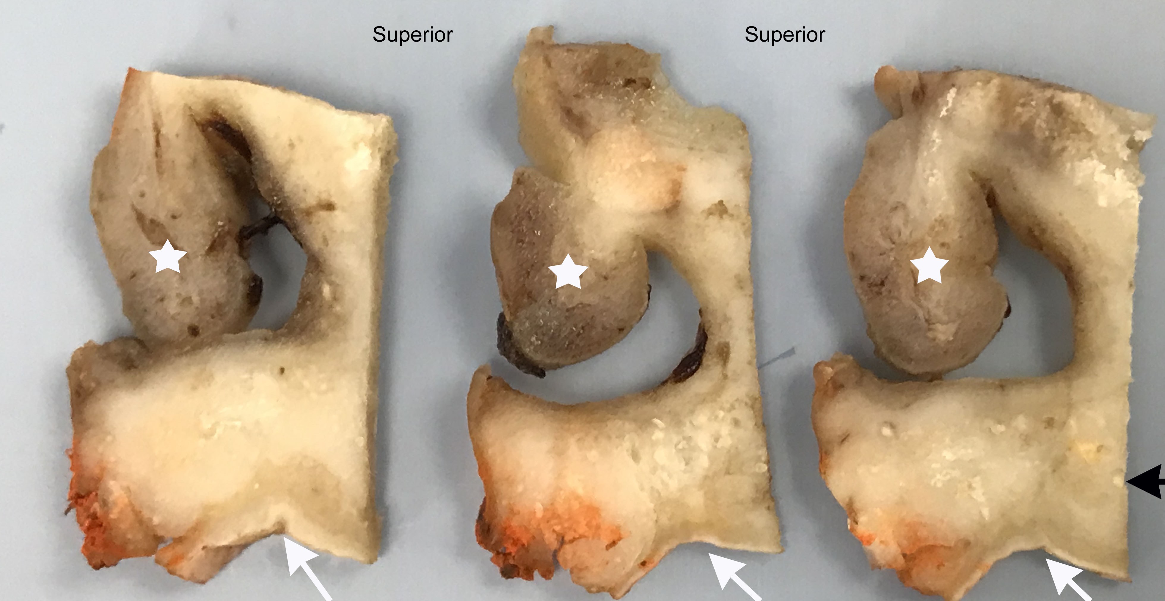
Cut section turbinate to palate
Contributed by Bin Xu, M.D., Ph.D. and Kelly Magliocca, D.D.S., M.P.H.
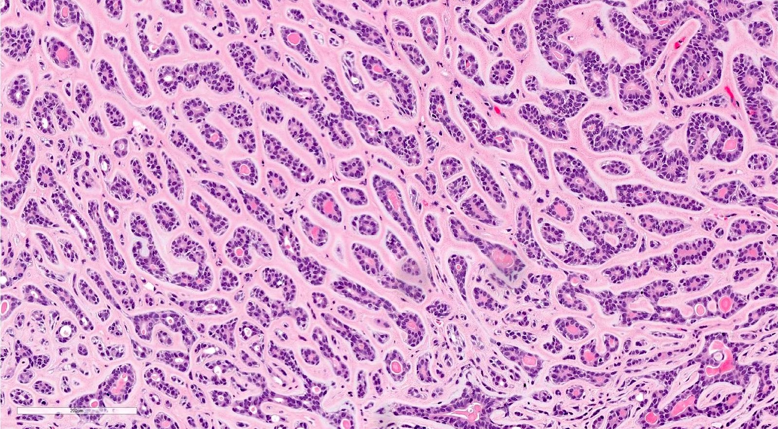
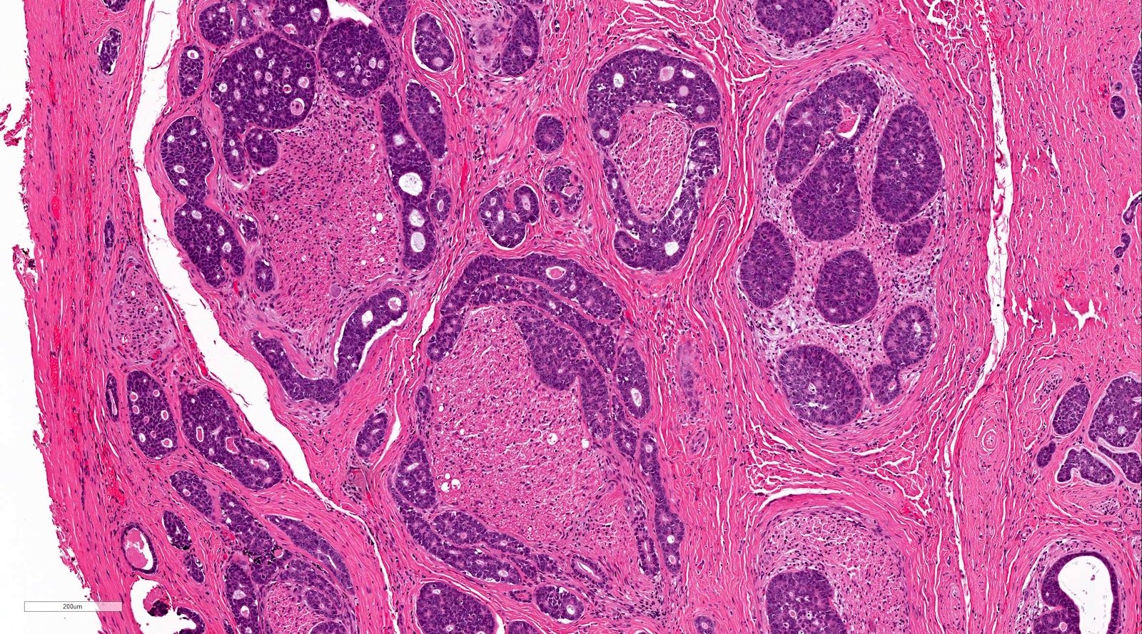
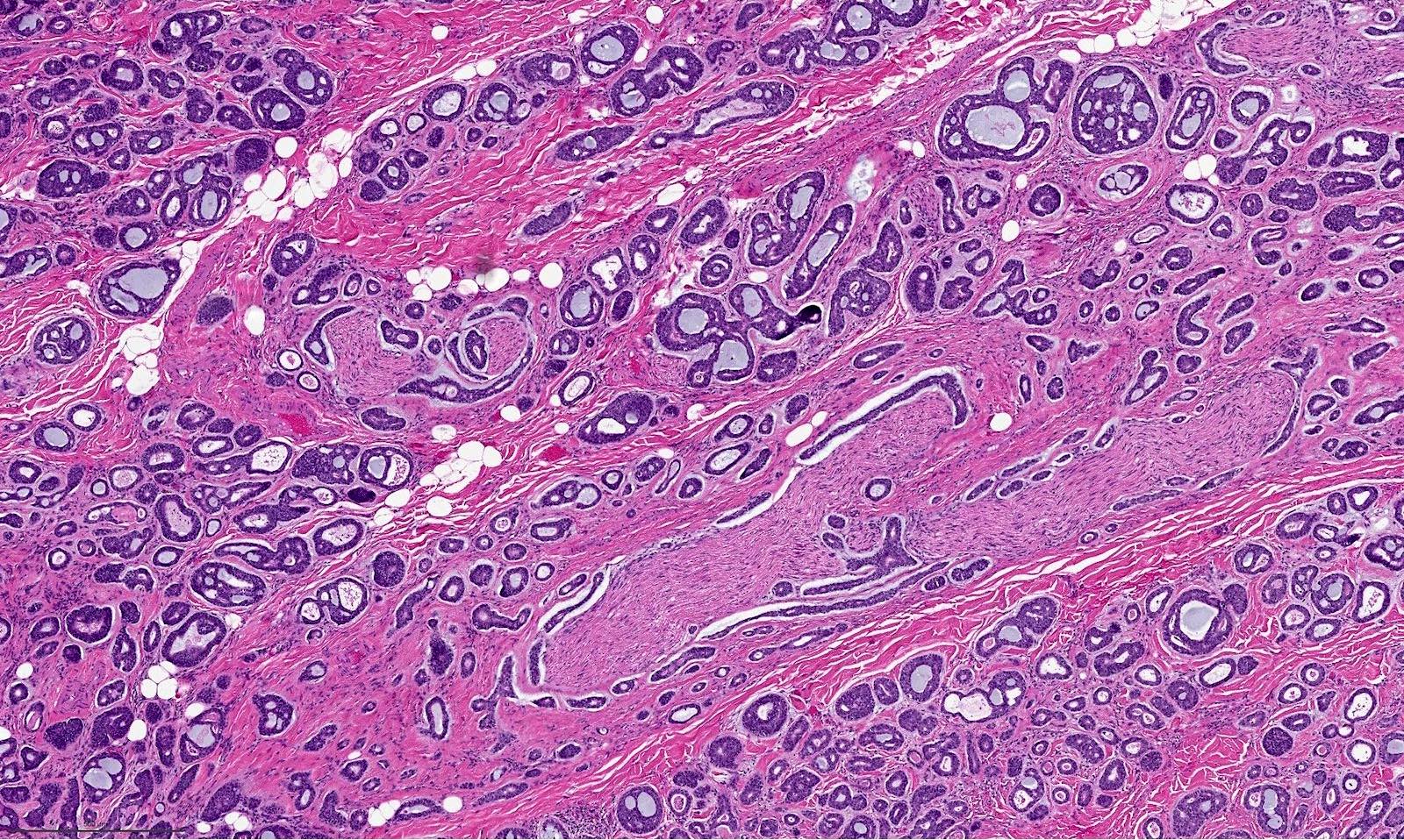
Tubular pattern
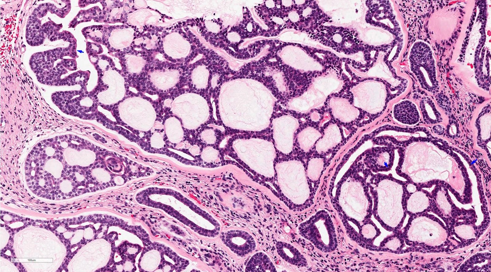
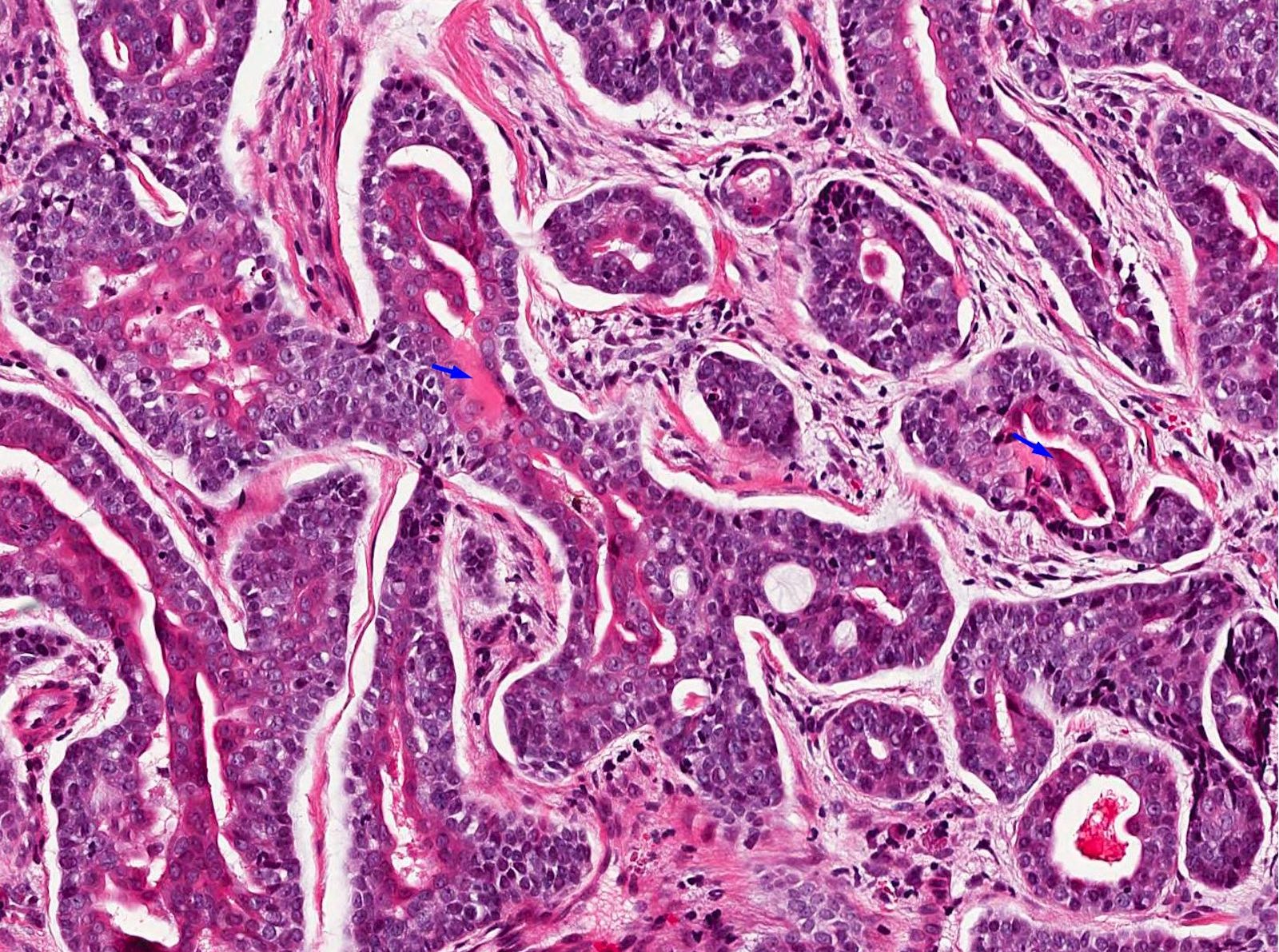
Cribriform pattern
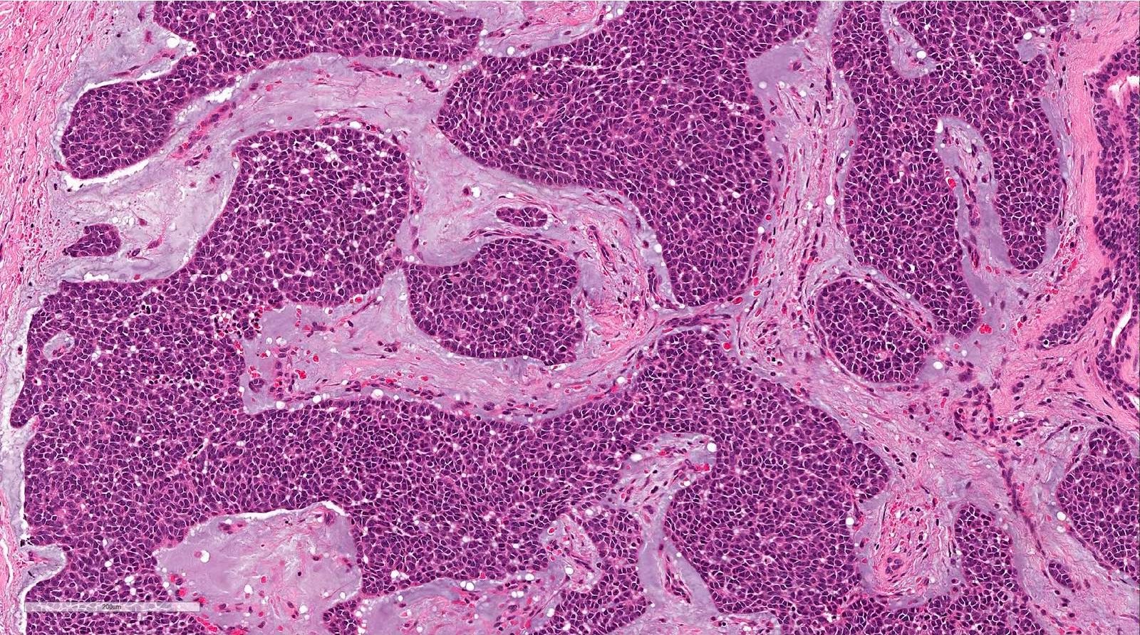
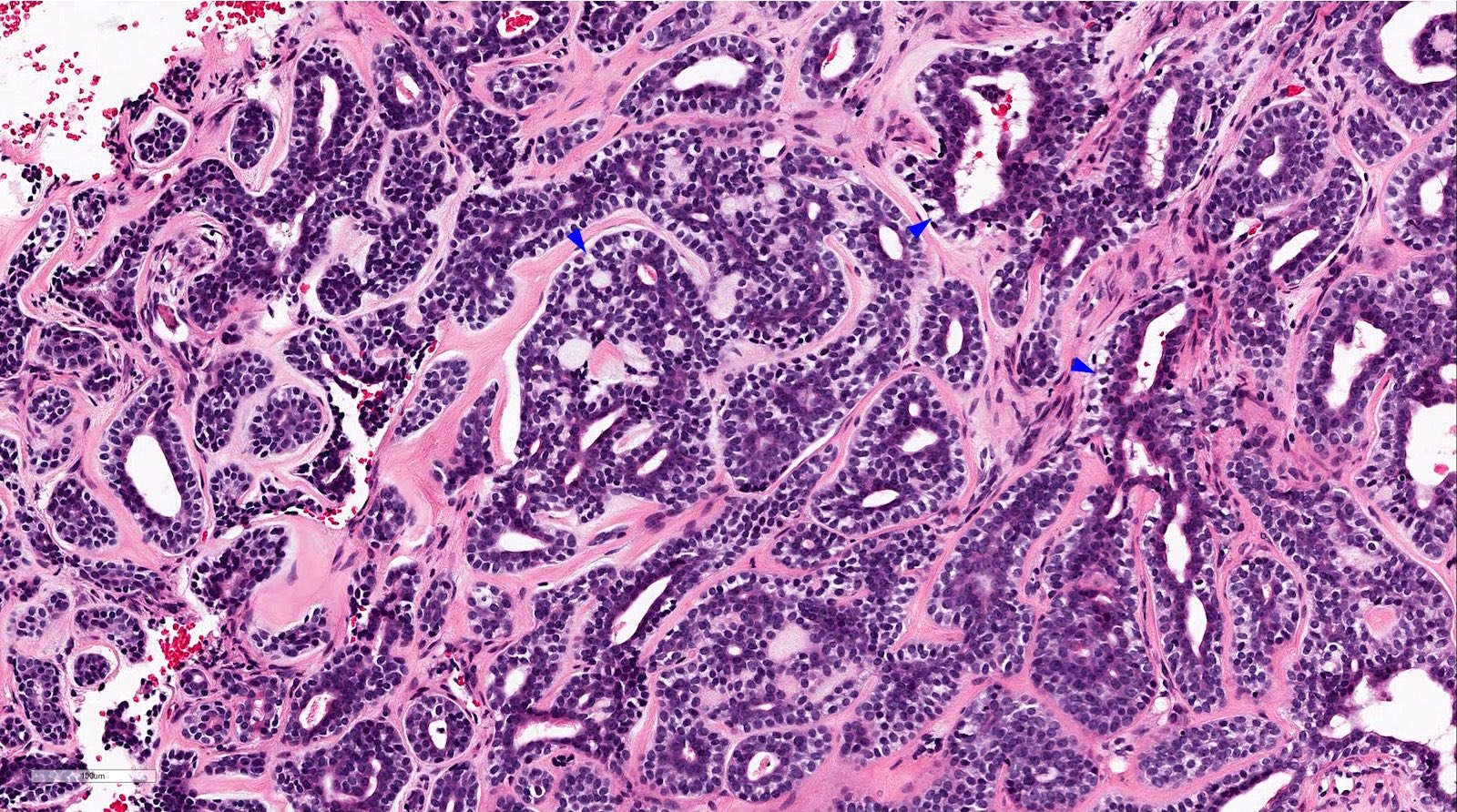
Solid pattern
High grade transformation
Perineural invasion
Squamous metaplasia
Clear myoepithelial cells
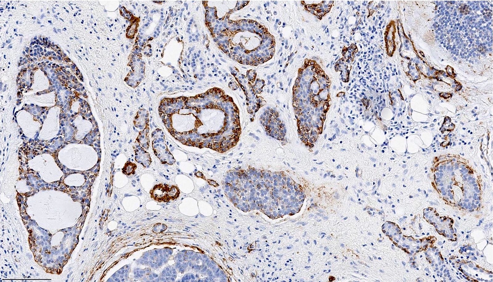
p40
SMA
CAM5.2
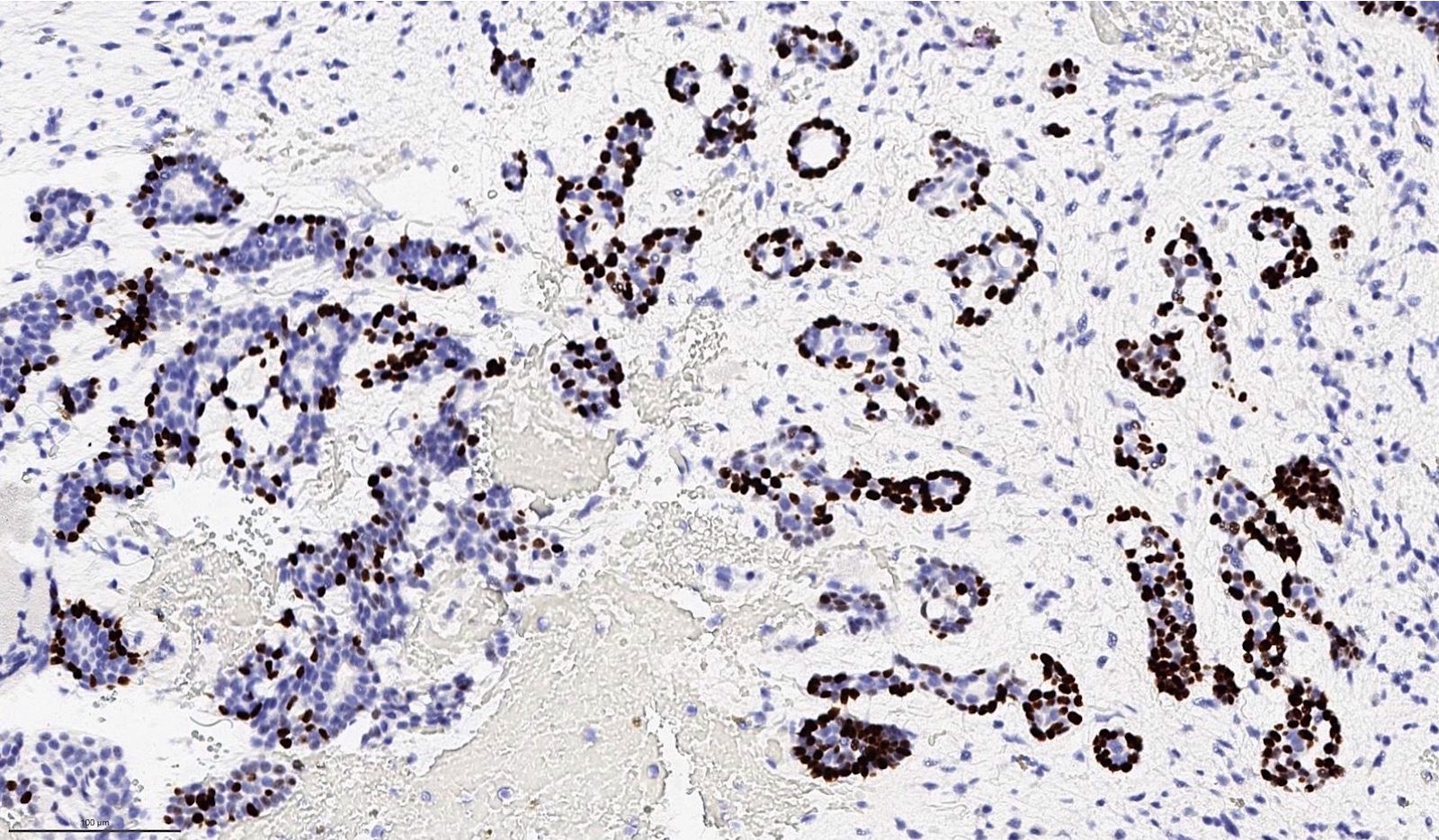
MYB
H&E of turbinate to palate
Contributed by Bin Xu, M.D., Ph.D. and Jen-Fan Hang, M.D.
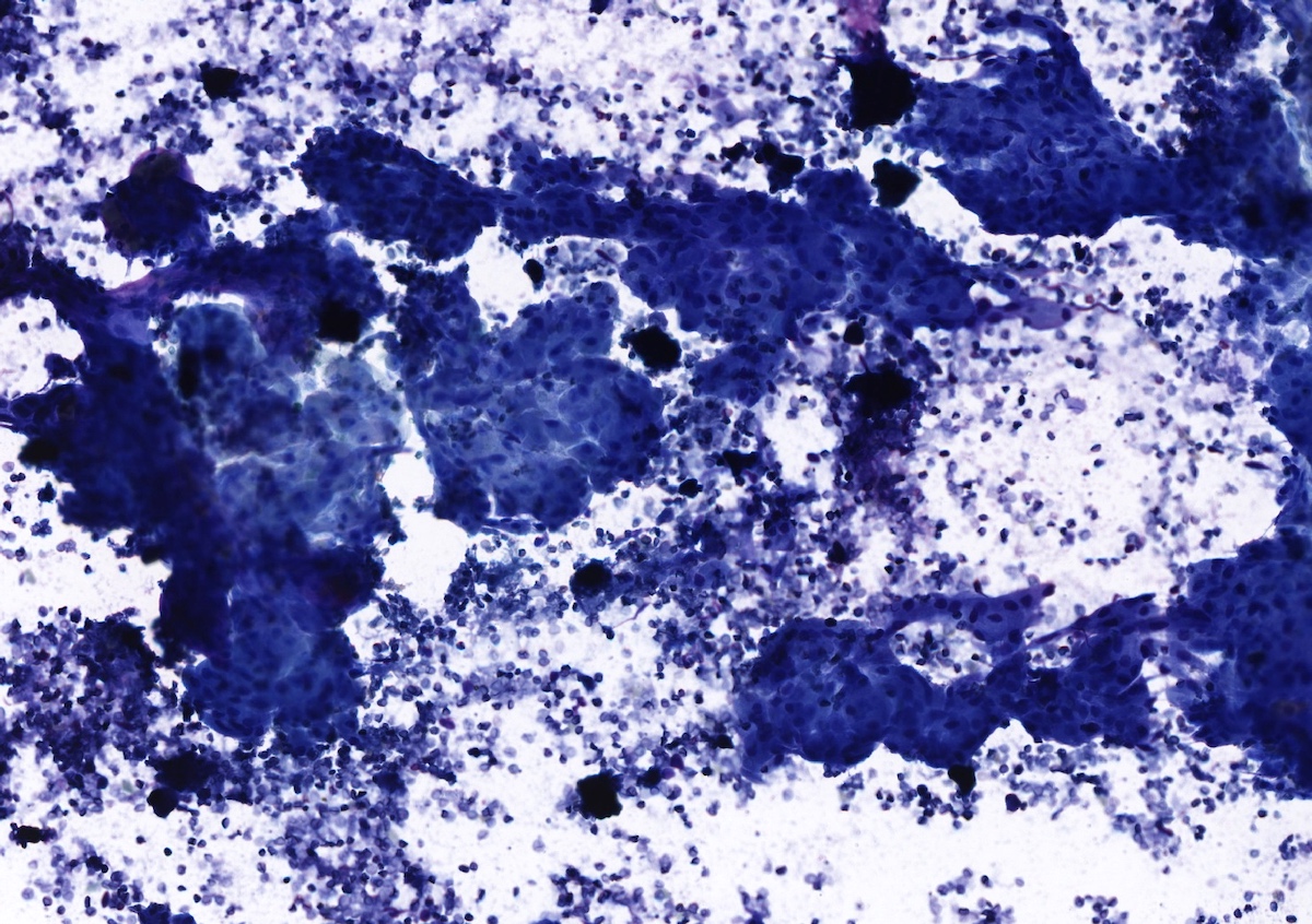
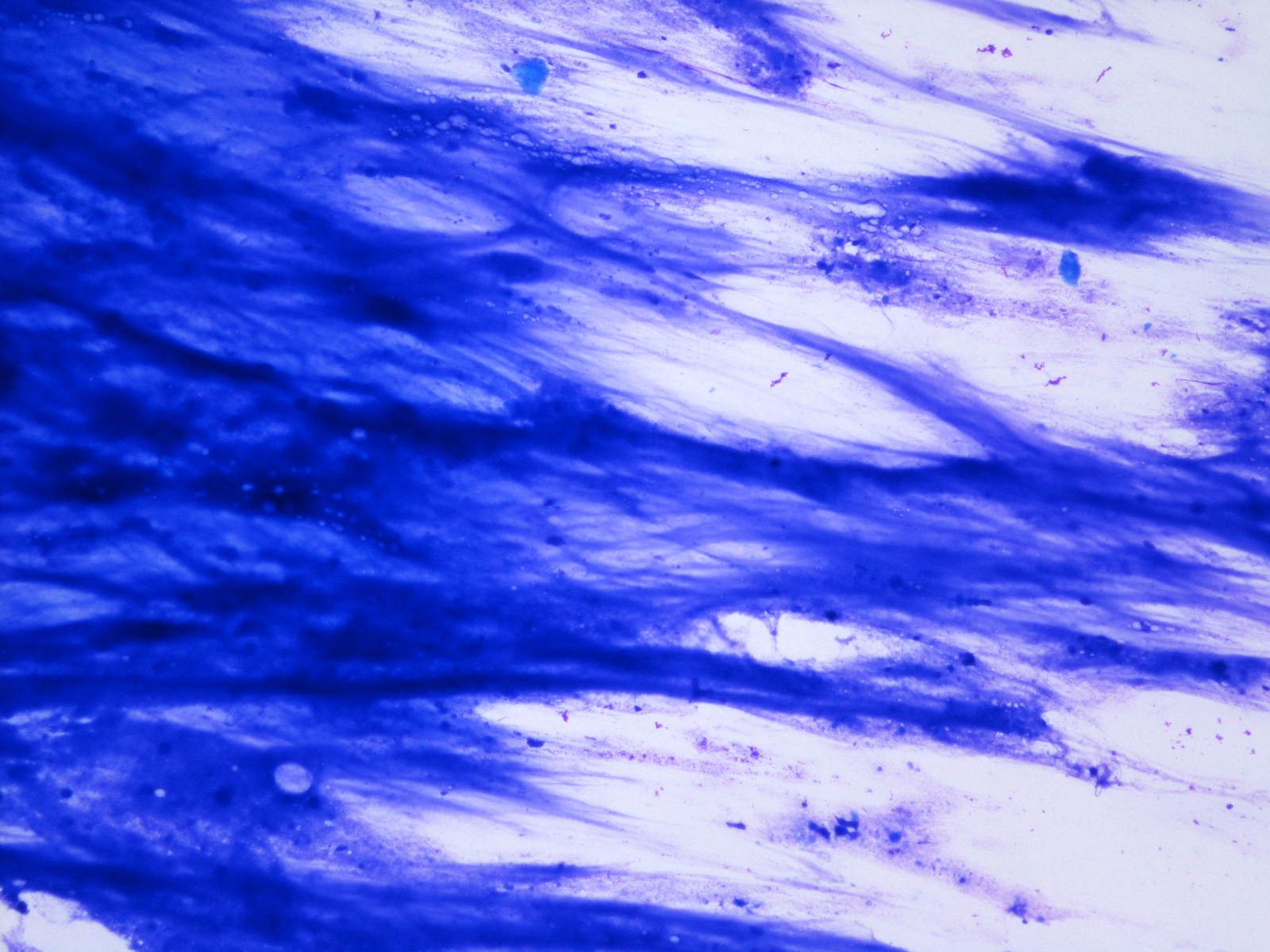
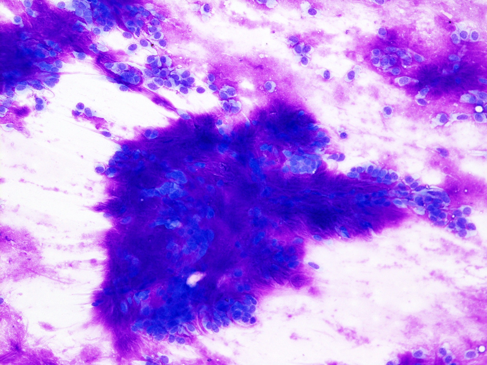
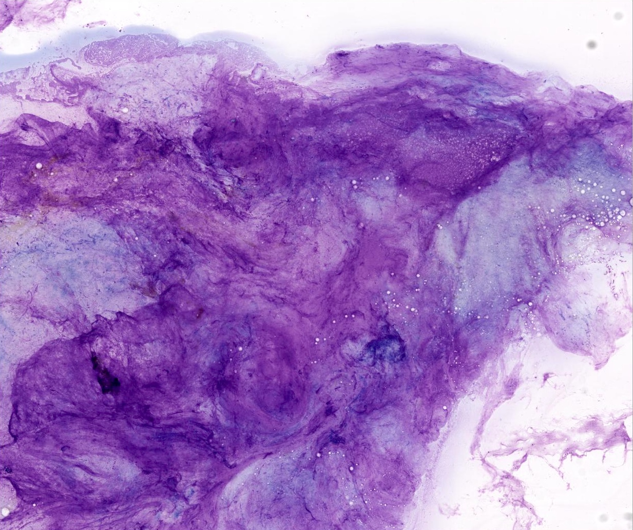
Smear, Diff-Quik
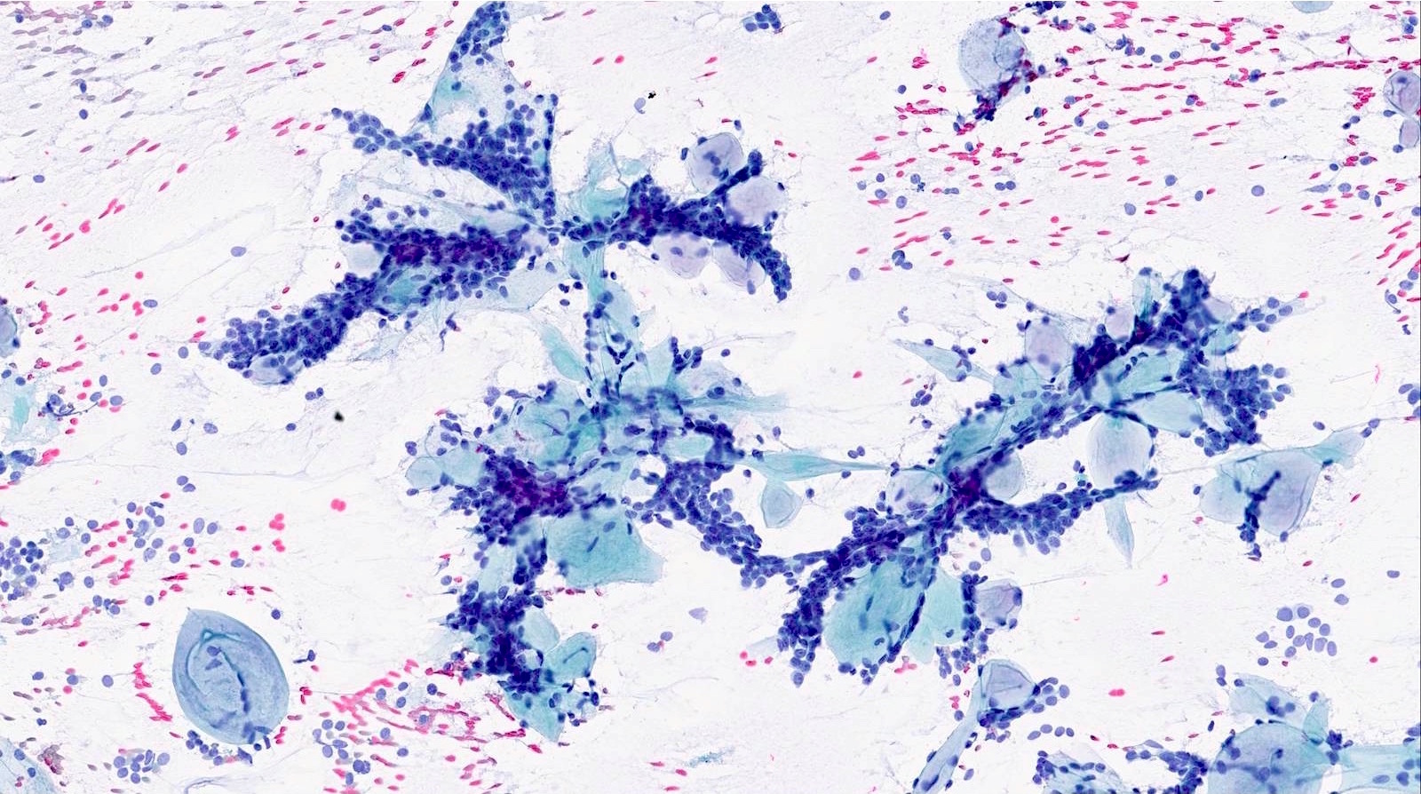
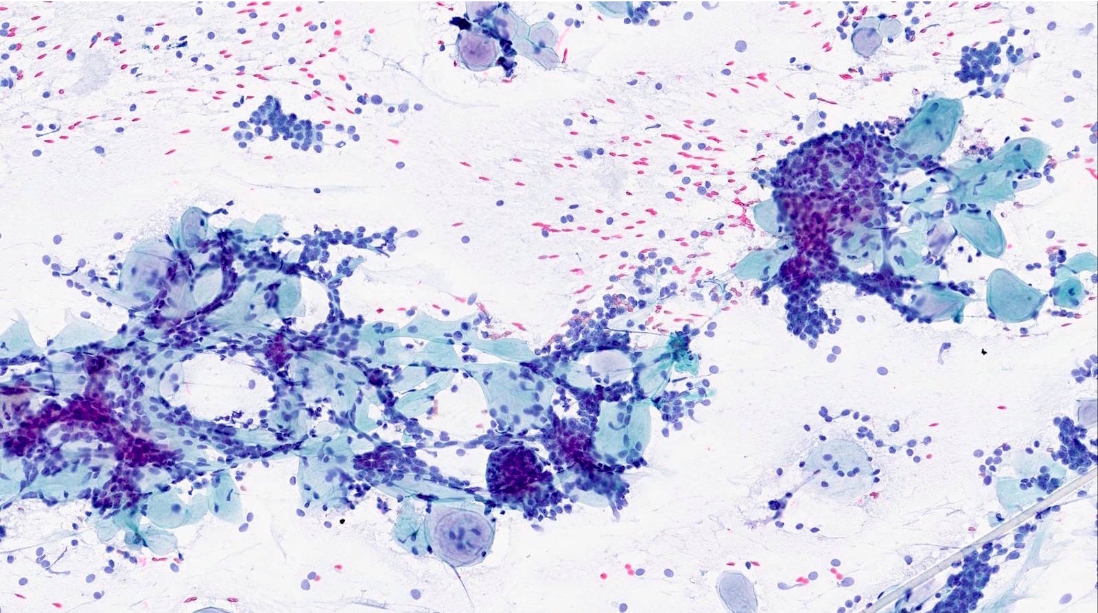
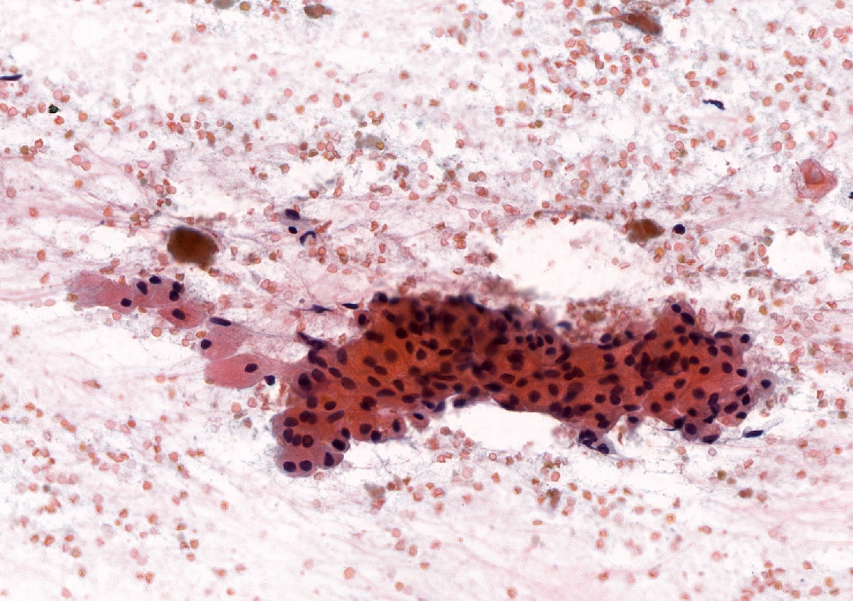
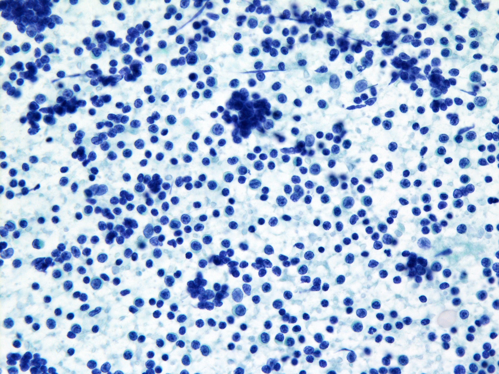
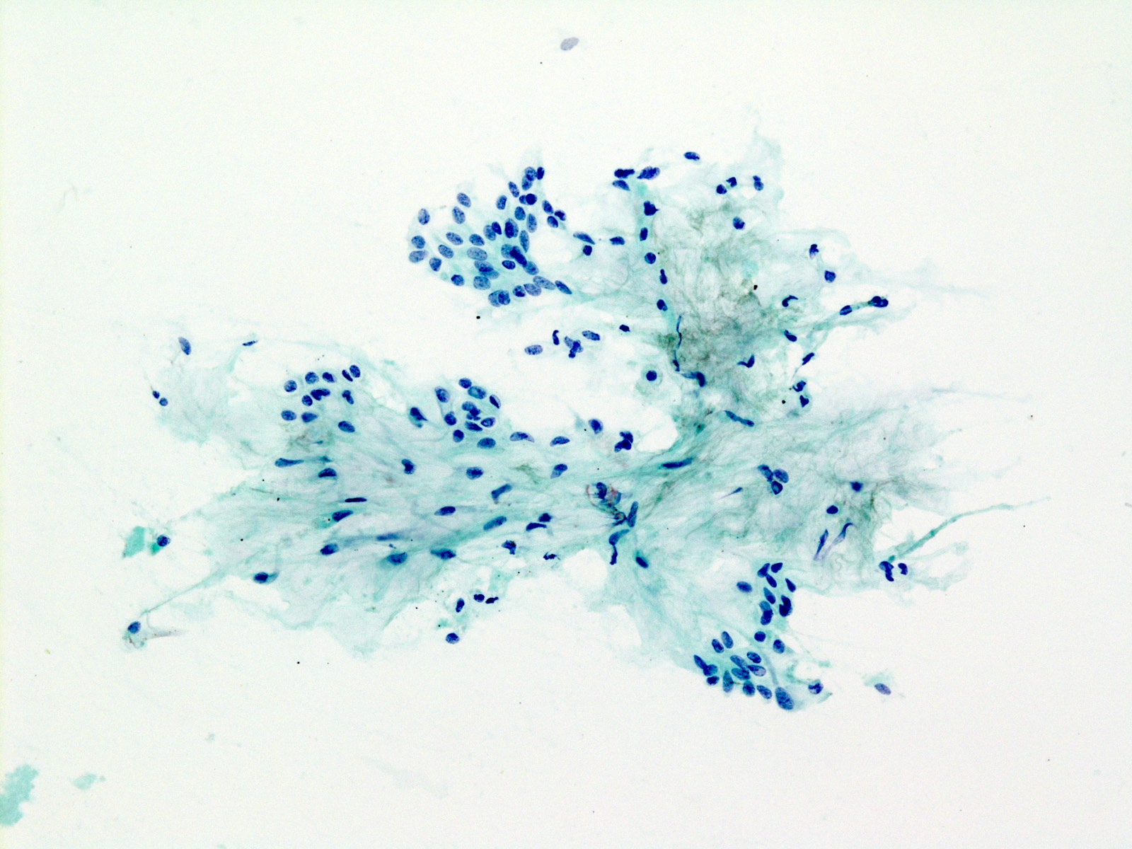
Smear, Papanicolaou
Basaloid cells
Microcystic pattern
Branching pattern
Contributed by Theoni Haralabopoulos, M.D. and AFIP
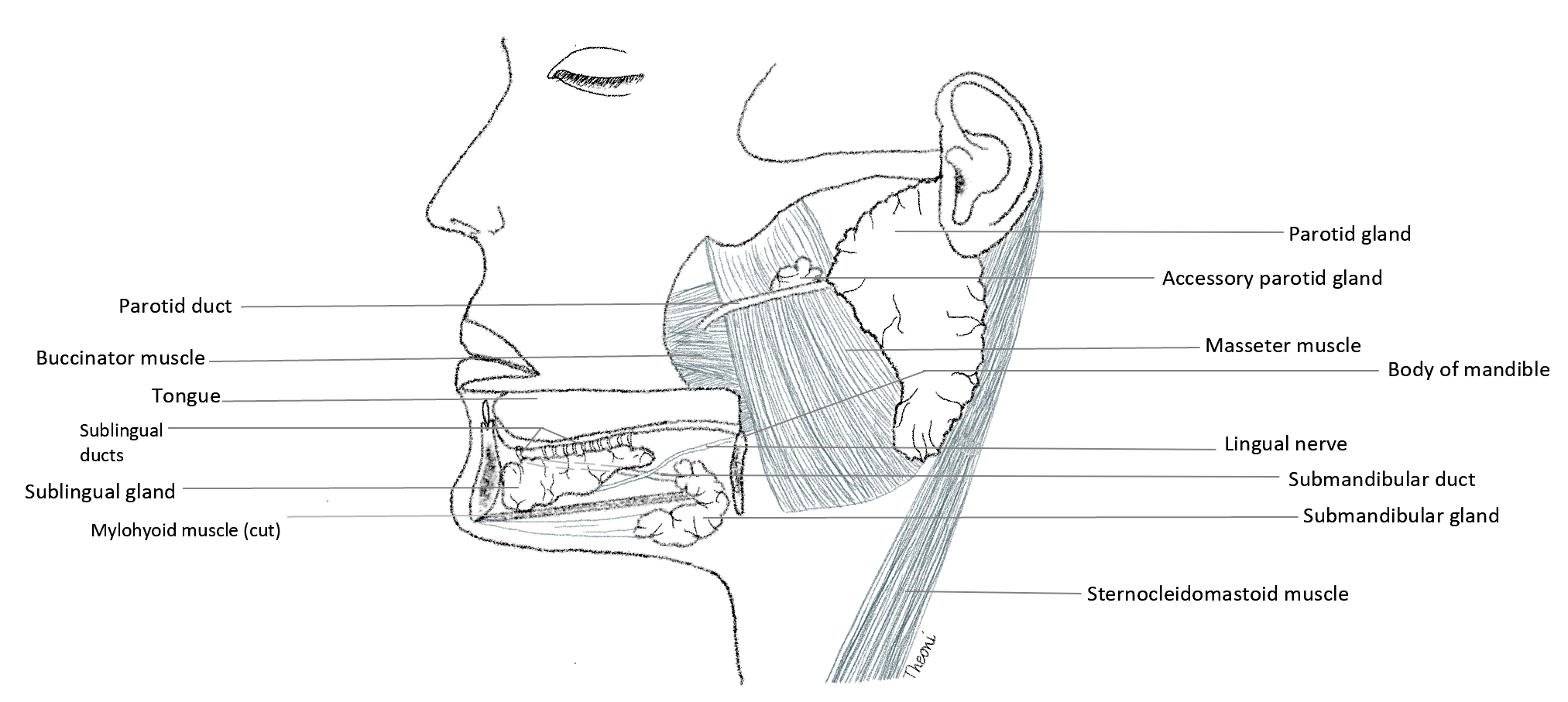
3 major salivary glands (left side)
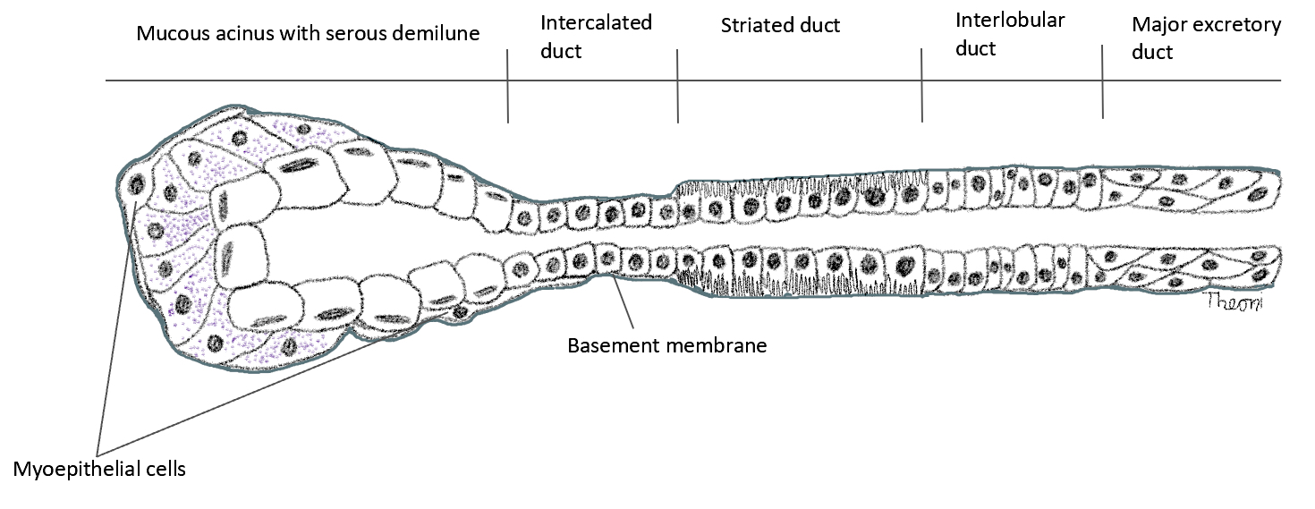
Schematic representation of acinus and ductal system
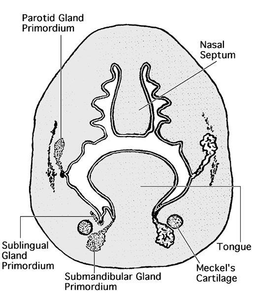
Embryological development of oral cavity
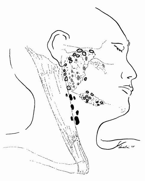
Parotid gland lymph nodes
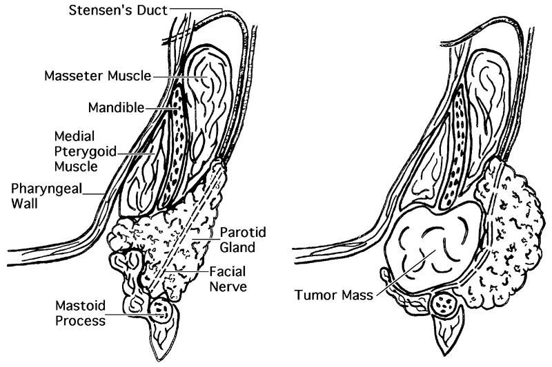
Horizontal section through parotid gland
Images hosted on other servers:

Overview of major salivary gland anatomy
Contributed by Ruta Gupta, M.B.B.S., M.D. and Theoni Haralabopoulos, M.D.

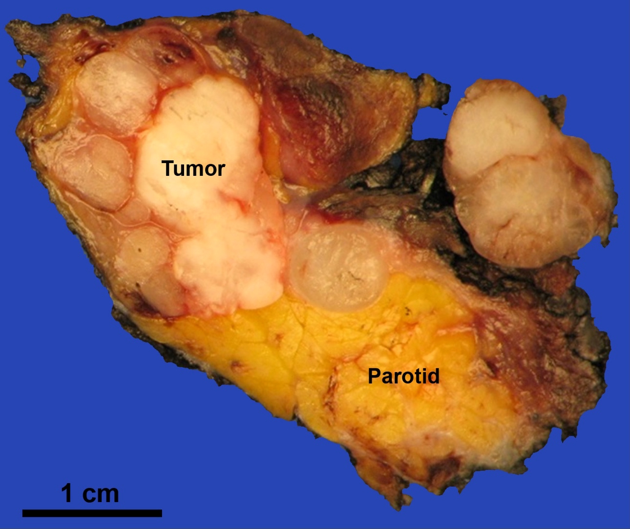
Parotid gland
Cut surface of parotid gland
Submandibular gland
Cut surface of submandibular gland
Sublingual gland

Cut surface of sublingual gland
Contributed by Ruta Gupta, M.B.B.S., M.D. and Theoni Haralabopoulos, M.D.
Major salivary glands:
Parotid gland

Serous acini of Parotid gland

Striated ducts of parotid gland
Interlobular duct of parotid gland
Intraparotid lymph node
Intraparotid lymph node with glandular inclusions
Sebaceous cells in parotid gland
Submandibular gland
Mucous acinar cells of submandibular gland
Sublingual gland

Seromucous acini of sublingual gland
Minor salivary glands:
Minor salivary glands
Minor salivary glands are unencapsulated
Special stains and immunohistochemistry:
Mucicarmine stain of sublingual gland

PAS stain of sublingual gland
SMA positivity of myoepithelial cells

EMA positivity of ductal cells

AE1 / AE3 positivity of all cell types
p63 positivity of myoepithelial cells
Shotgun histology parotid gland
Shotgun histology submandibular gland
Shotgun histology sublingual gland
Contributed by Umamaheshwari Golconda, M.D.
Rare atypical cells
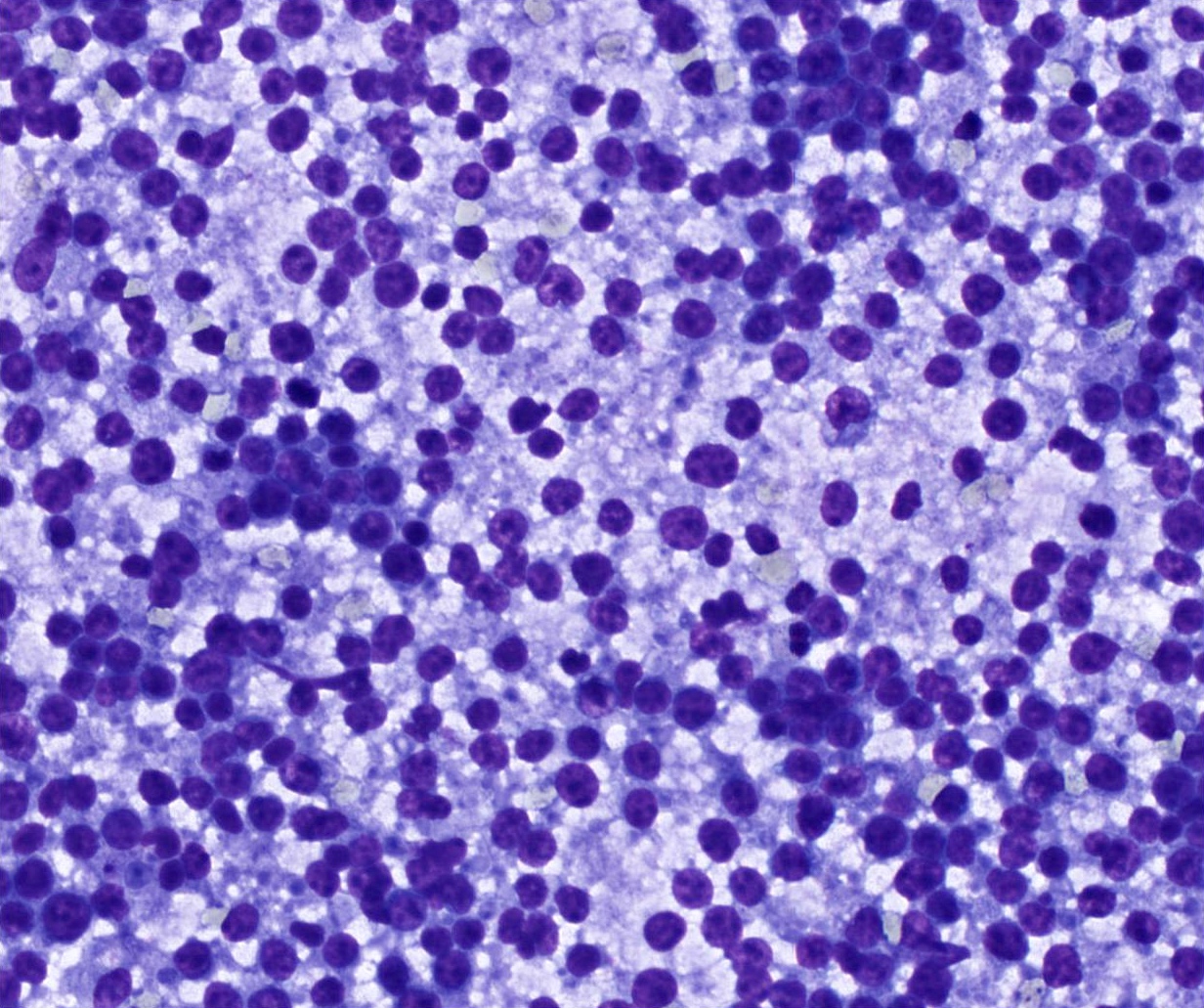
Lymphoid rich aspirate

Abundant extracellular mucin
Contributed by Natasha Prosser, B.Sc., M.B.B.S.

Parotid gland tumor
Images hosted on other servers:

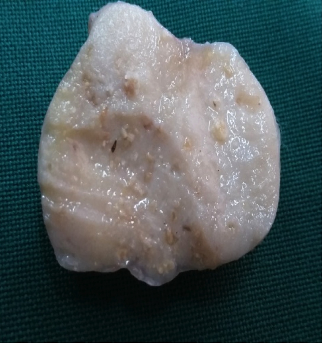
Bisected specimen with focal hemorrhage
Contributed by Natasha Prosser, B.Sc., M.B.B.S. and Ruta Gupta, M.D.

Peripheral palisading


Biphasic tumor composition
Solid pattern
Tubular and trabecular growth patterns

Membranous pattern

Glandular elements
Squamous morules
Invasion
Perineural invasion
DPAS special stain
Beta catenin IHC

Cytokeratin 7 IHC
p63 IHC

Ki67 IHC
Images hosted on other servers:

Basal cell adenoma in left parotid gland


Basal cell adenoma versus pleomorphic adenoma of the parotid gland
Contributed by Shuanzeng Wei, M.D., Ph.D.
Solid pattern
Trabecular pattern

Tubular pattern
Membranous type
Contributed by Shuanzeng Wei, M.D., Ph.D.

Diff-Quik stain
Pap stain
Membranous type Pap stain
Images hosted on other servers:

Ultrasound examination (1B) and MRI (1C)
Images hosted on other servers:

Oral nodule

Oral swelling / tumefaction / lesion

Oral lesion after resection, postresection zone

Oral nodule

Nodules (lip)

Oral nodules / lesions
Images hosted on other servers:
2 resected nodules

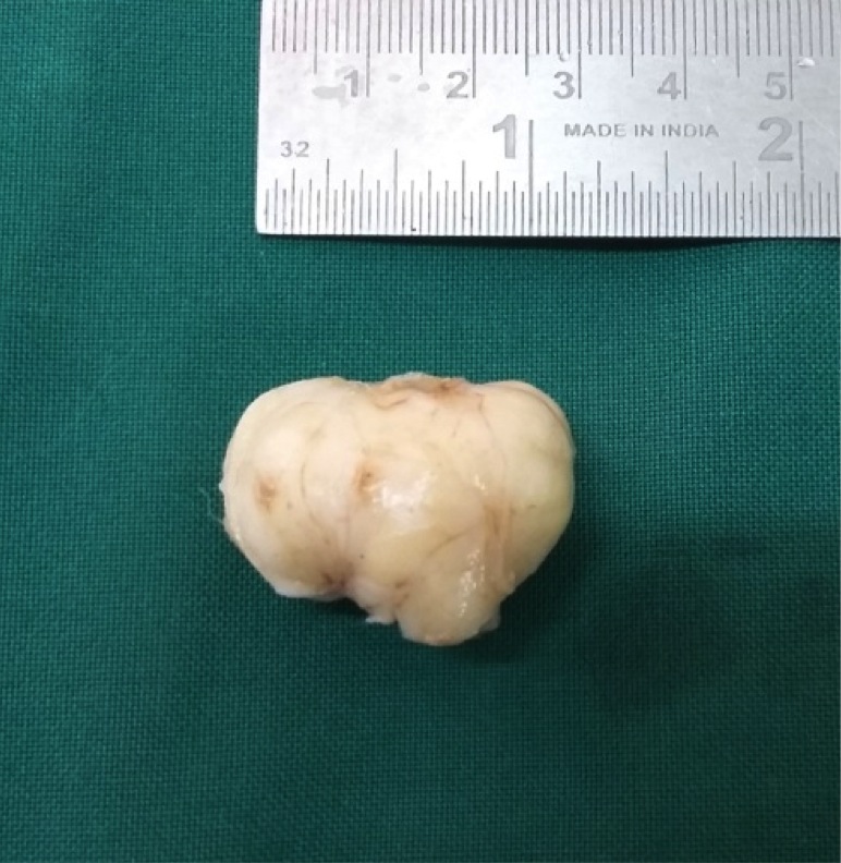
Resected specimen

Resected specimen / nodule

Resected lesion

Resected nodule
Contributed by Adriana Handra-Luca, M.D., Ph.D. (slides courtesy of Emmanuelle Vaz, M.D.)
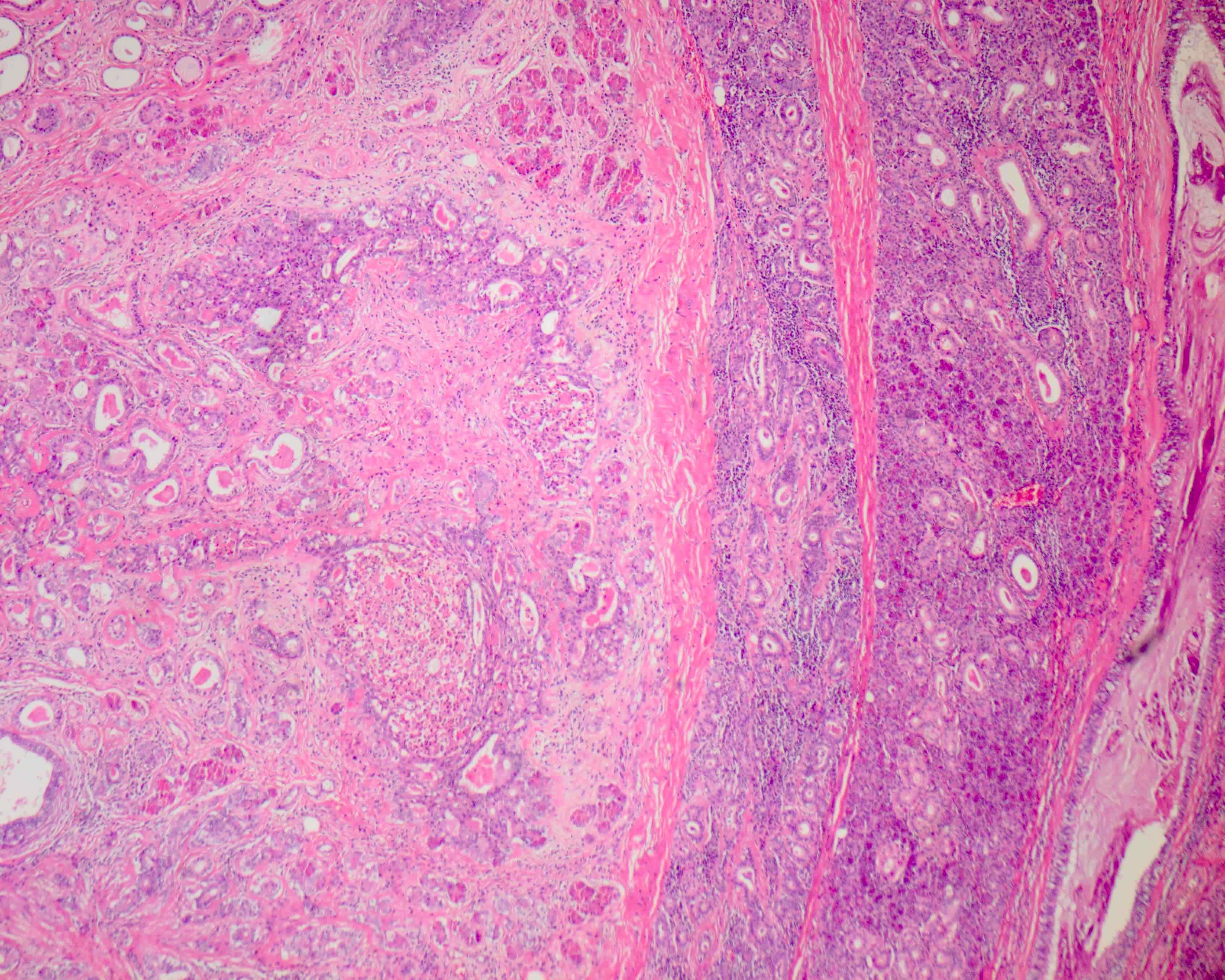
Cystic tumor zone
Solid tumor zone
Microscopy
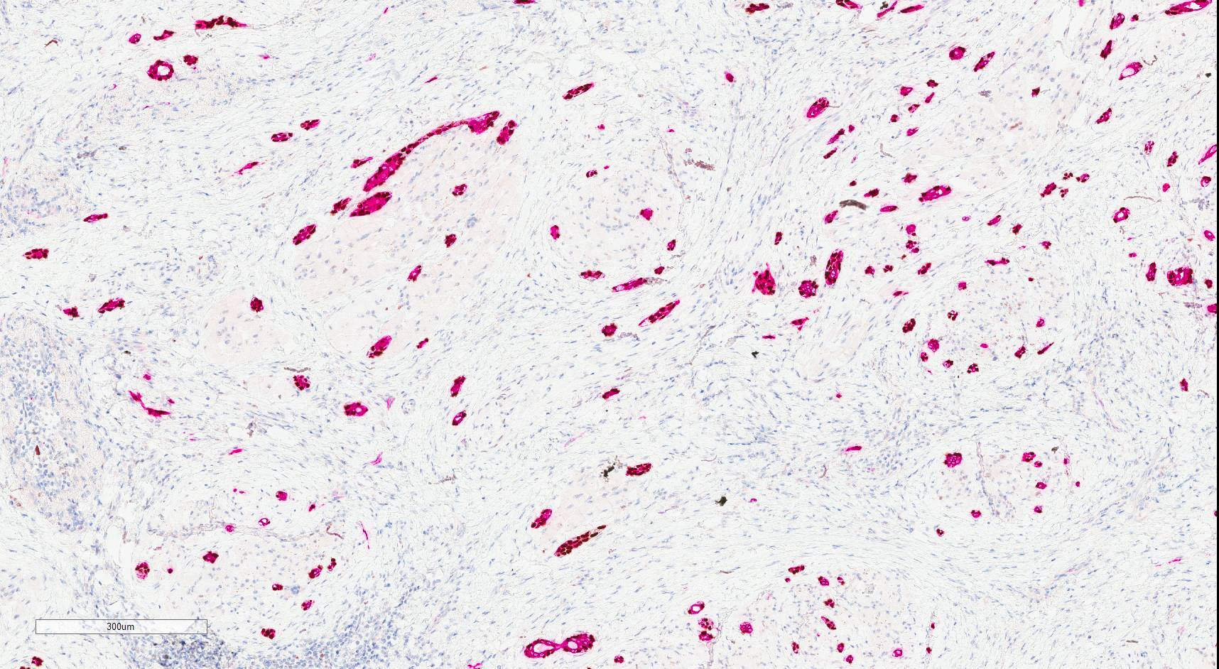
Canalicular pattern
Alcian blue stain
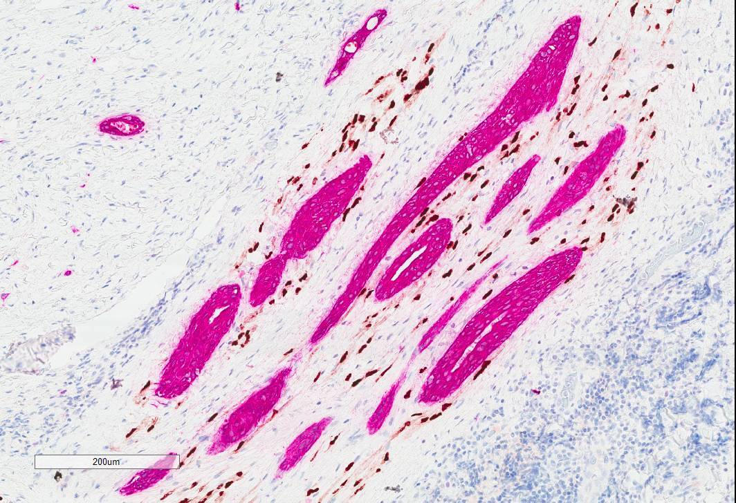
BCL2 expression
Images hosted on other servers:

Cytology of parotid case

Slides with cytology material
Images hosted on other servers:

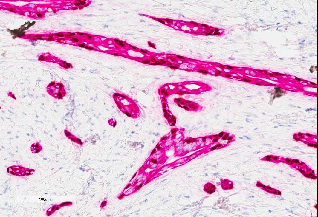
Apical zone of columnar cells

Basal zone of canalicular cells

Zone of 2 parallel rows of tumor cells

Basal zone of tumor cells
Images hosted on other servers:


Axial T2 weighted and ADC mapping

Salivary duct carcinoma ex PA, axial T2 weighted
Contributed by Ruta Gupta, M.B.B.S., M.D.
Parotid mass
2 distinct tumor components
Parotid mass involving skin
Contributed by Ruta Gupta, M.B.B.S., M.D.
2 distinct microscopic components
Interface of distinct components
Infiltrative component
DCIS-like morphology

HER2 IHC
Androgen receptor IHC
Images hosted on other servers:

CT of head and neck

CT showing cystic necrotic areas

MRI of parotid lesion

MRI of heterogeneous mass lesion
Images hosted on other servers:

Swelling of right parotid
Contributed by Alexander Tang, M.B.B.S.

Cut section of tan, fleshy carcinosarcoma
Contributed by Manish Mahadeorao Bundele, M.B.B.S., M.D.
Carcinomatous and intervening sarcomatous components

Carcinosarcoma with residual PA

Sarcomatoid component with storiform pattern
Undifferentiated carcinoma with necrosis

Undifferentiated carcinoma / pleomorphic sarcoma

Focal squamoid component

Focal squamoid component

p40+ / SOX10- squamoid component

p40+ squamoid component
Focal SOX10 staining

Nonspecific CD117 staining

High proliferative index

Diffuse positive p53

AE1 / AE3 + sarcomatoid component
Calponin weak positive

Variably increased p53 staining

Myoepithelial cells in PA
Contributed by Manish Mahadeorao Bundele, M.B.B.S., M.D.

Cell block showing sheets of carcinoma cells
Cell block

Parotid FNA (high grade carcinoma)


Parotid FNA (high grade carcinoma)

Pleural fluid metastasis
Cytokeratin positive
CK7

Nonspecific CD117 positive
Images hosted on other servers:

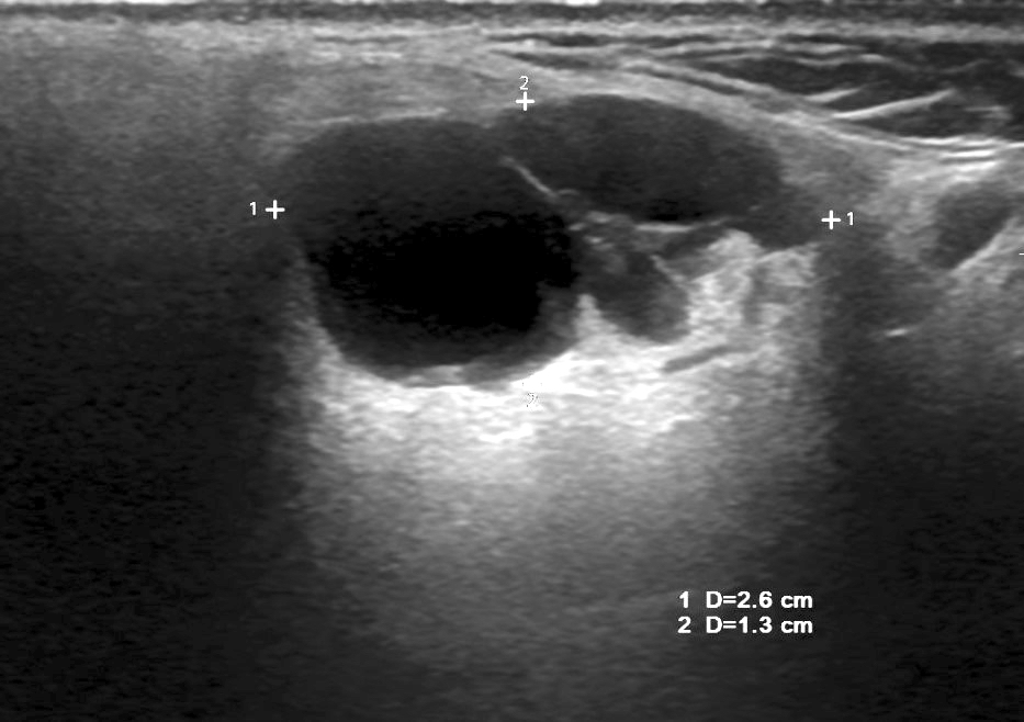
Hypoechoic mass
Images hosted on other servers:

Smooth mass

Sialolith
Contributed by James S. Lewis, M.D.
Preservation of lobular architecture
Intense chronic inflammation
Acinar destruction
Contributed by Tony Ng, M.D., Ph.D.
CT scan
Images hosted on other servers:

Echo, CT, MRI
Contributed by Pallavi Parashar, D.D.S.
Submucosal mass
Images hosted on other servers:


Buccal mass
Contributed by Erin Chapman, M.D
Well demarcated mass
Contributed by Pooja Navale, M.D. (Case #483), Tony Ng, M.D., Ph.D. and Erin Chapman, M.D
Circumscribed lesion
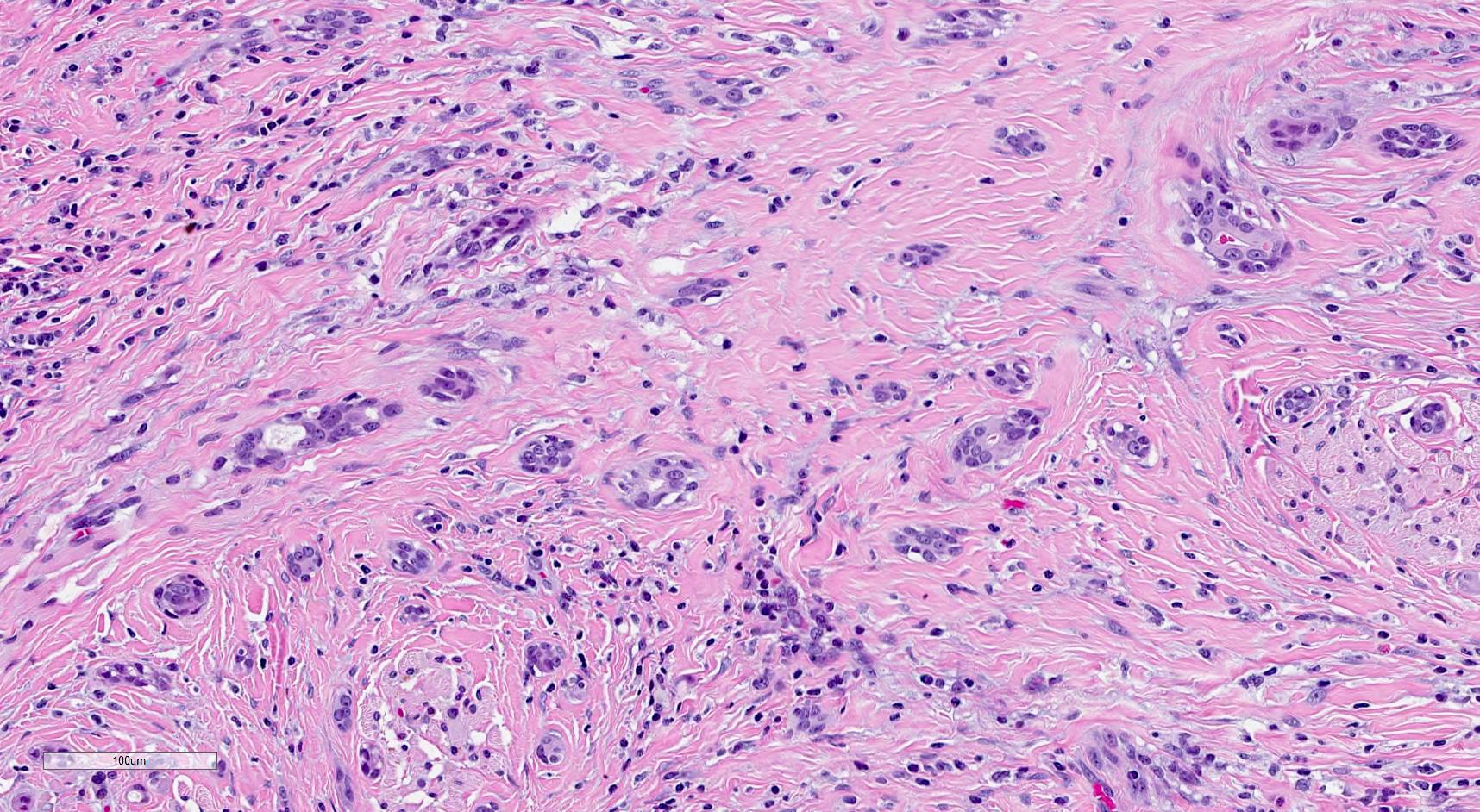
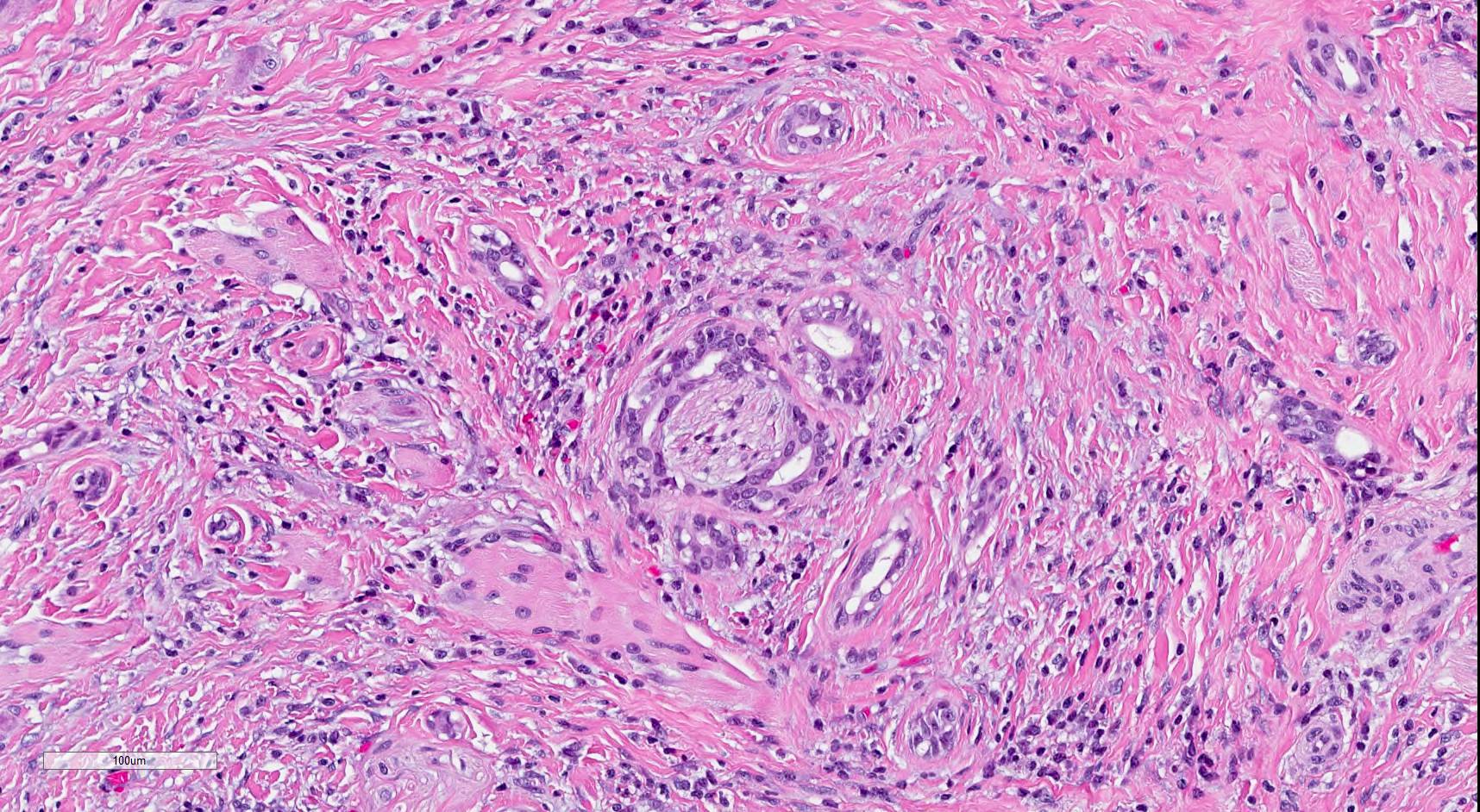
Cord-like to trabecular architecture
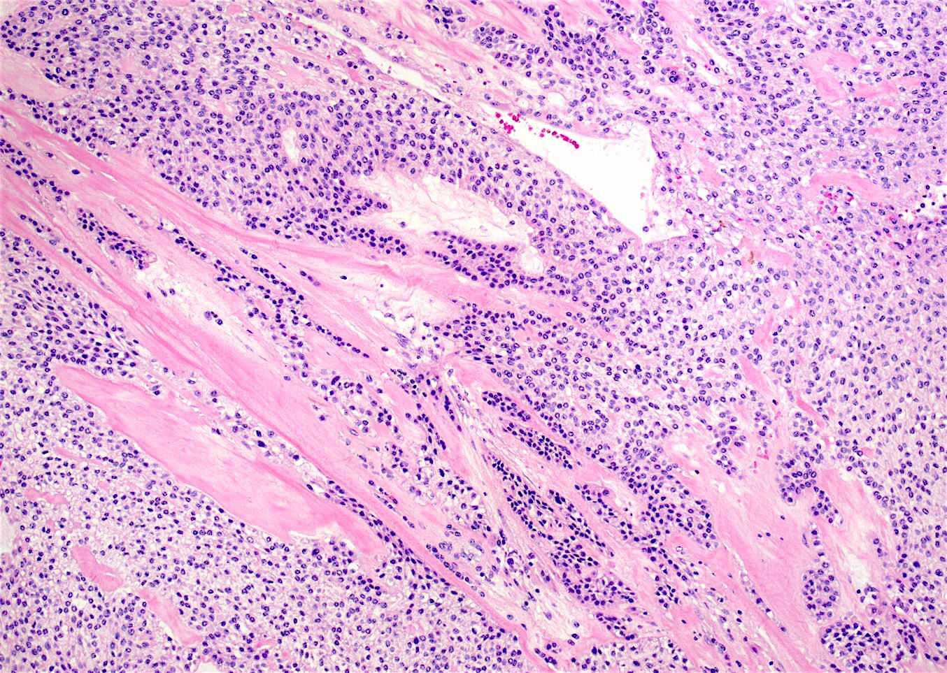
Hyalinzied stroma
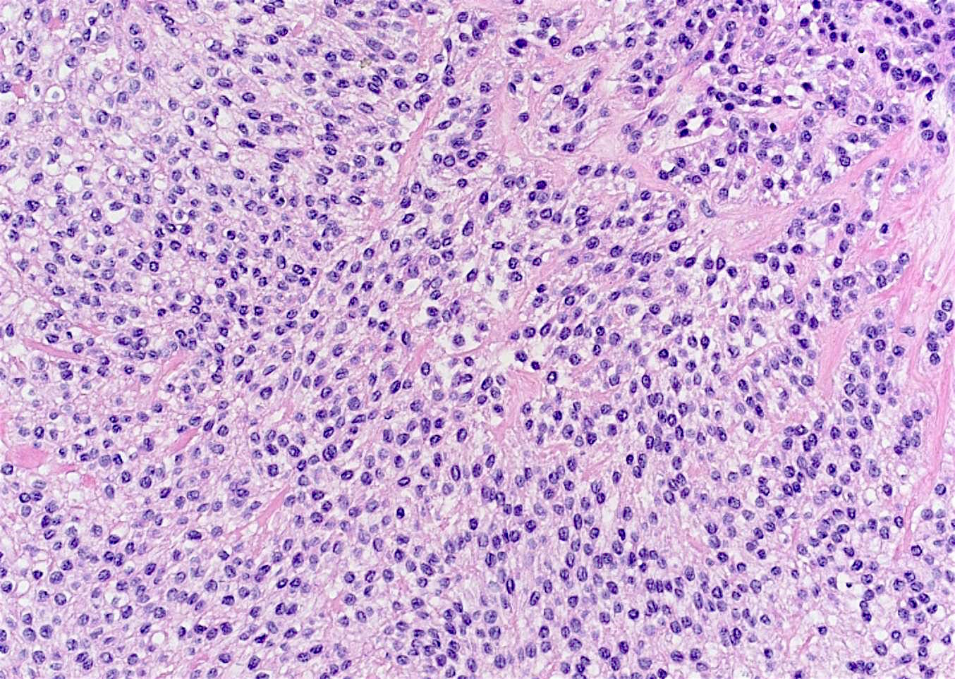
Anastomosing nests
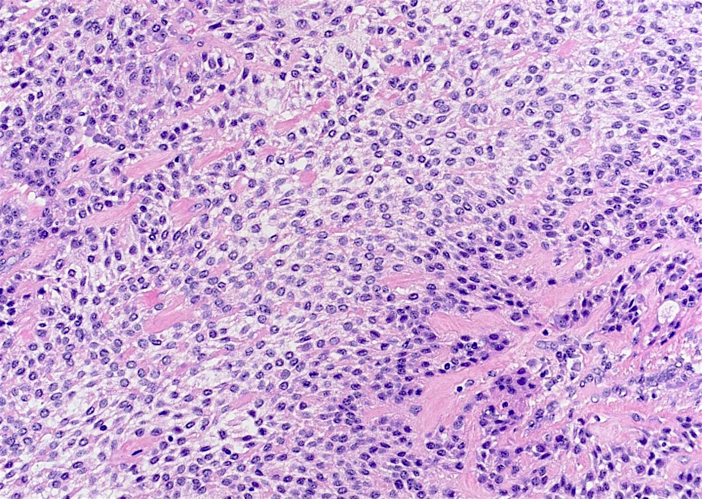
Monotonous cells
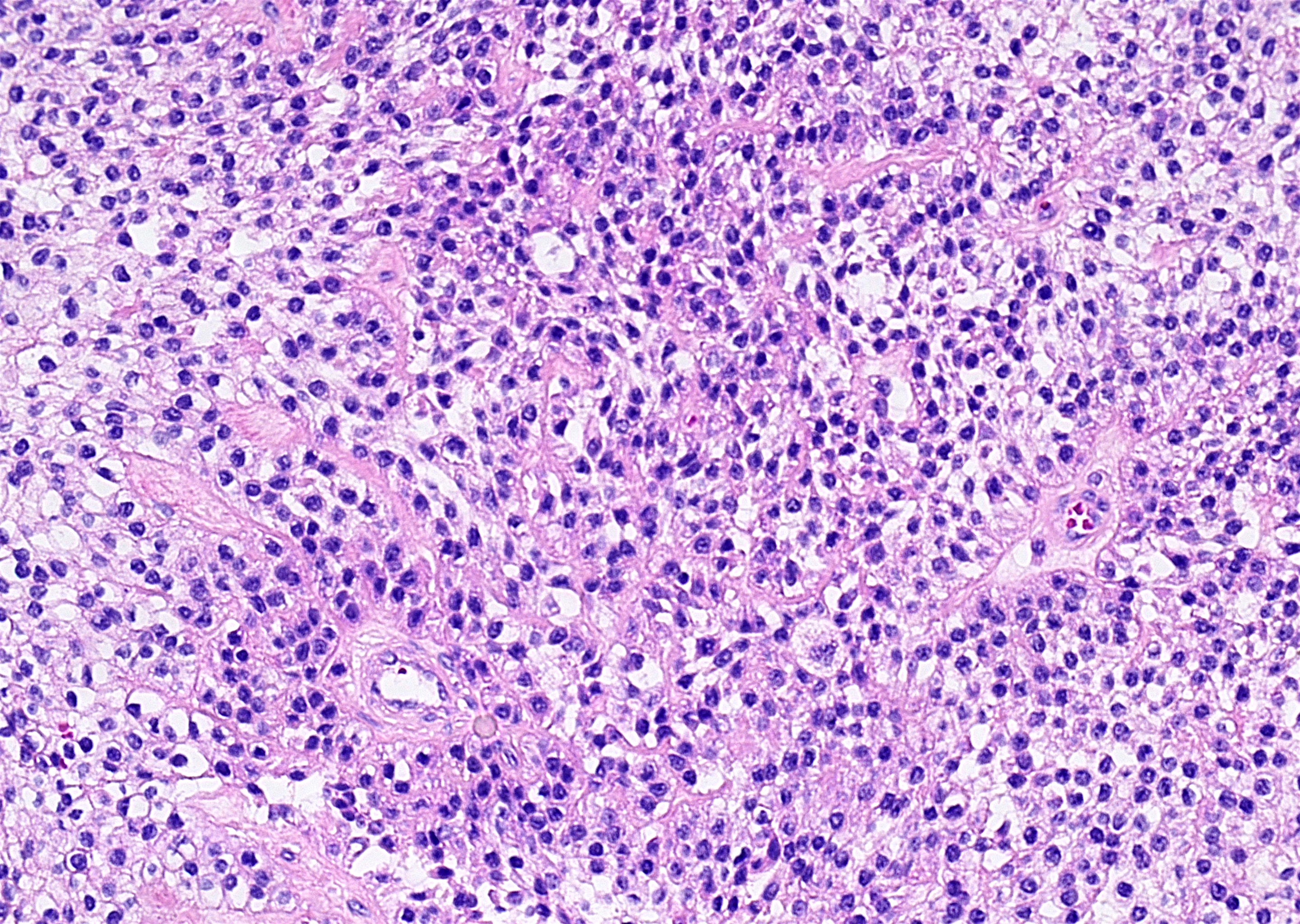
Clear to eosinophilic cytoplasm
Bland nuclei
Centrally placed nuclei
Inconspicuous nucleoli
Soft palate mass
Clear cell proliferation
Base of tongue mass
Clear cell and columnar phenotype
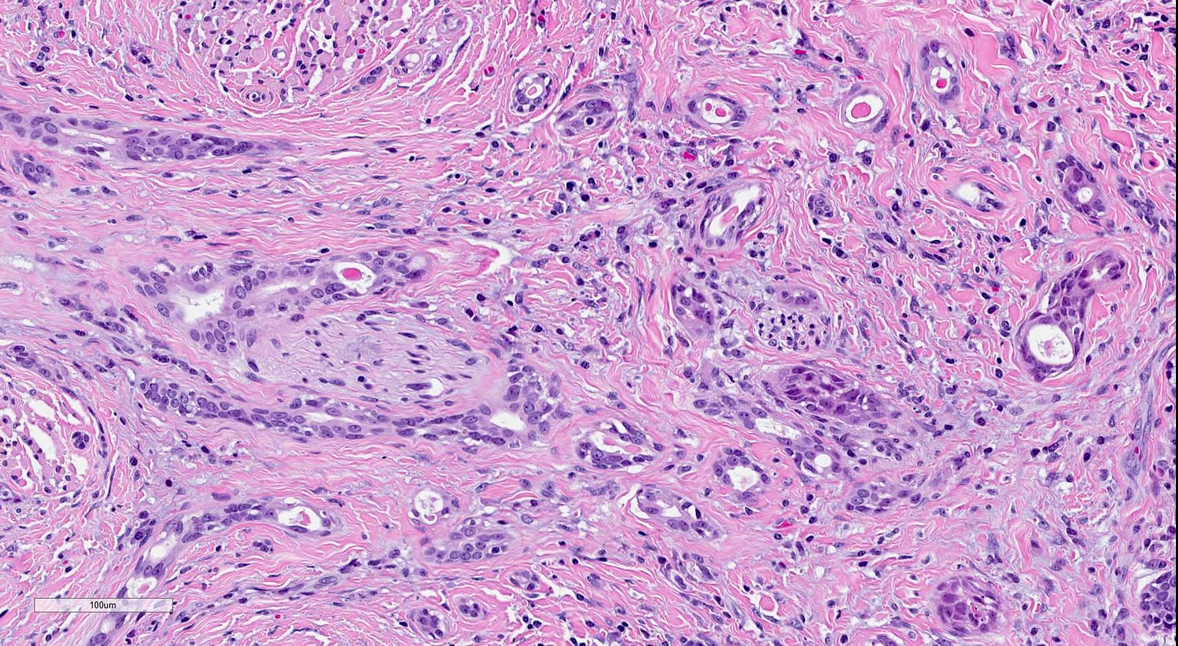
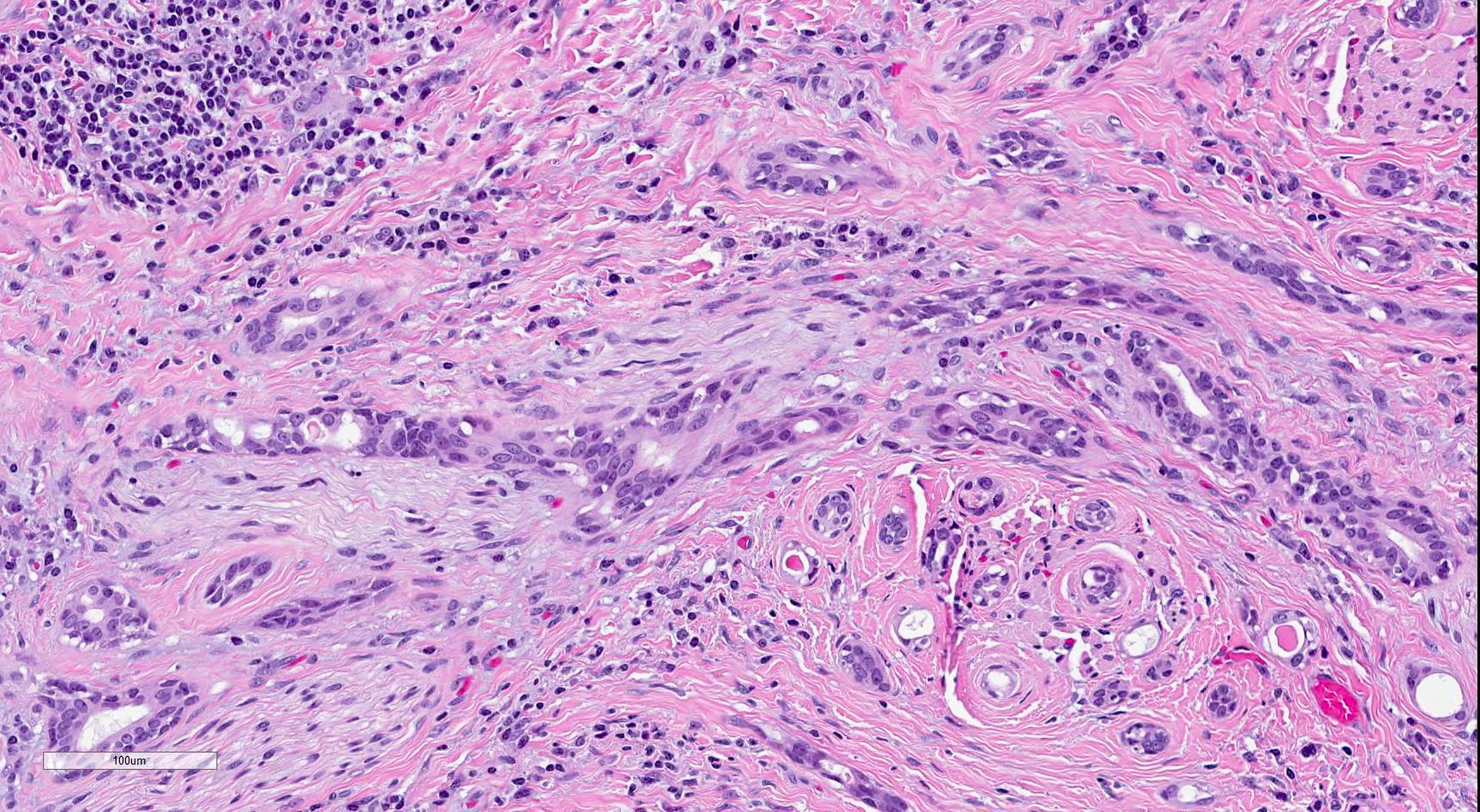
Buccal mass
Nests and cords of clear cells

Submucosal tissue
Posterior maxilla submucosal tumor
Islands and cords of tumor cells
p40
PASD
Contributed by Aditya Talwar, M.D.
Palpable mass, left neck
Left parotid lesion
Contributed by Jalal B. Jalaly, M.B.B.S., M.S.
Parotid gland
Contributed by Aditya Talwar, M.D.
Parotid gland cyst

Epithelial lining
Oncocytic variant
Contributed by Aditya Talwar, M.D.
Salivary gland lesion, Diff-Quik
Salivary gland lesion, Pap stain
Images hosted on other servers:

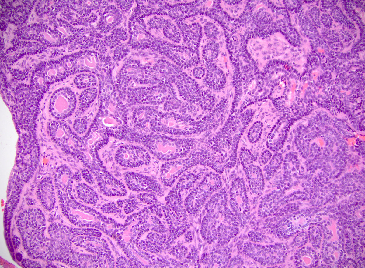
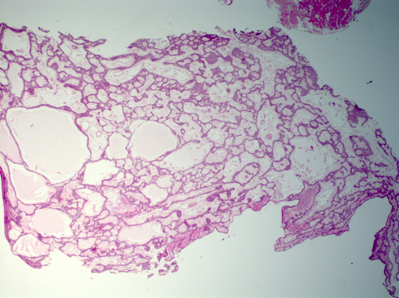
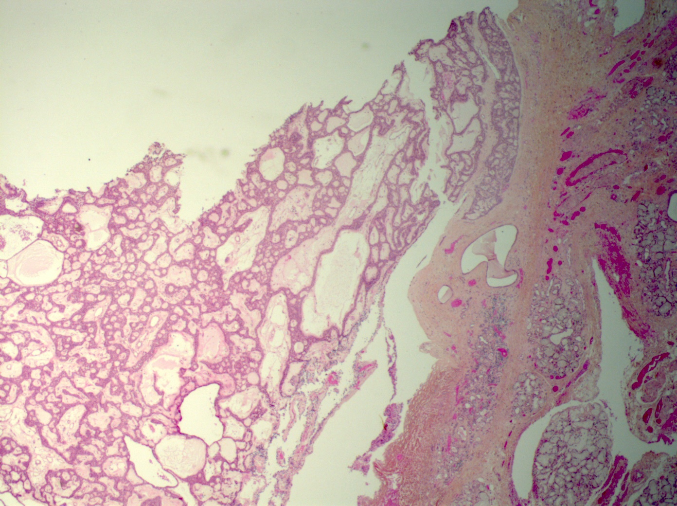
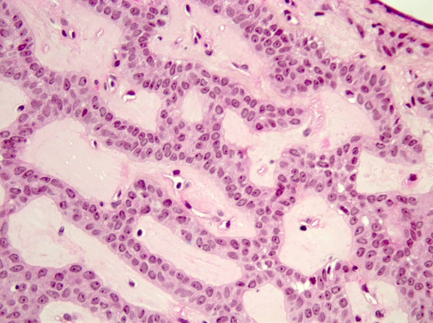
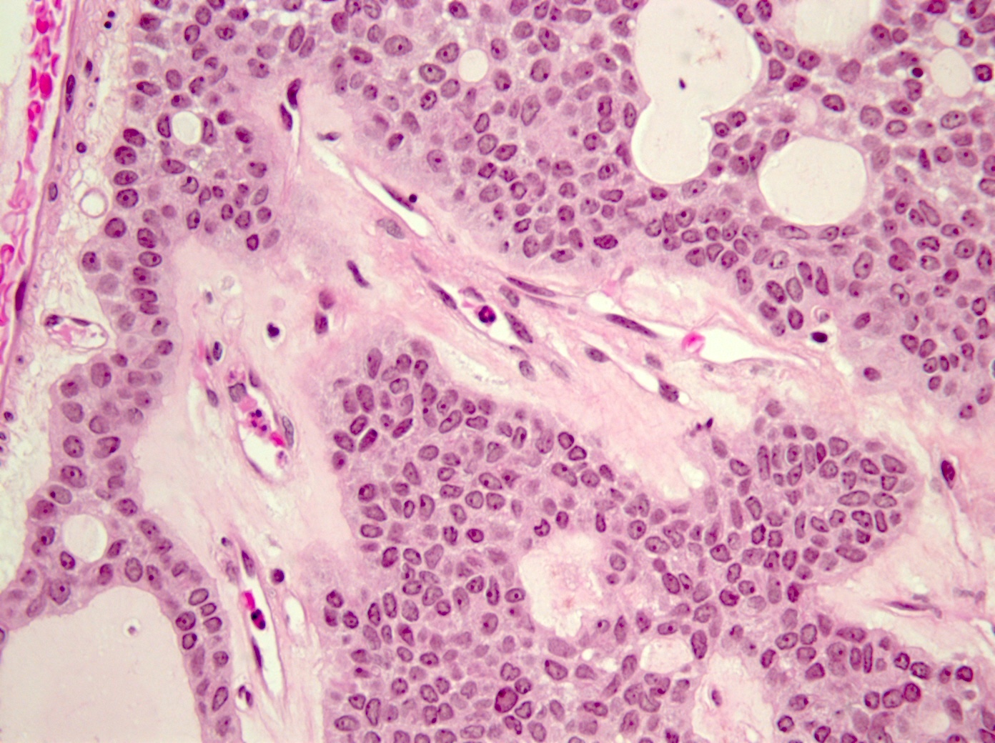
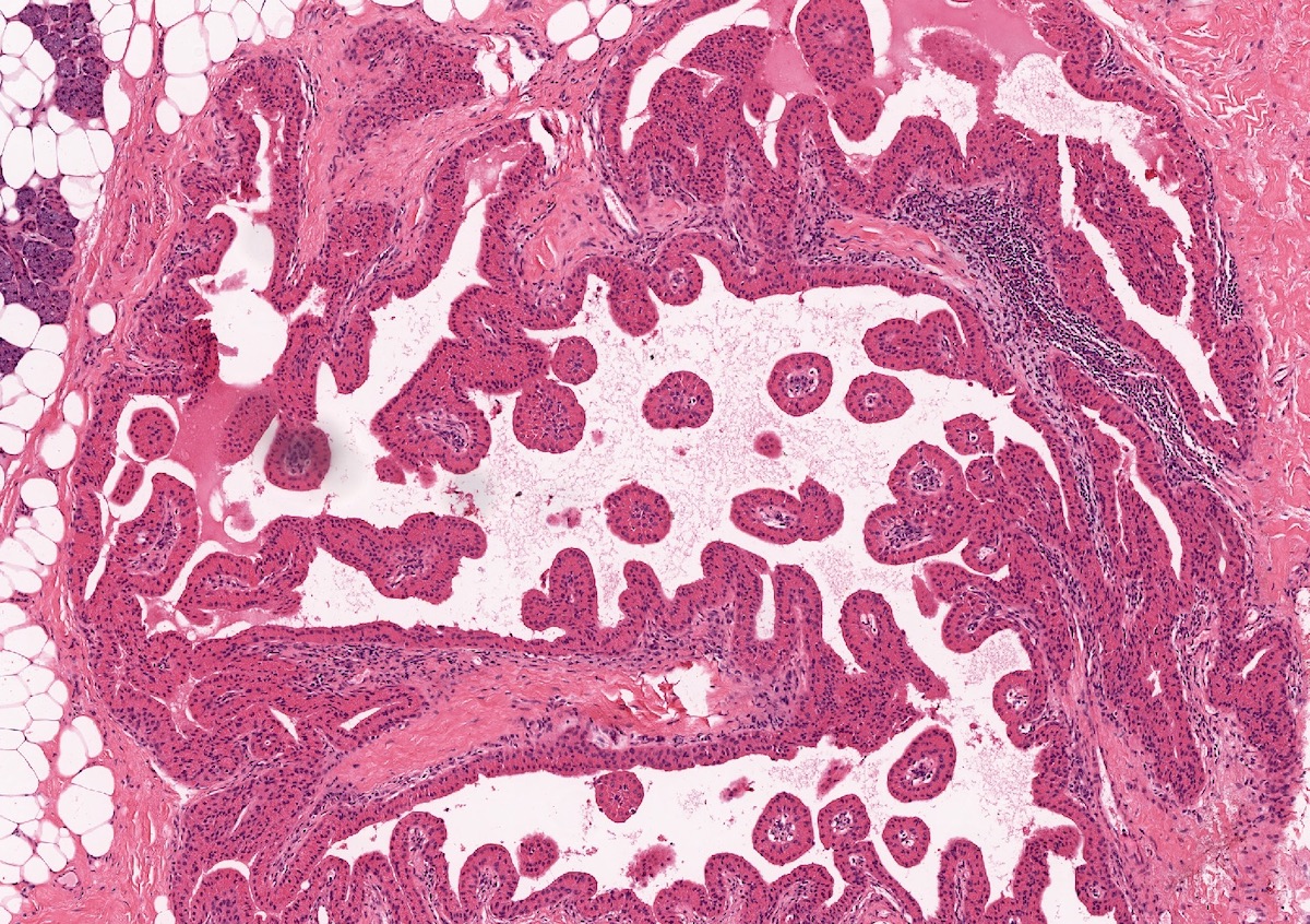
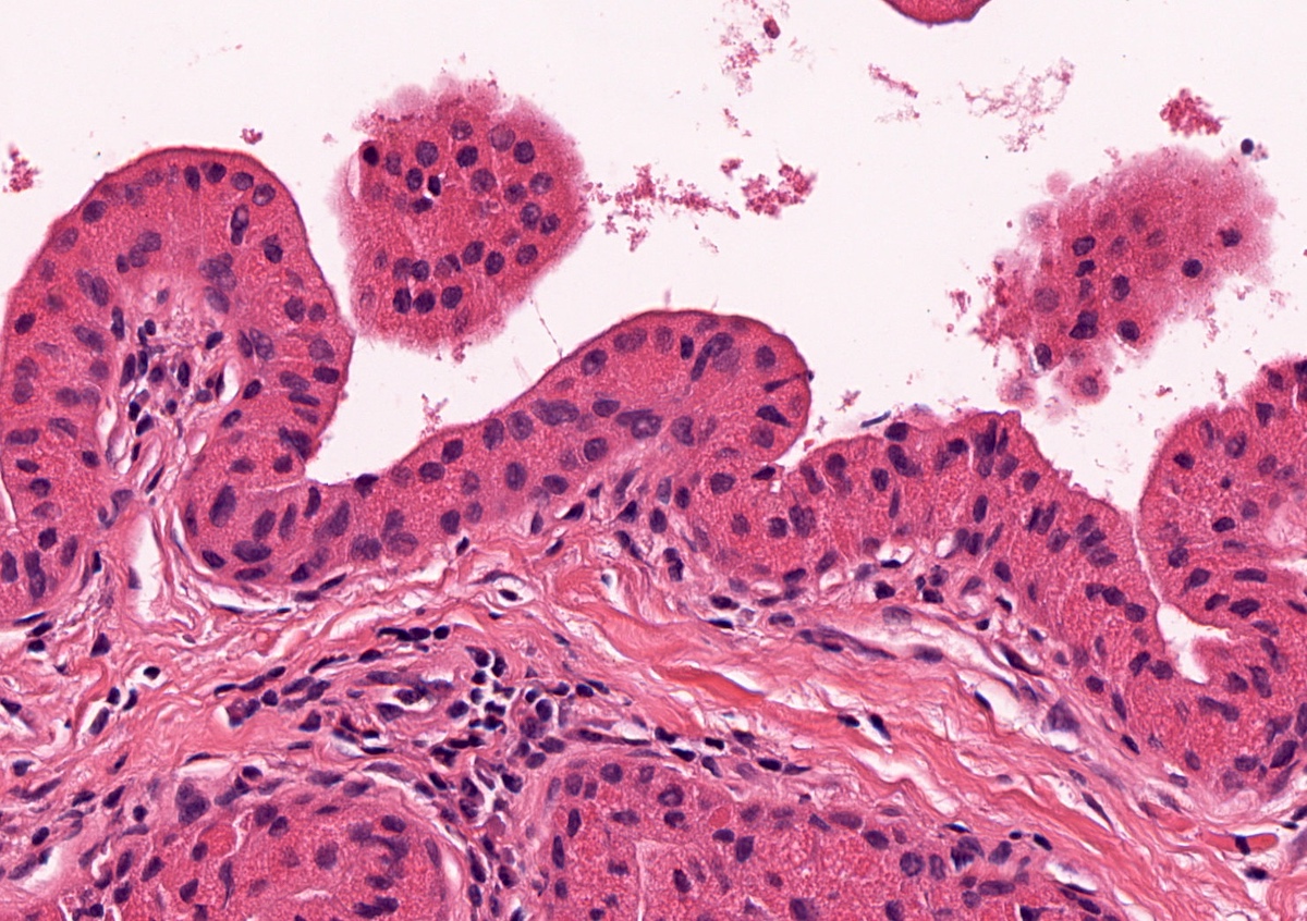
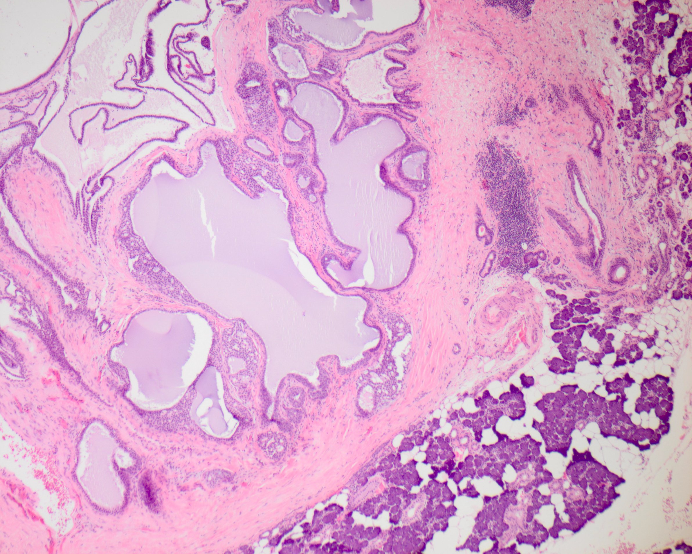
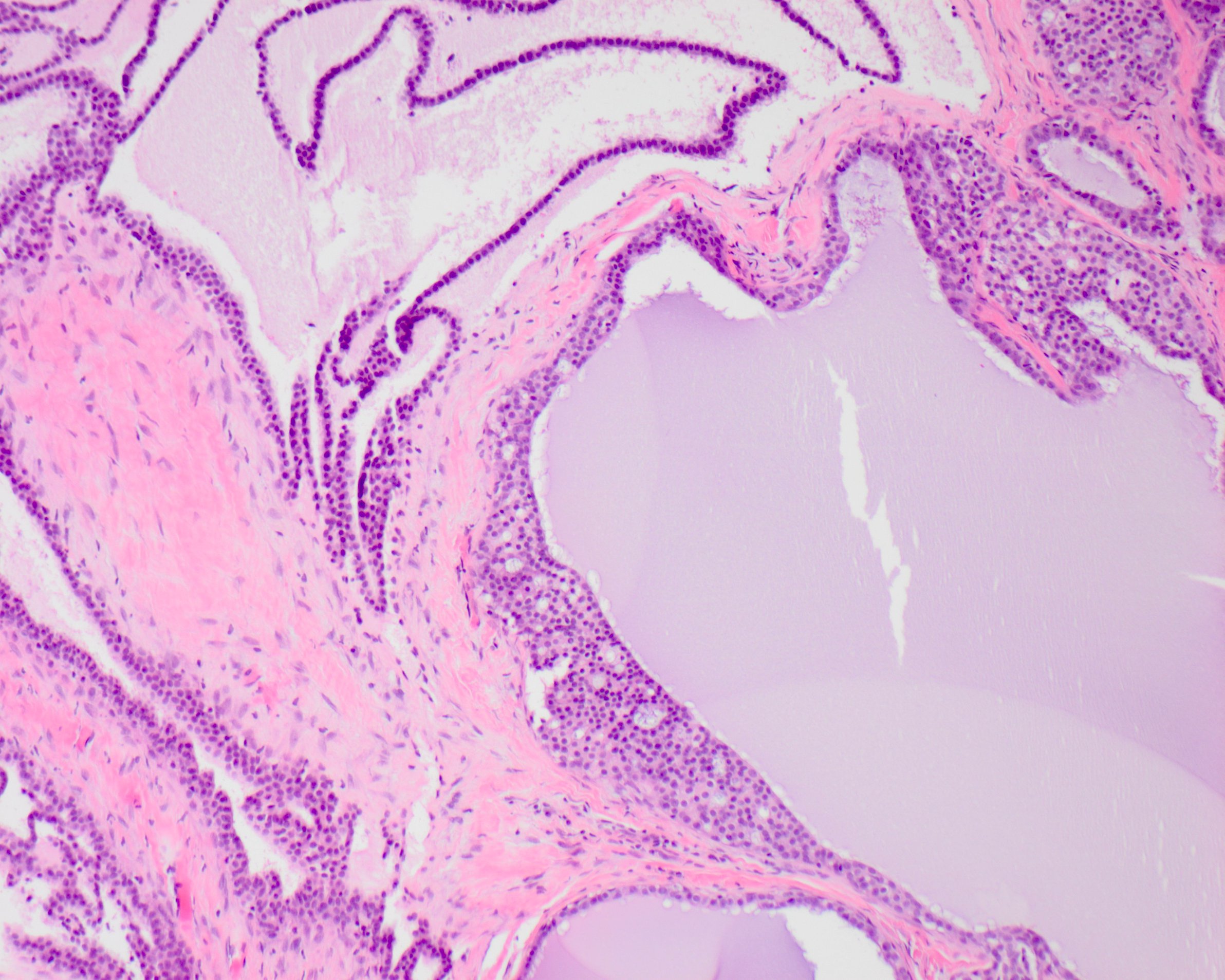
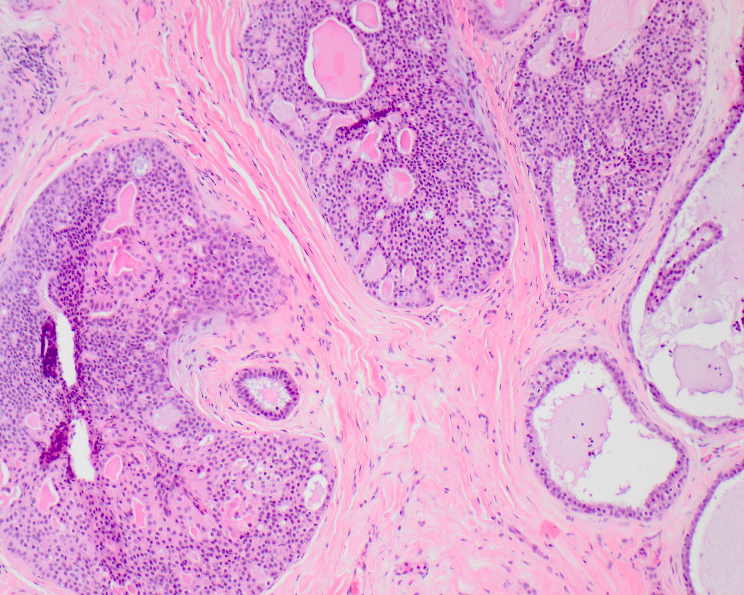
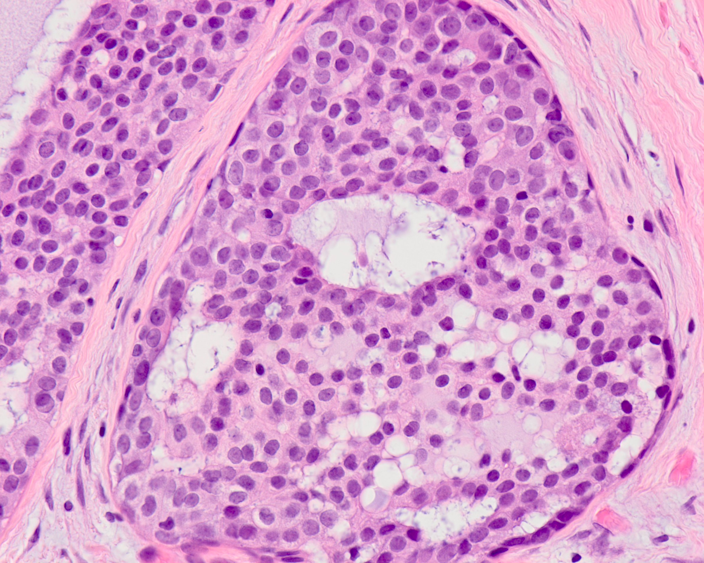
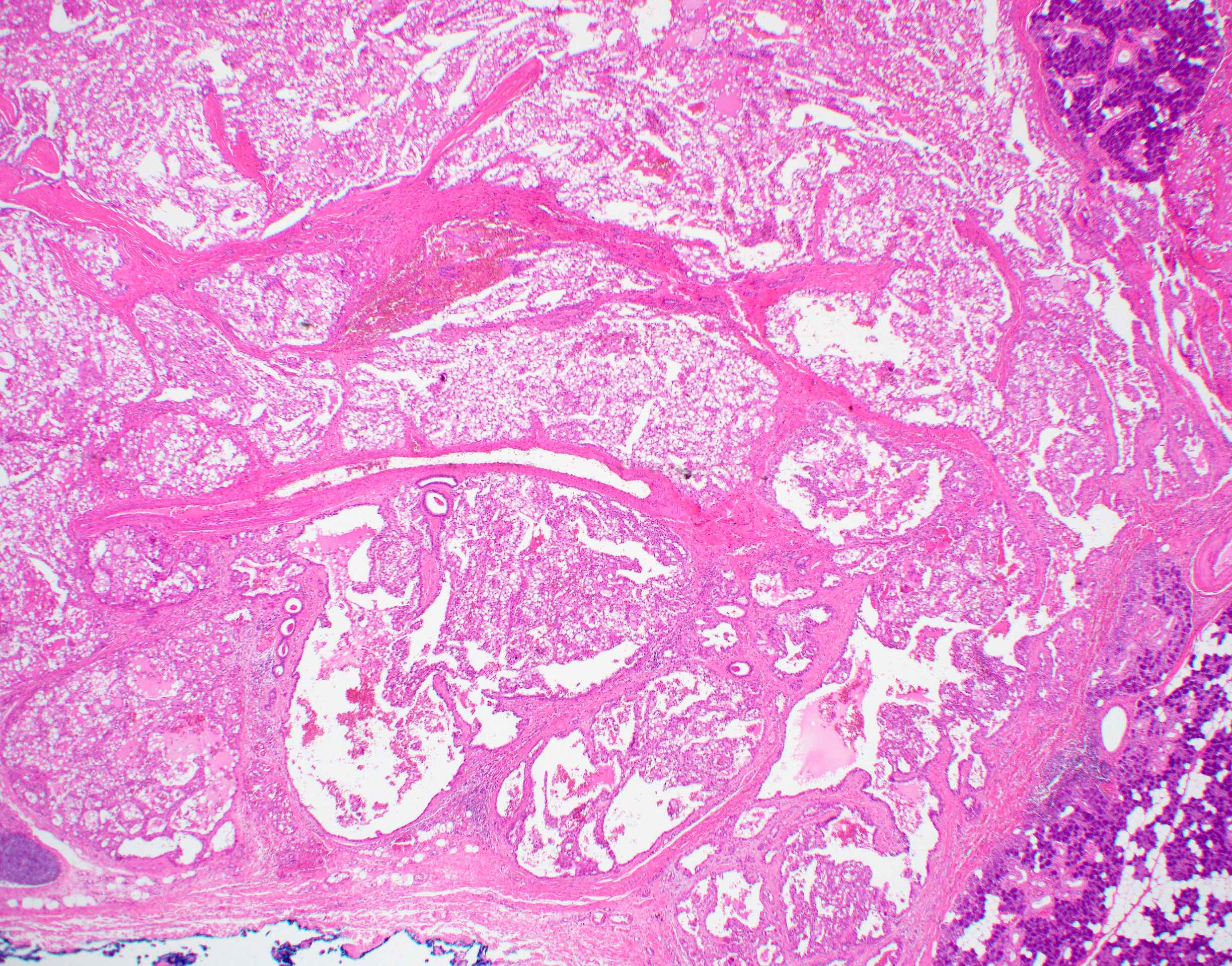
Upper lip tumor has papillary
proliferation of epithelial
cells with branching fronds
in cystically dilated duct lumen
Images hosted on other servers:

A: secretary granules, rough endoplasmic reticulum, golgi apparatus
and mitochondria; B: microvilli on luminal surface of epithelial cells
appear to be secretory; C: annulate lamellae composed of parallel
arrays of cisternae have small annuli or fenestrae
Contributed by Ruta Gupta, M.B.B.S., M.D.
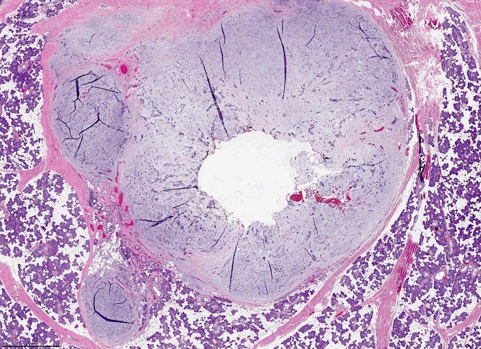
Solid tumor
Contributed by Ruta Gupta, M.B.B.S., M.D.
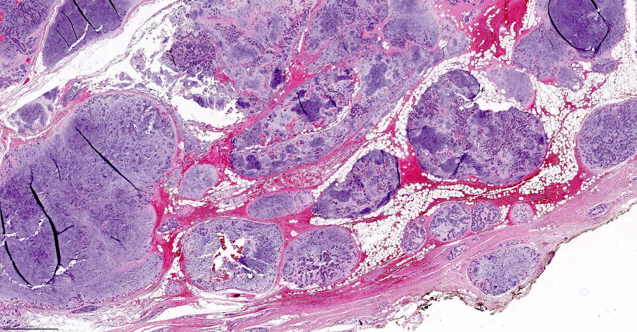
Nodular growth pattern
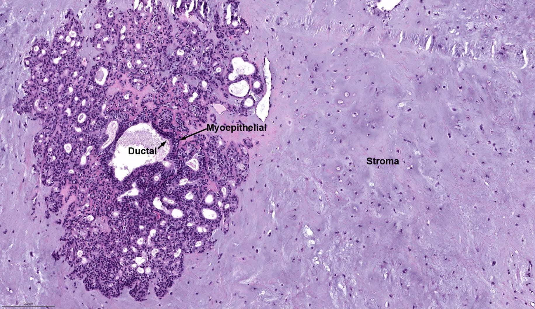
Biphasic morphology

Biphasic pattern
Spindled myoepithelial cells
Clear myoepithelial cells
Images hosted on other servers:

Figure 2B

Lacrimal gland

IgG4 positive plasma cells
Contributed by Kim A. Ely, M.D.
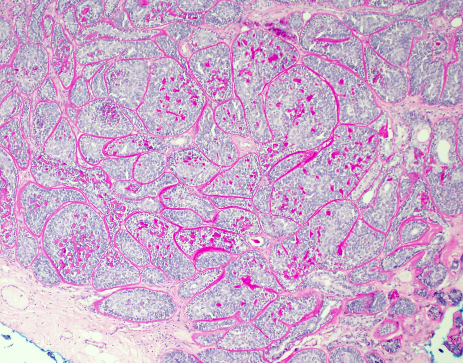
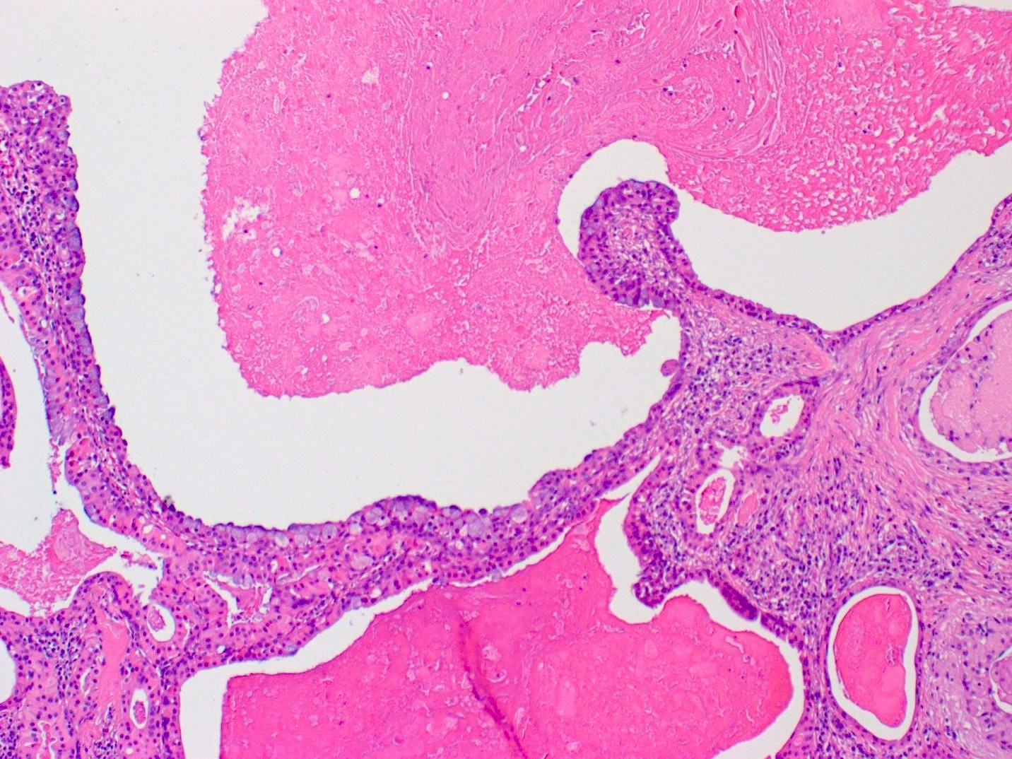
Well circumscribed
Cystic foci
Cribriform pattern
Intercalated duct-like subtype
Images hosted on other servers:


37 year old man with parotid tumor
Case #49

Well circumscribed tumor
Images hosted on other servers:

Parotid sialoangiolipoma
Case #49


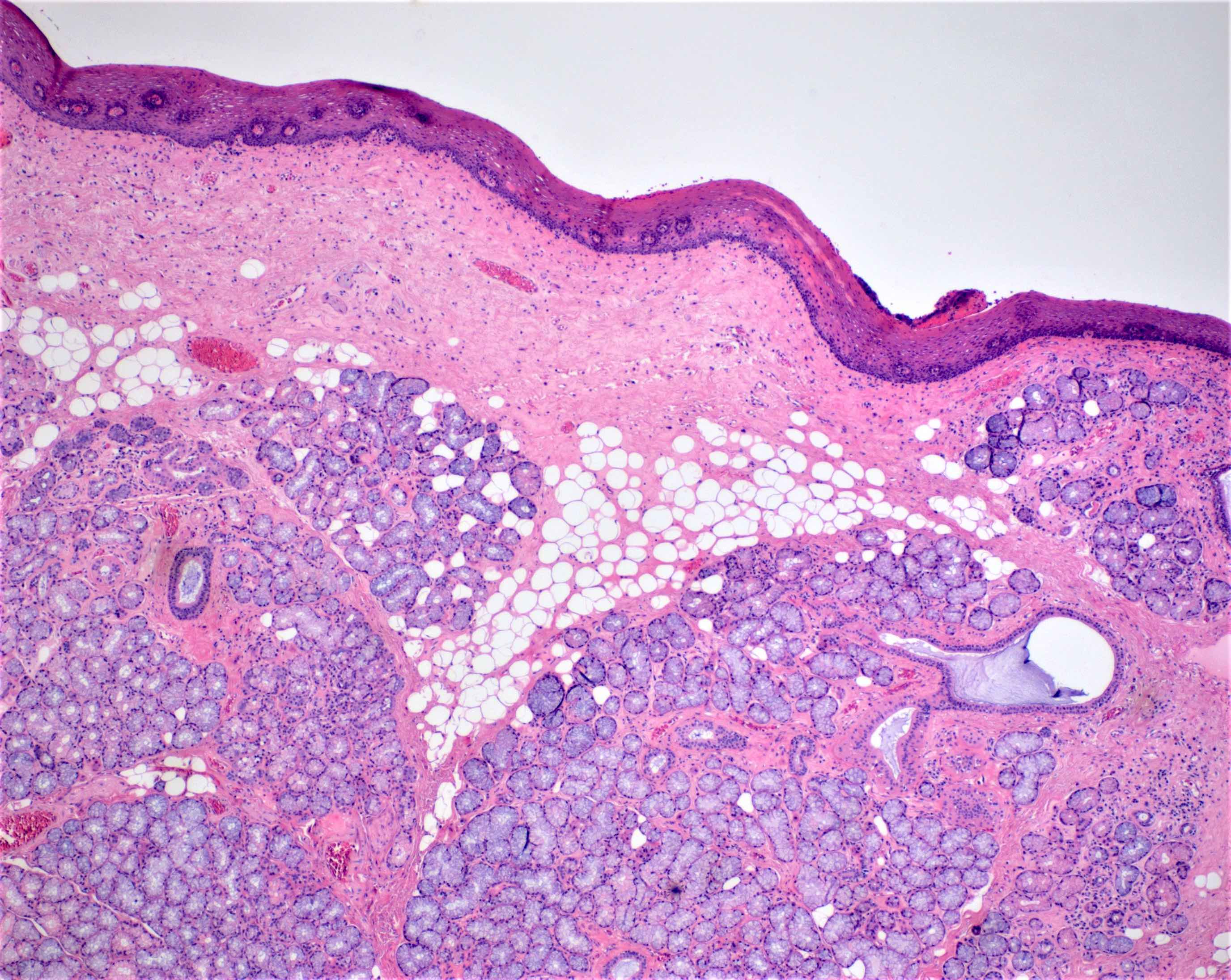
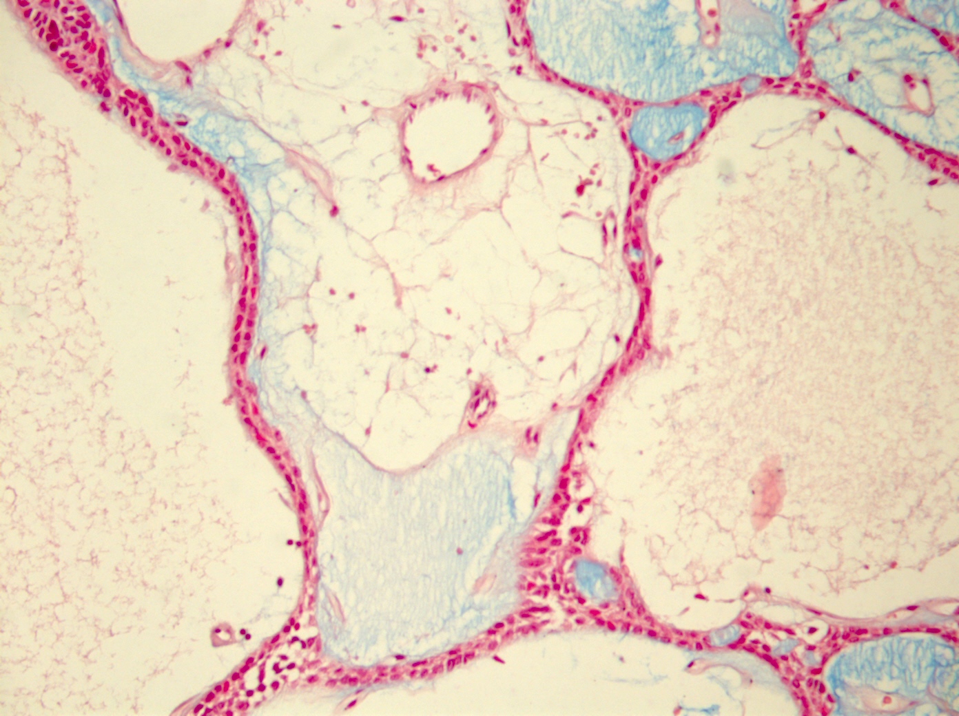
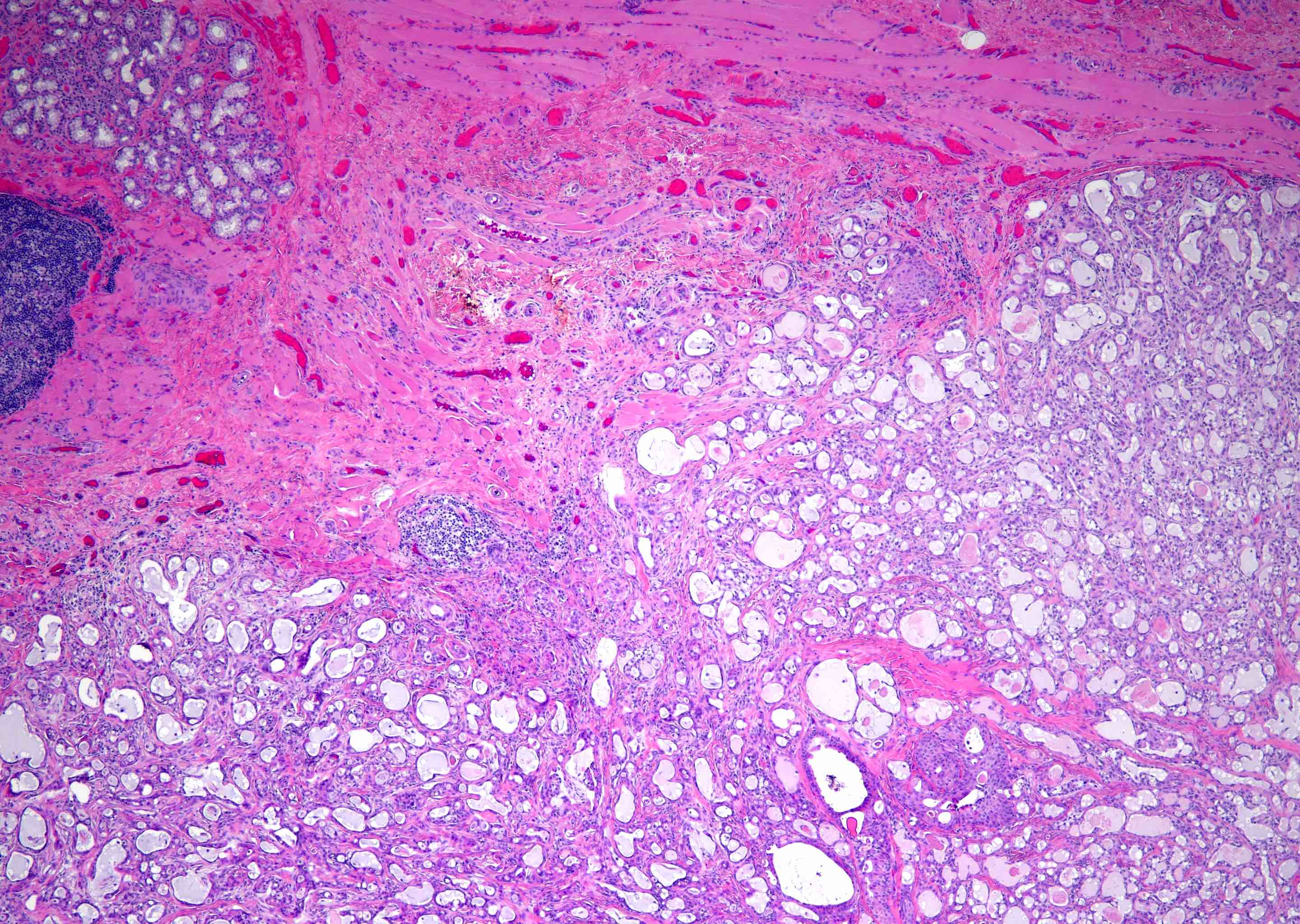
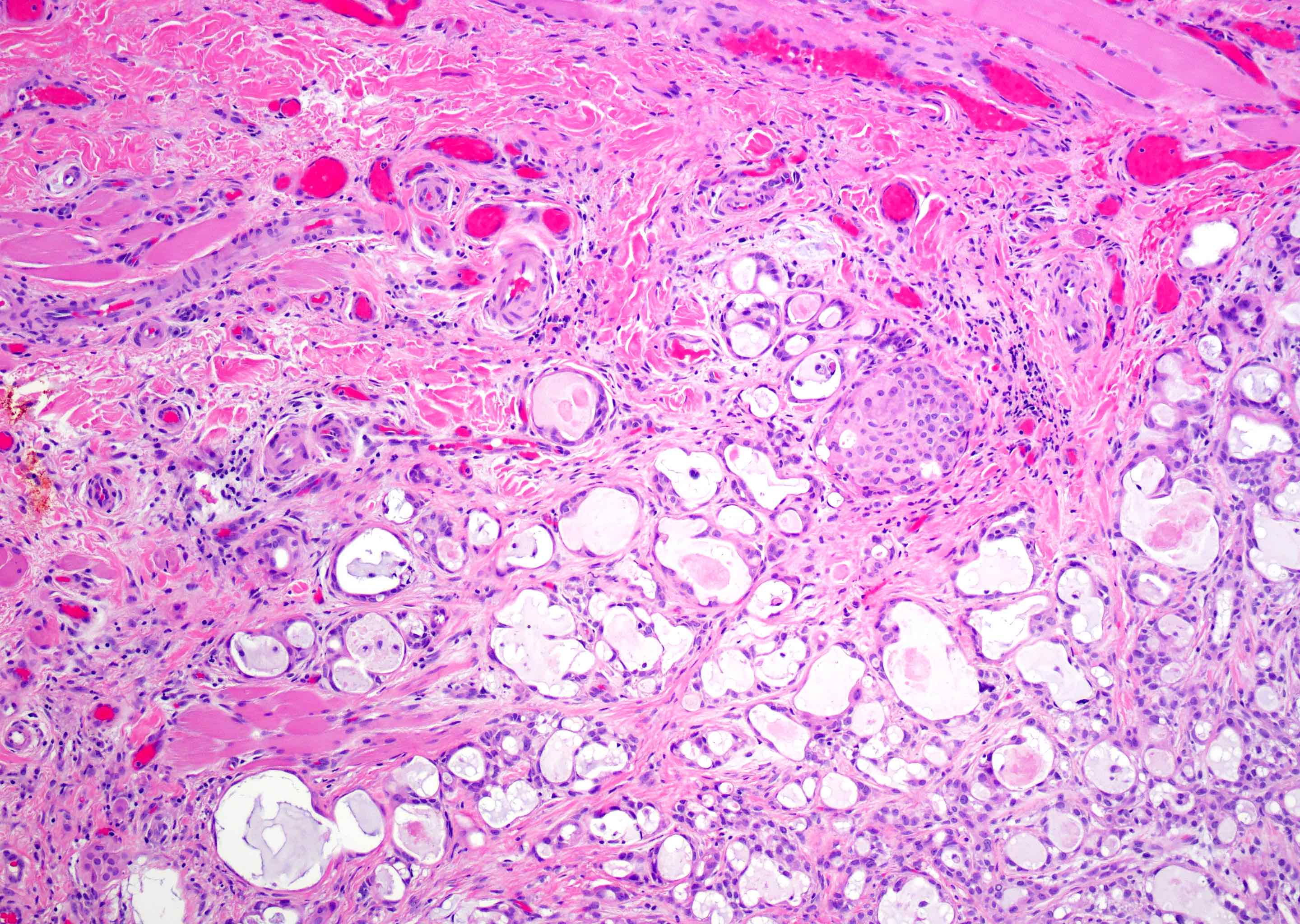
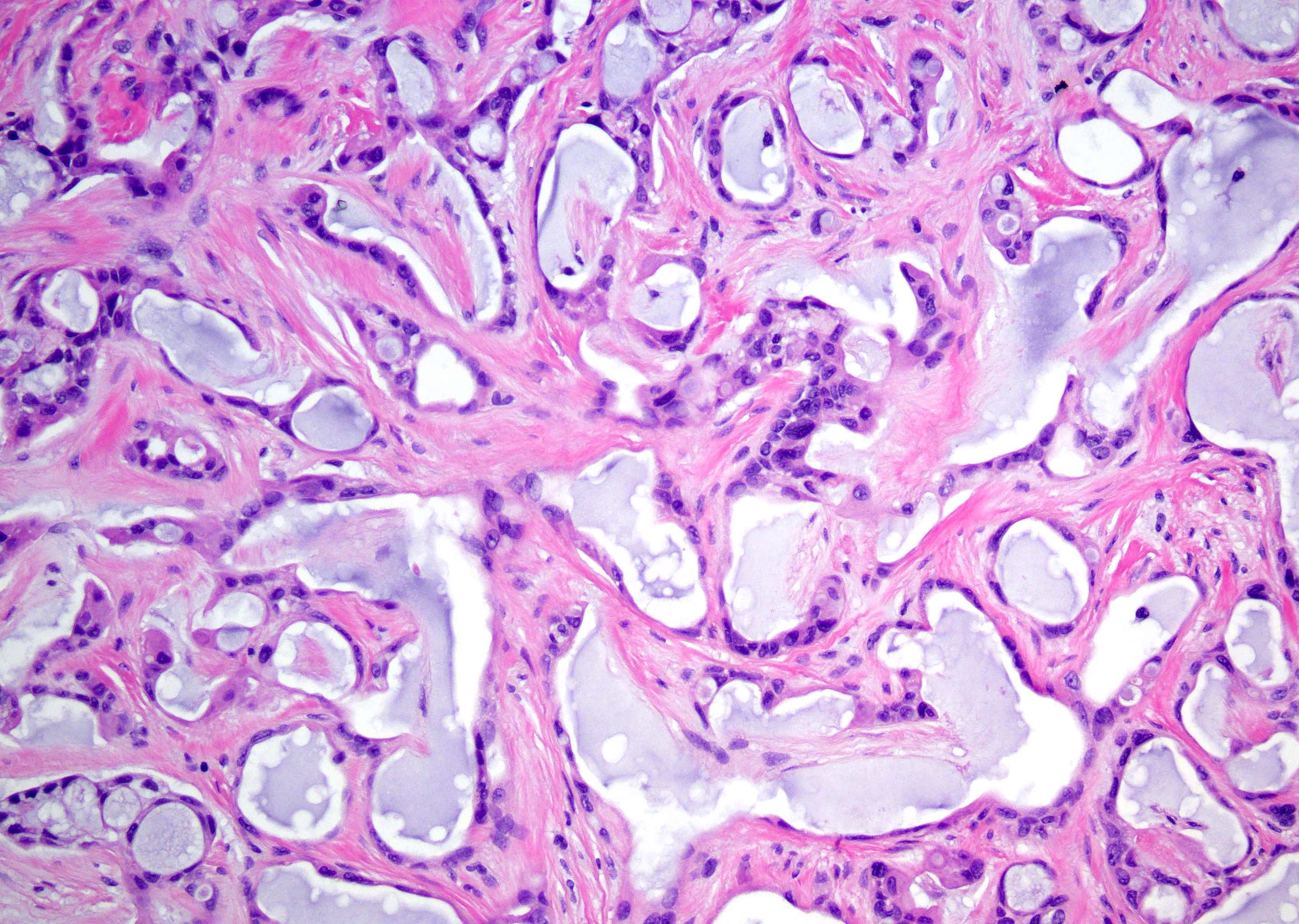
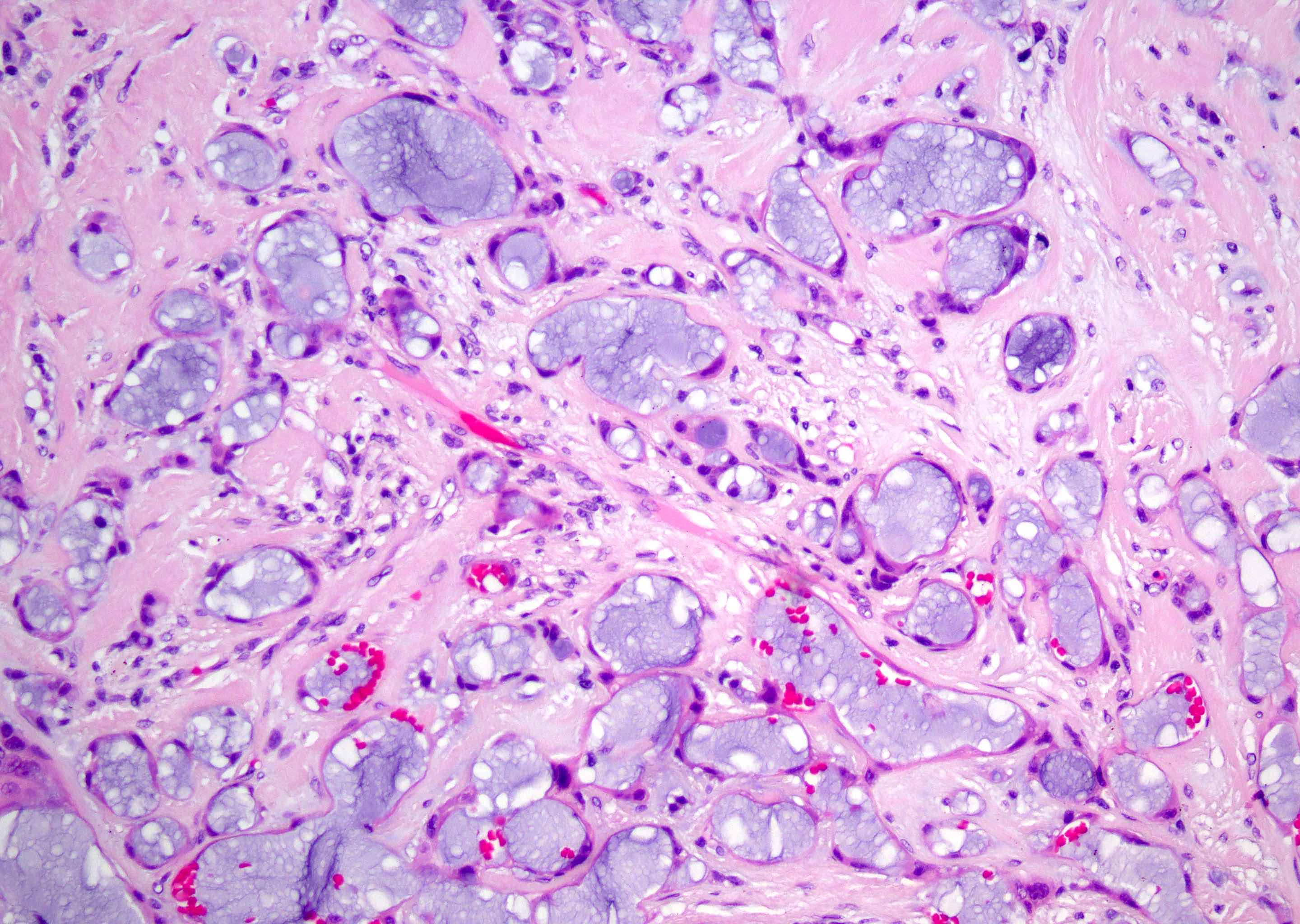
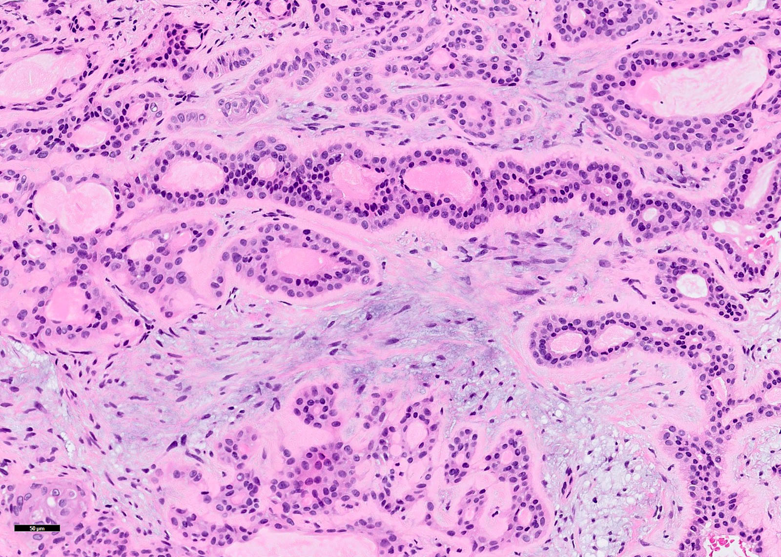
Acini and dilated ducts
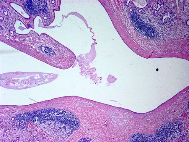

Ducts with fibrosis of wall infiltrated by lymphocytes
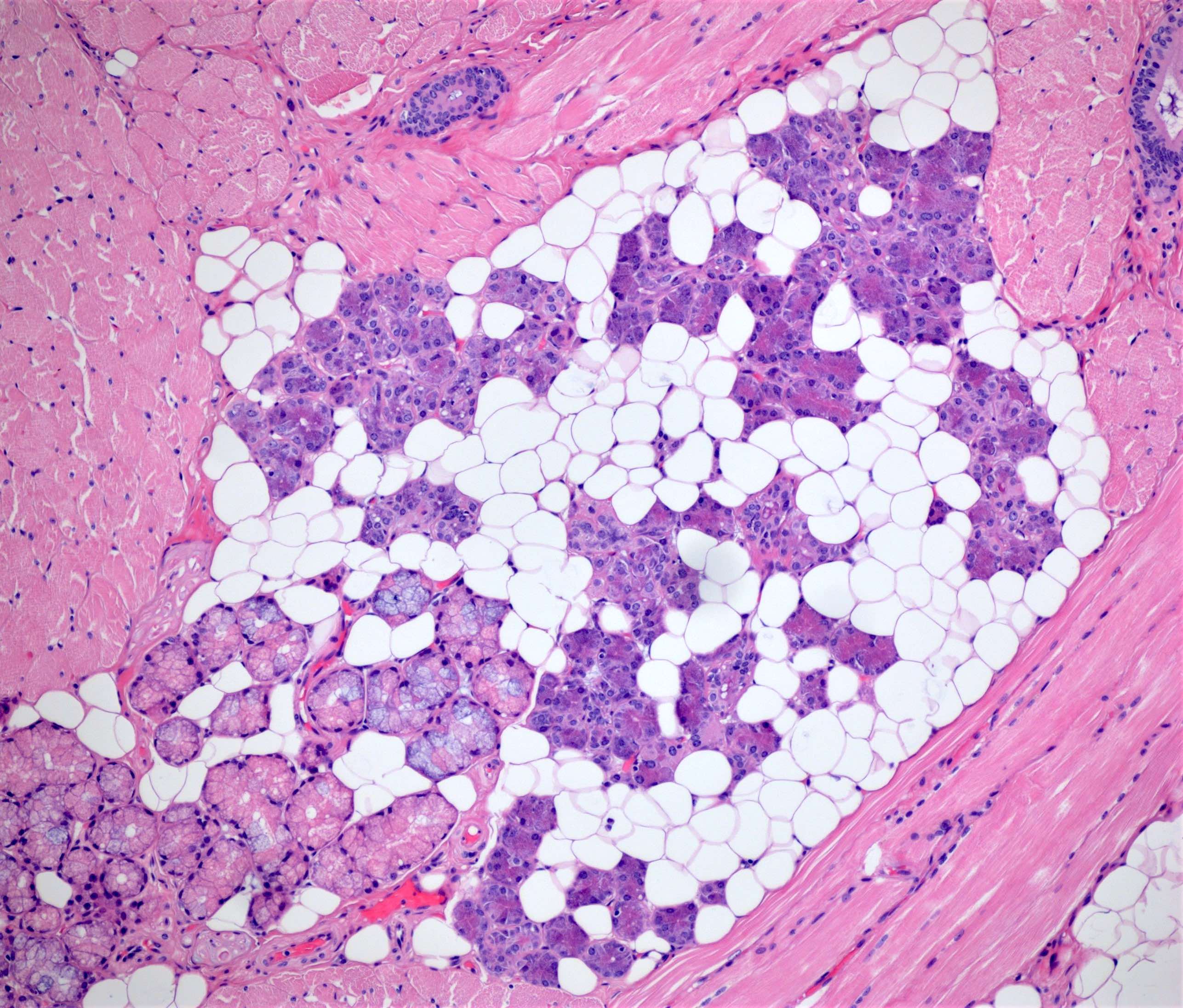
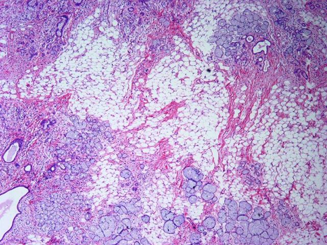
Peripheral
lipomatous tissue
with central salivary
gland elements
Lipoma-like areas
Images hosted on other servers:

Lower lip tumor

Submandibular sialoangiolipoma


Parotid sialoangiolipoma

Pleomorphic lipoma with classic floret-like cells

Chondroid lipoma
of lip has chondroid
and lipomatous
features

Oncocytic lipoadenoma of parotid

Spindle cell lipoma of parotid gland
Images hosted on other servers:

In parotid gland of HIV+ man
Case #103
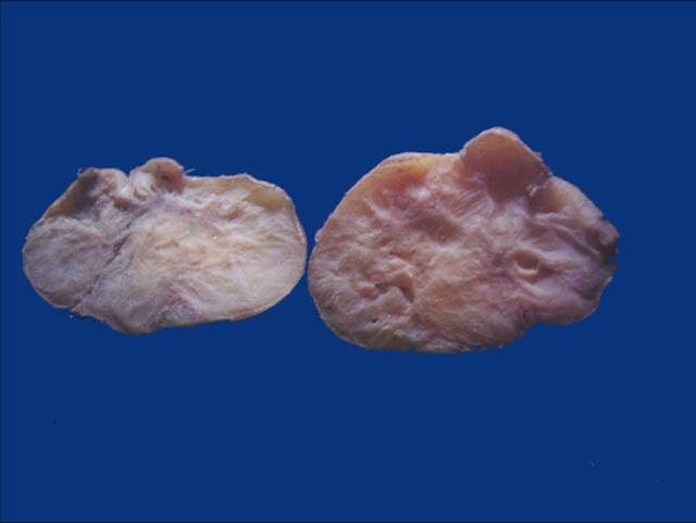
Large nodule is
sebaceous adenoma,
small nodule on left
is oncocytoma
Images hosted on other servers:


Parotid gland masses
Case #103

Large nodule
Images hosted on other servers:


Benign parotid mass with sebaceous features and lymphoid stroma

Parotid gland mass


With synchronous squamous cell carcinoma
Case #103
Diff-Quik touch prep
Contributed by Michael Kraut, M.D.
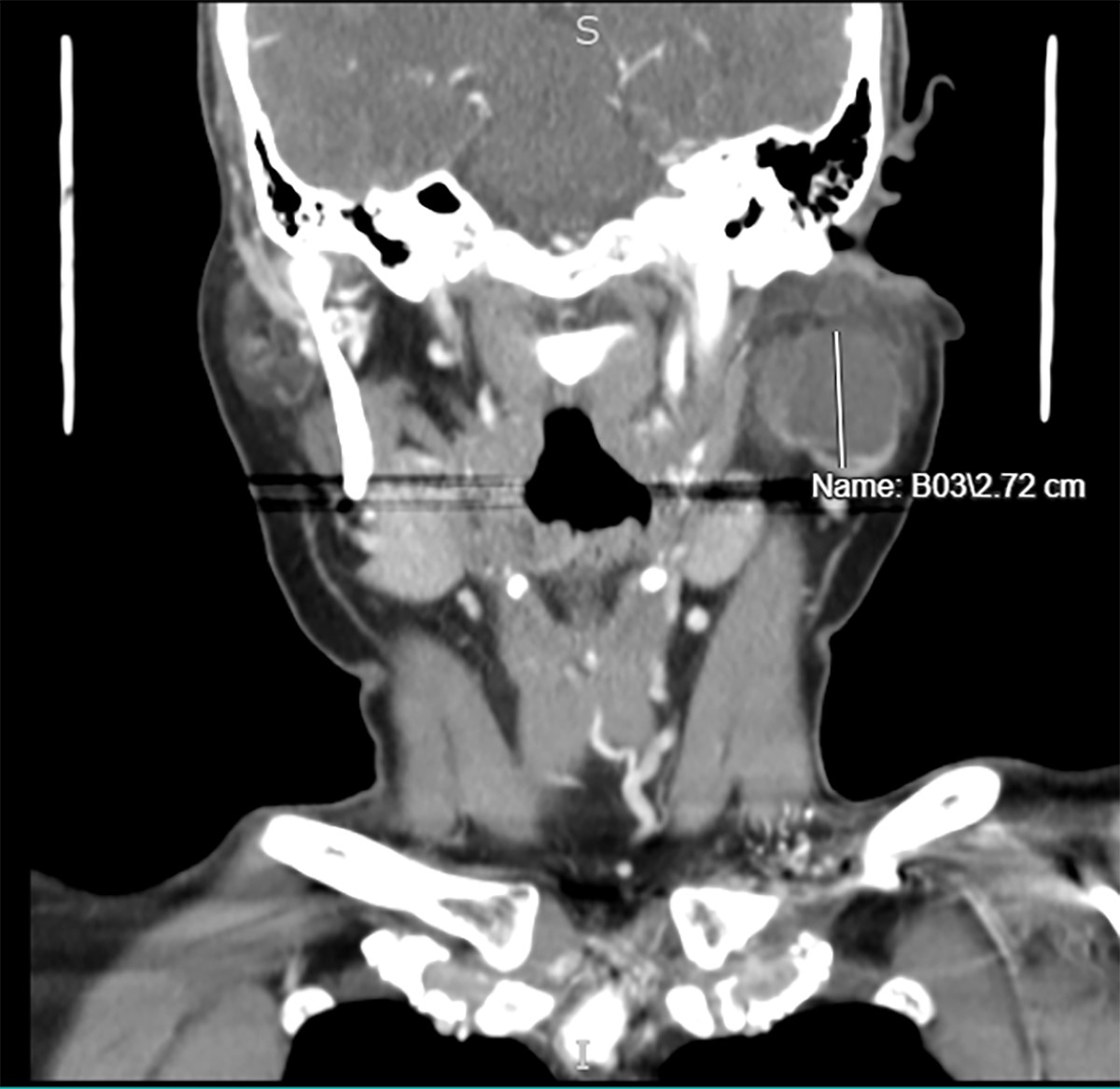
Lymphoepithelial cyst on coronal section
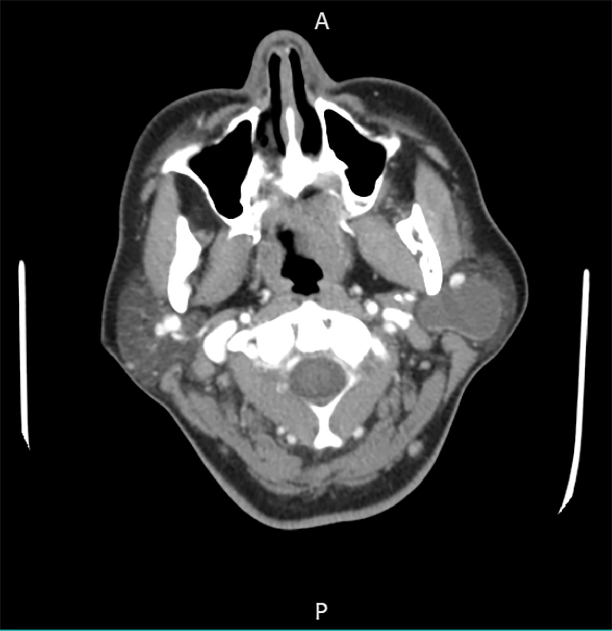
Lymphoepithelial cyst on axial section
Images hosted on other servers:

Well defined mass
Images hosted on other servers:

Diffuse swelling involving the left parotid gland
Images hosted on other servers:

Unilocular cyst with straw colored fluid
Contributed by Zahra Maleki, M.D.
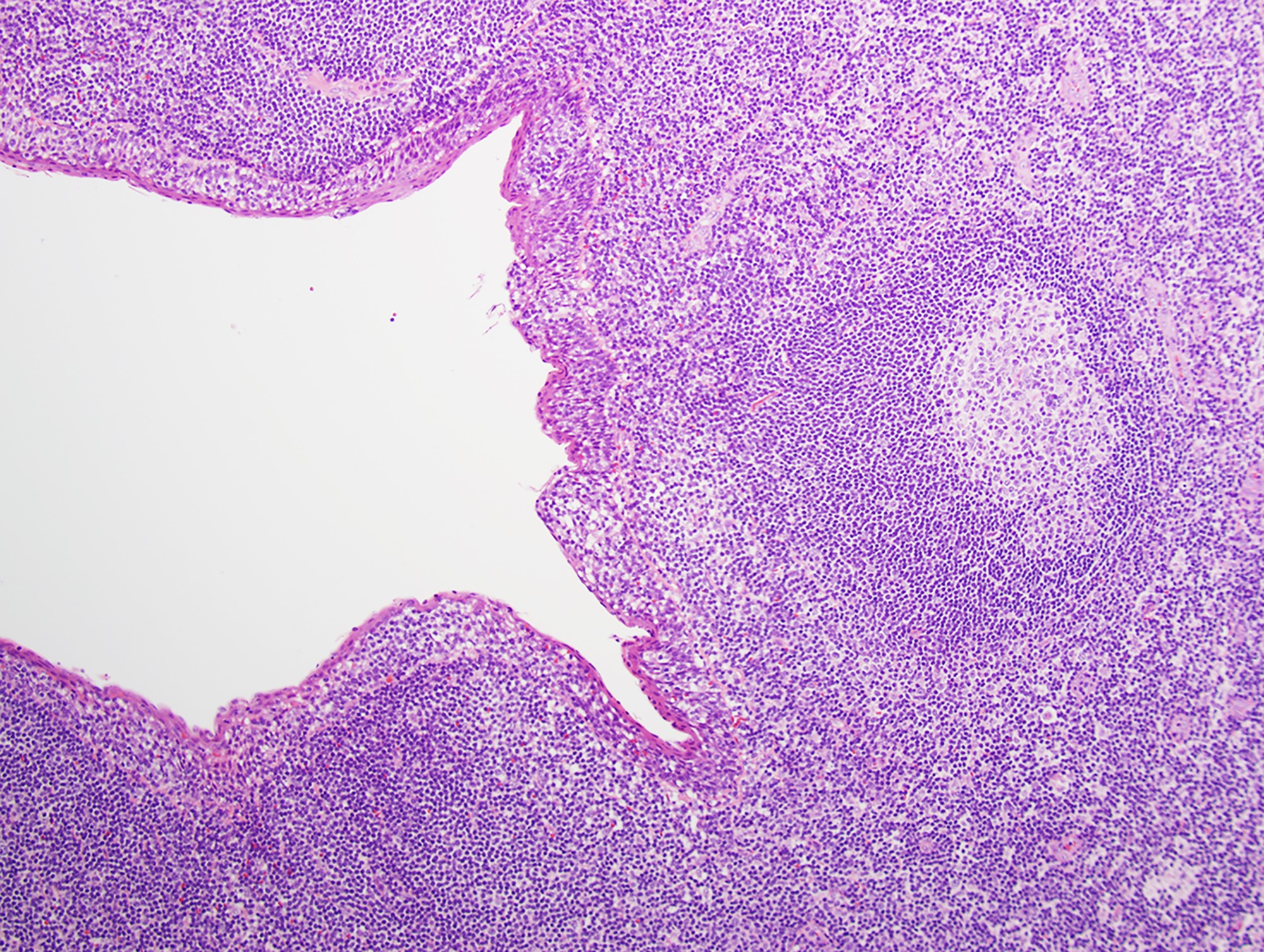
Epithelial and lymphoid component
Squamous lining and lymphoid tissue
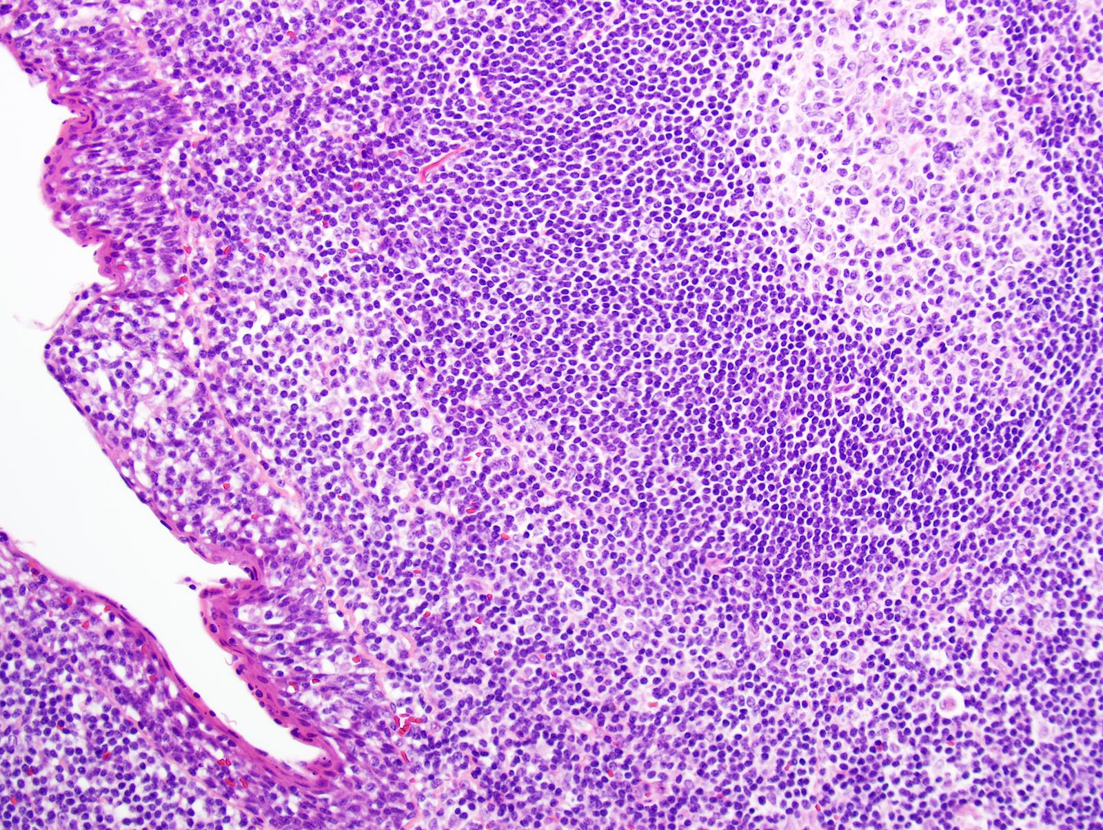
Epithelium surrounded by inflammatory cells
Epithelial and inflammatory cells
Contributed by Zahra Maleki, M.D.
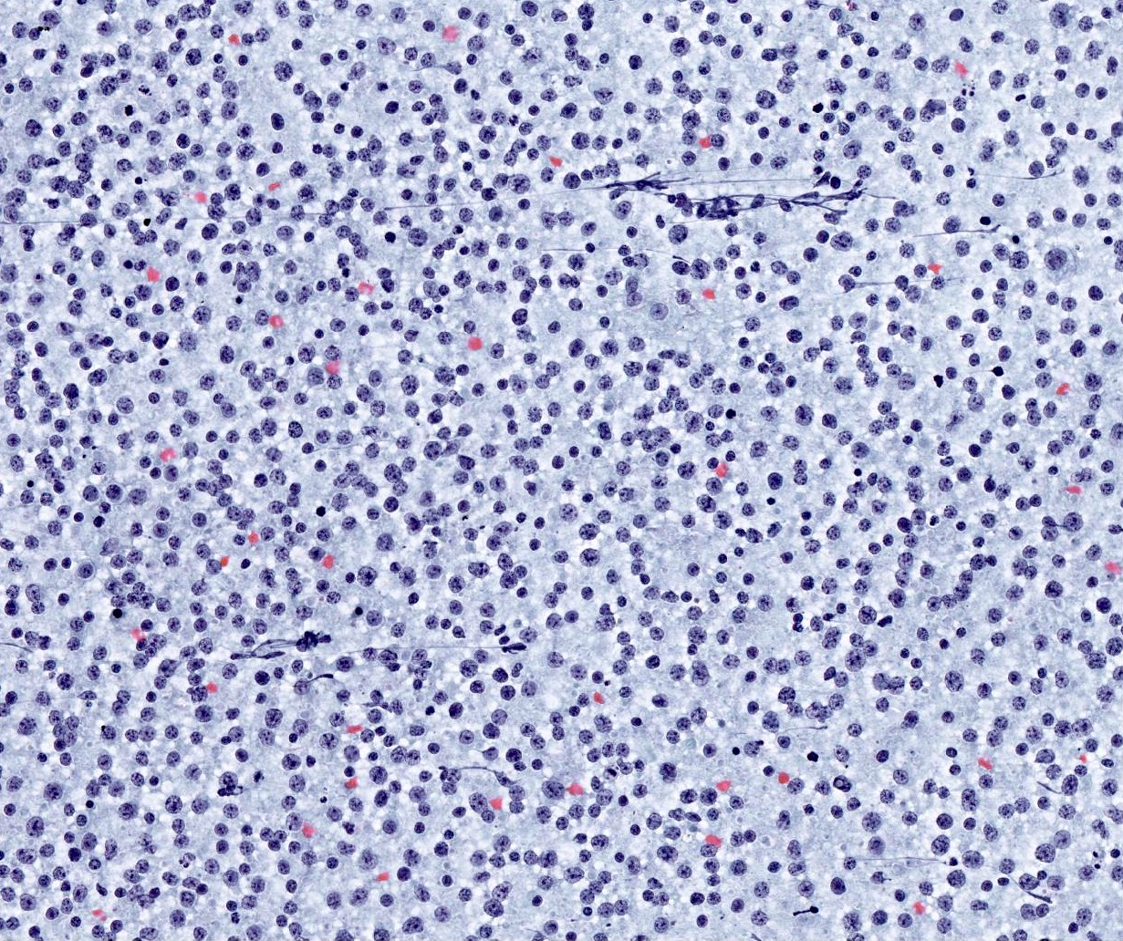
Numerous lymphocytes and histiocytes
Large epithelial fragment
Dispersed squamous cells
Polymorphous lymphocytes
Cyst content
Contributed by James S. Lewis, M.D.
Maintenance of lobular architecture
Intense chronic inflammation
Plasma cells
Lymphoepithelial lesion
Contributed by Jen-Fan Hang, M.D.

Mucoepidermoid carcinoma
Acinic cell carcinoma

Salivary duct carcinoma
Adenoid cystic carcinoma
Secretory carcinoma
Malignant category 1
Malignant category 2
Contributed by Michael Mikula, M.D.
Unencapsulated and well circumscribed
Subtle invasive growth
Microcysts
Basophilic secretions

Fibromyxoid stroma

Monotonous hyperchromatic nuclei
S100
p63
Images hosted on other servers:

SS18 rearrangement by FISH
Images hosted on other servers:
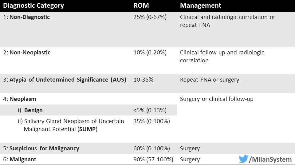
Milan system
Contributed by Jen-Fan Hang, M.D.
Nondiagnostic

Nonneoplastic acinar cells

Benign skeletal muscle
Cystic fluid only
Atypia of undetermined significance (AUS)
Mucinous cyst
Atypical lymphoid proliferation
Neoplasm: benign
Pleomorphic adenoma (PA): fibrillary extracellular matrix
PA: plasmacytoid
myoepithelial cells
PA with squamous metaplasia
Warthin tumor
Warthin tumor
Oncocytoma
Neoplasm: salivary gland neoplasm of uncertain malignant potential (SUMP)
With oncocytic / oncocytoid features

With basaloid features
Malignant
Mucoepidermoid carcinoma
Acinic cell carcinoma
Salivary duct carcinoma
Adenoid cystic carcinoma
Secretory carcinoma
Milan system
Algorithmic approach to everyday salivary gland cytology
Contributed by Ulrike Hamper, M.D., M.B.A.

Ultrasound, cyst
Contributed by Zahra Maleki, M.D.
Cystic lined cavity
Contributed by Zahra Maleki, M.D.

Fine needle aspiration

Contributed by Kelly Magliocca D.D.S., M.P.H.
Cut surface, high grade
Contributed by Rema A. Rao, M.D. and Saeed Asiry, M.D.

Tumor architecture
Tumor cell types
Epidermoid cells
Mucus cells
Intermediate cells
Clear cell changes
Case #346

4 year old boy

Ki67

Mucicarmine
Contributed by Rema A. Rao, M.D. and Saeed Asiry, M.D.
Background mucin

Neoplastic cells
Mucus cells
Neoplastic cells in sheets
Keratinizing cells
Single keratinizing cells
Crowded squamous cells
Images hosted on other servers:


Low grade and oncocytic tumors
Images hosted on other servers:

FISH analysis of MAML2
Images hosted on other servers:

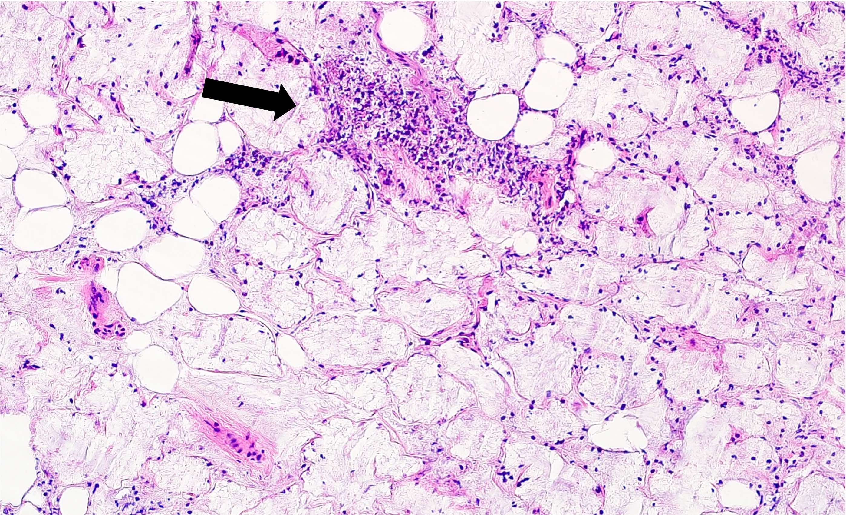
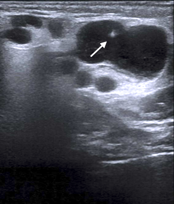
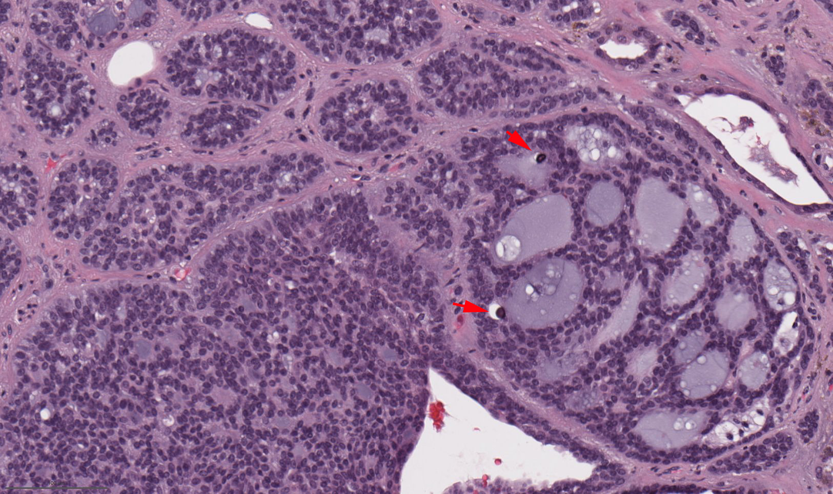
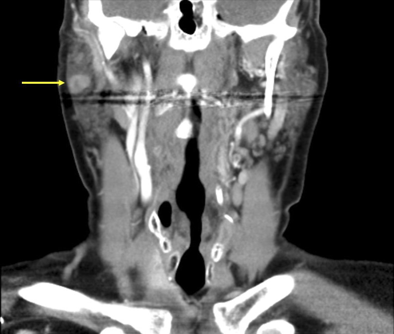
Multinodular parotid mass (arrows)
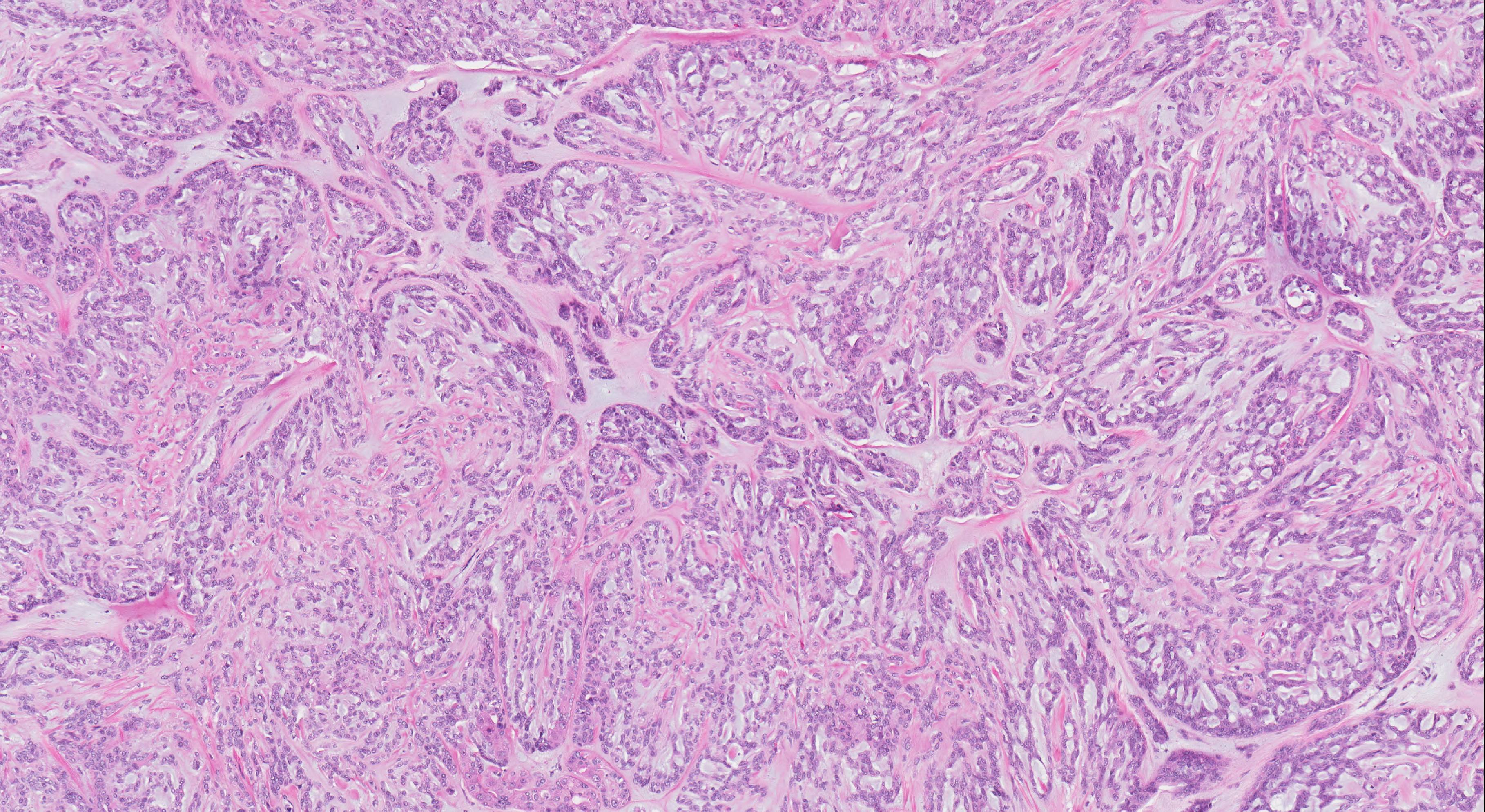
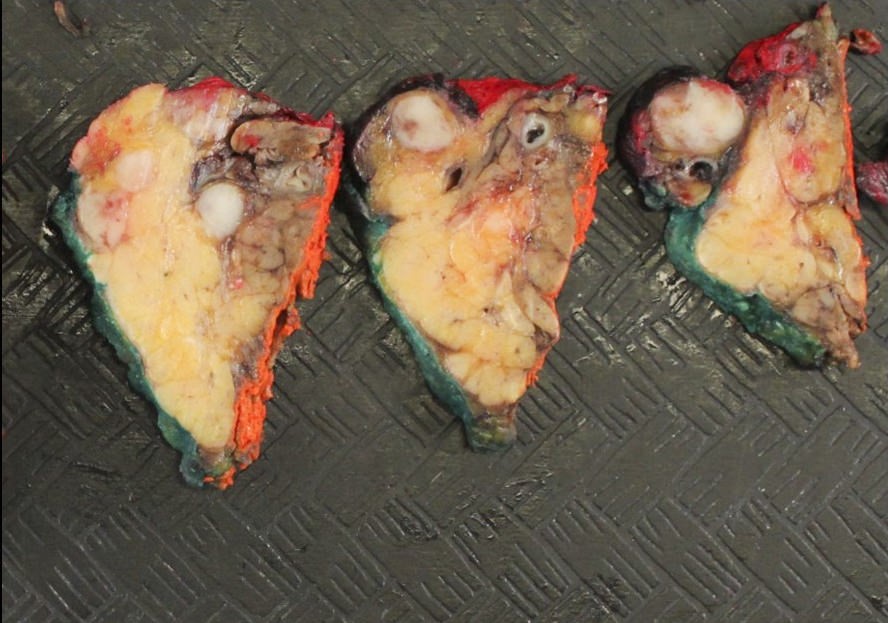
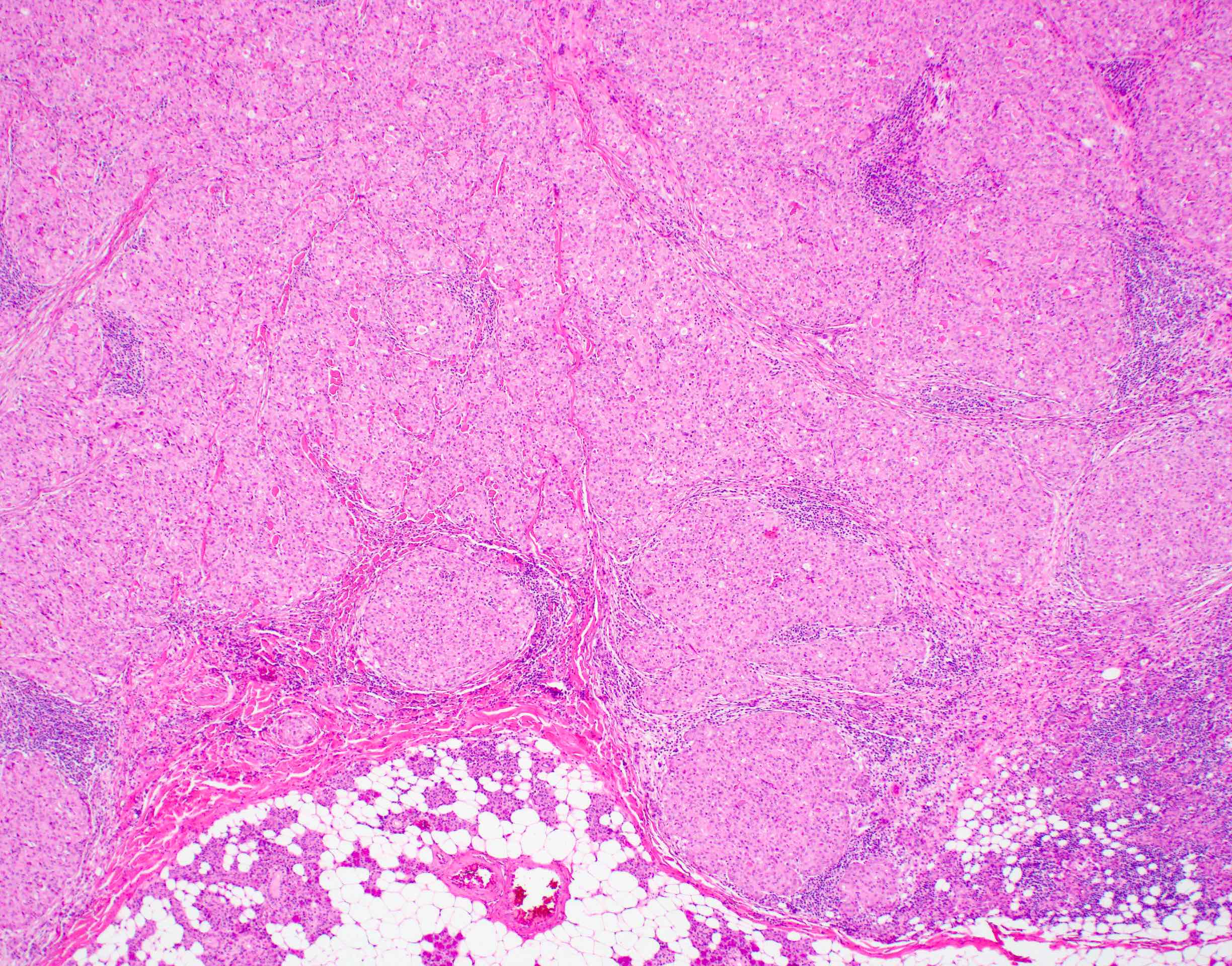
Contributed by Abeer Salama, M.D. and Bin Xu, M.D., Ph.D.
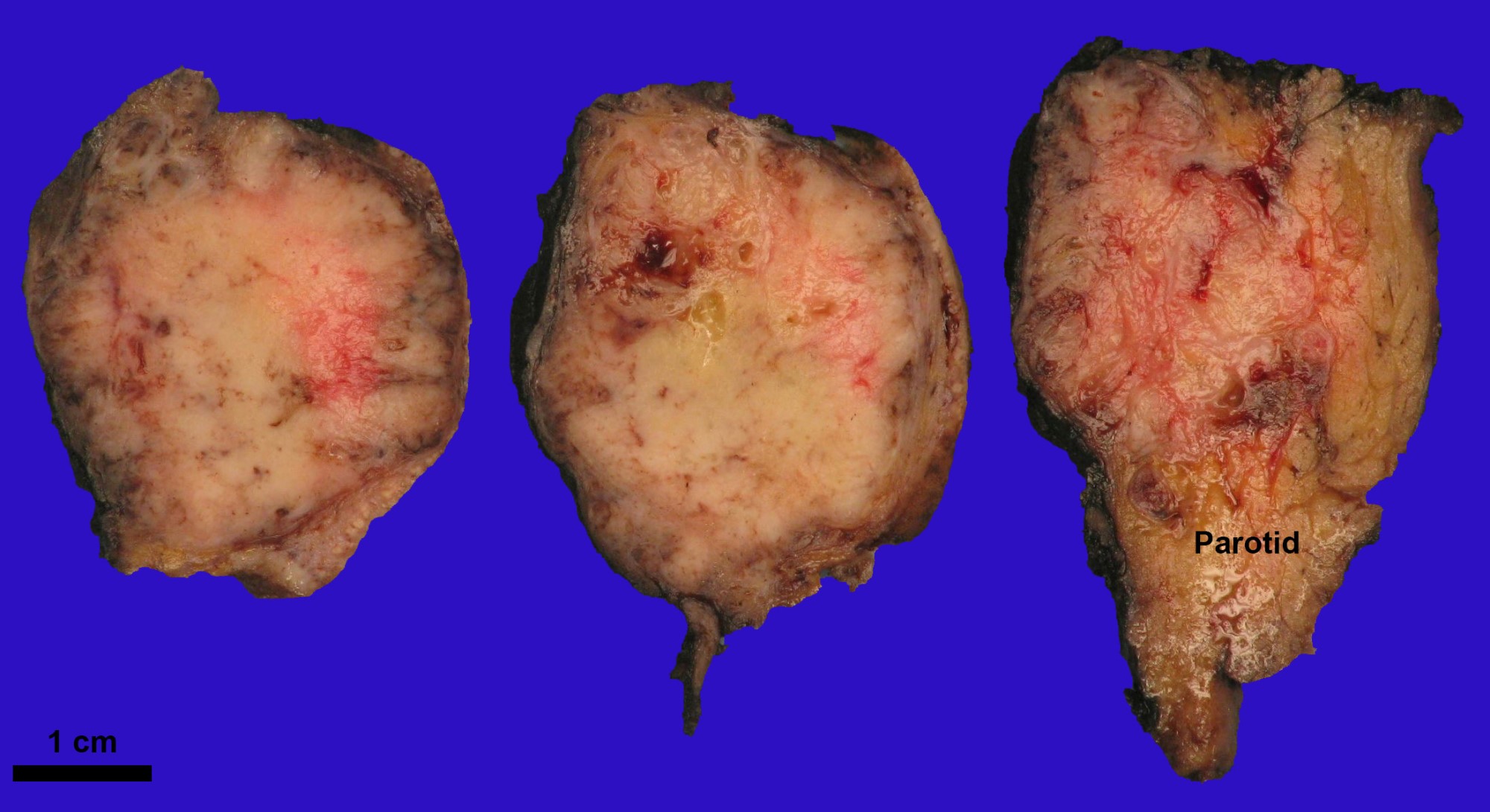
Lobulated parotid mass
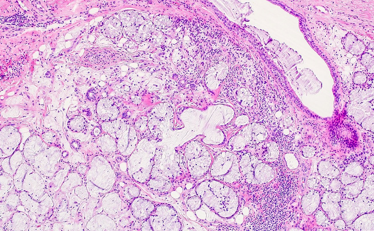
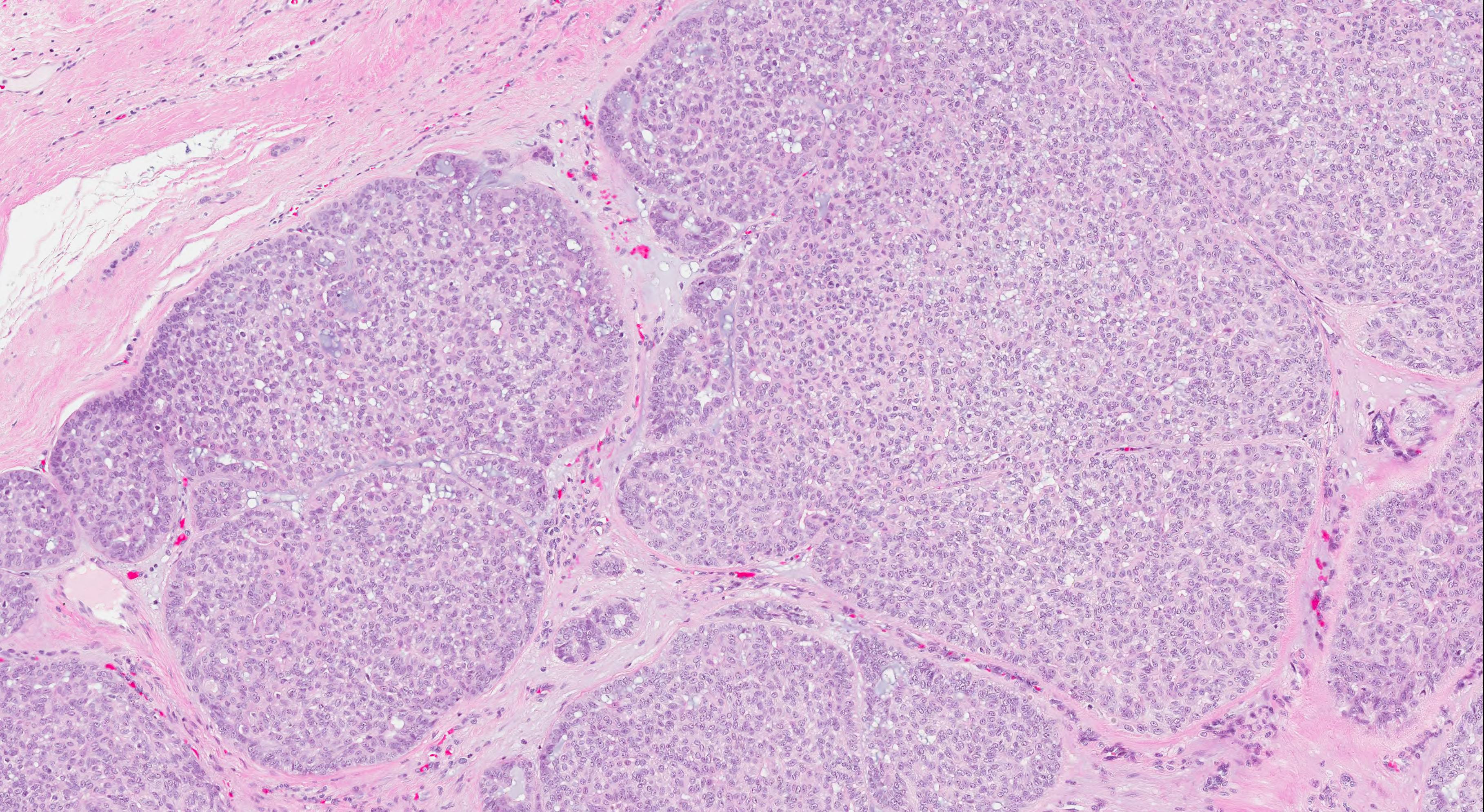
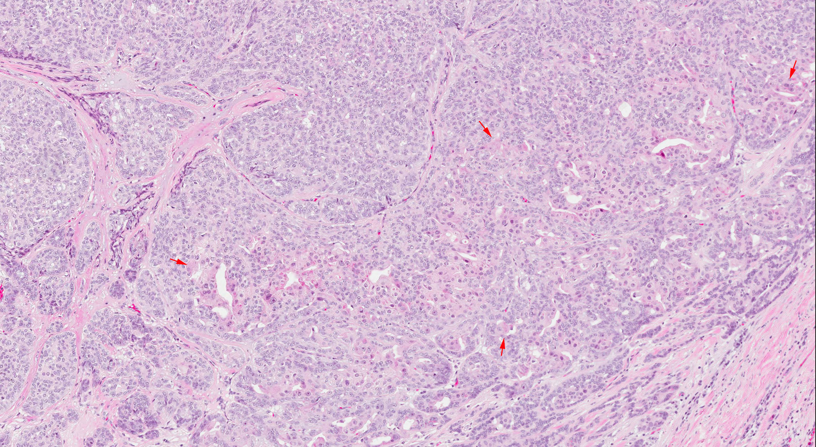
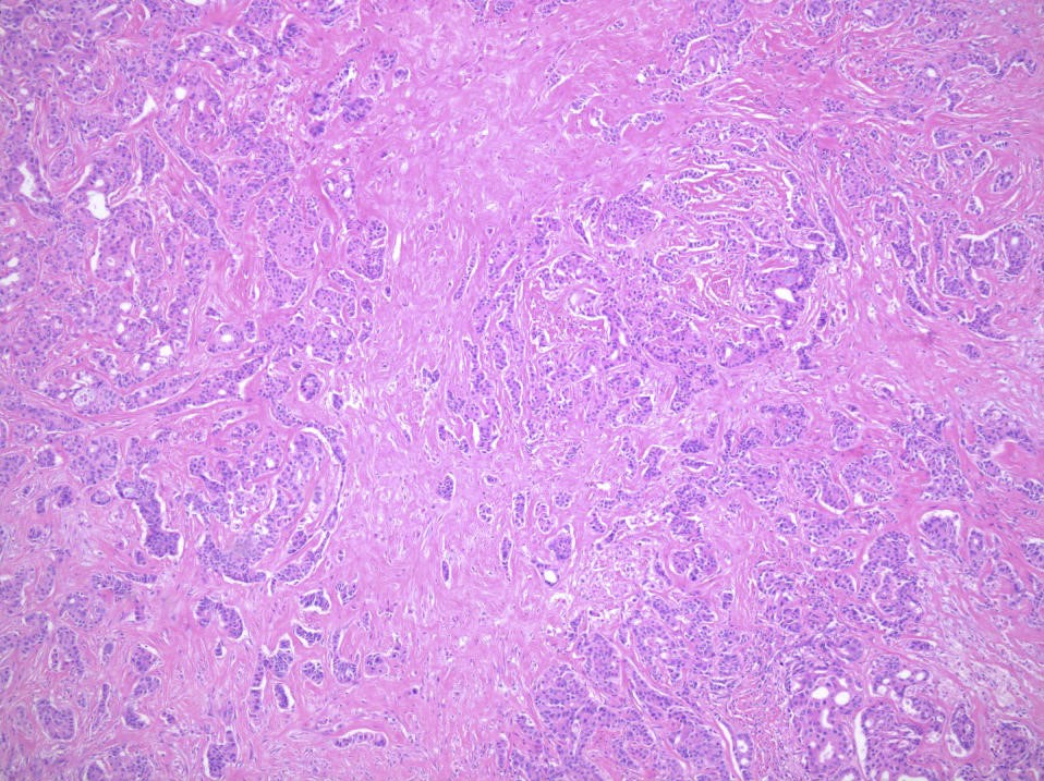
Contributed by Abeer Salama, M.D. and Bin Xu, M.D., Ph.D.

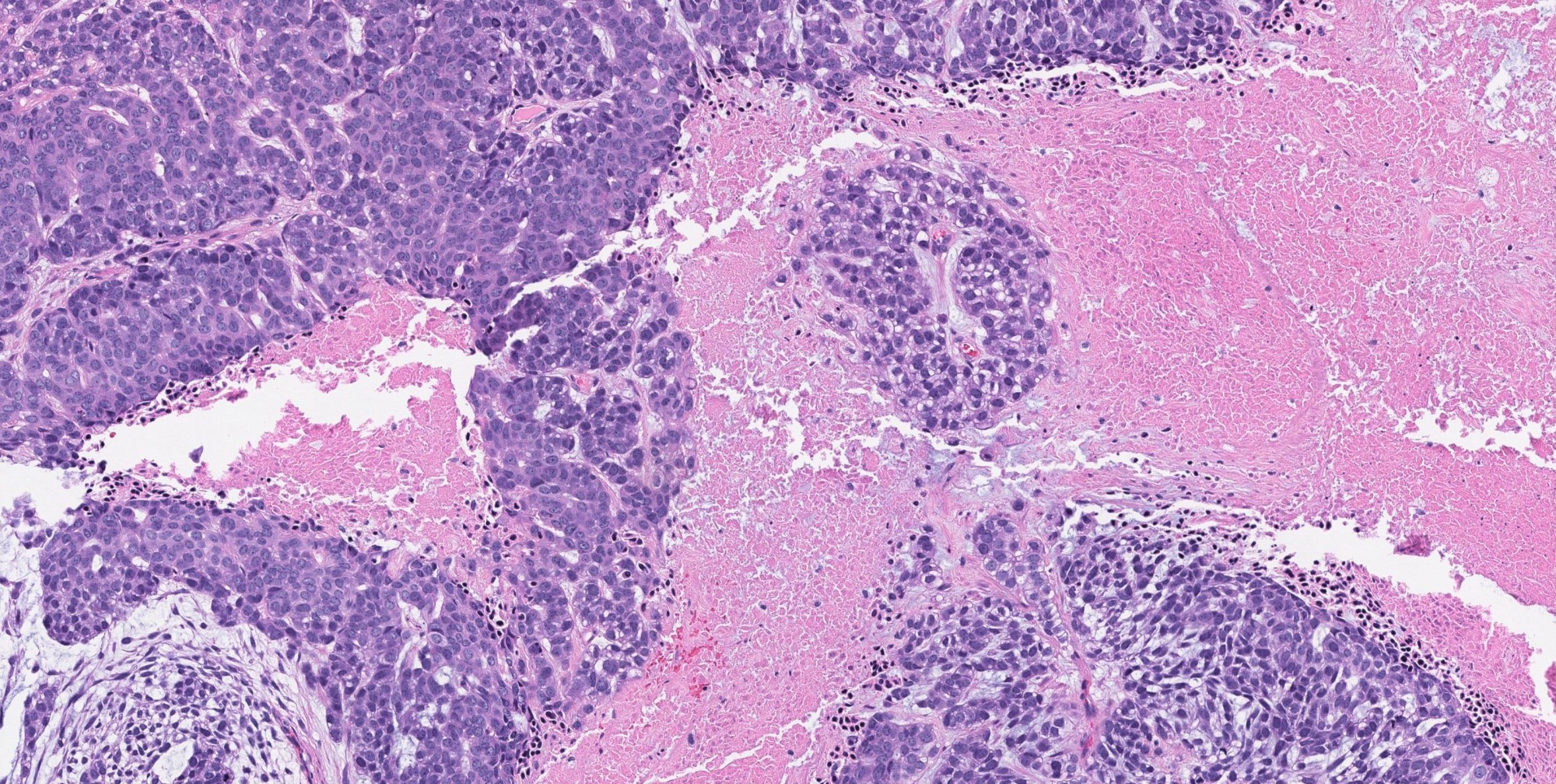
Multinodular invasive growth
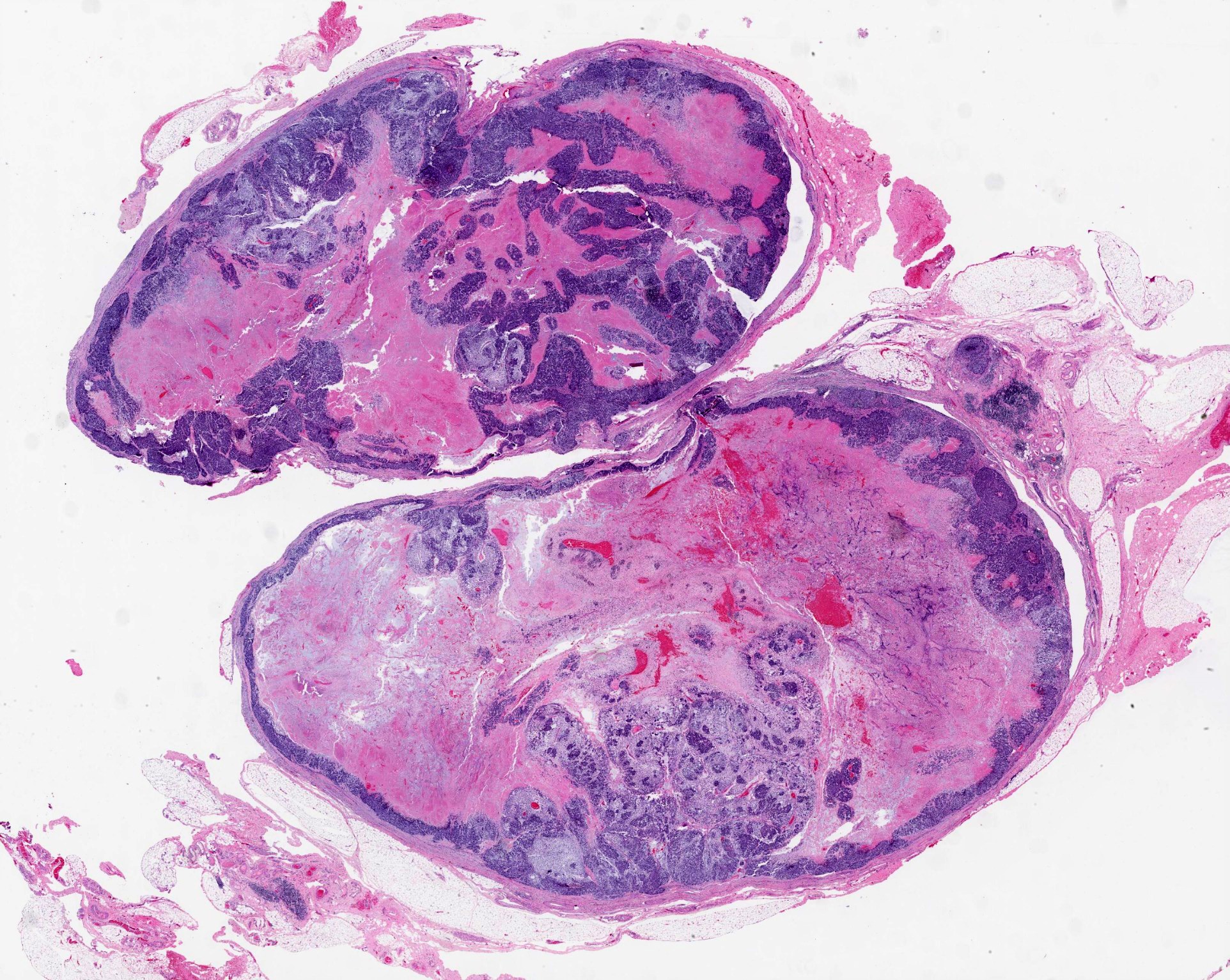
Expansile lobulated growth
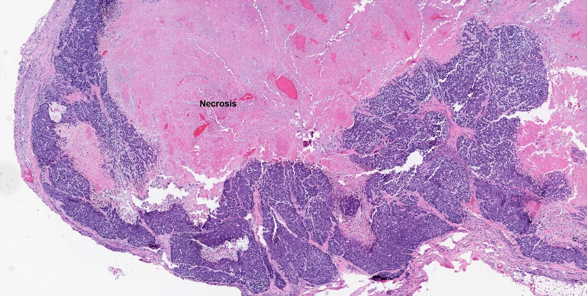
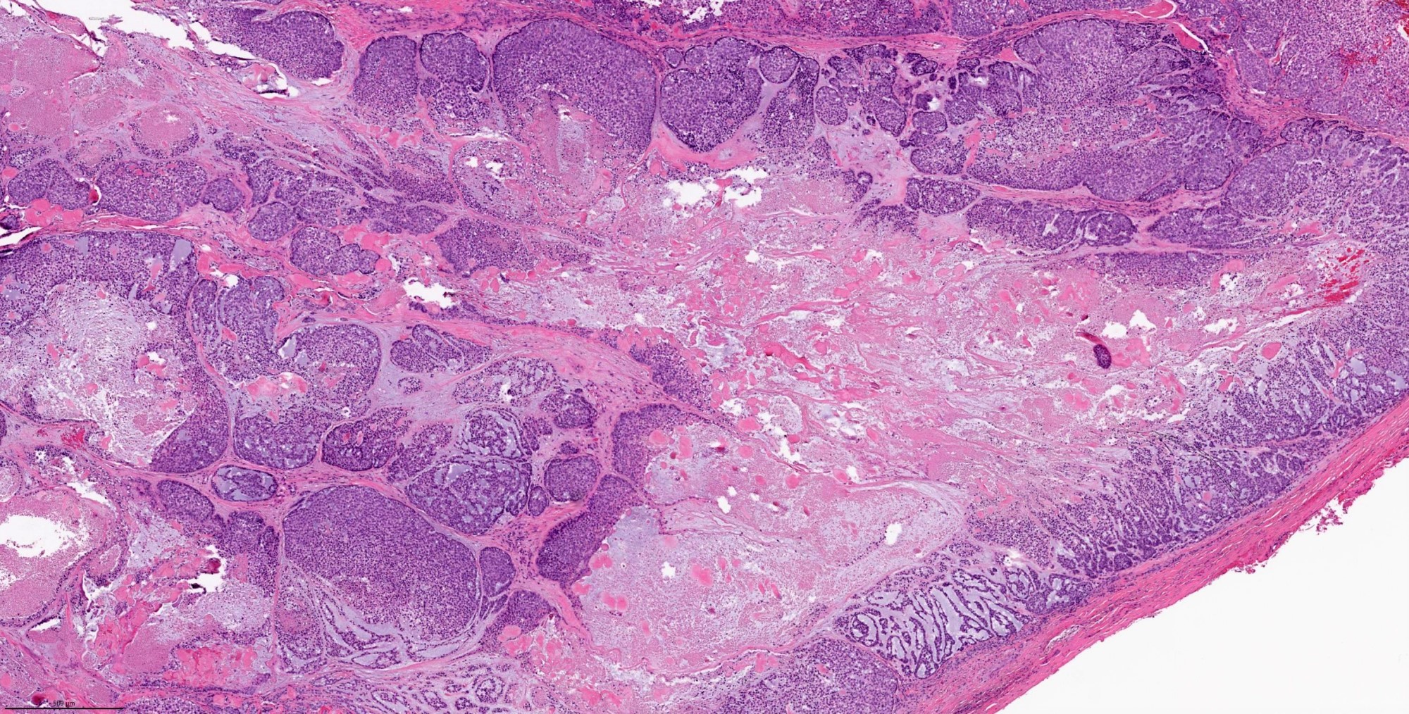
Necrotic hypocellular central zone
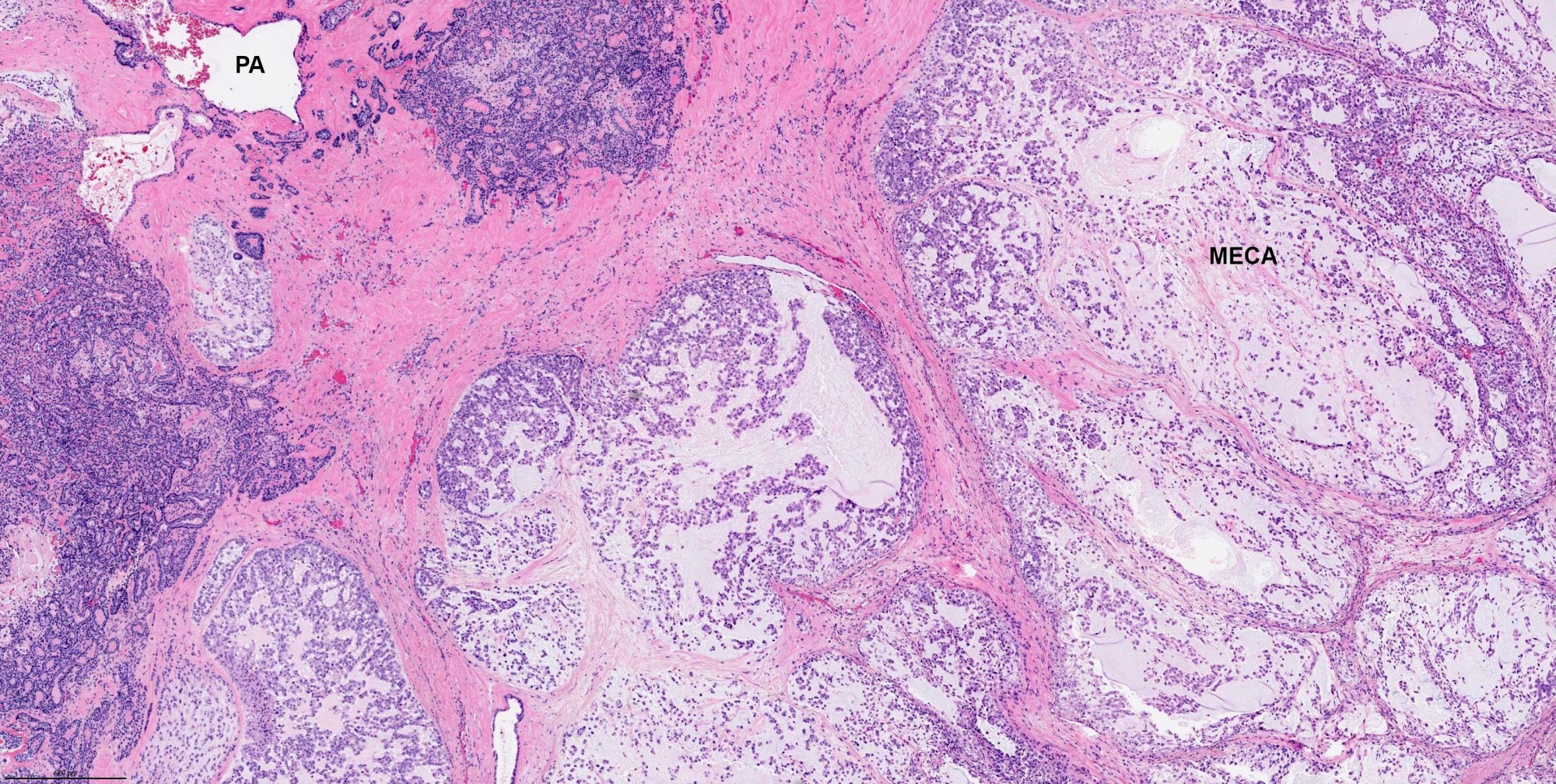
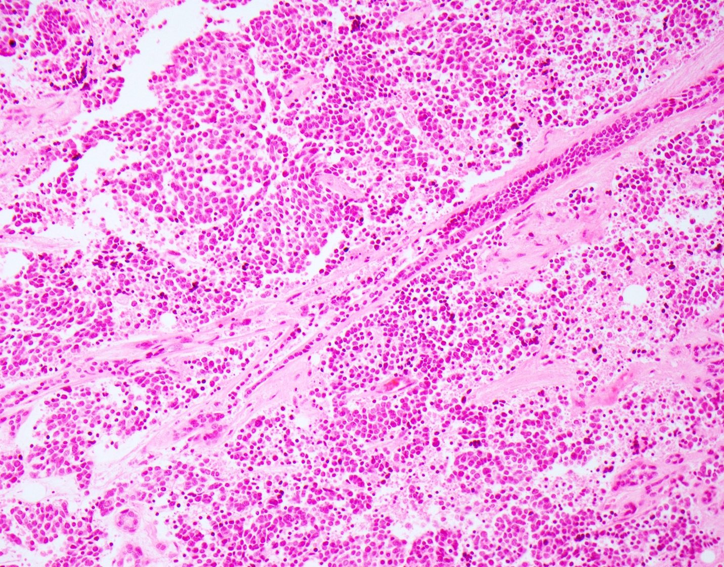
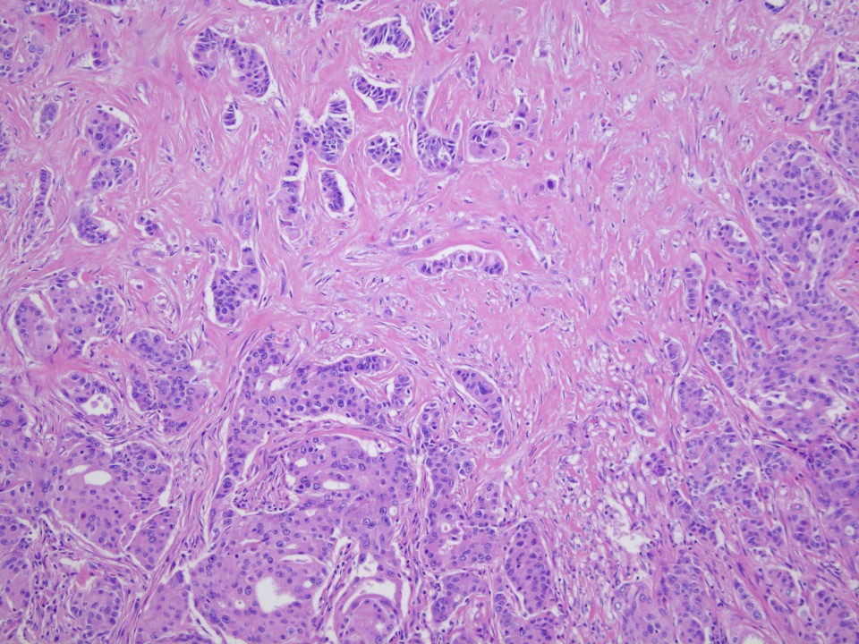
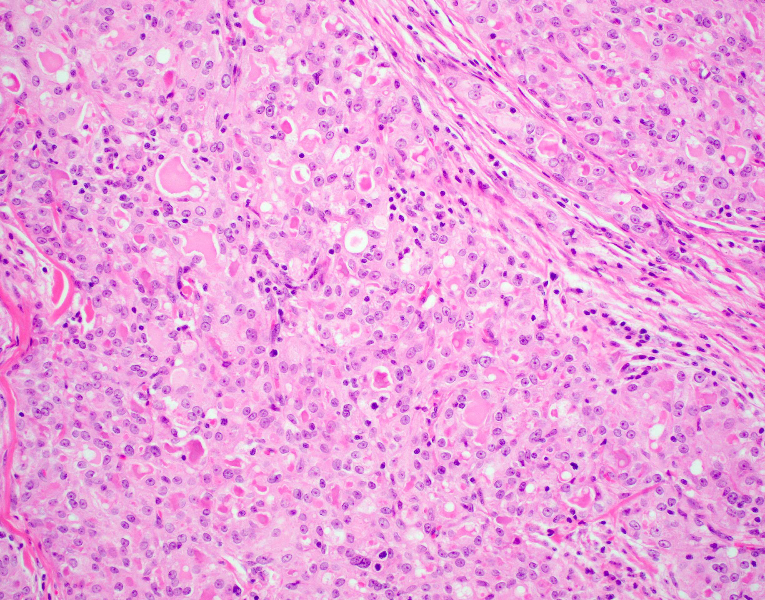
Myoepithelial carcinoma ex PA
Epithelioid features
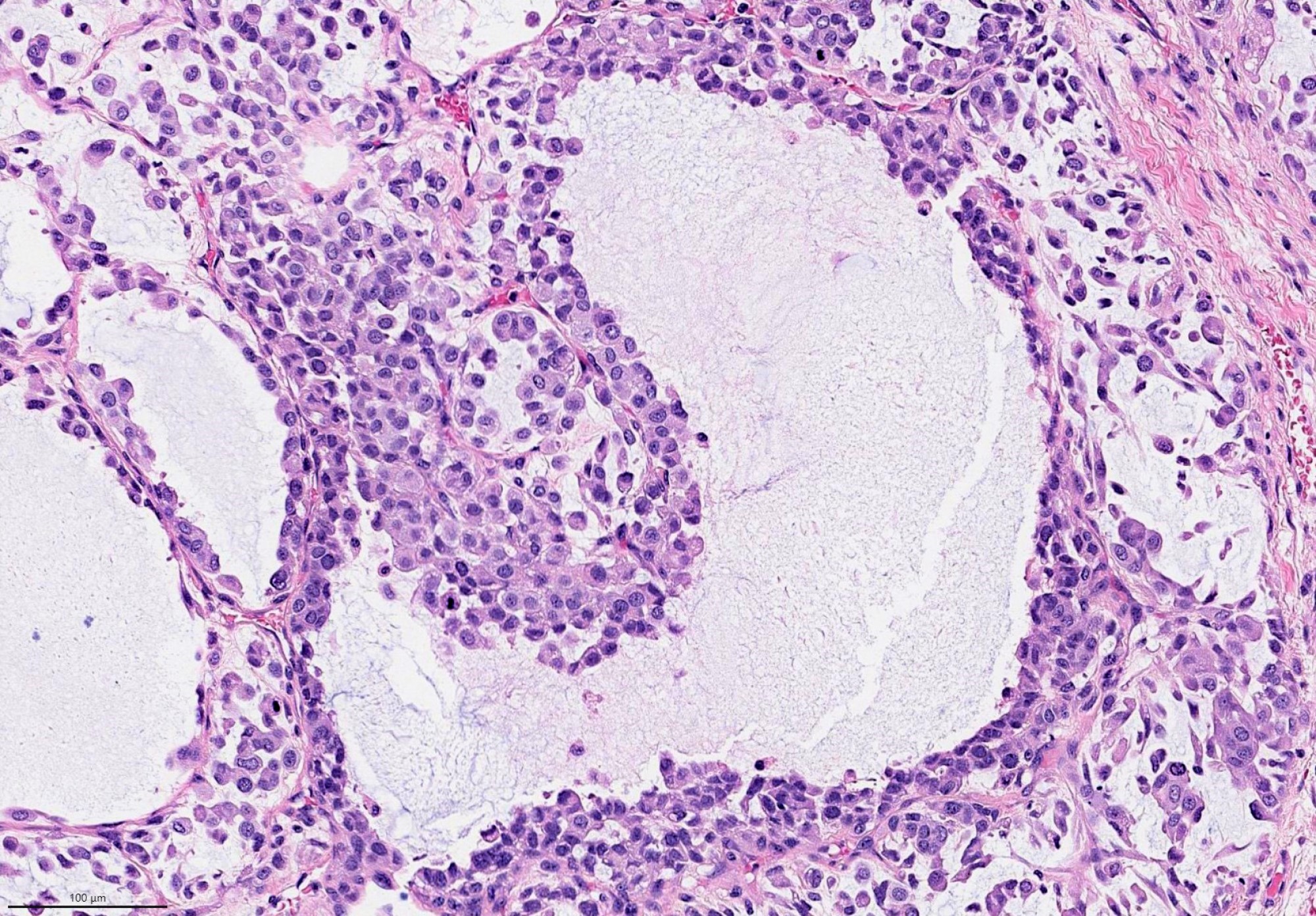
Plasmacytoid features
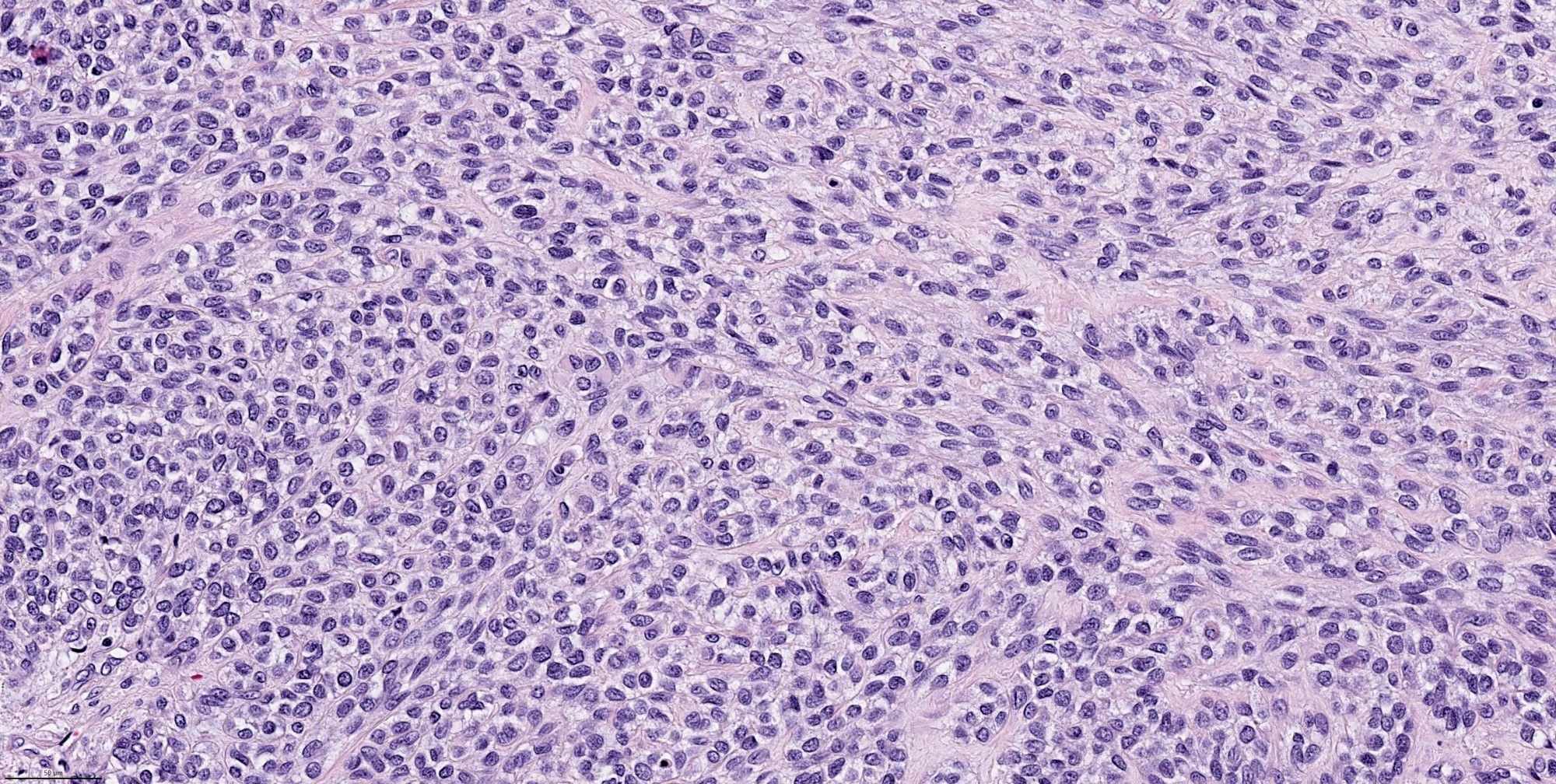
Clear cell features
Spindle cell features
S100 immunostain
CAM5.2 immunostain
Calponin immunostain
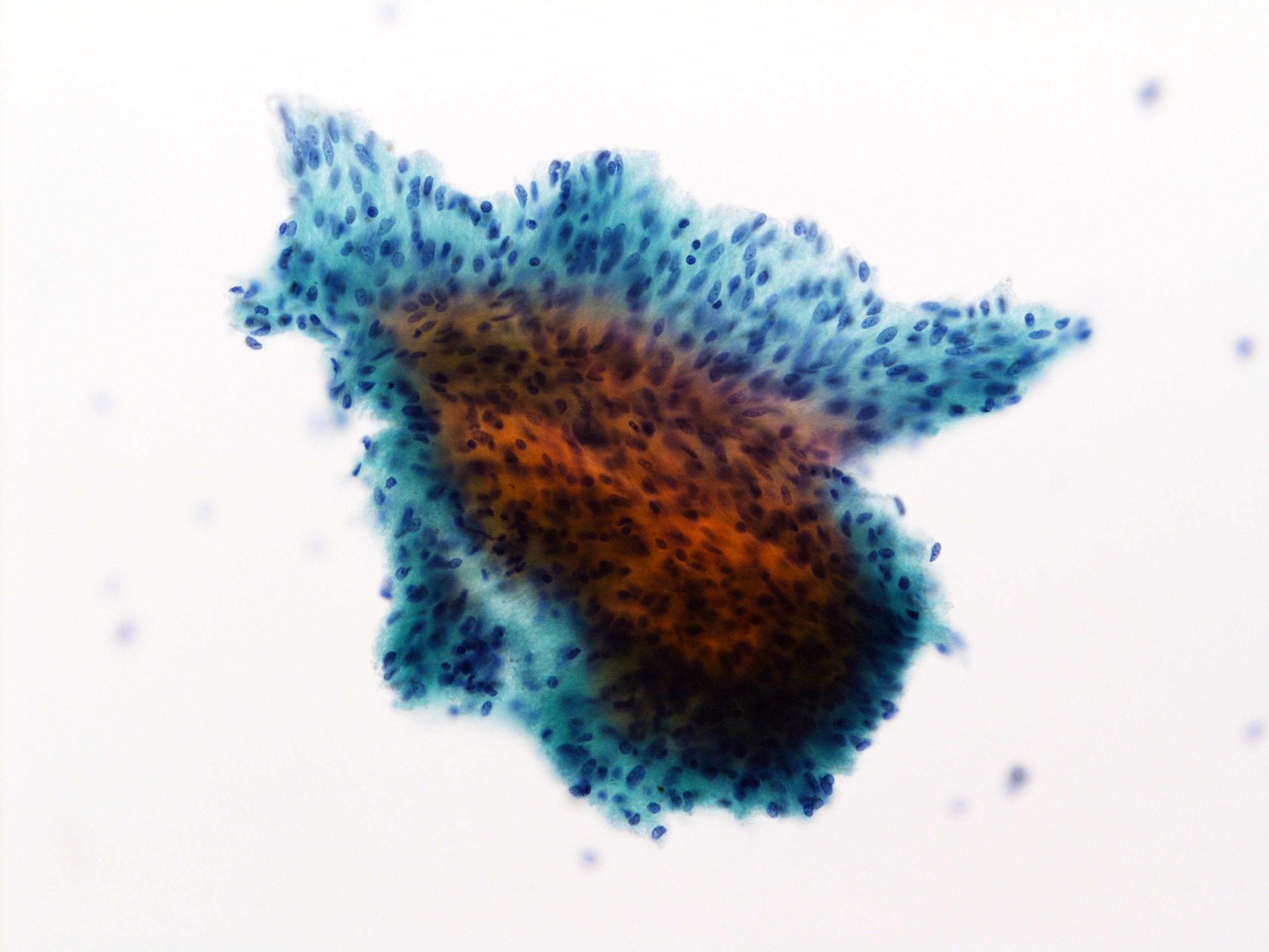
Images hosted on other servers:

Diff-Quik stain
Images hosted on other servers:

Molecular signature (oncoprint)
Images hosted on other servers:

Plasmacytoid myoepithelioma of minor salivary glands
Contributed by Xiaofeng Zhao, M.D., Ph.D. and Shuanzeng Wei, M.D., Ph.D.
Spindle cell type
Epithelioid type
Nest / trabecular pattern
Contributed by Shuanzeng Wei, M.D., Ph.D.

Cellular specimen

Monotonous epithelioid cells
Images hosted on other servers:

Rare bony erosion
Contributed by Douglas Damm, D.D.S., Ashley Clark, D.D.S.,
Eric Mencarelli, M.D., D.D.S. and Molly Housley Smith, D.M.D.

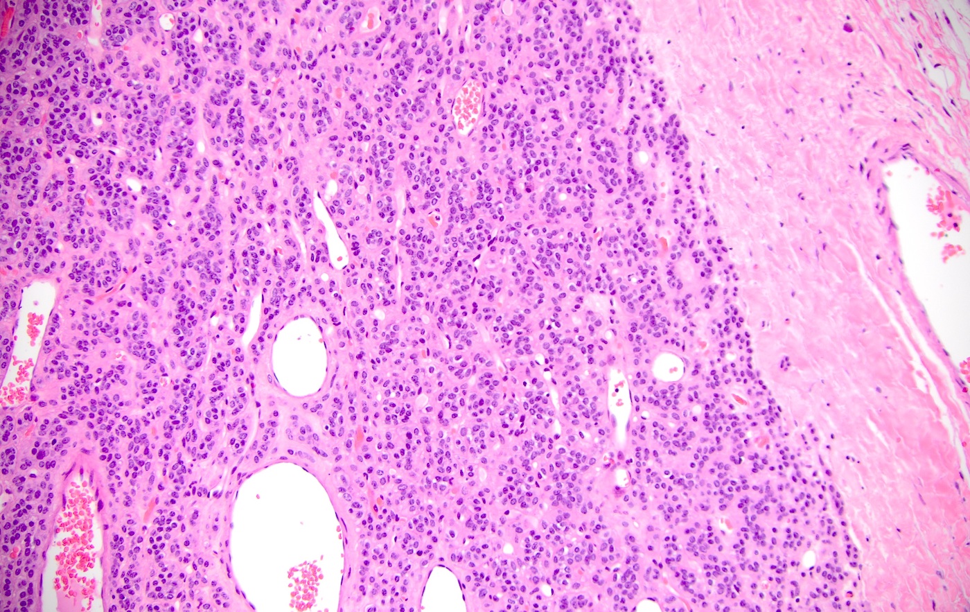
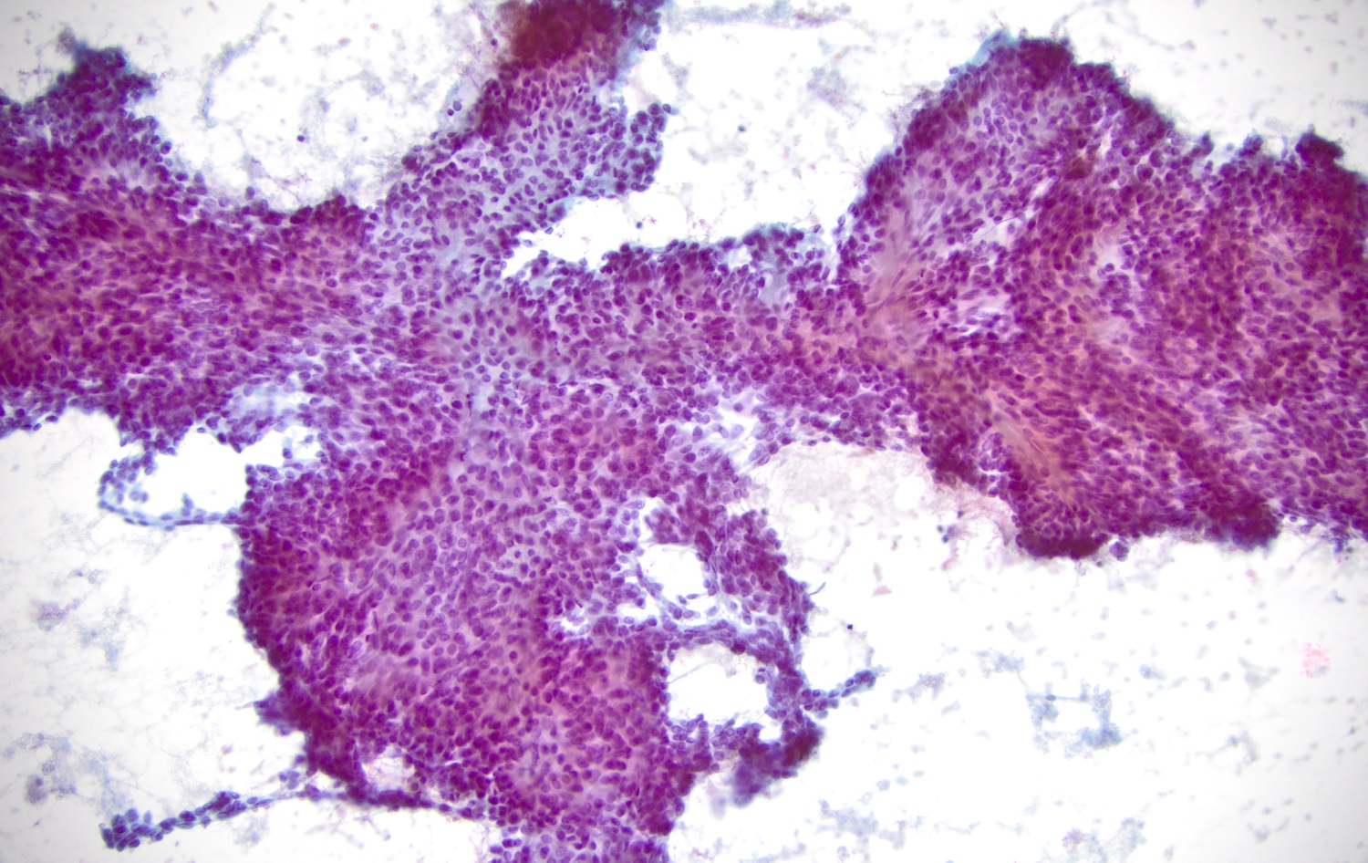
Palatal ulceration and erythema
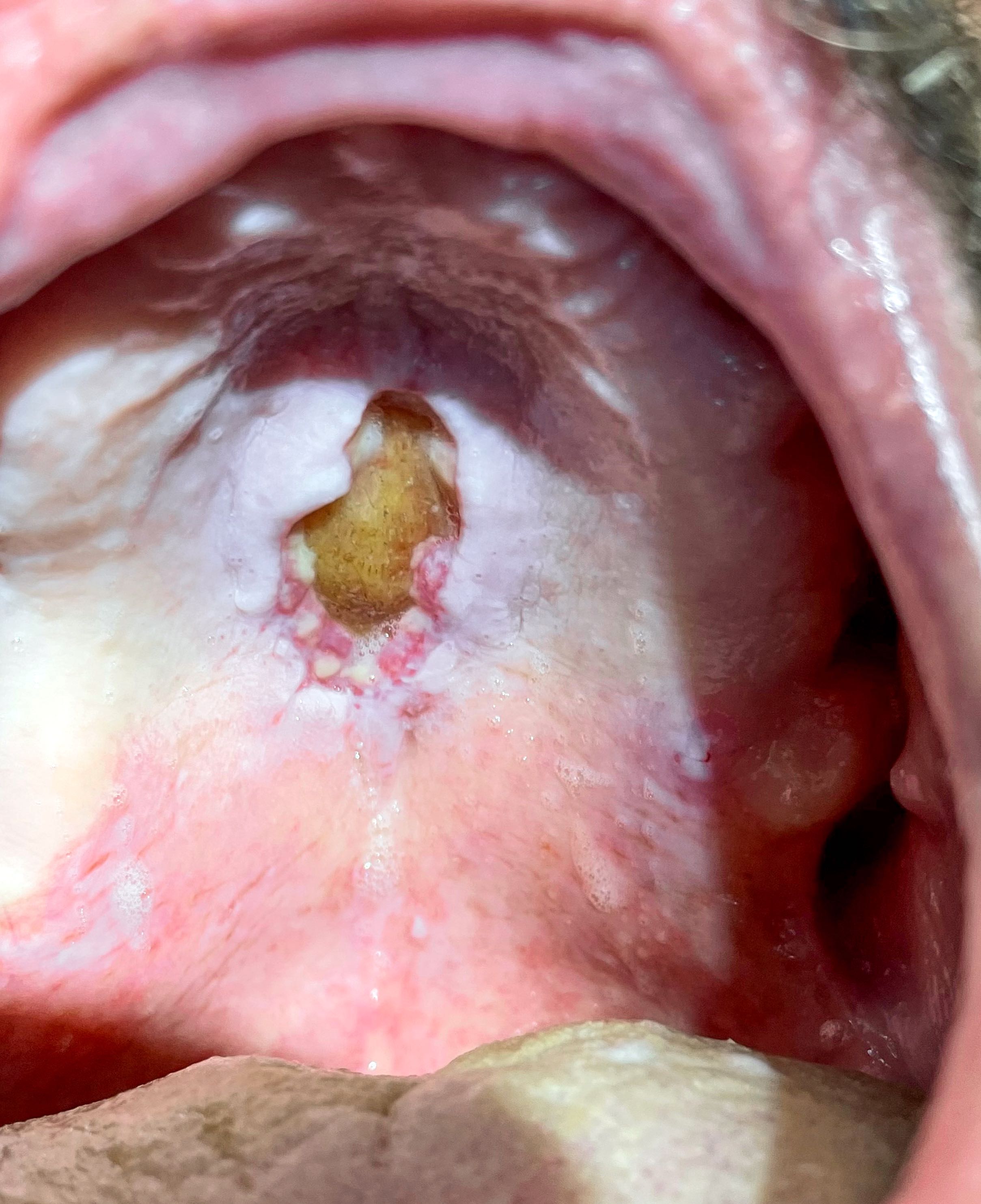
Midline palatal ulceration
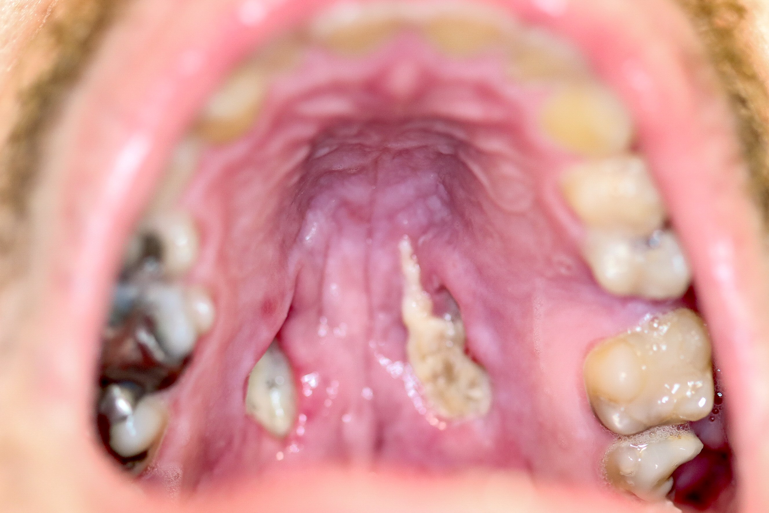

Bilateral palatal ulcerations
Contributed by Kelly Magliocca, D.D.S., M.P.H. (Case #497) and Molly Housley Smith, D.M.D.
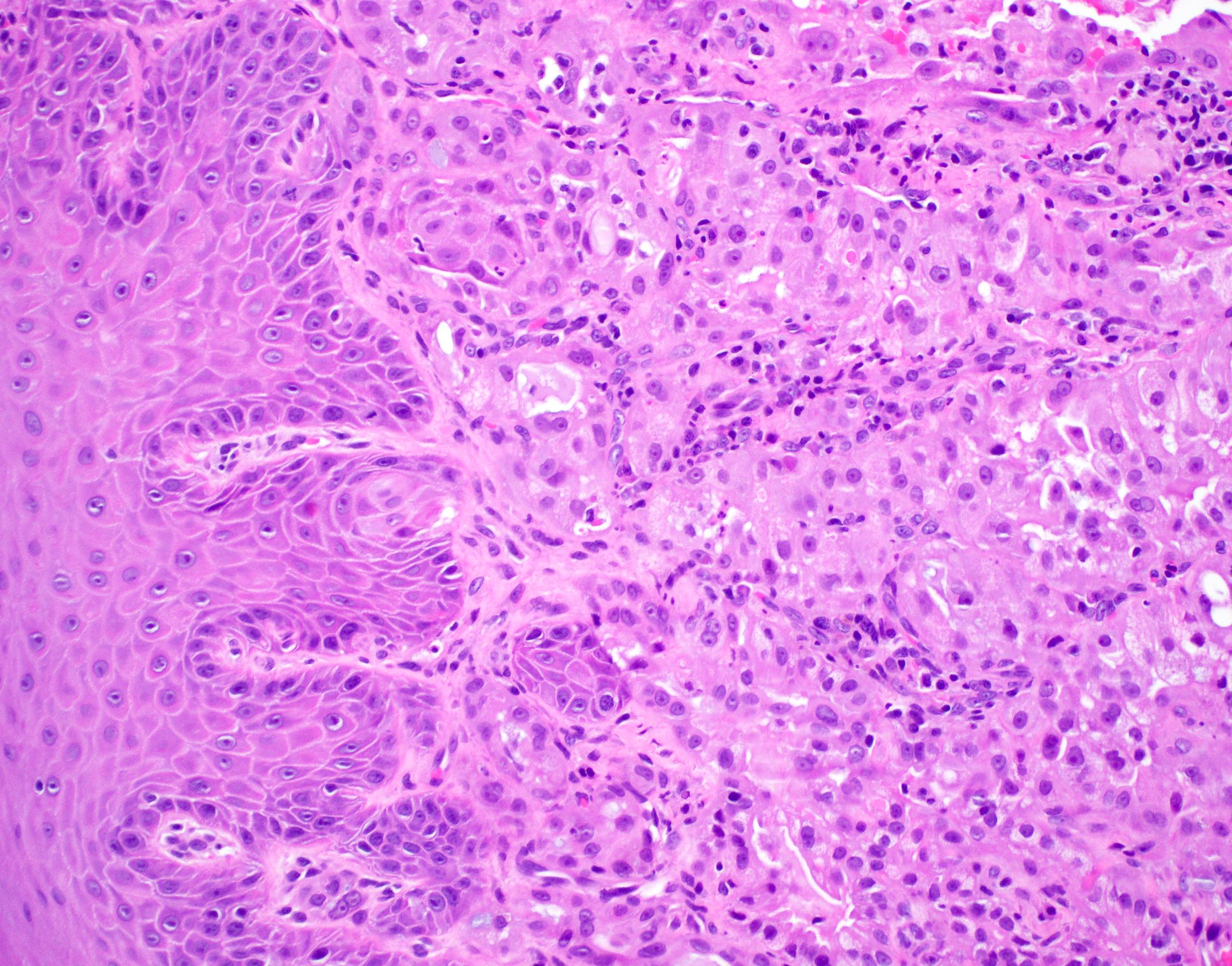
Metaplasia of residual salivary ducts
Pseudoepitheliomatous
hyperplasia

Bland cytology
Spilled mucin

Surface ulceration
Glandular necrosis
Acute inflammation

Healing stage
Contributed by Jen-Fan Hang, M.D.
Pleomorphic adenoma (PA): fibrillary extracellular matrix
PA: plasmacytoid
myoepithelial cells
PA with squamous metaplasia
Warthin tumor
Warthin tumor
Oncocytoma
Benign neoplasm 1
Benign neoplasm 2
Contributed by Jen-Fan Hang, M.D.

SUMP subcategories
Contributed by Jen-Fan Hang, M.D.
Basaloid SUMP


Basaloid SUMP

Oncocytic / oncocytoid SUMP
Spindle cell features

Squamoid features
Images hosted on other servers:

Parotid mass
Contributed by Lulu Sun, M.D., Ph.D. and Rebecca Chernock, M.D.
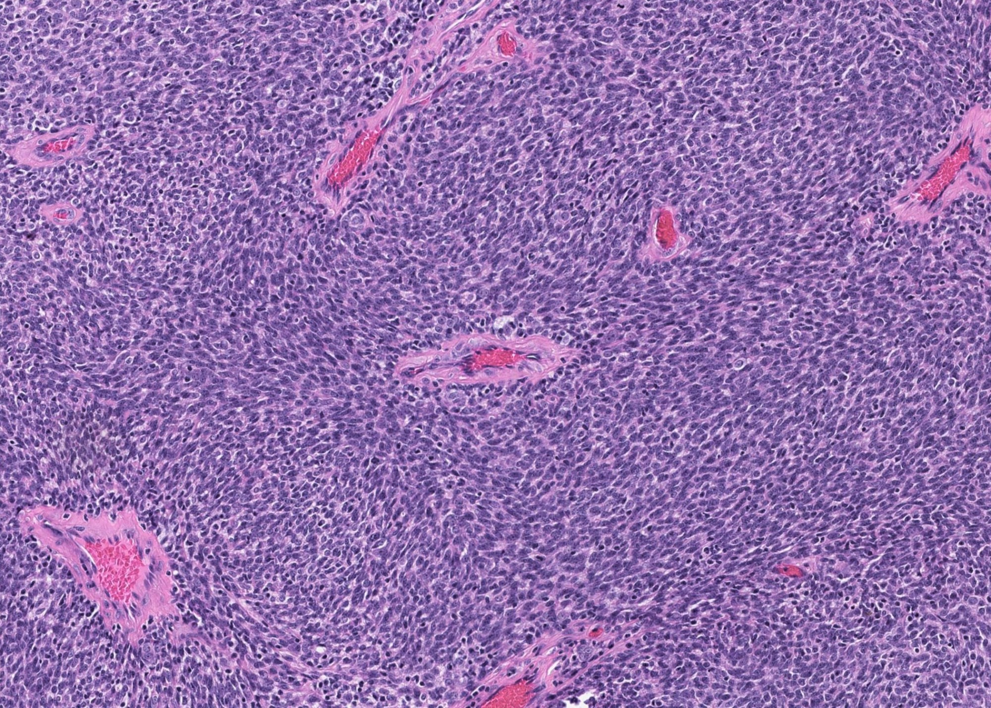
Small cell neuroendocrine carcinoma
Infiltration of salivary parenchyma
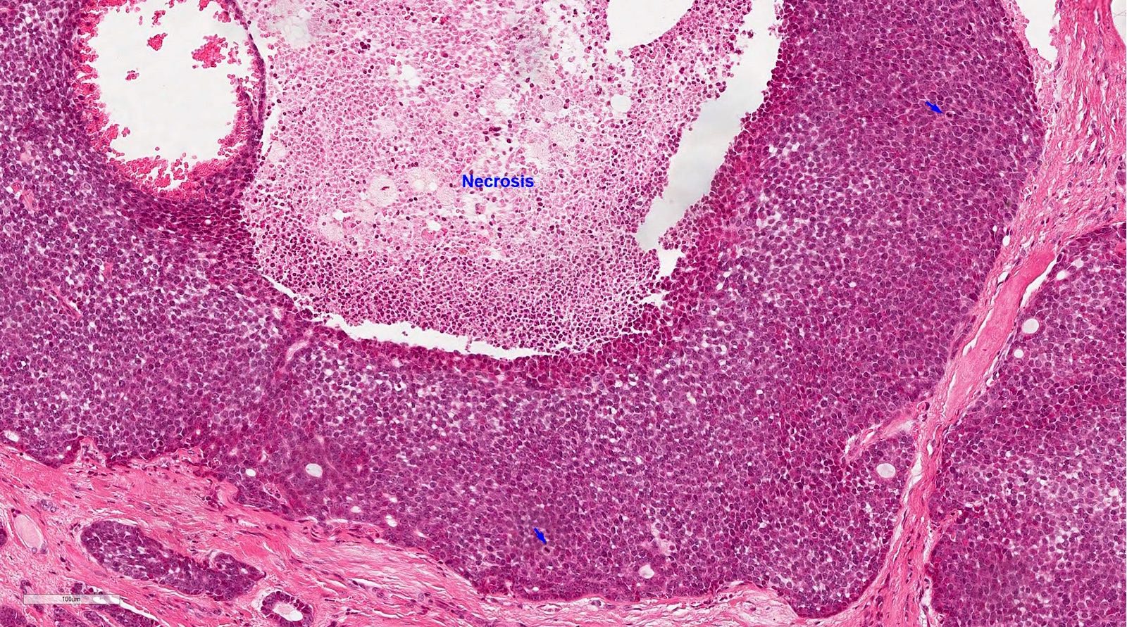
Necrosis
Nuclear features
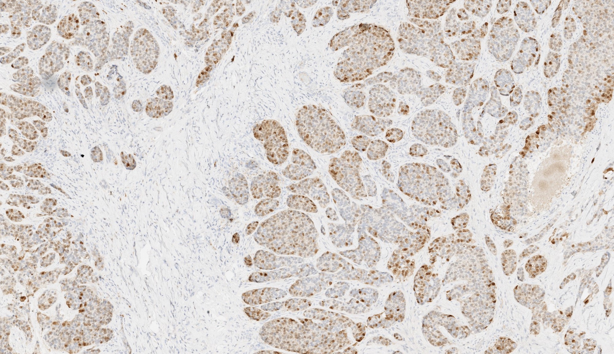
Chromogranin

Synaptophysin
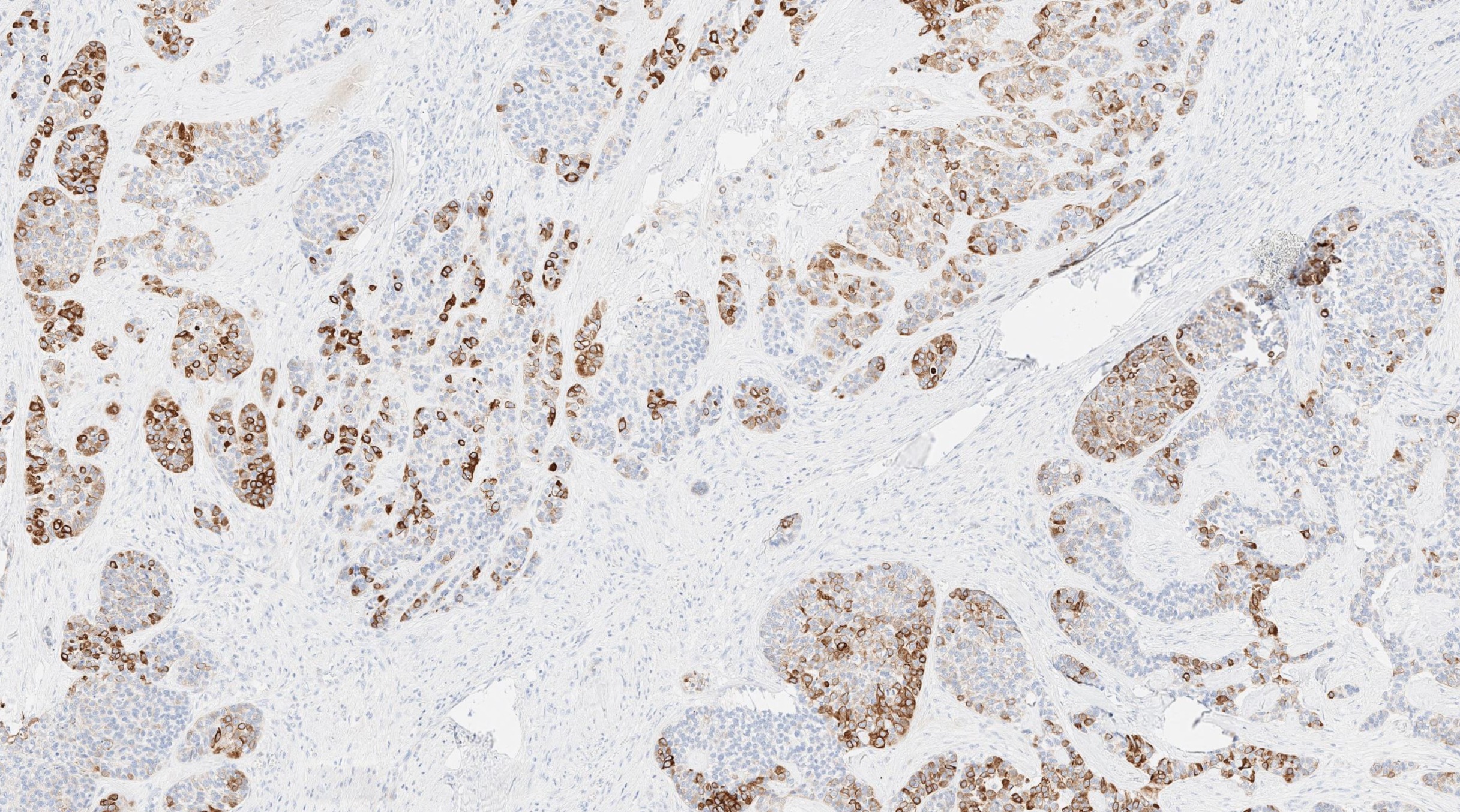
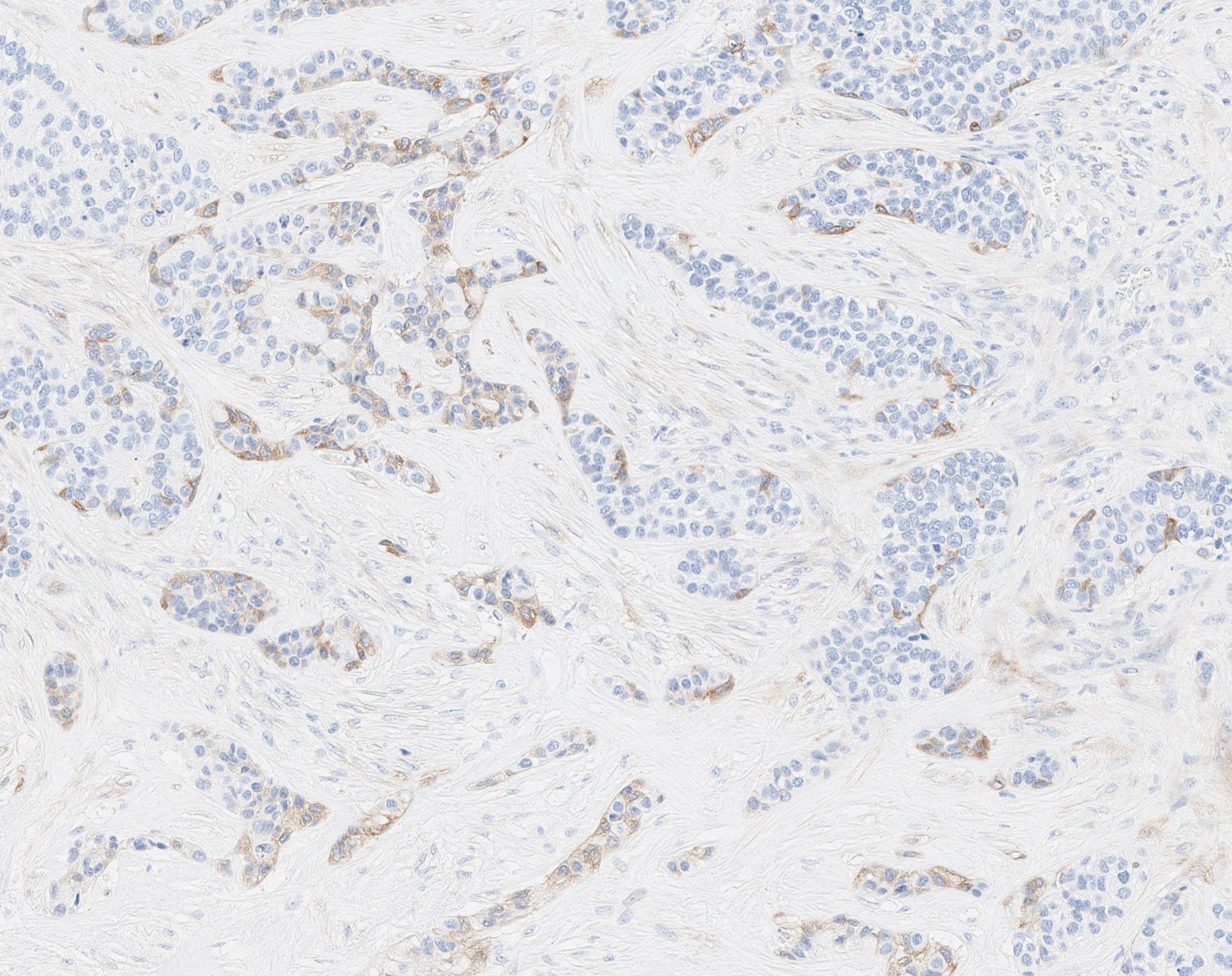
Cytokeratin 5/6
MCPyV IHC
Large cell neuroendocrine carcinoma
Nested architecture
Necrosis
Cytologic features
Images hosted on other servers:

Parotid gland specimen
Images hosted on other servers:


Various images
Contributed by Jen-Fan Hang, M.D.
Nonneoplastic acinar cells
Benign skeletal muscle
Cystic fluid only
Contributed by Jose Manuel Gutierrez Amezcua, M.D. and Tamar C. Brandler, M.D., M.S.

Deep parotid lobe nodule
Solid and cystic parotid lesion
Parotid solid enhancing lesion
Enlarged periparotid lymph node
Contributed by Jose Manuel Gutierrez Amezcua, M.D., Pascale G. Levine, M.D. and Tamar C. Brandler, M.D., M.S.
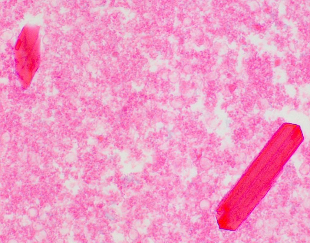
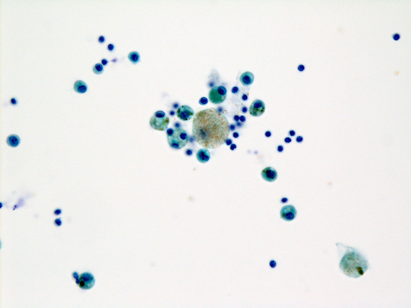
Amylase crystalloids
Cyst contents
Cyst with lymphoid and squamous cells
Benign epithelial cells and lymphocytes
Lymphoepithelial cyst excision
Granulomatous sialadenitis

Giant cell
Acute sialadenitis
Chronic sialadenitis

Reactive lymph node
Images hosted on other servers:


Various images
Contributed by Jen-Fan Hang, M.D.
Clean background
Bland oncocytes
Images hosted on other servers:

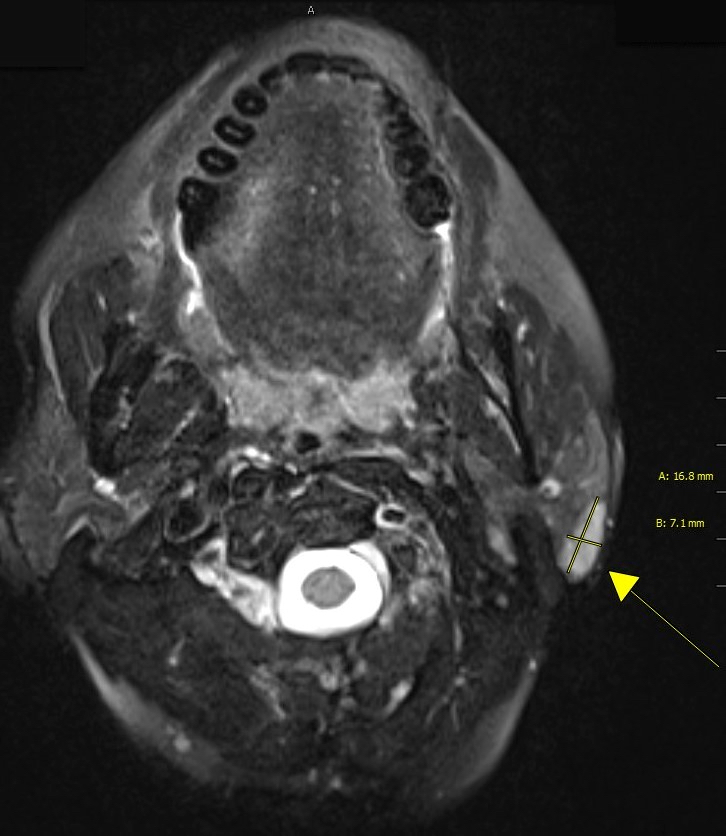
MRI parotid tumor
Images hosted on other servers:

Minor salivary gland tumor

Mass over mandible

Parotid tumor

Large submandibular mass

Parotid tumor
Contributed by Bin Xu, M.D., Ph.D. and Kelly Magliocca D.D.S., M.P.H.


Primary pleomorphic adenoma
Recurrent pleomorphic adenoma

Pleomorphic adenoma
Case #392
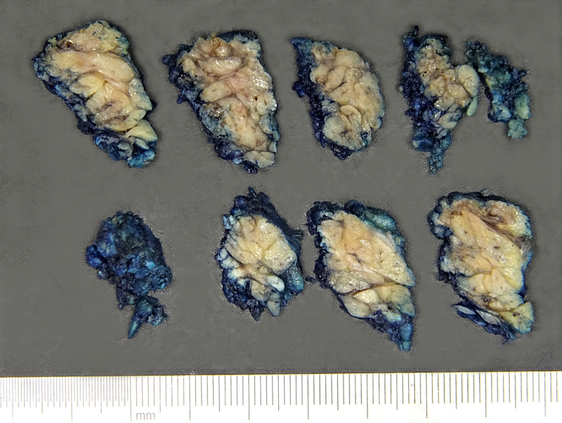
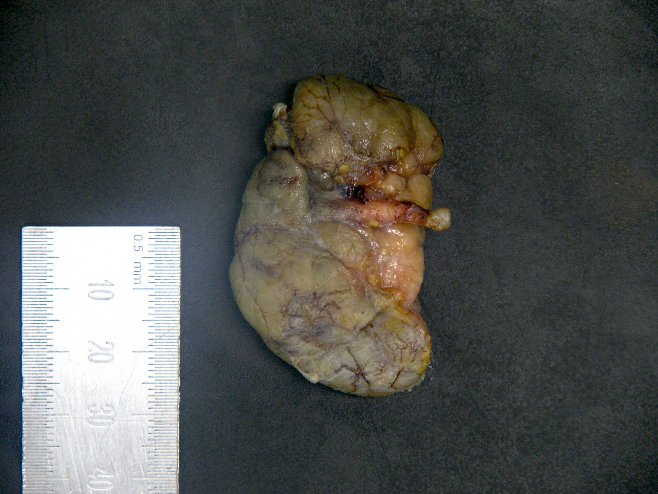
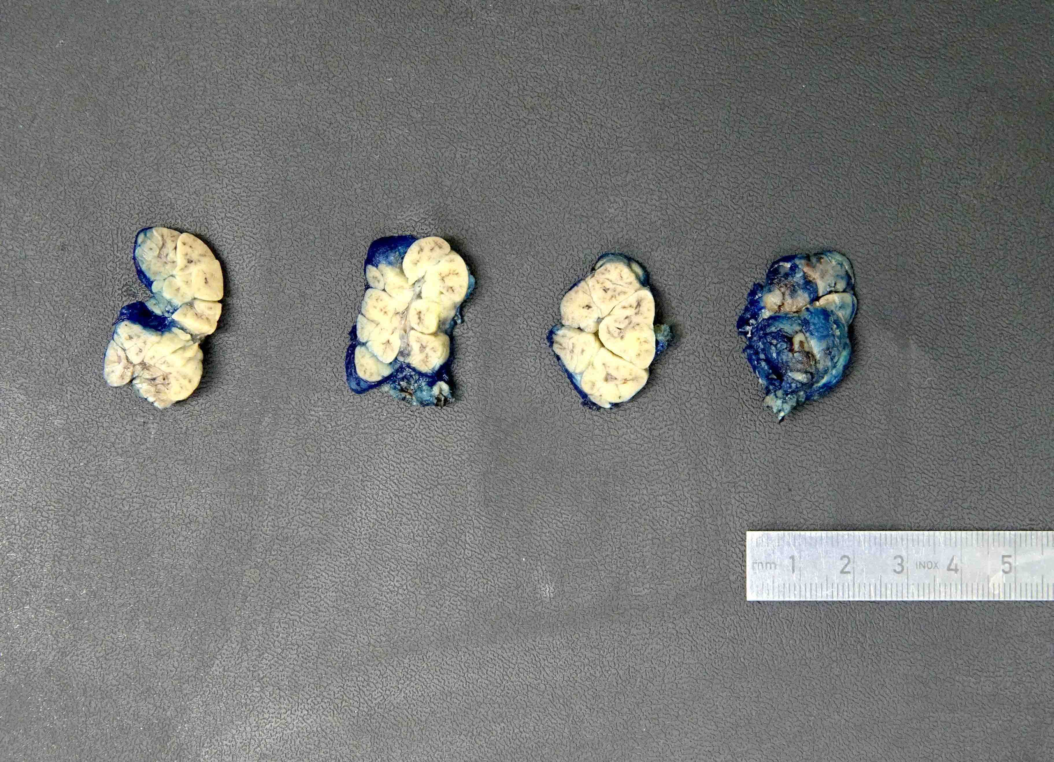
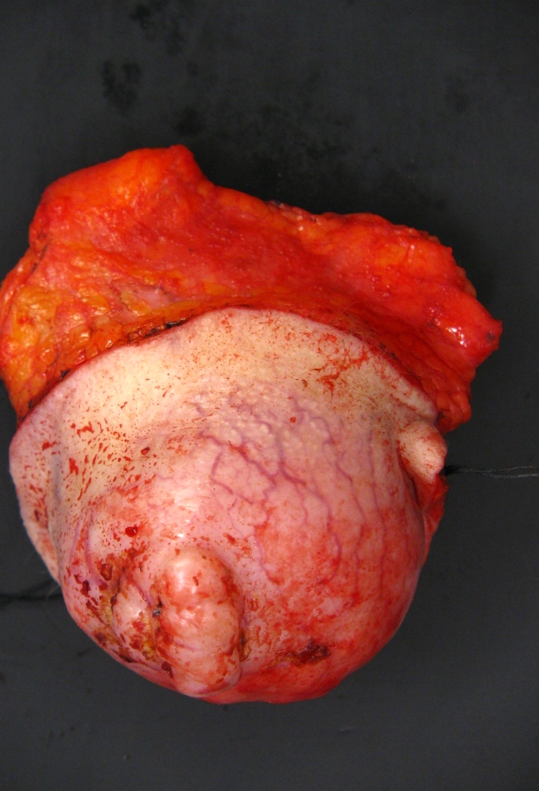
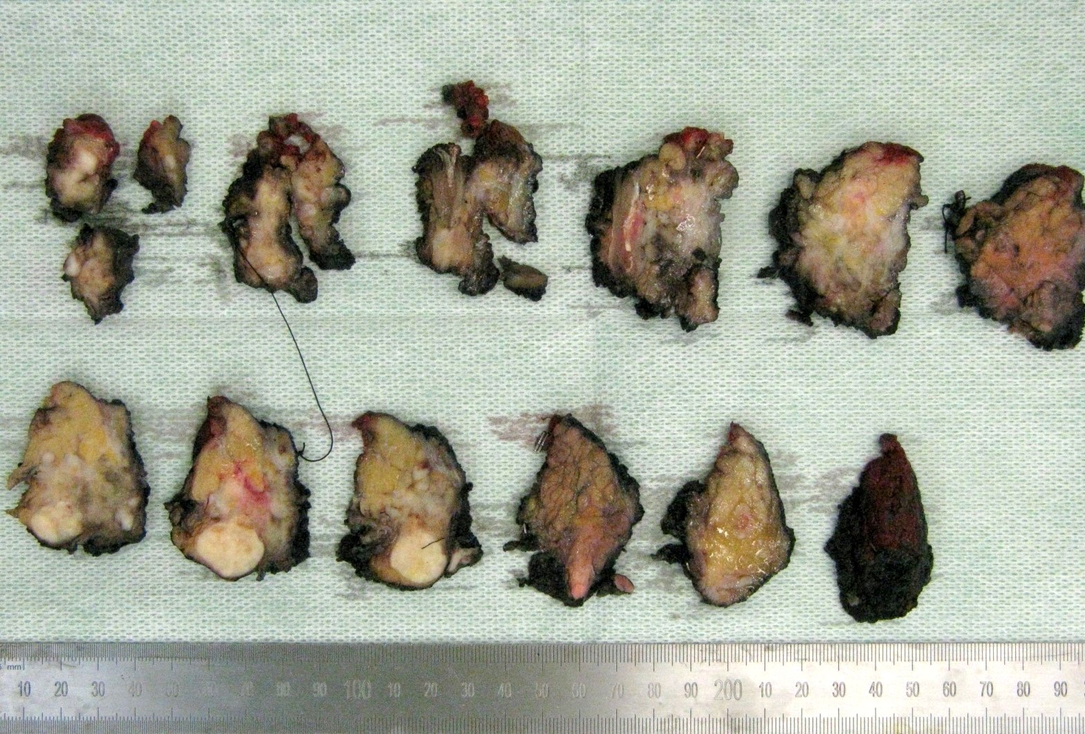
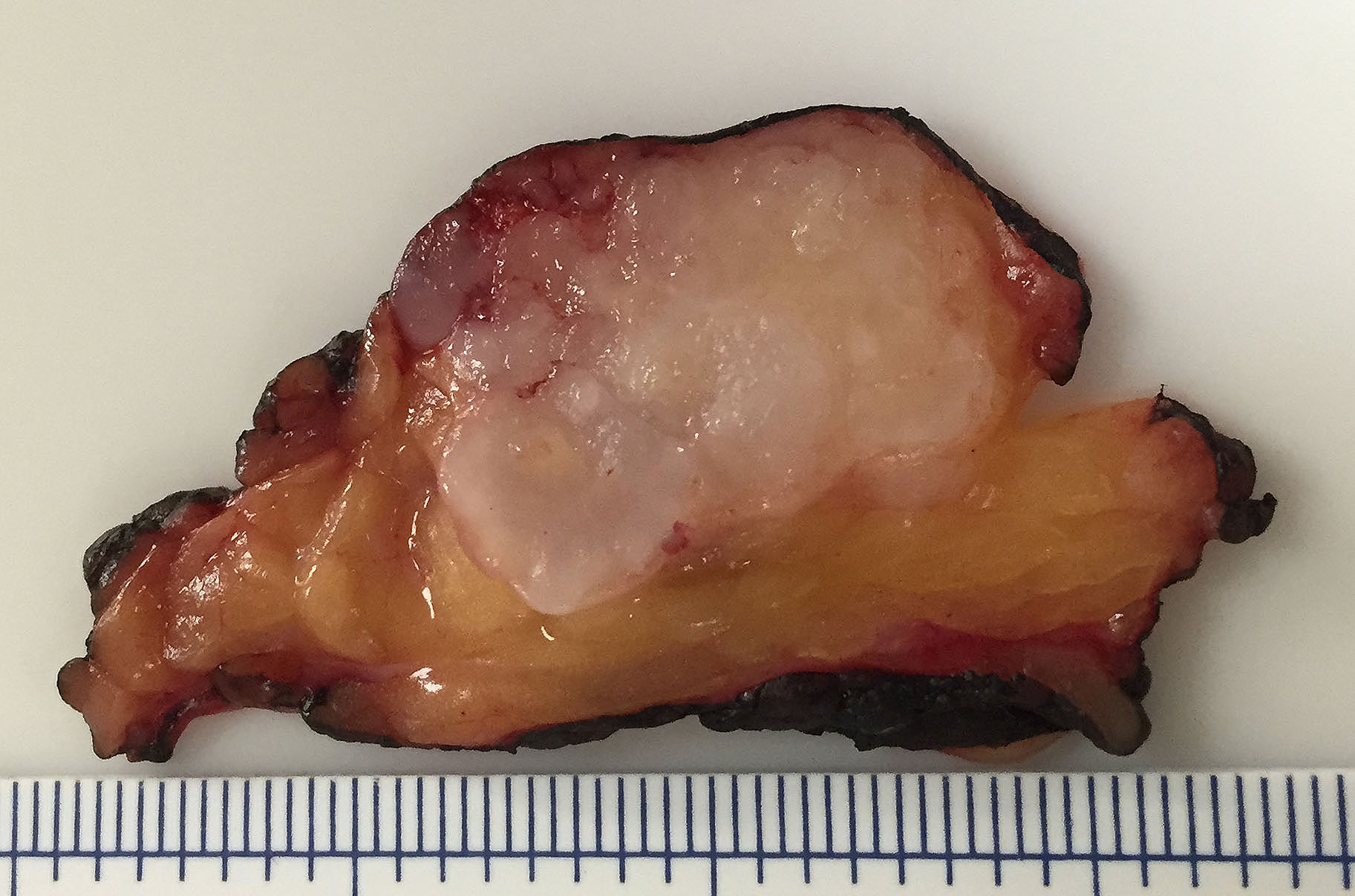
External surface
Cut surface
Contributed by Bin Xu, M.D., Ph.D. and Andrey Bychkov, M.D., Ph.D.

Encapsulated mass

Bosselated interface
Pseudopods
Recurrent pleomorphic adenoma
Triphasic tumor
Clear myoepithelial cells

Plasmacytoid myoepithelial cells

Spindle myoepithelial cells

Squamous and mucinous metaplasia

Adipose and osseous metaplasia
Tyrosine crystals
Biphasic population
Case #404
Pleomorphic adenoma
Myoepithelial cells
Chondroid stroma
Myoepithelial cells
Contributed by Bin Xu, M.D., Ph.D. and Jen-Fan Hang, M.D.
Diff-Quik smear
Papanicolaou stain
Fibrillary extracellular matrix
Plasmacytoid
myoepithelial cells
Squamous metaplasia
Images hosted on other servers:

FISH for PLAG1
Images hosted on other servers:

CT
Images hosted on other servers:

Submucosal mass involving maxilla

Palate nodule
Contributed by Bin Xu, M.D., Ph.D.
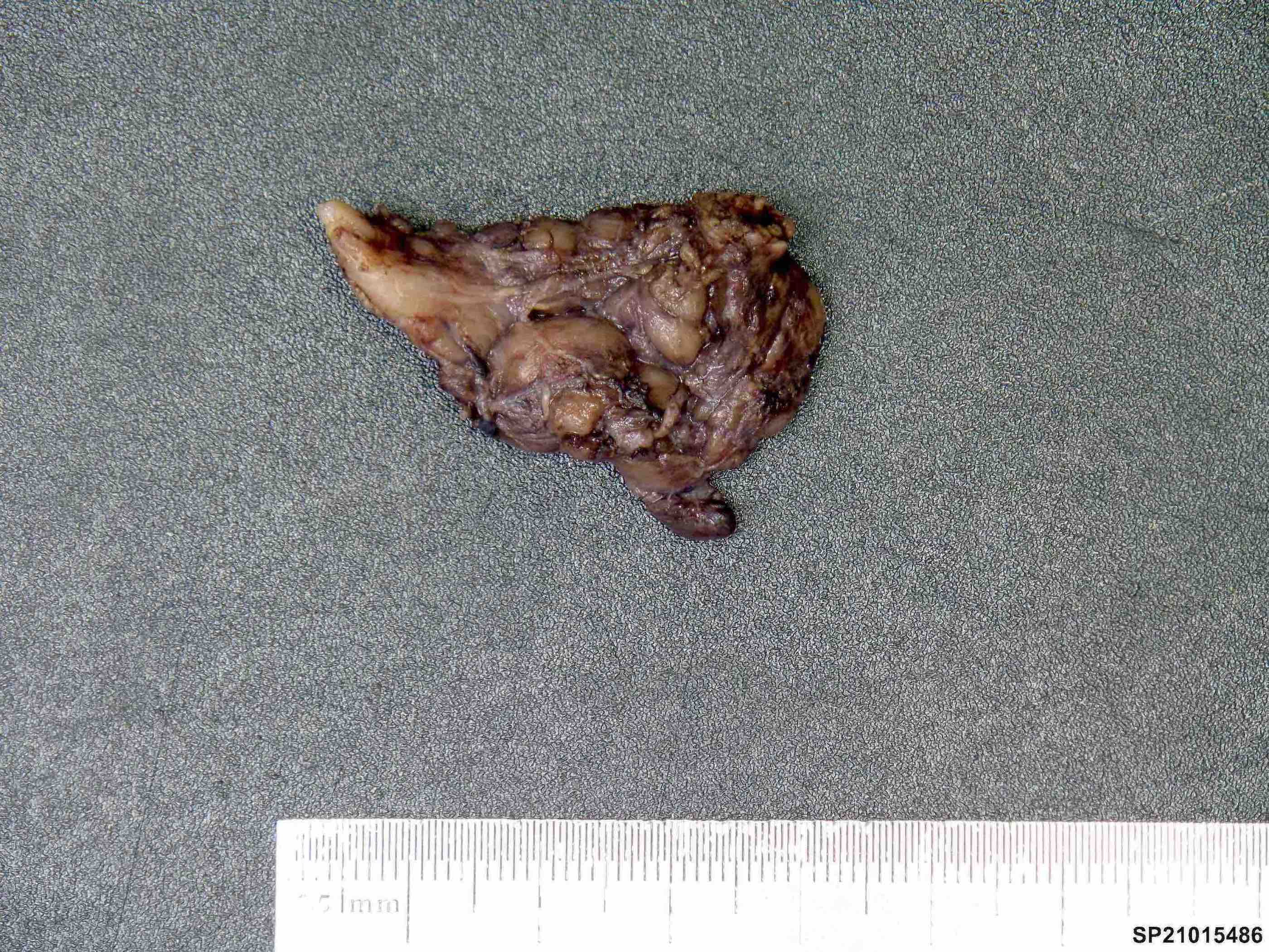
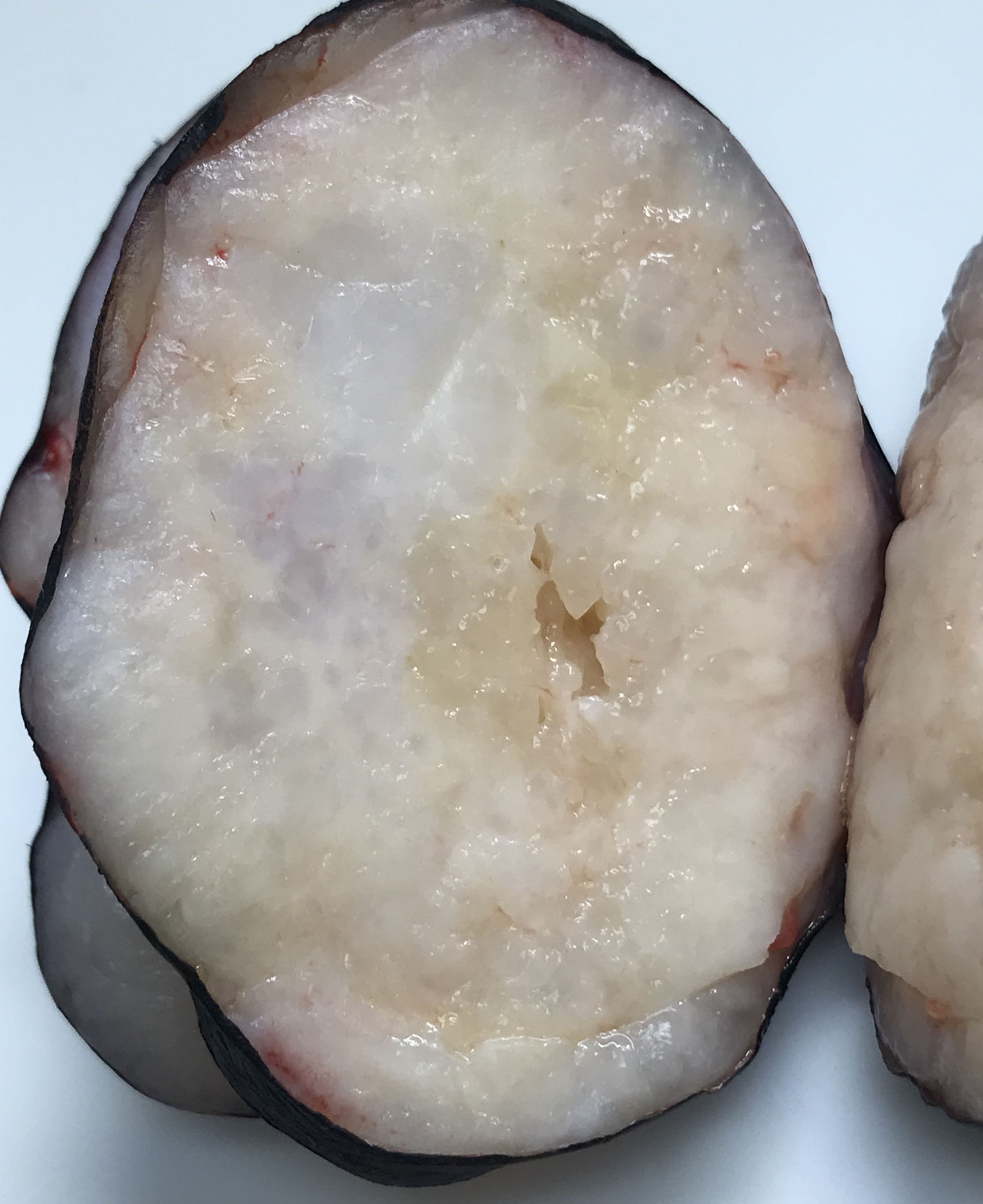
Macroscopic appearance
Contributed by Bin Xu, M.D., Ph.D.
Infiltrative mass
Optical clearing of nuclei

Targetoid arrangement and perineural invasion
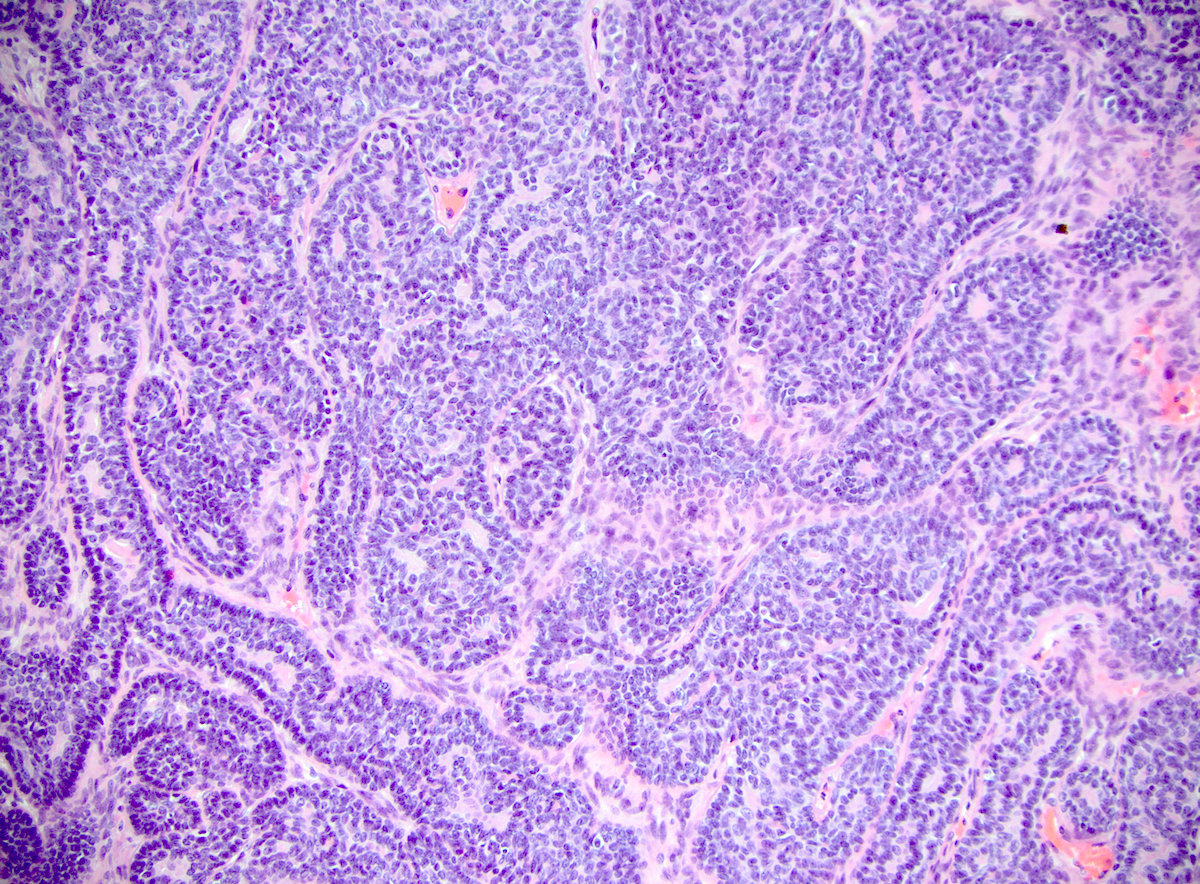
Papillary architecture
Cribriform architecture
Reticular pattern
Solid pattern
Oncocytic changes
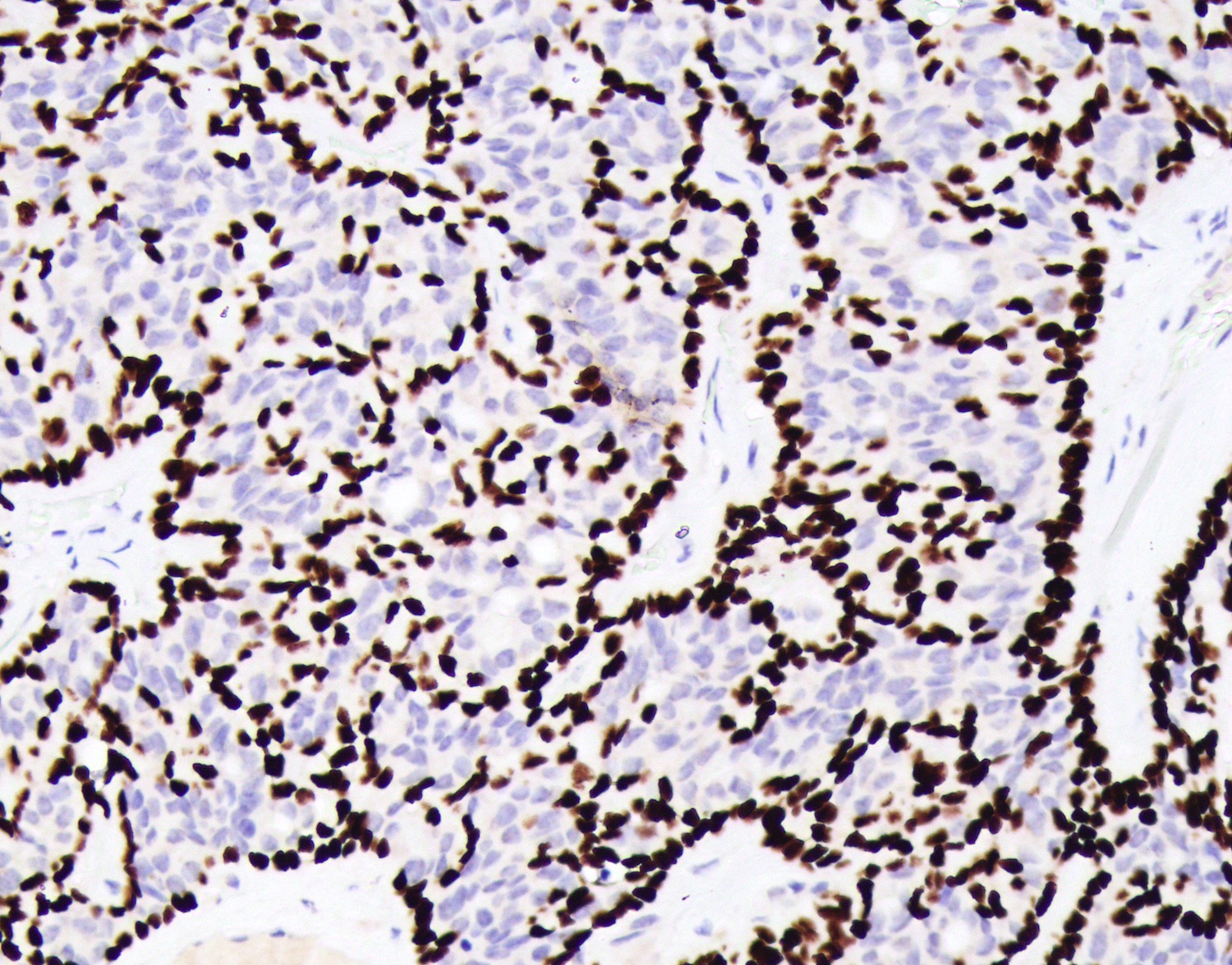
Cribriform adenocarcinoma of salivary gland

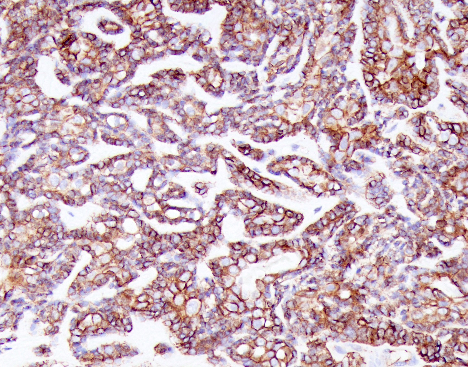
S100

CK7
Images hosted on other servers:

Papanicolaou stain
Images hosted on other servers:

PRKD mutations and fusions
Contributed by Jalal B. Jalaly, M.B.B.S., M.S.
CT cross sectional view
CT coronal view
Images hosted on other servers:

Nasopharyngoscopy
Contributed by Jalal B. Jalaly, M.B.B.S., M.S.
Enlarged intraparotid lymph nodes
Contributed by Jalal B. Jalaly, M.B.B.S., M.S.
Infiltrating nests and glands
Eosinophilic cytoplasm
Comedo necrosis
High grade nuclear cytology
Pleomorphic nuclei
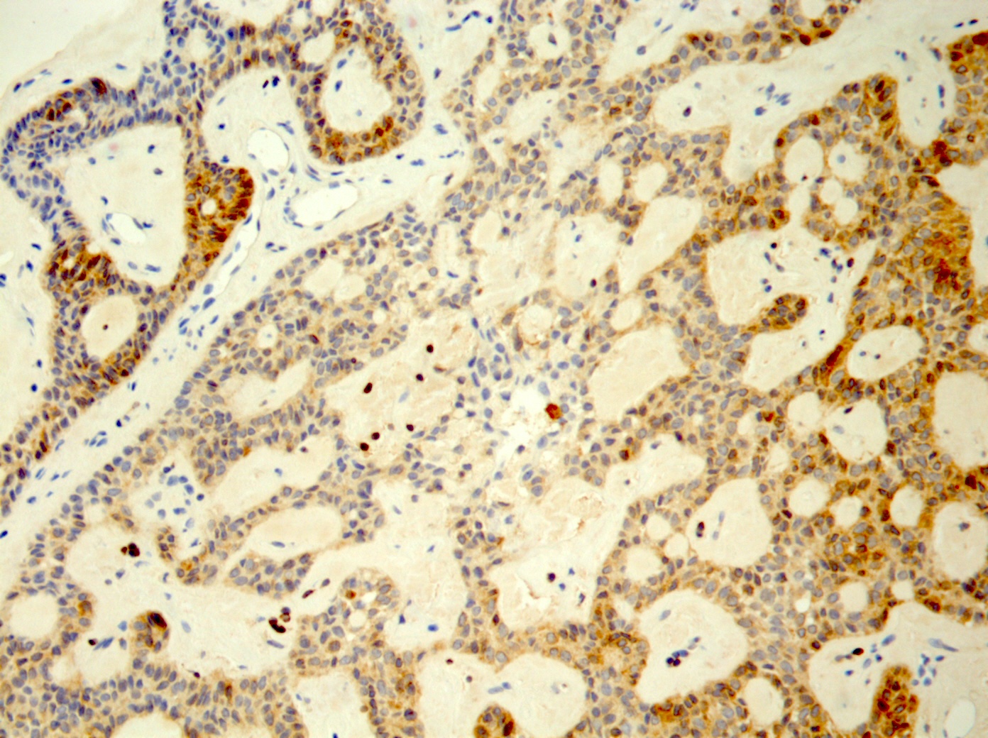
AR+
Carcinoma ex pleomorphic adenoma
AR+
HER2+
Intracytoplasmic mucin
Mucicarmine+
AR+
Contributed by Jalal B. Jalaly, M.B.B.S., M.S. and Jen-Fan Hang, M.D.
Diff-Quik
Pap stain
ThinPrep
Prominent nucleoli
Hyperchromasia
Images hosted on other servers:

Tongue tumor

Floor of mouth mass and enlarged lymph node
Images hosted on other servers:

Ulcerated palate tumor
Images hosted on other servers:

Tongue tumor
Contributed by Marc Pusztaszeri, M.D.
Infiltrative tubules and poorly formed glands
Perineural invasion
Perineural invasion

CK7 and p63
CK5/6 and p40
CK AE1 / AE3 and SOX10
Images hosted on other servers:


Parotid lesion
Images hosted on other servers:

Palate lesion
Contributed by Jiancong Liang, M.D.
Preservation of lobular architecture
Variably sized ducts

Hyperplastic intraluminal epithelium

Acini with eosinophilic granules
Contributed by Ruta Gupta, M.B.B.S., M.D.
Cystic tumor
Solid tumor
Contributed by Ruta Gupta, M.B.B.S., M.D.
Solid architecture
Solid and cystic architecture
Solid architecture
Macrocystic lesion
Papilliform architecture
Colloid-like secretions
Intraluminal secretions
Cytological features
Macrocystic lesion
Diastase resistant secretions
S100
MUC4
Pan-TRK
Contributed by Ruta Gupta, M.B.B.S., M.D.
Cell block
Contributed by Jeffrey Krane, M.D., Ph.D. and Jen-Fan Hang, M.D.
Sheets and clusters cells
Nuclei are uniform
Papillary pattern
Vacuolated cytoplasm
Papillary and secretory
Images hosted on other servers:


FNA cytology of secretory carcinoma
Contributed by Ruta Gupta, M.B.B.S., M.D.
ETV6 rearrangement by FISH
NTRK3 rearrangement by FISH
Salivary gland neoplasms characterized by gene fusions
Case presentations of various salivary gland pathologies
Images hosted on other servers:


Floor of mouth tumor


Mucoepidermoid carcinoma arising in a background of sialadenoma papilliferum
Images hosted on other servers:


Recurrent and metastatic tumor

Nests of basaloid cells
Images hosted on other servers:

ACR / EULAR
classification criteria
for primary Sjögren
syndrome
Images hosted on other servers:

Sialography findings: normal versus Sjögren

Ultrasound findings
suggestive of
primary Sjögren
syndrome
Images hosted on other servers:


Bilateral parotid gland enlargement
Contributed by Alice Ormandy, M.B.B.S., M.P.H. and Salman Marvi, M.D.
Multiple nodular lymphocytic aggregates

Lymphocytic aggregates with germinal centers

Nodular lymphocytic aggregate

Core biopsy parotid

Germinal center


Submandibular gland lymphoid aggregates

Submandibular gland germinal centre
Contributed by Alice Ormandy, M.B.B.S., M.P.H.

Lymphoid clusters

Lymphocytic infiltrate
Contributed by Manish Mahadeorao Bundele, M.B.B.S., M.D. and Rebecca Tian Mei Au, M.B.B.S.
Suspicious for acinic cell carcinoma
Suspicious for adenoid cystic carcinoma
Suspicious for mucoepidermoid carcinoma
Suspicious for large cell lymphoma
Suspicious for metastatic carcinoma
Suspicious for high grade basal cell adenocarcinoma
Contributed by Sapna Balgobind, M.B.B.Ch.
Cut surface
Contributed by Sapna Balgobind, M.B.B.Ch. and @AnaPath10 on Twitter
Parotid gland with
circumscribed
tumor
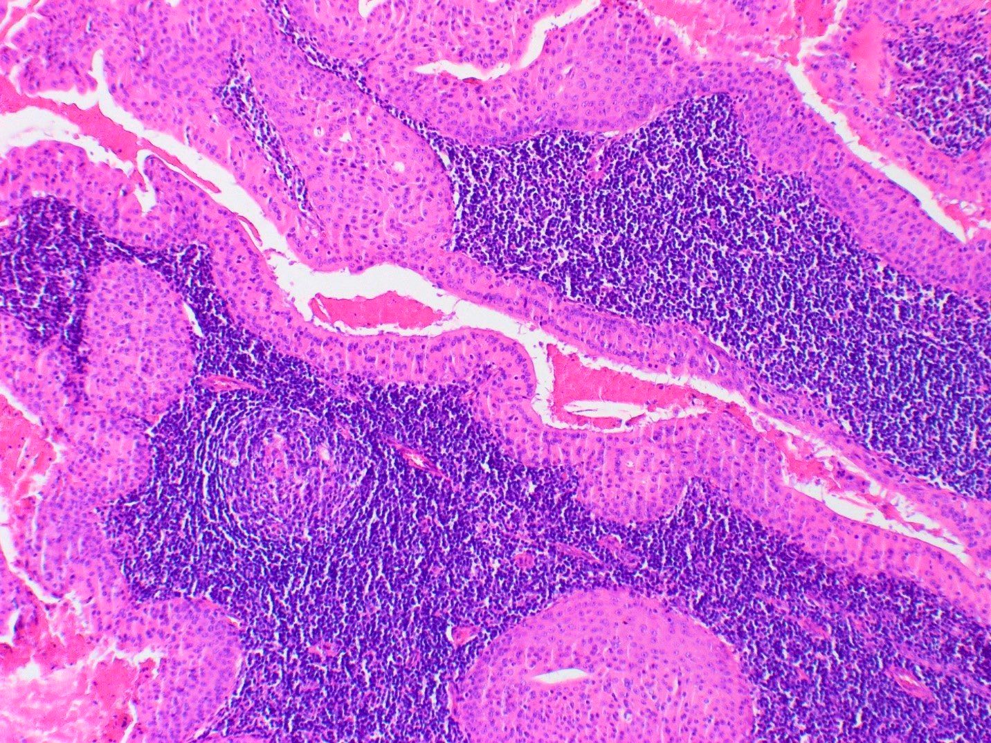
Bilayered oncocytic epithelium with lymphoid background
Mucinous metaplasia
Squamous metaplasia
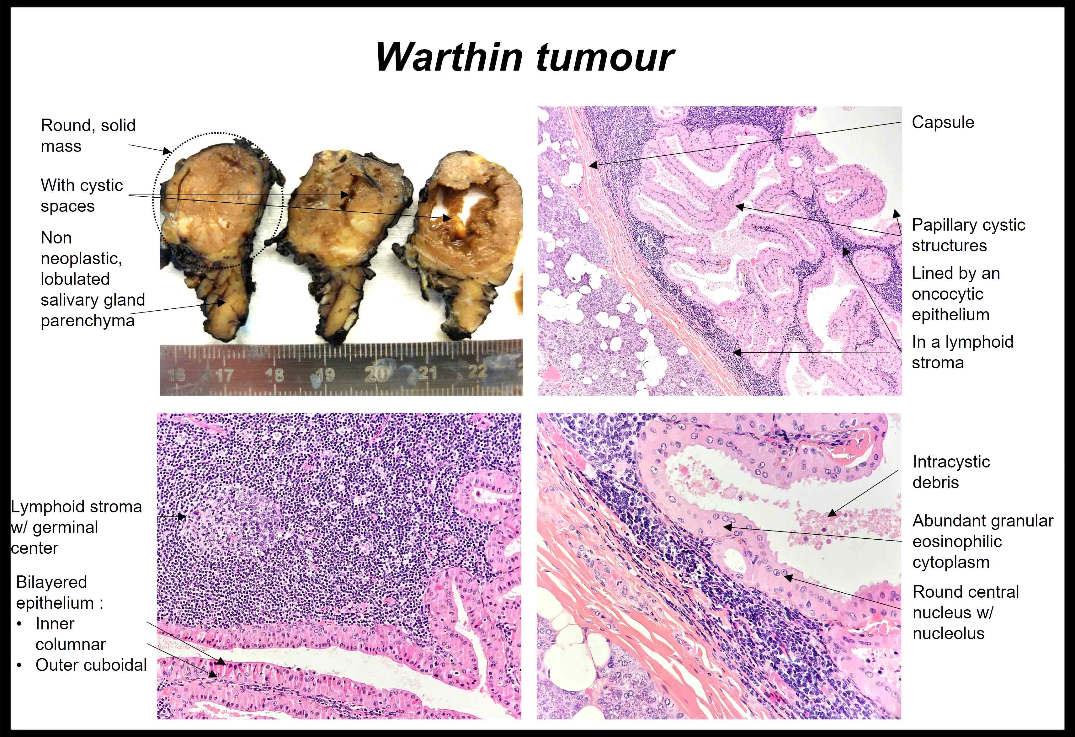
Warthin tumor
p40 staining

p63 staining
Contributed by Sapna Balgobind, M.B.B.Ch.
Oncocytic cells in groups
Oncocytic cells
Proteinaceous debris
Oncocytic cells with dense cytoplasm
Bishop: 2021
Cardesa: 2016
Carlson: 2015
Faquin: 2023
Franchi: 2020

García : 2019
Gnepp: 2021
IARC: 2024
Jiang: 2023
Mody: 2022
Neville: 2023
Stelow: 2020
Thompson: 2022
VandenBussche: 2017
Wenig: 2017
Find related Pathology books: cytopathology, head & neck/endocrine
