
Ear
Middle ear and inner ear tumors - benign / nonneoplastic
Middle ear neuroendocrine tumor
Author: Nat Pernick, M.D.

Last author update: 1 January 2006
Last staff update: 2 May 2023
Copyright: 2003-2024, PathologyOutlines.com, Inc.
PubMed Search: Middle ear adenoma
See Also: Laryngeal neuroendocrine neoplasm, Oropharnygeal neuroendocrine carcinoma, Salivary neuroendocrine carcinoma, Sinonasal neuroendocrine carcinoma, including small cell carcinoma
Table of Contents
Definition / general | Case reports | Treatment | Gross description | Microscopic (histologic) description | Microscopic (histologic) images | Positive stains | Negative stains | Electron microscopy description | Electron microscopy images | Differential diagnosis | Additional referencesCite this page: Pernick N. Middle ear neuroendocrine tumor. PathologyOutlines.com website. https://www.pathologyoutlines.com/topic/earmiddleearadenoma.html. Accessed May 13th, 2024.
Definition / general
- Rare, benign glandular neoplasm originating from middle ear mucosa
- First described in 1976 (Laryngoscope 1976;86:1123, Clin Otolaryngol Allied Sci 1976;1:17)
- Neuroendocrine and epithelial differentiation; also called carcinoid tumor or amphicrine tumor (Ultrastruct Pathol 2001;25:73)
- Rosai believes they form a continuum with carcinoid tumor and could be considered adenocarcinoid tumors
- No gender preference, usually 20's to 40's, but wide age range
- Affects all sites in middle ear
- Occasionally perforates tympanic membrane and extends into external auditory canal
- Not associated with chronic otitis media or cholesteatoma
- Excellent prognosis; rarely is locally aggressive, invades vital structures, has regional metastases (Laryngoscope 2005;115:1660) or causes death
Case reports
- 21 year old man and 34 year old woman with carcinoid tumors of the middle ear (Am J Clin Pathol 1987;87:592)
- 27 year old man and 42 year old woman with middle ear adenoma (J Clin Pathol 1991;44:652)
- 63 year old woman with 1 cm middle ear mass (Case #376)
Treatment
- Complete surgical excision
- Mastoidectomy may be necessary for large lesions
- Recurs with inadequate excision
Gross description
- Gray-white to red-brown, firm/rubbery masses
- Relatively well circumscribed
- Not encapsulated
- No hemorrhage, mean 0.8 cm
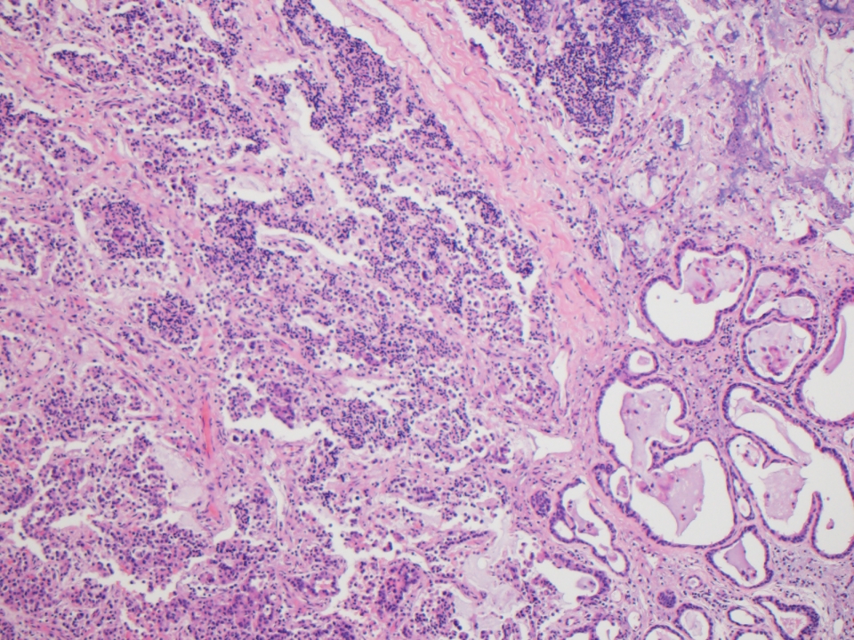
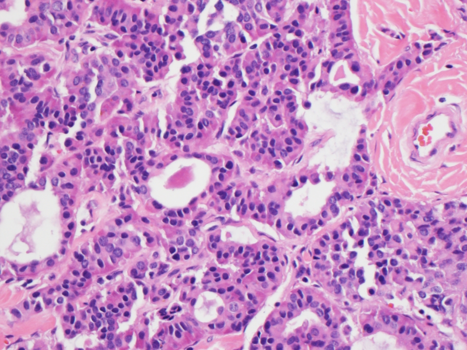
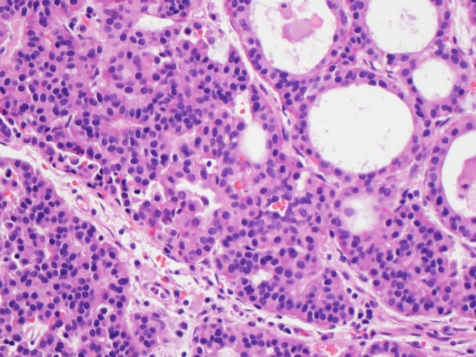
Microscopic (histologic) description
- Variable patterns (sheets, solid, trabecular, cystic, cribriform, glandular, NOT papillary) of glands or tubules composed of uniform single layer of cuboidal or columnar cells with variable eosinophilic cytoplasm and round/oval hyperchromatic nuclei, eccentric nucleoli (if present)
- May appear plasmacytoid, may have significant pleomorphism
- May produce PAS+ mucin
- Sparse fibrous or myxoid stroma
- No/rare mitotic figures, no necrosis
- May have neuroendocrine differentiation morphologically and immunohistochemically
Microscopic (histologic) images
Case #376
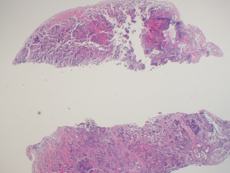
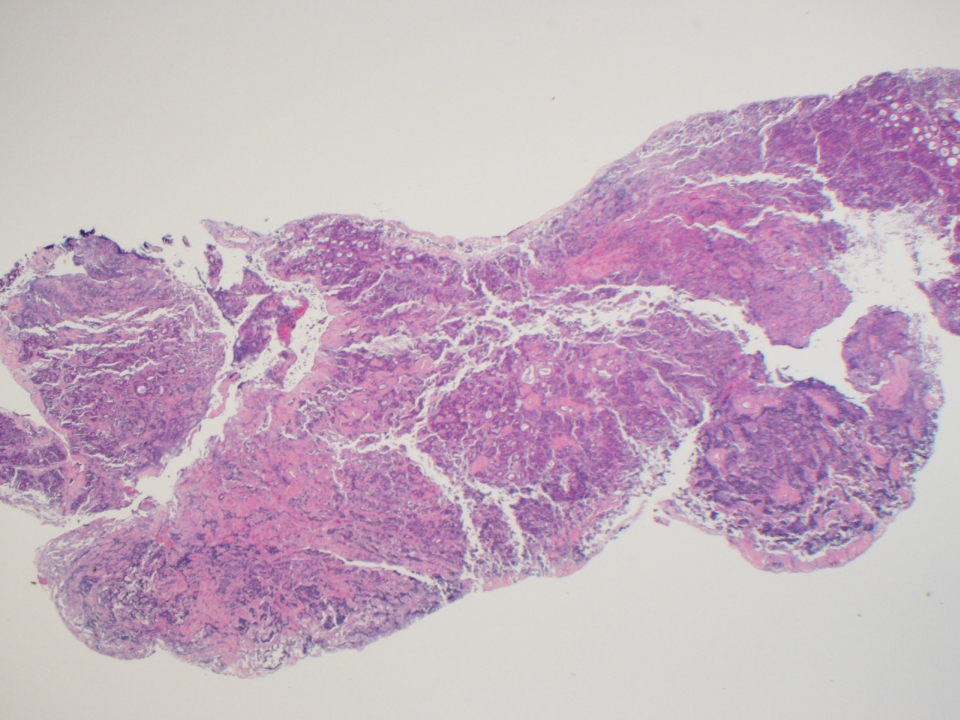
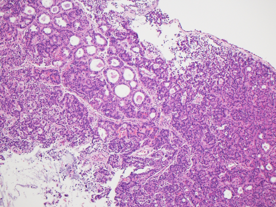
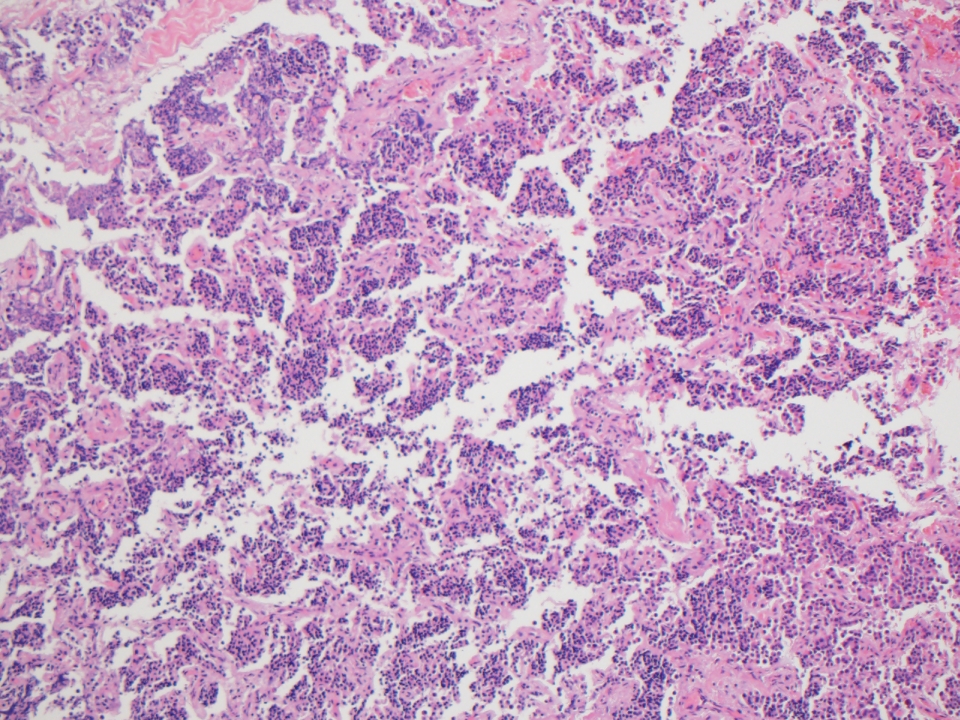


Various images
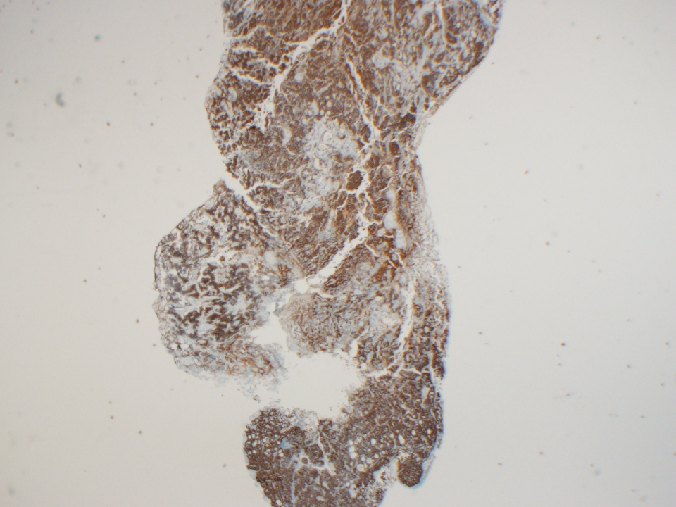
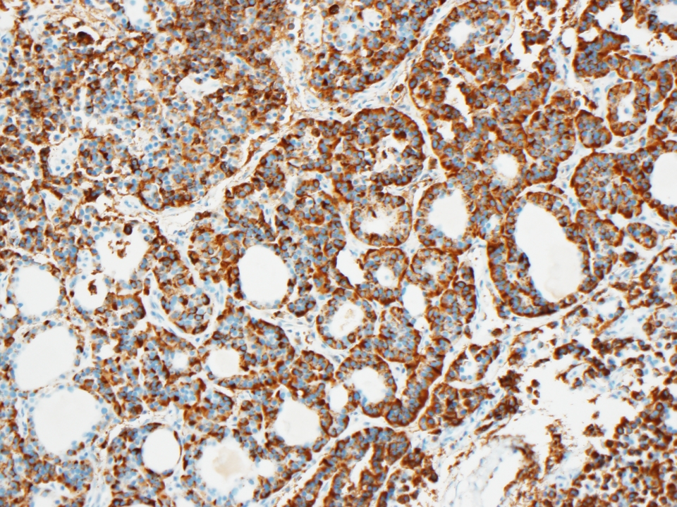
Chromogranin
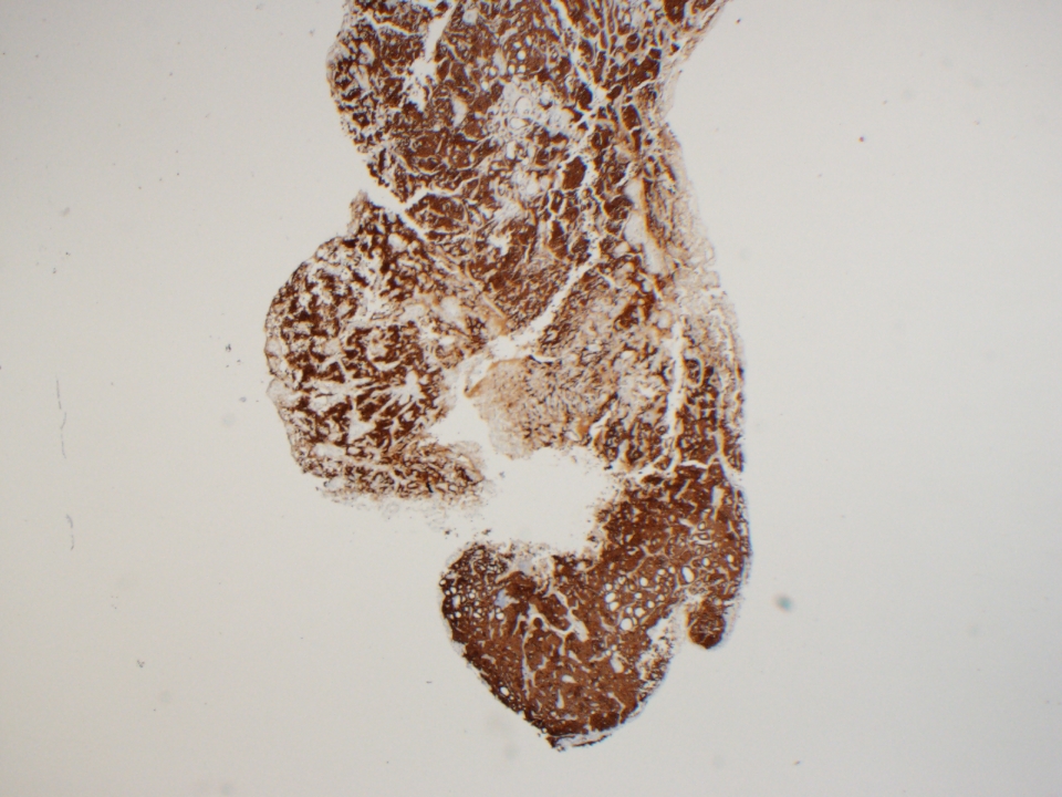

Synaptophysin
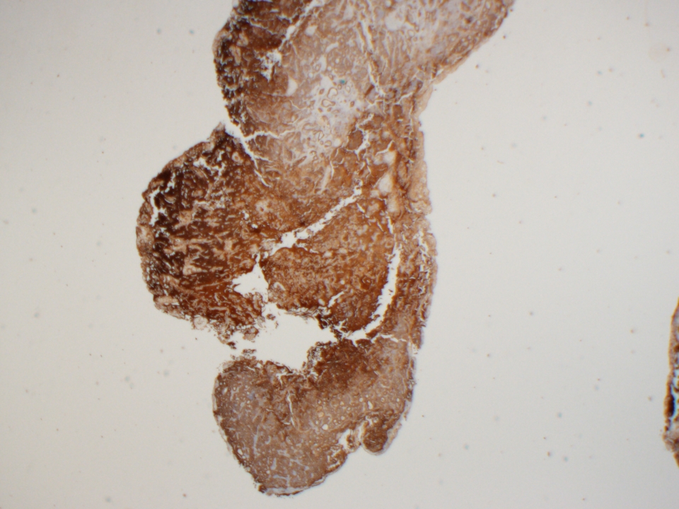

Cytokeratin AE1 / 3
Images hosted on other servers:

Trabeculae and ribbons

Prominent glandular pattern
Positive stains
- Keratin (90%), CK7 (90%), chromogranin (88%), CAM5.2 (81%), mucin (intraluminal), lysozyme, neuron specific enolase (50%), synaptophysin (31%), serotonin (25%), S100 (15%), CK20 (6%, focal)
Negative stains
- Actin
Electron microscopy description
- Desmosomes and microvilli
- Often membrane bound dense core granules
- May have glandular differentiation (Am J Clin Pathol 1985;84:541)
Electron microscopy images
Images hosted on other servers:

Neurosecretory granules
Differential diagnosis
- Acoustic neuroma
- Glandular metaplasia: focal or haphazard in background of chronic otitis media
- Jugulotympanic paraganglioma: zellballen surrounded by S100+ sustentacular cells
- Meningioma
- Middle ear adenocarcinoma: marked pleomorphism, mitotic activity, necrosis, invasion of bone and soft tissue
Additional references
