

Fallopian tubes & broad ligament
Fallopian tube tumor-like lesions
Paratubal cysts
Author: Stephanie L. Skala, M.D.
Editorial Board Member: Gulisa Turashvili, M.D., Ph.D.
Deputy Editor-in-Chief: Jennifer A. Bennett, M.D.

Last author update: 20 September 2021
Last staff update: 18 January 2022
Copyright: 2002-2024, PathologyOutlines.com, Inc.
PubMed Search: Paratubal cysts fallopian tubes


Table of Contents
Definition / general | Essential features | Terminology | ICD coding | Epidemiology | Sites | Pathophysiology | Etiology | Clinical features | Diagnosis | Radiology description | Prognostic factors | Case reports | Treatment | Gross description | Gross images | Microscopic (histologic) description | Microscopic (histologic) images | Virtual slides | Sample pathology report | Differential diagnosis | Board review style question #1 | Board review style answer #1 | Board review style question #2 | Board review style answer #2Cite this page: Skala SL. Paratubal cysts. PathologyOutlines.com website. https://www.pathologyoutlines.com/topic/fallopiantubesparatubalcyst.html. Accessed May 10th, 2024.
Definition / general
- Fluid filled cyst with ciliated lining adjacent to fallopian tube
Essential features
- Ciliated cyst adjacent to fallopian tube
- Typically asymptomatic
- Almost always benign, with rare reports of borderline tumor
Terminology
- Paraovarian cyst
- Hydatid cyst
- Not recommended: hydatid of Morgagni
ICD coding
- ICD-10: N83.8 - other noninflammatory disorders of ovary, fallopian tube and broad ligament
Epidemiology
- Common benign incidental finding (~7 - 10% of women) (J Pediatr Surg 2011;46:2161)
- All age groups, most commonly third to fifth decade
Sites
- Paratubal (between fallopian tube and ovary)
Pathophysiology
- Unknown
Etiology
- Believed to originate from mesothelium or be remnant of Müllerian duct or Wolffian duct
Clinical features
- Most cysts are small and asymptomatic (< 1 to 8 cm; rarely, 20+ cm)
- Size of paratubal cysts may correlate with obesity (J Pediatr Adolesc Gynecol 2017;30:571)
- May be found during surgery or incidentally on radiological study performed for another reason
- Larger lesions may become symptomatic, causing pressure or pain (J Pediatr Surg 2011;46:2161)
- May lead to torsion of adnexa, resulting in acute pain (J Pediatr Surg 2011;46:2161)
Diagnosis
- Typically noted incidentally on intraoperative or gross examination
Radiology description
- While not often diagnosed on imaging, paratubal cysts are unilocular and anechoic or hypoechoic on ultrasound (J Clin Endocrinol Metab 2000;85:1021)
Prognostic factors
- Paratubal cysts are benign
- Rarely gives rise to serous borderline tumor or even more rarely malignancy (Appl Immunohistochem Mol Morphol 2017;25:e21)
Case reports
- 15 year old girl with giant paratubal serous cystadenoma (J Pediatr Adolesc Gynecol 2020;33:438)
- 17 year old girl with paratubal borderline tumor (J Pediatr Adolesc Gynecol 2016;29:74)
- 34 year old nulligravid woman with paratubal cyst and bilateral hydrosalpinges diagnosed during evaluation of infertility (Taiwan J Obstet Gynecol 2014;53:239)
Treatment
- Surgical excision of the paratubal cyst represents definitive treatment for symptomatic patients (J Clin Endocrinol Metab 2000;85:1021)
- Benign follow up is expected; treatment is not required for asymptomatic patients
Gross description
- Simple fluid filled cyst(s) near fallopian tube
Gross images
Images hosted on other servers:

Benign appearing cyst
Microscopic (histologic) description
- Simple fluid filled cyst lined by ciliated tubal type epithelium
- Focal papillary projections may be seen
Microscopic (histologic) images
Contributed by Stephanie L. Skala, M.D.
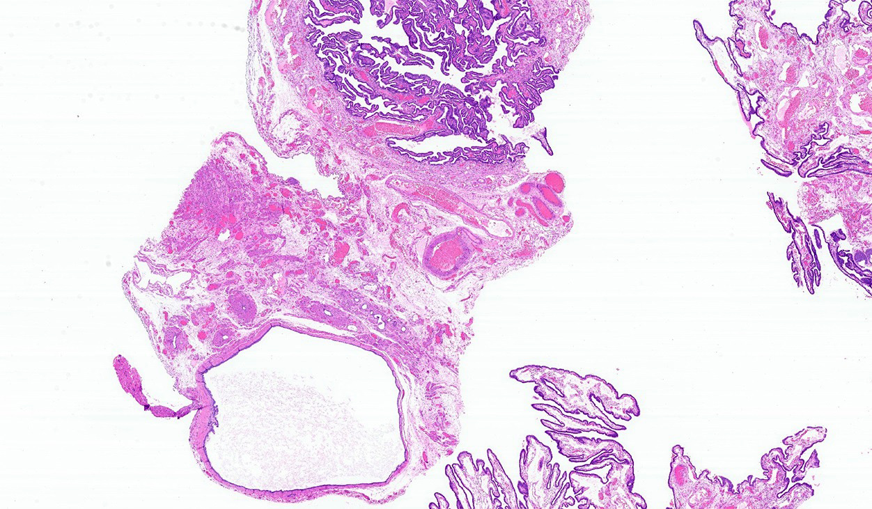
Simple cyst near tube
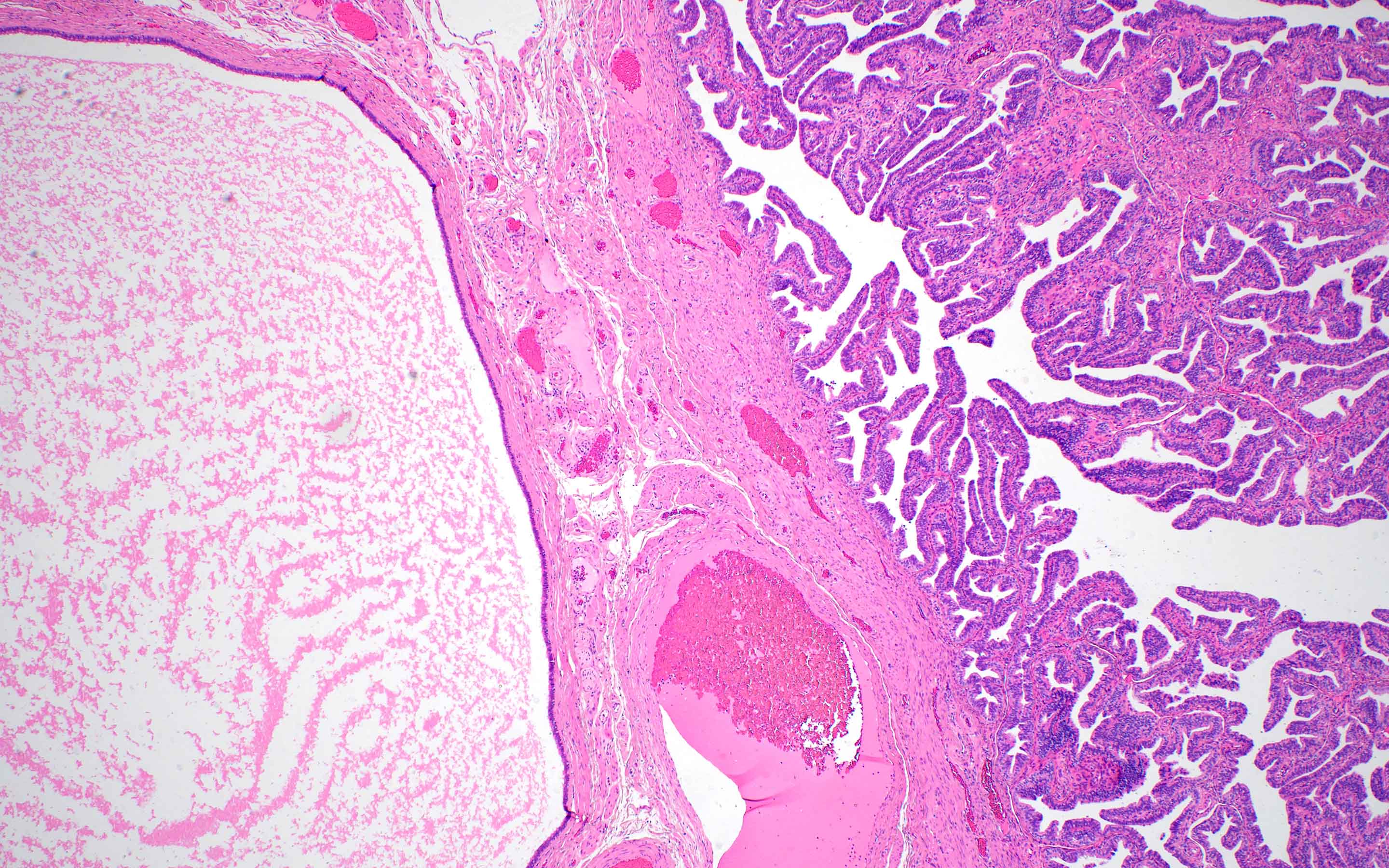
Simple cyst adjacent to fallopian tube
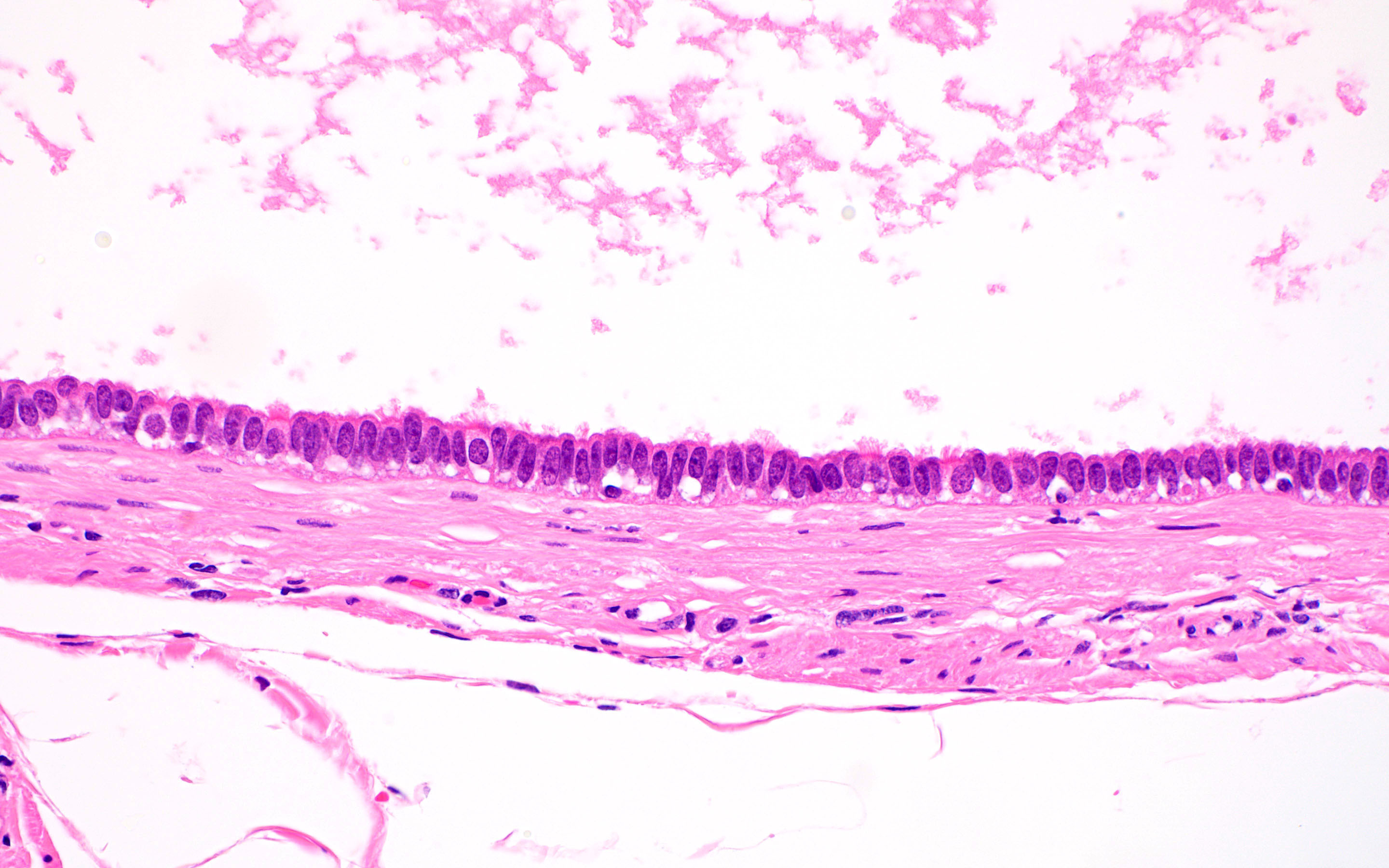
Bland, ciliated, tubal type epithelium
Virtual slides
Images hosted on other servers:

Benign fallopian tube with simple paratubal cysts
Sample pathology report
- Fallopian tubes, bilateral salpingectomy:
- Benign fallopian tubes with paratubal cysts
Differential diagnosis
- Endometriotic cyst:
- Associated with endometrial type stroma with or without hemosiderin laden macrophages
- Serous cystadenoma:
- Histologically identical to paratubal cyst but > 1 cm in size
- Hydrosalpinx:
- Dilation of the fallopian tube lumen with attenuation of the tubal epithelium with or without diminished plicae
- Distinction based largely on location of the cystic space within rather than near the fallopian tube
Board review style question #1

Upon review of a salpingectomy specimen, the lesion shown above is identified next to the fallopian tube. What is the expected outcome?
- Benign follow up
- Local recurrence
- Metastatic disease
- Progression to carcinoma
Board review style answer #1
Board review style question #2
Which of the following is true about paratubal cysts?
- Large paratubal cysts may cause torsion
- Large paratubal cysts often progress to borderline tumors
- Paratubal cysts are frequently diagnosed on MRI
- Paratubal cysts are an unusual finding
Board review style answer #2
