

Ovary
Germ cell tumors
Teratoma-mature
Editorial Board Member: Gulisa Turashvili, M.D., Ph.D.
Deputy Editor-in-Chief: Jennifer A. Bennett, M.D.


Last author update: 10 August 2021
Last staff update: 3 April 2024
Copyright: 2003-2024, PathologyOutlines.com, Inc.
PubMed Search: Mature teratoma
Table of Contents
Definition / general | Essential features | Terminology | ICD coding | Epidemiology | Sites | Pathophysiology | Etiology | Diagrams / tables | Clinical features | Diagnosis | Radiology description | Radiology images | Prognostic factors | Case reports | Treatment | Gross description | Gross images | Frozen section description | Microscopic (histologic) description | Microscopic (histologic) images | Virtual slides | Molecular / cytogenetics description | Sample pathology report | Differential diagnosis | Board review style question #1 | Board review style answer #1 | Board review style question #2 | Board review style answer #2Cite this page: Welter SM, Khalifa MA. Teratoma-mature. PathologyOutlines.com website. https://www.pathologyoutlines.com/topic/ovarytumorteratomamature.html. Accessed April 26th, 2024.
Definition / general
- Benign tumor of the ovary composed of mature tissue representing at least 2 embryonic layers (ectoderm, mesoderm or endoderm)
Essential features
- Mature tissue representing at least 2 embryonic layers
- If malignant transformation, prognosis is related to the type of malignancy
Terminology
- Mature cystic teratoma
- Not recommended: dermoid cyst, mature solid teratoma
Epidemiology
- Most common ovarian tumor (20% of all ovarian tumors, 95% of all ovarian germ cell tumors)
- Reproductive age
- 10% bilateral (Cancer 1971;27:343)
Sites
- Ovary
Pathophysiology
- Most widely accepted origin is from the primordial germ cell (Mod Pathol 2017;30:1467)
Etiology
- No known causative agents
Diagrams / tables

Clinical features
- Slow growing, often incidental finding
- Rarely present with or are associated with:
- Torsion or rupture
- Mucinous tumors of the ovary (e.g., mucinous cystadenoma) (Clin Diagn Res 2016;10:ED11)
- Autoimmune hemolytic anemias (Saudi Med J 2019;40:397)
- Paraneoplastic encephalitis (Ann Neurol 2007;61:25)
Diagnosis
- Mainly by ultrasound
- Histologic examination
Radiology description
- Wide variety of presentations, depending on the tissues present (see Diagrams / tables)
Radiology images
Images hosted on other servers:


Ultrasound characteristics


CT characteristics

MRI characteristics
Prognostic factors
- Benign tumor
- Malignant transformation occurs rarely
- Usually in older patients
- Most common is squamous cell carcinoma
- Prognosis dependent on stage
- Microscopic foci of immature neuroepithelium does not warrant diagnosis of immature teratoma and will not affect prognosis (Int J Gynecol Pathol 1987;6:203)
- Gliomatosis peritonei (mature glial tissue implanted on peritoneal surface) does not adversely affect prognosis (J Ovarian Res 2016;9:45)
- Anti–n-methyl-D-aspartate receptor (NMDAR) encephalitis will have best outcome with early treatment (immunotherapy and tumor removal) (Lancet Neurol 2013;12:157)
Case reports
- 15 year old girl with meningioma arising in a mature cystic teratoma (Cancer Imaging 2020;20:15)
- 20 year old woman with mature teratoma in ectopic ovary (J Minim Invasive Gynecol 2019;26:348)
- 22 year old woman with secondary retroperitoneal mature teratoma 4 years after resection of mature teratoma of the ovary (Radiol Case Rep 2019;14:692)
- 59 year old woman with squamous cell carcinoma in a mature teratoma of ovary (BMC Cancer 2019;19:217)
- 71 year old woman with ovarian clear cell carcinoma arising in a mature cystic teratoma (J Ovarian Res 2018;11:74)
Treatment
- Cystectomy in young women to preserve fertility
- Salpingo-oophorectomy in older women
Gross description
- Smooth cyst that may contain hair, teeth, cartilage, bone or sebaceous material
- Generally < 10 cm
- Raised protuberance in cyst wall (Rokitansky nodule)
- Reference: StatPearls: Cystic Teratoma [Accessed 29 July 2021]
Gross images
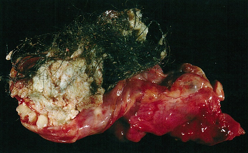
Contributed by Shannon Mingo Welter, M.D., AFIP and @Andrew_Fltv on Twitter
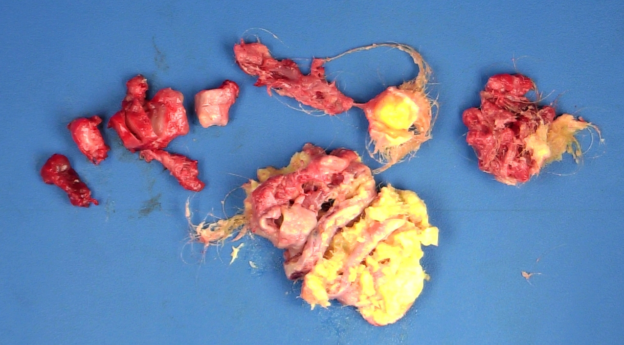
Fragmented ovarian mature teratoma

Ovarian mature teratoma
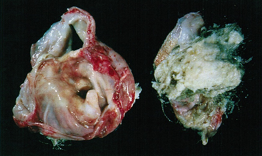
Mature teratoma
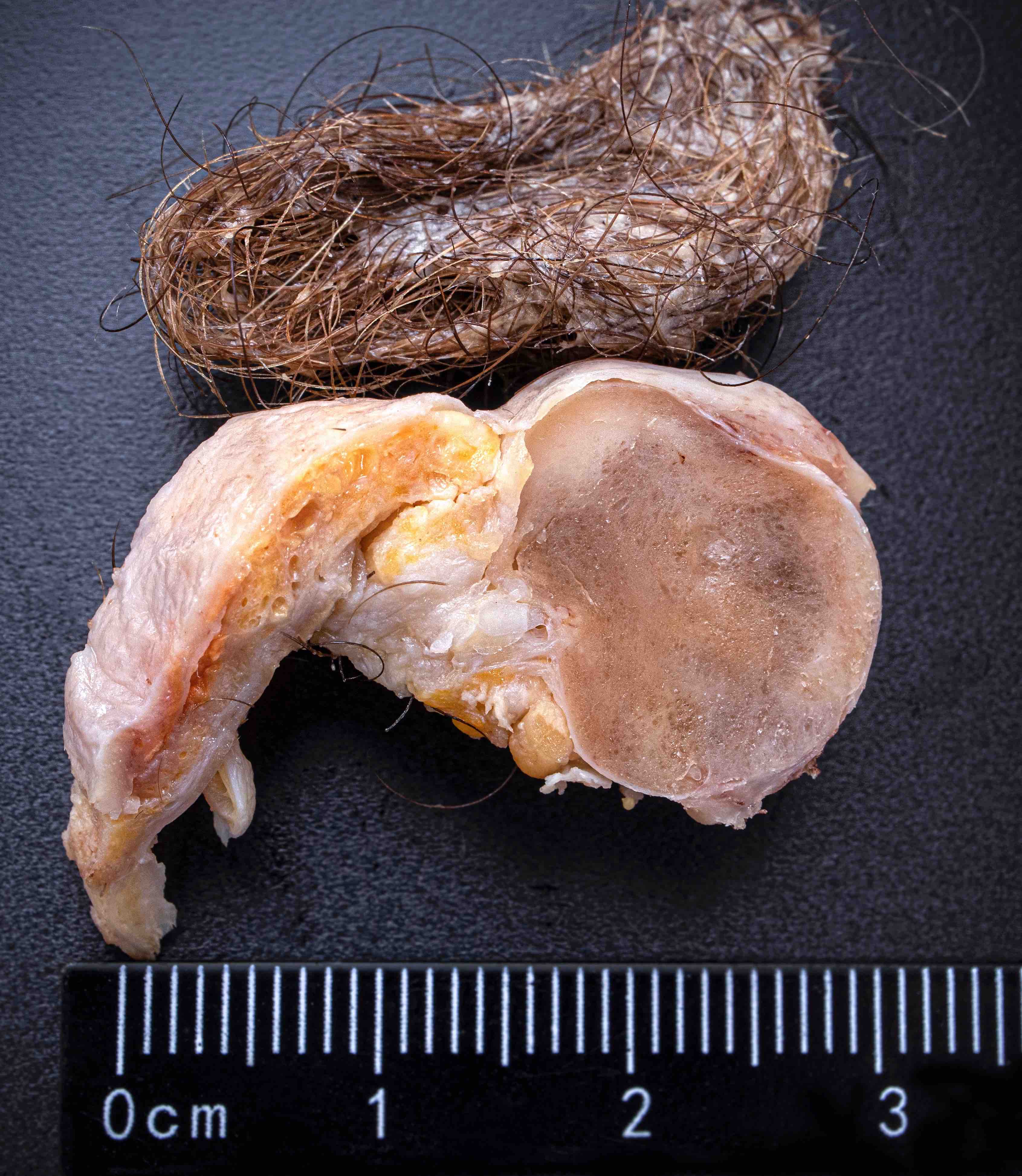
Mature teratoma
Frozen section description
- Rarely performed but will show mixture of mature tissues
- Solid areas should be sampled to exclude immature teratoma
Microscopic (histologic) description
- Mixture of mature, benign tissues
- Ectodermal (most common): squamous epithelium, sebaceous glands, hair follicles, brain tissue
- Mesodermal (second most common): bone, cartilage, smooth muscle, fibroadipose tissue
- Endodermal: intestinal or respiratory epithelium, thyroid, salivary gland
- Microscopic foci of immature neuroepithelium (less than or equal to 4 foci or 21 mm2) does not warrant diagnosis of immature teratoma and will not affect prognosis (Int J Gynecol Pathol 1987;6:203)
- Fat necrosis and foreign body reaction may be seen
- Cases associated with NMDAR encephalitis usually show neuroglial tissue associated with lymphoid aggregates with germinal centers, low number of mature neurons and a hypercellular astrocyte population (Am J Surg Pathol 2019;43:949)
- Reference: StatPearls: Cystic Teratoma [Accessed 29 July 2021]
Microscopic (histologic) images
Contributed by Shannon Mingo Welter, M.D. and @Andrew_Fltv on Twitter
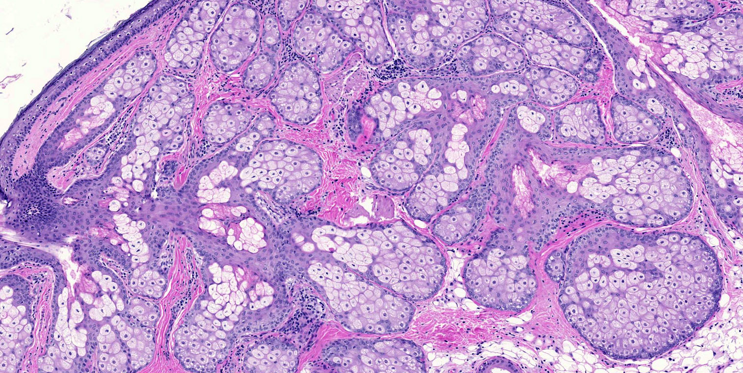
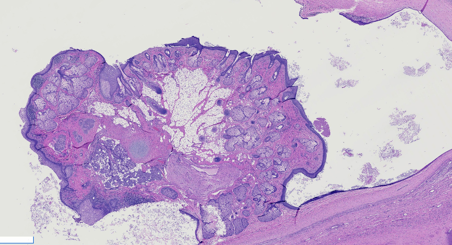
Skin with sebaceous glands
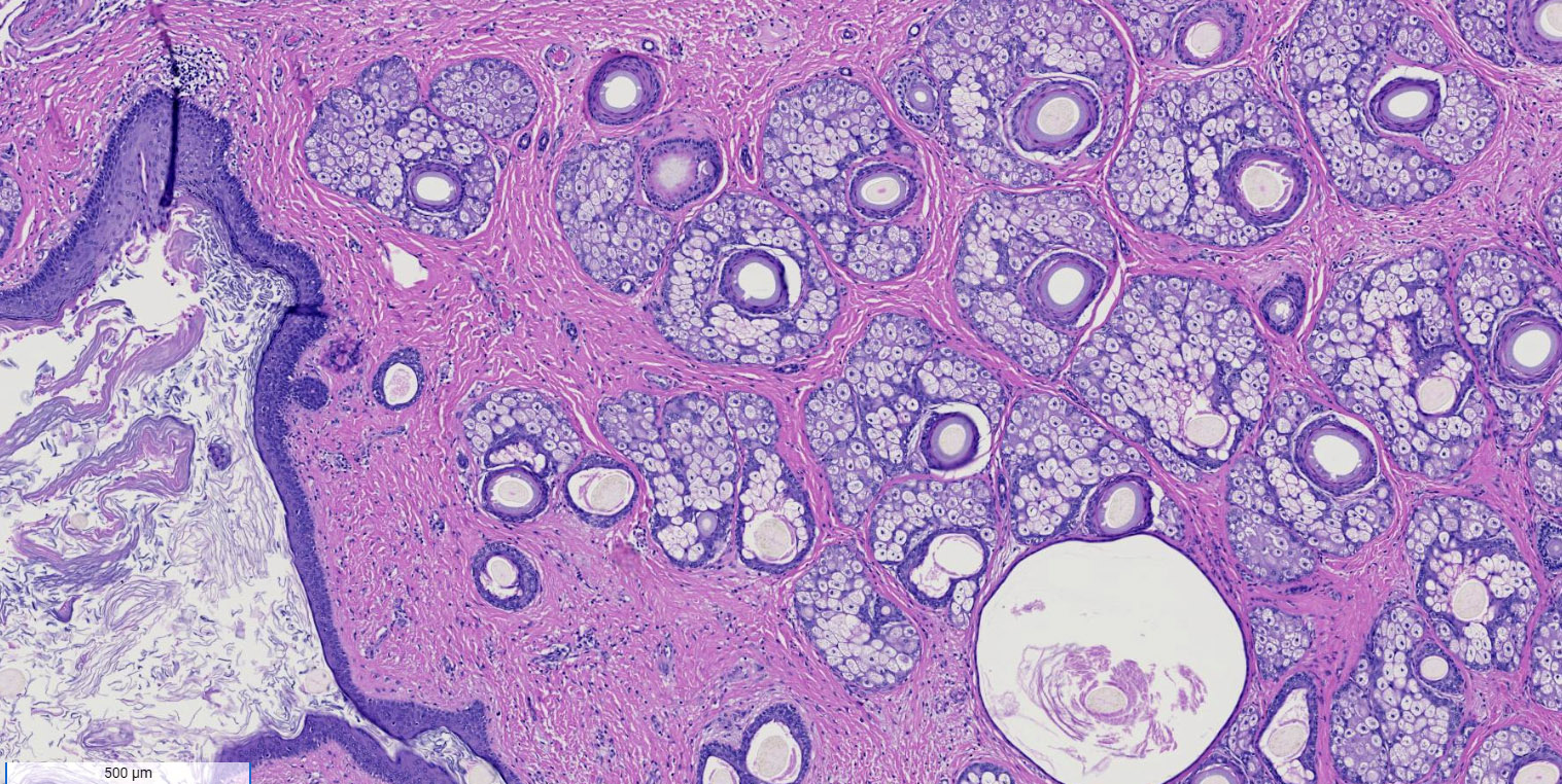
Skin with keratin and adnexa
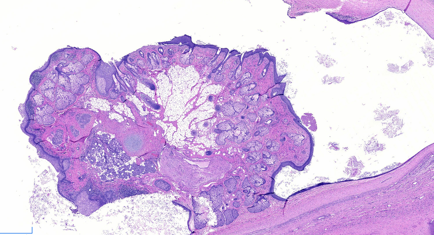
Rokitansky nodule
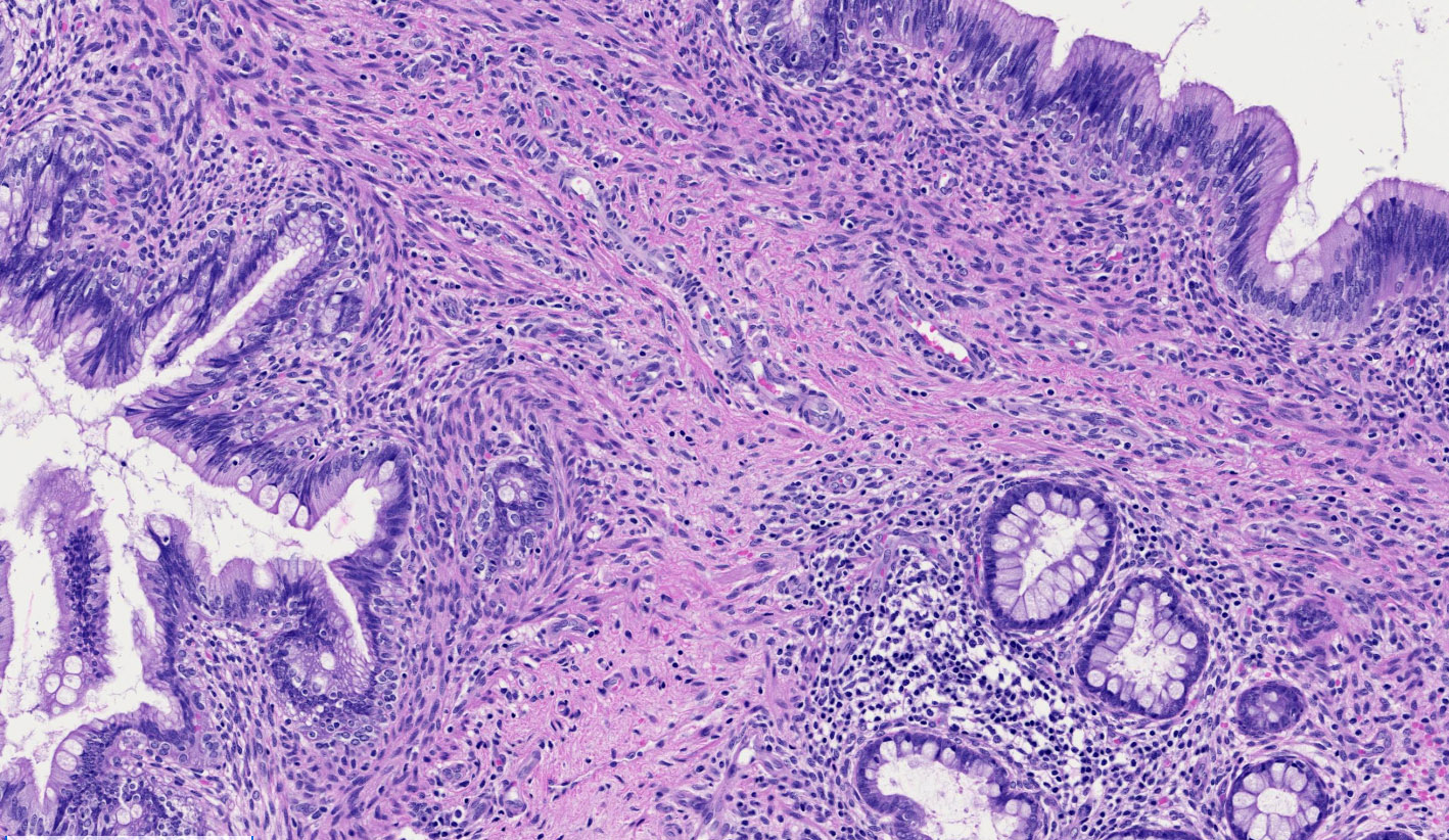
Mucin producing epithelium
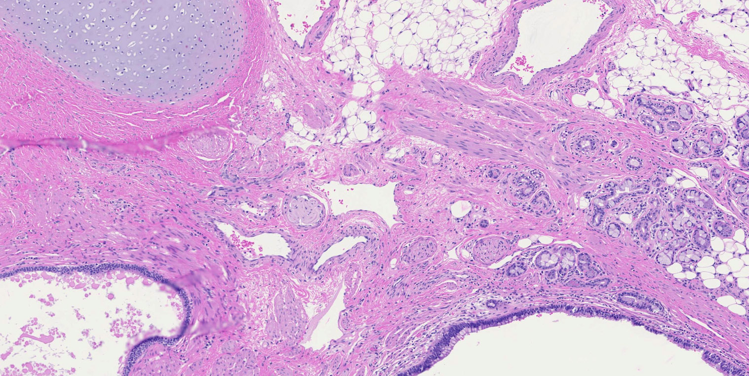
Multiple tissue types

Orderly arrangement of tissues
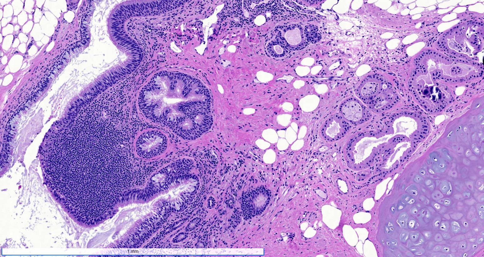
Cartilage and other mature tissues
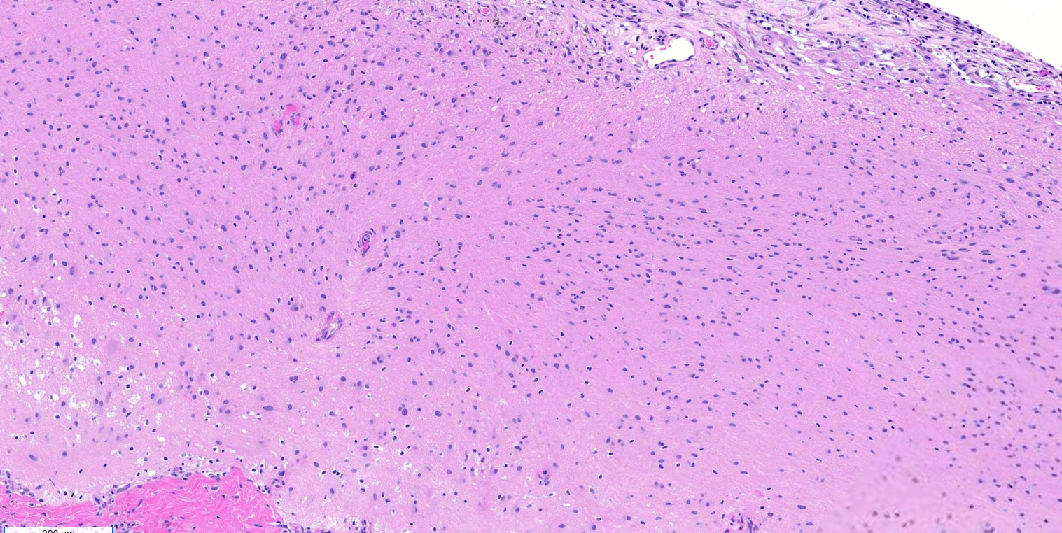
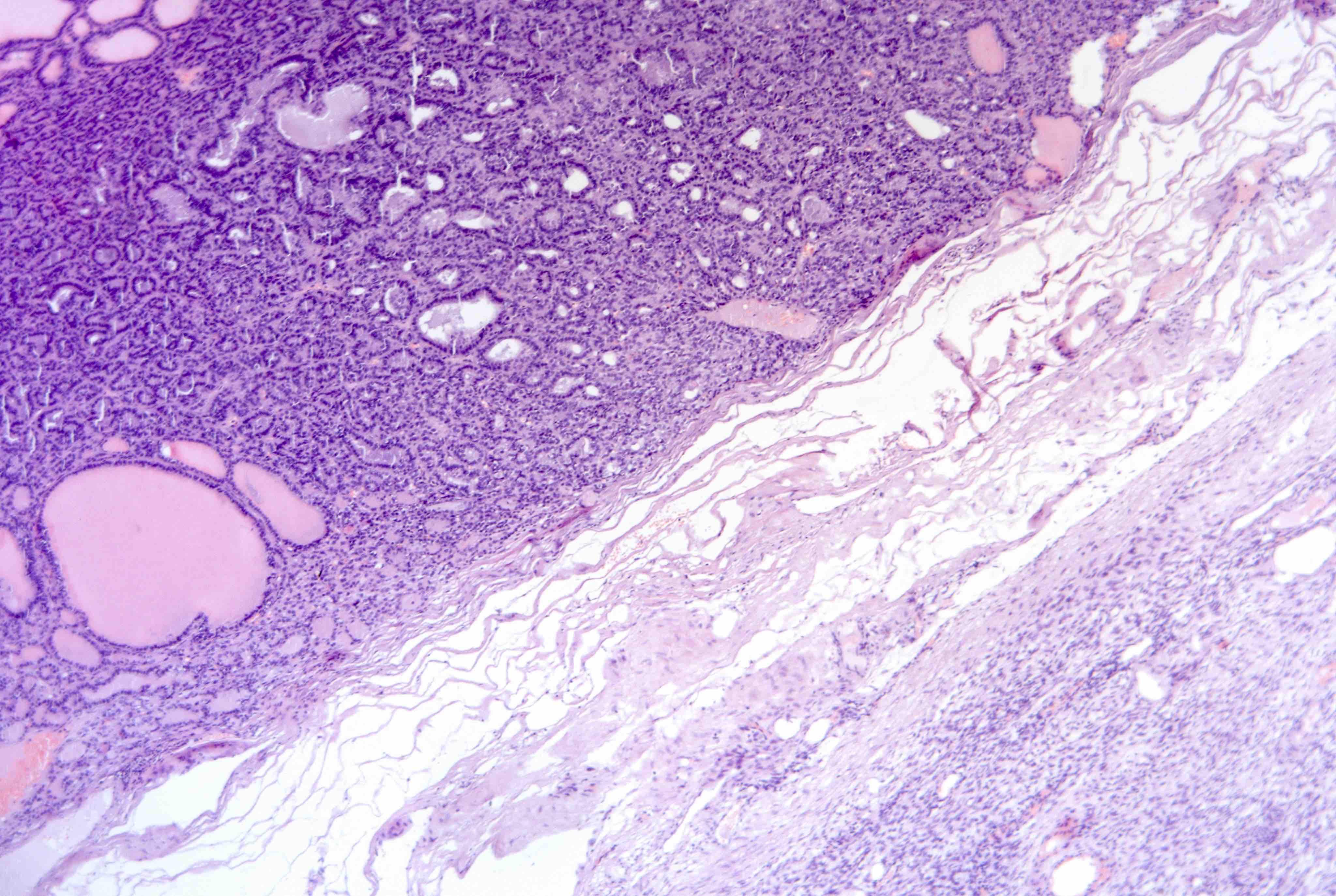
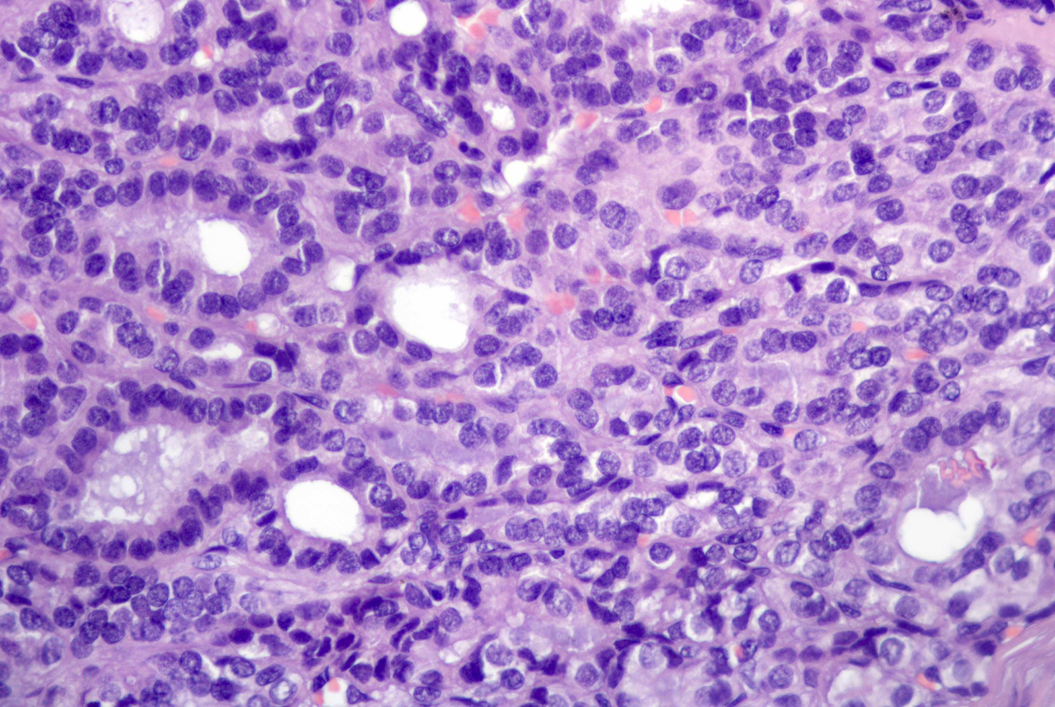
Glial tissue
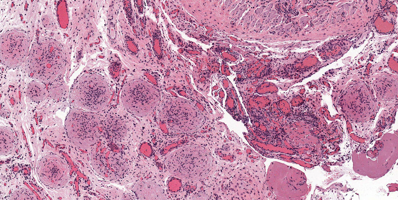
Gliomatosis peritonei
Mature teratoma
Virtual slides
Images hosted on other servers:

Mature teratoma with
well differentiated
neuroendocrine tumor
Molecular / cytogenetics description
- Diploid with normal (46XX) karyotype
Sample pathology report
- Ovary, right, cystectomy:
- Mature teratoma
Differential diagnosis
- Immature teratoma:
- Contains immature tissues (greater than 4 foci or greater than 21 mm2) (Int J Gynecol Pathol 1987;6:203)
- Younger patients
- Larger and fast growing
- Monodermal teratoma:
- Contain a single germ layer
- Neuroectodermal cyst - lined by ependymal cells
- Epidermoid cyst - lined by squamous epithelium (no sebaceous glands present)
- Struma ovarii - thyroid tissue is the dominant or sole component
- Strumal carcinoid - well differentiated neuroendocrine tumor admixed / juxtaposed with thyroid tissue
- Contain a single germ layer
Board review style question #1
A 25 year old pregnant woman is seen in the obstetrics clinic for a routine ultrasound. During the exam, a cystic mass is noted near her left ovary. The mass is eventually surgically removed and a pathologic examination shows the features in the picture shown above. What is an essential feature of this tumor?
- Gliomatosis peritonei
- Immature tissue
- Mature tissue representing at least 2 embryonic layers
- Size greater than 10 cm
Board review style answer #1
C. Mature tissue representing at least 2 embryonic layers. Mature teratoma can contain microscopic foci of immature neuroectodermal tissue in the cyst wall. This finding does not change the prognosis and should not lead to the tumor being classified as immature teratoma. Gliomatosis peritonei is a rare finding associated with teratoma and size of tumor is variable with most less than 10 cm. Mature teratomas must contain at least 2 embryonic layers.
Comment Here
Reference: Teratoma - mature
Comment Here
Reference: Teratoma - mature
Board review style question #2
A 19 year old woman presents with a symptom of left adnexal fullness. On physical examination, a 9 cm mass is palpated near the left ovary. The mass is surgically removed and a gross picture is shown above. What is the most likely diagnosis?
- Yolk sac tumor
- Immature teratoma
- Lipoma
- Mature teratoma
Board review style answer #2
D. Mature teratoma. The photo shows a mass with yellow sebaceous material and hair. In a 19 year old woman, this most likely represents a mature teratoma. Mature teratomas are the most common ovarian tumor and are especially common in women of reproductive age.
Comment Here
Reference: Teratoma - mature
Comment Here
Reference: Teratoma - mature
