
Stains & molecular markers
Renal cell carcinoma (RCC)

Copyright: 2003-2024, PathologyOutlines.com, Inc.
PubMed Search: RCC marker[TIAB] OR RCC Ma[TIAB]
Renal cell carcinoma (RCC)
Author: Chen Yang, M.D.
Editorial Board Member: Christian M. Schürch, M.D., Ph.D.
Deputy Editor-in-Chief: Maria Tretiakova, M.D., Ph.D.
Last author update: 16 February 2022
Last staff update: 16 February 2022
Copyright: 2003-2024, PathologyOutlines.com, Inc.
PubMed Search: RCC marker[TIAB] OR RCC Ma[TIAB]

Table of Contents
Definition / general | Essential features | Terminology | Pathophysiology | Interpretation | Uses by pathologists | Microscopic (histologic) images | Positive staining - normal | Positive staining - disease | Negative staining | Sample pathology report | Board review style question #1 | Board review style answer #1Cite this page: Yang C. Renal cell carcinoma (RCC). PathologyOutlines.com website. https://www.pathologyoutlines.com/topic/stainsrcc.html. Accessed May 11th, 2024.
Definition / general
- Anti renal cell carcinoma (RCC) antibodies detect a 200 kD glycoprotein (gp200), which is a surface membrane molecule located on the brush border of proximal renal tubules (Am J Surg Pathol 2001;25:1485)
- Also expressed on the luminal surface of Bowman capsule, parathyroid parenchymal cells and colloid of thyroid follicles
Essential features
- Could be helpful in classification of primary and metastatic renal cell carcinomas (Appl Immunohistochem Mol Morphol 2007;15:310, J Cutan Pathol 2007;34:381)
- Positive in clear cell, papillary and chromophobe renal cell carcinomas (Am J Surg Pathol 2001;25:1485)
- Negative in other renal primary tumors
Terminology
- Also called RCC Ma (renal cell carcinoma marker)
Pathophysiology
- RCC was originally identified as a marker of proximal convoluted tubule brush borders and luminal surface of Bowman capsule in 1989 (Cancer Res 1989;49:1802)
- This scaffolding extracellular matrix protein also known as podocalyxin and represents a human embryonal carcinoma antigen (Biochem Biophys Res Commun 2003;300:285)
Interpretation
- Membranous stain
Uses by pathologists
- Used for identification of clear cell, papillary and chromophobe renal cell carcinoma
- However, due to the lack of sensitivity and specificity and emerging utility for PAX8, CAIX, CK7 and CD117 (c-kit), RCC has become less commonly used by pathologists (Histopathology 2020;77:659 Hum Pathol 2021 May 4 [Epub ahead of print])
Microscopic (histologic) images
Contributed by Chen Yang, M.D.
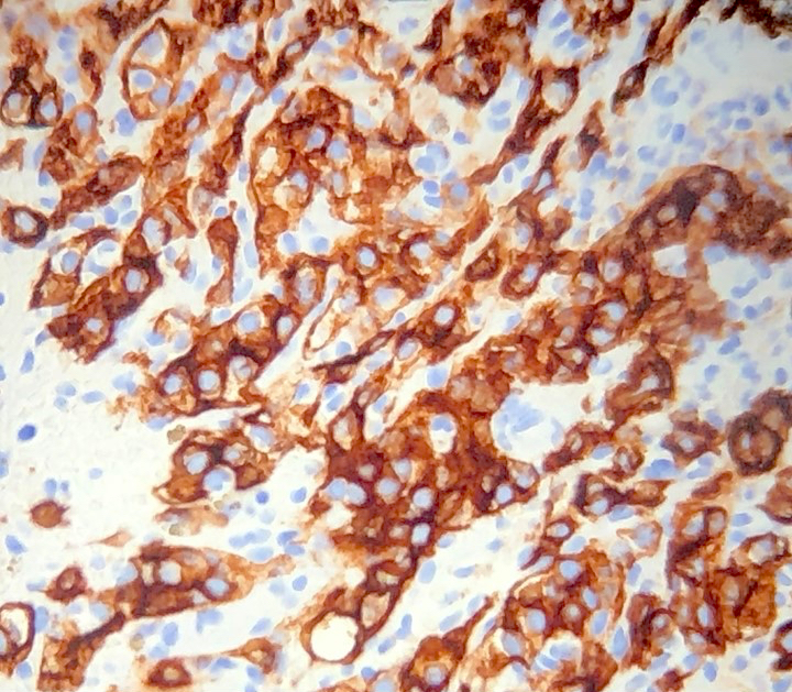
Tumor cells
Positive staining - normal
- Brush border of proximal renal tubules
- Luminal surface of Bowman capsule
- Parathyroid parenchymal cells and colloid of thyroid follicles
Positive staining - disease
- Renal cell carcinoma, clear cell (77 - 84%), papillary (96%) and chromophobe (~10%) type (Am J Surg Pathol 2001;25:1485, Appl Immunohistochem Mol Morphol 2014;22:635, Diagn Cytopathol 2005;33:3)
- Expressed in 10 of 16 (63%) metastatic cutaneous renal cell carcinomas (J Cutan Pathol 2007;34:381)
Negative staining
- Oncocytoma (0/15)
- Transitional cell carcinoma (0/20)
- Mesoblastic nephroma (0/3)
- Cystic nephroma (0/3)
- Lymphoma (0/8)
- Angiomyolipoma (0/3)
- Mixed stromal and epithelial tumor (0/4)
- Nephroblastoma (0/8)
- Collecting duct (0%)
- Reference: Am J Surg Pathol 2001;25:1485
Sample pathology report
- Lymph node, retroperitoneal, biopsy:
- Metastatic carcinoma, suggestive of renal origin (see comment)
- Comment: Sections of the retroperitoneal biopsy show the presence of metastatic tumor cells with abundant clear cytoplasm. A panel of immunohistochemistry stains show tumor cells to be positive for CD10 and RCC Ma, while being negative for CK7, CK20, PAX8, CD117 and CAIX. While the immunohistochemistry profile is not entirely concordant with a renal primary, especially with negative PAX8 and CAIX, the positivity of both CD10 and RCC Ma would suggest possibility of renal primary in the context of known history of renal cell carcinoma. Clinical and radiological correlation is recommended.
Board review style question #1
Which of the following renal neoplasms is most likely negative for RCC?
- Chromophobe renal cell carcinoma
- Clear cell renal cell carcinoma
- Collecting duct carcinoma
- Papillary renal cell carcinoma
Board review style answer #1

