
8 December 2010 - Case #192
All cases are archived on our website. To view them sorted by case number, diagnosis or category, visit our main Case of the Month page. To subscribe or unsubscribe to Case of the Month or our other email lists, click here.
Thanks to Drs. Ed Hartle, Robert Sterling and Peter Banks, Lake Norman Regional Medical Center, North Carolina, for contributing this case.
Case #192
Clinical history:
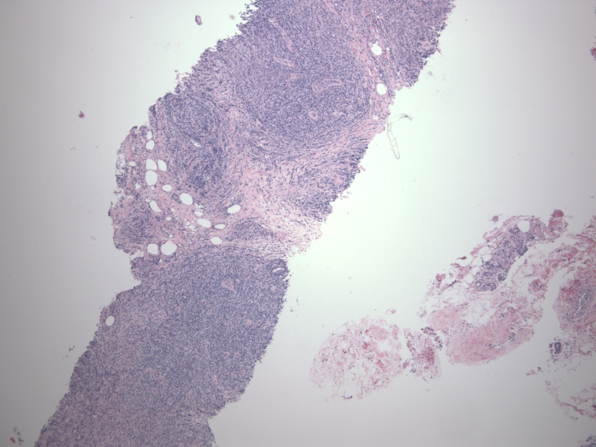
A 37 year old woman presented with a palpable left breast mass. Mammogram and ultrasound showed a 4.5 cm mass without calcifications, characterized as ACR BIRADS category IV suspicious. A needle core biopsy was obtained.
Microscopic images:

What is your diagnosis?
Diagnosis: High grade malignant lymphoma, peripheral T cell, type not further specified
Immunostains:

Discussion:
The diagnosis was confirmed by molecular probes which demonstrated a clonal T cell population using primers for the T gamma region.
Primary breast lymphomas are rare, constituting < 1% of all breast malignancies. Patients tend to be either ages 30 - 35 years (diffuse large B cell) or 55 - 60 years (diffuse large B cell or MALT type). Screening mammography may be increasing the detection of low grade lymphoma.
The criteria for primary breast lymphoma are: (a) sufficient tissue for diagnosis; (b) close interaction between the lymphoma infiltrate and breast tissue; (c) no evidence of widespread lymphoma at time of diagnosis; (d) no prior diagnosis of nonbreast lymphoma (Cancer 1972;29:1705). Secondary breast lymphoma has a similar incidence as primary breast lymphoma, and is the most common metastatic disease to the breast.
T cell lymphomas of the breast are extremely rare (Mod Pathol 2000;13:599). As in this case, these tumors are often considered to be diffuse large B cell lymphomas until immunostains are performed for T and B cell markers.
T cell lymphomas of the breast often have aggressive behavior. In this case, the patient responded only briefly to therapy. She subsequently developed jaundice with increased liver function tests. A liver biopsy showed involvement by T cell lymphoma and she subsequently died of her disease.
All cases are archived on our website. To view them sorted by case number, diagnosis or category, visit our main Case of the Month page. To subscribe or unsubscribe to Case of the Month or our other email lists, click here.
Thanks to Drs. Ed Hartle, Robert Sterling and Peter Banks, Lake Norman Regional Medical Center, North Carolina, for contributing this case.
Website news:
(1) Support PathologyOutlines.com by doing your holiday shopping at Amazon.com. Click here, or on any Amazon banner on the left side of our Home Page to visit Amazon.com. Amazon not only sells books, but DVDs, clothing and even food items. By visiting Amazon via these links, they will send us 4-5% of the cost of all items you purchase (out of their profits), at no additional cost to you.
(2) We posted an article on our Management Page, Pathology Salaries and the Big Picture, by Mick Raich, Vachette Pathology.
Visit and follow our Blog to see recent updates to the website.
Case #192
Clinical history:
A 37 year old woman presented with a palpable left breast mass. Mammogram and ultrasound showed a 4.5 cm mass without calcifications, characterized as ACR BIRADS category IV suspicious. A needle core biopsy was obtained.
Microscopic images:
What is your diagnosis?
Click here for diagnosis and discussion:
Diagnosis: High grade malignant lymphoma, peripheral T cell, type not further specified
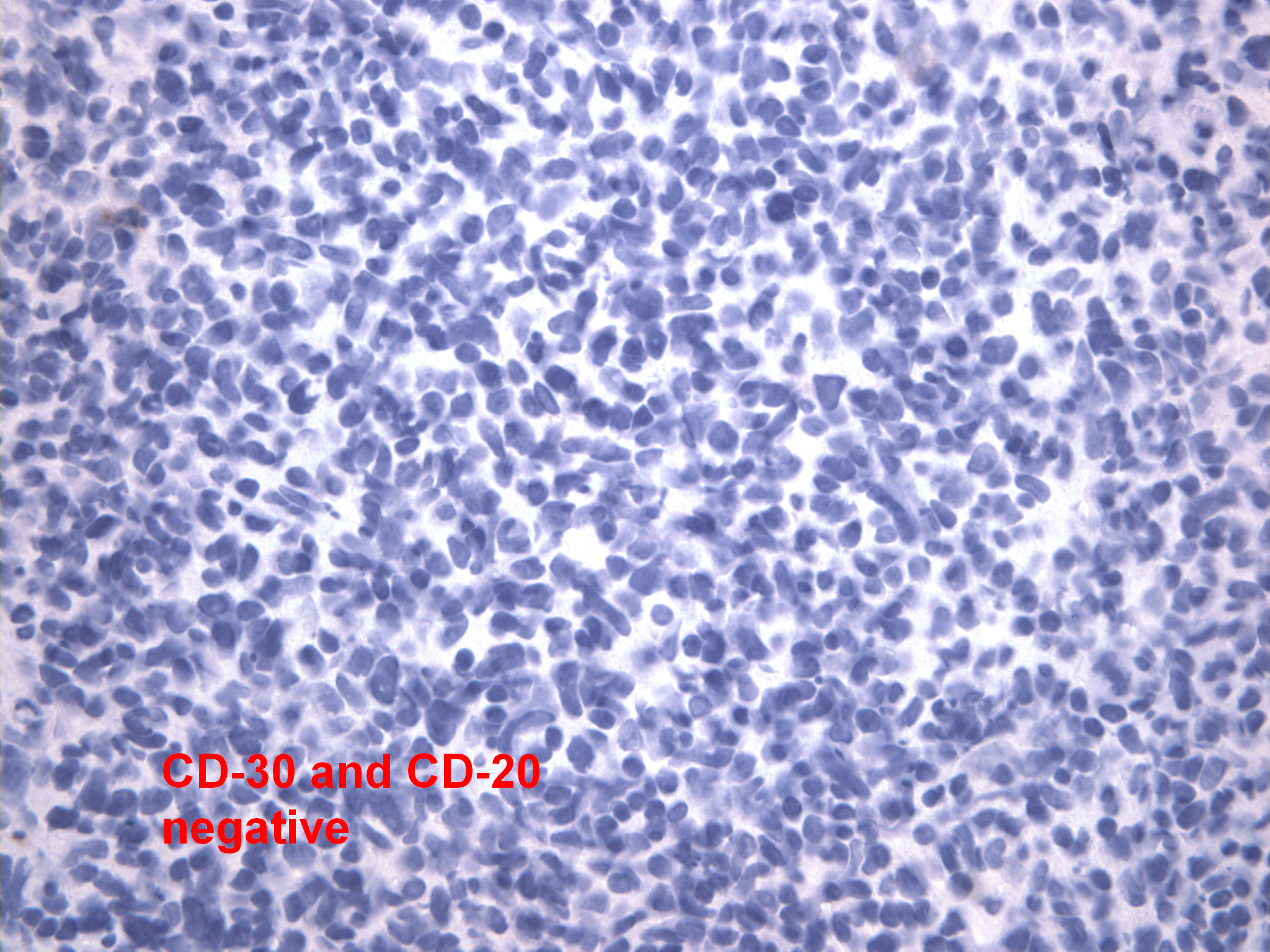
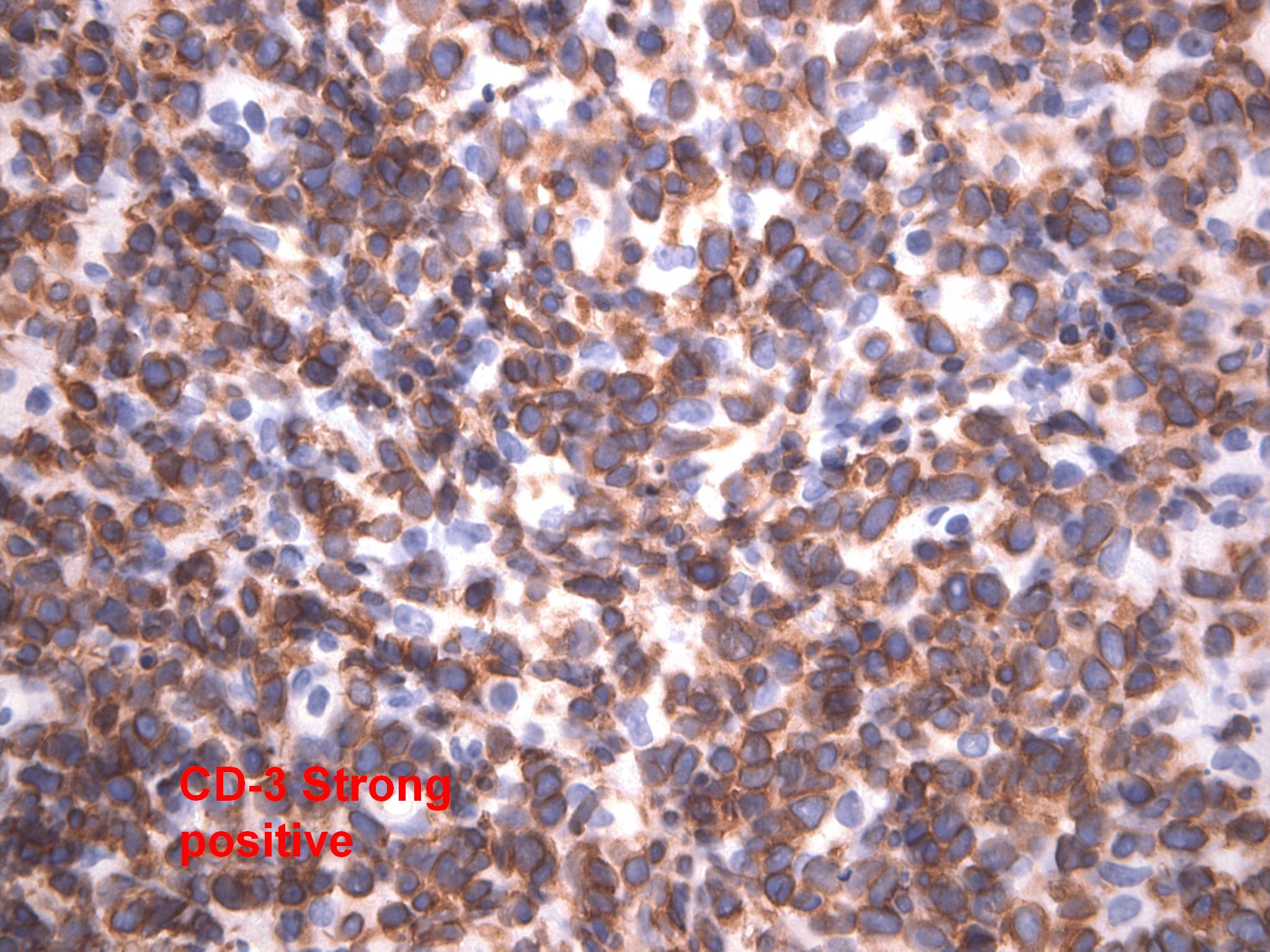
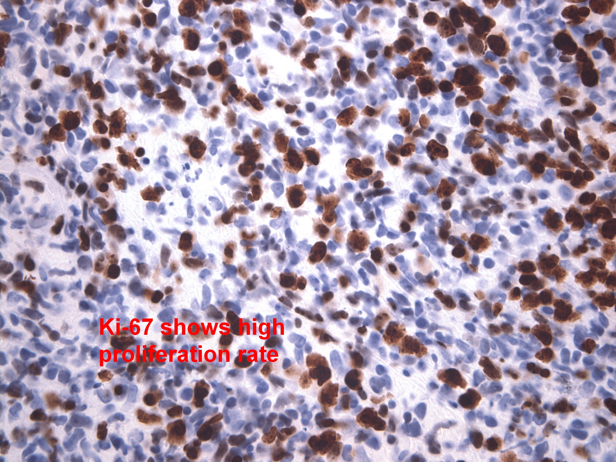
Immunostains:
Left: negative for keratin; Right: negative for CD20 (B cell marker) and CD30
Left: strongly positive for CD3 (T cell marker); Right: strongly positive for Ki67
Discussion:
The diagnosis was confirmed by molecular probes which demonstrated a clonal T cell population using primers for the T gamma region.
Primary breast lymphomas are rare, constituting < 1% of all breast malignancies. Patients tend to be either ages 30 - 35 years (diffuse large B cell) or 55 - 60 years (diffuse large B cell or MALT type). Screening mammography may be increasing the detection of low grade lymphoma.
The criteria for primary breast lymphoma are: (a) sufficient tissue for diagnosis; (b) close interaction between the lymphoma infiltrate and breast tissue; (c) no evidence of widespread lymphoma at time of diagnosis; (d) no prior diagnosis of nonbreast lymphoma (Cancer 1972;29:1705). Secondary breast lymphoma has a similar incidence as primary breast lymphoma, and is the most common metastatic disease to the breast.
T cell lymphomas of the breast are extremely rare (Mod Pathol 2000;13:599). As in this case, these tumors are often considered to be diffuse large B cell lymphomas until immunostains are performed for T and B cell markers.
T cell lymphomas of the breast often have aggressive behavior. In this case, the patient responded only briefly to therapy. She subsequently developed jaundice with increased liver function tests. A liver biopsy showed involvement by T cell lymphoma and she subsequently died of her disease.
