Bone & joints
Other nonneoplastic
Osteoporosis
Author: Nat Pernick, M.D.

Last author update: 1 August 2013
Last staff update: 19 January 2022
Copyright: 2003-2025, PathologyOutlines.com, Inc.
PubMed Search: Osteoporosis[title]
Table of Contents
Definition / general | Radiology description | Diagnosis | Treatment | Gross description | Microscopic (histologic) description | Microscopic (histologic) imagesCite this page: Pernick N. Osteoporosis. PathologyOutlines.com website. https://www.pathologyoutlines.com/topic/boneosteoporosis.html. Accessed September 16th, 2025.
Definition / general
- Reduction in bone mass due to increased bone porosity, which predisposes bones to fracture
- Usually refers to postmenopausal or senile loss of bone severe enough to cause fractures
- Affects entire skeleton due to metabolic bone disease, but may be localized due to limb disuse
- Usually due to increased bone resorption, with normal levels of bone formation
- Osteopenia: defined as radiologic decrease in density of skeleton
- Primary causes: due to postmenopausal condition, older age (15 million cases in US) or idiopathic
- Secondary causes (due to identifiable conditions): endocrine (hyperparathyroidism, thyroid disorders, hypogonadism, pituitary tumors, type I diabetes, Addison’s disease), neoplasms (myeloma, carcinomatosis), gastrointestinal disturbances (malnutrition, deficiency of vitamins C or D), drugs (corticosteroids, chemotherapy), osteogenesis imperfecta, immobilization, homocystinuria, anemia
- Menopause: postmenopausal women may lose 2% of cortical bone and 9% of cancellous bone / year; osteoporosis affects women more than men because estrogen deficiency leads to increased osteoclast activity, and osteoblasts cannot keep pace
- Age related changes: osteoblasts have reduced reproductive and biosynthetic potential in elderly
- Immobilization: important cause because mechanical forces stimulate bone remodeling; zero gravity (astronauts), immobilization cause reduced skeletal mass; athletes have higher bone density; weight training is more effective than jogging in increasing skeletal mass
- Genetics: variation in Vitamin D receptor type accounts for 75% of maximal peak bone mass achieved; Vitamin D intake and parathyroid hormone levels are not significant causes, although low calcium intake in women is an important cause
- Other risk factors: Whites / Asians, smoking, alcohol abuse
- Bone mass: peak bone mass occurs in young adults, based on physical activity, muscle strength, diet, hormones; subsequent remodeling causes small deficit in bone formation with each resorption / ormation cycle, which causes bone loss of 0.7% per year
- Sites: cancellous compartment of vertebral bone (with high surface area) affected first, causing loss of vertebral height in elderly, leading to dowager’s hump; also thinning of cortex; hip and wrist also affected
Radiology description
- Flattening of vertebral bodies, widening and swelling of intervertebral discs, fish - mouth appearance
- Usually thoracic and upper lumbar spine
Diagnosis
- Radiographic measurement of bone density, iliac crest biopsy
Treatment
- Calcium, Vitamin D and exercise to build up / maintain bone mass
- Biphosphonates (inhibit post-menopausal bone loss)
Gross description
- Loss of cancellous bone, accentuation of vertical trabeculae in spine
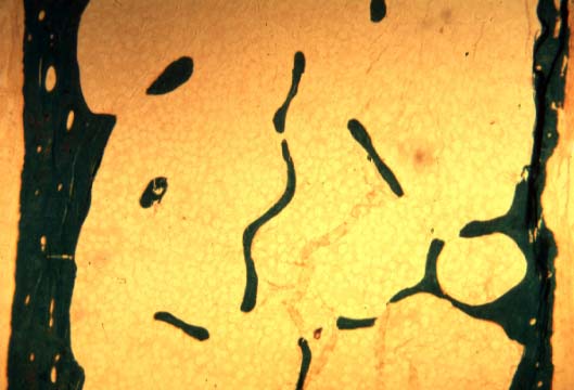
Microscopic (histologic) description
- Thin trabeculae disconnected from each other
- Increase in osteoclastic activity (may be uneven) or increased percentage of surface with resorptive pitting
Microscopic (histologic) images
Images hosted on other servers:
Osteoporosis
