
Breast
Fibroepithelial tumors
Fibroadenomatoid change
Author: Melissa Alexander, M.D., Ph.D.
Editorial Board Member: Gary Tozbikian, M.D.
Editor-in-Chief: Debra L. Zynger, M.D.

Last author update: 5 October 2020
Last staff update: 19 July 2023 (update in progress)
Copyright: 2002-2024, PathologyOutlines.com, Inc.
PubMed Search: fibroadenomatous change OR fibroadenomatoid change

Table of Contents
Definition / general | Essential features | Terminology | ICD coding | Epidemiology | Sites | Pathophysiology | Etiology | Clinical features | Diagnosis | Radiology description | Radiology images | Prognostic factors | Case reports | Treatment | Gross description | Microscopic (histologic) description | Microscopic (histologic) images | Cytology description | Sample pathology report | Differential diagnosis | Additional references | Board review style question #1 | Board review style answer #1Cite this page: Alexander M. Fibroadenomatoid change. PathologyOutlines.com website. https://www.pathologyoutlines.com/topic/breastfibroadenomatoidchange.html. Accessed May 13th, 2024.
Definition / general
- Benign, often incidental finding in a background of fibrocystic changes
- Lesion with features resembling a fibroadenoma but lacking sharp circumscription
Essential features
- Proliferation of intralobular stroma with the formation of stromal nodularity that often appears to blend in with the surrounding breast tissue
- Morphologically reminiscent of a fibroadenoma but not forming a well circumscribed mass
- May represent an incipient fibroadenoma
- Often multifocal and surrounded by fibrocystic changes
Terminology
- Also known as fibroadenomatous change, fibroadenomatoid hyperplasia, fibroadenomatoid mastopathy, fibroadenomatosis, sclerosing lobular hyperplasia
- Less frequently referred to as mixed lesion, reflecting the common association of fibroadenomatoid change and fibrocystic changes
ICD coding
- ICD-10: N60.2 - fibroadenosis of breast
Epidemiology
- F > M
- Mean age 28 - 34 years
- Represents 5 - 7% of nonneoplastic surgical biopsies in one study; 8% of nonneoplastic breast diagnoses in another
- References: AJR Am J Roentgenol 1998;171:1331, Am J Epidemiol 1978;108:112, Hum Pathol 1984;15:336
Sites
- Most common site is the upper outer quadrant of the breast (Hum Pathol 1984;15:336)
Pathophysiology
- Proliferation of intralobular stroma
- May represent a stage in the evolution of a fibroadenoma, produced by the coalescence of fibroadenomatoid nodules
- Reference: Hum Pathol 1984;15:336
Etiology
- Unknown; possibly due to or influenced by reproductive hormones, similar to fibroadenoma
Clinical features
- May be detected by imaging modalities (see Radiology below), as a palpable mass or incidentally in breast tissue sampled for a different lesion
Diagnosis
- Breast imaging modalities with core biopsy (stereotactic, ultrasound or MRI guided) or surgery followed by histologic examination of resected tissue
- References: AJR Am J Roentgenol 1995;165:291, AJR Am J Roentgenol 1998;171:1331
Radiology description
- Hypoechoic mass
- Asymmetric increased density
- Suspicious granular clustered microcalcifications (more frequent in women > 50 years old)
- No suspicious findings in patient with palpable mass
- Imaging findings in one study of fibroadenomatoid hyperplasia showed 53% were well defined mass
- 33% had normal mammographic findings
- 13% had an asymmetric density
- 1% with mammographic calcifications
- References: AJR Am J Roentgenol 1995;165:291, AJR Am J Roentgenol 1998;171:1331, Hum Pathol 1984;15:336
Radiology images
Images hosted on other servers:

Well circumscribed nodule

Suspicious granular microcalcifications
Prognostic factors
- No increased risk of malignancy
Case reports
- 12 year old girl with 3 month history of enlarging right breast mass (Cytojournal 2006;3:8)
- 14 year old girl with a 3 month history of painless enlarging right breast lump (Acta Cytol 2001;45:765)
- 24 year old woman with 7 cm mass (Gynecol Obstet Invest 2014;77:134)
- 69 year old man with heart failure treated with digoxin and spironolactone presents with bilateral gynecomastia (Am J Surg Pathol 1990;14:774)
Treatment
- No treatment required
Gross description
- Lesion appears dense and poorly circumscribed on cut section
- Difficult to distinguish from fibrocystic changes by gross examination
- Reference: Malays J Pathol 1991;13:101
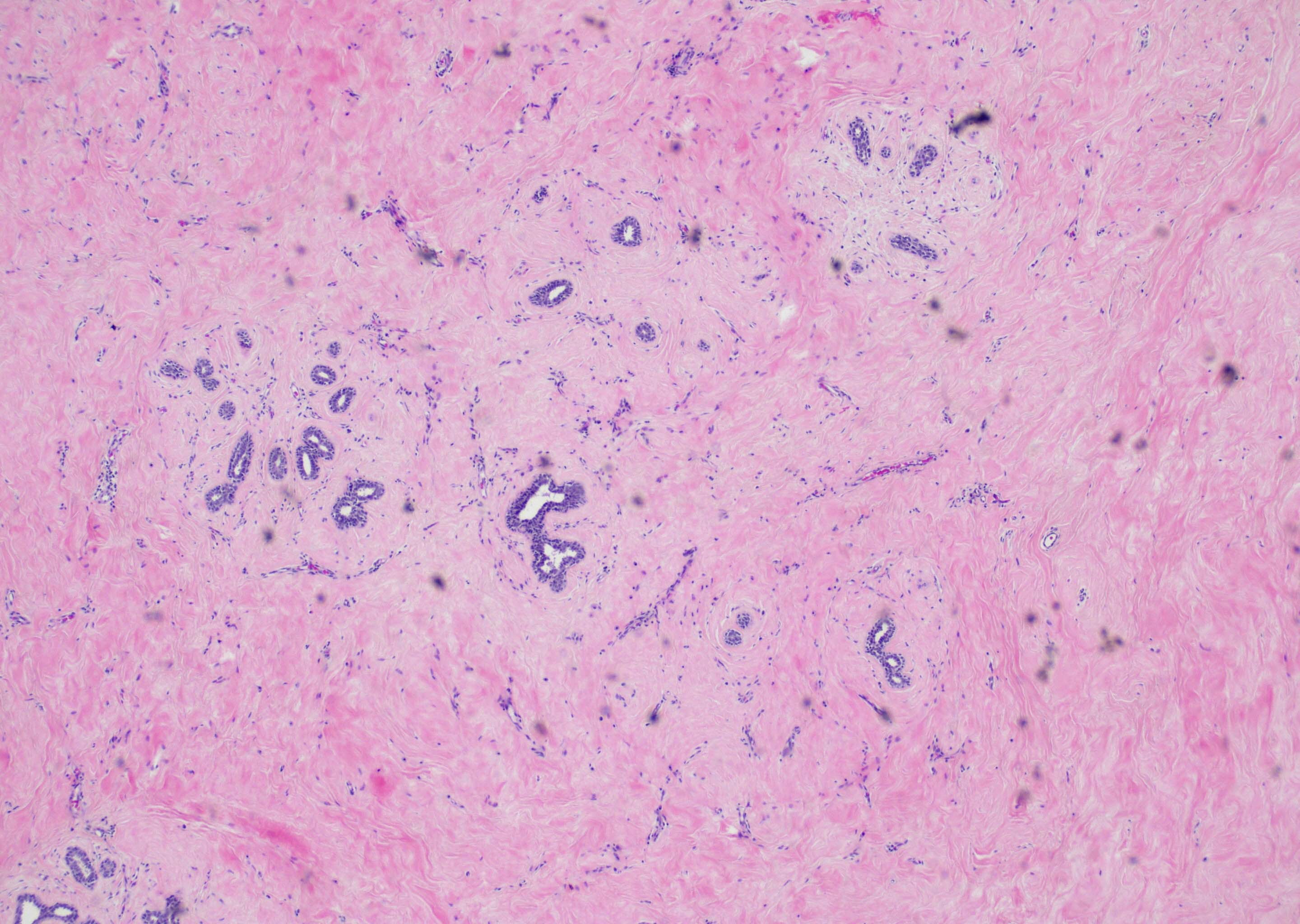
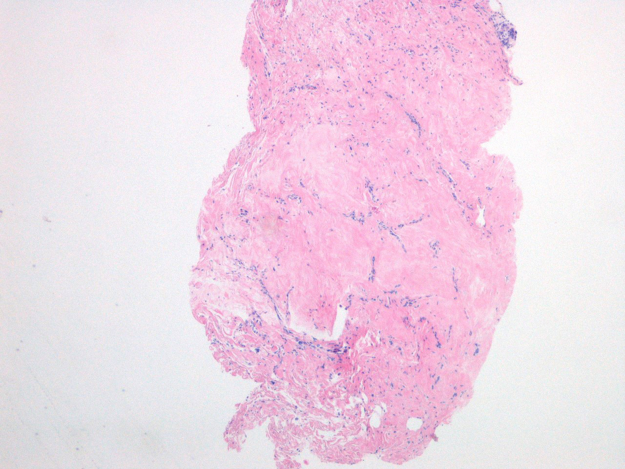
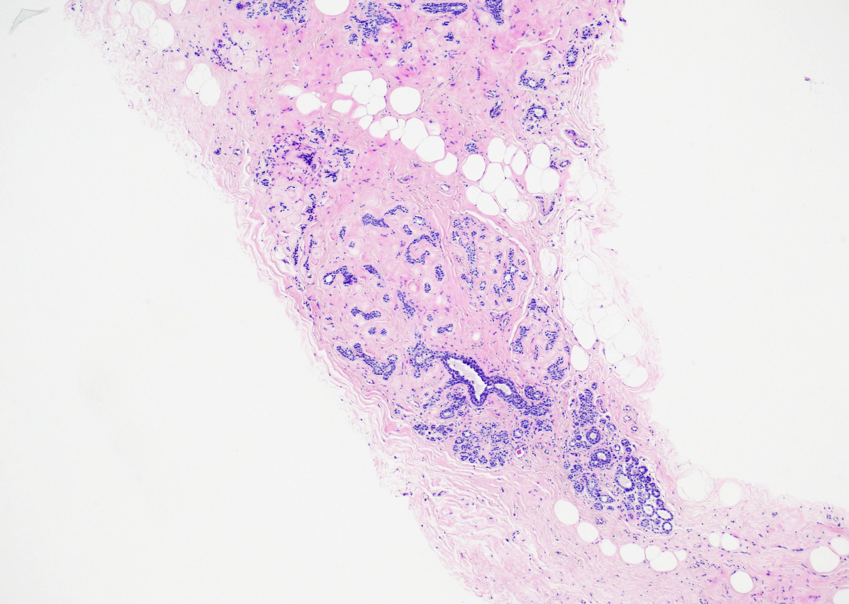
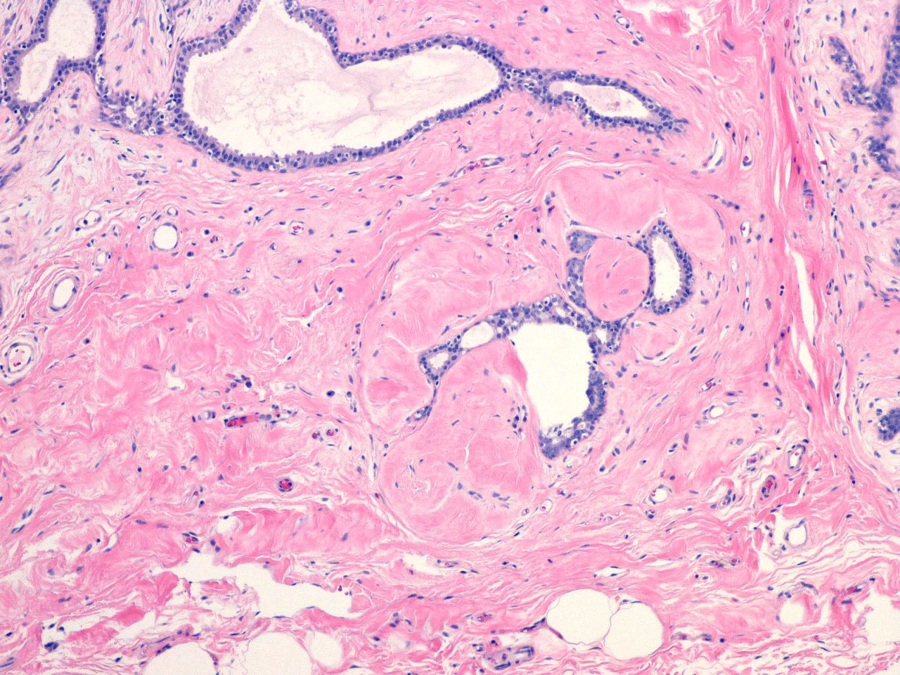
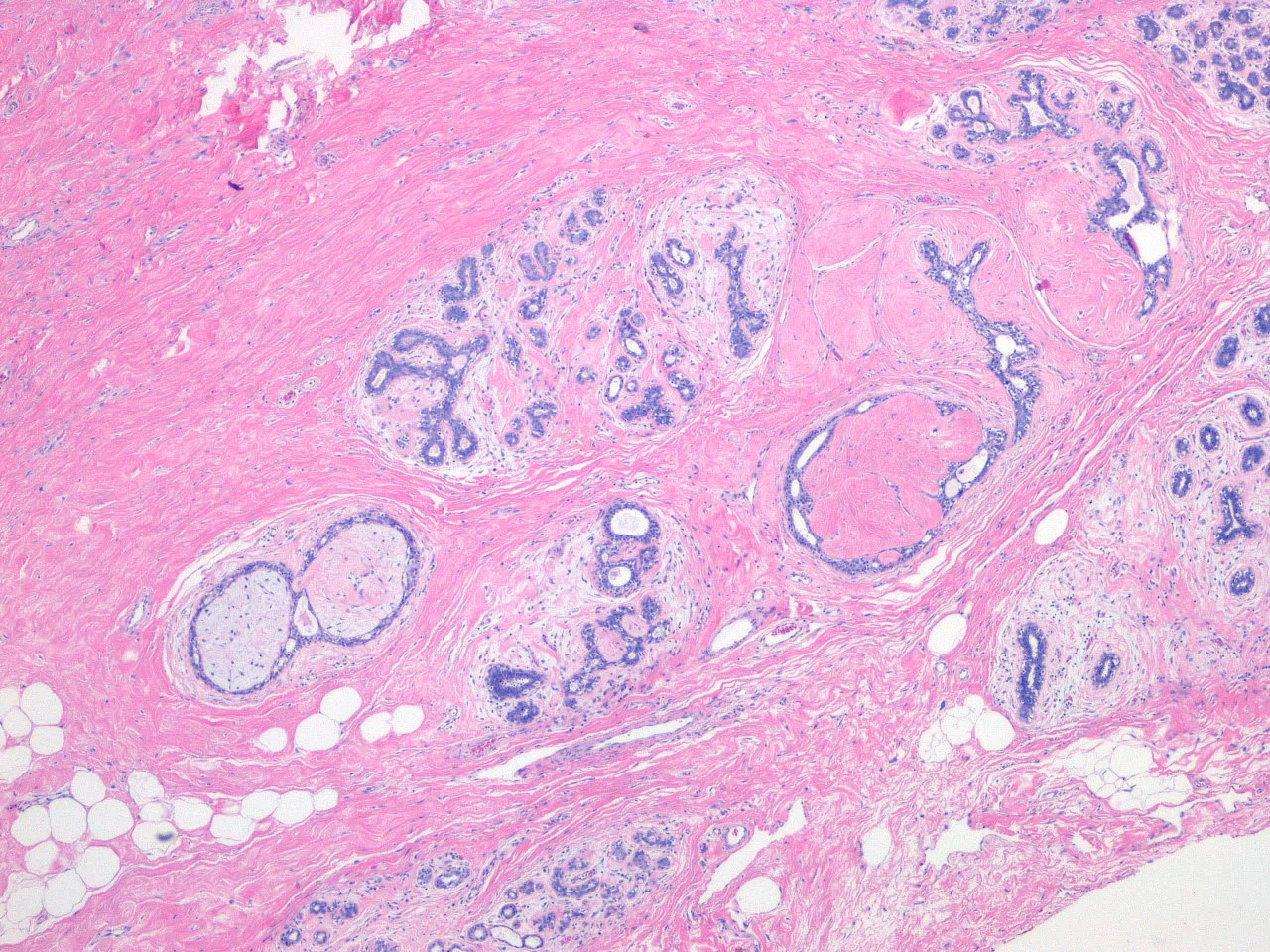
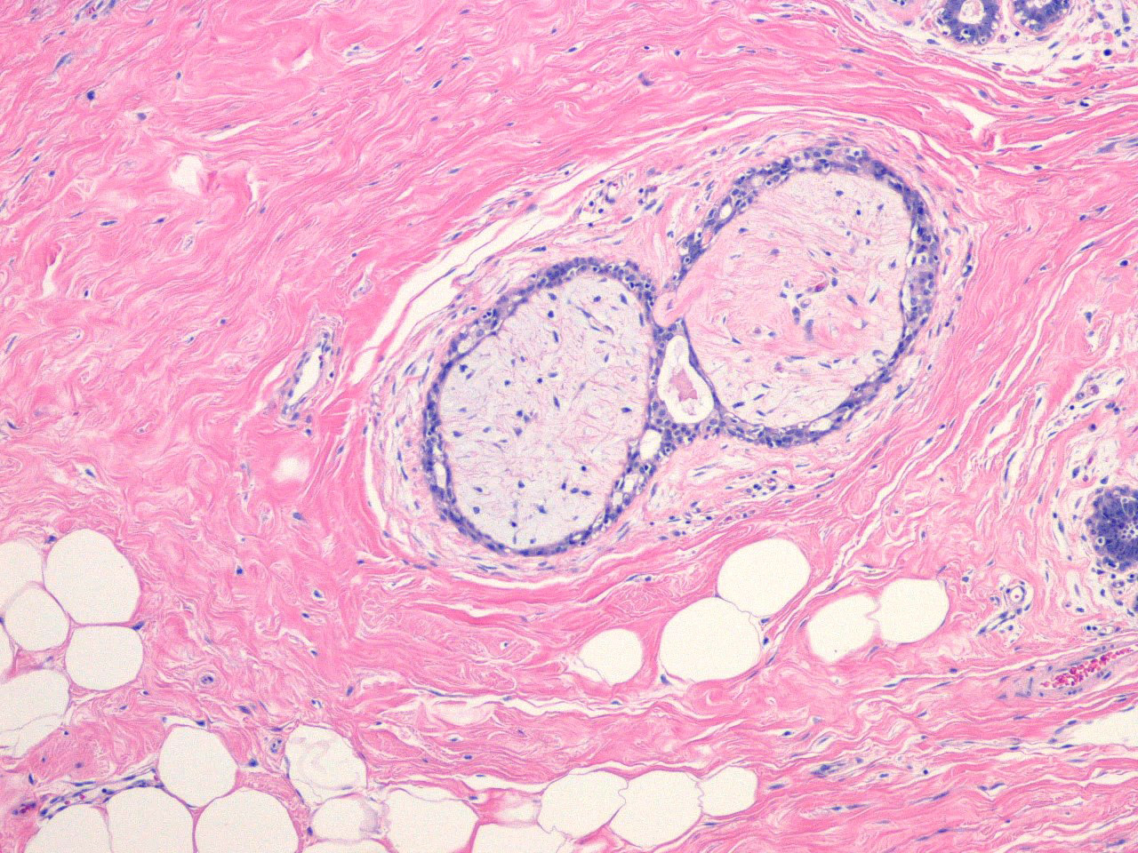
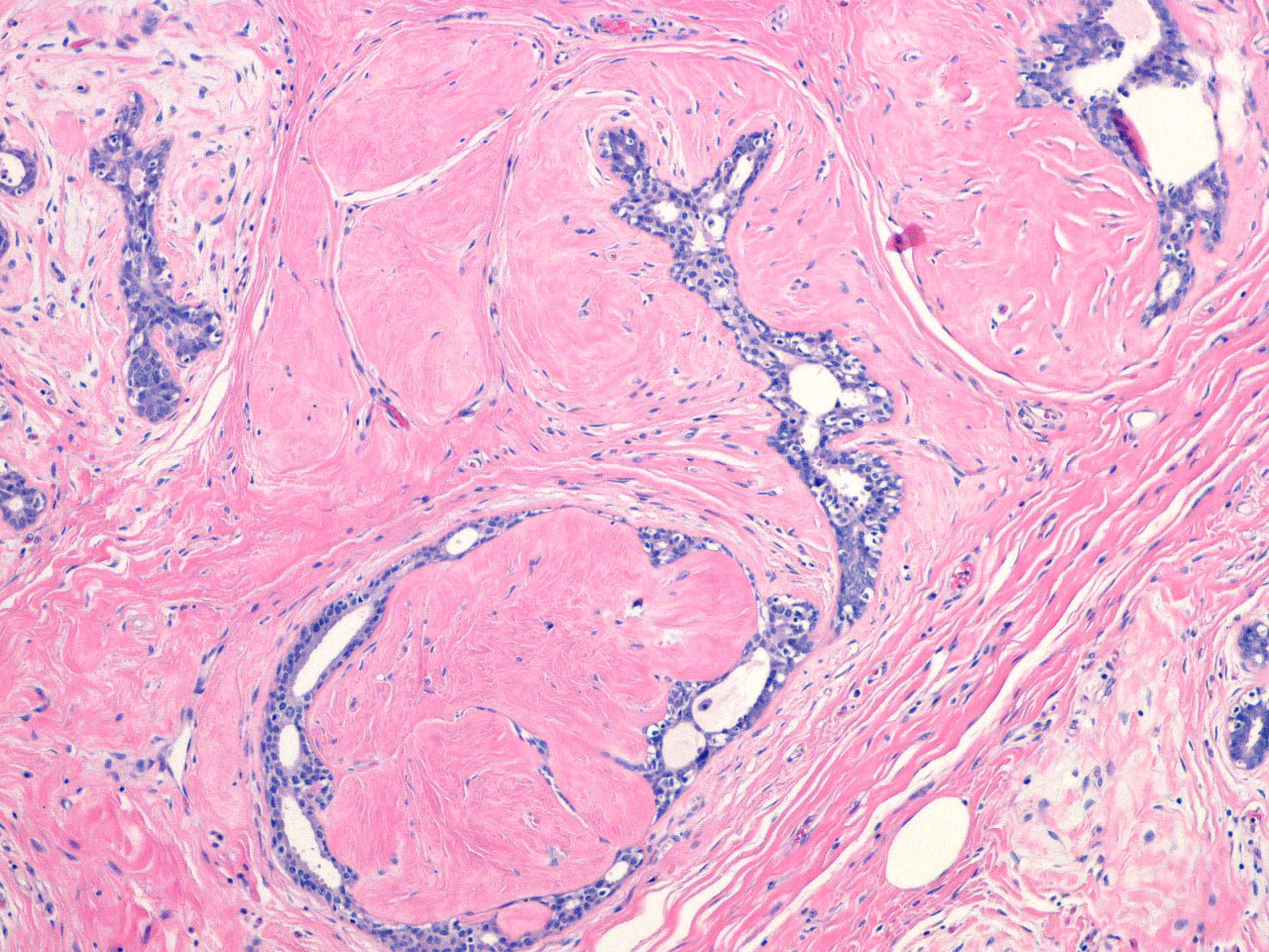
Microscopic (histologic) description
- Proliferation of intralobular stroma containing epithelial elements, sometimes closely resembling a microfibroadenoma or microfibroadenomata
- Stroma may appear loose and cellular or dense and hyalinized
- Often multifocal in a background of fibrocystic changes, including cysts and adenosis
- Periphery may appear to blend into the surrounding breast tissue
- References: Hum Pathol 1984;15:336, Pathology 1987;19:393, Malays J Pathol 1991;13:101, AJR Am J Roentgenol 1998;171:1331, Schnitt: Biopsy Interpretation of the Breast, 3rd Edition, 2017
Microscopic (histologic) images
Contributed by Melissa Alexander, M.D., Ph.D.
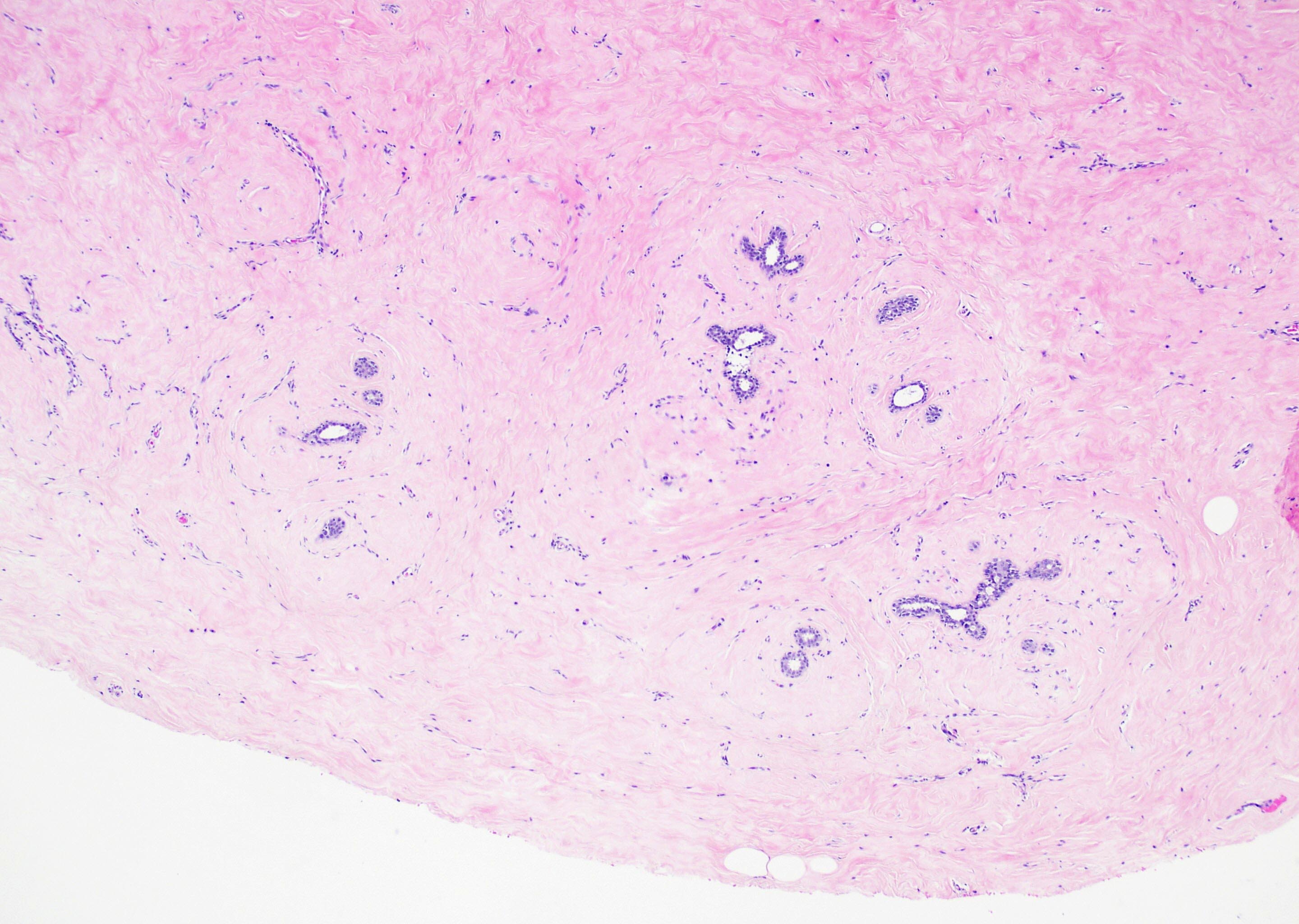
Edges blending with surrounding
Vague stromal nodularity
Poorly circumscribed
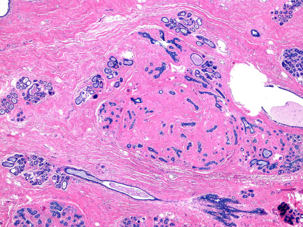
Background fibrocystic change
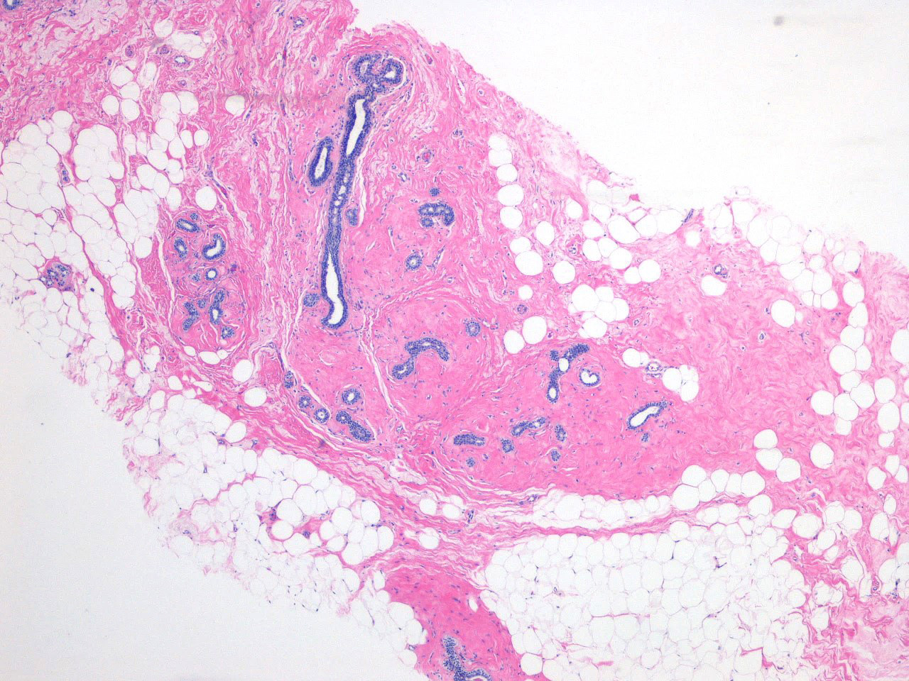
Multiple nodules
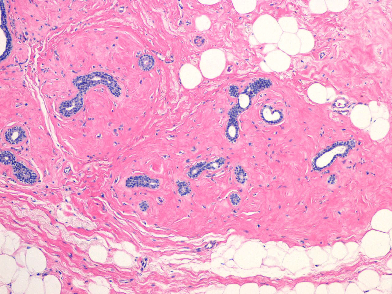
Multiple nodules
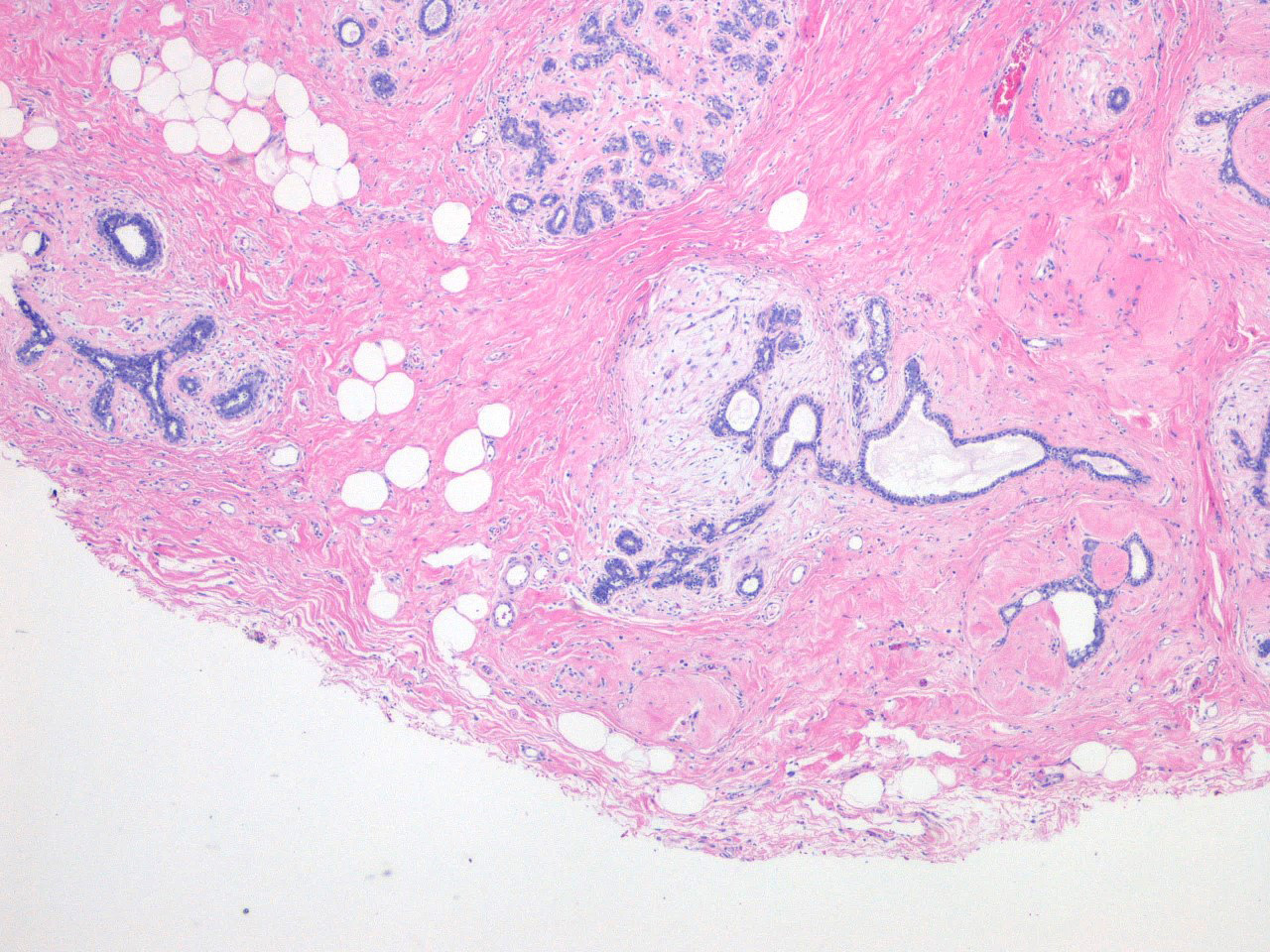
Loose stroma versus dense hyalinized
Dense stroma
Loose versus dense nodules
Loose stroma, compressed glands
Fibroadenomatoid nodule, compressed glands
Cytology description
- Elements similar to fibroadenoma, including stromal fragments; however, lacking other elements of fibroadenoma including branching antler horn epithelial clusters (Cytojournal 2006;3:8, Acta Cytol 2001;45:765)
Sample pathology report
- Breast, right, core biopsy:
- Fibroadenomatoid change associated with calcifications (see comment)
- Comment: Correlation with specimen radiograph was performed.
Differential diagnosis
- Fibroadenoma:
- Well circumscribed mass comprised of stromal and epithelial components sharply demarcated from the surrounding breast parenchyma
Additional references
Board review style question #1
A poorly circumscribed nodular focus was identified in a breast needle core biopsy, shown in the image. The best diagnosis for this histologic finding is which of the following?
- Apocrine metaplasia
- Duct ectasia
- Columnar cell change
- Fibroadenomatoid change
- Microcalcifications
Board review style answer #1
