Breast
Other nonneoplastic
Juvenile papillomatosis
Author: Monika Roychowdhury, M.D.

Last author update: 1 August 2012
Last staff update: 19 June 2025 (update in progress)
Copyright: 2002-2025, PathologyOutlines.com, Inc.
PubMed Search: juvenile papillomatosis
Table of Contents
Definition / general | Terminology | Epidemiology | Clinical features | Case reports | Clinical images | Gross description | Gross images | Microscopic (histologic) description | Microscopic (histologic) images | Cytology description | Cytology imagesCite this page: Roychowdhury M. Juvenile papillomatosis. PathologyOutlines.com website. https://www.pathologyoutlines.com/topic/breastjuvenilepapillomatosis.html. Accessed August 22nd, 2025.
Definition / general
- Grossly distinct multinodular mass with clustering of cystic formations resembling swiss cheese, composed of epithelial proliferation and clustered cysts
- First described by Rosen in 1980 (Am J Surg Pathol 1980;4:3)
Terminology
- Not part of WHO breast classification
Epidemiology
- Mean age 19 years
- 2 / 3 are less than age 20 years
Clinical features
- Resembles fibroadenoma clinically
- 10% develop breast carcinoma, higher risk if recurrent, bilateral disease and family history of breast cancer (Am J Clin Pathol 1990;93:599)
- 26 - 58% have positive family history of breast cancer (Cancer 1982;49:2591, Am J Clin Pathol 1986;86:745), 15% have bilateral disease
- May be associated with secretory carcinoma (Jpn J Clin Oncol 1985;15:457)
Case reports
- Juvenile papillomatosis of the breast in a male infant with Noonan syndrome (Pediatr Blood Cancer 2005;45:991)
- 6 year old girl with juvenile secretory carcinoma and juvenile papillomatosis (J Pediatr Surg 1987;22:637)
- 16 year old girl with multiple juvenile papillomatosis of the breast (Breast Cancer 1998;5:187)
- 17 year old boy with juvenile papillomatosis of the breast (G Chir 2011;32:374)
Clinical images
Contributed by Mark R. Wick, M.D.
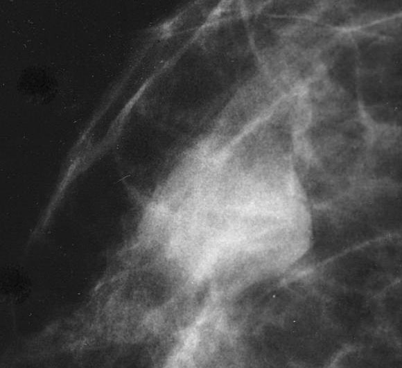
Mammogram
Gross description
- Localized multinodular mass with clustering of cystic formations resembling swiss cheese
Gross images
Contributed by Mark R. Wick, M.D.
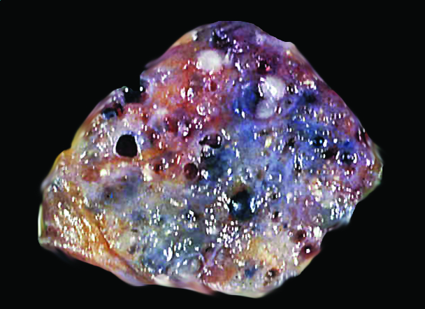
Juvenile papillomatosis
Microscopic (histologic) description
- Florid epithelial hyperplasia and papillomatosis, cysts with foamy histiocytes and sclerosing adenosis
- Variable apocrine metaplasia, atypia and necrosis
Microscopic (histologic) images
Contributed by Mark R. Wick, M.D.
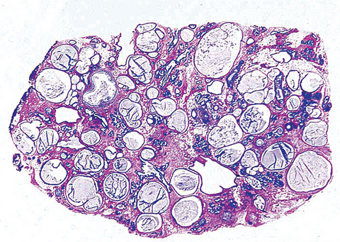
Juvenile papillomatosis
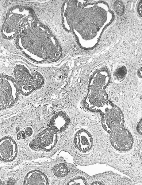
AFIP images
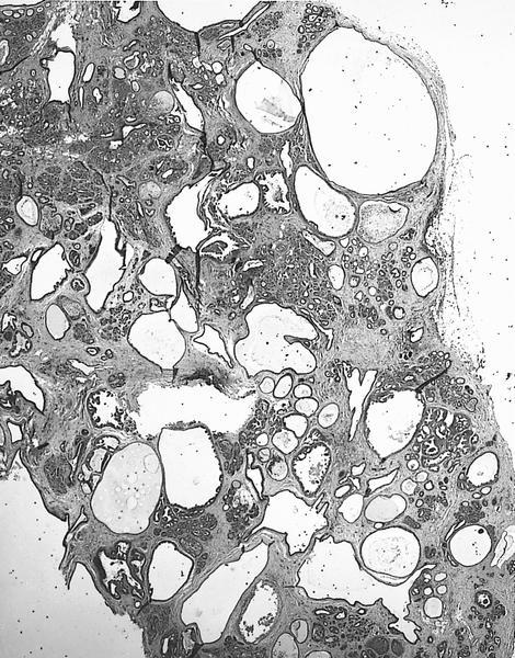
Multiple cystically dilated ducts

Hyperplastic epithelium in dilated duct
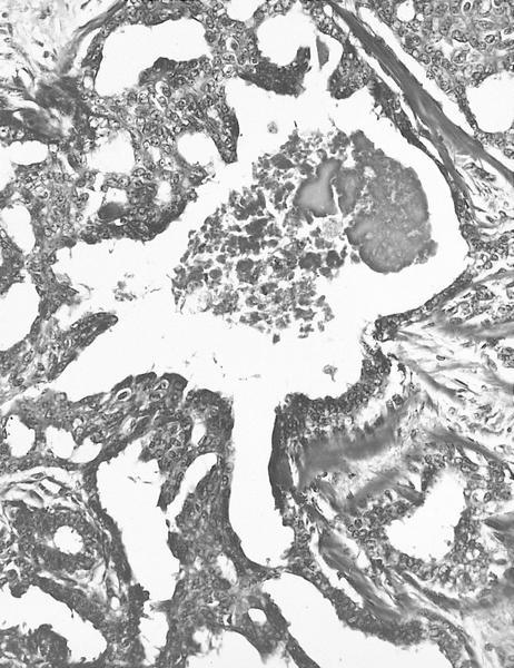
Intraductal epithelial proliferation with necrosis
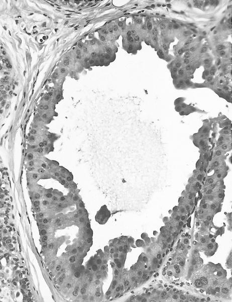
Apocrine metaplasia
of hyperplastic
intraductal
epithelium
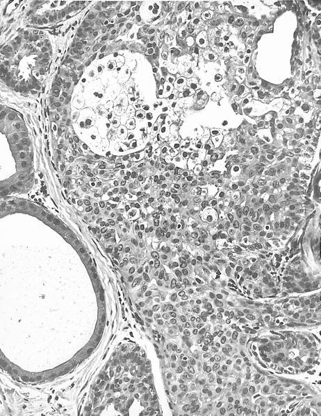
Hyperplastic epithelium and foam cells

With sclerosing adenosis
and apocrine metaplasia
adjacent to cystically
dilated ducts
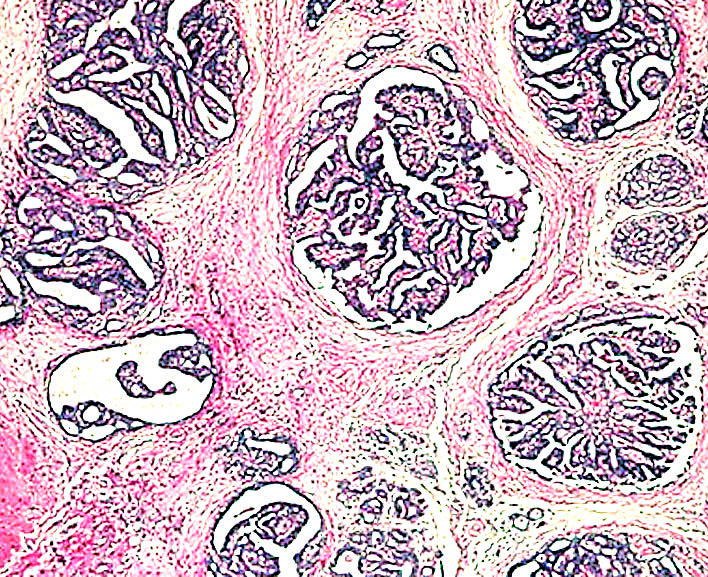
Multiple small intraductal papillomas in 15 year old girl
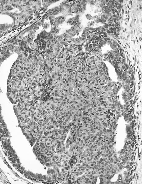
High power shows hyperplastic
intraductal epithelium, myoepithelial
cells are associated with the
delicate fibrovascular stalks
Cytology description
- Difficult to diagnose
- Cystic fluid, but mass persists after aspiration
- Sheets of hyperplastic breast epithelium with areas resembling fibroadenoma, macrophages and apocrine cells (Diagn Cytopathol 1993;9:457)
Cytology images
Contributed by Areej M. Al Nemer, M.D.
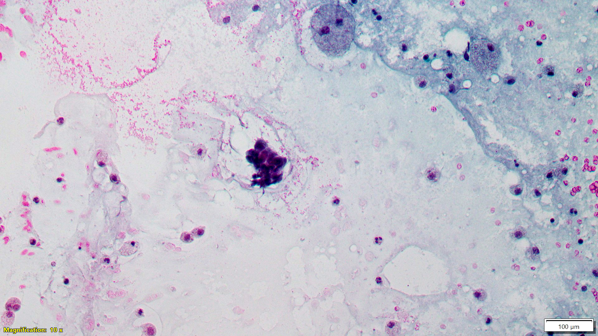
Suspicious nipple discharge
