Stains & CD markers
CD56

Copyright: 2002-2025, PathologyOutlines.com, Inc.
PubMed Search: CD56
CD56
Author: Wiebke Solass, M.D.
Editorial Board Members: Brandon Umphress, M.D., Christian M. Schürch, M.D., Ph.D.
Last author update: 18 December 2023
Last staff update: 18 December 2023
Copyright: 2002-2025, PathologyOutlines.com, Inc.
PubMed Search: CD56
Table of Contents
Definition / general | Essential features | Terminology | Pathophysiology | Interpretation | Case reports | Uses by pathologists | Prognostic factors | Microscopic (histologic) images | Positive staining - normal | Positive staining - disease | Negative staining | Sample pathology report | Additional references | Practice question #1 | Practice answer #1Cite this page: Solass W. CD56. PathologyOutlines.com website. https://www.pathologyoutlines.com/topic/cdmarkerscd56.html. Accessed September 30th, 2025.
Definition / general
- Homophilic binding glycoprotein with a role in cell - cell adhesion (Wikipedia: Neural Cell Adhesion Molecule [Accessed 8 February 2023])
Essential features
- Marker of natural killer (NK) cells and NK lymphomas
- Subsets of T cell lymphomas can show expression of CD56
- Sensitive marker for neuroendocrine tumors in the majority of small cell carcinoma / carcinoid tumors
- Not specific for neuroendocrine differentiation; should always be used in conjunction with other markers of neuroendocrine lineage (synaptophysin, chromogranin)
Terminology
- CD56 antigen is a 140 kDa isoform of the neural cell adhesion molecule (NCAM)
- Posttranslational modifications to the polypeptide include N and O glycosylations, acylation, sulphation and phosphorylation
- Different NCAM isoforms have molecular weights ranging from 135 to 220 kDa
- CD56 antigen is moderately expressed on a subpopulation of peripheral blood large granular lymphocytes and on all cells with NK activity; it is also expressed by subsets of T lymphocytes
- CD56 antibodies do not react with granulocytes, monocytes or B cells
- Reference: Beckman Coulter: CD34 Antibodies [Accessed 19 July 2023]
Pathophysiology
- Nerves
- Regulates homophilic (like - like) interactions between neurons and between neurons and muscle
- Associates with fibroblast growth factor receptor and stimulates tyrosine kinase activity of receptor to induce neurite outgrowth
- When neural crest cells stop making NCAM and N-cadherin and start displaying integrin receptors, cells separate and migrate (J Cell Sci 2002;115:283)
- Hematopoiesis
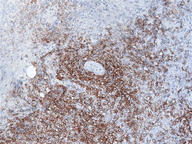
- Prototypic marker of NK cells
- Also presents on a subset of CD4+ and CD8+ T cells
- NK cells include a less mature (CD56 [bright] / CD16-) subset that is more common in lymph nodes and a more mature (CD56 [dim] / CD16+) subset that is more numerous in peripheral blood (Hum Pathol 2011;42:679)
- Adhesion
- Contributes to cell - cell or cell - matrix adhesion during development
Interpretation
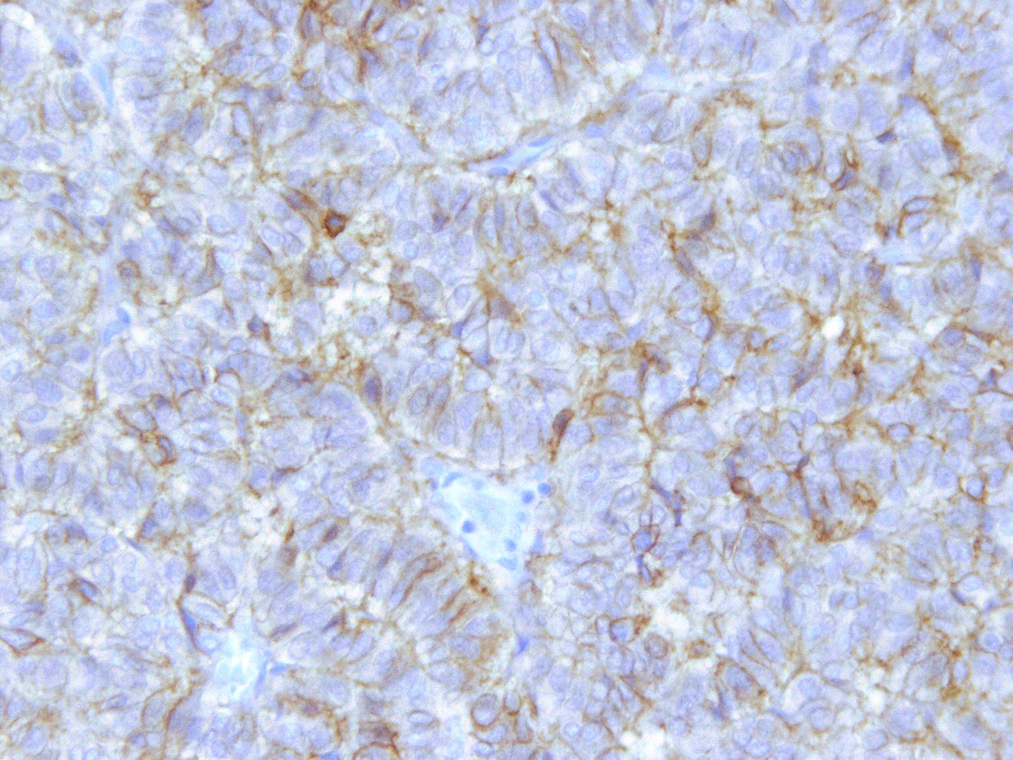
- Membranous expression is interpreted as positive staining
Case reports
- 6 year old girl with B cell acute lymphoblastic leukemia (B ALL) and CD56 and CD57 coexpression (Pediatr Hematol Oncol 2004;21:677)
- 56 year old woman with CD56+ plasma cell leukemia (University of Pittsburgh: Case 470 [Accessed 8 February 2023])
Uses by pathologists
- Marker of NK cells and NK lymphomas
- Detects residual myeloma and residual acute myeloid leukemia in bone marrow (Am J Clin Pathol 2009;132:60, Am J Clin Pathol 2008;129:934)
- Differentiates plasma cells in myeloma (CD56+) from reactive plasmacytosis or monoclonal gammopathy of undetermined significance (MGUS) (CD56-) (Am J Pathol 2002;160:1293, Am J Clin Pathol 2009;132:728)
- Detects neuroendocrine disorders (J Clin Pathol 2002;55:535)
- Particularly if extensive crush artifact (J Clin Pathol 2005;58:978)
- Other neuroendocrine markers are synaptophysin and chromogranin
- Predicts poor prognosis for cutaneous lymphoproliferative disorders other than cutaneous T cell lymphoma (J Clin Pathol 2007;60:981)
- Most uterine smooth muscle neoplasms express CD56 (Int J Gynecol Pathol 2021;40:315)
Prognostic factors
- Myelomas that are negative for CD56 may have a poorer prognosis (Leuk Lymphoma 2004;45:61)
- Preliminary study suggests CD56+ lymphocytes may be associated with regression of melanoma (Hum Pathol 2011;42:1960)
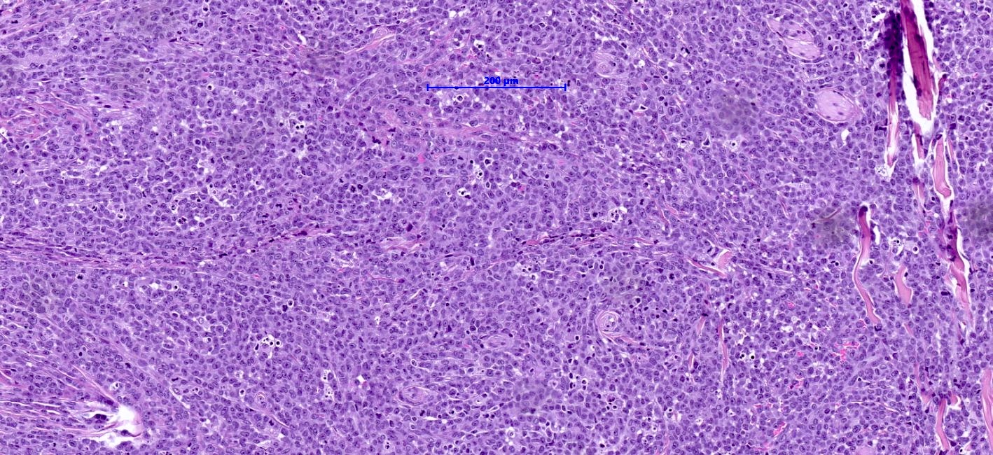
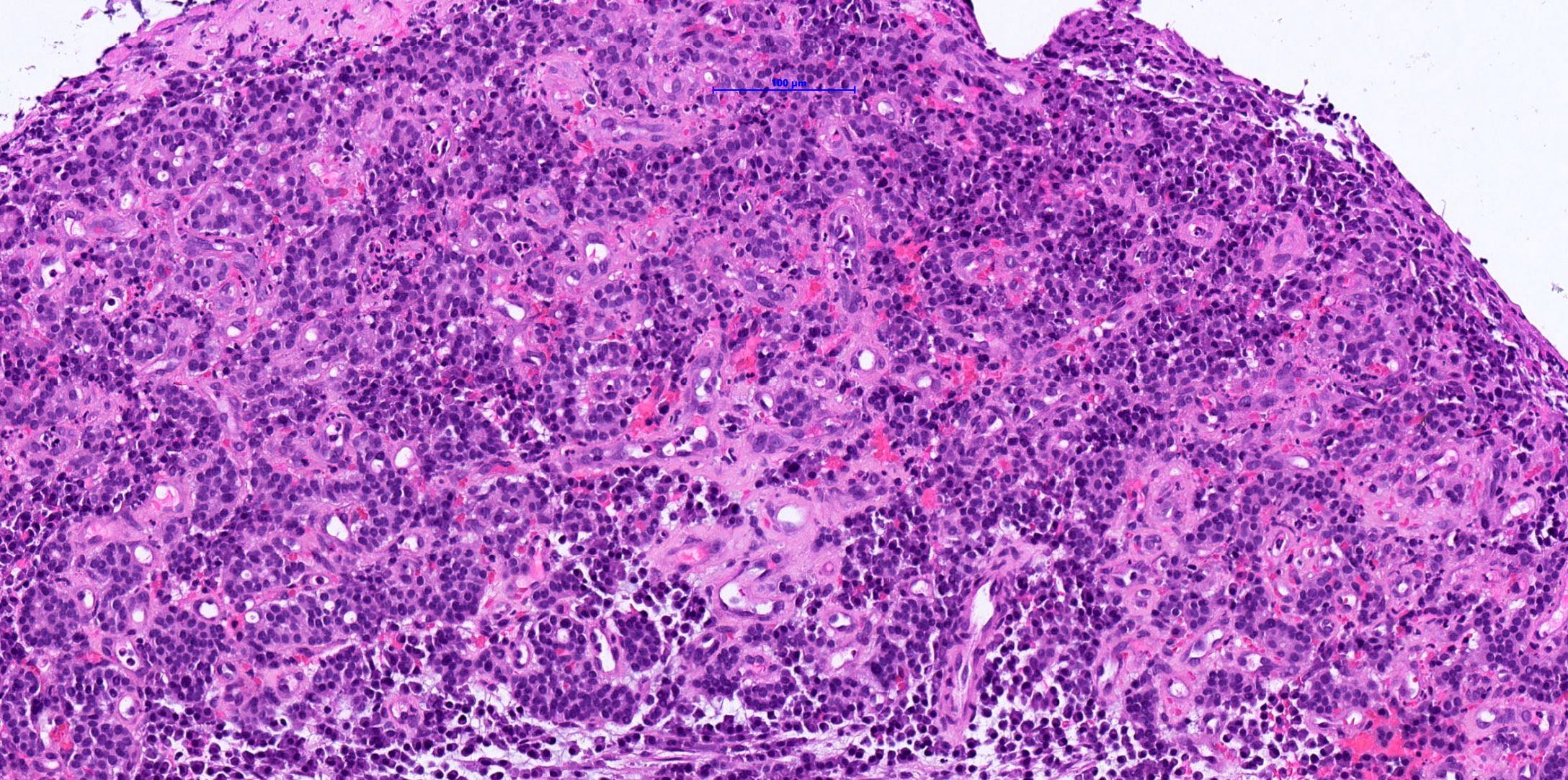
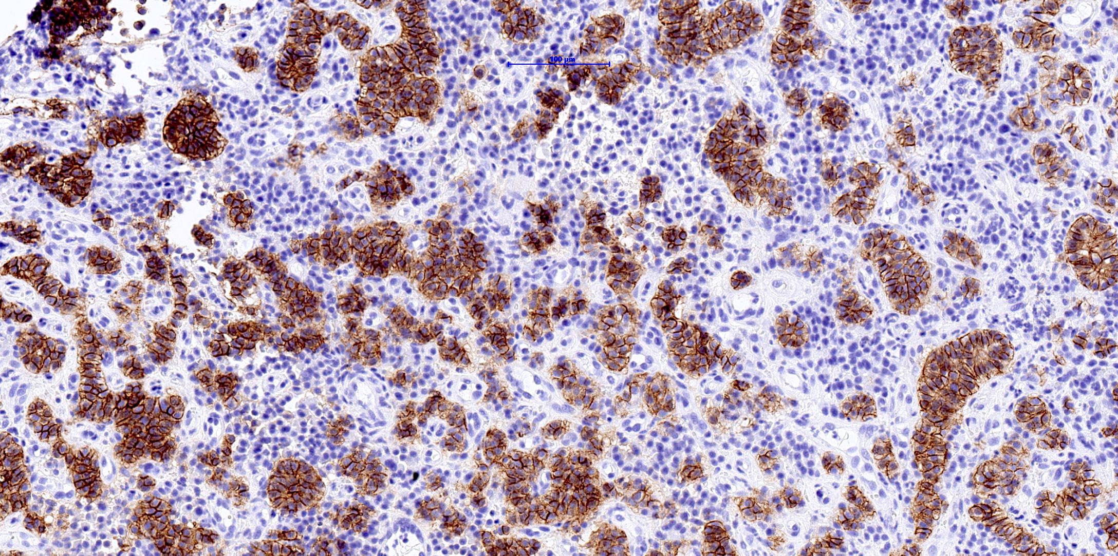
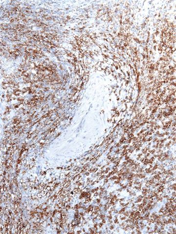
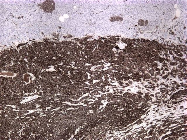
Microscopic (histologic) images
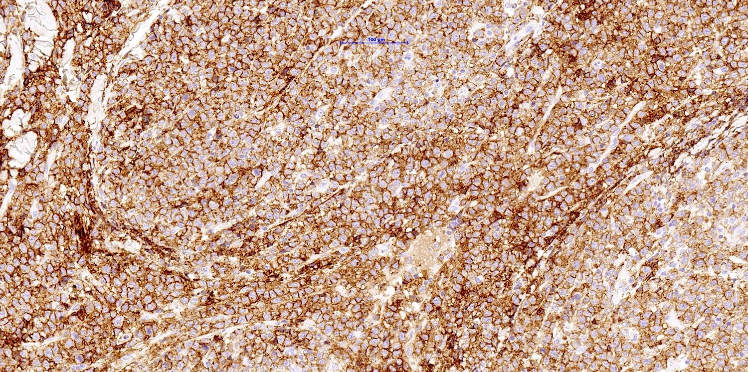
Positive staining - normal
- NK cells (80 - 90%), large granular lymphocytes, activated T cells, osteoblasts
- Cerebellum and cortex at neuromuscular junctions, neuroendocrine tissue, neurons (membranous pattern), glia
- Ovarian stromal cells
- Rete testis cells (clusters of sloughed cells in hydrocele and spermatocele specimens may mimic small cell carcinoma) (Hum Pathol 2010;41:88)
- Skeletal muscle
Positive staining - disease
- Hematopoietic disorders
- Acute myeloid leukemia (variable) (Am J Clin Pathol 2007;128:550, Am J Clin Pathol 2009;132:101)
- Blastic plasmacytoid dendritic cell neoplasm (Arch Pathol Lab Med 2007;131:149)
- Enteropathy associated T cell lymphoma (Am J Clin Pathol 2007;127:701, Am J Surg Pathol 2011;35:1557)
- Myeloid sarcoma (variable) (Am J Clin Pathol 2000;114:807)
- Myelofibrosis (Am J Clin Pathol 2010;133:314)
- Myeloma, NK / T cell lymphoma (Am J Clin Pathol 2008;130:343, Am J Surg Pathol 2011;35:1195)
- Pediatric anaplastic large cell lymphoma (some) (Arch Pathol Lab Med 2006;130:1859)
- Primary cutaneous gamma delta T cell lymphoma (Cureus 2023;15:e35442)
- Other
- Alveolar rhabdomyosarcoma (Hum Pathol 2009;40:341, Mod Pathol 2008;21:795)
- Cardiac ischemic damage (Verh Dtsch Ges Pathol 2004;88:246)
- Extrahepatic biliary atresia (Am J Surg Pathol 2003;27:1454)
- Meningiomas and pulmonary meningothelial-like nodules (Am J Surg Pathol 2009;33:487)
- Intrahepatic clear cell cholangiocarcinoma (Am J Surg Pathol 2007;31:902)
- Merkel cell carcinoma (J Dermatol Sci 2003;31:219)
- Mesotheliomas (some)
- Neuroblastoma (adult) (World Neurosurg 2022;159:e23)
- Merkel cell carcinoma (Mod Pathol 2007;20:1113)
- Neuroendocrine carcinomas (Am J Surg Pathol 2006;30:684, Am J Clin Pathol 2010;133:618)
- Ovarian fibroma, fibrothecoma, leiomyoma (Am J Surg Pathol 2008;32:884)
- Ovarian sex cord stromal tumors (Int J Gynecol Pathol 2021;40:315)
- Pancreatic neuroendocrine tumor and solid pseudopapillary tumor (usually) (Mod Pathol 2006;19:1409)
- Pheochromocytoma (World J Surg Oncol 2022;20:185)
- Small cell carcinomas of cervix, lung and prostate (Int J Gynecol Pathol 2005;24:113, Am J Surg Pathol 2006;30:705)
- Sustentacular cell tumor (Am J Surg Pathol 2006;30:268)
- Synovial sarcoma (usually) (Mod Pathol 2006;19:659)
- Thyroid tumors (various) (Am J Clin Pathol 2003;120:64, Am J Surg Pathol 2010;34:1582)
- Uterine plexiform tumor / tumorlet
- Wilms tumor (Am J Surg Pathol 2009;33:454)
Negative staining
- Normal: B cells, granulocytes, monocytes, plasma cells
- Acute lymphocytic leukemia (ALL), large granular NK cell lymphocytosis (some cases), peripheral T cell lymphoma, plasma cell leukemia, primitive neuroectodermal tumors (PNET) / Ewing sarcoma, subcutaneous panniculitis-like T cell lymphoma (Am J Pathol 2004;165:1117, Arch Pathol Lab Med 2009;133:303)
- Chronic myelomonocytic leukemia (occasionally positive) (Leukemia 1993;7:1570)
- Ossifying fibromyxoid tumor of soft parts (43% positive) (Am J Surg Pathol 2011;35:1615)
Sample pathology report
- Lower right lung lobe, mucosal biopsies:
- Atypical carcinoid / neuroendocrine tumor of the lung with associated areas of necrosis and portions of respiratory mucosa (see comment)
- Comment: In this case, a neuroendocrine tumor of the lung is present, which shows areas of necrosis. Although the mitotic count in this section is low, the presence of necrosis advocates the diagnosis of an atypicial carcinoid.
- The neoplastic cells are positive for CD56, synaptophysin, chromogranin A and express SSTR2.
Additional references
Practice question #1
Which of the following statements is true about CD56?
- It can differentiate MGUS from myeloma because MGUS plasma cells are CD56+
- It is a marker of B cells
- It is a marker of natural killer (NK) cells and NK lymphomas
- It is a marker of plasma cells
Practice answer #1
C. It is a marker of natural killer (NK) cells and NK lymphomas. Answers B and D are incorrect because these cells are typically negative for CD56. Answer A is incorrect because although it can differentiate MGUS from myeloma, MGUS plasma cells are CD56- and myeloma plasma cells are CD56+.
Comment Here
Reference: CD56
Comment Here
Reference: CD56