

Cervix
Inflammatory / infectious
Lactobacillus
Editorial Board Member: Ricardo R. Lastra, M.D.
Deputy Editor-in-Chief: Gulisa Turashvili, M.D., Ph.D.

Last author update: 7 June 2023
Last staff update: 7 June 2023
Copyright: 2003-2024, PathologyOutlines.com, Inc.
PubMed Search: Lactobacillus cervix

Table of Contents
Definition / general | Essential features | Terminology | ICD coding | Epidemiology | Sites | Pathophysiology | Clinical features | Laboratory | Treatment | Cytology description | Cytology images | Videos | Differential diagnosis | Additional references | Board review style question #1 | Board review style answer #1 | Board review style question #2 | Board review style answer #2Cite this page: Al Dallal H, Salih ZT. Lactobacillus. PathologyOutlines.com website. https://www.pathologyoutlines.com/topic/cervixcytologylactobacillus.html. Accessed April 26th, 2024.
Definition / general
- Lactobacillus spp. is a major component of normal vaginal flora
- Lactobacilli are gram positive rods
- They are beneficial because they produce lactic acid, which reduces the vaginal pH and possibly protects from infection by Candida and other pathogens
Essential features
- Lactobacillus spp. is normal vaginal flora that is commonly seen in cervical Pap smears
- Lactobacilli are blue thick rods usually found on the top of intermediate squamous cells
Terminology
- Also known as Döderlein bacilli or Bacillus vaginalis
ICD coding
- ICD-10: B96.89 - other specified bacterial agents as the cause of diseases classified elsewhere
Epidemiology
- Lactobacillus colonizes the mouth, intestine and vagina
- Lactobacillus is predominant during the second half of the menstrual period (luteal phase)
- They cannot thrive in alkaline media; menstrual flow increases vaginal PH and diminishes their growth (Sex Transm Dis 1990;17:51)
- Conditions commonly associated with Lactobacillus (Sex Transm Dis 1990;17:51)
- Pregnancy
- Using progestational drugs including oral contraceptives
- Diabetes
- Premenarchal girls have diminished growth of Lactobacillus due to alkaline vaginal PH (Pediatrics 1978;62:57)
Sites
- Lactobacillus is ubiquitous in the environment and colonizes plants and animals
- In humans, they colonize the mouth, intestine and vagina
Pathophysiology
- Lactobacilli metabolize the glycogen contained within exfoliated intermediate squamous cells which result in cytolysis
- This cellular pattern is commonly seen during the second (luteal) phase of the menstrual cycle
- They play a critical role in preventing illness, including bacterial vaginitis, yeast infection, sexually transmitted disease and even cancer (Microbes Infect 2002;4:319)
- Mechanism by which Lactobacillus prevents pathogens includes
- Blocking adhesion receptors
- Competing for nutrients
- Producing lactic acid by creating acidic vaginal media
Clinical features
- Excessive lactobacterial cytolysis can be associated with vaginosis-like symptoms (cytolytic vaginosis) and may cause (Diagn Cytopathol 2003;29:156)
- Vulvovaginal itching
- Burning
- Discharge
Laboratory
- Biochemical test
- Microbiology culture
- Microscopy: gram positive bacilli, usually seen on cervical Paps; however, are not reported because they are normal flora
Treatment
- No treatment is needed
Cytology description
- Lactobacilli are blue thick rods usually found on the top of the intermediate squamous cells
- They can lyse glycogen rich intermediate cells which may cause cytolysis
- Cytolysis is characterized by bare normal size intermediate cell nuclei, fragments of squamous cytoplasm and abundant bacterial rods
- Abundant cytolysis (> 50%) may be mentioned as quality indicator in Bethesda system but the specimen should not be regarded as unsatisfactory
Cytology images
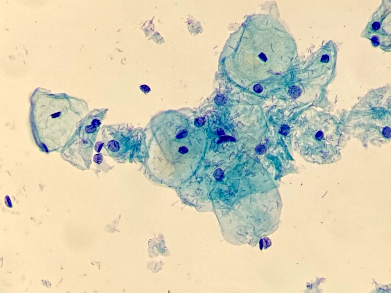
Contributed by Hiba Al Dallal, M.D.
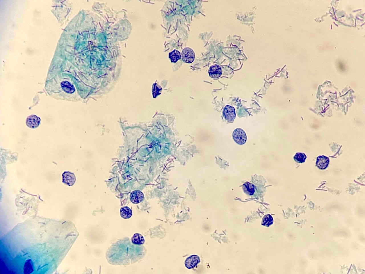
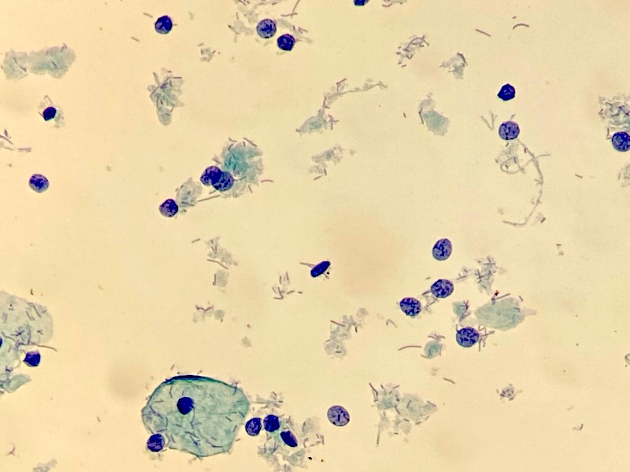
Cytolysis
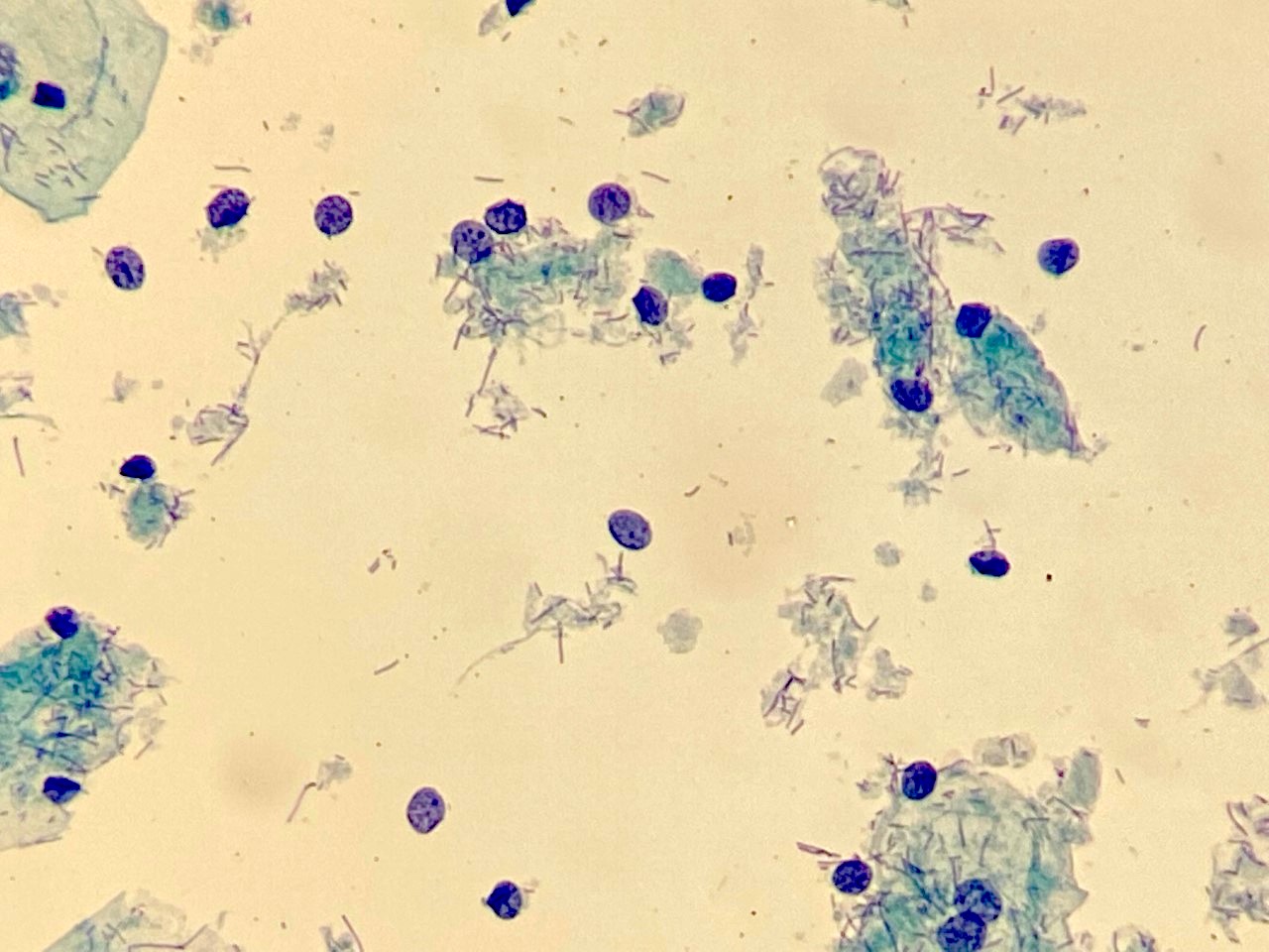
Cytolysis
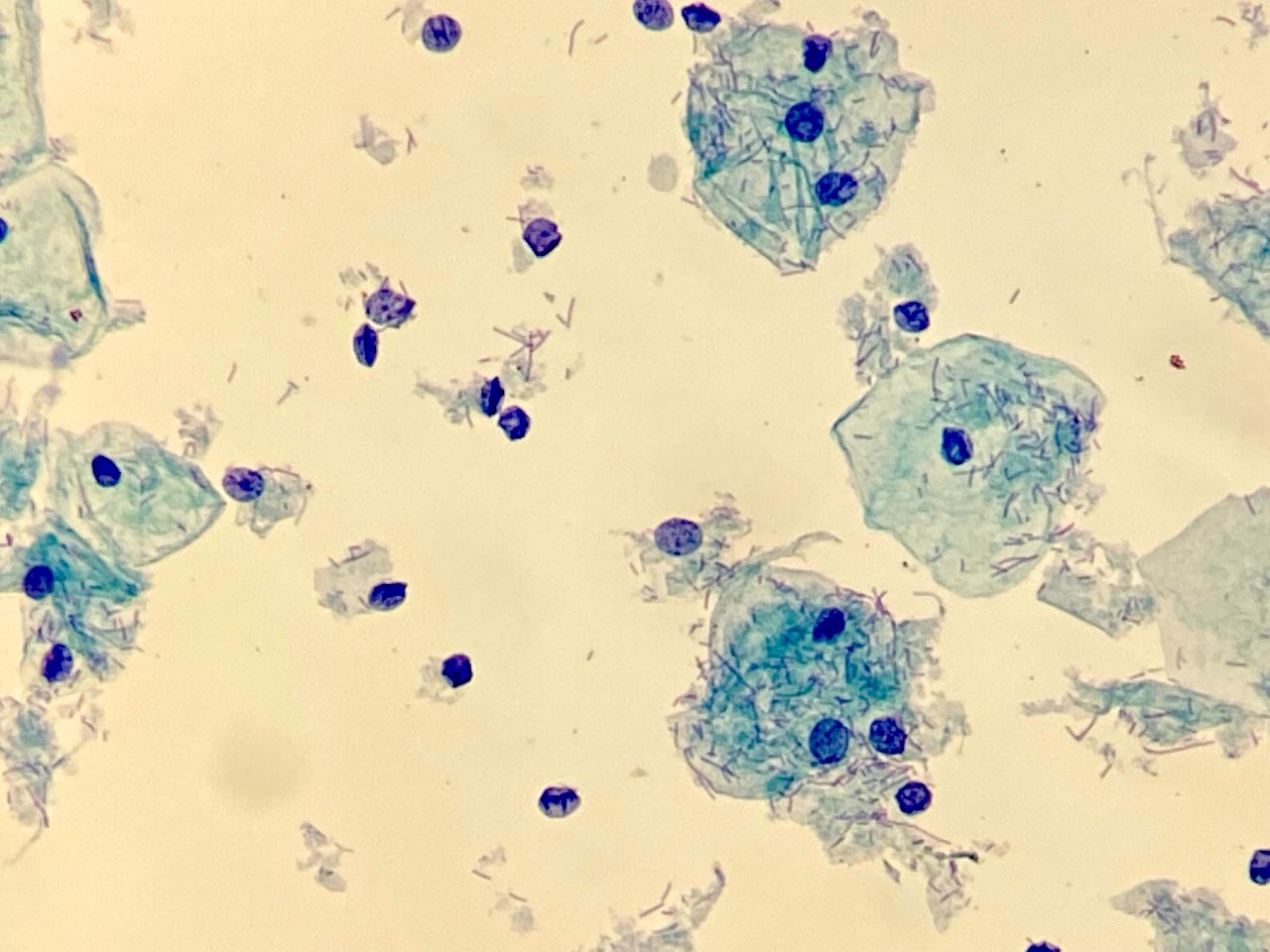
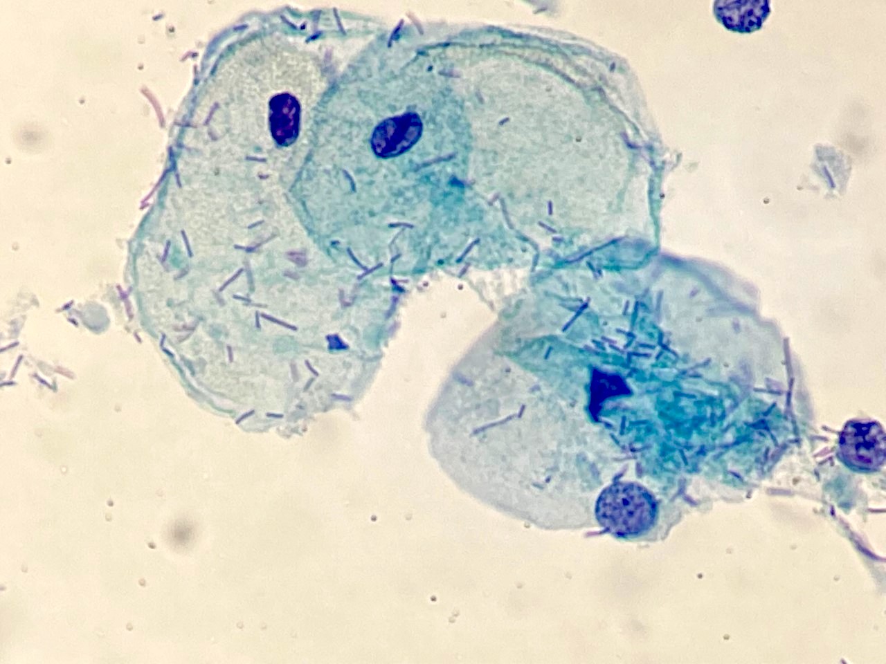
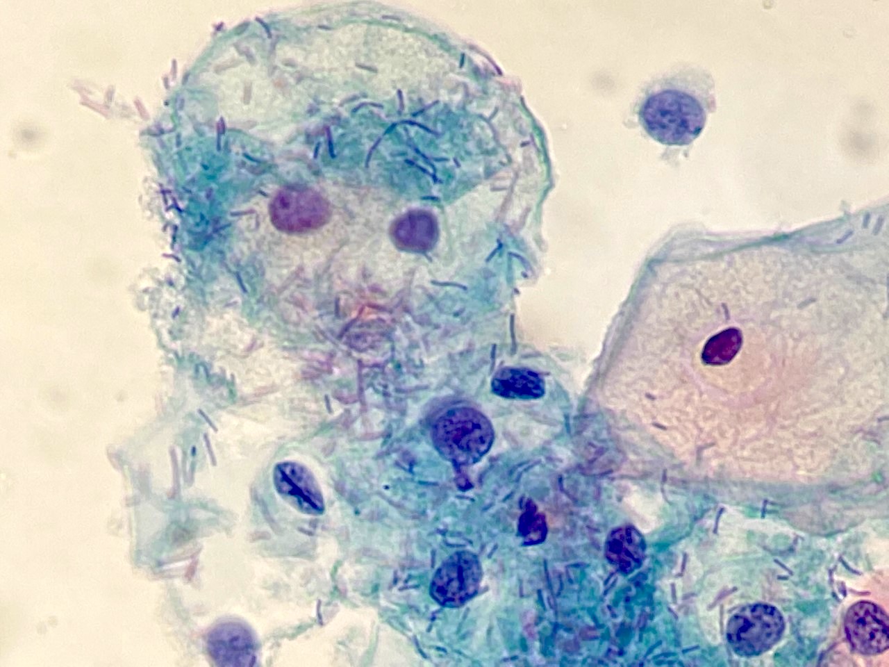
Lactobacillus on the top of squamous cells
Videos
Lactobacillus, normal vaginal bacterial rods
Differential diagnosis
- Bacterial vaginosis:
- Coccoid overgrowth over superficial cells, causing a bacterial haze
- Corynebacterium:
- Arranged as groups as opposed to short chains but difficult to differentiate by morphology in a cervical smear (Gray: Diagnostic Cytopathology, 3rd Edition, 2010)
- Leptothrix:
- Very long thread-like bacteria that may form loops
Additional references
Board review style question #1
Lactobacilli are normal vaginal flora that may cause which of the following?
- Cytolysis of intermediate squamous cells
- Decrease vaginal PH
- Dysplastic changes
- Overgrowth of other pathogens
Board review style answer #1
A. Cytolysis of intermediate squamous cells. Lactobacilli are able to use glycogen rich intermediate cells and are a normal finding that is commonly observed in the second half of the menstrual cycle as well as in pregnancy and diabetic women.
Answer B is incorrect because because Lactobacilli increase vaginal PH.
Answer C is incorrect because no dysplastic changes are seen.
Answer D is incorrect because no other pathogens / bacteria are seen.
Comment Here
Reference: Lactobacillus
Comment Here
Reference: Lactobacillus
Board review style question #2
Cytolysis is observed in > 50% of cells on the cervical Pap slide. How should a pathologist report it according to the Bethesda system?
- Lactobacilli should be reported as pathogenic
- The specimen is satisfactory but should be mentioned as quality indicator
- The specimen should be rejected
- The specimen should be reported as unsatisfactory
Board review style answer #2
B. The specimen is satisfactory but should be mentioned as quality indicator. Answer B is correct because the specimen is satisfactory but should be mentioned as a quality indicator. Abundant cytolysis (more than 50%) may be mentioned as a quality indicator in Bethesda system but the specimen should not be regarded as unsatisfactory. The specimen is satisfactory but should be mentioned as quality indicator. Answer A is incorrect because according to the Bethesda system, there is no need to report Lactobacilli as they are normal vaginal flora. Answer C is incorrect because the specimen should not be rejected according to the Bethesda system. Answer D is incorrect because the specimen should not be reported as unsatisfactory according to the Bethesda system.
Comment Here
Reference: Lactobacillus
Comment Here
Reference: Lactobacillus
